Embed Size (px)
Citation preview

AS Biology Unit 2 page 1
HGS Biology A-level notes NCM/11/12
AQA AS Biology Unit 2
Contents
Specification 2
Gas exchange 4
The Circulatory System 11
Haemoglobin 18
Plant Cells 21
Gas exchange in plants 24
Water Transport in Plants 26
Biodiversity 33
Intraspecific Diversity 34
Interspecific Diversity 39
DNA 43
DNA replication 46
Gene Expression 48
Chromosomes 51
Mitosis and the Cell Cycle 55
Meiosis and Sexual Reproduction 58
Antibiotic Resistance 61
Classification 65
Appendix 1 – Mathematical Requirements 74
Appendix 2 – The Unit 2 Exam 76
These notes may be used freely by A level biology students and teachers, and they may be copied and edited. I would be interested to hear of any
comments and corrections.
Neil C Millar ([email protected]) Head of Biology, Heckmondwike Grammar School,
High Street, Heckmondwike WF16 0AH

AS Biology Unit 2 page 2
HGS Biology A-level notes NCM/11/12
Biology Unit 2 Specification
Physiology
Surface Area to Volume Ratio The relationship between the size of an organism or structure and surface area to volume ratio. Explain the significance of the relationship between size and surface area to volume ratio for the exchange of substances and of heat. Gas Exchange Changes to body shape and the development of systems in larger organisms as adaptations that facilitate exchange as the ratio reduces. Use knowledge and understanding of the principles of diffusion to explain the adaptations of gas exchange surfaces. Gas exchange across the body surface of a single-celled organism; in the tracheal system of an insect (tracheae and spiracles); across the gills of a fish (gill lamellae and filaments including the countercurrent principle) and by leaves of dicotyledonous plants (mesophyll and stomata). Structural and functional compromises between the opposing needs for efficient gas exchange and the limitation of water loss shown by terrestrial insects.
The Circulatory system Over large distances, efficient supply of materials is provided by mass transport. The general pattern of blood circulation in a mammal. Names are only required of the coronary arteries and of blood vessels entering and leaving the heart, liver and kidneys. The structure of arteries, arterioles and veins in relation to their function. The structure of capillaries and their importance in metabolic exchange. The formation of tissue fluid and its return to the circulatory system. Haemoglobin and Oxygen Transport Haemoglobin is a protein with a quaternary structure. The role of haemoglobin in the transport of oxygen. The loading, transport and unloading of oxygen in relation to the oxygen dissociation curve. The effects of carbon dioxide concentration. The haemoglobins are a group of chemically similar molecules found in many different organisms. Different organisms possess different types of haemoglobin with different oxygen transporting properties. Relate these to the environment and way of life of the organism concerned. Plant Cells There are fundamental differences between plant cells and animal cells. The structure of a palisade cell from a leaf as seen with an optical microscope. The appearance, ultrastructure and function of cell wall and chloroplasts. Explain adaptations of other plant cells. Use an optical microscope to examine temporary mounts of plant cells, tissues or organs. Polysaccharides The structures of β-glucose and the linking of β-glucose by glycosidic bonds formed by condensation to form cellulose. The basic structure and functions of starch, glycogen and cellulose and the relationship of structure to function of these substances in animals and plants.
Water Transport in Plants The structure of a dicotyledonous root in relation to the pathway of water from root hairs through the cortex and endodermis to the xylem. Apoplastic and symplastic pathways. The roles of root pressure and cohesion tension in moving water through the xylem. Transpiration and the effects of light, temperature, humidity and air movement. Structural and functional compromises between the opposing needs for efficient gas exchange and the limitation of water loss shown by xerophytic plants. Measure the rate of water uptake by means of a simple potometer.
Biodiversity
Causes of Intraspecific Diversity Variation exists between members of a species. Similarities and differences between individuals within a species may be the result of genetic factors, differences in environmental factors, or a combination of both. Collect and analyse data relating to intraspecific variation. Analyse and interpret data relating to interspecific and intraspecific variation. Appreciate the tentative nature of any conclusions that can be drawn relating to the causes of variation. Loss of Genetic Diversity Similarities and differences between organisms may be defined in terms of variation in DNA. Differences in DNA lead to genetic diversity. The influence of the following on genetic diversity: selection for high-yielding breeds of domesticated animals and strains of plants; the founder effect; genetic bottlenecks. Discuss ethical issues involved in the selection of domesticated animals. Species Diversity Diversity may relate to the number of species present in a community. An index of diversity describes the relationship between the number of species and the number of individuals in a community. Calculation of an index of diversity from the formula d = N (N – 1) / Σ n (n – 1) where N = total number of organisms of all species and n = total number of organisms of each species. Calculate the index of diversity from suitable data. Loss of Species Diversity The influence of deforestation and the impact of agriculture on species diversity. Interpret data relating to the effects of human activity on species diversity and be able to evaluate associated benefits and risks. Discuss the ways in which society uses science to inform the making of decisions relating to biodiversity. Sampling The need for random sampling, and the importance of chance in contributing to differences between samples. The concept of normal distribution about a mean. Mean and standard deviation as measures of variation within a sample. Candidates will not be required to calculate standard deviation in questions on written papers.

AS Biology Unit 2 page 3
HGS Biology A-level notes NCM/11/12
Genetics
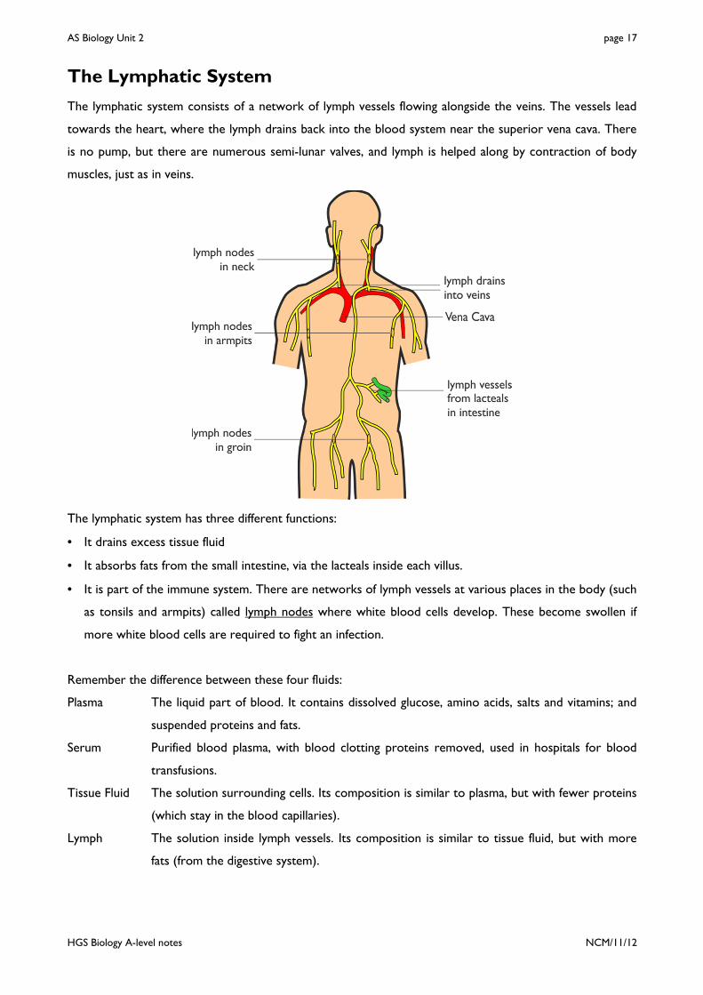
DNA Structure The double-helix structure of DNA enabling it to act as a stable information-carrying molecule, in terms of the components of DNA nucleotides: deoxyribose, phosphate and the bases adenine, cytosine, guanine and thymine; two sugar-phosphate backbones held together by hydrogen bonds between base pairs; specific base pairing. DNA is the genetic material in bacteria as well as in most other organisms. Analyse, interpret and evaluate data concerning early experimental work relating to the role and importance of DNA. DNA Replication The semi-conservative replication of DNA in terms of: breaking of hydrogen bonds between polynucleotide strands; attraction of new DNA nucleotides to exposed bases and base pairing; role of DNA helicase and of DNA polymerase. Gene Expression Genes are sections of DNA that contain coded information as a specific sequence of bases. Genes code for polypeptides that determine the nature and development of organisms. The base sequence of a gene determines the amino acid sequence in a polypeptide. A sequence of three bases, called a triplet, codes for a specific amino acid. In eukaryotes, much of the nuclear DNA does not code for polypeptides. There are, for example, introns within genes and multiple repeats between genes. Mutations Mutations are changes in DNA and result in different characteristics. Differences in base sequences of alleles of a single gene may result in non-functional proteins, including non-functional enzymes. Chromosomes A gene occupies a fixed position, called a locus, on a particular strand of DNA. In eukaryotes, DNA is linear and associated with proteins. In prokaryotes, DNA molecules are smaller, circular and are not associated with proteins. Mitosis and the Cell Cycle During mitosis, the parent cell divides to produce two daughter cells, each containing an exact copy of the DNA of the parent cell. DNA is replicated during interphase. Mitosis increases the cell number in this way in growth and tissue repair. Name and explain the events occurring during each stage of mitosis. Recognise the stages from drawings and photographs. Relate understanding of the cell cycle to cancer and its treatment. Cell Differentiation The cells of multicellular organisms may differentiate and become adapted for specific functions. Tissues as aggregations of similar cells, and organs as aggregations of tissues performing specific physiological functions. Organs are organised into systems. Meiosis and Sexual Reproduction The importance of meiosis in producing cells which are genetically different. Meiosis only in sufficient detail to show the formation of haploid cells; independent segregation of homologous chromosomes; and genetic recombination by crossing over. Gametes are genetically different as a result of
different combinations of maternal and paternal chromosomes. Antibiotics and Resistance Antibiotics may be used to treat bacterial disease. One way in which antibiotics function is by preventing the formation of bacterial cell walls, resulting in osmotic lysis. Mutations in bacteria may result in resistance to antibiotics. Resistance to antibiotics may be passed to subsequent generations by vertical gene transmission. Resistance may also be passed from one species to another when DNA is transferred during conjugation. This is horizontal gene transmission. Antibiotic resistance in terms of the difficulty of treating tuberculosis and MRSA. Apply the concepts of adaptation and selection to other examples of antibiotic resistance. Evaluate methodology, evidence and data relating to antibiotic resistance. Discuss ethical issues associated with the use of antibiotics. Discuss the ways in which society uses scientific knowledge relating to antibiotic resistance to inform decision-making. Classification A species may be defined in terms of observable similarities and the ability to produce fertile offspring. Candidates should appreciate the difficulties of defining species and the tentative nature of classifying organisms as distinct species.
The principles and importance of taxonomy. Classification systems consist of a hierarchy in which groups are contained within larger composite groups and there is no overlap. One hierarchy comprises Kingdom, Phylum, Class, Order, Family, Genus, Species. The phylogenetic groups are based on patterns of evolutionary history. Originally classification systems were based on observable features but more recent approaches draw on a wider range of evidence to clarify relationships between organisms. Genetic comparisons can be made between different species by direct examination of their DNA or of the proteins encoded by this DNA.
• Comparison of DNA base sequences is used to elucidate relationships between organisms. These comparisons have led to new classification systems in plants. Similarities in DNA may be determined by DNA hybridisation.
• Comparisons of amino acid sequences in specific proteins can be used to elucidate relationships between organisms. Immunological comparisons may be used to compare variations in specific proteins.
Interpret data relating to similarities and differences in base sequences in DNA and in amino acid sequences in proteins to suggest relationships between different organisms. The role of courtship in species recognition. Courtship behaviour as a necessary precursor to successful mating.

AS Biology Unit 2 page 4
HGS Biology A-level notes NCM/11/12
Gas Exchange in Organisms
All organisms need to exchange oxygen and carbon dioxide with their surroundings for respiration (or in
plants for photosynthesis). These gases diffuse between the organism and the surroundings. From Fick's law
we know that:
distance
difference ionconcentratarea surfaceDiffusion of Rate
×∝
So the rate of exchange of gases therefore depends on the organism's surface area that is in contact with
the surroundings. The requirements for respiration depends on the mass or volume of the organism, so the
ability to meet the requirements depends on (surface area ÷ volume), which is known as the
surface area : volume ratio. As organisms get bigger their volume and surface area both get bigger, but not
by the same amount. This can be seen by performing some simple calculations concerning different-sized
organisms. In these calculations each organism is assumed to be cube-shaped to make the calculations
easier. The surface area of a cube with length of side L is 6L², while the volume is L³.
organism length SA (m²) vol (m³) SA:vol ratio (m-1)
bacterium 1 µm (10-6 m) 6 x 10-12 10-18 6,000,000:1 amoeba 100 µm (10-4 m) 6 x 10⁻⁸ 10-12 60,000:1 bee 10 mm (10-2 m) 6 x 10⁻⁴ 10-6 600:1 pig 1 m (100 m) 6 x 100 100 6:1 whale 100 m (102 m) 6 x 10⁴ 106 0.06:1
So as organisms get bigger their surface area : volume ratio gets smaller. A bacterium is all surface with not
much inside, while a whale is all insides with not much surface. This means that as organisms become bigger
it becomes more difficult for them to exchange materials with their surroundings. In fact this problem sets
a limit on the maximum size for a single cell of about 100µm. In anything larger than this materials simply
cannot diffuse fast enough to support the reactions needed for life. Very large single cells like birds' eggs are
mostly inert food storage with a thin layer of living cytoplasm round the outside.
Organisms much larger than 100µm have to be multicellular, which means that their bodies are composed
of many small cells, rather than one big cell. Each cell in a multicellular organism is no bigger than about
30µm, and so can exchange materials quickly and independently. Each human contains about 1014 cells.
Large organisms therefore need specialised exchange systems with a large surface area. These systems
include lungs, gills, intestines, roots and leaves.

AS Biology Unit 2 page 5
HGS Biology A-level notes NCM/11/12
Heat Exchange
Organisms also need to exchange heat with their surroundings, and here large animals have an advantage in
having a small surface area : volume ratio: they lose less heat than small animals. Large mammals keep warm
quite easily and don't need much insulation or heat generation. Small mammals and birds lose their heat
very readily, so need a high metabolic rate in order to keep generating heat, as well as thick insulation. So
large mammals can feed once every few days, while small mammals must feed continuously. Human babies
also lose heat more quickly than adults, which is why they need woolly hats.
Diffusion and Mass Flow
In unit 1 we saw how materials moved across cell membranes; and we saw that there were basically two
methods: diffusion and active transport. In unit 2 we shall look at how materials move over larger distances
inside living organisms. Again there are basically two methods: diffusion and mass flow.
1. In diffusion solutes move in a random direction due to their thermal energy. Diffusion does not require
any energy (other than the thermal energy of the surroundings), so it is referred to as a passive process.
If there is a concentration difference between two places then the random movement results in the
substance diffusing down its concentration gradient from a high to a low concentration. Diffusion is very
slow and is only useful over small distances (< 100 µm). It cannot be used to move substances over
large distances in living organisms.
2. In mass flow a fluid (liquid or gas) moves in a particular direction due to a force. In living organisms this
usually means the bulk movement of water (the solvent) together with all its dissolved solutes and
suspended objects. So mass flow is like a river carrying everything with it. Mass flow always requires a
source of energy to pump the fluid, but it has the advantage of being much faster than diffusion,
especially over large distances. Mass flow is completely independent of concentration differences.
Examples of mass flow include: circulatory systems in animals, xylem and phloem systems in plants, filter
feeder currents, and ventilation.

AS Biology Unit 2 page 6
HGS Biology A-level notes NCM/11/12
Gas Exchange in Small Organisms
Small organisms don't have specialised gas exchange systems like lungs or gills, but instead simply exchange
gases through the surface of their bodies. To maximise their rate of gas exchange they have developed
particular body shapes to increase their surface area : volume ratio. Compared to larger, more active
vertebrates, most invertebrates also have relatively low metabolic rates, so don't need a fast rate of gas
exchange.
Single-celled Organisms
Microscopic single-celled organisms, like bacteria or Amoeba, have
a large surface area : volume ratio, so they can exchange gases
quickly directly though their cell surface.
Sponges – Hollow Body
Sponges are the simplest of all animals and are all marine. Their
tube-shaped bodies can grow quite large (50 mm in diameter).
Sponges increase their surface area : volume ratio by being hollow,
with thin walls only a few cells thick. Beating flagella maintain a flow
of water through the body cavity.
Tapeworms – Flattened Body
Tapeworms are parasites that live in the digestive systems of many
animals including humans. They can be very long. Tapeworms
increase their surface area : volume ratio by having flattened
bodies, typically only 0.2 mm thick. This also decreases the
diffusion distance. Tapeworms are sedentary and have an
extremely low metabolic rate.
Earthworms – Circulatory System
Earthworms can grow to be several mm in diameter, but most of
this is the worm's gut, with the tissues taking up a thin layer on the
outside. This layer is still too thick for diffusion, so earthworms
have developed a rudimentary circulatory system (containing
haemoglobin) to carry gases between the body surface and the
underlying tissues.

AS Biology Unit 2 page 7
HGS Biology A-level notes NCM/11/12
Gas Exchange in Insects
Insects are fairly small, but they are also very active, so they need to respire quickly. They have a rigid
exoskeleton, which is waterproof to prevent the insects drying out, but it also prevents gas exchange.
Insects increase their rate of gas exchange by having openings in the exoskeleton called spiracles, which
lead to a network of tubes called tracheae, which branch into many smaller tracheoles that carry air
directly to the cells. These tracheae and tracheoles are held open by rings of hard chitin (a polysaccharide).
The tracheoles penetrate deep into the insects tissues, carrying air quickly and directly to every cell. At the
ends of the tracheoles oxygen diffuses directly into the cells, and carbon dioxide diffuses out, down their
concentration gradients.
When the insect is at rest, water diffuses out of its cells into the ends of the tracheoles, just as it does in
human alveoli. This reduces the surface area in contact with the cells and reduces the rate of diffusion. But
when insects are flying their muscle cells produce lactic acid, which lowers the water potential in the cells,
so the water diffuses by osmosis from the tracheoles into the muscle cells. This makes diffusion of oxygen
much faster, so actively-respiring cells automatically get oxygen quicker.
Some larger insects, like houseflies and grasshoppers, ventilate their tracheal system by using muscles to
squeeze the trachea and so suck air in and out. This increases their rate of gas exchange. To counteract
problems of water loss some insects have hairs around the spiracles, and some can close their spiracles
when they are inactive.

AS Biology Unit 2 page 8
HGS Biology A-level notes NCM/11/12
Gas Exchange in Fish
Gas exchange is more difficult for fish than for mammals because the concentration of dissolved oxygen in
water is less than 1%, compared to 20% in air. Fish have developed specialised gas-exchange organs called
gills, which are composed of thousands of filaments. The filaments in turn are covered in feathery lamellae
each only a few cells thick containing blood capillaries. This structure gives a large surface area and a short
distance for gas exchange.
Water flows over the filaments and lamellae, and oxygen can diffuse down its concentration gradient the
short distance between water and blood. Carbon dioxide diffuses the opposite way down its concentration
gradient. The gills are covered by muscular flaps called opercula on the side of a fish's head. The gills are so
thin that they cannot support themselves without water, so if a fish is taken out of water the gills collapse
and the fish suffocates.
Ventilation in Fish
Fish ventilate their gills with sea water to maintain the gas concentration gradient. But, unlike humans, fish
ventilation is one-way rather than tidal. Water enters through the mouth but exits through the opercula
valves. This one-way ventilation is necessary because water is denser and more viscous than air, so it would
take too much energy to change its momentum every breath. Some fish (like tuna, mackerels and
anchovies) swim constantly with their mouths open, using their swimming movement to ventilate their gills,
but most fish use their mouth muscles for ventilation, which means they can ventilate even when not
swimming.

AS Biology Unit 2 page 9
HGS Biology A-level notes NCM/11/12
Inspiration
1. The mouth opens.
2. The muscles in the mouth contract, lowering the
floor of the mouth, and the opercula muscles
contract, pushing the opercula outwards.
3. This increases the volume of the buccal cavity
and the opercular cavity.
4. This decreases the pressure of water in the
buccal cavity below the outside water pressure.
5. The outside water pressure closes the opercular
valve.
6. Water flows in through the open mouth and
over the gills from high pressure to low pressure.
Expiration
1. The mouth closes.
2. The mouth and opercular muscles relax, raising
the floor of the buccal cavity.
3. This decreases the volume of the buccal cavity.
4. This increases the pressure of water in the
buccal cavity above the outside water pressure.
5. This pressure forces the opercula valves open.
6. Water flows out over the gills and through the
opercula valve from high pressure to low
pressure.
These pressure changes are shown in this graph. The rule is that water always flows from a high pressure
to a low pressure. This graph shows that water flows in one direction only.

AS Biology Unit 2 page 10
HGS Biology A-level notes NCM/11/12
Counter Current Exchange
Because fish have a one-way flow, they can make use of another trick to improve their efficiency of gas
exchange: a counter current system. If water and blood flowed past each other in the same direction
(parallel or concurrent flow) then the oxygen concentration in the water and blood quickly becomes the
same, so no further diffusion can take place, and only 50% of the oxygen can be extracted from the water:
In the countercurrent system the blood flows towards the front of the fish in the gill lamellae while the
water flows towards the back. This means that there is always a higher concentration of oxygen in the
water than in the blood, so oxygen continues to diffuse into the blood along the whole length of the
lamellae. Using this system fish gills can extract about 80% of the dissolved oxygen from the water:

AS Biology Unit 2 page 11
HGS Biology A-level notes NCM/11/12
Human Circulatory System
Humans have a double circulatory system with a 4-chambered heart. In humans the right side of the heart
pumps blood to the lungs only and is called the pulmonary circulation, while the left side of the heart
pumps blood to the rest of the body – the systemic circulation.
The circulation of blood round the body was first observed by Ibn-Al-Nafis (1213-1288) in Cairo and
independently rediscovered by William Harvey in England in 1628. Until then people assumed that blood
ebbed and flowed through the same tubes, because they hadn't seen capillaries. This diagram illustrates the
blood vessels to the main organs. The underlined vessels are listed in the specification.

AS Biology Unit 2 page 12
HGS Biology A-level notes NCM/11/12
Blood Vessels
Blood circulates in a series of different kinds of blood vessels as it circulates round the body.
Heart � Aorta � Arteries � Arterioles � Capillaries � Venules � Veins � Vena Cava � Heart
The purpose of these different vessels is to deliver blood to capillary beds, where substances are
exchanged between cells and blood. No cell in the body is more than 100µm away from a capillary.
Each kind of vessel is adapted to its function.
Arteries carry blood from the heart to every tissue in the
body. They continually branch into smaller and smaller vessels.
Arteries have thick walls (over 100 cells thick) composed mainly
of elastic tissue allowing the artery to expand without bursting
and so withstand the high pressure of blood from the heart. The
arteries close to the heart are particularly elastic and expand
during systole and recoil again during diastole, helping to even
out the pulsating blood flow.
Arterioles are the smallest arteries. Each arteriole leads to one
capillary bed. Arterioles have thinner walls (about 10 cells thick),
composed mainly of smooth muscle tissue to regulate the blood
flow to the capillary bed. The muscles can contract
(vasoconstriction) to close off the capillary beds; or relax
(vasodilation) to open up the capillary bed. These changes are
happening constantly under the involuntary control of the
medulla in the brain, and are most obvious in the capillary beds
of the skin, causing the skin to change colour from pink (skin
arterioles dilated) to blue (skin arterioles constricted). There is
not enough blood to fill all the body’s capillaries, and at any
given time up to 20% of the body’s capillary beds are closed off.

AS Biology Unit 2 page 13
HGS Biology A-level notes NCM/11/12
Capillaries are where the transported substances actually
enter and leave the blood. Capillaries are very narrow and their
walls are composed of single squamous endothelial cells with
gaps between them, making capillaries very permeable. There
are a vast number of capillaries (108 m in one adult!), so they
have a huge surface area : volume ratio, helping the rapid
diffusion of substances between blood and cells.
Veins carry blood from every tissue in the body to the heart.
The smallest veins, called venules, collect the blood from
capillary beds and feed into larger veins. The blood has lost
almost all its pressure in the capillaries, so it is at low pressure
inside veins and is moving slowly. Veins therefore don’t need
thick walls and they have a larger lumen than arteries, to reduce
the resistance to flow. They also have semi-lunar valves to stop
the blood flowing backwards. It is particularly difficult for blood
to flow upwards through the legs to heart, and the flow is
helped by contractions of the leg and abdominal muscles:
The body relies on constant contraction of these muscles to get the blood back to the heart, and this
explains why soldiers standing still on parade for long periods can faint, and why sitting still on a long flight
can cause swelling of the ankles and Deep Vein Thrombosis (DVT or “economy class syndrome”), where
small blood clots collect in the legs.
Note the correct words:
Muscles contract and relax
Elastic tissues stretch and recoil
Tubes constrict and dilate

AS Biology Unit 2 page 14
HGS Biology A-level notes NCM/11/12
Summary of Different Blood vessels
Arteries Arterioles Capillaries Veins
Function is to carry blood from the heart to the tissues
Function is to carry blood from arteries to one capillary bed
Function is to allow exchange of materials between the blood and the tissues
Function is to carry blood from tissues to the heart
Thick walls with elastic layers to resist high pressure
Thick walls with smooth muscle to control flow to capillary bed
Very thin, permeable walls, only one cell thick to allow exchange of materials
Thin walls, mainly collagen, since blood at low pressure
Small lumen Small lumen Very small lumen. Blood cells must distort to pass through.
Large lumen to reduce resistance to flow
No valves (except in heart)
No valves No valves Many valves to prevent back-flow
Blood at high pressure Blood pressure falls Blood pressure falls Blood at low pressure
Blood usually oxygenated (except in pulmonary circulation)
Blood usually oxygenated (except in pulmonary circulation)
Blood changes from oxygenated to deoxygenated (except in pulmonary circulation)
Blood usually deoxygenated (except in pulmonary circulation)

AS Biology Unit 2 page 15
HGS Biology A-level notes NCM/11/12
This diagram shows some of the changes that take place as the blood flows round the body.

AS Biology Unit 2 page 16
HGS Biology A-level notes NCM/11/12
Tissue Fluid
No exchange of materials takes place in the arteries and veins, whose walls are too thick and impermeable.
Substances are all exchanged between the blood and the cells in capillary beds, but they do not actually
move directly between the blood and the cell: they first diffuse into the tissue fluid that surrounds all cells,
and then diffuse from there to the cells.
1. At the arterial end of the capillary bed the blood is still at high pressure, so blood plasma is forced out
through the permeable walls of the capillary. Cells and proteins are too big to leave the capillary, so they
remain in the blood. So tissue fluid is formed by pressure filtration, not diffusion.
2. This fluid now forms tissue fluid surrounding the cells. Materials are exchanged between the tissue fluid
and the cells by all four methods of transport across a cell membrane.
• gases and lipid-soluble substances (such as steroids) cross by lipid diffusion;
• water crosses by osmosis;
• ions cross by facilitated diffusion;
• glucose and amino acids cross by active transport.
3. At the venous end of the capillary bed the blood is at low pressure, since it has lost so much plasma.
The blood and tissue fluid are now at around the same pressure, so tissue fluid returns by diffusion, not
mass flow.
• Solutes (such as carbon dioxide, urea, salts, etc.) enter the blood by diffusion, down their
concentration gradients.
• Water returns to the blood by osmosis down its water potential gradient. The blood has lost a lot
of water but retained soluble proteins, so has a low water potential.
4. Not all the fluid that left the blood returns to it, so there is excess tissue fluid. This excess drains into
lymph vessels, which are found in all capillary beds. Lymph vessels have very thin walls, like capillaries,
and tissue fluid can easily diffuse inside, forming lymph.
AS Biology Unit 2 page 17
HGS Biology A-level notes NCM/11/12
The Lymphatic System
The lymphatic system consists of a network of lymph vessels flowing alongside the veins. The vessels lead
towards the heart, where the lymph drains back into the blood system near the superior vena cava. There
is no pump, but there are numerous semi-lunar valves, and lymph is helped along by contraction of body
muscles, just as in veins.
The lymphatic system has three different functions:
• It drains excess tissue fluid
• It absorbs fats from the small intestine, via the lacteals inside each villus.
• It is part of the immune system. There are networks of lymph vessels at various places in the body (such
as tonsils and armpits) called lymph nodes where white blood cells develop. These become swollen if
more white blood cells are required to fight an infection.
Remember the difference between these four fluids:
Plasma The liquid part of blood. It contains dissolved glucose, amino acids, salts and vitamins; and
suspended proteins and fats.
Serum Purified blood plasma, with blood clotting proteins removed, used in hospitals for blood
transfusions.
Tissue Fluid The solution surrounding cells. Its composition is similar to plasma, but with fewer proteins
(which stay in the blood capillaries).
Lymph The solution inside lymph vessels. Its composition is similar to tissue fluid, but with more
fats (from the digestive system).

AS Biology Unit 2 page 18
HGS Biology A-level notes NCM/11/12
Transport of Oxygen
Oxygen is carried in red blood cells bound to the protein haemoglobin. A red blood cell contains about
300 million haemoglobin molecules and there are 5 million red blood cells per cm³ of blood. The result of
this is that blood can carry up to 20% oxygen, whereas pure water can only carry 1%. The haemoglobin
molecule consists of four polypeptide chains, with a haem prosthetic group at the centre of each chain.
Each haem group contains one iron atom, and one oxygen molecule binds to each iron atom. So one
haemoglobin molecule can bind up to four oxygen molecules. This means there are 4 binding steps, as
shown in this chemical equation:
A sample of blood can therefore be in any state from completely deoxygenated (0% saturated) to fully
oxygenated (100% saturated). Since deoxyhaemoglobin and oxyhaemoglobin are different colours, it is easy
to measure the % saturation of a sample of blood in a colorimeter. As the chemical equation shows, oxygen
drives the reaction to the right, so the more oxygen there is in the surroundings, the more saturated the
haemoglobin will be. This relation is shown in the oxygen dissociation curve:
The concentration of oxygen in the
surroundings can be measured as a
% (there’s about 20% oxygen in air),
but it’s more correct to measure it
as a partial pressure (PO2,
measured in kPa). Luckily, since the
pressure of one atmosphere is
about 100 kPa, the actual values for
PO2 and %O2 are the same (e.g.
12% O2 has a PO2 of 12 kPa).

AS Biology Unit 2 page 19
HGS Biology A-level notes NCM/11/12
The graph is read by starting with an oxygen concentration in the environment surrounding the blood
capillaries on the horizontal axis, then reading off the state of the haemoglobin in the blood that results
from the vertical axis.
This curve has an S (or sigmoid) shape, and shows several features that help in the transport of oxygen in
the blood:
• In the alveoli of the lungs oxygen is constantly being brought in by ventilation, so its concentration is
kept high, at around 14 kPa. As blood passes through the capillaries surrounding the alveoli the
haemoglobin binds oxygen to become almost 100% saturated (point a). Even if the alveolar oxygen
concentration falls a little the haemoglobin stays saturated because the curve is flat here.
• In tissues like muscle, liver or brain, oxygen is used by respiration, so its concentration is low, typically
about 4 kPa. At this PO2 the haemoglobin is only 50% saturated (point b), so it unloads about half its
oxygen (i.e. from about 100% saturated to about 50% saturated) to the cells, which use it for
respiration.
• In tissues that are respiring quickly, such as contracting muscle cells, the PO2 drops even lower, to about
2 kPa, so the haemoglobin saturation drops to about 10% (point c), so almost 90% of the oxygen is
unloaded, providing more oxygen for the muscle cells.
• Actively-respiring tissues also produce a lot of CO2, which dissolves in tissue fluid to make carbonic acid
and so lowers the pH. The chemical equation on the previous page shows that hydrogen ions drive the
reaction towards the deoxyhaemoglobin state, so low pH reduces the % saturation of haemoglobin at
any PO2. This is shown on the graph by the dotted line, which is lower than the normal dissociation
curve. This downward shift is called the Bohr effect, after the Danish scientist who first discovered it. So
at a PO2 of 2kPa, the actual saturation is nearer 5% (point d), so 95% of the oxygen loaded in the lungs is
unloaded in respiring tissues.
Remember that oxygen can only diffuse in and out of the blood from capillaries, which are permeable.
Blood in arteries and veins is “sealed in”, so no oxygen can enter or leave the blood whatever the
conditions surrounding the blood vessel. So as haemoglobin travels from the lungs to a capillary bed in a
body tissue and back to the lungs, it “switches” from one position on the dissociation curve to another
position, without experiencing the intermediate stages of the curve.

AS Biology Unit 2 page 20
HGS Biology A-level notes NCM/11/12
Different Haemoglobins
Different animals possess different types of haemoglobin with different oxygen transporting properties.
These properties are related to the animal’s way of life, so they are an adaptation that helps the animal
survive in its environment.
A human fetus obtains its oxygen from the placenta not the
lungs. In the placenta the mother’s and fetus’s capillary beds are
intertwined (but the bloods don’t mix). Fetal haemoglobin is a
different kind from the “adult” form, with a higher affinity for
oxygen at low partial pressures, so its oxygen dissociation
curve is shifted up. So this different haemoglobin allows oxygen
to diffuse from the mother’s blood to the fetus, yet still be
unloaded in the fetal tissues. Fetal haemoglobin is gradually
replaced by “adult” haemoglobin during the first year after
birth.
Lugworms live in the mud in estuaries and seashores. When
the tide is out the lugworm stays in a burrow filled with sea
water. But the oxygen concentration in this burrow can fall
very low as the lugworm respires, so the lugworm has
haemoglobin with a very high affinity for oxygen: its oxygen
dissociation curve is shifted up. This allows the lugworm to
obtain oxygen even when the PO2 is as low as 2kPa.
Mice lose heat very quickly due to their large surface area :
volume ratio, so they have a high metabolic rate to generate
more heat. Their tissues therefore have a constant demand for
oxygen for respiration. The oxygen dissociation curve for
mouse haemoglobin is shifted down compared to humans, so
plenty of oxygen is unloaded to all tissues all the time.

AS Biology Unit 2 page 21
HGS Biology A-level notes NCM/11/12
Plant Cells
The next section is on exchange and transport systems in plants. Plant cells contain a number of organelles
not found in animal cells.
• Chloroplasts. Bigger and fatter than mitochondria,
chloroplasts are where photosynthesis takes place, so are
only found in photosynthetic organisms (plants and algae).
Like mitochondria they are enclosed by a double membrane,
but chloroplasts also have a third membrane called the
thylakoid membrane. The thylakoid membrane is folded into
thylakoid disks, which are then stacked into piles called grana.
The space between the inner membrane and the thylakoid is
called the stroma. The thylakoid membrane contains
chlorophyll and chloroplasts also contain starch grains,
ribosomes and circular DNA.
• Vacuoles. These are membrane-bound sacs containing water
or dilute solutions of salts and other solutes. Most cells can
have small vacuoles that are formed as required, but plant
cells usually have one very large permanent vacuole that fills
most of the cell, so that the cytoplasm (and everything else)
forms a thin layer round the outside. Plant cell vacuoles are
filled with cell sap, and are very important in keeping the cell
rigid, or turgid. Some unicellular protoctists have feeding
vacuoles for digesting food, or contractile vacuoles for
expelling water.
• Cell Wall. This is a thick layer outside the cell membrane
used to give a cell strength and rigidity. Cell walls consist of a
network of fibres, which give strength but are freely
permeable to solutes (unlike membranes). A wickerwork
basket is a good analogy. Plant cell walls are made mainly of
cellulose, but can also contain hemicellulose, pectin, lignin and
other polysaccharides. There are often channels through
plant cell walls called plasmodesmata, which link the
cytoplasms of adjacent cells. Fungal cell walls are made of
chitin.
stalked particles(ATP synthase)
outer membrane
inner membrane
thylakoidmembrane
granum(thylakoid stack)
stroma
starch grain
cell wall
cell membrane
cytoplasm
tonoplastmembrane
permanentvaluolenucleus
cell wall
cell membrane
cytoplasm
middlelamellaplasmodesmata

AS Biology Unit 2 page 22
HGS Biology A-level notes NCM/11/12
Polysaccharides
In unit 1 we looked at monosaccharides and disaccharides. Here we look at polysaccharides.
Polysaccharides are long chains of many monosaccharides joined together by glycosidic bonds. There are
three important polysaccharides:
1. Starch is the plant storage polysaccharide. It is insoluble and forms starch granules inside many plant
cells. Being insoluble means starch does not change the water potential of cells, so does not cause the
cells to take up water by osmosis. It is not a pure substance, but is a mixture of amylose and
amylopectin. Amylose is poly-(1-4) glucose, so is a long glucose chain that coils up into a helix held
together by hydrogen bonds.
Amylopectin is poly(1-4) glucose with about 4% (1-6) branches. This gives it a more open molecular
structure than amylose. Because it has more ends, it can be broken more quickly than amylose by
amylase enzymes. Both amylose and amylopectin are broken down by the enzyme amylase into maltose,
though at different rates.
2. Glycogen is the animal storage polysaccharide, is found mainly in muscle and liver cells. It is similar in
structure to amylopectin: poly (1-4) glucose with 9% (1-6) branches. Because it is so highly branched, it
can be mobilised (broken down to glucose for energy) very quickly. It is broken down to glucose by the
enzyme glycogen phosphorylase.
3. Cellulose is only found in plants, where it is the main component of cell walls. It is poly (1-4) glucose,
but with a different isomer of glucose. Starch and glycogen contain α-glucose, while cellulose contains β-
glucose, with a different position of the hydroxyl group on carbon 1. This means that in a cellulose chain
alternate glucose molecules are inverted.
This apparently tiny difference makes a huge difference in structure and properties. The α bond is
flexible so starch molecules can coil up, but the β bond is rigid, so cellulose molecules form straight
chains. Hundreds of these chains are linked together by hydrogen bonds between the chains to form

AS Biology Unit 2 page 23
HGS Biology A-level notes NCM/11/12
cellulose microfibrils. These microfibrils are very strong and rigid, and give strength to plant cells, and
therefore to young plants and also to materials such as paper, cotton and sellotape.
The β-glycosidic bond cannot be broken by amylase, but requires a specific cellulase enzyme. The only
organisms that possess a cellulase enzyme are bacteria, so herbivorous animals, like cows and termites
whose diet is mainly cellulose, have mutualistic bacteria in their guts so that they can digest cellulose.
Carnivores and omnivores cannot digest cellulose, and in humans it is referred to as fibre.
Starch and Glycogen Cellulose
α glycosidic bonds β glycosidic bonds
flexible chains straight chains
H bonds within each chain, forming helix H bonds between chains, forming microfibrils
Can form H-bonds with water, so can be soluble Can't form H bonds with water, so insoluble
Reacts with iodine to form blue-black complex Doesn't react with iodine
Easy to digest Difficult to digest
Storage role Structural role

AS Biology Unit 2 page 24
HGS Biology A-level notes NCM/11/12
Gas Exchange in Plants
All plant cells respire all the time, and during the day many plant cells also photosynthesise, so plants also
need to exchange gases. The main gas exchange surfaces in plants are the spongy mesophyll cells in the
leaves. Leaves of course have a huge surface area, and the irregular-shaped, loosely-packed spongy cells
increase the area for gas exchange still further. Leaves therefore have a large internal surface area : volume
ratio.
Gases enter the leaf through stomata (singular stoma), which are usually in the under surface of the leaf.
There are often several thousand stomata per square centimetre of leaf surface. Stomata are enclosed by
guard cells, which can close the stomata to reduce water loss. Since leaves are so thin, gases can quickly
diffuse through the intercellular air spaces inside the leaf, which are in direct contact with the spongy and
palisade mesophyll cells.
Like terrestrial animals, plants have a problem of water loss. Water diffuses down its concentration
gradient from the xylem vessels and mesophyll cells into the air spaces in the leaves. Plants have a number
of strategies for reducing this loss:
• The upper surface of the leaf is covered in a waterproof cuticle, made of lipids secreted by the upper
epidermal cells.
• The sub-stomatal air space remains moist (like the alveolar air space in lungs) to reduce the water
concentration gradient so less water evaporates from the spongy cells.
• The guard cells can close the stomata to stop water loss when conditions are very dry. Unfortunately
this also prevents gas exchange, stopping photosynthesis and respiration. So plants can't close their
stomata for very long.

AS Biology Unit 2 page 25
HGS Biology A-level notes NCM/11/12
Plants do not need a ventilation mechanism because their leaves are highly exposed, so the air surrounding
them is constantly being replaced in all but the stillest days. In addition, during the hours of daylight
photosynthesis increases the oxygen concentration in the sub-stomatal air space, and decreases the carbon
dioxide concentration. These increase the concentration gradients for these gases, speeding up the rate of
diffusion.
The cells in leaf tissues are highly adapted to their functions:
• The palisade mesophyll cells are adapted for photosynthesis. They have a thin cytoplasm densely packed
with chloroplasts, which can move around the cell on the cytoskeleton to regions of greatest light
intensity. The palisade cells are closely packed together in rows to maximise light collection, and in
plants adapted to low light intensity there may be two rows of palisade cells.
• The spongy mesophyll cells are adapted for gas exchange. They are loosely-packed with unusually large
intercellular air spaces where gases can collect and mix. They have fewer chloroplasts than palisade
cells, so do less photosynthesis.

AS Biology Unit 2 page 26
HGS Biology A-level notes NCM/11/12
Water Transport in Plants
Vast amounts of water pass through plants. A large tree can use water at a rate of 1 dm³ min-1. Only 1% of
this water is used by the plant cells for photosynthesis and turgor, and the remaining 99% evaporates from
the leaves and is lost to the atmosphere. This evaporation from leaves is called transpiration. Plants don’t
have a circulatory system like animals, but they do have a sophisticated mass transport system for carrying
water and dissolved solutes to different parts of the plant, often over large distances. Both diffusion and
mass flow are used to move substances, just as in animals. We shall look at the transport system in
dicotyledonous (broad-leaved) plants only. Monocotyledons (narrow-leaved plants) have slightly different
structures.
Xylem Tissue
Water is transported through plants through xylem vessels. Xylem tissue is composed of dead cells joined
together to form long empty tubes. Different kinds of cells form wide and narrow tubes, and the end cells
walls are either full of holes, or are absent completely. Before death the cells form thick cell walls
containing lignin, which is often laid down in rings or helices, giving these cells a very characteristic
appearance under the microscope. Lignin makes the xylem vessels very strong, so that they don’t collapse
under pressure, and they also make woody stems strong.
To help to understand how water moves through a plant, its movement can be split into three sections:
through the roots, stem and leaves:

AS Biology Unit 2 page 27
HGS Biology A-level notes NCM/11/12
1. Movement through the Roots (Diffusion)
Roots are composed of many different tissues, each with a specific function.
• Epidermis. A single layer of cells often with long
extensions called root hairs, which increase the surface
area enormously. A single plant may have 1010 root
hairs.
• Cortex. A thick layer of packing cells often containing
stored starch.
• Endodermis. A single layer of tightly-packed cells
containing a waterproof layer called the casparian strip.
This prevents the movement of water between the cells.
• Pericycle. A layer of undifferentiated meristematic (growing) cells.
• Vascular Tissue. This contains xylem and phloem cells, which are continuous with the stem vascular
bundles. The arrangement is different, and the xylem usually forms a star shape with 2-6 arms, called the
stele.
Water moves through the root by two pathways:
• The Symplast Pathway consist of the living cytoplasms of the cells in the root. Water is absorbed into
the root hair cells by osmosis, since the cells have a lower water potential that the water in the soil.
Water then diffuses from the epidermis through the root to the xylem down a water potential gradient.
The cytoplasms of all the cells in the root are connected by plasmodesmata through holes in the cell
walls, so there are no further membranes to cross until the water reaches the xylem, and so no further
osmosis.

AS Biology Unit 2 page 28
HGS Biology A-level notes NCM/11/12
• The Apoplast Pathway consists of the cell walls between cells. The cell walls are quite thick and very
open, so water can simply diffuse through cell walls down the water potential gradient. There are no cell
membranes to cross so this is diffusion, not osmosis. However the apoplast pathway stops at the
endodermis because of the waterproof casparian strip, which seals the cell walls. At this point water has
to cross the cell membrane by osmosis and enter the symplast. This allows the plant to have some
control over the uptake of water into the xylem. Around 90% of water transport through the root uses
the apoplast pathway, as the available volume is greater.
The uptake of water by osmosis actually produces a force that pushes water up the xylem. This force is
called root pressure, which can be measured by placing a manometer over a cut stem, and is of the order
of 100 kPa (about 1 atmosphere). This helps to push the water a few centimetres up short and young
stems, but is nowhere near enough pressure to force water up a long stem or a tree. Root pressure is the
cause of guttation, sometimes seen on wet mornings, when drops of water are forced out of the ends of
leaves.
2. Movement through the Stem (Mass Flow)
The xylem vessels form continuous pipes from the roots to the leaves. Water can move up through these
pipes at a rate of 8m h-1 (2 mm s-1), and can reach a height of over 100m. Since the xylem vessels are dead,
open tubes, no osmosis can occur within them, and water moves by mass flow. The driving force for the
movement is transpiration in the leaves. This causes low pressure in the leaves, so water is sucked up the
stem to replace the lost water. The column of water in the xylem vessels is therefore under tension (a
stretching force). Fortunately water has a high tensile strength due to the tendency of water molecules to
stick together by hydrogen bonding (cohesion), so the water column does not break under the tension
force. This mechanism of pulling water up a stem is sometimes called the cohesion-tension mechanism.
The very strong lignin walls of the xylem vessels stops them collapsing under the suction pressure, but in
fact the xylem vessels (and even whole stems and trunks) do shrink slightly during the day when
transpiration is maximum.

AS Biology Unit 2 page 29
HGS Biology A-level notes NCM/11/12
3. Movement through the Leaves (Diffusion)
The xylem vessels ramify in the leaves to form a branching system of fine vessels called leaf veins. Water
diffuses from the xylem vessels in the veins through the adjacent cells down its water potential gradient. As
in the roots, it uses the symplast pathway through the living cytoplasm and the apoplast pathway through
the non-living cell walls. Water evaporates from the spongy cells into the sub-stomatal air space, and
diffuses out through the stomata.
Each stoma is surrounded by two guard cells, which, unlike the rest of the epidermal cells, contain
chloroplasts. The chloroplasts allow the guard cells to photosynthesise and produce ATP, which they use
to drive active transport ion pumps, which mean they can quickly alter their water potential.
• To open the stoma the guard cells pump ions into the cell, which lowers their water potential so water
enters by osmosis. The cells become turgid and bend apart so the stoma between them opens.
• To close the stoma the guard cells pump ions out of the cell, which raises their water potential so water
leaves by osmosis. The cells become flaccid and straighten so the stoma between them closes.
In this way exit of water can be controlled.

AS Biology Unit 2 page 30
HGS Biology A-level notes NCM/11/12
Evaporation of water is an endothermic process, since it requires energy to turn water from a liquid to a
gas. This energy is provided by heat from the sun, so the sun is therefore the source of energy for all the
water movements in plants. This is separate from the sun's role in providing light energy for
photosynthesis. The mechanism of water movement in plants is summarised in this diagram:

AS Biology Unit 2 page 31
HGS Biology A-level notes NCM/11/12
Factors affecting Transpiration
The rate of transpiration can be measured in the lab using a potometer (“drinking meter”):
A potometer actually measures the rate of water uptake by the cut stem, not the rate of transpiration; and
these two are not always the same. During the day plants often transpire more water than they take up
(i.e. they lose water and may wilt), and during the night plants may take up more water than they transpire
(i.e. they store water and become turgid). The difference can be important for a large tree, but for a small
shoot in a potometer the difference is usually trivial and can be ignored.
The potometer can be used to investigate how various environmental factors affect the rate of
transpiration.
• Temperature. High temperature increases the rate of evaporation of water from the surface of
spongy cells because it increases the kinetic energy of the water molecules. This raises the water
potential in the sub-stomatal air space and means the molecules are moving faster, so transpiration
increases.
• Humidity. High humidity means a higher water potential in the air surrounding the stomata, so a lower
water potential gradient between the sub-stomatal air space and the air outside, so less evaporation.
• Air movements. Wind blows away saturated air from around stomata, replacing it with drier air with
a lower water potential, so increasing the water potential gradient and increasing transpiration.
• Light. Light stimulates plants to open their stomata to allow gas exchange for photosynthesis. As a side
effect this also increases the rate of transpiration. This is a problem for some plants as they may lose
water during the day and wilt.
If plants are losing too much water and their cells are wilting, they close their stomata, reducing
transpiration and water loss. So long periods of light, heat, or dry air could result in a decrease in
transpiration when the stomata close.

AS Biology Unit 2 page 32
HGS Biology A-level notes NCM/11/12
Adaptations to dry habitats
Plants in different habitats are adapted to cope with different problems of water availability.
Xerophytes plants adapted to a dry habitat
Halophytes plants adapted to a salty habitat – in practice this is effectively a dry habitat
Hydrophytes plants adapted to a freshwater habitat
Mesophytes plants adapted to a habitat with adequate water
Some adaptations of xerophytes are:
Adaptation How it works Example
thick waxy cuticle stops uncontrolled evaporation through palisade cells
conifer needles
small leaf surface area less area for evaporation conifer needles, cactus spines
low stomata density fewer gaps in leaves marram grass, pine
sunken stomata maintains humid air around stomata marram grass, pine
stomatal hairs maintains humid air around stomata marram grass, couch grass
folded leaves maintains humid air around stomata marram grass
succulent leaves and stem stores water cacti
extensive roots maximise water uptake cacti

AS Biology Unit 2 page 33
HGS Biology A-level notes NCM/11/12
Biodiversity
Biodiversity simply means the variety of all the life on Earth. The 1992 United Nations Earth Summit in Rio
de Janeiro defined biodiversity as "the variability among living organisms from all sources, including, 'inter alia',
terrestrial, marine, and other aquatic ecosystems, and the ecological complexes of which they are part: this includes
diversity within species, between species and of ecosystems". This definition is adopted by the United Nations
Convention on Biological Diversity. There are thus three levels of biodiversity:
All three levels of biodiversity are important because all living organisms are inter-related and depend upon
each other in numerous ways. Human activities are reducing biodiversity at all three levels, and these losses
may impact on the well-being and survival of humans. Understanding of biodiversity has led to conservation
– the attempt to conserve biodiversity worldwide.
In this unit we will study intraspecific and interspecific diversity, and how the diversity is being reduced. We
study ecosystem diversity in unit 4.

AS Biology Unit 2 page 34
HGS Biology A-level notes NCM/11/12
Intraspecific Diversity (Genetic Diversity)
Intraspecific diversity can be due to variation in DNA (i.e. different alleles) or variation in environment.
Members of the same species all have the same genes, but different combinations of alleles. New alleles
arise through mutation and existing alleles are recombined by meiosis and random fertilisation during
sexual reproduction so that every individual within a species is genetically unique. The number of different
alleles present within a species is called the genetic diversity of the species. There are two types of
variation within a species: continuous variation and discontinuous variation.
Continuous Variation
Sometimes the character has a continuous range of
values (like height). The frequency histogram is a
fairly smooth curve.
Discontinuous Variation
Sometimes the characteristic has just a few discrete
categories (like blood group). The frequency
histogram has separate bars (or sometimes peaks).
In continuous variation the characteristics:
• have no distinct categories into which individuals
can be placed
• tend to be quantitative, with each category
continuous with the next one
• are controlled by a large number of genes (i.e.
polygenic characteristics)
• are significantly affected by the environment
In discontinuous variation the characteristics:
• have a few distinct categories into which
individuals can be placed
• tend to be qualitative, with no overlap between
categories (e.g. red, male)
• are controlled by one gene, or a small number of
genes
• are unaffected, or only slightly affected, by the
environment.
Continuous characteristics are very common in
humans and other organisms. Some examples are
height, hair colour, heart rate, muscle efficiency,
intelligence, growth rate, rate of photosynthesis, etc.
Discontinuous characteristics are rare in humans
and other animals, but are more common in plants.
Some examples are human blood group, detached
ear lobes, flower colour, seed colour, etc. These
characteristics are very useful for geneticists
because they give clear-cut results.

AS Biology Unit 2 page 35
HGS Biology A-level notes NCM/11/12
The vast majority of intraspecific variation is caused by a combination of genes and environment. In some
cases this is obvious, such as human height, which is a combination of genes and diet (you won’t reach your
potential “genetic height” if you have poor diet). Some cases are less obvious. Cat coat colour is controlled
by a small number of genes coding for enzymes that make coloured pigments in skin cells. But some alleles
of these genes form enzymes that are temperature-sensitive, giving different coloured cells in warm parts of
the body (near the core) and cold parts (the extremities). The development of cancer in humans depends
on having certain alleles and certain environmental factors like smoking or viral infection.
Twin Studies
It is very difficult to determine the relative effects of genetics and environment on variation. A useful
technique for studying the causes of variation in humans is twin studies. Variation in characteristics between
identical twins is compared to variation in the same characteristics between non-identical twins (or just
normal siblings). Since the identical twins have identical genes, then differences between them are probably
due to environmental causes.

AS Biology Unit 2 page 36
HGS Biology A-level notes NCM/11/12
Loss of Intraspecific Diversity
As we’ve seen, intraspecific or genetic diversity means the number of different alleles present within a
species. Genetic diversity is important because it is the basis of evolution and survival of a species. A
species with a high genetic diversity is likely to have some individuals with the characteristics required to
survive a change in the environment, so some members of the species will survive. Low genetic diversity
means a species is more likely to become extinct due to environmental changes. Genetic diversity is
generally higher in large populations and lower in small populations. Some populations have very low
genetic diversity, due to natural or human causes. These causes include genetic bottlenecks, the founder
effect and selective breeding.
Genetic Bottlenecks
A genetic bottleneck happens when a population is drastically reduced in size due to a natural catastrophe
or a continual more gradual change in the environment. The few individuals left will only have a small range
of alleles between them, so if they reproduce and the population increases again there will be reduced
genetic diversity. Many of the original variety of alleles will have been lost in individuals who didn’t survive.
• A classic example of a population bottleneck is the northern elephant seal, which was hunted almost to
extinction by humans, with a population of just 20 by the end of the 19th century. Although the
population has now recovered to around 30 000, the northern elephant seal has a far lower genetic
diversity than the southern population, which was not intensely hunted.
• Cheetahs are a threatened species partly due to their very low genetic diversity. This is probably due to
a genetic bottleneck at the end of the last glacial period ten thousand years ago.
• An extreme example is the Golden Hamster, of which the vast majority are descended from a single
litter found in the Syrian Desert around 1930.
• We now know that humans have very low genetic diversity compared to other primate species. Analysis
of mitochondrial and Y-chromosome DNA from humans suggests that modern humans went through a
genetic bottleneck 70 000 years ago, when the world population fell to 15 000 due to environmental
changes following the eruption of the Toba supervolcano in Indonesia.

AS Biology Unit 2 page 37
HGS Biology A-level notes NCM/11/12
The Founder effect
The founder effect occurs when a small number of individuals colonise a new habitat and start a new,
isolated population. Since the few individuals will only have a small range of alleles between them, the
founder effect is an example of a genetic bottleneck, and is sometimes called a colonisation bottleneck.
Founder effects are common throughout evolutionary history, and are readily seen in remote islands (such
as the Hawaiian or Galapagos islands), where colonisation is difficult and rare. A few animals or a few plant
seeds may by chance float or “raft” to a remote island during a storm, and give rise to new populations.
These modern populations will have low genetic diversity, reflecting the small range of alleles in the small
founding population. In extreme cases a founding population can be as small as a single pregnant female
animal or a single plant seed.
The founder effect can also be seen in human populations. For example the island of Pingelap in Micronesia
suffered a typhoon in 1775 that reduced the population on the island to only 20. The islanders today have a
high frequency of a particular form of total colour blindness, since one of the typhoon survivors was a
carrier for this allele. The Afrikaners of South Africa have a high incidence of Huntington’s disease, since
one of the original Dutch settlers had the disease due to the presence of a dominant allele.
Selective Breeding
Selective breeding, or artificial selection, means the controlled breeding of animals or plants by humans so
that only individuals with certain characteristics are allowed to reproduce. Since these characteristics are
(at least partly) genetically controlled, this means selecting certain alleles and rejecting others, so the
genetic diversity of these animals and plants is reduced. The purpose of selective breeding is to change
species so that they are more useful to humans, resulting in new breeds of animals and varieties of plants.
These can be very different from the wild populations and are sometimes recognised as new species, since
they can no longer interbreed with the wild populations. Humans have been practicing selective breeding
for ten thousand years, when humans first became farmers, gradually resulting in all our domesticated farm
animals, pets, crops and house plants we have today.
Since domesticated animals and plants have such a low genetic diversity they are not able to survive well in
the wild, being out-competed by wild species with greater diversity. They are often highly susceptible to
changes in the environment, such as drought, predators or disease.
In the case of domesticated animals, the intense selection can lead to the development of physical problems
with the animals, which would normally disappear in the wild due to competition. This leads to ethical
problems of whether we are causing harm to the animals by selective breeding, and we must weigh the
advantages to humans against the harm to the animals. Some examples will illustrate the problems.

AS Biology Unit 2 page 38
HGS Biology A-level notes NCM/11/12
Modern cattle (Bos Taurus) were domesticated from the wild
auroch (Bos primigenius) around 6000 BC in Asia and Africa. Wild
aurochs are now extinct, the last one dying in Poland in 1627.
Different breeds have different selected characteristics.
• Dairy cattle are selected for milk yield and now produce 35
litres per day (ten times the production in the wild). They
produce calves, and therefore milk, constantly from the age of
two, and their calves, who would normally suckle for 6-12
months, are removed after just two days, so the milk can be collected for humans. 90% of all dairy cows
in Europe are the same Holstein-Friesian breed, with very little genetic diversity anywhere.
• Beef cattle are selected for rapid growth and large muscle mass. Some are so large that they can barely
walk and suffer from arthritis and other joint problems; and many are unable to reproduce without
artificial insemination. While the natural lifespan of a cow is 25 years, beef cattle are slaughtered at 5
years old.
Chickens were domesticated from wild Jungle Fowl, which still exist in India,
some 3400 years ago. Different breeds of chicken are bred with different
characteristics.
• Egg-laying chickens lay around 30 eggs each year, compared to 20-30 eggs for
wild Jungle Fowl. They need to be fed special high-calcium diets so they can
make the egg shells, but even so they tend to suffer from bone disease due to
a lack of calcium.
• Meat chickens (broilers) are bred to grow quickly and have large leg and
breast muscles. Their fast growth makes them susceptible to infectious disease and they also suffer joint
problems.
Potatoes (Solanum tuberosum) were domesticated from the Peruvian wild potato (Solanum brevicaule) ten
thousand years ago and introduced to Europe in 1536. Selective breeding for large, toxin-free tubers has
resulted in low genetic diversity in the cultivated potato, especially in Europe, where only a few varieties
were introduced. The effect of this low genetic diversity was dramatically illustrated by the Irish potato
famine of 1845, where the fungal disease “potato blight” completely devastated the entire Irish potato crop.
Potatoes were very widely grown in Ireland, and were almost all the same “Lumper” variety, which grew
well in the Irish climate but was particularly susceptible to blight. Over a million people died of starvation
and a further million escaped famine by emigrating from Ireland, mainly to the USA. In recent years
cultivated potatoes have been cross bred with some of the many wild populations that still exist in Central
and South America in order to increase genetic diversity and provide resistance to disease.

AS Biology Unit 2 page 39
HGS Biology A-level notes NCM/11/12
Interspecific Diversity (Species Diversity)
All the organisms living in a habitat are collectively called its community, and interspecific diversity or
species diversity means the variety of species in a community. Species diversity is useful because it tell us
about the complexity, quality and stability of an ecosystem.
In order to measure species diversity we need to take samples again. For example we could place a number
of random quadrats in the area, or we could draw a line (called a transect) through the area and look at all
the species within a certain distance of the line. The same sampling technique must be used in all areas that
are to be compared. The simplest measurement is just to count the number of species in the samples - the
species richness. However richness alone is not a good measure of diversity because it doesn’t take into
account the size of each species population – its abundance. For example a wild meadow and a wheat field
might both have 25 species, but in the meadow the species are equally abundant, while in the wheat field
95% of all the plants are the single species of wheat. A good measure of diversity takes into account the
species richness and their abundance. One common measure is the Simpson Diversity Index (D):
Simpson Diversity
Index )(
)(
1
1
−∑
−=nn
NND
where N = total number of individuals (total abundance) n = number of individuals in each species
The higher the index, the higher the species diversity. A community where one species is dominant over
others has a lower diversity than one where the species are more equitable. For example these two
communities each have 100 individuals in 3 species:
(a) species abundance n(n-1) A
B C
90 5 5
8010
20
20
( )23.1
8050
99100 =×=D
total 100 8050 (b) species abundance n(n-1) A
B C
34 33 33
1122
1056
1056
( )06.3
3234
99100 =×=D
total 100 3234
So (b) is more diverse than (a). A few dominant species tend to decrease the diversity index.

AS Biology Unit 2 page 40
HGS Biology A-level notes NCM/11/12
Loss of Species Diversity
Species are currently becoming extinct at such an alarming rate that biologist agree we are experiencing a
mass extinction. This mass extinction, only the sixth in the history of life of Earth, started almost ten
thousand years ago, but has accelerated dramatically in the last 200 years. The current rate of extinction is
at least 100 times the usual background level, and around half of all of species could be extinct by the end
of the 21st century. This obviously represents a colossal loss of species diversity. What makes this mass
extinction unique is that it is largely anthropogenic, i.e. caused by human activities, such as deforestation,
agriculture, habitat destruction, hunting, pollution and climate change. We shall examine agriculture and
deforestation.
Effect of Agriculture
The huge increases in human population over the last few hundred years has been possible due to the
development of intensive farming, including selective breeding; large farms; monoculture; mechanisation and
the use of agrochemicals like fertilisers and pesticides.
• Selective breeding, as we’ve already seen, reduces genetic diversity within a species.
• Large farms with large fields are cheaper and more efficient to run, but they have resulted in the
destruction of thousands of miles of hedgerows, used as field boundaries. Hedgerows provide habitats
for at least 30 species of trees and shrubs, 65 species of nesting birds, 1500 species of insects and 600
species of wildflowers. These in turn provide food for small mammals. Hedgerows also act as wildlife
corridors, allowing animals to move safely between woodlands.
• Monoculture increases the productivity of farmland by growing only the best variety of crops, which can
be sowed and harvested quickly using dedicated machinery. This increases yield and reduces labour
costs. However monoculture reduces species and genetic diversity and renders all crops in a region
susceptible to disease. Monoculture also reduces animal species diversity, because there are few niches.
• Fertilisers are required to maintain soil fertility, but they can pollute surrounding groundwater causing
eutrophication and killing aquatic animals. Pesticides are sprayed on crops to prevent attack by insects
and other invertebrate animals, but many pesticides have a broad spectrum, killing a wide range of
animals and so reducing diversity. Herbicides kill competing plants (“weeds”) that might reduce crop
yield.

AS Biology Unit 2 page 41
HGS Biology A-level notes NCM/11/12
Effect of Deforestation
Ten thousand years ago, forests covered most of the land surface of the Earth. Today less than 20% of that
forest remains. The UK was once covered with oak and beech woodland, but almost none of this original
forest remains. The two main reasons humans clear forests are:
• to use the land for agriculture, housing, mining or reservoirs
• to use the timber for fuel, charcoal, paper or building materials. In Britain much of the oak forests were
cut down in the 17th and 18th centuries to make ships for the navy.
Forests have a high biodiversity because a mature forest has many different species of plants in several
layers; each adapted to their own conditions of light and nutrient availability. The different plants have
different animals feeding on them and living in them; and the different primary consumers have different
secondary consumers feeding on them. So forests contain complex food webs with high diversity. By
contrast, a field of crops has very low diversity with very few plant species (often just the crop and a few
weeds) and so few animal species as well. Deforestation therefore reduces biodiversity.
As the diagram shows, forests have a deeper and more extensive root system, so binding the soil together.
Without this root system, soils can be eroded, leading to desertification. Forests also have a high
productivity: i.e. there is a lot of plant material produced per square meter of land, and a lot of
photosynthesis takes place. So deforestation reduces the rate at which carbon dioxide is removed from the
atmosphere and so increases the greenhouse effect and global warming.
The tropical rainforests have a particularly high biodiversity: they cover around 7% of the Earth’s surface
but account for 50% of the all its species. These rainforests have only recently been targeted by humans,
but now they are being cut down at an alarming rate using modern technologies as the local human
populations increase.

AS Biology Unit 2 page 42
HGS Biology A-level notes NCM/11/12
Blank Page

AS Biology Unit 2 page 43
HGS Biology A-level notes NCM/11/12
DNA
DNA is perhaps the most important molecule in biology. It contains the instructions that make every single
living organism on the planet, and yet it is only in the past 50 years that we have begun to understand it.
DNA stands for deoxyribonucleic acid, and it is called a nucleic acid because it is a weak acid, first found in
the nuclei of cells. DNA is a polymer, composed of monomers called nucleotides.
Nucleotides
Nucleotides contain the elements CHONP, and have three parts to them:
or more simply:
• a phosphate group ( −24PO ), which is negatively charged, and gives nucleic acids their acidic properties.
• a pentose sugar ( a 5-carbon sugar) called deoxyribose.
• a nitrogenous base. There are four different bases (and you don't need to know their structures), but
they all contain the elements carbon, hydrogen, oxygen and nitrogen. Since there are four bases, there
are four different nucleotides:
Base: Adenine (A) Cytosine (C) Guanine (G) Thymine (T) Nucleotide: Adenosine Cytidine Guanosine Thymidine
The bases are usually known by their first letters only, so you don't need to learn the full names.
The nucleotide above is shown with a single phosphate group, but in fact nucleotides can have one, two or
three phosphate groups. So for instance you can have adenosine monophosphate (AMP), adenosine
diphosphate (ADP) and adenosine triphosphate (ATP). These nucleotides are very common in cells and
have many roles other than just part of DNA. For example, ATP is used as an energy store, while AMP and
GTP are used as messenger chemicals.

AS Biology Unit 2 page 44
HGS Biology A-level notes NCM/11/12
Nucleotide Polymerisation
Nucleotides polymerise by forming phosphodiester bonds between
carbon 3' of the sugar and an oxygen atom of the phosphate. This is
a condensation reaction. The bases do not take part in the
polymerisation, so there is a sugar-phosphate backbone with the
bases extending off it. This means that the nucleotides can join
together in any order along the chain. Two nucleotides form a
dinucleotide, three form a trinucleotide, a few form an
oligonucleotide, and many form a polynucleotide.
A polynucleotide has a free phosphate group at one end, called the
5' end because the phosphate is attached to carbon 5' of the sugar,
and a free OH group at the other end, called the 3' end because it's
on carbon 3' of the sugar. The terms 3' and 5' are often used to
denote the different ends of a DNA molecule.
Structure of DNA
The three-dimensional structure of DNA was discovered in 1953 by Watson and Crick in Cambridge, using
experimental data of Wilkins and Franklin in London, for which work they won a Nobel Prize. The main
features of the structure are:
• DNA is double-stranded, so there are two polynucleotide stands alongside each other. The strands are
antiparallel, i.e. they run in opposite directions.
• The two strands are wound round each other to form a double helix (not a spiral, despite what some
textbooks say).
• The two strands are joined together by hydrogen bonds between the bases. The bases therefore form
base pairs, which are like rungs of a ladder.
• The base pairs are specific. A only binds to T (and T with A), and C only binds to G (and G with C).
These are called complementary base pairs. This means that whatever the sequence of bases along one
strand, the sequence of bases on the other stand must be complementary to it. (Incidentally,
complementary, which means matching, is different from complimentary, which means being nice.)

AS Biology Unit 2 page 45
HGS Biology A-level notes NCM/11/12
The structure of DNA makes it well-suited to its job of storing and expressing genetic information.
• The bases are protected on the inside of the molecule and the two strands are held together by
numerous hydrogen bonds, so DNA a very stable molecule and is not easily damaged.
• There are four different bases, which can appear in any order, so their sequence can encode
information, like writing with a 4-letter alphabet.
• DNA is a very long molecule, so it store a great deal of information (human DNA has 3 billion base-
pairs).
• The two complementary strands means there are two copies of the information, which is useful for
repair, copying and error checking.

AS Biology Unit 2 page 46
HGS Biology A-level notes NCM/11/12
Replication of DNA
DNA is copied, or replicated, before every cell division, so that one identical copy can go to each daughter
cell. The method of DNA replication is obvious from its structure: the double helix unzips and two new
strands are built up by complementary base-pairing onto the two old strands.
1. Replication starts at a specific sequence on the DNA molecule called the replication origin.
2. The enzyme DNA helicase unwinds and separates the two strands of DNA, breaking the hydrogen
bonds between the base pairs.
3. The new DNA is built up from the four nucleotides (A, C, G and T) that are present in the nucleoplasm.
4. These nucleotides attach themselves to the bases on the old strands by complementary base pairing.
Where there is a T base, only an A nucleotide will bind, and so on.
5. The enzyme DNA polymerase joins the new nucleotides to each other by strong covalent
phosphodiester bonds, forming the sugar-phosphate backbone. This enzyme is enormously complex and
contains 18 subunits.
6. Another enzyme winds the new strands up to form double helices.
7. The two new DNA molecules are identical to the old molecule. Each new DNA molecule contains one
"new" strand and one "old" strand.
DNA replication can takes a few hours, and in fact this limits the speed of cell division. One reason bacteria
can reproduce so fast is that they have a relatively small amount of DNA. In eukaryotes replication is
speeded up by taking place at thousands of sites along the DNA simultaneously.

AS Biology Unit 2 page 47
HGS Biology A-level notes NCM/11/12
The Meselson-Stahl Experiment
This replication mechanism is sometimes called semi-conservative replication, because each new DNA
molecule contains one new strand and one old strand. This need not be the case, and alternative theories
suggested that a "photocopy" of the original DNA could be made, leaving the original DNA conserved
(conservative replication), or the old DNA molecule could be dispersed randomly in the two copies
(dispersive replication). The evidence for the semi-conservative method came from an elegant experiment
performed in 1958 by Matthew Meselson and Franklin Stahl. They used the bacterium E. coli together with
the technique of density gradient centrifugation, which separates molecules on the basis of their density.

AS Biology Unit 2 page 48
HGS Biology A-level notes NCM/11/12
Function of DNA
Genes are made of DNA, and we know that genes control characteristics (like height or flower colour), so
the function of DNA is to control characteristics. But how does a DNA molecule do this? The important
feature is the order, or sequence, of bases along the DNA molecule:
So the function of DNA is to make proteins, and indeed a gene can also be defined as a section of DNA
that codes for a polypeptide. It is the numerous proteins in a cell (mostly enzymes) that control what the
cell does, and therefore the characteristics of the organism. So there are two definitions of a gene that say
the same thing:
A gene is an inherited factor that controls a particular characteristic.
A gene is a section of DNA that codes for a particular polypeptide.
This process of making proteins and so controlling characteristics is called gene expression (because the
gene "expresses" itself).
The Genetic Code
There are 20 different amino acids and only 4 different bases, so the bases are read in groups of three. This
gives 43 or 64 combinations, more than enough to code for 20 amino acids. A group of three bases coding
for an amino acid is called a codon, and the meaning of each of the 64 codons is called the genetic code.
Because there are more codons than amino acids, most amino acids are coded for by more than codon.
For example CCA, CCT, CCC and CCG all code for the amino acid glycine. Some codons also mark the
beginning and end of a gene.

AS Biology Unit 2 page 49
HGS Biology A-level notes NCM/11/12
Coding and Non-Coding DNA
Surprisingly, a lot of the DNA in eukaryotes does not code for polypeptides. In fact, only about 2% of the
DNA in a eukaryotic cell is coding DNA. The rest, called non-coding DNA, does not form genes. There
are two kinds of non-coding DNA:
• Non-coding regions of DNA within a gene are called introns (for interruption sequences), while the
coding parts of DNA are called exons (for expressed sequences). All eukaryotic genes have introns, and
they are usually longer than the exons, so genes are often much longer than they really need to be!
• Non-coding regions of DNA between genes are called satellite DNA. Satellite DNA often contains
simple base sequences repeated many times (sometime thousands of times).
Non-coding DNA was originally termed junk DNA, but in fact it probably serves many different functions.
• Some non-coding DNA is structural, helping to coil the DNA molecule into chromosomes.
• Some non-coding DNA has a control function, regulating when genes are expressed.
• Some non-coding DNA is involved in DNA replication.
• Some non-coding DNA contains unused copies of genes (pseudogenes).
No one knows exactly how many genes we humans have to control all our characteristics, but the current
best estimate is around 20 thousand. The sum of all the genes in an organism is called the genome, and this
table shows the estimated number of genes in different organisms:
Species Common name length of DNA (kbp)* no of genes
phage λ virus 48 60
Eschericia coli bacterium 4 639 4 000 Saccharomyces cerevisiae Yeast 13 500 6 000 Caenorhabditis elegans nematode worm 90 000 ~10 000 Drosophila melaogaster fruit fly 165 000 ~10 000 Homo sapiens human 3 150 000 ~20 000 *kbp = kilo base pairs, i.e. thousands of nucleotide monomers.

AS Biology Unit 2 page 50
HGS Biology A-level notes NCM/11/12
Mutations
Mutations are changes in genes, which are passed on to daughter cells. DNA is a very stable molecule, and
it doesn't suddenly change without reason, but bases can change when DNA is being replicated. Normally
replication is extremely accurate, and there are even error-checking procedures in place to ensure
accuracy, but very occasionally mistakes do occur (such as a T–C base pair). So a mutation is a base-pairing
error during DNA replication.
A change in a gene could cause a change in the protein encoded by the gene, and so cause a change in the
cell function:
Many of the proteins in cells are enzymes, and most changes in enzymes will stop them working (because
there are far more ways of making an inactive enzyme than there are of making a working one). When an
enzyme stops working a reaction in a cell doesn't happen, so the cell's function is changed. It's just possible
(though unlikely) that a mutation could make a modified enzyme that actually worked faster than the
original enzyme. This means cell's function could be improved.
Since mutations change genes, they give rise to new alleles (i.e. different versions of genes). A cell with the
original, functional gene has one allele, while a cell with a mutated, non-functional version of the same gene
has a different allele. For example in a flower a “red” allele might encode a functional enzyme that makes a
red pigment, while a “white” allele might encode a non-functional enzyme so the flower stays white.
So there are three possible phenotypic effects of a mutation:
• Most mutations have no phenotypic effect because they don't change the protein or they are not
expressed in this cell. These are called silent mutations, and we all have a few of these.
• Of the mutations that have a phenotypic effect, most will have a deleterious effect.
• Very rarely a mutation can have a beneficial phenotypic effect, such as making an enzyme work faster, or
a structural protein stronger, or a receptor protein more sensitive. A small mutation in a control gene
can have a very large phenotypic effect, such as developing extra limbs or flowering at a different time.
Although rare, these beneficial mutations are important as they drive evolution. Examples include
modified enzymes that make bacteria resistant to antibiotics, cows that produce milk constantly,
sweetcorn that tastes sweet and almonds that aren't poisonous.
change in base of DNA
different codon
different amino acid in protein
change in primary structure
of protein
change in secondary and tertiary
structure of protein
change in protein function
change in cell
function

AS Biology Unit 2 page 51
HGS Biology A-level notes NCM/11/12
DNA and Chromosomes
The DNA molecule in a single human cell is 1 m long, so is 10 000 times longer than the cell in which it
resides (< 100µm). (Since an adult human has about 1014 cells, all the DNA is one human would stretch
about 1014 m, which is a thousand times the distance between the Earth and the Sun.) In order to fit into
the cell nucleus the DNA in eukaryotes is cut into shorter lengths and each length is tightly wrapped up
with histone proteins to form a complex called chromatin.
Just before cell division the DNA is replicated, and more histone proteins are synthesised, so there is
temporarily twice the normal amount of chromatin. Following replication the chromatin then coils up even
tighter to form short fat bundles called chromosomes. These are about 100 000 times shorter than fully
stretched DNA, and therefore 100 000 times thicker, so are thick enough to be seen with the light
microscope. Each chromosome is roughly X-shaped because it contains two replicated copies of the DNA.
The two arms of the X are therefore identical. They are called chromatids, and are joined at the
centromere. (Do not confuse the two chromatids with the two strands of DNA.) The complex folding of
DNA into chromosomes is shown below.
one chromatid
centromere
micrograph of a single chromosome
Prokaryotic DNA
DNA in prokaryotes has the same structure to DNA in eukaryotes, and in fact use genetic engineering
techniques (unit 5) bacterial DNA can be inserted into eukaryotic DNA, where it functions normally.
However:
• Prokaryotic DNA is much shorter
• Prokaryotic DNA circular (i.e. a closed loop)
• Prokaryotic DNA is not associated with histone proteins.

AS Biology Unit 2 page 52
HGS Biology A-level notes NCM/11/12
Karyotypes and Homologous Chromosomes
If a dividing cell is stained with a
special fluorescent dye and
examined under a microscope
during cell division, the individual
chromosomes can be
distinguished. They can then be
photographed and studied. This is
a difficult and skilled procedure,
and it often helps if the
chromosomes are cut out and
arranged in order of size.
This display is called a karyotype,
and it shows several features:
• Different species have different number of chromosomes, but all members of the same species have the
same number. Humans have 46 (this was not known until 1956), chickens have 78, goldfish have 94, fruit
flies have 8, potatoes have 48, onions have 16, and so on. The number of chromosomes does not
appear to be related to the number of genes or amount of DNA.
• Each chromosome has a characteristic size, shape and banding pattern, which allows it to be identified
and numbered. This is always the same within a species. The chromosomes are numbered from largest
to smallest.
• Chromosomes come in pairs, with the same size, shape and banding pattern, called homologous pairs
("same shaped"). So there are two chromosome number 1s, two chromosome number 2s, etc., and
humans really have 23 pairs of chromosomes.
• One pair of chromosomes is different in males and females. These are called the sex chromosomes, and
are non-homologous in one of the sexes. In humans the sex chromosomes are homologous in females
(XX) and non-homologous in males (XY). In other species it is the other way round. The non-sex
chromosomes are called autosomes, so humans have 22 pairs of autosomes, and 1 pair of sex
chromosomes.

AS Biology Unit 2 page 53
HGS Biology A-level notes NCM/11/12
It is important to understand exactly what homologous chromosomes are. We have two copies of each
chromosome because we inherit one copy from each parent, so each homologous pair consists of a
maternal and paternal version of the same chromosome. Since the homologous chromosomes contain the
same genes, this also means we have two copies of each gene (again, one from each parent). This is why we
write two letters for each gene in a genetic cross. The two homologous chromosomes may have the same
versions (or alleles) of the gene (e.g. AA), or they may have different alleles, because one copy is a mutation
(Aa).
Sometimes the chromosomes in a cell nucleus are represented by rods
called ideograms, although these structures never actually exist because
the chromatin is usually uncoiled. Each ideogram represents the long
coiled DNA molecule in one chromosome. This diagram shows a pair of
homologous chromosomes with two genes marked. The plant cell
containing these chromosomes is homozygous for the seed shape gene
(RR) and heterozygous for the flower colour gene (Pp).
The only time chromosomes can actually be seen is during cell division.
At this point in the cell cycle each chromosome is made of two identical
chromatids, because each DNA molecule has now been replicated. This
diagram shows the same pair of homologous chromosomes during
mitosis. The two chromatids in each chromosome contain the same
alleles because they're exact replicas of each other. But again the two
homologous chromosomes contain the same genes but different alleles.
Chromatin DNA + histone complex during interphase
Chromosome compact X-shaped form of chromatin formed (and visible) during mitosis
Chromatids the two arms of an X-shaped chromosome. The two chromatids are identical since they are formed by DNA replication.
Homologous chromosomes
two chromosome of the same size and shape, one originating from each parent. They contain the same genes, but different alleles.

AS Biology Unit 2 page 54
HGS Biology A-level notes NCM/11/12
Gene Loci
Since the DNA molecule extends from one end of a chromosome to the other, and the genes are
distributed along the DNA, then each gene has a defined position on a chromosome. This position is called
the locus of the gene, and the loci of thousands of human genes are now known. There are on average
about 1 000 genes per chromosome, although of course the larger chromosomes have more than this, and
the smaller ones have fewer. This diagram shows the loci of a very few example genes in humans:

AS Biology Unit 2 page 55
HGS Biology A-level notes NCM/11/12
Mitosis and the Cell Cycle
New cells are formed by division of existing cells using mitosis, forming two “daughter cells”, which are
genetically identical to each other. Mitosis is used to make new cells for:
• Growth, when an organism is growing in mass.
• Replacement, to replace cells that are lost e.g. each day humans replace 107 gut epithelial cells that are
lost in faeces; 107 skin epidermal cells that are lost in house dust; and 1011 red blood cells that are
recycled.
• Repair, to replace cells that are damaged or killed by injury e.g. to mend a broken bone or a cut in the
skin.
• Reproduction, by eukaryotic organisms that reproduce asexually.
The life of a cell from one division to the next is called the cell cycle and has two
main phases:
1. Interphase. This is when the cell grows and does whatever it does (e.g.
respires, synthesises molecules, secretes hormones, contracts, transmits nerve
impulses, etc.). Typically 90% of the cell cycle is spent in interphase. Interphase
can be sub-divided into:
• Growth phase G1, where the cell grows back to its original size. Genes are expressed into
whatever proteins are needed by this cell, and organelles are replicated.
• Synthesis phase S, where DNA and histones are replicated in preparation for mitosis. This can
take a few hours.
• Growth phase G2, where spindle proteins are synthesised, ready for mitosis
2. Mitotic Phase. This is where the cell divides to make two daughter cells. The mitotic phase can be
sub-divided into four phases (prophase, metaphase, anaphase and telophase). Mitosis is actually just
nuclear division, and is followed by cytoplasmic division, or cytokinesis, to complete cell division. The
details of each of these phases are shown below.
In different cell types the cell cycle can last from hours to years. For example bacterial cells can divide
every 30 minutes under suitable conditions, skin cells divide about every 12 hours on average, liver cells
every 2 years, and muscle cells never divide at all after maturing, so remain in the growth phase for
decades.
Binary Fission in Prokaryotes
The cell cycle applies to eukaryotic cells only. Prokaryotic cells don’t have a nucleus or chromosomes or
spindle fibres, so don’t carry out mitosis, instead dividing by binary fission. In binary fission the circular
DNA is replicated then the cell simply elongates and splits in two.
AS Biology Unit 2 page 56
HGS Biology A-level notes NCM/11/12
Details of Mitosis
These diagrams show the detailed stages of mitosis. This cell has n = 2; i.e. 2 pairs of homologous
chromosomes.
Interphase
• no chromosomes visible
• DNA, histones and centrioles all replicated
Prophase
• chromosomes condensed and visible
• centrioles at opposite poles of cell
• nucleolus disappears
Metaphase
• nuclear envelope disappears
• chromosomes align along equator of cell
• spindle fibres (microtubules) connect centrioles to
chromosomes
Anaphase
• centromeres split, allowing chromatids to separate
• chromatids move towards poles, centromeres first,
pulled by motor proteins "walking" along the microtubule
tracks
Telophase
• spindle fibres disperse
• nuclear envelopes form
• chromatids uncoil and become too thin to see
Cytokinesis
• In animal cells a ring of actin filaments forms round the
equator of the cell, and then tightens to form a cleavage
furrow, which splits the cell in two.
• In plant cells vesicles move to the equator, line up and
fuse to form two membranes called the cell plate. A new
cell wall is laid down between the membranes, which
fuses with the existing cell wall.
AS Biology Unit 2 page 57
HGS Biology A-level notes NCM/11/12
Cell Differentiation
Following mitosis in a multicellular organism, cells differentiate to become specialised to perform different
functions. Differentiation involves switching on the genes required in that cell type (e.g. the haemoglobin
gene in a red blood cell), and switching off all others (e.g. amylase).
Cell differentiation leads to higher levels of organisation:
• A tissue is a group of similar cells performing a particular function. Animal tissues include epithelial,
connective, nerve, muscle, blood, endocrine, adipose, glandular. Plant tissues include epidermis,
meristem, vascular, mesophyll, cortex.
• An organ is a group of physically-linked different tissues working together as a functional unit. For
example the stomach is an organ composed of epithelium, muscular, glandular and blood tissues. A plant
leaf is also an organ, composed of mesophyll, epidermis and vascular tissues.
• A system is a group of organs working together to carry out a specific complex function. Humans have
seven main systems: the circulatory, digestive, nervous, respiratory, reproductive, urinary and muscular-
skeletal systems.
In a unicellular organism (like bacteria or yeast) all the cells are alike, and each performs all the functions of
the organism, so there is no differentiation.
Control of the Cell Cycle and Cancer
The cell cycle is normally tightly controlled, so that cells only divide when they need to. There are
checkpoints at the start of each stage of the cell cycle, where a variety of factors, including control genes,
decide whether a cell can continue to the next stage of the cycle. Sometimes the control genes mutate, due
to carcinogenic agents such as viruses, ionising radiation, chemicals or other environmental triggers. The
mutated control genes won’t control the checkpoints properly, so cells divide uncontrollably, leading to
cancer.
We can use our knowledge of the cell cycle and its control to devise anti-cancer drugs. These usually block
the cell cycle, so compensating for the broken checkpoints. For example (you don’t need to learn these):
• Adriamycin and cytoxan inhibit DNA helicase, so stop DNA replication
• Methotrexate inhibits nucleotide synthesis, so stops DNA replication
• Taxol and vincristine inhibit the formation of the mitotic spindle, so stop mitosis

AS Biology Unit 2 page 58
HGS Biology A-level notes NCM/11/12
Meiosis and Sexual Reproduction
Meiosis is the special cell division used by sexually-reproducing organisms to make gametes. It starts with
DNA replication, like mitosis, but then proceeds with two divisions one immediately after the other.
Meiosis therefore results in four daughter cells rather than the two cells formed by mitosis.
Since there are two divisions we refer to the stages as “metaphase 1” or anaphase 2”, etc., but you don’t
need to know the details of the stages in meiosis. Meiosis differs from mitosis in two important aspects:
Firstly, in meiosis the chromosome number is halved from the normal diploid number (2n) to the haploid
(half) number (n). This is necessary so that the chromosome number remains constant from generation to
generation, so in sexual reproduction meiosis is always followed by fertilisation at some point in the life
cycle:
The halving is done in a particular way: meiosis ensures that each haploid cell has one of each homologous
pair of chromosomes. So for example human gametes have 23 chromosomes: one of each homologous
pair. Remember that other species have different haploid numbers.
The changes in chromosome number can also be shown as a graph of DNA mass over time:

AS Biology Unit 2 page 59
HGS Biology A-level notes NCM/11/12
Secondly, in meiosis the chromosomes are re-arranged during meiosis to form new combinations of alleles.
This genetic recombination is vitally important and is a major source of genetic variation. It means for
example that of all the millions of sperm produced by a single human male, the probability is that no two
will be identical. The whole point of meiosis and sex is to introduce genetic variation, which allows species
to adapt to their environment and so to evolve. There are three sources of genetic variation in sexual
reproduction: Independent assortment in meiosis; crossing over in meiosis; and random fertilisation. We’ll
look at each of these in turn.
Independent Assortment
This happens during the first meiotic division, when the homologous chromosomes join together to form
bivalents that line up on the equator. Each bivalent is made up of two homologous chromosomes, which
originally came from two different parents (they’re often called maternal and paternal chromosomes). Since
they can line up in any orientation on the equator, the maternal and paternal versions of the different
chromosomes can be mixed up in the final gametes.
In this simple example with 2 homologous chromosomes (n=2) there are 4 possible different gametes (22).
In humans with n=23 there are over 8 million possible different gametes (223). Although this is an
impressively large number, there is a limit to the mixing in that genes on the same chromosome must
always stay together. This limitation is solved by crossing over.

AS Biology Unit 2 page 60
HGS Biology A-level notes NCM/11/12
Crossing Over
This also happens in the first meiotic division, when the bivalents first form. While the two homologous
chromosomes are joined in a bivalent, bits of one chromosome are swapped (crossed over) with the
corresponding bits of the other chromosome.
The points at which the chromosomes actually cross over are called chiasmata (singular chiasma), and they
involve large, multi-enzyme complexes that cut and join the DNA. There is always at least one chiasma in a
bivalent, but there are usually many, and it is the chiasmata that actually hold the bivalent together. The
chiasmata can be seen under the microscope and they can give the bivalents some characteristic strange
shapes. There are always equal amounts crossed over, so the chromosomes stay the same length.
Crossing over means that maternal and paternal alleles can be combined even though they are on physically
different chromosomes. In the example in the diagram some gametes will have the genotype combinations
Br or bR, which would not be possible without crossing over. This potentially allows any combination of
alleles to form and, since there are 20 000 genes in humans, there is the potential to make an
astronomically large number of combinations. This explains why every gamete is genetically unique.
Random Fertilisation
This takes place when two gametes fuse to form a zygote. Each gamete has a unique combination of genes,
and any of the numerous male gametes can fertilise any of the numerous female gametes. So every zygote is
unique.

AS Biology Unit 2 page 61
HGS Biology A-level notes NCM/11/12
Antibiotics and Resistance
Antibiotics are antimicrobial agents produced naturally by other microbes (usually fungi or bacteria). The
first antibiotic was discovered in 1896 by Ernest Duchesne and "rediscovered" by Alexander Flemming in
1928 from the filamentous fungus Penicilium notatum. Neither investigator appreciated the importance of
what he had found, and the antibiotic substance, named penicillin, was not purified until the 1940s (by
Florey and Chain), just in time to be used at the end of the second world war. The discovery of antibiotics
was probably the greatest medical advance of the 20th century, saving many millions of lives. When
antibiotics were first introduced after the Second World War, they were seen as "miracle drugs" because
they cured all bacterial diseases. However, within a few years, some bacteria became resistant to antibiotics
and now some common bacterial infections are once again becoming untreatable. Bacterial resistance to
antibiotics is due to genetics, mutations and natural selection.
Action of Antibiotics
Many chemicals kill microbes. But a therapeutically useful antimicrobial agent must be selectively toxic i.e. it
must kill pathogenic microbes already growing in human tissue, without also killing the host human cells.
Antibiotics do this by inhibiting enzymes that are unique to prokaryotic cells, such those involved in
synthesising the bacterial cell wall or 70S ribosomes. For example:
• penicillin (and related antibiotics ampicillin, amoxicillin and methicillin) Inhibits an enzyme involved in the
synthesis of peptidoglycan for bacterial cell wall. This weakens the cell wall, killing bacterial cells by
osmotic lysis.
• Streptomycin, tetracycline and erythromycin inhibit enzymes in 70S ribosomes. This stops protein
synthesis so prevents cell division.

AS Biology Unit 2 page 62
HGS Biology A-level notes NCM/11/12
Development of Antibiotic Resistance
How do bacteria become resistant to antibiotics? Resistance first develops due to a mutation. Bacteria
reproduce asexually, so all the offspring should be the same, but sometimes, at random, mutations occur
when DNA is replicated. These mutations may have any effect (and most will be fatal), but just occasionally
a mutation occurs that makes that bacterium resistant to an antibiotic. For example
• A mutation could slightly alter an enzyme, changing its substrate specificity so that its active site will now
bind penicillin. Some bacteria now have a penicillinase enzyme that breaks down penicillin, rendering the
antibiotic useless. Antibiotics are often similar to normal bacterial metabolites, and a small mutation in
an existing enzyme can modify its active site to fit an antibiotic.
• A mutation could slightly alter a ribosome enzyme so that the inhibitor streptomycin can no longer bind
and inhibit the enzyme.
Such mutations are very rare, but bacteria reproduce so rapidly, and there are so many bacterial cells, that
new resistance mutations do crop up at a significant rate (a few times per year somewhere on the planet).
Remember that development of antibiotic resistance is a random event, and is not caused by the presence
of the antibiotic. It is certainly not an adaptation that bacteria acquire.

AS Biology Unit 2 page 63
HGS Biology A-level notes NCM/11/12
How does resistance spread from one mutated bacterial cell to other cells? By vertical and horizontal gene
transmission.
Vertical Gene Transmission
Imagine a community of different bacterial species living in
your gut, and one particular cell has just mutated to become
resistant to penicillin. What happens next? It will reproduce
by binary fission and pass on its resistance gene to all its
offspring, forming a new strain of bacteria in your gut. If
there is no antibiotic present in your gut (most likely) this
mutated strain may well die out due to competition with all
the other bacteria, and the mutation will be lost again.
However, if you are taking penicillin, then penicillin will be
present in the bacteria's environment, and these mutated
cells are now at a selective advantage: the antibiotic kills all
the normal bacterial cells, leaving only the mutant cells alive.
These cells can then reproduce rapidly without competition and will colonise the whole environment. This
a good example of natural selection at work. The mutant cells have been selected by the environment and
so the frequency of the resistance allele in the population has increased.
Horizontal Gene Transmission
Bacteria have a trick that no other organisms can do: they can transfer genes between each other by
conjugation. This is the transfer of DNA between bacterial cells via a cytoplasmic bridge or pilus. From
time to time two bacterial cells can join together (conjugate), and DNA passes from one cell (the donor) to
the other (the recipient). The transferred DNA is usually one or more plasmids.
Conjugation means a resistance gene can spread from the bacterium in which it arose to other, perhaps
more dangerous, species. It is also the cause of multiple resistance. It is highly unlikely that a single strain
will mutate twice to develop resistance to antibiotics, but it is perfectly likely that it could receive genes for
resistance to different antibiotics by horizontal gene transfer. This has led to strains of bacteria that are
resistant to many (or even all) antibiotics.

AS Biology Unit 2 page 64
HGS Biology A-level notes NCM/11/12
Impact of Resistance on the use of Antibiotics
Antibiotic-resistant bacteria can spread to other people by any of the normal methods of spreading an
infection: through faeces, water, food, sneezing, infected instruments, etc. A common source of antibiotic-
resistant bacteria (and especially multiple-resistant bacteria) is hospitals. This is partly because hospitals
have a high concentration of people with bacterial infections, but also because the use of antibiotics means
any antibiotic-resistant strains can multiply in the absence of competition.
A good example is Staphylococcus aureus, a bacterium responsible for a variety of diseases from
staphylococcal food poisoning to toxic shock syndrome. This species has been resistant to penicillin for
years, due to the possession of the penicillinase enzyme. Methicillin is unaffected by penicillinase and so was
effective against S. aureus. However, within a year of the introduction of methicillin, methicillin-resistant
strains of S. aureus (MRSA) were found in hospitals where methicillin was in regular use. Infections by
MRSA were very difficult to treat, responding only to the antibiotic vancomycin. In 1997 vanocmycin-
resitant, methicillin-resistant Staphylococcus aureus appeared in Japan. These bacteria are effectively
untreatable at present.
As we saw in unit 1, Mycobacterium tuberculosis, the bacterium that causes TB, has developed resistance to
streptomycin. An extreme drug-resistant TB strain (XDR-TB) has been found in South Africa that kills 98%
of those infected within two weeks, and is resistant to most antibiotics. Resistant strains of M. tuberculosis
can only be treated using a cocktail of four different antibiotics over a prolonged period of months, which is
expensive and unlikely to be achieved in developing countries.
So what can be done? One solution is to use a wider range of antibiotics, and there is a constant industrial
search for new anti-microbial agents. However, as the example of MRSA has shown, sooner or later the
bacteria will develop resistance, so this is only a short-term solution.
A better long-term strategy is to stop using antibiotics (or at least minimise their use). This will end the
selection pressure in favour of resistant strains. Most resistant strains are inferior to the "wild type" strains
in other respects (perhaps they reproduce more slowly), so in an antibiotic-free environment the mutants
are generally out-competed and will die out. The antibiotic resistance genes will therefore disappear from
the gene pool of that population. Unfortunately we are now in an "antibiotic culture" where many doctors
prescribe antibiotics routinely for common ailments such as the flu (even though they have no effect),
simply to keep the patient happy. And farmers, especially in the USA, routinely feed their livestock small
concentrations of antibiotics, just in case they come into contact with an infection. If this overuse of
antibiotics continues, then most antibiotics will become useless, and we will revert to the pre-antibiotic age.

AS Biology Unit 2 page 65
HGS Biology A-level notes NCM/11/12
Classification
The Species Concept
There are millions of different types of organisms on the planet, including animals, plants, fungi and bacteria,
and each type is unique. Each type has found a unique way of living; of solving the problems of surviving and
reproducing successfully. We call each different type a species, but what exactly is a species? For such a
fundamental and apparently simple concept, the species is surprising hard to define, but a modern definition
has three aspects:
• Organisms in the same species are similar in appearance (morphology), behaviour and biochemistry, and
have the same ecological niche.
• Organisms in the same species can breed together in their natural environment to produce fertile
offspring but cannot breed with members of other species.
• Organisms in the same species share a common ancestor.
It can still be quite difficult to decide whether two organisms belong to the same species and species
allocations are constantly being changed as new evidence is found. There can be problems applying each
part of the definition:
• Morphology. Decisions based on morphology can be quite arbitrary. For example if two insects have
identical anatomy but different coloured wing markings do they belong in different species? A common
problem is that two unrelated organisms may have very similar features and niches because they are
adapted to living in similar environments. In this case biochemical and DNA analysis can be used to find
how closely related they are.
• Interbreeding. Two organisms that would not normally mate in the wild (perhaps because they live in
different parts of the world) may be able to interbreed in the artificial environment of a zoo or a lab.
This is particularly true of plants, which can quite easily be hybridised. Even in the wild, some closely-
related species can interbreed, such as a horse and donkey to produce a mule, but the offspring is
usually sterile, so not a viable organism. And this part of the definition doesn’t apply to the millions of
asexually-reproducing organisms at all.
• Ancestry. Ancestry is very difficult to determine, and has really only become possible using modern
DNA and protein sequence analysis. However even this has pitfalls, as we shall see.
Speciation
How do new species arise? New species arise when one existing species splits into two reproductively-
isolated populations that go their separate ways (unit 4). This most commonly happens when the two
populations become physically separated from each other and evolve independently in different
environments. This process of speciation is gradual and can take tens of thousands of years before the two
populations are so different that they meet the criteria for being different species. Populations of the same
species that are currently isolated are called subspecies (or sometimes breeds, varieties or races) and they
may in time become distinct species, or they may remain an interbreeding single species.

AS Biology Unit 2 page 66
HGS Biology A-level notes NCM/11/12
Taxonomy Biologists have so far found and named over a million different species, and the more we find, the more we
realise how few we know. The total number of living species may well exceed 10 million, and if we include
extinct species the figure is increased by a factor of 100. A good
classification system is needed to keep track of all this variety.
Linnaeus
Many different classifications have been devised, but the one biologists
use is based on the work of the Swede Carolus Linnaeus in the mid-
18th century. Linnaeus introduced three important innovations.
• He devised a hierarchical structure for classification. In a hierarchy organisms with similar characteristics
are grouped together, and these groups are contained within larger composite groups. There is no
overlap between groups, and a species can only appear once. These two diagrams represent hierarchical
classifications, and both forms are used in biology.
• He gave each rank in the hierarchy a standard name. There are seven ranks or levels in the biologists’
classification. The smallest group of similar organisms is the species; closely related species are grouped
into genera (singular genus), genera into families, families into orders, orders into classes, classes into
phyla (singular phylum), and phyla into kingdoms. So you need to remember KPCOFGS.
• He introduced the binomial nomenclature for naming organisms unambiguously. This simply consists of
the first two ranks: the generic name (with a capital letter) and the specific name (with a small letter),
and he used Latin names rather than different local-language names. The binomial names are italicised
when printed (or underlined if hand-written) and after a first mention, the generic name can be
abbreviated to the first letter e.g. Panthera leo (lion) and P.tigris (tiger). For most purposes the binomial
name is enough to identify an organism, but a full 7-rank lineage can be given to avoid confusion.
Linnaeus’s universal standard system replaced non-standard common and local names, and is still universally
used today. A group of similar organisms at any level is called a taxon, and the science of classification is
called taxonomy.

AS Biology Unit 2 page 67
HGS Biology A-level notes NCM/11/12
This tree diagram shows the complete lineages of six species, to illustrate the ranks:
Traditionally, all life has been grouped into five kingdoms (shown above), but recent DNA evidence has
shown that there is a huge and fundamental division within the prokaryote kingdom. A new rank has now
been introduced above kingdom – the domain. Animals have now been relegated to being just one kingdom
out of at least 20!

AS Biology Unit 2 page 68
HGS Biology A-level notes NCM/11/12
Phylogenetics
The aim of taxonomists today is to develop phylogenies; family trees representing true evolutionary
relationships, rather than just convenient groupings. This is remarkably difficult to do, and phylogenies are
constantly being revised in the light of new evidence. This diagram (from the Ancestor’s Tale) shows the
phylogeny of the mammals:
To work out phylogenies, txonomists use a variety of characteristics, including:
• Morphology – visible structures, like number of legs or leaf shape.
• Ultrastructure – microscopic features such as a cell wall or chloroplast
• Embryology – the stages of embryo development from a zygote to an adult.
• Palaeontology – the structure and age of fossils
• Ethology – behaviour patterns (animals only)
• Biochemistry – the metabolic pathways used by an organism
• Molecular biology – the sequence of an organism’s DNA or proteins
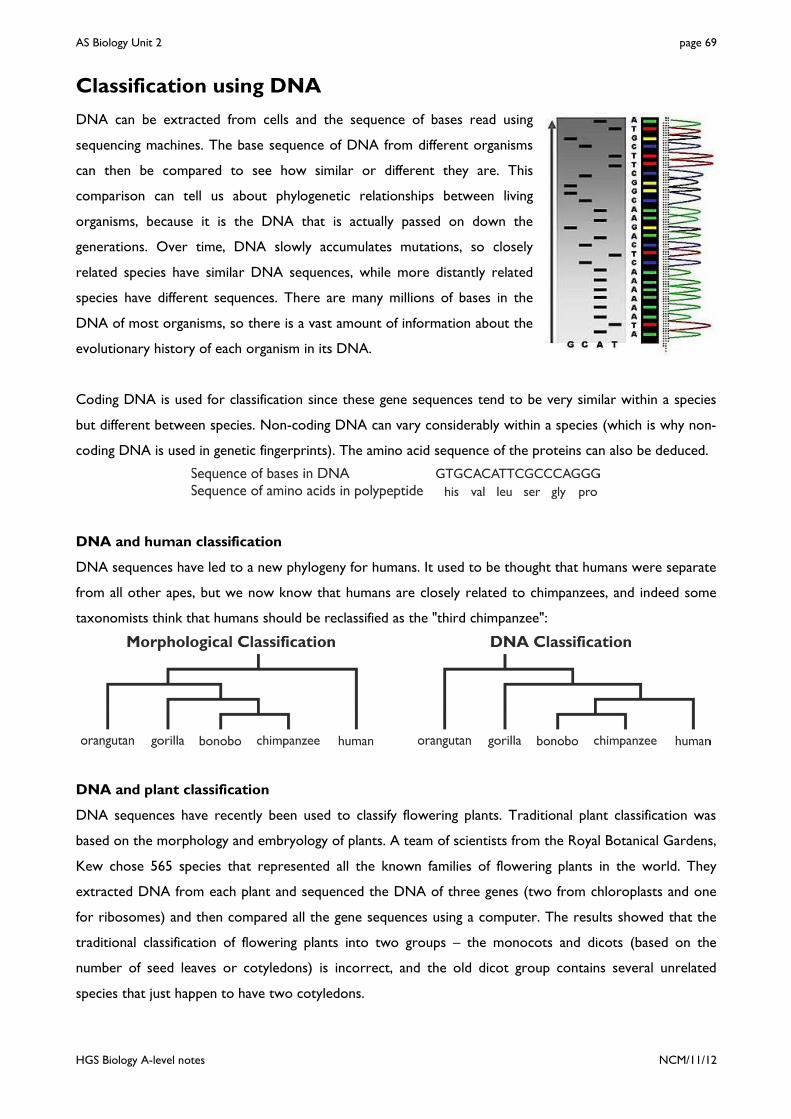
AS Biology Unit 2 page 69
HGS Biology A-level notes NCM/11/12
Classification using DNA
DNA can be extracted from cells and the sequence of bases read using
sequencing machines. The base sequence of DNA from different organisms
can then be compared to see how similar or different they are. This
comparison can tell us about phylogenetic relationships between living
organisms, because it is the DNA that is actually passed on down the
generations. Over time, DNA slowly accumulates mutations, so closely
related species have similar DNA sequences, while more distantly related
species have different sequences. There are many millions of bases in the
DNA of most organisms, so there is a vast amount of information about the
evolutionary history of each organism in its DNA.
Coding DNA is used for classification since these gene sequences tend to be very similar within a species
but different between species. Non-coding DNA can vary considerably within a species (which is why non-
coding DNA is used in genetic fingerprints). The amino acid sequence of the proteins can also be deduced.
DNA and human classification
DNA sequences have led to a new phylogeny for humans. It used to be thought that humans were separate
from all other apes, but we now know that humans are closely related to chimpanzees, and indeed some
taxonomists think that humans should be reclassified as the "third chimpanzee":
DNA and plant classification
DNA sequences have recently been used to classify flowering plants. Traditional plant classification was
based on the morphology and embryology of plants. A team of scientists from the Royal Botanical Gardens,
Kew chose 565 species that represented all the known families of flowering plants in the world. They
extracted DNA from each plant and sequenced the DNA of three genes (two from chloroplasts and one
for ribosomes) and then compared all the gene sequences using a computer. The results showed that the
traditional classification of flowering plants into two groups – the monocots and dicots (based on the
number of seed leaves or cotyledons) is incorrect, and the old dicot group contains several unrelated
species that just happen to have two cotyledons.

AS Biology Unit 2 page 70
HGS Biology A-level notes NCM/11/12
DNA hybridisation
Although the complete genomes of a few species have been sequenced (including humans), DNA
sequencing is slow and expensive. Furthermore it is usually only practical to compare the sequences of few
genes (such as the three in the plant example), and this can give unreliable evidence. A simpler and quicker
technique, which can also compare large amounts of DNA at a time, is DNA hybridisation. The method is:
The difference between the two T50H temperatures tells us how similar the DNA sequences are. As a
general rule, a difference in T50H of 1°C is equivalent to a 1% difference in the base sequences.
DNA hybridisation has been used to investigate the relationship between humans and other apes:
Species whose DNA is being compared Difference in T50H (°C) Human + chimpanzee 1.6 Human + gorilla 2.3 Human + orang-utan 3.6

AS Biology Unit 2 page 71
HGS Biology A-level notes NCM/11/12
Classification using Proteins
Since protein sequences are determined by DNA sequences, we
can also use the amino acid sequence in proteins to investigate
phylogenies. Typical proteins used for this purpose are
haemoglobin (found in all animals) and cytochrome c (a
respiratory enzyme found in all eukaryotic organisms). The β
chain of haemoglobin usually contains 146 amino acids, and this
table shows the number of differences in amino acid sequence of
β-chain of haemoglobin of different species compared to humans.
Animal Species
No. of a.a. differences
Human 0 Gorilla 1 Gibbon 2 Rhesus monkey 8 Dog 15 Horse 25 Mouse 27 Gray Kangaroo 38 Chicken 45 Frog 67
Protein Immunology
Proteins are more difficult to sequence than DNA, and only a few proteins have been sequenced for
different species, but again there is a quicker way to compare proteins using immunology. This technique is
based on the specific binding of antibodies to proteins, causing agglutination. The method is:
The more similar a protein is to species A protein, the better the antibodies will bind and the more
precipitate is formed. This technique has been used to compare the blood protein albumin in different
primates:
Species Human Chimpanzee Gorilla Orang-utan Gibbon Lemur % precipitation 100 95 95 85 82 35
This evidence is not as good as the DNA evidence, but it supports the same phylogeny.

AS Biology Unit 2 page 72
HGS Biology A-level notes NCM/11/12
Classification using Courtship Behaviour
Animals use courtship behaviour as part of sexual reproduction. Courtship behaviour is innate, in other
words it is genetically programmed, so all members of the same species show exactly the same courtship
behaviour, while members of different species show different behaviours. Courtship behaviour can thus be
used to identify individuals as members of the same or different species.
Courtship behaviour allows animals to:
• Recognise members of their own species. This is particularly important where many very similar species
live in the same habitat. Reproduction between members of different species may be possible, but won’t
lead to fertile offspring, so should be avoided.
• Attract a mate of the opposite sex.
• Identify a mate that is capable of breeding. Both partners need to be sexually mature, fertile and
receptive to mating. Many females only produce eggs at specific times, often just once a year.
• Synchronise the production of eggs and sperm.
• Form a pair bond and help raise the offspring.
Courtship behaviour has been extensively studied in the three-
spined stickleback by Niko Tinbergen. During the breeding
season, males develop red bellies and build a nest in a territory,
which they defend from other males. The female is attracted by
the red belly (diagram a). The male performs a zigzag dance and
the female follows him to the nest (b). The female enters the
nest and the male prods the base of her tail (c), which stimulates
her to lays eggs in the nest. The male then drives the female
from the nest, enters it himself, and releases sperm to fertilise
the eggs (d).
Only members of the same species of stickleback will respond in
the correct way, allowing mating to proceed, so this ensures that
males don’t fertilise eggs from a different species. This is
important for species that use external fertilisation.
Tinbergen found that the female will follow almost any small red
object to the nest, and any object touching her near the base of
her tail will cause her to release her eggs.

AS Biology Unit 2 page 73
HGS Biology A-level notes NCM/11/12
Another example of courtship behaviour for
species recognition is the firefly. Many very
similar species coexist in the same habitat,
so species recognition is important.
Fireflies can produce flashes of light using a
special chemical reaction catalysed by the
enzyme luciferase. Mating takes place at
night, and the males of each species of firefly
produces a unique pattern of light flashes
combined with a unique flight path (straight,
zigzag or looping). Females will only mate
with males showing the correct pattern of
flashes and flight, ensuring mating only takes
place between members of the same
species. The flashing and flight patterns in
males are entirely genetically-determined.
AS Biology Unit 2 page 74
HGS Biology A-level notes NCM/11/12
Appendix 1 – Mathematical Requirements
Biology is a quantitative science, and a reasonable mathematical ability is expected in an A-level biology
exam. The AQA specification states that you can be tested on any of these mathematical topics:
Calculations
• Use standard form; ratios, fractions and percentages.
• Calculate xxx n ;1; ; mean; and standard deviation.
• Calculate percent change and rate of change.
• Calculate circumferences and areas of circles; and surface areas and volumes of cuboids and cylinders
when provided with appropriate formulae.
• Use units with prefixes (n, µ, m, k, M, G) and use an appropriate number of significant figures.
• Make estimates of the results of calculations without using a calculator.
• Rearrange equations and substitute numerical values into equations using appropriate units.
Handling data
• Understand the terms mean, median and mode and standard deviation.
• Understand the use of logarithms for quantities that range over several orders of magnitude.
• Construct and interpret frequency tables, bar charts and histograms.
• Use a scatter diagram to identify positive and negative correlation between two variables.
• Plot graphs from data (using appropriate institute of biology conventions) and read data from graphs.
• Understand the principles of sampling as applied to biological data.
Maths Tips
• There will be maths questions! You must be confident with units and prefixes (especially m, µ and n).
• You need to know the formulae for magnification; percent change and gradient of a graph.

AS Biology Unit 2 page 75
HGS Biology A-level notes NCM/11/12
Sampling
In biology investigations we want to find out something about the natural world, but we can’t possibly
observe every single member of a species. Instead we make our measurements on a sample of the total
population, and hope that our conclusion from the sample can be applied to the whole population. Since
organisms vary the sample must be chosen carefully:
• It must be a random sample, to avoid sample bias, such as accidentally (or deliberately) picking all the
tallest individuals. We can use random numbers to choose coordinates to place quadrats or traps, or to
pick individuals.
• It must be a large sample, to minimise the chance of picking a skewed sample and to allow for bad
measurements or anomalies. The sample size is often called n. How big should n be? This depends on
the measurements, but n should never be less than 10, and preferably at least 100 or 1000.
Assuming the characteristic can be measured quantitatively, then
the measurements can be summarised on a histogram, where the
horizontal axis shows the variable being measured and the vertical
axis shows the number of measurements, or frequency, in each
group (histograms are also called frequency histograms). If there
are enough measurements the histogram approaches a smooth
curve. The curve is usually a symmetrical, bell-shaped normal
distribution curve, with most of the repeats close to some central value. Many biological phenomena follow
this pattern: e.g. peoples' heights, number of peas in a pod, the breathing rate of insects, etc.
The whole sample can be summarised by two parameters:
• The mean ( x ; also known as the arithmetic mean or average) is mid-point of the sample (actually the
central point of the normal distribution curve).
• The standard deviation (SD) is variation (or “spreadiness”) of the sample (actually the width of the
normal distribution curve). The more variation there is in the sample, the more spread out the
measurements are, and the larger the standard deviation.
Whenever a mean is calculated a standard deviation should also be
calculated to show how reliable the mean is. Sample means and
standard deviations can easily be calculated using computers or
calculators. Standard deviations can be plotted on a chart as error
bars to show graphically the reliability of the mean values. If the
error bars overlap, then we can say that the samples aren’t really
different. If they don’t overlap, then we can say that the samples are significantly different.

AS Biology Unit 2 page 76
HGS Biology A-level notes NCM/11/12
Appendix 2 – The Unit 2 Exam
The three AS biology units are assessed as shown in this table:
Unit Assessment Details Raw
marks UMS marks
Unit 1 1h 15min exam 5-7 short answer questions plus 2 longer questions: 1 comprehension and 1 continuous prose.
60 100
Unit 2 1h 45min exam 9 short answer questions plus 2 longer questions: 1 data handling and 1 HSW.
85 140
Unit 3 AS EMPA 2-3 practical sessions with short written task sheets plus a 1h 15min exam.
50 60
Biology is not just about learning facts (though there is a lot to learn): it’s largely about understanding
principles and being able to apply these principles to unfamiliar situations (which is what happens in real
life). It’s also important to understand How Science Works, and the role of evaluation and critical thinking.
So the A2 biology exams test all these aspects. Of the 60 raw marks in the unit 1 exam, about 25 will be
for biological knowledge; 25 will be for applying that knowledge to unfamiliar situations and analysing data;
and 10 will be for How Science Works, including planning, analysing and evaluating experiments. So expect
lots of questions about data analysis. These are designed to test your knowledge of unit 1 biology in
unfamiliar contexts.
Biological principles
The following basic biological principles from unit 1 can be examined in unit 2.
• Proteins and polysaccharides are made up of monomers that are linked by condensation.
• Many of the functions of proteins may be explained in terms of molecular structure and shape.
• Enzymes are proteins and their rates of reaction are influenced by a range of factors: temperature, the
presence of inhibitors, pH and substrate concentration.
• Substances are exchanged by passive or active transport across exchange surfaces. The structure of
plasma membranes enables control of the passage of substances across exchange surfaces.

AS Biology Unit 2 page 77
HGS Biology A-level notes NCM/11/12
Exam Technique
• 40% of all exam marks lost are lost due to poor exam technique.
• Read the question! You will only get marks for doing exactly what it asks, e.g. if a question says “explain
how A causes B” then start at A and finish at B.
• Do what the question says. If it says “use the diagram to...” or “use the graph to...”, then you must do
so.
• If a question says “give two reasons…” then give exactly two. You will lose marks if you give three.
• Read the whole question before answering any of it. This helps understanding.
• Use technical terms in every answer. In general a technical term used correctly is worth one mark.
“Meiosis causes alleles to be recombined” is more likely to earn a mark than “meiosis mixes genes”.
• Look at the marks. Don’t write too much for a 1-mark answer, and do write 3 good things for a 3-mark
answer.
Exam Strategy • 40% of the marks are aimed at E-grade candidates, so should be fairly easy to get. You could try finding
and doing these questions first.
• In longer answers (5 or more marks) try writing your answer in bullet points, where each statement is
worth one mark. That will force you to be logical and put a technical term into every sentence, and it
will help the examiner to find your points.
Describing and Explaining data • Underline the words “describe” and “explain” on your paper, to remind you to do the right one.
• If you are asked to describe some results (from a table or a graph) look for different phases e.g. “as X
increases Y increases up to 30 days then levels off”. Always quote a value from the graph – usually the
X-value where the graph chances shape.
• If you have to describe a graph with fluctuating or “noisy” data (a jagged line), try drawing a smooth line
of best fit through the data first, and then describe that.
• If you are asked to describe some results from a table it might be a good idea to sketch a quick graph on
the exam paper so you can see the pattern more clearly.
How Science Works • There will be How Science works questions in all exams, so check you know all the terms.
• If you are asked to design an investigation, the marks will be for fair testing (though don’t use this term):
how to change the independent variables; how to quantify the dependent variable; naming some control
variables; doing repeats.
You need to understand all the How Science Works words on the next page (items marked * are only in A2).

AS Biology Unit 2 page 78
HGS Biology A-level notes NCM/11/12
Types of DataData
(measurements, singular datum)
Quantitative or Numeric Data
(numbers)
Qualitative or Categoric Data
(words)
Continuous Datacan have any value
e.g. 7.34, -294.6, 2x10⁵
Discrete Dataonly whole numbers
e.g. no. of atoms
Ordered Datacan be ranked
e.g. small, medium, large
Nominal Datacan't be ranked e.g. male, female
Controlled Experiment (When all relevant variables are controlled, so that observed changes in the dependent variable must be due to changes in the independent variable.
Fair Test)
PlaceboA dummy pill, injection or treatment that has no physiological effect (e.g. a sugar pill or saline injection). Used in a clinical trial to allow for the - the observation that symptoms can improve when patients believe they are being helped.
placebo effect
HypothesisA suggested explanation of observations or results that can be tested. Also known as a scientific hypothesis. A good hypothesis can be used to make predictions.
ProtocolA method or technique that has been shown to produce valid and reliable results.
RCTThe best experimental design for a drug trial. RCT standsControlled Trial, or in more detail, a Randomised, Placebo-Controlled, Double-blind Trial. This design ensures that the trial is free from .
means the study and control groups are allocated randomly means the study group (taking the drug to be tested) is
compared to a placebo group (who are given a placebo).means that neither the subjects nor the investigators know
who is in the study or placebo groups. This avoids bias.
valid biasRandomised Placebo-controlled
Double-blind
�
�
�
for Randomised
Control Experiment (Control)An additional experiment designed to eliminate alternative explanations for the main experiment, and so show that observed changes in the dependent variable must be due only to changes in the independent variable.
Control GroupA group or sample treated in the same way as the experimental group, except for the factor being investigated e.g. a placebo group in a drugs trial. By comparing the results for two groups it can be shown that observed changes in the dependent variable must be due only to changes in the independent variable.
Experimental Design
Statistical Test*Someth ing that tests whether observed differences or correlations are significant, or just due to chance.
Null Hypothesis*The statement that is tested by a statistical test. The null hypothesis is fixed for each test, but always says that there is no difference or no association. The null hypothesis has nothing to do with a scientific hypothesis.
P-value*The result of a stats test, expressed as a probability. It is the probability that the results are due to chance. If <0.05 then we reject the null hypothesis, otherwise we accept it.
P
Causal RelationWhen changes in one variable the changes in another variable. Can only be shown by a controlled experiment.
cause
Correlation (or Association)When one variable changes with another variable, so there is a relation between them. The strength of a correlation can be measured using a correlation coefficient. A correlation need not be a . causal relation
Statistical Analysis
ReplicatesRepeats of a measurement.
RangeThe highest and lowest replicates, or the interval between them.
Standard Deviation (SD)A measure of the dispersal of the replicates about the mean. In a normal distribution 68% of the replicates will be within 1 standard deviation of the mean, and 95% will be within 2 standard deviations of the mean.
Standard Error of the mean (SEM)*A measure of the uncertainty, or error, of a calculated mean. The smaller the standard error, the more reliable the mean.
95% Confidence Interval (CI)*Another measure of the error of the mean. We can be 95% confident that the true mean lies in the range (mean ± CI). The top and bottom of this range are called the .confidence limits
Raw DataThe original measurements or re co r d i n g s be fore any manipulation or processing.
Mean Average or T he m id -p o i n t o f t h e replicates. = sum of replicates / N
Simple Analysis
Random ErrorsInaccuracies due to mistakes, poor technique, or random variation. Random errors are very common, but can be improved by taking many replicates. Data with a small random error is said to be .precise
Systematic ErrorsInaccurate measurements in one d ire c t i on on ly, d ue to poo r
or poor technique. Systematic errors can be improved by taking more replicates.
calibrationnot
Data with a small systematic error is said to be reliable.
Zero ErrorA particular kind of systematic error, where the instrument does not return to zero.
BiasWhen the observer chooses some results and ignores others, to support a particular view. Also called
.cherry
picking
CalibrationEnsuring that a measuring instrument gives correct readings by fixing known points then constructing a scale between them.
Anomaly or OutlierA measurement that falls far outside the expected range and is therefore probably due to experimental error. Anomalies should be rejected, since they skew the mean, but it is very difficult to distinguish between anomalies and normal biological variation.
Accurate DataMeasurement that are close to the true value.
Precise Data1.
va lues when repeated . The replicates therefore have a small
.
2. Data measured on sensitive equipment with a suitably fine scale, e.g. 20 mm is more precise than 2 cm.
range
Measurements that give similar
Quality of Data
AnecdoteAn observation or story from real life. Anecdotes are not evidence and cannot be used to support a hypothesis, but they can be useful to suggest a new testable hypothesis.
Valid DataThe best quality data, i.e. data that is
and and obtained from an , experiment that addresses the stated aim. Valid data is assumed to be accurate.
precise reliableunbiased controlled
Reliable DataFindings that can be repeated. This includes by the original investigator; by other scientists; by other techniques; or those that agree with secondary sources.
EvidenceAny data or observations that to support a particular hypothesis.
are used
True ValueThe real value of a measurement, if it could be measured with no errors at all.
Types of Variable
independent variabledep
enden
t va
riab
le
Dependent VariableThe variable you , to see how it is affected by the independent variable.
measure
Confounding VariablesAny variables that could also affect the
dependent variable. Confounding variables should be
controlled in a fair test.
Independent VariableThe variable you
, to see how it affects the dependent variable. You may also measure it when you change it.
choose to change
Control variablesConfounding variables that
are kept constant (controlled) during the
experiment. If you can't control a variable (such as weather in a field investigation),
you should at least monitor it


![Unit 4 Populations & Environment - Chemstuff · Photosynthesis and Respiration Unit 4 Populations & Environment Practice Exam Questions [Total 13 marks] 3 The Calvin cycle occurs](https://img.pdfslide.net/doc/110x75/5fd567e814077f0a20239bac/unit-4-populations-environment-chemstuff-photosynthesis-and-respiration.jpg)