Embed Size (px)
Citation preview

Universidade de Lisboa
Faculdade de Farmácia
Characterization of allergens from
several tree nuts and their role in
plant food allergy
Caracterização de alergenos de frutos secos e o seu papel na alergia a
alimentos vegetais
Cátia Liliana Morais Pereira
Mestrado em Controlo da Qualidade e Toxicologia dos Alimentos
2013

Universidade de Lisboa
Faculdade de Farmácia
Characterization of allergens from
several tree nuts and their role in
plant food allergy
Cátia Liliana Morais Pereira
Supervised by:
Mestrado em Controlo da Qualidade e Toxicologia dos Alimentos
2013
Prof. Doutora Maria Manuela Beirão Catarino Professora Catedrática
Prof. Doutora Araceli Díaz Peralez Professora Associada

iii
ACKNOWLEDGEMENTS
Well, there are a lot of people that helped me to get through all this
project so, let’s go:
First of all, a big thank you to Professor Maria Manuela Catarino, who
gave me the opportunity to do this thesis under the ERASMUS program, and for
all the orientation and support.
To Ali, for welcoming me in her group with a big smile and arms wide
open. For the confidence placed in me, for all the support and orientation. But
especially, for the contagious enthusiasm.
A mis compis de labo! To Maria and Amaya, without them, I would be
lost! Maria, thank you so much for the entire patience you have for my one
thousand doubts and questions and everything I ask you. You are a saint!
Thanks for everything you taught me, I will never forget (or if I do… I will call you
immediately!). And to Amaya, my companion in all the bus trips to the lab, for
your contagious smile and for your confidence! You rock and I hope you are
showing the French the Spanish power! Vos echo de menos!!
To my ERASMUS friends, because, if I haven’t met them my ERASMUS
experience wouldn’t be the same. And Madrid wouldn’t have the same magic.
Especial thanks to Jelka, Anne and Inessa. I will never forget you girls!
To my friends, who were fortunate to not have me around for 6 months!
Hahaha But gave me a lot of support and never let me give up.
To Márcio, for the patience, understanding and for letting me pursuit all
my dreams.
Lastly, but most importantly, to my family for always believing in me and
without them I couldn’t have done this. Thank you Dad, Mum and Gonçalo.

iv
ABSTRACT
Food allergies are a common issue in western countries. In the last
decade, these diseases has increased significantly, and nowadays it is estimated
that affects 2-8% of the population. Within the food allergies, plant food is the
most frequent in adult population and the most part of the plant food allergens
belong to protein families with defense or storage functions.
Among plant food allergies there is a special interest in tree nut allergy.
In the course of history, nuts have been part of the diet around the world. Tree
nuts have a high nutritional value and they are very important in the human
diet. However, in the developed world, the allergic reactions caused by tree
nuts represent one of the first causes of food allergies in children and the first in
adults.
Understanding the mechanism by which a harmless protein to the
organism is capable of inducing an allergic response is the basis to prevent and
treat this type of disease.
Until now, in food allergy, the only possible treatment is avoiding the
consumption of the culprit food. Although, the existence of cross-reactivity
between allergens and the specific sensitization profiles of each patient, makes
it difficult to know which foods are related and which ones the patient should
avoid.
In order to develop safe and effective immunotherapy, it is necessary to
characterize the allergens involved both at molecular and immunological level.
The major allergens described in tree nuts are 7S vicilins, 11S legumins,
2S albumins, lipid transfer proteins (LTPs) and thaumatin-like proteins (TLPs).
In this thesis, the allergenic molecular basis of these proteins was studied
in order to try to understand the possible mechanisms that are mediating
sensitization and cross-reactivity and the prevalence of these proteins in a
Spanish population, with the use of protein microarrays.

v
Key words: food allergy, seed storage proteins, protein microarrays, tree
nuts, LTPs, TLPs, cross-reactivity

vi
RESUMO
As alergias alimentares são um problema comum nos países ocidentais.
Na última década, estas doenças têm aumentado significativamente e
actualmente é estimado que afectem 2-8% da população. Nas alergias
alimentares, a alergia a alimentos vegetais é a mais frequente na população
adulta e a maioria dos alergenos de alimentos vegetais pertencem a famílias de
proteínas com funções de defesa e armazenamento.
Entre as alergias a alimentos vegetais, há um interesse especial na alergia
a frutos secos. No decurso da história, os frutos secos têm feito parte da dieta
em todo o mundo. Os frutos secos têm um elevado valor nutricional e são muito
importantes na dieta humana. Contudo, no mundo desenvolvido, as reacções
alérgicas causadas pelos frutos secos, representam uma das primeiras causas de
alergia alimentar em crianças e a primeira em adultos.
Conhecer o mecanismo pelo qual uma proteína inofensiva ao organismo
é capaz de induzir uma resposta alérgica, é a base para prevenir e tratar este
tipo de doença.
Até agora, na alergia alimentar, o único tratamento possível é evitar o
consumo do alimento culpado pela alergia. Todavia, a existência de
reactividade-cruzada entre alergenos e os perfis especifícos de sensibilização
dos patientes, torna difícil saber que alimentos estão relacionados e quais os
alimentos que o paciente deve evitar.
De modo a desenvolver imunoterapia segura e eficaz é necessário
caracterizar os alergenos envolvidos, tanto a nível molecular como a nível
imunológico.
Os alergenos maioritários descritos nos frutos secos são vicilinas 7S,
leguminas 11S, albuminas 2S, proteínas de transferência de lípidos (LTPs) e
proteínas similares a taumatinas (TLPs).

vii
Nesta tese, a base molecular alergénica destas proteínas foi estudada de
modo a perceber os possíveis mecanismos que medeiam a sensibilização e a
reactividade-cruzada e a prevalência destas proteínas numa população
Espanhola, com a utilização de microarrays de proteínas
Palavras-chave: alergia alimentar, proteínas de armazenamento de sementes,
microarrays de proteínas, frutos secos, LTPs, TLPs, reactividade cruzada

viii
INDEX
ACKNOWLEDGEMENTS .................................................................................. III
ABSTRACT ...................................................................................................... IV
RESUMO ........................................................................................................ VI
INDEX .......................................................................................................... VIII
FIGURES INDEX .............................................................................................. XI
LIST OF ABREVIATIONS ................................................................................. XIV
1. INTRODUCTION ........................................................................................ 1
1.1. Allergy mechanisms ....................................................................................... 1
1.2. Sensitization Pathways ................................................................................... 3
1.2.1. Gastrointestinal sensitization ............................................................................. 3
1.2.2. Respiratory sensitization .................................................................................... 5
1.2.3. Skin sensitization................................................................................................. 6
1.3. Food allergy ................................................................................................... 7
1.4. Allergen cross-reactivity ................................................................................. 7
1.5. Seed storage proteins ..................................................................................... 8
1.5.1. 2S albumins .................................................................................................... 8
1.5.2. Globulins ......................................................................................................... 9
1.6. Pathogenesis related proteins ...................................................................... 10
1.6.1. Lipid Transfer Proteins ................................................................................. 10
1.6.2. Thaumatin-like Proteins ............................................................................... 11
1.7. Tree nut allergy ............................................................................................ 11
1.7.1. Hazelnut ....................................................................................................... 12
1.7.2. Walnut .......................................................................................................... 13
1.7.3. Chestnut ....................................................................................................... 13
1.7.4. Peanut .......................................................................................................... 14
1.7.5. Pine nut ........................................................................................................ 14
1.7.6. Sunflower seed ............................................................................................. 15

ix
1.7.7. Cashew nut ................................................................................................... 15
1.8. Diagnosing allergy ........................................................................................ 15
1.8.1. Skin prick test (SPT) ...................................................................................... 16
1.8.2. Double blind placebo controlled food challenge ......................................... 17
1.8.3. Immunodetection: ELISA and Western-Blot ................................................ 18
1.8.4. Protein Microarrays ..................................................................................... 19
2. AIMS ...................................................................................................... 21
3. MATERIAL AND METHODS ...................................................................... 22
3.1. Patient’s Sera ............................................................................................... 22
3.2. Methods ...................................................................................................... 22
3.2.1. Protein extraction and protein quantification ............................................. 22
3.2.1.1. Protein extraction ................................................................................. 22
3.2.1.2. Electrophoretic methods ...................................................................... 23
3.2.2. Immunochemical methods .......................................................................... 23
3.2.2.1. Western-Blot with polyclonal antibodies ............................................. 23
3.2.2.2. Western-Blot with patient’s sera .......................................................... 24
3.2.3. Isolation of allergens .................................................................................... 25
3.2.3.1. 2S Albumins isolation ............................................................................ 25
3.2.3.2. 11S isolation .......................................................................................... 25
3.2.3.3. LTP and TLP isolation ............................................................................ 25
3.2.4. Quality of purified proteins .......................................................................... 26
3.2.5. Protein microarrays ...................................................................................... 26
3.2.5.1. Printing .................................................................................................. 26
3.2.5.2. Hybridization ......................................................................................... 26
3.2.5.3. Array reading ........................................................................................ 27
3.2.6. Statistical analysis......................................................................................... 27
4. RESULTS ................................................................................................. 29
4.1. Study of the cross-reactivity among tree nut species .................................... 29
4.2. Characterization of allergens in tree nuts and their prevalence in an adult
population ............................................................................................................. 31
4.2.1. Purification and characterization of 2S albumins from walnut ................... 31
4.2.2. Purification and characterization of 11S from hazelnut .............................. 31
4.2.3. Purification and characterization of LTP from Hazelnut, walnut
and chestnut ................................................................................................. 33

x
4.2.4. Purification and characterization of TLP from hazelnut and chestnut ........ 36
4.2.5. Study of the prevalence of the most relevant allergens .............................. 40
5. DISCUSSION ............................................................................................ 43
6. CONCLUSIONS ........................................................................................ 52
7. REFERENCES ........................................................................................... 53

xi
FIGURES INDEX
Figure 1: Paraceluar transport (Ménard, S., et al., 2010) ...................................... 4
Figure 2: Transcelular transport. (Ménard, S., et al., 2010) ................................... 5
Figure 3: The dual-allergen exposure hypothesis of food allergy. ......................... 6
Figure 4: Skin prick tests to common allergens in the forearm. Compared with histamine, the positive control, indicating the presence of allergen-specific IgE to different allergens (Tordesillas, L., 2012)......................................................... 17
Figure 5: Schematic illustration of an immunoblotting (Western-blot) (Adapted from: Goldsby, R.A., et al., 2003). ........................................................................ 19
Figure 6: Schematic description of protein microarray testing of allergen-specific IgE. Allergens are potted onto slides in triplicate. Serum incubation is followed by a second anti-human IgE antibody for detection. The mean fluorescence intensity is determined by two different laser settings (Jahn-Schimid, B., et al., 2003). .................................................................................................................... 20
Figure 7: SDS-PAGE from PBS extracts of cashewnut (C) (10µg), hazelnut (H) (5µg), peanut (P) (10µg), chestnut (Ct) (10 µg), walnut (W) (15 µg), pine nut (Pn) (10 µg) and sun seeds (S) (10 µg): A, staining with Blue Coomassie; B, Immunodetection with pool serum from Jimenez Diaz Foundation (FJD); C, Immunodetection with pool serum from Hospital La Princesa University Hospital; D, Immunodetection with pool serum from Basurto Hospital (Bilbao). .............................................................................................................................. 29
Figure 8: SDS-PAGE from PBS extracts of cashewnut (C) (10µg), hazelnut (H) (5 µg), peanut (P) (10µg), chestnut (Ct) (10µg) walnut (W) (15µg), pine nut (Pn) (10µg), and sun seeds (S) (10 µg),: A, Immunodetection against Pru p 2.0201 (TLP) (1:5000); B, Immunodetection against Hela 1 (11S legumin) (1:10000); C, Immunodetection against Pru p 3 (LTP) (1:1000) ................................................ 30
Figure 9: SDS-PAGE from PBS extracts (10 µg) of walnut; A, staining with blue comassie of the extract (E) and of the retained fraction of the molecular exclusion chromatography (ME); B, Immunodetection with polycolonal antibodies against 2S. ........................................................................................... 31
Figure 10: SDS-PAGE from PBS extracts (10µg) (E) of hazelnut, the retained fraction obtained by anion-exchange chromatography in Waters cartridge (5µg)

xii
(A) and in FPLC (5µg) (R) A, staining with Blue Coomassie; B, Immunodetection with polyclonal antibodies against Hela 1 (11S legumin) (1:10000) .................... 32
Figure 11: SDS-PAGE from PBS extracts (10µg) of hazelnut (H), walnut (W) and chestnut (Ct): A, staining with Blue Coomassie; B, Immunodetection with polyclonal antibodies against Pru p 3 (LTP) (1:1000). .......................................... 33
Figure 12: SDS-PAGE of the retained fraction obtained by cation-exchange chromatography in Waters cartridge (5µg) of hazelnut (H), walnut (W) and chestnut (Ct): A, staining with Blue Coomassie; B, Immunodetection with polyclonal antibodies against Pru p 3 (LTP) (1:1000). .......................................... 34
Figure 13: Purification of LTPs from hazelnut, walnut and chestnut. A,B and C purified by HPLC reverse-phase. All selected picks contain the purified proteins. .............................................................................................................................. 34
Figure 14: SDS-PAGE from purified proteins (5µg) of hazelnut (H), walnut (W) and chestnut (Ct) . A, staining with Blue Coomassie; B, Immunodetection with polyclonal antibodies against Pru p 3 (LTP) (1:1000). .......................................... 35
Figure 15: MALDI-spectrum of the purified allergen Jug r 3. ............................... 35
Figure 16: MALDI-spectrum of the purified allergen Cor a 8. .............................. 36
Figure 17: MALDI-spectrum of the purified allergen Cas s 8. .............................. 36
Figure 18: SDS-PAGE from PBS extracts (10µg) of hazelnut (H) and chestnut (C): A, staining with Blue Coomassie; B, Immunodetection with polyclonal antibodies against Pru p 2.0201(TLP) (1:5000). ..................................................................... 37
Figure 19: SDS-PAGE of the retained fraction obtained by cation-exchange chromatography in Waters cartridge (5µg) of hazelnut (H) and chestnut (Ct): A, staining with Blue Coomassie; B, Immunodetection with polyclonal antibodies against Pru p 2.0201 (TLP) (1:5000). .................................................................... 37
Figure 20: Purification of LTPs from hazelnut and chestnut. A and B purified by HPLC reverse-phase. All selected picks contain the purified proteins. ................ 38
Figure 21: SDS-PAGE from purified proteins (5µg) TLP hazelnut and TLP chestnut A, staining with Blue Coomassie; B, Immunodetection with polyclonal antibodies against Pru p 2.0201 (TLP) (1:5000). .................................................................... 39
Figure 22: MALDI-spectrum of the purified allergen TLP hazelnut. .................... 39

xiii
Figure 23: MALDI-spectrum of the purified allergen TLP chestnut. .................... 40
Figure 24: Recognition frequencies of the different allergens by geographical area. Frequencies, shown as percentage of positive response (%), were obtained by incubating the allergens microarray with single sera from allergic patients of Jimenez Diaz Foundation (FJD), La Princcesa University Hospital and Basurto Hospital (Bilbao). Percentage positive responses and significant differences (p>0,05) are indicated. ......................................................................................... 42
Figure 25: Frequency of sensitizations obtained by the different allergens microarray using sera from all patients. .............................................................. 42

xiv
LIST OF ABREVIATIONS
Ana o 1 Cashew nut allergen (7S)
Ana o 2 Cashew nut allergen (11S)
Ana o 3 Cashew nut allergen (2S)
APC Antigen presenting cell
Ara h 1 Peanut allergen (7S)
Ara h 2 Peanut allergen (2S)
Ara h 3 Peanut allergen (11S)
Ara h 6 Peanut allergen (2S)
Ara h 9 Peanut allergen (LTP)
BALT Bronchial-associated lymphoid tissue
Bet v 1 Birch tree allergen
Cas s 5 Chestnut allergen (chitinase)
Cas s 8 Chestnut allergen (LTP)
Cor a 1 Hazelnut allergen (Bet v 1-like)
Cor a 2 Hazelnut allergen (2S)
Cor a 8 Hazelnut allergen (LTP)
Cor a 9 Hazelnut allergen (11S)
Cor a 11 Hazelnut allergen (7S)
Da Dalton
DBPCFC Double-blind placebo-controlled food challenge
FJD Jimenez Diaz Foundation
FPLC Fast-Performance Liquid Chromatography
GALT Gut-associated lymphoid tissue

xv
Hel a 1 Sunflower seed allergen (11S)
Hel a 2S Sunflower seed allergen (2S)
Hel a Oleosins Sunflower seed allergen (Oleosin)
HPLC High-Performance Liquid Chromatography
IgE Immunoglobin E
IgG Immunoglobin G
Jug r 1 Walnut allergen (2S)
Jug r 2 Walnut allergen (7S)
Jug r 3 Walnut allergen (LTP)
Jug r 4 Walnut allergen (11S)
LTPs Lipid Transfer Proteins
MALDI Matrix-assisted laser desorption/ionization
MHC Major Histocompatibility Complex
PAGE Polyacrilamide electrophoresis gel
PBS Phosphate buffered saline
PR Pathogenesis related
Pru p 2.0201 Peach allergen (TLP)
Pru p 3 Peach allergen (LTP)
PVDF Polyvinylidene difluoride
RP Reversed phase
SALT Skin-associated lymphoid tissue
SPT Skin Prick Test
TCR T-Cell Receptor
TLPs Thaumatin-like proteins
TOF Time of Flight mass spectrometry

1
1. INTRODUCTION
Allergies are immunogenic diseases that nowadays affect more than 30%
of the world population and their development depend both on genetic and
environmental factors (Kay, A.B., 1997). The most common sources of allergens
are dust mites, mold, pollen and plants (Hoffmann-Sommergruber, K., 2000).
Allergies can be produced by inhalation, ingestion or contact with allergens that
are capable of inducing the synthesis of immunoglobulin E (IgE) (Huby, R.D.J., et
al., 2000). In IgE-mediated reactions, clinical symptoms are caused by the rapid
and massive release of inflammatory mediators. Moreover, this type of disease
involves an important cost for the European health systems, sometimes bigger
than that produced by season flu (Holgate, S., et al., 2006).
Food allergies are the most common cause of anaphylaxis evaluated in
emergency rooms in all age groups and they occasionally result in fatalities
(Nowak-Wegrzyn, A., and Sampson, H.A., 2011).
1.1. Allergy mechanisms
The first attempt to define food allergy came in 1984 from the American
Academy of Allergy and Immunology, and the National Institute of Allergy and
Infectious Diseases. They classified adverse food reactions in two types, food
allergy (hypersensitivity) and food intolerance, depending on the existence or
not of an underlying immunological mechanism, respectively (San Miguel
Moncin, M.M., et al., 2007).
Food allergy is constituted by a hypersensitivity reaction in which
symptoms appear quickly and are caused by exposure to exogenous
macromolecules, also known as antigens or allergens. This reaction results in a
marked increase in reactivity and responsiveness to the antigen or allergen on
ensuing exposure, resulting in adverse health effects and normally mediated by

2
IgE. Meanwhile, food intolerances are non-immune-mediated reactions whose
symptoms can sometimes take days to manifest (Mills, E. N. C., 2003). This way,
food allergy can be divided into IgE-mediated and non-IgE mediated reactions,
where the food allergens are the antigenic molecules which induce the
immunologic response (Fernandéz-Rivas, M. and Miles S., 2004).
The IgE mediated reactions are also called type I Hypersensitive, and
involve a complex immunologic process, which is divided in two phases, the
sensitization and subsequent reaction to re-exposure to an allergen.
Upon contact with the mucosa, the antigen is taken up by the antigen
presenting cells (APC), which will process it in the endocitic pathway. The
generated peptides are presented on the cell surface bound to a receptor
known as major histocompatibility complex (MHC) type II. Then, these APCs
such as mucosal macrophages, progenitor B-cells and dendritic cells migrate to
the regional lymph nodes where the antigen presentation will occur to T-cells.
The T-cell receptors of CD4+ T helper cells (TCR) recognize the complex of
foreign peptide plus MHC molecule on the APC surface. After this interaction
the CD4+ T cells are activated and differentiate into Th2 cells (Berin, M.C., and
Shreffler, W.G., 2008). Thus, they secrete specific cytokines, as interleukins,
responsible for differentiation towards one particular subtype of T-helper
lymphocytes and for the proliferation of B lymphocytes that synthetize specific
IgE antibodies. The IgE molecules enter into circulation and bind to their high
affinity receptors (FcεRI), which are present in mast cells, blood basophils and
dendritic cells.
Consecutive exposures to the allergen trigger the second phase, where a
huge number of IgE molecules are bound to the surface of the effector cells
such as mast cells and basophils. Thus, the cross-linking is produced, triggering
the cell degranulation, with release of preformed pharmacological mediators
such as histamine, and subsequent newly formed mediators such as cytokines
and leukotriene (Bird, J.A., 2010).

3
1.2. Sensitization Pathways
The allergic response needs that the responsible allergen gets in contact
with the immune system. This occurs when the antigen crosses through
epithelial barriers as the skin, intestines and the respiratory tract. The epithelia
represent a physical barrier against the entrance of infectious microorganisms,
toxins and harmful antigens, and also biochemical barriers, as the cells that
actively participate in development of immune responses (Bartemes, K.R., and
Kita, H., 2012). Therefore, epithelia dysfunction causes an increased risk of
sensitization (Groschwitz, K.R., and Hogas, S.P., 2009; Perrier, C., and Corthésy,
B., 2010).
1.2.1. Gastrointestinal sensitization
After ingestion, digestive enzymes degrade food proteins into small
peptides which are absorbed in the intestine. However, some proteins resist the
enzymatic activities, reaching the intestinal lumen intact and then they interact
with the lymphoid tissue associated to the digest tract, or GALT (gut-associated
lymphoid tissue), and may conduct an immune response (Ménard, S., et al.,
2010). The ability of molecules to cross the epithelium is dependent on its size,
shape, polarity, three-dimensional structure and aggregation status (Perrier, C.
and Corthesy, B., 2001).
The gut epithelia monolayer is the main element of the epithelial barrier.
Its integrity depends on the binding complexes between cells, especially the
tight junctions. Below the monolayer, is the lamina propria, which is rich in
immune cells (dendritic cells, lymphocytes, macrophages and others), and
above is covered by a mucus layer. The monolayer avoids the entry of harmful
antigens and infectious organisms, being permeable only to water, electrolytes
and essential nutrients (Groschwitz, K.R., and Hogas, S.P., 2009). However, some

4
components of the human diet can be carried through the monolayer. The
intestinal transport of molecules from the intestinal lumen to the lamina propria
can occur through two distinct mechanisms: paracellular and transcellular
transport. The paracellular transport, occurs through the adjacent epithelial
cells, where is allowed the transportation of molecules smaller than 600 Da. The
thigh junctions situated at the apical area, regulate the paracelullar
permeability, and, when there are some dysfunction, the risk of sensitization
increases once the augmentation in the permeability allows the transportation
of non-degraded proteins (figure 1) (Perrier, C., and Chortésy, B., 2010).
Figure 1: Paraceluar transport (Ménard, S., et al., 2010)
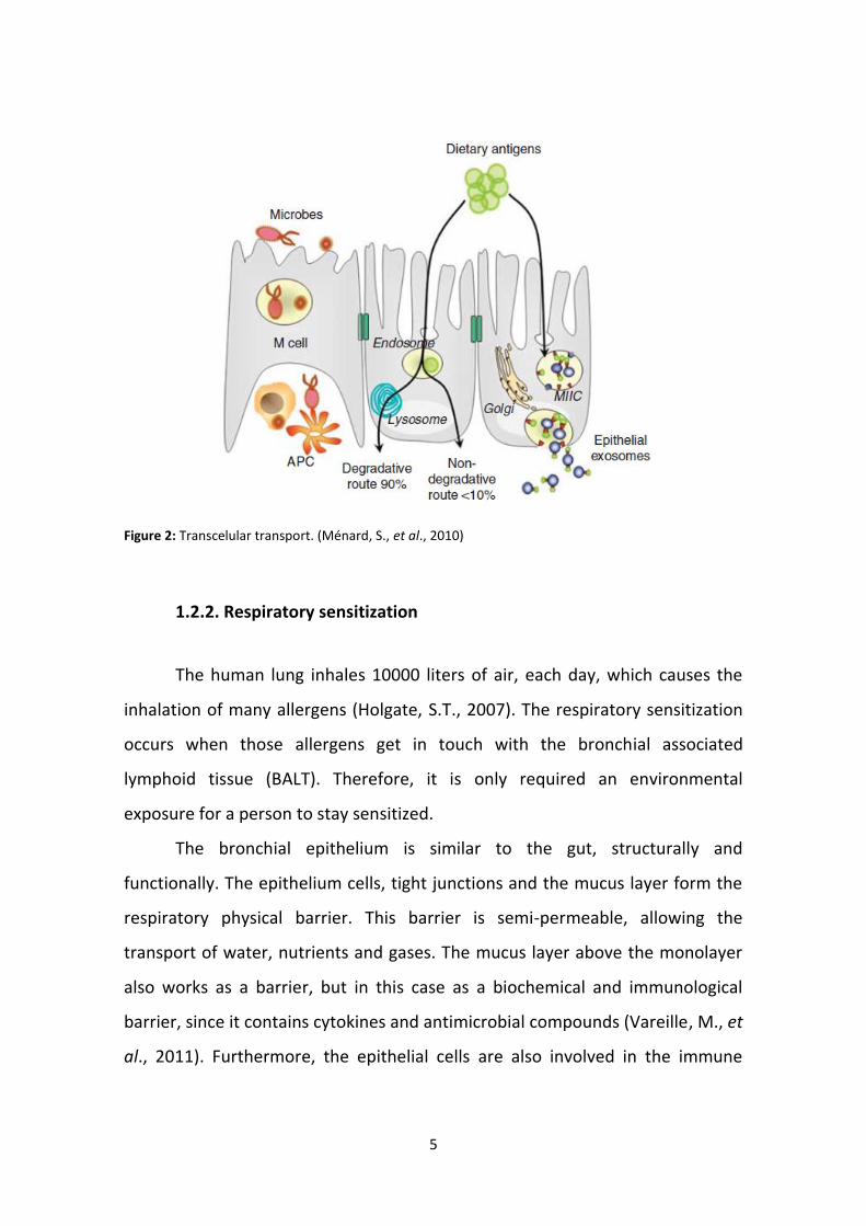
In the transcelullar transport, proteins enter into the cell thanks to the
formation of endosomes, being degraded in small peptides by the activity of the
lysosomes. The resulting peptides are released into the basolateral side, playing
an important role in the body’s immunity. Early endosomes may emerge with
vesicles rich in MHCII molecules which expose protein-fragments even partially
degraded. In the end, the vesicle releases its content, in the basolateral side,
(figure 2) (Ménard, S., et al., 2010).
5
Figure 2: Transcelular transport. (Ménard, S., et al., 2010)
1.2.2. Respiratory sensitization
The human lung inhales 10000 liters of air, each day, which causes the
inhalation of many allergens (Holgate, S.T., 2007). The respiratory sensitization
occurs when those allergens get in touch with the bronchial associated
lymphoid tissue (BALT). Therefore, it is only required an environmental
exposure for a person to stay sensitized.
The bronchial epithelium is similar to the gut, structurally and
functionally. The epithelium cells, tight junctions and the mucus layer form the
respiratory physical barrier. This barrier is semi-permeable, allowing the
transport of water, nutrients and gases. The mucus layer above the monolayer
also works as a barrier, but in this case as a biochemical and immunological
barrier, since it contains cytokines and antimicrobial compounds (Vareille, M., et
al., 2011). Furthermore, the epithelial cells are also involved in the immune

6
reaction mediated by the release of the immune response mediators (Bartemes,
K.R., and Kita, H., 2012).
1.2.3. Skin sensitization
Another sensitization pathway is the skin sensitization which explains the
development of food allergies without previous consumption of the food. The
dual-allergen exposure hypothesis suggests that the exposition to food allergens
by the oral route induces oral tolerance, meanwhile, the exposition to these
allergens by cutaneous route induce sensitization (figure 3) (Lack, G., 2008).
Figure 3: The dual-allergen exposure hypothesis of food allergy (Lack, G., 2008).
This cutaneous sensitization occurs when the allergens cross through the
skin and interact with the skin-associated lymphoid tissue (SALT). Allergens get
in touch with Langerhans cells, inducing the activation of T-cells towards an
immune response type I.

7
1.3. Food allergy
Nowadays, the prevalence of food allergies is about 3% in adults, and
between 6 and 8% in children (Burks, W., and Ballmer-Weber, B.K., 2006). Food
allergens, apparently, do not share specific biochemical characteristics which
determine a protein to be an allergen. Why, in the same family, a protein is
allergic and other not, is something that is still unknown. Within the food
allergies, plant food is the most frequent in adult population. To this day, many
plant food allergens have been identified (Breiteneder, H., and Radauer. C.,
2004), most of whom belong to only 31 protein families of over 2600 plant
protein families described. The most part of the plant food allergens belong to
protein families with defense or storage functions (Chapman, M.D., et al, 2007).
Some families of allergens do not follow this rule, such as profilins (Radauer, C.,
and Hoffmann-Sommergruber, K., 2004).
Seed storage proteins accumulate mainly in mature seeds and they are
mobilized during germination to provide a source of nitrogen for the early
stages of development (Higgins, T.J.V., 1984). The families of storage proteins
described as allergens are vicilins, legumins, 2S albumins and prolamins. Plant
defense proteins are involved in mechanisms of response to pests, pathogens
and stressful environment. This group includes enzyme inhibitors, thionins,
thaumatins, lipid transfer proteins (LTPs), chitinases, lectins, and more. There
are 14 families of defense proteins, eight of which include members that have
been described as allergens (Hoffmann-Sommergruber, K., 2002).
1.4. Allergen cross-reactivity
Cross-reactivity is a major problem in allergy and the term is used to
describe clearly defined clinical features that show reactivity to a source without
previous exposure (Ferreira, F., et al., 2004). The phenomenon where an IgE

8
antibody raised against one allergen, binds or recognizes a similar protein from
another source, it is called allergen cross-reactivity (Ferreira, F., et al., 2004). Its
molecular basis is the presence of homologous allergens in different species
that are recognized by the same type of specific IgE. The IgE cross-reactions are
due to shared features at the level of primary and tertiary structures of proteins
(Aalberse, R.C., et al., 2001).
1.5. Seed storage proteins
1.5.1. 2S albumins
Albumins are a group of seed storage proteins soluble in water at low
concentrations and with a sedimentation coefficient (S20.w) of 2. They are widely
distributed in dicot seeds, including cultivated species such as brassicas,
legumes, sunflower, cotton, and castor bean (Shewry, P. R., et al., 2004).
The 2S albumins are characterized for being small globular proteins rich
in sulfur amino acids as cysteine and methionine (Shewry, P. R. et al., 1995).
They are also rich in other amino acids, such as glutamine and arginine which
contain 2 and 3 nitrogen atoms, respectively, which have a main role on the
storage of nitrogen. Besides the storage of nitrogen, another biological role is
the storage of sulfur and carbon germination and seedling growth (Férnandez-
Rivas, M. et al., 2004).
All 2S albumins contain 8 conserved cystine residues and 4 disulfide
bonds, having, this way, higher proportions of cysteine than other proteins.
Although they vary considerably in their amino acid sequence, they are typically
synthetized as a pre-protein which is processed to give small and large subunits,
with 4-5kDa and 9-11kDa, respectively, with 4 disulfide bonds, two between the
subunits and two within the large subunit (Monsalve, R.I., et al., 2004). This

9
disulfide bond arrangement confers an unusually high stability, both to thermal
denaturation and to digestion by proteolytic enzymes, which allows the
allergens to reach the gastrointestinal tract, almost intact, and also favors
increased cellular uptake, reduced neutralization by secretory antibodies and
decreased degradation in the blood stream (Férnandez-Rivas, M., et al., 2007).
1.5.2. Globulins
The globulins belong to the cupin superfamily and are present in
monocotyledonous and dicotyledonous plants (Shewry, P. R. et al., 2004). Their
basic structure is composed by a double stranded β helix domain, which led to
the name “cupin” based on the Latin for a small barrel or cask (Mills, E.N.C., et
al., 2003). This family is divided in two different groups of proteins, with
different sedimentation coefficients, the 7S vicilins, with S20.w 7/8 and the 11S
proteins with S20.w 11/12 (Shewry, P.R., et al., 2004). Globulins are also deficient
in cysteine and methionine, although 11S usually contain slightly higher levels of
these amino acids.
The 11S globulins, also called legumins, are the most widely group of
seed storage proteins occurring in most dicotyledonous species, in cereals oats
and rice (Shewry, P.R., et al., 1995). Legumins are hexameric proteins with a
molecular mass of 300-400 kDa, comprising 6 subunits of 60 kDa. These
subunits are processed in a large (acidic) and small (basic) chains with
approximately 40 kDa and 20 kDa, respectively, remaining associated by a single
disulfide bond (Férnandez-Rivas, M., et al., 2007).
The 7S are often called vicilins, because of their presence in the Viciae
group of legumes. They are trimeric proteins with a molecular mass of 150 to
200 kDa. The post translational proteolysis and glycosylation give origin to
subunits with 40 to 80 kDa (Breiteneder, H. and Radauer, C., 2004). This group

10
of proteins lack cysteine residues, hence they cannot form disulfide bonds
(Shewry, P.R., et al., 1995).
1.6. Pathogenesis related proteins
1.6.1. Lipid Transfer Proteins
Lipid transfer proteins (LTPs) form a broad family of proteins very
widespread in nature. They are constituted by 7 to 9 kDa monomeric proteins
that are held together by 4 disulfide bonds to form a hydrophobic tunnel
(Breiteneder, H. and Radauer, C., 2004; Salcedo, G., et al., 2007). The disulfide
bonds are responsible for their high stability, thus being resistant to proteolysis,
harsh pH changes, and thermal treatments and can refold to their native
structure on cooling (Marion, D., et al., 2004).
The tree-dimensional structure of the members of this family is highly
conserved, with four alpha-helices separated by short turns and an unstructured
C-terminal coil (Douliez, J.P., et al, 2000; Salcedo, G., et al., 2007).
LTPs have been isolated from fruits leaves, roots and pollen and were
identified as major allergen on peach, apple and apricot in Mediterranean
populations (Breitneder, H., and Radauer, C., 2004; Salcedo, G., et al., 2007; Diaz
Perales, A., et al, 2000). Usually they accumulate in the outer epidermal layers
of plant organs thus explaining the stronger allergenicity of peels compared
with pulps of several species, such as peach (Fernandéz-Rivas, M., and Cuevas,
M., 1999).
Initially, investigators thought that the main function of this family was
associated with their in vitro properties of facilitating inter-membrane
nonspecific transfer of lipids, thus the name Lipid Transfer Proteins, but further
studies have highlighted their importance in vivo action in the defense of plants
from different kinds of pathogens and environmental stress (Molina A., et al.,

11
1993; Pastorello, E.A., et al., 2001; Salcedo, G., et al., 2007). Due to this new
function, they were classified into the pathogenesis related proteins (PR), with
the name of PR-14 (Breiteneder H., and Ebner, C., 2000; Salcedo, G., et al.,
2007).
1.6.2. Thaumatin-like Proteins
Thaumatin-like proteins (TLPs) are universal in plants and because their
expression is induced by environmental stresses as pathogen invasion drought,
wounding and cold hardiness, plant TLPs were assigned to form family 5 of the
Pathogen Related proteins (PR5) (Liu, J., et al., 2010).
TLPs are polypeptides of about 200 amino acid residues that share
sequences similarity with thaumatin (Breiteneder, H., 2004).
This family of proteins is also called as antifungal proteins, and has a
molecular mass ranging from 21 to 26 kDa (Hoffmann-Sommergruber, K., and
Mills, E.N.C., 2009). They are formed by 16 conserved cysteins, which form 8
disulfide bridges. These disulfide bridges help to stabilize the molecule and
allow the correct folding and high stability under extreme thermal and pH
conditions as well as the resistance to protease degradation (Férnandez-Rivas,
M., et al., 2007).
1.7. Tree nut allergy
Tree nuts are fruits that have a dry and hard seed-vessel surrounding
their seeds. They belong to different botanical families but they are grouped
together for their functional analogy. They have a high nutritional value and
thus are very important in the human diet. Tree nuts are directly consumed or
as part of the bakery and pastry products, ice creams, sauces, etc. The intake of
tree nut has increased in the last years especially because consumption of this

12
fruits has been proven to be a healthy dietary habit (Férnandez-Rivas, M., et al.,
2007). Several studies show that nuts have a beneficial effect on the outcome
of coronary disease and cholesterol serum levels (Kris-Etherton, P.M., et al.,
2007). This group mainly includes almonds, hazelnuts, cashew nuts, walnuts,
chestnuts, pine nuts, pistachios nuts, sun seeds and peanuts.
In the developed world, the allergic reactions caused by tree nuts
represent one of the first cause of food allergies in children and the first in
adults. The frequencies in which individuals get sensitized rely on age, atopic
stage of the patient and on the consumption of these foods in different
countries. The presence of pollinosis in the region is another factor that affects
the sensitization to these foods (Burks, W., and Ballmer-Weber, B., 2006).
The sensitization to just one tree nut is more common in children,
meanwhile adult patients present sensitization to multiple tree nuts.
1.7.1. Hazelnut
Hazelnut (Corylus avellana) is a very common allergenic food, often
involved in severe allergic reactions, as reported in several studies. This type of
tree nut is widely used in pre-packaged foods, especially in the pastry industry
and in ice-creams. The vast and varied use of the hazelnut in the food industry
represents a significant risk to the individuals that are allergic to this type of tree
nut, since the labels do not identify the ingredients in small quantities, or food
that can be present through contamination (Ortolani, C., et al., 2000)
The type of allergic reaction and the specific allergens recognized can
vary according to the geographic region. The main allergen in hazelnut, was
identified as a pathogen protein, with a molecular mass of 17 kDa (Cor a 1),
which is homologous to the main allergen of birch tree (Bet v 1), causing the
allergy to hazelnut to be particularly prevalent in individuals with respiratory
diseases, to birch, pollen, hazelnut and alder (Roux, K.H., et al., 2003).

13
This tree nut consists of 5 different proteins, in which the major one, Cor
a 1, is divided in four isoforms, Cor a 1.01-0.4. Besides Cor a 1, hazelnut is
formed by other allergens as a Profilin, Cor a 2 (14kDa), a Lipid Transfer Protein,
Cor a 8 (9 kDa) (Shocker, F., et al., 2003), a legumin like protein (11S), Cor a 9
(40 kDa) (Beyer, K., et al., 2002), a 7S Vicilin-like protein, Cor a 11 (48 kDa)
(Lauer, I., et al., 2004) and finally, by a thaumatin like protein (25 kDa).
1.7.2. Walnut
The English walnut (Juglans Regia) is an important tree nut associated
with allergic reactions to food. In the US, more patients are allergic to walnut
than any other tree nut (Wallowitz, M., et al., 2006).
The first protein described was a 2S albumin, Jug r 1, with a molecular
mass of 15 to 16 kDa (Sordet, C., et al., 2009). After that, a second protein was
discovered, and this time it was a 7S globulin, named Jug r 2 (44 kDa) (Teuber,
S.S., et al., 1998).
Besides Jug r 1 and Jug r 2, walnut is also formed by an LTP, with 9 kDa,
called Jug r 3 and a 11S-legumin like protein, Jug r 4 (Roux, K.H., 2003).
1.7.3. Chestnut
Fresh and boiled chestnuts are commonly ingested around the world and
its allergy is widely reported in the latex-fruit syndrome (Lee, S., et al., 2005).
Few studies address allergy to chestnuts in patients reacting primarily to this
food.
Chestnut reactivity is frequently associated to clinical allergy not only to
fruits but also to other tree nuts (Rico, P., et al., 2004).

14
There have been few studies to identify the IgE binding components
within chestnut allergen extracts, and until now only Cas s 5, a chitinase, Cas s 8,
an LTP with 9 kDa and a TLP with 24 kDa were isolated (Roux, K.H., et al., 2003).
1.7.4. Peanut
Allergy to peanut (Arachis hypogea) is common and can be a potentially
severe form of food allergy. During the last decade there has been an increased
concern over the avoidance of mortality associated with peanut allergy (Grundy,
J., et al., 2002). The prevalence of self-reported peanut allergy is estimate to be
around 0.5 % to 1 % of the unselected population in both adults and children
(Sicherer, S.H., 1999).
Consumption of peanut in western countries may also be rising because
of its use as a source of protein in health food, the popularity of vegetarianism
and increased use of prepared foods (Fleisher, D.M., et al., 2003).
The major allergens from peanut are: Ara h 1, a 7S Vicilin, Ara h 2, a 2S
albumin, Ara h 3, a 11S legumin, Ara h 6, 59 % homologous to Ara h 2 but 2-4
kDa smaller and Ara h 9 a lipid transfer protein (Zhuang, Y., and Dreskin, S.C.,
2012).
1.7.5. Pine nut
Pine nuts, seeds from the tree Pinus pinea, are frequently used as
seasoning in the cuisine of the Mediterranean area of Spain (Marinas, D., et al.,
1998). There have been several reports of allergic and anaphylactic responses to
pine nut but little is known about its allergenic peptides (Roux, K., et al., 2003).

15
1.7.6. Sunflower seed
Sunflower seed (Hellianthus annuus) is extensively used in oil form, in
margarine, as a condiment and in bread. However, few clinical reports have
indicated that this nut causes anaphylactic reactions in sunflower seed-allergic
subjects. The main allergens in this type of nut are: Hel a 1 (11S legumin)
(Jimenez, A., et al., 1994), Hel a 2S albumin (Kelly, J.D., et al., 2000), and Hel a
Oleosins.
1.7.7. Cashew nut
Cashew nuts (Anacardium ocidentale) are globally popular and are valued
for their sensory qualities, especially the unique flavor and texture (Teuber, S.,
et al., 2002). This type of nut is associated with IgE mediated anaphylaxis and
cashew nut allergy is the second most commonly reported tree nut allergy in
the United States (Robotham, J., et al., 2005). Allergy to cashew nuts is
becoming more recognized in clinical practice, but there is few information on
the clinical characteristics with which informs practice (Clark, A.T., et al., 2007).
The major allergens of cashew nut are: Ana o 1, a 7S Vicilin, Ana o 2, a 11S
Legumin and Ana o 3, a 2S Albumin.
1.8. Diagnosing allergy
Identification of the culprit allergens and potentially cross-reactive
structures is of supreme importance in order to practice appropriate allergen
avoidance (Ebo, D.G., et al., 2004) or elimination from the diet followed by a
positive reaction to an oral challenge with the food in study. There is no test
able to identify all patients with food allergies due to the limited diagnostic
value of many tests used (Cocco, R., and Solé, D., 2009).

16
Diagnostic tests used can be in vivo (clinical methods), such as skin prick
test, or in vitro (laboratorial methods), including immunoassays (Dreborg, S.,
2001).
In the case of food allergies, they are suspected more often than found
by specific diagnostic methods and only less than 20% are confirmed by oral
challenges. Because IgE mediated reactions occur so soon after ingestion, the
patient’s history is vital for the correct diagnosis (Cianferoni, A., and Spergel,
J.M., 2009).
1.8.1. Skin prick test (SPT)
Skin prick test (SPT) is an important diagnostic method of atopy and it
comprises the skin reactivity related to the quality, potency and standardization
of allergen extracts. This method is frequently used for food-specific IgE and can
be easily performed, it is cheap and safe and the results are available in 15 min.
The diagnostic accuracy of SPTs depends on the quality of the food allergen
extracts used. For many foods, in specially the plant-derived ones, commercial
extracts of SPTs show a low sensitivity leading to a high rate of false-negative
results. This happens due to the low abundance or the lack of stability of several
allergens to endogenous enzymatic process taking place in plant food extracts.
When this occurs, skin testing with native foods shows a clearly superior
performance (Asero, R., et al., 2007)
This test consists in gently “pricking” a few drops of the purified allergen
on the skin surface (usually on the forearm), as well as positive and negative
controls (histamine and glycerin, respectively). A positive SPT is characterized by
a wheal-and–flare reaction in the injection site, around 15 minutes after the
contact with the allergen. This reaction occurs due to the degranulation of mast
cells when bound to the allergen-specific IgE. The larger the size of the wheal,
the more likely a patient will react to the food but it does not predict the

17
severity of the reaction. These immediate hypersensitivity skin tests have high
negative predictive values (around 95 %) but their positive predictive accuracies
are only about 50% (Cianferoni, A., and Spergel, J.M., 2009).
Factors as age, previous exposures and type of food might change the
predictive value of the wheal size. As so, negative values are very helpful to
exclude food allergies.
Figure 4: Skin prick tests to common allergens in the forearm. Compared with histamine, the positive control, indicating the presence of allergen-specific IgE to different allergens (Tordesillas, L., 2012).
1.8.2. Double blind placebo controlled food challenge
Oral food challenges are essential to establish the identity of specific food
triggers and are a diagnostic test which provides strong evidence of a food
allergy, and allows the clinician to recommend a correct elimination diet
(Cianferoni, A., and Spergel, J.M., 2009).
Double-blind placebo-controlled food challenge remains the gold
standard for the diagnosis of food allergy, although it is time consuming, costly
and might be life-threatening (Cocco, R., and Solé, D., 2009).
The food is given to the patient in fasting conditions, starting with a low
dose unlikely to cause symptoms. Incremental amounts of food are given at
time intervals, between 15 and 60 minutes, until a positive reaction appears or
the patients eat an amount of the food corresponding to a normal serving, and

18
the severity of the symptoms is compared to a placebo group in order to
confirm diagnosis (Asero, R., et al., 2007).
1.8.3. Immunodetection: ELISA and Western-Blot
During the last two decades there has been a great increase in the
number and variety of immunodiagnostics tests performed. The cause of this
increment has been due to the development of methods which use labeled
antigens or antibodies, resulting in tests with very high levels of sensitivity and
specificity (Voller, A., et al., 1978).
Enzyme-linked immunosorbent assay (ELISA) is a highly sensitive
immunochemical method for detection or quantification of an antibody or
antigen by giving a colored soluble reaction product. The reaction occurs due to
the use of a ligand (such as an anti-immunoglobulin) conjugated to an enzyme,
usually peroxidase or alkaline phosphatase, that leads to the change of the color
of the substrate. By keeping the known antigen constant and diluting the serum
to be tested, it can be produced a curve of decreasing optical sensity (OD)
readings which will indicate the amount of antibody in a given serum when
compared to a standard control (Zabriskie, J.B., 2009). ELISA is a fairly simple
assay and is easily completed within a day, allowing the analysis of a large
number of samples in a relatively short time. The data are objective as the OD of
each well is automatically read and expressed as a numerical value from a
continuous scale. Similar to immunoblotting and immunoprecipitation, ELISA
data provides information on antigen identity. ELISA assays can be divided in
direct and indirect ELISA, depending on the amount of given antigen or antibody
we want to measure (Roitt, I.M., and Delves, P.J., 2001; Voller, A., et al. 1978).
Immunoblotting, also called Western blot, is used to identify target
antigens (Figure 5) (Zabriskie, J.B., 2009). This method is explained in the

19
chapter 3.2.2.1. Nevertheless, is time-consuming and thus not suitable for
routine screening of a large number of serum samples (Rusznack, C., and Davies,
R.J., 1998). However, it allows to compare different proteins, toxins and cellular
products at the same time and being therefore considered a simple procedure
by immunologists.
Figure 5: Schematic illustration of an immunoblotting (Western-blot) (Adapted from: Goldsby, R.A., et
al., 2003).
1.8.4. Protein Microarrays
Protein microarrays, also called protein chips, are a powerful method to
detect large numbers of protein interactions in parallel, such as protein-
antibody, protein-protein, enzyme-substrate, etc., with high efficiency and
sensitivity (Hall, D.A., et al., 2007).

20
They consist in a solid phase ligand binding assays using proteins
immobilized on support surfaces and are able to detect proteins, monitor their
expression levels, interactions and functions, making possible a complete
analysis by automated means. Besides these advantages, this method requires
low amount of sample and gives origin to an abundance of data in a single
experiment (Taussing, M., and Stoevesandt, O., 2009).
In allergy, the application of protein microarrays relates to improved
diagnosis of IgE profiling, replacement of uncomfortable procedures,
multiplexed screening of minimal samples, discovery of novel allergens and
development of antigen-specific therapies. As in Western-blot assays, we can
detect antigens or antibodies in blood samples which for sure accelerate
immunodiagnostics significantly (Hall, D.A., et al., 2007; Templin M.F., et al.,
2002). This method is described in more detail in the chapter 3.2.5.
Figure 6: Schematic description of protein microarray testing of allergen-specific IgE. Allergens are applied onto slides in triplicate. Serum incubation is followed by a second anti-human IgE antibody for detection. The mean fluorescence intensity is determined by two different laser settings (Jahn-Schimid, B., et al., 2003).

21
2. AIMS
The main objective of this work was to purify and characterize biochemically
and immunologically, proteins belonging to the principal allergen families from
hazelnut, walnut, chestnut, cashew nut, peanut, pine nut and sunflower seeds.
Specific aims:
a) The analysis of patterns of IgE binding of sera against extracts of
hazelnut, walnut, chestnut, cashew nut, peanut, pine nut and
sunflower seeds by immunodetection.
b) The study of the prevalence of the most relevant purified allergens:
hazelnut, walnut, chestnut, peanut and sunflower seed allergens
using microarray.

22
3. MATERIAL AND METHODS
3.1. Patients’ Sera
Patients’ sera with a convincing clinical history of allergic reactions to the
ingestion of tree nuts, confirmed with double-blind placebo-controlled food
challenge (DBPCFC) and with positive skin prick test (SPT), were selected from
the allergy services from Basurto Hospital (Bilbao; n=78), Jimenéz Diaz
Foundation (FJD) (Madrid; n=23), and La Princesa University Hospital (Madrid;
n=30).
3.2. Methods
3.2.1. Protein extraction and protein quantification
3.2.1.1. Protein extraction
Raw nuts (hazelnut, walnut and chestnut) were purchased from a local
market. One kilogram of each nut was peeled and chopped with a blender and
the flour was dissolved in acetone [1:5 (p/v)], for 1 hour, at 4ºC. The acetone
supernatant was eliminated with vacuum filtration and the dried ground was
suspended in PBS buffer [1:5 (p/v), sodium phosphate 10 mM; pH 7.4: NaCl 1.5
M] for 1 hour, at 4ºC in agitation. After that it was centrifuged at 8000 rpm, for
30min at room temperature. The supernatant was dialyzed against distilled
water, with a cut-off of 6000-8000 Da membrane for 48 h in agitation, at 4.ºC.
After dialysis, all extracts were frozen in liquid nitrogen and lyophilized,
followed by quantification with the Bradford method (Bradford, M.M., 1976).

23
3.2.1.2. Electrophoretic methods
Samples were separated by SDS-PAGE on Bio-Rad Miniprotean III System
gels (15% polyacrylamide) (Bio-Rad Laboratories, Hercules, CA, USA), following
the method of Laemmli (Laemmli, U.K., 1970).
For the separation gel, was used a polyacrylamide solution at 15 % (p/v)
in Tris-HCl 0,125 M, pH 8.8, SDS 0,1 % (p/v); and for the stacking gel was used a
polyacrylamide solution at 4 % (p/v) in Tris.HCl 0.125 M, pH 6.8, SDS 0.1 % (p/v).
The samples were dissolved in Tris-HCl 0.0625M, pH 6.8, SDS 2 % (p/v), urea 8
M; bromophenol blue 0.00 1%, and β-mercaptoethanol 10 %.
The electrophoresis was carried at 20 mA/gel, at a maximum of 200 mV,
using as an electrophoresis buffer Tris 0.25M, glycine 1.29M, SDS 1 % (p/v), pH
8.3.
Protein staining was performed with Coomassie Brillant Blue R-250
[Coomassie R-250 0.25 % in methanol: acetic (50:9) (v/v)].
3.2.2. Immunochemical methods
3.2.2.1. Western-Blot with polyclonal antibodies
SDS-PAGE-replicas were electrotransferred into polyvinylidene difluoride
(PVDF) membranes.
Membranes were previously activated with methanol and washed with
distilled water, and then equilibrated in a transfer buffer (Tris 50 mM, Boric 50
mM, pH 8.3). The electroblotting was carried out at 70 V for 1 hour, in ice.
For the immunodetection, membranes were blocked with 5 % milk in PBS
for 1 hour in agitation or overnight at 4.ºC. After blocking, membranes were
incubated with the primary antibody – anti –Hela 1 (11S globulin, dilution 1:10
000), anti- Pru p 3 (LTP, dilution 1:1000), and anti-Pru p 2.0201 (TLP, dilution

24
1:5000) in PBS-blocking Buffer (10:1 v/v), 1 hour at room temperature and
agitation.
After washing 3x 10 minutes in PBS, Tween 0.05%, membranes were
incubated with secondary antibodies anti-IgG- alkaline Phosphatase-conjugated
in defatted milk-PBS (dilution 1:5000), for 1 hour in agitation. After washing
again 3x 10min, BICP/NBT liquid substrate (SIGMA) was added in dark until
reactive bands appeared.
3.2.2.2. Western-Blot with patients’ sera
For the immunodetection with patient sera, the same protocol as 3.2.2.1
was used with the following modifications.
After blocking, the membranes were incubated with a serum pool
diluted 1:3 (in blocking solution 1:10 PBS), overnight at 4ºC, in agitation and
covered. After washing, anti-human IgE rabbit monoclonal antibody HRP-
conjugated (Invitrogen Corporation, De Schelp, the Neederlands) (dilution
1:1000 in PBS-Blocking) was added for 1 hour in agitation, covered at room
temperature. Membranes were washed as before, and chemiluminescent
substrate (SuperSigma West Pico Chemiluminescent Substrate, Thermo
Scientific, Rockford, USA) was added for 5 minutes and chemiluminescence
measured in a dark room. The membranes were placed in a support with a
chemiluminescence high performance film and left exposing.

25
3.2.3. Isolation of allergens
3.2.3.1. 2S Albumins isolation
After lyophilization, the extract was subjected to a molecular exclusion
chromatography on HiLood 16/60 Superdex 75 column. Fractions were
separated with PBS buffer 35 mM, pH 7.5, NaCl 1 M (0,8mL/min).
3.2.3.2. 11S isolation
After lyophilization, hazelnut extract was fractioned by an anion-
exchange chromatography with a 20 mM ethanolamine, pH 9.0, on a Vac 6cc
cartridge (Waters, Ireland). Elution was carried out 1 M NaCl in the same buffer
(1 mL/min). The retained fraction of the extract was repurified by FPLC on a
mono-Q1 (Biorad) column in the same buffer.
3.2.3.3. LTP and TLP isolation
Hazelnut, walnut and chestnut extracts, were fractioned by a cation-
exchange chromatography with 10 mM formic acid, pH 4.0 on a VacRC (Waters,
Ireland) cartridge. The retained material was then eluted with 0.75mM NaCl in
the same buffer (1 mL/min). The retained fractions were purified by a RP-HPLC
on a Nucleosil 300-C4 column (7x250 mm; particle size 5 µm; Tecknokroma,
Barcelona, Spain). Elution was performed with a linear gradient of acetonitrile in
0.1 % trifluoroacetic acid (15 % for 10 min and 15-85 % over 150 min;
0.5mL/min).

26
3.2.4. Quality of purified proteins
Purified proteins were quantified by the method of bicinchoninic acid
test (BCA) (Smith et al, 1985) and purity was measured by SDS-PAGE following
by confirmation with Western-blot, mass spectrophotometric analysis with a
AXIMA CFR MALDI-TOF, and fingerprinting after tryptic digestion, using standard
methods.
3.2.5. Protein microarrays
3.2.5.1. Printing
Purified proteins were printed (0.25 mg/mL and 0.125 mg/mL in 1X
Protein Binding Buffer (Whatman, USA) containing 0.02% Tween 20) on epoxy-
activated glass slides (TeleChem International, Sunnyvale, CA, USA) with 16
microarrays per slide, using a MicroGrid II TAS microarrayer (BioRobotics,
Genomic Solutions, USA). Each condition was printed triplicate by the arrayer.
3.2.5.2. Hybridization
Each microarray well was blocked with 150 µL of commercial Blocking
Solution 1X (Sigma) for 1 hour in agitation, in darkness. After blocking, 80µL of
serum was added to the slides, and left incubated overnight, at 4 ºC and
covered. After, the slides were washed with 100 µL of washing solution (PBS 1X
+ Tween20 at 0,1%), 3 times, for 5 minutes. Secondary antibody, Anti-
IgE*PE647, was added in a dilution 1:100 in PBS, 70 µL to each well, and
incubated 1 hour at room temperature, in agitation, and covered. As a blank
control, one microarray well per slide was always incubated solely with PBS
(Sigma, St. Louis, CO, USA) instead of serum and, after washing incubated with

27
the fluorescence secondary antibody. Finally, the slide was washed again 3
times for 5 minutes, centrifuged to eliminate the remaining washing solution
and scanned at 650 nm.
3.2.5.3. Array reading
After centrifuged, the slide was put in the scanner with the printing
downwards and fitted with the flange. To scan the slide, was used the program
GenePix™ (Genomics Solutions, US), at a wavelength of 650 nm. After that, the
scanning area was selected and the chip was scanned 2 times to avoid the files
from being too big. Spots with obvious defects and those replicate spots having
a signal-to-noise ratio less than 3, as measured by GenePix™ software, were
removed from the analysis. Only those allergen spots in which at least two of
the three replicates fulfilled the analytical criteria were considered for
quantification. The IgE binding of each allergen spot was calculated as the final
fluorescence intensity, obtained by subtracting the local background B from the
observed value, measured by GenePix™ software and then the fluorescence
intensity from the blank control by applying the following equation: I=(F650–
B)sample – (F650–B)blank. Fluorescence intensity levels higher than 200 units were
considered to be positive.
3.2.6. Statistical analysis
Fluorescence levels from each patient’s serum were analyzed by
contingency tests. Differences in the quantitative variables were analyzed by the
non-parametric Kruskal-Wallis test. Differences among regions were analyzed
by the Dunn’s multiple comparisons test, followed by a two-way ANOVA test,

28
with Turkey’s multiple comparisons test, to analyze the allergens that cause the
differences. Values of ρ<0.05 were considered significant for all tests.

29
4. RESULTS
4.1. Study of the cross-reactivity among tree nut species
We selected the most relevant allergenic nuts such as cashew nut,
hazelnut, peanut, chestnut, walnut, pine nut and sunflower seeds to study their
implication in cross-reactivity.
For this, sera from patients with clear clinical history to one of these nuts
were obtained from the Allergy Services of La Princesa University Hospital
(Madrid), of Jimenez Diaz Foundation (FJD) (Madrid) and Basurto Hospital
(Bilbao), from Spain.
Protein PBS extracts of the selected nuts were fractionated by SDS-PAGE
and stained with Coomassie Blue (figure 7 A).
Replicas were electro-transferred and incubated with a serum pool of
patients from each hospital (figure 7, B, C and D). We observed different IgE
binding profiles in each case, and different IgE binding bands in each nut. Thus,
not all extracts were recognized by IgE, therefore showing that not all extracts
are implied in cross-reactivity.
Figure 7: SDS-PAGE from PBS extracts of cashewnut (C) (10 µg), hazelnut (H) (5 µg), peanut (P) (10 µg),
chestnut (Ct) (10 µg), walnut (W) (15 µg), pine nut (Pn) (10 µg) and sun seeds (S) (10 µg): A, staining with
Coomassie Blue; B, Immunodetection with pool serum from Jimenez Diaz Foundation (FJD); C,
Immunodetection with pool serum from La Princesa Hospital University Hospital; D, Immunodetection
with pool serum from Basurto Hospital (Bilbao).

30
In order to identify the nature of these IgE binding allergens recognized
by the different pools, we incubated SDS-PAGE replicas with specific polyclonal
IgG antibodies produced against peach TLP (Pru p 2.0201), peach LTP (Pru p 3)
and sunflower seed 11S legumin (Hel a 1) (figure 8).
Figure 8: SDS-PAGE from PBS extracts of cashew nut (C) (10 µg), hazelnut (H) (5 µg), peanut (P) (10 µg), chestnut (Ct) (10 µg) walnut (W) (15 µg), pine nut (Pn) (10 µg), and sun seeds (S) (10 µg),: A, Immunodetection against Pru p 2.0201 (TLP) (1:5000); B, Immunodetection against Hel a 1 (11S legumin) (1:10000); C, Immunodetection against Pru p 3 (LTP) (1:1000)
A clear self-recognition was observed when the membrane was
incubated with anti-peach TLP antibody, although a reactive band was shown by
pine nut extract and a weaker one in the hazelnut. In the case of anti-Hel a 1,
we can observe reactive bands in the majority of the nuts such as cashew nut,
hazelnut, peanut, chestnut, walnut, pine nut and sunflower seeds. This means
there is a high concentration of members of this protein family in the nuts.
By contrast, with the antibodies produced against LTP, only in chestnut
extract we could see a 9 kDa band. In the pine nut extract we could also observe
higher reactive bands.

31
4.2. Characterization of allergens in tree nuts and their prevalence in an adult
population
4.2.1. Purification and characterization of 2S albumins from walnut
In order to purify a 2S albumin from walnut, PBS extract was
fractionated, by electrophoresis in presence of SDS (SDS-PAGE) (figure 9, A).
After, the extract was subjected to a molecular exclusion chromatography and
the retained fraction showed a band of about 15 kDa (figure 9, A and B).
Figure 9: SDS-PAGE from PBS extracts (10 µg) of walnut; A, staining with Coomassie Blue of the extract (E) and of the retained fraction of the molecular exclusion chromatography (ME); B, Immunodetection with polycolonal antibodies against 2S.
4.2.2. Purification and characterization of 11S from hazelnut
One of the major allergens in hazelnut belongs to the 11S seed storage
protein family, called Cor a 9. The 11S seed storage protein was fractioned and
isolated from mature hazelnut extract, based on their solubility in water and salt
solution, by electrophoresis in presence of SDS (SDS-PAGE), as shown in figure
10.

32
Figure 10: SDS-PAGE from PBS extracts (10 µg) (E) of hazelnut, the retained fraction obtained by anion-exchange chromatography in Waters cartridge (5 µg) (A) and in FPLC (5 µg) (F) A, staining with Coomassie Blue; B, Immunodetection with polyclonal antibodies against Hela 1 (11S legumin) (1:10000)
After extraction, unlike the other purified proteins mentioned above, an
anion-exchange chromatography was performed in order to purify the 11S from
hazelnut (figure10).
The retained fraction showed the presence of 11S, both in Blue
Coomassie and in the immunodetection with polyclonal antibodies against 11S.
This method was followed by FPLC with a mono Q column, which revealed two
bands corresponding to the acidic and basic fraction of the 11S, as known as Cor
a 9.
In order to confirm the purity of Cor a 9 a fingerprint was performed. The
results can be found in this website:
http://www.matrixscience.com/cgi/protein_view.pl?file=../data/20130308/FtE
mfzStT.dat&hit=13
A B
E A F E A F
50- 75-
25- 32-
15-
10-
kDa

33
4.2.3. Purification and characterization of LTP from hazelnut, walnut and
chestnut
Raw extracts from hazelnut, walnut and chestnut, were fractionated by
electrophoresis in presence of SDS (SDS-PAGE), showing, in all of them, a
complex pattern of bands between 10 and 200 kDa (figure 11 A).
Figure 11: SDS-PAGE from PBS extracts (10 µg) of hazelnut (H), walnut (W) and chestnut (Ct): A, staining with Coomassie Blue; B, Immunodetection with polyclonal antibodies against Pru p 3 (LTP) (1:1000).
Among the majority of bands, components with molecular mass ranging
between 9 and 10 kDa, along with larger ones, were recognized by polyclonal
antibodies, against Pru p 3 (figure 11 B).
In order to purify LTPs recognized by Western-blot, extracts were,
fractionated by cation-exchange chromatography (figure 12). The retained
fractions with 9 kDa bands were repurified by reversed-phase HPLC (figure 13).
25-
10-
200-
75-
15-
37-
A B
H W Ct H W Ct

34
Figure 12: SDS-PAGE of the retained fraction obtained by cation-exchange chromatography in Waters cartridge (5 µg) of hazelnut (H), walnut (W) and chestnut (Ct): A, staining with Coomassie Blue; B, Immunodetection with polyclonal antibodies against Pru p 3 (LTP) (1:1000).
Figure 13: Purification of LTPs from hazelnut (A), walnut (B) and chestnut (C) by HPLC reverse-phase. All selected picks contain the purified proteins.
The presence of LTPs was confirmed by SDS-PAGE and recognized by
polyclonal antibodies (figure 14).
1
Time (min)
A
Ab
s (2
80 n
m)
Ab
s (2
80
nm
)
Ab
s (2
80
nm
)
Ab
s (2
80
nm
)
Time (min) Time (min) Time (min)
Cor a 8
Jug r 3
Cas s 8
A B C
kDa
25-
10-
75-
15-
A
37- 50-
B
H W Ct H W Ct

35
Figure 14: SDS-PAGE from purified proteins (5µg) of hazelnut (H), walnut (W) and chestnut (Ct) . A, staining with Coomassie Blue; B, Immunodetection with polyclonal antibodies against Pru p 3 (LTP) (1:1000).
Even more, the nature of these proteins was confirmed by mass
spectrometry using MALDI-TOF.
All graphs showed one peak at approximately 9 kDa and their half peak at
4.5 kDa, thus indicating that Jug r 3 (figure 15), Cor a 8 (figure 16) and Cas s 8
(figure 17) were purified.
Figure 15: MALDI-spectrum of the purified allergen Jug r 3.
10-
25-
100-
75-
15-
kDa A B
H W Ct H W Ct

36
Figure 16: MALDI-spectrum of the purified allergen Cor a 8.
Figure 17: MALDI-spectrum of the purified allergen Cas s 8.
4.2.4. Purification and characterization of TLP from hazelnut and
chestnut
In order to purify TLPs, it was used a similar method as for the
purification of LTPs. PBS extracts were fractionated in SDS-PAGE, showing
similar electrophoretic patterns. The immunodetection with polyclonal
antibodies against TLP revealed one band of about 25 kDa, in each extract
(figure 18).

37
Figure 18: SDS-PAGE from PBS extracts (10 µg) of hazelnut (H) and chestnut (C): A, staining with Coomassie Blue; B, Immunodetection with polyclonal antibodies against Pru p 2.0201(TLP) (1:5000).
Knowing that each extract contains TLPs, they were used to purify TLP
following the same protocol as used for the purification of LTPs. First, both
extracts were fractionated by cation-exchange chromatography (figure 19 A)
and the retained fractions showed the presence of TLPs (figure 19 B).
Figure 19: SDS-PAGE of the retained fraction obtained by cation-exchange chromatography in Waters cartridge (5 µg) of hazelnut (H) and chestnut (Ct): A, staining with Coomassie Blue; B, Immunodetection with polyclonal antibodies against Pru p 2.0201 (TLP) (1:5000).
25-
10-
75-
15-
50-
kDa A B
H Ct H Ct
kDa
25-
10-
200-
75-
15-
37-
A B
H Ct H Ct

38
After, the retained fraction of each extract was separated by reversed-
phase HPLC, thus obtaining one peak for hazelnut (figure 20 A) and two majority
peaks for chestnut (figure 20 B).
Figure 20: Purification of LTPs from hazelnut (A) and chestnut (B) by HPLC reverse-phase. All selected picks contain the purified proteins.
The two major peaks from chestnut showed a single band in SDS-PAGE,
with a molecular weight of approximately 25 kDa, and the single peak from
hazelnut showed a similar band (figure 21).
A B
Time (min) Time (min)
Ab
s (2
80
nm
)
Ab
s (2
80
nm
)
TLP Hazelnut
TLP Chestnut

39
Figure 21: SDS-PAGE from purified proteins (5µg) TLP hazelnut and TLP chestnut A, staining with
Coomassie Blue; B, Immunodetection with polyclonal antibodies against Pru p 2.0201 (TLP) (1:5000).
To confirm the purity of each purified protein, mass spectrometry was
performed using a MALDI-TOF device.
All graphs showed one peak at approximately 25 kDa and their half peak
at 12 kDa, thus indicating that TLP hazelnut (figure 22) and TLP chestnut (figure
23) were purified.
Figure 22: MALDI-spectrum of the purified allergen TLP hazelnut.

40
Figure 23: MALDI-spectrum of the purified allergen TLP chestnut.
4.2.5. Study of the prevalence of the most relevant allergens
In order to establish the role of each protein family in tree nut allergy and
their putative involvement in cross-reactivities with other foods, purified
proteins Cor a 8, Cas s 8, Jug r 3, Cor a 9, TLP hazelnut and TLP chestnut, among
proteins from sunflower seeds (2S, 11S and oleosins), and Jug r 1 which were
already purified, were used to perform in vitro assays by microarrays.
These proteins were printed on a protein microarray. This microarray was
tested with the sera from 131 allergic patients from 3 different hospitals.
Basurto Hospital in Bilbao had the higher percentage of sensitized
patients. Most of the allergens (12/16), in this region, had a positive response
between 20 and 60% (figure 24).
In La Princesa University Hospital, all 16 allergens had a positive
response, with percentages between 5 and 30% (figure 24).
Foundation Jimenez Diaz (FJD), showed the lowest positive responses,
with values lower than 20% of positive response. In this region 11 from 16
allergens showed a positive response (figure 24).
Ara h 1 (7S), Ara h 2 (2S), Cor a 14 (2S), Cor a 9 (LTP), Hel a 2 (11S) and Jug
r 1 (2S), were the allergens that showing a higher positive response in every
region (figure 25).

41
With respect to peanut allergens, only Ara h 3 (11S) and Ara h 9 (LTP) had
the lowest positive responses (figure 25). Ara h 3 was not recognized in patients
from FJD and Ara h 9 was not recognized by patients from Bilbao (figure 24).
Chestnut showed the lowest percentages, with values below 20% (figure
25), and none of the two allergens (TLP chestnut and Cas s 8) were present in
the three regions (figure 24).
Hazelnut showed good percentages in almost every allergen, least Cor a 8
with only 10% of recognition (figure 25) and was only recognized in patients
from La Princesa University Hospital (figure 24).
Walnut was the only group of tree nuts in which the two allergens (Jug r
1 and Jug r 3) were recognized in all 3 regions with percentages between 10 and
60% (figure 24).
Regarding allergen groups, Ara h 2, Cor a 14, Jug r 1, Hel a 2S and Jug r 1,
all 2S albumins, were highly recognized in every region, except Hel a 2S that did
not showed recognition in patients from FJD (figure 24).
In the LTP group, Ara h 9, Cor a 8, Cas s 8 and Jug r 3, only Jug r 3 was
recognized by patients from the 3 regions (figure 24). Ara h 9 and Cor a 8
showed the lower positive responses (figure 25). Cor a 8 was only recognized by
patients from La Princesa University Hospital.
In the legumin group, 11S, Ara h 3, Cor a 9 and Hel a 1 had a high positive
response in every region, except Ara h 3 that was not recognized in patients
from FJD (figure 24).
Both TLPs were recognized in less than 15% of the patients, with
exception of TLP hazelnut that showed a positive response of 60% in Bilbao. TLP
hazelnut and TLP chestnut were not recognized in patients from FJD and Bilbao,
respectively (figure 24).

42
Figure 24: Recognition frequencies of the different allergens by geographical area. Frequencies, shown as percentage of positive response (%), were obtained by incubating the allergens microarray with single sera from allergic patients of Jimenez Diaz Foundation (FJD), La Princcesa University Hospital and Basurto Hospital (Bilbao). Percentage positive responses and significant differences (p>0,05) are indicated.
Figure 25: Frequency of sensitizations obtained by the different allergens microarray using sera from all patients.

43
5. DISCUSSION
Food allergy is a topical issue in western countries. As happens with other
allergic diseases prevalence appears to be increasing with special interest in tree
nut allergy. The prevalence of food allergy is suffering an increase in recent
years in both adults and children. This pathology, usually, begins in the first
years of life to decrease progressively during childhood to adulthood (Moncín,
M.M.S.M., et al., 2007).
In the course of history, nuts have been part of the diet around the
world. Tree nuts have a high nutritional value, meaning that, they are really
important in human diet, and their consumption has intensified in the past few
years due to investigations that show that tree nuts have a beneficial effect on
human health, especially on the outcome of coronary disease and cholesterol
serum levels (King, J.C., et al., 2008), although their beneficial effects, the truth
is that in the developed world, the allergic reactions caused by tree nuts
represent one of the first cause of food allergies in children and the first in
adults (Sampson, H.A., 1999).
When a patient is diagnosed as positive to one tree nut species, the
treatment prescribed, is the strict avoidance of the offending food (Crespo, J.F.,
et al., 2006). Despite, most of the professionals recommend the avoidance of all
nuts and tree nuts, usually, the patient is only sensitized to one or two species,
and not to all the 20 different tree nut species. Not all species cross-react, but
what we need to know is which species are related. Thus, it is important to
characterize the allergens responsible for the allergy to tree nuts, and
understand their cross-reactivity. By these mean, it will be possible to improve
the lifestyle of these allergic patients.
In this study, extracts from cashew nut, hazelnut, peanut, chestnut,
walnut, pine nut and sunflower seeds were used to observe their implication in
cross-reactivity among nuts. Patients from different regions and with tree nut

44
allergy presenteded a complex profile of bands by immunodetection against the
tree nut extracts showing different IgE binding profiles in each case.
The results suggest that not all nuts show cross-reactivity. The
relationship between the chemical and functional properties of a protein and its
allergenic ability remains unknown, thus these relationships should be defined
to improve the recommendations to patients.
In this study, only chestnut nut, hazelnut and peanut showed a clear
relationship, as the IgE band profile is very similar in the three extracts. In order
to clarify and demonstrate these relationships, inhibition assays would be
necessary.
The structural characteristics of proteins are major determinant factors
of cross-reactivity. As mentioned before, IgE cross-reactions occur because of
shared features at the level of primary and tertiary structure of proteins and
cross-reactivity seems to require more than 70% sequence identity (Ferreira, F.,
et al., 2004). IgE cross-reactivity between the different tree nuts has been
demonstrated by several authors. Sutherland et al., demonstrated partial cross-
reactivity between hazelnut and macadamia nut (Macadamia integrifolia)
(Sutherland, M.F., et al., 1998). Hazelnut has also been shown to own multiple
cross-reacting proteins with sesame seed and poppy seed (Vochs, E., et al.,
1993). Walnut and hazelnut seems to cross-react with the 2S albumin from
almond (Poltronieri, P., et al., 2002). Pistachio nut appears to cross-react with
cashew nut (Fernandez, C., et al., 1995). The 11S legumin of cashew nut, walnut
and hazelnut appears to share the same IgE epitopes (Wallowitz, M.L., et al.,
2004). Cross-reactivity was demonstrated, at the T cell level, among cashew nut,
walnut and peanut (Kulis, M., et al., 2009). And, some studies have suggested
that there may be common allergens between pistachio, peanut, walnut,
chestnut, almond and cashew nuts, as well as pine nut and almond (Marinas, D.,
et al., 1998; Parra, F.M., et al., 1993).

45
According to these previous studies, the responsible for the cross-
reaction is the protein and not the extract. For this reason, it is necessary to
identify the nature of the allergens implicated in this type of sensitization. Thus
polyclonal antibodies against Pru p 2.0102 (anti-TLP), Hel a 1 (11s) and Pru p 3
(LTPs), were used for analysingt the tree nut extracts.
The presence of specific antibodies against Hela 1, in almost every
extract, showed that there is a higher concentration of legumins (11S) in tree
nuts, which, from literature, is the most widely group of seed storage proteins in
almost every dicotyledonous species. The fact that the double-stranded β-helix
comprises the cupin fold, leads to a remarkably stable structural motif, resisting
to thermal denaturation and proteolysis. This high stability may play a major
role in allowing sufficient immunologically active fragments to pass down the
gastrointestinal tract, and is responsible, in part, for the thermostable nature of
the allergenic activity of these proteins. This, attached to the abundance of
storage globulins in the diet, must contribute to their ability to act as potent
allergens (Mills, E.N.C., et al., 2002). In spite of all this powerful properties,
there is little evidence of IgE cross-reactivity between allergens from this family
(Breiteneder, H., and Mills, E.N.C., 2005).
Regarding LTPs, only chestnut and pine nut showed a 9kDa band, but that
does not mean that walnut or hazelnut are not abundant in LTPs. In fact walnut
is very abundant in Jug r 3 and the same happens to Cor a 8 in hazelnut, the
problem is the affinity of these proteins to the antibody.
In the past few years a number of allergens have been identified in a
variety of food and plants. Not only has this information well-marked the
proteins that are directly responsible for food allergies but it has also revealed
structural relationships between allergens (Willison, L.N., et al., 2008).
The characterization of allergens and the study of their expression can, in
consequence, prevent unwanted contacts in patients allergic to plant food
(Sicherer, S.H., and Sampson, H.A., 2006). Thus based on the IgE reactive bands

46
against the different extracts, majority allergens from hazelnut, walnut and
chestnut were isolated (allergens from the other extracts were already isolated
and stored in the lab.)
In this study, we isolated LTPs from hazelnut, walnut and chestnut. From
literature, and as mentioned above, LTPs have a molecular mass of 9 kDa and
because they have an isoelectric point of about 9.0, to purify the proteins we
had to use a cation-exchange chromatography. In this method the column is
coated by a negatively charged resin that binds all the substances positively
charged. The retained fraction was repurified by reversed-phase HPLC and
confirmed by MALDI-TOF. Finally, we isolated Cor a 8 from hazelnut, Jug r 3
from walnut and Cas s 8 from chestnut.
Members of the lipid transfer protein family are known to be important
allergens associated with plant food allergy, particularly in the Mediterranean
region. The importance of LTPs as allergens may lie in their high resistance to
heat treatments and degradation by digestive enzymes (Asero, R., et al., 2000),
which enables them to maintain their integrity in the digestive tract, where they
can induce an immune response associated with the gastrointestinal epithelium
(Marion, D., et al., 2004). This demeanor has led to them being reckoned as true
food allergens. Due to their high stability, LTPs are associated with severe
systemic symptoms, with or without Oral Allergy Syndrome (OAS) (Fernandez-
Rivas, M., et al., 2003). Cross-reactivity has been described between LTP from
botanically unrelated fruits, vegetables and pollen (Asero, R., et al., 2002; Diaz-
Perales, A., et al., 2000). Cross-reactivity can vary between geographical areas,
since they depend on the local flora that produces pollen and the dietary
patterns of the population.
In order to purify TLPs, from hazelnut and chestnut, the same method as
for LTPs was used. These proteins have an isoelectric point of 8.5, thus cation-
exchange chromatography was used. The repurified proteins were eluted, by
RP-HPLC, some minutes after LTPs, because of their molecular mass of about 24

47
kDa. Mass spectrometry was performed to confirm the purity of TLP hazelnut
and TLP chestnut.
Thaumatin-like proteins are panallergens and may be responsible for
cross-reactivity among foods and pollen (Breiteneder, H., 2004). TLPs have been
identified in several food allergies such as apple, cherry, kiwi, grape and some
pollen (Palacín, A., et al., 2010). They have a compact three-dimensional
structure maintained by eight disulfide bridges, which helps their resistance to
the process of digestion in the gut and heat treatments at acidic pHs
(Breiteneder, H., 2004; Perri, F., et al., 2008). This features could favour the
permanence of this type of allergens in foodstuff (after cooked or industrial
processing), and therefore their allergenic potential (Marzban, G., et al., 2009).
In order to purify 2S albumins, molecular exclusion chromatography was
performed. This technique was chosen based on the protein profile of walnuts.
This type of nut has a lot of proteins and we only wanted the 30-70 kDa fraction.
The 2S albumins are defined on the basis of their sedimentation
coefficient and are widely distributed in both mono- and di-cotyledonous plants
(Ferreira, F., et al., 2004). They are heterodymeric proteins with the two
subunits linked by dissulphide bonds and they are rich in arginine, glutamine,
asparagine and often cysteine. These proteins are subjected to modification
after their synthesis (Breiteneder, H., and Ebner, C., 2000). This family is thought
to sensitize via the gastrointestinal tract, meaning that these proteins can resist
and survive to harsh conditions as acid pH, denaturing effects of surfactants and
proteolytic activities of digestive enzymes (Moreno, F.J., and Clemente, A.,
2008). Thereafter, these proteins could be absorbed facilitating the exposition
of these allergens to the immune system in order to elicit an allergic response
(Moreno, F.J., et al., 2005; Murtagh, G.J., et al., 2003). Thus, the stability to food
processing is an important feature in the assessment of the intrinsic
allergenicity of 2S proteins (Mills, E.N.C., et al., 2009). Regardless 2S albumins

48
high structural homology, cross-reactivity between members of this family
seems to be uncommon (Moreno, F.J., and Clemente, A., 2008).
The 11S legumins, with an isoelectric point of 6.0, were purified by
anion-exchange chromatography. In this method, the column is coated by a
positively charged resin that binds all the substances negatively charged,
followed by a re-purification by FPLC. The 11 S have shown in an electrophoretic
pattern with 2 bands, corresponding to the acidic and the basic subunits, with
40 and 20 kDa, respectively. Performing a peptide mass fingerprint we conclude
that we had purified Cor a 9.
The analysis of the IgE binding of different allergenic families using data
from large numbers of patients could clarify recognition patterns and help in the
prediction the prevalence of each allergen in food allergy (Hiller, R., et al.,
2002). For this purpose, a representative panel of LTP, TLP, 11S, 2S and oleosins
from hazelnut, peanut, chestnut, walnut and sunflower seeds, was used, based
on the most frequent sensitization associated with tree nut allergy in three
different populations in Spain.
A microarray approach was chosen as the best in vitro high-throughput
immunological assay to test a large number of proteins and sera, based on the
small quantities of protein and sera required and which can help us in the
improvement the diagnosis and treatment of allergy (Beyer, K., and Teuber, S.S.,
2005; Sanz, M.I., et al., 2011; Shreffler, W.G., 2011). Other advantages are the
lower costs and the more fast processing of the samples (Salcedo, G., and Díaz-
Perales, A., 2010).
In this study LTPs showed little recognition in every region, comparing to
all allergens. Only in patients from La Princesa Hospital, every LTP was
recognized by patients, unlike in Bilbao, that from four LTP, only Jug r 3 and Cas
s 8 were recognized. This LTP was also the only protein recognized by patients
from the three different regions. Thus we can say that patients from Madrid,

49
especially in La Princesa, are more sensitized to LTPs than in Bilbao and Jug r 3,
the LTP from walnut, is the allergen that has the higher prevalence in all regions.
Lipid transfer proteins have been a widely studied family regarding plant
food allergenic reactions and their role in food and/or pollen cross-reactivity
(Salcedo, G., et al., 2007). As said before, allergens from this family have been
found in most vegetable sources, and the model member of LTPs is the peach
LTP, Pru p 3. This allergen is recognized by 75% of patients who suffer from
peach allergy, the most frequent plant food allergy in Spain and has been
involved in food cross-reactivity, especially those involving fruits and nuts
(Salcedo, G., et al., 2004)..
In a previous study (Palacín, A., et al., 2012), using microarrays as well,
fruit allergic patients from Bilbao and Madrid showed an over 70 % of positive
response to Pru p 3. Nvetheless, in our study, the prevalence of LTPs has never
been higher than 30%, meaning that tree nut LTPs were not principal allergens,
although all of them showed cross-reactivity with Pru p 3.
With respect to TLPs, they did not obtain a high positive response (up to
15%) in the studied regions. Between the two TLPs, TLP hazelnut showed a
higher recognition in patients from Bilbao (30%) thus meaning, that TLPs may
beimportant allergens in the north of Spain.
As mentioned above, members of the thaumatin-like protein (TLP) family
have been identified as important allergens in different vegetable sources, but
as LTPs family, TLPs are very present in peach fruit (Pru p 2.0201) (Breiteneder,
H., 2004). This family is thought to be responsible for cross-reactivity between
pollen and fruit, however at present this is not supported by enough
experimental data. A previous study about TLPs and microarrays (Palacín, A., et
al., 2012) showed that Pru p 2.0201 had a prevalence of more than 50% both in
Bilbao and in Madrid.

50
Comparing to our study, none of the two TLPs have recognition higher
than 30%. This means that, as happens with LTPs, the first sensitizer is not the
TLPs from tree nuts, but rather Pru p 2.0201, that cross-reacts with other TLPs.
However, it seems that seed storage proteins (2S albumins and 11S
legumins) show a relevant role in tree nut allergies. All 2S showed high
recognition in almost every region. Jug r 1 and Cor a 14 were the allergens with
more recognition. The 2S albumins are unique among seed proteins in terms of
the higher sulfur content in some species compared with other storage proteins
(Teuber, S., et al., 2008) and they are present in almost edible seed.
The 11S legumin were highly recognized by all patients, being Hel a 1, the
11S from sunflower seed, the allergen with highest recognition in every region.
Ara h 1, was the only vicilin (7S) studied, and showed a high prevalence in
the different regions. Vicilins represent one of the most abundant proteins
found in vegetables used for human diet (Shin, D.S., et al., 1998). The enzymatic
activity, of this class of proteins, is unknown, but it is thought to interact with
each other to form unique higher order oligomeric structures that may help in
packaging these proteins into seeds (Lawrence, M.C., et al., 1994). Ara h 1 was
the first major allergen identified in peanut and more than 90% of patients with
positive challenges to peanut have specific IgE to Ara h 1 (Mills, E.N.C., et al.,
2004). Peanuts are often subjected to roasting, a thermal treatment performed
at low levels of hydration, unlike walnuts, that are often eaten raw. It has been
reported that, Ara h 1, has a dramatic increase in allergen activity of peanut
extracts, from roasted compared with raw nuts (Mills, E.N.C., et al., 2004). Due
to the fact that peanut is one of the tree nuts most consumed all over the world
and because Ara h 1 showed to be an abundant protein that survives intact in
most food processing methods and is stable to digestion, the prevalence to this
allergen is really high all over Spain (Shin, D.S., et al., 1998). Because of the low
prevalence of peanut allergy in Spain, it must be assumed that this positive

51
response to Ara h 1, occurs due to the cross-reactivity with vicilins from other
tree nuts, as walnut and hazelnut.

52
6. CONCLUSIONS
Protein extracts from chestnut, peanut, walnut and hazelnut are
recognized specifically by patients’ sera according to their origin. No
cross-reactivity was observed among the studied extracts.
Through microarray approaches, we analyzed the IgE binding of different
allergenic patients. Patients from Madrid (center of Spain) showed higher
sensitization to LTPs than patients from Bilbao (north of Spain).
LTP family is a relevant allergen in the studied nuts, Jug r 3, the LTP from
walnut, was the allergen with higher prevalence in all regions.
TLPs from tree nuts are not the first sensitizers but rather peach TLP (Pru
p 2.0201).
The 2S albumins and 11S legumins showed a relevant role in tree nut
allergies.

53
7. REFERENCES
Aalberse, R.C., Akkerdaas, J.H., Ree van, R., Cross-reactivity of IgE antibodies to allergens, Allergy, 2001; 56: 478-490 Asero, R., Ballmer-Weber, B.K., Beyer, K., Conti, A., Dubakiene, R., Fernandez-Rivas,
M., Hoffmann-Sommerguber, K., Lidholm, J., Mustakov, T., Elberink, J.N.G.O.,
Pumphrey, R.S.H., Skov, P.S., Ree van, R., Vlieg-Boersta, B., Hiller, R., Hourihane, J.O.,
Kowalski, M., Papadopoulos, N.G., Wal, J.M., Mills, E.N.C., Vieths, S., IgE-mediated
food allergy diagnosis: current status and new perspectives, Molecular Nutrition and
Food Research, 2007; 51: 135-147
Asero R., Mistrello, G., Roncarolo, D., Vries, S.C., Gautier, M.F., Ciurana, C.L.F.,
Verbeek, E., Mohammadi, T., Knul-Brettlova, V., Akkerdaas, JH., Bulder, I., Aaslberse,
R.C., van Ree, R., Lipid transfer protein: a pan-allergen in plant-derived foods that is
highly resistant to pepsin digestion, International Archives of Allergy and Immunology,
2000; 122:20-32
Ausubel, F.M., Brent, R., Kingston, R.E., Moore, S.D., Seidman, J.G., Smith, J.A.,
Struhl, K., Current Protocols in Molecular Biology, John Wiley & sons, Inc., 2003
Bartemes, K.R., and Kita, H., Dynamic role of epithelium-derived cytokines in asthma,
Clinical Immunology, 2012; 143(3): 222-235
Berin, M.C., and Shreffler, W.G., Th2 adjuvants: implications for food allergy, Journal of Allergy and Clinical Immunology, 2008; 121(6): 1311-20. Beyer, K., Grishina, G., Bardina, L., Grishin, A., Sampson, H.A., Identification of an 11S
globulin as a major hazelnut food allergen in hazelnut-induced systemic reactions,
Journal of Allergy and Clinical Immunology, 2002; 110(3): 517-523
Beyer, K., Teuber, S.S., Food allergy diagnostics: scientific and unproven procedures,
Current Opinion Allergy and Clinical Immunnology, 2005; 5: 261-266
Bird, J.A., Food allergy: update on clinical interventions leading to desensitization and tolerance, Pediatric Allergy, Immunology and Pulmonology, 2010; 23(4):231-236 Bradford, M.M., A rapid and sensitive method for the quantitation of microgram quantities of protein utilizing the principle of protein-dye binding. Analytical Biochemistry, 1976; 72: 248-54. Breiteneder, H., Thaumatin-like proteins – a new family of pollen and fruit allergens, Allergy, 2004; 59: 479-481

54
Breiteneder, H., Mills, E.N.C., Plant food allergens – structural and functional aspects of allergenicity, Biotechnology Advances, 2005; 23(6): 395-399 Breiteneder, H., and Radauer C., A classification of plant food allergens, Journal of Allergy and Clinical Immunology, 2004; 113(5):821-830 Burks, W., and Ballmer-Weber, B.K., Food allergy, Molecular Nutrition and Food Research, 2006; 50(7): 595-603 Chapman, M.D., Pomés, A., Breiteneder, H., Ferreira, F., Nomenclature and structural biology of allergens, Journal of Allergy and Clinical Immunology, 2007; 119(2): 414-420 Cianferoni, S., Spergel, J.M., Food allergy: review, classification and diagnosis,
Allergology International, 2009; 58:457-466
Clark, A.T., Anagnostou, K., Ewan, P.W., Cashew nut causes more severe reactions
than peanut: case-matched comparasion in 141 children, Allergy, 2007; 62: 913-916
Cocco, R., Solé, D., Patch test in the diagnosis of food allergy, Allergologia et
Immunopathologia, 2009; 37(4):205-207
Crespo, J.F., James, J.M., Fernandez-Rodriguez, C., Rodriguez, J., Food allergy: nuts
and tree nuts, British Journal of Nutrition, 2006; 96(2); S95-S102
Diaz-Peralez, A., Lombardero, M., Sánchez-Monge, R., García-Selles, F.J., Pernas, M.,
Férnandez-Rivas, M., Barber, D., Salcedo, G., Lipid-transfer proteins as potencial plant
panallergens: cross-reactivity among proteins of Artemisia pollen, Castanea nut and
Rosaceae fruit with different IgE binding capacities, Clinical and Experimental Allergy,
2000; 30(10); 1403-1410
Douliez, J.P., Michon, T., Elmorjani, K., Marion, D., Mini review: structure, biological
and technological functions of lipid transfer proteins and indolines, the major lipid
binding proteins from cereal kernels, Journal of Cereal Science, 2000; 32(1): 1-20
Dreborg, S., Diagnosis of food allergy: tests in vivo and in vitro, Pediatric Allergy and
Immunology, 2001; 12(14): 24-30
Ebo, D.G., Hagendorens, M.M., Bridts, C.H., Scherwegh, A.J., De Clerck, L.S., Stevens,
W.J., In vitro allergy diagnosis: should we follow the flow?, Clinicl Experimental Allergy,
2004; 34: 332-339
Fernandez, C., Fiandor, A., Martinez-Garate, A., Martinez, Q.J., Allergy to pistachio:
cross-reacivity between pistachio nut and other Anacardiaceae. Clinical Experimental
Allergy, 1995; 25:1254-1259

55
Férnandez-Rivas, M., Barber, D., Rodríguez, R., Salcedo, G., Alérgenos alimentarios,
Tratado de Alergología, ergon ed., 2007; 45: 807-830
Férnandez-Rivas, M., Cuevas, M., Peels of Rosaceae fruits have a higher allergenicity
than pulps, Clinical and Experimental Allergy, 1999; 29(9): 1239-1247
Férnandez-Rivas, M., González-Mancebo, E., Rodríguez-Perez, R., Benito, C., Sánchez-
Monge, R., Salcedo, G., Alonso, M.D., Rosado, A., Tejedor, M.A., Vila, C., Casas, M.L.,
Clinically relevant peach allergy is related to peach lipid transfer protein, Pru p 3, in the
Spanish population, Journal of Allergy and Immunology, 2003; 112(4):789-795
Férnandez-Rivas, M., and Miles, S., Food allergies: clinical and psychosocial perspectives, Plant food allergens. Oxford, UK: Blackwell Science Ltd, 2004:1–23. Ferreira, F., Hawranek, T., Gruber, P., Wopfner, N. Mari, A., Allergic cross-reactivity: from gene to the clinic, Allergy, 2004; 59: 243-267 Fleischer, D.M., Conover-Walker, M.K., Christie, L., Burks, A.W., Wood, R.A., The natural progression of peanut allergy: resolution and possibility of recurrence, Journal of Allergy and Clinical immunology, 2003; 112(1): 183-189 Goldsby, R.A., Kindt, T.K., Osborne, B.A, Kuby, L., Immunology (5th ed.) , San Francisco: W.H. Freeman, 2003. Groschwitz, K.R., Hogas, S.P., Intestinal barrier function: molecular regulation and disease pathogenesis, Journal of Allergy and Clinical Immunology, 2009; 124(1): 3-20 Grundy, J., Matthews, S., Bateman, B., Dean, T., Arshad, S.H., Rising prevalence of allergy to peanut in children: data from 2 sequential cohorts, Journal of Allergy and Clinical Immunology, 2002; 110(5): 784-789 Hall, D.A., Ptacek, J., Snyder, M., Protein microarray technology, Mechanisms of
Ageing and Development, 2007; 128: 161-167
Higgins, T.J.V., Synthesis and regulation of major proteins in seeds, Annual Review of Plant Physiology, 2008; 35: 191-221 Hiller, R., Laffer, S., Harwanegg, C., Huber, M., Schmidt, W.M., et al., Microarrayed allergen molecules: diagnostic gatekeepers for allergy treatment, FASEB J, 2002; 16: 414-416 Hoffmann-Sommergruber, K., Plant allergens and pathogenesis-related proteins. What do they have in common?, International Archives of Allergy Immunol. 2000; 122(3):155-66. Hoffmann-Sommergruber, K., Pathogenesis-related (PR)- proteins identified as allergens, Biochemical Society Transactions, 2002; 30: 930-935

56
Hoffmann-Sommergruber, K., Mills, E.N.C., Food allergen protein families and their
structural characteristics and application in component-resolved diagnosis: new data
from the EuroPrevall project, Analytical and Bioanalytical Chemistry, 2009; 395: 25-35
Holgate, S.T., Epithelium dysfunction in asthma, Journal of Allergy and Clinical
Immunology, 2007; 120(6); 1233-1244
Holgate, S.T., Holloway, J., Wilson, S., Howarth, P.H., Haitchi, H.M., Babu, S., Dabies,
D.E., Understanding the pathophysiology of severe asthma to generate new
therapeutic opportunities, Journal of Allergy and Clinical Immunology, 2006;
117(3):496-506
Huby, R.D.J., Dearman, R.J., Kimber, I., Why are some proteins Allergens?,
Toxicological Sciences, 2000; 55:235-246.
Jimenez, A., Moreno, C., Martinez, J., Martinez, A., Bartolome, B., Guerra F., Palacios,
R., Sensitization to sunflower pollen: only na occupational allergy?, International
Archives of Allergy and Immunology, 1994; 195(3): 297-307
Kay, A.B., Allergy and allergic diseases. Oxford: BlackweII Science, 1997
Kelly, J.D., Hlywka, J.J., Hefle, S.L., Identification of sunflower seed IgE binding
proteins, Int Arch Allergy and immunology, 2000; 121(1): 19-24
King, J.C., Blumberg, J., Ingwersen, L., Jenab, M., Tucker, K.L., Tree nuts and peanuts
as components of a healthy diet, The Journal of Nutrition, 2008; 138(9): 1736S-1740S
Kris-Etherton, P.M., Hu, F.B., Ros, E., Sabaté, J., The role of tree nuts and peanuts in
the prevention of coronary heart disease: multiple potential mechanisms, The Journal
of Nutrition, 2007; 138(9): 17465-17515
Kulis, M., Pons, L., Burks, A.W., In vivo and T cell cross-reactivity between walnut,
cashew and peanut, International Archives of Allergy and Immunology, 2009; 148(2):
109-117
Lack, G., Epidemiologic risks for food allergy, Journal of Allergy and Clinical
Immunology, 2008; 121(6): 1331-1336
Laemmli, U.K., Cleavage of structural proteins during the assembly of the head of bacteriophage T4. Nature, 1970; 227: 680-685.
Lauer, I., Foetisch, K., Kolaricj, D., Ballmer-Weber, B.K., Conti, A., Altmann, F., Vieths,
S., Scheurer, S., Hazelnut (Corylus avellana) vicilin Cor a 11: molecular characterization
of a glycoprotein and its allergenic activity, Biochemical Journal, 2004; 383:327-334

57
Lawrence, M.C., Izard, T., Beuchat, M., Blagrove, R.J., Colman, P.M., Structure of
phaseolin at 2-2 a resolution: implications for a common vicilin/legumin structure and
the genetic engineering of seed storage proteins, Journal of Molecular Biology, 1994;
238(5): 748-776
Lee, S., Yoon S., Kim, S., Choi, S., Park, H., Chestnut as a food allergen: identification of
major allergens, Journal of Korean Medical Science, 2005; 20:573-578
Liu, J.J., Sturrock, R., Ekramoddoullah, A.K.M., The superfamily of thaumatin-like
proteins: its origin, evolution, and expression towards biological function, Plant Cell
Reports, 2010; 29(5): 419:436
Marinas, D., Vila, L., Sanz, M.L., Allergy to pine nuts, Allergy, 1998; 53(2): 220-222
Marion, D., Douliez, J.P., Gautier, M.F., Elmorjani, K., Plant lipid transfer proteins:
relationships between allergenicity and structural, biological and technological
properties, Plant food allergens, Oxford, UK: Blackwell Science Ltd, 2004: 57-69
Marzban, G., Herndl, A., Pietrozotto, S., Banerjbee, S., Obinger, C., Hahn, R., Boscia,
D., Katinger, H., Laimer, M., Conformational changes of Mal d 2, a thaumatin-like
protein allergen, induced by food processing, Food Chemistry, 2009; 112(4): 803-811
Ménard, S., Cerf-Bensussan, N., Heyman, M., Multiple facets of intestinal permeability
and epithelial handling of dietary antigens, Mucosal Immunology, 2010; 3: 247-259
Mills, E.N.C., Jenkins, J., Marigheto, N., Beltont, P.S., Gunning, A.P., Morris, V.J.,
Allergens of the cupin superfamily, Biochemical Society Transactions, 2002; 30(6): 925-
929
Mills, E.N.C., Madsen, C., Shewry, P.R., Wichers, H.J., Food allergens of plant origin-
their molecular and evolutionary relationships, Trends in Food Science and Technology,
2003; 14: 145-156
Mills, E.N.C., Sancho, A.I., Impact of food processing on the structural and allergenic
properties of food allergens, Molecular Nutrition and Food Research, 2009; 53: 963-
969
Molina, A., Segura, A., García-Olmedo, F., Lipid transfer proteins (nsLTP) from barley
and maize leaves are potent inhibitors of bacterial and fungal plant pathogens, FEBS
Letters, 1993; 316(2): 119-122
Monsalve, R.I., Villalba, M., Rico, M., Shewry, P.R., Rodríguez, R., The 2S albumin
proteins, Plant food allergens, Oxford, UK: Blackwell Science Ltd, 2004: 42-56
Moreno F.J., Cement, A., 2S albumin storage proteins: what makes them food
allergens?, The Open biochemistry Journal, 2008; 2: 16-28

58
Moreno, F.J., Maldonando, B.M., Wellner, N., Mills, E.N.C., Thermostability and in
vitro digestibility of a purified major allergen 2s albumin (Ses I 1) from white sesame
seeds (Sesamum indicium L.)., BBA-Proteins and Proteomics, 2005; 1752(2): 142-153
Murtagh, C.J., Archer, D.B., Dumoulin, M., Ridout, S., Matthews, S., Arshad, S.H.,
Alcocer, M.J., In vitro stability an immunoreactivity of the native and recombinant
plant food 2S albumins Ber e 1 and SFA-8, Clinical Experimental Allergy, 2003; 33(8):
1147-1152
Nowak-Wegrzyn, A., and Sampson, H.A., Future therapies for food allergies, Journal
of Allergy and Clinical Immunology, 2011; 127(3):558-573
Ortolani, C., Balmer-Weber, B.K., Hansen, K.S., Ispano, M., Wüthrich, B.,, Bindslev-
Jensen, C., Ansaloni, R., Vannuci, L., Pravettoni, V., Scibilia, J., Poulsen, L.K.,
Pastorello, E.A., Hazelnut allergy: a double-blind, placebo-controlled food challenge
multicenter study, Journal of Allergy and Clinical Immunology, 2000; 105(3): 577-581.
Palacín, A., Gómez-Casado, C., Rivas, L.A., Aguirre, J., Tordesillas, L., Bartra, J.,
Blanco, C., Carrillo, T., Cuesta-erranz, J., Frutos, C., García-Alvarez-Eire, G., Férnandez,
F.J., Gamboa, P., Muñoz, R., Sanchez-Monge, R., Sirvent, S., Torres, M.J., Varela-
Losada, Rodríguez, R., Parro, V., Blanca, M., Salcedo, G., Díaz-Perales-A., Graph based
study of allergen cross-reactivity of plant lipid transfer proteins (LTPs) using microarray
in a multicenter study, Plos One, 2012; 7(12): 1-10
Palacín, A., Rivas, L.A., Gómez-Casado, C., Aguirre, J., Tordesillhas, L., Bartra, J.,
Blanco, C., Carrillo, T., Cuesta-Herranz, J., Bonny, J.A.C., Flores, E., García-Alvarez-
Eire, M.G., García-Nunez, I., Férnandez, F.J., Gamboa, P., Muñoz, R., Sánchez-Monge,
R., Torres, M., Losada, S.V., Villalba, M., Vega, F., Parro, V., Blanca, M., Salcedo, G.,
Díaz-Perales, A., The involvement of thaumatin-like proteins in plant food cross-
reactivity: a multicenter study using a specific protein microarray, Plos One, 2012; 7(9):
1-11.
Palacín, A., Tordesillas, L., Gamboa, P., Sanchez-Monge, R., Cuesta-Herranz, J., Sanz,
M.L., Barber, D., Salcedo, G., Díaz-Perales, A., Characterization of peach thaumatin-
like proteins and their identification as major peach allergens, Clinical and
Experimental Allergy, 2010; 40: 1422-1430
Parra, F.M., Cuevas, M., Lezaun, A., Alonso, M.D., Beristain, A.M, Losada, E., Pistachio
nut hypersenstivity: identifiction of pistachio nut allergens. Clinical Experimental
Allergy, 1993: 23: 996-1001
Pastorello, E.A., Pompei, C., Pravettoni, V., Brenna, O., Farioli, L., Trambaioli, C.,
Conti, A., Lipid transfer proteins and 2S albumins as allergens, Allergy, 2001; 5(67): 45-
47

59
Perri, F., Romitelli, F., Rufini, F., Secundo, F., Di Stacio, E., Giardina, B., Vitali, A.,
Different structural behaviors evidenced in thaumatin-like proteins: a spectroscopic
study, The Protein Journal, 2008; 27(1): 13-20
Perrier, C., Chorthésy, B., Gut permeability and food allergies, Clinical and
Experimental Allergy, 2010; 41: 20-28
Poltronieri, P., Capello, M.S., Dohmae, N., Identification and characterization of the
IgE-binding proteins 2S albumin and conglutin gamma in almond (Prunus dulcis) seeds.
Int Arch Allergy Immunology, 2002; 128: 97-104
Radauer, C., and Hoffmannn-Sommergruber, K., Profilins, Plant food allergens, Oxford, UK: Blackwell Science Ltd, 2004:105-124 Rico, P., Crespo, J.F., Feliu, A., Rodriguez, J., Chestnut allergy: beyond the latex fruit-syndrome, The Journal of Allergy and Clinical Immunology, 2004; 113(2): 149 Robotham, J.M., Wang, F., Seamon, V., Teuber, s.S., Sathe, s.K., Sampson, H.A.,
Beyer, K., Seavy, M., Roux, K.H., Ana o 3, an important cashew nut (Anacardium occidentale L.) allergen of the 2S albumin family, Journal of Allergy and Clinical Immunology, 2005; 115(6): 1284-129 Roitt, I.M., Delves, P.J., Roitt’s Essential Immunology, 10th ed. Oxford: Blackwell Science; 2001. Roux, K.H., Teuber, S.S., Sathe, S.K., Tree nut allergens, International Archive of
Allergy and Immunology, 2003; 131: 234-244
Rusznak, C., Davies, R.J., Diagnosing allergy, BMJ, 1998; 316(7132): 686-689
Salcedo, G., Díaz-Perales, A., Component-resolved diagnosis of allergy: more is better?,
Clinical and Experimental Allergy, 2010; 40: 836-838
Salcedo, G., Sánchez-Monge, R., Barber, D., Díaz-Perales, A., Plant non-specific lipid
transfer proteins: na interface between plant defence and human allergy, Biochimica et
Biophysica Acta-Molecular and Cell Biology of Lipids, 2007; 1771(6): 781-791
Sampson, H.A., Food allergy part 1: immunopathogenesis and clinical disorders,
Journal of Allergy and Clinical Immunology, 1999; 103(5): 717-728
San Miguel Moncin, M.M., García Figueroa, B., Sanz Larruga, M.L., Lasa Luaces, E.M.,
Concepto, epidemología y fisiopatología de la alergia a los alimentos, Tratado de
Alergología, ergon ed., 2007; 44: 789-801

60
Sanz, M.I., Blázquez, A.B., García, B.E., Microarray of allergenic component-based
diagnosis in food allergy, Current Opinion Allergy and Clinical Immunology, 2011; 11:
204-209
Shin, D.S., Compadre, C.M., Maleki, S.J., Kopper, R.A., Sampson, H., Huang, S.K.,
Burks, A.W., Bannon, G.A., Biochemical and structural analysis of the IgE binding sites
on Ara h 1, an abundant and highly allergenic peanut protein, The Journal of Biological
chemistry, 1998; 273: 13753-13759
Shocker, F., Lüttkopf, D., Scheurer, S., Petersen, A., Cisteró-Bahima, A., Enrique, E.,
Miguel-Moncín, M.S., Akkerdaas, J., Ree van, R., Vieths, S., Becker, W.F.,
Recombinant lipid transfer protein Cor a 8 from hazelnut: a new tool for in vitro
diagnosis of potentially severe hazelnut allergy, Journal of Allergy an Clinical
Immunology, 2004; 133(1):141-147
Shreffler, W.G., Microarrayed recombinant allergens for diagnostic testing, Journal of
Allergy and Clinical Immunology, 2011; 127: 843-849
Shwery, P.R., Jenkins, J.A., Beaudoin, F., Mills, E.N.C., The classification and
evolutionary relationships of plant proteins in relation to food allergies, Plant food
allergens, Oxford, UK: Blackwell Science Ltd, 2004: 23-41
Shwery, P.R., Napier, J.A., Tatham, A.S., Seed storage proteins: structures and biosynthesis, The Plant Cell, 1995; 7:945-956
Sicherer, S.H., Munõz-Furlong, A., Burks, A.W., Sampson, H.A., Prevalence of peanut and tree nut allergy in the US determined by a random digit dial telephone survey, Journal of Allergy and Clinical Immunology, 1999; 103(4): 559-562 Sicherer, S.H., Sampson, H.A., 9. Food allergy, Journal of Allergy and Clinical Immunology, 2006; 117(2,2): S470-S475 Sordet, C., Culerrier, R., Granier, C., Rancé, F., Didier, A., Expression of Jug r 1, the 2S
albumin allergen from walnut (Juglans regia), as a correctly folded and functional
recombinant protein, Peptides, 2009; 30(7): 1213-1221
Sutherland, M.F., O’Hehir, R.E., Czarny, D., Suphioglu, C., Macadamia nut
anaphylaxis: demonstration of specific IgE reactivity and partial cross-reactivity with
hazelnut. Journal of Allergy and Clinical Immunology, 1999; 104(1): 889-890
Taussing, M., Stoevesandt, O., Protein arrays resource page: review [internet].
[Updated 2009 May 20; cited 2012 Aug 20]; Available from:
http://www.functionalgenomics.org.uk/

61
Templin, M.F., Stoll, D., Schrenk, M., Traub, P.C., Vohringer, C.F., Joos, T.O., Protein
microarray technology, Drug Discovery Today, 2002; 7(15): 815-822
Teuber, S.S., Dandekar, A., Peterson, W.R., Sellers, C.L., Cloning and sequencing og a
gene enconding a 2S albumin seed storage protein precursor from English walnut
(Juglans regia), a major food allergen, Journal of Allergy and Clinical Immunology,
1998; 101(6 Pt 1):807-814
Teuber, S.S., Sathe, S.K., Peterson, W.R., Roux, K.H., Characterization of the soluble
allergenic proteins of cashew nut (Anacardium occidentale L.), Journal of Agricultural
and Food Chemestry, 2002; 50: 6543-6549
Tordesillas, L., Molecular basis of cross-reactivity in plant food allergens; Madrid, 2012
Vareille, M., Kieninger, E., Edwards, M.R., Regamey, N., The airway epithelium: soldier
in the fight against respiratory viruses, Clinical Microbiology Reviews, 2011; 24(1): 210-
229
Vocks, E., Borga, A., Szliska, C., Common allergenic structures in hazelnut, rye grain,
sesame seeds, kiwi and poppy seeds. Allergy, 1993; 48: 168-172
Voller, A., Bartlett, A., Bidwell, D.E., Enzyme immunoassays with special reference to
ELISA techniques, Journal of Clinical Pathology, 1978; 31: 507-520
Wallowitz, M., Peterson, W.R., Uratsu, S., Comstock, S.S., Dandekar, A.M., Teuber,
S.S., Jug r 4, a legumin group food allergen from walnut (Juglans regia Cv. Chandler),
Journal of Agricultural and food chemistry, 206; 54(8369-8375)
Wallowitz, M.L., Teuber, S., Beyer, K., Sampson, H.A, Roux, K.H., Sathe, S.K., Wang,
F., Robotham, J., Cross-reactivity of walnut, cashew and hazelnut legumin proteins in
tree nut allergic patients, Journal of Allergy and Clinical Immunology, 2004; 113(2):
S156
Willison, L.N., Tawde, P., Robotham, J.M., Penny Iv, R.M., Teuber, S.S., Sathe, S.K.,
Roux, K.H., Pistachio vicilin, Pis v3, is immunoglobulin E-reactive and cross-reacts with
the homologous cashew allergen, Ana o 1, Clinical and Experimental Allergy, 2008;
38(7): 1229-1238
Zabriskie, J.B., Essential Clinical Immunology, New York: Cambridge University Press;
2009
Zhuang, Y., Dreskin, S.C., Redefining the major peanut allergens, Immunologic
Research, 2013; 55(1-3): 125-134

62