Embed Size (px)
Citation preview

UNIVERSIDADE DO PORTO
FACULDADE DE CIÊNCIAS
Phylogeography of Androctonus scorpions from the Maghreb Region
por
Pedro Jorge Lobo Martins Coelho
Mestrado em Biodiversidade, Genética e Evolução
Setembro, 2013
Pedro Jorge Lobo Martins Coelho

2
Phylogeography of Androctonus scorpions from the Maghreb Region
Tese de Mestrado em Biodiversidade e Recursos Genéticos apresentada à
Faculdade de Ciências da Universidade do Porto
Orientação: Doutor Arie van der Meijden
Centro de Investigação em Biodiversidade e Recursos Genéticos da
Universidade do Porto
Co-orientação: Doutor James Harris
Centro de Investigação em Biodiversidade e Recursos Genéticos da
Universidade do Porto

3
Acknowledgments
I would like to thank all the people that were directly or indirectly involved in my
master’s thesis;
To Dr. Arie van der Meijden, first of all, for welcoming me in his work but most
importantly for all the guidance and tutorship provided. For the constant optimism and
belief, a truthfully inspirational teacher that never let me fall;
To Dr. James Harris for making me part of a wonderful workgroup and helping me with
useful suggestions;
To Diana Pedroso for the great companionship working on the lab and all the help
during my work;
To Pedro Sousa for his altruism as a coworker and his patience as a teacher;
To Sérgio Henriques for his tireless and cheerful morale making every field trip a great
experience;
To all the anonymous interveners that are my friends in CIBIO making this institution a
fantastic workplace;
To my loved ones for making this journey count.

4
Abstract Venomous animals, such as scorpions, have always evoked fascination to mankind.
DNA sequence data has become the most used molecular data in estimating
evolutionary history and scorpion researchers have started to use these tools to
understand the phylogenetic history of this group that was previously difficult to
ascertain. In this thesis, mtDNA markers were used to produce the first multi-gene
(COI, 16S and 12S) phylogeny of Androctonus scorpions. A total of 110 new
sequences from six species were used to investigate phylogeographical patterns in
North Africa using Maximum Likelihood (ML) and Bayesian Inference (BI) methods.
The study also produced the first sequence data for two species and the first
sequences for specimens from three countries. In addition, the geographic sampling
coverage of Androctonus was greatly enhanced with unreported locations, confirming
former conjectures regarding their range. In the analysis of Androctonus, high levels of
genetic diversity were found within 13 well-resolved clades that also presented
geographical coherence. The bulk of the diversity in the Maghreb is found in Morocco,
where this study shows a greater level of cryptic variation than was previously
identified. The level of pairwise genetic distances between endemic clades within
Morocco can be as high as the distance between clades occurring thousands of
kilometers away in other parts of North Africa. In Tunisia this study corroborates a
phylogeographical split in A. australis found in earlier studies, and shows the two
separated clades extend well beyond Tunisia. Scorpion venom is known to vary
regionally, even within single species. Androctonus are highly poisonous scorpions and
studies identifying regional diversity, such as presented here, can have direct
application in developing therapeutic measures. Additionally, a molecular phylogeny of
the Scorpiones order was produced. 20 species of seven scorpion families were
sequenced for three mitochondrial genes (12S, 16S and COI). This phylogeny was part
of a study that focused on the scorpion’s pincers (pedipalps), particularly on the
influence that different cuticular shapes have on the pinching performance. The
phylogeny was used to account for the phylogenetic signal in the studied traits by a
technique called Independent Contrasts. In that study, we further assessed the
evolution of the performance of morphological variants, delimiting groups with similar
shapes and testing their performance in silico under natural loading condition. Our work
is valuable for identifying DNA markers that are informative in scorpion phylogenetic
estimate both at the species and family levels. More importantly, these results will
hopefully lead on to future integrative studies uniting distinct areas such as toxicology
and functional morphology, propelling our knowledge of these animals.

5
Resumo Os animais venenosos, como são exemplo os escorpiões, desde sempre suscitaram
fascínio ao Homem. A sequenciação do ADN é a mais importante fonte de informação
utilizada em biologia evolutiva, no entanto o seu uso na reconstrução da filogenia dos
escorpiões encontra-se ainda a dar os primeiros passos. Na presente tese, foram
usados marcadores de mtDNA para construir a primeira filogenia multi-gene (COI, 16S
e 12S) em escorpiões Androctonus. Foi usado um total de 110 novas sequências
provenientes de seis espécies para examinar padrões filogeográficos no Norte de
África usando as metodologias de Máxima Verosimilhança e Inferência Baiesiana. Do
estudo resultaram também as primeiras sequências para duas espécies bem como
para espécimes originários de três países inexplorados para este género de escorpião.
Adicionalmente, a amostragem de Androctonus efetuada ampliou largamente a área
de distribuição conhecida de várias espécies no Magrebe, confirmando antigas
suposições em relação à sua distribuição. Na análise dos Androctonus foram
encontrados 13 clados que apresentam suporte significativo e coerência morfológica e
geográfica. O maior volume de diversidade genética no Magrebe foi encontrado em
Marrocos, pais onde se encontrou variação críptica superior à esperada. Entre clados
endémicos de Marrocos o nível de distância genética entre pares pode apresentar-se
tão elevado quanto as distâncias genéticas entre clados que distam milhares de
quilómetros de distância entre si no Norte de África. Na Tunísia, este estudo
corroborou a presença de uma divisão filogeográfica em A. australis encontrada em
estudos anteriores e demonstrou que estes clados se estendem muito além deste
país. É conhecido que o veneno de escorpião varia regionalmente, mesmo intra-
especificamente. Os escorpiões do género Androctonus são altamente venenosos e
estudos que identifiquem diversidade regional, como aqui se apresenta, podem ter
aplicação direta no desenvolvimento de medidas terapêuticas no tratamento de casos
de escorpionismo. Adicionalmente, foi produzida uma filogenia a nível da ordem
Scorpiones. Foram sequenciados três genes mitocondriais a partir de 20 espécies
provenientes de sete famílias de escorpião. Esta filogenia fez parte dum estudo focado
nas chelas dos pedipalpos do escorpião (pedipalpos), em particular na influência que
diferentes formas da cutícula apresentam sobre a sua performance de aperto. Esta
filogenia foi usada para acomodar o sinal filogenético presente nas características
estudadas através da técnica designada por Independent Contrasts. Neste estudo
abordamos igualmente a evolução da performace de variantes morfológicas,
delimitando grupos com formas semelhantes e testando o seu desempenho sob
condições de carga naturais in silico.

6
O nosso trabalho é útil na identificação de marcadores de DNA informativos para o
cálculo de filogenias de escorpião aos níveis de família e espécie. Mais importantes
ainda, estes resultados desbloquearão futuros estudos integrativos, unindo áreas de
investigação distintas, como por exemplo a toxicologia e a morfologia funcional,
impulsionando o nosso conhecimento destes animais.

7
Table of contents Aknowledgements ........................................................................................................ 3
Abstract ........................................................................................................................ 4
Resumo ....................................................................................................................... 5
List of figures ............................................................................................................... 8
List of tables .............................................................................................................. 10
Thematic Organization ............................................................................................... 11
Chapter I .................................................................................................................. 13
1.1 General Introduction ....................................................................................... 14
1.1.1 State of the Art ............................................................................................ 14
1.1.2 Origins and Biology .................................................................................... 14
1.1.3 Phylogeny and Molecular Studies .............................................................. 16
1.1.4 Venom and the Buthidae Family ................................................................ 17
1.2 Objectives ....................................................................................................... 19
Chapter II .................................................................................................................. 21
2.1 Preface 1 ......................................................................................................... 22
2.1.1 Androctonus scorpions ............................................................................... 22
2.1.2 Androctonus taxonomy .............................................................................. 24
2.1.3 Why study Androctonus scorpions? ............................................................ 26
2.1 Deep intraspecific divergences in the medically-relevant fat-tailed scorpions (Androctonus, Scorpiones)................................................................. 30
Chapter III ................................................................................................................. 55
3.1 Preface 2 .......................................................................................................... 56
3.2 “Packing a pinch: functional implications of chela shapes in scorpions using finite element analysis” .............................................................................. 59
Chapter IV ................................................................................................................ 83
4.1 Final Remarks ................................................................................................. 84
4.2 Future Research ............................................................................................. 85
5. References ............................................................................................................ 86

8
List of Figures
Chapter I
1.1 General Introduction
Figure 1 The first known terrestrial scorpions; Lower Carboniferous scorpions from the East Kirkton Quarry near Edinburgh, Scotland. To the left, a fourth stadium juvenile mesoscorpion. To the right, a detail of chelicelar proximal end of pedipalps, and anterior end of oral tube (From Jeram, 2000) ............................... 15
Figure 2 Estimated annual global incidence and mortality following scorpion stings. “Enven” refers to envenomings and “M” refers to million. From Chippaux, 2012) ................................................................................................................... 17
Chapter II
2.1 Preface 1
Figure 3 Dorsal view of Androctonus australis. The body is divided in two zones, the prosoma, containing the pediapalops, and the ophistosoma. The ophistosoma e subdivided in the mesosoma (abdominal region) and the metasoma bearing the venomous sting. Relevant structures and body segments are listed in the figure (taken from Stockmann and Ythier, 2010) ............................................................ 23
Figure 4 Maximum Likelihood tree of Old World and New World Buthidae of the 16S mtDNA region. From Fet et al., 2003 ............................................................ 27
2.2 “Deep intraspecific divergences in the medically-relevant fat-tailed scorpions (Androctonus, Scorpiones)”
Graphical Abstract “Deep intraspecific divergences in the medically-relevant fat-tailed scorpions (Androctonus, Scorpiones)” ........................................................ 30
Figure 1 Map representing the sampling locations across North Africa of Androctonus scorpions. Insert shows Egyptian samples. Pet trade acquired samples were without locality data, and are not shown. Symbols correspond to the phylogenetic clades (see figure 2) ....................................................................... 35
Figure 2: Estimate of phylogenetic relationships of Androctonus (outgroup not shown). Posterior probabilities values and bootstrap support values are shown above and below nodes respectively. These samples are marked with the same colors and shapes in all figures ............................................................................ 38

9
Chapter III
3.2 “Packing a pinch: functional implications of chela shapes in scorpions using finite element analysis”
Figure 1 (A) Linear measurements on the chela Length (L), Height (H), movable finger length (T-LJ). (B,C) Measurements on chela movable finger, exemplified on the movable finger of Pandinus cavimanus. (B) Side view of a transparent wireframe mesh. (C) Dorsal view, solid wireframe mesh. (D) Dorsal view showing angle (a) between center of joint axis (LJ-MJ) and finger tip (T). LJ, lateral joint; MJ, medial joint; MI, muscle insertion; T, tip. The line LJ-MJ is the axis of rotation for the movable finger. ......................................................................................... 64
Figure 2 Hierarchical clustering of chela shapes based on movable finger length, manus width and height, normalized for chela length. Cluster a contains the more elongate chela morphologies, cluster b contains the more robust chelae. Clusters a1, a2 and a3 have progressively smaller aspect ratios. Cluster b1 contains short strong chelae, whereas cluster b2 contains more flattened morphologies, typical of species that live in rock crevices and under bark. ................................................ 71
Figure 3 FEA models with calculated Von Mises stress visualized as colors. Despite producing much less force, chelae with a high aspect ratio and long fingers (cluster a), experience much higher stresses than the more robust chelae of cluster b. High deformation can be seen in Leiurus quinquestriatus. High stresses indicate a risk of breakage, and the long-fingered chelae of cluster a are therefore at more at risk of breaking their fingers when exerting their maximum force. ................................................................................................................... 75
Figure 4 Linearized tree with branch lengths based on 12S, 16S and CO1 sequences. Phylogenetic independent contrast calculations (Table 4) were based on this phylogram. ............................................................................................... 77

10
List of Tables
Chapter II
2.1 Preface 1
Table 1 Androctonus taxonomy – Taxonomical descriptions compiled in three scorpiologists works. Central African and Asian taxa are omitted. (A. aeneas = A. bicolor). ................................................................................................................ 26
2.2 “Deep intraspecific divergences in the medically-relevant fat-tailed scorpions (Androctonus, Scorpiones)”
Table 1 Geographical referencing of the sampled specimens, their codes and respective countries of origin. Coordinates are in the WGS84 datum, in decimal degrees. M.D. indicates missing data ................................................................. 49
Table 2: List of primers and PCR conditions used for molecular analyses. PCR conditions start with temperature (ºC) of each step followed by the time in seconds in brackets .......................................................................................................... 36
Table 3 Genetic distances between and within groups. Numbers below the diagonal are mean p-distances calculated between clades whereas the corresponding variances, based on 4000 bootstrap replicates, are shown above the diagonal. Mean p-distances within clades are shown to the right above and below nodes respectively. These samples are marked with the same colors and shapes in all figures ............................................................................................. 41
3.2 “Packing a pinch: functional implications of chela shapes in scorpions using finite element analysis”
Table 1 Species used in this study, and measured and derived morphological parameters. Asterisk indicates measurements from a similar-size specimen ....... 63
Table 2: Details of the chela models used in this study. ...................................... 65
Table 3 Mann–Whitney tests of statistical difference between named clusters of morphological and performance parameters. P-values higher than 0.05 are shown in grey.................................................................................................................. 73
Table 4 Phylogenetically independent contrasts. Values below the diagonal are Pearson correlation coefficients, values above the diagonal are the corresponding P-values. Correlation coefficients with a P-value < 0.05 are marked in bold. P-values above 0.05 are in grey. Correlation coefficients that result from correlation of non-independent variables are in brackets. All non-ratio and non-angle values were log10-transformed. ...................................................................................... 78
11
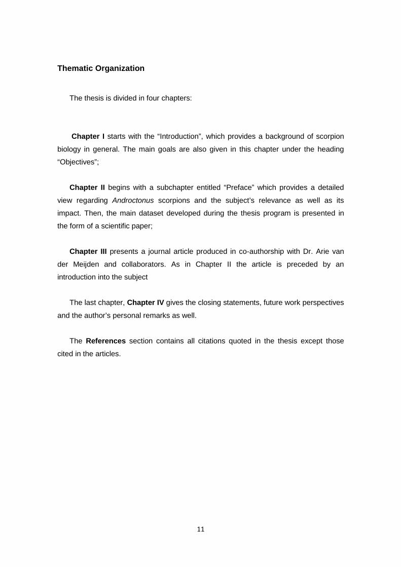
Thematic Organization
The thesis is divided in four chapters:
Chapter I starts with the “Introduction”, which provides a background of scorpion
biology in general. The main goals are also given in this chapter under the heading
“Objectives”;
Chapter II begins with a subchapter entitled “Preface” which provides a detailed
view regarding Androctonus scorpions and the subject’s relevance as well as its
impact. Then, the main dataset developed during the thesis program is presented in
the form of a scientific paper;
Chapter III presents a journal article produced in co-authorship with Dr. Arie van
der Meijden and collaborators. As in Chapter II the article is preceded by an
introduction into the subject
The last chapter, Chapter IV gives the closing statements, future work perspectives
and the author’s personal remarks as well.
The References section contains all citations quoted in the thesis except those
cited in the articles.

12

13
Chapter I
General Introduction
Objectives

14
1.1 General Introduction
1.1.1 State of the Art
Scorpions are a primitive and fascinating group of animals. Since early human
societies scorpions have been the object of worship and fear as well. This negative
reputation is built around myths and popular beliefs but also because few scorpions
are, in fact, capable of causing human death. Their appearance as aquatic organisms
dates back 450 million years but it was only upon transition to land that they
experienced a great radiation in the family tree (Polis, 1990). A unique set of biological
features allowed them to colonize all continents (except for Antarctica) and
environments. These nocturnal predators are abundant in tropical forests and even in
elevated mountains but the bulk of genera diversity occurs in arid and desert
environments (Brownell and Polis, 2001).
Scientific research concerning scorpion biology is an integrative field, combing
aspects of physiology, behavior, ecology and evolutionary biology. Today, as an
outcome, scorpions are an arachnid model to investigate broader questions of
organismal biology.
1.1.2 Origins and Biology
Scorpions are an old lineage of terrestrial arthropods and one of the most
cosmopolitan orders of the Arachnida class after spiders (Aranae), mites and ticks
(Acari), pseudoscorpions (Pseudoscorpiones) and opiliones (Opiliones Sundevall,
1833) (Savory, 1977). They first appear in the fossil record in the middle Silurian
(around 425 – 450 million years ago). They are a sister group to opiliones and together
form a taxonomic grouping named Stomothecata. The position Stomothecata occupy in
the Arachnid class is still debated but might be a sister clade to all the other subclasses
(Shultz, 2007). Ecologically there is also debate if scorpions shared the aquatic setting
with Eurypterids, a group of arthropods that was thought to be closely related do
scorpions (Gess, 2013; Legg et al., 2013). Around the middle Carboniferous (250 – 300
million years), the fossil record starts to show the first terrestrial scorpions (Polis and
Sissom, 1990) (Figure 1). Today, there are almost 2000 extant species in the
Scorpiones order (extinct species are not inserted in the scope of this work) (Rein,
2012). Nevertheless, it constitutes a minor percentage of all arachnid species, circa 1,6
% (Hallan, 2005).

15
There are two key phases in scorpion evolution: the aquatic-to-inland transition
and colonization of land. On one hand, the shift to land represented great
morphological changes that impacted directly on the role as terrestrial predators. On
the other hand, taking over land-masses produced a quick radiation into more than 50
families (Brownell and Polis, 2001). Surprisingly, this genetic outburst was not followed
by major morphological modifications, unlike insects (Hexapoda, Insecta) and the most
abundant arachnids (ticks and spiders). Although most families from that initial
radiation are extinct, they were much like the present species morphologically
(Brownell and Polis, 2001). Although there are ecomorphological adaptations, these do
not obscure the general resemblance among scorpions.
Figure 1: The first known terrestrial scorpions; Lower Carboniferous scorpions from the East Kirkton Quarry near Edinburgh, Scotland. To the left, a fourth stadium juvenile mesoscorpion. To the right, a detail of chelicerae, proximal end of pedipalps, and anterior end of oral tube (From Jeram, 2000).
Apart from polar areas, scorpions exist everywhere. They dwell in the highly
eroded intertidal coast of Baja California, the Ural and Caucasus elevated mountains
and even strictly in caves. In these habitats, some are specialized burrowers, whereas
others are vagrant, saxicolous or arboreal (Lamoral, 1979; Polis, 1990). Two
environments are, however, of top position considering genetic diversity. When it
comes to species diversity, tropical forests are home to the largest number of
scorpions. On the other hand, desert and arid landscapes harbor most of the extant

16
genera. It is not a coincidence that scorpions are customarily associated with deserts.
In such habitats, scorpions appear to be one of the most successful and important
predators in terms of density, diversity and biomass (Polis and Sissom, 1990). They
are not only widely spread, but they are ecologically essential in certain communities.
They can reach population densities of 5000 - 10000 animals per hectare, exceeded
only by ants and termites (Brownell and Polis, 2001). Desert species can endure
temperatures several degrees higher than most other desert arthropods are able to
tolerate. They also conserve water more efficiently than any other arthropod. Some
scorpions can live indefinitely without drinking water since water intake can be assured
solely by food. (Polis and Sissom, 1990).
1.1.3 Phylogeny and Molecular Studies
Phylogeny is the study of how organisms are related within an evolutionary
framework. Scorpions probably constitute a monophyletic taxon (Brownell and Polis,
2001).
Relationships are inferred from a variety of sources, for example, morphological
characters, DNA sequences and even behavioral data. Today, DNA sequence data is
the most important source of information in the construction of phylogenies. The
discovery of the polymerase chain reaction (PCR), the development of automated
sequencing and the sophistication of computational power (Caterino et al., 2000) and
bioinformatic software greatly potentiated phylogenetic studies in evolutionary biology.
Mitochondrial DNA as a molecular marker (Zhang and Hewitt, 1996) remains the most
common source of sequence data for studies of mid-to late Cenozoic-age divergences
(Sunnucks, 2000).
Mitochondrial DNA is the standard molecular marker in phylogeographic studies,
being maternally inherited and usually without recombination and selection (Piganeau
et al., 2004). This means that the variants present are not intermixed and so, the
mtDNA characters represent the presumed historical sequence of mutation events
accompanying the differentiation of maternal lines (Avise et al., 1987; Ballard and
Rand, 2005; Ballard and Whitlock, 2004). The mitochondrial is present in all
Eukaryotes (there are punctual exceptions) and presents a higher number of equal
copies, making this marker easy to assay (Avise et al., 1987; Ballard and Rand, 2005;
Ballard and Whitlock, 2004; Kocher et al., 1989). There has to be some caution,
however, using mtDNA for phylogenetic analyses. Cases where non-independent
replication occurs have been reported (Simon et al., 1994) and this phenomenon was
described in scorpions (Gantenbein et al., 2005). Nonetheless, the majority of scorpion

17
molecular phylogenetic studies choose this marker because there is a comprehensive
mtDNA database available and, therefore, a basis for comparative analyses through
public databases of DNA sequences. Further, there are no primers for nuclear DNA
markers widely used in the Scorpiones order.
1.1.4 Venom and the Buthidae family
All scorpions are capable of producing venom, a complex mixture of more than 200
identified toxins. The degree of toxicity is greatly variable among species. Venom
delivery is related with defense against predation and prey incapacitation. Scorpionism
(the severe or lethal poisoning by scorpion venom) depends largely on the distribution
and abundance of venomous species relatively to human populations. Incidents have
high occurrence in American and Asian countries. However, it is in North Africa,
particularly in the Saharan area, where it is a public threat (Chippaux and Goyffon,
2008). For example, in Morocco there is an annual mortality of 0.27 per 100 000
inhabitants, but in Tunisia this number can reach levels up to 6.67 deaths per 100 000
inhabitants each year in certain regions (Bouaziz et al., 2008; Touloun et al., 2012).
About 30 species of scorpion are recognized as potentially dangerous for humans
(Keegan, 1980). With one exception, species with highly potent, mammal-specific
neurotoxins belong to a single family, the Buthidae (Koch, 1837). Further details are
given in “Chapter II”
Figure 2: Estimated annual global incidence and mortality following scorpion stings. “Enven” refers to envenomings and “M” refers to million. From Chippaux, 2012)

18
The Buthidae is the largest scorpion family. This family is known from the fossil
record since the Paleocen-Eocene (Baltic amber) and represents one of the basal
evolutionary lineages of extant scorpions, so-called orthobothriotaxic Type A (Vachon,
1974). They are ecologically diverse and successful, widely spread across the globe,
occupying all six faunal regions (Lourenço, 2000). It contains approximately half of all
known extant scorpion species and is divided into 89 genera, most of which are found
in the Afrotropical region (Rein, 2012). Until 2005, the Buthidae scorpions were
considered to be phylogenetically basal to the other families. Since then, the
Pseudochactidae were placed as the most ancient of all extant families (Fet and
Soleglad, 2005). While the genus-level diversity in Buthidae is much higher in the Old
World compared to the New World, the opposite is observed at the species-level.
The fundamental reason for studying medically relevant scorpions was clearly
stated by Simmard and Watt (Polis, 1990): “we know practically nothing about the
natural history or field behavior of any of the deadly species”.

19
1.3 Objectives
The goal of this thesis was to produce a well supported phylogeny for the genus
Androctonus in North-Africa using a combination of DNA markers capable of resolving
interspecific relationships. In addition, we also constructed a molecular phylogeny for a
broad sampling across all scorpion families. These objectives were accomplished by:
• Testing primers for informative mitochondrial DNA sequence markers for
molecular phylogenetics;
• Uncovering levels of Maghrebian Androctonus diversity through DNA sequence
data from multiple mitochondrial genes;
• Testing the current Androctonus taxonomy and biogeographical framework
against the resulting molecular dataset;
• Using the same markers to construct a high-level phylogenetic hypothesis of
the major groups of scorpions;

20

21
CHAPTER II
Preface 1
Deep intraspecific divergences in the medically-relevant fat-tailed scorpions (Androctonus, Scorpiones)

22
2.1 Preface 1
2.1.1 Androctonus scorpions
The Palearctic biogeographic region presents especially rich faunas of arid
Buthidae scorpions. Among the most common and widely distributed buthid genera in
this region is the Androctonus scorpion (Fet et al., 2003). They have a wide geographic
range (from Togo to India) and their radiation has been associated with the aridification
of the Paleartic deserts (Fet, 1994).
Androctonus scorpions are commonly known as the fat-tail scorpions. They are
one of the most common and widely distributed buthid genera from the Paleartic region
(together with Buthus Leach, 1815, Hottentotta Birula, 1908 and Mesobuthus Vachon,
1950). The common name is attributed due to the thickness and strength of their
metasoma, the “tail”, (Figure 3). The name Androctonus is derived from a Greek word
which roughly translates into “man-killer”, a reminder of its highly toxic venom. These
scorpions occur in semi-arid and arid regions of desert areas but they have also been
found in anthropological disturbed landscapes (Stockmann and Ythier, 2010). They are
moderately sized scorpions with adults measuring up to ten centimeters, although
larger specimens are known. Preferred habitats include: stony soils, cactus hedges,
arid mountainous regions and high plateaus. They can also be found on steep slopes
of sand dunes but they avoid humid coastal areas (Gaban, 1997). Androctonus usually
dig burrows and hide under stones or in natural crevices. They can live four to five
years in captivity, depending on the temperature and feeding regime. In terms of
toxicity it is one of the most venomous scorpions. A. australis LD50 values can be as
low as 0.32 mg/kg (Goyffon et al., 1982). Notwithstanding, Androctonus is popular
among exotic animal traders particularly A. australis and A. amoreuxi which are
commonly available. Both the occurrence in the vicinity of human populations and the
population densities they reach make Androctonus a particularly dangerous genus
(Chippaux and Goyffon, 2008).

23
Figure 3: Dorsal view of Androctonus australis. The body is divided in two zones, the prosoma, containing the pedipalps, and the ophistosoma. The ophistosoma is subdivided in the mesosoma (abdominal region) and the metasoma bearing the venomous sting. Relevant structures and body segments are listed in the figure (taken from Stockmann and Ythier, 2010).

24
2.1.2 Androctonus taxonomy
The taxonomy of particular scorpion groups is somewhat controversial. Buthus
scorpions are a clear example, in which more than 15 species have been described in
the last decade based solely on morphological characters (e.g., Qi and Lourenço,
2007; Lourenço and Duhem, 2009; Lourenço et al., 2010; Yağmur et al., 2011). Trying
to identify them is, for most authors, a daunting task due to conflicting keys and
overlapping character ranges. The Androctonus genus does not qualify to be
considered as complex as the genera Buthus or Tityus. Nonetheless, Androctonus
taxonomy underwent repeated taxonomic changes in recent years.
Androctonus scorpions were already known to science since Linnaeus as
Scorpio australis Linnaeus, 1758 [= Androctonus australis (Linnaeus, 1758)], and this
remains the type species. Olivier (1807) and Audouin (1826) also described two
species under the genus Scorpio Linnaeus, 1758. The genus Androctonus, as it is
recognized nowadays, was later described by Ehrenberg (Ehrenberg and Hemprich,
1828) with two sub-genera Prionurus Ehrenberg and Leiurus Ehrenberg. The 19th and
early 20th centuries were a rich period in Androctonus taxonomy: five species and five
subspecies attributed to Androctonus were described: Olivier (1807), Audoin, (1826),
Koch (1839), Pocock (1897, 1900, 1902), (Pallary, 1924) and Caporiacco, 1932. These
authors however abandoned the name Androctonus and rather used the name
Pionurus either as a sub-genus of the “catch-all” genus Buthus or as a genus itself.
With one exception, all described taxa were only later assigned to the genus
Androctonus. It was only in 1948 that the first of several exhaustive works on North
African scorpiofauna were produced by Vachon. As a signature legacy throughout his
lifework, Vachon standardized Androctonus taxonomy and turned it into a coherent
genus (Vachon, 1952, 1949). Following Kraepelin’s scheme of diagnostic characters
(Kraepelin, 1903) rather than Birula’s (Birula, 1910), Vachon’s innovative work provided
a list of nine Androctonus taxa from North Africa, including subspecies. Six of those
were described previously though scattered between genera Buthus and Prionurus;
three others were newly described by himself (Table 1). As an influential scorpiologist,
Vachon pioneered modern scorpion taxonomy and, as a sign of that, all species he
described (with one exception) are still considered valid Androctonus taxa today. Later,
he would also revise Mauritanian (Vachon, 1953) and Afghan Androctonus scorpions
(Vachon, 1958). Advances in the second half of the 20th century were more directed
towards biochemical taxonomy. Goyffon and Lamy produced important works not only
about the morphological differentiation among Tunisian A. australis subspecies but also
about their biochemical differences (Goyffon and Lamy, 1973; Lamy et al., 1974). This

25
period was rich on the biochemical and molecular understanding of some of the
scorpions’ structures and venom. A. australis was the main target of these studies but
other species were used as well. Few papers on North African Androctonus taxonomy
were produced, but it is worth noting Fet’s contribution with one subspecies, A.
amoreuxi levyi (Fet, 1997). The most important work in this period would come in the
form of a book. Victor Fet and associates compiled and reviewed almost 250 years of
scorpion taxonomy. Catalog of the Scorpions of the World became one of the most
important tools for modern day research in this field (Fet et al., 2000). In it, eight
species and 14 subspecies were recognized for Androctonus. Few changes were
made over Goyffon, Lamy and associates’ descriptions but also in Asian species
(Table 1). After a halt of almost 60 years since the last major taxonomical revision,
Lourenço made a taxonomical review of Androctonus (Lourenço, 2005). Important
changes included: A. australis and A. mauritanicus were no longer considered to have
subspecies; A. crassicauda gonneti was raised to the status of full species; A. aeneas
was placed in the synonymy of A. bicolor; A. liouvillei was reconsidered as a valid
species (Table 1). Lourenço’s contribution did not end with this survey. As a
scorpiologist with more than 360 scorpion taxa described, he introduced four more
Androctonus species and revalidated another one. Today, the genus Androctonus
features 19 species of which almost two thirds occur in North Africa (see Table 1).

26
Table 1 Androctonus taxonomy – Taxonomical descriptions compiled in three scorpiologists works. Central African and Asian taxa are omitted. (A. aeneas = A. bicolor).
2.1.3 Why study Androctonus scorpions?
The first molecular phylogeny of the family Buthidae (Fet et al., 2003) included a
sequence of A. amoreuxi. This work was the first and, so far, the only DNA phylogeny
between buthid genera. With a weak support, Androctonus is placed more closely
together with Leiurus Ehrenberg, 1828 rather than with Buthus (Figure 4). This was a
surprising result because, apart from historically being confused with one another
morphologically, Androctonus and Buthus scorpions share the same number and
arrangement of pedipalp manus granules (3+1 terminal). On the other hand, the
proposed clade unites the most venomous species pair of scorpions: Androctonus
australis and Leiurus quinquestriatus (Ehrenberg, 1828). The importance around the
relationship between toxic scorpions directly relates to the emergence of such potent
venoms.
Taxon Author Year Taxon Author Year
Vach
on, 1
948,
195
2 A. australis Linnaeus 1758
Fet e
t al.,
200
0
A. australis Linnaeus 1758 A. crassicauda Olivier 1807 A. crassicauda Olivier 1807 A. amoreuxi Audouin 1826 A. amoreuxi Audouin 1826 A. aeneas Koch 1839 A. bicolor Ehrenberg 1828 A. mauritanicus Pocock 1902 A. mauritanicus Pocock 1902 A. hoggarensis Pallary 1929 A. hoggarensis Pallary 1929 A. sergenti Vachon 1948 A. sergenti Vachon 1948
Lour
enço
, 200
5
A. australis Linnaeus 1758
Cur
rent
taxo
nom
y
A. australis Linnaeus 1758 A. crassicauda Olivier 1807 A. crassicauda Olivier 1807 A. amoreuxi Audouin 1826 A. amoreuxi Audouin 1826 A. bicolor Ehrenberg 1828 A. bicolor Ehrenberg 1828 A. mauritanicus Pocock 1902 A. mauritanicus Pocock 1902 A. liouvillei Pallary 1924 A. liouvillei Pallary 1924 A. hoggarensis Pallary 1929 A. hoggarensis Pallary 1929 A. gonneti Vachon 1948 A. gonneti Vachon 1948 A. sergenti Vachon 1948 A. sergenti Vachon 1948
A. aleksandrplotkini Lourenço & Qi 2007
A. maroccanus Lourenço, Yhtier & Leguin 2009

27
Figure 4: Maximum Likelihood tree of Old World and New World Buthidae of the 16S mtDNA region. From Fet et al., 2003.
The study of venom toxins provides insights on scorpion evolution as well. For
instance, New World buthids have separate mammal and insect-specific neurotoxins
for Na+ channels but Old World buthids have non-specific toxins that act both on
mammals and insects. This independent evolution of venoms probably emerged after
the Laurasia versus Gondwana split and is a sign of the selective pressures during the
aridification of the Paleartic in the Tertiary period (Fet et al., 2003). This period
witnessed a rapid radiation of small burrowing mammals in arid landscapes. Rodents
would be a direct competitor for territory in the subsoil but also important nocturnal
predators as is the case today (McCormick and Polis, 1990). This could explain why
specific mammal targeted toxins used in defense are present in burrow-living scorpions
rather than vegetation-inhabiting New World buthids. There are, however, some

28
exceptions of scorpion toxins that follow a different path on sodium channels: they act
both in alpha and beta sites of these proteins’ domains. They were first isolated in A.
australis starting a discussion that mammal-targeted buthid toxins “originated from
North Africa” (Loret and Hammock, 2001). Although an interesting statement it is not
testable given our current knowledge:
• The Asian range of A. australis is still debated. Lourenço (2005) defined
Egypt as the eastern limit of distribution raising the middle eastern A.
australis baluchicus to species level;
• The center of origin is unknown. There is no wide biogeographical study of
Androctonus distribution, diversity and relationship among their species;
• The toxin in question, AahIT4, or related toxins have not been tested in an
evolutionary framework mostly because these experiments are nearly
impossible to replicate. This happens because toxicological literature
provides poor taxonomic identifications. A. australis is often oversimplified
as the “Sahara scorpion” and sampling has equally poor georeferencing
(sometimes non-existent).
As highlighted by Victor Fet and coworkers (2003), technical difficulties such as
the above mentioned lead to “unjustified and superficial conclusions about evolution of
both species and toxins”. A molecular study of Androctonus scorpions would serve as
the starting point to overcome some obstacles by providing quantitative criteria to
delimit taxa and species distribution. In conjugation with the available buthid phylogeny,
the exceptional properties of A. australis can be tested as an ancestral or
synapomorphic character, a key feature to understand scorpion evolution. More
recently, similar compounds have been identified in Centruroides suffussus suffusus
(Espino-Solis et al., 2011).

29

30
2.2 Deep intraspecific divergences in the medically-relevant fat-tailed scorpions
(Androctonus, Scorpiones)
(Graphical abstract)

31
Deep intraspecific divergences in the medically-relevant fat-tailed scorpions (Androctonus, Scorpiones)
P. Coelho1, P. Sousa1,2, D. J. Harris1,2 & A. van der Meijden1*
1 CIBIO-UP, Centro de Investigação em Biodiversidade e Recursos Genéticos, Campus Agrário de Vairão, Vairão 4485-661,
Portugal
2 Departamento de Biologia, Faculdade de Ciências, Rua do Campo Alegre, Porto FC4 4169-007, Portugal
* Corresponding author. E-mail address: [email protected]
Keywords: Androctonus; Scorpions; Phylogeny; Biogeography; Cryptic Diversity
Abstract
North Africa is the major hotspot of scorpion sting incidents in the world, and
Androctonus scorpions, commonly known as fat-tailed scorpions, are among the most
medically relevant scorpions in North Africa. Since venom composition within species is
known to vary regionally, the improvement of therapeutic management depends on a
correct assessment of the existing regional specific and sub-specific variation. The
genus Androctonus contains 19 species distributed from Togo and Mauritania in the
west, North Africa, through the Middle East and to as far east as India. With ten
species, a substantial amount of this genus’ diversity occurs in North Africa, although
other regions remain understudied. In this study, we assessed the phylogeographical
patterns in six species of Androctonus scorpions from North Africa using mitochondrial
DNA markers. We sequenced COI, 12S, 16S and ND1 genes from 110 individuals.
Despite lacking basal resolution in the tree we found taxonomical and geographically
coherent clades. We discovered deep intraspecific variation in the widespread A.
amoreuxi and A. australis, which consisted of several well supported clades. Genetic
distances between some of these clades are as high as those found between species.
North African A. australis have a deep split in Tunisia around the Chott el-Djerid salt-
lake. A surprising split between A. amoreuxi scorpions was found in Morocco. We also
found deep divergences in A. mauritanicus, corresponding to areas attributed to
invalidated subspecies. In addition we uncovered a clade of specimens from coastal
south Morocco, which could not be ascribed to any know species using morphological
characters. Based on these findings we recommend a reassessment of venom potency
and anti-venom efficacy between these deep intraspecific divergent clades.

32
1. Introduction
Worldwide, 1.2 million people are stung by scorpions every year. Scorpionism,
defined as the severe to lethal incident as a consequence of a scorpion sting (Lourenço
and Cuellar, 1995) may be responsible for 3250 global annual mortalities which are
mostly concentrated in a few high-risk areas (Chippaux and Goyffon, 2008). North
Africa in particular is considered a high-risk area for scorpionism (Chippaux and
Goyffon, 2008), with the genera Leiurus and Androctonus being the foremost cause of
serious envenomation in this area (Goyffon and Guette, 2005; Graham, 2011;
Habermehl, 1994). Five Androctonus species are considered as dangerous to man,
particularly the widespread A. australis (Linnaeus, 1758) and A. mauritanicus (Pocock,
1902), which are the most dangerous Androctonus in the Maghreb region (Goyffon and
Guette, 2005). A. australis is known for dangerously envenomating humans and
possessing a high toxicity (LD50 = 0.32 mg/kg; Watt and Simard, 1984). For this reason,
A. australis was one of the first species of scorpions to have its venom purified for
neurotoxin characterization (Miranda et al., 1966). As in snakes (Daltry et al., 1996;
Prasad et al., 1999), scorpion venom is known to have considerable intraspecific
regional variation in composition (el Ayeb and Rochat, 1985; Newton et al., 2007;
Smertenko et al., 2001), and thus a different response to antivenom treatment (Omran
and McVean, 2000). Furthermore, other species such as A. amoreuxi (Audouin, 1826)
may also cause more cases of scorpionism than currently thought (Goyffon et al.,
2012). It is therefore important to study the phylogeographical patterns of Androctonus
over a great part of their distribution as it may have direct applications in therapeutic
management.
The scorpion genus Androctonus was first described by Ehrenberg in 1828. Vachon
(1952) stabilized the genus’ taxonomy, transforming it into a morphological and
geographical coherent group with seven species known in North Africa. Lourenço
(2005) produced an important taxonomical revision of the genus: the subspecies of A.
australis and A. mauritanicus were no longer considered valid, A. crassicauda gonneti
Vachon, 1948 was raised to the status of species, A. aeneas C. L. Koch, 1839 was
placed in the synonymy of A. bicolor Ehrenberg, 1828 and A. liouvillei (Pallary, 1924)
was raised to the species level. Since then, two more taxa were described from North
Africa, A. aleksandrplotkini Lourenço & Qi, 2007 and A. maroccanus Lourenço Yhtier &
Leguin, 2009, and one was reconsidered as a valid taxon, A. eburneus Pallary, 1928.

33
The genus is distributed from Togo to Morocco in the Atlantic coast of Africa (Lourenço
and Qi, 2007; Lourenço, 2008) to the Maghreb countries and Egypt (Vachon, 1952),
the Middle East (Levy and Amitai, 1980), reaching across Afghanistan (Vachon, 1958)
to India (Tikader and Bastawade, 1983). Some species have been cited from Saudi
Arabia, Yemen and Lebanon, but these records require additional confirmation (Fet et
al., 2000). In this work, we assess five species of the Maghrebian countries except for
Libya, plus Egypt and the Sinai Peninsula. A. mauritanicus occurs in Morocco. This
country shares a further three species with Algeria, Tunisia and/or Egypt (A. liouvillei,
A. australis and A. amoreuxi), while A. bicolor occurs in Algeria and Tunisia.
The radiation of buthids has been associated with the aridification of the Palearctic
Region (Fet et al., 2003, 1998). Little is known about the biogeography of Androctonus
however. Recently, molecular tools have been used to assess the phylogeny of some
species in Tunisia. Ben Ali et al., (2000), using nuclear DNA ITS regions (ITS-rDNA),
found paraphyletic clades in three well accepted taxa (A. bicolor, A. australis and A.
amoreuxi). With the same outcome, Ben Othmen et al., (2004), using allozymic
differentiation, found little support for the monophyly of A. australis and A. amoreuxi
individually. However, the same authors recovered three well-supported monophyletic
lineages using 16S-rDNA, each corresponding to a species, thus demonstrating the
usefulness of this gene as a barcoding marker (Ben Othmen et al., 2009). Furthermore,
they found a phylogenetic pattern of A. australis in Tunisia, where each of its two
lineages are distributed to the north or south of the Chott el-Djerid salt lake. However
their molecular dating makes it unlikely that the salt lake formation has generated a
vicariant evolution of Tunisian A. australis. In recent years, molecular studies using the
mitochondrial COI gene already uncovered considerable cryptic diversity in other
scorpion genera in the Maghreb region (Gantenbein and Largiadèr, 2003; Sousa et al.,
2012, 2011, 2010).
In this study, we assess the patterns of diversity estimated from COI, 16S and 12S
sequence data across the Maghreb region and Egypt. We provide the first molecular
phylogeny for Androctonus scorpions in Morocco, Algeria and Egypt.

34
2. Materials and Methods
2.1 Taxon sampling
We collected samples in the field in three countries: Morocco, Tunisia and Egypt
(figure 1). Algerian samples were donated by Dr. Said Larbes. Additional specimens
were purchased through the pet-trade. Sampling points are illustrated in Figure 1 and
further details are provided in Table 1. Scorpions were captured with long forceps and
preserved in 96% ethanol. To minimize the impact on scorpion populations, non-lethal
sampling methods were used when possible, and consisted of removing the distal part
of one of the second leg pairs. The scorpion was subsequently placed in the sand,
facilitating the clotting process and thus minimizing haemolymph loss. Scorpions can
recover partially amputated appendages after a few molts and it is common for
scorpions to be active and functional even when missing appendages (pers. obs.).
Voucher specimens were collected at selected sites. All specimens are deposited in
the collection of CIBIO, Centro de Investigação em Biodiversidade e Recursos
Genéticos, Universidade do Porto, Vairão, Vila do Conde, Portugal.
Some key species that could not be sampled in the field, e.g. Egyptian A. bicolor, were
obtained through reputable animal traders. Morphological identification was done in the
lab using keys by Vachon, (1952) and Lourenço, (2005). A single specimen of
Opisthacanthus asper (Peters, 1861) (Scorpiones: Liochelidae) was used as outgroup
(details provided in Table 1).

Figure 1: Map representing the sampling locations across North Africa of Androctonus scorpions. Insert shows Egyptian samples. Pet trade acquired samples were
without locality data, and are not shown. Symbols correspond to the phylogenetic clades (see figure 2).

DNA extraction and PCR conditions
Fresh or preserved leg muscle tissue (or metasoma muscle for smaller specimens)
was used for DNA extraction. Dissection occurred, preferentially, from the third leg in
order to minimize the loss of important taxonomical characters. Total DNA was
extracted using proteinase K digestion (10 mg/ml concentration) followed by a standard
salt extraction protocol (Bruford et al., 1992).
Four mitochondrial fragments were amplified: 16S ribosomal RNA (16S rRNA),
12S ribosomal RNA (12S rRNA), Cytochrome C Oxidase subunit 1 (COI) and NADH
Dehydrogenase 1 (ND1) (see Table 2). Polymerase chain reactions were performed in
a final volume of 25 µL and using 1.0 µL each of 10 pmol primer, 12.5 µL REDTaq
ReadyMix PCR Reaction Mix with MgCl2 (Sigma-Aldrich, St. Louis, USA), 1.0 µL of the
purified DNA and 9.5 µL of water. Difficult PCR reactions were amplified with a
Touchdown-PCR approach. Purified PCR templates were sequenced using dye-
labelled dideoxy terminator cycle sequencing on an ABI 3730XL at Macrogen Inc. The
sequencing primers were the same as those used in the PCRs.
Table 2: List of primers and PCR conditions used for molecular analyses. PCR conditions start with temperature (ºC) of each step followed by the time in seconds in brackets.
2.3 Data analysis
Chromatographs were checked manually for sequencing errors using FinchTV
1.4.0 (Geospiza, Inc.; Seattle, USA). Sequences were edited using MEGA 5 (Tamura
et al., 2011) and aligned using default settings of MUSCLE (Edgar, 2004) for non-
coding sequences and ClustalW (Larkin et al., 2007) for COI and ND1.
Gene Primer name Sequence (5' → 3') Source PCR conditions
16S rRNA
18-mer (forward) CGATTTGAACTCAGATCA Simon et al, 1994 94(180), [94(30), 50(45), 72(60)]x35, 72(300), 12(∞) 20-mer (reverse) GTGCAAAGGTAGCATAAT Gantenbein et al,
2000 12S
rRNA 12S-F_AvdM AGAG-TGACGGGCAATATGTG van der Meijden
et al, 2012 94(180), [94(30), 52(45),
72(60)]x35, 72(300), 12(∞) 12S-R_AvdM CAGCGGCTGCGGTTATAC
COI
LCO1490 (forward) GGTCAACAAATCATCATAAAGATATTGG Folmer et al. 1994
94(180), [94(30), 48(45), 72(60)]x35, 72(300), 12(∞) HCO219 (reverse) TAAACTTCAGGGTGACCAAAAAATCA
COI_avdm_F WTYCTACIAATCAYAARGATATTGG van der Meijden et al, 2012
94(180), [94(30), 49(45), 72(60)]x35, 72(300), 12(∞) COI_avdm_R TAMACYTCIGGGTGWCCAAAAAAYCA
ND1
ND1-LR-N-12945 (forward) CGACCTCGATGTTGAATTAA
Hedin, 1997 94(180), [94(30), 47(45), 72(60)]x35, 72(300), 12(∞) ND1-N1-J-12261
(reverse) TCGTAAGAAATTATTTGAGC

37
Phylogeny reconstruction was performed using Maximum Likelihood (ML) and
Bayesian Inference (BI) methods. A homogeneity partition test executed in Paup* 4.0
rejected homogeneity of the different genes. We therefore carried out both a portioned
and a combined analysis. The best fitting models of sequence evolution were
determined by the AIC criterion in JModeltest 2.1.2 (Darriba et al., 2012) both for the
separate genes and the combined dataset. ML tree searches were performed using
PhyML, version 3.0 (Guindon et al., 2010). Bootstrap branch support values were
calculated with 1000 replicates. The BI analysis was conducted with MrBayes 3.2.2,
with 5.000.000 generations, sampling trees every 10th generation (and calculating a
consensus tree after omitting the first 12.500 trees). Mean genetic distances between
and within clades were calculated with MEGA5 based on COI sequences. Variance
was estimated with 4000 bootstrap replicates, with uncorrected p-distances (Table 3).
GenBank accession numbers are given in Table 1.
In addition we downloaded all 16S rDNA sequences in Ben Othmen et al., (2009)
from GenBank and we combined these with our 16S dataset. Phylogeny reconstruction
of this extended dataset was performed with ML (tree not shown).
3. Results
3.1 Sequence data
We sequenced 110 Androctonus specimens and one outgroup (O. asper) (Table
1). The combined dataset consists of an alignment of 1457 basepairs (bp) (606 bp of
COI, 374 bp of 16S rRNA and 477 bp of 12S rRNA). The dataset included 777 variable
sites (53.3 % of the total nucleotide positions), 680 bp were constant (46.7 %) and 511
positions were parsimony informative (35.1%). Due to difficulties in amplifying ND1 only
41 sequences were available, and thus this gene was not included in the combined
analysis.
3.2 Overall phylogeny
Four of the six species were recovered as monophyletic (Figure 2). We identified 13
clades that correspond to biogeographical and/or taxonomical units. Unfortunately,
most of the deeper branches present low values of bootstrap support (ML) and
posterior probability (BI). Consequently, the relationships between species could not
always be resolved. ML and BI trees using a dataset that included ND1 sequences
(not shown) did not differ in the topology of well supported clades. Trees derived from
the individual genes did not contradict the result from the concatenated dataset.

38
Figure 2: Estimate of phylogenetic relationships of Androctonus (outgroup not shown). Posterior probabilities values and bootstrap support values are shown above and below nodes respectively. These samples are marked with the same colors and shapes in all figures.

39
3.3 Species-level clades
Androctonus amoreuxi, is divided into three clades, two of them containing
specimens from Morocco (A1 and A2), Tunisia and Algeria (A2) and one from Egypt
(A3). Clades A1 and A2 are placed as sister groups with a high support (BI = 1.00; ML
= 87) and the A3 clade has no well-supported affinities but may be sister taxon to A.
australis. The western part of the range, from Morocco to Tunisia, is divided into the
two clades; a northern (A2) and a southern clade (A1); the Southern Moroccan
amoreuxi (A1) clade extends South, from the regions of Assa and Zag in the South, to
the southern area of the Anti-Atlas Mountains’ and North towards Taouz, in the Erfoud
area, close to the Morocco-Algerian border, but also in the area of Midelt, in the Middle-
Atlas Mountains (figure 1). The Northwest African amoreuxi (A2) clade groups
specimens from three countries: in Morocco it is distributed in the Erfoud area and also
to the areas surrounding Bouarfa and Ain-Beni-Mathar (High Plateau in Northeast
Morocco); In Algeria it is represented in one location, Ghardaia (between the Great
Ergs); in Tunisia it is represented in one location, close to Tozeur (between Chott el-
Gharsa and Chott el-Djerid salt lakes). The third clade, A3 groups specimens that were
acquired via the pet-trade from Egypt, none of which are georeferenced. Pairwise
genetic distances between these clades are in a range between 7.5% and 9.2% (Table
3). Downloaded 16S sequences of A. amoreuxi are split between clades A1 and A2
with Ben Othmen et al., (2009)’s sequence (andr05) placed in the A2 clade whereas
the sequence of (Fet et al., 2003) is placed within our A1 clade (tree not shown).
The L clade (liouvillei clade) unites all Moroccan A. liouvillei scorpions. A total of
15 specimens were collected, ranging from the High-Atlas Mountains (Agdz), to the
Northeastern range of the Middle-Atlas (Outat El Haj) and to the High Plateau along the
Algerian borderland (e.g. Figuig). Average genetic distance within the clade is of 0.8%.
It appears to be sister taxon to A. bicolor.
A. mauritanicus, is presented in three clades; M2 and M3 are sister clades
(BI=0.60; ML=80) whereas M1 was unrelated to these. Specimens from the M1 clade
are located in the northern range of the Marrakech-Tensif-El Haouz administrative
region. The specimens from the M2 clade occur in a horizontal stretch from the peaks
of the Anti-Atlas (close to Ighern) along the Souss valley with the westernmost sample
being 6 km from the seacoast. The M3 clade is comprised of specimens from two
sampling locations, both from the Guelmim area in the southwest slope of the Anti-
Atlas mountains (see figure 1). Genetic distances between clades range from 9.0% to
10.5% while the highest genetic distance within these clades, in clade M2, is 2.3%
(Table 3).

40
A group of four dark-colored Androctonus, which morphologically resemble A.
liouvillei most closely, are designated here as A. cf. liouvillei (see section 4). These
specimens (clade G) were found in one sampling point in the southern limit of the Anti-
Atlas Mountains near the mouth of the Oued Draa.
Androctonus australis forms a monophyletic unit, divided into four clades. The
AU3 (east Tunisian australis) and AU4 (Egyptian australis) clades are sister groups in
the Bayesian tree (BI = 0.80), which is not the case in the ML analysis. Clades AU1
(Algerian australis) and AU2 (west Tunisian australis) are sister groups in both trees
(BI=0.95; ML=99). Starting from the East, the AU4 clade is exclusive to Egypt and is
distributed in at least three areas: Mersa Matruh, the West Mediterranean coast of
Egypt and north from Mount Sinai. Some of the specimens present in this clade are not
geographically referenced however, as they were obtained through the pet-trade. The
AU3 clade (East Tunisian australis) is distributed solely in Tunisia: specimens range as
far west as Kebili (Southeast of the Chott el-Djerid salt lake), north to the Chot el-Fejaj
salt lake and as far south as the Tataouine desert region (Figure 1). The AU2 clade
groups specimens from two countries: Tunisia and Algeria. In Tunisia this clade is
distributed in an area compressed between Gafsa (in the east), Chott el-Djerid salt lake
(in the South) and Chot el-Gharsa salt lake (in the west). In Algeria this clade is spread
in two regions: in M'Doukal, a region enclosed between the Ouled-Nail (in the
southwest), the Tell Atlas (in the North) and the Aurès mountains (in the East); and
near Hassi Fedoul, at the foot of the Tell Atlas, the salt water lakes (in the west) and
the Ouled-Nail mountains (in the east). The AU1 clade is comprised of specimens from
a single location, Ghardaia, located between the Great Ergs of Algeria (Figure 1).
Pairwise genetic distances between these clades range from 3.9% to 8.0%. The AU1
and AU4 clades present the lowest values of within group genetic distance (0.4%).
Among A. australis, the highest within group distance was found in clade AU2 (mean p-
distance = 1.2%). Our sampling confirms former suspicions on this species’ occurrence
in the Sinai (Levy and Amitai, 1980). The extended 16S dataset shows that Ben
Othmen et al., (2009)’s “northern and southern lineage” sequences are placed
throughout the AU2 and AU3 clades respectively (tree not shown).
The B clade (bicolor Clade) unites all A. bicolor specimens. Notably, this clade
has longer branches within it than any of the other clades in this study. For this reason
we divided this clade into three subclades (B1, B2 and B3) and quantified the genetic
distances between them. A. bicolor is present in Hassi Fedoul (Tell Atlas, Algeria), near
Gabès (central Tunisia) (Figure 1) and Egypt (no GPS point available). Genetic
distances between these subclades range from 4.2% to 7.9%. These subclades were
too small for mean p-distance within clades to be calculated.

41
Table 3 Genetic distances between and within groups. Numbers below the diagonal are mean p-distances calculated between clades whereas the corresponding variances, based on 4000 bootstrap replicates, are shown above the diagonal. Mean p-distances within clades are shown to the right.
4. Discussion
Genetic methods, using one or very few mitochondrial genes, have proven very
successful in uncovering cryptic diversity in North African scorpions (Froufe et al.,
2008; Sousa et al., 2012, 2011). Despite using three genes with a high number of
informative sites, the dataset did not provide sufficient resolution below the species
level. The relationships among these species therefore could not be resolved. Species-
level clades, however, were well resolved and received high support
.
4.1 Androctonus australis
There is a deep split between clades AU1 and AU2 on one side, and clades
AU3 and AU4 on the other. The two clades from the same species in Tunisia (clades
AU2 and AU3) are therefore more closely related to clades distributed in countries as
far east as Egypt (AU4) and as far west as Algeria (AU1) than to each other. The
genetic distance also reflects a higher distance between them than between the pairs
AU1-AU2 and AU3-AU4. Geographical factors might explain the intraspecific variation
observed. Tunisia has a heterogeneous landscape, with a mountainous North and low-
lying deserts in the South, separated by salt lakes. There is a large salt lake between
Distance between clades Within Clades A1 0,010 0,011 0,011 0,011 0,011 0,011 0,011 0,010 0,011 0,010 0,010 0,011 0,011 0,012 0,016 0,002 A1 A2 0,075 0,011 0,012 0,011 0,012 0,012 0,010 0,011 0,011 0,010 0,011 0,011 0,011 0,011 0,013 0,003 A2 A3 0,092 0,089 0,012 0,011 0,011 0,011 0,012 0,012 0,011 0,011 0,011 0,012 0,011 0,013 0,003 0,001 A3
AU1 0,090 0,102 0,090 0,007 0,009 0,011 0,012 0,011 0,012 0,012 0,011 0,012 0,012 0,012 0,004 0,002 AU1 AU2 0,087 0,102 0,093 0,039 0,010 0,010 0,012 0,011 0,012 0,011 0,011 0,011 0,011 0,012 0,012 0,003 AU2 AU3 0,090 0,098 0,097 0,061 0,069 0,009 0,012 0,011 0,011 0,012 0,010 0,011 0,011 0,012 0,009 0,002 AU3 AU4 0,094 0,093 0,089 0,080 0,080 0,056 0,013 0,011 0,012 0,012 0,011 0,012 0,012 0,012 0,004 0,001 AU4 B1 0,088 0,086 0,096 0,093 0,090 0,097 0,108 0,008 0,011 0,011 0,012 0,011 0,012 0,012 n/c n/c B1 B2 0,077 0,082 0,100 0,093 0,087 0,087 0,096 0,042 0,010 0,011 0,011 0,011 0,011 0,013 0,010 0,004 B2 B3 0,094 0,100 0,099 0,106 0,099 0,098 0,107 0,079 0,074 0,011 0,010 0,011 0,011 0,012 n/c n/c B3 L 0,087 0,081 0,096 0,099 0,099 0,108 0,108 0,087 0,085 0,083 0,011 0,011 0,011 0,011 0,008 0,002 L
M1 0,084 0,098 0,095 0,087 0,087 0,081 0,096 0,085 0,080 0,090 0,096 0,011 0,011 0,012 0,012 0,003 M1 M2 0,110 0,103 0,115 0,109 0,108 0,099 0,116 0,096 0,099 0,094 0,099 0,105 0,010 0,011 0,023 0,004 M2 M3 0,103 0,104 0,097 0,107 0,102 0,100 0,107 0,109 0,104 0,095 0,098 0,093 0,090 0,011 0,014 0,003 M3 G 0,101 0,087 0,111 0,100 0,104 0,096 0,102 0,109 0,109 0,106 0,085 0,111 0,104 0,088 0,004 0,002 G
A1 A2 A3 AU1 AU2 AU3 AU4 B1 B2 B3 L M1 M2 M3 G p-distance St. err.

42
the regions where clades AU2 and AU3 are situated. Salt lakes as big as Chott el-
Djerid might act as physical barriers for scorpion dispersion, as suggested by Ben
Othmen et al. (2009). Although their work dealt only with Tunisian Androctonus, had a
smaller sampling and used only 16S rRNA data, they also found a deep divergence
between the north-western and south-eastern Tunisian A. australis the same as the
one found between the clades AU2 and AU3. Their “northern” and “southern lineages”
are genetically and geographically congruent with the AU2 and AU3 clades
respectively. They explored the possibility of the lake’s formation causing the vicariant
event. However the dates for the lake’s formation do not coincide with their divergence
time estimation. The geological evidence on the date of formation of Chott el-Djerid salt
lake is ambiguous; it is dated to be 65 Ma old if it is considered the remains of an
Eocene inland sea (Levy and Amitai, 1980), or it is dated between 90 ka – 150 ka if it is
considered a continental lake formed by tectonic and climate changes (Ben Othmen et
al., 2009). Ben Othmen et al. (2009) calculated the date for the divergence of the
Tunisian lineages to be 3.43 - 9.90 Ma and thus, unlikely an effect of Chott el-Djerid’s
formation in either paleogeographic scenario. However, their data divergence time
estimate was not based on calibration points, but rather on a constant mutation rate,
and should be regarded with caution. Alternatively, the species may have become split
into two major lineages on either side of the holocene humid corridor (Drake et al.,
2011), which divided the Sahara in a roughly south-north direction. The recent
desertification of this humid corridor would have allowed the two diverged lineages to
come into secondary contact around Chott el-Djerid. Unpublished data shows Chott el-
Djerid also acts as a geographical barrier for Buthus scorpions (Pedroso et al., in
prep.). Further studies of this contact zone towards the south, in conjunction with
proper molecular divergence time estimates, could be helpful in understanding the
history of the holocene humid corridor, and the recent desertification process of the
Sahara desert.
A. australis is one of the most widespread scorpions in North Africa and one of
the leading causes of scorpionism in North Africa (Chippaux and Goyffon, 2008). Since
venom composition and efficacy is known to exhibit regional variation both in snakes
(Daltry et al., 1996; Prasad et al., 1999) and scorpions (Newton et al., 2007; Omran
and McVean, 2000), these two attributes may also differ between the clades we
identified in this study, and should be investigated separately.

43
4.2 Androctonus mauritanicus
A. mauritanicus is, together with A. australis, among the most dangerous
scorpions in the Maghreb (Chippaux and Goyffon, 2008). Between 1996 and 2006 in
Morocco, 53% of deaths that resulted from sting cases come from incidents with A.
mauritanicus (Touloun et al., 2012). Although these scorpions are found in median
altitudes in some of the highest mountains of Morocco they do not seem to be
restricted to the mountains, because specimens from the same clade can occur in low
altitudes as well (e.g. clade M2 and M3). There are, however, important mountain
ranges in between the clades such as: the High-Atlas between M1 and M2 clades and,
to some extent, the Anti-Atlas between M2 and M3 clades. It seems, therefore, that
oreography might be a probable cause for differentiation in this species as is the case
in Buthus scorpions (Habel et al., 2012). Vachon (1952) described two subspecies
inhabiting Morocco: A. m. mauritanicus and A. m. bourdoni. The distribution we found
for clade M1 largely coincides with the known distribution of A. m. mauritanicus, while
the distribution we found for clade M2 coincides with the known distribution of A. m.
bourdoni’s. Although Lourenço (2005) did not find evidence to support these two
subspecies, the genetic distances between them are at a level of those found between
full species of Androctonus (Table 3). In addition, clades M2 and M3 are themselves
separated by a similar species-level genetic distance. An exploratory morphological
analysis of the specimens of clade M3 found a very granulated prosoma and tergites,
which does not fit the description of A. mauritanicus. These morphological characters
also distinguish them from the specimens in clade M2, showing these scorpions are
distinct both genetically and morphologically. A more detailed morphological
assessment is clearly needed prior to a taxonomic revision.´
4.3 Androctonus cf. liouvillei
Near Tan Tan Plage, a coastal town, we found four unusual specimens. They
are morphologically different from A. mauritanicus and A. sergenti Vachon, 1948 (also,
dark colored species). They seem to be closer related morphologically with A. liouvillei,
and although we only captured 4 adult male specimens, they were found to have a
higher pectinal teeth count (33) than what was reported by Lourenço (2005), who
reported a 28 to 32 pectinal teeth count for A. liouvillei males, already a broader
interval than in Vachon (1952), of 28 to 30 pectinal teeth. They also have a more
slender pedipalp chela of 3.6 ratio than what was reported for A. liouvillei by Vachon
(1952), with a 3.2 ratio. No data is available in Lourenço (2005). As no other species

44
are known for that region, we designated the G clade specimens as A. cf liouvillei, a
species known from this region. However, our estimate of relationships based on
mtDNA clearly indicates that these are not related to A. liouvillei (Figure 2), although
exact relationships remain poorly resolved. Further sampling will be necessary to study
the relationships between this population and other species present in Morocco, but it
appears to be an undescribed new species.
4.4 Androctonus liouvillei
The L clade is formed by A. liouvillei scorpions. Although this clade is sister to
the A. bicolor, genetic distances between them are near the species-level,
corroborating Lourenço's (2005) morphologically based interpretation that A. liouvillei is
a true species and distinct from A. bicolor.
4.5 Androctonus amoreuxi
The distribution of clades A1 and A2 meet in the Erfoud area. Although there
are no obvious geographical formations separating them, the genetic distance between
clades is 7.5%, which is similar to the distance recovered between the A. australis
clades. Again, as currently recognized the species appears to be paraphyletic, with
clade A3 sister to A. australis although with very weak support levels. Although some
A. amoreuxi specimens were separated by the largest geographic distance of any
species in this study, we could not find morphological differences between them in
taxonomically relevant characters (Lourenço, 2005; Vachon, 1952). The fact that the
genetic distance between clades A2 and A3 is relatively high (8.9 %) can point to the
validity of A. amoreuxi’s subspecies: A. a. amoreuxi in the Maghreb and A. a. levyi Fet,
1997 in the Sinai Peninsula. However, further morphological study of the intervening
populations will be necessary prior to a taxonomic revision.
4.6 Androctonus bicolor.
The B clade has longer internal branches compared to other clades, but is
clearly a monophyletic lineage, sister taxon to A. liouvillei. The most recent taxonomic
survey of Androctonus genus estimates that A. bicolor, although a valid taxon,
“correspondant très probablement à une espèce polymorphe” (Lourenço, 2005). As our
samples reflect, A. bicolor scorpions are distributed over a large geographical distance,
which might explain the deep divergences between these subclades.

45
Summarizing, we were able to identify cryptic variation in A. australis, A.
mauritanicus and A. amoreuxi. Some cases were simpler, in which we could confirm
previous suspicions on the validity of a distinct group, such as A. liouvillei. Further work
should aim at sequencing nuclear genes in order to resolve deeper relationships within
the genus. It would also be very interesting to obtain samples of regional species like
A. aleksandrplotkini A. hoggarensis (Pallary, 1929), A. maroccanus and A. sergenti.
Those, together with a more extensive sampling of Algeria, Libya and Mauritania could
provide a prime perspective on the patterns and processes shaping Androctonus
evolution and current distribution in North Africa.
5. Conclusions
We here show that A. australis, A. mauritanicus and A. amoreuxi have deeply
divergent subclades. Such variation can be reflected in the venom these animals
produce, as seen in other scorpion species (Newton et al., 2007; Omran and McVean,
2000; Smertenko et al., 2001). We therefore suggest that the venom of A. australis, A.
mauritanicus and A. amoreuxi should be studied with these deep divergences in mind.
Antivenom developed for scorpions in one region should be tested for efficacy against
venoms from regions (Fatani et al., 2010) where scorpions correspond to different
clades as identified here.
Furthermore despite lack of basal resolution, our results have clear bearing on
the taxonomic status of some of the (sub)species included in our study. In addition, we
identified a population of A. liouvillei-like specimens near Tan-Tan (clade G) which
merits further study as a potential new species.
Acknowledgements
We are indebted to Dr. Said Larbes of the Département de Biologie, Faculté des Sciences Biologiques et Agronomiques, Université M. Mammeri, Tizi-Ouzou, Algeria for supplying important samples from Algeria. We thank Abdullah Nagy of the St. Catherine wildlife preserve, Sérgio Henriques, Diana Pedroso and our colleagues at the CIBIO institute for assistance in the field. Thanks to Arendo Flipse for supplying some of the Egyptian scorpions. This work was funded by Fundação para a Ciência e Tecnologia, BIABDE/74349/2006 (to DJH), SFRH/BD/74934/2010 (to PS), SFRH/BPD/48042/2008 (AvdM) and a FCT I&D project (PTDC/BIA-BEC/104644/2008) to AvdM. This work was supported by a post-doctoral grant from the Fundação para a Ciência e a Tecnologia, Portugal [SFRH/BPD/48042/2008 to AvdM] and an FCT research grant [PTDC/BIA-BEC/104644/2008 to AvdM].

46
References
Audouin, 1826. Explication sommaire des planches d’arachnides de l'Égypte et de la Syrie publiées par J. C. Savigny, membre de l'Institut; offrant un exposé des caractères naturels des genres avec la distinction des espèces. In: Description de l’Égypte, Ou Recueil Des Observations et Des Recherches Qui Ont Été Faites En Égypte Pendant L'expédition de L'armée Française. Histoire Naturelle, pp. 99–186.
Avise, J.C., Arnold, J., Ball, R.M., Bermingham, E., Lamb, T., Neigel, J.E., Reeb, C.A., Saunders, N.C., 1987. Intraspecific phylogeography: the mitochondrial DNA bridge between population genetics and systematics. Annu. Rev. Ecol. Syst. 18, 489–522.
Ballard, J.W.O., Rand, D.M., 2005. The Population Biology of Mitochondrial DNA and Its Phylogenetic Implications. Annu. Rev. Ecol. Evol. Syst. 36, 621–642.
Ballard, J.W.O., Whitlock, M.C., 2004. The incomplete natural history of mitochondria. Mol. Ecol. 13, 729–44.
Ben Ali, Z., Boursot, P., Said, K., Lagnel, J., Chatti, N., Navajas, M., 2000. Comparison of ribosomal ITS regions among Androctonus spp. scorpions (Scorpionida: Buthidae) from Tunisia. J. Med. Entomol. 37, 787–90.
Ben Othmen, A., Chatti, N., Ben Ali-Haouas, Z., Ouldbrahim, I., Said, K., 2004. Allozymic differentiation of Tunisian populations of Androctonus species and Buthus occitanus (Scorpiones: Buthidae). Biol. J. Linn. Soc. 81, 255–265.
Ben Othmen, A., Said, K., Mahamdallie, S.S., Testa, J.M., Haouas, Z., Chatti, N., Ready, P.D., 2009. Phylogeography of Androctonus species (Scorpiones: Buthidae) in Tunisia: Diagnostic characters for linking species to scorpionism. Acta Trop. 112, 77–85.
Bouaziz, M., Bahloul, M., Kallel, H., Samet, M., Ksibi, H., Dammak, H., Ahmed, M.N. Ben, Chtara, K., Chelly, H., Hamida, C. Ben, Rekik, N., 2008. Epidemiological, clinical characteristics and outcome of severe scorpion envenomation in South Tunisia: multivariate analysis of 951 cases. Toxicon 52, 918–26.
Brownell, P.H., Polis, G.A., 2001. Scorpion Biology and Research. Oxford University Press, New York.
Bruford, M.W., Hanotte, O., Brookfield, J.F.Y., Burke, T., 1992. Single-locus and multilocus DNA fingerprinting. In: Hoelzel, A.R. (Ed.), Molecular Genetic Analysis of Populations: A Practical Approach. IRL Press, Oxford, pp. 225–270.
Caterino, M.S., Cho, S., Sperling, F.A., 2000. The current state of insect molecular systematics: a thriving Tower of Babel. Annu. Rev. Entomol. 45, 1–54.
Chippaux, J.-P., 2012. Emerging options for the management of scorpion stings. Drug Des. Devel. Ther. 6, 165–73.
Chippaux, J.-P., Goyffon, M., 2008. Epidemiology of scorpionism: a global appraisal. Acta Trop. 107, 71–9.
Daltry, J.C., Wüster, W., Thorpe, R.S., 1996. Diet and snake venom evolution. Nature 379, 537–40.

47
Darriba, D., Taboada, G.L., Doallo, R., Posada, D., 2012. jModelTest 2: more models, new heuristics and parallel computing. Nat. Methods 9, 772.
Drake, N.A., Blench, R.M., Armitage, S.J., Bristow, C.S., White, K.H., 2011. Ancient watercourses and biogeography of the Sahara explain the peopling of the desert. Proc. Natl. Acad. Sci. U. S. A. 108, 458–62.
Edgar, R.C., 2004. MUSCLE: multiple sequence alignment with high accuracy and high throughput. Nucleic Acids Res. 32, 1792–7.
Ehrenberg, Hemprich, 1828. Arachnoidea, Scorpiones. In: Symbolae Physicae Seu Icones et Descriptiones Animalium Evertebratorum Sepositis Insectis Quae Ex Itinere per Africanum Borealem et Asiam Occidentalem. Berlin.
El Ayeb, M., Rochat, H., 1985. Polymorphism and quantitative variations of toxins in the venom of the scorpion Androctonus australis Hector. Toxicon 23, 755–60.
Espino-Solis, G.P., Estrada, G., Olamendi-Portugal, T., Villegas, E., Zamudio, F., Cestele, S., Possani, L.D., Corzo, G., 2011. Isolation and molecular cloning of beta-neurotoxins from the venom of the scorpion Centruroides suffusus suffusus. Toxicon 57, 739–46.
Fatani, A.J., Ahmed, A. a E., Abdel-Halim, R.M., Abdoon, N. a, Darweesh, A.Q., 2010. Comparative study between the protective effects of Saudi and Egyptian antivenoms, alone or in combination with ion channel modulators, against deleterious actions of Leiurus quinquestriatus scorpion venom. Toxicon 55, 773–86.
Fet, V., 1994. 31. Fauna and Zoogeography of Scorpions (Arachnida: Scorpions) in Turkmenistan. In: Biogeography and Ecology of Turkmenistan. Kluwer Academic Publisher, Dordecht, pp. 525–534.
Fet, V., 1997. Notes on the taxonomy of some old world scorpions (Scorpiones: Buthidae, Chactidae, Ischnuridae, Scorpionidae). J. Arachnol. 25, 245–250.
Fet, V., Gantenbein, B., Gromov, A.A. V, Lowe, G., Lourenço, W.R., 2003. The first molecular phylogeny of Buthidae (Scorpiones). Euscorpius 4, 1–10.
Fet, V., Polis, G.A., Sissom, W.D., 1998. Life in sandy deserts: the scorpion model. J. Arid Environ. 39, 609–622.
Fet, V., Sissom, W., Lowe, G., Braunwalder, M., 2000. Catalog of the scorpions of the world (1758-1998). New York Entomological Society, New York.
Fet, V., Soleglad, M., 2005. Contributions to scorpion systematics. I. On recent changes in high-level taxonomy. Euscorpius 31, 1–13.
Froufe, E., Sousa, P., Alves, P.C., Harris, D.J., 2008. Genetic diversity within Scorpio maurus (Scorpiones: Scorpionidae) from morocco: Preliminary evidence based on CO1 mitochondrial DNA sequences. Biologia (Bratisl). 63, 1157–1160.
Gantenbein, B., Eightley, P.D., 2004. RATES OF MOLECULAR EVOLUTION IN NUCLEAR GENES OF EAST MEDITERRANEAN SCORPIONS. Evolution (N. Y). 58, 2486–2497.
Gantenbein, B., Fet, V., Gantenbein-Ritter, I. a, Balloux, F., 2005. Evidence for recombination in scorpion mitochondrial DNA (Scorpiones: Buthidae). Proc. Biol. Sci. 272, 697–704.

48
Gantenbein, B., Largiadèr, C.R., 2003. The phylogeographic importance of the Strait of Gibraltar as a gene flow barrier in terrestrial arthropods: a case study with the scorpion Buthus occitanus as model organism. Mol. Phylogenet. Evol. 28, 119–130.
Gess, R.W., 2013. The earliest record of terrestrial animals in Gondwana: A scorpion from the Famennian (Late Devonian) Witpoort Formation of South Africa. African Invertebr. 54, 373–379.
Goyffon, M., Dabo, A., Coulibaly, S., Togo, G., Chippaux, J., 2012. Dangerous scorpion fauna of Mali. J. Venom. Anim. Toxins Incl. Trop. Dis. 18, 361–368.
Goyffon, M., Guette, C., 2005. Scorpions dangereux du Niger. Bull. la Société Pathol. Exot. 98, 293–295.
Goyffon, M., Lamy, J., 1973. Une nouvelle sous-espèce d’Androctonus australis L. (Scorpion, Buthidae): Androctonus australis garzonii n. ssp. Caractéristiques morphologiques, écologiques et biochimiques. Bull. la Société Zool. Fr. 98, 137–144.
Goyffon, M., Vachon, M., Broglio, N., 1982. Epidemiological and clinical characteristics of the scorpion envenomation in Tunisia. Toxicon 20, 337–44.
Graham, M.R., 2011. Scorpions of the World Scorpions of the World. Roland Stockmann , Eric Ythier . 2010. N.A.P. Editions. France. 567 pp. ISBN: 978-2-913688-11-7. 75€. J. Arachnol. 39, 166–167.
Guindon, S., Dufayard, J.-F., Lefort, V., Anisimova, M., Hordijk, W., Gascuel, O., 2010. New algorithms and methods to estimate maximum-likelihood phylogenies: assessing the performance of PhyML 3.0. Syst. Biol. 59, 307–21.
Habel, J.C., Husemann, M., Schmitt, T., Zachos, F.E., Honnen, A.-C., Petersen, B., Parmakelis, A., Stathi, I., 2012. Microallopatry caused strong diversification in Buthus scorpions (Scorpiones: Buthidae) in the Atlas Mountains (NW Africa). PLoS One 7, e29403.
Habermehl, G., 1994. Gift-Tiere und ihre Waffen: Eine Einführung für Biologen, Chemiker und Mediziner. Ein Leitfaden für Touristen, 5th editio. ed. Springer Verlag, Heidelberg.
Herrel, A., Huyghe, K., Vanhooydonck, B., Backeljau, T., Breugelmans, K., Grbac, I., Van Damme, R., Irschick, D.J., 2008. Rapid large-scale evolutionary divergence in morphology and performance associated with exploitation of a different dietary resource. Proc. Natl. Acad. Sci. U. S. A. 105, 4792–5.
Jeram, A.J., 2000. Paleontology. In: Polis, G.A., Brownell, P. (Eds.), Scorpion Biology and Research. Oxford University Press, Oxford, pp. 370–392.
Kocher, T.D., Thomas, W.K., Meyer, A., Edwards, S. V, Pääbo, S., Villablanca, F.X., Wilson, A.C., 1989. Dynamics of mitochondrial DNA evolution in animals: amplification and sequencing with conserved primers. Proc. Natl. Acad. Sci. U. S. A. 86, 6196–6200.
Kraepelin, K., 1903. Scorpione und Solifugen Nordost-Afrikas, gesammelt 1900 und 1901 von Carlo Freihern von Erlanger und Oscar Neumann. Zool. Jahrbücher. Abteilung für Syst. Geogr. und Biol. der Tiere 18, 557–578.
Lamoral, B.H., 1979. The Scorpions of Namibia (Arachnida-Scorpionida). Ann. Natal Museum 23, 497–784.
Lamy, J., Le Pape, G., Weill, J., 1974. Hétérogénéité de l’espèce Androctonus australis (Scorpiones, Buthidae). Création d'une nouvelle sous-espèce A. australis africanus n. spp.

49
sur la base de critères biochimiques et génétiques. C. R. Hebd. Seances Acad. Sci. 278, 3223–3226.
Larkin, M.A., Blackshields, G., Brown, N.P., Chenna, R., McGettigan, P.A., McWilliam, H., Valentin, F., Wallace, I.M., Wilm, A., Lopez, R., Thompson, J.D., Gibson, T.J., Higgins, D.G., 2007. Clustal W and Clustal X version 2.0. Bioinformatics 23, 2947–8.
Legg, D., Sutton, M., Edgecombe, G., 2013. Arthropod fossil data increase congruence of morphological and molecular phylogenies. Nat. Commun.
Levy, G., Amitai, P., 1980. Fauna Palaestina. Arachnida I: Scorpiones. Israel Academy of Sciences and Humanities, Jerusalem.
Linnaeus, 1758. Systema naturae per regna tria naturae, secundum classes, ordines, genera, species, cum characteribus, differentiis, synonymis, locis, 10th ed.
Loret, E., Hammock, B., 2001. Structure and neurotoxicity of scorpion toxins. In: Brownell, P.H., G. A. Polis (Eds.), Scorpion Biology and Research. Oxford University Press, Oxford, pp. 204–233.
Lourenço, W., Cuellar, O., 1995. Scorpions, scorpionism, life history strategies and parthenogenesis. J. Venom. Anim. Toxins 1, 51–62.
Lourenço, W., Duhem, B., 2009. Saharo-Sindian buthid scorpions; description of two new genera and species from Occidental Sahara and Afghanistan. Zookeys 14, 37–54.
Lourenço, W., Qi, J., 2007. A new species of Androctonus Ehrenberg, 1828 from Mauritania (Scorpiones, Buthidae). Boletín Soc. Entomológica Aragon. 1, 215–219.
Lourenço, W.R., 2000. A new genus of Malagasy scorpion, possible link between the Microcharmidae and the Buthidae. C. R. Acad. Sci. III. 323, 877–81.
Lourenço, W.R., 2005. Nouvelles considérations taxonomiques sur les espèces du genre Androctonus Ehrenberg, 1828 et description de deux nouvelles espèces (Scorpiones, Buthidae). Rev. Suisse Zool. 112, 145–171.
Lourenço, W.R., 2008. A new species of Androctonus Ehrenberg, 1828 from Togo (Scorpiones, Buthidae). Entomol. Mitteilungen aus dem Zool. Museum Hambg. 15, 37–44.
Lourenço, W.R., Yağmur, E.A., Duhem, B., 2010. A new species of Buthus Leach, 1815 from Jordan. Zool. Middle East 49, 95–99.
McCormick, S.J., Polis, G.A., 1990. Prey, predators and parasites. In: Polis, G.A. (Ed.), The Biology of Scorpions. Stanford University Press, Stanford, CA, pp. 294–320.
Miranda, F., Rochat, H., Rochat, C., Lissitzky, S., 1966. Complexes moléculaires présentés par les neurotoxines animales—I. Neurotoxines des venins de scorpions (Androctonus australis hector et Buthus occitanus tunetanus). Toxicon 4, 123–136.
Newton, K.A., Clench, M.R., Deshmukh, R., Jeyaseelan, K., Strong, P.N., 2007. Mass fingerprinting of toxic fractions from the venom of the Indian red scorpion, Mesobuthus tamulus: biotope-specific variation in the expression of venom peptides. Rapid Commun. Mass Spectrom. 21, 3467–76.
Olivier, 1807. Voyage dans l`Empire Othoman, l`Égypte et la Perse, vol. 3. ed. Paris.

50
Omran, M.A.A., McVean, A., 2000. Intraspecific Variation in Scorpion Leiurus quinquestriatus Venom Collected From Egypt (Sinai And Aswan Deserts). Toxin Rev. 19, 247–264.
Pallary, 1924. Description de trois scorpions nouveaux du Maroc. Arch. l’Institut Pasteur d'Algérie 2, 219–222.
Piganeau, G., Gardner, M., Eyre-Walker, A., 2004. A broad survey of recombination in animal mitochondria. Mol. Biol. Evol. 21, 2319–25.
Pocock, 1897. Descriptions of some new species of scorpions from India. J. Bombay Nat. Hist. Soc. 11, 102–117.
Pocock, 1900. Arachnida. The Fauna of British India, including Ceylon and Burma. Published under the authority of the Secretary of State for India. In: Council. W. T. Blandford, London, pp. 1–279.
Pocock, 1902. A contribution to the systematics of scorpions. Ann. Mag. Nat. Hist. 10, 364–380.
Polis, G.A., 1990. Introduction to the Biology of Scorpions. In: Polis, G.A. (Ed.), The Biology of Scorpions. Stanford University Press, Stanford, pp. 1–8.
Polis, G.A., Sissom, W.D., 1990. Life History. In: Polis, G.A. (Ed.), The Biology of Scorpions. Stanford University Press, Stanford, pp. 161–223.
Prasad, N., Uma, B., Bhatt, S., Gowda, V., 1999. Comparative characterisation of Russell’s viper (Daboia/Vipera russelli) venoms from different regions of the Indian peninsula. Biochim. Biophys. Acta 1428, 121–136.
Qi, J.-X., Lourenço, W.R., 2007. Distribution of endemic relict groups of Saharan scorpions, with the description of new genus and species from Mauritania. C. R. Biol. 330, 80–5.
Rein, J.O., 2012. The Scorpion files [WWW Document]. The Scorpion Files. URL http://www.ntnu.no/ub/scorpion-files/ (accessed 9.3.13).
Savory, T.H., 1977. The Arachnida, 2d ed. ed. Academic, London.
Shultz, J.W., 2007. A phylogenetic analysis of the arachnid orders based on morphological characters. Zool. J. Linn. Soc. 150, 221–265.
Simmard, Watt, 1990. Venoms and Toxins. In: The Biology of Scorpions. Stanford University Press, pp. 414–444.
Simon, C., Frati, F., Beckenbach, A., Crespie, B., Liu, H., Flook, P., Crespi, B., Liu, H., 1994. Evolution, weighting, and phylogenetic utility of mitochondrial gene sequences and a compilation of conserved polymerase chain reaction primers. Ann. Entomol. Soc. Am. 87, 651–701.
Smertenko, A., Omran, M.A.A., Hussey, P.J., McVean, A., 2001. Toxin evolution in Scorpion Venom: evidence for toxin divergence under strong negative selection in Leiurus quinquestriatus subspecies. Toxin Rev. 20, 229–244.
Sousa, P., Froufe, E., Alves, P., Harris, D.J., Alves, P.C., 2010. Genetic diversity within scorpions of the genus Buthus from the Iberian Peninsula: mitochondrial DNA sequence data indicate additional distinct cryptic lineages. J. Arachnol. 28, 206–211.

51
Sousa, P., Froufe, E., Harris, D., Alves, P.C., Van der Meijden, A., 2011. Genetic diversity of Maghrebian Hottentotta ( Scorpiones : Buthidae ) scorpions based on CO1 : new insights on the genus phylogeny and distribution. African Invertebr. 52, 135–143.
Sousa, P., Harris, D.J., Froufe, E., Meijden, A., 2012. Phylogeographic patterns of Buthus scorpions (Scorpiones: Buthidae) in the Maghreb and South-Western Europe based on CO1 mtDNA sequences. J. Zool. 288, 66–75.
Stockmann, R., Ythier, E., 2010. Scorpions of the World. N.A.P Editions, Verrières-le-Bruisson, France.
Sunnucks, P., 2000. Efficient genetic markers for population biology. Trends Ecol. Evol. 15, 199–203.
Tamura, K., Peterson, D., Peterson, N., Stecher, G., Nei, M., Kumar, S., 2011. MEGA5: molecular evolutionary genetics analysis using maximum likelihood, evolutionary distance, and maximum parsimony methods. Mol. Biol. Evol. 28, 2731–9.
Tikader, B., Bastawade, D., 1983. The Fauna of India. Vol. 3. Scorpions (Scorpionida: Arachnida), … : Arachnida Vol. III. Zoological Survey of India, Calcutta.
Touloun, O., Boumezzough, A., Slimani, T., 2012. Scorpion envenomation in the region of Marrakesh Tensift Alhaouz (Morocco): epidemiological characterization and therapeutic approaches. Serket 13, 38–50.
Vachon, M., 1949. Description des Scorpions du Nord de l’Afrique. In: Études Sur Les Scorpions III (. Institut Pasteur d’Algérie, pp. 288–316.
Vachon, M., 1952. Etudes sur les scorpions. Institut Pasteur d’Algérie, Alger.
Vachon, M., 1953. Contribution a l’étude du peuplement de la Mauritanie. Scorpions. Bull. l ’lnstitut Français d 'Afrique Noire 15, 1012–1028.
Vachon, M., 1958. Scorpionidea (Chelicerata) de l’Afghanistan. The 3rd Danish Expedition to Central Asia. (Zoological Results 23). Vidensk. Meddelelser fra Dansk Naturhistrorisk Foren. i Købehavn 120, 121–187.
Vachon, M., 1974. Etude des caractères utilisés pour classer les familles et les genres de Scorpions (Arachnides). 1. La trichobothriotaxie en Arachnologie, Sigles trichobothriaux et types de trichobothriotaxie chez les Scorpions. Bull. du Muséum Natl. d’Histoire Nat. 3, 857–958.
Van der Meijden, a., Herrel, A., Summers, A., 2010. Comparison of chela size and pincer force in scorpions; getting a first grip. J. Zool. 280, 319–325.
Van der Meijden, A., Kleinteich, T., Coelho, P., 2012. Packing a pinch: functional implications of chela shapes in scorpions using finite element analysis. J. Anat. 220, 423–34.
Watt, D.D., Simard, J.M., 1984. Neurotoxic Proteins in Scorpion Venom. Toxin Rev. 3, 181–221.
Yağmur, E.A., Koç, H., Lourenço, W.R., 2011. A new species of Buthus Leach, 1815 from Cyprus (Scorpiones, Buthidae). Zookeys 27–38.
Zhang, D.X., Hewitt, G.M., 1996. Nuclear integrations: challenges for mitochondrial DNA markers. Trends Ecol. Evol. 11, 247–51.
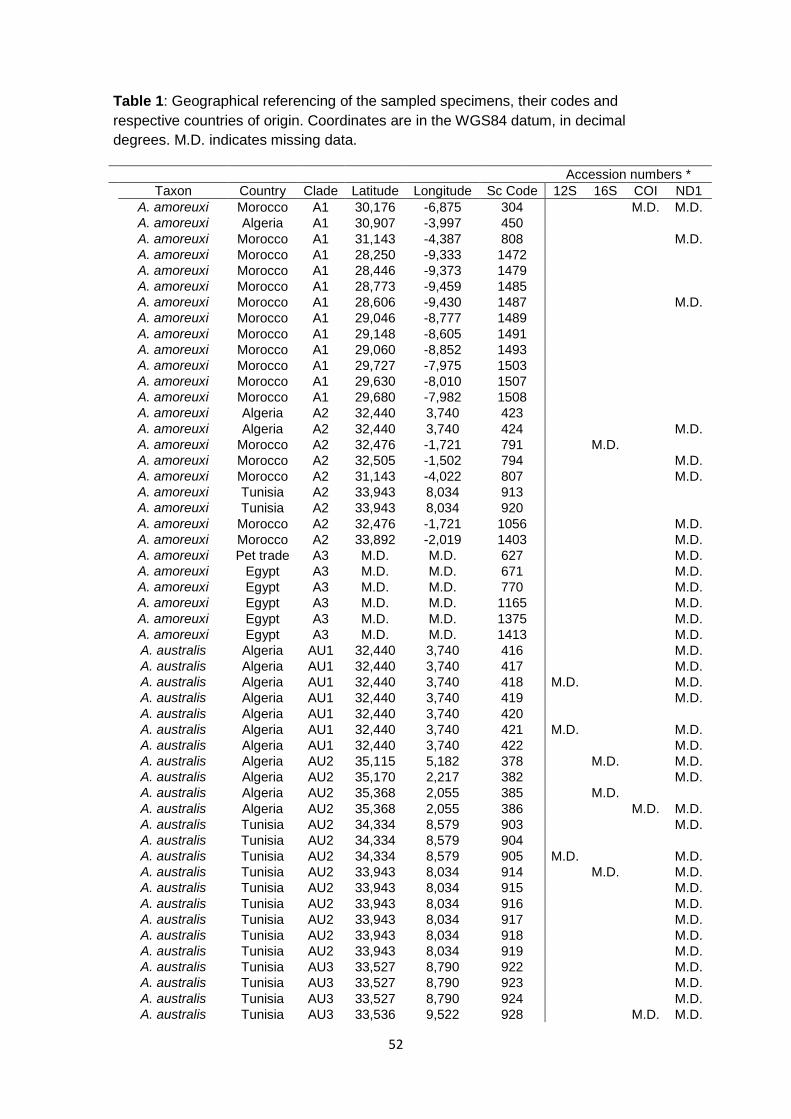
52
Table 1: Geographical referencing of the sampled specimens, their codes and respective countries of origin. Coordinates are in the WGS84 datum, in decimal degrees. M.D. indicates missing data.
Accession numbers *
Taxon Country Clade Latitude Longitude Sc Code 12S 16S COI ND1
A. amoreuxi Morocco A1 30,176 -6,875 304 M.D. M.D.
A. amoreuxi Algeria A1 30,907 -3,997 450
A. amoreuxi Morocco A1 31,143 -4,387 808 M.D.
A. amoreuxi Morocco A1 28,250 -9,333 1472
A. amoreuxi Morocco A1 28,446 -9,373 1479
A. amoreuxi Morocco A1 28,773 -9,459 1485
A. amoreuxi Morocco A1 28,606 -9,430 1487 M.D.
A. amoreuxi Morocco A1 29,046 -8,777 1489
A. amoreuxi Morocco A1 29,148 -8,605 1491
A. amoreuxi Morocco A1 29,060 -8,852 1493
A. amoreuxi Morocco A1 29,727 -7,975 1503
A. amoreuxi Morocco A1 29,630 -8,010 1507
A. amoreuxi Morocco A1 29,680 -7,982 1508
A. amoreuxi Algeria A2 32,440 3,740 423
A. amoreuxi Algeria A2 32,440 3,740 424 M.D.
A. amoreuxi Morocco A2 32,476 -1,721 791 M.D.
A. amoreuxi Morocco A2 32,505 -1,502 794 M.D.
A. amoreuxi Morocco A2 31,143 -4,022 807 M.D.
A. amoreuxi Tunisia A2 33,943 8,034 913
A. amoreuxi Tunisia A2 33,943 8,034 920
A. amoreuxi Morocco A2 32,476 -1,721 1056 M.D.
A. amoreuxi Morocco A2 33,892 -2,019 1403 M.D.
A. amoreuxi Pet trade A3 M.D. M.D. 627 M.D.
A. amoreuxi Egypt A3 M.D. M.D. 671 M.D.
A. amoreuxi Egypt A3 M.D. M.D. 770 M.D.
A. amoreuxi Egypt A3 M.D. M.D. 1165 M.D.
A. amoreuxi Egypt A3 M.D. M.D. 1375 M.D.
A. amoreuxi Egypt A3 M.D. M.D. 1413 M.D.
A. australis Algeria AU1 32,440 3,740 416 M.D.
A. australis Algeria AU1 32,440 3,740 417 M.D.
A. australis Algeria AU1 32,440 3,740 418 M.D. M.D.
A. australis Algeria AU1 32,440 3,740 419 M.D.
A. australis Algeria AU1 32,440 3,740 420
A. australis Algeria AU1 32,440 3,740 421 M.D. M.D.
A. australis Algeria AU1 32,440 3,740 422 M.D.
A. australis Algeria AU2 35,115 5,182 378 M.D. M.D.
A. australis Algeria AU2 35,170 2,217 382 M.D.
A. australis Algeria AU2 35,368 2,055 385 M.D.
A. australis Algeria AU2 35,368 2,055 386 M.D. M.D.
A. australis Tunisia AU2 34,334 8,579 903 M.D.
A. australis Tunisia AU2 34,334 8,579 904
A. australis Tunisia AU2 34,334 8,579 905 M.D. M.D.
A. australis Tunisia AU2 33,943 8,034 914 M.D. M.D.
A. australis Tunisia AU2 33,943 8,034 915 M.D.
A. australis Tunisia AU2 33,943 8,034 916 M.D.
A. australis Tunisia AU2 33,943 8,034 917 M.D.
A. australis Tunisia AU2 33,943 8,034 918 M.D.
A. australis Tunisia AU2 33,943 8,034 919 M.D.
A. australis Tunisia AU3 33,527 8,790 922 M.D.
A. australis Tunisia AU3 33,527 8,790 923 M.D.
A. australis Tunisia AU3 33,527 8,790 924 M.D.
A. australis Tunisia AU3 33,536 9,522 928 M.D. M.D.

53
A. australis Tunisia AU3 33,533 9,991 932 M.D.
A. australis Tunisia AU3 33,533 9,991 933 M.D.
A. australis Tunisia AU3 33,533 9,991 934 M.D.
A. australis Tunisia AU3 32,785 10,373 938 M.D.
A. australis Tunisia AU3 32,785 10,373 939 M.D.
A. australis Tunisia AU3 32,785 10,373 940 M.D.
A. australis Tunisia AU3 33,650 10,317 948
A. australis Tunisia AU3 33,650 10,317 950 M.D.
A. australis Tunisia AU3 33,650 10,317 951 M.D.
A. australis Pet trade AU4 M.D. M.D. 626 M.D.
A. australis Egypt AU4 31,279 27,055 954 M.D.
A. australis Egypt AU4 31,279 27,055 957 M.D. M.D.
A. australis Egypt AU4 28,809 34,228 981 M.D. M.D.
A. australis Egypt AU4 28,822 34,182 992
A. australis Egypt AU4 28,822 34,182 993
A. australis Egypt AU4 M.D. M.D. 1374
A. australis Egypt AU4 M.D. M.D. 1414
A. bicolor Algeria B 35,208 1,541 377 M.D.
A. bicolor Algeria B 35,208 1,541 383 M.D. M.D.
A. bicolor Tunisia B 33,846 10,128 947
A. bicolor Tunisia B 33,650 10,317 949
A. bicolor Egypt B M.D. M.D. 1093
A. bicolor Egypt B M.D. M.D. 1371 M.D.
A. cf. liouvillei Morocco G 28,479 -11,212 1453 M.D.
A. cf. liouvillei Morocco G 28,479 -11,212 1454
A. cf. liouvillei Morocco G 28,479 -11,212 1455
A. cf. liouvillei Morocco G 28,479 -11,212 1456 M.D. M.D.
A. liouvillei Morocco L 30,668 -6,380 210 M.D. M.D.
A. liouvillei Morocco L 34,286 -3,169 780 M.D.
A. liouvillei Morocco L 34,286 -3,169 781 M.D.
A. liouvillei Morocco L 33,892 -2,019 786 M.D.
A. liouvillei Morocco L 33,213 -2,019 787 M.D.
A. liouvillei Morocco L 32,087 -1,241 793
A. liouvillei Morocco L 32,571 -2,015 796 M.D.
A. liouvillei Morocco L 32,571 -2,015 797
A. liouvillei Morocco L 33,289 -3,780 816 M.D.
A. liouvillei Morocco L 33,508 -3,528 817 M.D. M.D.
A. liouvillei Morocco L 33,892 -2,019 830 M.D.
A. liouvillei Morocco L 32,087 -1,241 839 M.D.
A. liouvillei Morocco L 33,213 -2,019 1055 M.D.
A. liouvillei Morocco L 33,892 -2,019 1207 M.D.
A. liouvillei Morocco L 33,892 -2,019 1394 M.D.
A. mauritanicus Morocco M1 32,225 -8,166 15
A. mauritanicus Morocco M1 32,526 -7,863 287
A. mauritanicus Morocco M1 32,661 -7,793 290 M.D.
A. mauritanicus Morocco M1 32,661 -7,793 291
A. mauritanicus Morocco M1 32,661 -7,793 292 M.D. M.D.
A. mauritanicus Morocco M1 M.D. M.D. 625 M.D.
A. mauritanicus Morocco M2 30,183 -9,580 1467
A. mauritanicus Morocco M2 30,183 -9,580 1468
A. mauritanicus Morocco M2 30,183 -9,580 1469 M.D.
A. mauritanicus Morocco M2 30,159 -8,481 1549 M.D.
A. mauritanicus Morocco M2 30,059 -9,084 1558
A. mauritanicus Morocco M2 30,098 -8,938 1589 M.D. M.D.
A. mauritanicus Algeria M3 29,068 -10,248 438 M.D.
A. mauritanicus Morocco M3 29,087 -9,898 1443
A. mauritanicus Morocco M3 29,087 -9,898 1444
A. mauritanicus Morocco M3 29,087 -9,898 1445

54

55
Chapter III
Preface 2
Packing a pinch: functional implications of chela shapes in scorpions using finite element analysis

56
3.1 Preface 2
As mentioned before, scorpions appeared in the middle Silurian (425 – 450
Mya) as aquatic organisms. 100 My later, when they conquered land masses,
significant morphological changes such as the shrinkage in body size and the
enclosure of the external book gills followed their terrestrialization (Jeram, 2000). Since
this period, between the Late Devonian and the Early Carboniferous (325 – 350 Mya),
scorpions maintained the general external body plan featuring:
a) A segmented ophistosoma;
b) Mesosoma and metasoma differentiated from each other;
c) Robustly chellate pedipalps and chelicerae;
d) Eight walking legs;
e) Pectines (ventral sensory organ exclusive to Scorpions);
f) Terminal telson with a venom delivering apparatus.
Since then few changes occurred making scorpions morphologically conserved
in different habitats. However specific adaptations are associated to certain
environments. Lamoral (1979) and Polis (1990) identified several ecomorphotypes
based on a qualitative review of the local scorpion faunas of sub-Saharan Africa and
North America respectively. More recently, some of these proposed ecomorphotypes
were corroborated quantitatively based on only the chela shape (see featuring article).
The first comparative studies on scorpion performance traits have shown that in a
sample of very few species, a clear non-phylogenetic component can be uncovered in
both the morphology and performance of the chelae (van der Meijden et al., 2010,
2012). This highly suggests independent evolution of these morphotypes and thus
adaptive significance for this trait. However, to study adaptive features comparatively,
an estimate of the phylogeny of the included taxa is required.

57
The last two centuries’ studies on scorpions have focused on the morphological
aspects that are more informative for taxonomy. Because of that there is a great deal of
morphological diversity that remains understudied. For instance, to understand the
adaptive importance of morphological structures we need to look beyond taxonomy
and phylogenetics. It is necessary to include functional parameters, such as
performance data, and to compare them between different organisms. Comparative
analyses greatly enhance our understanding of the wealth of morphological diversity
that exists in the animal world and allows us to uncover historical trial-and-error based
design. These studies have successfully provided insights into, for example, the
ecology of the vertebrate jaw-cranial muscoskeletal system (Herrel et al., 2008). It has
been one of the goals for CIBIO’s Integrative Biogeography, Ecology and Evolution
research group to use scorpions as a model for the evolution of associated suites of
characters that are related to specific environments.
The Scorpiones is a minor order within Arachinda representing less than 2% of
the number of arachnid species (Rein, 2013). However, its systematics remains, for the
most part, unresolved. Competing hypothesis divide researchers (Prendini & Wheeler,
2005; Soleglad & Fet, 2003) and much debate is focused at the family level. These
theories are anchored in cladistic approaches based mostly in morphological traits but
they are open for future findings with DNA sequence data. With the introduction of DNA
sequence data, several authors attempted to solve high-level scorpion relationships
(e.g. Hassanin, 2006; Masta et al., 2008). The use of molecular markers has proven
more efficient at lower taxonomical levels. The fast emergence of molecular
phylogenetic data on scorpion genera (e.g. Fet et al., 2003a; Prendini et al., 2003) and
species (e.g. Euscorpius: Fet et al., 2003b; Buthus Sousa et al, 2011; Hottentotta:
Sousa et al., 2010; Mesobuthus: Gantanbein et al., 2003) is a new challenge for
scorpion taxonomy.
Packing a pinch: functional implications of chela shapes in scorpions using finite
element analysis represents some of the work that the candidate developed as a
collaborator. Under the supervision of Dr. Arie van der Meijden, also the lead author,
the candidate assisted with the measuring of bite forces. Bite force measurement
consists of the record of several trials per specimen of scorpion’s chelae pinch forces.
The recordings are made using a custom built force sensor (further details are given in

58
the Materials and Methods section). The candidate was able to work under a climate-
controlled room either in the laboratory or in field conditions. Field measurement is an
essential part of this work because we could make records with animals that were
removed from natural conditions in the same day. Although scorpions perform well
under captivity, field measurements are also useful to record scorpions that cannot be
obtained from the pet trade.
As mentioned above, to retrieve the phylogenetic signal from the evolutionary history of
the studied traits we sequenced 20 scorpions of 8 families. 12S, 16S and COI
mitochondrial genes were used to build the phylogenetic tree. All the molecular
laboratory protocols were performed by the MSc candidate following the same
methodology applied for sequencing Androctonus scorpions. The MSc’s contribution
did not included morphological measurements, specimen scanning or any data
analysis. The candidate wrote part of the Phylogenetic analysis section of Materials
and methods, while the remainder of the article was written by Arie van der Meijden
and Dr. Thomas Kleinteich.
The produced work was accepted for publication in the Journal of Anatomy at the 26th
January, 2012.

59
3.2 Packing a pinch; Functional implications of chela shapes in scorpions using finite element analysis.
Arie van der Meijden1, Thomas Kleinteich2,3, Pedro Coelho1
1. CIBIO, Centro de Investigação em Biodiversidade e Recursos Genéticos
Campus Agrário de Vairão, 4485-661 Vairão, Portugal
2. University of Washington, Friday Harbor Laboratories, 620 University Road,
Friday Harbor, WA, USA, 98250
3. Christian-Albrechts-Universität Kiel, Zoology Department, Am Botanischen
Garten 1-9, Kiel, Germany, 24098
Running title: Comparative FEA of scorpion chelae Abstract Scorpions depend on their pedipalps for prey capture, defense, mating and sensing
their environment. Some species additionally use their pedipalps for burrowing or
climbing. Because pincers or chelae at the end of the pedipalps vary widely in shape,
they have been used as part of a suite of characters to delimit ecomorphotypes. We
here evaluate the influence of the different chela cuticular shapes on their performance
under natural loading conditions. Chelae of 20 species, representing seven families
and spanning most of the range of chela morphologies, were assigned to clusters
based on chela shape parameters using hierarchical cluster analysis. Several clusters
were identified to approximately correspond to described scorpion ecomorphotypes.
Finite element models of the chela cuticulae were constructed from CT scans and
loaded with estimated pinch forces based on in vivo force measurements. Chela shape
clusters differed significantly in mean Von Mises stress and strain energy. Normalized
FEA showed chela shape to significantly influence Von Mises stress and strain energy
in the chela cuticula, with Von Mises stress to vary up to an order of magnitude, and
strain energy varying up to two orders of magnitude. More elongate, high-aspect ratio
chela forms showed significantly higher mean stress than more robust low-aspect ratio
forms. This suggests that elongate chelae are at a higher risk of failure when operating
near the maximum pinch force. Phylogenetic independent contrasts (PIC) were
calculated based on a partly resolved phylogram with branch lengths based on an
alignment of the 12S, 16S and CO1 mitochondrial genes. PIC showed cuticular stress

60
and strain in the chela to correlate with several shape parameters, such as aspect
ratio, movable finger length, and chela height, independently from phylogenetic history.
Our results indicate that slender chela morphologies may be less suitable for high-force
functions such as burrowing and defense. Further implications of these findings for the
ecology and evolution of the different chela morphologies are discussed.
Key words Finite element analysis, Scorpiones, independent contrasts Introduction
Scorpions are a large and ancient group of chelicerates with close to 2000 described
species (Fet et al., 2000), which have successfully adapted to a diversity of habitats on
all continents except Antarctica. Despite occurring in a wide variety of habitats,
scorpions have changed little in overall body plan since the Silurian (Dunlop et al.,
2008). Their most notable features are the pedipalps carrying the chelae (the
“pincers”), and the metasoma (the “tail”) carrying the venomous telson (the “stinger”).
All these structures are used in prey subjugation and defense. The pedipalps are
additionally used in mating, climbing, digging, and serve as a sensory array similar to
the antennae of insects and crustaceans (Alexander, 1959; Fet et al., 2003).
The chelae are formed by the last two segments of the pedipalps; the manus or tibia
which contains the muscles and which also forms the immovable finger, and the
movable finger or tarsus. The movable finger is adducted by three muscle bundles in
the chela manus (Gilai & Parnas, 1970). In addition, there is also a small closing
muscle in the next segment (patella), which is connected to the movable finger by a
long ligament (Snodgrass, 1952; Gilai & Parnas, 1970). The movable finger is
abducted by the elastic recoil of resilin in the joint (Govindarajan & Rajulu, 1974;
Sensenig & Shultz, 2004), opening the chela. The movable finger rotates around an
axis defined by two joints, located on the median and lateral sides of the chela.
Despite conservancy in their ecological role as mostly terrestrial predators, scorpions
have been known to have specific adaptations to certain environments. Several
ecomorphotypes were recognized based on a qualitative review of the local scorpion
faunas of sub-Saharan Africa (Lamoral, 1979) and North America (Polis, 1990). In
these studies, the shape of the chelae is considered an important character in the
delimitation of five putative ecomorphotypes. Robust chelae with a high muscle-filled
manus and relatively shorter fingers that are reminiscent of chelae in durophagous
crabs are ascribed to fossorial (digging) species. Sand-dwelling (psammophilous)

61
species have more elongate chelae with long fingers. Species that hide in or under
rocks (saxicoline) or bark (corticoline) have dorso-ventrally compressed bodies and
appendages including the chelae, to facilitate living in narrow spaces (Newlands,
1972). Actively foraging scorpions with slender bodies and appendages are sometimes
considered as an additional ecomorphotype; the errant ecomorphotype (Polis, 1990).
Chelae of the errant ecomorphotype are similar to the chelae in psammophilous
species. These different putative ecomorphotypes have never been quantitatively
corroborated.
The described different morphologies change the functional properties of the chelae.
The volume of the manus is largely determined by the amount of muscle in that
segment, which partly determines the force a scorpion can produce with its chelae.
Like in durophagous crabs (Yamada & Boulding, 1998), the height of the manus
relative to the length of the chela is therefore indicative of pinch force (Van der Meijden
et al., 2010). The pinch force of a scorpion is further determined by the lever system
that transfers the force from the muscles to the tips of the fingers of the chelae. The
mechanical advantage of this lever system is very variable in scorpions. As the outlever
(the external part of the movable finger) of a first class lever system gets longer relative
to the inlever (the internal part of the movable finger, to which the muscles attach), the
mechanical advantage of the lever system may increase speed at the cost of force (but
see McHenry, 2011). Longer fingers may thus give the scorpion an advantage to catch
elusive insect prey by increased closing speed or gape size. Since this arrangement
makes the chelae relatively inefficient in producing high forces, such long-fingered
chelae are feeble weapons to fend of predators. The robust chelae of fossorial species
allow for more muscle to be packed into the chela and for a longer inlever inside the
chela manus, thus improving the mechanical advantage for force production. Newlands
(1969; 1972) suggested that the large chelae may also be used in blocking the burrow,
to keep out predators. Other authors have noted that scorpions with robust chelae use
their sting less in prey incapacitation than scorpions with more slender chelae
(Stahnke, 1966; Baerg, 1961; McCormick & Polis, 1990), which suggests a trade-off
between the chelae and the stinger in prey incapacitation. Although morphological
characters of the chela are important in taxonomic studies, the functional implications
of shape to the performance of the chela have received little attention. Knowledge on
the functional consequences of different chela shapes, however, is crucial to
understand their adaptive value and to identify functional demands during scorpion
evolution. We here for the first time evaluate the functional implications of the shape of
the chela cuticula in scorpions. We use 3-dimensional Finite Element Models to

62
estimate the stresses that the chela cuticula of different chela morphologies experience
under biologically realistic force production.
Materials and Methods Species selection
A total of 20 species (Table 1) was selected from seven families to represent the range
of chela morphology found in extant scorpions. Animals were selected based on
availability through field collecting or pet-trade.
Morphological measurements
Morphological measurements were taken using digital calipers. Several preserved
specimens were measured per species and average chela measurements were
calculated. For some species however, only a single specimen was available.
Morphological data are shown in table 1. In addition to linear measurements, also the
curvature of the movable finger was determined using a custom Matlab script (available
from the corresponding author upon request). The ventral view of the chela movable
finger was used to fit a circle to the inside and outside curve of the movable finger. The
average radius of these two circles was then divided by the length of the movable
finger to attain a size-independent metric of curvature. In addition the angle between
the axis of rotation of the movable finger to the line connecting the tip of the movable
finger to the center of the axis of rotation was calculated (α in figure 1c). The angle
between the in-lever and out-lever of the movable finger was calculated as the angle
between the plane defined by the finger tip (T), the median joint (MJ) and lateral joint
(LJ), and the plane defined by the insertion point of the muscles (MI), the median joint
(MJ) and lateral joint (LJ) (see figure 1a).
In order to group similarly shaped chelae, we performed a clustering analysis on
normalized linear measurements of the chela. Manus height, manus width and length
of movable finger were normalized by division with total chela length. These normalized
data were used to identify clusters of similarly shaped chelae. Clustering analysis was
performed in R (R core development team). The data were clustered hierarchically
using the Ward method based on Euclidian distance between species. Clusters in the
data were also identified using the k-clustering method, with the variable k (number of
clusters), identified through the expectation maximization algorithm as implemented in
the R package “Mclust”.

Table 1. Species used in this study, and measured and derived morphological parameters. Asterisk indicates measurements from a similar sized specimen.

Figure 1. a; Linear measurements on the chela Length (L), Height (H), movable finger length (T-LJ). b-c; Measurements on chela movable finger, exemplified on the movable finger of Pandinus cavimanus. a. Side view of a transparent wireframe mesh. b. Dorsal view, solid wireframe mesh. c. Dorsal view showing angle (α) between center of joint axis (LJ-MJ) and finger tip (T). LJ; lateral joint, MJ; medial joint, MI: muscle insertion, T; tip. The line LJ-MJ is the axis of rotation for the movable finger.
Pinchforce measurement and estimation
Live animals were maintained as described in Van der Meijden et al. (2010). In vivo
pinch forces were measured using either a Kistler force transducer (type 9203, Kistler
Inc., Switzerland) mounted on a purpose-built holder (see Herrel et al., 1999), or using
a similar setup with a Sauter FH20 external force sensor (Sauter ltd., Germany).
Measurements were made in a climate-controlled room at 23-24˚C. During pinch-force
measurements, scorpions were restrained between sponge pads in which a cutout was
made to accommodate the body, or by placing a padded clamp over the last segments

65
of the metasoma to allow safe handling. Most specimens pinched readily, but some
were stimulated to pinch by stroking the inside of the chela with the tip of a pair of
tweezers. Five trials were performed, separated by at least one day. Only the
maximum force per chela was retained for further analyses.
Not for all species included in this study were live specimens available to measure
pinch forces. Further, some species were too small for accurate measurements using
parallel plates. For species for which in vivo pinch forces were not available, we
estimated pinch forces based on the aspect ratios of the chelae. Chela aspect ratio
was previously demonstrated to be a good predictor for pinch forces (R2 = 0.90; Van
der Meijden et al. 2010). We further expanded the dataset from Van der Meijden et al.
(2010) to comprise 170 specimens from 20 species, representing four families
(Buthidae, Iuridae, Liochelidae, Scorpionidae; data to be published elsewhere). The
maximum pinch forces were log10 transformed, and standardized for the size of the
animal by regression of the forces against log10 transformed prosoma length. Body
mass or total length cannot be used as a proxy for overall size in scorpions, as both
body mass and total length can change significantly depending on feeding state. The
residuals were then regressed against the aspect ratio of the chela (length/height).
Pinch force was predicted based on a line function that was fit to the data using least
squares (R2 = 0.54, Pearson's correlation coefficient -0.73). The resulting predicted
maximum pinch forces are shown in table 1.
Specimen scanning and model reconstruction
We CT scanned the chelae of the specimens examined herein with a Skyscan 1076
micro CT scanner that was available at the Small Animal Tomographic Analysis Facility
at the Seattle Children’s Research Institute in Seattle, WA, USA. The specimens were
scanned at a source voltage of 31 kV and a source currency of 187 µA at 35 µm
resolution. The raw x-ray images that were recorded during CT imaging where then
converted with the software tool NRecon (Skyscan) into a volumetric data set that
consisted of a stack of TIFF image files.
The volumetric dataset was imported into the 3D visualization software Amira 5.3.3
(Visage Imaging). In Amira, we used the Labels function to separate the cuticles of the
scorpion chelae from the remainder structures that where visible in the CT data set. We
further separated the cuticle of the manus from the cuticle of the movable finger and
saved them as two separate materials in the Labels dataset that is generated during
segmentation in Amira. Based on the materials in the Labels dataset, we calculated
polygonal surfaces for the manus and the movable finger with Amira. The resulting

66
surfaces were then edited in the Surface Editor Mode of Amira to equally distribute the
polygons over the surface, to reduce the total number of polygons, and to fix occurring
intersections between polygons that were in close proximity to each other. For each
specimen, we measured the surface area and the volume with Amira. We then
converted the polygonal surfaces to solid models that were build from tetrahedral
elements, i.e. the elements were defined by four nodes. Numbers of elements ranged
from 20,000 to 2.4 million depending on the size of the specimen (Table 2).
Finite Element Analysis
The solid models for the manus and the finger of each specimen were imported as
IDEAS files into the Finite Element modeling and analyzing software Marc Mentat 2005
R3 (MSC Software). Because the gapes between manus and finger in different
specimens differed, we had to manually adjust the gape angles in Marc Mentat. For
this, we oriented the models to align the axis that connects the centers of the two joints
between manus and finger with the origin of the coordinate system in Marc Mentat.
This allowed us to rotate the manus and the finger independently without dislocation at
the joint and to set the gape angle at 15° for each specimen. For a more realistic
estimate on the reaction forces that act on the joint between manus and finger under
load we declared elements of the manus and of the finger to be on two different
Contact Bodies and we assigned the nodes in the joint area as Contact Areas. Within
Contact Areas, Marc Mentat treats elements that are closer to each other than a
defined threshold value (1/20 of the size of the smallest element) as contacting
elements. For the finite element analysis, the contact between finger and manus was
treated as two-sided (i.e. the finger contacts the manus and vice versa) and we used
the Optimized Contact Constraints option to avoid penetration between finger and
manus.
We treated the cuticle of the chelae as isotropic material with a Young’s modulus of 7
GPa and a Poisson’s ratio of 0.3. Young’s moduli of arthropod cuticle have been
reported to cover a wide range from 0.1 GPa to 20 GPa (Vincent & Wegst, 2004).
Although the value for the Young’s modulus of 7 GPa that we have chosen herein is
only a estimate based on the assumption that scorpion chelae have a rather hard
cuticle and absolute output values are biased by this estimate, finite element modeling
provides a powerful tool for comparisons between species, even if knowledge on the
exact material attributes is not available (Dumont et al., 2009).

Table 2. Details of the chela models used in this study.

We applied the following constraints (in Marc Mentat Boundary Conditions) to the
models: (1) all nodes in the areas of muscle attachment on the manus and the finger
where fixed to prevent translation or rotation for each degree of freedom; (2) bite force
was distributed over four nodes on each, the finger and the manus, and modeled as
point loads. We ran two separate analyses for each specimen. (1) We used the
measured and predicted force values in Table 1 as bite force (absolute approach with
unscaled results) and (2) we calculated the force per surface area ratios for each
specimen and applied bite forces that were scaled to fit the smallest force per surface
area ratio in our specimen sample (comparative approach with scaled results).
We used three different measures to quantify the biting performance of scorpion chelae
with Finite Element Analysis: (1) maximum Von Mises stress, (2) mean Von Mises
stress, and (3) total strain energy. Von Mises stress is used as a measure for how
close a material is to failure. Areas in the chelae with higher Von Mises stresses are
closer to failure than areas that experience lower Von Mises stresses. However,
maximum Von Mises stresses can be hard to interpret in Finite Element models
because the constrained areas (i.e. the applied point loads and fixed nodes) tend to
show artificially high Von Mises stresses (Dumont et al., 2009). Mean Von Mises stress
is supposed to be a more reliable measure of overall performance based on the
assumption that chelae shapes that perform better under load will show lower Von
Mises stresses over each node than shapes that perform poorly. Total strain energy
reflects the amount of work that is used for deformation of the shapes, rather than for
biting. It is assumed that scorpions that perform better during biting exhibit lower total
strain energies, i.e. their chelae deform less under load. Von Mises stress and total
strain energy are standard outputs of Marc Mentat.
Because in the comparative approach, all models had identical force to surface area
ratios, Von Mises stresses were directly comparable between species (Dumont et al.,
2009). However, total strain energy scales with the volume of the models instead of the
surface area. To make the calculated values for total strain energy directly comparable
between species, we calculated an energy scaling factor that was based on the
specimen with the lowest force per sixth root of the volume ratio. Volume corrected
total strain energies where then calculated by multiplication of the square of the energy
scaling factor with the raw total strain energies. A detailed derivation of the equations to
scale finite element models was provided by Dumont et al. (2009).
The numbers of elements for each specimen varied notably despite similar edge
lengths of the elements (Table 2). This was caused by the different sizes of the
specimens. To estimate the effect of mesh-size on the performance parameters

69
evaluated herein, we re-meshed the finite element model of the specimen with the least
amount of elements (Orthochirus innesi), to generate models with eight, respectively 64
times the number of the original elements (Supplementary material). Finite element
analysis with the re-meshed datasets showed only slight variations in mean Von Mises
stress and total strain energy that are negligible compared to the interspecific variation.
This indicates that mean Von Mises stress and total strain energy can be used for
comparisons among datasets that consist of different amounts of elements; max. Von
Mises stress increases in the re-meshed models, which is likely to be caused by an
increased localization of point loads.
Phylogenetic analysis and independent contrasts
Total DNA was extracted from fresh or preserved (96% ethanol) muscle tissue using
standard high-salt protocols (Bruford et al., 1992). A fragment of the mitochondrial
cytochrome C oxidase, subunit I (COI) gene was amplified by PCR using primers
LCO1490 and HCO2198 (Folmer et al., 1994) or primers in the same locus based on
an alignment of CO1 sequences available in GenBank as of 4-2011. The sequence of
these primers was: forward (5’-WTYCTACIAATCAYAARGATATTGG-3’) and reverse
(5’-TAMACYTCIGGGTGWCCAAAAAAYCA-3’). A fragment of the mitochondrial 16S
rDNA gene was amplified using the primers LR-J-12887 (Simon et al., 1994) as
forward primer, and a scorpion-specific reverse primer (Gantenbein et al., 2000). 12S
primers were designed based on available 12S sequences in GenBank: forward primer
12S_F_AvdM (5’-AGAG-TGACGGGCAATATGTG-3)’ and reverse primer 12s_r_AvdM
(5’-CAGCGGCTGCGGTTATAC-3’).
Purified PCR templates were sequenced using dye-labeled dideoxy terminator cycle
sequencing on an ABI 3130 automated DNA sequencer or on an ABI 3730XL at
Macrogen Inc. using the corresponding PCR primers. Chromatograms were checked
and when necessary corrected using FinchTV, version 1.4.0 (Geospiza, Inc.; USA;
http://www.geospiza.com). The obtained DNA sequences were aligned using MEGA 5
(Tamura et al., 2011). The coding sequence of CO1 was aligned based on the
translated amino acid sequence, and 12S and 16S rRNA sequences were aligned
using Muscle (Edgar, 2004) as incorporated into MEGA 5 using the default settings.
Two methods of phylogenetic analysis, maximum likelihood (ML) and Bayesian
Inference (BI), were conducted using PhyML, version 3.0.1 (Guindon & Gascuel, 2003)
and MrBayes 3.1.2 (Ronquist & Huelsenbeck, 2003) respectively. The best fit models
of nucleotide evolution were determined under the Akaike information criterion in
JModeltest 0.1.1 (Posada, 2008). Nodal support for the topologies recovered in the ML
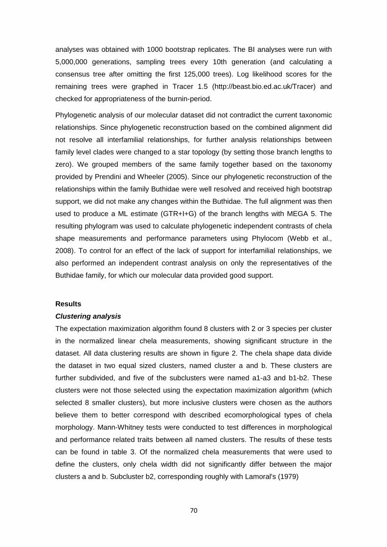
70
analyses was obtained with 1000 bootstrap replicates. The BI analyses were run with
5,000,000 generations, sampling trees every 10th generation (and calculating a
consensus tree after omitting the first 125,000 trees). Log likelihood scores for the
remaining trees were graphed in Tracer 1.5 (http://beast.bio.ed.ac.uk/Tracer) and
checked for appropriateness of the burnin-period.
Phylogenetic analysis of our molecular dataset did not contradict the current taxonomic
relationships. Since phylogenetic reconstruction based on the combined alignment did
not resolve all interfamilial relationships, for further analysis relationships between
family level clades were changed to a star topology (by setting those branch lengths to
zero). We grouped members of the same family together based on the taxonomy
provided by Prendini and Wheeler (2005). Since our phylogenetic reconstruction of the
relationships within the family Buthidae were well resolved and received high bootstrap
support, we did not make any changes within the Buthidae. The full alignment was then
used to produce a ML estimate (GTR+I+G) of the branch lengths with MEGA 5. The
resulting phylogram was used to calculate phylogenetic independent contrasts of chela
shape measurements and performance parameters using Phylocom (Webb et al.,
2008). To control for an effect of the lack of support for interfamilial relationships, we
also performed an independent contrast analysis on only the representatives of the
Buthidae family, for which our molecular data provided good support.
Results
Clustering analysis The expectation maximization algorithm found 8 clusters with 2 or 3 species per cluster
in the normalized linear chela measurements, showing significant structure in the
dataset. All data clustering results are shown in figure 2. The chela shape data divide
the dataset in two equal sized clusters, named cluster a and b. These clusters are
further subdivided, and five of the subclusters were named a1-a3 and b1-b2. These
clusters were not those selected using the expectation maximization algorithm (which
selected 8 smaller clusters), but more inclusive clusters were chosen as the authors
believe them to better correspond with described ecomorphological types of chela
morphology. Mann-Whitney tests were conducted to test differences in morphological
and performance related traits between all named clusters. The results of these tests
can be found in table 3. Of the normalized chela measurements that were used to
define the clusters, only chela width did not significantly differ between the major
clusters a and b. Subcluster b2, corresponding roughly with Lamoral's (1979)

71
Figure 2. Hierarchical clustering of chela shapes based on movable finger length, manus width and height, normalized for chela length. Cluster a contains the more elongate chela morphologies, cluster b contains the more robust chelae. Clusters a1, a2 and a3 get progressively lower aspect ratios. Cluster b1 contains short strong chelae, whereas cluster b2 contains more flattened morphologies, typical of species that live in rock crevices and under bark.
infrasaxicolous, lithosaxicolous and infracorticolous types, differed significantly from all
other subclusters in chela height and movable finger length. There was no statistical
difference between any of the clusters in chela length relative to the length of the
prosoma (the latter being a good indicator of overall size).
Finite element analysis Absolute approach. Under realistic loading conditions (i.e. the applied point loads
corresponded to either actually measured in vivo bite forces or bite force estimates
based on chela aspect ratio) maximum Von Mises stress, mean Von Mises stress, and
total strain energy differed between species by several orders of magnitude (Table 1).

72
This is in part accounted for by the different sizes of the specimens. Maximum Von
Mises stress was highest in Hadogenes paucidens (7.32e8 Pa) and lowest in Liocheles
australasiae (3.76e7 Pa). Mean Von Mises stress varied between 2.89e6 Pa in Scorpio
and 3.64e7 Pa in Hadrurus arizonensis. Total strain energy was lowest in the very
small Orthochirus (7.62e-6 J) and highest in the largest specimen included in this
study, Pandinus cavimanus (1.25e-2 J).
Comparative approach. Scaling of the models based on surface area (for Von Mises
stress), respectively volume (for total strain energy), allowed us to remove the effects
of the size of the specimens from the analysis and to reveal the impacts of the different
shapes on the performance. In our sample, the short fingered species Scorpio
fuliginosus and Ophistacanthus madagascariensis showed the least overall
deformation under load, expressed in the lowest values for total strain energy.
Contrary, species with elongated fingers like Hoffmannius sp. and Leiurus
quinquestriatus showed the most pronounced deformations of the chelae. The same
pattern also emerges for mean Von Mises stress; L. quinquestriatus encounters
highest mean Von Mises stresses and Scorpio shows the lowest values. Maximum Von
Mises stress is highest in Hadogenes paucidens and lowest in Hoffmannius. This
however is most likely caused by the presence of artificially high point loads in some of
our models and the pattern of highest stresses in H. paucidens, and lowest stresses in
Hoffmannius is not reflected by the plots of Von Mises stress over the 3D surfaces
(Figure 3).
The fingers of the chela act under load like a beam, i.e. they experience tensile stress
along the edge where the load is applied and stress by compression along the edge
opposite the load; between the compressed and tensioned edges lies a neutral axis
with low stresses (Figure 3). In species with slender, elongated fingers, the stresses on
the movable part are similar to the counteracting fixed finger. In species with short
chelae (e.g. Scorpio, Pandinus cavimanus), the roughly triangular shape of the fixed
finger in lateral view tends to reduce the stress by compression on the edge opposite to
the load. This results in an asymmetric stress distribution with higher stresses in the
movable finger, compared to the fixed finger. The major clades a and b differed
significantly in mean Von Mises stress and (size corrected) strain energy. As the Mann-
Whitney test is based on ranking of the values, small datasets, such as the subclusters
in this study, can be ordered in a limited number of ways. This leads to a tendency to
converge on a small number of p-values, which accounts for the recurring value of
some of the p-values in table 3. In addition, we also tested for differences in the angle
between the inlever and the outlever of the movable finger between the clusters, but
found no statistically significant differences.

73
Table 3. Mann-Whitney tests of statistical difference between named clusters of morphological and performance parameters. P-values higher than 0.05 are shown in grey font.

74
Phylogenetic analysis and independent contrasts
The total aligned dataset consisted of 1534 positions for 20 species. Bootstrap support
was high within the Buthidae, but interfamilial relationships could not be recovered with
high support (not shown). Branch lengths were therefore calculated starting with a
phylogeny with a polytomy uniting all family level clades (Figure 4).
The phylogenetic independent contrasts (Table 4) show both mean Von Mises stress
and strain energy to correlate highly with several aspects of chela morphology,
including size corrected height, width, outlever length, aspect ratio, mechanical
advantage, and the ratio of the length of the movable finger to the distance between
the joints. This corresponds with the observation from the clustering analysis that more
elongate chelae experience higher stresses in the cuticula. Noteworthy is that the angle
that the main axis of the movable finger makes with its axis of rotation is positively
correlated with the length of the movable finger (“outlever” in Table 4), approaching a
90 degree angle for relatively longer fingers such as those of Orthochirus, Hottentotta
and Parabuthus. Less surprising is that AR is highly correlated with the mechanical
advantage of the chela. Neither mean Von Mises stress or strain energy correlated
significantly with the angle between in- and outlever, the angle the outlever makes with
the axis of rotation, or the curvature of the finger. This may indicate that these latter
variables are independent from the performance of the chela in resisting stresses
during pinching, and may be relevant for another function. The PIC analysis of the
Buthidae only also showed high correlation coefficients for mean Von Mises stress and
strain energy with aspect ratio, mechanical advantage and the ratio of finger length to
the distance between joints EJ-MJ (0.79; 0.88; 0.91 respectively for both Von Mises
stress and strain energy).

75
Figure 3. FEA models with calculated Von Mises stress visualized as colors. Despite producing much less force, chelae with a high aspect ratio and long fingers (cluster a), experience much higher stresses than the more robust chelae of cluster b. High deformation can be seen in L. quinquestriatus. High stresses indicate a risk of breakage, and the long-fingered chelae of cluster a are therefore at more at risk of breaking their fingers when exerting their maximum force.
Discussion
In order to provide a basis for comparing the FEA results for similarly shaped chelae,
we carried out a cluster analysis. In the hierarchical cluster analysis, we identified
clusters which corresponded in part with previously described ecomorphotypes. The
major clusters differed significantly in stress and bending energy, with the more
elongate chela forms experiencing much higher stresses and deformations (for which
bending energy is a metric). Phylogenetic independent contrasts also show stress and
deformation parameters to correlate highly with chela shape parameters. Despite the
lack of a fully resolved molecular phylogeny for the taxa in this study, the PIC analysis
strongly suggests chela shape and performance to correlate independently from
phylogenetic history.
The clustering analysis revealed significant structure in the limited number of
normalized measurements used. Although the expectation maximization algorithm
found up to 8 clusters in the data, we chose a more conservative number of five
clusters from the hierarchical cluster analysis. Several of the clusters roughly
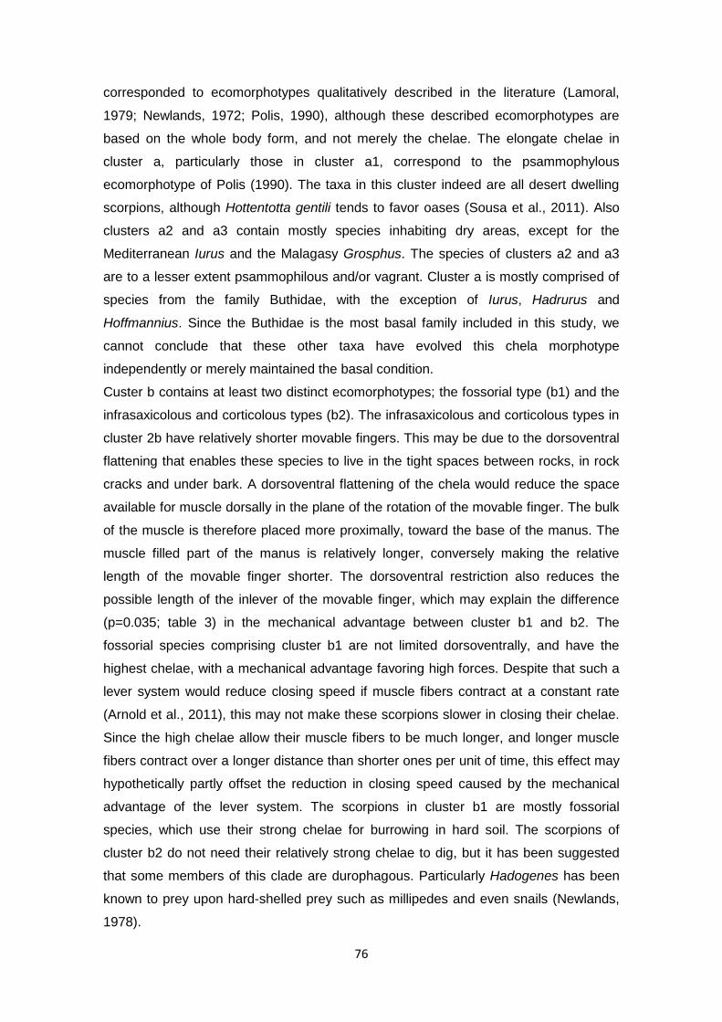
76
corresponded to ecomorphotypes qualitatively described in the literature (Lamoral,
1979; Newlands, 1972; Polis, 1990), although these described ecomorphotypes are
based on the whole body form, and not merely the chelae. The elongate chelae in
cluster a, particularly those in cluster a1, correspond to the psammophylous
ecomorphotype of Polis (1990). The taxa in this cluster indeed are all desert dwelling
scorpions, although Hottentotta gentili tends to favor oases (Sousa et al., 2011). Also
clusters a2 and a3 contain mostly species inhabiting dry areas, except for the
Mediterranean Iurus and the Malagasy Grosphus. The species of clusters a2 and a3
are to a lesser extent psammophilous and/or vagrant. Cluster a is mostly comprised of
species from the family Buthidae, with the exception of Iurus, Hadrurus and
Hoffmannius. Since the Buthidae is the most basal family included in this study, we
cannot conclude that these other taxa have evolved this chela morphotype
independently or merely maintained the basal condition.
Custer b contains at least two distinct ecomorphotypes; the fossorial type (b1) and the
infrasaxicolous and corticolous types (b2). The infrasaxicolous and corticolous types in
cluster 2b have relatively shorter movable fingers. This may be due to the dorsoventral
flattening that enables these species to live in the tight spaces between rocks, in rock
cracks and under bark. A dorsoventral flattening of the chela would reduce the space
available for muscle dorsally in the plane of the rotation of the movable finger. The bulk
of the muscle is therefore placed more proximally, toward the base of the manus. The
muscle filled part of the manus is relatively longer, conversely making the relative
length of the movable finger shorter. The dorsoventral restriction also reduces the
possible length of the inlever of the movable finger, which may explain the difference
(p=0.035; table 3) in the mechanical advantage between cluster b1 and b2. The
fossorial species comprising cluster b1 are not limited dorsoventrally, and have the
highest chelae, with a mechanical advantage favoring high forces. Despite that such a
lever system would reduce closing speed if muscle fibers contract at a constant rate
(Arnold et al., 2011), this may not make these scorpions slower in closing their chelae.
Since the high chelae allow their muscle fibers to be much longer, and longer muscle
fibers contract over a longer distance than shorter ones per unit of time, this effect may
hypothetically partly offset the reduction in closing speed caused by the mechanical
advantage of the lever system. The scorpions in cluster b1 are mostly fossorial
species, which use their strong chelae for burrowing in hard soil. The scorpions of
cluster b2 do not need their relatively strong chelae to dig, but it has been suggested
that some members of this clade are durophagous. Particularly Hadogenes has been
known to prey upon hard-shelled prey such as millipedes and even snails (Newlands,
1978).
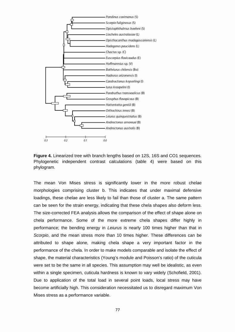
77
Figure 4. Linearized tree with branch lengths based on 12S, 16S and CO1 sequences. Phylogenetic independent contrast calculations (table 4) were based on this phylogram.
The mean Von Mises stress is significantly lower in the more robust chelae
morphologies comprising cluster b. This indicates that under maximal defensive
loadings, these chelae are less likely to fail than those of cluster a. The same pattern
can be seen for the strain energy, indicating that these chela shapes also deform less.
The size-corrected FEA analysis allows the comparison of the effect of shape alone on
chela performance. Some of the more extreme chela shapes differ highly in
performance; the bending energy in Leiurus is nearly 100 times higher than that in
Scorpio, and the mean stress more than 10 times higher. These differences can be
attributed to shape alone, making chela shape a very important factor in the
performance of the chela. In order to make models comparable and isolate the effect of
shape, the material characteristics (Young’s module and Poisson’s ratio) of the cuticula
were set to be the same in all species. This assumption may well be idealistic, as even
within a single specimen, cuticula hardness is known to vary widely (Schofield, 2001).
Due to application of the total load in several point loads, local stress may have
become artificially high. This consideration necessitated us to disregard maximum Von
Mises stress as a performance variable.

78
Table 4. Phylogenetic independent contrasts. Values below the diagonal are Pearson correlation coefficients, values above the diagonal are the corresponding p -values. Correlation coefficients with a p-value < 0,05 are marked in bold. P-values above 0,05 are in grey font. Correlation coefficients that result from correlation of non-independent variables are in brackets. All non-ratio and non-angle values were log-10 transformed.
The lack of a robust molecular phylogenetic hypothesis for the high-level relationships
between scorpions makes it hard to make any statements about the independent
evolution of ecomorphotypes. It is well known that chela shape can vary widely within
scorpion families, such as the Buthidae and Vaejovidae (Stockmann & Ythier, 2010).
Also in our clustering analysis, members of the same family (Iuridae) were present in
different clusters. This suggests chela shape may well be a homoplastic character.
Since changes in the overall shape of the chela may change the relative position of
taxonomic characters (Prendini, 2000), systematists using morphological characters of
the chela, such as the relative positions of the trichobothria, may need to test for the
independence of their characters to the overall chela shape parameters which correlate
highest with performance, such as height, width and relative finger length. Since only
relatively fast-evolving mitochondrial genes were used to infer branch lengths, the
branch lengths used to calculate the phylogenetic independent contrasts may be an
underestimate of the rate of evolution in the basal part of the tree. However, the PIC
method analysis has been shown to be a robust method in relation with branch length
distributions (Diaz-Uriarte & Garland, 1996; Ackerly, 2000).

79
Concluding remarks Our work presents the first mechanical models for the computational assessment of
performance of the scorpion chelae, and allows chela shape types to be used as an
approximation for performance. Despite the limited number of taxa included, our results
clearly show that more elongate chelae experience higher stresses and deformations
than more robust chelae. This makes these chelae less suitable for tasks that require
the chela to perform near its maximum, such as defense, subduing of hard prey, and
burrowing. This may be the reason why scorpions with slender chelae use their sting
more in the incapacitation of prey (Stahnke, 1966; McCormick & Polis 1990).
Acknowledgements We thank Arendo Flipse for supplying several of the scorpions. Thanks to Pedro Sousa for identifying the specimens. We very much appreciate access to the Skyscan CT scanner that was provided by Tim Cox and the team of the small animal tomography facility in Seattle, WA, USA. This work was supported by an FCT I&D grant to AvdM (PTDC/BIA-BEC/104644/2008). AvdM was supported by FCT postdoctoral fellowship SFRH/BPD/48042/2008 and TK was supported by the funding initiative Evolutionary Biology of the Volkswagen Foundation (I 84/206). We are grateful for the comments of two anonymous reviewers which improved the manuscript.
References
Ackerly DD (2000) Taxon sampling, correlated evolution, and independent contrasts. Evolution 54, 1480–1492.
Alexander A (1959) A survey of the biology of scorpions of South Africa. Afr Wildl 13, 99–106.
Arnold AS, Richards CT, Ros IG, et al. (2011) There is always a trade-off between speed and force in a lever system: comment on McHenry (2010). Biol Lett (doi:10.1098/rsbl.2011.0431)
Bruford MW, Hanotte O, Brookfield JFY, et al. (1992) Single-locus and multilocus DNA fingerprint. In Molecular Genetic Analysis of Populations: A Practical Approach (ed. Hoezel AR), pp 225–270. IRL Press, Oxford UK.
Diaz-Uriarte R, Garland T (1996) Testing hypotheses of correlated evolution usingphylogenetically independent contrasts: sensitivity to deviations from Brownian motion. Sys Biol 45, 27–47.
Dumont ER, Grosse IR, Slater GJ (2009) Requirements for comparing the performance of finite element models of biological structures. J Theor Biol 256, 96–103.

80
Dunlop JA, Tetlie OE, Prendini L (2008) Reinterpretation of the Silurian scorpion Proscorpius osborni (Whitfield): Integrating data from paleozoic and recent scorpions. Palaeontology 51, 303–320.
Edgar RC (2004) MUSCLE: multiple sequence alignment with high accuracy and high throughput. Nucleic Acids Res 32, 1792–1797.
Fet V, Sissom WD, Lowe G, et al. (2000) Catalog of the Scorpions of the World (1758–1998). New York Entomological Society: New York.
Fet V, Neff D, Graham MR, et al. (2003) Metasoma of Orthochirus (Scorpiones, Buthidae): Are scorpions evolving a new sensory organ? Révista Ibérica de Arachnología 8, 69–72.
Folmer O, Black M, Hoeh W, et al. (1994) DNA primers for amplification of mitochondrial cytochrome c oxidase subunit I from diverse metazoan invertebrates. Mol Mar Biol Biotechnol 3, 294–299.
Gantenbein B, Kropf C, Largiadèr CR, et al. (2000) Molecular and morphological evidence for the presence of a new buthid taxon (Scorpiones: Buthidae) on the island of Cyprus. Rev Suisse Zool 107, 213–232.
Gilai A, Parnas I (1970) Neuromuscular physiology of the closer muscles in the pedipalp of the scorpion Leiurus quinquestriatus. J Exp Biol 52, 325–344.
Govindarajan S, Rajulu GS (1974) Presence of resilin in a scorpion Palamnaeus swammerdami and its role in the food-capturing and sound-producing mechanisms. Experientia 30, 908–909.
Guindon S, Gascuel O (2003) A simple, fast, and accurate algorithm to estimate large phylogenies by maximum likelihood. Syst Biol 52, 696–704.
Herrel A, Spithoven L, Van Damme R, et al. (1999) Sexual dimorphism of head size in Gallotia galloti: testing the niche divergence hypothesis by functional analysis. Funct Ecol 13, 289–297.
Lamoral BH (1979) The scorpions of Namibia (Arachnida: Scorpionida). Ann Natal Mus 23, 497–784.
McCormick SJ, Polis GA (1990) Prey, Predators, and Parasites. In The Biology of Scorpions (ed. Polis GA), pp 294–320. Stanford University Press, Palo Alto, USA.
McHenry MJ (2011) There is no trade-off between force and velocity in a dynamic lever system. Biol Lett 7, 384–386.
Newlands G (1969) Scorpion defensive behaviour. Afr Wildlife 23, 147–153.
Newlands G (1972) Ecological adaptations of Kruger National Park scorpionids (Arachnida: Scorpionides). Koedoe 15, 37–48.
Newlands G (1978) Arachnida (except Acari). In Biogeography and ecology of southern Africa. (ed. Werger MJA) pp. 677–684. Junk: The Hague, the Netherlands.
Polis GA (1990) Ecology. In The Biology of Scorpions (ed. Polis GA), pp 247–293. Stanford University Press: Palo Alto, USA.

81
Posada D (2008) jModelTest: Phylogenetic Model Averaging. Mol Biol Evol 25, 1253–1256.
Prendini L (2000) Phylogeny and classification of the superfamily Scorpionoidea Latreille 1802 (Chelicerata, Scorpiones): An exemplar approach. Cladistics 16, 1–78.
Prendini L, Wheeler WC (2005) Scorpion higher phylogeny and classification, taxonomic anarchy, and standards for peer review in online publishing. Cladistics 21, 446–494.
R Development Core Team R: A Language and Environment for Statistical Computing , R Foundation for Statistical Computing, Vienna, Austria, 011, ISBN 3-900051-07-0, http://www.R-project.org
Ronquist F, Huelsenbeck JP (2003) MrBayes version 3.0: Bayesian phylogenetic inference under mixed models. Bioinformatics 19, 1572–1574.
Schofield RMS (2001) Metals in cuticular structures. In Scorpion biology and research. (eds. Brownell P, Polis GA) pp 234–256. Oxford University Press: New York, USA
Sensenig AT, Shultz JW (2004) Elastic energy storage in the pedipalpal joints of scorpions and sun-spiders (Arachnida, Scorpiones, Solifugae). J Arachnol 32, 1–10.
Simon C, Frati F, Beckenbach A, et al. (1994) Evolution, weighting, and phylogenetic utility of mitochondrial gene sequences and a compilation of conserved polymerase chain reaction primers. Ann Entomol Soc Am 87, 651–701.
Snodgrass RE (1952) A textbook of Arthropod anatomy. Cornell University Press: Ithaca, N.Y.
Sousa P, Froufe E, Harris DJ, et al. (2011) Genetic diversity of Maghrebian Hottentotta (Scorpiones: Buthidae) scorpions based on CO1: new insights on the genus phylogeny and distribution. Afr Invertebrates. 52, 135–143.
Stahnke HL (1966) Some aspects of scorpion behavior. Bull South Calif Acad Sci 65, 65–80.
Stockmann R, Ythier E (2010) Scorpions of the world. N.A.P. Editions: Verrières-le-Bruisson, France.
Tamura K, Peterson D, Peterson N, et al. (2011) MEGA5: Molecular Evolutionary Genetics Analysis using Maximum Likelihood, Evolutionary Distance, and Maximum Parsimony Methods. Mol Biol Evol, Pre-published online. doi: 10.1093/molbev/msr121
Van der Meijden A, Herrel A, Summers A (2010) Comparison of chela size and pincer force in scorpions; getting a first grip. J Zool 280, 319–325.
Vincent JFV, Wegst UGK (2004) Design and mechanical properties of insect cuticle. Arthropod Struct Dev 33, 187–199.
Webb CO, Ackerly DD, Kembel SW (2008) Phylocom: software for the analysisof phylogenetic community structure and character evolution. Bioinformatics 24, 2098–2100.
Yamada SB, Boulding EG (1998) Claw morphology, prey size selection and foraging efficiency in generalist and specialist shell-breaking crabs. J Exp Mar Biol Ecol 220, 191–211.

82

83
Chapter IV
Final Remarks and Future Research References

84
4.1 Final Remarks
From an original plan to sequence nuclear genes we faced technical difficulties
at different levels in the whole process. Initially, we tried to amplify 18S and 28S rRNA
genes in order to obtain nuclear DNA. However, analysis of the dataset produced by
the resulting sequences led to the conclusion that these genes have been duplicated in
our study organisms, or at least that concerted evolution was not causing the
sequences to be identical but rather two divergent groups occurred within individuals.
Unfortunately, this event made it impossible to infer phylogenetic patterns and,
consequently, this dataset had to be discarded. Then, our attention turned to
mitochondrial DNA (mtDNA) in order to sequence phylogenetically-informative DNA
that could be used not only at the genus level, but also at the sub-family level.
Cytochrome b (Cytb) was first selected as it is routinely used in phylogenetic studies.
However we faced technical difficulties trying to amplify this fragment. As time
consuming as it was, we felt obligated to choose other mitochondrial genes that could
produce the whole dataset quicker. Therefore, we chose the 12S rRNA, 16S rRNA and
COI genes. We were able to produce the whole dataset with less than 10% of missing
data. In the middle of the process, we made other attempts to use phylogenetic
informative genes at different resolutions inside the Scorpiones order. Histone 3 (H3)
was part of the laboratorial work produced in 2011/2012 that was chosen based on its
wide use in phylogenetic studies of the Aranae order. Like CytB it showed sub-
standard amplification. The last attempt fell upon ND1 mtDNA gene and, although it
had low amplification success, it was informative. In addition, it is a protein coding gene
like COI. In a period where we already had two non-coding and two protein-coding
genes we made a final but unsuccessful endeavor to amplify informative nuclear
genes. We used primers for six nuclear genes from Gantenbein and Eightley, 2004,
using a total of eight primer combinations. As we could not successfully amplify these
last genes we chose to complete our studies with the 16S + 12S + COI or ND1 dataset.

85
4.2 Future Research
Technical obstacles played against the original goal of obtaining phylogenies
that could resolve relationships within the Scorpiones order, between families, genera
and species with strong resolution. A lack of slower evolving nuclear markers
hampered basal inference on the Androctonus and the scorpion families’ phylogenies.
Future work should be guided towards the improvement of DNA markers that can
produce well resolved phylogenies within different scorpions. The effect of model
choice (Jones et al., 2006) and testing for recombination (Gantenbein et al., 2005)
should be taken into consideration in future studies as well.
Additionally, new strategies must be created to overcome sampling difficulties in
traditionally inaccessible countries. Political instability on one hand and the lack of
georeferenced sampling on the other might contribute to an underestimation of
scorpion’ diversity levels.

86
5.References
Audouin, 1826. Explication sommaire des planches d’arachnides de l'Égypte et de la Syrie publiées par J. C. Savigny, membre de l'Institut; offrant un exposé des caractères naturels des genres avec la distinction des espèces. In: Description de l’Égypte, Ou Recueil Des Observations et Des Recherches Qui Ont Été Faites En Égypte Pendant L'expédition de L'armée Française. Histoire Naturelle, pp. 99–186.
Avise, J.C., Arnold, J., Ball, R.M., Bermingham, E., Lamb, T., Neigel, J.E., Reeb, C.A., Saunders, N.C., 1987. Intraspecific phylogeography: the mitochondrial DNA bridge between population genetics and systematics. Annu. Rev. Ecol. Syst. 18, 489–522.
Ballard, J.W.O., Rand, D.M., 2005. The Population Biology of Mitochondrial DNA and Its Phylogenetic Implications. Annu. Rev. Ecol. Evol. Syst. 36, 621–642.
Ballard, J.W.O., Whitlock, M.C., 2004. The incomplete natural history of mitochondria. Mol. Ecol. 13, 729–44.
Ben Ali, Z., Boursot, P., Said, K., Lagnel, J., Chatti, N., Navajas, M., 2000. Comparison of ribosomal ITS regions among Androctonus spp. scorpions (Scorpionida: Buthidae) from Tunisia. J. Med. Entomol. 37, 787–90.
Ben Othmen, A., Chatti, N., Ben Ali-Haouas, Z., Ouldbrahim, I., Said, K., 2004. Allozymic differentiation of Tunisian populations of Androctonus species and Buthus occitanus (Scorpiones: Buthidae). Biol. J. Linn. Soc. 81, 255–265.
Ben Othmen, A., Said, K., Mahamdallie, S.S., Testa, J.M., Haouas, Z., Chatti, N., Ready, P.D., 2009. Phylogeography of Androctonus species (Scorpiones: Buthidae) in Tunisia: Diagnostic characters for linking species to scorpionism. Acta Trop. 112, 77–85.
Bouaziz, M., Bahloul, M., Kallel, H., Samet, M., Ksibi, H., Dammak, H., Ahmed, M.N. Ben, Chtara, K., Chelly, H., Hamida, C. Ben, Rekik, N., 2008. Epidemiological, clinical characteristics and outcome of severe scorpion envenomation in South Tunisia: multivariate analysis of 951 cases. Toxicon 52, 918–26.
Brownell, P.H., Polis, G.A., 2001. Scorpion Biology and Research. Oxford University Press, New York.
Bruford, M.W., Hanotte, O., Brookfield, J.F.Y., Burke, T., 1992. Single-locus and multilocus DNA fingerprinting. In: Hoelzel, A.R. (Ed.), Molecular Genetic Analysis of Populations: A Practical Approach. IRL Press, Oxford, pp. 225–270.
Caterino, M.S., Cho, S., Sperling, F.A., 2000. The current state of insect molecular systematics: a thriving Tower of Babel. Annu. Rev. Entomol. 45, 1–54.
Chippaux, J.-P., 2012. Emerging options for the management of scorpion stings. Drug Des. Devel. Ther. 6, 165–73.
Chippaux, J.-P., Goyffon, M., 2008. Epidemiology of scorpionism: a global appraisal. Acta Trop. 107, 71–9.
Daltry, J.C., Wüster, W., Thorpe, R.S., 1996. Diet and snake venom evolution. Nature 379, 537–40.
Darriba, D., Taboada, G.L., Doallo, R., Posada, D., 2012. jModelTest 2: more models, new heuristics and parallel computing. Nat. Methods 9, 772.

87
Drake, N.A., Blench, R.M., Armitage, S.J., Bristow, C.S., White, K.H., 2011. Ancient watercourses and biogeography of the Sahara explain the peopling of the desert. Proc. Natl. Acad. Sci. U. S. A. 108, 458–62.
Edgar, R.C., 2004. MUSCLE: multiple sequence alignment with high accuracy and high throughput. Nucleic Acids Res. 32, 1792–7.
Ehrenberg, Hemprich, 1828. Arachnoidea, Scorpiones. In: Symbolae Physicae Seu Icones et Descriptiones Animalium Evertebratorum Sepositis Insectis Quae Ex Itinere per Africanum Borealem et Asiam Occidentalem. Berlin.
El Ayeb, M., Rochat, H., 1985. Polymorphism and quantitative variations of toxins in the venom of the scorpion Androctonus australis Hector. Toxicon 23, 755–60.
Espino-Solis, G.P., Estrada, G., Olamendi-Portugal, T., Villegas, E., Zamudio, F., Cestele, S., Possani, L.D., Corzo, G., 2011. Isolation and molecular cloning of beta-neurotoxins from the venom of the scorpion Centruroides suffusus suffusus. Toxicon 57, 739–46.
Fatani, A.J., Ahmed, A. a E., Abdel-Halim, R.M., Abdoon, N. a, Darweesh, A.Q., 2010. Comparative study between the protective effects of Saudi and Egyptian antivenoms, alone or in combination with ion channel modulators, against deleterious actions of Leiurus quinquestriatus scorpion venom. Toxicon 55, 773–86.
Fet, V., 1994. 31. Fauna and Zoogeography of Scorpions (Arachnida: Scorpions) in Turkmenistan. In: Biogeography and Ecology of Turkmenistan. Kluwer Academic Publisher, Dordecht, pp. 525–534.
Fet, V., 1997. Notes on the taxonomy of some old world scorpions (Scorpiones: Buthidae, Chactidae, Ischnuridae, Scorpionidae). J. Arachnol. 25, 245–250.
Fet, V., Gantenbein, B., Gromov, A.A. V, Lowe, G., Lourenço, W.R., 2003. The first molecular phylogeny of Buthidae (Scorpiones). Euscorpius 4, 1–10.
Fet, V., Polis, G.A., Sissom, W.D., 1998. Life in sandy deserts: the scorpion model. J. Arid Environ. 39, 609–622.
Fet, V., Sissom, W., Lowe, G., Braunwalder, M., 2000. Catalog of the scorpions of the world (1758-1998). New York Entomological Society, New York.
Fet, V., Soleglad, M., 2005. Contributions to scorpion systematics. I. On recent changes in high-level taxonomy. Euscorpius 31, 1–13.
Froufe, E., Sousa, P., Alves, P.C., Harris, D.J., 2008. Genetic diversity within Scorpio maurus (Scorpiones: Scorpionidae) from morocco: Preliminary evidence based on CO1 mitochondrial DNA sequences. Biologia (Bratisl). 63, 1157–1160.
Gantenbein, B., Eightley, P.D., 2004. RATES OF MOLECULAR EVOLUTION IN NUCLEAR GENES OF EAST MEDITERRANEAN SCORPIONS. Evolution (N. Y). 58, 2486–2497.
Gantenbein, B., Fet, V., Gantenbein-Ritter, I. a, Balloux, F., 2005. Evidence for recombination in scorpion mitochondrial DNA (Scorpiones: Buthidae). Proc. Biol. Sci. 272, 697–704.
Gantenbein, B., Largiadèr, C.R., 2003. The phylogeographic importance of the Strait of Gibraltar as a gene flow barrier in terrestrial arthropods: a case study with the scorpion Buthus occitanus as model organism. Mol. Phylogenet. Evol. 28, 119–130.

88
Gess, R.W., 2013. The earliest record of terrestrial animals in Gondwana: A scorpion from the Famennian (Late Devonian) Witpoort Formation of South Africa. African Invertebr. 54, 373–379.
Goyffon, M., Dabo, A., Coulibaly, S., Togo, G., Chippaux, J., 2012. Dangerous scorpion fauna of Mali. J. Venom. Anim. Toxins Incl. Trop. Dis. 18, 361–368.
Goyffon, M., Guette, C., 2005. Scorpions dangereux du Niger. Bull. la Société Pathol. Exot. 98, 293–295.
Goyffon, M., Lamy, J., 1973. Une nouvelle sous-espèce d’Androctonus australis L. (Scorpion, Buthidae): Androctonus australis garzonii n. ssp. Caractéristiques morphologiques, écologiques et biochimiques. Bull. la Société Zool. Fr. 98, 137–144.
Goyffon, M., Vachon, M., Broglio, N., 1982. Epidemiological and clinical characteristics of the scorpion envenomation in Tunisia. Toxicon 20, 337–44.
Graham, M.R., 2011. Scorpions of the World Scorpions of the World. Roland Stockmann , Eric Ythier . 2010. N.A.P. Editions. France. 567 pp. ISBN: 978-2-913688-11-7. 75€. J. Arachnol. 39, 166–167.
Guindon, S., Dufayard, J.-F., Lefort, V., Anisimova, M., Hordijk, W., Gascuel, O., 2010. New algorithms and methods to estimate maximum-likelihood phylogenies: assessing the performance of PhyML 3.0. Syst. Biol. 59, 307–21.
Habel, J.C., Husemann, M., Schmitt, T., Zachos, F.E., Honnen, A.-C., Petersen, B., Parmakelis, A., Stathi, I., 2012. Microallopatry caused strong diversification in Buthus scorpions (Scorpiones: Buthidae) in the Atlas Mountains (NW Africa). PLoS One 7, e29403.
Habermehl, G., 1994. Gift-Tiere und ihre Waffen: Eine Einführung für Biologen, Chemiker und Mediziner. Ein Leitfaden für Touristen, 5th editio. ed. Springer Verlag, Heidelberg.
Herrel, A., Huyghe, K., Vanhooydonck, B., Backeljau, T., Breugelmans, K., Grbac, I., Van Damme, R., Irschick, D.J., 2008. Rapid large-scale evolutionary divergence in morphology and performance associated with exploitation of a different dietary resource. Proc. Natl. Acad. Sci. U. S. A. 105, 4792–5.
Jeram, A.J., 2000. Paleontology. In: Polis, G.A., Brownell, P. (Eds.), Scorpion Biology and Research. Oxford University Press, Oxford, pp. 370–392.
Kocher, T.D., Thomas, W.K., Meyer, A., Edwards, S. V, Pääbo, S., Villablanca, F.X., Wilson, A.C., 1989. Dynamics of mitochondrial DNA evolution in animals: amplification and sequencing with conserved primers. Proc. Natl. Acad. Sci. U. S. A. 86, 6196–6200.
Kraepelin, K., 1903. Scorpione und Solifugen Nordost-Afrikas, gesammelt 1900 und 1901 von Carlo Freihern von Erlanger und Oscar Neumann. Zool. Jahrbücher. Abteilung für Syst. Geogr. und Biol. der Tiere 18, 557–578.
Lamoral, B.H., 1979. The Scorpions of Namibia (Arachnida-Scorpionida). Ann. Natal Museum 23, 497–784.
Lamy, J., Le Pape, G., Weill, J., 1974. Hétérogénéité de l’espèce Androctonus australis (Scorpiones, Buthidae). Création d'une nouvelle sous-espèce A. australis africanus n. spp. sur la base de critères biochimiques et génétiques. C. R. Hebd. Seances Acad. Sci. 278, 3223–3226.

89
Larkin, M.A., Blackshields, G., Brown, N.P., Chenna, R., McGettigan, P.A., McWilliam, H., Valentin, F., Wallace, I.M., Wilm, A., Lopez, R., Thompson, J.D., Gibson, T.J., Higgins, D.G., 2007. Clustal W and Clustal X version 2.0. Bioinformatics 23, 2947–8.
Legg, D., Sutton, M., Edgecombe, G., 2013. Arthropod fossil data increase congruence of morphological and molecular phylogenies. Nat. Commun.
Levy, G., Amitai, P., 1980. Fauna Palaestina. Arachnida I: Scorpiones. Israel Academy of Sciences and Humanities, Jerusalem.
Linnaeus, 1758. Systema naturae per regna tria naturae, secundum classes, ordines, genera, species, cum characteribus, differentiis, synonymis, locis, 10th ed.
Loret, E., Hammock, B., 2001. Structure and neurotoxicity of scorpion toxins. In: Brownell, P.H., G. A. Polis (Eds.), Scorpion Biology and Research. Oxford University Press, Oxford, pp. 204–233.
Lourenço, W., Cuellar, O., 1995. Scorpions, scorpionism, life history strategies and parthenogenesis. J. Venom. Anim. Toxins 1, 51–62.
Lourenço, W., Duhem, B., 2009. Saharo-Sindian buthid scorpions; description of two new genera and species from Occidental Sahara and Afghanistan. Zookeys 14, 37–54.
Lourenço, W., Qi, J., 2007. A new species of Androctonus Ehrenberg, 1828 from Mauritania (Scorpiones, Buthidae). Boletín Soc. Entomológica Aragon. 1, 215–219.
Lourenço, W.R., 2000. A new genus of Malagasy scorpion, possible link between the Microcharmidae and the Buthidae. C. R. Acad. Sci. III. 323, 877–81.
Lourenço, W.R., 2005. Nouvelles considérations taxonomiques sur les espèces du genre Androctonus Ehrenberg, 1828 et description de deux nouvelles espèces (Scorpiones, Buthidae). Rev. Suisse Zool. 112, 145–171.
Lourenço, W.R., 2008. A new species of Androctonus Ehrenberg, 1828 from Togo (Scorpiones, Buthidae). Entomol. Mitteilungen aus dem Zool. Museum Hambg. 15, 37–44.
Lourenço, W.R., Yağmur, E.A., Duhem, B., 2010. A new species of Buthus Leach, 1815 from Jordan. Zool. Middle East 49, 95–99.
McCormick, S.J., Polis, G.A., 1990. Prey, predators and parasites. In: Polis, G.A. (Ed.), The Biology of Scorpions. Stanford University Press, Stanford, CA, pp. 294–320.
Miranda, F., Rochat, H., Rochat, C., Lissitzky, S., 1966. Complexes moléculaires présentés par les neurotoxines animales—I. Neurotoxines des venins de scorpions (Androctonus australis hector et Buthus occitanus tunetanus). Toxicon 4, 123–136.
Newton, K.A., Clench, M.R., Deshmukh, R., Jeyaseelan, K., Strong, P.N., 2007. Mass fingerprinting of toxic fractions from the venom of the Indian red scorpion, Mesobuthus tamulus: biotope-specific variation in the expression of venom peptides. Rapid Commun. Mass Spectrom. 21, 3467–76.
Olivier, 1807. Voyage dans l`Empire Othoman, l`Égypte et la Perse, vol. 3. ed. Paris.
Omran, M.A.A., McVean, A., 2000. Intraspecific Variation in Scorpion Leiurus quinquestriatus Venom Collected From Egypt (Sinai And Aswan Deserts). Toxin Rev. 19, 247–264.

90
Pallary, 1924. Description de trois scorpions nouveaux du Maroc. Arch. l’Institut Pasteur d'Algérie 2, 219–222.
Piganeau, G., Gardner, M., Eyre-Walker, A., 2004. A broad survey of recombination in animal mitochondria. Mol. Biol. Evol. 21, 2319–25.
Pocock, 1897. Descriptions of some new species of scorpions from India. J. Bombay Nat. Hist. Soc. 11, 102–117.
Pocock, 1900. Arachnida. The Fauna of British India, including Ceylon and Burma. Published under the authority of the Secretary of State for India. In: Council. W. T. Blandford, London, pp. 1–279.
Pocock, 1902. A contribution to the systematics of scorpions. Ann. Mag. Nat. Hist. 10, 364–380.
Polis, G.A., 1990. Introduction to the Biology of Scorpions. In: Polis, G.A. (Ed.), The Biology of Scorpions. Stanford University Press, Stanford, pp. 1–8.
Polis, G.A., Sissom, W.D., 1990. Life History. In: Polis, G.A. (Ed.), The Biology of Scorpions. Stanford University Press, Stanford, pp. 161–223.
Prasad, N., Uma, B., Bhatt, S., Gowda, V., 1999. Comparative characterisation of Russell’s viper (Daboia/Vipera russelli) venoms from different regions of the Indian peninsula. Biochim. Biophys. Acta 1428, 121–136.
Qi, J.-X., Lourenço, W.R., 2007. Distribution of endemic relict groups of Saharan scorpions, with the description of new genus and species from Mauritania. C. R. Biol. 330, 80–5.
Rein, J.O., 2012. The Scorpion files [WWW Document]. The Scorpion Files. URL http://www.ntnu.no/ub/scorpion-files/ (accessed 9.3.13).
Savory, T.H., 1977. The Arachnida, 2d ed. ed. Academic, London.
Shultz, J.W., 2007. A phylogenetic analysis of the arachnid orders based on morphological characters. Zool. J. Linn. Soc. 150, 221–265.
Simmard, Watt, 1990. Venoms and Toxins. In: The Biology of Scorpions. Stanford University Press, pp. 414–444.
Simon, C., Frati, F., Beckenbach, A., Crespie, B., Liu, H., Flook, P., Crespi, B., Liu, H., 1994. Evolution, weighting, and phylogenetic utility of mitochondrial gene sequences and a compilation of conserved polymerase chain reaction primers. Ann. Entomol. Soc. Am. 87, 651–701.
Smertenko, A., Omran, M.A.A., Hussey, P.J., McVean, A., 2001. Toxin evolution in Scorpion Venom: evidence for toxin divergence under strong negative selection in Leiurus quinquestriatus subspecies. Toxin Rev. 20, 229–244.
Sousa, P., Froufe, E., Alves, P., Harris, D.J., Alves, P.C., 2010. Genetic diversity within scorpions of the genus Buthus from the Iberian Peninsula: mitochondrial DNA sequence data indicate additional distinct cryptic lineages. J. Arachnol. 28, 206–211.
Sousa, P., Froufe, E., Harris, D., Alves, P.C., Van der Meijden, A., 2011. Genetic diversity of Maghrebian Hottentotta ( Scorpiones : Buthidae ) scorpions based on CO1 : new insights on the genus phylogeny and distribution. African Invertebr. 52, 135–143.

91
Sousa, P., Harris, D.J., Froufe, E., Meijden, A., 2012. Phylogeographic patterns of Buthus scorpions (Scorpiones: Buthidae) in the Maghreb and South-Western Europe based on CO1 mtDNA sequences. J. Zool. 288, 66–75.
Stockmann, R., Ythier, E., 2010. Scorpions of the World. N.A.P Editions, Verrières-le-Bruisson, France.
Sunnucks, P., 2000. Efficient genetic markers for population biology. Trends Ecol. Evol. 15, 199–203.
Tamura, K., Peterson, D., Peterson, N., Stecher, G., Nei, M., Kumar, S., 2011. MEGA5: molecular evolutionary genetics analysis using maximum likelihood, evolutionary distance, and maximum parsimony methods. Mol. Biol. Evol. 28, 2731–9.
Tikader, B., Bastawade, D., 1983. The Fauna of India. Vol. 3. Scorpions (Scorpionida: Arachnida), … : Arachnida Vol. III. Zoological Survey of India, Calcutta.
Touloun, O., Boumezzough, A., Slimani, T., 2012. Scorpion envenomation in the region of Marrakesh Tensift Alhaouz (Morocco): epidemiological characterization and therapeutic approaches. Serket 13, 38–50.
Vachon, M., 1949. Description des Scorpions du Nord de l’Afrique. In: Études Sur Les Scorpions III (. Institut Pasteur d’Algérie, pp. 288–316.
Vachon, M., 1952. Etudes sur les scorpions. Institut Pasteur d’Algérie, Alger.
Vachon, M., 1953. Contribution a l’étude du peuplement de la Mauritanie. Scorpions. Bull. l ’lnstitut Français d 'Afrique Noire 15, 1012–1028.
Vachon, M., 1958. Scorpionidea (Chelicerata) de l’Afghanistan. The 3rd Danish Expedition to Central Asia. (Zoological Results 23). Vidensk. Meddelelser fra Dansk Naturhistrorisk Foren. i Købehavn 120, 121–187.
Vachon, M., 1974. Etude des caractères utilisés pour classer les familles et les genres de Scorpions (Arachnides). 1. La trichobothriotaxie en Arachnologie, Sigles trichobothriaux et types de trichobothriotaxie chez les Scorpions. Bull. du Muséum Natl. d’Histoire Nat. 3, 857–958.
Van der Meijden, a., Herrel, A., Summers, A., 2010. Comparison of chela size and pincer force in scorpions; getting a first grip. J. Zool. 280, 319–325.
Van der Meijden, A., Kleinteich, T., Coelho, P., 2012. Packing a pinch: functional implications of chela shapes in scorpions using finite element analysis. J. Anat. 220, 423–34.
Watt, D.D., Simard, J.M., 1984. Neurotoxic Proteins in Scorpion Venom. Toxin Rev. 3, 181–221.
Yağmur, E.A., Koç, H., Lourenço, W.R., 2011. A new species of Buthus Leach, 1815 from Cyprus (Scorpiones, Buthidae). Zookeys 27–38.
Zhang, D.X., Hewitt, G.M., 1996. Nuclear integrations: challenges for mitochondrial DNA markers. Trends Ecol. Evol. 11, 247–51.

![II CONGRESSO O PORTO ROMÂNTICO - Repositório Aberto · 2019-07-14 · 13 O Nacional. [Porto] 29, (11 Set 1847), p . 3 14 O Nacional. [Porto] 42, (20 Fev 1850), p . 1 15 O Nacional](https://img.pdfslide.net/doc/110x75/5f2048683ecd4031f22826df/ii-congresso-o-porto-romntico-repositrio-aberto-2019-07-14-13-o-nacional.jpg)