Embed Size (px)
Citation preview

UNIVERSIDADE ESTADUAL DE CAMPINAS
FACULDADE DE CIÊNCIAS MÉDICAS
DANIELE BRAZ TORRES
Análise dos efeitos da restrição proteica in utero no BNST e na Amígdala de ratos: Estudo da estrutura dendrítica neural, de
parâmetros funcionais e moleculares.
CAMPINAS
2016

DANIELE BRAZ TORRES
Análise dos efeitos da restrição proteica in utero no BNST e na Amígdala de ratos: Estudo da estrutura dendrítica neural, de parâmetros funcionais e
moleculares.
Tese apresentada à Faculdade de Ciências Médicas da
Universidade Estadual de Campinas como parte dos requisitos
exigidos para a obtenção do título de Doutora em Ciências
Orientadora: Profa. Dra. Patricia Aline Boer
Coorientador: Prof. Dr. Jose Antonio Rocha Gontijo
ESTE EXEMPLAR CORRESPONDE À VERSÃO FINAL DA TESE DEFENDIDA PELA ALUNA DANIELE BRAZ TORRES, E ORIENTADA PELA PROFA. DRA. PATRICIA ALINE BOER.
CAMPINAS
2016


BANCA EXAMINADORA DA DEFESA DE DOUTORADO DANIELE BRAZ TORRES
ORIENTADOR: PATRICIA ALINE BOER
COORIENTADOR: JOSE ANTONIO ROCHA GONTIJO
MEMBROS:
1. PROF(A). DR(A). PATRICIA ALINE BOER
2. PROF(A). DR(A). GLÁUCIA MONTEIRO DE CASTRO
3. PROF(A). DR(A). SYLVIA MARIA CIASCA
4. PROF(A). DR(A). JOÃO PEREIRA LEITE
5. PROF(A). DR(A). PAULO DALGALARRONDO
Programa de Pós-Graduação em Fisiopatologia Médica da Faculdade de Ciências Médicas da Universidade Estadual de Campinas.
A ata de defesa com as respectivas assinaturas dos membros da banca examinadora encontra-se no processo de vida acadêmica do aluno.
Data: 14 de abril de 2016

Dedico este trabalho com todo meu amor e gratidão aos meus pais, José Messias
Xavier Torres e Dirce Braz Torres.

“Tudo posso Naquele que me fortalece.”
Filipenses 4:13

AGRADECIMENTOS
A Deus em primeiro lugar, pelo dom da vida e da saúde que me foi dada.
Por ter realizado verdadeiros milagres nesta caminhada, pela força que tenho
certeza que vem do Pai. E por todas as inúmeras oportunidades que me ofereceu.
A minha orientadora Prof. Dra. Patricia Aline Boer que me abriu as portas
do seu laboratório em 2009 e também as portas da sua casa para me receber nos
últimos meses. Obrigada Pati pela ajuda e confiança que sempre teve comigo. Por
ter criado inúmeras oportunidades de trabalho e de experiências de vida. Serei
eternamente grata!
Ao Prof. Dr. José Antônio Rocha Gontijo pela ajuda intelectual formidável,
pela confiança no meu trabalho e por junto com a Prof. Patrícia ter me oferecido
tantas boas oportunidades. Obrigada Professor!
Aos meus co-orientadores na Universidade do Minho Dr. Nuno Sousa e Dr.
Ana João Rodrigues pela ajuda, confiança e por sempre me receberem com tanto
carinho em vossos laboratórios. Todo meu carinho e gratidão!
A todos os funcionários da Unicamp e do ICVS pela ajuda e colaboração
com meu trabalho, sem vocês não seria possível.
A todos os colegas de laboratório a Agnes, Helô, Daniel, Ana, Augusto,
Noemi, Gabriel, Silmara, Lais, Ana Leticia, Thais e a Ize pela ajuda com o
trabalho, pelos momentos de camaradagem e alegrias.
De forma muito especial agradeço a amiga que fiz a Agnes, que esta
comigo desde o inicio desta jornada, juntas ficamos mais fortes e conseguimos
acredito eu ir além das nossas expectativas. E a Helô que também sempre tão
querida e disposta a escutar e ajudar a todos do laboratório! Obrigada meninas
por serem tão pacientes e aguentarem minhas chatices. Vocês merecem um
troféu por isso!

A todos os colegas de laboratório do ICVS, em especial a Carina, Paula
Silva e a Barbara que tiveram grande contribuição com este trabalho. A Sofia que
sempre foi tão querida!
A amiga especial Liliana Santos, ter sua amizade tão longe da minha família
foi de grande importância. Obrigada pelo carinho e amizade sincera!
Aos animas que apesar da falta de escolha, contribuíram com sua vida para
que este trabalho fosse possível.
A Capes, Cnpq e a Fapesp pelo suporte financeiro a este trabalho.
Ao meu namorado Luciano e toda sua família, em especial a Dona Adélia e
a Tia Lurdes por terem me recebido em Portugal durante o Doutorado Sanduiche.
Vocês tornaram este período mais acolhedor e foram sempre minha família
também, todo meu amor e gratidão a vocês!
A minha família a base de tudo, meu porto seguro. Ao longo deste período
foram as palavras amigas e confortáveis, aquele carinho que afaga e nos da força
pra continuarmos e mesmo sendo clichê, sem vocês não seria possível!
Ao meu avô Vicente que é a alegria e vitalidade em nossa família e a avô
Maria (in memoriam), é sempre tão bom voltar pra casa e recarregar as energias
com todo este amor!
Meus irmãos Juliano e Leticia minha Cunhada Daniele e meu sobrinho
Lucas que nasceu no fim deste doutorado e encheu minha vida de energia, amor e
alegrias, amo vocês!

RESUMO
O núcleo da estria terminal (BNST) e a amígdala têm sido associados à
modulação do comportamento ansioso e do medo. Devido à sua plasticidade,
tanto a morfologia quanto a composição de neurotransmissores destas regiões
podem ser influenciadas por eventos adversos durante o período perinatal. A
restrição proteica in utero, com consequente exposição do feto a concentração
elevada de glicocorticoides (GC), está associa à desregulação do eixo HPA e a
resposta exarcebada ao estresse na vida adulta. Recentemente, um estudo
conduzido em nosso laboratório demonstrou que a restrição proteica gestacional
promove alterações neuroquímicas e morfológicas no BNST que se associam ao
comportamento de ansiedade em ratos machos adultos. Entretanto, neste estudo
não foi possível afirmar se as alterações encontradas são de fato decorrentes da
programação gestacional ou secundárias a outros fatores desconhecidos que
poderiam determinar sua ocorrencia na vida adulta. Assim, o objetivo do presente
estudo foi investigar, em animais com idade precoce, os possíveis efeitos da
restrição proteica gestacional durante o desenvolvimento do BNST e da amigdala.
Nosso estudo mostrou que a prole submetida à restrição proteica gestacional
apresenta diminuição significativa do peso corporal ao nascer, que persiste
reduzido até o 14° dia de vida. Além disso, a quantificação das células do BNST e
os estudos esteriológicis mostram redução significativa do número total de células
e do volume da região antero-dorsal do BNST em animais do grupo LP, no 14º dia
de vida em comparação ao grupo NP de mesma idade. Houve também redução
no comprimento e na arborização dendrítica dos neurônios do BNST na prole LP.
Demonstramos ainda redução na expressão de receptores de glicocorticóides
(GR) e mineralocorticóides (MR) nos animais do grupo LP com 7 e 14 dias de
vida, associada a redução na expressão do BNDF e aumento na expressão do
receptor CRF1 no BNST de animais do grupo LP com 7 dias de vida, apesar de
não ter havido alteração nos níveis plasmáticos de corticosterona. Nos animais do
grupo LP com 14 dias observamos redução na expressão dso receptores 5HT1A
e aumento na expressão dos receptores 5HT2A nos animais do grupo LP com 7 e
14 dias de vida. Nossos resultados também demonstraram aumento significativo

da concentração de norepinefrina e 5HIAA no BNST e redução do turnorver de
dopamina e seu precurssor DOPAC no BNST, respectivamente, nos animais do
grupo LP com 7 e 14 dias. Quanto à amigdala nós não observamos alterações
significativas no comprimento e no número de ramificações dos dendritos de
neurônios localizados na amigdala basolateral dos animais do grupo LP com 14
dias comparativamente aos animais do grupo NP de mesma idade. Da mesma
forma, o número de células na amigdala foi similar nos animais dos grupos LP e
NP no14º dia. Houve redução significativa na concentração de norepinefrina,
epinefrina e dopamina bem como na expressão de CRF e BDNF na amigdala de
animais do grupo LP no 7º dia, assim como houve redução na expressão de GR,
MR e CRF na amigdala dos animais do grupo LP no14º dia. Em conclusão, esta é
a primeira descrição na literatura da ocorrência de modulação da plasticidade,
tanto com relação ao número de neurônios e outros tipos celulares quanto da
neuroquímica do BNST precocemente, decorrente da restrição proteica
gestacional. Além disso, as alterações neuroquímicas observadas na amigdala e
no BNST podem contribuir para as alterações comportamentais induzidas pela
restrição proteica gestacional bem como alterações em outras regiões cerebrais.
Estas diferenças podem representar a adaptação que ocorre durante o
desenvolvimento embrionário às altas concentrações de glicocorticóides maternos
e pode estar relacionada à hiperatividade do eixo HPA e ao comportamento de
ansiedade observado na idade adulta em indivíduos submetidos à restrição
proteica gestacional.
Palavras chaves: BNST, amigdala, programação fetal, receptores esteróides,
CRF, BDNF, serotonina, dopamina, catecolamina, análise dendrítica.

ABSTRACT
The bed nucleus of the stria terminalis (BNST) and amygdala have been
associated with the modulation of anxiety-like behavior and fear. Due to their
plasticity, its morphology and neurotransmitter compounds may be affected by
perinatal adverse events such as protein restriction in utero, when the fetus is
exposed to high maternal glucocorticoids (GC) levels, leading to a deregulation of
HPA axis and exacerbated stress answer in adult life. We have recently
demonstrated that gestational protein-restricted intake causes neurochemical and
morphological changes in the BNST associated with anxiety-like behavior in male
offspring adulthood. However, our study did not allow asserting the full mode
whether these disorders are from gestational programming or secondary to other
factors that could determine these changes in adulthood. Thus, our objective was
investigating the possible effects of gestational protein restriction on development
of BNST and amygdala in early age. The current study shows a significant
decrease in body birth weight that remains up to 14 day of age, in gestational
protein-restricted offspring. Otherwise, BNST cell quantification and stereology
studies show a significant reduction of the total cells number associated with
reduced volume of the anterodorsal BNST division in 14 d-old LP offspring
compared to age-matched NP group. Also, we found reduction of the dendritic
length and arborizations in the BNST neurons of the LP offspring. The present
work demonstrates a decreased expression of gluco- and mineralocorticoid
receptors (GR and MR) in 7 and 14-d old accompanied, by fall in BDNF and
enhanced CRF1 receptor expression in the BNST of the 7-d old LP offspring,
despite of unchanged corticosterone plasma level. Additionally, the 14 d-old LP
offspring presents a reduced 5HT1A receptor subtype levels, reciprocally
accompanied, by increased 5HT2A receptors in the 7 and 14-d old LP. Our findings
also show a significant increase in the BNST norepinephrine and 5HIAA levels
and, reduced dopamine turnover and 3,4-DOPAC BNST levels, respectively in 7-d
old LP and 14 d-old LP. In complementary study we did not observe any significant
changes in the basolateral amygdala neuronal dendrites length and ramifications in
14 d-old NP compared with age-matched gestational protein-restricted offspring.

Also, the amygdala cells number was similar in 14 day-old LP and NP offspring.
We demonstrate a significant decrease in the amygdala content of
norepinephrine, epinephrine, dopamine, CRF and BDNF in 7 day-old rats, as well
as reduction in GR, MR and CRF expression in 14 day-old LP offspring. In
conclusion, as far as we know is the first description of the modulation of neuron
and non-neuron plasticity and neurochemistry of the BNST in the early life, by
gestational protein-restricted intake. Also, the BNST and amygdala neurochemical
changes observed in the current study may contribute to behavioral alterations
induced by gestational protein restriction and these may be a primer for alterations
in other brain regions. These findings may represent the adaptation during
embryonic development to elevate maternal corticosteroids exposure and could be
related to HPA axis hyperactivity and anxiety-like behavior observed in maternal
protein-restricted offspring adulthood.
Keywords: BNST, amygdala, fetal programming, steroid receptors, CRF, BDNF,
serotonin, dopamine, catecholamine, dendritic analysis

LISTA DE SIGLAS E ABREVIATURAS
OMS Organização Mundial da Saúde
BPN baixo peso ao nascer
RCIU retardo do crescimento fetal intra-utero
SNC sistema nervoso central
11- HSD2 enzima 11-hidroxiesteróide desidrogenase tipo 2 placentária
HHPA eixo hipocampo-hipotálamo-pituitária-adrenal
GC glicocorticoides
MR Receptores de mineralocorticóides
ACTH Hormônio adrenocorticotrofico
PVN núcleo paraventricular
BNST núcleo da estria terminal
CRF hormônio liberador de corticotrofina
VTA núcleo ventral tegmentar
CeA núcleos central da amigdala
CeM núcleo medial da amigdala medial
BNSTL divisão lateral do BNST
MeA amígdala medial
BNSTM divisão medial do BNST
BLA núcleo basolateral da amígdala
BLAC parte caudal do BNST
TDAH déficit de atenção e hiperatividade
CRF1 fator liberador de corticotropina receptor 1
5-HT serotonina

5-HT1-7 família de receptores de serotonina dos tipos 1 a 7
5-HT1A receptor de serotonina tipo 1A
BDNF fator neurotrófico derivado do cérebro
ICVS Instituto de Ciências da Vida
DGAV Direção Geral de Alimentação e Veterinário
NP Normal Protein
LP Low Protein
DAG distância ano-genital
µm micrometros
NP7D Normal Protein sete dias
NP14D Normal Protein quatorze dias
LP7D Low Protein sete dias
LP14D Low protein quatorze dias
PFA paraformolaldeído
WB western blotting
RIPA Radio Immuno Preciptation Assay Buffer
rpm rotações por minuto
HPLC / CE cromatografia líquida de alto desempenho, combinada com detecção eletroquímica
COBEA Colégio brasileiro de experimentação animal
CEEA comissão de ética na experimentação animal
CEMIB Unicamp Centro Multidisciplinar para Investigações Biológicas da Unicamp
RIA radioimunoensaio

LISTA DE TABELAS
Tabela 1: Formulação da ração utilizada (g/kg).
Tabela 2: Padrões utilizados

LISTA DE FIGURAS
Figura 1. Desenho experimental
Figura 2: Compilação das alterações neuroquímicas e estruturais observadas em
animais do grupo LP.

SUMÁRIO
1. INTRODUÇÃO ............................................................................................... 18
1.1 Plasticidade do BNST e da amígdala ..................................................................... 23
1.2 Conexões entre a amígdala e o BNST ....................................................................... 255
1.3 Mediadores do medo e da ansiedade ........................................................................... 26
2. JUSTIFICATIVA E OBJETIVOS ..................................................................... 27
3. MATERIAL E MÉTODOS ............................................................................... 29
3.1 Primeira parte .................................................................................................................. 29
3.1.1 Reconstrução neuronal 3D ................................................................................... 322
3.1.2 Estereologia ............................................................................................................ 333
3.1.3 Western Blotting ..................................................................................................... 333
3.1.4 Dosagem de neurotransmissores por HPLC ....................................................... 355
3.2 Segunda parte. ................................................................................................................ 36
3.2.1 Corticosterona basal ................................................................................................ 37
3.2.2 Fracionamento isotrópico ............................................................................................ 39
3.3 Análise Estatística ......................................................................................................... 400
4. RESULTADOS ................................................................................................ 411
4.1 Artigo 1 ........................................................................................................................... 411
4.2 Artigo 2 ........................................................................................................................... 733
5. CONSIDERAÇÔES GERAIS ............................................................................ 98
6. CONCLUSÃO ................................................................................................ 1000
7. REFERÊNCIAS BIBLIOGRÁFICAS............................................................ 1000
8. ANEXO ....................................................................................................... 1144
8.1 Certificado Comitê de Ética- Unicamp .......................................................................1144
8.2 Certificado Comitê de Ética-ICVS ..............................................................................1155

18
1. INTRODUÇÃO
Embora o número de pessoas que passam fome no mundo tenha caído ele
ainda é inaceitável, abrangendo 795 milhões de pessoas A prevalência da
desnutrição em regiões em desenvolvimento também vem diminuindo, mas ainda
afeta 12,9% da população. No Brasil são 3,4 milhões de pessoas subalimentadas
apesar da queda de 82% entre 2002 e 2014 (FAO, 2015).
Tanto em países desenvolvidos quanto naqueles em desenvolvimento a
desnutrição materno-infantil tem repercussões evidentes sobre a saúde da
população. A desnutrição não é causada apenas pela falta de alimentos, mas
também pela dieta ocidental que prioriza gordura e carboidratos em detrimento de
proteínas e vitaminas.
De acordo com dados de 2013 da Organização Mundial da Saúde (OMS), a
desnutrição materna continua sendo um problema de saúde pública mundial e
está relacionada ao baixo peso ao nascer (BPN). Anualmente nascem cerca de 20
milhões de crianças com BPN em todo mundo, representando cerca de 15,5% de
todos nascimentos. O baixo peso ao nascer foi incluído no ultimo relatório da OMS
que definiu uma meta global de reduzir o BPN em 30% até 2025.
O período pré-natal constitui uma convergência crítica dos fatores de curto
e longo prazo que afetam a saúde ao longo da vida (Procter & Campbell, 2014). A
quantidade inadequada de nutrientes essenciais durante os períodos cruciais de
desenvolvimento fetal pode levar a reprogramação dos tecidos fetais, predispondo
o individuo a doenças crônicas na vida adulta fundamentando o conceito da
programação fetal (Ashton, 2000). O pesquisador inglês David Barker et al (1989)
foi pioneiro em afirmar que a nutrição durante o período intrauterino e a infância
estava associada à risco aumentado para o aparecimento de doença cardíaca no
adulto e, alguns anos mais tarde também afirmou que crianças que nascem com
baixo peso tem maior risco de desenvolver doença cardiovascular quando adultas,
independente da exposição a outros fatores de risco clássicos como obesidade,
19
sedentarismo e tabagismo. Estas constatações levaram Barker, em 1989, a
interpretar o ambiente fetal como um novo componente na etiologia das doenças
cardiovasculares e fundamentaram aquela que ficou conhecida como hipótese de
Barker. Posteriormente, Barker (1995; 1997; 1998) obteve novas evidências da
associação entre o baixo peso ao nascer e o desenvolvimento de doenças
cardiovasculares no adulto, fortalecendo a hipótese de que o retardo do
crescimento intra-utero (RCIU) aumenta o risco de desenvolver doenças
cardiovasculares na fase adulta.
Entretanto, os primeiros estudos são da década de 60, quando Rose (1964)
mostrou que na família de pacientes com doença isquêmica cardíaca, havia o
dobro de registros de irmãos natimortos e de mortes prematuras na infância em
relação aos demais pacientes. Forsdahl, em 1967 demonstrou que os índices de
mortalidade infantil em anos anteriores correlacionavam-se à prevalência de
doença cardíaca aterosclerótica daquele ano na Noruega.
Roseboom e colaboradores, (2006) fizeram um levantamento importante de
indivíduos que foram gestados durante o período da “Fome Holandesa”. Este foi
um período (1944-45) de escassez intensa de alimentos imposta pelo exercito
alemão durante a II Gerra Mundial. O suprimento foi cortado inclusive para
crianças, lactantes e mulheres gravidas, que normalmente tinham direito a uma
quantidade extra. Eles observaram que estes indivíduos apresentaram baixo peso
ao nascer e redução da circunferência do crânio e quando adultos os mesmos
apresentaram desequilíbrio metabólico. Mais tarde foram estabelecidas
associações entre o surgimento de doenças em adultos e o período de exposição
à fome durante a gestação e ficou evidente que aqueles expostos a fome logo no
inicio da gestação apresentavam complicações maiores quando adultos (Painter et
al, 2005; Rosebbom et al, 2001).
Porém, apenas no ano de 1991, o conceito de programação fetal foi
definido por Alan Lucas, como sendo a “resposta permanente do organismo a um
estímulo ou insulto durante um período crítico do desenvolvimento embrionário e
fetal”. Portanto, o ambiente materno atua como uma previsão das condições que o

20
feto irá encontrar após o nascimento. O feto responde e adapta-se a esta previsão
usando estratégias a fim de maximizar sua chance de sobrevivência (Hales,
2013).
Desde então vários modelos experimentais de subnutrição gestacional
foram desenvolvidos, nos quais o baixo peso da prole ao nascer foi associado à
elevação pressórica na idade adulta (Persson & Jansson, 1992; Woodall et al,
1996; Prentice 1991; Godfrey et al, 1996). Dentre os modelos experimentais têm
sido bastante utilizada variações no conteúdo de proteína da dieta gestacional
para produzir restrição leve (12% de caseína), moderada (9%) e severa (6%)
(Langley-Evans et al, 1996). Esses experimentos resultaram em alterações
variáveis no peso dos recém-natos e no tamanho das placentas. Os animais
submetidos à restrição desenvolveram hipertensão arterial a partir da quarta
semana de vida a qual foi mantida até a idade adulta (Langley-Evans et al, 1994;
1995).
As proteínas são formadas por agrupamentos de aminoácidos resultantes
da ingestão de dietas e são necessárias para diversas funções do sistema
nervoso central (SNC), dentre outros. Assim o déficit de proteína pode afetar
diretamente o desenvolvimento cerebral (Lima et al, 1993; Morgane et al, 2002).
Diversos trabalhos demonstram que o baixo peso no nascimento é um fator de
risco para o desenvolvimento de desordens psicológicas relacionadas ao
desenvolvimento neural, tais como o autismo (Burd et al, 1999) e depressão
podendo levar ao suicídio (Barker et al, 1995). Entretanto, Welberg & Seckl (2001)
demonstraram que o baixo peso no nascimento não é um fator crucial per se
servindo, provavelmente, como um marcador de possíveis efeitos deletérios
durante o desenvolvimento do sistema nervoso.
Existem diversos fatores que podem estar envolvidos na gênese da
programação fetal, no entanto, o mais estudado é a diminuição na concentração e
atividade da enzima 11-hidroxiesteróide desidrogenase tipo 2 (11- HSD2)
placentária (Benediktsson et al, 1993; Stewart et al, 1995; Langley-Evans et al,

21
1996; Langley-Evans, 1997). A diminuição na11-HSD2 placentária, devida à
restrição proteica gestacional, resulta no aumento da exposição fetal aos
corticosteróides endógenos maternos (Langley-Evans et al., 1996). Normalmente
o feto é exposto a 10%-20% de cortisol e este cortisol presente na circulação fetal
é uma combinação do cortisol produzido pelo feto mais o cortisol materno derivado
da placenta materna (Seth et al, 2015).
O cérebro é extremamente sensível à programação pré-natal e a exposição
intra-uterina a glicocorticoides tem influência ampla na estrutura nervosa e
formação de sinapses (Ding et al, 2016). Diversos agentes como fatores de
crescimento, de transcrição e nutrientes podem afetar permanentemente o
desenvolvimento neural (Welberg & Seckl, 2001). Particularmente os esteroides
têm propriedades poderosas na programação neural (Matsumoto & Arai, 1997).
O fato da resposta ao estresse ocasionar liberação de grande quantidade de
corticoides torna-o um candidato óbvio como fator programador no paradigma do
estresse pré-natal (Welberg & Seckl, 2001).
Nos ratos o período que consiste no pico do desenvolvimento do SNC se da
entre o 7º e 14º dia de vida, perdurando até 35º (Morgane et al, 2002). Durante
esse período estão ocorrendo fenômenos como neurogênese, gliogênese e
intensa mielinização em diferentes regiões do cérebro (Levitisky & Barnes, 1972;
Morgane et al, 2002)
Existem evidências de que a função pós-natal do eixo hipocampo-hipotálamo-
pituitária-adrenal (HHPA) pode ser alterada por eventos pré-natais. Tais alterações
podem ocasionar, no adulto, exposição cronicamente aumentada a
glicocorticoides (GC), bem como resposta exacerbada a estímulos estressantes.
Essas alterações são geralmente atribuídas a modificações na capacidade de
retroalimentação hipotálamo-hipofisária de esteroides, decorrente de modulações
na expressão de genes codificantes de receptores de GC (incluindo receptores
glicocorticóides – GR - e mineralocorticóide – MR - no hipocampo) que levam a
redução destes receptores. Paralelamente, ocorrem alterações em diversos

22
neurotransmissores de varias outras regiões do encéfalo (para revisão ver
Welberg & Seckl, 2001).
Alterações na expressão destes receptores podem levar, em longo prazo,
à disfunção na regulação da concentração plasmática de adrenocorticotrofina
(ACTH) e de GC. Em fetos de porcos, foi determinada a presença de RNAm para
GR por todo o cérebro, sendo a maior concentração encontrada nos núcleos
paraventriculares (PVN) hipotalâmicos. Já o RNAm para MR está presente
exclusivamente no sistema límbico (hipocampo, amígdala, e giro dentado)
ocorrendo maior concentração no hipocampo (Lingas, et al, 1999). Estes autores
demonstraram, em porcos, que a restrição nutricional materna altera a expressão
destes receptores em períodos gestacionais relacionados ao maior
desenvolvimento neural.
Como os sistemas de neurotransmissores e GC neurais interagem para
modular tanto o comportamento quanto a atividade do eixo HHPA (Mcewen, 1987)
é possível que os efeitos do estresse pré-natal sejam mediados por alterações
permanentes nestes sistemas (Welberg & Seckl, 2001). Devemos ainda
considerar que a regulação do eixo HHPA pelo hipocampo e outras estruturas
límbicas é mediada, em parte, por reles sinápticos no núcleo da estria terminal
(BNST) (Cullinan et al, 1993; Feldman et al, 1990; Gray et al, 1993; Herman et al,
1994; Onaka & Yagi, 1998; Prewitt & Herman 1998; Zhu et al., 2001). O BNST
está situado numa posição chave para regular não somente o comportamento de
ansiedade, mas também as respostas ao estresse, implicadas em neuropatologias
e distúrbios neuropsicológicos. Além disso, evidencias sugerem que o BNST tem
alta plasticidade podendo esta ser influenciada pelo estresse (Walker et al, 2003).
Estudos desenvolvidos em nossos laboratórios, avaliando ratos machos
adultos submetidos a restrição proteica gestacional, revelaram redução da
arborização dendritica de neurônios do BNST paralelamente ao aumento
significativo da concentração plasmática de corticosterona e da ansiedade. Desta
forma, demostramos pela primeira vez que o BNST é programado pela restrição

23
proteica gestacional resultando em alterações na neuroquímica e na morfologia
levando a alterações comportamentais (artigo submetido).
Sabe-se ainda que o comportamento de ansiedade é mediado pela
amígdala, provavelmente via hormônio liberador de corticotrofina (CRF) (Schulkin
et al, 1994) cuja transcrição é facilitada pelos corticosteróides (Makino et al, 1994;
Hsu et al, 1998; Hatalski et al, 1998). A prole de ratas submetidas à carboxolano
(que inativa a 11-HSD) apresenta expressão aumentada de GR na amígdala,
podendo aumentar a sensibilidade a concentrações elevadas de corticosteróides
(Welberg et al, 2000) induzindo aumento do CRF.
Além disso, estudos demonstram que a exposição gestacional a
glicocorticoides induz um fenótipo hiperemocional na vida adulta (Oliveira et al,
2006).
Como já está bem estabelecido o papel da amígdala e do BNST nos
comportamentos de medo e ansiedade (Davis 1992, 2010) ambos relacionados à
resposta ao estresse, estas estruturas constituem alvos de estudo importantes nos
modelos de programação fetal.
1.1 Plasticidade do BNST e da amígdala
O núcleo da estria terminal (BNST) foi definido originalmente por Johnston,
em 1923, como uma estrutura de massa cinzenta que envolve a estria terminal e
se expande em suas extremidades caudal e rostral. A extremidade caudal descrita
por Johnston (1923) é agora considerada como parte da amígdala. A região
rostral, encontrando-se na zona ventral do septo lateral e dorsal para a área pré-
óptica do hipotálamo, é denominada BNST. Johnston (1923) também observou
que o BNST forma uma ligação contínua com estruturas da amígdala fato que
originou o termo “amígdala estendida”.
24
O BNST é uma estrutura em posição privilegiada para integrar informações
de estresse e regular tanto o estreses quanto os sistemas de recompensa. Tem
sido demonstrado que tanto a exposição crônica a estressores como tratamentos
farmacológicos (ex: corticosterona) influenciam na plasticidade do BNST (revisto
em Hammack et al, 2010). Diversos estudos demonstram que a expressão de
CRF no BNST foi aumentada após situação onde o indivíduo é subjugado
socialmente por período prolongado (estresse social crônico), exposto a estresse
crônico moderado ou ao tratamento crônico com corticosterona (Makino et al,
1994; Watts & Sanchez-Watts 1995; Schulkin et al, 1998). Adicionalmente, tem
sido demonstrado que estes tratamentos aumentam os sinais de neuroplasticidade
no BNST. A exposição ao modelo de estresse crônico inesperado foi associada ao
aumento no volume do BNST e no comprimento dos dendritos neuronais (Pêgo et
al, 2008).
A imobilização crônica aumentou o número de pontos de ramificações da
arborização dendrítica de neurônios do BNST (Vyas et al, 2002; 2003) enquanto o
modelo de estresse variado por uma semana aumentou o comprimento dendrítico
de neurônios deste núcleo.
Têm sido observadas correlações funcionais relacionadas à
neuroplasticidade do BNST já que a exposição crônica a drogas de abuso tem
sido relacionada ao aumento das correntes excitatórias pós-sinápticas originadas
no núcleo ventral tegmentar (VTA) que se associam a neurônios do BNST, bem
como aumento na expressão de transportadores de norepinefrina no BNST
(Macey et al, 2003).
Elevações na concentração de CRF e da neuroplasticidade do BNST tem
sido associadas ao aumento do comportamento de ansiedade e anedonia (um
sintoma de depressão, Stout et al, 2000). Consequentemente, a exposição crônica
a estressores podem alterar a neuroquímica, a morfologia e a função do BNST
levando ao aumento do medo e da ansiedade. Recentemente, Oliveira et al (2012)
observaram que a administração de dexametasona em ratas prenhes leva a
programação do BNST e da amígdala da prole paralelamente ao fenótipo de

25
hiperansiedade e alteração no comportamento de medo. Eles encontraram
aumento no volume e no comprimento dendrítico no BNST e redução do volume e
do comprimento dendrítico na amígdala.
Assim, podemos concluir que exposição crônica a estressores leva a
modulações estruturais e funcionais no BNST, já que este apresenta grande
plasticidade, que medeiam desordens de ansiedade.
1.2 Conexões entre a amígdala e o BNST
Alheid et al (1998) e Alheid & Heimer (1988) demonstraram que os núcleos
central (CeA) e medial (CeM) da amigdala e o BNST são conectados por colunas
de células localizadas por toda a estria terminal, pelas fibras do trato que
conectam estes núcleos da amígdala ao BNST e também pela parte localizada
ventralmente, do tronco cerebral basal. Estes autores também mostraram que o
CeA emite projeções principalmente para a divisão lateral do BNST (BNSTL) e a
amígdala medial (MeA) emite projeções principalmente para a divisão medial do
BNST (BNSTM). Estas projeções foram denominadas como células de amígdala
estendida. Além disso, o CeA e o BNSTL são muito similares anatomicamente em
termos de “inputs”, “outputs”, tipos celulares, e conteúdo neuroquímico,
especialmente no que diz respeito aos níveis elevados de peptídeos encontrados
em ambas as estruturas (Alheid et al, 1995). O núcleo basolateral da amígdala
(BLA) também emite projeções não só para o CeA, mas também para o BNSTL,
especialmente partindo da região caudal do BLA (Dong et al, 2001; McDonald,
1983; Weller & Smith, 1982).
Duas áreas ricas em CRF que medeiam a resposta a estressores incluem o
BNST e o CeA. Tem sido sugerido que a ativação do BNST ou do CeA coordenam
a rede de respostas comportamentais apropriadas para a luta frente as ameaças
percebidas e, ao mesmo tempo, engajando sistemas catabólicos periféricos que
suportem estas alterações comportamentais. Consequentemente, tanto o CeA

26
quanto o BNST tem sido implicados na mediação da resposta ao estresse bem
como tem sido considerados críticos na modulação de comportamentos afetivos
(medo e ansiedade) (Walker et al, 2003, 2009.
1.3 Mediadores do medo e da ansiedade
É amplamente conhecido que as catecolaminas desempenham papel
importante no sistema neuroquímico do cérebro, e estão envolvidas em uma série
de funções cerebrais, dentre elas a resposta ao medo e a ansiedade, bem como
distúrbios neuropsiquiátricos como esquizofrenia, dependência de drogas,
transtorno de déficit de atenção e hiperatividade (TDAH) e doença de Parkinson
(Genro et al. 2010 ; Howes & Kapur 2009 ; Melis et al. 2005 ; Oades et al. 2005 ;
Piazza & Le Moal 1996). Além disso, neurotransmissores clássicos como
dopamina e noradrenalina são responsáveis por controlar uma variedade de
funções, incluindo a locomoção, função autonômica, secreção hormonal e os
comportamentos complexos que estão associados com afeto, emoção e
recompensa (Torres et al, 2003).
Em resposta ao estresse, o CRF regula a atividade do eixo HHPA através
da ativação do receptor CRF1 (Davis, 1992; Merali et al., 2004; Muller , 2003).
Adicionalmente à liberação de CRF no sangue, os neurônios do PVN apresentam
projeções para outros locais do sistema nervoso central como BNST, CeA e VTA,
resultando em uma variedade de respostas nestas regiões cerebrais (para revisão
ver Corominas et al., 2010).
Além de regular a atividade do eixo HHPA, em resposta a estressores, a
liberação de CRF desencadeia mudanças em outros sistemas de
neurotransmissores, tais como a serotonina (5-HT) (Millan, 2005; Holsboer, 2003;
Nestler et al., 2002; Leonard, 2005; Holmes et al, 2003).
Um substancial corpo de evidências na literatura tem implicado o sistema 5-
HT na modulação do comportamento de medo e ansiedade (Graeff et al, 1996;

27
Handley et al 1993; Handley, 1995; Lowry et al, 2005; 2008). Os receptores de 5-
HT têm sido classificados em sete famílias distintas (5-HT 1-7) que medeiam
ações excitatórias e inibitórias quando ativados por 5-HT. Hammack et al, 2010
injetaram antagonistas seletivos do receptor 5-HT1A no BNST e observaram
aumento dose-dependente no comportamento de ansiedade, isto é, um
comportamento ansiolítico. Estes mesmos autores verificaram que a ativação de
5-HT2A, 5-HT2c e ou 5-HT7 é ansiogênica.
O sistema 5-HT é suscetível ao estresse e ao cortisol. O estresse agudo
causa liberação de 5-HT pelas células do núcleo da rafe, mas o estresse em longo
prazo pode depletar estes estoques (Fontenot et al., 1995). Esta depleção pode
ser permanente: em macacos que foram estressados por 14 meses e mantidos
em recuperação por mais 14 meses as concentrações de 5-HT no córtex pré-
frontal ventral nunca retornou aos valores observados antes do estresse (Fontenot
et al., 1995). O estresse pode afetar as células da rafe através dos receptores de
glicocorticóides, os quais estão presentes em células serotonérgicas (Laaris et al.,
1995) e afetam o tipo e a quantidade de proteínas produzidas por estas células.
No BNST GR (Kream et al, 1983) e MR (para revisão ver Gomez-Sanchez, 2011)
também estão implicados no comportamento ansiogênico e ansiolítico,
respectivamente.
Além disso, o fator neurotrófico derivado do cérebro (BDNF), uma neurotrofina
extremamente responsiva ao estresse, a qual exerce papel central no
desenvolvimento, na plasticidade, na fisiologia e nas doenças do sistema nervoso
podendo estar implicado nos comportamentos de medo e ansiedade (Hammack et
al, 2010).
2. JUSTIFICATIVA E OBJETIVOS
Tendo em vista a fundamentação apresentada acima o desenvolvimento do
presente projeto se JUSTIFICA diante dos seguintes fatores:

28
A prevalente desnutrição materno-infantil, com evidentes repercussões sobre a
saúde e a necessidade de desenvolvimento de políticas de saúde publica que
minimizem seus efeitos;
A extrema sensibilidade do SNC à programação pré-natal;
A programação fetal como fator de risco para o desenvolvimento de desordens
psicológicas;
A comprovada implicação da programação fetal em alteração do eixo HHPA
podendo ocasionar, no adulto, exposição aumentada cronicamente a GC e/ou
exacerbação na resposta ao estresse;
O aumento da exposição fetal a corticosteroides em modelos de programação
fetal;
A já estabelecida ação moduladora neural dos corticosteroides podendo
ocasionar alterações estruturais tanto no volume quanto na estrutura fina (por
exemplo: número de sinapses e morfologia dendrítica) de neurônios;
O BNST e a Amígdala medeiam respostas a estressores e o comportamento de
ansiedade e medo estando implicados em distúrbios psicológicos;
A exposição materna a restrição proteica gestacional leva, na idade adulta, a
importantes mudanças neuroquímicas e morfológicas no BNST tornando estes
animais mais ansiosos.
A inexistência de trabalhos avaliando no inicio da vida a amígdala e o BNST
neste modelo;
A ocorrência de desenvolvimento neural intenso entre o 7º e 14º dia de vida em
ratos;
Foram OBJETIVOS do presente projeto estudar em ratos machos
submetidos à restrição proteica in útero, comparativamente aos seus controles, no
7º e 14º dia de vida, a concentração de corticosterona basal e na amigdala e no
BNST:
O volume e número de neurônios e outros tipos celulares.
A estrutura dendritica de neurônios;

29
A concentração de MR, GR, CRF, CRF1, receptores de 5-OH-
Triptamina (5HT1A e 5HT2A) e BDNF;
A concentração de catecolaminas e serotonina;
3. MATERIAL E MÉTODOS
A primeira parte do estudo foi realizada na Universidade do Minho, Portugal
durante o estágio de doutorado sanduiche.
3.1 Primeira parte
Os experimentos foram realizados nos laboratórios do Instituto de Ciências
da Vida (ICVS), na Escola de Ciências da Saúde da Universidade do Minho,
Braga, Portugal. Todo o estudo foi aprovado pela Direção Geral de Alimentação e
Veterinária (DGAV, número 023432/2013-08-30).
Ratos Wistar Hannover machos e fêmeas foram obtidos do Charles Rivers
Laboratories (Barcelona, Espanha) com nove semanas de vida. Após período de
quarentena foram realocados no biotério do ICVS.
Os animais permaneceram com água e ração padrão para roedores ad
libitum. Os mesmos foram mantidos no biotério com temperatura e umidade
controlada, com sistema de luz 12h/12h.
Após o período de adaptação às condições ambientais de biotério, os
animais foram submetidos ao acasalamento em sistema de harém durante todo o
período noturno a partir da décima segunda semana de vida e, após constatação
da presença de espermatozoides no lavado vaginal foram consideradas prenhas.
Então, a partir deste momento foram separadas individualmente de forma
aleatoria em dois grupos experimentais. Um grupo passou a ser alimentado com
ração normoproteica contendo 17% de caseína (n=20) sendo denominado grupo
NP (normal protein). O outro grupo recebeu ração hipoproteica com 6% de

30
caseína (n=20) denominado grupo LP (low protein). Ambos os grupos receberam
água e ração ad libitum. A ração (Tabela 1) foi produzida pela Ultragene
(Equipamento para Experimentação e Investigação Cientifica Laboratorial de
Portugal).
Tabela 1: Formulação da ração utilizada (g/kg).
Semanalmente foi verificado o peso dos animais bem como seu consumo
alimentar.
Ao nascimento as dietas foram retiradas e retornou-se a dieta padrão para
roedores e água ad libitum.
Neste momento também foi mensurada a distância ano-genital (DAG) para
verificar o sexo dos filhotes. Os animais com DAG inferior a 2 mm foram

31
consideradas fêmeas e aqueles com distancia superior a 2 mm machos. O peso
ao nascer de cada filhote também foi verificado.
A ninhada foi reduzida a, no máximo, 8 filhotes por progenitora a fim de que
todos os animais tivessem a mesma oferta alimentar e cuidado materno.
No sétimo e décimo quarto dia de vida pós-natal os grupos foram divididos
da seguinte forma:
Normal Protein sete dias (NP7D)
Normal Protein quatorze dias (NP14D)
Low Protein sete dias (LP7D)
Low protein quatorze dias (LP14D)
Figura 1. Desenho experimental

32
3.1.1 Reconstrução neuronal 3D
No 7º e 14º dia pós-natal, os animais (n= 4 NP e 4 LP de 4 diferentes
progenitoras) foram perfundidos com solução salina 0,9%, sob anestesia profunda,
com ketamina (75mg/kg) e xilasina (10mg/kg), e processada de acordo com o
protocolo descrito por Gibb & Kolb (1998). Os cérebros foram removidos e imersos
em solução de Golgi-Cox (solução 1:1 de dicromato de potássio 5% e cloreto de
mercúrio 5% diluído 4: 10 com cromato de potássio 5% - Glaser & Van Der Loos,
1981) durante 14 dias; os cérebros foram então transferidos para uma solução de
sacarose 30% (mínimo 3 dias), antes de serem cortados em um vibratomo.
Secções coronais (200 µm de espessura) foram coletadas em sacarose 6% e
secas em lâminas. Posteriormente alcalinizados em amônia 18,7%, em Dektol
(Kodak), fixados em Kodak Rapid Fix (preparada conforme instruções da
embalagem com omissão da solução B), desidratados em serie crescente de
álcool, diafanizadas em xilol e montadas.
Para cada neurônio selecionado, todos os ramos da árvore dendrítica foram
reconstruídos em ampliação de 600x usando um microscópio motorizado
(Axioplan 2, Carl Zeiss, Alemanha), com objetivas de imersão, e ligado a uma
câmara (DXC-390, Sony Corporation, Tóquio, Japão) e software Neurolucida
(Microbrightfield, VT). A análise 3D dos neurônios reconstruídos foi realizada
utilizando software Neurolucida (Microbrightfield). Foram comparadas as
mudanças globais, o comprimento total das árvores e número de ramificações
dendríticas entre os grupos, (Uylings & Van Pelt 2002). Para avaliar as diferenças
na organização dendrítica, uma versão em 3D da análise de Sholl (Sholl 1956;
Uylings & Van Pelt 2002) foi realizada. Para isso, contamos o número de
intersecções com os dendritos de esferas concêntricas posicionados em intervalos
radiais de 10 μm para o BNST e 20 μm para Amígdala, também foi avaliado o
comprimento da árvore dendrítica localizada entre duas esferas consecutivas.
Neurônios completos do BNST e da Amígdala foram selecionados para
análise de sholl.

33
3.1.2 Estereologia
Para estimativa do volume e número total de células utilizamos a
estereologia. Então, no 7º e 14º dia pós-natal, os animais (n= 4 NP e 4 LP de 4
diferentes progenitoras) foram anestesiados com ketamina (75mg/kg) e xilasina
(10mg/kg), e então perfundidos com solução salina 0,9% e paraformolaldeído
(PFA) a 4%. Os cérebros foram removidos e imersos em PFA por mais 2 semanas
e desidratados em uma bateria com concentração crescente de álcool,
permanecendo em álcool 100% por 4 dias, sendo este trocado 2 vezes por dia.
Após este período o material foi incluído em Tecnovit 7100 (Heraeus Kulzer,
Gmbh). Secções de 30µm foram obtidas utilizando navalha de tungstênio,
coletando apenas os cortes pares que foram corados com Giensa 20%. As
estimativas de volume e número de células foram obtidas utilizando software
Estereo Investigator ® (MicroBrightField, Williston, VT, EUA) e um microscópio
motorizado (Axioplan 2, Carl Zeiss, Hamburgo, Alemanha) acoplado a uma
câmara (DXC- 390, Sony Corporation, Tóquio, Japão). O princípio de Cavalieri foi
usado para avaliar o volume de cada região. O número médio de células foi
estimado utilizando método óptico. Coeficientes de erro foram calculados de
acordo com as fórmulas publicadas anteriormente para números celulares e para
volume estimado (Oliveira et al, 2012).
Foram analisados o volume e número médio de células do BNST
anterodorsal e anteroventral.
3.1.3 Western Blotting
A expressão proteica foi avaliada pela técnica de western blotting (WB).
Animais (n= 10 NP e 10 LP de 5 diferentes progenitoras) com 7 e 14 dias de vida
pós-natal foram decapitados e seus crânios foram rapidamente congelados em
nitrogênio líquido a fim de evitar degradação durante a macrodissecção. O BNST
34
e Amigdala foram cuidadosamente dissecados em uma placa de gelo e
armazenados em eppendorfs em freezer -80ºC.
Extração de proteinas: O tecido foi homegeneizado em tampão de extração
RIPA (Radio Immuno Preciptation Assay Buffer) com seringa 23G ou
homegeneizador e, em seguida, adicionamos 10% de triton x 100 e 10% de SDS e
incubamos em gelo por 1h.
As amostras foram centrifugadas por 10 minutos a 13.000 rpm (rotações
por minuto) a 4ºc e foi coletado o sobrenadante.
Quantificação de proteínas: As proteínas totais foram quantificadas
utilizando-se o método de Bradford e a leitura foi feita a 595nm.
As amostras foram diluidas em tampão de Laemmli, aquecido a 95°C por 5
minutos e a quantidade correspondente de proteína foi aplicados em gel SDS-
PAGE e colocado em aparelho de eletroforese Bio-Rad (Mini-Protean, Bio-Rad). A
eletroforese das proteínas no gel foi feita a 120V. Depois da separação
eletroforética as proteínas foram transferidas para membrana de nitrocelulose e
incubadas com anticorpo primário a 4°C durante a noite.
Posteriormente, as membranas foram lavadas com solução basal e
incubadas com anticorpo secundário específico por 2 horas em temperatura
ambiente. As bandas imunoreativas foram detectadas utilizando-se solução
quimioluminescente. As imagens foram obtidas em fotodocumetador ChemiDoc
XRS system (Biorad; 170870) e a intensidade das bandas quantificada por
densidade ótica no software TINA. Os valores da densidade obtidos foram
utilizados para análise estatística.

35
ANTICORPOS UTILIZADOS:
GR (H-300- Santa Cruz 8992) anti-rabbit → 1:300
MR (H-300- Santa Cruz) anti-rabbit → 1:300
BDNF (Abcam; ab46176) anti-rabbit →1:4.000
5HT1A (ab101914) anti-goat → 1:1.000
5HT2A (ab160228) ani-rabbit → 1:1.000
CRF (S-19- Snata Cruz 1761) anti-goat → 1:500
CRF1 (ab 59023) anti-goat → 1:500
Alfa tubulina (DSHB; AA4.3) anti-mouse→ 1:200
Anti-goat (Santa Cruz biotechnology; sc-2020) →1:7.500
Anti-rabbit (BioRad; 170-6515) →1:10.000
Anti-mouse (BioRad 1721011) →1:10.000
3.1.4 Dosagem de neurotransmissores por HPLC
A concentração de catecolaminas e serotonina foram analisadas por
cromatografia líquida de alto desempenho, combinada com detecção
eletroquímica (HPLC / CE) usando um instrumento Gilson (Gilson, Middleton, WI,
EUA), equipado com uma coluna analítica (Supleco 'Supelcosil LC-18, 3 mM,
Bellefonte, PA, EUA; taxa de fluxo: 1,0 ml • min 1) (n= 6 NP e 6 LP de 5 diferentes
progenitoras).
PREPARO DAS AMOSTRAS
Foi adicionado 200ul de ácido perclorico ( 0,2) em cada amostra e, após 30
minutos de repouso em gelo, foram sonicadas e centrifugadas por 10 minutos a
13.000rpm a 4ºC.
O sobrenadante foi recolhido para um eppendorf com filtro e novamente
centrifugado por 8 minutos a 10.000rpm a 4ºC e o pellet armazenado.

36
O sobrenadante resultante foi filtrado através de uma coluna de HPLC Spin-X
(Costar, Lowell, MA, EUA) para remover os detritos e alíquotas de 150µl foram
injetadas no sistema de HPLC, usando uma fase móvel de fosfato de potássio 0,7
M aquoso (pH 3,0) em 10% de metanol, ácido 1-heptano (222mgl 1) e Na-EDTA
(40mgl 1). Uma curva padrão utilizando concentrações conhecidas de todas as
catecolaminas foi corrida a cada dia.
A dosagem de proteina total foi feita a partir do pellet armazenado utilizando
o método de Bradford a fim de normalizar a leitura realizada em HPLC.
Tabela 2: Padrões utilizados
3.2 Segunda parte. Realizada na Universidade Estadual de Campinas (Unicamp)

37
Todo o estudo foi feito de acordo com os princípios éticos na
Experimentação animal adotado pelo Colégio brasileiro de Experimentação Animal
(COBEA) e foi aprovado pela Comissão de Ética na Experimentação Animal
(CEEA) protocolo 3908-1 da Universidade Estadual de Campinas. Os estudos
foram realizados no Laboratório de Metabolismo Hidrossalino no Núcleo de
Medicina e Cirurgia Experimental da Unicamp.
Os animais foram obtidos no Centro Multidisciplinar para Investigações
Biológicas da Unicamp (CEMIB – Unicamp). Foi utilizando o mesmo desenho
experimental descrito anteriormente.
3.2.1 Corticosterona basal
Amostras de sangue foram coletadas de animais de 7 e 14 dias de vida (n=
14 NP e 15 LP animais de 5 progenitoras diferentes), no momento da eutanásia
por decapitação, entre 8 e 9h. Após a coleta o sangue foi centrifugado 10 minutos
a 13.000 rpm, o sobrenadante foi retirado e rapidamente estocado em freezer -
80ºC.
A dosagem foi realizada pelo método de radioimunoensaio (RIA) usando
um kit comercial de corticosterona da R&D SystemsTM a biotechne brand seguindo
instruções do fabricante:
PRÉ-TRATAMENTO DA AMOSTRA
O pré-tratamento remove potenciais interferências de proteínas ligadas à
proteína e de corticosterona ligada à proteína. As amostras foram analisadas
dentro de 8h após o pré-tratamento.
Foi então adicionado 150µl de amostra e 150µl de pré-tratamento, agitado
cuidadosamente e deixado em repouso por 15 minutos em temperatura ambiente.
Após este período o material foi centrifugado por 4 minutos em centrifuga

38
previamente resfriada a 4ºC a 12.000 rpm e coletado o sobrenadante. O fator de
diluição da curva padrão foi 2.
DOSAGEM EM MICROPLACA
Em seguida ao pré-tratamento foi adicionado 50µl de anticorpo 1º anti
corticosterona à microplaca excluindo o controle negativo. A placa foi coberta com
película adesiva e incubada por 1h em temperatura ambiente sob agitação.
A seguir, após 4 lavagens consecutivas dos pocinhos foi adicionado 100µl de
tratamento F, 50µl de amostra ou amostra padrão em duplicata e 50µl do
conjugado de corticosterona em todos os pocinhos. A placa foi novamente coberta
com película adesiva e incubada em temperatura ambiente por 2h sob agitação.
Após mais 4 lavagens foi adicionado 200µl solução substrato a todos os pocinho e
a placa foi incubada por 30 minutos em temperatura ambiente na bancada
protegida da luz.
Adicionamos 100µl de solução stop em todos os pocinhos e determinamos a
densidade ótica a 450nm e a 540nm.
ANALISE
O Valor da leitura em 540nm foi descontado da leitura em 450nm para
corrigir imperfeições ópticas da placa. A seguir foi feita a média de todas as
medidas e descontado o valor do branco. A partir das leituras das amostras
padrão foi construída a curva padrão e equação da curva que foi utilizada para
calcular a concentração de corticosterona corrigida pelo fator de diluição.
A dose mínima detectada pelo kit foi de 0,028ng/ml.

39
3.2.2 Fracionamento isotrópico
Para estimar o número total de células e de neurônios aplicamos a técnica
de fracionamento isotrópico (Herculano-Housel & Lent, 2005).
Os animais foram anestesiados no 7º ou 14º dia de vida (n= 7 NP e 7 LP
animais de 3 progenitores diferentes) com ketamina (75mg/kg) e xilasina
(10mg/kg) e perfundidos com solução salina 0,9% e paraformoldeído (PFA) a 4%.
Depois os cérebros foram removidos e o BNST e a amigdala foram dissecados,
pesados e imersos em PFA por 48 horas, em agitação e desidratados em uma
bateria crescente de álcool, permanecendo em álcool 100%.
Cada amostra foi homogeneizada em um vidro homogeneizador com 3ml
de solução de dissociação (40 Mm de citato de sódio e 1% de Triton X-100) e
transferidos com pipetas de vidro para tubos de centrifugação graduados.
Após esta etapa homogeneizou-se manualmente 20 vezes a solução por
tombamento, imediatamente uma alíquota de 1ml foi retirada e centrifugada
durante 5’ a 6000 rpm. O sobrenadante foi descartado e o pellet foi suspendido
para 1ml com PBS sendo que esta lavagem (processo de centrifugação e re-
suspenção do pellet) foi realizada 3x.
Após a última lavagem o pellet foi suspendido e incubado a temperatura
ambiente por 24h com anti-NeuN igG (1:200 EM pbs; Chemicon, Temeluca, CA).
No dia seguinte a alíquota foi lavada 3x e os núcleos foram incubados com
anticorpo secundário, Goat Anti-Mouse conjugado a CY3 (1:200 em 40% PBS,
10g (normal Goat Serum e 50% DAPI) por 2h, sendo então lavados e suspendidos
em PBS para contagem dos núcleos de neurônios em microscópio de
fluorescência.
Após homogeneização mecânica uma alíquota de 10µl foi pipetada na
câmara de Neubauer, aguardou-se por 5’ e então fez-se a leitura, contando-se
primeiramente os núcleos marcados com DAPI. Em realizou-se a contagem dos
núcleos de neurônios NeuN+ pelo Cy3 .
40
Foi contado o número de células contidos em pelo menos 40µl e
multiplicado pelo volume total da solução.
3.3 Análise Estatística
Quando foram avaliadas duas variáveis com dispersão paramétrica
utilizando o teste t de Student. Quando avaliadas mais de duas variáveis com
distribuição paramétrica foi realizada a Análise de Variância (ANOVA) com post-
hoc de Bonferroni. Quando a distribuição dos dados não assumiu uma curva
normal foi utilizado o teste estatístico de Kruskal-Wallis. O nível crítico definido foi
de 5% (p<0,05). Os resultados estão expressos em Média ± Desvio Padrão da
Média. Outliers foram identificados e eliminados da análise quando extrapolaram
para mais ou para menos 2x o Desvio Padrão em relação à Média. O software
para análise dos dados e confecção dos gráficos foi o GraphPad Prism v5.00

41
4. RESULTADOS
4.1 Artigo 1
EARLY MORPHOLOGICAL AND NEUROCHEMICAL CHANGES OF THE BED NUCLEUS OF STRIA TERMINALIS (BNST) IN GESTATIONAL PROTEIN-
RESTRICTED OFFSPRING D. B. Torres1, A. Lopes1, A.J. Rodrigues2, C. I. Cunha2, B. Coimbra2, A. P. Ventura-
Silva2, J.A.R. Gontijo1, N. Sousa2 and P.A. Boer1
1Fetal Programming and Hydroelectrolyte Metabolism Laboratory, Internal
Medicine Department, School of Medicine, State University of Campinas,
Campinas, SP, Brazil; 2Life and Health Sciences Research Institute, School of
Health Sciences, University of Minho, Braga, Portugal.
Financial support: This work was supported by Fundação de Amparo à Pesquisa
do Estado de São Paulo (2005/54362-4 and 2013/12486-5), Coordenação de
Aperfeiçoamento de Pessoal de Nível Superior (CAPES) and Conselho Nacional
de Desenvolvimento Científico e Tecnológico (CNPq).
Correspondence address:
Patrícia Aline Boer, BSC, PhD, Department of Internal Medicine, School of
Medicine, State University of Campinas, Campinas, SP, Brazil
Phone: +55 19 35217346, Fax: +55 19 3521-8925
E-mail: [email protected]
Abstract The bed nucleus of the stria terminalis (BNST) is a structure involved in the stress-
response and it is associated with the modulation of anxiety-like behavior and fear.
Due to its plasticity, its behaviors can be affected by prenatal events as protein

42
restriction in utero, where the fetus is exposed to high maternal glucocorticoids
levels, leading to a deregulation of HPA axis and exacerbated stress answer at
adult life. Through severe maternal low protein diet with 6% casein we analyzed
males offspring with 7 and 14 days post natal and found low birth weight until the
14th day. It was not significant effect on levels of basal corticosterone, however we
found expressive changes on BNST morphology with reduction of length of
dendrites, of volume of dorsal anterior region even as decreased at total numbers
of cells, glial cells and BNST neurons. These changes may be involved with the
expressed depletion of BDNF receptors that is essential for plasticity and neural
survival. Beyond that, the gestational protein restriction led to reduction on GR and
MR receptors expression. Considering that MR is involved in the basal activity
maintenance, it’s may be an indicative of HPA axis deregulation. Besides that, this
axis has strong influence on the catecholaminergic and serotonergic activity, which
was affected by the significant rise of norepinephrine and 5HIAA levels and
reduction of dopamine turnover, showing that gestational protein restriction exerts
a stressor effect on BNST trough the activation of these systems. Our results
reveal that gestational protein restriction alters BNST morphology and
neurochemistry in the beginning of postnatal life and may be involved with the
anxiety processes in the adult life.
Keywords: BNST, Fetal programming, glucocorticoids, CRF, BDNF,
Catecholamine.
Introduction Maternal malnutrition is even nowadays one major public health issue
worldwide (WHO 2013). Inadequate levels of nutrients during pregnancy can lead
to permanent alterations in fetal tissues and to an increased risk of developing
metabolic and endocrine dysfunctions and cardiovascular diseases in adulthood
(Ashton, 2000). Researchers have shown that both nutritional and psychological
stress during pregnancy can lead to a low weight at birth and are risk factors for
the development of psychiatry disorders (Burd et al, 1999).

43
One of the mechanisms involved in the development of these disorders is the
elevated exposure of the fetus to maternal glucocorticoids (GC) due to a low
concentration and decreased activity of the placental enzyme 11beta-
hydroxysteroid dehydrogenase (11β-HSD) type 2 (Benediktsson et al, 1993;
Stewart et al, 1995; Langley-Evans et al, 1996; Langley-Evans, 1997). These
prenatal alterations may lead to a chronic increase in GC as well as exacerbated
response to stressful stimuli in adulthood (for review see Welberg & Seckl, 2001).
There is a wide range of evidence that shows that the postnatal activity of the
hypothalamic-pituitary-adrenal (HPA) axis, the main mechanism of stress-response
in the brain, can be altered by prenatal events (Barker, 1995).
In particular, the brain is extremely sensitive to fetal programing and the intra-
uterus exposure to GC can deeply influence the structure of the nervous system
and synaptic formation (Ding et al, 2016). Among the different brain areas involved
in the stress-response the bed nucleus of the stria terminalis (BNST) seems
particularly sensitive to stress and early-life exposure to glucocorticoids (Oliveira
et al, 2012) showing elevated plasticity (Walker et al, 2003; Pêgo et al, 2008). The
BNST is positioned in a privileged location for the regulation of the stress-response
and, in particular, the BNST has been strongly associated with the modulation of
anxiety-like behavior. The sensitivity of the BNST to stress can be, at least in part,
explained by the presence of corticosteroid receptors in this brain region. In fact,
GC gluco- (GR) (Kream et al, 1983) and mineralocorticoid receptors (MR) (for
review Gomez-Sanchez, 2011) within the BNST have been associated respectively
with in anxiogenic and anxiolytic roles.
The BNST is rich in corticotrophin releasing factor (CRF) neurons. CRF is a
neuropeptide that has long been associated with the stress-response and with
anxiety-behavior. CRF can act through two different receptors: CRF1 and CRF2.
While the role of CRF2 is still not completely understood the activation CRF1 has
been strongly associated with an increase in anxiety (Davis, 1992; Merali et al,
2004; Muller, 2003). In the brain, in response to stress CRF is also released by
neurons in the paraventricular nucleus of the hypothalamus (PVN) leading to the
activation of the HPA axis. Considering the elevated connectivity of both BNST and

44
PVN neurons with projections to other areas in the central nervous system (CNS)
such as the central nucleus of the amygdala (CeA), hippocampus, prefrontal cortex
and ventral tegmental area (VTA), the release of CRF in these areas can influence
the activity of many others regions (for review see Corominas et al, 2010).
As mentioned before in response to stressful stimuli CRF is able to regulate
the activity of the HPA axis and this will also influence other neurotransmitter
systems such as serotonin (5-HT) (Millan, 2005; Holsboer, 2003; Nestler et al,
2002; Leonard, 2005; Holmes et al, 2003). The serotonergic system is highly
sensitive to both stress and corticosteroids; acute stress leads to the release of 5-
HT by cells in the raphe nucleus but chronic stress can lead to the depletion of 5-
HT in this area (Fontenot et al, 2005). Many other factors are also important for the
stress response and among those the brain derived neurotropic factor (BDNF)
seems to be extremely sensitive to stress. BDNF is a neurotropic factor that has a
key role for physiology and pathology development in the CNS and for brain
plasticity mechanisms and has been implicated in both fear and anxiety-behaviors
(Hammack et al, 2009).
We found an impoverished dendritic arborization in BNST neurons, in
parallel-enhanced anxiety-like behavior and elevated plasmatic corticosterone
levels, in adult rats submitted to gestational protein restriction (submitted article).
Thus, BNST is “programmed” by gestational protein restriction, resulting in fine
structural changes and neurochemical adaptations of these brain regions that
constitute potential underlying causes of the altered behavioral states.
In the current study, we aim to analyze the early effects of gestational
protein restriction on BNST morphology and neurochemistry.
Materials and methods
Animals and treatments - The experiments were conducted on age-matched
female offspring of sibling-mated Wistar Hannover rats (250-300g). The
experiments were done in accordance with the general guidelines established by
the Brazilian College of Animal Experimentation (COBEA) and approved by the
45
Institutional Ethics Committee (CEEA/UNICAMP #3908-1) and National Institutes
of Health guidelines on animal care and experimentation and approved by Director
General Veterinary (DGV; the Portuguese National Institute of Veterinary 023-
432/08.30.2013). A part of our site colonies originated from the breeding stock
supplied by Charles River Laboratories, Barcelona, Spain. Another part was
originated from a breeding stock supplied by CEMIB/Unicamp, Campinas, SP,
Brazil. The animals were housed in pairs under standard laboratory conditions
(lights on from 8 a.m. to 8 p.m.) and had access to food and water ad libitum and
followed up to 12 weeks of age. The rats were placed to mate and the day that
sperm were seen in the vaginal smear was designated as day 1 of pregnancy. The
dams were divided in two groups: one maintained on isocaloric standard rodent
laboratory chow with normal protein content [NP] (17% protein) and other received
a diet with low protein content [LP] (6% protein) ad libitum throughout the entire
pregnancy. Food consumption was determined every day (subsequently
normalized for body weight). All groups returned to the NP chow intake after
delivery. On the day of birth the male pups were weighted and they were kept only
8 pups per female.
Radioimmunoassay (RIA) method - Basal corticosterone was determined on blood samples that were collected from animals at postnatal day 7 and 14,
between 8 and 9 am. After collection the blood was centrifuged (10-min at 13000
rpm) and the supernatant was quickly removed and stored in a freezer at -80°C.
The dosage was performed by RIA using a commercial kit of R&D corticosterone
SystemsTM Biotechne the brand following the manufacturer's instructions.
Morphological analyses methods - At 7th and 14th postnatal day male rats from
NP (n=27/per age from 10 different mothers) and LP (n=27/per age from 10
different mothers) groups were deeply anesthetized with a mixture of ketamine and
xylasine (75 and 10mg/kg respectively, i.p.) and monitoring the corneal reflex
controlled the level of anesthesia. For stereology and isotropic fractioning the rats
were transcardially perfused with saline containing heparin (5%, for 15 min, under

46
constant pressure) followed by fixative solution: 0.1M phosphates buffer (PB; pH
7.4) containing 4% (w/v) paraformaldehyde. For 3D reconstruction we used only
saline for perfusion.
Stereology - After perfusion the brains were removed and placed on the same
fixative solution for 2 weeks, dehydrated and included in Tecnovit 7100 (Heraeus
Kulzer. Gmbh). Coronal sections (30µm thick) were collected and stained with
Giemsa 20%. The anterodorsal and anteroventral BNST volume and cell number
were determined using the software Stereo Investigator (MicroBrightField. Williston
VT USA) and a monotorized microscope (Axioplan 2 Carl Zeiss Hamburg
Germany) attached to a camera (DXC- 390. Sony Corporation. Tokyo. Japan).
Cavalier’s principle was used to evaluate the volume of each region. Average cell
number was estimated using the optical fractionator method. Coefficients of error
were calculated based on previously published formulas for cell number and for
volume estimates (Oliveira et al, 2012).
Isotropic fractionator method - For total cells and neuron quantification we used
the technique described by Herculano-Houzel & Lent (2005). The brains of 14-day-
old rats (3 NP and 3LP, from different mothers) were removed and BNST was
dissected. A suspension of nuclei was obtained through mechanical dissociation in
a standard solution (40mM sodium citrate and 1% Triton X-100), using a 40-ml
glass Tenbroeck tissue homogenizer. After washed several times with dissociation
solution and centrifuged (10-min at 4000 g) pelleted nuclei were suspended in
phosphate-buffered saline (PBS) containing 1% 4’. 6-diamidino-2-phenylindole
(DAPI, Molecular Probes, Eugene, OR, USA). DAPI-stained nuclei were counted
under a fluorescence microscope at 400×objective. The nuclear density in the
suspension was determined by averaging over at least eight samples the total
number of cells in the original tissue was estimated by multiplying mean nuclear
density by total suspension volume. Neuron number was estimated after
immunohistochemistry using anti-NeuN antibody (1:300 in PBS; Chemicon
Temecula, CA, USA) and CY3 goat anti-mouse secondary antibody (1:400 in 40%

47
PBS, 10% goat serum and 50% DAPI; Accurate Chemicals, Westbury, NY, USA)
under the fluorescence microscope (Scabora et al., 2013). Total number of other
BNST cells nuclei is calculated by subtracting the number of NeuN containing
nuclei from the total number of nuclei.
Neuronal 3D reconstruction and dendritic tree analysis - Brain from 14 days
old animals (4 NP and 4 LP from different mothers) were Golgi–Cox staining
according to a published protocol (Gibb R & Kolb B, 1998). Briefly brains were
removed and immersed in Golgi–Cox solution (Glaser & Van der Loos, 1981) for
14 days; brains were then transferred to a 30% sucrose solution (3-days) before
being cut on a vibratome. Coronal sections (200 µm thick) were collected in 6%
sucrose and blotted dry onto gelatin-coated microscope slides. They were
subsequently alkalinized in 18.7% ammonia developed in Dektol (Kodak), fixed in
Kodak Rapid Fix, dehydrated and cleared in xylene before being mounted and
coverslipped. Slides were coded before morphometric analysis in both sets. For
dendritic tree analyze the criteria used to select neurons for reconstruction were as
follows: (i) full impregnation of the neurons along the entire length of the dendritic
tree; (ii) dendrites without significant truncation of branches; (iii) relative isolation
from neighboring impregnated neurons to avoid interference with the analysis; and
(iv) no morphological changes attributable to incomplete dendritic impregnation of
Golgi–Cox stain (Pêgo et al. 2008). Accordingly we chose neurons with bipolar
conformation confined to the anteromedial area (BNSTam) for dendritic analysis
using the following criteria: (i) presence of transverse anterior commissure; (ii)
rostral location to the stria terminalis main bundle; and (iii) selection of neurons
adjacent to the anterior commissure. These landmarks correspond to the rostral
portion of the medial division described by McDonald (1983). For each selected
neuron all branches of the dendritic tree were reconstructed at 600x magnification
using a motorized microscope (Carl Zeiss Axioplan 2) attached to a camera (DXC-
390; Sony Co, Japan) and Neurolucida software (Micro Bright Field, VT, USA).
Three-dimensional analysis of the reconstructed neurons was performed using

48
NeuroExplorer software (MicroBrightField). For the dendritic analysis were
reconstructed 91 neurons.
Neurochemical analyses methods - Male rats at 7 and 14 days were decapitated
and the heads were snap-frozen in liquid nitrogen. The BNST was dissected and
stored at -80°C until further analysis.
Western blot (WB) - The tissue was homogenized in extraction buffer RIPA (Radio
Immune Precipitation Assay Buffer) and then 10% triton x 100 and 10% SDS were
added to the homogenate. The tissue extracts were centrifuged (1300 rpm at 4°C
for 40 min) and the supernatants used as a sample. Protein quantification was
performed using the Bradford method. The samples were treated with a Laemmle
buffer containing 100-mmol/l dithiothreitol (DTT) heated in a boiling water bath for
4 min and subjected to 8% sodium dodecyl sulfate-polyacrylamide gel
electrophoresis (SDS–PAGE) in a Bio-Rad mini-gel apparatus (Mini-Protean Bio-
Rad). Electrotransfer of proteins from the gel to the nitrocellulose membranes was
performed for 90 min at 120 V (constant) in a Bio-Rad miniature transfer apparatus
(Mini-Protean). The non-specific protein binding to the nitrocellulose was reduced
by preincubating the filter for 2 h at 22°C in a blocking buffer (BSA 5%, 10mmol/l
Tris, 150mmol/l NaCl and 0.02% Tween 20). The nitrocellulose blots were
incubated at 4°C overnight with antibodies against GR (H-300-Santa Cruz 8992),
MR (H-300-Santa Cruz), BDNF (Abcam; ab46176), 5HT1A (ab101914), 5HT2A
(ab160228), CRF (S-19- Santa Cruz 1761), CRF1 (ab 59023) and Alfa tubulin
(DSHB; AA4.3) diluted in a blocking buffer (2.5% BSA, 10mmol/l Tris, 150mmol/l
NaCl and 0.02% Tween 20). Immunoreactivity bands were detected using the
enhanced chemiluminescence method (RPN 2108 ECL Western blotting analysis
system; Amersham Biosciences) and were detected by a ChemiDoc XRS system
(Biorad; 170870). The band intensities were quantitated by optical densitometry
(TINA software) of the developed autoradiographs that were used at exposures in
the linear range.

49
High performance liquid chromatography combined with electrochemical detection (HPLC/CE) - The level of catecholamine and serotonin in the BNST was
assessed by HPLC/CE using a Gilson instrument (Golson, Middleton, WI, USA)
equipped with an analytical column (Supleco Supelcosil LC-18, 3mM, Bellefonte,
PA, USA, flow rate: 1.0ml/min). Percloric acid (200ul) was added to each sample
that were incubated for 30 min in ice and then sonicated and centrifuged
(13000rpm, 10min, 4ºC). Supernatant was collected to a 1.5ml tube and then
centrifuged for 8min (10000rpm, 4ºC) and the resulting pellet was saved for later
use. The supernatant was then filtered through an HPLC Spin-X column (Costar.
Lowell, MA, USA) to remove debris and 150ul aliquots were injected into the HPLC
system using a mobile phase of 0.7 M aqueous potassium phosphate (pH 3.0) in
10% methanol 1-heptanesulfonic acid (222mg l-1) and Na-EDTA (40mg l-1). A
standard curve (Sigma H-7752) with known concentrations of each catecholamine
was run each day for 5HIAA (Sigma H-8876), Dopamine (Sigma H-8502), DOPAC
(Sigma D-9128), HVA (Sigma H-1252), Epinephrine (Sigma E-4375) and
Norepinephrine (Sigma 74460).
Data presentation and statistical analysis - All data is reported as mean ± SD.
Data obtained over time was analyzed using appropriate two-way analysis of
variance (two-way ANOVA). Post hoc comparisons between selected means were
made by Bonferroni’s contrast test when initial two-way ANOVA indicated statistical
differences between experimental groups. Comparison involving only two samples
of independent observations tends within or between groups was made using a
Student’s test. The band intensities were quantitated by optical densitometry
(software TINA). The Tukey–Kramer test for multiple comparisons was used for
analysis. The level of significance was set at P ≤ 0.05.
RESULTS We have not observed differences in food intake and body weight when compared
NP and LP dams during pregnancy. However, the male pups body weight was

50
significantly reduced from birth to the 14th day of life in LP offspring when
compared to NP male pups (Fig 1).
Fig. 1. Body weight at birth and with 7 and 14 days of life (**p =0.002,
***p ≤ 0.0001).
Corticosterone serum levels The corticosterone basal levels were not affected by gestational diet treatment in
either postnatal day 7 (NP: 1.1 ± 0.02, n=7 vs. LP: 1.1 ± 0.03, n=7, p=0.8) or 14
(NP: 1.36 ± 0.01, n=7 vs. LP: 1.37 ± 0.01, n=8, p = 0.5) in both experimental
groups.
Stereology The volume of anterodorsal and anteroventral BNST divisions was not altered in 7
day-old offspring. Otherwise, 14 day-old LP rats showed 13% decreased
anterodorsal BNST division volume when compared with NP offspring (Fig. 2). The
total cells number was not altered in both divisions and time points.

51
Fig 2. Volume and cells number in the anterodorsal and anteroventral divisions of
the BNST (*p= 0.04).
BNST total cells and neurons quantification On the 14th postnatal day the BNST total cells number was 61% reduced in LP
when compared to that found in age-matched NP group (p = 0.03). The calculus of
BNST neurons and non-neurons cells showed a reduction of 57,6% in the number
of neurons (p=0.04) and 66% in other cells (p=0.02) (Fig.3).

52
Fig 3. Number of neurons and other cells in the BNST of 14 days old animals.
Dendritic tree analysis
We found significant reduction of 13,5% in dendritic length of BNST neurons from
14 days-old LP rats when compared with age-matched NP (p=0.04). Sholl analysis
revealed a reduction of 140µm at the length of dendrites and 13% reduced
ramifications in the neurons of the LP offspring compared to NP rats (Fig.4).

53
Fig. 4. The gestational protein restriction led to a decrease in the length of BNST
dendrites after 14 days of life. By Sholl analysis we can observe de distance of
dendrites from pericaria and dendrites bifurcations in LP and NP groups.
Western blot analysis Western blot comparative analysis in the BNST of 7 day-old offspring from NP and
LP groups revealed that the levels of GR and BDNF from LP group were
significantly reduced in 31% (n=8, p=0.03) and 22% (n=5, p=0.01), respectively.
The expression of CRF1 was significantly enhanced (7%, n=4, p=0.05). In BNST
was observed a reduction of MR (16%, n=4, p=0.16) and CRF expression (19%,
n=4, p=0.22) associated with an enhance of 5HT1A (6%, n=4, p=0.29) and 5HT2A
(20%, n=4, p=0.09) in LP offspring when compared to NP age-matched group,
although expressions has not achieved statistical significance.
The comparative analysis in the 14 d-old offspring showed that the levels of MR
and 5HT1A from LP group were significantly reduced in 31% (n=9, p=0.003) and
8% (n=4, p=0.03), respectively. Although do not statistically significant, the present
study shows a reduced GR (3%, n=9, p=0.38) and CRF1 (18%, n=9, p=0.14)
expressions and, enhanced 5HT2A (8%, n=8, p=0.77) and BDNF (29% (n=5,
p=0.15) in LP when compared to NP age-matched group. (Fig.5).

54
IB: GR7 days
14 daysIB: α tubulin
NP NP NP NP LP LP LP LP
IB: GRIB: α tubulin
NP LP NP LP0
50
100
150
7 days 14 days
*
GR
/ tu
bulin
(%)
NP LP NP LP
0
50
100
150
200
7 days 14 days
5HT 2
A/
tubu
lin (%
)
IB: 5HT2A7 days
14 days
IB: α tubulin
NP NP NP NP NP LP LP LP LP
IB: 5HT2AIB: α tubulin
NP LP NP LP
0
50
10 0
15 0
7 d ay s 1 4 d a ys
5HT 1A
/ tu
bulin
(%)
IB: 5HT1A7 days
14 days
IB: α tubulin
IB: 5HT1A
IB: α tubulin
NP NP NP NP NP LP LP LP LP
IB: CRF17 days
14 days
IB: α tubulin
NP NP NP NP LP LP LP LP
IB: CRF1IB: α tubulin
NP LP
0
50
100
150
200
7 days
CR
F/
tubu
lin (%
)
7 days IB: CRFIB: α tubulin
NP NP NP NP NP LP LP LP LP
NP LP NP LP
0
20
40
60
80
100
120
**
7 days 14 days
**
MR/
tubu
lin (%
)
IB: MR7 days
14 days
IB: α tubulin
NP NP NP NP NP LP LP LP LP
IB: MRIB: α tubulin

55
High-Performance Liquid Chromatography (HPLC) The HPLC analysis shows a significant increase in norepinephrine BNST
concentration in 7 d-old and increased 5 HIAA BHST concentrations in 14 d-old LP
offspring compared to NP age-matched group. Additionally, 14 day-old LP rats
present a significant decreased of dopamine turnover when compared to control
offspring (Table 1).
Table 1: Determination of the concentration of neurotransmitters by HPLC analysis of neurochemical or BNST: Values represent the mean ± standard
deviation. Statistical comparisons were made with the Student t test and statistical
significance for p ≤ 0.05.
NP LP NP LP
0
50
100
150
200
*
7 days 14 days
BD
NF/
tubu
lin (%
)IB: BDNF 7 days
14 days
IB: α tubulin
NP NP NP NP NP LP LP LP LP
IB: BDNF IB: α tubulin
Fig 5. Effects of maternal protein
restriction on expression of BNST proteins
Protein levels for 5HT2A, 5HT1A, GR,
MR, BDNF, CRF and CRF1 in the BNST
of NP and LP animals at postnatal day 7
and 14. The results of scanning
densitometry were expressed as relative
to NP, assigning a value of 100% to the
control rats. Columns and bars represent
the mean ± SD. *P ≤0.05, NP versus LP.

56
DISCUSSION According to 2013 data from the WHO maternal malnutrition is still a worldwide
public health issue and it is directly correlated with low weight at birth and to
several health issues in adulthood. The concept of fetal programming defined by
David Baker (Baker et al, 1989) establishes that insults occurring during gestation
can lead to several diseases in adulthood such as metabolic and cardiovascular
disorders. In the current study, we hypothesize an association between gestational
protein restriction and BNST morphological and neurochemistry changes
proposing that, the BNST is a CNS area in which permanent changes underlie, at
least in part, the development of behavioral disorders in this experimental model.
Nutritional undernutrition during critical periods of development may lead to several
metabolic, morphological and neurochemical disorders. This study confirms
previous studies of our laboratory (Mesquita et al, 2010; Lima et al, 2012; Scabora
et al, 2013) showing a significant reduction of offspring birthweight in gestational
protein-restricted group accompanied by renal, cardiac and neuronal dysfunctions
when compared to NP offspring. These studies demonstrate that effect was

57
associated with a significant enhance in arterial blood pressure beyond the 7-week
of age in LP offspring (Mesquita et al., 2010).
Otherwise, BNST cell quantification and stereology studies show a
significant reduction of the total cells number, decreased ratio between neurons
and non-neuron cells associated with reduced volume of the anterodorsal BNST
division in 14 d-old LP offspring when compared to that found in age-matched NP
group. Also, in the present study, Sholl analysis revealed a reduction of the length
of dendrites and reduced ramifications in the neurons of the LP offspring. Several
factors may be involved in fetal programming but the hypothesis more strongly
considered involve a fetal high exposure to GC. Maternal undernutrition or protein
restriction leads to increased and persistent fetal exposure to maternal GC, which
promotes disruption of the HPA axis balance and consequently, faster fetal tissues
and organs maturation (Drake, 2007). Recently, we and other authors have
demonstrated that gestational protein malnutrition leads to CNS developmental
changes particularly, by reducing dendritic arborization (Lopes et al, 2013; Kim
Sung-Yon et al, 2013), the number of synaptic ends and, in the neural myelination
(Lima & Voigt, 1999). Supporting the present data, we priory demonstrate that
gestational protein restriction cause a significant reduction of the BNST dendritical
length and in the anterodorsal BNST division volume in 16 wk-old protein-restricted
offspring (submitted article). Since the BNST is highly plastic, studies have
demonstrated that encephalic region during fetal development, is extremely
vulnerable to environmental stresses and exposure to endogen and exogenous
corticosterone high levels, which in turn lead to several morphological and
functional disorders (for review see Hammack et al, 2010). In fact, exposure to
unpredictable chronic psychological stress is associated with an increase in the
volume and dendritic length in the BNST (Pêgo et al, 2008) and chronic
immobilization paradigms increases dendritic arborization in BNST neurons (Vyas
et al, 2002; 2003). The BNST is considered the main integrator nucleus of
excitatory and inhibitory inputs that regulate the HPA axis (Forray & Gysling, 2004).
Studies have shown that the gestational exposure to psychological stress
(Weinstock et al, 1992) or by administration of stress hormones (Fameli et al,

58
1994) presents an increased corticosterone plasma level in the offspring. The
activation of HPA axis promoted by stress stimulus ends with the release of
corticosteroids by the adrenals (Ieraci et al, 2016). Corticosteroids are the main
hormones for the maturation in the final days of gestation (Wood 2016) and the
lack of balance in the expression of their receptors (MR and GR) can increase the
vulnerability of the CNS to adverse effects (Sousa, et al 2008). In this work we
observed a decrease in the expression of GR and MR both 7 and 14 postnatal
days. Nevertheless the expression of MR was decreased in 28% in 14 days old
animals. Endogenous GCs in basal conditions have higher affinity for MR (De Kloet
et al, 1998) and the activation of MR seems to be involved with survival actions
and is primarily involved in the maintenance of basal activity. Chronic stress can
lead to the continuous activation of both receptors causing dendritic atrophy and
deficits in synaptic plasticity (Sousa et al, 2008). Taking in account this results and
once corticosteroids modulate structural alterations in the CNS that include
changes in cellularity, structural volume and also, in synaptic and dendritic
branching and morphology (Leão et al, 2007), we suppose that present finding may
also be explained by this phenomenon.
Furthermore, the present work demonstrates by western blotting studies, a
decreased expression of gluco- and mineralocorticoid receptors in 7 and 14-d old
accompanied, by fall in BDNF and enhanced CRF1 receptor expression in the
BNST of the 7-d old LP offspring when compared to NP control rats, despite of
unchanged corticosterone plasma level. The basal levels of corticosterone may be
used as an indication of higher or lower stress levels (Ventura-Silva et al, 2012).
Several studies have demonstrated that the gestational exposure to stress
(Weinstock et al, 1992) or the administration of stress hormones (Fameli et al,
1994) during gestation lead to an increase in the plasma concentration of
corticosterone in the offspring. Prenatal stress is associated with alterations in the
HPA axis in the offspring (for review see Charil et al, 2010). Previous studies from
our lab (submitted article) have shown that animals that suffered gestational
protein restriction have higher levels of plasmatic corticosterone at 16 weeks of
age. In the current study, however, 7 and 14 d-old LP offspring compared to NP

59
group did not present any difference in the basal corticosterone levels. This results
may be explained by well-characterized stress hyporresponsive period (SHRP)
associated to decreased activity of HPA axis, that may last until the 14th postnatal-
day (Sapolsky & Meane 1986). At this time, there is a reduced secretion of
corticosterone, which remains low until the second postnatal week (Sapolsky &
Meane 1986). The SHRP may assume as a protective mechanism ensuring low
levels of glucocorticoids during early postnatal development (Mesquita et al, 2007).
BDNF is strongly connected with the serotonergic system and both are involved
with memory processes and mood (Van Donkelaar et al, 2009). The two systems
may act together to regulate neuronal plasticity and survival of new neurons
(Mattson, 2004). Our study shows a significant decrease in the expression of
BDNF in 7-d old LP offspring. The current study shows a significant decrease in
the neurons and non-nuron cells and in volume of anterodorsal portion of BNST of
14-d old LP offspring. We may state that this reduction may be associated with the
decrease in the expression of BDNF since this factor acts directly in glial and
neuronal progenitor cells (Rial, 2016). Also, sustained our study in this model,
previous results have been showed that stress exposure decreases the level of
BDNF in brain regions associated with depression (Duman, 2004; Barrientos et al,
2003).
In addition, the 14 d-old LP offspring BNST presents a reduced 5HT1A
receptor subtype levels, reciprocally accompanied, by increased 5HT2A receptors
compared to age-matched NP offspring. It is also known that different ways of
exposure to stress can alter the serotonergic system which is involved in emotional
behavior and anxiety disorders (Ressler & Nemeroff, 2000). Both chronic stress
and treatment with anxiogenic drugs have been shown to activate a subset of
serotonergic neurons in raphe nucleus that has main targets limbic areas such as
the BNST (Grahn et al, 1999a; Lowry et al, 2000; Singewald et al, 2000). 5HT1A
and 5HT2A are expressed widely throughout the central nervous system
including neocortex, hippocampus, septum, amygdala, raphe nucleus, basal
ganglia, thalamus and the olfactory tubercle. Especially high concentrations of
these receptors on the apical dendrites of pyramidal cells in layer V of the cortex

60
may modulate cognitive processes, working memory and attention (Ciranna,
2006). The mammalian 5HT1A and 5HT2A are subtypes of the serotonin receptor G
protein-coupled receptor (GPCR). 5-HT1A receptor agonists are involved
in neuromodulation of behavioral activity, learning and memory in rodents.
Activation of central 5-HT1A receptors triggers the inhibition of norepinephrine and
enhances acetylcholine release, depending on species and areas of the brain.
Also, 5-HT1A receptor activation has been shown to increase dopamine release in
the medial prefrontal cortex, striatum, and hippocampus, and may be useful for
improving the symptoms of schizophrenia and Parkinson's disease. 5-
HT1A receptor agonists relieve the anxiety and depression meanly by synaptic
serotonin increasing concentration. The 5HT2A is the main excitatory receptor
subtype among the GPCRs for serotonin (5-HT). Study has describes that
overdensity of post-synaptic 5HT2A receptor is involved in the pathogenesis of
depression. Interestingly stressors that activate the BNST also activate central
serotonergic systems (Dilts & Boadle-Biber, 1995. Grahn et al, 1999b. Takase al,
2004). Additionally, several studies have implicated the serotonergic system in the
modulation of fear and anxiety-like behavior (Graeff et al, 1996; Handley et al,
1993; Handley, 1995; Lowry et al, 2005). In the present study, we may
hypothesized that reduced BNST 5HT1A and elevated 5HT2A expression may be
related to anxiogenic and fear behavior observed for us, in previous study
(submitted article).
Additionally, the BNST of 7-d old LP rats presents an enhanced level of
norepinephrine compared with the NP age-matched offspring. This phenomenon
was accompanied by decreased BNST dopamine turnover and DOPAC level,
a metabolite of the neurotransmitter dopamine in 14 day-old protein-restricted
offspring. As mentioned above, it is widely known that catecholamine play an
important role in the neurochemistry of the brain and are involved in a series of
brain functions among them the response to fear and anxiety. Sympathetic nervous
terminals as well as chromaffin cells in adrenals are the main sources of circulating
catecholamines (Lymperopoulos et al, 2016). In the CNS catecholamines work as
neurotransmitters in the synaptic cleft and are a crucial part of the maintenance of

61
homeostasis quickly responding to any stressor that threatens the homeostasis of
the organism (Andreis, 2016; Riedemann et al, 2010).
Our findings show a significant increase in the BNST norepinephrine levels
of as well as serotonin precursor (5HIAA) in 7-d old LP offspring. The BNST is a
structure that receives projections from noradrenergic receptors from the brain
stem and for this plays a series of responses to stress. Nociceptive stimuli or
immobilization stress in rats cause an increase in the BNST norepinephrine
suggesting that an aversive stimulus activates noradrenergic projections to the
BNST (Onaka, 1998; Pacak et al, 1995).
Finally, the present study shows a reduced dopamine turnover and 3,4-
Dihydroxyphenylacetic acid (DOPAC), a metabolite of the neurotransmitter
dopamine, BNST concentration, just in 14 d-old LP offspring. Here, these results
suggest that at 14 d-old LP animals present a decreased neuronal release and
degradation of dopamine. Oliveira et al., (2012) have demonstrated that prenatal
administration of dexamethasone does not alter the concentration of dopamine,
DOPAC and HVA in the BNST of the adult offspring. Nevertheless there were
alterations in the HPA axis (Oliveira et al, 2006), which can act as a modulator of
dopaminergic circuits (Piazza & Le Moal. 1996).
Thus, the current study, as far as we know is the first description of the
modulation of dendritic plasticity, morphology and neurochemistry of the BNST in
the early life, by protein restriction during developmental period. These interesting
findings may represent the adaptation during embryonic development to exposure
to elevated maternal corticosteroids as a consequence of nutritional stress. It is
important to consider that different stimuli as well as intensities and time can lead
to different responses in the CNS morphology (Oliveira et al, 2012). Also,
additional studies must be done, but the present study suggests strongly that
morphological and neurochemical results may be associated with the development
of psychiatric disorders in adulthood.

62
REFERECES
ANDREIS DT; SINGER M. Catecholamines for inflammatory shock: a Jekyll-and-
Hyde conundrum. Intensive Care Med. 2016 Feb 12. [Epub ahead of print]
ASHTON, N. Perinatal development and adult blood pressure. Braz J Med Biol Res 2000; 33:731-40.
BARKER, DJ. The fetal and infant origins of disease. Eur J Clin Invest. 1995;
25:457-63.
BARKER, DJP.; OSMOND, C.; GOLDING, J.; KUH, D.; WADSWORTH, MEJ.
Growth in utero, blood pressure in childhood and adult life, and mortality from
cardiovascular diasease. BMJ. 1989; 298:564-567.
BARRIENTOS RM; SPRUNGER DB; CAMPEAU S; HIGGINS EA; WATKINS LR;
RUDY JW; MAIER SF. Brain-derived neurotrophic factor mRNA downregulation
produced by social isolation is blocked byintrahippocampal interleukin-1 receptor
antagonist. Neuroscience. 2003;121(4):847-53.
BENEDIKTSSON, R.; LINDSAY, RS.; NOBLE, J.; SECKL, JR.; EDWARDS, C.R.
Glucocorticoid exposure in utero: new model for adult hypertension. Lancet. 1993;
34: 339-41
BURD, L.; SEVERUD, R.; KERBESHIAN, J.; KLUG, MG. Prenatal and perinatal
risk factors for autism. J Perinat Med 1999; 27: 441-450.
CHARIL A; LAPLANTE DP; VAILLANCOURT C; KING S. Prenatal stress and brain
development. Brain Res Rev. 2010 Oct 5;65(1):56-79. doi:
10.1016/j.brainresrev.2010.06.002. Epub 2010 Jun 13.

63
CIRANNA L. Serotonin as a modulator of glutamate- and GABA-mediated
neurotransmission: implications in physiological functions and in pathology. Curr Neuropharmacol. 2006 Apr;4(2):101-14.
COROMINAS M.; RONCERO C.; CASAS M. Corticotropin releasing factor and
neuroplasticity in cocaine addiction. Life Sci. 2010; 86: 1–9.
DAVIS, M. The role of the amygdala in fear and anxiety. Ann. Rev. Neurosci. 1992; 15:353–375.
DE KLOET ER; VREUGDENHIL E; OITZL MS; JOËLS M. Brain corticosteroid
receptor balance in health and disease. Endocr Rev. 1998 Jun;19(3):269-301.
DILTS RP; BOADLE-BIBER MC. Differential activation of the 5-hydroxytryptamine-
containing neurons of the midbrain raphe of the rat in responseto randomly
presented inescapable sound. Neurosci Lett. 1995 Oct 13;199(1):78-80.
DING, Y X; SHI, Y HAN, W J; CUI, H. Regulation of glucocorticoid
related genes and receptors/regulatory enzyme expression in intrauterine growth
restriction filial rats. Life Sci. 2016 Feb 23. pii: S0024-3205(16)30128-X. doi:
10.1016/j.lfs.2016.02.079. [Epub ahead of print].
DRAKE AJ; TANG JI; NYIRENDA MJ. Mechanisms underlying the role of
glucocorticoids in the early life programming of adult disease. Clin Sci (Lond). 2007
Sep;113(5):219-32.
DUMAN RS. Role of neurotrophic factors in the etiology and treatment of mood
disorders. Neuromolecular Med. 2004;5(1):11-25.
FAMELI M; KITRAKI E; STYLIANOPOULOU F. Effects of hyperactivity of the
maternal hypothalamic-pituitary-adrenal (HPA) axis during pregnancy on the

64
development of the HPA axis and brain monoamines of the offspring. Int J Dev Neurosci. 1994 Nov;12(7):651-9.
FONTENOT, M.B.; KAPLAN, J.R.; MANUCK, S.B.; ARANGO, V.; MANN, J.J.
Long-term effects of chronic social stress on serotonergic indices in the prefrontal
cortex of adult male cynomolgus macaques. Brain Res. 1995; 705:105-108.
FORRAY MI; GYSLING K. Role of noradrenergic projections to the bed nucleus
of the stria terminalis in the regulation of the hypothalamic-pituitary-adrenal axis.
Brain Res Brain Res Rev. 2004 Dec;47(1-3):145-60.
GIBB, R.; KOLB, B. A method for vibratome sectioning of Golgi-Cox stained whole
rat brain. J Neurosci Methods 1998; 79: 1–4.
GLASER, E.M.; VAN, D.E.R.; LOOS, H. Analysis of thick brain sections by
obverse-reverse computer microscopy: application of a new, high clarity Golgi-
Nissl stain. J Neurosci Methods 1981; 4:117–125.
GOMEZ-SANCHEZ, EP. Mineralocorticoid receptors in the brain and
cardiovascular regulation: minority rule? Trends Endocrinol Metab. 2011 May;22(5):179-87. Epub 2011 Mar 21.
GRAEFF, F.G.; GUIMARÃES, F.S.; DE ANDRADE, T.G.; DEAKIN, J.F. Role of 5-
HT in stress, anxiety, and depression. Pharmacol Biochem Behav. 1996;
54(1):129-41.
GRAHN RE; MASWOOD S; MCQUEEN MB; WATKINS LR; MAIER SF. Opioid-
dependent effects of inescapable shock on escape behavior and conditioned fear
responding aremediated by the dorsal raphe nucleus. Behav Brain Res. 1999a
Mar;99(2):153-67.
65
GRAHN RE; WILL MJ; HAMMACK SE; MASWOOD S; MCQUEEN MB; WATKINS
LR; MAIER SF. Activation of serotonin-immunoreactive cells in
the dorsal raphe nucleus in rats exposed to an uncontrollablestressor. Brain Res. 1999b Apr 24;826(1):35-43.
HAMMACK, SE.; ROMAN, CW.; LEZAK, KR.; KOCHO-SHELLENBERG, M.
GRIMMIG, B.; FALLS, WA.; BRAAS, K; MAY, V. Roles for pituitary adenylate
cyclase-activating peptide (PACAP) expression and signaling in the bed nucleus of
the stria terminalis (BNST) in mediating the behavioral consequences of chronic
stress. J Mol Neurosci. 2010;42(3):327-40. Epub 2010 Apr 20.
HAMMACK, S.E.; GUO, J.D.; HAZRA, R.; DABROWSKA, J.; MYERS,
K.M.; RAINNIE, D.G. The response of neurons in the bed nucleus of the stria
terminalis to serotonin: implications for anxiety. Prog Neuropsychopharmacol Biol Psychiatry. 2009; 13;33(8):1309-20. Epub 2009 May 23.
HANDLEY, S.L. 5-Hydroxytryptamine pathways in anxiety and its treatment.
Pharmacol Ther 1995; 66:103–48.
HANDLEY, S.L.; MCBLANE, J.W.; CRITCHLEY, M.A.; NJUNG'E, K. Multiple
serotonin mechanisms in animal models of anxiety: environmental, emotional and
cognitive factors. Behav Brain Res. 1993; 20;58(1-2):203-10.
HERCULANO-HOUZEL S; LENT R. Isotropic fractionator:
a simple, rapid method for the quantification of total cell and neuron numbers in
the brain. J Neurosci. 2005 Mar 9;25(10):2518-21.
HOLMES, A.; HEILIG, M.; RUPNIAK, N.M.; STECKLER, T.; GRIEBEL, G.
Neuropeptide systems as novel therapeutic targets for depression and anxiety
disorders. Trends Pharmacol. Sci. 2003; 24:580–588.

66
HOLSBOER F. Corticotropin-releasing hormone modulators and depression. Curr. Opin. Investig. Drugs. 2003; 4:46–50.
KIM SY; ADHIKARI A; LEE SY; MARSHEL JH; KIM CK; MALLORY CS; LO
M, PAK S; MATTIS J; LIM BK; MALENKA RC; WARDEN MR; NEVE R;, TYE
KM; DEISSEROTH K.
Diverging neural pathways assemble a behavioural state from separable features i
n anxiety. Nature. 2013 Apr 11;496(7444):219-23. doi: 10.1038/nature12018. Epub
2013 Mar 20.
KREAM, J.; MULAY, S.; FUKUSHIMA, DK.; SOLOMON, S. Determination of
plasma dexamethasone in the mother and the newborn after administration of the
hormone in a clinical trial. J Clin Endocrinol Metab. 1983; 56(1):127-33.
LANGLEY-EVANS, SC. Intrauterine programming of hypertension by
glucocorticoids. Life Sci. 1997; 60:1213-21.
LANGLEY-EVANS, SC.; WELHAM, SJ.; SHERMAN, RC.; JACKSON, AA.
Weanling rats exposed to maternal low-protein diets during discrete periods of
gestation exhibit differing severity of hypertension. Clin Sci (Lond). 1996; 91:607-
15.
LEÃO P; SOUSA JC; OLIVEIRA M; SILVA R; ALMEIDA OF; SOUSA N.
Programming effects of antenatal dexamethasone in
the developing mesolimbic pathways. Synapse. 2007 Jan;61(1):40-9.
LEONARD BE. The HPA and immune axes in stress: the involvement of the
serotonergic system. Eur. Psychiatry. 2005; 20:302–306.
LIMA AD; VOIGT T. Astroglia inhibit the proliferation of neocortical cells and
prevent the generation of small GABAergic neurons invitro. Eur J Neurosci. 1999
Nov;11(11):3845-56.

67
LIMA MC; SCABORA JE; LOPES A; MESQUITA FF; TORRES D; BOER PA;
GONTIJO JAR. Early changes of hypothalamic angiotensin II receptors expression
in gestational protein-restricted offspring: effect on water intake, blood pressure
and renal sodium handling. J Renin Angiotensin Aldosterone Syst. 2013
Sep;14(3):271-82. doi: 10.1177/1470320312456328. Epub 2012 Aug 30.
LINGAS, R.; DEAN, F.; MATTHEWS, S.G. Maternal nutrient restriction (48h)
modifies brain corticosteroid receptor expression and endocrine function in the fetal
guinea pig. Brain Res, 1999; 846: 236-242.
LOPES A; TORRES DB; RODRIGUES AJ; CERQUEIRA JJ; PÊGO JM; SOUSA
N; GONTIJO JA; BOER PA. Gestational protein restriction induces CA3 dendritic
atrophy in dorsal hippocampal neurons but does not alter learning and memory
performance in adult offspring. Int J Dev Neurosci. 2013 May;31(3):151-6. doi:
10.1016/j.ijdevneu.2012.12.003. Epub 2012 Dec 30
LOWRY CA; RODDA JE; LIGHTMAN SL; INGRAM CD. Corticotropin-releasing
factor increases in vitro firing rates of serotonergic neurons in the rat dorsal
raphenucleus: evidence for activation of a topographically organized
mesolimbocortical serotonergic system. J Neurosci. 2000 Oct 15;20(20):7728-36.
LOWRY, C.A.; JOHNSON, P.L.; HAY-SCHMIDT, A.; MIKKELSEN, J.; SHEKHAR
A. Modulation of anxiety circuits by serotonergic systems. Stress 2005; 8:233–46.
LYMPEROPOULOS A; BRILL A; MCCRINK KA. GPCRs of adrenal chromaffin
cells & catecholamines: The plot thickens. Int J Biochem Cell Biol. 2016 Feb 3.
pii: S1357-2725(16)30021-8. doi: 10.1016/j.biocel.2016.02.003. [Epub ahead of
print]

68
MATTSON MP; MAUDSLEY S; MARTIN B. BDNF and 5-HT: a dynamic duo in
age-related neuronal plasticity and neurodegenerative disorders. Trends Neurosci. 2004 Oct;27(10):589-94.
MCDONALD AJ. Neurons of the bed nucleus of the stria terminalis: a golgi study in
the rat. Brain Res Bull. 1983 Jan;10(1):111-20.
MERALI Z, KHAN S, MICHAUD, D.S.; SHINOSITOL, PHOSPHATEPY SA,
ANISMAN H. Does amygdaloid corticotropin-releasing hormone (CRF) mediate
anxiety-like behaviors? Dissociation of anxiogenic effects and CRF release. Eur. J. Neurosci. 2004; 20:229–239.
MESQUITA AR; PÊGO JM; SUMMAVIELLE T; MACIEL P; ALMEIDA OF; SOUSA
N. Neurodevelopment milestone abnormalities in rats exposed to stress in early
life. Neuroscience. 2007 Jul 29;147(4):1022-33. Epub 2007 Jun 22.
MESQUITA FF, GONTIJO JAR, BOER PA. Maternal undernutrition and the
offspring kidney: from fetal to adult life. Braz J Med Biol Res. 2010
Nov;43(11):1010-8. Epub 2010 Oct 29.
MILLAN MJ. Serotonin 5-HT2C receptors as a target for the treatment of
depressive and anxious states: focus on novel therapeutic strategies. Therapie. 2005; 60:441–460.
MULLER MB. Limbic corticotropin-releasing hormone receptor 1 mediates anxiety-
related behavior and hormonal adaptation to stress. Nat. Neurosci. 2003; 6:1100–
1107.
NESTLER EJ.; BARROT, M.; DILEONE, RJ.; EISCH, AJ.; GOLD, SJ.;
MONTEGGIA, LM. Neurobiology of depression. Neuron. 2002; 34:13–25.

69
OLIVEIRA M; BESSA JM; MESQUITA A; TAVARES H; CARVALHO A; SILVA
R; PÊGO JM; CERQUEIRA JJ; PALHA JA; ALMEIDA OF; SOUSA N. Induction of
a hyperanxious state by antenatal dexamethasone: a case for less detrimental
natural corticosteroids. Biol Psychiatry. 2006 May 1;59(9):844-52. Epub 2005 Sep
28.
OLIVEIRA M; RODRIGUES AJ; LEÃO P; CARDONA D; PÊGO JM; SOUSA N.
The bed nucleus of stria terminalis and the amygdala as targets of antenatal
glucocorticoids: implications for fear and anxiety responses.
Psychopharmacology (Berl). 2012 Apr;220(3):443-53. doi: 10.1007/s00213-011-
2494-y. Epub 2011 Sep 21.
ONAKA, T.; YAGI, K. Role of noradrenergic projections to the bed nucleus of the
stria terminalis in neuroendocrine and behavioral responses to fear-related stimuli
in rats. Brain Res. 1998; 788:287– 293.
PACAK K; MCCARTY R; PALKOVITS M; KOPIN IJ; GOLDSTEIN DS. Effects of
immobilization on in vivo release of norepinephrine in the bed nucleus of the stria
terminalis inconscious rats. Brain Res. 1995 Aug 7;688(1-2):242-6.
PÊGO, JM.; MORGADO, PLG.; CERQUEIRA, JJ., ALMEIDA, OFX.; SOUSA, N.
Dissociation of the morphological correlates of stress-induced anxiety and fear.
European Journal of Neuroscience 2008; 27:1503–1516.
PIAZZA PV; LE MOAL ML. Pathophysiological basis of vulnerability to drug abuse:
role of an interaction between stress, glucocorticoids, and dopaminergic neurons.
Annu Rev Pharmacol Toxicol. 1996;36:359-78.

70
RESSLER KJ; NEMEROFF CB. Role of serotonergic and noradrenergic systems
in the pathophysiology of depression and anxiety disorders. Depress Anxiety.
2000;12 Suppl 1:2-19.
RIAL D; LEMOS C; PINHEIRO H; DUARTE JM; GONÇALVES FQ; REAL
JI; PREDIGER RD; GONÇALVES N; GOMES CA; CANAS PM; AGOSTINHO
P CUNHA RA. Depression as a Glial-Based Synaptic Dysfunction. Front Cell Neurosci. 2016 Jan 22;9:521. doi: 10.3389/fncel.2015.00521. eCollection 2015.
RIEDEMANN T; PATCHEV AV; CHO K; ALMEIDA OF. Corticosteroids: way
upstream. Mol Brain. 2010 Jan 11;3:2. doi: 10.1186/1756-6606-3-2.
SAPOLSKY RM; MEANEY MJ. Maturation of the adrenocortical stress response:
neuroendocrine control mechanisms and the stresshyporesponsive period. Brain Res. 1986 Mar;396(1):64-76.
SCABORA JE; LIMA MC; LOPES A; LIMA IP; MESQUITA FF; TORRES DB;
BOER PA; GONTIJO JAR. Impact of taurine supplementation on blood pressure in
gestational proteinrestricted offspring: Effect on the medial solitary tract nucleus
cell numbers, angiotensin receptors, and renal sodium handling. J Renin Angiotensin Aldosterone Syst. 2015 Mar;16(1):47-58. doi:
10.1177/1470320313481255. Epub 2013 Mar 6.
SINGEWALD N; KOUVELAS D; KAEHLER ST; SINNER C; PHILIPPU A.
Peripheral chemoreceptor activation enhances 5-hydroxytryptamine release in
the locus coeruleus of consciousrats. Neurosci Lett. 2000 Jul 28;289(1):17-20.
SOUSA N; CERQUEIRA JJ; ALMEIDA OF. Corticosteroid receptors and
neuroplasticity. Brain Res Rev. 2008 Mar;57(2):561-70. Epub 2007 Jul 17.

71
STEWART, PM.; WHORWOOD, CB.; MASON, J.I. Type 2 11 beta-hydroxysteroid
dehydrogenase in foetal and adult life. J Steroid Biochem Mol Biol. 1995; 55(5-
6):465-71
TAKASE LF; NOGUEIRA MI; BARATTA M; BLAND ST; WATKINS LR; MAIER SF;
FORNAL CA; JACOBS BL. Inescapable shock activates serotonergic neurons in
all raphe nuclei of rat. Behav Brain Res. 2004 Aug 12;153(1):233-9.
VAN DONKELAAR EL; VAN DEN HOVE DL; BLOKLAND A; STEINBUSCH HW;
PRICKAERTS J. Stress-mediated decreases in brain-derived neurotrophic factor
as potential confounding factor for acutetryptophan depletion-induced
neurochemical effects. Eur Neuropsychopharmacol. 2009 Nov;19(11):812-21.
doi: 10.1016/j.euroneuro.2009.06.012. Epub 2009 Jul 28.
VENTURA-SILVA AP; PÊGO JM; SOUSA JC; MARQUES AR; RODRIGUES
AJ; MARQUES F; CERQUEIRA JJ; ALMEIDA OF; SOUSA N.
Stress shifts the response of the bed nucleus of the stria terminalis to
an anxiogenic mode. Eur J Neurosci. 2012 Nov;36(10):3396-406. doi:
10.1111/j.1460-9568.2012.08262.x. Epub 2012 Aug 29.
VYAS, A.; BERNAL, S.; CHATTARJI, S. Effects of chronic stress on dendritic
arborization in the central and extended amygdala. Brain Res. 2003;
7;965(12):290-4.
VYAS, A.; MITRA, R.; SHANKARANARAYANA, R.B.S.; CHATTARJI S.Chronic
stress induces contrasting patterns of dendritic remodeling in hippocampal and
amygdaloid neurons. Neurosci. 2002 1:22(15)6810-8.
WALKER, D.L.; TOUFEXIS, D.J.; DAVIS, M. Role of the bed nucleus of the stria
terminalis versus the amygdale in fear, stress, and anxiety. European Journal of Pharmacology 2003; 463:199– 216

72
WALKER, DL.; TOUFEXIS, DJ.; DAVIS, M. Role of the bed nucleus of the stria
terminalis versus the amygdale in fear, stress, and anxiety. European Journal of Pharmacology 2003; 463:199– 216
WEINSTOCK M; MATLINA E; MAOR GI; ROSEN H; MCEWEN BS. Prenatal
stress selectively alters the reactivity of the hypothalamic-pituitary adrenal system
in the female rat. Brain Res. 1992 Nov 13;595(2):195-200
WELBERG, L.A.; SECKL, J.R. Prenatal stress, glucocorticoids and the
programming of the brain. J Neuroendocrinol 2001; 13: 113-128.
WOOD CE; KELLER-WOOD M. The critical importance of the fetal hypothalamus-
pituitary-adrenal axis. F1000Res. 2016 Jan 28;5. pii: F1000 Faculty Rev-115. doi:
10.12688/f1000research.7224.1. e Collection 2016.
WORLD HEALTH ORGANIZATION. Global targets 2025 to improve maternal,
infant and young child nutrition. Available
from: http://www.who.int/nutrition/topics/nutrition_globaltargets2025/en/index.html (
cited 30 May 2013).

73
4.2 Artigo 2
GESTATIONAL PROTEIN RESTRICTION ALTERS EARLY AMYGDALA NEUROCHEMISTRY IN MALE OFFSPRING
D. B. Torres1, A. Lopes1, A.J. Rodrigues2, C. I. Cunha2, B. Coimbra2, A. P. Ventura-
Silva2, J.A.R. Gontijo1, N. Sousa2 and P.A. Boer1
1Fetal Programming and Hydroelectrolyte Metabolism Laboratory, Internal
Medicine Department, School of Medicine, State University of Campinas,
Campinas, SP, Brazil; 2Life and Health Sciences Research Institute, School of
Health Sciences, University of Minho, Braga, Portugal.
Financial support: This work was supported by Fundação de Amparo à Pesquisa
do Estado de São Paulo (2005/54362-4 and 2013/12486-5) and Coordenação de
Aperfeiçoamento de Pessoal de Nível Superior (CAPES) and Conselho Nacional
de Desenvolvimento Científico e Tecnológico (CNPq).
Correspondence address:
Patrícia Aline Boer, BSC, PhD, Department of Internal Medicine, School of
Medicine, State University of Campinas, Campinas, SP, Brazil
Phone: +55 19 35217346, Fax: +55 19 3521-8925
E-mail: [email protected]
Abstract
The amygdala’s cytology and neurochemical composition has been poorly
documented in gestational protein-restricted offspring, therefore, in the present

74
study we investigate the effects of gestational protein restriction on whole
amygdala neurochemical compound in parallel with cytological content and neuron
structure (basolateral amygdala) in male offspring, in key moments of post-natal
neural development. The current study shows a significant decrease in body
birthweigh that remain up to 14 day of age, in gestational protein-restricted
offspring . Additionally, this study also confirms several previous that have shown
that protein restriction intake did not alter the brain weight in youth or adults
offspring. In the current study, the amygdala neuronal 3D dendritic analysis of
dendrites length and dendritic ramifications by Sholl analysis, was not altered in 14
d-old NP compared with age-matched gestational protein-restricted offspring. Also,
the amygdala neurons and non-neuronal cells number was similar in 14 day-old LP
and NP offspring. In the present study we also investigate the effects of gestational
protein restriction on whole amygdala neurochemical compound in male offspring,
in key moments of post-natal neural development. Here, we demonstrate
significant decrease in the amygdala content of norepinephrine, epinephrine,
dopamine, CRF and BDNF in 7 day-old rats, as well as reduction in the expression
GR and MR and CRF in 14 day-old LP offspring. In conclusion, the amygdala
neurochemical changes observed in this study may contribute to behavioral
alterations induced by gestational protein restriction and may also be a primer for
alterations in other brain regions. Also, these findings may represent the adaptation
during embryonic development to exposure to elevated maternal corticosteroids as
a consequence of nutritional stress.
Keywords: Amygdala, gestational protein-restricted intake, neuron morphology,
neurochemistry, dendritic analysis.
Introduction
The concept of “fetal programming” suggests that the fetus may be
programmed during intrauterine development (Barker, 1995; Lucas, 1991).
Stressful stimuli during this period may be risk factors for the development of

75
neuropsychiatric disorders in adulthood (Borges, 2014). Gestational protein
restriction promotes an increased fetal exposure to maternal glucocorticoids by
decreased placental concentration and activity of the enzyme type-2 11 β-HSD2,
which promotes faster maturation of fetal tissues and organs leading to a
decreased birthweight offspring and, increasing risk of behavioral disorders (Lopes,
2013; Mesquita, 2010; Scabora, 2012, Burd et al., 1999).
One of the mechanisms involved in the development of these disorders is the
elevated exposure of the fetus to maternal glucocorticoids (GC) due to a low
concentration and decreased activity of the placental enzyme 11beta-
hydroxysteroid dehydrogenase (11β-HSD) type 2 (Benediktsson et al, 1993;
Stewart et al, 1995; Langley-Evans et al, 1996; Langley-Evans, 1997). These
prenatal alterations may lead to a chronic increase in GC as well as exacerbated
response to stressful stimuli in adulthood (for review see Welberg & Seckl, 2001).
There is a wide range of evidence that shows that the postnatal activity of the
hypothalamic-pituitary-adrenal (HPA) axis, the main mechanism of stress-response
in the brain, can be altered by prenatal events (Barker, 1995). Recently, we have
demonstrated hyperanxious phenotype associated with atrophy of the dendritic
neurons arborization from bed nucleus of the stria terminalis (BNST) in gestational
protein-restricted adult offspring (unpublished data). No work has shed light on the
amygdala morphology and neurochemical compounds of gestational protein
restriction programed animals.
The amygdala plays a crucial role coordinating behavioral, autonomic, and
neuroendocrine stress responses, via mostly excitatory influences on the
hypothalamus and brainstem (Aggleton, 2000). Anxiety-like behavior and fear-
enhancing effect are enhanced by amygdala steroids stimulation (Venkova et al
2010) through local activation of high density of MR and GR (Herman et al 1989;
Oitzl et al 2001). In the central amygdaloid nucleus (CeA), MR and GR are
expressed in corticotrophin-releasing factor (CRF) neurons, a key modulator of
stress-related anxiety and the role of the amygdala in the modulation of the HPA
axis, suggesting a regulatory effect of glucocorticoid on this nucleus (Honkaniemi
et al., 1992; Davis, 1992; Merali et al., 2004; Muller et al., 2003). Also, studies have

76
demonstrate that type 1 CRF receptor could modulates a number of
neurotransmitter systems such as serotonin (5HT) and dopamine (Millan, 2005;
Holsboer, 2003; Nestler et al., 2002; Leonard, 2005; Holmes et al, 2003). In
addition, they may be interacting with brain derived neurotrophic factor (BDNF).
This network is deeply involved in fear and anxiety (Hammack et al, 2009; Duman,
2004; Kumari, et al 2016).
The amygdala’s cytology and neurochemical composition has been poorly
documented in gestational protein-restricted offspring, therefore, in ten present
study we investigate the effects of gestational protein restriction on whole
amygdala neurochemical compound in parallel with cytological content and neuron
structure (basolateral amygdala) in male offspring, in key moments of post-natal
neural development.
Materials and methods
Animals and treatments - The experiments were conducted on age-matched
female offspring of sibling-mated Wistar Hannover rats (250-300g). The
experiments were done in accordance with the general guidelines established by
the Brazilian College of Animal Experimentation (COBEA) and approved by the
Institutional Ethics Committee (CEEA/UNICAMP #3908-1) and National Institutes
of Health guidelines on animal care and experimentation and approved by Director
General Veterinary (DGV; the Portuguese National Institute of Veterinary 023-
432/08.30.2013). A part of our site colonies originated from the breeding stock
supplied by Charles River Laboratories, Barcelona, Spain. Another part was
originated from a breeding stock supplied by CEMIB/Unicamp, Campinas, SP,
Brazil. The animals were housed in pairs under standard laboratory conditions
(lights on from 8 a.m. to 8 p.m.) and had access to food and water ad libitum and
followed up to 12 weeks of age. The rats were placed to mate and the day that
sperm were seen in the vaginal smear was designated as day 1 of pregnancy. The
dams were divided in two groups: one maintained on isocaloric standard rodent
laboratory chow with normal protein content [NP] (17% protein) and other received
a diet with low protein content [LP] (6% protein) ad libitum throughout the entire
pregnancy. Food consumption was determined every day (subsequently

77
normalized for body weight). All groups returned to the NP chow intake after
delivery. On the day of birth the male pups were weighted and they were kept only
8 pups per female. At 7th and 14th postnatal day the brain, thymus and adrenals
were weighted.
Morphological analyses methods - At 7th and 14th postnatal day male rats from
NP (n=27/per age from 10 different mothers) and LP (n=27/per age from 10
different mothers) groups were deeply anesthetized with a mixture of ketamine and
xylasine (75 and 10mg/kg respectively, i.p.) and monitoring the corneal reflex
controlled the level of anesthesia. For isotropic fractioning the rats were
transcardially perfused with saline containing heparin (5%, for 15 min, under
constant pressure) followed by fixative solution: 0.1M phosphates buffer (PB; pH
7.4) containing 4% (w/v) paraformaldehyde. For 3D reconstruction we used only
saline for perfusion.
Isotropic fractionator method - For total cells and neuron quantification we used
the technique described by Herculano-Houzel & Lent (2005). The brains of 14-day-
old rats (3 NP and 3 LP, from different mothers) were removed and amygdala was
dissected. A suspension of nuclei was obtained through mechanical dissociation in
a standard solution (40mM sodium citrate and 1% Triton X-100), using a 40-ml
glass Tenbroeck tissue homogenizer. After washed several times with dissociation
solution and centrifuged (10-min at 4000 g) pelleted nuclei were suspended in
phosphate-buffered saline (PBS) containing 1% 4,6-diamidino-2-phenylindole
(DAPI, Molecular Probes, Eugene, OR, USA). DAPI-stained nuclei were counted
under a fluorescence microscope at 400×objective. The nuclear density in the
suspension was determined by averaging over at least eight samples the total
number of cells in the original tissue was estimated by multiplying mean nuclear
density by total suspension volume. Neuron number was estimated after
immunohistochemistry using anti-NeuN antibody (1:300 in PBS; Chemicon
Temecula, CA, USA) and CY3 goat anti-mouse secondary antibody (1:400 in 40%
PBS, 10% goat serum and 50% DAPI; Accurate Chemicals, Westbury, NY, USA)
under the fluorescence microscope (Scabora et al., 2013). Total number of other

78
amygdala cells nuclei is calculated by subtracting the number of NeuN containing
nuclei from the total number of nuclei.
Neuronal 3D reconstruction and dendritic tree analysis - The amygdalafrom 14
days old offspring (4 NP and 4 LP from different mothers) were Golgi–Cox staining
according to a published protocol (Gibb R & Kolb B, 1998). Briefly brains were
removed and immersed in Golgi–Cox solution (Glaser & Van der Loos, 1981) for
14 days; brains were then transferred to a 30% sucrose solution (3-days) before
being cut on a vibratome. Coronal sections (200 µm thick) were collected in 6%
sucrose and blotted dry onto gelatin-coated microscope slides. They were
subsequently alkalinized in 18.7% ammonia developed in Dektol (Kodak), fixed in
Kodak Rapid Fix, dehydrated and cleared in xylene before being mounted and
coverslipped. Slides were coded before morphometric analysis in both sets. The
basolateral amygdala, strongly associated with fear and anxiety behavior was
selected for neuron reconstruction. For dendritic tree analyze the criteria used to
select neurons for reconstruction were as follows: (i) full impregnation of the
neurons along the entire length of the dendritic tree; (ii) dendrites without
significant truncation of branches; (iii) relative isolation from neighboring
impregnated neurons to avoid interference with the analysis; and (iv) no
morphological changes attributable to incomplete dendritic impregnation of Golgi–
Cox stain (Pêgo et al. 2008). For each selected neuron all branches of the dendritic
tree were reconstructed at 600x magnification using a motorized microscope (Carl
Zeiss Axioplan 2) attached to a camera (DXC-390; Sony Co, Japan) and
Neurolucida software (Micro Bright Field, VT, USA). Three-dimensional analysis of
the reconstructed neurons was performed using NeuroExplorer software
(MicroBrightField). For the dendritic analysis were reconstructed 91 neurons.
Neurochemical analyses methods - Male rats at 7 and 14 days were decapitated
and the heads were snap-frozen in liquid nitrogen. The amygdala was dissected
and stored at -80 °C until further analysis.

79
Western blot (WB) - The tissue was homogenized in extraction buffer RIPA (Radio
Immune Precipitation Assay Buffer) and then 10% triton x 100 and 10% SDS were
added to the homogenate. The tissue extracts were centrifuged (1300 rpm at 4°C
for 40 min) and the supernatants used as a sample. Protein quantification was
performed using the Bradford method. The samples were treated with a Laemmle
buffer containing 100-mmol/l dithiothreitol (DTT) heated in a boiling water bath for
4 min and subjected to 8% sodium dodecyl sulfate-polyacrylamide gel
electrophoresis (SDS–PAGE) in a Bio-Rad mini-gel apparatus (Mini-Protean Bio-
Rad). Electrotransfer of proteins from the gel to the nitrocellulose membranes was
performed for 90 min at 120 V (constant) in a Bio-Rad miniature transfer apparatus
(Mini-Protean). The non-specific protein binding to the nitrocellulose was reduced
by preincubating the filter for 2 h at 22°C in a blocking buffer (BSA 5%, 10mmol/l
Tris, 150mmol/l NaCl and 0.02% Tween 20). The nitrocellulose blots were
incubated at 4°C overnight with antibodies against GR (H-300-Santa Cruz 8992),
MR (H-300-Santa Cruz), BDNF (Abcam; ab46176), 5HT1A (ab101914), 5HT2A
(ab160228), CRF (S-19- Santa Cruz 1761), CRF1 (ab 59023) and Alfa tubulin
(DSHB; AA4.3) diluted in a blocking buffer (2.5% BSA, 10mmol/l Tris, 150mmol/l
NaCl and 0.02% Tween 20). Immunoreactivity bands were detected using the
enhanced chemiluminescence method (RPN 2108 ECL Western blotting analysis
system; Amersham Biosciences) and were detected by a ChemiDoc XRS system
(Biorad; 170870). The band intensities were quantitated by optical densitometry
(TINA software) of the developed autoradiographs that were used at exposures in
the linear range.
High performance liquid chromatography combined with electrochemical detection (HPLC/CE) - The level of catecholamine and serotonin in the amygdala
was assessed by HPLC/CE using a Gilson instrument (Golson, Middleton, WI,
USA) equipped with an analytical column (Supleco Supelcosil LC-18, 3mM,
Bellefonte, PA, USA, flow rate: 1.0ml/min). Percloric acid (200ul) was added to
each sample that were incubated for 30 min in ice and then sonicated and
centrifuged (13000rpm, 10min, 4ºC). Supernatant was collected to a 1.5ml tube

80
and then centrifuged for 8min (10000rpm, 4ºC) and the resulting pellet was saved
for later use. The supernatant was then filtered through an HPLC Spin-X column
(Costar. Lowell, MA, USA) to remove debris and 150ul aliquots were injected into
the HPLC system using a mobile phase of 0.7 M aqueous potassium phosphate
(pH 3.0) in 10% methanol 1-heptanesulfonic acid (222mg l-1) and Na-EDTA (40mg
l-1). A standard curve (Sigma H-7752) with known concentrations of each
catecholamine was run each day for 5HIAA (Sigma H-8876), Dopamine (Sigma H-
8502), DOPAC (Sigma D-9128), HVA (Sigma H-1252), Epinephrine (Sigma E-
4375) and Norepinephrine (Sigma 74460).
Data presentation and statistical analysis - All data is reported as mean ± SD.
Data obtained over time was analyzed using appropriate two-way analysis of
variance (two-way ANOVA). Post hoc comparisons between selected means were
made by Bonferroni’s contrast test when initial two-way ANOVA indicated statistical
differences between experimental groups. Comparison involving only two samples
of independent observations tends within or between groups was made using a
Student’s test. The band intensities were quantitated by optical densitometry
(software TINA). The Tukey–Kramer test for multiple comparisons was used for
analysis. The level of significance was set at P ≤ 0.05.
Results
The birthweight of LP offspring was significantly decreased when compared with
that obtained in NP group. This reduction was also verified at LP 7 and 14 day-old
offspring when compared to age-matched NP rats. The brain, thymus and adrenals
weight were similar in both groups, when normalized for body weight (Table 1).
Table 1. Body birthweight, brain, thymus and adrenal weight of offspring from NP
and LP groups. Mean ± SEM. Statistically difference accepted when p < 0.05.

81
Neuronal 3D reconstruction and dendritic tree analysis
The neuronal 3D dendritic analysis studying dendrites length and dendritic
ramifications by Sholl analysis, in neurons from amygdala was not different in 14 d-
old NP and LP offspring (Fig. 1).
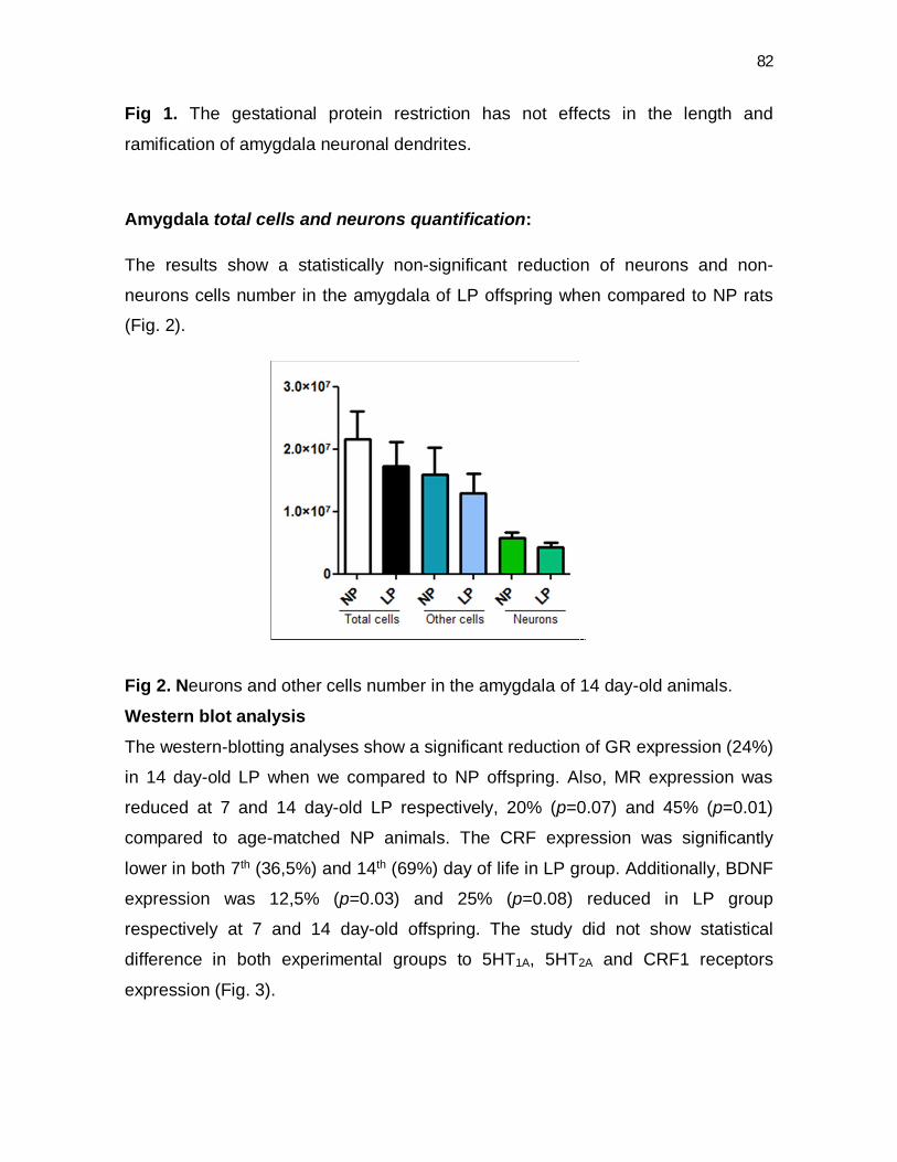
82
Fig 1. The gestational protein restriction has not effects in the length and
ramification of amygdala neuronal dendrites.
Amygdala total cells and neurons quantification:
The results show a statistically non-significant reduction of neurons and non-
neurons cells number in the amygdala of LP offspring when compared to NP rats
(Fig. 2).
Fig 2. Neurons and other cells number in the amygdala of 14 day-old animals.
Western blot analysis The western-blotting analyses show a significant reduction of GR expression (24%)
in 14 day-old LP when we compared to NP offspring. Also, MR expression was
reduced at 7 and 14 day-old LP respectively, 20% (p=0.07) and 45% (p=0.01)
compared to age-matched NP animals. The CRF expression was significantly
lower in both 7th (36,5%) and 14th (69%) day of life in LP group. Additionally, BDNF
expression was 12,5% (p=0.03) and 25% (p=0.08) reduced in LP group
respectively at 7 and 14 day-old offspring. The study did not show statistical
difference in both experimental groups to 5HT1A, 5HT2A and CRF1 receptors
expression (Fig. 3).
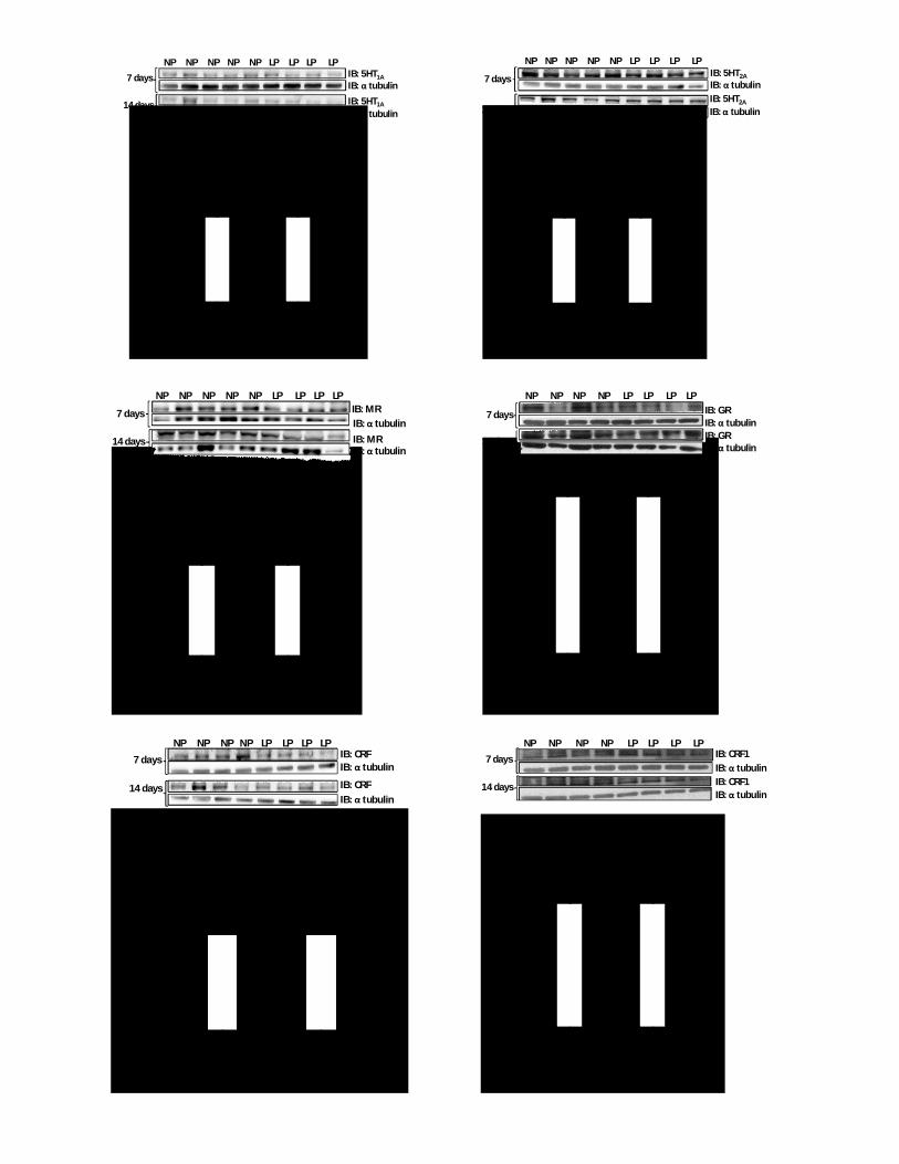
83
NP LP NP LP0
50
100
150
200
7 days 14 days
5HT 1A
/ tu
bulin
(%)
IB: 5HT1A7 days
14 days
IB: α tubulin
IB: 5HT1A
IB: α tubulin
NP NP NP NP NP LP LP LP LP
NP LP NP LP0
50
100
150
200
7 days 14 days
5HT 2
A/
tubu
lin (%
)
IB: 5HT2A7 days
14 days
IB: α tubulin
NP NP NP NP NP LP LP LP LP
IB: 5HT2A
IB: α tubulin
NP LP NP LP0
50
100
150
200
7 days 14 days
*
MR/
tubu
lin (%
)
IB: MR7 days
14 days
IB: α tubulin
NP NP NP NP NP LP LP LP LPNP
IB: MRIB: α tubulin
NP LP NP LP
0
20
40
60
80
100
120
7 days 14 days
GR/
tubu
lin (%
) *
IB: GR7 days
14 days
IB: α tubulin
NP NP NP NP LP LP LP LP
IB: GRIB: α tubulin
IB: CRF7 days
14 days
IB: α tubulin
NP NP NP NP LP LP LP LP
IB: CRFIB: α tubulin
NP LP NP LP0
50
100
150
200
7 days 14 days
*
*CRF/
tubu
lin (%
)
NP LP NP LP0
50
100
150
7 days 14 days
CRF1
/ tu
bulin
(%)
IB: CRF17 days
14 days
IB: α tubulin
NP NP NP NP LP LP LP LP
IB: CRF1IB: α tubulin

84
High-Performance Liquid Chromatography (HPLC) The concentrations of norepinephrine and DOPA, assessed by HPLC were
significantly decreased in 7 day-old LP animals when compared to age-matched
NP offspring. No differences were found in the level of other neurotransmitters
analyzed.
Table 2: Determination of the concentration of neurotransmitters by HPLC: Values represent the mean ± standard deviation. Statistical comparisons were
made with the Student t test and statistical significance was accepted for p≤0.05.
NP LP NP LP
0
50
100
150
200
7 days 14 days
*
BDN
F/
tubu
lin (%
)IB: BDNFIB: α tubulinIB: BDNFIB: α tubulin
7 days
14 days
NP NP NP NP NP LP LP LP LP
Fig 3. Western blot analysis of the 5HT2A,
5HT1A, GR, MR, BDNF, CRF and CRF1
expression in the amygdala of animals from
NP and LP groups with either 7 or 14 days
old. Results of scanning densitometry are
presented as relative to NP, assigning a value
of 100% to NP rats. Columns and bars
represent the mean ± SD. *P < 0.05, NP
versus LP.

85
Discussion
In accordance with previous reports, in the current study, we also observe a
significant decrease in body birth weight that remains up to 14 day of age, in
gestational protein-restricted offspring. Additionally, this study also confirms
several previous that have shown that protein restriction intake did not alters the
brain weight in adults offspring (Lingas et al, 1999). Recent study in our laboratory
has demonstrated that 16 week-old male offspring from gestational protein-
restricted dam, also, did not present any changes in the brain mass. In this study,
we also do not show significant difference in the brain mass at 7 and 14 day-old LP
rats. In this sense, our data add support to the “selfish brain” theory, which
proposes that intrauterine adverse events such as emotional stress, either
undernutrition, restriction of specific nutrients or hypoxia, may program the brain to
maintain stability in its own energy sources. Particularly, alterations in early life,
program the developing neuronal substrate to decrease neuroendocrine activity to,
in turn, decrease the somatic growing rate, guarantying an adequate nutrition to
the developing brain (Lumbers, et al, 2001). This theory can explain the reduced
body weight of LP animals without affecting the brain mass.
The present study also evaluated the post-mortem adrenal and thymus weight. At
7 day-old LP offspring the results show an increased thymus weight that not
remain in 14 day-old offspring. This thymus change may be related to steroidal
plasma levels that could be associated with alteration in lymphoid tissues
proliferation of protein-restricted animals. In fact, it has been strongly suggested
that neuronal plasticity may be modulated by the immune system (Li et al; 2016).
Here surprisingly, the adrenal mass was unchanged in both age-matched groups.
This finding, despite fact that stress in adults rats induces an increased adrenal
mass (Ventura-Silva et al., 2012), in the current study, it seems that the gestational
protein restriction does not affect this organ in the times studied.
In the current study, the amygdala neuronal 3D dendritic analysis of dendrites
length and dendritic ramifications by Sholl analysis, not show alteration in 14 d-old
NP compared with age-matched gestational protein-restricted offspring. Also, the
amygdala neurons and non-neuronal cells number was similar in 14 day-old LP
86
and NP offspring. It is noteworthy that both dendrites’ volume and length was
evaluated in specific region on basolateral amygdala while the cells’ number was
determined on the whole amygdala. The dendritical length and arborization was
determined in the basolateral amygdala, once this nucleus is the main responsible
to modulate signals coming from several regions of CNS and, furthermore, is
strongly associated with fear and anxiety behavior (Izquierdo et al, 2016; Li et al,
2016).
By the way, Pêgo et al., (2008) did not report any morphological alterations
in the amygdala in animals that were exposed to chronic unpredictable stress while
chronic immobilization stress leads to an hypertrophy of dendrites in this brain
structure (Mitra & Sapolsky , 2008). The intensity and duration of stress exposure
are therefore, seems to be key factors for the development of morphological and
neurochemical alterations in the amygdala (Wilson et al, 2015). Thus, studies
showing that chronic immobilization stress increases dendritic length, the number
of ramifications and the number of spines in neurons of the basolateral amygdala
(Mitra et al, 2005; Vyas et al, 2002). Additionally, these morphological changes are
related with anxiety-like behavior in chronic stressed-animals (Vyas & Chattarji,
2004).
It is broadly confirmed that gestational protein restriction may induce fetal
programming leading to modification in the brain structure and behavior of offspring
that remain up to adulthood (Ashton, 2000; Procter & Campbell, 2014, Barker,
1995; Forsdahl, 1967). Animal studies have shown that early-life undernutrition has
a deep impact on fetal tissues and organs development, causing permanent
changes in a wide range of morphological and physiological functions, including
brain function (Alamy & Bengelloun, 2012). However, the amygdala’s functional
neurochemical modulation has been poorly documented in gestational protein-
restricted offspring, therefore, in the present study we investigate the effects of
gestational protein restriction on whole amygdala neurochemical compound in
male offspring, in key moments of post-natal neural development. Here, we
demonstrate significant decrease in the norepinephrine, epinephrine and
dopamine levels, as well as reduction in the expression GR and MR steroid

87
receptors and CRF in the amygdala in 7 and 14 day-old LP offspring compared to
control group.
The amygdala plays a crucial role coordinating behavioral, autonomic, and
neuroendocrine stress responses, via mostly excitatory influences on the
hypothalamus and brainstem (Aggleton, 2000). It stimulates the HPA axis through
indirect projections to the hypothalamic paraventricular nucleus (PVN), including a
disinhibitory pathway via the bed nucleus of the stria terminalis (Feldman et al.,
1990; Herman et al., 2003, Freese & Amaral, 2009). PVN stimulation releases
corticotrophin-releasing factor (CRF) into the portal vasculature, which binds to
CRF receptors in the anterior pituitary stimulating the release of
adrenocorticotrophic hormone (ACTH), which stimulates the synthesis and release
of glucocorticoids by the adrenals (Ulrich-Lai and Herman, 2009). Glucocorticoids
inhibit their own release via negative feedback through binding to glucocorticoid
receptors (GRs) in the pituitary, PVN, and extrahypothalamic brain regions (Myers
et al., 2012). In the present study, the reduced expression of amygdala GR and
MR may have a key role on excitatory HPA axis response, priory recorded, in LP
offspring.
The amygdala’s excitatory influence on the HPA axis stress response has
been primarily demonstrated in adult animals, with electrical stimulation increasing
secretion of glucocorticoids (Redgate & Fahringer, 1973; Ehle et al., 1977) and
lesions resulting in blunted HPA axis stress responses (Kalin et al., 2004; Machado
& Bachevalier, 2008). Recent study has demonstrated administration of
corticosteroid into amygdala nucleus promotes an anxiety-like behavior (Venkova
et al 2010) by MR and GR stimuli (Herman et al 1989). Also, reporters suggested
that both receptors are implicated in glucocorticoids fear-enhancing effect (Oitzl et
al 2001). In the central amygdala nucleus (CeA) MR and GR are expressed in CRF
neurons, suggesting direct glucocorticoid regulation of CRF expression in this
nucleus (Honkaniemi et al., 1992). The amygdala is also rich in corticotrophin-
releasing factor (CRF), a specific modulator of stress-related anxiety and the role
of the amygdala in the modulation of the HPA axis may be through the activation of
CRF receptor (CRF1) (Davis, 1992; Merali et al., 2004; Muller et al., 2003).

88
However, recent data from Raper et al (2014) has demonstrate that early
amygdala damage (decreasing GR/MR and CRF amygdala levels) alters the
typical development of the primate HPA axis resulting in increased rather than
decreased activity, presumably via alterations in central CRF and GR systems in
neural structures that control its activity. Thus, in contrast to evidence that the
amygdala stimulates both CRF and HPA axis systems in the adult, our data
suggest an opposite inhibitory role of the amygdala on the HPA axis during early
development, which fits with decreased GR, MR and CRF levels observed in the
present study. The CRF receptors activation may also induces alterations in other
neurotransmitter systems such as serotonin (5HT), dopamine and norepinephrine
(Millan, 2005; Holsboer, 2003; Nestler et al., 2002; Leonard, 2005; Holmes et al,
2003) and the amygdala CRF reduction in protein-restricted offspring, may affect
this neurotransmitter release. In addition, they may be interacting with brain
derived neurotrophic factor (BDNF), network deeply involved in fear and anxiety
(Hammack et al, 2009; Duman, 2004; Kumari, et al 2016).
In the current work, the animals submitted to gestational nutritional restrictions
presented a decrease in amygdala norepinephrine, epinephrine and dopamine
levels. It is well known that stress exposure leads to a cascade of neuroendocrine
response that promotes release of corticosteroids and catecholamines (Wolf,
2015). Nevertheless, authors report a decrease in dopaminergic and/or
noradrenergic release in the depression pathophysiology (Guiard et al, 2008). In
vitro studies suggest that the excitability of neurons in the amygdala is modulated
by dopamine (Kroner et al, 2005) and that there is a strong relation between
catecholamines and HPA axis alterations (Leonard, 2001). On the other hand,
norepinephrine activates CRF, which, in turn, can induce the release of
norepinephrine, leading to a cycle (Koob, 1999). This modulatory system may be
blunted in the current study. These results may be explained by well-characterized
stress hyporresponsive period (SHRP) associated to decreased activity of HPA
axis, that may last until the 14th postnatal-day (Sapolsky & Meane 1986). At this
time, there is a reduced secretion of corticosterone, which remains low until the
second postnatal week (Sapolsky & Meane 1986). The SHRP may assume as a

89
protective mechanism ensuring low levels of glucocorticoids during early postnatal
development (Sapolsky et al, 1986; Mesquita et al, 2007). The decrease in the
expression of catecholamines in gestational protein-restricted model, may be one
of mechanisms that contribute to the lower activity of the HPA axis during this
period of life.
In conclusion, the amygdala’s role on behavioral, autonomic, and neuroendocrine
stress responses has been poorly studied in programmed models, with scarce
studies reporting the morphological and neurochemistry characteristics and the
relationship with the key role of the amygdala in the stress response (Herman &
Cullinan 1997; Van de Kar & Blair, 1999). The amygdala neurochemical changes
observed in this study may contribute to behavioral alterations induced by
gestational protein restriction and may also be a primer for alterations in other brain
regions. Also, these findings may represent the adaptation during embryonic
development to exposure to elevated maternal corticosteroids as a consequence of
nutritional stress. Additional studies must be done, but the present study suggests
strongly that amygdala morphological and neurochemical disorders may be
associated with the development of psychiatric disorders in adulthood.
REFERENCES
AGGLETON JP. The amygdala. A functional analysis. (2000) Ed 2. New York:
Oxford Universit Press.
ALAMY M; BENGELLOUN WA. Malnutrition and brain development: an analysis of
the effects of inadequate diet during different stages of life inrat. Neurosci Biobehav Rev. 2012 Jul;36(6):1463-80. doi: 10.1016/j.neubiorev.2012.03.009.
Epub 2012 Apr 3.
ASHTON, N. Perinatal development and adult blood pressure. Braz J Med Biol Res 2000; 33:731-40.

90
BARKER, D.J. The fetal and infant origins of disease. Eur J Clin Invest. 1995;
25:457-63.
BORGES S; COIMBRA B; SOARES-CUNHA C; VENTURA-SILVA AP; PINTO
L; CARVALHO MM; PÊGO JM; RODRIGUES AJ; SOUSA N.
Glucocorticoid programing of the mesopontine cholinergic system. Front Endocrinol (Lausanne). 2013 Dec 13;4:190. doi: 10.3389/fendo.2013.00190. e
Collection 2013.
BURD, L.; SEVERUD, R.; KERBESHIAN, J.; KLUG, M.G. Prenatal and perinatal
risk factors for autism. J Perinat Med 1999; 27: 441-450.
DAVIS, M. The role of the amygdala in fear and anxiety. Ann. Rev. Neurosci. 1992; 15:353–375.
DING, Y X; SHI, Y HAN, W J; CUI, H. Regulation of glucocorticoid
related genes and receptors/regulatory enzyme expression in intrauterine growth
restriction filial rats. Life Sci. 2016 Feb 23. pii: S0024-3205(16)30128-X. doi:
10.1016/j.lfs.2016.02.079. [Epub ahead of print].
DUMAN RS. Role of neurotrophic factors in the etiology and treatment of mood
disorders. Neuromolecular Med. 2004;5(1):11-25.
EHLE AL, MASON JW, PENNINGTON LL Plasma growth hormone and cortisol
changes following limbic stimulation in conscious monkeys. Neuroendocrinology
(1977) 23:52– 60.
FELDMAN S, CONFORTI N, SAPHIERD. The preoptic area and bed nucleus of
the stria terminalis are involved in the effects of the amygdala on adrenocortical
secretion. Neuroscience (1990) 37:775–779.
FORSDAHL, S. Are poor living conditions in childhood and adolescence an
important risk factor for arteriosclerotic heart disease? British journal of Preventive and Social Medicine, 1967; 31: 91-95.
91
FREESE JL, AMARAL DG Neuroanatomy of the primate amygdala. In:The human
amygdala (Whalen PJ, Phelps EA, eds), (2009) pp 1–42. New York: Gilford.Furay
AR, Bruest
GIBB, R.; KOLB, B. A method for vibratome sectioning of Golgi-Cox stained whole
rat brain. J Neurosci Methods 1998; 79: 1–4.
GLASER, E.M.; VAN, D.E.R.; LOOS, H. Analysis of thick brain sections by
obverse-reverse computer microscopy: application of a new, high clarity Golgi-
Nissl stain. J Neurosci Methods 1981; 4:117–125.
GUIARD BP; EL MANSARI M; BLIER P. Cross-
talk between dopaminergic and noradrenergic systems in
the rat ventral tegmental area, locus ceruleus, and dorsal hippocampus. Mol Pharmacol. 2008 Nov;74(5):1463-75. doi: 10.1124/mol.108.048033. Epub 2008
Aug 14.
HAMMACK, S.E.; ROMAN, C.W.; LEZAK, K.R.; KOCHO-SHELLENBERG, M.
GRIMMIG, B.; FALLS, W.A.; BRAAS, K.; MAY, V. Roles for pituitary adenylate
cyclase-activating peptide (PACAP) expression and signaling in the bed nucleus of
the stria terminalis (BNST) in mediating the behavioral consequences of chronic
stress. J Mol Neurosci. 2010;42(3):327-40. Epub 2010 Apr 20.
HAMMACK, S.E.; GUO, J.D.; HAZRA, R.; DABROWSKA, J.; MYERS,
K.M.; RAINNIE, D.G. The response of neurons in the bed nucleus of the stria
terminalis to serotonin: implications for anxiety. Prog Neuropsychopharmacol Biol Psychiatry. 2009; 13;33(8):1309-20. Epub 2009 May 23.
HERCULANO-HOUZEL S; LENT R. Isotropic fractionator:
a simple, rapid method for the quantification of total cell and neuron numbers in
the brain. J Neurosci. 2005 Mar 9;25(10):2518-21.
HERMAN JP, CULLINAN WE. Neurocircuitry of stress: central control of the
hypothalamo–pituitary–adrenocortical axis. Trends Neurosci 1997; 20: 78–84.

92
HERMAN JP, FIGUEIREDO H, MUELLER NK, ULRICH-LAI Y, OSTRANDER MM,
CHOI DC, CULLINAN WE Central mechanisms of stress integration: hierarchical
circuitry controlling hypothalamo-pituitary-adrenocortical responsiveness.F ront
Neuroendocrinol (2003)24:151–180.
HERMAN JP, PATEL PD, AKIL H, WATSON SJ. Localization and regulation of
glucocorticoid and mineralocorticoid receptor messenger RNAs in the hippocampal
formation of the rat. Mol Endocrinol 1989, 3:1886–1894.
HOLMES, A.; HEILIG, M.; RUPNIAK, N.M.; STECKLER, T.; GRIEBEL, G.
Neuropeptide systems as novel therapeutic targets for depression and anxiety
disorders. Trends Pharmacol. Sci. 2003; 24:580–588.
HOLSBOER, F. Corticotrophin-releasing hormone modulators and depression.
Curr. Opin. Investig. Drugs. 2003; 4:46–50.
HONKANIEMI J., M. PELTO-HUIKKO, L. RECHARDT, J. ISOLA, A. LAMMI, K.
FUXE, J. GUSTAFSSON, A. WIKSTROM, T. HOKFELT. Colocalization of peptide
and glucocorticoid receptor immunoreactivities in rat central amygdaloid nucleus,
Neuroendocrinology 55 1992 451–459.
IZQUIERDO I, FURINI CR, MYSKIW JC. Fear Memory. Physiol Rev. 2016
Apr;96(2):695-750. doi: 10.1152/physrev.00018.2015.
JOTHIE RICHARD E; ILLURI R; BETHAPUDI B; ANANDHAKUMAR S; BHASKAR
A; CHINAMPUDUR VELUSAMI C MUNDKI; NAJEDDU D; AGARWAL A. Anti-
stress Activity of Ocimum sanctum: Possible Effects on Hypothalamic-Pituitary-
Adrenal Axis. Phytother Res. 2016 Feb 22. doi: 10.1002/ptr.5584. [Epub ahead of
print]
KALIN NH, SHELTON SE, DAVIDSON RJ The role of the central nucleus of the
amygdala in mediating fear and anxiety in the primate. J Neurosci (2004) 24:5506
–5515.

93
KOOB GF. Corticotropin-releasing factor, norepinephrine, and stress. Biological Psychiatry. Volume 46, Issue 9, 1 November 1999, Pages 1167–1180.
KRÖNER S, ROSENKRANZ JA, GRACE AA, BARRIONUEVO G. Dopamine
modulates excitability of basolateral amygdala neurons in vitro. J Neurophysiol. 2005 Mar;93(3):1598-610. Epub 2004 Nov 10.
KRÖNER S, ROSENKRANZ JA, GRACE AA, BARRIONUEVO G.
Dopamine modulates excitability of basolateral amygdala neurons in vitro. J Neurophysiol. 2005 Mar;93(3):1598-610. Epub 2004 Nov 10.
KUMARI A; SINGH P; BAGHEL MS; THAKUR MK.
Social isolation mediated anxiety like behavior is associated with enhanced expres
sion and regulation of BDNFin the female mouse brain. Physiol Behav. 2016 Feb
23;158:34-42. doi: 10.1016/j.physbeh.2016.02.032. [Epub ahead of print]
LEONARD BE. Stress, norepinephrine and depression. J Psychiatry Neurosci. 2001;26 Suppl:S11-6.
LEONARD BE. The HPA and immune axes in stress: the involvement of the
serotonergic system. Eur. Psychiatry. 2005; 20:302–306.
LI M; GUO K; ADACHI Y; IKEHARA S.
Immune Dysfunction Associated with Abnormal Bone Marrow-
Derived Mesenchymal Stroma Cells in Senescence Accelerated Mice. Int J Mol Sci. 2016 Jan 29;17(2). pii: E183. doi: 10.3390/ijms17020183.
LINGAS R; DEAN F; MATTHEWS SG.
Maternal nutrient restriction (48 h) modifies brain corticosteroid receptor expression
and endocrine function in the fetal guinea pig. Brain Res. 1999 Nov 6;846(2):236-
42.
LOPES A; TORRES DB; RODRIGUES AJ; CERQUEIRA JJ; PÊGO JM; SOUSA
N; GONTIJO JA; BOER PA. Gestational protein restriction induces CA3 dendritic
atrophy in dorsal hippocampal neurons but does not alter learning and memory
94
performance in adult offspring. Int J Dev Neurosci. 2013 May;31(3):151-6. doi:
10.1016/j.ijdevneu.2012.12.003. Epub 2012 Dec 30.
LUCAS A. Programming by early nutrition in man. Ciba Found Symp. 1991;156:38-50; discussion 50-5.
LUMBERS ER, YU ZY, GIBSON KJ. The selfish brain and the barker hypothesis.
Clin Exp Pharmacol Physiol. 2001 Nov;28(11):942-7.
LUMBERS ER; YU ZY; GIBSON KJ. THE SELFISH BRAIN AND
THE BARKER HYPOTHESIS. Clin exp pharmacol physiol. 2001
NOV;28(11):942-7.
MACHADO CJ, BACHEVALIER J Behavioral and hormonal reactivity to threat:
effects of selective amygdala, hippocampal, or orbital frontal lesions in monkeys.
Psychoneuroendocrinology (2008) 33:926 –941.
MERALI Z, KHAN S, MICHAUD, D.S.; SHINOSITOL, PHOSPHATEPY SA,
ANISMAN H. Does amygdaloid corticotrophin-releasing hormone (CRF) mediate
anxiety-like behaviors? Dissociation of anxiogenic effects and CRF release. Eur. J. Neurosci. 2004; 20:229–239.
MESQUITA FF, GONTIJO JAR, BOER PA. Maternal undernutrition and the
offspring kidney: from fetal to adult life. Braz J Med Biol Res. 2010
Nov;43(11):1010-8. Epub 2010 Oct 29.
MILLAN, M.J. Serotonin 5-HT2C receptors as a target for the treatment of
depressive and anxious states: focus on novel therapeutic strategies. Therapies. 2005; 60:441–460.
MITRA R, JADHAV S, MCEWEN BS, VYAS A, CHATTARJI S. Stress duration
modulates the spatiotemporal patterns of spine formation in the basolateral
amygdala. Proc Natl Acad Sci USA. 2005;102:9371–9376.[
MITRA R, SAPOLSKY RM.
Acute corticosterone treatment is sufficient to induce anxiety and amygdaloid dend

95
itic hypertrophy. Proc Natl Acad Sci U S A. 2008 Apr 8;105(14):5573-8. doi:
10.1073/pnas.0705615105. Epub 2008 Apr 7.
MULLER, M.B. Limbic corticotrophin-releasing hormone receptor 1 mediates
anxiety-related behavior and hormonal adaptation to stress. Nat. Neurosci. 2003;
6:1100–1107.
MYERS B, MCKLVEEN JM, HERMAN JP Neural regulation of the stress response:
the many faces of feedback. Cell Mol Neurobiol (2012) 32:683– 694.
NESTLER, E.J.; BARROT, M.; DILEONE, R.J.; EISCH, A.J.; GOLD, S.J.;
MONTEGGIA, L.M. Neurobiology of depression. Neuron. 2002; 34:13–25.
OITZL MS, REICHARDT HM, JOELS M, DE KLOET ER. Point mutation in the
mouse glucocorticoid receptor preventing DNA binding impairs spatial memory.
Proc Natl Acad Sci USA 2001, 98:12790–12795.
PÊGO, J. M.; MORGADO, P.L.G.; CERQUEIRA, J.J., ALMEIDA, O.F.X.; SOUSA,
N. Dissociation of the morphological correlates of stress-induced anxiety and fear.
European Journal of Neuroscience 2008; 27:1503–1516.
PROCTER, S B.; Campbell CG. Position of the Academy of Nutrition and Dietetics:
Nutrition and Lifestyle for a Healthy Pregnancy Outcome. J Acad Nutr Diet. 2014;
114(7):1099-103. doi: 10.1016/j.jand.2014.05.005.
RAPER J, SHANNON B.Z. STEPHENS, AMY HENRY, TRINA VILLARREAL,
JOCELYNE BACHEVALIER, KIM WALLEN AND MAR M. SANCHEZ. Neonatal
Amygdala Lesions Lead to Increased Activity of Brain CRF Systems and
Hypothalamic-Pituitary-Adrenal Axis of Juvenile Rhesus Monkeys The Journal of Neuroscience 2014 • 34(34):11452–11460
REDGATE ES, FAHRINGER EE A comparison of the pituitary adrenal activity
elicited by electrical stimulation of preoptic, amygdaloid and hypothalamic sites in
the rat brain. Neuroendocrinology (1973) 12:334 –343.

96
SCABORA JE, LIMA MC, LOPES A, LIMA IP, MESQUITA FF, TORRES DB,
BOER PA, GONTIJO JAR. Impact of taurine supplementation on blood pressure in
gestational proteinrestricted offspring: Effect on the medial solitary tract nucleus
cell numbers, angiotensin receptors, and renal sodium handling. J Renin Angiotensin Aldosterone Syst. 2015 Mar;16(1):47-58. doi:
10.1177/1470320313481255. Epub 2013 Mar 6.
SHOLL, D.A. The measurable parameters of the cerebral cortex and their
significance in its organization. Prog Neurobiol . 1956; 2:324–333.
SOUSA N; CERQUEIRA JJ; ALMEIDA OF. Corticosteroid receptors and
neuroplasticity. Brain Res Rev. 2008 Mar;57(2):561-70. Epub 2007 Jul 17.
ULRICH-LAI YM, HERMAN JP Neural regulation of endocrine and autonomic
stress responses. Nat Rev Neurosci (2009) 10:397– 409. 2009 Jun;10(6):397-
409. doi: 10.1038/nrn2647.
UYLINGS, H.B.M.; VAN, P.J. Measures for quantifying dendritic arborizations.
Network: comput neural syst 2002 13:397–414
VAN DE KAR LD, BLAIR ML. Forebrain pathways mediating stress-induced
hormone secretion. Front Neuroendocrinol. 1999 Jan;20(1):1-48.
VENKOVA K, ANTHONY C, MYERS JB, GREENWOOD-VAN MEERVELD B.
Exposure of the amygdala to elevated levels of corticosterone alters colonic motility
in response to acute psychological stress. Neuropharmacology 2010,58:1161–
1167.
VENTURA-SILVA AP; PÊGO JM; SOUSA JC; MARQUES AR; RODRIGUES
AJ; MARQUES F; CERQUEIRA JJ; ALMEIDA OF; SOUSA N.
Stress shifts the response of the bed nucleus of the stria terminalis to
an anxiogenic mode. Eur J Neurosci. 2012 Nov;36(10):3396-406. doi:
10.1111/j.1460-9568.2012.08262.x. Epub 2012 Aug 29.

97
VYAS A, CHATTARJI S. Modulation of different states of anxiety-like behavior by
chronic stress. Behav Neurosci. 2004;118:1450–1454.
VYAS A, MITRA R, SHANKARANARAYANA RAO BS, CHATTARJI S. Chronic
stress induces contrasting patterns of dendritic remodeling in hippocampal and
amygdaloid neurons. J Neurosci. 2002;22:6810–6818.
WELBERG, L.A.; SECKL, J.R. Prenatal stress, glucocorticoids and the
programming of the brain. J Neuroendocrinol 2001; 13: 113-128.
WILSON MA; GRILLO CA; FADEL JR; REAGAN LP. Stress as a one-armed
bandit: Differential effects of stress paradigms on the morphology, neurochemistry
and behavior in the rodent amygdala. Neurobiol Stress. 2015 Jun 9;1:195-208.
doi: 10.1016/j.ynstr.2015.06.001. eCollection 2015.
WOLF OT, ATSAK P, DE QUERVAIN DJ, ROOZENDAAL B, WINGENFELD K.
Stress and memory: A selective review on recent developments in the
understanding of stress hormone effects on memory and their clinical relevance. J Neuroendocrinol. 2015 Dec 28. doi: 10.1111/jne.12353. [Epub ahead of print]
YANG YL; CHAO PK; LU KT. Systemic and intra-
amygdala administration of glucocorticoid agonist and antagonist modulate extincti
on of conditioned fear. Neuropsychopharmacology. 2006 May;31(5):912-24.

98
5. CONSIDERAÇÔES GERAIS
A amígdala estimula o eixo HPA através de projeções indiretas para o PVN,
incluindo uma via excitatória através do BNST (Feldman et al, 1990; Herman et al,
2003 Freese & Amaral, 2009). O estimulo ao PVN promove a liberação de CRF
nos vasos portais, que se liga aos receptores de CRF da hipófise estimulando a
secreção de ACTH, que por sua vez estimula a síntese e liberação de GC pelas
glândulas suprarrenais (Ulrich-Lai e Herman, 2009). Os GC inibem a sua própria
libertação através de alças de retroalimentação negativas, pela ligação a GR na
hipófise, no PVN e em regiões extra-hipotalâmicas do cérebro, tais como
hipocampo e núcleos da amigdala (Myers et al., 2012).
A influência excitatória da amígdala sobre a resposta do eixo HPA foi
demonstrada primeiramente em animais adultos, pela estimulação eléctrica desta
causando um aumento na secreção de GC (Mason, 1959; Redgate & Fahringer,
1973; Ehle et al, 1977) e, através de lesões eletrolíticas em núcleos amigdaloides
que resultaram na redução na atividade do eixo HPA associada a um estado de
estresse (Beaulieu et ai, 1986; Feldman et al., 1994; Kalin et al, 2004).
Estudos em roedores mostram que a amígdala encontra-se funcionalmente
em estado latente antes do desmame, quando os filhotes são mais dependentes
de cuidados maternos, e acredita-se que este estado seja para inibir reações
medo da mãe. Posteriormente, ocorre a ativação dos núcleos da amígdala para
que seja possível a ocorrência de reações de medo/defesa coincidindo com o
aumento dos níveis séricos e liquóricos de corticosterona durante fase de
transição do desenvolvimento (pós-natal imediato) para fase de independência do
filhote, após 16o. dia de vida (Rincón-Cortés & Sullivan, 2014).
Raper e colaboradores (2014) sugerem que durante o desenvolvimento, a
amigdala tem influência inibitória na atividade basal do eixo HPA, tanto em
primatas quanto em roedores, mudando mais tarde, para um estado excitatório na
adolescência e idade adulta. Várias doenças psiquiátricas (p.e. autismo,
esquizofrenia dentre outras) envolvem alterações no desenvolvimento da amigdala

99
e desregulação do controle da síntese e dos níveis liquóricos e séricos de CRF e
de suas ações sobre o eixo HPA (Schumann et al., 2011; Tottenham, 2014)
Desta forma, a redução do CRF (36,6 e 69% respectivamente no 7º e 14º
dia de vida) observada e, dos receptores de GC na amígdala e BNST de animais
oriundos de mães submetidas á restrição proteica gestacional, pode atenuar a
ação inibitória destas estruturas sobre o eixo HPA (Figura 1). Assim, nossos
resultados confirmam prévios estudos mostrando que modificações morfológicas
e/ou neuroquímicas da amigdala durante o desenvolvimento podem comprometer
a ação destes núcleos cerebrais na modulação de respostas ao estresse e ao
medo (Rincón-Cortés & Sullivan, 2014; Raper et al, 2014).
Figura 2. Compilação das alterações neuroquímicas e estruturais observadas em
animais do grupo LP.

100
6. CONCLUSÃO
Os resultados do presente estudo mostram que a restrição proteica
gestacional leva a alterações neuroquímicas na amígdala e neuroquímicas e
morfológicas no BNST do sétimo ao décimo quarto dias de vida da prole. Estes
achados sugerem a interferências do estresse nutricional durante a gestação,
sobre o desenvolvimento de vias de sinalização dos glicocorticoides e do CRF que
implica diferentes estruturas do sistema nervoso central resultando em
hiperatividade destas e, consequentemente do eixo HPA, associada à elevação do
corticosteroide plasmático e do estado de ansiedade, já observado na idade adulta
nestes animais.
7. REFERÊNCIAS BIBLIOGRÁFICAS
ALHEID, G.F.; BELTRAMINO, C.A.; OLMOS J.S.; HEIMER, L. Supracapsular
portions of the rat extended amygdala, European Neuroscience MeetingAmsterdam 1995; 104.
ALHEID, G.F.; BETRAMINO, C.A.; OLMOS, J.S.; FORBES, M.S.; SWANSON
D.J.; HEIMER, L. The neuronal organization of the supracapsular part of
the stria terminalis in the rat: the dorsal component of the extended
amygdale. Neuroscience 1998; 84:967 996.
ALHEID, G.F.; HEIMER, L. New perspectives in basal forebrain organization of
special relevance for neuropsychiatric disorders: the striatopallidal, amygdaloid,
and corticopetal components of substantia innominat. Neuroscience. 1988;
27(1):1-39.
ASHTON, N. Perinatal development and adult blood pressure. Braz J Med Biol Res 2000; 33:731-40.
BARKER, D.J. In utero programming of chronic disease. Clin Sci (Lond). 1998;
95:115-28.

101
BARKER, D.J. Intra-uterine programming of the adult cardiovascular system. Curr Opin Nephrol Hypertens. 1997;6:106-10.
BARKER, D.J. The fetal and infant origins of disease. Eur J Clin Invest. 1995;
25:457-63.
BARKER, D.J.P.; OSMOND, C.; GOLDING, J.; KUH, D.; WADSWORTH, M.E.J.
Growth in utero, blood pressure in childhood and adult life, and mortality from
cardiovascular diasease. BMJ. 1989; 298:564-567.
BARKER, D.J.P.; OSMOND, C.; RODIN, I.; FALL, C.H.D.; WINTER, P.D. Low
weight gain in infancy and suicide in adult life. Br Med J 1995; 311: 1203.
BEAULIEU S, DI PAOLO T, BARDEN N. Control of ACTH secretion by
the central nucleus of the amygdala: implication of the serotoninergic system and
its relevance to the glucocorticoid delayed negative feedback mechanism.
Neuroendocrinology. 1986;44(2):247-54.
BENEDIKTSSON, R.; LINDSAY, R.S.; NOBLE, J.; SECKL, J.R.; EDWARDS, C.R.
Glucocorticoid exposure in utero: new model for adult hypertension. Lancet. 1993;
34: 339-41
BURD, L.; SEVERUD, R.; KERBESHIAN, J.; KLUG, M.G. Prenatal and perinatal
risk factors for autism. J Perinat Med 1999; 27: 441-450.
COROMINAS, M.; RONCERO, C.; CASAS, M. Corticotropin releasing factor and
neuroplasticity in cocaine addiction. Life Sci. 2010; 86: 1–9.
CULLINAN, W.E.; HERMAN, J.P.; WATSON, S.J. Ventral subicular interaction with
the hypothalamic paraventricular nucleus: evidence for a relay in the bed nucleus
of the stria terminalis. J. Comp. Neurol. 1993; 332:1 –20.
DAVIS, M. The role of the amygdala in fear and anxiety. Ann. Rev. Neurosci. 1992a; 15:353–375.

102
DAVIS, M.; WALKER, D.L.; MILES, L.; GRILLON, C. Phasic vs sustained fear in
rats and humans: role of the extended amygdala in fear vs anxiety.
Neuropsychopharmacology. 2010; 35(1):105-35.
DING, Y X; SHI, Y HAN, W J; CUI, H. Regulation of glucocorticoid
related genes and receptors/regulatory enzyme expression in intrauterine growth
restriction filial rats. Life Sci. 2016 Feb 23. pii: S0024-3205(16)30128-X. doi:
10.1016/j.lfs.2016.02.079. [Epub ahead of print].
DONG, H.W.; PETROVICH, G.D.; WATTS, A.G.; SWANSON, L.W. Basic
organization of projections from the oval and fusiform nuclei of the bed nuclei of the
stria terminalis in adult rat brain. J. Comp. Neurol. 2001 436, 430–455.
FELDMAN S, CONFORTI N, ITZIK A, WEIDENFELD J.
Differential effect of amygdaloid lesions on CRF-
41, ACTH and corticosterone responses following neural stimuli. Brain Res. 1994
Sep 26;658(1-2):21-6.
FELDMAN, S.; CONFORTI, N.; SAPHIER, D. The preoptic area and bed nucleus
of the stria terminalis are involved in the effects of the amygdale on adrenocortical
secretion. Neuroscience 1990; 37:775– 779.
FONTENOT, M.B.; KAPLAN, J.R.; MANUCK, S.B.; ARANGO, V.; MANN, J.J.
Long-term effects of chronic social stress on serotonergic indices in the prefrontal
cortex of adult male cynomolgus macaques. Brain Res. 1995; 705:105-108.
FORSDAHL, S. Are poor living conditions in childhood and adolescence an
important risk factor for arteriosclerotic heart disease? British journal of Preventive and Social Medicine, 1967; 31: 91-95.
GENRO JP; KIELING C; ROHDE LA; HUTZ MH. Attention-
deficit/hyperactivity disorder and the dopaminergic hypotheses. Expert Rev Neurother. 2010 Apr;10(4):587-601. doi: 10.1586/ern.10.17.

103
GIBB, R.; KOLB, B. A method for vibratome sectioning of Golgi-Cox stained whole
rat brain. J Neurosci Methods 1998; 79: 1–4.
GLASER, E.M.; VAN, D.E.R.; LOOS, H. Analysis of thick brain sections by
obverse-reverse computer microscopy: application of a new, high clarity Golgi-
Nissl stain. J Neurosci Methods 1981; 4:117–125.
GODFREY, K.; ROBINSON, S.; BARKER, D.J, OSMOND C, COX V. Maternal
nutrition in early and late pregnancy in relation to placental and fetal growth. BMJ.
1996;17;312 (7028):410-4.
GOMEZ-SANCHEZ, E.P. Mineralocorticoid receptors in the brain and
cardiovascular regulation: minority rule? Trends Endocrinol Metab. 2011
May;22(5):179-87. Epub 2011 Mar 21.
GRAEFF, F.G.; GUIMARÃES, F.S.; DE ANDRADE, T.G.; DEAKIN, J.F. Role of 5-
HT in stress, anxiety, and depression. Pharmacol Biochem Behav. 1996;
54(1):129-41.
GRAY, T.S.; PIECHOWSKI, R.A.; YRACHETA, J.M.; RITTENHOUSE, P.A,
BETHEA, C.L.; VAN.; DE KAR, L.D. Ibotenic acid lesions in the bed nucleus of the
stria terminalis attenuate conditioned stress-induced increases in prolactin, ACTH
and corticosterone. Neuroendocrinology 1993; 57:517–524.
HALES CN, BARKER DJP. Type 2 (non-insulin-dependent) diabetes mellitus: the
thrifty phenotype hypothesis. International Journal of Epidemiology
2013;42:1215–1222.
HAMMACK, S.E.; ROMAN, C.W.; LEZAK, K.R.; KOCHO-SHELLENBERG, M.
GRIMMIG, B.; FALLS, W.A.; BRAAS, K.; MAY, V. Roles for pituitary adenylate
cyclase-activating peptide (PACAP) expression and signaling in the bed nucleus of
the stria terminalis (BNST) in mediating the behavioral consequences of chronic
stress. J Mol Neurosci. 2010;42(3):327-40. Epub 2010 Apr 20.

104
HAMMACK, SE; ROMAN, CW; LEZAK, KR; KOCHO-SHELLENBERG, M;
GRIMMIG, B, FALLS, WA; BRAAS, K; MAY, V. Roles for pituitary adenylate
cyclase activating peptide (PACAP) expression and signaling in the bed nucleus of
the stria terminalis (BNST) in mediating the behavioral consequences of chronic
stress. J Mol Neurosci. 2010 Nov;42(3):327-40. doi: 10.1007/s12031-010-9364-7.
Epub 2010 Apr 20.
HAMMACK, S.E.; GUO, J.D.; HAZRA, R.; DABROWSKA, J.; MYERS,
K.M.; RAINNIE, D.G. The response of neurons in the bed nucleus of the stria
terminalis to serotonin: implications for anxiety. Prog Neuropsychopharmacol Biol Psychiatry. 2009; 13;33(8):1309-20. Epub 2009 May 23.
HANDLEY, S.L. 5-Hydroxytryptamine pathways in anxiety and its treatment.
Pharmacol Ther 1995; 66:103–48.
HANDLEY, S.L.; MCBLANE, J.W.; CRITCHLEY, M.A.; NJUNG'E, K. Multiple
serotonin mechanisms in animal models of anxiety: environmental, emotional and
cognitive factors. Behav Brain Res. 1993; 20;58(1-2):203-10.
HATALSKI, C.G.; GUIRGUIS, C.; BARAM, T.Z. Corticotropin releasing factor
mRNA expression in the hypothalamic paraventricular nucleus and the central
nucleus of the amygdala is modulated by repeated acute stress in the immature
rat. J Neuroendocrinol 1998; 10: 663-669.
HERCULANO-HOUZEL S; LENT R. Isotropic fractionator: a simple, rapid method
for the quantification of total cell and neuron numbers in the brain. J Neurosci. 2005 Mar 9;25(10):2518-21.
HERMAN JP, FIGUEIREDO H, MUELLER NK, ULRICH-LAI Y, OSTRANDER
MM, CHOI DC, CULLINAN WE.
Central mechanisms of stress integration: hierarchical circuitry controlling hypothal

105
amo-pituitary-adrenocorticalresponsiveness. Front Neuroendocrinol. 2003
Jul;24(3):151-80.
HERMAN, J.P.; CULLINAN, W.E.; WATSON, S.J. Involvement of the bed nucleus
of the stria terminalis in tonic regulation of paraventricular hypothalamic CRH and
AVP mRNA expression. J. Neuroendocrinol. 1994; 6:433– 442.
HOLMES, A.; HEILIG, M.; RUPNIAK, N.M.; STECKLER, T.; GRIEBEL, G.
Neuropeptide systems as novel therapeutic targets for depression and anxiety
disorders. Trends Pharmacol. Sci. 2003; 24:580–588.
HOLSBOER, F. Corticotropin-releasing hormone modulators and depression. Curr. Opin. Investig. Drugs. 2003; 4:46–50.
HOWES OD; KAPUR S. The dopamine hypothesis of schizophrenia: version III--
the final common pathway. Schizophr Bull. 2009 May;35(3):549-62. doi:
10.1093/schbul/sbp006. Epub 2009 Mar 26.
HSU, D.T.; CHEN, F.L.; TAKAHASHI, L.K.; KALIN, N.H. Rapid stress-induced
elevations in corticotropin-releasing hormone mRNA in rat central amygdala
nucleus and hypothalamic paraventricular nucleus: an in situ hybridization analysis.
Brain Res 1998; 788: 305-310.
JOHNSTON, J.B. Further contributions to the study of the evolution of the
forebrain. Anatomical Laboratory, University of Minnesota 1923.
KREAM, J.; MULAY, S.; FUKUSHIMA, D.K.; SOLOMON, S. Determination of
plasma dexamethasone in the mother and the newborn after administration of the
hormone in a clinical trial. J Clin Endocrinol Metab. 1983; 56(1):127-33.
LAARIS, N.; HAJDAHMANE, S.; HAMON, M.; LANFUMEY, L. Glucocorticoid
receptor-mediated inhibition by corticosterone of 5-HT1A autoreceptor functioning
in the rat dorsal raphe nucleus. Neuropharmacology. 1995; 34:1201-121

106
LANGLEY-EVANS, S.C. Intrauterine programming of hypertension by
glucocorticoids. Life Sci. 1997; 60:1213-21.
LANGLEY-EVANS, S.C.; JACKSON, A.A. Captopril normalizes systolic blood
pressure in rats with hypertension induced by fetal exposure to maternal low
protein diets. Comp Biochem Physiol A Physiol. 1995; 110:223-28.
LANGLEY-EVANS, S.C.; WELHAM, S.J.; SHERMAN, R.C.; JACKSON, A.A.
Weanling rats exposed to maternal low-protein diets during discrete periods of
gestation exhibit differing severity of hypertension. Clin Sci (Lond). 1996; 91:607-
15.
LANGLEY-EVANS.; S.C.; JACKSON, A.A. Increased systolic blood pressure in
adult rats induced by fetal exposure to maternal low protein diets. Clin Sci (Lond), 1994; 86:217-22,
LEONARD, B.E. The HPA and immune axes in stress: the involvement of the
serotonergic system. Eur. Psychiatry. 2005; 20:302–306.
LEVITSKY DA; BARNES RH. Nutritional and environmental interactions in the
behavioral development of the rat: long-term effects. Science. 1972 Apr
7;176(4030):68-71.
LIMA JG, OLIVEIRA LM, LACHAT JJ, DAL-BO CM, ALMEIDA SS. Comparison of
the effects of lab chow and casein diets based on body andbrain
development of rats. Braz J Med Biol Res. 1993 Oct;26(10):1069-76.
LINGAS, R.; DEAN, F.; MATTHEWS, S.G. Maternal nutrient restriction (48h)
modifies brain corticosteroid receptor expression and endocrine function in the fetal
guinea pig. Brain Res, 1999; 846: 236-242.
LOWRY, C.A.; HALE, M.W.; EVANS, A.K.; HEERKENS, J.; STAUB, D.R.;
GASSER, P.J. Serotonergic systems, anxiety, and affective disorder: focus on the
107
dorsomedial part of the dorsal raphe nucleus. Ann NY Acad Sci 2008; 1148:86–
94.
LOWRY, C.A.; JOHNSON, P.L.; HAY-SCHMIDT, A.; MIKKELSEN, J.; SHEKHAR
A. Modulation of anxiety circuits by serotonergic systems. Stress 2005; 8:233–46.
LUCAS A. Programming by early nutrition in man. Ciba Found Symp. 1991;156:38-50; discussion 50-5.
MACEY D.J.; SMITH, H.R.; NADER, M.A.; PORRINO, L.J. Chronic cocaine self-
administration upregulates the norepinephrine transporter and alters functional
activity in the bed nucleus of the stria terminalis of the rhesus monkey. J
Neurosci. 2003; 1,23(1):12-6.
MAKINO, S.; GOLD, P.;W.; SCHULKIN, J. Corticosterone effects on corticotropin-
releasing hormone messenger-RNA in the central nucleus of the amygdala and the
parvocellular region of the paraventricular nucleus of the hypothalamus. Brain Res
1994; 640: 105-112.
MASON WA, HARLOW HF, RUEPING RR. The development of manipulatory
responsiveness in the infant rhesus monkey. J Comp Physiol Psychol. 1959 Oct;52:555-8.
MATSUMOTO, A.; ARAI, Y. Sexual differentiation of neuronal circuitry in the
neuroendocrine hypothalamus. Biomed Rev 1997; 7: 5-15.
MCDONALD AJ. Neurons of the bed nucleus of the stria terminalis: a golgi study in
the rat. Brain Res Bull. 1983 Jan;10(1):111-20.
MCEWEN, B.S. Glucocorticoid±biogenic amine interactions in relation to mood and
behavior. Biochem Pharmacol 1987; 36: 1755-1763.
MELIS, M; SPIGA, S; DIANA, M. The Dopamine Hypothesis of Drug Addiction:
Hypodopaminergic State. Int Rev Neurobiol. 2005;63:101-54.

108
MERALI Z, KHAN S, MICHAUD, D.S.; SHINOSITOL, PHOSPHATEPY SA,
ANISMAN H. Does amygdaloid corticotropin-releasing hormone (CRF) mediate
anxiety-like behaviors? Dissociation of anxiogenic effects and CRF release. Eur. J. Neurosci. 2004; 20:229–239.
MILLAN, M.J. Serotonin 5-HT2C receptors as a target for the treatment of
depressive and anxious states: focus on novel therapeutic strategies. Therapie. 2005; 60:441–460.
MORGANE PJ, MOKLER DJ, GALLER JR. Effects of prenatal protein malnutrition
on the hippocampal formation. Neurosci Biobehav Rev. 2002 Jun;26(4):471-83.
MULLER, M.B. Limbic corticotropin-releasing hormone receptor 1 mediates
anxiety-related behavior and hormonal adaptation to stress. Nat. Neurosci. 2003;
6:1100–1107.
NESTLER, E.J.; BARROT, M.; DILEONE, R.J.; EISCH, A.J.; GOLD, S.J.;
MONTEGGIA, L.M. Neurobiology of depression. Neuron. 2002; 34:13–25.
OADES, RD; SADILE, AG; SAGVOLDEN, T; VIGGIANO, D; ZUDDAS,
A; DEVOTO, P; AASE, H; JOHANSEN, EB; RUOCCO, LA; RUSSELL VA.
The control of responsiveness in ADHD by catecholamines: evidence for dopamin,
noradrenergic and interactive roles. Dev Sci. 2005 Mar;8(2):122-31.
OLIVEIRA M; RODRIGUES AJ; LEÃO P; CARDONA D; PÊGO JM; SOUSA N.
The bed nucleus of stria terminalis and the amygdala as targets of antenatal
glucocorticoids: implications for fear and anxiety responses.
Psychopharmacology (Berl). 2012 Apr;220(3):443-53. doi: 10.1007/s00213-011-
2494-y. Epub 2011 Sep 21.
OLIVEIRA M; BESSA JM; MESQUITA A; TAVARES H; CARVALHO A; SILVA
R; PÊGO JM; CERQUEIRA JJ; PALHA JA; ALMEIDA OF; SOUSA N. Induction of
a hyperanxious state by antenatal dexamethasone: a case for less detrimental

109
natural corticosteroids. Biol Psychiatry. 2006 May 1;59(9):844-52. Epub 2005 Sep
28.
ONAKA, T.; YAGI, K. Role of noradrenergic projections to the bed nucleus of the
stria terminalis in neuroendocrine and behavioral responses to fear-related stimuli
in rats. Brain Res. 1998; 788:287– 293.
ORGANIZAÇÃO DAS NAÇÕES UNIDAS PARA A ALIMENTAÇÃO E A
AGRICULTURA (FAO), Fundo Internacional para o Desenvolvimento Agrícola
(IFAD) e Programa Alimentar Mundial (PAM).The State of Food Insecurity in the
World. 2015. http://www.fao.org/3/a-i4646e.pdf
PAINTER RC, ROSEBOOM TJ, BLEKER OP. Prenatal exposure to the Dutch
famine and disease in later life: An overview. Reproductive Toxicology 20 (2005)
345–352.
PÊGO, J. M.; MORGADO, P.L.G.; CERQUEIRA, J.J., ALMEIDA, O.F.X.; SOUSA,
N. Dissociation of the morphological correlates of stress-induced anxiety and fear.
European Journal of Neuroscience 2008; 27:1503–1516.
PERSSON, E.; JANSSON, T. Low birth weight is associated with elevated adult
blood pressure in the chronically catheterized guinea-pig. Acta Physiol Scand.
1992; 145(2):195-6.
PIAZZA PV; LE MOAL ML. Pathophysiological basis of vulnerability to drug abuse:
role of an interaction between stress, glucocorticoids, and dopaminergic neurons.
Annu Rev Pharmacol Toxicol. 1996;36:359-78.
PRENTICE, A.M. Can maternal dietary supplements help in preventing infant
malnutrition? Acta Paediatr Scand. 1991; Suppl.374:67-77.
PREWITT, C.M.F.; HERMAN, J.P. Anatomical interactions between the central
amygdaloid nucleus and the hypothalamic paraventricular nucleus of the rat—a
dual tract-tracing analysis. J. Chem. Neuroanat. 1998; 15:173– 185.

110
PROCTER, S B.; Campbell CG. Position of the Academy of Nutrition and Dietetics:
Nutrition and Lifestyle for a Healthy Pregnancy Outcome. J Acad Nutr Diet. 2014;
114(7):1099-103. doi: 10.1016/j.jand.2014.05.005.
RAPER J, SHANNON B.Z. STEPHENS, AMY HENRY, TRINA VILLARREAL,
JOCELYNE BACHEVALIER, KIM WALLEN AND MAR M. SANCHEZ. Neonatal
Amygdala Lesions Lead to Increased Activity of Brain CRF Systems and
Hypothalamic-Pituitary-Adrenal Axis of Juvenile Rhesus Monkeys The Journal of Neuroscience 2014 • 34(34):11452–11460
RINCÓN-CORTÉS M, SULLIVAN RM.
Early life trauma and attachment: immediate and enduring effects on neurobehavio
ral and stress axisdevelopment. Front Endocrinol (Lausanne). 2014 Mar 21;5:33.
doi: 10.3389/fendo.2014.00033. eCollection 2014.
ROSE G. Familial patterns in ischaemic heart disease. Br J Prev Soc Med. 1964
Apr;18:75-80.
ROSEBOOM T, ROOIJ S, PAINTER R. The Dutch famine and its long-term
consequences for adult health. Early Human Development (2006) 82, 485—491.
ROSEBOOM TJ, VAN DER MEULEN JHP, RAVELLI ACJ, OSMOND C, BARKER
DJP, BLEKER OP. Effects of prenatal exposure to the Dutch famine on adult
disease in later life: an overview. Molecular and Cellular Endocrinology 185
(2001) 93–98.
SCHULKIN, J.; GOLD, P.W.; MCEWEN, B.S. Induction of corticotropin-releasing
hormone gene expression by glucocorticoids: implication for understanding the
states of fear and anxiety and allostatic load. Psychoneuroendocrinology. 1998;
23(3):219-43. Review.
SCHULKIN, J.; MCEWEN, B.S.; GOLD, P.W. Allostasis, amygdala, and
anticipatory angst. Neurosci Biobehav Rev 1994; 18: 385-396.

111
SCHUMANN CM, BAUMAN MD, AMARAL DG. Abnormal structure or function of
the amygdala is a common component of neurodevelopmental disorders.
Neuropsychologia (2011) 49:745–759.
SECKL, J.R. Physiologic programming of the fetus, Clin. Perinatol. 1998; 25:
939–964.
SETH, S; LEWIS, A J; SAFFERY, R; LAPPAS, M; GALBALLY, M.
Maternal Prenatal Mental Health and Placental 11βHSD2 Gene Expression: Initial
Findings from the Mercy Pregnancy and Emotional Wellbeing Study. Int J Mol Sci. 2015 Nov 17;16 (11):27482-96. doi: 10.3390/ijms161126034. Int J Mol Sci.
2015 Nov; 16(11): 27482–27496.
SHOLL, D.A. The measurable parameters of the cerebral cortex and their
significance in its organization. Prog Neurobiol . 1956; 2:324–333.
STEWART, P.M.; WHORWOOD, C.B.; MASON, J.I. Type 2 11 beta-
hydroxysteroid dehydrogenase in foetal and adult life. J Steroid Biochem Mol Biol. 1995; 55(5-6):465-71
STOUT, S.C.; MORTAS, P.; OWENS, M.J.; NEMEROFF, C.B.; MOREAU, J.
Increased corticotropin-releasing factor concentrations in the bed nucleus of the
stria terminalis of anhedonic rats. Eur J Pharmacol. 2000; 28;401(1):39-46.
TORRES, GE; GAINETDINOV, RR; CARON, MG. Plasma
membrane monoamine transporters: structure, regulation and function. Nat Rev Neurosci. 2003 Jan;4(1):13-25.
TOTTENHAM N, HERTZIG ME, GILLESPIE-LYNCH K, GILHOOLY T, MILLNER
AJ, CASEY BJ. Elevated amygdala response to faces and gaze aversion in autism
spectrum disorder. Soc Cogn Affect Neurosci. 2014 Jan;9(1):106-17. doi:
10.1093/scan/nst050. Epub 2013 Apr 16.

112
ULRICH-LAI YM, HERMAN JP.
Neural regulation of endocrine and autonomic stress responses. Nat Rev Neurosci. 2009 Jun;10(6):397-409. doi: 10.1038/nrn2647.
UYLINGS, H.B.M.; VAN, P.J. Measures for quantifying dendritic arborizations.
Network: comput neural syst 2002 13:397–414
VYAS, A.; BERNAL, S.; CHATTARJI, S. Effects of chronic stress on dendritic
arborization in the central and extended amygdala. Brain Res. 2003;
7;965(12):290-4.
VYAS, A.; MITRA, R.; SHANKARANARAYANA, R.B.S.; CHATTARJI S.Chronic
stress induces contrasting patterns of dendritic remodeling in hippocampal and
amygdaloid neurons. Neurosci. 2002 1:22(15)6810-8.
WALKER, D.L.; MILES, L.A.; DAVIS, M. Selective participation of the bed nucleus
of the stria terminalis and CRF in sustained anxiety-like versus phasic fear-like
responses. Prog Neuropsychopharmacol Biol Psychiatry. 2009 Nov 13;33(8):1291-
308. Epub 2009; 10. Review.
WALKER, D.L.; TOUFEXIS, D.J.; DAVIS, M. Role of the bed nucleus of the stria
terminalis versus the amygdale in fear, stress, and anxiety. European Journal of Pharmacology 2003; 463:199– 216
WATTS, A.G.; SANCHEZ-WATTS, G. Region-specific regulation of neuropeptide
mRNAs in rat limbic forebrain neurones by aldosterone and corticosterone. J
Physiol. 1995; 484:721-36.
WATTS, A.G.; SANCHEZ-WATTS, G. Region-specific regulation of neuropeptide
mRNAs in rat limbic forebrain neurones by aldosterone and corticosterone. J
Physiol. 1995; 484:721-36.
WELBERG, L.A.; SECKL, J.R. Prenatal stress, glucocorticoids and the
programming of the brain. J Neuroendocrinol 2001; 13: 113-128.

113
WELBERG, L.A.M.; SECKL, J.R.; HOLMES, M.C. Inhibition of 11b-hydroxysteroid
dehydrogenase, the feto±placental barrier to maternal glucocorticoids, permanently
programs amygdala GR mRNA expression and anxietylike behaviour in the
offspring. Eur J Neurosci 2000; 12:1047-1054.
WELBERG, L.A.M.; SECKL, J.R.; HOLMES, M.C. Inhibition of 11b-hydroxysteroid
dehydrogenase, the feto±placental barrier to maternal glucocorticoids, permanently
programs amygdala GR mRNA expression and anxietylike behaviour in the
offspring. Eur J Neurosci 2000; 12:1047-1054.
WELLER, K.L.; SMITH, D.A. Afferent connections to the bed nucleus of the stria
terminalis Brain Research 1982; 232: 255-270
WOODALL, S.M.; JOHNSTON, B.M.; BREIER, B.H.; GLUCKMAN, P.D. Chronic
maternal undernutrition in the rat leads to delayed postnatal growth and elevated
blood pressure of offspring. Pediatr Res. 1996; 40: 438-443.
WORLD HEALTH ORGANIZATION. Global targets 2025 to improve maternal,
infant and young child nutrition. Available
from: http://www.who.int/nutrition/topics/nutrition_globaltargets2025/en/index.html (
cited 30 May 2013).
ZHU, W.; UMEGAKI, H.; SUZUKI, Y.; MIURA, H.; IGUCHI, A. Involvement of the
bed nucleus of the stria terminalis in hippocampal cholinergic system-mediated
activation of the hypothalamo – pituitary – adrenocortical axis in rats. Brain Res.
2001; 916:101– 106.

114
8. ANEXO
8.1 Certificado Comitê de Ética- Unicamp

115
8.2 Certificado Comitê de Ética-ICVS