Embed Size (px)
Citation preview
UNIVERSITA' DEGLI STUDI DI NAPOLI "FEDERICO II"
FACOLTA' DI MEDICINA VETERINARIA
DOTTORATO DI RICERCA IN
PRODUZIONE E SANITÀ DEGLI ALIMENTI DI ORIGINE ANIMALE
INDIRIZZO: SCIENZE DELL’ALLEVAMENTO ANIMALE
XXVII CICLO
Natural antioxidants during in vitro culture improve
embryo quality in cattle
Tutor: Co-Tutor: Chiar. mo Prof. Chiar.ma Prof.ssa Giuseppe De Rosa Bianca Gasparrini
Coordinatore: Chiar.ma Prof.ssa
Maria Luisa Cortesi
Candidato: Dott. Gianluigi Zullo
TRIENNIO 2012-2015
1
INDEX page
CHAPTER 1: Biotechnology of reproduction
1.1 INTRODUCTION 4
1.2 ARTIFICIAL INSEMINATION (AI) 7
1.3 MULTIPLE OVULATION AND EMBRYO TRANSFER (MOET) 9
1.3.1 Super-Ovulation (SO) 9
1.3.2 Embryo Transfer (ET) 11
1.4 OVUM PICK-UP (OPU) 13
1.5 IN VITRO EMBRYO PRODUCTION (IVEP) 15
1.5.1 Oocytes collection and evaluation 16
1.5.2 In Vitro Maturation (IVM) 19
1.5.3 In Vitro Fertilization (IVF) 22
1.5.4 In Vitro Culture (IVC) 27
1.6 EVALUATION OF EMBRYO QUALITY 32
1.6.1 Non-invasive techniques 33
1.6.2 Invasive techniques 37
1.7 CRYOPRESERVATION 40
1.7.1 Cryopreservation methods 44
1.7.2 Vitrification techniques 47
1.7.3 Embryo cryopreservation 52
2
EXPERIMENTAL PART 54
CHAPTER 2: Introduction to experimental part
2.1 ANTIOXIDANT SUPPLEMENTATION DURING IVC 55
2.2 AIM OF THE WORK 60
CHAPTER 3: Experiment 1: Effect of resveratrol 61 supplementation during culture on the quality and cryotolerance of bovine in vitro produced embryos
3.1 Materials and methods 62
3.2 Statistical analysis 67
3.3 Results 67
3.4 Discussion 71
CHAPTER 4: Experiment 2: Effect of L-Ergothioneine 75 supplementation during culture on the quality and cryotolerance of bovine in vitro produced embryos
4.1 Materials and methods 77
4.2 Statistical analysis 84
4.3 Results 85
4.4 Discussion 91

3
CHAPTER 5: Experiment 3: Effect of Crocetin 97 supplementation during culture on the quality and cryotolerance of bovine in vitro produced embryos
5.1 Materials and methods 100
5.2 Statistical analysis 108
5.3 Results 108
5.4 Discussion 116
CONCLUSIONS 122
REFERENCES 126

4
CHAPTER 1: Biotechnology of reproduction
1.1 INTRODUCTION
The term “biotechnology” (biological technology) means all applications that manipulate
biology in order to obtain goods and services. Among the various definitions, the most
complete is that of UN Convention on Biological Diversity: “Biotechnology means any
technological application that uses biological systems, living organisms or derivatives
thereof, to make or modify products or processes for specific use”. Nowadays, the
competitiveness in the livestock sector depends on the ability to respond as quickly as
possible and with a little outlay to the consumer needs, who is increasingly attentive and
sensitive to the quality of the product. Thus, in this area the use of veterinary
biotechnologies is important to take advantage of biological systems. In recent years, many
livestock holdings started to use veterinary biotechnologies and, in particular, reproductive
biotechnologies, so that farmers can increase production performance and address the
production towards an unthinkable quality, while at the same time obtaining the reduction of
risks of disease transmission, in order to achieve a more appropriate response to the market
needs and specific demands. When we talk about reproductive technologies, we are
referring to techniques ranging from the simple artificial insemination (AI) to the complex
cloning of animals, without forgetting the multiple ovulation and embryo transfer (MOET),
the ovum pick-up (OPU) combined with in vitro embryo production (IVEP),
cryopreservation of genetic material (gametes and embryos) and embryo/semen sexing.
All of these techniques can be used individually or simultaneously because no technique is
exclusive of the others, even if they have some limitations such as the economic impact.

5
However, rapid genetic improvement in cattle in the developing world has been possible by
the application of biotechnology that resulted in significant economic returns (Madan,
2005). Undoubtedly, further researches on both in vivo and in vitro bovine embryos
production will be the impetus for additional reviews to document new knowledge in this
area.
In addition to the improvement of agro-food production, reproductive biotechnologies are
also used to improve the quality of human life, to maintain biodiversity and to preserve
numerous endangered animal species. Furthermore, the combined application with cellular
and molecular biology and genomics techniques, make reproductive technologies valid
research tools for the characterization of the genes and the production of transgenic animals.
In the human field, these technologies have allowed to face successfully cases of infertility,
to fight against cancer, cellular aging, pre- and post-natal diseases. In effect, the increased
worldwide research in the field provided valuable information about biological and
pathological processes of the human organism.
Assisted reproductive technologies (ART) involving the in vitro production of
preimplantation stage embryos are imperative for the treatment of infertility and/or for
fertility management in both human and veterinary reproductive medicine (Rodriguez-
Martinez, 2012; Clarke, 2006).In fact, these in vitro techniques gave new opportunities for
cattle producers, particularly in the dairy industry, to overcome infertility and to increase
dissemination of animals with high genetic merit.
The total number of in vitro produced (IVP) bovine embryos transferred to recipients
worldwide in 2011 alone was 373,836 (Stroud, 2012). Despite this widespread use, the
fertilization and culturing of early livestock embryos in suboptimal, foreign

6
microenvironments leads to a high frequency (50–70%) of early embryonic demise during
about 5-7 days of in vitro development (Keskintepe and Brackett, 1996; Leidenfrost et al.,
2011). In addition, numerous studies have shown that in vitro-produced bovine embryos
differ from their in vivo-derived counterparts. These differences include timing of
development, alterations of morphology (Boni et al., 1999), metabolism (Khurana and
Niemann, 2000), intactness of zona pellucida (Duby et al., 1997), blastocyst numbers
(Leibfried-Rutledge et al., 1987), cryotolerance (Leibo and Loskutoff, 1993), relevant gene
expression (Niemann and Wrenzycki, 2000) and, ultimately, pregnancy rates (Hasler, 2000).
It is a self-evident truism to refer to the in-vitro environment as ‘inappropriate’, ‘stressful’
and ‘evidently inferior’ compared with the in-vivo counterpart. Ambitions are usually
restricted to narrowing the gap between the two systems, although a more daring goal may
also be outlined. Indeed, there are many examples even in reproductive biology that the
natural way is not necessarily the optimal or the most efficient one. In cattle, the overall
efficiency of in-vitro embryo production is about 100-fold higher than the in-vivo process if
the developmental rate is measured from the antral follicle stage or from the spermatozoon
to the blastocyst. The latter difference can be even higher in humans when intracytoplasmic
sperm injection is applied. This thesis argues that mammalian embryo culture in vitro
should not be regarded as an imperfect copy of the in-vivo procedures, but an artificial
process with its own frames, limitations and possibilities. Acknowledging the current
limitations must not restrict the search for future perspectives. (Vajta et al., 2010)
By using in-vitro systems, endless choices are offered (within and especially behind the
traditional medium, gas, temperature and oil frames) to establish conditions and involve
steps that fully exploit all resources of mammalian embryo development, including those

7
not used by the natural process, or to defend embryos from a potentially or definitely
harmful maternal effect.
Nothing proves that the future cannot be brighter.
1.2 ARTIFICIAL INSEMINATION (AI)
The artificial insemination was one of the first techniques, in the context of reproductive
biotechnology, to be used in breeding animals and it is the main operational tool for genetic
selection in cattle. In little more than half a century this procedure has become a routine
technique for almost all of the farms, particularly in dairy cattle.
Artificial insemination of semen into the uterus was developed in 1930s for cattle and
within a decade became an asset to the dairy industry all over the world (Hamilton and
Symington, 1939; Shelton, 1946). The method, timing in relation to the onset of estrus, and
the site of semen deposition were compared and reviewed (Vandemark, 1952). For optimum
results, a rectovaginal method of AI is performed during the interval between the middle to
the end of estrus, and semen is deposited in the lumen of uterine body. The most important
advantage of AI is to allow the production of multiple generations of offspring per bull in a
short time; this permits rapid genetic evaluation of the genetic merit of the animal through
the performance of the daughters (Progeny Test). In fact, AI is the best tool to increase the
paternal contribution to the genetic improvement, as well as it is considered a relevant
advantage to prevent the sexually transmitted diseases (STDs).
The turning point which led to the large use of this technique occurred in the ‘60-70 years,
when the technique of semen cryopreservation was improved, resulting in the possibility to
store semen for many years and to fertilize animals that were situated several thousand

8
kilometers away from the bull (Pickett and Berndtson, 1974). The availability of genetic
material from tested bulls has provided a starting point necessary to continue more
effectively the selection process began empirically by farmers. In the last 60 years, milk
production has doubled while concurrently the dairy cattle population has been reduced to
half. This paradigm shift in productivity and number of cattle has been possible in large part
by artificial breeding techniques.
Actually, according to Baruselli (2007), AI in buffalo has a limited use worldwide
especially due the difficulties in the estrous detection and hence, in identifying the most
adequate time for insemination. These phenomena led invariably to decreased efficiency
when the traditional AI is employed. This way, the use of fixed time artificial insemination
(FTAI) is an advantage because it can be scheduled to pre-determined hours of the day,
simplifying the management currently required in the traditional AI program.
In an earlier trial (Neglia et al., 2003a) it was demonstrated that Ovsynch was a more
effective synchronization protocol than PRID followed by PG2α and PMSG for FTAI in
cyclic buffalo cows, as indicated by the greater pregnancy rates (44.4 vs 30%).
Furthermore, in addition to freezing, the possibility of sexing semen (Rath and Johnson,
2008; Garner and Seidel, 2008) in the AI programs has given impetus to this phenomenal
growth.
9
1.3 MULTIPLE OVULATION AND EMBRYO TRANSFER (MOET)
The MOET is the technology currently used to get more than 80% of the embryos produced
for commercial purposes in the world. This technique consists of the administration of
exogenous hormones in order to obtain multiple ovulations in animals that normally ovulate
a single follicle. Generally, superovulation is performed between the 9th and the 11th day of
the bovine estrous cycle, in coincidence with the emergency of the second follicular wave.
Subsequently, on day 6-7 after AI embryos are recovered by flushing the uterine horns of
the donors with isotonic buffered solutions. The embryos can be transferred in synchronized
recipients or frozen and transferred at a later time.
1.3.1 Superovulation (SO)
Since the late 1970s, nonsurgical embryo recovery techniques have been used in
combination with hormone-induced superstimulation (Peippo et al., 2011) of ovaries
leading to multiple ovulations, for in vivo embryo production in cattle (Hasler, 2006).
Several regimens have been used for superstimulating cattle (Mapletoft et al., 2002). Most
often superstimulation is induced with FSH. Purified ovine or porcine pituitary extracts used
for superstimulation induction usually contain both FSH and LH, albeit in variable ratios.
Whereas FSH is responsible for follicular growth during the early stages of follicular
growth (<9 mm in diameter), LH is necessary for follicular growth in the final stages (Mihm
et al., 2006). Because FSH has a short biological half-life (approximately 5 h), both the
frequency of administration and dose affect embryo production rates (Monniaux et al.,

10
1983). Division of the daily dose between at least two injections instead of a single injection
and use of declining doses of FSH, e.g., using eight declining doses, administrated twice a
day starting 5 days before AI (between Days 8–11 of the estrous cycle) improved embryo
production rates (Monniaux et al., 1983). However, it was subsequently reported that a
single treatment of FSH, diluted in a slow-release formulation, resulted in embryo
production rates comparable with those obtained using the traditional twice-daily protocol
(Kimura et al., 2007; Bó et al., 2010).
Nevertheless, there are several limiting factors that affect the response of animals to
superovulation: these can be intrinsic, such as age and race, and extrinsic factors such as the
season, the environment where animals are located, nutrition of donors and recipients, the
amount of repeated treatments, as well as the health and physiological conditions of the
genital tract, such as tubal patency and cycles regularity.
The number of ovulated oocytes is dependent on the number of follicles >2 mm in diameter
available at initiation of superstimulation (Singh et al., 2004). Protocols that control
follicular wave emergence and ovulation have great impact on the feasibility of the use of
superstimulation treatment, as it can be started at a self-appointed time. The number of
small follicles can be increased by manual ablation of a dominant follicle (Huhtinen et al.,
1992). Alternatively, follicular development before superstimulation can be controlled with
exogenous hormones. For example, progesterone treatment of donors before FSH treatment
inhibits development of a dominant follicle and increases the number of small follicles (3–4
mm in diameter) in the ovary (Callejas et al., 2008).
Aging is associated with a reduction in the number of 2- to 5-mm follicles in a follicular
wave, as well as follicular and ovulatory response to superstimulation (Malhi et al., 2008).
11
The number of small follicles within a follicular wave is variable among animals, but
relatively constant within individuals (Ireland et al., 2007). This observed relationship is not
dependent on breed, age, management or physiology of cattle, but is inversely associated
with serum FSH concentrations; in fact, nutritional supplementation strategies reported to
date have not significantly improved the superstimulation outcome of well-fed donors
(Velazquez, 2011). Variations in superstimulation response among individual animals result
from hormonal status of the donors which can also be subjected to manipulation.
Furthermore, plasma anti-Müllerian hormone concentrations before superstimulation
treatment can predict potential to produce transferable embryos (Rico et al., 2009;
Monniaux et al., 2010).
Superstimulation in combination with AI, embryo recovery, and embryo transfer, hastened
genetic improvement relative to application of conventional AI per se. Furthermore, these
techniques offer the advantage of utilizing valuable cows and heifers as embryo donors,
while concurrently relegating their less valuable counterparts for use as embryo recipients.
1.3.2 Embryo transfer (ET)
The term embryo transfer refers to the process of “transferring” an embryo, collected either
from a donor or grown in vitro to the reproductive tract of a recipient animal. Regardless of
the species, this technique has been widely studied or applied; in general, it is a valuable
tool in animal biotechnology with many applications (Dziuk, 1975). In livestock the
outcome of embryo transfer depends on the quality of both recipient and embryo. In dairy
breeds, heifers are better recipients than cows, especially for manipulated (e.g., biopsied
and/or frozen-thawed) embryos (Hasler, 2006; Hasler et al., 2002). This observation could

12
be partly explained by blood progesterone concentrations, which are higher in heifers than
in cows (Sartori et al., 2004).
Embryonic mortality after ET is highest during the first 2 to 3 weeks of pregnancy (Berg et
al., 2010). During the preattachment period, embryo-maternal communication is crucial for
successful establishment of pregnancy (Wolf et al., 2003), when synchrony between the
embryo and the uterine endometrium is essential. Asynchronous conditions result in failure
of the embryo to attach, leading to early embryonic mortality, among other complications
(Barnes, 2000). Insufficient priming of the uterus or reduced developmental competence of
the embryo may also affect signaling between the embryo and endometrium. For example,
embryo-secreted interferontau induces changes in endometrial function necessary for
attachment (Klein et al., 2006). Transfer of a Day 7 embryo to a Day 7 embryo recipient
followed by its recovery on Days 14 to 16 provides an efficient way to determine its
developmental competence (Berg et al., 2010).
Embryo recipients may have spontaneous or induced ovulation before transfer, as evidenced
by the presence of a corpus luteum (CL). The embryo is transferred into the uterine horn
ipsilateral to the ovary where ovulation has occurred. In cattle, synchrony between embryo
and recipient is important, allowing only ±1 day of asynchrony (Gordon, 2003). This
requires precise estrus detection, or predictable and synchronous induction of ovulation.
Synchrony between donor and recipients, superovulation protocols, embryo quality, and
many other factors that contribute to success have been exhaustively examined over the
years (Machaty et al., 2012) However, experienced embryo transfer practitioners also
recognize the importance of suitable husbandry and management practices on success.
Technical skills during the transfer procedure are also important to minimize endometrial
13
damage. It is evident that success in bovine embryo transfer requires a marriage of
reproductive physiology, basic animal husbandry, and veterinary science to produce
consistent and acceptable results” (Stroud and Hasler, 2006).
1.4 OVUM PICK-UP (OPU)
The advent of Ovum Pick-Up (OPU), i.e. the in vivo oocyte collection from live donors, has
dramatically increased the worldwide interest in IVEP procedure, as a valid alternative to
the in vivo embryo production. The OPU is a non invasive and repeatable technique for the
recovery of immature oocytes from live donors by ultrasound-guided transvaginal follicular
puncturing. In 1987 OPU technique, which was already used in the human field, was first
adapted in cattle in an attempt to overcome the disadvantages linked to the MOET
programs. Indeed, OPU can be carried out on a wider range of donors, such as acyclic,
prepubertal animals, pregnant cows up to the 3rd-4th month of gestation, animals with non
perfect conditions of the reproductive tract or at the end of their productive career and cows
that are not sensitive to the hormonal stimulation treatments. In addition, unlike the MOET
method, it has no negative impact on the treated animal and may have a therapeutic effect in
animals with ovarian cysts or similar diseases, because it does not interfere with animal
reproductive and productive cycles and does not require a pretreatment with gonadotropins.
The oocyte collection can be performed once or twice a week without negative impact on
the fertility of animals (Chastant-Maillard et al., 2003). It has been observed that the
number of good quality oocytes, i.e. oocytes included in IVEP system that are able to
develop to the blastocyst stage, is higher when the collection is carried out twice a week at a
distance of 3-4 days, compared to that performed once a week (Hanenberg and van
14
Wagtendonk-de Leeuw, 1997; Merton et al., 2003). In the latter case, there is a larger
number of oocytes with cumulus expansion and atresia (Garcia and Salaheddine, 1998). On
the contrary, with the repetition of collection for each animal twice a week, it is possible to
get a more homogeneous population of oocytes with an improvement of their quality
(Merton et al., 2003, Hanenberg and van Wagtendonk-de Leeuw, 1997). In effect, through
the aspiration of all visible ovary follicles the follicular population is reduced to zero, a new
follicular wave starts and the subsequent follicular aspiration 4 days later does not permit
the establishment of dominance, resulting in improved oocyte quality and increased embryo
yields (Boni et al., 1993). This is one of the factors determining the improved competence
of OPU-derived compared to abattoir-derived oocytes. In addition, when OPU is applied,
collected in vivo oocytes are transferred in a maturation medium (Caracciolo di Brienza et
al., 2001) more rapidly than with using abattoir-derived ovaries. It has also been speculated
that the better developmental competence of OPU-derived vs abattoir-derived oocytes is
related to the shorter exposure to environmental stress (Neglia et al., 2003b). Indeed,
abattoir-derived oocytes spend a longer time between excision of ovaries from the
peritoneal cavity and laboratory processing and are probably affected by cellular damages
due to autolytic processes, especially when they reside in excised ovaries for prolonged
periods. It follows that, in the latter case, another important factor to consider is the time
interval between ovary collection and processing in the laboratory. In our setting, the time
lapse between collection of ovaries at slaughter and their arrival at the lab usually varies
between 3 and 6 hours.
Furthermore, OPU is more competitive than MOET in terms of embryo yields; in fact,
although the number of embryos produced per OPU session is lower (on average 1) than
15
that collected with MOET (5 on average), the repeatability, i.e. the shorter interval between
sessions (3-4 days vs 75 days, respectively) results in a significant increase of the number of
embryos produced in the medium-long term (Zicarelli, 2001). Therefore, within a certain
period of time, OPU/IVEP method permits a larger embryos production per donor compared
to any other available technique: in cattle it has been estimated that on average it is possible
to get one calf per year per donor with the IS, 20-25 and 80-100 calves per year,
respectively with MOET and OPU/IVEP (Zicarelli, 2001).
Therefore, OPU opens new perspectives allowing an increase of species reproductive
efficiency. In addition, the possibility of using this technique on animals with a high
productive merit results in a significant reduction of generational interval and, consequently,
in a major acceleration of genetic progress.
1.5 IN VITRO EMBRYO PRODUCTION (IVEP)
Developments in bovine ART are more substantial and in much broader use than those in
any other nonhuman species. Further, based on these advances, cattle are often used as a
model to study ovarian function and embryogenesis. In this regard, bovine embryos have
many characteristics that make them a good model for the human. For example, cattle and
humans are both typically mono-ovulators, and their embryos have similar size and energy
metabolism (Baumann et al., 2007).
In the first experiments, in vitro embryo production was limited to the so-called genetic
rescue of prestigious animals that were dead or slaughtered because at the end of their
productive career. The first success of the assisted reproduction technologies occurred in
humans with the birth of Louise Brown in 1978 (Steptoe and Edwards, 1978). Also in the
16
context of animal production we did not have to wait too long for success of equal
importance; in fact, in 1981 Virgil, the first calf born from in vitro fertilization, was born
(Brakett et al., 1982).
Currently the oocytes can be recovered from abattoir-derived ovaries by aspirating the
follicular fluid using a hypodermic needle attached to a syringe or a vacuum system or from
live animals through the OPU technique. Afterwards, collected oocytes are introduced in the
IVEP system, which includes different steps such as in vitro maturation (IVM), in vitro
fertilization (IVF) and in vitro culture (IVC) to the stage of transferable embryos, which
may be intended for different uses, such as embryo transfer (ET), sexing and/or freezing.
1.5.1 Oocytes collection and evaluation
Considering the difficulties related to working with live animals, and higher costs linked to
OPU technique, in the laboratory routine to reduce costs and time and to increase oocytes
availability, is preferable to use abattoir-derived ovaries, as a source of oocytes for
experimental purposes aimed to improve the efficiency of the different steps of the IVEP.
The Cumulus-oocyte complexes (COCs) can be collected from slaughtered ovaries by
various methods, such as follicular dissection, the "slicing" (shredding), the
"Transillumination-Aspiration Ovary" (TAO) and the follicular aspiration. The follicular
dissection is a technique that permits individual follicles isolation, allowing the recovery of
a considerable number of good quality oocytes, but it has the drawback to be a time-
consuming technique and to be not suitable in certain species, such as buffalo, that have
deep follicles in the ovarian stroma. The slicing of ovarian tissue provides a higher number
of oocytes, but they show an extremely heterogeneous quality; furthermore, it is long and

17
impractical method because the use of scalpel blades is necessary and also because a longer
time may compromise the oocytes viability, as well as during follicular dissection.
Generally, this technique is used for animals with a very small ovarian follicles size, which
does not allow the aspiration by needle, such as in rodents.
As regards the TAO, recovery times are not so long and this technique allows to recover
about 50% of oocytes more than suction, thanks to the direct visualization of cortical
follicles to aspire through transillumination. However, the TAO is not yet very widespread
and at present the most frequently used method is the aspiration, because it is more practical
and more efficient in terms of numerical ratio oocytes/time. The follicular aspiration allows
to recover all oocytes of surface follicles with diameter between 2 and 8 mm; subsequently,
the oocytes can be visually assessed to select those with a good morphology and discard the
poor quality ones. Overall, by follicular aspiration of bovine ovary it is possible to collect
from 6 to 10 good quality oocytes.
Recovered COCs are classified and divided according to the cytoplasm and cumulus
morphology, because there is a definite correlation between the morphology and
competence, that is considered the oocyte ability to be fertilized and develop to the
blastocyst stage (Wurth et al., 1992). According to the cytoplasm homogeneity and to the
number of cumulus cell layers, oocytes were classified into different categories (Wurth et
al., 1994; Boni et al., 2002). However, it has also been shown that both the in vitro
maturation and the developmental competence of bovine oocytes with heterogeneous
cytoplasm are higher than those of oocytes with homogeneous cytoplasm (Blondin and
Sirard, 1995; Fukuda and Enari, 1993). This was further confirmed by Nagano et al. (1999),
who observed that oocytes with heterogeneous cytoplasm show a greater ability to be

18
fertilized, and speculated that a degree of cytoplasm heterogeneity is due to a better
distribution of cortical granules, which leads to a reduced incidence of polyspermy.
Another important factor in the oocytes classification is the cumulus cells layers
surrounding the oocyte; the cumulus cells play a fundamental role during the maturation
process (Cox et al., 1993). Cumulus cells are also important during fertilization, because
they attract and select the sperm (Chian et al., 1996), facilitate the processes of sperm
capacitation, acrosome reaction, and penetration (Fukui, 1990; Cox et al., 1993) and prevent
premature the zona pellucida (ZP) hardening (Katska et al., 1989).
In conclusion, the oocyte morphology can be used, with a certain reliability, to predict the
gamete developmental competence; according to our classification, a progressive decrease
of efficiency is recorded from Grade A to Grade D oocytes, with Grade A and B considered
suitable for IVEP (Figure 1). As well as the recovery rate, oocytes quality can also be
influenced by several factors, such as the suction pressure during collection, the source of
gametes, the time between collection and processing, the temperature during transportation
and the season (Di Francesco et al., 2007).
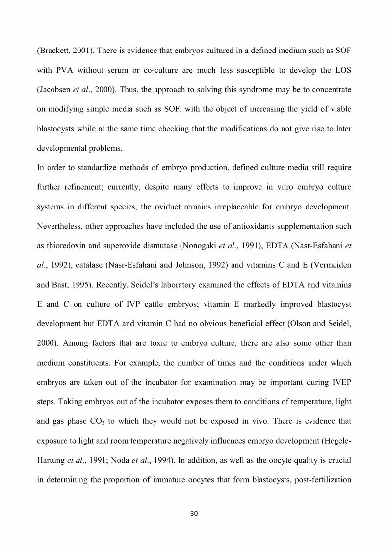
Figure 1. Grade A and B bovine oocytes

19
1.5.2 In Vitro Maturation (IVM)
The results obtained in the field of in vitro embryo production were possible thanks to
studies about in vivo physiological conditions of animals. The hypothalamic-pituitary-
ovarian axis, with the production respectively of Gn-RH (gonadotropin releasing hormone),
gonadotropins (LH and FSH) and ovarian steroids such as 17-β estradiol and progesterone
(Seren and Parmeggiani, 1997), controls the ovarian cycle and, thus, the reproductive
function of the animal.
A fundamental requirement for a successful fertilization is undoubtedly the appropriate
maturational status of the oocytes at the time they encounter the sperm (Gasparrini et al.,
2008). The oocyte maturation process, that involves both the nuclear and cytoplasmic
compartments, is fundamental for the acquisition of full developmental competence
(Gasparrini et al., 2008). During late fetal development, bovine oocytes arrest at prophase I
of meiosis, also defined as germinal vesicle (GV), for the typical appearance of the nucleus.
Oocytes recovered by OPU or by abattoir-derived ovaries are just immature oocytes, still
arrested at this stage of prophase. Under physiological conditions, completion of the first
meiotic division occurs after puberty is attained. Before each ovulation, meiosis of the
oocyte in the dominant follicle resumes after the LH surge; the resumption of meiosis takes
place, starting with germinal vesicle break down (GVBD), then half of the chromosomes are
extruded in the form of the first polar body and the cell cycle is arrested again, this time at
the metaphase stage of the second meiotic division (MII).
Resumption of the first meiotic division, progression to metaphase II, the cumulus
expansion, and the accompanying cytoplasmic changes that are required for the oocyte to
become capable of normal fertilization are collectively termed oocyte maturation (Smith,
20
2001). Complete functional oocyte maturation, including nuclear, cytoplasmic, and
membrane components, is necessary for the ooplasm to become capable of reprogramming
the nucleus of the fertilizing spermatozoon, promoting male pronuclear formation, and
initiating the first mitotic cell division in the newly formed embryo (Eppig, 1996). Nuclear
maturation comprises the resumption and progression of meiosis until the second metaphase
stage and is easy to detect. Conversely, cytoplasmic maturation refers to processes that
prepare the oocyte for activation, which is more difficult to evaluate directly.
In 1972, Hunter et al. have reported the first in vitro maturation (IVM) of bovine oocytes,
and since then subsequent studies have always tried to formulate a medium that could
mimic natural conditions for the oocytes development. Among factors affecting mammalian
embryo development in vitro, the duration of IVM plays a critical role, since an
inappropriate timing of maturation results in abnormal chromatin (Dominko and First,
1997), oocyte aging (Hunter and Greve, 1997) and reduced development (Marston and
Chang, 1964). Furthermore, although sperm can penetrate oocytes prior to completion of
oocyte maturation (Chian et al., 1992), subsequent development is generally reduced.
Therefore, it appears that the optimum time for in vitro fertilization is at completion of
meiosis, that occurs at different times in different species, varying from 18-24 h in cattle
(Sirard et al., 1989; Neglia et al., 2001) to 36-48 h in pig (Prather and Day, 1998). Galli et
al. (2003) confirmed that in vitro maturation of bovine oocytes involves removal of
prophase I oocytes (together with surrounding cumulus cells) from antral follicles and their
culture for 20 to 24 h; during this interval, oocytes extrude their first polar bodies and reach
metaphase II (Gilchrist and Thompson, 2007). Supplementation of the maturation medium
with selected serum, such as fetal calf serum (FCS), hormones and other active factors

21
markedly enhanced quality of the matured oocyte (Brackett and Zuelke, 1993). Among the
various factors of the serum, glucose has a very important role in the maturation, given that
the oocytes resume meiosis but fail to reach metaphase II in its absence (Kimura et al.,
2008). Because maturation was better aided by the presence of proestrous or estrous cow
serum, which contained high concentrations of LH (Brackett et al., 1989; Younis et al.,
1989), it was replaced in the media with purified bovine LH, containing FSH (<1%) and
thyroid stimulating hormone (<4%), leading to development of a defined maturation
medium (Zuelke and Brackett, 1990). Further refinement of conditions for in vitro
maturation to improve oocyte and embryo quality included addition of biologically active
agents, e.g. FSH and various growth factors (Brackett, 2001).
Numerous growth factors involved in the signaling between the developing oocyte and the
surrounding granulosa cells have been identified; these are classified, according to their
structure and biological activity in: Epidermal Growth Factor (EGF); Fibroblast Growth
Factor (FGF); Insuline-like Growth Factor (IGF) and Transforming Growth Factor-β (TGF-
β). Most recent experimental data indicate that EGF-like growth factors (e.g., amphiregulin,
epiregulin, and betacellulin) accumulate in the follicle at ovulation and are potent
stimulators of oocyte maturation and cumulus expansion (Hsieh et al., 2009); therefore, the
inclusion of such factors in the maturation medium may improve the quality of matured
oocytes.
It is important to remember that the oocyte is affected by environment in which it develops
and that in vitro maturation of oocytes is influenced by specific physical culture conditions
and others not well defined (Holm and Callesen, 1998). The osmolarity (Yamauchi et al.,
1999), ionic composition, temperature (Lenz et al., 1983), pH, CO2 (Geshi et al., 1999), and

22
oxygen tension, as well as, the ratio between the number of oocytes and the volume of the
medium, are important parameters that are part of the first category, while the serum and
somatic cells are examples belonging to the second category (Gordon, 1994). Nowadays, in
optimal conditions of in vitro maturation more than 90% of the oocytes reach metaphase II
(Figure 2), showing an improvement of quantitative aspect of IVEP system.
Figure 2. Matured bovine oocytes
1.5.3 In Vitro Fertilization (IVF)
The next step after maturation is in vitro fertilization (IVF), which consists in co-incubation
of the two gametes, male and female; this is a complex process which leads to the fusion of
the two gametes, returning to the number of somatic chromosomes, followed by the
beginning of embryonic development. In particular, to facilitate union of the male and
female gametes to form a zygote under laboratory conditions, selected and capacitated
sperm are co-incubated with mature cumulus-oocyte complexes (Peippo et al., 2011).

23
Many factors may affect the IVF efficiency, such as the adequate in vitro environment for
gametes survival, the sperm viability and capability, the appropriate time of insemination,
the duration of gametes co-incubation, the presence of cumulus cells and also the
acquisition of the oocyte developmental competence during the complex process of
cytoplasmic maturation. In fact, it is likely that the fertilization failure is related to
inadequacies of the IVF system, but a previous inappropriate maturation of the oocytes
should not be ruled out.
Sperm must undergo a series of physiological changes in the female reproductive tract to
become capable of fertilizing oocytes (Austin, 1951; Chang, 1951), a process termed
capacitation (Austin, 1952); this process must be induced in vitro. Although several agents
have been proven to induce sperm capacitation in vitro, heparin, a highly sulfated
biomolecule, is still the most efficient method in the majority of the domestic species
(Parrish, 2014). During capacitation, bound proteins from the cauda epididymis and the
seminal plasma are removed from the sperm surface; this is essential for a series of events
leading to fertilization (Rodriguez-Martinez, 2007). Proteoglycans and glycosaminoglycans
were identified as factors present in the uterine tube with a role in capacitation. This led to
the development of an effective method to induce capacitation of bovine sperm using
heparin, a well-known glycosaminoglycan, more effective in vitro than other compounds
present in uterine tubal fluid (Parrish et al., 1986).
Under physiological conditions sperm swim from the low bicarbonate milieu of the
epididymis to the bicarbonate-rich environment of the uterine tube (Mastroianni and
Komins, 1975). Therefore, cleaned sperm are most often incubated in media containing high
bicarbonate concentrations that stimulate a protein kinase A-dependent protein
24
phosphorylation cascade closely associated with capacitation (Harrison, 2004). Bicarbonate
also induces a disorder in the phospholipid packing of the sperm plasma membrane that
increases membrane fluidity (Harrison and Miller, 2000) contributing to an albumin-driven
efflux of cholesterol from the plasma membrane and translocation of seminolipid from the
apical to the equatorial area of the sperm head (Gadella et al., 1994). Seminolipid is a
sperm-specific glycolipid in the outer leaflet of the plasma membrane that normally
prevents the acrosome reaction; its translocation destabilizes the membrane and renders it
fusogenic, thereby facilitating the acrosome reaction after contact with the ZP (Gadella et
al., 1995). Hyaluronic acid was also effective for capacitating bovine sperm. In that regard,
it was suggested that by swimming through a hyaluronic acid containing medium, sperm are
exposed to high shearing forces that probably remove de-capacitation factors from their
surfaces (Shamsuddin et al., 1993). Additional treatments have also been described as
effective in the stimulation of the capacitation process, including a combination of heparin
and caffeine (Niwa and Ohgoda, 1988) and treatment with the calcium ionophore A23187
(Hanada, 1985) or adenosine (Breininger et al., 2010).
Most frequently either a Tyrode’s albumin lactate pyruvate (TALP) based medium (Bavister
and Yanagimachi, 1977) or a synthetic uterine tubal fluid based medium (Tervit et al.,
1972), both without glucose and with heparin supplementation, are used for IVF (Galli et
al., 2003). Others substances in addition to capacitation agent are added to fertilization
medium to improve motility and fertilizing ability of semen, such as the complex
penicillamine, hypotaurine and epinephrine (PHE). The penicillamine is able to increase the
percentage of spermatozoa that undergo the acrosome reaction when used in the presence of
epinephrine (Monaco, 2007). The hypotaurine increases sperm motility and, in combination

25
with adrenalin, is responsible for an increase in the percentage of oocytes penetration
(Monaco, 2007).
Treatment of bull sperm before IVF generally involves selection of cells with highest
progressive motility. Concurrently, this step also removes undesirable sperm, seminal
plasma, cryoprotective agents, and other factors. Selection is sometimes achieved by
allowing sperm to swim up into a medium for 30 to 60 min (Parrish et al., 1986). Another
method commonly used for sperm separation involves a discontinuous Percoll gradient
centrifugation (Lessley and Garner, 1983). Percoll consists of colloidal silica particles
coated with polyvinylpyrrolidone; centrifugation of sperm through a gradient of 45% and
90% Percoll separates sperm according to their density. Because sperm with good nuclear
morphology and cellular integrity are denser, they are deposited in the Percoll layer with
greatest density (Le Lannou and Blanchard, 1988). Because motile sperm align their
movements with centrifugal force, they also deposit faster during centrifugation (Rhemrev
et al., 1989).
Ideally, the optimal sperm concentration is determined for each bull in preliminary
experiments to achieve maximum in vitro fertilization with the lowest level of polyspermy;
however, in our experience sperm concentration used is usually 106 per mL in cattle, but it
depends on the quality of the semen. In addition, it has been suggested that prolonged
gamete co-incubation under the conditions of IVF, in which high concentrations of
spermatozoa are incubated in small volumes of medium, results in the production of high
levels of hydrolytic enzymes (Rehman et al., 1994) and free radicals (Aitken, 1994) that
damage the oocytes. The time required for fertilization varies depending on species and on
fertilizing capacity of semen; however, it does not ever exceed 20 hours of co-incubation.
26
It has been demonstrated that the optimal sperm-oocyte co-incubation time for maximizing
the blastocyst yield in buffalo is 16 h (Gasparrini et al., 2008). Shortening the gamete co-
incubation length to 8 h has resulted in a significant reduction of oocyte cleavage, similar to
that reported in cattle (Ward et al., 2002; Kochhar et al., 2003). During this interval, sperm
pass through the cumulus cell layers surrounding the oocytes, attach and bind to the ZP, and
after undergoing the acrosome reaction, penetrate the ZP. The fertilizing spermatozoon will
then bind to and fuse with the oocyte plasma membrane, followed by activation of the
oocyte, and formation of the male and female pronuclei (Schultz and Kopf, 1995).
However, in most laboratories co-incubation time of bovine gametes is usually 18 to 20 h,
for practical reasons. Finally, it is worth noting that there are marked differences in the
kinetics of sperm penetration of different bulls; this parameter is related to the blastocyst
rate (Rubessa et al., 2009) and can be a useful indicator to predict the ability of in vitro
fertilization of the bulls. The great variability in the rate of penetration suggests to include
this assessment in the preliminary screening of the bulls before their use for in vitro
fertilization programs, also in order to determine the ideal time of co-incubation to
maximize reproductive performance.
Based on several reports, the use of sperm from bulls with proven fertilizing ability affects
the success of bovine IVF (Aurich and Hahn, 1994; Yang et al., 1995). In some cases, it
may be advantageous to use semen derived from a limited number of bulls with proven
fertility, although this cannot be applied in cattle-breeding programs where embryos must
be derived from specific animals (Gordon, 2003). Furthermore, a bull with best
morphological and genetic traits has not often a high fertility, and it may also happen that
that the fertility in the field does not correspond to that in vitro. This problem is very
27
limiting the relationship between embryo production laboratories and private farms;
normally the farmers choose the semen referring to bulls catalogs possessing all the
characteristics of the offspring but hardly shows the percentages of in vitro fertility.
1.5.4 In Vitro Culture (IVC)
Although IVEP is currently successfully carried out in most domestic species, the
knowledge of epigenetic factors, which regulate embryonic development, is still poor and an
optimal medium for in vitro embryo culture has not yet been formulated. In fact, only 30-
45% of matured oocytes reach the blastocyst stage following IVF and embryo culture, with
pregnancy rates of 40–60% following embryo transfer (Hasler et al., 1995). Furthermore,
embryos have a high capability to adapt to suboptimal culture conditions in vitro, but the
sensitivity to the environment can result in long-term alterations in fetal and postnatal
growth (Hoelker et al., 2014) .The optimization of the IVC systems is therefore necessary,
as it is known that the optimal conditions of culture result in an increase of quality, vitality
and resistance to freezing of embryos, and in a reduction of birth defects and of embryonic
and neonatal mortality incidence (Lonergan, 2007; Kane, 2003).
There are two fundamental morphological events that characterize development of
preimplantation embryos: (a) compaction, represented by progressive reduction of the space
between the dividing cells and the increase of the perivitelline space; (b) the initial
formation of the blastocyst. Within pre-implantation embryo development it is also possible
to identify an early stage, which corresponds to the presence of embryo into the oviduct, and
a late stage that coincides with its passage into the uterus (Figure 3); in the first stage, the
embryo is a undifferentiated entity, non-vascularized, not sensitive to hormones and growth

28
factors and which divide repeatedly itself by mitosis without, however, showing any signs
of growth (Turner et al., 1992). In contrast, in the late stage of development, the embryo is
no longer an undifferentiated element, as confirmed by formation of the first embryonic
epithelium, the trophectoderm, and the inner cell mass, and shows a significant increase of
growth and responsiveness to hormones and growth factors.
The culture system initially started with the in vivo culture in an intermediate host, such as
ligated oviduct of sheep (Galli et al., 1998); this system provides the in vitro culture of
zygotes up to 48 hours after fertilization and their subsequent transfer into the oviduct of the
intermediate host. However, this process has high costs, requires appropriate facilities, is
questionable from an ethical point of view and it is not practical for the production of
embryos on a large scale. Later, a co-culture system was introduced using a complex tissue
culture medium with various cell types, such as oviductal epithelial cells and cumulus cells;
in other co-culture systems, the developing embryos were incubated together with a wide
variety of somatic cells, including BRL (buffalo rat liver) cells (Hasler et al., 1995; Reed et
al., 1996) and Vero cells (Carnegie et al., 1997). In those years, much effort to improve the
IVC system has been concentrated on the type of co-culture cells, but then their
supplementation was identified as part of the reason for the large offspring syndrome
(LOS), which is characterized primarily by higher birth weight coupled with congenital
malformations (Walker et al., 1996; Young et al., 1998). The syndrome is caused by the
exposure of pre-elongation embryos to unusual environmental conditions. It is not clear
exactly what environmental changes are important but a major cause may be embryo culture
conditions, and in some cases at least, the use of serum and co-culture (Sinclair et al., 1999).
A better understanding of the necessary culture conditions led to the development of semi-
29
defined media, with embryos incubated in the absence of feeder cells with little or no serum
added. Numerous studies have been done to improve the efficiency of embryo production
and eventually the synthetic oviductal fluid (SOF) medium with BSA (bovine serum
albumin), originally based on the biochemical composition of sheep uterine tubal fluid
(Tervit et al., 1972), as well as Charles Rosenkrans medium (Rosenkrans et al., 1993),
which became the most popular for embryo culture, were developed. Finally, novel embryo
culture systems based on microfluidic technology offer unique advantages. Such systems
would facilitate the gradual change of the culture medium to meet the precise requirements
of the developing embryo, while overcoming substantial limitations of conventional culture
systems (Krisher and Wheeler, 2010). However, the costs and practical issues have not
allowed these new system to be widely used.
Several factors are known to affect the in vitro embryo development, such as ammonia,
oxygen radicals, growth factors, etc. Among these, the increased oxidative stress is a major
factor impairing mammalian in vitro embryo development (Gasparrini et al., 2000).
Preventing oxidative damage by maintaining a physiological O2 tension (5% to 8%) coupled
with low glucose concentrations was later found to enhance early embryo development
(Iwata et al., 1998), and it was also discovered that less dilute conditions are beneficial
because of autocrine and/or paracrine factors (Gardner, 1994). Completely defined media,
developed by modifications of the SOF medium with glutamine or citrate and nonessential
amino acids (Keskintepe et al., 1995), and by replacing BSA with polyvinyl-alcohol (PVA),
also support development to blastocyst (Keskintepe and Brackett, 1996). Thus, defined
conditions not only facilitate the improvement of culture media to provide a better, more
consistent environment for the developing embryo, but are also critical in disease control
30
(Brackett, 2001). There is evidence that embryos cultured in a defined medium such as SOF
with PVA without serum or co-culture are much less susceptible to develop the LOS
(Jacobsen et al., 2000). Thus, the approach to solving this syndrome may be to concentrate
on modifying simple media such as SOF, with the object of increasing the yield of viable
blastocysts while at the same time checking that the modifications do not give rise to later
developmental problems.
In order to standardize methods of embryo production, defined culture media still require
further refinement; currently, despite many efforts to improve in vitro embryo culture
systems in different species, the oviduct remains irreplaceable for embryo development.
Nevertheless, other approaches have included the use of antioxidants supplementation such
as thioredoxin and superoxide dismutase (Nonogaki et al., 1991), EDTA (Nasr-Esfahani et
al., 1992), catalase (Nasr-Esfahani and Johnson, 1992) and vitamins C and E (Vermeiden
and Bast, 1995). Recently, Seidel’s laboratory examined the effects of EDTA and vitamins
E and C on culture of IVP cattle embryos; vitamin E markedly improved blastocyst
development but EDTA and vitamin C had no obvious beneficial effect (Olson and Seidel,
2000). Among factors that are toxic to embryo culture, there are also some other than
medium constituents. For example, the number of times and the conditions under which
embryos are taken out of the incubator for examination may be important during IVEP
steps. Taking embryos out of the incubator exposes them to conditions of temperature, light
and gas phase CO2 to which they would not be exposed in vivo. There is evidence that
exposure to light and room temperature negatively influences embryo development (Hegele-
Hartung et al., 1991; Noda et al., 1994). In addition, as well as the oocyte quality is crucial
in determining the proportion of immature oocytes that form blastocysts, post-fertilization

31
culture environment has a major influence on the quality of the blastocyst; in fact, it was
reported that the embryo quality and viability is mainly affected by the culture system
following IVF (Rizos et al., 2002a). A substantial amount of evidence exists to demonstrate
that the culture conditions to which the embryo is exposed, particularly in the post-
fertilization period, can have perturbing effects on the pattern of gene expression and on the
embryo quality, with potentially important long-term consequences (Lonergan, 2007). In
accordance, it was reported that the expression of seven genes involved in apoptosis,
oxidative stress, gap junction formation and differentiation in blastocysts was different
between in vitro produced and in vivo cultured embryos, i.e. using the ewe oviduct (Rizos et
al. 2002b). Also, the different culture systems have profound effects on embryo freezability
(Rizos et al., 2003). For example, if the in vitro produced zygotes were implanted into the
sheep oviduct they had the greatest potential to survive freezing and thawing; on the
contrary, if zygotes were cultured with serum-supplemented SOF, they exhibited low
cryotolerance (Thompson, 1996).
Collectively, in vivo and in vitro studies support the notion that the environment of
the embryo is critical for its future. The identification and characterization of the short-term
effects of in vitro culture raises the question about long-term consequences and safety of
assisted reproductive technologies (Lonergan, 2007). Currently almost all embryo transfer
in cattle is carried out non-surgically using frozen blastocysts and this necessitates the use of
a reliable culture system. In combination with optimal methods of in vitro maturation (IVM)
and IVF, optimal methods of embryo culture would greatly facilitate the use of IVP
embryos. In dairy cattle this would lead to a better use of the genetic merit of high yielding
dairy cows.
32
Figure 3. IVP bovine embryo at different developmental stages
1.6 EVALUATION OF EMBRYO QUALITY
Over the past 30 years, basic and applied studies on classical and
advanced embryo technologies have generated a vast literature on factors regulating oocyte
and embryo development and quality. In addition, over this period,
commercial bovine embryo transfer has become a large international business.
It is generally accepted that bovine embryos produced in vitro have lower developmental
capacity and quality than in vivo-produced embryos (Hasler et al., 1995; Galli and Lazzari
1996; van Wagtendonk-de Leeuw et al., 2000). Relatively little has changed in
the techniques of producing embryos in vivo although there is increasing evidence of the
importance of, for example, peripheral and follicular endocrine profiles for the subsequent
developmental competence of the embryo. Most of immature bovine oocytes fail to develop
to the blastocyst stage during the process of in vitro production, with developmental rates
being generally limited to approximately 35-40%. Furthermore, pregnancy rates after ET on
cryopreserved in vitro produced embryos are lower than those recorded with in vivo
embryos, due to their poorer quality (Gomez et al., 2009). These unsatisfactory low
33
developmental capacities represent a major hurdle for further implementation of embryo
technologies in the bovine (e.g. ovum pick-up followed by in vitro embryo production).
Therefore, accurate assessment of embryo quality is crucial to increase the efficiency of in
vitro embryo production.
However, it is important to define the term ‘quality’ at the outset. The ultimate test of the
quality of an oocyte or zygote or cleavage-stage embryo remains its ability to develop to the
blastocyst stage, to establish a pregnancy and to produce live and healthy offspring. Thus, it
is necessary to identify reliable parameters that reflect developmental competence.
1.6.1 Non-invasive techniques An accurate assessment of oocyte and/or embryo developmental competence is crucial for
high in vitro developmental rates, for successful establishment of pregnancy after transfer to
recipients to enable improvements in terms of the overall whole efficiency and economy of
assisted reproductive techniques. Traditional methods predict the developmental
competence of an oocyte or embryo indirectly on the basis of scoring morphological
properties and developmental kinetics or on the basis of the origin of the embryo or oocyte,
which indicates that the developmental environment in which the embryo has developed is
correlated to subsequent developmental capacity.
Numerous morphological criteria have been reported to be correlated with oocyte and/or
embryo quality (Camargo et al., 2006; Wang and Sun, 2007). Morphological factors related
to the quality of cumulus-oocyte complexes (COC) and/or oocytes include COC
morphology (Blondin and Sirard, 1995; de Wit et al., 2000; de Wit and Kruip, 2001; Boni et
al., 2002), the number of cumulus cell layers (Blondin and Sirard, 1995; Zeuner et al., 2003;
34
Warriach and Chohan, 2004; Yuan et al., 2005), oocyte diameter (Fair et al., 1995; Fair,
2003) and oocyte coloration (Nagano et al., 2006). Furthermore, the size of the periviteline
space (De Sutter et al., 1996) and the thickness and organization of the ZP (Talevi et al.,
1997; Gabrielsen et al., 2001) have been correlated with developmental capacity. Similar
studies in cattle established ZP birefringence as a marker of the developmental capacity of
unfertilized (Held et al., 2012) and fertilized (Koester et al., 2011) oocytes and zygotes.
Recently, a correlation between ZP birefringence and in vitro development was reported for
equine oocytes (Mohammadi-Sangcheshmeh et al., 2013). Additional characteristics
indicative of oocyte quality in various species, including cattle, consist of mitochondrial
distribution (Bavister and Squirrell, 2000; Stojkovic et al., 2001; Au et al., 2005) and
glucose-6-phosphate dehydrogenase (G6PDH) activity measured by Brilliant cresyl blue
(BCB) staining of immature oocytes (Pujol et al., 2004; Alm et al., 2005; Bhojwani et al.,
2007). Blastocyst development is already one step along the road to the production of live
offspring. However, it is essential that the embryos reaching the blastocyst stage are of the
highest quality possible to ensure optimal pregnancy rates after transfer. Nowadays, reliable
markers of the developmental competence of blastocysts are rare, assessment
of embryo quality is still a challenge and morphological assessment is at present the most
popular method for embryo selection prior to transfer. Embryo quality assessment is most
commonly performed by non-invasive morphological evaluation of embryos based on
embryo color, blastomere symmetry, cytoplasmic granulation and fragmentation, although
this technique is prone to subjectivity (Lindner and Wright, 1983; Merton, 2002).
The quality of bovine blastocysts has been described simply on the basis of morphological
criteria, such as the darkness of the cytoplasm (Pollard and Leibo, 1994) caused by a higher

35
lipid content (Abe et al., 2002) that is correlated with metabolic changes (Khurana and
Niemann, 2000). In human embryos, assessment of the diameter of Day 5 blastocysts
revealed that blastocyst diameter correlated positively with subsequent developmental
capacity (Racowsky et al., 2003). Accordingly, bovine blastocyst diameter was identified as
being correlated with cell count and hatching probability (Hoelker et al., 2006).
Although there is a consensus that morphological assessment is subjective, the grading of
bovine oocytes according to morphological criteria still provides some basic information for
the preselection of oocytes to maximize in vitro embryo development. In contrast, reliable
morphological predictors at the blastocyst stage for viability after embryo transfer are
completely lacking, which could be explained by the suggestion that attainment of the
blastocyst stage is considered more a reflection of past achievements than a guarantee of
future fitness (McEvoy et al., 2000). Despite this, the most important criterion to determine
the quality of embryos to be transferred is the timing of development to specific
morphological stages (Leibfried-Rutledge et al., 1987; McKiernan and Bavister, 1994;
Bavister, 1995; Van Soom et al., 1997). Extensive research to find a better non-invasive,
rapid and simple embryo quality parameter involved studies on developmental kinetics (Van
Soom et al., 1997; Lonergan, 2000) and oxygen consumption measured by respirometer
(Lopes et al., 2007). In fact, oxygen consumption of preimplantation embryos is another
important parameter that may provide valuable information on metabolic processes and
subsequent embryo viability. The kinetics of development of early bovine embryos have
been explored extensively (Grisart et al., 1994; Van Langendonckt et al., 1997; Holm and
Callesen, 1998; Holm et al., 1998; 2002; Lockwood et al., 1998; Lonergan et al., 1999). A
clear relationship exists between the time of first cleavage after insemination in vitro and

36
developmental competence, with the bovine oocytes cleaving earlier (~30 h after
insemination) being more likely to reach the blastocyst stage compared with their later
cleaving counterparts (~36 h after insemination) (Dinnyés et al., 1999; Lonergan et al.,
1999). This finding has been confirmed for bovine embryos and for many other species,
including rhesus monkeys (Bavister et al., 1983), humans (McKiernan and Bavister, 1994;
Sakkas et al., 1998; Shoukir et al., 1998; Fenwick et al., 2002), buffaloes (Totey et al.,
1996) and mice (Warner et al., 1998), highlighting that the timing of the first cleavage
division is linked to developmental ability of embryos. In addition, the duration of the first
cell cycles before embryonic genome activation are shorter in in vivo-derived that in vitro-
derived zygotes (Holm et al., 2002). Accordingly, it is generally accepted that early
blastocyst formation and expansion is positively correlated with pregnancy rates after
transfer, representing the method of choice for the selection of the best in vitro derived
blastocysts (Figures 4, 5 and 6) before transfer to recipients.
Figure 4. Expanded blastocyst Figure 5. Hatching blastocyst Figure 6. Hatched blastocyst
37
1.6.2 Invasive techniques
Invasive techniques need extensive manipulation and/or fixation, which impede further
embryo culture or transfer. Nevertheless, they are very valuable in fundamental research and
are very frequently used in cattle IVP in combination with other techniques in order to fine-
tune embryo quality assessment (Overström, 1996). Several invasive techniques are
available for embryo quality evaluation, such as total cell number determination by nucleus
staining using propidium iodide (PI) or Hoechst (Pursel et al., 1985); evaluation of
differentiation in inner cell mass (ICM) and trophectoderm (TE) by differential staining
(Van Soom et al., 2001; 2002); cryotolerance (Shehab-El-Deen et al., 2009) and detection
of apoptotic cells using different staining techniques (Wydooghe et al., 2011).
In addition, quantitative examination of gene expression is an additional valuable tool to
assess the viability of cultured embryos. Groups of embryos of different morphologies,
reproductive types (in vivo - in vitro) and developmental kinetics, which may have
developed in contrasting environments, not only exhibit different developmental
competence, but also display differences with respect to mRNA expression patterns
(Hoelker et al., 2014). Thus, it is reasonable to consider these mRNA patterns as molecular
signatures of embryo developmental competence. Furthermore, some research has shown
that fast cleaving bovine embryos have an altered gene expression (Lonergan, 2000; Ward
et al., 2001; Fair et al., 2004; Gutiérrez-Adán et al., 2004; Dode et al., 2006) or altered
polyadenylation status of several developmentally important gene transcripts (Pocar et al.,
2001; Brevini-Gandolfi et al., 2002) and a higher chance of reaching advanced
developmental stages in comparison with late cleaving embryos (Van Soom et al., 1992;
Dinnyés et al., 1999; Lonergan et al., 1999; Lequarré et al., 2003; Favetta et al., 2004;
38
Gutiérrez-Adán et al., 2004). A possible hypothesis is that fast cleaving embryos contain all
the necessary gene products and proteins to support further development without any
problem, whereas an aberration of some gene products and proteins in slow cleavers could
be related to a higher chance to go into developmental arrest or apoptosis (Vandaele and
Van Soom, 2011).
Studies focusing on the relationship between bovine blastocyst quality and transcript
abundance (Lonergan et al., 2006; Wrenzycki et al., 2005; 2007; Duranthon et al., 2008)
provide valuable information about bovine embryonic gene expression, but the conclusions
that can be drawn from a small number of differentially expressed candidate genes are
limited because it is hard to interpret what the consequences are for subsequent in vivo
development.
Development of embryos produced by IVF is characterized by a proportion showing
abnormal development, delayed cell division or blastomere fragmentation, with many
embryos either arresting growth prior to the blastocyst stage or failing to implant following
transfer (Trounson and Bongso, 1996). However, counting the total number of blastomeres,
the ratio of ICM to TE and the percentage of apoptotic nuclei provide more detailed
information on embryo quality and the developmental potential of embryos after transfer to
recipients.
It is now well recognized that the ICM differentiates into all tissues of the developing fetus
and the TE forms the placenta and embryonic membranes and is involved in maternal
recognition of pregnancy (Roberts et al., 1990). The quality of these two cell components of
the developing embryo determines the outcome after transfer to recipient females. Different
methods have been applied for differential staining (DS) of blastocysts in different species,

39
including isolation of ICM by micromanipulation (Gardner et al., 1973), or antibody-based
immunosurgery using complement mediated membrane lysis in TE cells (Solter and
Knowles et al., 1978). These methods are not only time consuming, they are also expensive
and depend on the efficiency of the antibody, which is often inconsistent. Further scientific
studies have showed that attempts to develop a chemically defined system, using calcium
ionophore to permeabilize TE cells (de la Fuente and King et al., 1997), as well as
permeabilization of TE cells by brief exposure of blastocysts to a non-ionic detergent
solution containing propidium iodide (PI), followed by fixation and DAPI staining,
permitted the differential visualization of ICM and TE compartments (Thouas et al., 2001).
The proportion of ICM cells in blastocysts is crucial for implantation (Iwasaki et al., 1990),
and increased cell death in these cells will compromise subsequent development, since
critical threshold numbers of ICM cells are required for normal post-implantation
development (Tam, 1988).
Apoptosis is thought to be the default pathway of defective cells in embryos (Raff et al.,
1993), and cell death by apoptosis occurs predominantly in the ICM in both mouse (Hardy,
1997) and bovine embryos (Byrne et al., 1999). Bovine embryos undergo apoptosis
predominantly at the morulae stage and during blastocyst development (Byrne et al., 1999).
Hence, apoptosis may be associated with cell population control and maintenance of cellular
quality in the ICM lineage, by eliminating abnormal or defective cells that possess damaged
DNA or chromosomal abnormalities (Brison, 2000). The percentage of apoptotic cells is
significantly higher in blastocysts produced in vitro as compared with in-vivo derived
embryos (Bergeron et al., 1998), and apoptosis is regulated by the activity of pro- and anti-
apoptotic genes during pre-implantation development (Bergeron et al., 1998). The
40
identification of apoptotic cells relies mainly on morphological changes such as chromatin
condensation and nuclear and cytoplasmic fragmentation into apoptotic bodies. However,
apoptotic cells are not easily distinguishable by light microscopy from other elements with
condensed chromatin, such as mitotic cells in telophase (Migheli et al., 1995). The in-situ
terminal deoxynucleotidyl transferase mediated dUTP nick end labelling (TUNEL)
technique is commonly used for detection of apoptosis in cells. However, the reliability of
TUNEL has been questioned, as major DNA fragmentation is a late event in apoptosis
(Collins et al., 1997). Therefore, a combination of morphological assessment of blastocysts
showing the presence of fragmented and condensed nuclei and TUNEL labelling is used to
assess apoptosis rate in blastocysts. Nevertheless, this method does not permit a clear
determination of the cell types undergoing apoptosis in blastocysts, i.e. whether or not the
ICM or TE are equally affected. However, using DS method in combination with TUNEL
technique, it has been possible to carry out both a qualitative and quantitative analysis of in
vitro embryos produced. The two different methods provides a means of assessing the effect
of culture conditions on cell number of both embryo compartments (ICM and TE), as well
as providing information on the localization of apoptotic nuclei within the blastocyst
(Fouladi-Nashta et al., 2005).In conclusion, development of additional accurate laboratory
methods and future efforts to assess embryo quality will improve the efficiency of embryo
production from in-vitro culture systems.
Another important parameter for pregnancy establishment and maintenance after ET is the
embryo cryotolerance. This is of the utmost importance, as most of the embryos transferred
around the world are cryopreserved. For this reason, we believe that it is worth to discuss
the topic of cryopreservation in a separate paragraph.

41
1.7 CRYOPRESERVATION
The concept of preserving life in a suspended animation state with the use of low
temperature is not new. Cryobiology history can be traced back to antiquity. As early as in
2500 BC low temperatures were used in Egypt in medicine and the use of cold was
recommended by Hippocrates to stop bleeding and swelling. With the emergence of modern
science, Robert Boyle studied the effects of low temperatures on animals and in 1949 bull
semen was cryopreserved for the first time by a team of scientists led by Christopher Polge.
The study on cryopreservation moved into the human world in the 1950s with pregnancies
obtained after insemination with frozen sperm. The maintaining of the potential viability of
living reproductive cells, embryos and tissues of several mammalian species after long-term
storage represents a tool of great opportunity for both human and animal reproductive
applications (Woods et al., 2004; Vajta and Kuwayama, 2006). This is possible with the use
of cryopreservation, a process where cells or whole tissues are preserved by cooling to low
sub-zero temperatures such as −196 °C (the boiling point of liquid nitrogen). At these low
temperatures, any biological activity, including the biochemical reactions that would lead to
cell death, is effectively stopped.
Cryopreservation occurs naturally. Antifreezing is actually required for ensuring survival of
various organisms. However, the simple immersion in liquid nitrogen for certain samples,
such as embryos and oocytes, does not maintain the necessary viability to make them usable
after thawing. Increased understanding of the mechanism of freezing injury to cells
empathized the importance of cooling methods to obtain maximum survival on thawing of
the living cells. Because embryos are viable at 37°C and no biological activity takes place at
-196 °C, the time of greatest danger during cryopreservation appears to be during the
42
transitions of temperature: during cooling to -196 °C and during subsequent rewarming to
37°C. When water is cooled below its freezing point, it solidifies in a crystalline structure
known as ice. Because ice is less dense than liquid water, it necessarily follows that ice
crystals occupy a greater volume than does the liquid water from which they were formed.
As adjacent volumes of liquid water within a cell solidify, their expansion into ice causes
pressure and shearing forces on intracellular organelles, which can suffer considerable
damage. Avoidance of ice crystal formation therefore is one of the principal goals of
successful cryopreservation. As water transitions from liquid to ice, any solutes in the liquid
phase are excluded from the solid. This lowers the freezing point of the remaining unfrozen
solution. As the temperature drops and the solid form proliferates, the concentration of
electrolytes and other solutes can reach very high levels (Lovelock, 1954). These
concentrations can be quite toxic to intracellular proteins, and thus avoidance of these
solution effects is a second major goal of successful cryopreservation. During rewarming,
the solid ice melts and releases free water, resulting in decreasing osmolarity of the
surrounding solution. When rewarming is slow, there is danger of free water thawing and
recrystallizing, thus causing further damage. When rewarming is rapid, sudden drops in
extracellular osmotic pressure may lead to rapid shifts of free water across and into the cell,
leading to swelling and cell damage (Mazur, 1980). This is called osmotic shock, and its
avoidance is a third major goal of successful cryopreservation. Therefore, simply immersing
embryos into liquid nitrogen is not an effective strategy for successful cryopreservation. All
successful methods must avoid these three issues: ice crystal formation, solution effects, and
osmotic shock.
43
Up to this point in time, all cryopreservation strategies, including all methods of embryo
cryopreservation, have used additional chemicals to avoid cell damage. These chemicals are
called cryoprotectants and generally can be divided into two categories: permeating and
non-permeating.
Permeating cryoprotectants are small molecules that readily permeate the membranes of
cells. They form hydrogen bonds with water molecules and prevent ice crystallization. At
low concentrations in water, they lower the freezing temperature of the resulting mixture.
However, at high enough concentrations, they inhibit the formation of the characteristic ice
crystal and lead to the development of a solid, glasslike, so-called vitrified state in which
water is solidified, but not expanded. In this manner, the permeating cryoprotectants satisfy
the first goal of successful cryopreservation, the avoidance of ice crystals. Permeating
cryoprotectants play a second important role in cryopreservation, and this is to protect the
cell from solution effects. They achieve this goal by remaining in solution and by thus
effectively diluting the remaining electrolytes. This effect is described by the phase rule,
which states that in a two-phase system, such as liquid water and ice at a given pressure, the
total solute concentration in the liquid phase is constant for a given temperature (Friedler et
al., 1988). The most commonly used permeating cryoprotectants are: Propylene Glycol
(PROH), Ethylene Glycol (ET), Glycerol and Dimethyl sulfoxide (DMSO).
In contrast to the permeating cryoprotectants, nonpermeating cryoprotectants remain
extracellular. They act by drawing free water from within the cell, thus dehydrating the
intracellular space. As a result, when they are used in combination with a permeating
cryoprotectant, the net concentration of the permeating cryoprotectant is increased in the
intracellular space. This further assists the permeating cryoprotectant in preventing ice-

44
crystal formation. The nonpermeating cryoprotectants play an important role during
thawing. During thawing, the water generated by the melting ice rapidly decreases the
extracellular osmotic pressure. Osmotic shock may occur if the intracellular cryoprotectant
cannot diffuse out quickly enough to prevent excessive influx of free water and the
swelling, or even rupture, of the cell. Therefore, freezing and thawing protocols commonly
use a high concentration of nonpermeating cryoprotectants during the thawing phase. The
most commonly used nonpermeating cryoprotectant is sucrose, but other disaccharides and
other nonpermeating agents also may be used.
In conclusion, different are the advantages to use cryopreservation: the progress in
cryopreservation would improve domestic animal breeding by genetic selection programs
(Vajta, 2000), to contribute at the maintenance of biodiversity through wildlife species
conservation (Pukazhenthi and Wildt, 2004) and preserve the human fertility.
1.7.1 Cryopreservation methods
All oocytes and embryos suffer considerable morphological and functional damage during
cryopreservation. The extent of the injury depends on factors including the size and shape of
the cells, the permeability of the membranes, and the quality and sensitivity of the oocytes
and embryos. All these factors may be highly variable depending on the species,
developmental stage and origin (e.g., in vitro produced or in vivo derived embryos).
However, oocytes and embryos also have a remarkable, sometimes surprising ability to
repair this damage fully or partially, and, for optimal cases, to continue normal
development. The purpose of cryopreservation procedures is to minimize the damage and
help cells to regenerate. Almost all cryopreservation strategies are based on two main
45
factors: cryoprotectants and cooling–warming rates. Since the first successes of mammalian
embryo cryopreservation in the sixties of the last century, two major groups of methods can
be delineated: traditional slow-rate freezing and vitrification. Storage, warming and
rehydration, i.e. removal of cryoprotectants differ only slightly between the two procedures
(with some exceptions) and the main difference exists in the addition of cryoprotectants and
cooling. Traditional slow-rate freezing was introduced first, and for the majority of domestic
animal and human embryologists this remains the only acceptable approach. Over time,
methods have become highly standardized with a considerable industrial and commercial
background. Traditional slow-rate freezing can be interpreted as an attempt to create a
delicate balance between various factors causing damage, including ice crystal formation,
fracture, toxic and osmotic damage. The controlled cooling rate allows solution exchange
between the extracellular and intracellular fluids without serious osmotic effects and
deformation of the cells (this fact is reflected in the other name of the procedure:
equilibrium freezing). Controlled-rate and slow freezing are well established techniques
pioneered in the early 1970s which enabled the first birth from a human frozen embryo in
1984. Since then many studies have developed to preserve human and animal samples from
freeze damages until the development of a new technique, the vitrification, that provides the
benefits of cryopreservation without damage due to ice crystal formation.
The phenomenon of the solidification of water without ice crystal formation is also called
vitrification. However, the term “vitrification” in cryobiology refers to the other group of
cryopreservation methods, where the main purpose is to ensure ice free solidification of the
whole solution containing the sample. The process itself can be described as an extreme
increase in viscosity of solutions. The benefits are unquestionable: ice crystal formation, one
46
of the most dangerous causes of cryoinjury, is entirely eliminated except for a transitional
and very short freezing of the solutions during warming that is generally regarded harmless
to the oocytes and embryos. Unfortunately we have to pay the price of this benefit.
Vitrification can only be induced in exceptional situations: with dangerously-high
concentrations of cryoprotectants and/or with extreme increase of the cooling and warming
rates. One characteristic of vitrification is that as the cooling rate chosen increases, the
cryoprotectant concentration can be lowered and vice versa. Extensive research in the past
20 years has resulted in new, sometimes radically-new approaches and created an acceptable
compromise between the positive and negative by decreasing cryoprotectant toxicity and
increasing cooling rates. As a result, vitrification has become a competitive alternative to
slow-rate freezing. Moreover, the high cooling and warming rates applied at vitrification
provide a unique benefit compared to the traditional freezing: the possible partial and
sometimes total elimination of chilling injury, as the sample passes through the dangerous
temperature zone quickly enough to disallow sufficient time for damage to develop. During
vitrification, permeating cryoprotectants are added at a high concentration while the embryo
is at room temperature. Because the toxicity of this high concentration of permeating
cryoprotectant is substantial, the embryo cannot be kept at this temperature for long.
Instead, a very short time is allowed for equilibration, after which the embryos are plunged
directly into liquid nitrogen. To further protect against ice-crystal formation, an extremely
rapid rate of cooling is used. For this reason, novel cryovessels have been used that allow
direct contact between liquid nitrogen and the embryo-containing solution and that have in
common a very high surface-to-volume ratio (Park et al., 2000; Mukaida et al., 2003). This
47
form of ultrarapid freezing must be followed by ultrarapid thawing to prevent ice
recrystallization. To achieve vitrification, three important factors should be considered:
1) Cooling rate, which is achieved with liquid nitrogen or liquid nitrogen slush. When using
liquid nitrogen, the sample is plunged into liquid nitrogen resulting in cooling rates of
hundreds to tens of thousands degrees Celsius per minute, depending on the container, the
volume, the thermal conductivity, the solution composition, etc. (Yavin and Arav, 2007). It
was shown for oocytes and embryos that increasing the cooling rate would improve survival
rates by up to 37%.
2) Viscosity of the medium in which the embryos are suspended. This is defined by the
concentration and behavior of various cryoprotectants and other additives during
vitrification. The higher is the concentration of cryoprotectants, the higher is the glass
transition temperature (Tg), thus lowering the chance of ice nucleation and crystallization.
The combination of different cryoprotectants is often used to increase viscosity and Tg, and
reduce the level of toxicity.
3) Volume; the smaller is the volume, the higher is the probability of vitrification (Arav,
1992; Arav et al., 2002). Smaller volumes allow better heat transfer, thus facilitating higher
cooling rates. Many techniques have been developed to reduce sample volume with an
explosion of methods appearing in the literature during the last decade (Yavin and Arav,
2007).
1.7.3 Vitrification techniques
Vitrification was first introduced in embryology for cryopreservation of mouse embryos
(Rall and Fahy, 1985). In the subsequent decade, a moderate number of publications dealing

48
with successes in cryopreservation of oocytes and embryos from several domestic animal
species were published (Pereira and Marques, 2008). The latest development in the practical
application of ultrarapid vitrification methods was based on the concept of minimum
volume cooling (Hamawaki et al., 1999). Different carrier tools were applied to minimize
the volume and to submerge the sample quickly into the liquid nitrogen.
These techniques can generally be divided into two categories, surface techniques and
tubing techniques.
The surface techniques include:
1) Solid Surface Vitrification (SSV) method, that combines the advantages of containerless
vitrification in microdrops and the increased heat exchange of a cold metal surface (Dinnyés
et al., 2000). A metal cube, in some cases covered with aluminum foil, is partially
submerged into liquid nitrogen. Microdrops of vitrification solution, containing the
embryos, are dropped onto the cold upper surface of the metal cube (Figure 7) and are
instantaneously vitrified. Warming was performed by placing the drop of vitrified solution
in a warming solution directly. This approach makes a shortcut to exclude entirely the vapor
formation by cooling metal surfaces in liquid nitrogen, and placing the solution containing
the biological sample on the surface of metal.
Figure 7. Solid Surface Vitrification

49
2) The Cryoloop method consists in the use of a solution film formed between files of a
nylon loop (Figure 8) for holding the sample (Lane et al., 1999a; 1999b). Due to the
absence of solid support and the extremely small volume, the cooling rate upon immersion
in liquid nitrogen may be as high as 700,000 °C/min (Isachenko et al., 2003) thus also
permitting cooling in liquid nitrogen vapor. However, in spite of the unquestionable benefits
of this system from the point of potential disease transmission, one may have concerns
regarding the safety of storage, as this very sensitive and fragile system may increase the
risk of accidental warming over the safe temperature zone, especially if storage of the
container is also performed in the vapor of liquid nitrogen (Fuller and Paynter, 2004).
Figure 8. Cryoloop
3) In the Cryotop procedure a thin plastic film strip attached to a plastic handle (Figure 9) is
the carrier tool (Hamawaki et al., 1999; Kuwayama and Kato, 2000). Loading is performed
with the use of a glass capillary under the control of a stereomicroscope. Before cooling,
almost all medium is removed, so the embryos and oocytes are only covered with a very
thin solution layer. Submersion of the Cryotop into the liquid nitrogen or into the
rehydrating solution after vitrification results in extremely high cooling and warming rates,
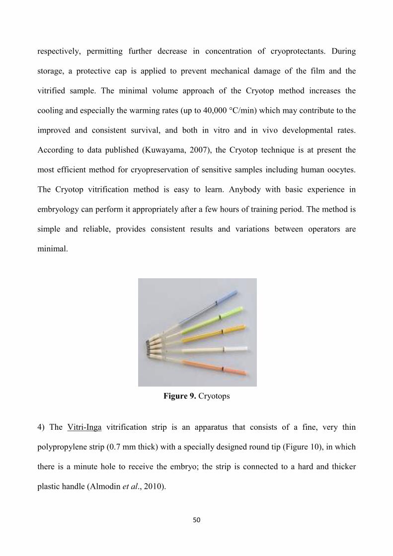
50
respectively, permitting further decrease in concentration of cryoprotectants. During
storage, a protective cap is applied to prevent mechanical damage of the film and the
vitrified sample. The minimal volume approach of the Cryotop method increases the
cooling and especially the warming rates (up to 40,000 °C/min) which may contribute to the
improved and consistent survival, and both in vitro and in vivo developmental rates.
According to data published (Kuwayama, 2007), the Cryotop technique is at present the
most efficient method for cryopreservation of sensitive samples including human oocytes.
The Cryotop vitrification method is easy to learn. Anybody with basic experience in
embryology can perform it appropriately after a few hours of training period. The method is
simple and reliable, provides consistent results and variations between operators are
minimal.
Figure 9. Cryotops
4) The Vitri-Inga vitrification strip is an apparatus that consists of a fine, very thin
polypropylene strip (0.7 mm thick) with a specially designed round tip (Figure 10), in which
there is a minute hole to receive the embryo; the strip is connected to a hard and thicker
plastic handle (Almodin et al., 2010).
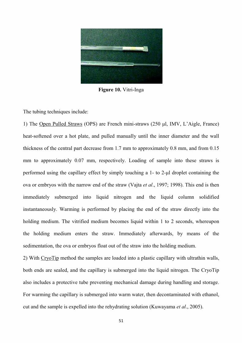
51
Figure 10. Vitri-Inga
The tubing techniques include:
1) The Open Pulled Straws (OPS) are French mini-straws (250 µl, IMV, L’Aigle, France)
heat-softened over a hot plate, and pulled manually until the inner diameter and the wall
thickness of the central part decrease from 1.7 mm to approximately 0.8 mm, and from 0.15
mm to approximately 0.07 mm, respectively. Loading of sample into these straws is
performed using the capillary effect by simply touching a 1- to 2-µl droplet containing the
ova or embryos with the narrow end of the straw (Vajta et al., 1997; 1998). This end is then
immediately submerged into liquid nitrogen and the liquid column solidified
instantaneously. Warming is performed by placing the end of the straw directly into the
holding medium. The vitrified medium becomes liquid within 1 to 2 seconds, whereupon
the holding medium enters the straw. Immediately afterwards, by means of the
sedimentation, the ova or embryos float out of the straw into the holding medium.
2) With CryoTip method the samples are loaded into a plastic capillary with ultrathin walls,
both ends are sealed, and the capillary is submerged into the liquid nitrogen. The CryoTip
also includes a protective tube preventing mechanical damage during handling and storage.
For warming the capillary is submerged into warm water, then decontaminated with ethanol,
cut and the sample is expelled into the rehydrating solution (Kuwayama et al., 2005).

52
3) Cryopette is a cryopreservation device that is intended to be used to contain, freeze using
vitrification procedures. Consists of a polycarbonate Cryo-Straw with a silicone rubber
displacement bulb. This device has been optimized as a closed system for cryopreservation
procedures (Portmann et al., 2010).
Each of these two groups has its specific advantages. In the surface methods, if the size of
the drop (~ 0.1 mL) can be controlled, high cooling rate can be achieved because these
systems are open, and high warming rates are achieved by direct exposure to the warming
solution. The tubing systems have the advantage of achieving high cooling rates in closed
systems, thus making them safer and easier to handle. Decreasing the vitrified volume and
increasing the cooling rate allow a moderate decrease in cryoprotectant concentration so as
to minimize its toxic and osmotic hazardous effects (Yavin et al., 2009).
1.7.3 Embryo cryopreservation
The first mammalian species whose embryos were successfully cryopreserved was the
mouse in the early 1970s when Whittingham et al. (1972) obtained live mice after the
transfer of frozen-thawed morulae. The earliest successes with mouse embryo
cryopreservation were achieved with slow cooling, at a constant rate, to deep subzero
temperatures (−80°C). Observations of embryos during this cooling procedure demonstrated
the extreme dehydration encountered during the process when ice formed from pure water
as extracellular ice formed around the cells, leaving the embryos in the small, residual
hypertonic unfrozen fraction (Leibo et al., 1974). Conditions that permitted intracellular ice
to form (by cooling too fast and leaving water molecules capable of nucleating ice crystal
growth) were universally found to be damaging (Mazur, 1970). It was also argued that slow

53
warming was necessary to avoid cell volume stress. In the intervening time, further
investigations demonstrated that this low-temperature (and successful embryo
cryopreservation) could be achieved if the embryos were slowly cooled only to a relatively
high subzero temperature and then transferred directly to –196°C. In this case, warming
needed to be rapid to achieve high viability. These findings, developed by Willadsen and
Cambridge to freeze sheep embryos (Willadsen, 1977), have also laid the foundations of
cryopreservation techniques widely used in commerce. The first experiment with the
technique of vitrification date, however, until 1985, when Rall and Fahy applied this
technique for the first time in mouse embryos, while the first pregnancy achieved by
transfer of vitrified bovine embryos has been reported by Massip et al. in 1986.
Since then, many experiments and significant improvements were achieved in the field of
embryo cryopreservation. In recent years embryos have been successfully cryopreserved in
most domestic species, especially cattle (Lazar et al., 2000; Zhang et al., 1993), sheep (Ptak
et al., 1999; Dattena et al., 2000), goats (Baril et al., 1989; El Gayar and Holtz, 2001;
Traldi, 2000), horse (Hochi et al., 1994) and pig (Hayashi et al., 1989; Berthelot et al.,
2000). Across all these different species, the protocols applied to embryo cryopreservation
tend to fall into one of two broad categories, slow controlled-rate cooling and ultrarapid
cooling or vitrification. Throughout these experiences, it became obvious that embryo
cryopreservation would too become centrally important in reproductive technologies by
allowing optimal utilization of resources in the development of breeding programs in
animals (Massip et al., 2004) by permitting genetic banking (for example of rare animals),
and by enhancing the clinical approach to infertility treatment.

54
EXPERIMENTAL PART

55
CHAPTER 2: Introduction to experimental part
2.1 ANTIOXIDANT SUPPLEMENTATION DURING IVC
The high incidence of developmental failure of in vitro produced embryos has been
attributed to a wide range of non mutually exclusive dysfunctions, including poor gamete
quality, chromosomal abnormalities, telomere uncapping, and suboptimal culture conditions
that induce oxidative stress. Indeed, increased oxidative stress is the major factor affecting
in vitro mammalian embryo development. Oxidative stress has been implicated in many
different types of cell injuries, including membrane lipids peroxidation, oxidation of amino
acids and nucleic acids, apoptosis and necrosis which may subsequently decrease the
viability of IVP embryos (Johnson and Nasr-Esfahani, 1994; Kitagawa et al., 2004). To
overcome this, exogenous antioxidant compounds have been frequently used to increase
antioxidant capacity of embryos via increasing intracellular levels of Reactive Oxygen
Species (ROS) scavengers, such as glutathione (GSH).
Reactive oxygen species, represented by superoxide anion (O2-), the hydroxyl radical (OH-)
and hydrogen peroxide (H2O2), are originated from embryo metabolism (enzymatic
mechanism) and/or surrounded environment. In addition, the relative contribution of each
ROS source differs according to developmental stages and species. It is also well known
that several exogenous factors and culture conditions could increase ROS production by
embryos. ROS can induce oxidative modifications of the cell components, thus indirectly
causing DNA fragmentation, protein oxidation, lipid peroxidation and mitochondrial

56
damage; then, ROS activity could change most types of cellular molecules and pathways,
inducing block and regression of cellular development.
As a result, embryos have multiple protective mechanisms against ROS as complementary
systems. The extrinsic protection of embryo presented in follicular and oviductal fluids
mainly involves antioxidants such as hypotaurine, taurine and ascorbic acid. Embryo
intrinsic protection involves enzymes including superoxide dismutase (SOD), catalase
(CAT), glutathione peroxidase (GPx) and gamma glutamylcysteine synthetase (GCS). The
transcripts for these enzymes were described in the oviduct. These important transcripts are
saved in order to get to capability of embryonic development (Guérin et al., 2001). In
addition to antioxidant enzymes, non-enzymatic antioxidants have important roles for
keeping intra- and extra-cellular redox balance. One of such non-enzymatic antioxidants is
GSH. This is a tripeptide thiol synthesized by glutamic acid, cysteine and glycine in the
pathway of γ-glutamyl cycle (Cornell and Meister, 1976). GSH acts as a strong reducing
agent as well as serving an electron donor for GPx; in addition, it is known to have many
important functions in cells or embryos not only for protection from oxidative injuries, but
also for DNA synthesis, transcription, cell cycles, cytokine activity and apoptosis (Liang et
al., 1989; Chiba et al., 1996). Thus, for an effective culture of bovine embryos, it is
important to regulate intracellular GSH by controlling cysteine and cysteine as a major
source of GSH against the oxidative stress.
Also, sub-optimal in vitro culture conditions, specifically high glucose concentrations,
exposure to light and higher oxygen concentrations, when compared with in vivo
conditions, have also been associated with increased ROS generation and perturbed
developmental ability (Luvoni et al., 1996; Hashimoto et al., 2000; Guérin et al., 2001;

57
Kitagawa et al., 2004). Nevertheless, the assumption that improved embryo development
under low oxygen concentrations could be due to reduced ROS production is yet to be
conclusively demonstrated (Harvey et al., 2002; Harvey, 2007). To reduce the toxicity of
ROS for improving embryo development, antioxidants supplementation is effective to
regulate intra- and extra-embryonic environments; in fact, maintaining of intra- and extra-
cellular redox balance is very important for providing suitable environments for metabolic
and gene expressions followed by embryo development (Takahashi, 2012). Furthermore,
Lopes et al. (2010) showed that oxygen consumption and ROS production are increased at
the time of fertilization and cell cleavage in bovine zygotes.
Therefore, in order to protect the embryos from increased oxidative stress, and the
consequent damages, in the last years there has been an increased interest in strategies based
on the supplementation of antioxidant compounds during in vitro embryo culture. Indeed,
various antioxidants such as β-mercaptoethanol (β-ME), cysteine and cysteamine have been
added to the IVM (de Matos and Furnus, 2000; Gasparrini et al., 2000; Gasparrini et al.,
2003; Gasparrini et al., 2006) and IVC media (Hosseini et al., 2009; George et al., 2008) to
improve the development of pre-implantation embryos. Several protein and non-protein
antioxidants have been reported to improve embryonic development (Table 1). Addition of
free radical scavenger enzymes such as SOD, CAT or thioredoxin (TRX) has been proven
beneficial for embryo development, by scavenging ROS and serving embryos a low
oxidative stress conditions in mouse (Natsuyama et al., 1993), pig (Ozawa et al., 2006) and
cow (Ali et al., 2003). In these species, melatonin, known as an antioxidant protein, also
improved the embryo development (Papis et al., 2007; Rodriguez-Osorio et al., 2007; Tian
et al., 2010).
58
In addition to GSH, other non-protein antioxidants, such as vitamins, mineral or chelating
agents, supports embryo development in vitro. Antioxidant vitamins such as α-tocopherol,
β-carotene, ascorbate and vitamin C are among the major dietary antioxidants that directly
scavenge ROS and provide a condition of protection against the damaging effects of ROS.
Supplementation of vitamins to culture medium improves embryo development in mouse
(Wang et al., 2002), pig (Huang et al., 2011) and cow (Olson and Seidel, 2000).
Finally, supplementation of polyphenols also promoted development of pig (You et al.,
2010), mouse (Perez-Pasten et al., 2010) and bovine embryos (Wang et al., 2007).
Polyphenols are a wide variety of organic molecule contained in vegetables, fruit and plant-
derived beverages such as tea, red wine and extra virgin olive oil. They are characterized by
the presence of several groups involved in phenolic structures and include the flavonoids
and phenolic acids; in particular, flavonoids promoted development of bovine embryos
culture both in high (20%) and low (5%) O2 tension, by reducing of intracellular ROS (Lee
et al., 2011).
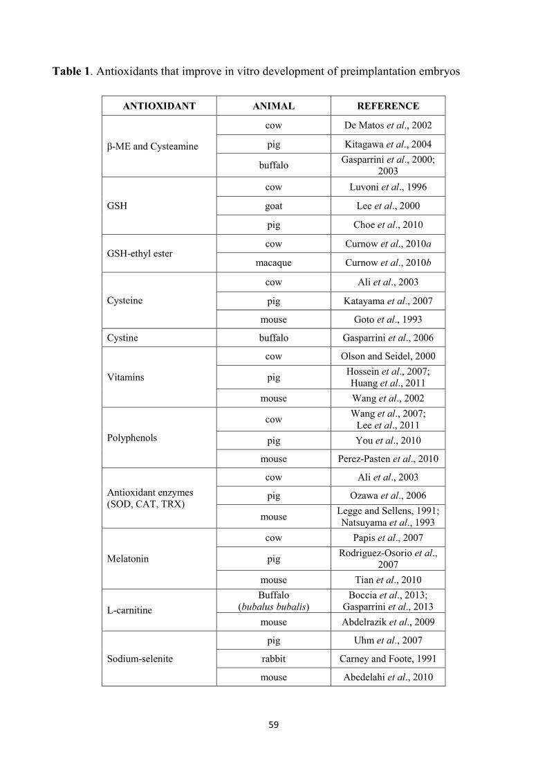
59
Table 1. Antioxidants that improve in vitro development of preimplantation embryos
ANTIOXIDANT ANIMAL REFERENCE
β-ME and Cysteamine
cow De Matos et al., 2002
pig Kitagawa et al., 2004
buffalo Gasparrini et al., 2000;
2003
GSH
cow Luvoni et al., 1996
goat Lee et al., 2000
pig Choe et al., 2010
GSH-ethyl ester cow Curnow et al., 2010a
macaque Curnow et al., 2010b
Cysteine
cow Ali et al., 2003
pig Katayama et al., 2007
mouse Goto et al., 1993
Cystine buffalo Gasparrini et al., 2006
Vitamins
cow Olson and Seidel, 2000
pig Hossein et al., 2007; Huang et al., 2011
mouse Wang et al., 2002
Polyphenols
cow Wang et al., 2007;
Lee et al., 2011
pig You et al., 2010
mouse Perez-Pasten et al., 2010
Antioxidant enzymes (SOD, CAT, TRX)
cow Ali et al., 2003
pig Ozawa et al., 2006
mouse Legge and Sellens, 1991; Natsuyama et al., 1993
Melatonin
cow Papis et al., 2007
pig Rodriguez-Osorio et al.,
2007
mouse Tian et al., 2010
L-carnitine
Buffalo (bubalus bubalis)
Boccia et al., 2013; Gasparrini et al., 2013
mouse Abdelrazik et al., 2009
Sodium-selenite
pig Uhm et al., 2007
rabbit Carney and Foote, 1991
mouse Abedelahi et al., 2010

60
2.2 AIM OF THE WORK
As previously described, to improve the development and quality of pre-implantation
bovine embryos, different antioxidant compounds have been tested during in vitro embryo
culture, in the attempt to reduce ROS generation and subsequent oxidative stress conditions.
Nevertheless, despite the improvements achieved, efforts are still needed to identify the
most effective supplements to improve embryo quality, that plays a critical role in
determining both cryotolerance and pregnancy maintenance after ET.
Therefore, the aim of this thesis was to investigate whether supplementation of bovine
culture medium with different antioxidant compounds found in several vegetal sources, such
as Resveratrol (Experiment 1), L-Ergothioneine (Experiment 2) and Crocetin (Experiment
3), improves in vitro blastocyst development, viability and quality. In each experiment the
latter parameter was assessed by different approaches, such as the timing of development to
specific morphological stages, resistance to cryopreservation, total cells number, ratio of
ICM:TE cells and apoptosis index. The rationale for the choice of the natural antioxidants
that we tested is reported separately in a brief introduction of each experiment.

61
CHAPTER 3: Experiment 1
Effect of resveratrol supplementation during culture on the quality and cryotolerance of bovine in vitro produced embryos
Resveratrol (3,4’,5-trihydroxystilbene) is a phytoalexin (Figure 11) identified in more than
70 plant species including grapes, plums, and peanuts (Baur and Sinclair, 2006). It is also an
important antioxidant polyphenolic compound, which contributes to red wine’s beneficial
effects on the prevention of human cardiovascular disease. Recently, the interest in
resveratrol has increased exponentially, following the major findings that this molecule has
positive effects on cancer chemoprevention, cardioprotection, inflammatory processes and
several aspects of metabolism, leading to increased lifespan of various organisms, from
yeasts to vertebrates (Pirola and Fröjdö, 2008). Several studies have been conducted to
identify the biologic functions and activities of resveratrol for mammalian reproduction
(Kwak et al., 2012; Huang et al., 2013). A positive effect of resveratrol on in vitro
embryonic development was demonstrated in swine, as indicated by enhanced blastocyst
formation and improved embryo quality (Lee et al., 2010). Recently, Wang et al. (2014)
demonstrated that the addition of resveratrol to the in vitro maturation medium increases
progesterone secretion and decreases estradiol secretion by cumulus cells, promoting
cumulus expansion and polar body formation in bovine oocytes and improves blastocyst
yield. Therefore, the aim of this work was to evaluate whether supplementation of culture
medium with resveratrol improves in vitro blastocyst development and embryo quality in
cattle, the latter assessed both in terms of cryotolerance and allocation of blastocyst cells
into the inner cell mass (ICM) and trophectoderm (TE) lineages.
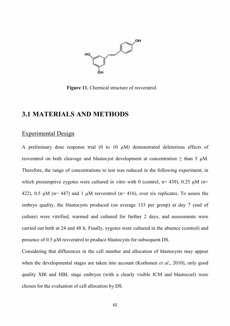
62
Figure 11. Chemical structure of resveratrol.
3.1 MATERIALS AND METHODS
Experimental Design
A preliminary dose response trial (0 to 10 µM) demonstrated deleterious effects of
resveratrol on both cleavage and blastocyst development at concentration ≥ than 5 µM.
Therefore, the range of concentrations to test was reduced in the following experiment, in
which presumptive zygotes were cultured in vitro with 0 (control, n= 439), 0.25 µM (n=
422), 0.5 µM (n= 447) and 1 µM resveratrol (n= 416), over six replicates. To assess the
embryo quality, the blastocysts produced (on average 153 per group) at day 7 (end of
culture) were vitrified, warmed and cultured for further 2 days, and assessments were
carried out both at 24 and 48 h. Finally, zygotes were cultured in the absence (control) and
presence of 0.5 µM resveratrol to produce blastocysts for subsequent DS.
Considering that differences in the cell number and allocation of blastocysts may appear
when the developmental stages are taken into account (Korhonen et al., 2010), only good
quality XBl and HBL stage embryos (with a clearly visible ICM and blastocoel) were
chosen for the evaluation of cell allocation by DS.

63
Reagents and media
Unless stated otherwise, all chemicals were obtained from Sigma-Aldrich Chemical
(Milano, Italy).
The oocyte aspiration medium was TCM 199 supplemented with 25 mM Hepes, 2 mM
sodium bicarbonate, 2 mM sodium pyruvate, 1 mM L-glutamine, 10 µL/mL amphotericin B
(H199) supplemented with 2% bovine serum (BS) and 95.6 SI/mL heparin. The IVM
medium was TCM 199 supplemented with 15% bovine serum (BS), 0.5 µg/mL FSH, 5
µg/mL LH, 0.8 mM L-glutamine and 50 µg/mL gentamycin. The IVF medium was
Tyrode’s modified medium (Parrish et al., 1986) without glucose and bovine serum albumin
(BSA), supplemented with 5.3 SI/mL heparin, 30 µM penicillamine, 15 µM hypotaurine, 1
µM epinephrine and 1% of BS. The IVC medium consisted of Synthetic Oviduct Fluid
(SOF) medium (Tervit et al., 1972), with 30 µL/mL essential amino acids, 10 µL/mL non-
essential amino acids and 5 % BS. Resveratrol was dissolved in dimethyl sulfoxide (DMSO)
to make a 1000X stock (1 mM) that was kept frozen until day of use, when the lower stock
concentrations were obtained by dilutions. Then the relative stock solutions were diluted 1:
1000 in SOF, to reduce to the minimum the amount of DMSO during culture. Likewise, in
the control group DMSO (1:1000) was added.
The base medium for the vitrification and warming solutions was H199 with 20% fetal calf
serum (FCS). The equilibration medium (VS1) consisted of 7.5% DMSO and 7.5% ethylene
glycol (EG) in base medium and the vitrification solution (VS2) consisted of 16.5% DMSO
and 16.5% EG with 0.5 M sucrose in the base medium. The warming solutions consisted in
0.25 M and 0.15 M sucrose in the base medium.

64
In vitro embryo production
Abattoir-derived oocytes were matured and fertilized in vitro according to our standard
procedure (Rubessa et al., 2011). Briefly, bovine ovaries were recovered from a local
abattoir and transported to the laboratory, in physiological saline at 30 to 35 °C. Cumulus-
oocyte complexes (COCs) were aspirated from follicles of 2 to 8 mm in diameter and only
those with uniform cytoplasm and multilayered cumulus cells were selected, washed twice
in the aspiration medium and once in the IVM medium. Groups of 25 COCs were matured
in 400 µL of IVM medium, covered with mineral oil, in four well plates (NuncTM,
Roskilde, Denmark), for 22 h at 39 °C, and 5% CO2 in air. In vitro matured COCs were
washed and transferred, 25 per well, into 300 µL of IVF medium covered with mineral oil.
Frozen sperm from a bull previously tested for IVF were thawed at 37 °C for 40 s and
selected by centrifugation (25 min at 300 X g) on a Percoll discontinuous gradient (45 and
80%). The pellet was reconstituted into 2 mL of IVF medium and centrifuged twice, at 160
and 108 X g for 10 min. The pellet was diluted with IVF medium and added in the
fertilization wells at the concentration of 1 x 10⁶ sperm/mL. Gametes were co-incubated for
20 h at 39 °C, in 5% CO2 in air, after which presumptive zygotes were vortexed for 2 min to
remove cumulus cells in Hepes-TCM with 5% BS, washed twice in the same medium and
randomly distributed (30 to 50 per well, into 400 µL of IVC medium) into different culture
groups differing on the basis of resveratrol concentrations. Zygotes were incubated in a
humidified mixture of 5% CO2, 7% O2 and 88% N2 in air at a temperature of 39 °C. On Day
7 cleavage and blastocyst rates were recorded. The embryos obtained by the end of culture
(Day 7) were scored for quality on the basis of morphological criteria (Robertson and
Nelson, 2010) and the percentage of transferable embryos, i.e. Grade 1 and 2 tight morulae
65
and blastocysts (TMBL), and that of Grade 1 and 2 blastocysts (BL) were recorded.
Furthermore, the stage of development was assessed and embryos were categorized as slow
developing embryos, i.e. tight morulae (TM), early blastocysts (eBl), blastocysts (Bl), and
as fast developing embryos, i.e. those that reached the stages of expanded blastocysts (XBl)
and hatched blastocysts (HBl) at the end of culture.
Vitrification and Warming
On Day 7 the blastocysts obtained (from eBl to HBl) were vitrified by the Cryotop method,
previously described (Kuwayama and Kato, 2000). All equilibration and dilution steps were
carried out at 37 °C. Each blastocyst was exposed to VS1 in 200 µL droplet for 3 min, then
transferred into a 20 µL droplet of VS2 for 20 to 25 s, and loaded with a glass capillary onto
the top of the film strip of each cryotop in 0.1 µL volume. After loading, almost all the
solution was removed and the Cryotop was quickly immersed into liquid nitrogen, protected
with the cap and stored under liquid nitrogen. For warming, after removal of the cap, the
Cryotop strip was immersed directly into 1 mL of the 0.25 M sucrose solution for 1 min and
the embryo was transferred into 200 µL droplet of 0.15 M sucrose solution for 5 min.
Warmed blastocysts were washed in H199 medium + 10% FCS and cultured for two days.
After 24 h and 48 h post-warming culture survival rates were evaluated with morphological
criteria, on the basis of the integrity of the embryo membrane and the ZP, and re-expansion
of the blastocoel. Furthermore, the percentages of embryos that resumed their development
and reached a more advanced developmental stage after culture (development rate), as well
as the hatching rates, were recorded.

66
Differential staining
Only expanded and hatched blastocysts produced in the presence (0.5 µM) or absence of
resveratrol on Day 7, were subjected to a DS protocol previously described (Thouas et al.,
2001) with slight modifications. Briefly, the ZP of viable expanding or expanded blastocysts
was removed with 0.2% pronase in Hepes-buffered SOF. After three washes in PBS with
0.1% PVP, blastocysts were first incubated in 500 µL of propidium iodide solution (PBS
with 1% Triton X-100 and 100 µg/mL propidium iodide) for 1 min. Blastocysts were then
immediately transferred into 500 µL of a fixative solution of 100% ethanol with 25 µg/mL
bisbenzimide (Hoechst 33342) for 5 min. The fixed and stained embryos were subsequently
transferred into glycerol and mounted onto a glass microscope slide in a drop of glycerol,
flattened with a coverslip to a level where all nuclei appeared at the same focal plane, and
visualized using a fluorescent microscope (Eclipse E-600 - Nikon) under ultraviolet light
with excitation at 460 to 560 nm. A digital image of each embryo (Figure 12) was taken
using NIS-Elements-F software and a high-resolution color digital camera (Nikon digital
sight DS-Fi 1C) and the numbers of TE (red) and ICM (blue) nuclei were counted.
Figure 12. Representative DS pictures of bovine embryos in different developmental stages examined by DAPI filter: XBL (a), Hatching BL (b) and HBL (c).
a b c

67
3.2 STATISTICAL ANALYSIS
Differences among groups in the percentages of blastocysts, as well as in the survival rates
after vitrification-warming, were analyzed by Chi square test using SPSS 16.0 statistical
software (SPSS Inc., 2007). DS data were analyzed by the Student’s t test.
3.3 RESULTS
Low concentrations of resveratrol (0.25 and 0.5 µM) during culture did not affect cleavage
rate; on the contrary, a decreased cleavage (P<0.05) was recorded at the highest
concentration (1 µM) compared to the control. Although the percentage of transferable
embryos, i.e. tight morulae/blastocysts, was improved (P<0.05) when embryos were
cultured with 0.25 and 0.5 µM resveratrol, both the percentage of Grade 1 and 2 blastocysts
and that of fast developing blastocysts were not affected (Table 2). The latter parameter was
reduced (P<0.05) in the culture group supplemented with 1 µM resveratrol compared to the
other resveratrol-treated groups.

68
Table 2. Percentages of cleavage, tight morulae-blastocysts (TMBL), Grade 1 and blastocysts (Grade 1,2 BL) and fast developing embryos (fast BL) under different resveratrol concentrations.
a,b Values within a column with different superscript indicate significant differences at P<0.05
Interestingly, treatment with resveratrol improved the cryotolerance of IVP bovine embryos,
as indicated by higher development and hatching rates following post-warming culture.
Indeed, despite similar survival rates, 0.5 µM resveratrol increased (P<0.05) development
rate after 24 h and both development and hatching rate (P<0.01) after 48 h culture
compared to the control (Table 3).
Resveratrol (µM)
n. COC
n. (%) Cleaved
n. (%) TMBL
n. (%) Gd 1,2 BL
n. (%) Fast BL
0 (control) 439 315 (71.8)a 194 (61.6)a 180 (57.1)a 74 (23.5)ab
0.25 422 281 (66.6)ab 187 (66.5)b 162 (57.7)a 78 (27.8)a
0.5 447 304 (68.0)ab 213 (70.1)b 180 (59.2)a 79 (26.0)a
1 416 266 (63.9)b 146 (54.9)a 124 (46.6)b 49 (18.4)b

69
Table 3. Survival, development and hatching rates after 24 and 48 h culture following vitrification-warming of blastocysts produced in the different culture systems.
Post-warming culture
Resveratrol (µM)
n. 24 h 48 h
n. (%) Survival
n. (%) Development
n. (%) Hatching
n. (%) Survival
n. (%) Development
n. (%) Hatching
0 (control) 175 163 (93.1) 55 (31.4)a 30 (17.1)ab 146 (83.4) 88 (50.3)Aa 50 (30.9)A
0.25 158 152 (96.2) 52 (32.9)a 21 (13.3)a 131 (82.9) 80 (50.6)Aa 58 (36.7)A
0.5 168 157 (93.5) 75 (44.6)b 42 (25.0)b 151 (89.9) 113 (67.3)Bb 99 (58.9)B
1 112 108 (96.4) 36 (32.1)a 13 (11.6)a 98 (87.5) 60 (53.6)a 40 (35.7)A
a,b Values within a column with different superscript indicate significant differences at P<0.05 *The post thaw culture did not contain resveratrol, the resveratrol concentration listed in this table refer to levels added during
embryo development.

70
No differences were recorded in the number of ICM, TE and total cells, as well as in the
ratio of ICM:total cells between blastocysts produced in the absence (control) or presence of
0.5 µM resveratrol (Table 4). The blastocysts stained were individually classified into four
groups according to the ICM:total cells ratio, as previously described (Koo et al., 2002) :
group I: <20% ; group II: 20-40%; group III: 40-60%; group IV: > 60%. In both
experimental groups the majority of the blastocysts fell into group II (78.3 and 72.3%,
respectively in the control and resveratrol groups). In the control the remaining blastocysts
were equally distributed into Group I and III (10.9%), whereas in the presence of resveratrol
the proportion of blastocysts classified into Group I and III were 4.3% and 23.4%,
respectively. No blastocysts fell into Group IV under both culture conditions.
Table 4. Allocation of cells (mean ± SE) in bovine blastocysts cultured in the absence (control) or presence of 0.5 µM resveratrol.
Group n.
blastocyst ICM TE Total cells
ICM:Total cells
Control 46 34.1 ± 1.6 88.1 ± 4.6 122.2 ±5.4 29.1 ± 1.2
Resveratrol 47 36.4 ± 1.4 85.3 ± 4.1 121.7 ± 4.6 31.3 ± 1.3

71
3.4 DISCUSSION
This study demonstrated that resveratrol supplementation during in vitro culture improves
the cryotolerance of in vitro produced bovine embryos. The preliminary dose response trial
highlighted a distinct dose-dependent effect of resveratrol, with significant decrease of both
cleavage and blastocyst rates at the concentration of 5 µM and no blastocyst production at
10 µM. This suggested to reduce the concentrations (0 to 1 µM) to test in the present
experiment. Despite an increase of the transferable embryos (TMBL) obtained with 0.25
and 0.5 µM resveratrol, no improvement was recorded in the blastocyst yields, as well as in
the percentage of fast developing blastocysts. This is in contrast with other studies carried
out in cattle and swine where resveratrol was added during IVM (Lee et al., 2010; Wang et
al., 2014). It is known, however, that the optimization of the IVM system leads to increased
embryo yields, whereas the improvement of culture conditions affects particularly embryo
quality (Lonergan et al., 2001). The beneficial effect was already evident after 24 h of post-
warming culture, when the development rate, i.e. the percentage of embryos reaching a
more advanced stage, significantly increased. Furthermore, a significant improvement of
both development and hatching rates was recorded after 48 h post-warming culture.
Interestingly, the latter parameter showed a 2-fold increase compared to the control; this is
important because the ability of an embryo to hatch after cryopreservation is a known
reliable indicator of its quality (Semple et al., 1995). In fact, hatching may be impaired in
cryopreserved embryos, due to the occurrence of zona hardening (Carroll et al., 1990;
Matson et al., 1997).
The beneficial effect on both the proportion of transferable embryos and the cryotolerance
was already lost at the concentration of 1 µM. A dose-dependent effect of resveratrol was

72
previously demonstrated in human cultured cells, with low concentrations promoting cell
division and high concentrations exerting anti-proliferation and pro-apoptosis effects
(Kuwajerwala et al., 2002; Szende et al., 2000). A dose-dependent effect was also recorded
in porcine parthenogenetic embryos, with increased blastocyst production at low
concentrations and toxic effects at high concentrations (Lee et al., 2010). In addition,
porcine parthenogenetic blastocysts produced in the presence of low levels of resveratrol
showed lower expression of Bcl-2 and Caspase-3, suggesting respectively a pro-
proliferative and anti-apoptotic effect. Although apoptosis is considered a normal process in
pre-implantation embryos to eliminate deviating cells, a high incidence of apoptotic cells is
correlated with abnormal morphology of the embryo (Hardy et al., 1989). In fact, the
percentage of apoptotic cells is significantly higher in the in vitro produced bovine
blastocysts than in their in vivo-developed counterparts (Gjørret et al., 2003). We speculate
that the decreased cleavage and blastocyst rates recorded in the presence of elevated
concentrations of resveratrol (from 1 to 10 µM) may be due to the induction of apoptosis,
although this was not assessed. However, the beneficial effect on embryo quality indicated
by the improved cryotolerance of the IVP embryos does not seem to be related to the pro-
proliferation activity demonstrated for resveratrol in other studies (Kuwajerwala et al.,
2002; Szende et al., 2000). In fact, the chronology of embryo development, i.e. the
percentage of embryos reaching a more advanced stage by the end of culture, was not
improved. Furthermore, neither the total blastocyst cell number nor the relative allocation
into the ICM and TE were affected by resveratrol treatment in this study. In addition, the
percentage of blastocysts falling into the different ICM: total cells categories was similar. In
both culture groups the highest proportion of blastocysts had an ICM:total cells ratio in the
73
range from 20 to 40%, that is considered normal for IVP embryos (Koo et al., 2002; Van
Soom et al., 1997). It is known that the perfect allocation of the blastocyst cells into ICM
and TE is critical, as both cell lineages are vital for embryonic and fetal survival (Hardy et
al., 1989; Van Soom et al., 1997).
It is therefore likely that the beneficial effect of resveratrol on IVP blastocyst cryotolerance
is related to its known antioxidant activity. Increased oxidative stress, resulting in
pathological levels of ROS, is indeed a major factor impairing mammalian in vitro embryo
development (Kitagawa et al., 2004; Nasr-Esfahani et al., 1990). Following
cryopreservation, oocytes and embryos become more sensitive to the oxidative stress,
resulting in lipid peroxidation, membrane injury and structural destruction (Tatone et al.,
2010). It was reported that glutathione (GSH) content sharply decreases after
cryopreservation in bovine (George et al., 2008) and porcine IVF embryos (Somfai et al.,
2007a), confirming that oxidative stress occurs during the freezing-thawing process. One of
the effects of resveratrol is to maintain the redox state of cells and protect them against the
harmful effects of oxidative injuries. Resveratrol supplementation during IVM was
demonstrated to improve oocyte developmental competence by increasing GSH and
decreasing ROS levels in swine and cattle oocytes (Kwak et al., 2012; Lee et al., 2010;
Wang et al., 2014). Therefore, it is likely that the improved cryotolerance of IVP bovine
embryos here reported is due to the known scavenging properties of resveratrol (Mahal et
al., 2005). However, the molecular mechanism by which resveratrol exerts its effect on pre-
implantation embryos is still not entirely clear. It was demonstrated that the structure of
resveratrol is similar to that of the synthetic estrogen diethylstilbestrol (Lobo, 1995), that
was shown to bind to human estrogen receptors in binding assay (Gehm et al., 1997).
74
It was also reported that resveratrol during IVM increases progesterone secretion and
decreases estrogen secretion by bovine cumulus cells (Wang et al., 2014). These findings
suggest a relationship between resveratrol and steroids, supporting a hypothesis whereby
low concentrations of resveratrol may act as an agonist for specific steroid hormone
receptors, during pre-implantation embryo development in vitro.
In conclusion, these results demonstrated that supplementation of culture medium with low
levels of resveratrol improves the quality, and hence the resistance to cryopreservation, of
IVP bovine embryos, without affecting blastocyst rates and allocation of cells into ICM and
TE lineages. Further studies are needed to comprehend the mechanism underlying the
beneficial effect and to assess the effect of resveratrol on development to term after transfer
of cryopreserved embryos.

75
CHAPTER 4: Experiment 2
Effect of L-Ergothioneine supplementation during culture on the quality and cryotolerance of bovine in vitro produced embryos
L-Ergothioneine (LE) is a powerful scavenger of OH and an inhibitor of iron or copper ion-
dependent generation of OH from H2O2 (Akanmu et al., 1991). L-Ergothioneine (2-
mercaptohistidine trimethylbetaine) is a naturally occurring amino acid that contains an
imidazole-2-thione moiety (Figure 13). The thione group exists in a tautomeric thiol-thione
equilibrium, with the thione being the predominant form at physiological pH, which
distinguishes ergothioneine from other biological thiol compounds.
Figure 13. Chemical structure of L-Ergothioneine.
Ergothioneine is synthesized by some bacteria and fungi, but not by animals (Hartman,
1990; Melville et al., 1955). Humans acquire ergothioneine from their diet, including
mushrooms, oats, corn, and meat (Ey et al., 2007). Ergothioneine is found in brain,
erythrocytes, liver, kidney, heart, seminal fluid, and ocular tissues of several species
(Mitsuyama and May, 1999). However, the biological functions of ergothioneine remain
incompletely understood. Ergothioneine has been shown to exert radioprotective effects

76
(Van den Broeke and Beyersbergen van Henegouwen, 1993); scavenge singlet oxygen,
hydroxyl radicals, hypochlorous acid, and peroxyl radicals (Asmus et al., 1996; Aruoma et
al., 1997); inhibit peroxynitrite-dependent nitration of proteins and DNA (Aruoma et al.,
1999); protect retinal neurons from N-methyl- D-aspartate (NMDA)-induced excitotoxicity
in vivo (Moncaster et al., 2002); and protect against diabetic embryopathy in pregnant rats
(Guijarro et al., 2002).
A previous study demonstrated that LE inhibits cell death caused by H2O2 and DNA
oxidation by peroxynitrite in a human neuronal hybridoma cell line in culture and it was
suggested that LE could have a role as an antioxidant, in vivo non-toxic thiol compound and
obtain the oxidative stability in pharmacological preparations (Aruoma et al., 1999).
Recently, it has been shown that antioxidant activity of LE is the highest, and it eliminates
the most active free radicals compared to some classic well-known antioxidants such as
glutathione, uric acid and trolox (Franzoni et al., 2006).
It has been known that the constant maintenance of the pro-oxidant balance in embryos is
difficult and complicated during the addition of antioxidants (Guérin et al., 2001); thus,
further studies are needed to limit and minimize the oxidative stress during embryo culture.
In a recent work in sheep, the effects of LE and L-ascorbic acid (LAA) on the maturation
rate of immature oocytes and embryonic development of in vitro matured oocytes were
compared. The results showed that, the addition of L-ergothioneine into IVM, IVF and IVC
medium has a beneficial effect on in vitro maturation of oocytes and embryonic
development, especially from cleavage to morulae stages compared to LAA and Control
groups (Öztürkler et al., 2010). To our current knowledge, there is no study available in
literature on the effects of an antioxidant with strong and unique features, such as LE in
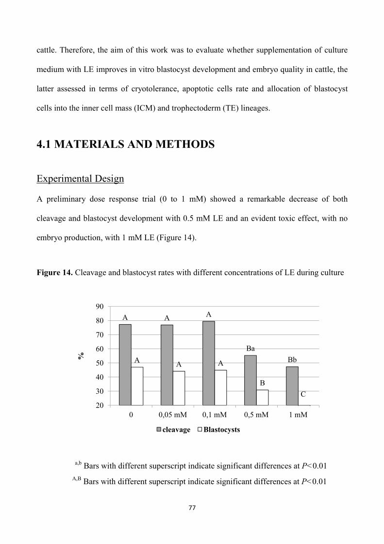
77
cattle. Therefore, the aim of this work was to evaluate whether supplementation of culture
medium with LE improves in vitro blastocyst development and embryo quality in cattle, the
latter assessed in terms of cryotolerance, apoptotic cells rate and allocation of blastocyst
cells into the inner cell mass (ICM) and trophectoderm (TE) lineages.
4.1 MATERIALS AND METHODS
Experimental Design A preliminary dose response trial (0 to 1 mM) showed a remarkable decrease of both
cleavage and blastocyst development with 0.5 mM LE and an evident toxic effect, with no
embryo production, with 1 mM LE (Figure 14).
Figure 14. Cleavage and blastocyst rates with different concentrations of LE during culture
a,b Bars with different superscript indicate significant differences at P<0.01
A,B Bars with different superscript indicate significant differences at P<0.01
A A A
Ba
BbAA A
B
20
30
40
50
60
70
80
90
0 0,05 mM 0,1 mM 0,5 mM 1 mM
%
cleavage Blastocysts
C
78
Therefore, the range of concentrations to test was reduced in Experiment 2.1, in which
presumptive zygotes were cultured in vitro with 0 (control, n= 282), 0.05 mM (n= 286) and
0.1 mM LE (n= 285), over five replicates. The blastocysts produced (on average 94 per
group) at day 7 (end of culture) were vitrified, warmed and cultured for further 2 days, and
assessments were carried out both at 24 and 48 h.
In Experiment 2.2, zygotes were cultured in the absence (control, n= 412) and presence of
0.1 mM LE (n= 411), over four replicates and the blastocysts produced on day 8 underwent
both DS (n= 47 for both groups) and TUNEL staining to assess apoptosis (n= 65 and 64,
respectively).
Considering that differences in the cell number and allocation of blastocysts may appear
when the developmental stages are taken into account (Korhonen et al., 2010), only good
quality XBl and HBL stage embryos (with a clearly visible ICM and blastocoel) were
chosen for the evaluation of cell allocation by DS. In addition, for TUNEL staining BL,
XBL and HBL were used.
Reagents and media
Unless stated otherwise, all chemicals were obtained from Sigma-Aldrich Chemical
(Milano, Italy). The In Situ Cell Death Detection Kit (fluorescein) and the RNA-free DNase
was obtained from Roche Diagnostics Corporation (Indianapolis, IN, USA).
The aspiration medium was TCM 199 supplemented with 25 mM Hepes, 2 mM sodium
bicarbonate, 2 mM sodium pyruvate, 1 mM L-glutamine, 10 µL/mL amphotericin B (H199)
supplemented with 2% bovine serum (BS) and 95.6 SI/mL heparin. The IVM medium was
TCM 199 supplemented with 15% bovine serum (BS), 0.5 µg/mL FSH, 5 µg/mL LH, 0.8

79
mM L-glutamine and 50 µg/mL gentamycin. The IVF medium was Tyrode’s modified
medium (Parrish et al., 1986) without glucose and bovine serum albumin (BSA),
supplemented with 5.3 SI/mL heparin, 30 µM penicillamine, 15 µM hypotaurine, 1 µM
epinephrine and 1% of BS. The IVC medium consisted of Synthetic Oviduct Fluid (SOF)
medium (Tervit et al., 1972), with 30 µL/mL essential amino acids, 10 µL/mL non-essential
amino acids and 5 % BS.
The base medium for the vitrification and warming solutions was H199 with 20% fetal calf
serum (FCS). The equilibration medium (VS1) consisted of 7.5% DMSO and 7.5% ethylene
glycol (EG) in base medium and the vitrification solution (VS2) consisted of 16.5% DMSO
and 16.5% EG with 0.5 M sucrose in the base medium. The warming solutions consisted in
0.25 M and 0.15 M sucrose in the base medium.
In vitro embryo production
Abattoir-derived oocytes were matured and fertilized in vitro according to our standard
procedure (Rubessa et al., 2011). Briefly, bovine ovaries were recovered from a local
abattoir and transported to the laboratory, in physiological saline at 30 to 35 °C. Cumulus-
oocyte complexes (COCs) were aspirated from follicles of 2 to 8 mm in diameter and only
those with uniform cytoplasm and multilayered cumulus cells were selected, washed twice
in the aspiration medium and once in the IVM medium. Groups of 25 COCs were matured
in 400 µL of IVM medium, covered with mineral oil, in four well plates (NuncTM,
Roskilde, Denmark), for 22 h at 39 °C, and 5% CO2 in air. In vitro matured COCs were
washed and transferred, 25 per well, into 300 µL of IVF medium covered with mineral oil.
Frozen sperm from a bull previously tested for IVF were thawed at 37 °C for 40 s and

80
selected by centrifugation (25 min at 300 X g) on a Percoll discontinuous gradient (45 and
80%). The pellet was reconstituted into 2 mL of IVF medium and centrifuged twice, at 160
and 108 X g for 10 min. The pellet was diluted with IVF medium and added in the
fertilization wells at the concentration of 1 x 10⁶ sperm/mL. Gametes were co-incubated for
20 h at 39 °C, in 5% CO2 in air, after which presumptive zygotes were vortexed for 2 min to
remove cumulus cells in Hepes-TCM with 5% BS, washed twice in the same medium and
randomly distributed (30 to 50 per well, into 400 µL of IVC medium) into different culture
groups differing on the basis of LE concentrations. Zygotes were incubated in a humidified
mixture of 5% CO2, 7% O2 and 88% N2 in air at a temperature of 39 °C. On days 7 and 8,
Experiment 2.1 and 2.2 respectively, cleavage and blastocyst rates were recorded. The
embryos obtained by the end of culture were scored for quality on the basis of
morphological criteria (Robertson and Nelson, 2010) and the percentage of transferable
embryos, i.e. Grade 1 and 2 tight morulae and blastocysts (TMBL), and that of Grade 1 and
2 blastocysts (BL) were recorded. Furthermore, the stage of development was assessed and
embryos were categorized as slow developing embryos, i.e. tight morulae (TM), early
blastocysts (eBl), blastocysts (Bl), and as fast developing embryos, i.e. those that reached
the stages of expanded blastocysts (XBl) and hatched blastocysts (HBl) at the end of
culture.
Experiment 2.1: Vitrification and Warming
On Day 7 the blastocysts obtained (from eBl to HBl) were vitrified by the Cryotop method,
previously described (Kuwayama and Kato, 2000). All equilibration and dilution steps were
carried out at 37 °C. Each blastocyst was exposed to VS1 in 200 µL droplet for 3 min, then
81
transferred into a 20 µL droplet of VS2 for 20 to 25 s, and loaded with a glass capillary onto
the top of the film strip of each cryotop in 0.1 µL volume. After loading, almost all the
solution was removed and the Cryotop was quickly immersed into liquid nitrogen, protected
with the cap and stored under liquid nitrogen. For warming, after removal of the cap, the
Cryotop strip was immersed directly into 1 mL of the 0.25 M sucrose solution for 1 min and
the embryo was transferred into 200 µL droplet of 0.15 M sucrose solution for 5 min.
Warmed blastocysts were washed in H199 medium + 10% FCS and cultured for two days.
After 24 h and 48 h post-warming culture survival rates were evaluated with morphological
criteria, on the basis of the integrity of the embryo membrane and the ZP, and re-expansion
of the blastocoel. Furthermore, the percentages of embryos that resumed their development
and reached a more advanced developmental stage after culture (development rate), as well
as the hatching rates, were recorded.
Experiment 2.2: Differential staining
Blastocysts (only XBL and HBL) produced in the presence (0.1 mM) or absence of LE on
Day 8, were subjected to a DS protocol previously described (Thouas et al., 2001) with
slight modifications. Briefly, the ZP of viable expanding or expanded blastocysts was
removed with 0.2% pronase in Hepes-buffered SOF. After three washes in PBS with 0.1%
PVP, blastocysts were first incubated in 500 µL of propidium iodide solution (PBS with 1%
Triton X-100 and 100 µg/mL propidium iodide) for 1 min. Blastocysts were then
immediately transferred into 500 µL of a fixative solution of 100% ethanol with 25 µg/mL
bisbenzimide (Hoechst 33342) for 5 min. The fixed and stained embryos were subsequently
transferred into glycerol and mounted onto a glass microscope slide in a drop of glycerol,

82
flattened with a coverslip to a level where all nuclei appeared at the same focal plane, and
visualized using a fluorescent microscope (Eclipse E-600; Nikon) under ultraviolet light
with excitation at 460 to 560 nm. A digital image of each embryo was taken using NIS-
Elements-F software and a high-resolution color digital camera (Nikon digital sight DS-Fi
1C) and the numbers of TE (red) and ICM (blue) nuclei were counted.
Experiment 2.2: TUNEL staining
The blastocysts (BL, XBl and HBL), produced in the presence (0.1 mM) or absence of LE
on Day 8 were subjected to a TUNEL staining protocol previously described (Paula-Lopes
and Hansen, 2002). The TUNEL assay was used to detect DNA fragmentation associated
with late stages of the apoptotic cascade. The enzyme terminal deoxynucleotidyl transferase
is a DNA polymerase that catalyzes the transfer of a fluorescein isothiocyanate-conjugated
dUTP nucleotide to a free 3’ hydroxyl group characteristic of DNA strand breaks. Embryos
were removed from SOF culture medium and washed three times in drops of PBS
containing 1 mg/mL PVP (PBS-PVP) by transferring the embryos from drop to drop.
Embryos were fixed in a 50 µL drop of 4% (w/v) paraformaldehyde in PBS for 15 min at
room temperature, washed twice in PBS-PVP, and stored in 600 µL of PBS-PVP at 4°C
until the time of assay. All steps of the TUNEL assay were conducted using microdrops in a
humidified box.
On the day of the TUNEL assay, embryos were transferred to a 50 µL drop of PBS-PVP and
then permeabilized in a drop of 0.5% (v/v) Triton X-100 in PBS, containing 0.1% (w/v)
sodium citrate for 30 min at room temperature. Controls for the TUNEL assay were
incubated in 50 µL of RQ1-RNA free DNase (50 U/mL) at 37°C in the dark for 10 min.

83
Positive controls and treated embryos were washed in PBS-PVP and incubated with 25 µL
of TUNEL reaction mixture (containing fluorescein isothiocyanate-conjugated dUTP and
the enzyme terminal deoxynucleotidyl transferase as prepared by and following the
guidelines of the manufacturer) for 1 h at 37°C in the dark. Negative controls were
incubated in the absence of terminal deoxynucleotidyl transferase.
Embryos were then washed three times in PBS-PVP and incubated in a 25 µL drop of
Hoechst 33342 (1 µg/mL) for 15 min in the dark. Embryos were washed three times in PBS-
PVP to remove excess Hoechst 33342, mounted on 10% (w/v) poly-L-lysine coated slides
using 3- to 4 µL drops of glycerol, and coverslips were placed on the slides. Labeling of
TUNEL and Hoechst 33342 nuclei was observed using a fluorescent microscope (Eclipse E-
600; Nikon). Each embryo was analyzed for total cell number (blue nuclei) and TUNEL-
positive blastomeres (green nuclei) with DAPI and FITC filters, respectively, using a 20X
and 40X objectives. Digital images were acquired (Figures 15, 16, 17 and 18) using NIS-
Elements-F software and a high-resolution color digital camera (Nikon digital sight DS-Fi
1C).
Figure 15. Positive control Figure 16. Negative control

84
Figure 17. Representative pictures showing total cells staining examined by DAPI filter (a) and TUNEL labeling examined by FITC filter (b) of the same HBL cultured in the control medium
Figure 18. Representative pictures showing total cells staining examined by DAPI filter (a) and TUNEL labeling examined by FITC filter (b) of the same HBL cultured in the medium enriched with 0.1 mM LE
4.2 STATISTICAL ANALYSIS
Differences among groups in the percentages of blastocysts, as well as in the survival rates
after vitrification-warming, were analyzed by Chi square test using SPSS 16.0 statistical
software (SPSS Inc., 2007). DS and TUNEL staining data were analyzed by the Student’s t
test.
a b
a b
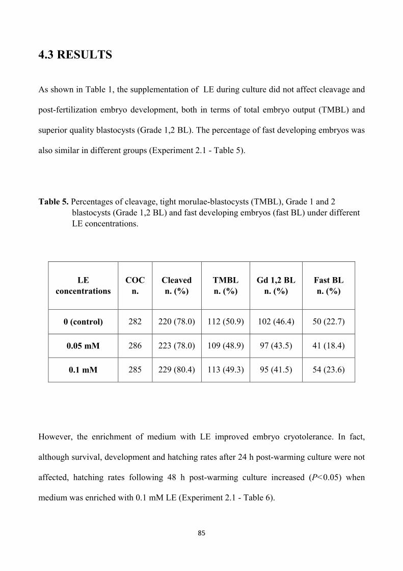
85
4.3 RESULTS
As shown in Table 1, the supplementation of LE during culture did not affect cleavage and
post-fertilization embryo development, both in terms of total embryo output (TMBL) and
superior quality blastocysts (Grade 1,2 BL). The percentage of fast developing embryos was
also similar in different groups (Experiment 2.1 - Table 5).
Table 5. Percentages of cleavage, tight morulae-blastocysts (TMBL), Grade 1 and 2 blastocysts (Grade 1,2 BL) and fast developing embryos (fast BL) under different LE concentrations.
However, the enrichment of medium with LE improved embryo cryotolerance. In fact,
although survival, development and hatching rates after 24 h post-warming culture were not
affected, hatching rates following 48 h post-warming culture increased (P<0.05) when
medium was enriched with 0.1 mM LE (Experiment 2.1 - Table 6).
LE concentrations
COC n.
Cleaved n. (%)
TMBL n. (%)
Gd 1,2 BL n. (%)
Fast BL n. (%)
0 (control) 282 220 (78.0) 112 (50.9) 102 (46.4) 50 (22.7)
0.05 mM 286 223 (78.0) 109 (48.9) 97 (43.5) 41 (18.4)
0.1 mM 285 229 (80.4) 113 (49.3) 95 (41.5) 54 (23.6)
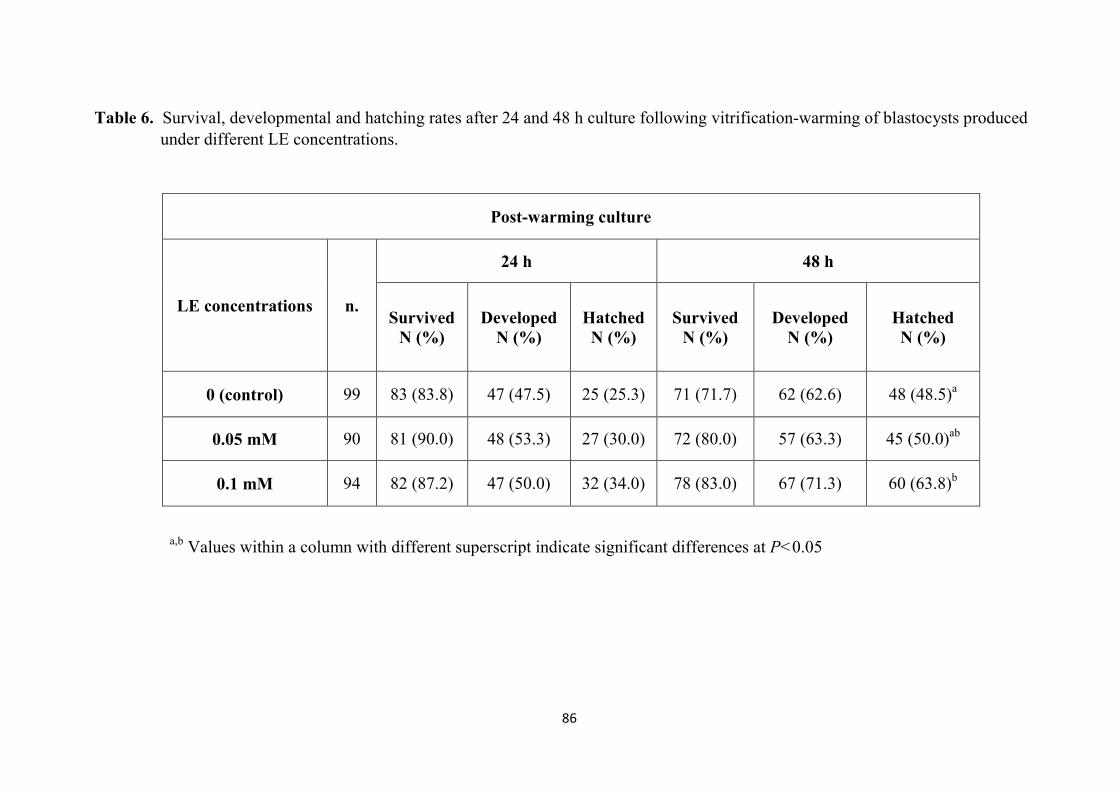
86
Table 6. Survival, developmental and hatching rates after 24 and 48 h culture following vitrification-warming of blastocysts produced under different LE concentrations.
Post-warming culture
LE concentrations n.
24 h 48 h
Survived N (%)
Developed N (%)
Hatched N (%)
Survived N (%)
Developed N (%)
Hatched N (%)
0 (control) 99 83 (83.8) 47 (47.5) 25 (25.3) 71 (71.7) 62 (62.6) 48 (48.5)a
0.05 mM 90 81 (90.0) 48 (53.3) 27 (30.0) 72 (80.0) 57 (63.3) 45 (50.0)ab
0.1 mM 94 82 (87.2) 47 (50.0) 32 (34.0) 78 (83.0) 67 (71.3) 60 (63.8)b
a,b Values within a column with different superscript indicate significant differences at P<0.05

87
Furthermore, the average number of ICM cells, as well as the ratio of ICM:total cells of
blastocysts were higher (P<0.01 and P<0.05, respectively) in the 0.1 mM LE group than in
the control, whereas no differences were recorded in the number of TE and total cells
(Experiment 2.2 - Table 7).
Table 7. Allocation of cells (Mean ± SE) in bovine blastocysts cultured in the absence (control) or presence of 0.1 mM LE.
a,b Values within a column with different superscript indicate significant differences at P<0.05
A,B Values within a column with different superscript indicate significant differences at P<0.01
The blastocysts stained were individually classified into four groups according to the
ICM:total cells ratio, as previously described (Koo et al., 2002): group I: <20%; group II:
20-40%; group III: 40-60%; group IV: > 60%. As shown in Figure 19 (Experiment 2.2), in
the LE group a higher (P<0.05) percentage of the blastocysts fell into group II compared to
the control (85.1 and 66.0 %, respectively). In contrast, the percentage of blastocysts falling
in Group I was lower (P<0.05) in the LE group than in the control (14.9 and 34.0 %,
respectively). No blastocysts fell into Group III and IV under both culture conditions.
Group n.
blastocyst ICM TE Total
ICM: total cells %
Control 47 37.1 ± 1.2A 126.9 ± 3.5 164.0 ± 4.0 22.8 ± 0.7a
0.1 mM LE 47 42.4 ± 1.6B 129.1 ± 3.5 171.6 ± 4.5 24.7 ± 0.7b

88
Figure 19. Percentage of blastocysts with different proportion of ICM:total cells ratio in the presence or absence of LE.
a,b Bars with different superscript indicate significant differences at P<0.05
Both the average number and the proportion of apoptotic cells in blastocysts were reduced
(P<0.01) in the presence of 0.1 mM LE (Experiment 2.2 - Table 8), whereas the total cell
number was unaffected. However, when these parameters were analyzed in relation to the
stage of development, the total cell number was higher (P<0.01) in the LE group but only in
case of HBL (Figure 20). Within each developmental stage (BL, XBl and HBL) the
incidence of apoptotic cells out of the total decreased (P<0.01) in the enriched group
(Figure 21).
a
a
b
b
0
10
20
30
40
50
60
70
80
90
< 20% 20-40%
N. (
%)
of b
last
ocys
ts
Ratio of ICM: total cells numbers
control
LE
n=16n=31
n=7n=40

89
Table 8. Cell allocation on day 8 bovine blastocysts by TUNEL staining in the absence
(control) or presence of 0.1 mM LE.
Groups n. Total cells Apoptotic
cells % apoptotic
cells Apoptotic/total
cells ratio
Mean ± SE Mean ± SE Mean ± SE Mean ± SE
Control 65 126.6 ± 5.2 9.1 ± 0.3A 8.1 ± 0.5A 0.08 ± 0.006A
0.1 mM LE 64 133.5 ± 6.1 4.3 ± 0.2B 3.6 ± 0.3B 0.03 ± 0.005B
A,B Values within a column with different superscript indicate significant differences at P<0.01
Figure 20. Total cell numbers of day 8 blastocysts cultured with or without LE according to the developmental stage.
A,B Bars with different superscript indicate significant differences at P<0.01 * The number of BL, XBL and HBL stained were respectively 25, 19 and 21 in the control and 25, 20 and 19 in the LE group.
A
B
60
80
100
120
140
160
180
200
BL XBL HBL
Tot
al c
ells
nu
mb
er
control ergothioneine
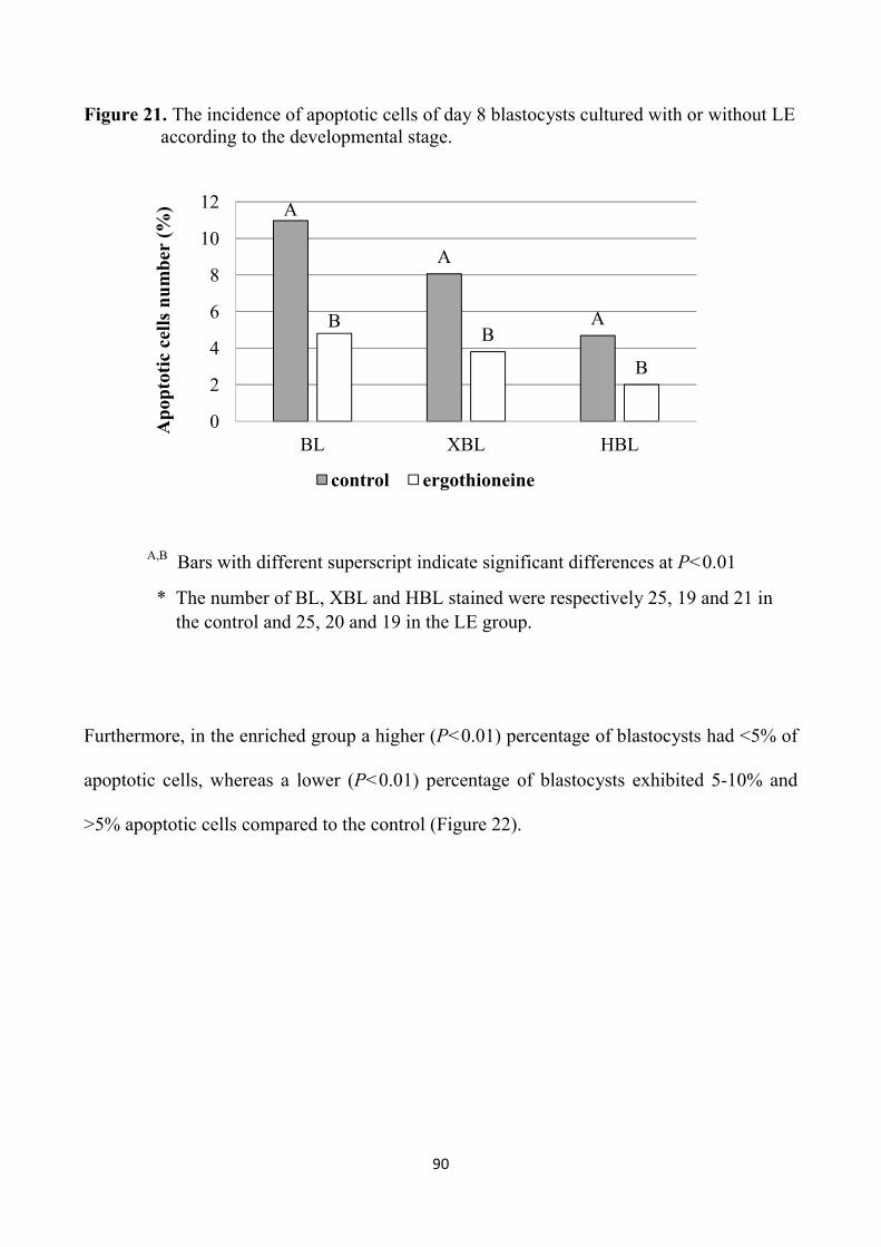
90
Figure 21. The incidence of apoptotic cells of day 8 blastocysts cultured with or without LE according to the developmental stage.
A,B Bars with different superscript indicate significant differences at P<0.01
* The number of BL, XBL and HBL stained were respectively 25, 19 and 21 in the control and 25, 20 and 19 in the LE group.
Furthermore, in the enriched group a higher (P<0.01) percentage of blastocysts had <5% of
apoptotic cells, whereas a lower (P<0.01) percentage of blastocysts exhibited 5-10% and
>5% apoptotic cells compared to the control (Figure 22).
A
A
ABB
B
0
2
4
6
8
10
12
BL XBL HBL
Ap
opto
tic
cell
s n
um
ber
(%
)
control ergothioneine

91
Figure 22. Percentage of blastocysts with low (<5%), average (5-10%) and high (>10%) levels of apoptosis in the control and LE groups.
A,B Bars with different superscript indicate significant differences at P<0.01
4.4 DISCUSSION
The results of the present study demonstrated that enrichment of culture medium with low
doses of the natural antioxidant LE, improves embryo quality, as indicated by increased
cryotolerance, reduced incidence of apoptotic cells and higher percentage of blastocysts
with the most physiological ICM:total cells ratio.
The preliminary dose response trial showed a negative effect on both cleavage and
blastocyst rates with 0.5 mM LE and an evident toxic effect, with further reduction of
cleavage and no blastocyst production at the concentration of 1 mM. This is in contrast with
A
A
A
B
B
B0
10
20
30
40
50
60
70
80
<5% 5-10% >10%
% o
f b
last
ocys
ts w
ith
dif
fere
nt
apop
toti
c ra
tio
control ergothioneine

92
previous tissue culture studies reporting that ergothioneine was not toxic to cells with an
LD50 of 50 mM (OXIS Technical Document, 1997). Also, the concentration that was
embryotoxic in the present study (1 mM) is close to the concentration that was estimated to
be present in human and mammalian tissues (1-2 mM). It has been reported that
ergothioneine is present in millimolar concentrations in specific tissues such as erythrocytes,
kidney, seminal fluid and liver (Grigat et al., 2007). However, according to the OXIS
technical Document (1997) the circulating levels of ergothioneine are estimated to be in the
high micromolar range.
After the preliminary trial Experiment 2.1 was carried out with 0 (control), 0.5 and 0.1 mM
LE, and showed that the supplementation of culture medium with the antioxidant did not
affect cleavage rate and embryonic development. This finding is in disagreement with the
results recorded in the only work present in literature in the sheep, in which 10 mM LE
supplemented both during IVM and IVC increased maturation rates, cleavage and
development to morulae (Öztürkler el al., 2010). However, in that work both cleavage and
embryo development were very poor (on average respectively 26 and 4%) and it is known
that it is easier to detect a beneficial effect in a suboptimal system. What is more surprising
is that the concentration that was beneficial in the sheep is 10 times higher than the
concentration that was toxic on bovine embryos in the present study. Due to the paucity of
information available we can only speculate that this is related to species-specific
differences. The most important issue arising from the results of the study is the improved
embryo quality obtained in culture medium enriched with 0.1 mM LE. Indeed, we followed
the strategy to enrich IVC medium with a natural antioxidant just in order to improve
embryo quality, that is known to be mainly affected by culture environment (Lonergan et
93
al., 2001). Although embryo output was not affected, it was demonstrated that 0.1 mM LE
increased embryo cryotolerance that is a highly reliable marker of embryo quality (Imai et
al., 2002; Lonergan et al., 2003a; 2003b; Rizos et al., 2003). The beneficial effect was
evident after 48 h of post-warming culture, when a significant increase of hatching rate was
recorded. The increased hatching rate after culture of vitrified-warmed blastocysts is a
strong indicator of viability because cryopreservation results in zona hardening that
interferes with the process of hatching (Carroll et al., 1990; Matson et al., 1997). Our work
was the first to evaluate the effect of LE on embryo cryotolerance and hence we cannot
compare our results with others’; the only work available in literature, in fact, has been
limited to assess the effect of LE on ovine in vitro embryo development in quantitative
terms. However, previous studies demonstrated that supplementation with antioxidants
during culture improves embryo quality and cryotolerance (Hosseini et al., 2009; Takahashi,
2012). In the Experiment 1 of this thesis we also recorded a significant improvement of
cryotolerance when culture medium was enriched with resveratrol. The improved
cryotolerance of bovine embryos obtained with LE during culture is likely due to its high
antioxidant capacity, as oxidative stress occurs during the freezing-thawing process, leading
to lipid peroxidation, membrane injury and structural damage (Somfai et al., 2007b). This
may be due to the decrease of GSH levels following cryopreservation in bovine embryos
(George et al., 2008). Therefore, LE addition during culture increases the capability of
bovine embryos to hatch after cryopreservation, that is the most important parameter to
assess the embryo resistance to cryopreservation (Semple et al., 1995).
In addition to embryo cryotolerance, total cell count and particularly cell allocation to the
inner cell mass (ICM) and the trophectoderm (TE) lineages are commonly used as embryo
94
quality parameters. It is well documented that the perfect allocation of the blastocyst cells
into ICM and TE is critical for embryonic survival (Koo et al., 2002; Knijn et al., 2003) and
post-implantation development (Iwasaki et al., 1990), as both cell lineages are essential
(Hardy et al., 1989; Van Soom et al., 1997). In the present study blastocysts produced in the
medium enriched with LE showed an increase in the average number of ICM cells
compared to the control, whereas total cell count and TE cells were unaffected. This
resulted in a slightly higher ICM:total cell ratio in the enriched group. It is known that ICM
cells contribute to all embryonic tissues and part of the extraembryonic membranes,
whereas the TE cells mainly form the outer layer of placenta (Fouladi-Nashta, 2005). A well
defined ICM is known to be essential to support development after embryo transfer. In fact,
it has been reported that the number of ICM cells in IVP bovine blastocysts is lower than
that of in vivo produced blastocysts (Iwasaki et al., 1990; Du et al., 1996). High total cell
count and normal ICM proportion (20-40%) are known to indicate good embryo quality and
survival after transfer (Iwasaki et al., 1990; Cho et al., 2002; Russell et al., 2006). In this
work the majority of blastocysts had an ICM proportion falling within the optimal range
(20-40%) and the remaining blastocysts showed an ICM:total cells ratio lower than 20%.
Interestingly, differences were found between groups. Indeed, when embryos were cultured
in the presence of LE the percentage of blastocysts with the most appropriate ICM:total
cells ratio (20-40%) significantly increased, confirming a beneficial effect on embryo
quality.
Beside cell distribution in ICM and TE lineages, apoptotic cell ratio is another important
embryo quality marker, providing an additional criterion to morphological evaluation of
embryo to effectively predict embryo viability (Pomar et al., 2005). In the present study we

95
demonstrated that LE during culture significantly reduces both the average number and the
incidence of apoptotic cells in the blastocysts. The incidence of apoptotic cells in blastocysts
produced in the presence or absence of LE was assessed by TUNEL labeling. Apoptosis is
considered the default pathway of defective cells in embryos (Raff et al., 1993) and
predominantly occurs in the ICM in both mouse (Hardy, 1997) and bovine embryos (Byrne
et al., 1999). During embryonic development, apoptosis plays a role in cell population
control and maintenance of cellular quality in the ICM lineage, removing abnormal or
defective cells that possess damaged DNA or chromosomal abnormalities (Brison, 2000).
Although apoptosis is a normal process in pre-implantation embryos to eliminate deviating
cells, a high incidence of apoptotic cells indicates an abnormal morphology of the embryo
(Hardy et al., 1989). In fact, the incidence of apoptotic cells is higher in vitro produced
embryos than in vivo produced embryos (Bergeron et al., 1998). In the present study both
the number and the incidence of apoptotic cells decreased in the LE-enriched group within
each developmental stage, i.e. blastocysts, expanded blastocysts and hatched blastocysts.
Furthermore, when only HBL were considered the total cell count also increased in the LE
group compared to the control. This finding is only in apparent contrast with the data
obtained with the DS previously discussed. In fact, embryos that underwent DS were only
expanded and hatched blastocysts and numbers were too low to allow an analysis within the
stage. What is overwhelming is the difference between groups in the distribution of
blastocysts with low, average and high proportion of apoptotic cells. The enrichment of IVC
medium with LE significantly increased the proportion of blastocysts with low apoptotic
cells compared to the control (75 vs 22%). It follows that in the LE group the percentages of
blastocysts showing both average and high apoptotic index were significantly reduced. In

96
particular, only 1.6% of the blastocysts showed a high apoptotic index vs 23.1 % of the
control. It is known that high levels of ROS have deleterious effects on cellular function by
inducing oxidative damage to the intracellular components and by triggering apoptosis
(Guérin et al., 2001; Yang et al., 1998). The accumulation of superoxide radicals and a
decline in SOD levels are involved in impaired embryo development and apoptotic cell
death, whereas antioxidants can inhibit apoptosis (Guérin et al., 2001). Increased H2O2
levels were found to be associated to apoptosis also in human fragmented embryos (Yang et
al., 1998).
In conclusion, the results of this study taken together demonstrated that enrichment of
bovine IVC medium with low doses of LE improves the overall embryo quality, that is the
most important factor affecting post-implantation development after embryo transfer. The
beneficial effect on embryo quality is highlighted by the improved cryotolerance following
culture, and by the increased proportion of blastocysts with a correct ICM:total cells ratio
and with a low percentage of apoptotic cells.

97
CHAPTER 5: Experiment 3
Effect of Crocetin supplementation during culture on the quality and cryotolerance of bovine in vitro produced embryos
Saffron (Crocus sativus Linnaeus) from iridaceous genus is a stable plant with a height
about 30 cm with long and green leaves. Saffron flowers are purple, well ordered with a
long tube that ends to 3 petals and sepals. The interested segment of saffron is its stigma.
Saffron is considered as an important herb in medical, cosmetics and hygienic industries; it
has a sweet smell and a biting taste which has been in use from long times before as a food
additive and coloring, flavoring agent (Abdullaev, 1993). Commercial saffron is the dried
tripartite stigma of the Crocus sativus L. flowers. It is cultivated in many countries such as
Azerbaijan, France, Greece, India, Iran, Italy, Spain, Turkey, Egypt, and Mexico (Abe and
Saito, 2000; Lage and Cantrell, 2009).
Saffron is one of the most used pigments. It consists of mixtures of pigments and other
unidentified substances. Nevertheless, volatile factors (safranal), bitter principles
(picrocrocin) and dye materials, such as crocetin and its glycoside crocin (Ríos et al., 1996),
were classified agents of the therapeutic effects of saffron. Regarding therapeutic
characteristics, saffron is beneficial for curing nervous pains, insomnia, paroxysm, asthma,
rheumatism, gingivitis, cough, gastric disorders, sleeplessness, uterus chronic hemorrhage,
femininity disorders, scarlet fever, influenza, cardiovascular disorders and brain damages
(Bisset and Wichtl, 2001; Fatehi et al., 2003; Gainer and Jones, 1975; Modaresi et al., 2008;
Mohajeri et al., 2008; Wuthrich et al., 1997). In traditional medicine, saffron has been
utilized with various applications, such as sexual potential stimulant (Abe and Saito, 2000),

98
anti-spasm (Hosseinzadeh and Talebzadeh, 2005), anti-depression (Moshiri et al., 2006),
sedative and anti-inflammation (Modaresi et al., 2008), anti-tumoral (Abdullaev et al.,
2002; Escribano et al., 1996) anti-flatulence, regulating menstruation and increasing factor
of body transpiration (Abdullaev and Espinosa-Aguirre, 2004; Akhondzadeh et al., 2004;
Hosseinzadeh and Younesi, 2002; Ochiai et al., 2007).
In addition, saffron extract effect on declining blood pressure and its loosening influences
on smooth muscles on rats and guinea pigs has been examined (Fatehi et al., 2003). Other
studies have demonstrated that saffron extract reduces ischemia and blood re-establishment
in kidney and skeleton muscles in rats (Hosseinzadeh et al., 2007). Saffron potential anti-
arrhythmias role has been reported in curing supraventricular tachycardia arrhythmias in
rabbit (Khori et al., 2007). Saffron extract anti-diabetic effects and its efficiency in
reduction of blood sugar and fat amount in rats have also been proven (Mohajeri et al.,
2008). Further studies have displayed that saffron extract has anti-mutation (Fernandez,
2004) properties and prevents the nucleic acid synthesis in malignant cells in humans (Nair
et al., 1992; 1995). Also, radical scavenger effects of saffron, as well as learning and
memory-improving properties (Abe et al., 1999; Zhang et al., 1994) and promotion of the
diffusion of oxygen in different tissues (Palozza and Krinsky, 1992), have also been
reported.
The carotenoids components of saffron, known as biological antioxidants, play important
roles in human health via the protection of cells and tissues from damaging effects of free
radicals and singlet oxygen (Edge et al., 1997). Saffron ability to protect cells from
oxidative stress by scavenging free radicals comes from the two main chemical components
of saffron: crocin and crocetin (Bors et al., 1982). Crocetin is a unique carotenoid with short
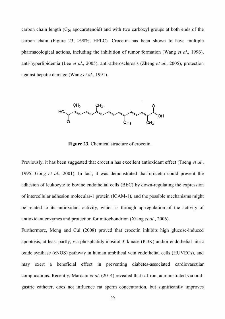
carbon chain length (C20 apocarotenoid) and with two carboxyl groups
carbon chain (Figure 23; >98%,
pharmacological actions, including the inhibition
anti-hyperlipidemia (Lee et al
against hepatic damage (Wang
Figure
Previously, it has been suggested that crocetin has excellent antioxidant effect (Tseng
1995; Gong et al., 2001). In fact, it was demonstrated that
adhesion of leukocyte to bovine endothelial cells
of intercellular adhesion molecular
be related to its antioxidant activity, which is through
antioxidant enzymes and protection for
Furthermore, Meng and Cui (2008)
apoptosis, at least partly, via phosphatidylinositol 3' kinase
oxide synthase (eNOS) pathway in
may exert a beneficial effect in preventing diabetes
complications. Recently, Mardani
gastric catheter, does not influ
99
apocarotenoid) and with two carboxyl groups
>98%, HPLC). Crocetin has been shown to
pharmacological actions, including the inhibition of tumor formation (
et al., 2005), anti-atherosclerosis (Zheng et al
Wang et al., 1991).
Figure 23. Chemical structure of crocetin.
Previously, it has been suggested that crocetin has excellent antioxidant effect (Tseng
In fact, it was demonstrated that crocetin could
bovine endothelial cells (BEC) by down-regulati
intercellular adhesion molecular-1 protein (ICAM-1), and the possible mechanisms
be related to its antioxidant activity, which is through up-regulation of the activity of
antioxidant enzymes and protection for mitochondrion (Xiang et al., 2006).
Meng and Cui (2008) proved that crocetin inhibits high glucose
phosphatidylinositol 3' kinase (Pl3K) and/or
pathway in human umbilical vein endothelial cells
beneficial effect in preventing diabetes-associated cardiovascular
complications. Recently, Mardani et al. (2014) revealed that saffron, administrated
influence rat sperm concentration, but significantly improve
apocarotenoid) and with two carboxyl groups at both ends of the
HPLC). Crocetin has been shown to have multiple
umor formation (Wang et al., 1996),
et al., 2005), protection
Previously, it has been suggested that crocetin has excellent antioxidant effect (Tseng et al.,
crocetin could prevent the
regulating the expression
1), and the possible mechanisms might
regulation of the activity of
., 2006).
crocetin inhibits high glucose-induced
) and/or endothelial nitric
human umbilical vein endothelial cells (HUVECs), and
associated cardiovascular
administrated via oral-
but significantly improves
100
motility and normal sperm morphology, suggesting that saffron can protect sperm against
DNA damage and chromatin anomalies. In addition, it was shown that the supplementation
of low concentrations of natural saffron aqueous extract (SAE) during IVM resulted in both
higher rates of maturation and blastocyst formation of mouse oocytes (Tavana et al., 2012);
the improved rate of in vitro embryo development may be due to antioxidative effects of
SAE components to which oocytes are exposed during IVM procedure.
To date there are no studies available on the effects of both saffron extract and its active
components, such as crocin and crocetin, on in vitro embryonic development in cattle.
Therefore, the aim of this work was to evaluate whether supplementation of culture medium
with crocetin improves in vitro blastocyst development and embryo quality in cattle, the
latter assessed in terms of cryotolerance, apoptotic cells rate and allocation of blastocyst
cells into the inner cell mass (ICM) and trophectoderm (TE) lineages.
5.1 MATERIALS AND METHODS
Experimental Design
A preliminary dose response trial (0 to 50 µM) showed no significant difference of both
cleavage and blastocyst development between the control and crocetin treated groups up to
the concentration of 10 µM, with an evident toxic effect at the highest concentration tested
(50 µM), indicated by a substantial decreased (P<0.01) embryo production (Figure 24).
Also, the preliminary assessment of embryo cryotolerance showed a negative effect with 10
µM crocetin.
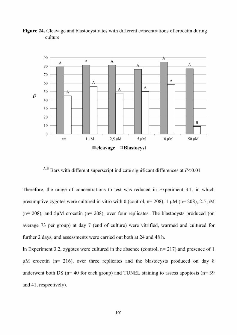
101
Figure 24. Cleavage and blastocyst rates with different concentrations of crocetin during culture
A,B Bars with different superscript indicate significant differences at P<0.01
Therefore, the range of concentrations to test was reduced in Experiment 3.1, in which
presumptive zygotes were cultured in vitro with 0 (control, n= 208), 1 µM (n= 208), 2.5 µM
(n= 208), and 5µM crocetin (n= 208), over four replicates. The blastocysts produced (on
average 73 per group) at day 7 (end of culture) were vitrified, warmed and cultured for
further 2 days, and assessments were carried out both at 24 and 48 h.
In Experiment 3.2, zygotes were cultured in the absence (control, n= 217) and presence of 1
µM crocetin (n= 216), over three replicates and the blastocysts produced on day 8
underwent both DS (n= 40 for each group) and TUNEL staining to assess apoptosis (n= 39
and 41, respectively).
A A AA
A
A
A
A
A A
A
B
0
10
20
30
40
50
60
70
80
90
ctr 1 µM 2,5 µM 5 µM 10 µM 50 µM
%
cleavage Blastocyst

102
Considering that differences in the cell number and allocation of blastocysts may appear
when the developmental stages are taken into account (Korhonen et al., 2010), only good
quality XBl and HBL stage embryos (with a clearly visible ICM and blastocoel) were
chosen for the evaluation of cell allocation by DF staining. In addition, for TUNEL staining
BL, XBL and HBL were used.
Reagents and media
Unless otherwise stated, all chemicals were obtained from Sigma-Aldrich Chemical
(Milano, Italy). The In Situ Cell Death Detection Kit (fluorescein) and the RNA-free DNase
was obtained from Roche Diagnostics Corporation (Indianapolis, IN, USA).
The aspiration medium was TCM 199 supplemented with 25 mM Hepes, 2 mM sodium
bicarbonate, 2 mM sodium pyruvate, 1 mM L-glutamine, 10 µL/mL amphotericin B (H199)
supplemented with 2% bovine serum (BS) and 95.6 SI/mL heparin. The IVM medium was
TCM 199 supplemented with 15% bovine serum (BS), 0.5 µg/mL FSH, 5 µg/mL LH, 0.8
mM L-glutamine and 50 µg/mL gentamycin. The IVF medium was Tyrode’s modified
medium (Parrish et al., 1986) without glucose and bovine serum albumin (BSA),
supplemented with 5.3 SI/mL heparin, 30 µM penicillamine, 15 µM hypotaurine, 1 µM
epinephrine and 1% of BS. The IVC medium consisted of Synthetic Oviduct Fluid (SOF)
medium (Tervit et al., 1972), with 30 µL/mL essential amino acids, 10 µL/mL non-essential
amino acids and 5 % BS.
The base medium for the vitrification and warming solutions was H199 with 20% fetal calf
serum (FCS). The equilibration medium (VS1) consisted of 7.5% DMSO and 7.5% ethylene
glycol (EG) in base medium and the vitrification solution (VS2) consisted of 16.5% DMSO

103
and 16.5% EG with 0.5 M sucrose in the base medium. The warming solutions consisted in
0.25 M and 0.15 M sucrose in the base medium.
In vitro embryo production
Abattoir-derived oocytes were matured and fertilized in vitro according to our standard
procedure (Rubessa et al., 2011). Briefly, bovine ovaries were recovered from a local
abattoir and transported to the laboratory, in physiological saline at 30 to 35 °C. Cumulus-
oocyte complexes (COCs) were aspirated from follicles of 2 to 8 mm in diameter and only
those with uniform cytoplasm and multilayered cumulus cells were selected, washed twice
in the aspiration medium and once in the IVM medium. Groups of 25 COCs were matured
in 400 µL of IVM medium, covered with mineral oil, in four well plates (NuncTM,
Roskilde, Denmark), for 22 h at 39 °C, and 5% CO2 in air. In vitro matured COCs were
washed and transferred, 25 per well, into 300 µL of IVF medium covered with mineral oil.
Frozen sperm from a bull previously tested for IVF were thawed at 37 °C for 40 s and
selected by centrifugation (25 min at 300 X g) on a Percoll discontinuous gradient (45 and
80%). The pellet was reconstituted into 2 mL of IVF medium and centrifuged twice, at 160
and 108 X g for 10 min. The pellet was diluted with IVF medium and added in the
fertilization wells at the concentration of 1 x 10⁶ sperm/mL. Gametes were co-incubated for
20 h at 39 °C, in 5% CO2 in air, after which presumptive zygotes were vortexed for 2 min to
remove cumulus cells in Hepes-TCM with 5% BS, washed twice in the same medium and
randomly distributed (30 to 50 per well, into 400 µL of IVC medium) into different culture
groups differing on the basis of LE concentrations. Zygotes were incubated in a humidified
mixture of 5% CO2, 7% O2 and 88% N2 in air at a temperature of 39 °C. On days 7 and 8,
104
Experiment 3.1 and 3.2 respectively, cleavage and blastocyst rates were recorded. The
embryos obtained by the end of culture were scored for quality on the basis of
morphological criteria (Robertson and Nelson, 2010) and the percentage of transferable
embryos, i.e. Grade 1 and 2 tight morulae and blastocysts (TMBL), and that of Grade 1 and
2 blastocysts (BL) were recorded. Furthermore, the stage of development was assessed and
embryos were categorized as slow developing embryos, i.e. tight morulae (TM), early
blastocysts (eBl), blastocysts (Bl), and as fast developing embryos, i.e. those that reached
the stages of expanded blastocysts (XBl) and hatched blastocysts (HBl) at the end of
culture.
Experiment 3.1: Vitrification and Warming
On Day 7 the blastocysts obtained (from eBl to HBl) were vitrified by the Cryotop method,
previously described (Kuwayama and Kato, 2000). All equilibration and dilution steps were
carried out at 37 °C. Each blastocyst was exposed to VS1 in 200 µL droplet for 3 min, then
transferred into a 20 µL droplet of VS2 for 20 to 25 s, and loaded with a glass capillary onto
the top of the film strip of each cryotop in 0.1 µL volume. After loading, almost all the
solution was removed and the Cryotop was quickly immersed into liquid nitrogen, protected
with the cap and stored under liquid nitrogen. For warming, after removal of the cap, the
Cryotop strip was immersed directly into 1 mL of the 0.25 M sucrose solution for 1 min and
the embryo was transferred into 200 µL droplet of 0.15 M sucrose solution for 5 min.
Warmed blastocysts were washed in H199 medium + 10% FCS and cultured for two days.
After 24 h and 48 h post-warming culture survival rates were evaluated with morphological
criteria, on the basis of the integrity of the embryo membrane and the ZP, and re-expansion
105
of the blastocoel. Furthermore, the percentages of embryos that resumed their development
and reached a more advanced developmental stage after culture (development rate), as well
as the hatching rates, were recorded.
Experiment 3.2: Differential staining
Blastocysts (only XBL and HBL) produced in the presence (1 µM) or absence of crocetin on
Day 8, were subjected to a DS protocol previously described (Thouas et al., 2001) with
slight modifications. Briefly, the ZP of viable expanding or expanded blastocysts was
removed with 0.2% pronase in Hepes-buffered SOF. After three washes in PBS with 0.1%
PVP, blastocysts were first incubated in 500 µL of propidium iodide solution (PBS with 1%
Triton X-100 and 100 µg/mL propidium iodide) for 1 min. Blastocysts were then
immediately transferred into 500 µL of a fixative solution of 100% ethanol with 25 µg/mL
bisbenzimide (Hoechst 33342) for 5 min. The fixed and stained embryos were subsequently
transferred into glycerol and mounted onto a glass microscope slide in a drop of glycerol,
flattened with a coverslip to a level where all nuclei appeared at the same focal plane, and
visualized using a fluorescent microscope (Eclipse E-600; Nikon) under ultraviolet light
with excitation at 460 to 560 nm. A digital image of each embryo was taken using NIS-
Elements-F software and a high-resolution color digital camera (Nikon digital sight DS-Fi
1C) and the numbers of TE (red) and ICM (blue) nuclei were counted.

106
Experiment 3.2: TUNEL staining
The blastocysts (BL, XBl and HBL), produced in the presence (1 µM) or absence of
crocetin on Day 8 were subjected to a TUNEL staining protocol previously described
(Paula-Lopes and Hansen, 2002).
The TUNEL assay was used to detect DNA fragmentation associated with late stages of the
apoptotic cascade. The enzyme terminal deoxynucleotidyl transferase is a DNA polymerase
that catalyzes the transfer of a fluorescein isothiocyanate-conjugated dUTP nucleotide to a
free 3’ hydroxyl group characteristic of DNA strand breaks. Embryos were removed from
SOF culture medium and washed three times in drops of PBS containing 1 mg/mL PVP
(PBS-PVP) by transferring the embryos from drop to drop. Embryos were fixed in a 50 µL
drop of 4% (w/v) paraformaldehyde in PBS for 15 min at room temperature, washed twice
in PBS-PVP, and stored in 600 µL of PBS-PVP at 4°C until the time of assay. All steps of
the TUNEL assay were conducted using microdrops in a humidified box.
On the day of the TUNEL assay, embryos were transferred to a 50 µL drop of PBS-PVP and
then permeabilized in a drop of 0.5% (v/v) Triton X-100 in PBS, containing 0.1% (w/v)
sodium citrate for 30 min at room temperature. Controls for the TUNEL assay were
incubated in 50 µL of RQ1-RNA free DNase (50 U/mL) at 37°C in the dark for 10 min.
Positive controls and treated embryos were washed in PBS-PVP and incubated with 25 µL
of TUNEL reaction mixture (containing fluorescein isothiocyanate-conjugated dUTP and
the enzyme terminal deoxynucleotidyl transferase as prepared by and following the
guidelines of the manufacturer) for 1 h at 37°C in the dark. Negative controls were
incubated in the absence of terminal deoxynucleotidyl transferase. Embryos were then
washed three times in PBS-PVP and incubated in a 25 µL drop of Hoechst 33342 (1 µg/mL)
107
for 15 min in the dark. Embryos were washed three times in PBS-PVP to remove excess
Hoechst 33342, mounted on 10% (w/v) poly-L-lysine coated slides using 3- to 4 µL drops
of glycerol, and coverslips were placed on the slides. Labeling of TUNEL and Hoechst
33342 nuclei was observed using a fluorescent microscope (Eclipse E-600; Nikon). Each
embryo was analyzed for total cell number (blue nuclei) and TUNEL-positive blastomeres
(green nuclei) with DAPI and FITC filters, respectively, using a 20X and 40X objectives.
Digital images were acquired (Figures 25, 26, 27 and 28) using NIS-Elements-F software
and a high-resolution color digital camera (Nikon digital sight DS-Fi 1C).
Figure 25. Representative pictures showing total cells staining examined by DAPI filter (a) and TUNEL labeling examined by FITC filter (b) of the same HBL cultured in the control medium
Figure 26. Representative pictures showing total cells staining examined by DAPI filter (a) and TUNEL labeling examined by FITC filter (b) of the same HBL cultured in the medium enriched with 1 µM crocetin
a b
a b

108
Figure 27. Positive control Figure 28. Negative control
5.2 STATISTICAL ANALYSIS
Differences among groups in the percentages of blastocysts, as well as in the survival rates
after vitrification-warming, were analyzed by Chi square test using SPSS 16.0 statistical
software (SPSS Inc., 2007). DS and TUNEL staining data were analyzed by the Student’s t
test.
5.3 RESULTS
As shown in Table 1, the supplementation of crocetin during culture did not affect cleavage;
however, post-fertilization embryo development was higher in the 1 µM crocetin group
compared to the others, both in terms of total embryo output (TMBL; P<0.01) and superior
quality blastocysts (Grade 1,2 BL; P<0.05). Moreover, the percentage of fast developing
embryos (Figure 29) was also increased in 1µM (P<0.01) and in 5 µM (P<0.05) crocetin
groups compared to the control (Experiment 3.1 - Table 9).

109
Figure 29. Fast developing embryos
Table 9. Percentages of cleavage, tight morulae-blastocysts (TMBL), Grade 1 and 2 blastocysts (Grade 1,2 BL) and fast developing embryos (fast BL) under different crocetin concentrations.
Crocetin concentrations
COC n.
Cleaved n. (%)
TMBL n. (%)
Gd 1,2 BL n. (%)
Fast BL n. (%)
0 (control) 208 153 (73.6) 66 (43.1)A 62 (40.5)a 27 (17.7)Aa
1 µM 208 155 (74.5) 94 (60.7)B 84 (54.2)b 53 (34.2)B
2.5 µM 208 170 (81.7) 83 (48.9)A 77 (45.3)a 41 (24.1)Aa
5 µM 208 157 (75.5) 82 (52.2)A 76 (48.4)a 43 (27.4)Ab
a,b Bars with different superscript are significantly different; P<0.05 A,B Bars with different superscript are significantly different; P<0.01

110
In addition, the enrichment of medium with crocetin improved embryo cryotolerance. In
fact, although survival, development and hatching rates after 24 h post-warming culture
were not affected, hatching rates following 48 h post-warming culture increased (P<0.05)
when medium was enriched with 1 µM crocetin concentration (Experiment 3.1 - Table 10).

111
Table 10. Survival, developmental and hatching rates after 24 and 48 h culture following vitrification-warming of blastocysts produced under different crocetin concentrations.
Post-warming culture
Crocetin concentrations
n.
24 h 48 h
Survived N (%)
Developed N (%)
Hatched N (%)
Survived N (%)
Developed N (%)
Hatched N (%)
0 (control) 62 54 (87.1) 32 (51.6) 14 (22.6) 41 (66.1) 37 (59.7) 26 (41.9)a
1 µM 79 59 (74.7) 41 (51.9) 22 (27.8) 58 (73.4) 55 (69.6) 48 (60.8)b
2.5 µM 75 38 (50.7) 20 (26.7) 11 (14.7) 30 (40.0) 24 (32.0) 18 (24.0)a
5 µM 74 42 (56.8) 28 (37.8) 3 (4.6) 36 (48.6) 34 (45.9) 23 (31.0)a
a,b Values within a column with different superscript indicate significant differences at P<0.05

112
As shown in Table 3, no differences were recorded in the average number of ICM, TE and
total cells, as well as in the ratio of ICM:total cells between 1 µM crocetin and control
groups (Experiment 3.2 - Table 11).
Table 11. Allocation of cells (Mean ± SE) in bovine blastocysts cultured in the absence (control) or presence of 1 µM crocetin.
Group n.
blastocyst ICM TE Total
ICM: total cells %
Control 40 29.2 ± 1.7 122.6 ± 2.8 151.8 ± 3.7 19.0 ± 0.8
1 µM Crocetin
40 30.5 ± 1.4 126.6 ± 3.8 157.0 ± 4.5 19.4 ± 0.7
The blastocysts stained were individually classified into four groups according to the
ICM:total cells ratio, as previously described (Koo et al., 2002): group I: <20%; group II:
20-40%; group III: 40-60%; group IV: > 60%. No differences between groups were
observed in the percentages of blastocysts falling into group I (67.5 and 65 %, in crocetin
and control groups, respectively) and into group II (32.5 and 35 %, in crocetin and control
groups respectively). No blastocysts fell into Group III and IV under both culture
conditions.

113
On the other hand, when embryos were cultured in the presence of 1 µM crocetin
(Experiment 3.2) the total blastocyst cell number increased (P<0.05) and both the average
number and the percentage of apoptotic cells in blastocysts decreased (P<0.01), as shown in
Table 12.
Table 12. Cell allocation on day 8 bovine blastocysts by TUNEL staining in the absence (control) or presence of 1 µM crocetin.
Groups n. Total cells Apoptotic cells % apoptotic cells
Mean ± SE Mean ± SE Mean ± SE
Control 35 141.1 ± 7.1a 10.9 ± 0.5A 8.6 ± 0.7A
1 µM Crocetin
37 162.8 ± 8.0b 7.9 ± 0.3B 5.3 ± 0.3B
A,B Values with different superscript are significantly different; P<0.01 a,b Values with different superscript are significantly different; P<0.05
In addition, when these parameters were analyzed in relation to the stage of development,
the total cell number was higher (P<0.01) in the crocetin group but only in case of HBL
(Figure 30). Within each developmental stage (BL, XBl and HBL) the incidence of
apoptotic cells out of the total decreased (P<0.01) in the enriched group (Figure 31).

114
Figure 30. Total cell numbers of day 8 blastocysts cultured with or without crocetin according to the developmental stage.
A,B Bars with different superscript are significantly different; P<0.01
* The number of BL, XBL and HBL stained were respectively 7, 6 and 22 in the control and 8, 7 and 21 in the crocetin group.
A
B
0
20
40
60
80
100
120
140
160
180
200
BL XBL HBL
Tot
al c
ells
nu
mb
er
Control Crocetin
115
Figure 31. The incidence of apoptotic cells of day 8 blastocysts cultured with or without crocetin according to the developmental stage.
A,B Bars with different superscript are significantly different; P<0.01 * The number of BL, XBL and HBL stained were respectively 7, 6 and 22 in the control and 8, 7 and 21 in the LE group.
Furthermore, in the crocetin-enriched group a higher (P<0.05) percentage of blastocysts had
<5% of apoptotic cells compared to the control; in particular, no blastocysts exhibited >10%
apoptotic cells in the enriched group vs about 30% in the control (P<0.01; Figure 32).
A
A
A
B
B B
0
2
4
6
8
10
12
14
16
BL XBL HBL
Ap
opto
tic
cell
s n
um
ber
(%
)
Control Crocetin

116
Figure 32. Percentage of blastocysts with low (<5%), average (5-10%) and high (>10%) levels of apoptosis in the control and crocetin groups
A,B Bars with different superscript are significantly different; P<0.01 a,b Values with different superscript are significantly different; P<0.05
5.4 DISCUSSION
The aim of this work was to evaluate whether supplementing in vitro culture medium with
crocetin, one of the active biological components of saffron, improves bovine embryo
development and quality. The results of the present study demonstrated that enrichment of
culture medium with low doses of the natural antioxidant crocetin (1 µM), improves both
embryo output and quality, as indicated by increased blastocyst yields, developmental
chronology and cryotolerance and by reduced incidence of apoptotic cells.
a
A
b
B0
10
20
30
40
50
60
< 5% 5-10% >10%
% o
f b
last
ocys
ts w
ith
dif
fere
nt
apop
toti
c ra
tio
control crocetin
117
The preliminary dose response trial showed an evident toxic effect only for 50 µM
concentration of crocetin, that was associated to a substantial decreased embryo production.
This is in contrast with previous frog (Xenopus) embryo culture studies reporting that
crocetin was not toxic to embryo development, until a concentration of 100 µM (Martin et
al., 2002). On the other hand, previous studies reported that high concentrations of crocetin
tested by the DPPH antioxidant activity assay, exhibited lower antioxidant effects (Kanakis
et al., 2009).
However, the results of the preliminary trial suggested to reduce the range of concentrations
to test in Experiment 3.1, where the dose-response study was carried out with 0 (control), 1,
2.5 and 5 µM crocetin. The fact that cleavage rate was not affected by the treatment was
expected, as the enrichment was carried out only during culture with the aim to improve
particularly embryo quality, that is known to be mainly affected by culture environment
(Lonergan et al., 2001). Nevertheless, in contrast with the results of the previous
experiments of this thesis, in which other antioxidant compounds were tested, crocetin
supplementation at the concentration of 1 µM positively influenced post-fertilization
embryo development, as indicated by higher total embryo output, superior quality
blastocysts and faster developing embryos. The increased embryo production recorded in
the presence of crocetin was relevant, as the treatment provided about 17% more
transferable embryos (TMBL) and 14% more superior quality blastocysts (G 1 & 2 BL) than
the control culture group. No other studies have been published on the effect of crocetin on
embryo development; however, an improved embryo development was recorded in mice
when IVM medium was supplemented with low doses of saffron extracts (Tavana et al.,
2012).

118
In addition, a clear indication of an improvement of embryo quality in culture medium
enriched with 1 µM crocetin derives from the 2-fold increase in the percentage of fast
developing embryos, i.e. the percentages of blastocysts reaching the most advanced stages
of development such as XBl and HBl by the end of culture (Day 7), that was observed
compared to the control. In fact, one the most important criterion to determine the quality of
embryos to be transferred is the timing of development to specific morphological stages
(Leibfried-Rutledge et al., 1987; McKiernan and Bavister, 1994; Bavister, 1995; Van Soom
et al., 1997).
It has also been demonstrated that embryos with a faster kinetics of development survive
cryopreservation better than those with slower developmental speed (Nedambale et al.,
2004). In agreement with this result, embryo cryotolerance was also significantly increased
when 1 µM crocetin was present during culture. It is well known that the major factor
affecting the cryotolerance of in vitro produced embryos is their quality (Han et al., 1994)
that has been proven to be determined by the culture system. It results that embryo
resistance to cryopreservation is a highly reliable marker of embryo quality (Imai et al.,
2002; Lonergan et al., 2003a; 2003b; Rizos et al., 2003). Also in this experiment the
beneficial effect on embryo cryotolerance was evident after 48 h of post-warming culture,
when a significant increase of hatching rate was recorded. This is known to be the most
reliable parameter to assess embryo cryotolerance, as cryopreservation hardens the ZP,
consequently preventing blastocyst hatching process (Carroll et al., 1990; Matson et al.,
1997), necessary for the appropriate elongation of the conceptus after ET. This was the first
work that evaluated the effect of crocetin on embryo cryotolerance. The only work available
in literature, in fact, has been limited to assess the effect of crocetin on frog in vitro embryo

119
development in morphological terms (Martin et al., 2002). In particular, it was demonstrated
that concentrations much higher than our effective dose may have theratogenic effects.
However, previous studies demonstrated that supplementation with antioxidants during
culture improves embryo quality and cryotolerance (Hosseini et al., 2009; Takahashi, 2012).
In the Experiments 1 and 2 of this thesis we also recorded a significant improvement of
cryotolerance when culture medium was enriched with either resveratrol or L-ergothioneine.
The improved cryotolerance of bovine embryos obtained with crocetin during culture is
likely due to its high antioxidant capacity, as oxidative stress occurs during the freezing-
thawing process, leading to lipid peroxidation, membrane injury and structural damage
(Somfai et al., 2007b). This may be deducted from the work performed by Xiang et al.
(2006), in which it was demonstrated that 1 µM crocetin concentration prevents the
adhesion of leukocyte to bovine endothelial cells (BEC) culture, by down-regulating the
expression of intercellular adhesion molecular-1 protein (ICAM-1), increasing the activity
of SOD and significantly inhibiting the generation of malonicdialdehyde (MDA), a products
of lipid peroxidation and a classic indicator of oxidative stress.
In addition to cryotolerance, we also assessed embryo quality by morphological parameters,
such as the total cell count and particularly the cell allocation to the ICM and the TE, as
well as the apoptotic cell ratio, an additional criterion proven to effectively predict embryo
viability (Pomar et al., 2005). In fact, it is well documented that both cell lineages are
essential for embryonic survival (Koo et al., 2002; Knijn et al., 2003) and post-implantation
development (Iwasaki et al., 1990). The distribution of blastocyst cells in the ICM and TE
lineages was not affected by the crocetin treatment and most of the embryos had an
ICM:total cells ratio lower than 40% in both groups. On the other hand, crocetin during
120
culture significantly reduced both the average number and the incidence of apoptotic cells in
the blastocysts. Although apoptosis physiologically occurs during pre-implantation embryo
development, to control cell population by eliminating defective cells (Brison, 2000), a high
incidence of apoptotic cells is correlated to abnormal morphology of the embryo (Hardy et
al., 1989). In fact, in vitro produced embryos are known to exhibit a higher incidence of
apoptosis than their in vivo counterparts (Bergeron et al., 1998). In the present study both
the number and the incidence of apoptotic cells decreased in the crocetin-enriched group
within each developmental stage, i.e. blastocysts, expanded blastocysts and hatched
blastocysts. Furthermore, when only HBL were considered the total cell count also
increased in the crocetin group compared to the control. A similar result was found in
Experiment 2, in which the difference in cell number was also evident only at the hatched
stage. Therefore, also in this case the finding is only in apparent contrast with the data
obtained with the DS. As previously stated, embryos that were examined by DS were only
expanded and hatched blastocysts, and the low numbers did not allow to analyze this
parameter within the stage. What is overwhelming is the difference between groups in the
percentage of blastocysts with low, average and high proportion of apoptotic cells. The
enrichment of IVC medium with crocetin significantly increased the proportion of
blastocysts with low apoptotic cells (<5%) compared to the control (46 vs 20%). In addition,
no blastocysts showed a high apoptotic index in the enriched group vs 31.4 % of the control.
The reduced incidence of apoptosis recorded in the presence of crocetin is also likely due to
its antioxidant properties. It is known that oxidative stress that normally occurs during in
vitro culture triggers apoptosis (Guérin et al., 2001; Yang et al., 1998). Impaired embryo
development and apoptotic cell death have been previously associated with increased levels
121
of reactive oxygen species and decreased levels of SOD (Guérin et al., 2001). It has also
reported that antioxidants during culture inhibit apoptosis (Guérin et al., 2001). These
results are in agreement with those obtained in Experiment 2, in which apoptosis was
reduced by enriching culture medium with LE.
In conclusion, the results of this study taken together demonstrated that enrichment of
bovine IVC medium with low doses of crocetin improves blastocyst production and the
overall embryo quality, that is the most important factor affecting post-implantation
development after embryo transfer. The beneficial effect on embryo quality is highlighted
by the enhanced post-fertilization embryo development, especially in term of superior
quality blastocysts and fast developing embryos, as well as by the improved cryotolerance
following culture, and by the increased proportion of blastocysts with a low percentage of
apoptotic cells.

122
CONCLUSIONS
Currently, the ovum pick-up and IVEP technology is the best tool available to improve the
maternal contribution to the genetic improvement. There has been an increasing interest in
the large scale embryo production through IVEP around the world; in some countries, such
as Brazil, the majority of the bovine embryos produced for commercial purposes comes
from IVEP. However, despite numerous efforts carried out over the years, a critical issue is
still given by the poor quality of the IVP embryos and an optimal medium for IVC has still
not been formulated. This hampers the benefit/cost ratio of the technology, due to the lower
pregnancy rates recorded after ET of cryopreserved IVP embryos. A substantial amount of
evidence exists to demonstrate that the culture conditions to which the embryo is exposed,
particularly in the post-fertilization period, can have perturbing effects on the pattern of
gene expression and on the embryo quality, with potentially important long-term
consequences (Lonergan, 2007).
Several factors are known to affect the in vitro embryo development, such as ammonia,
oxygen radicals, growth factors, etc. Among these, the increased oxidative stress is a major
factor impairing mammalian in vitro embryo development. It has been demonstrated that
oxidative stress results in several intracellular damages, lipid peroxidation leading to
reduced cryotolerance, apoptosis and abnormal allocation of cells into ICM and TE lineages
(Somfai et al., 2007b, Guérin et al., 2001; Yang et al., 2008). Therefore, in order to protect
the embryos from increased oxidative stress, and the consequent damages caused by
suboptimal culture conditions, in the last years there has been an increased interest in
strategies based on the supplementation of antioxidant compounds during in vitro embryo
123
culture. To reduce the toxicity of ROS for improving embryo development, antioxidants
supplementation is effective to regulate intra- and extra-embryonic environments; in fact,
maintaining of intra- and extra-cellular redox balance is very important to provide suitable
environments for metabolic and gene expressions followed by embryo development
(Takahashi, 2012). Currently, addition of several enzymatic and non-enzymatic, as well as
protein and non-protein antioxidants seem to be a very effective method to improve embryo
development by scavenging ROS and serving embryos a low oxidative stress conditions.
This was the rationale of the thesis that focused on evaluating the effects of different natural
antioxidants during bovine in vitro embryo culture on embryo development and quality.
The choice of the candidate molecules fell on Resveratrol (Experiment 1), L-Ergothioneine
(Experiment 2) and Crocetin (Experiment 3) because beneficial properties and positive
effects in other species and environmental conditions were previously reported. The results
of this thesis demonstrated that improving IVC medium environment, by natural antioxidant
compounds supplementation, enhances the overall embryo quality, affecting the efficiency
of embryo development, cryotolerance, cell differentiation and apoptotic rate.
More specifically, resveratrol improved the cryotolerance of in vitro produced bovine
embryos, as shown by a significant improvement of both development and hatching rates
recorded after 48 h post-warming culture (Experiment 1). Hatching rate showed a 2-fold
increase compared to the control and blastocysts exhibited a correct allocation of cells into
the ICM and TE.
Similarly, in Experiment 2 LE during culture increased embryo cryotolerance, especially
after 48 h of post-warming culture, when a significant increase of hatching rate was
observed. Blastocysts produced in the medium enriched with LE showed an increase in the

124
average number of ICM cells compared to the control, and an increased percentage of
blastocysts with the most appropriate ICM:total cells ratio (20-40%), confirming a
beneficial effect on embryo quality. Moreover, both the number and the incidence of
apoptotic cells decreased in the LE-enriched group within each developmental blastocyst
stage.
In contrast to previous antioxidant compounds, in Experiment 3 crocetin supplementation at
the concentration of 1 µM increased blastocyst yields in term of higher total embryo output,
superior quality blastocysts and fast developing embryos. In addition, crocetin improved the
cryotolerance of IVP embryos compared to the control, as indicated by higher hatching rates
recorded after 48 h post-warming culture. Blastocysts produced in the absence (control) and
presence of crocetin had a similar number of ICM, TE and total cells, as well a similar ratio
of ICM:total cells. On the other hand, crocetin during culture significantly reduced both the
average number and the incidence of apoptotic cells in the blastocysts. In particular, the
enrichment of IVC medium with crocetin significantly increased the proportion of
blastocysts with low apoptotic cells, and eliminated blastocysts showing a high apoptotic
index in.
Handling of mammalian oocytes and embryos in vitro is an effective technique to obtain
offspring by embryo transfer. The improved embryo quality obtained with all the
antioxidants tested is a very important finding, as better embryo quality may may lead to
increased pregnancy rate after ET of IVP embryos. Indeed, increasing the number of good
quality embryos is the most practical and clinically useful approach in embryo transfer of
mammalian pre-implantation embryos.

125
In conclusion, optimization of IVC systems by the formulation of antioxidant-enriched
culture media is critical to increase embryo quality, viability and resistance to freezing,
likely promoting a reduction of birth defects and of embryonic and neonatal mortality
incidence. These results lay the basis for future perspective studies. In particular, it is worth
investigating whether the inclusion of resveratrol, LE and crocetin also during post-warming
embryo culture would further improve embryo quality. It is likely, indeed that a surplus
source of exogenous antioxidant may prevent DNA fragmentation during the critical period
of cryopreservation and post-warming embryo culture. In addition, further studies are
needed to investigate the exact mechanisms by which these antioxidants protect the cells.
Finally, these promising results strongly encourage to evaluate the post implantation embryo
developmental competence after ET of IVP embryos cultured in enriched media.
126
REFERENCES
Abdelrazik H., Sharma R., Mahfouz R., Agarwal A. (2009). L-carnitine decreases DNA damage and improves the in vitro blastocyst development rate in mouse embryos. Fertil. Steril., 91 (2): 589-96.
Abdullaev F.I. (1993). Biological effects of saffron. Biofactors, 4 (2): 83–86.
Abdullaev F.I. and Espinosa-Aguirre J.J. (2004). Biomedical properties of saffron and its potential use in Cancer therapy and chemoprevention trials. Review. Cancer Detection and Prevention, 28: 426-432.
Abdullaev Jafariva J., Caballero-Ortega H., Riverón-Nigrete L., Pereda-miranda R., Rivera- Luna R., Manuel Hernández J., Pérez-López I., Espinosa-Aguirre J.J. (2002). In vitro evaluation of chemopreventive potential of saffron. Rev. Inves.
Clin., 54 (5): 430-436.
Abe H., Yamashita S., Satoh T. and Hoshi H. (2002). Accumulation of cytoplasmic lipid droplets in bovine embryos and cryotolerance of embryos developed in different culture systems using serum-free or serum-containing media. Mol.
Reprod. Dev., 61: 57–66.
Abe K. and Saito H. (2000). Effects of saffron extract and its constituent crocin on learning behaviour and long-term potentiation. Phytother. Res., 14: 149-152.
Abe K., Sugiura M., Yamaguchi S., Shoyama Y., Saito H. (1999). Saffron extract prevents acetaldehyde-induced inhibition of long-term potentiation in the rat dentate gyrus in vivo. Brain Res., 851 (1-2): 287-9.
Abedelahi A., Salehnia M., Allameh A.A., Davoodi D. (2010). Sodium selenite improves the in vitro follicular development by reducing the reactive oxygen species level and increasing the total antioxidant capacity and glutathione peroxide activity. Hum. Reprod., 25 (4): 977-85.
Aitken J.R. (1994). A free radical theory of male infertility. Reprod. Fertil. Dev.
Sci.,19-24.
Akanmu D., Cecchini R., Aruoma O.I., Halli B. (1991). The antioxidant action of ergothionine. Arch. Biochem. Biophys., 288: 10-16.
Akhondzadeh S., Fallah-pour H., Afkham K., Khalighi F. (2004). Comparison of crocus sativus L. and imipramine mild to moderate depression: A pilot double-blind [ISRCTN45683816]. Journal List, BMC Complement Altern. Med., 4: 12.

127
Ali A.A., Bilodeau J.F., Sirard M.A. (2003). Antioxidant requirements for bovine oocytes varies during in vitro maturation, fertilization and development. Theriogenology, 59 (3-4): 939-49.
Alm H., Torner H., Lohrke B., Viergutz T., Ghoneim I.M. and Kanitz W. (2005). Bovine blastocyst development rate in vitro is influenced by selection of oocytes by brillant cresyl blue staining before IVM as indicator for glucose-6-phosphate dehydrogenase activity. Theriogenology, 63: 2194–2205.
Almodin C.G., Minguetti-Camara V.C., Paixao C.L. and Pereira P.C. (2010). Embryo development and gestation using fresh and vitrified oocytes. Human
Reproduction, 25: 1192–1198.
Arav A. (1992). Vitrification of oocytes and embryos. In New Trends in Embryo Transfer. Eds. Lauria A. & Gandolfi F. Cambridge: Portland Press, edn 1, pp. 255–264.
Arav A., Yavin S., Zeron Y., Natan D., Dekel I. and Gacitua H. (2002). New trends in gametes cryopreservation. Molecular and Cellular Endocrinology, 187: 77–81.
Aruoma O.I., Spencer J.P. and Mahmood N. (1999). Protection against oxidative damage and cell death by the natural antioxidant ergothioneine. Food Chem.
Toxicol., 37: 1043-1053.
Aruoma O.I., Whiteman M.E. and Halliwell B. (1997). The antioxidant action of ergothioneine: Assessment of its ability to scavenge peroxynitrite. Biochem.
Biophys. Res. Commun., 231: 389–391.
Asmus K.D., Bensasson R.V., Bernier J.L., Houssin R. and Land E.J. (1996). One electron oxidation of ergothioneine and analogues investigated by pulse radiolysis: Redox reactions involving ergothioneine and vitamin C. Biochem.
J., 315: 625–629.
Au H.K., Yeh T.S., Kao S.H., Tzeng C.R. and Hsieh R.H. (2005). Abnormal mitochondrial structure in human unfertilized oocytes and arrested embryos. Ann. N. Y. Acad. Sci., 1042: 177–185.
Aurich C. and Hahn J. (1994). In vitro fertilization: bulls should not be forgotten. Tierzuchter, 46: 20-3.
Austin C.R. (1951). Observations on the penetration of the sperm in the mammalian egg. Aust. J. Sci. Res. B., 4: 581–96.
Austin C.R. (1952). The capacitation of the mammalian sperm. Nature, 170: 326.
Baril G., Casamitjana P., Perrin J. and Vallet J.C. (1989). Embryo production, freezing and transfer in Angora, Alpine and Saanen goats. Zuchthygiene, 24
(Berlin): 101–115.
128
Barnes F.L. (2000). The effects of the early uterine environment on the subsequent development of embryo and fetus. Theriogenology, 53: 649–58.
Baruselli P.S. (2007). Fixed-time artificial insemination in buffalo. Ital. J.
Anim.Sci., 6 (Suppl. 2): 107-118.
Baumann C.G., Morris D.G., Sreenan J.M., Leese H.J. (2007). The quiet embryo hypothesis: molecular characteristics favoring viability. Mol. Reprod. Dev., 74: 1345–53.
Baur J.A. and Sinclair D.A. (2006). Therapeutic potential of resveratrol: the in vivo evidence. Nat. Rev. Drug Discov., 5: 493–506.
Bavister B.D. (1995). Culture of preimplantation embryos: facts and artifacts. Hum.
Reprod. Update, 1: 91–148.
Bavister B.D. and Squirrell J.M. (2000). Mitochondrial distribution and function in oocytes and early embryos. Hum. Reprod., 15 (Suppl. 2): 189–198.
Bavister B.D. and Yanagimachi R. (1977). The effects of sperm extracts and energy sources on the motility and acrosome reaction of hamster spermatozoa in vitro. Biol. Reprod., 16: 228-37.
Bavister B.D., Boatman D.E., Leibfried L., Loose M. and Vernon M.W. (1983). Fertilization and cleavage of rhesus monkey oocytes in vitro. Biol. Reprod., 28: 983–999.
Berg D.K., van Leeuwen J., Beaumont S., Berg M., Pfeffer P.L. (2010). Embryo loss in cattle between days 7 and 16 of pregnancy. Theriogenology, 73: 250–60.
Bergeron L., Perez G.I., Macdonald G., Shi L., Sun Y., Jurisicova A., Varmuza S., Latham K.E., Flaws J.A., Salter J.C., Hara H., Moskowitz M.A., Li E., Greenberg A., Tilly J.L., Yuan J. (1998). Defects in regulation of apoptosis in caspase-2-deficient mice. Genes and Development, 12: 1304–1314.
Berthelot F., Martinat-Botte F., Locatelli A., Perreau C. and Terqui M. (2000). Piglets born after vitrification of embryos using the open pulled straw method. Cryobiology, 41: 116-124.
Bhojwani S., Alm H., Torner H., Kanitz W. and Poehland R. (2007). Selection of developmentally competent oocytes through brilliant cresyl blue stain enhances blastocyst development rate after bovine nuclear transfer. Theriogenology, 67: 341–345.
Bisset N.G. and Wichtl M. (2001). Herbal Drugs and phytopharmaceuticals. A Handbook for Practice on a scientific Basis, (2 ed.). Med. Pharm. Scientific
publisher, CRC Press, London, 167-169.
129
Blondin P. and Sirard M.A. (1995). Oocyte and Follicular Morphology As Determining Characteristics for Developmental Competence in Bovine Oocytes 1. Molecular Reproduction and Development, 41: 54-62.
Bó G.A., Guerrero D.C., Tríbulo A., Tríbulo H., Tríbulo R., Rogan D., Mapletoft R.J. (2010). New approaches to superovulation in the cow. Reprod. Fertil.
Dev., 22: 106 –12.
Boccia L., De Blasi M., Zullo G., Longobardi V., Rubessa M., Vecchio D. and Gasparrini B. (2013). L-carnitine during in vitro culture enhances the cryotolerance of buffalo (Bubalus bubalis) in vitro-derived embryos. Reprod.
Fertil. Dev., 25 (1): 214.
Boni R., Tosti E., Roviello S., Dale B. (1999). Intercellular communication in in vivo-and in vitro-produced bovine embryos. Biology of reproduction, 61: 1050–1055.
Boni R., Wurth Y.A., Roviello S., Barbieri V., Kruip TH.A.M. (1993). Effetto della distanza tra prelievo e recupero sulle caratteristiche morfologiche e sull’efficienza di produzione embrionale in vitro di oociti prelevati mediante Ovum Pick-up. Proc. XLVII S.I.S.Vet. 1: 475–480.
Boni R., Cuomo A., Tosti E. (2002). Developmental potential in bovine oocytes is related to cumulus-oocyte complex grade, calcium current activity, and calcium stores. Biol. Reprod., 66 (3): 836-42.
Bors W., Saran M., Michel C. (1982). Radical intermediates involved in the bleaching of the carotenoid crocin. Hydroxyl radicals, superoxide anions and hydrated electrons. Int. J. Radiat. Biol. Relat. Stud. Phys. Chem. Med., 41 (5): 493-501.
Brackett B. G., Bousquet D., Boice M. L., Donawick W. J., Evans J. F., Dressel M. A. (1982). Normal development following in vitro fertilization in the cow. Biol.
Reprod., 27: 147-158.
Brackett B.G. (2001). Advances in animal in vitro fertilization. In: Wolf D.P., Zelinski-Wooten M. (Eds.), Contemporary Endocrinology: Assisted Fertilization and Nuclear Transfer in Mammals. Humana Press.
Brackett B.G. and Zuelke K.A. (1993). Analysis of factors involved in the in vitro production of bovine embryos. Theriogenology, 39: 43–64.
Brackett B.G., Younis A.I., Fayrer-Hosken R.A. (1989). Enhanced viability after in vitro fertilization of bovine oocytes matured in vitro with high concentrations of luteinizing hormone. Fertil. Steril., 52: 319–24.

130
Breininger E., Cetica P.D., Beconi M.T. (2010). Capacitation inducers act through diverse intracellular mechanisms in cryopreserved bovine sperm. Theriogenology, 74 (6): 1036-49.
Brevini-Gandolfi T., Lonergan P., Cillo F., Favetta L.A., Fair T., Gandolfi F. (2002). Evolution of mRNA polyadenylation between oocyte maturation and first embryonic cleavage in cattle and its relation with developmental competence. Molecular Reproduction & Development, 63: 510-517.
Brison D.R. (2000). Apoptosis in mammalian preimplantation embryos: regulation by survival factors. Human Fertility (Cambridge), 3: 36-47.
Byrne A.T., Southgate J., Brison D.R., Leese H.J. (1999). Analysis of apoptosis in the preimplantation bovine embryo using TUNEL. Journal of Reproduction
and Fertility, 117: 97–105.
Callejas S., Alberio R., Cabodevila J., Aller J., Catalano R., Teruel M., Dulout F. (2008). Effect of progesterone administration on the ovarian response to superovulatory treatments in cattle. Anim. Reprod. Sci., 107: 9 –19.
Camargo L.S.A., Viana J.H.M., Sa´ W.F., Ferreira A.M., Ramos A.A., Vale Filho V. R. (2006). Factors influencing in vitro embryo production. Anim. Reprod., 3: 19–28.
Caracciolo di Brienza V., Neglia G., Masola N., Gasparrini B., Di Palo R., Campanile, G., (2001). In vitro embryo production in chemically defined media. Atti 1° Congresso Nazionale sull‟Allevamento del Bufalo, 3-5 ottobre 2001, Eboli (SA), Italy. pp. 341-344
Carnegie J.A., Durnford R., Algire J., Morgan J. (1997). Evaluation of mitomycin-treated Vero cells as a co-culture system for IVM/IVF-derived bovine embryos. Theriogenology, 48: 377-389.
Carney E.W. and Foote R.H. (1991). Improved development of rabbit one-cell embryos to the hatching blastocyst stage by culture in a defined, protein-free culture medium. J. Reprod. Fertil., 91 (1): 113-23.
Carroll J., Depypere H., Mathews C. (1990). Freeze-thaw induced changes of the zona pellucida explain decreased rates of fertilization in frozen-thawed mouse oocytes. J. Reprod. Fertil., 90 (2): 547–553.
Chang M.C. (1951). Fertilizing capacity of spermatozoa deposited into the fallopian tubes. Nature, 168: 697–8.
Chastant-Maillard S., Quinton H., Lauffenburger J., Cordonnier-Lefort N., Richard C., Marchal J., Mormede P., Renard J.P. (2003). Consequences of transvaginal follicular puncture on well-being in cows. Reproduction, 125: 555-563

131
Chian R.C., Niwa K., Nakahara H. (1992). Effect of sperm penetration in vitro on completion of first meiosis by bovine oocytes arrested at various stages in culture. J. Reprod. Fertil., 96: 73–78.
Chian R.C., Park C.K., Sirard M.A. (1996). Cumulus cells act as a sperm trap during in vitro fertilization of bovine oocytes. Theriogenology, 45: 258.
Chiba T., Takahashi S., Sato N., Ishii S., Kikuchi K. (1996). Fas-mediated apoptosis is modulated by intracellular glutathione in human T cells. Eur. J. Immunol.,
26 (5): 1164-9.
Cho S.R., Cho S.K., Lee S.L., Lee H.J., Choe S.Y., Rho G.J. (2002). Enhanced cryosurvival of bovine blastocysts produced in vitro in serum-free medium. J.
Assist. Reprod. Genet., 19: 487–492.
Choe C., Shin Y.W., Kim E.J., Cho S.R., Kim H.J., Choi S.H., Han M.H., Han J., Son D.S., Kang D. (2010). Synergistic effects of glutathione and β-mercaptoethanol treatment during in vitro maturation of porcine oocytes on early embryonic development in a culture system supplemented with L-cysteine. J. Reprod. Dev., 56 (6): 575-82.
Clarke G.N. (2006). A.R.T. and history, 1678–1978. Hum. Reprod., 21: 1645–1650.
Collins J.A., Schandi C.A., Young K.K., Vesely J., Willingham M.C. (1997). Major DNA fragmentation is a late event in apoptosis. Journal of Histochemistry and
Cytochemistry, 45: 923–934.
Cornell J.S. and Meister A. (1976). Glutathione and gamma-glutamyl cycle enzymes in crypt and villus tip cells of rat jejunal mucosa. Proc. Natl. Acad. Sci. (USA),
73 (2): 420-2.
Cox J.F., Hormazabal J., Santa Maria A. (1993). Effect of cumulus on in vitro fertilization of bovine matured oocytes. Theriogenology, 40: 1259-1267.
Curnow E.C., Ryan J.P., Saunders D.M., Hayes E.S. (2010a). Developmental potential of bovine oocytes following IVM in the presence of glutathione ethyl ester. Reprod. Fertil. Dev., 22 (4): 597-605.
Curnow E.C., Ryan J.P., Saunders D.M., Hayes E.S. (2010b). In vitro developmental potential of macaque oocytes, derived from unstimulated ovaries, following maturation in the presence of glutathione ethyl ester. Hum. Reprod., 25 (10): 2465-74.
Dattena M., Ptak G., Loi P. and Cappai P. (2000). Survival and viability of vitrified in vitro and in vivo produced ovine blastocysts. Theriogenology, 53: 1511-1519.
132
de la Fuente R. and King W.A. (1997). Use of a chemically defined system for the direct comparison of inner cell mass and trophectoderm distribution in murine, porcine and bovine embryos. Zygote, 5: 309–320.
de Matos D.G. and Furnus C.C. (2000). The importance of having high glu-tathione (GSH) level after bovine in vitro maturation on embryodevelopment effect of beta- mercaptoethanol, cysteine and cystine. Theriogenology, 53 (3): 761–771.
de Matos D.G., Gasparrini B., Pasqualini S.R., Thompson J.G. (2002). Effect of glutathione synthesis stimulation during in vitro maturation of ovine oocytes on embryo development and intracellular peroxide content. Theriogenology, 57 (5): 1443-51.
De Sutter P., Dozortsev D., Qian C., Dhont M. (1996). Oocyte morphology does not correlate with fertilization rate and embryo quality after intracytoplasmic sperm injection. Hum. Reprod., 11: 595–597.
de Wit A.A. and Kruip T.A. (2001). Bovine cumulus–oocyte-complexquality is reflected in sensitivity for alpha-amanitin, oocyte-diameter and developmental capacity. Anim. Reprod. Sci., 65: 51–65.
de Wit A.A., Wurth Y.A. and Kruip T.A. (2000). Effect of ovarian phase and follicle quality on morphology and developmental capacity of the bovine cumulus–oocyte complex. J. Anim. Sci., 78: 1277–1283.
Di Francesco S., Boccia L., Di Palo R., Esposito G., Attanasio L., De Rosa A., Gasparrini B. (2007). Influence of temperature and time during ovary transportation on in vitro embryo production efficiency in the buffalo species (Bubalus bubalis). In: Proceedings of the 8th World Buffalo Congress (Caserta, Italy). Ital. J. Anim. Sci., 6 (2): 755-758.
Dinnyés A., Dai Y., Jiang S. and Yang X. (2000). High developmental rates of vitrified bovine oocytes following parthenogenetic activation, in vitro fertilization, and somatic cell nuclear transfer. Biology of Reproduction, 63: 513–518.
Dinnyés A., Lonergan P., Fair T., Boland M.P. and Yang X. (1999). Timing of the first cleavage post-insemination affects cryosurvival of in vitroproduced bovine blastocysts. Mol. Reprod. Dev., 53: 318–324.
Dode M.A.N., Dufort I., Massicotte L., Sirard M.A. (2006). Quantitative expression of candidate genes for developmental competence in bovine two-cell embryos. Molecular Reproduction & Development, 73: 288-297.
Dominko T. and First N.L. (1997). Timing of meiotic progression in bovine oocytes and its effect on early embryo development. Mol. Reprod. Dev., 47: 456-67.

133
Du F., Looney C.R., Yang X. (1996). Evaluation of bovine embryos produced in vitro vs. in vivo by differential staining of inner cell mass and trophectoderm cells. Theriogenology, 45 (1): 211.
Duby R., Hill J., O'Callaghan D., Overstrom E., Boland M. (1997). Changes induced in the bovine zona pellucida by ovine and bovine oviducts. Theriogenology, 47: 332–332.
Duranthon V., Watson A.J. and Lonergan P. (2008). Preimplantation embryo programming: transcription, epigenetics, and culture environment. Reproduction, 135: 141–150.
Dziuk P. (1975). Embryo transfer: an experimental tool with practical applications. BioScience, 2: 102–6.
Edge R., McGarvey D.J., Truscott T.G. (1997). The carotenoids as anti-oxidants - a review. J. Photochem. Photobiol. B., 41 (3): 189-200.
El Gayar M. and Holtz W. (2001). Technical note: Vitrification of goat embryos by the open pulled-straw method. J. Anim. Sci., 79: 2436-2438.
Eppig J.J. (1996). Coordination of nuclear and cytoplasmic oocyte maturation in eutherian mammals. Reprod. Fertil. Dev., 8: 485–9.
Escribano J., Alonso G.L., Coca-Prados M., Fernandez J.A. (1996). Crocin, safranal and picrocrocin from saffron (Crocus sativus) inhibit the growth of human cancer cells in vitro. Cancer Letters, 100 (1-2): 23-30.
Ey J., Schömig E. and Taubert D. (2007). Dietary sources and antioxidant effects of ergothioneine. J. Agric. Food Chem., 55: 6466–6474.
Fair T. (2003). Follicular oocyte growth and acquisition of developmental competence. Anim. Reprod. Sci., 78: 203–216.
Fair T., Hyttel P. and Greve T. (1995). Bovine oocyte diameter in relation to maturational competence and transcriptional activity. Mol. Reprod. Dev., 42: 437–442.
Fair T., Murphy M., Rizos D., Moss C., Martin F., Boland M.P., Lonergan P. (2004). Analysis of differential maternal mRNA expression in developmentally competent and incompetent bovine two-cell embryos. Molecular Reproduction
& Development, 67: 136-144.
Fatehi M., Rashidabady T. and Fatehi- Hassanabad Z. (2003). Effects of crocus sativus petals' extract on rat blood pressure and on responses induced by electrical field stimulation in the rat isolated vas deferens and guinea-pig ileum. Journal of Ethnopharmacology, 84: 199-203.

134
Favetta L.A., Robert C., St. John E.J., Betts D.H., King W.A. (2004). P66shc, but not p53 is involved in early arrest of in vitro-produced bovine embryos. Molecular
Human Reproduction, 10: 383-392.
Fenwick J., Platteau P., Murdoch A.P. and Herbert M. (2002). Time from insemination to first cleavage predicts developmental competence of human preimplantation embryos in vitro. Hum. Reprod., 17: 407–412.
Fernandez J.A. (2004). Biology, Biotechnology and biomedicine of saffron: Recent
Res. Devel. Plant. Pci., 2: 127-59.
Fouladi-Nashta A.A., Alberio R., Kafi M., Nicholas B., Campbell K.H.S., Webb R. (2005). Differential staining combined with TUNEL labelling to detect apoptosis in preimplantation bovine embryos. Reprod. Biomed. Online, 10 (4): 497–502.
Franzoni R.C., Galeta F., Laurenza I., Barsotti M., Di Stefano R., Bocchetti R., Regoli F., Carpi A., Balbarini A., Migliore L., Santoro G. (2006). An in vitro study on the free radical scavenging capacity of ergothioneine: Comparison with reduced glutathione, uric acid and trolox. Biomed. Pharmacother., 60: 453-457.
Friedler S., Giudice L.C., Lamb E.J. (1988). Cryopreservation of embryos and ova. Fertil. Steril., 49: 743– 64.
Fukuda Y. and Enari N. (1993). Developmental ability of in vitro matured-in vitro fertilized bovine embryos derived from oocytes with homogeneous or heterogenous ooplasm 4. Proc.7th World Conf. on Anim. Prod., 2: 276-277. Abstract
Fukui Y. (1990). Effect of follicle cells on acrosome reaction, fertilization and developmental competence of bovine oocytes matured in vitro. Mol. Reprod.
Dev., 26:40-6.
Fuller B. and Paynter S. (2004). Fundamentals of cryobiology in reproductive medicine. Reprod. Biomed., 9: 680–91.
Gabrielsen A., Lindenberg S., Petersen K. (2001). The impact of the zona pellucida thickness variation of human embryos on pregnancy outcome in relation to suboptimal embryo development. A prospective randomized controlled study. Hum. Reprod., 16: 2166–2170.
Gadella B.M., Gadella T.W.Jr., Colenbrander B., van Golde L.M., Lopes-Cardozo M. (1994). Visualization and quantification of glycolipid polarity dynamics in the plasma membrane of the mammalian spermatozoon. J. Cell. Sci., 107: 2151–63.
135
Gadella B.M., Lopes-Cardozo M., van Golde L.M., Colenbrander B., Gadella T.W.Jr. (1995). Glycolipid migration from the apical to the equatorial subdomains of the sperm head plasma membrane precedes the acrosome reaction. Evidence for a primary capacitation event in boar spermatozoa. J.
Cell. Sci., 108: 935–46.
Gainer J.I. and Jones J.R. (1975). The use of crocetin in experimental atherosclerosis. Experienta, 31: 548-549.
Galli C. and Lazzari G. (1996). Practical aspects of IVM/IVF in cattle. Anim. Repr.
Sci., 42: 371-379.
Galli C., Duchi R., Crotti G., Lazzari G. (1998). Embryo production by Ovum Pick-up in Water Buffalo. Theriogenology, 50: 259.
Galli C., Duchi R., Crotti G., Turini P., Ponderato N., Colleoni S., Lagutina I., Lazzari G. (2003). Bovine embryo technologies. Theriogenology, 59: 599–616.
Garcia A. and Salaheddine M. (1998). Effects of repeated ultrasound-guided transvaginal follicular aspiration on bovine oocyte recovery and subsequent follicular development. Theriogenology, 50: 575-585.
Gardner D.K. (1994). Mammalian embryo culture in the absence of serum or somatic cell support. Cell. Biol. Int., 18: 1163–79.
Gardner R.L., Papaioannou V.E., Barton S.C. (1973). Origin of the ectoplacental cone and secondary giant cells in mouse blastocysts reconstituted from isolated trophoblast and inner cell mass. Journal of Embryology and Experimental
Morphology, 30: 561–572.
Garner D.L., Seidel G.E. Jr. (2008). History of commercializing sexed semen for cattle. Theriogenology, 69: 886 –95.
Gasparrini B., Boccia L., Marchandise J., Di Palo R., George F., Donnay I., Zicarelli L. (2006). Enrichment of in vitro maturation medium for buffalo (Bubalus bubalis) oocytes with thiol compounds: effects of cystine on glutathione synthesis and embryo development. Theriogenology, 65 (2): 275-287.
Gasparrini B., De Rosa A., Attanasio L., Boccia L., Di Palo R., Campanile G., Zicarelli L. (2008). Influence of the duration of in vitro maturation and gamete co-incubation on the efficiency of in vitro embryo development in Italian Mediterranean Buffalo (Bubalus bubalis). Anim. Reprod. Sci., 105, (3): 354-364.

136
Gasparrini B., Longobardi V., Zullo G., Albero G., Cimmino R., Bifulco G., Salzano A., Zicarelli L. (2013). Effect of L-carnitine on buffalo in vitro embryo development. In: Proceedings of the Scientific Association of Animal
Production (A.S.P.A.), XX National Conference. June 11-13 , Bologna, Italy; Italian Journal of Animal Science, 12:s1; P029, page 7.
Gasparrini B., Neglia G., Di Palo R., Campanile G., Zicarelli L. (2000). Effect of cysteamine during in vitro maturation on buffalo embryo development. Theriogenology, 54: 1537-1542.
Gasparrini B., Sayoud H., Neglia G., de Matos D., Donnay I., Zicarelli L. (2003). Glutathione synthesis during in vitro maturation of buffalo (Bubalus bubalis) oocytes: effects of cysteamine on embryo development. Theriogenology, 60: 943-952.
Gehm B.D., McAndrews J.M., Chien P.Y., Jameson J.L. (1997). Resveratrol, a polyphenolic compound found in grapes and wine, is an agonist forthe estrogen receptor. Proc. Natl. Acad. Sci., 94 (USA): 14138–14143.
George F., Daniaux C., Genicot G., Verhaeghe B., Lambert P., Donnay I. (2008). Set up of a serum-free culture system for bovine embryos:embryo development and quality before and after transient transfer. Theriogenology, 69: 612–623.
Geshi M., Yonai M., Sakaguchi M., Nagai T. (1999). Improvement of in vitro co-culture systems for bovine embryos using a low concentration of carbon dioxide and medium supplemented with beta-mercaptoethanol. Theriogenology, 51: 551-558.
Gilchrist R.B. and Thompson J.G. (2007). Oocyte maturation: emerging concepts and technologies to improve developmental potential in vitro. Theriogenology,
67: 6–15.
Gjørret J.O., Knijn H.M., Dieleman S.J., Avery B., Larsson L.I., Maddox-Hyttel P. (2003). Chronology of apoptosis in bovine embryos produced in vivo and in vitro. Biol. Reprod., 69 (4): 1193–1200.
Gomez E., Muñoz M., Rodríguez A., Caamaño J.N., Facal N., Díez C. (2009). Vitrification of bovine blastocysts produced in vitro inflicts selective damage to the inner cell mass. Reprod. Domest. Anim., 44 (2): 194-9.
Gong G., Liu T., Li L. (2001). Antioxidative activity of crocetin in vitro. Journal of
China Pharmaceutical University, 32: 306–309.
Gordon I. (1994). In: Persley G.J. (Ed.), Laboratory production of cattle embryos. Wallingford, UK (USA): CAB International, pp. 170-226.

137
Gordon I. (2003). In Laboratory Production of Cattle Embryos (2nd edition). Biotechnology in Agriculture Series. Wallingford, UK (USA): CAB
International, 27: 352-353.
Goto Y., Noda Y., Mori T., Nakano M. (1993). Increased generation of reactive oxygen species in embryos cultured in vitro. Free Radic. Biol. Med., 15 (1): 69-75.
Grigat S., Harlfinger S., Pal S., Striebinger R., Golz S., Geerts A., Lazar A., Schömig E., Gründemann D. (2007). Probing the substrate specificity of the ergothioneine transporter with methimazole, hercynine, and organic cations. Biochem. Pharmacol., 74: 309–16.
Grisart B., Massip A. and Dessy F. (1994). Cinematographic analysis of bovine embryo development in serum-free oviduct-conditioned medium. J. Reprod.
Fertil., 101: 257–264.
Guérin P., El Mouatassim S., Menezo Y. (2001). Oxidative stress and protection against reactive oxygen species in the pre-implantation embryo and its surroundings. Hum. Reprod. Update, 7: 175–189.
Guijarro M.V., Indart A., Aruoma O.I., Viana M. and Bonet B. (2002). Effects of ergothioneine on diabetic embryopathy in pregnant rats. Food Chem. Toxicol.,
40: 1751–1755.
Gutiérrez-Adán A., Rizos D., Fair T., Moriera P.N., Pintado B., De La Fuente J., Boland M.P., Lonergan P. (2004). Effect of speed of development on mRNA expression pattern in early bovine embryos cultured in vivo or in vitro. Molecular Reproduction & Development, 68: 441-448.
Hamawaki A., Kuwayama M., Hamano S. (1999). Minimum volume cooling method for bovine blastocyst vitrification. Theriogenology, 51: 165. Abstract
Hamilton J.G., Symington E.L. (1939). Artificial insemination of dairy cattle. Can.
J. Comp. Med., 3: 337– 40.
Han Y.M., Yamashina H., Koyama N., Lee K.K., Fukui Y. (1994). Effects of quality and developmental stage on the survival of IVF-derived bovine blastocysts cultured in vitro after freezing and thawing. Theriogenology, 42: 645–5.
Hanada A. (1985). In vitro fertilization in cattle with particular reference to sperm capacitation by ionophore A23187. Jpn J. Anim. Reprod., 31: 56–61.
Hanenberg E. H. A. T. and van Wagtendonk-de Leeuw A.M. (1997). Comparison of 3, 4 or 7 day interval between oocytes effects of bovine IVF embryos and subsequent calving results. Theriogenology, 47: 158. Abstract
Hardy K. (1997). Cell death in the mammalian blastocyst. Molecular Human
Reproduction, 3: 919–925.
138
Hardy K., Handyside A.H., Winston R.M. (1989). The human blastocyst: cell number, death and allocation during late preimplantation development in vitro. Development, 107 (3): 597–604.
Harrison R.A. (2004). Rapid PKA-catalysed phosphorylation of boar sperm proteins induced by the capacitating agent bicarbonate. Mol. Reprod. Dev., 67: 337–52.
Harrison R.A. and Miller N.G. (2000). cAMP-dependent protein kinase control of plasma membrane lipid architecture in boar sperm. Mol. Reprod. Dev., 55: 220–8.
Hartman P.E. (1990). Ergothioneine as antioxidant. Methods Enzymol., 186: 310–318.
Harvey A.J. (2007). The role of oxygen in ruminant preimplantation embryo development and metabolism. Anim. Reprod. Sci., 98: 113–128.
Harvey A.J., Kind K.L., Thompson J.G. (2002). REDOX regulation of early embryo development. Reproduction, 123: 479–486.
Hashimoto S., Minami N., Yamada M., Imai H. (2000). Excessive concentration of glucose during in vitro maturation impairs the developmental competence of bovine oocytes after in vitro fertilization: relevance to intracellular reactive oxygen species and glutathione contents. Mol. Reprod. Dev., 56: 520–526.
Hasler J.F. (2000). In-vitro production of cattle embryos: problems with pregnancies and parturition. Human Reproduction, 15: 47–58.
Hasler J.F. (2006). The Holstein cow in embryo transfer today as compared to 20 years ago. Theriogenology, 65: 4 –16.
Hasler J.F., Cardey E., Stokes J.E., Bredbacka P. (2002). Nonelectrophoretic PCR-sexing of bovine embryos in a commercial environment. Theriogenology, 58: 1457–69.
Hasler J.F., Henderson W.B., Hurtgen P.J., Jin Z.Q., McCauley A.D., Mower S.A., Neely B., Shuey L.S., Stokes J.E., Trimmer S.A. (1995). Production, freezing and transfer of bovine IVF embryos and subsequent calving results. Theriogenology, 43: 141–52.
Hayashi S., Kobayashi K., Mizuno J., Saitoh K. and Hirano S. (1989). Birth of piglets from frozen embryos. Vet. Rec., 125: 43-44.
Hegele-Hartung C., Schumacher A., Fischer B. (1991). Effects of visible light and room temperature on the ultrastructure of preimplantation rabbit embryos: a time course study. Anat. Embryol., 183: 559–571.

139
Held E., Mertens E.M., Mohammadi-Sangcheshmeh A., Salilew-Wondim D., Besenfelder U., Havlicek V., Herrler A., Tesfaye D., Schellander K. and Hölker M. (2012). Zona pellucida birefringence correlates with developmental capacity of bovine oocytes classified by maturational environment, COC morphology and G6PDH activity. Reprod. Fertil. Dev., 24: 568–579.
Hochi S., Fujimoto T., Choi Y.H., Braun J. and Oguri N. (1994). Pregnancies following transfer of equine embryos cryopreserved by vitrification. Theriogenology, 42: 483-488.
Hoelker M., Held E., Salilew-Wondim D., Schellander K. and D. Tesfaye. (2014). Molecular signatures of bovine embryo developmental competence. Reproduction, Fertility and Development, 26: 22–36.
Hoelker M., Schmoll F., Schneider H., Rings F., Gilles M., Tesfaye D., Jennen D., Tholen E., Griese J. and Schellander K. (2006). Bovine blastocyst diameter as a morphological tool to predict embryo cell counts, embryo sex, hatching ability and developmental characteristics after transfer to recipients. Reprod. Fertil.
Dev., 18: 551–557.
Holm P. and Callesen H. (1998). In vivo versus in vitro produced bovine ova: similarities and differences relevant for practical application. Reprod. Nutr.
Dev., 38: 579-594.
Holm P., Booth P.J. and Callesen H. (2002). Kinetics of early in vitro development of bovine in vivo- and in vitro-derived zygotes produced and/or cultured in chemically defined or serum-containing media. Reproduction, 123: 553–565.
Holm P., Shukri N.N., Vajta G., Booth P., Bendixen C. and Callesen H. (1998). Developmental kinetics of the first cell cycles of bovine in vitro produced embryos in relation to their in vitro viability and sex. Theriogenology, 50: 1285–1299.
Hossein M.S., Hashem M.A., Jeong Y.W., Lee M.S., Kim S., Kim J.H., Koo O.J., Park S.M., Lee E.G., Park S.W., Kang S.K., Lee B.C., Hwang W.S. (2007). Temporal effects of alpha-tocopherol and L-ascorbic acid on in vitro fertilized porcine embryo development. Anim. Reprod. Sci., 100 (1-2): 107-17.
Hosseini S.M., Forouzanfar M., Hajian M., Asgari V., Abedi P., Hosseini L., Ostadhosseini S., Moulavi F., Safahani Langrroodi M., Sadeghi H., Bahramian H., Eghbalsaied Sh., Nasr-Esfahani M.H. (2009). Antioxidant supplementation of culture medium during embryo development and/or after vitrification-warming; which is the most important? J. Assist. Reprod. Genet., 26 (6): 355-64.
Hosseinzadeh H. and Talebzadeh F. (2005). Anticonvulsant evaluation of safranal and crocin from crocus sativus in mice. Fitoterapia, 76 (7-8): 722-4.

140
Hosseinzadeh H. and Younesi H.M. (2002). Antinociceptive and anti-inflammatory effects of Crocus sativus L. stigma and petal extracts in mice. BMC Pharmacol., 2: 7.
Hosseinzadeh, H., Modaghegh M.H., Saffari Z. (2007). Crocus sativus L. (Saffron) extract and its active constituents (Crocin and Safranal) on ischemia-reperfusion in rat skeletal muscle. Evid. Based. Complement. Alternat. Med., 6 (3): 343-50.
Hsieh M., Zamah A.M., Conti M. (2009). Epidermal growth factor-like growth factors in the follicular fluid: role in oocyte development and maturation. Semin. Reprod. Med., 27: 52–61.
Huang F.J., Chin T.Y., Chan W.H. (2013). Resveratrol protects against methylglyoxal-induced apoptosis and disruption of embryonic development in mouse blastocysts. Environ. Toxicol., 28: 431–441.
Huang Y., Tang X., Xie W., Zhou Y., Li D., Zhou Y., Zhu J., Yuan T., Lai L., Pang D., Ouyang H. (2011). Vitamin C enhances in vitro and in vivo development of porcine somatic cell nuclear transfer embryos. Biochem. Biophys. Res.
Commun., 411 (2): 397-401.
Huhtinen M., Rainio V., Aalto J., Bradbacka P., Mäki-Tanila A. (1992). Increased ovarian responses in the absence of a dominant follicle in superovulated cows. Theriogenology, 37: 457–63.
Hunter R.H., Lawson R.A., and Rowson L.A. (1972). Maturation, transplantation and fertilization of ovarian oocytes in cattle. J. Reprod. Fertil., 30: 325-328.
Hunter R.H.F. and Greve T. (1997). Could artificial insemination of cattle be more fruitful? Penalties associated with aging eggs. Reprod. Domest. Anim., 32: 137-42.
Imai K., Matoba S., Dochi O., Shimohira I. (2002). Different factors affect developmental competence and cryotolerance in in vitro produced bovine embryo. J. Vet. Med. Sci., 64 (10): 887–891.
Ireland J.J., Ward F., Jimenez-Krassel F., Ireland J.L., Smith G.W., Lonergan P., Evans A.C. (2007). Follicle numbers are highly repeatable within individual animals but are inversely correlated with FSH concentrations and the proportion of good-quality embryos after ovarian stimulation in cattle. Hum.
Reprod., 22: 1687–95.
Isachenko E., Isachenko V., Katkov I.I., Dessole S., Nawroth F. (2003). Vitrification of mammalian spermatozoa in the absence of cryoprotectants: from past partial difficulties to present success. Reprod. Biomed., 6: 191–200.
141
Iwasaki S., Yoshiba N., Ushijima H., Watanabe S., Nakahara T. (1990). Morphology and proportion of inner cell mass of bovine blastocysts fertilized in vitro and in vivo. Journal of Reproduction and Fertility, 90: 279–284.
Iwata H., Akamatsu S., Minami N., Yamada M. (1998). Effects of antioxidants on the development of bovine IVM/IVF embryos in various concentrations of glucose. Theriogenology, 50: 365–75.
Jacobsen H., Schmidt M., Holm P., Sangild P.T., Greve T., Callesen H. (2000). Ease of calving, blood chemistry, insulin and bovine growth hormone of newborn calves derived from embryos produced in vitro in culture systems with serum and co-culture or with PVA. Theriogenology, 54: 147–158.
Johnson M.H. and Nasr-Esfahani M.H. (1994). Radical solutions and cultural problems: could free oxygen radicals be responsible for the impaired development of preimplantation mammalian embryos in vitro? Bioessays., 16: 31–8.
Kanakis CD, Tarantilis PA, Pappas C, Bariyanga J, Tajmir-Riahi HA, Polissiou MG. (2009). An overview of structural features of DNA and RNA complexes with saffron compounds: Models and antioxidant activity. J. Photochem. Photobiol.
B., 95 (3): 204-12.
Kane M.T. (2003). A review of in vitro gamete maturation and embryo culture and potential impact on future animal biotechnology. Anim. Reprod. Sci., 79: 171–190.
Katayama M., Rieke A., Cantley T., Murphy C., Dowell L., Sutovsky P., Day B.N. (2007). Improved fertilization and embryo development resulting in birth of live piglets after intracytoplasmic sperm injection and in vitro culture in a cysteine-supplemented medium. Theriogenology, 67 (4): 835-47.
Katska L., Kauffold P., Smorag Z., Duschinski V., Torner H., Kanitz W. (1989). Influence of hardening of the zona pellucida on in vitro fertilization of bovine oocytes. Theriogenology, 32: 767–77.
Keskintepe L. and Brackett B.G. (1996). In vitro developmental competence of in vitro-matured bovine oocytes fertilized and cultured in completely defined media. Biol. Reprod., 55: 333–9.
Keskintepe L., Burnley C.A., Brackett B.G. (1995). Production of viable bovine blastocysts in defined in vitro conditions. Biol. Reprod., 52: 1410–7.
Khori V., Naieb Pour M., Mir Abbasi A., Rakhshan E. (2007). The Effect of aqueous extract of Croucus sativus on the basic and functional electrophysiological properties of isolated perfused rabbit AV-Nodal preparation. Journal of
Gorgan University of Medical Sciences, 8 (3): 1-7.
142
Khurana N.K. and Niemann H. (2000). Energy metabolism in preimplantation bovine embryos derived in vitro or in vivo. Biol. Reprod., 62: 847–856.
Kimura K., Hirako M., Iwata H., Aoki M., Kawaguchi M., Seki M. (2007). Successful superovulation of cattle by a single administration of FSH in aluminum hydroxide gel. Theriogenology, 68: 633–9.
Kimura K., Hisataka I., Thompson J.G. (2008). The effect of glucosamine concentration on the development and sex ratio of bovine embryos. Anim.
Reprod. Sci., 103: 228–238.
Kitagawa Y., Suzuki K., Yoneda A., Watanabe T. (2004). Effects of oxygen concentration and antioxidants on the in vitro developmental ability, production of reactive oxygen species (ROS), and DNA fragmentation in porcine embryos. Theriogenology, 62: 1186–97.
Klein C., Bauersachs S., Ulbrich S.E., Einspanier R., Meyer H.H., Schmidt S.E., Reichenbach H.D., Vermehren M., Sinowatz F., Blum H., Wolf E. (2006). Monozygotic twin model reveals novel embryo- induced transcriptome changes of bovine endometrium in the preattachment period. Biol. Reprod., 74: 253–64.
Knijn H.M., Gjorret J.O., Vos P., Hendriksen P.J.M., van der Weijden B.C., Maddox-Hyttel P., Dieleman S.J. (2003). Consequences of in vivo development and subsequent culture on apoptosis, cell number, and blastocyst formation in bovine embryos. Biol. Reprod., 69: 1371–1378.
Kochhar H.S., Kochhar K.P., Basrur P.K., King W.A. (2003). Influence of the duration of gamete interaction on cleavage, growth rate and sex distribution of in vitro produced bovine embryos. Anim. Reprod. Sci., 77: 33-49.
Koester M., Mohammadi-Sangcheshmeh A., Montag M., Rings F., Schimming T., Tesfaye D., Schellander K. and Höelker M. (2011). Evaluation of bovine zona pellucida characteristics in polarized light as a prognostic marker for embryonic developmental potential. Reproduction, 141: 779–787.
Koo D.B., Kang Y.K., Choi Y.H., Park J.S., Kim H.N., Oh K.B., Son D.S., Park H., Lee K.K., Han Y.M. (2002). Aberrant allocations of inner cell mass and trophectoderm cells in bovine nuclear transfer blastocysts. Biol. Reprod., 67 (2): 487–492.
Korhonen K., Kananen K., Ketoja E., Matomäki J., Halmekytö M. and Peippo J. (2010). Effects of serum-free in vitro maturation of bovine oocytes on subsequent embryo development and cell allocation in two developmental stages of day 7 blastocysts. Reprod. Dom. Anim., 45: 42–49.
Krisher R.L. and Wheeler M.B. (2010). Towards the use of microfluidics for individual embryo culture. Reprod. Fertil. Dev., 22: 32–9.
143
Kuwajerwala N., Cifuentes E., Gautam S., Menon M., Barrack E.R., Reddy G.P. (2002). Resveratrol induces prostate cancer cell entry into s phase and inhibits DNA synthesis. Cancer Res., 62 (9): 2488–2492.
Kuwayama M. (2007). Highly efficient vitrification for cryopreservation of human oocytes and embryos: the Cryotop method. Theriogenology, 67 (1): 73-80.
Kuwayama M. and Kato O. (2000). All-round vitrification method for human oocytes and embryos. Journal of Assisted Reproduction and Genetics, 17: 477. Abstract
Kuwayama M., Vajta G., Ieda S. and Kato O. (2005). Comparison of open and closed methods for vitrification of human embryos and the elimination of potential contamination. Reprod. Biomed. Online, 11: 608–614.
Kwak S.S., Cheong S.A., Jeon Y., Lee E., Choi K.C., Jeung E.B., Hyun S.H. (2012). The effectsof resveratrol on porcine oocyte in vitro maturation and subsequent embryonic development after parthenogenetic activation and in vitro fertilization. Theriogenology, 78: 86–101.
Lage M. and Cantrell C. L. (2009). Quantification of saffron (Crocus sativus L.) metabolites crocins, picrocrocin and safranal for quality determination of the spice grown under different environmental Moroccan conditions. Scientia
Horticulturae, 121: 366–373.
Lane M., Forest K.T., Lyons E.A. and Bavister B.D. (1999a). Live births following vitrification of hamster embryos using a novel containerless technique. Theriogenology, 51: 167. Abstract
Lane M., Schoolcraft W.B., Gardner D.K. and Phil D. (1999b). Vitrification of mouse and human blastocysts using a novel cryoloop container-less technique. Fertility and Sterility, 72: 1073–1078.
Lazar L., Spak J. and David V. (2000). The vitrification of in vitro fertilized cow blastocysts by the open pulled straw method. Theriogenology, 54: 571-578.
Le Lannou D. and Blanchard Y. (1988). Nuclear maturity and morphology of human spermatozoa selected by Percoll density gradient centrifugation or swim-up procedure. J. Reprod. Fertil., 84: 551–6.
Lee C.S., Koo D.B., Fang N., Lee Y., Shin S.T., Park C.S., Lee K.K. (2000). Potent and stage-specific action of glutathione on the development of goat early embryos in vitro. Mol. Reprod. Dev., 57 (1): 48-54.
Lee I.A., Lee J.H., Baek N.I., Kim D.H. (2005). Antihyperlipidemic effect of crocin isolated from the fructus of Gardenia jasminoides and its metabolite crocetin. Biological and Pharmaceutical Bulletin, 28: 2106–2110.
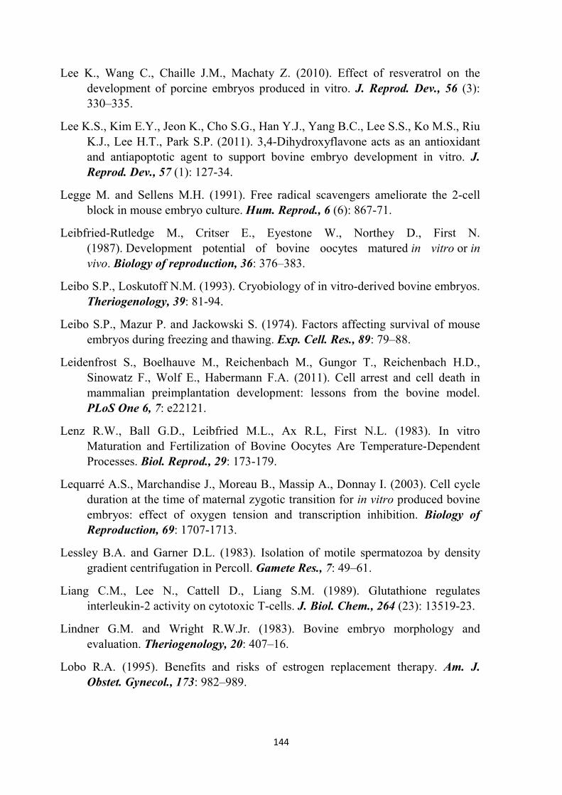
144
Lee K., Wang C., Chaille J.M., Machaty Z. (2010). Effect of resveratrol on the development of porcine embryos produced in vitro. J. Reprod. Dev., 56 (3): 330–335.
Lee K.S., Kim E.Y., Jeon K., Cho S.G., Han Y.J., Yang B.C., Lee S.S., Ko M.S., Riu K.J., Lee H.T., Park S.P. (2011). 3,4-Dihydroxyflavone acts as an antioxidant and antiapoptotic agent to support bovine embryo development in vitro. J.
Reprod. Dev., 57 (1): 127-34.
Legge M. and Sellens M.H. (1991). Free radical scavengers ameliorate the 2-cell block in mouse embryo culture. Hum. Reprod., 6 (6): 867-71.
Leibfried-Rutledge M., Critser E., Eyestone W., Northey D., First N. (1987). Development potential of bovine oocytes matured in vitro or in vivo. Biology of reproduction, 36: 376–383.
Leibo S.P., Loskutoff N.M. (1993). Cryobiology of in vitro-derived bovine embryos. Theriogenology, 39: 81-94.
Leibo S.P., Mazur P. and Jackowski S. (1974). Factors affecting survival of mouse embryos during freezing and thawing. Exp. Cell. Res., 89: 79–88.
Leidenfrost S., Boelhauve M., Reichenbach M., Gungor T., Reichenbach H.D., Sinowatz F., Wolf E., Habermann F.A. (2011). Cell arrest and cell death in mammalian preimplantation development: lessons from the bovine model. PLoS One 6, 7: e22121.
Lenz R.W., Ball G.D., Leibfried M.L., Ax R.L, First N.L. (1983). In vitro Maturation and Fertilization of Bovine Oocytes Are Temperature-Dependent Processes. Biol. Reprod., 29: 173-179.
Lequarré A.S., Marchandise J., Moreau B., Massip A., Donnay I. (2003). Cell cycle duration at the time of maternal zygotic transition for in vitro produced bovine embryos: effect of oxygen tension and transcription inhibition. Biology of
Reproduction, 69: 1707-1713.
Lessley B.A. and Garner D.L. (1983). Isolation of motile spermatozoa by density gradient centrifugation in Percoll. Gamete Res., 7: 49–61.
Liang C.M., Lee N., Cattell D., Liang S.M. (1989). Glutathione regulates interleukin-2 activity on cytotoxic T-cells. J. Biol. Chem., 264 (23): 13519-23.
Lindner G.M. and Wright R.W.Jr. (1983). Bovine embryo morphology and evaluation. Theriogenology, 20: 407–16.
Lobo R.A. (1995). Benefits and risks of estrogen replacement therapy. Am. J.
Obstet. Gynecol., 173: 982–989.
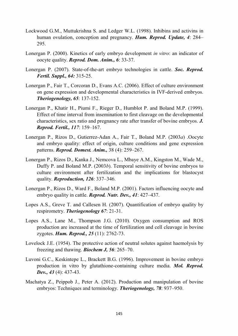
145
Lockwood G.M., Muttukrishna S. and Ledger W.L. (1998). Inhibins and activins in human ovulation, conception and pregnancy. Hum. Reprod. Update, 4: 284–295.
Lonergan P. (2000). Kinetics of early embryo development in vitro: an indicator of oocyte quality. Reprod. Dom. Anim., 6: 33-37.
Lonergan P. (2007). State-of-the-art embryo technologies in cattle. Soc. Reprod.
Fertil. Suppl., 64: 315-25.
Lonergan P., Fair T., Corcoran D., Evans A.C. (2006). Effect of culture environment on gene expression and developmental characteristics in IVF-derived embryos. Theriogenology, 65: 137-152.
Lonergan P., Khatir H., Piumi F., Rieger D., Humblot P. and Boland M.P. (1999). Effect of time interval from insemination to first cleavage on the developmental characteristics, sex ratio and pregnancy rate after transfer of bovine embryos. J.
Reprod. Fertil., 117: 159–167.
Lonergan P., Rizos D., Gutierrez-Adan A., Fair T., Boland M.P. (2003a) .Oocyte and embryo quality: effect of origin, culture conditions and gene expression patterns. Reprod. Domest. Anim., 38 (4): 259–267.
Lonergan P., Rizos D., Kanka J., Nemcova L., Mbaye A.M., Kingston M., Wade M., Duffy P. and Boland M.P. (2003b). Temporal sensitivity of bovine embryos to culture environment after fertilization and the implications for blastocyst quality. Reproduction, 126: 337–346.
Lonergan P., Rizos D., Ward F., Boland M.P. (2001). Factors influencing oocyte and embryo quality in cattle. Reprod. Nutr. Dev., 41: 427–437.
Lopes A.S., Greve T. and Callesen H. (2007). Quantification of embryo quality by respirometry. Theriogenology 67: 21-31.
Lopes A.S., Lane M., Thompson J.G. (2010). Oxygen consumption and ROS production are increased at the time of fertilization and cell cleavage in bovine zygotes. Hum. Reprod., 25 (11): 2762-73.
Lovelock J.E. (1954). The protective action of neutral solutes against haemolysis by freezing and thawing. Biochem J, 56: 265–70.
Luvoni G.C., Keskintepe L., Brackett B.G. (1996). Improvement in bovine embryo production in vitro by glutathione-containing culture media. Mol. Reprod.
Dev., 43 (4): 437-43.
Machatya Z., Peippob J., Peter A. (2012). Production and manipulation of bovine embryos: Techniques and terminology. Theriogenology, 78: 937–950.
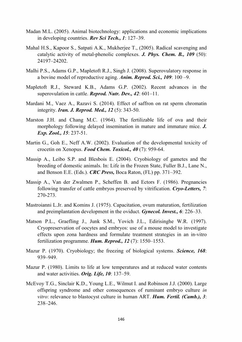
146
Madan M.L. (2005). Animal biotechnology: applications and economic implications in developing countries. Rev Sci Tech., 1: 127–39.
Mahal H.S., Kapoor S., Satpati A.K., Mukherjee T., (2005). Radical scavenging and catalytic activity of metal-phenolic complexes. J. Phys. Chem. B., 109 (50): 24197–24202.
Malhi P.S., Adams G.P., Mapletoft R.J., Singh J. (2008). Superovulatory response in a bovine model of reproductive aging. Anim. Reprod. Sci., 109: 100 –9.
Mapletoft R.J., Steward K.B., Adams G.P. (2002). Recent advances in the superovulation in cattle. Reprod. Nutr. Dev., 42: 601–11.
Mardani M., Vaez A., Razavi S. (2014). Effect of saffron on rat sperm chromatin integrity. Iran. J. Reprod. Med., 12 (5): 343-50.
Marston J.H. and Chang M.C. (1964). The fertilizable life of ova and their morphology following delayed insemination in mature and immature mice. J.
Exp. Zool., 15: 237-51.
Martin G., Goh E., Neff A.W. (2002). Evaluation of the developmental toxicity of crocetin on Xenopus. Food Chem. Toxicol., 40 (7): 959-64.
Massip A., Leibo S.P. and Blesbois E. (2004). Cryobiology of gametes and the breeding of domestic animals. In: Life in the Frozen State, Fuller B.J., Lane N., and Benson E.E. (Eds.). CRC Press, Boca Raton, (FL) pp. 371–392.
Massip A., Van der Zwalmen P., Scheffen B. and Ectors F. (1986). Pregnancies following transfer of cattle embryos preserved by vitrification. Cryo-Letters, 7: 270-273.
Mastroianni L.Jr. and Komins J. (1975). Capacitation, ovum maturation, fertilization and preimplantation development in the oviduct. Gynecol. Invest., 6: 226–33.
Matson P.L., Graefling J., Junk S.M., Yovich J.L., Edirisinghe W.R. (1997). Cryopreservation of oocytes and embryos: use of a mouse model to investigate effects upon zona hardness and formulate treatment strategies in an in-vitro fertilization programme. Hum. Reprod., 12 (7): 1550–1553.
Mazur P. (1970). Cryobiology; the freezing of biological systems. Science, 168: 939–949.
Mazur P. (1980). Limits to life at low temperatures and at reduced water contents and water activities. Orig. Life, 10: 137–59.
McEvoy T.G., Sinclair K.D., Young L.E., Wilmut I. and Robinson J.J. (2000). Large offspring syndrome and other consequences of ruminant embryo culture in vitro: relevance to blastocyst culture in human ART. Hum. Fertil. (Camb.), 3: 238–246.

147
McKiernan S.H. and Bavister B.D. (1994). Timing of development is a critical parameter for predicting successful embryogenesis. Hum. Reprod., 9: 2123–2129.
Melville D.B., Horner W.H., Otken C.C. and Ludwig M.L. (1955). Studies of the origin of ergothioneine in animals. J. Biol. Chem., 213: 61–68.
Meng L. and Cui L. (2008). Inhibitory effects of crocetin on high glucose-induced apoptosis in cultured human umbilical vein endothelial cells and its mechanism. Arch. Pharm. Res., 31 (3): 357-363.
Merton J.S., de Roos A.P.W., Mullaart E., de Ruigh L., Kaal L., Vos P.L.A.M., Dieleman S.J. (2003). Factors affecting oocyte quality and quantity in commercial applications of embryo technologies in the cattle breeding industry. Theriogenology, 59: 651-674.
Merton S. (2002). Morphological evaluation of embryos in domestic animals. In: Van Soom A. and Boerjan M. (Eds), Assessment of mammalian embryo quality: invasive and non-invasive techniques. Dordrecht, The Netherlands: Kluwer Academic Publishers, pp. 31–55.
Migheli A., Attanasio A., Schiffer D. (1995). Ultrastructural detection of DNA strand breaks in apoptotic neural cells by in situ endlabelling techniques. Journal of Pathology, 176: 27–35.
Mihm M., Baker P.J., Ireland J.L., Smith G.W., Coussens P.M., Evans A.C., Ireland J.J. (2006). Molecular evidence that growth of dominant follicles involves a reduction in follicle-stimulating hormone dependence and an increase in luteinizing hormone dependence in cattle. Biol. Reprod., 74: 1051–9.
Mitsuyama H. and May J.M. (1999). Uptake and antioxidant effects of ergothioneine in human erythrocytes. Clin. Sci., 97: 407–411.
Modaresi M., Messripour M., Nazern H. and Asadi M. (2008). Effect of Saffron Extract on Pituitary- testis Axis in Mice. International Journal of Health
Science, 1 (1): 7-8.
Mohajeri D., Amouoghli Tabrizi B., Mousavi Gh., Mesgari M. (2008). Anti-diabetic activity of Crocus sativus L. (Saffron) stigma ethanolic extract in alloxan-induced diabetic rats. Research Journal of Biological Science, 3 (9): 1102-1108.
Mohammadi-Sangcheshmeh A., Held E., Rings F., Ghanem N., Salilew-Wondim D., Tesfaye D., Sieme H., Schellander K. and Höelker M. (2013). Developmental competence of equine oocytes: impacts of zona pellucida birefringence and maternally derived transcript expression. Reprod. Fertil. Dev., 26 (3): 441-52.

148
Monaco E. (2007). Studio di alcune proteine del fluido oviduttale: effetto dell’osteopontina sulla produzione embrionale in vitro nella specie bovina. PhD Thesis; Università degli studi di Napoli Federico II; Italia.
Moncaster J.A., Walsh D.T., Gentleman S.M., Jen L.S. and Aruoma O.I. (2002). Ergothioneine treatment protects neurons against N-methyl-D-aspartate excitotoxicity: In an in vivo retinal model. Neurosci. Lett., 328: 55–59.
Monniaux D., Barbey S., Rico C., Fabre S., Gallard Y., Larroque H. (2010). Anti-müllerian hormone: a predictive marker of embryo production in cattle? Reprod. Fertil. Dev., 22: 1083–91.
Monniaux D., Chupin D., Saumande J. (1983). Superovulatory response of cattle. Theriogenology, 19: 55– 80.
Moshiri E., Basti A.A., Noorbala A.A., Jamshidi A.H., Hesameddin Abbasi S., Akhondzadeh S. (2006). Crocus sativus L. (petal) in the treatment of mild-to-moderate depression: A double-blind, randomized and placebo-controlled trial. Phytomedicine, 13: 607-11.
Mukaida T., Nakamura S., Tomiyama T., Wada S., Oka C., Kasai M., Takahashi K. (2003). Vitrification of human blastocysts using cryoloops: clinical outcome of 223 cycles. Hum. Reprod., 18: 384 –91.
Nagano M., Katagiri S. and Takahashi Y. (2006). Relationship between bovine oocyte morphology and in vitro developmental potential. Zygote, 14: 53–61.
Nagano M., Takahashi Y. and Katagiri S. (1999). In vitro fertilization and cortical granule distribution of bovine oocytes having heterogeneous ooplasm with dark clusters 1. Journal of Veterinary Medical Science, 61: 531-535.
Nair S.C., Salomi M.J., Varghese C.D., Panikkar B., Panikkar K.R. (1992). Effect of saffron on thymocyte proliferation, intracellular glutathione levels and its antitumor activity. Biofactors, 4 (1): 51-4.
Nair SC, Kurumboor SK, Hasegawa JH. (1995). Saffron chemoprevention in biology and medicine: a review. Cancer Biother., 10 (4): 257-64.
Nasr-Esfahani M.H. and Johnson M.H. (1992). Quantitative analysis of cellular glutathione in early preimplantation mouse embryos developing in vivo and in vitro. Hum. Reprod., 7: 1281–1290.
Nasr-Esfahani M.H., Aitken J.R., Johnson M.H. (1990). Hydrogen peroxide levels in mouse oocytes and early cleavage stage embryos developedin vitro or in vivo. Development, 109: 501–507.

149
Nasr-Esfahani M.H., Winston N.J., Johnson M.H., (1992). Effects of glucose, glutamine, ethylenediaminetetraacetic acid and oxygen tension on the concentration of reactive oxygen species and on development of the mouse preimplantation embryo in vitro. J. Reprod. Fertil., 96: 219–231.
Natsuyama S., Noda Y., Yamashita M., Nagahama Y., Mori T. (1993). Superoxide dismutase and thioredoxin restore defective p34cdc2 kinase activation in mouse two-cell block. Biochim. Biophys. Acta., 10 (1-2): 90-4.
Nedambale T.L., Dinnyés A., Yang X., Tian X.C. (2004). Bovine blastocyst development in vitro: timing, sex, and viability following vitrification. Biol.
Reprod., 71: 1671– 6.
Neglia G., Gasparrini B., Caracciolo di Brienza V., Di Palo R., Campanile G., Presicce G.A., Zicarelli L. (2003b). Bovine and buffalo in vitro embryo production using oocytes derived from abattoir ovaries or collected by transvaginal follicle aspiration. Theriogenology, 59: 1123-1130.
Neglia G., Gasparrini B., Di Palo R., De Rosa C., Zicarelli L., Campanile G. (2003a). Comparison of pregnancy rates with two estrus synchronization protocols in Italian Mediterranean Buffalo cows. Theriogenology, 60: 125–133.
Neglia G., Marino M., Di Palo R., Wilding M., Caracciolo di Brienza V., Dale B., Gasparrini B., Zicarelli L. (2001). A comparison of in vitro maturation in buffalo (Bubalus Bubalis) and bovine oocytes using confocal microscopy. Theriogenology, 55: 488.
Niemann H., Wrenzycki C. (2000). Alterations of expression of developmentally important genes in preimplantation bovine embryos by in vitro culture conditions: implications for subsequent development. Theriogenology, 53: 21–34.
Niwa K. and Ohgoda O. (1988). Synergistic effect of caffeine and heparin on in-vitro fertilization of cattle oocytes matured in culture. Theriogenology, 30: 733–41.
Noda Y., Shiotani M., Goto Y., Nakayama T., Umaoka Y., Mori T. (1994). Culture of human embryos in alpha modification of Eagle’s medium under low oxygen tension and low illumination. Fert. Steril., 62: 1022–1027.
Nonogaki T., Noda Y., Narimoto K., Umaoka Y., Mori T. (1991). Protection from oxidative stress by thioredoxin and superoxide dismutase of mouse embryos fertilized in vitro. Hum. Reprod., 6: 1305–1310.
Ochiai T., Shimeno H., Mishirna K., Iwasaki K., Fujiwara M., Tanaka H., Shoyama Y., Toda A., Eyanagi R., Soeda S. (2007). Protective effects of carotenoids from saffron on neuronal injury in vitro and in vivo. Biochim. Biophys. Acta.,
28 (4): 578-84.

150
Olson S.E. and Seidel G.E.Jr. (2000). Culture of in vitro-produced bovine embryos with vitamin E improves development in vitro and after transfer to recipients. Biol. Reprod., 62: 248–252.
Overström E.W. (1996). In vitro assessment of embryo viability. Theriogenology,
45: 3–16.
OXIS Technical Document, (1997). L-Ergothioneine. Compound Monograph. OXIS
Health Products, Portland, OR, (USA).
Ozawa M., Nagai T., Fahrudin M., Karja N.W., Kaneko H., Noguchi J., Ohnuma K., Kikuchi K. (2006). Addition of glutathione or thioredoxin to culture medium reduces intracellular redox status of porcine IVM/IVF embryos, resulting in improved development to the blastocyst stage. Mol. Reprod. Dev., 73 (8): 998-1007.
Öztürkler Y., Yildiz S., Güngör O., Pancarci S.M., Kaçar C., Ari U.Ç. (2010). The Effects of L-Ergothioneine and L-Ascorbic Acid on the In Vitro Maturation (IVM) and Embryonic Development (IVC) of Sheep Oocytes. Kafkas Univ.
Vet. Fak. Derg., 16 (5): 757-763.
Palozza P. and Krinsky N.I. (1992). Antioxidant effects of carotenoids in vivo and in vitro: an overview. Methods Enzymol., 213: 403-20.
Papis K., Poleszczuk O., Wenta-Muchalska E., Modlinski J.A. (2007). Melatonin effect on bovine embryo development in vitro in relation to oxygen concentration. J. Pineal. Res., 43 (4): 321-6.
Park S.P., Kim E.Y., Oh J.H., Nam H.K., Lee K.S., Park S.Y., Park E.M., Yoon S.H., Chung K.S., Lim J.H. (2000). Ultra-rapid freezing of human multipronuclear zygotes using electron microscope grids. Hum. Reprod., 15: 1787–90.
Parrish J.J. (2014). Bovine in vitro fertilization: In vitro oocyte maturation and sperm capacitation with heparin. Theriogenology, 81: 67–73.
Parrish J.J., Susko-Parrish J.L., Leibfried-Rutledge M.L., Critser E.S., Eyestone W.H., First N.L. (1986). Bovine in vitro fertilization with frozen-thawed semen. Theriogenology, 25: 591–600.
Paula-Lopes F.F. and Hansen P.J. (2002). Apoptosis is an adaptive response in bovine preimplantation embryos that facilitates survival after heat shock. Biochem. Biophys. Res. Commun., 295 (1): 37-42.
Peippo J., Machaty Z., Peter A. (2011). Terminologies for the preattachment bovine embryo. Theriogenology, 76: 1373–9.
Pereira R.M. and Marques C.C. (2008). Animal oocyte and embryo cryopreservation. Cell. Tissue Bank, 9 (4): 267-77.

151
Pérez-Pastén R., Martínez-Galero E., Chamorro-Cevallos G. (2010). Quercetin and naringenin reduce abnormal development of mouse embryos produced by hydroxyurea. J. Pharm. Pharmacol., 62 (8): 1003-9.
Pickett B.W., Berndtson W.E. (1974). Preservation of bovine spermatozoa by freezing in straws: a review. J. Dairy Sci., 57: 1287–301.
Pirola L. and Fröjdö S. (2008). Resveratrol: one molecule, many targets. IUBMB
Life, 60 (5): 323–332.
Pocar P., Brevini T.A., Perazzoli F., Cillo F., Modina S., Gandolfi F. (2001). Cellular and molecular mechanisms mediating the effects of polychlorinated biphenyls on oocyte developmental competence in cattle. Molecular Reproduction &
Development, 60: 535-541.
Polge C., Smith A. and Park A.S. (1949). Revival of spermatozoa after vitrification and dehydration at low temperature. Nature, 164: 666.
Pollard J.W. and Leibo S.P. (1994). Chilling sensitivity of mammalian embryos. Theriogenology, 41: 101–106.
Pomar F.J., Teerds K.J., Kidson A., Colenbrander B., Tharasanit T., Aguilar B., Roelen B.A. (2005). Differences in the incidence of apoptosis between in vivo and in vitro produced blastocysts of farm animal species: a comparative study. Theriogenology, 63: 2254–2268.
Portmann M., Nagy Z.P. and Behr B. (2010). Evaluation of blastocyst survival following vitrification/warming using two different closed carrier systems. Human Reproduction, 25: 261. Abstract
Prather R.S., Day B.N. (1998). Practical considerations for the in vitro production of pig embryos. Theriogenology, 49 (1): 23–32.
Ptak G., Dattena M., Loi P., Tischner M. and Cappai P. (1999). Ovum pick-up in sheep: efficiency of in vitro embryo production, vitrification and birth of offspring. Theriogenology, 52: 1105-1114.
Pujol M., Lopez-Bejar M. and Paramio M.T. (2004). Developmental competence of heifer oocytes selected using the brilliant cresyl blue (BCB) test. Theriogenology, 61: 735–744.
Pukazhenthi B.S. and Wildt D.E. (2004). Witch reproductive technologies are most relevant to studying, managing and conserving wildlife? Reprod. Fertil. Dev.,
16: 33-46.
Pursel V.G., Wall R.J., RExroad C.E., Hammer R.E., Brinster R.L. (1985). A rapid whole-mount staining procedure for nuclei of mammalian embryos. Theriogenology, 24: 687-700.

152
Racowsky C., Combelles C.M., Nureddin A., Pan Y., Finn A., Miles L., Gale S., O’Leary T. and Jackson K.V. (2003). Day 3 and Day 5 morphological predictors of embryo viability. Reprod. Biomed. Online, 6: 323–331.
Raff M.C., Barres B.A., Burne J.F., Coles H.S., Ishizaki Y., Jacobson M.D. (1993). Programmed cell death and the control of cell survival: lessons from the nervous system. Science, 262: 695–700.
Rall W.F. and Fahy G.M. (1985). Ice-free cryopreservation of mouse embryos at -196 °C by vitrification. Nature, 313: 573–5.
Rath D., Johnson L.A. (2008). Application and commercialization of flow cytometrically sex-sorted semen. Reprod. Domest. Anim., 43 (Suppl. 2): 338–46.
Reed W.A., Suh T., Bunch T.D., White K.L. (1996). Culture of in vitro fertilized bovine embryos with bovine oviductal epithelial cells, buffalo rat liver (BRL) cells, or BRL-cell-conditioned medium. Theriogenology, 45: 439–449.
Rehman N., Collins A.R., Suh T.K., Wright R.W. (1994). Effect of sperm exposure time on in vitro fertilization and embryo development of bovine oocytes matured in vitro. Theriogenology, 41: 1447-52.
Rhemrev J., Jeyendran R.S., Vermeiden J.P., Zaneveld L.J. (1989). Human sperm selection by glass wool filtration and two-layer, discontinuous Percoll gradient centrifugation. Fertil. Steril., 51: 685–90.
Rico C., Fabre S., Médigue C., di Clemente N., Clément F., Bontoux M., Touzé J.L., Dupont M., Briant E., Rémy B., Beckers J.F., Monniaux D. (2009). Anti-mullerian hormone is an endocrine marker of ovarian gonadotropin-responsive follicles and can help to predict superovulatory responses in the cow. Biol.
Reprod., 80: 50–9.
Ríos J., Recio M., Giner R. and Mañez S. (1996). An update review of saffron and its active compounds. Phytother. Res., 10 (3): 189–193.
Rizos D., Gutierrez-Adan A., Perez-Garnelo S., De La Fuente J., Boland M. P., Lonergan P. (2003). Bovine embryo culture in the presence or absence of serum: implications for blastocyst development, cryotolerance, and messenger RNA expression. Biol. Reprod., 68: 236–243.
Rizos D., Lonergan P., Boland M. P., Arroyo-Garcia R., Pintado B., de la Fuente J., Gutierrez-Adan A. (2002a). Analysis of differential messenger RNA expression between bovine blastocysts produced in different culture systems: implications for blastocyst quality. Biol. Reprod., 66: 589–595.

153
Rizos D., Ward F., Duffy P., Boland M. P., Lonergan P. (2002b). Consequences of bovine oocyte maturation, fertilization or early embryo development in vitro versus in vivo: implications for blastocyst yield and blastocyst quality. Mol.
Reprod. Dev., 61: 234–248.
Roberts R.M., Farin C.E., Cross J.C. (1990). Trophoblast proteins and maternal recognition of pregnancy. Oxford Review of Reproductive Biology, 12: 147–180.
Robertson I. and Nelson R.E. (2010). Certification and identification of embryo. In: Stringfellow D.A. & Givens M.D. (Eds.), Manual of the International Embryo Transfer Society. IETS 2010, Champaign, IL, (USA), pp. 86–105.
Rodriguez-Martinez H. (2007). Role of the oviduct in sperm capacitation. Theriogenology, 68: 138–46.
Rodriguez-Martinez H. (2012). Assisted reproductive techniques for cattle breeding in developing countries: a critical appraisal of their value and limitations. Reprod. Domest. Anim., 47 (Suppl. 1): 21–26.
Rodriguez-Osorio N., Kim I.J., Wang H., Kaya A., Memili E. (2007). Melatonin increases cleavage rate of porcine preimplantation embryos in vitro. J. Pineal.
Res., 43 (3): 283-8.
Rosenkrans C.F.Jr., Zeng G.Q., McNamara G.T., Schoff P.K., First N.L. (1993). Development of bovine embryos in vitro as affected by energy substrates. Biol.
Reprod., 49: 459–62.
Rubessa M., Boccia L., Campanile G., Longobardi V., Albarella S., Tateo A., Zicarelli L., Gasparrini B. (2011). Effect of energy source during culture on in vitro embryo development, resistance to cryopreservation and sex ratio. Theriogenology, 76 (7): 1347–1355.
Rubessa M., Di Fenza M., Mariotti E., Di Francesco S., De Dilectis C., Di Palo R., Zicarelli L., Gasparrini B. (2009). Kinetics of sperm penetration is correlated with in vitro fertility of buffalo (Bubalus Bubalis) bulls. Reprod. Fertil. Dev.,
21 (I): 206-207.
Russell D.F., Baqir S., Bordignon J., Betts D.H. (2006). The impact of oocyte maturation media on early bovine embryonic development. Mol. Reprod. Dev.,
73: 1255–1270.
Sakkas D., Shoukir Y., Chardonnens D., Bianchi P.G. and Campana A. (1998). Early cleavage of human embryos to the two-cell stage after intracytoplasmic sperm injection as an indicator of embryo viability. Hum. Reprod., 13: 182–187.

154
Sartori R., Haughian J.M., Shaver R.D., Rosa G.J., Wiltbank M.C. (2004). Comparison of ovarian function and circulating steroids in estrous cycles of Holstein heifers and lactating cows. J. Dairy Sci., 87: 905–20.
Schultz R.M. and Kopf G.S. (1995). Molecular basis of mammalian egg activation. Curr. Top. Dev. Biol., 30: 21–62.
Semple M.E., Betteridge K.J., Leibo S.P. (1995). Cryopreservation of in vitro-derived bovine embryos produced in a serum-free culture sys-tem. Theriogenology, 43: 320–420.
Seren E. and Parmeggiani A. (1997). Oestrus cycle in Italian Buffalo. Third course
on biotechnology of reproduction in buffaloes, Caserta, Italy, October 6-10: 21-28.
Shamsuddin M., Rodriguez-Martinez H., Larsson B. (1993). Fertilizing capacity of bovine spermatozoa selected after swim-up in hyaluronic acid-containing medium. Reprod. Fertil. Dev., 5: 307–15.
Shehab-El-Deen M.A., Leroy J.L.M.R., Maes D., Van Soom A. (2009). Cryotolerance of bovine blastocysts is affected by oocyte maturation in media containing palmitic or stearic acid. Reprod. Dom. Anim., 44 (1): 140-2.
Shelton F. (1946). Artificial insemination increases fertility. Indian Vet. J., 22: 467–71.
Shoukir Y., Chardonnens D., Campana A., Bischof P. and Sakkas D. (1998). The rate of development and time of transfer play different roles in influencing the viability of human blastocysts. Hum. Reprod., 13: 676–681.
Sinclair K.D., McEvoy T.G., Maxfield E.K., Maltin C.A., Young L.E., Wilmut I., Broadbent P.J., Robinson J.J. (1999). Aberrant fetal growth and development after in vitro culture of sheep zygotes. J. Reprod. Fertil., 116: 177–186.
Singh J., Dominiquez M., Jaiswal R., Adams G.P. (2004). A simple ultrasound test to predict the superstimulatory response in cattle. Theriogenology, 62: 1407–20.
Sirard M.A., Florman H., Leibfried-Rutledge M.L., First N.L. (1989). Timing of nuclear progression and protein synthesis necessary for meiotic maturation of bovine oocytes. Biol. Reprod., 40: 1527–1563.
Smith G.D. (2001). In vitro maturation of oocytes. Curr. Womens Health Rep., 1: 143–51.
Solter D. and Knowles B.B. (1978). Monoclonal antibody defining a stagespecific mouse embryonic antigen (SSEA-1). Proceedings of the National Academy of
Sciences of the USA, 75: 5565–5569.
155
Somfai T., Ozawa M., Noguchi J., Kaneko H., Kuriani Karja N.W., Farhudin M., Dinnyés A., Nagaic T., Kikuchia K. (2007b). Developmental competence of in vitro-fertilized porcine oocytes after in vitro maturation and solid surface vitrification: effect of cryopreservation on oocyte antioxidative system and cell cycle stage. Cryobiology, 55: 115–126.
Somfai T., Ozawa M., Noguchi J., Kaneko H., Ohnuma K., Karja N.W.K., Farhudin M., Maedomari N., Dinnyes A., Nagai T., Kikuchi K. (2007a). Vitrification causes damage of the antioxidant defense system in IVM porcine oocytes. Reprod. Fertil. Dev., 19: 184.
SPSS Inc., released 2007. SPSS for Windows, Version 16.0. SPSS Inc., Chicago.
Steptoe P.C. and Edwards R.G. (1978). Birth after the reimplantation of a human embryo [letter]. Lancet, 2: 366.
Stojkovic M., Machado S.A., Stojkovic P., Zakhartchenko V., Hutzler P., Goncalves P.B. and Wolf E. (2001). Mitochondrial distribution and adenosine triphosphate content of bovine oocytes before and after in vitro maturation: correlation with morphological criteria and developmental capacity after in vitro fertilization and culture. Biol. Reprod., 64: 904-909.
Stroud B. (2012). The year 2011 worldwide statistics of embryo transfer in domestic farm animals. IETS 2012. Statistics and Data Retrieval Committee Report.
Stroud B. and Hasler J.F. (2006). Dissecting why superovulation and embryo transfer usually work on some farms but not on others. Theriogenology, 65: 65–76.
Szende B., Tyihák E., Király-Véghely Z. (2000). Dose-dependent effect of resveratrol on proliferation and apoptosis in endothelial and tumor cell cultures. Exp. Mol. Med., 32 (2): 88–92.
Takahashi M. (2012). Oxidative stress and redox regulation on in vitro development of mammalian embryos. J. Reprod. Dev., 58 (1): 1-9.
Talevi R., Gualtieri R., Tartaglione G. and Fortunato A. (1997). Heterogeneity of the zona pellucida carbohydrate distribution in human oocytes failing to fertilize in vitro. Hum. Reprod., 12: 2773–2780.
Tam P.P. (1988). Postimplantation development of mitomycin C-treated mouse blastocysts. Teratology, 37: 205–212.
Tatone C., Di Emidio G., Vento M., Ciriminna R., Artini P.G. (2010). Cryopreservation and oxidative stress in reproductive cells. Gynecol.
Endocrinol., 26 (8): 563-7.

156
Tavana S., Eimani H., Azarnia M., Shahverdi A., Eftekhari-Yazdi P. (2012). Effects of saffron (Crocus sativus L.) aqueous extract on in vitro maturation, fertilization and embryo development of mouse oocytes. Cell. J., 13 (4): 259-64.
Tervit H.R., Whittingham D.G., Rowson L.E. (1972). Successful culture in vitro of sheep and cattle ova. J. Reprod. Fertil., 30: 493–7.
Thompson J.G. (1996) Defining the requirements for bovine embryo culture. Theriogenology, 45: 27-40.
Thouas G.A., Korfiatis N.A., French A.J., Jones G.M., Trounson A.O. (2001). Simplified technique for differential staining of inner cell mass and trophectoderm cells of mouse and bovine blastocysts. Reproductive
BioMedicine Online, 3: 25–29.
Tian X.Z., Wen Q., Shi J.M., Liang-Wang, Zeng S.M., Tian J.H., Zhou G.B., Zhu S.E., Liu G.S. (2010). Effects of melatonin on in vitro development of mouse two-cell embryos cultured in HTF medium. Endocr. Res., 35 (1): 17-23.
Totey S.M., Daliri M., Appa Rao K.B., Pawshe C.H., Taneja M. and Chillar R.S. (1996). Differential cleavage and developmental rates and their correlation with cell numbers and sex ratios in buffalo embryos generated in vitro. Theriogenology, 45: 521–533.
Traldi A.S. (2000). Vitrification of goat in vivo and in vitro produced embryos. In: Proc. 7th Int. Conf. Goats, (Tours, France): 1031.
Trounson A. and Bongso A. (1996). Fertilization and development in humans. Current Topics in Develomental Biology, 32: 59–101.
Tseng T.H., Chu C.Y., Huang J.M., Shiow S.J., Wang C.J. (1995). Crocetin protects against oxidative damage in rat primary hepatocytes. Cancer Letter, 97: 61–67.
Turner K., Goldstei D. and Rogers A. (1992). Variaton in the dry mass of mouse embryos throughout the preimplantation period. Hum. Reprod., 7 (1): 112-116.
Uhm S.J., Gupta M.K., Yang J.H., Lee S.H., Lee H.T. (2007). Selenium improves the developmental ability and reduces the apoptosis in porcine parthenotes. Mol. Reprod. Dev., 74 (11): 1386-94.
Vajta G. (2000). Vitrificazion of the oocytes and embryos of domestic animals. Anim. Reprod. Sci., 60-61: 357-364.
Vajta G. and Kuwayama M. (2006). Improving cryopreservation systems. Theriogenology, 65: 236–44.

157
Vajta G., Booth P.J., Holm P., Greve T., Callesen H. (1997). Successful vitrification of early stage bovine in vitro produced embryos with the open pulled straw (OPS) method. Cryo-Lett, 18: 191–5.
Vajta G., Holm P., Kuwayama M., Booth P.J., Jacobsen H., Greve T., Callesen H. (1998). Open pulled straw (OPS) vitrification: a new way to reduce cryoinjuries of bovine ova and embryos. Mol. Reprod. Dev., 51: 53–58.
Vajta G., Rienzi L., Cobo A., Yovich J. (2010). Embryo culture: can we perform better than nature? Reproductive BioMedicine Online, 20: 453– 469.
Van den Broeke L.T. and Beyersbergen van Henegouwen G.M. (1993). Thiols as potential UV radiation protectors: An in vitro study. J. Photochem. Photobiol.,
17: 279–286.
Van Langendonckt A., Donnay I., Schuurbiers N., Auquier P., Carolan C., Massip A. and Dessy F. (1997). Effects of supplementation with fetal calf serum on development of bovine embryos in synthetic oviduct fluid medium. J. Reprod.
Fertil., 109: 87–93.
Van Soom A., Boerjan M. and Hardy K. (2002). Differential staining of inner and outer cells to assess mammalian embryo quality. In: Van Soom A. and Boerjan M. (Eds), Assessment of mammalian embryo quality. Dordrecht, The Netherlands: Kluwer Academic Publishers, pp. 237-259.
Van Soom A., Van Vlaenderen I., Mahmoudzadeh A.R., Deluyker H., de Kruif A. (1992). Compaction rate of in vitro fertilized bovine embryos related to the interval from insemination to first cleavage. Theriogenology, 38: 905-919.
Van Soom A., Vanroose G. and de Kruif A. (2001). Blastocyst evaluation by means of differential staining: a practical approach. Reprod. Dom. Anim., 36: 29-35.
Van Soom A., Ysebaert M.T. and de Kruif A. (1997). Relationship between timing of development, morula morphology and cell allocation to inner cell mass and trophectoderm in in vitro produced bovine embryos. Mol. Reprod. Dev., 47: 47-56.
van Wagtendonk-de Leeuw A.M., Mullaart E., de Roos A.P., Merton J.S., den Daas J.H., Kemp B. and de Ruigh L. (2000). Effects of different reproduction techniques: AI MOET or IVP, on health and welfare of bovine offspring. Theriogenology, 53: 575-597.
Vandaele L. and Van Soom A. (2011). Intrinsic factors affecting apoptosis in bovine in vitro produced embryos. Verh K Acad Geneeskd Belg., 73 (1-2): 79-104.
Vandemark N.L. (1952). Time and site of insemination in cattle. Cornell Vet., 42: 215–22.
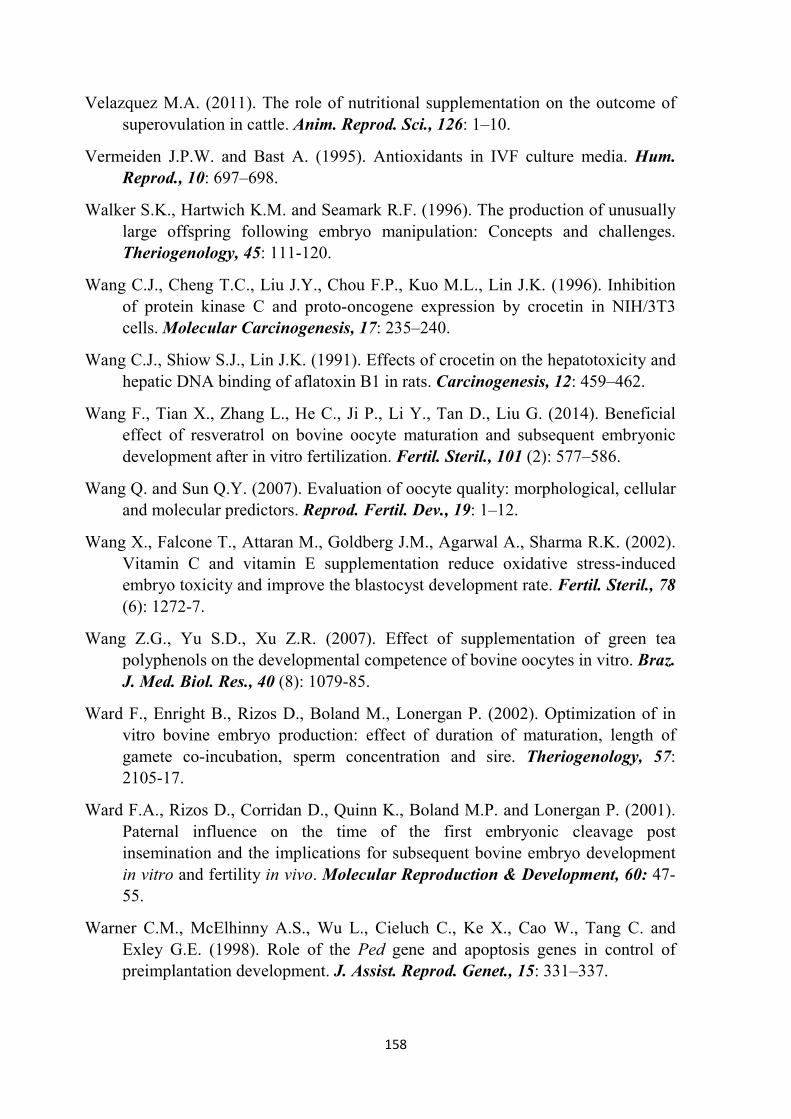
158
Velazquez M.A. (2011). The role of nutritional supplementation on the outcome of superovulation in cattle. Anim. Reprod. Sci., 126: 1–10.
Vermeiden J.P.W. and Bast A. (1995). Antioxidants in IVF culture media. Hum.
Reprod., 10: 697–698.
Walker S.K., Hartwich K.M. and Seamark R.F. (1996). The production of unusually large offspring following embryo manipulation: Concepts and challenges. Theriogenology, 45: 111-120.
Wang C.J., Cheng T.C., Liu J.Y., Chou F.P., Kuo M.L., Lin J.K. (1996). Inhibition of protein kinase C and proto-oncogene expression by crocetin in NIH/3T3 cells. Molecular Carcinogenesis, 17: 235–240.
Wang C.J., Shiow S.J., Lin J.K. (1991). Effects of crocetin on the hepatotoxicity and hepatic DNA binding of aflatoxin B1 in rats. Carcinogenesis, 12: 459–462.
Wang F., Tian X., Zhang L., He C., Ji P., Li Y., Tan D., Liu G. (2014). Beneficial effect of resveratrol on bovine oocyte maturation and subsequent embryonic development after in vitro fertilization. Fertil. Steril., 101 (2): 577–586.
Wang Q. and Sun Q.Y. (2007). Evaluation of oocyte quality: morphological, cellular and molecular predictors. Reprod. Fertil. Dev., 19: 1–12.
Wang X., Falcone T., Attaran M., Goldberg J.M., Agarwal A., Sharma R.K. (2002). Vitamin C and vitamin E supplementation reduce oxidative stress-induced embryo toxicity and improve the blastocyst development rate. Fertil. Steril., 78 (6): 1272-7.
Wang Z.G., Yu S.D., Xu Z.R. (2007). Effect of supplementation of green tea polyphenols on the developmental competence of bovine oocytes in vitro. Braz.
J. Med. Biol. Res., 40 (8): 1079-85.
Ward F., Enright B., Rizos D., Boland M., Lonergan P. (2002). Optimization of in vitro bovine embryo production: effect of duration of maturation, length of gamete co-incubation, sperm concentration and sire. Theriogenology, 57: 2105-17.
Ward F.A., Rizos D., Corridan D., Quinn K., Boland M.P. and Lonergan P. (2001). Paternal influence on the time of the first embryonic cleavage post insemination and the implications for subsequent bovine embryo development in vitro and fertility in vivo. Molecular Reproduction & Development, 60: 47-55.
Warner C.M., McElhinny A.S., Wu L., Cieluch C., Ke X., Cao W., Tang C. and Exley G.E. (1998). Role of the Ped gene and apoptosis genes in control of preimplantation development. J. Assist. Reprod. Genet., 15: 331–337.
159
Warriach H.M. and Chohan K.R. (2004). Thickness of cumulus cell layer is a significant factor in meiotic competence of buffalo oocytes. J. Vet. Sci., 5: 247–251.
Whittingham D.G., Leibo S.P., Mazur P. (1972). Survival of mouse embryos frozen to -196 degrees and -269 degrees C. Science, 178: 411–414.
Willadsen S.M. (1977). Factors affecting the survival of sheep embryos during deep-freezing and thawing. In: The Freezing of Mammalian Embryos (Ciba Found. Symposium). Elsevier-Excerpta Medica, Amsterdam, pp. 175-189.
Wolf E., Arnold G.J., Bauersachs S., Beier H.M., Blum H., Einspanier R., Fröhlich T., Herrler A., Hiendleder S., Kölle S., Prelle K., Reichenbach H.D., Stojkovic M., Wenigerkind H., Sinowatz F. (2003). Embryo-maternal communication in bovine - strategies for deciphering a complex cross-talk. Reprod. Domest.
Anim., 38: 276–89.
Woods E.J., Benson J.D., Agca Y., Critser J.K. (2004). Fundamental cryobiology of reproductive cells and tissues. Cryobiology, 48: 146-156.
Wrenzycki C., Herrmann D. and Niemann H. (2007). Messenger RNA in oocytes and embryos in relation to embryo viability. Theriogenology, 68 (Suppl. 1): S77–S83.
Wrenzycki C., Herrmann D., Lucas-Hahn A., Gebert C., Korsawe K., Lemme E., Carnwath J.W., Niemann H. (2005). Epigenetic reprogramming throughout preimplantation development and consequences for assisted reproductive technologies. Birth Defects Res. C. Embryo Today, 75: 1-9.
Wurth Y.A. and Kruip Th.A.M. (1992). Bovine embryo production in vitro after selection of the follicles and oocytes. In: Proceedings of the 12
th International
Congress of Animal Reproduction (ICAR), August 23–27, The Hague, The Netherlands, 1: 387–389
Wurth Y.A., Boni R., Hulshof S.C.J., Kruip Th.A.M. (1994). Bovine embryo production in vitro after selection of ovaries, follicles and oocytes. In: Wurth Y.A., Bovine Embryo Production In Vitro. Influencing Factors. Utrecht, The Netherlands. Utrecht University Press: 67–85. Thesis.
Wuthrich B., Schmid-Grendelmeyer P., Lunberg M. (1997). Anaphylaxis to saffron. Allergy, 52 (4): 476-477.
Wydooghe E., Vandaele L., Beek J., Favoreel H., Heindryckx B., De Sutter P., Van Soom A. (2011). Differential apoptotic staining of mammalian blastocysts based on double immunofluorescent CDX2 and active caspase-3 staining. Anal. Biochem., 416 (2): 228-30.

160
Xiang M., Qian Z.Y., Zhou C.H., Liu J., Li W.N. (2006). Crocetin inhibits leukocyte adherence to vascular endothelial cells induced by AGEs. J. Ethnopharmacol.,
107 (1): 25-31.
Yamauchi N., Sasada H., Soloy E., Dominko T., Kikuchi K., and Nagai T. (1999). Effects of hormones and osmolarity in the culture medium on germinal vesicle breakdown of porcine oocytes. Theriogenology, 52: 153-162.
Yang H.W., Hwang K.J., Kwon H.C., Kim H.S., Choi K.W., Oh K.S. (1998). Detection of reactive oxygen species (ROS) and apoptosis in human fragmented embryos. Hum. Reprod., 13: 998-1002.
Yang X., Giles J., Liu Z., Jiang S., Presicce G., Foote R., Wang C. (1995). In vitro fertilization and embryo development are influenced by IVF technicians and bulls. Theriogenology, 43: 360.
Yavin S. and Arav A. (2007). Measurement of essential physical properties of vitrification solutions. Theriogenology, 67: 81–89.
Yavin S., Aroyo A., Roth Z. and Arav A. (2009). Embryo cryopreservation in the presence of low concentration of vitrification solution with sealed pulled straws in liquid nitrogen slush. Human Reproduction, 24: 797–804.
You J., Kim J., Lim J., Lee E. (2010). Anthocyanin stimulates in vitro development of cloned pig embryos by increasing the intracellular glutathione level and inhibiting reactive oxygen species. Theriogenology, 74 (5): 777-85.
Young L.E., Sinclair K.D., Wilmut I. (1998). Large offspring syndrome in cattle and sheep. Rev. Reprod., 3: 155–163.
Younis A.I., Brackett B.G., Fayrer-Hosken R.A. (1989). Influence of serum and hormones on bovine oocyte maturation and fertilization in vitro. Gamete Res.,
23: 189–201.
Yuan Y.Q., Van Soom A., Leroy J.L., Dewulf J., Van Zeveren A., de Kruif A. and Peelman L.J. (2005). Apoptosis in cumulus cells, but not in oocytes, may influence bovine embryonic developmental competence. Theriogenology, 63: 2147–2163.
Zeuner A., Muller K., Reguszynski K. and Jewgenow K. (2003). Apoptosis within bovine follicular cells and its effect on oocyte development during in vitro maturation. Theriogenology, 59: 1421–1433.
Zhang L., Barry D.M., Denniston R.S., Bunch T.D. and Godke R.A. (1993). Birth of live calves after transfer of frozen-thawed bovine embryos fertilised in vitro. Vet. Rec., 132: 247-249.

161
Zhang Y., Shoyama Y., Sugiura M., Saito H. (1994). Effects of Crocus sativus L. on the ethanol-induced impairment of passive avoidance performances in mice. Biol. Pharm. Bull., 17 (2): 217-21.
Zheng S., Qian Z.-Y., Tang F. (2005). Suppression of vascular cell adhesion molecule-1 expression by crocetin contributes to attenuation of atherosclerosis in hypercholesterolemic rabbits. Biochemical Pharmacology, 70: 1192–1199.
Zicarelli L. (2001). Biotecnologie riproduttive dell’allevamento bufalino. Atti 1°
Congresso Nazionale sull’ Allevamento del Bufalo, 3-5 ottobre 2001, Eboli (SA); Italy: 163.
Zuelke K.A. and Brackett B.G. (1990). Luteinizing hormone-enhanced in vitro maturation of bovine oocytes with and without protein supplementation. Biol.
Reprod., 43: 784–7.