Embed Size (px)
Citation preview

Università degli Studi di Padova
Dipartimento di Biomedicina Comparata e Alimentazione
SCUOLA DI DOTTORATO DI RICERCA IN SCIENZE VETERINARIE
INDIRIZZO COMUNE
CICLO XXVIII
THE IMMUNE SYSTEM OF CETACEANS AND THEIR INTERACTION
WITH DOLPHIN MORBILLIVIRUS
Direttore della Scuola : Ch.mo Prof. GIANFRANCO GABAI
Coordinatore d’indirizzo: Ch.mo Prof. GIUSEPPE RADAELLI
Supervisore :Ch.mo Prof. SANDRO MAZZARIOL
Dottoranda : CINZIA CENTELLEGHE


È una follia odiare tutte le rose perché una spina ti ha punto,
abbandonare tutti i sogni perché uno di loro non si è realizzato,
rinunciare a tutti i tentativi perché uno è fallito.
È una follia condannare tutte le amicizie perché una ti ha tradito,
non credere in nessun amore solo perché uno di loro è stato infedele,
buttare via tutte le possibilità di essere felici solo perché qualcosa non è andato per il verso giusto.
Ci sarà sempre un’altra opportunità, un’altra amicizia, un altro amore, una nuova forza.
Per ogni fine c’è un nuovo inizio.
Antoine de Saint-Exupéry, “Il Piccolo Principe”


ABSTRACT
Immunology of marine mammals is a relatively new field of scientific studies and its monitoring
plays an important role on the individual and group management of these animals, as well as an
increasing value of environmental health indicator: cetaceans are viewed as environmental
sentinels. The current knowledge about the immune system of cetaceans and its function is
recognized as incomplete.
Therefore this study aims to implement the knowledge on the immune response in normal
conditions in cetaceans stranded along the Italian coastline in order to provide a base-line useful for
assessing the immune status of bottlenose dolphin (Tursiops truncatus) and striped dolphin
(Stenella coeruleoalba), the species most found in our seas and included in some international
conventions such as species with high protection.
The selection of cetaceans to be included in the study was based on the availability of samples of
spleen, thymus and lymph node tissue of the animals, on the way (in formalin or frozen) and the
state of preservation; They have in fact been preferred tissues of animals whose sampling occurred
within 48 hours of death in order to reduce as much as possible post-mortem alterations. Thereafter,
animals were divided into groups on the basis of information obtained from signaling, such as
species, sex, age, and environment of origin, on the outcome of virological investigations,
microbiological, parasitological and toxicological, if performed, and the cause and/or the death
mechanism.
On the lymph node slides obtained from the formalin-fix, paraffin-embedded samples it was made
basic hematoxylin-eosin staining and immunohistochemical staining using the following antibodies:
Monoclonal Mouse Anti-Human CD3 to identify T lymphocytes, Monoclonal Mouse Anti-Human
CD20 for the identification of mature B lymphocytes and Monoclonal Mouse Anti-Human HLA-DR
Antigen, Alpha-Chain for the identification of the major histocompatibility complex type II.
It is also arranged to validate by means of the Western blotting technique antibodies used in
immunohistochemistry (IHC). The use of these antibodies was valid only for humans and some
domestic animals (dogs and cats), but not in the species of interest to us such as bottlenose and
striped dolphins.
With the antibodies mentioned above, also the lymphoid tissues of dolphins stranded along the
coasts of the Canary Islands were tested and used as negative control since the death was probably
due to collisions with boats/ships.

Finally, a semi quantitative samples analysis was performed by acquisition of slides via slide
scanner for digital pathology (D-SIGHT) and a manual count of the number of cells positive for
each antibody in 10 fields to 40x magnification, considered to be representative of the entire organ,
and these data were statistically analyzed using the T TEST method. Statistical analysis showed no
significant correlation between the variables considered and the expression of the different
lymphocyte populations.
Future analysis should be aimed at analyzing the relationship between CD4+ and CD8+ cells in
order to understand the effect of the major immunomodulatory pathogens, such as the dolphin
morbillivirus, on sub-populations of T cells. It would also be important to compare the data
obtained on the immune system with those obtained from the same samples as regards the search
for environmental pollutants in order to understand the real role on the health of marine mammals
present in our seas.
The study was not focused only on the immune response of the guests to the different pathogens,
but we concentrate our work also on the most important pathogen currently causing die-offs of
marine mammals: the dolphin morbillivirus (DMV). In particular, from tissues of a positive DMV
fin whale (Balaenoptera physalus) stranded along the Italian coastline in October 2013 it was
possible to completely sequence the P / V / C gene (1520 bp), M (1007 bp) , N (1573 bp), F (1659
bp) and H (1814 bp) respectively coding for the phosphoprotein and two virulence factors (V and
C), for the matrix protein, the nucleoprotein, the fusion protein and the hemagglutinin protein. The
complete sequences of the mentioned genes were deposited in GenBank (GenBank provisional Acc.
No. KU977449, KU977450, KU977451, KU977452 and KU977453). The isolation of the virus in
the tissues was made possible by molecular techniques such as RT-PCR using primers designed ad
hoc and cloning using plasmid vector. The nucleotide changes, and consequently the amino acid
variations, of each individual gene were subsequently analyzed and compared with the viral genome
of the preceding epidemics. It was then possible to carry out a study of the tertiary structure of the
viruses to see how these single mutations had a role in the structural change of the proteins
themselves.
On the basis of this work we proceeded to identify an appropriate diagnostic technique usable even
for large whales where correct sampling and appropriate samples storage is not always possible in
field condition. We develop a nested RT-PCR which allows the unambiguous identification of a
200 bp fragment of the DMV genome corresponding to a highly conserved part of the gene H. This
technique permits, if the viral genome is particularly fragmented because of the poor tissue
conservation status, to verify the positivity to the DMV in tissue analysis, to isolate part of the
virus, otherwise difficult to be isolated, and it can be sequenced. This technique was later used
successfully for identification and sequencing of DMV in the tissues of three sperm stranded in
Vasto beach in 2014 (GenBank Acc. No. KU886570).
We can therefore say that the DMV is affecting species that until recently were considered species
not susceptible to infection, such as fin whale and sperm whale. Individuals of these new species
affected by the infection are mainly young animals or even pup and the virus isolation in a fetus
organs confirms the possibility of vertical transmission.
Given the species barrier carried out by the virus, its point-like changes in the genomic sequence of
the virus and incidence of positivity in 19% of the target species in 2015 we can claim to be in a
situation where the virus is endemic in the Mediterranean Sea resulting increase in the infectious
pressure.
Future studies will aim to understand more precisely the role of individual amino acid changes and
their influence on the virulence and pathogenicity of the virus through the use of crystallography, to
study the structure of the virus cellular receptor, CD150, to understand its real interaction with the
virus, and to try to understand the real significance of the DMV in the ecology of the entire
cetaceans population in the Mediterranean Sea.


RIASSUNTO
L’immunologia dei mammiferi marini è un campo relativamente recente degli studi scientifici e il
suo monitoraggio ha un ruolo importante sulla gestione individuale e di gruppo di questi animali,
nonché un crescente valore come indicatore della salute ambientale: i cetacei sono infatti
considerati sentinelle ambientali. Le conoscenze attuali relative al sistema immunitario dei cetacei e
alla sua funzione sono però incomplete.
Questo studio si prefigge dunque lo scopo di implementare le conoscenze sulla risposta immunitaria
in condizioni di normalità nei cetacei spiaggiati lungo le coste italiane al fine di fornire una base-
line utile per valutare lo stato immunitario di tursiope (Tursiops truncatus) e stenella (Stenella
coeruleoalba), specie maggiormente presenti nei nostri mari ed incluse in alcune convenzioni
internazionali come specie ad elevata protezione.
La selezione degli animali da inserire nello studio si è bastata sulla disponibilità di campioni di
milza, timo e tessuto linfonodale dei vari soggetti, sulle modalità (in formalina o congelati) e sullo
stato di conservazione degli stessi; sono stati infatti preferiti tessuti di animali il cui campionamento
è avvenuto dell’arco delle 48 ore dal decesso al fine di ridurre il più possibile le alterazioni post-
mortali. Successivamente gli animali sono stati divisi in gruppi sulla base delle informazioni
ottenute dal segnalamento come specie, sesso, classe di età e ambiente di provenienza, sull’esito di
indagini virologiche, microbiologiche, parassitologiche e tossicologiche, qualora eseguite, e la
causa e/o meccanismo del decesso.
Sulle sezioni di linfonodo ottenute dai campioni in formalina è stata effettuata la colorazione di base
ematossilina-eosina e le colorazioni immunoistochimiche usando gli anticorpi di seguito elencati:
Monoclonal Mouse Anti-Human CD3 per l’identificazione dei linfociti T, Monoclonal Mouse Anti-
Human CD20 per l’identificazione dei linfociti B maturi e Monoclonal Mouse Anti-Human HLA-
DR Antigen, Alpha-Chain per l’identificazione del complesso maggiore di istocompatibilità di tipo
II.
Si è inoltre provveduto a validare mediante la tecnica del Western Blotting gli anticorpi che
precedentemente sono stati usati in immunoistochimica (IHC). L’uso di questi anticorpi era validato
solo per l’uomo ed alcuni animali domestici (cane e gatto), ma non nelle specie di nostro interesse
quali tursiope e stenella.

Sono stati inoltre testati tramite IHC, con gli anticorpi sopra citati, i tessuti linfoidi di cetacei
spiaggiatesi lungo le coste delle isole Canarie usati come controllo negativo in quanto morti
verosimilmente a causa di collisioni con barche/navi.
Infine è stata effettuata un’analisi semi quantitativa dei campioni mediate l’acquisizione dei vetrini
tramite l’acquisitore D-SIGHT e una conta manuale del numero di cellule positive per ogni
anticorpo in 10 campi ad ingrandimento 40x ritenuti rappresentativi di tutto l’organo e su questi dati
è stata eseguita un’analisi statistica con il metodo del T TEST.
L’analisi statistica non ha mostrato alcuna correlazione significativa tra le numerose variabili prese
in esame e l’espressione delle diverse popolazioni linfocitarie. Analisi future dovranno essere volte
ad analizzare il rapporto tra CD4 e CD8 al fine di capire l’effetto dei principali patogeni
immunodepressori, quali il dolphin morbillivirus, sulle sub-popolazioni di linfociti T. Sarebbe
inoltre importante comparare i dati ottenuti sul sistema immunitario con quelli ottenuti dagli stessi
campioni per quanto riguarda la ricerca di agenti inquinanti ambientali per capirne in reale ruolo
sulla salute dei mammiferi marini presenti nei nostri mari.
La ricerca non si è però focalizzata solamente sulla risposta immunitaria degli ospiti ai vari
patogeni, ma ci siamo concentrati sul patogeno più importante tra quelli che al momento causano
morie tra i mammiferi marini: il dolphin morbillivirus (DMV). In particolare, a partire da tessuti di
un esemplare DMV positivo di balenottera comune (Balaenoptera physalus) spiaggiatesi lungo le
coste italiane nell’ottobre 2013 è stato possibile sequenziare completamente i geni P/V/C (1520 bp),
M (1007 bp), N (1573 bp), F (1659 bp) ed H (1814 bp) codificanti rispettivamente per la
fosfoproteina e due fattori di virulenza (V e C), per la proteina di matrice, la nucleoproteina, la
proteina di fusione e l’emoagglutinina. Le sequenze complete dei geni sopracitati cono state
depositate in GenBank (GenBank provisional Acc. No. KU977449, KU977450, KU977451,
KU977452 e KU977453). L’isolamento del virus nei tessuti dell’animale è stato possibile grazie a
tecniche biomolecolari quali RT-PCR con uso di primers disegnati ad hoc e clonaggio mediante
vettore plasmidico. I cambiamenti nucleotidici, e di conseguenza amminoacidici, di ogni singolo
gene sono stati successivamente analizzati e confrontati con il genoma virale delle precedenti
epidemie. È stato poi possibile eseguire uno studio della struttura terziaria del virus per visualizzare
come questi cambiamenti puntiformi avessero un ruolo nel cambiamento strutturale delle proteine
stesse.
Sulla base di tale lavoro di sequenziamento abbiamo provveduto a individuare una tecnica
diagnostica opportuna utilizzabile anche su animali di difficile gestione (difficoltà di
campionamento e di conservazione opportuna dei campioni) approntando una nested RT-PCR che

permetta l’identificazione univoca di un frammento di genoma di 200 bp corrispondente ad una
parte altamente conservata del gene H. Questa tecnica permette, qualora il genoma del virus sia
particolarmente frammentato a causa del cattivo stato di conservazione dell’animale, di verificare la
positività a DMV del tessuto in analisi, di isolare parte del virus, altrimenti difficilmente isolabile, e
di poterlo sequenziare. Tale tecnica è stata poi utilizzata con successo per l’identificazione e il
sequenziamento di DMV nei tessuti di 3 capodogli spiaggiatisi a Vasto nel 2014 (GenBank Acc.
No. KU886570).
È possibile dunque affermare che il DMV sta colpendo specie che fino a poco tempo fa erano
considerate specie non sensibili all’infezione, quali balenottera comune e capodoglio. Gli animali di
queste nuove specie colpiti dall’infezione sono prevalentemente animali giovani o addirittura
cuccioli e l’aver isolato il virus negli organi di un feto conferma la possibilità di trasmissione dello
stesso per via verticale.
Visto il salto di specie effettuato dal virus, i suoi cambiamenti puntiformi nella sequenza genomica
e un’incidenza di positività del virus nelle specie target del 19% nel 2015 possiamo affermare di
essere in una situazione in cui il virus è endemico nel Mar Mediterraneo con conseguente aumento
della pressione infettante.
Gli studi futuri in questo ambito saranno volti a capire in maniera più precisa il ruolo dei singoli
cambiamenti aminoacidici e la loro influenza sulla virulenza e patogenicità del virus grazie
all’impiego della cristallografia, a studiare la struttura del recettore cellulare del virus, il CD150, per
capire la sua reale interazione con il virus, e a cercare di capire la reale rilevanza del DMV
nell’ecologia dell’intera popolazione di balenottera comune nel mediterraneo.


CONTENTS
1. BACKGROUND ............................................................................................................................. 1
1.1 MARINE MAMMALS IMMUNE SYSTEM ............................................................................ 3
1.1.1 Lymph nodes ....................................................................................................................... 3
1.1.2 Thymus ................................................................................................................................ 4
1.1.3 Spleen .................................................................................................................................. 4
1.1.4 Immunophenotyping of cetacean lymphoid cells ................................................................ 5
1.1.5 Effect of environmental contaminants on the cetaceans immune system ........................... 6
1.1.6 Effect of diseases on the cetaceans immune system ............................................................ 7
1.2 DOLPHIN MORBILLIVIRUS .................................................................................................. 8
1.2.1 Molecular characteristics and cellular receptors .................................................................. 8
1.2.2 Pathology and pathogenesis ................................................................................................. 9
1.2.3 Viral transmission .............................................................................................................. 11
1.2.4 Epidemiology ..................................................................................................................... 11
1.2.5 Diagnosis ........................................................................................................................... 13
2. AIM ................................................................................................................................................ 19
3. MATERIALS AND METHODS ................................................................................................... 21
3.1 IMMUNE SYSTEM................................................................................................................. 21
3.1.1 Animals and sampling ....................................................................................................... 21
3.1.2 Microscopic and immunohistochemical (IHC) analyses ................................................... 21
3.1.3 Western Blotting analysis .................................................................................................. 22
3.1.4 Semi-quantitative analysis and statistical analysis ............................................................ 23
3.2 DOLPHIN MORBILLIVIRUS ................................................................................................ 26
3.2.1 Immunohistochemical (IHC) analysis ............................................................................... 26
3.2.2 Tissue sampling for nested RT-PCR protocol ................................................................... 26
3.2.3 Viral RNA extraction and retrotranscription for nested RT-PCR protocol ....................... 27
3.2.4 Viral RNA extraction and retrotranscription for complete DMV genome study .............. 27
3.2.5 Primer design and nested PCR steps ................................................................................. 28
3.2.6 Primer design, PCR protocol and cloning procedures for complete DMV genome study 28
3.2.7 Secondary structure prediction for complete DMV genome study ................................... 29
3.2.8 Nucleotide and amino-acid sequence comparison for complete DMV genome study ...... 29
3.2.9 Homology modelling for complete DMV genome study .................................................. 29
4. RESULTS ...................................................................................................................................... 33
4.1 IMMUNE SYSTEM................................................................................................................. 33

4.1.1 Microscopic and immunohistochemical (IHC) analyses ................................................... 33
4.1.2 Western Blotting analysis .................................................................................................. 34
4.1.3 Semi-quantitative analysis and statistical analysis ............................................................ 34
4.2 DOLPHIN MORBILLIVIRUS ................................................................................................ 40
4.2.1 Immunohistochemical (IHC) analyses .............................................................................. 40
4.2.2 Extraction methods ............................................................................................................ 40
4.2.3 Nested PCR ........................................................................................................................ 40
4.2.4 Complete DMV genome study .......................................................................................... 41
4.2.5 Secondary structure prediction for DMV complete genome study ................................... 41
4.2.6 Nucleotide and amino-acid sequence comparison for DMV complete genome study ...... 42
5. DISCUSSION ................................................................................................................................ 53
5.1 IMMUNE SYSTEM................................................................................................................. 53
5.2 DOLPHIN MORBILLIVIRUS ................................................................................................ 57
5.2.1 Nested PCR tecnique ......................................................................................................... 57
5.2.2 DMV in fin whales ............................................................................................................ 59
5.2.3 DMV in sperm whale ......................................................................................................... 60
6. GENERAL CONCLUSIONS AND FUTURE PERSPECTIVES ................................................ 63
7. REFERENCES............................................................................................................................... 67
8. LIST OF ORIGINAL PUBLICATION ......................................................................................... 81
9. SCIENTIFIC CONTRIBUTIONS TO CONGRESSES ................................................................ 83

1
1. BACKGROUND
The increasing susceptibility to diseases in different Mediterranean whales’ and dolphins’
populations has led to speculation about a possible negative influence of multiple environmental
factors on the immune system and therefore on the health status of marine mammals. Despite
current efforts in studying the immunology of marine mammals, several aspects of immune
functions in these species remain unknown.
Lymphoid organs of whales and dolphins are primarily affected by infectious agents and
inflammatory changes (Beineke et al., 2010). Many cases of distemper-like diseases due to cetacean
morbilliviruses, including the porpoise morbillivirus (PMV) and dolphin morbillivirus (DMV),
have been commonly observed in different cetacean species, such as striped dolphins (Stenella
coeruleoalba), bottlenose dolphins (Tursiops truncatus), common dolphins (Delphinus delphi) and
harbor porpoises (Phocoena phocoena) (Van Bressem et al., 2014). Besides nervous dysfunctions
as well as respiratory and gastrointestinal symptoms, morbillivirus infection leads to leukopenia and
immunosuppression in aquatic mammals (Di Guardo et al., 2005; Jensen et al., 2002; Kennedy,
1998; Muller et al., 2004; Wohlsein et al., 2007). Accordingly, affected animals are prone to
opportunistic infections, such as bacterial pneumonia, as well as parasitic and mycotic diseases
(Domingo et al., 1992; Jensen et al., 1998). Similar to distemper in carnivores, cetacean
morbillivirus disease induce an extensive lymphocytolysis and nuclear inclusion bodies in
remaining lymphoid cells associated with a generalized depletion of lymphoid organs (Beineke et
al., 2009). Multinucleated giant cell or syncytial cell formation of lymphoid cells are a frequent
feature in PMV- and DMV-infected cetaceans (Di Guardo et al., 2005; Kennedy, 1998).
Furthermore, a progressively expanding DMV host range is highlighted by recently documented
cases in Mediterranean fin whales (Di Guardo et al., 2011; Mazzariol et al., 2012, 2016; Casalone et
al., 2014), in sperm whales (Centelleghe et al., 2016) and, even more strikingly, also in an under
human care common seal (Phoca vitulina) (Mazzariol et al., 2013).
The recent discoveries of several new morbilli-related viruses in bats (Drexler et al., 2012), as well
as a new potentially feline morbillivirus associated with tubulointerstitial nephritis in domestic cats
(Woo et al., 2012) representing a basal divergence in the genus, are likely to lead to understanding
the evolution of morbilliviruses.
Morvilliviruses are a growing concerning viral group because of their ability to infect multiple
species, often endangered, as recently reported for canine distemper virus (CDV) infected Amour

2
tiger (Panthera tigris altaica) and lion (Panthera leo) populations (Seimon et al., 2013; Viana et al.,
2015) probably caused by a spill-over of CDV from domestic dog associated with severe decline in
wild carnivores worldwide and mass dog vaccination.
On the basis of what above stated, the present study is focused on understanding marine mammals
immune system characterization as well as on DMV, one of the main pathogens affecting the
immune system itself. In fact, adequate research efforts would be needed to understand the
interaction between cetaceans immune system, the ecosystem and the DMV tropism in relation to
the different cetacean species and to their susceptibility to infection

3
1.1 MARINE MAMMALS IMMUNE SYSTEM
The lymphatic system of mammals includes the lymph nodes, precisely structured masses of
lympho-reticular tissue occurring at intervals along the lymphatic vessels; furthermore, diffuse and
organized masses of lymphocytes are associated with mucosal surfaces, including tonsils; the spleen
and the thymus.
For many terrestrial species, the immune system has been studied in great detail and therefore its
development is known to vary among mammals. In some species, such as ruminants, the system is
fully developed at birth, while in rodents, for example, full development seems to require exposure
to environmental antigens (Banks, 1982). Some lymphoid organs, such as the spleen, appear to be
active throughout life, while others, such as the thymus, involute relatively early in life (Burkitt et
al. 1993).
For most cetaceans’ species, reports found in the literature about the development and structure of
the lymphoid system are scanty, fragmented, and frequently old. Notable exceptions include an
extensive microscopic examination on the lymphoid organs in belugas (Delphinapterus leucas) and
in bottlenose dolphins detailing the morphological architecture of the immune system (Romano et
al., 1993; Cowan and Smith, 1999). Another previous study also examined lymphoid organs in
selected marine mammal species, providing general histological information (Simpson and Gardner,
1972). However, no other comprehensive studies have been performed and published on this system
in cetacean species except for an evaluation of cellular and humoral immune responses in beluga
whale (Delphinapterus leucas) and harbor porpoise (Phocoena phocoena) and the
immunophenotyping of their lymphoid cells (Beineke et al., 2010).
1.1.1 Lymph nodes
Lymph nodes can be found grouped associated to specific systems and organs and in well defined
anatomical regions.
A striking feature of the lymph node groups of cetaceans is the variability of definition of the
individual nodes. In some animals nodes within a group may be closely applied to each other, but
remain distinct, while in other animals the nodes fuse to form an irregular, lobular mass (Beineke et
al., 2010).
The cervical and pelvic nodes have a relatively simple architecture, while the visceral nodes all
contain variable amounts of smooth muscle in the capsule and the trabeculae, depending on their
location. The mesenteric nodes are the most muscular of all the node groups. This muscle occurs as
a component of the thick capsule and effectively encapsulates the nodes and in addition to

4
extending along the trabeculae, forms an interlacing network throughout the node (Cowan and
Smith, 1999). The other visceral nodes all have lesser amounts of smooth muscle in the capsules
and trabeculae. For example, the nodes of the respiratory tract all have thick capsules, with small
amounts of smooth muscle. The most distinctive feature of these nodes is the antler-like branching
of the blunt collagenous trabeculae, a pattern which occurs within a short distance of the capsule.
The clear implication is that the visceral nodes are contractile organs, having an important, active
role in moving as well as filtering lymph (Cowan and Smith, 1999).
Both somatic and visceral node groups have the general structure of sinuses and cords, and in
responding to antigenic challenge will produce prominent follicles with germinal centres, even if
they are frequently absent (Romano et al., 1993). Marginal sinuses are incomplete. Germinal
centres, if present, are frequently found deep in the nodes, and some have likened this arrangement
to an inverted architecture, as described in pigs (Moskov et al. 1969).
1.1.2 Thymus
The thymus of dolphins presents all the typical features of the mammalian thymus, with cortex,
medulla, Hassall's corpuscles and epithelial reticulum. In the youngest animals, it extends from the
arch of the aorta, where it invests the brachiocephalic vessels and partly or completely overlies the
thyroid gland. Because of its color, soft texture, and lobular architecture, the thymus is easily
mistaken for adipose tissue.
The cetacean thymus follows the microscopically typical mammalian plan with a cortex, medulla,
and Hassall's corpuscles (Cave, 1980; Romano et al. 1993; Cowan, 1994). An epithelial reticulum
has been demonstrated using a labelled monoclonal antibody against cytokeratin (Cowan, 1994).
Since the thymus can be observed macroscopically in health adult individuals and even remnants in
senile harbor porpoises and bottlenose dolphins, thymic involution is regarded as a slow progressive
age-related process in these cetaceans (Cowan, 1994; Wunschmann et al., 1999). It is clear that
lymphocyte depletion occurs over time, but the age or rate of progression at which this occurs is not
determined for any cetacean species (Beineke et al., 2010).
1.1.3 Spleen
The spleen of dolphins is a slightly flattened globe, most often greyish-blue, but occasionally
covered in part with whitish patches, representing fibrous thickening of the capsule (Beineke et al.,
2010).

5
The general architecture of the spleen is similar among different cetacean species. The capsule is
double-layered, with a fibrous outer layer, and a fibromuscular inner layer. Depending on its state of
reactivity, the cut surface of the spleen may display white, pinpoint nodules easily visible, which
represent activated germinal centres (follicles) formed in peri-arterial lymphoid sheaths (Cowan and
Smith, 1999).
The relative size of the cetacean spleen is small compared with land mammals (Bryden, 1972),
approximating 0.2%of the animal's total body weight (Slijper, 1958). Some studies observed that, as
in most mammals, the cetacean spleen reaches maximum size with the onset of puberty, and
subsequently decreases in relative and absolute weight with increased age (Bryden, 1972). In
general, the cetacean spleen is a single organ, but accessory spleens are common, found in 21% of
common dolphin and 18% of striped dolphins (de Olivera e Silva, 2014).
As in land animals, the cetacean spleen is composed of white pulp, consisting of lymphoid nodules
developed at arterial terminals, evenly distributed throughout the red pulp. Peri-arterial lymphatic
sheaths are characteristically prominent. Lymphoid nodules are composed of small to medium-sized
lymphocytes (Romano et al. 1993). Germinal centres may be identified as white granules on gross
inspection, but as they reflect a reactive state, they are inconstant and often absent (Nakamine et al.
1992). In many cetacean species, the splenic capsule has 2 layers, an outer fibrous and an inner
muscular layer. Trabeculae extend from the capsule into the parenchyma, each bearing arteries and
veins. (Cave, 1980).
1.1.4 Immunophenotyping of cetacean lymphoid cells
Human cell surface antigens have been cataloged in international workshops (cluster of
differentiation [CD]) and markers for detecting CD-homologues have been described for several
animal species. However, only few reports mention the applicability of specific leukocyte markers
in cetaceans (Beineke et al., 2010).
The specificity of cross-reacting bovine, human, ovine and murine monoclonal antibodies directed
against different leukocyte subsets and the major histocompatibility complex class (MHC) II
antigen of peripheral blood lymphocytes of beluga whales and bottlenose dolphins have been
confirmed by immunoprecipitation and flow cytometry (De Guise et al., 1997; Romano et al., 1992;
Shirai et al., 1998). Furthermore, cross-reacting markers directed against various cell surface
antigens of the hematopoietic system, including T cell, B cell, histiocytic and MHC II antigens have
been established for common dolphin, striped dolphin, bottlenose dolphin and harbor porpoise
lymphoid tissues using immunohistochemistry (Beineke et al., 2001; Zabka and Romano, 2003).
Similarly, histiocytic cells, particularly resident and inflammatory macrophages can be detected by

6
cross-reacting human antibodies directed against the macrophage-associated antigens CD163,
CD204 and lysozyme in short-finned pilot whales (Globicephala macrorhynchus) and Risso’s
dolphins (Grampus griseus) using immunohistochemistry (Kawashima et al., 2004; Komohara et
al., 2006). Bottlenose dolphin specific monoclonal antibodies for the detection of CD2, CD19,
CD21 and CD45R antigens as well as the adhesion molecule b-2-integrine have been produced and
characterized by flow cytometry and immunoprecipitation (De Guise et al., 2002 and 2004).
Furthermore, T cells are recognized by the CD2 marker, while B lymphocytes are predominantly
labeled by monoclonal anti-CD19 and -CD21 specific antibodies, using immunohistochemistry (De
Guise et al., 2002). Besides, B cells and a subset of T cells are labeled by the CD45R marker (De
Guise et al., 1998).
1.1.5 Effect of environmental contaminants on the cetaceans immune system
The high trophic feeding level of top predator cetaceans predisposes them to the bioaccumulation of
persistent chemical compounds. Numerous studies reported the growing evidence for a potential
negative impact of environmental contaminants on the immune system and subsequently on the
health status of marine mammals (Siebert et al., 1999; Jepson et al., 2016).
Despite these hypotheses, a real effect of xenobiotics on the immune system, especially the dioxin-
like PCBs has been established in laboratory rodents (Ross et al., 1997). However, only few studies
focused on the influence of xenobiotics on the immune function of whales and dolphins.
A reduced mitogen-induced T cell proliferation associated with elevated PCB and p,p0-
dichlorodiphenyltrichloethene (DDT) blood levels has been determined in free-ranging bottlenose
dolphins on the coast of Florida, suggestive of a contaminant-induced inhibition of the cellular
immune response. However, definitive conclusions concerning their impact on the health status are
limited due to the small number of investigated dolphins and lack of controls animals (Lahvis et al.,
1995). In other field studies of harbor porpoise populations, thymic atrophy and splenic depletion
were significantly correlated to elevated body burdens of polybrominated diphenyl ether (PBDE)
and PCBs (Beineke at al., 2005), but remained undetermined if changes in thymus and spleen are
primarily contaminant-induced or a sequel of infectious disease, exhaustion and cachexia in most
stranded harbor porpoises.
However, lipolysis and mobilization of stored lipophilic compounds during physiological (i.e.
pregnancy and milking) and pathological (i.e. emaciation) condition might lead to the observed
elevated blood levels of xenobiotics in diseased harbor porpoises. In addition, lymphoid depletion is
primarily associated with elevated PBDE levels, while there is no correlation with the health status
and nutritional state in by caught animals, supporting the hypothesis of a contaminant-induced

7
immune deficiency (Beineke et al., 2005). The immunotoxic effect of several xenobiotics on
cetacean blood leukocytes at concentrations equivalent to those observed in wildlife marine
mammal populations has been verified in vitro.
Additional toxicants, such as methylmercury and heavy metals are also suspected to negatively
influence the immune system and increase disease susceptibility of harbor cetaceans (Siebert et al.,
1999; Jepson et al., 2016) and in sperm whales (Mazzariol et al., 2011)
In vitro experiments confirmed the negative influence of heavy metals on bottlenose dolphin
leukocytes. Particularly mercury and cadmium decrease cell viability, phagocytosis and
proliferation of leukocytes. In addition, they trigger lymphocyte apoptosis in concentrations
equivalent to those reported in free-ranging cetaceans (Camara Pellisso et al., 2008). Similarly, in
vitro exposure to mercury chloride and cadmium chloride decreases beluga whale splenocyte and
thymocyte proliferation. Furthermore, mercury compounds induce cell death of mitogen-stimulated
beluga whale thymocytes (De Guise et al., 1996).
1.1.6 Effect of diseases on the cetaceans immune system
Lymphoid organs of whales and dolphins are primarily affected by infectious agents and
inflammatory diseases. Epidemics and sporadic cases of distemper-like diseases due to cetacean
morbilliviruses, including the porpoise morbillivirus (PMV) and dolphin morbillivirus (DMV),
have been observed in different cetacean species, such as striped dolphins, bottlenose dolphins,
common dolphins, harbor porpoises and white-beaked dolphins (Lagenorhynchus albirostris).
Besides nervous dysfunctions as well as respiratory and gastrointestinal symptoms, morbillivirus
infection leads to leukopenia and immunosuppression in aquatic mammals (Di Guardo et al., 2005;
Wohlsein et al., 2007). Accordingly, affected animals are prone to opportunistic infections, such as
bacterial pulmonary infections as well as parasitic and mycotic diseases of the CNS (Domingo et
al., 1992). Similar to distemper in carnivores, cetacean morbillivirus infections induce an extensive
lymphocytolysis with acidophilic cytoplasmic and nuclear inclusion bodies in remaining lymphoid
cells associated with a generalized depletion of lymphoid organs (Beineke et al., 2010).

8
1.2 DOLPHIN MORBILLIVIRUS
Cetacean morbillivirus (CeMV) is a recently described member of the genus Morbillivirus,
subfamily Paramyxovirinae, family Paramyxoviridae, Order Mononegavirales. Other important
pathogens in the genus Morbillivirus are measles virus (MV) in humans and other primates,
rinderpest (RV) and peste des petits ruminants viruses (PPRV) in artiodactyls, canine and phocine
distemper viruses (CDV and PDV) in carnivores and tentatively, a paramyxovirus from domestic
cats currently named feline morbillivirus (Barret, 1999; Hall, 1995; Woo et al., 2012). CeMV
includes three well characterized strains: the porpoise morbillivirus (PMV), first isolated from
harbor porpoises (Phocoena phocoena) from Northern Ireland (McCullough et al., 1991), the
dolphin morbillivirus (DMV), first isolated from Mediterranean striped dolphins (Stenella
coeruleoalba) (Domingo et al., 1990) and the pilot whale morbillivirus (PWMV), recovered from a
long-finned pilot whale (Globicephala melas) stranded in New Jersey, USA (Taubenberger et al.,
2000).
1.2.1 Molecular characteristics and cellular receptors
Morbilliviruses are unsegmented, linear negative-sense, single-stranded RNA viruses. The DMV
genome is 15,702 nucleotides long and consists of six transcription units that encode six structural
proteins, the nucleocapsid protein (N), the phosphoprotein (P), the matrix protein (M), the fusion
glycoprotein (F), the haemagglutinin glycoprotein (H) and the RNA-dependent RNA polymerase
(L), as well as two virulence factor proteins (C and V) (Barret et al., 1993; Rima et al., 2005).
RNA viruses are characterized by an extremely high mutation rate (i.e.~10-2-10-5
mutations/site/replication) which makes them extremely prone to genotypic and phenotypic changes
that can lead to the emergence of variants with different immunological properties, virulence or host
tropism (Duffy et al., 2008).
PMV and DMV are antigenically more closely related to the ruminant morbilliviruses and MV than
to the distemper viruses (Osterhaus et al., 1995). Sequencing of the P, N, F and M genes further
demonstrated and confirmed that PMV and DMV are closely related and that they form a separate
group within the Morbillivirus genus, closer to the ruminant viruses and measles virus (MV) than to
the CDV/PDV group (Bolt et al., 1994; Banyard et al., 2008 and 2011). The close genetic
relationship between cetacean and ruminant morbilliviruses has led to the suggestion that they may
have a common ancestor (Barret et al., 1993, Van Bressem et al., 2014): cetaceans belong indeed to
the clade Cetartiodactyla. As several species of this clade are susceptible to RPV and PPRV (Kumar
et al., 2014), it is possible that a host jump occurred between a cetacean and another member of the

9
Cetartiodactyla, and that ecological isolation led to distinct virus species (Van Bressem et al., 2014).
The presence of similar host proteins and cell receptors in cetaceans and artiodactyls may favor
cross-species transmission (Ohishi et al., 2010; Shimizu et al., 2013). However, further studies are
needed to confirm this hypothesis.
The H glycoprotein is responsible for virus attachment to the host cell membrane and for cellular
entry. The F glycoprotein causes fusion with the host cell membrane and, together with the M
protein, invokes cell-to-cell fusion (Wild et al., 1991). H and F proteins interact with cellular
receptors that allow virus entry and determine host susceptibility, tissue tropism and viral
pathogenesis (Melia et al., 2014).
The signaling lymphocyte activation molecule (SLAM or CD150) and the poliovirus like receptor 4
(or nectin 4) have both been recently identified as the major receptors for wild-type morbilliviruses
in immune and polarized epithelial cells, respectively (Ohishi et al., 2010; Shimizu et a., 2013;
Melia et al., 2014). Most morbilliviruses, including MV, CDV, PDV, PPRV, and RPV use the
SLAM of their respective host species as a receptor (Tatsuo et al., 2001).
The SLAM receptors have immunoglobulin-like variable (V) and constant-2 (C2) domains in their
extracellular regions with the V domain providing an interface for the morbillivirus H glycoprotein.
Substitution in the amino acid residues of this interface may lead to a loss of, a reduction in, or an
increase in, viral infectivity. The morbillivirus H glycoprotein displays a strong affinity for this
domain in its respective host (Shimizu et al., 2013). However, a recent study showed that only one
amino acid change in H was required for functional adaptation of CDV to the human SLAM cell
receptor in vitro (Bieringer et al., 2013).
Three residue substitutions (G68, H90 and H130) that introduced charge alteration and possible
change in viral affinity were observed in the SLAM of the Delphinidae, while these residues were
mostly conserved in the receptor of the other cetacean families. As morbillivirus mass mortalities
have mostly been detected in the Delphinidae, it is possible that their SLAMs have a higher affinity
for CeMV resulting in increased viral infectivity and dissemination (Shimizu et al., 2013).
1.2.2 Pathology and pathogenesis
Most morbilliviruses are lymphotropic and epitheliotropic. After initial replication in the lymphoid
tissues, the virus is disseminated by infected lymphocytes through the lymphatic system and
spreads to epithelial cells (Ludlow et al., 2015).
Acutely fatal CeMV infection is generally associated with severe multifocal to diffuse interstitial
broncho-pneumonia characterized by necrosis of type I pneumocytes and bronchiolar epithelial
cells, interstitial edema, type II pneumocyte hyperplasia, and formation of large syncytia in the

10
alveolar and bronchiolar lumina. Intracytoplasmic and intranuclear inclusion bodies can be noted
and are sometimes numerous in respiratory epithelia, bronchiolar gland epithelia and the syncytial
cells. Generalized lymphoid depletion with germinal center necrosis is usually present and syncytial
cells (Warthin-Finkeldeytype) are often prominent in lymphoid tissues. There may be evidence of
viral replication (inclusion bodies) in epithelia and neural cells of other body systems. Multifocal
non-suppurative encephalitis may also be present (Kennedy et al., 1991; Domingo et al., 1992; Di
Guardo et al., 1995).
Animals that survive the acute stage of infection may succumb to opportunistic infections
(Toxoplasma gondii, herpesviruses, bacteria such as Photobacterium damselae, and fungi) as a
consequence of the profound immunosuppression. This typical pattern has been commonly seen in
odontocetes that died during worldwide outbreaks of CeMV (Di Guardo et al., 1995; Groch et al.,
2014; Stephens et al., 2014; Di Guardo et al., 2013; Stone et al., 2011; Fernandez et al., 2008;
Mazzariol et al., 2012; Soto et al., 2012). While some of the lesions typical of acute infection may
no longer be present or be largely obscured by the inflammatory response to the opportunistic
pathogens, non-suppurative demyelinating meningoencephalitis, often focally distributed, is a
feature of sub-acute infection (Domingo et al., 1992).
Animals may survive the acute and sub-acute manifestations of infection but succumb sometime
later to the secondary infections acquired as a result of viral immunosuppression, or from
complications of CNS infection. Typically these animals are in poor body condition at the time of
death and the proximate cause of death may be multifactorial. Invariably there are no or few lesions
directly attributable to CeMV but viral antigen may be detectable by IHC in some lymph nodes and
lungs and viral RNA may be amplified by RT-PCR (Lipscomb et al., 1994).
If the pathogenesis of CeMV is similar to that of MV, cetaceans that survived acute and sub-acute
infection could show prolonged RNA persistence in the blood and lymphoid organs and could be
molecularly positive in the absence of typical morbillivirus lesions (Lin et al., 2012).
Cetaceans that have cleared and resolved DMV systemic infection may develop a CNS form that is
characterized by the presence of lesions and virus only in the brain (Di Guardo et al., 2013; Di
Guardo and Mazzariol, 2016). This CNS form was consistently observed in striped dolphin
(Stenella coeruleoalba) after the two epidemics in the Mediterranean sea (Soto et al., 2011). By
contrast with the sub-acute cerebral CeMV infection, cytoplasmic or nuclear eosinophilic inclusions
were only occasionally detected and syncytial cells were not observed in the CNS form. Many
neuronal processes showed immunostaining for CeMV, and some areas had massive accumulation
of CeMV-antigen, while contiguous zones of the brain had almost no staining. This suggests that
the presence of CeMV was more the result of cell-to-cell spreading of infection rather than of a

11
multifocal infection indicative of blood-borne infection. The CNS form appears to share
histological characteristics with subacute sclerosing panencephalitis (SSPE) and old dog
encephalitis (ODE), chronic latent localized infections that affect humans and dogs, respectively,
and are caused by defective forms of MV and CDV (Headley et al., 2009; Di Guardo and
Mazzariol, 2016). In the three conditions perivascular cuffing, diffuse gliosis, and glial nodules with
neurophagia were the most prominent changes (Domingo et al., 1995). Antigen and viral RNA
could be detected in dolphin brains but the virus proved difficult to isolate. The mechanism for this
is unknown but RT-PCR studies on the brain of striped dolphin chronically affected by CeMV
suggest that the sequence of the P gene is different in these case (Soto et al., 2011). The role of cell
receptors in the pathogenesis of this form of the disease should be further examined (Di Guardo,
2012).
It is possible that CNS persistence plays a role in the maintenance of strains in a sea basin, although,
as a dead end infection, it is unlikely to contribute to virus transmission to other cetaceans (Van
Bressem et al., 2014).
1.2.3 Viral transmission
Morbillivirus horizontal transmission is thought to occur mostly after the inhalation of aerosolized
virus shed by infected individuals. This transmission is likely to be favored by a gregarious
behavior of some species and a high density of cetaceans (Van Bressem et al., 1999). Transmission
by inhalation of expired blowhole droplets possibly occurs during breathing in a synchronized
fashion when large numbers of cetaceans are travelling and feeding together or are engaged in
social activities (Van Bressem et al., 1999).
The first evidence that vertical transmission may occur was the detection of morbilliviral RNA in
brain, lung, spleen, lymph node, and liver from the seven-month fetus of a DMV-infected long-
finned pilot whale (Globicephala melas) stranded in the Balearic Islands in 2007 (Fernandez et al.,
2008). These data suggest that CeMV infected females may transmit the infection to their fetuses
and neonates in utero and probably during lactation (Van Bressem et al, 2014).
1.2.4 Epidemiology
Morbilliviruses are extremely infectious and are likely to infect most of the immunologically naive
individuals in a population. These viruses require large populations of susceptible individuals to
persist endemically, as there is no carrier state and infection confers lifelong immunity (Black,
1991).

12
However, the persistence of morbilliviruses in relatively small (possibly multispecies) host
metapopulations remains an important unsolved problem in disease ecology (Almberg et al., 2010).
Newborn individuals typically have maternal immunity if their mothers had previously been
infected. After some months, this immunity is lost and the young individuals are fully susceptible to
infection (Dobson et al., 1991).
CeMV infection has been detected using various techniques in several species of odontocetes and
mysticetes worldwide (Table 1.1). In the absence of, or decrease in, herd immunity, outbreaks of
lethal disease may occur in susceptible species, as has repeatedly been observed in Europe, the
Americas, and Australia since the late 1980s (Van Bressem et al., 2014).
In the Mediterranean Sea, DMV caused two well-documented outbreaks of mass mortality in
striped dolphin in 1990–1992 and in 2006–2008. The first outbreak started in Spain in 1990 and
extended to France, Italy, Greece and Morocco, ending in 1992. Although precise mortality rates
could not be determined, thousands of animals are thought to have died, most of these were adults
(Aguilar et al., 1993). Serological surveys carried out during and after the epidemic indicated that in
1997–1999 only adult dolphins had DMV antibodies and that the prevalence of seropositivity in a
small number of mature dolphins had decreased from 100% (N= 8) in 1990–1992 to 50% (N= 6) in
1997–1999. This suggested that the virus had not persisted in the animals after the epidemic ended,
presumably because their abundance in the western Mediterranean Sea was too low to support
endemic infection (Van Bressem et al., 2001). Histological and IHC surveillance further supported
this hypothesis.
Between the end of 2006 and the beginning of 2007, at least 27 morbillivirus-infected long-finned
pilot whales stranded along the southern Spanish Mediterranean coast and the Balearic Islands
(Raga et al., 2008). In early July 2007 DMV-infected were observed in the Gulf of Valencia (Raga
et al., 2008). The number of striped dolphins washed ashore from July through August 2007 in the
Gulf was similar to that recorded in 1990 during the same months. The outbreak extended to France
and Italy during the following months, also affecting mostly juveniles bottlenose dolphins likely
because adults were still protected by immunity acquired during the 1990–1992 epidemic (Di
Guardo et al., 2013; Raga et al., 2008). The virus strains amplified by RT-PCR were similar to
those isolated during the 1990–1992 epidemic but not identical (Van Bressem, 2014). As well as the
deaths caused by the acute infection, there were also several cases, ultimately lethal, of a chronic
CNS form of infection in 1991–1994 and 2008–2011 in the western Mediterranean and in 2009–
2011 in the Eastern Mediterranean (Soto et al., 2011; Di Guardo et al., 2013). In the Western
Mediterranean chronic morbillivirus, encephalitis represented the most common single cause of
stranding and death in mature striped dolphin in the years following a DMV epizootic (Soto et al.,

13
2011). Little is known about the impacts of the outbreak on populations of the other cetacean
species affected (Van Bressem et al., 2014).
As both the 1990–1992 and 2006–2007 DMV epidemics started close to, or in, the Gibraltar Strait,
it was suggested that DMV endemically infected cetaceans, possibly long-finned pilot whales
transmitted the infection to striped dolphins with which they occasionally form mixed groups (Van
Bressem, 2014). The finding of systemic morbillivirus infection in two adult striped dolphins
stranded on the southwestern (Atlantic) coast of Spain, close to Gibraltar in 2011 and 2012 further
indicates that this Strait plays an important role in the epidemiology of CeMV (Van Bressem,
2014). Environmental factors such as higher sea-surface temperatures and limited prey availability,
as well as fisheries interactions, inbreeding, migration, and high contaminant loads may
synergistically interact to increase the severity of the disease and favor transmission between
species (Anguilar et al., 1994; Fossi et al., 2007). When CeMV herd immunity significantly
decreases in Mediterranean cetaceans population, the animals will again be at risk for an epidemic
(Van Bressem et al., 2014).
Recently, other 2 outbreaks have been reported along the Italian coastline: DMV RNA was detected
by RT-PCR in brain and lung samples from 22 of 52 striped dolphins, in one of three bottlenose
dolphin and one new-born fin whales (Balaenoptera physalus) stranded along the Italian coastline
during an unusual mortality event in early 2013. However, none of the positive individuals had
characteristic morbillivirus lesions and other infectious agents were concurrently detected in a high
percentage of these individuals (Casalone et al., 2014; Mazzariol et al. 2015).
1.2.5 Diagnosis
Though virus isolation remains the gold standard for definitive diagnosis, it is challenging when
dealing with stranded cetacean carcasses because of the poor conservation status. RT-PCR followed
by sequencing has proven very helpful for obtaining rapid confirmation of CeMV infection, to
differentiate between PMV and DMV and to identify new strains (Barret et al., 1993; Banyard et al.,
2008; Grant et al., 2009). Histology and immunohistochemistry (IHC) have provided further
confirmation of the disease and insights into its pathogenesis and have permitted differentiation
between systemic disease and localized chronic infection of the central nervous system(CNS) (Soto
et al., 2011; Di Guardo et al. 1995). Serological studies have also been useful for studying CeMV
epidemiology, to assess the immune status of populations before and after an outbreak and to
predict the occurrence of new epidemics (Van Bressem et al., 2014).
- Histology and Immunohistochemistry

14
Histology and IHC techniques should always be used to confirm the molecular diagnosis of
systemic morbillivirus infection during an outbreak of mortality. Classical histological techniques
have been used to investigate CeMV disease and pathogenesis since the first harbor porpoise and
Mediterranean striped dolphin were suspected of dying of morbillivirus infection (Domingo et al.,
1990; Kennedy et al., 1988). Immunohistochemistry has greatly enhanced the sensitivity and
specificity of histopathological diagnosis by enabling the detection of morbillivirus antigen in cases
where tissue preservation is poor or where classical lesions have been obscured by opportunistic
pathogens. IHC studies have been conducted by using a commercially available monoclonal
antibody against CDV N protein (Sierra et al., 2014; Di Guardo et al., 2013), a monoclonal antibody
for PDV hemagglutinin (Domingo et al., 1990; Lipscomb et al., 1994), or a rabbit polyclonal
antiserum to rinderpest virus (Yang et al., 2006). Specific monoclonal antibody against DMV or
PMV proteins are not commercially available although they would be useful for accurate diagnosis
and research in the future.
- Serology
Virus neutralization (VN) tests, plaque reduction (PR) assays and indirect enzyme-linked
immunosorbent assays (iELISAs) are the main platforms used to detect antibodies against CeMV.
The iELISA allows the detection of antibodies directed against the N, P, F and H CeMV proteins
whereas only antibodies to the surface glycoproteins (H and F) are detected by the VN and PR
assays. Morbilliviruses are antigenically closely related and may cross-neutralize one another. Thus,
when working with cetaceans it is very important to use CeMV strains in the serological tests to
avoid false negatives (Barret et al., 1993).
Indirect ELISAs were developed to analyze hemolyzed serum samples that could be cytotoxic and,
as such, could prevent the detection of morbillivirus antibodies at low dilutions in virus
neutralization tests(Van Bressem et al., 1998). The iELISA appears to be more sensitive than the
classical VN test and may be useful as a serological tool for the mass screening of morbillivirus
antibodies in cetaceans. A competitive ELISA using monoclonal antibodies against CDV and PDV
was developed for testing sera from various species of marine mammals. Its main advantage over
iELISAs is that a single anti-mouse immunoglobulin conjugate can be used on serum from any
animal species (Saliki and Lehenbauer, 2001). However, sensitivity was lower for detection of
cetacean compared to carnivore morbilliviruses.
The VN test is highly sensitive and very specific and is considered the most reliable assay for the
detection of CeMV antibodies (Saliki and Lehenbauer, 2001). Antibody titers are expressed as the
reciprocal of the highest dilution of sera that completely neutralizes cytopathic effects. Titers of

15
1:16 or higher are considered to be indicative of exposure to CeMV, although higher thresholds can
be used to reduce the likelihood of false positives (Van Bressem et al., 2014).
- Reverse Transcription Polymerase Chain Reaction
A “universal” morbillivirus primer set (Barret et al., 1993), based on highly conserved regions of
the morbillivirus P gene has been successfully used in frozen samples to detect CeMV by reverse
transcription polymerase chain reaction (RT-PCR) during outbreaks worldwide (Van Bressem et al.,
2014).
Using a similar approach, a different research group designed a protocol that allows amplification
of degraded RNA in formalin-fixed paraffin embedded samples and in unfixed autolyzed tissues
(Krafft et al., 1995). Since then, other primers including sets of “universal” morbillivirus primers
based on the conserved N terminus of the morbillivirus N gene, were also successfully used to
detect CeMV (Raga et al., 2008).
A real-time RT-PCR (rtRT-PCR) that targets the hypervariable C terminal domain of the N gene
was developed for a rapid and differential detection of DMV and PMV. This test is rapid, very
sensitive and specific for either DMV or PMV and does not cross-react with CDV, PDV, RPV,
PPRV and MV. A rtRT-PCR assay that targeted the glyceraldehyde3-phosphate dehydrogenase
(GAPDH) gene, as a house-keeping gene, was developed to determine whether total RNA extracted
from stranded cetacean tissues is amplifiable (Gant et al., 2009). This test allowed for the detection
of GAPDH gene sequences from 14 marine mammal species and is essential for interpreting
negative results with the morbillivirus RT-PCRs. Another rtRT-PCR was later designed to amplify
a highly conserved region within the F gene and to differentiate between DMV, PMV, and PWMV
(Rubio Guerri et al., 2013).
More recently, a pan-marine mammal morbillivirus semi-nested RT-PCR using a degenerate set of
primers targeting conserved sequences of the P gene was described for the detection of both
pinniped and cetacean morbilliviruses (Sierra et al., 2014).
Clearly, with all the advances in molecular biology, diagnosing CeMV infection has become much
faster, easier and more reliable. RT-PCR assays should be used together with the other techniques
to distinguish among acute infection, prolonged persistence of morbillivirus RNA following CeMV
acute disease, and chronic infection. When CeMV infection is detected in a novel host species,
samples should be sequenced for species confirmation and identification and also sent to
morbillivirus reference centers for genetic confirmation of the species involved (Van Bressem et al.,
2014).

16
Significant progress in our understanding of the epidemiology, molecular biology and pathogenesis
of CeMV have been made since PMV and DMV were first detected in small odontocetes in
European waters in 1988–1992 (Van Bressem et al., 2014). Several techniques have been developed
to optimize the diagnosis of CeMV infection, to differentiate the strains and to reduce the possibility
of cross-contamination (Barret et al., 1993; Banyard et al., 2008; Gran et al., 2009). Serological
assessment may enable prediction of future outbreaks. The development of Next Generation
Sequencing technologies has greatly enhanced the detection and genetic characterization across all
forms of life (Van Bressem et al., 2014).
Therefore, standard sampling and preservation protocols should be used during suspected
morbillivirus outbreacks and complete genomes of CeMV strains should be sequenced (Van
Bressem et al., 2014).
Table 1.1. CeMV infection in odontocetes and mysticetes CeMV infection in odontocetes and
mysticetes in Mediterranean sea. Modify from Van Bressem et al., 2014.*
Species Years Countries Epidemiological
status Diagnosis Virus Literature cited
S. coeruleoalba 1990-1992 Spain, France,
Italy, Greece Epidemic
VI, IHC,S, RT-
PCR DMV
Domingo et al.,
1990; Di Guardo et
al., 1995; Aguilar et
al. 1993
S. coeruleoalba 2006-2008 Spain, France,
Italy Epidemic IHC, RT-PCR DMV
Di Guardo et al.,
2013; Raga et al.,
2008
T. truncatus 1994; 2007-
2008; 2011
Israel, Spain,
France, Italy, Periodic mortalities
IHC, RT-PCR,
S DMV
Van Bressem et al.,
2001; Di Guardo et
al., 2013
D. delphis 1990 Italy Unknown S CeMV Van Bressem et al.,
1993
G. melas 2006-2007 France, Spain Epidemic IHC, RT-PCR DMV Fernandez et al.,
2008
G. griseus 1997, 1999 Spain Unknown S CeMV Van Bressem et al.,
2001
B. acutorostrata
1993 Italy Unknown S Unknown
Di Guardo et al.,
1995
B. physalus 2011 Italy Periodic mortalities RT-PCR DMV Mazzariol et al.,
2012

17
*: Abbreviations are: VI = virus isolation, IHC = immunohistochemistry, S = serology, RT-PCR = reverse-transcriptase polymerase
chain reaction, PMV = porpoise morbillivirus, CeMV = cetacean morbillivirus, DMV = dolphin morbillivirus, PWMV = pilot whale
morbillivirus and CeMV.

18

19
2. AIM
In order to response to main answers on the difficult relationship between the immune system of
cetaceans and one of the main threats for their conservation, dolphin morbillivirus (DMV), the
present study is divided in two different topics, the marine mammals immune system and the
dolphin morbillivirus, in order to better investigate the two different aspects and then compared
results and observation obtained.
In order to increase the knowledge on the marine mammals immune system, its morphology and the
different cellular response, the fist topic was aimed to:
- Analyze the immune system of striped dolphin (Stenella coeruleoalba) and bottlenose
dolphin (Tursiops truncatus), the two species more often stranded along the Italian coastline.
- Characterize the lymphocyte sub-populations using monoclonal anti-human antibodies
(CD3, CD20 and HLA-DR) in paraffin embedded lymphatic tissues.
- Validate the use of these commercial monoclonal anti-human antibodies in the species under
study by Western Blotting technique.
- Perform a semi-quantitative analysis on positive immunolabelling cells in the dolphins
tissue samples.
- Look for a possible statistically significant correlation between the different lymphocyte
sub-populations and independent variables such as species, gender, age class, regional areas and the
presence of ongoing infections.
Since lymphoid organs of dolphins are one of the main targets of DMV, one of the emerging
pathogens for cetaceans worldwide, and DMV is gradually increasing its host range, the other
research topic was to increase the knowledge on this virus by:
- Developing a very sensitive molecular technique usable in stranded cetaceans tissues to
detect DMV, necessary when RNA degradation occur rapidly after animals death.
- Investigating the impact of DMV infection in new host species;
- Analyzing the complete DMV genome sequence to underlying any change in the viral
structure and its influence on the viral virulence and pathogenicity.

20

21
3. MATERIALS AND METHODS
Since the present study has two different targets, namely the characterization of immune system of
marine mammals and the investigations on dolphin morbillivirus, this chapter has been divided in
two different sections to give better details on the methodology used.
3.1 IMMUNE SYSTEM
3.1.1 Animals and sampling
To investigate on the marine mammals immune system study, samples of lymphatic tissue (spleen,
lymph node and/or thymus) taken from 16 animals, 12 striped dolphins (Stenella coeruleoalba) and
4 bottlenose dolphins (Tursiops truncatus), were selected among the more than 300 cetaceans
present in the Mediterranean Marine Mammals Tissue Bank (Table 3.1), based on the carcass
conservation status and anamnestic data. Since in most of the cases, data on life history of these
animals were available, age was estimated on total body length and/or on teeth microscopic
examination. Furthermore, only dolphins selected stranded along the Italian coastline or died in
Italian aquaria and fully necropsied were selected. In order to have a comparison with another basin
with different geographical features, 11 cetaceans stranded along the Canary Island and died for
ship strikes with no other pathological findings and/or molecular evidences of infection have been
included in the control group (Table 3.1).
3.1.2 Microscopic and immunohistochemical (IHC) analyses
The immune system of marine mammals were examined by routine microscopic examination and
immunohistochemical (IHC) analysis: lymph node and/or spleen and/or thymus were examined in
the present study were fixed in 4% buffered formalin, paraffin embedded and stained for routine
microscopic examination using hematoxylin and eosin.
For IHC analysis, staining was performed using an automatic immunostainer (Ventana Benchmark
XT, Roche-Diagnostic), which uses a kit with secondary antibody with a horseradish peroxidase
(HRP)-conjugated polymer that binds mouse and rabbit primary antibodies (ultraViews Universal
DAB, Ventana Medical System). All reagents were dispensed automatically except for the primary
antibody, which was dispensed by hand. They were used a monoclonal mouse anti-human CD3
(clone F7.2.38; Dako, Agilent pathology Solutions) at a dilution of 1:50, a monoclonal mouse anti-

22
human CD20 (Thermo Scientific) at a dilution of 1:800 both incubated for 13 minutes at room
temperature and a monoclonal mouse anti-human HLA-DR, alfa-chain (clone TAL.1B5; Dako,
Agilent pathology Solutions), at a dilution of 1:50 incubated for 32 minutes at room temperature
(Summarized in Table 3.2) .
3.1.3 Western Blotting analysis
Total membrane protein from cells of bottlenose dolphin, striped dolphin and human lymph node
samples was homogenized using Potter glass (Vetrotecnica, Italia) in 5 ml of buffer A (10 mM Tris,
150 mM NaCl, 5 mM pH 7.2 EDTA and cocktail inhibitor) and centrifuged at 10000 g for 30
minutes. The supernatant was then centrifuged at 125000 g for 1 hours and the membrane proteins
were dissolved in 0.2 ml of buffer B (10 mM Tris, 150 mM pH 7.2 NaCl) and diluted 1:1 in Sample
Buffer (Sigma-Aldrich, Co., St. Louis, MO, USA) and stored at −20 °C until use. Total protein
concentration was determined using BCA Protein Assay Kit (Pierce Biotechnology, USA).
Protein samples separation were performed using 12 % sodium dodecyl sulfate-polyacrylamide gel
electrophoresis (SDS-PAGE) according to Laemmli protocol (1970).
Protein (1.5µg/10µl for striped dolphin and bottlenose dolphin tissue, 0.3 µg/10µl for human tissue)
was loaded in a trans-blot (Elettrofor, Rovigo, Italia) and run in running buffer (TRIS-base 25mM,
Glicina 192m, Metanolo 20%; pH 8.3) at 350 V for 1 hours at 4°C.
After molecular weight separation, protein were then transferred to nitrocellulose membranes
in Ponceau staining and membranes were washed in deionized water and blocked overnight at room
temperature with 5% skin milk.
Dilution of CD3, CD20 and HLA-DR antibody was defined after appropriate dilution tests and
cross-reaction with the secondary anti-rabbit or anti-mouse antibody horseradish peroxidase-
conjugated (GAR-HRP and Gam-HRP respectively) (Table 3.3).
The primary and secondary antibody incubations were performed at room temperature in
nitrocellulose membranes with 5% skin milk, for 40 minutes and 1 hours respectively. Wash steps
before and after addition of secondary antibody consisted of three 10 minutes incubations in PBS
buffer 0.1% Tween-20 (Sigma-Aldrich, Co., St. Louis, MO, USA).
Finally, the blots were exposed to film (GE Healthcare, UK), digitally captured using ImageScanner
(Amerscham Biosciences, NJ, USA) and analyzed by ImageMaster TotalLab program (Amerscham
Biosciences, NJ, USA).

23
3.1.4 Semi-quantitative analysis and statistical analysis
A semi-quantitative analysis was performed using a slide scanner for digital pathology (D-sight, A.
Menarini diagnostic). Each IHC staining slide was scan and immunolabelled cells were counted by
two operators in 10 fields at original magnification 40x, considered to be representative of the entire
lymphatic tissue. The count was performed manually using an open source image processing
program designed for scientific multidimensional images (ImageJ, LOCI, University of Wisconsin-
Madison).
Statistical analyses were performed to find possible correlation between the different lymphocytic
sub-populations expression and independent variables such as species, gender, age class, regional
areas and the presence of ongoing infections.
For the statistical analysis the T-test was chosen because of the heterogeneity of the samples and the
amount of data. A statistically significant threshold was set at a p-value of 0.05; a p-value less than
0.05 was considered indicative of a strong association.
Table 3.1. Data concerning the 27 cetaceans under study*.
ID Species Sex Age class Stranding place Conserv. code Ongoing
infections
145 Tursiops truncatus M Pup UUC 2 None
167 Stenella
coeruleoalba M Adult Collesalvetti (LI) 2 None
170 Stenella
coeruleoalba F Adult Capalbio (GR) 2 None
196 Tursiops truncatus M Adult Cervia (RA) 2 Toxoplasma
spp.
212 Stenella
coeruleoalba F Adult Livorno (LI) 2 None
214 Stenella
coeruleoalba F Adult Porto Garibaldi (FE) 3 None
218 Stenella
coeruleoalba M Adult Lido di Classe (RA) 1/2 None
221 Stenella
coeruleoalba M Adult Lido di Volano (FE) 2 None
229 Tursiops truncatus M Pup UUC 1 None
251 Stenella
coeruleoalba M Giugliano (NA) 2 Morbillivirus
255 Stenella
coeruleoalba F Civitavecchia (RO) 1 None

24
262 Stenella
coeruleoalba M Napoli (NA) 2 Morbillivirus
267 Stenella
coeruleoalba F Adult Ortoliuzzo (ME) 2 None
273 Stenella
coeruleoalba Salerno (SA) 2 None
327 Stenella
coeruleoalba M Adult
Brancaleone Marina
(RC) 1 None
343 Tursiops truncatus M Pup UUC 1 None
CET 131 Delphinus delphis M Pup Guía de Isora (Tenerife) 2 None
CET 151 Stenella
coeruleoalba M Juvenile
La Graciosa (La
Graciosa) 1 None
CET 281 Stenella
coeruleoalba F Adult
Puerto del Carmen
(Lanzarote) 2 None
CET 293 Stenella
coeruleoalba M Adult Arico (Tenerife) 2 None
CET 371 Stenella
coeruleoalba F Adult Arona (Tenerife) 2 None
CET 374 Stenella
coeruleoalba M Adult
Playa Tebeto
(Fuerteventura) 2 None
CET 406 Delphinus delphis M Pup Santiago (Tenerife) 2 None
CET 483 Grampus griseus M Adult Puerto del Rosario
(Fuerteventura) 2 None
CET 606 Stenella
coeruleoalba F Adult Teguise (Lanzarote) 2 None
CET 616 Stenella
coeruleoalba F Adult Mogan (Gran Canaria) 2 None
CET 698 Stenella
coeruleoalba F Adult
Los Giunchos (La
Palma) 2 None
*: M = male; F = female; UUC = under human care
Table 3.2. Antibody used for IHC analysis
Mono/Polyclonal Antibody name Clone Target cells Antigen
localization
Monoclonal Mouse Anti-human CD3 F7.2.38 T lymphocytes Cell membrane
Monoclonal Rabbit Anti-human CD20 B lymphocytes Cell membrane
and cytoplasm
Monoclonal Mouse Anti-human HLA-DR
Alpha-chain TAL.1B5
Antigen presenting
cells Cell membrane

25
Table 3.3. Antibody dilutions for Western Blotting protocol*.
Anti-CD3 Anti-CD20 Anti-HLA-DR
mw 61 33 33
Primary Ab 1:500 1:2000 1:2000
Secondary Ab GAR-HRP 1:50000 1:50000
Secondary Ab GAM-HRP 1:8000
*: Ab = antibody; GAR-HRP = anti-rabbit antibody horseradish peroxidase-conjugated; GAM-HRP = anti-mouse antibody
horseradish peroxidase-conjugated

26
3.2 DOLPHIN MORBILLIVIRUS
3.2.1 Immunohistochemical (IHC) analysis
IHC analysis for dolphin morbillivirus (DMV) was performed on paraffin wax embedded sections
using two different primary antibodies: a murine monoclonal antibody against canine distemper
virus nucleoprotein (VMRD Inc., Pullman, WA, USA) and a rabbit hyperimmune anti–rinderpest
virus serum (provided by Pirbright Institute, Pirbright, UK). The slides were dewaxed for 15
minutes in xylene at room temperature, dehydrated for 5 minutes in absolute ethanol, in 90%
ethanol and in 70% ethanol respectively, then washed for 10 minute in distilled water. The sections
were then transferred to a 3% H202 solution for 20 minutes at room temperature, washed and
incubated in pH6 citrate buffer 50 ml at 95 ̊C for 15 minutes. The slides were washed in PBS buffer
and incubated in 5% bovine albumin for 30 minutes at room temperature, then with the primary
antibody for 1 hour at room temperature. The sections were at last incubated with the secondary
antibody (EnVision FLEX/HRP ready to use) for 1 hour at room temperature and for 2 minutes in
the diaminobenzidine (DAB) chromogen, then after washing in distilled water, specimens were
briefly counterstained in Mayer hematoxylin. Slides were then washed in tap water for 3 minutes
before being dehydrated through an ethanol series: 70%, 95%, and 100% for 2 minutes each.
IHC analysis were performed on available tissues (lung, brain, spleen and/or lymphoid tissues) of
the 5 large whales (BP#1 and BP#2, PM#1, PM#2 and PM#3) reported in Table 3.4.
3.2.2 Tissue sampling for nested RT-PCR protocol
In order to develop a nested RT-PCR protocol for DMV detection in badly preserved cetacean
carcasses and in stranded whales, tissues from 8 marine mammals showing microscopic and/or
biomolecular evidence of morbilliviral infection were selected, with 4 of them having already been
included in previous studies, as reported in Table 3.4 (one adult male bottlenose dolphin, Tursiops
truncatus, one under human care adult male harbor seal, Phoca vitulina, two young female fin
whales, Balaenoptera physalus) (Di Guardo et al., 2013; Mazzariol et al., 2013; Mazzariol et al.,
2016). In addition, one adult female striped dolphin (Stenella coeruleoalba) stranded on the
Tuscany coast of Italy in 2010, along with one foetus and two adult sperm whales (Physeter
macrocephalus) stranded in 2014 along the Italian coastline, were investigated by means of the
nested RT-PCR technique (Table 3.4). The preservation conditions of the examined animals were
assessed by a veterinary pathologist on microscopic observation, thereby evaluating the magnitude

27
of autolytic changes occurring in lung, brain and lymphoid tissues, which are common DMV (and
morbilliviral) infection’s targets (Van Bressem et al., 2014).
According to standard protocols, tissues routinely collected from the aforementioned animals were
represented by lung, brain, spleen and other lymphoid tissues (lymph nodes and/or tonsils). Due to
field conditions, which did not always allow an immediate samples’ collection and freezing, each
tissue was collected separately at room temperature during the necropsy, to be subsequently
transported and frozen at the end of all the procedures, approximately within 72 hours. The
conservation codes of the 8 aquatic mammal carcasses investigated herein are reported in Table 1:
these codes reflect the gross post mortem autolysis-related changes according to the time elapsed
between death and necropsy (Geraci and Lounsbury, 2005); they do not reflect the microscopic
autolytic changes observed in the different tissues from the animals under study.
3.2.3 Viral RNA extraction and retrotranscription for nested RT-PCR protocol
Total RNA extraction was performed with two different methodologies: (i) by pressure filtration
method, using PureLink RNA Mini Kit (Ambion, Thermo Scientific) following manufacturer’s
instructions and (ii) using TRIzol Reagent (Ambion, Thermo Scientific) following the supplier’s
protocol. Briefly, tissues were placed and homogenized in 1 ml TRIzol Reagent and incubated for 5
minutes at room temperature. Then 0.2 ml of chloroform per 1 ml TRIzol Reagent was added and
the mixture was vigorously shaken by hand for 15 sec and incubated at room temperature for 2-3
min. The RNA was then precipitated by mixing with isopropyl alcohol. The supernatant was
removed and pellets were washed twice with 75% ethanol, to be subsequently centrifuged at < 7500
rpm for 5 min at 4ºC. The RNA pellets were air-dried and then dissolved in 40-80µL RNAase-free
water. The obtained RNAs were quantified using NanoDrop 1000 (Thermo Scientific) and the
corresponding cDNAs were obtained employing a previously published primer named DMV2
(Bellière et al., 2011; Table 3.5). Six micrograms of total RNA were used for the retrotranscriptase
reaction, which was carried out according to manufacturer’s instructions (RevertAid First Strand
cDNA Synthesis Kit, Thermo Scientific).
3.2.4 Viral RNA extraction and retrotranscription for complete DMV genome study
In order to study the DMV complete viral genome previously described in a fin whale (Mazzariol et
al., 2016; Table 3.5), total RNA extraction from lung, brain and spleen tissue was performed using
PureLink RNA Mini Kit (Ambion, Thermo Scientific) following manufacturer’s instructions. The
obtained RNAs were quantified using NanoDrop 1000 (Thermo Scientific) and the corresponding

28
cDNAs were obtained employing a previous published primer named DMV2 (Bellière et al., 2011;
Table 3.5). From six to eight micrograms of total RNA were used for the retrotranscriptase reaction
carried out according to manufacturer’s indications (RevertAid First Strand cDNA Synthesis Kit,
Thermo Scientific).
3.2.5 Primer design and nested PCR steps
Three pairs of primers (DMV-10, DMV-11 and DMV-12; Table 3.5) tested for the first PCR step,
were previously used to detect three different DMV fragments (Mazzariol et al., 2016). The primers
chosen for first step of PCR (DMV-11; Table 3.5) are able to detect a 612 base pair (bp) sequence
of the most conserved region of DMV haemagglutinin (H) gene (Mazzariol et al., 2016). The set of
primers for the second round (DMV-13; Table 3.5) was designed based on the available DMV H
gene sequence (GenBank accession number AJ608288), in order to detect a 200 bp sequence within
the first 612 bp product. For the first PCR step, amplification was performed by means of a high-
fidelity polymerase (Phusion Hot Start II DNA Polymerase, Thermo Scientific), with the following
PCR conditions: 30 sec at 98°C; 35 cycles of 10 sec at 98°C, 30 sec at 56°C, 1 min at 72°C; 10 min
at 72°C. The second PCR step was carried out using the same high-fidelity polymerase and PCR
conditions, with the only change of the annealing temperature at 62°C. The nested PCR products
obtained were size-separated by agarose gel electrophoresis, to be subsequently displayed in
agarose gel and directly sequenced (3730xl DNA Analyzer, Thermo Scientific).
3.2.6 Primer design, PCR protocol and cloning procedures for complete DMV genome study
Fifteen pairs of primers was used to performed conventional PCR; four of these pairs and two single
primers (DMV-N1 and DMV-P2, DMV-10 pair, DMV-11 pair and DMV-12pair; DMV-C and
DMV-F6; Table 3.5) were previously used to detect different DMV fragments (Belliere et al., 2011;
Mazzariol et al., 2016). The other set of primers (Table 3.5) were designed using Prime3 based on
the available DMV gene sequence Genbank accession number AJ608288, in order to detect all the
six transcription units coding the nucleocapsid protein (N), the phosphoprotein (P), the matrix
protein (M), the fusion glycoprotein (F), haemagglutinin (H) as well as two virulence factor proteins
(C and V). Amplification was performed using a high-fidelity polymerase (Phusion Hot Start II
DNA Polymerase, Thermo Scientific), with the following PCR conditions: 30 sec at 98°C; 35
cycles of 10 sec at 98°C, 30 sec at 58°C, 1 min at 72°C; 10 min at 72°C. The PCR products
obtained were size-separated by agarose gel electrophoresis, to be then displayed in agarose gel
(3730xl DNA Analyzer, Thermo Scientific).
The PCR product obtained from lung, cerebral and spleen cDNA were purified, cloned into plasmid

29
vector PCR-Blunt II TOPO (Thermo Scientific) according to the manufacturer’s instructions, and
then sequenced. Three sequences from lung, cerebral and spleen plasmidic colonies were analyzed.
Programs in the DNASTAR Lasergene software package (http://www.dnastar.com/t-dnastar-
lasergene.aspx) were used to edit, assemble, and translate sequences.
3.2.7 Secondary structure prediction for complete DMV genome study
In our research, we analyzed primary structures of gene N, gene M, gene F, gene H and genes
P/V/C of DMV protein sequence, in order to identify differences in secondary structures from DMV
reported sequence (Genbank Acc. No. AJ608288) and our sequences. Each protein sequence was
subjected to consensus secondary structure prediction using PSIPRED (Jones, 1999) .
3.2.8 Nucleotide and amino-acid sequence comparison for complete DMV genome study
Accounting for their coding nature, sequences of DMV genes (i.e. H, F, M and N genes) collected
from fin whales (present study) and from a striped dolphin (Genbank Acc. No. AJ608288) were
aligned at amino acid level using the MAFFT algorithm implemented in TransaltorX and then back-
translated to nucleotide. Differences between the two strains as well as their effect (I.e. synonymous
or non-synonymous mutations) were recorded.
3.2.9 Homology modelling for complete DMV genome study
To evaluate the morphological changes caused by non-synonymous mutations the tertiary and/or
quaternary structure of relevant proteins were evaluated and compared between strains collected
from different host.
Sequences of DMV genes (i.e. H, F, M and N genes) collected from fin whales (present study) and
from striped dolphin (Genbank Acc. No. AJ608288) were translated at amino acid level and the best
protein template for which tertiary or quaternary structure has been experimentally determined was
searched using the SWISS-MODEL web server (Arnold et al., 2006).
The same program was used for modeling the protein structure through an homology modeling
approach (Biasini et al., 2014). For each protein the structural alignment was performed using the
MatchMaker method implemented in Chimera. This approach performs a fit after automatically
identifying which residues should be paired using both sequence and secondary structure, allowing
similar structures to be superimposed even when their sequence similarity is low. Similarly,
animation were created using the morph conformation tool implemented in the same software
(Petterson et al., 2004).

30
Table 3.4. Data concerning the eight aquatic mammals under study.*
Animal
number Species Sex Age class
Conservation
code§ Tissue
TT#1 Tursiops truncatus M Adult 1
Lung
Brain
Spleen
Lymph node
PV#1 Phoca vitulina M Adult 1
Lung
Brain
Lymph node
BP#1 Balaenoptera physalus F Juvenile 2
Lung
Spleen
Lymph node
BP#2 Balaenoptera physalus F New born 2
Lung
Brain
Spleen
SC#1 Stenella coeruleoalba F Adult 2
Lung
Lymph node
PM#1 Physeter macrocephalus F Adult 2
Lung
Brain
Spleen

31
PM#2 Physeter macrocephalus F Adult 2 Lung
PM#3 Physeter macrocephalus F Fetus 2 Lung
Kidney
*: RT-PCR = reverse transcription polymerase chain reaction; Y = yes; N = no; ND = not done
§: Carcass conservation status reflects informations according to Geraci and Lounsbury (2005); it does not correspond to the
autolysis degree observed during microscopical investigations.
Table 3.5. RT-PCR (conventional RT-PCR and nested RT-PCR) primer sets.*
Primer name
nt position
(referred to
AJ608288)
5′→3′ sequence (sense) Fragment length,
bp
Annealing
temperature
DMV-2 15702–15684 ATHCCCAGCTTTGTCTGGT cDNA production
DMV-1F 72-92 TCAATTGGCACAGGATTTGG
474 56°C
DMV-1R 545-525 CCAATGGGTTCCTCTGGTGT
DMV-2F 501-521 TCTATTCAAGCAGGGGAGGA
622 56°C
DMV-2R 1122-1102 TCGGCTGTGATCCCTAGTTC
DMV-N1 1203-1222 CAAGAGATGGTCAGGAGATC
1358 56°C
DMV-P2 2541-2521 GACAGGTGGTGCAACCCGAC
DMV-C 2132-2152 ATGTTTATGATCACGGCGGT
769 56°C
DMV-4R 2900-2880 AGGTGGCCTTCGATAGTTGA
DMV-5F 2439-2459 ACCAATTCCAACCTCAGTGC
716 56°C
DMV-5R 3154-3134 ATCCCACAGCAGAGCTCATT
DMV-6F 5178-5198 TGGTCGTCAACATTGAGTCAC
690 56°C
DMV-F6 5852-5835 CGCAAGACAGCTGGTGC
DMV-7F 5684-5704 GCCCTTCATCAGTCCATCAT
667 56°C
DMV-7R 6334-6314 ATTGTTGGAGCAACGGACTC
DMV-8F 6147-6167 CAGAGGTCAAGGGGGTGATA
700 56°C
DMV-8R 6827-6807 CGACAGTGCCTCCTACAACA

32
DMV-9F 6482-6502 GGCACCATAATTAGCCAGGA
778 56°C
DMV-9R 7240-7220 CCTGCAATGGCAAGTAGTCC
DMV-10F 7206-7226 GGGTGTGCTAGCCGTTATGT
718 56°C
DMV-10R 7904-7884 TTCGTCCTCATCAATCACCA
DMV-11F 7799-7819 CCGAACCTGATGATCCATTT
612 56°C
DMV-11R 8411-8391 CGTAAATGTCCATCCCTGCT
DMV-12F 8290-8310 AACCGGATCCCAGCTTATG
800 56°C
DVM-12R 9070-9050 CCAGGTGCACTTCAGGGTAT
DMV-13F 8052-8072 CATCATAGGGGGTGGTTTGA
200 62°C
DMV-13R 8232-8212 GGGGTGGTCTACTCTTGCAC
DMV-14F 3037-3057 CCAGCAGTCGAGAGAAATCC
723 56°C
DMV-14R 3759-3739 TCTCATTTAACCCCGCTGTC
DMV-15R 3464-3484 CTGGGATGTCAAGGGGTCTA
612 56°C
DMV-15F 4075-4055 GCCTGTGGGTCTCTCATCAT
DMV-16R 3920-3942 CAGACTCTCAGACAATGGATGC
586 56°C DMV16F 4505-4485 GCTCTGTTGATTCTGCTGGA
*: nt = nucleotide; bp = base pair

33
4. RESULTS
As already done for the previous chapters, in order to give clear an detailed information on the
results obtained from the performed investigations, studies on the immune system of marine
mammals and those on dolphin morbillivirus are treated separately.
4.1 IMMUNE SYSTEM
The investigation on the marine mammals immune system includes a total of 27 dolphins: 20
striped dolphins (Stenella coeruleoalba), 4 bottlenose dolphins (Tursiops truncatus), 2 common
dolphins (Delphinus delphis) and 1 Risso’s dolphin (Grampus griseus); considering the gender, 10
females (37%) and 16 males (59.3%) and for one animal sex determination was not available.
About the 22% of the examined animals were pup or sub-adult (6 cetaceans) while the other 63%
(17 animal) were adults; for 4 animal age class determination was not given.
About half of the marine mammals under study came from the Mediterranean basin (13 animals,
48,1%), the 40% stranded along the Canarian coastline; 3 animals were selected from those died
under human care.
4.1.1 Microscopic and immunohistochemical (IHC) analyses
The architecture of the lymph node varies depending on location: the major differences involve the
amount of muscle in the capsule and the trabeculae. The somatic nodes have very little smooth
muscle in this location, on the contrary, in the visceral nodes examined in the study (mainly the
mediastinic or the pre-scapular lymph nodes) smooth muscle encapsulates the node itself and in
addition to extending along trabeculae, forms an interlacing network throughout the node.
The cetacean spleen is composed of white pulp, consisting of lymphoid nodules developed at
arterial terminals, evenly distributed throughout the red pulp. Lymphoid nodules are composed of
small to medium-sized lymphocytes. Germinal centers may be identified as white granules on gross
inspection, but as they reflect a reactive state, they are inconstant and often absent.
Observation of microscopic examination carried out on the lymphoid organs of the selected animals
are summarized in Table 4.1. A total of 11 marine mammals presented normal lymphoid tissues, 11
showed hyperplastic, while the organs of 5 animals were evaluated to be hypoplastic.

34
In order to evaluate immunohistochemically different leuckocytes population anti-human CD20 and
anti-human CD3 antibodies (to evaluate B and T lymphocytes respectively) and the major
histocompatibility complex class II (MHC-II) anti-human HLA-DR antibody has been selected.
Their cross-reactivity was confirmed in striped dolphin and bottlenose dolphin lymphatic tissues
(lymph node, spleen and thymus). More in detail, positive immunostaining for anti-human CD20
antibody was evident within lymphocytes presented in the germinal center, in the mantle and
marginal zone, forming the follicle (Figure 4.1). Positive immunostaining reaction for anti-human
CD3 was detected in lymphocytes localized in the para-cortical zone (Figure 4.2). The medullary
area was formed by both CD20 and CD3 positive immunolabelled cells. Positive immunostaining
cells to anti-human HLA-DR antibody were detected in all the lymph node structure, where cell
presenting the antigen were distributed (plasma cells, macrophages and lymphocytic cells) (Figure
4.3).
The cetacean thymus follows the typical mammalian plan with a cortex, medulla, and Hassall's
corpuscles. The presence of T lymphocytes has been demonstrated using the monoclonal anti-
human CD3 antibody, forming the entire organs parenchyma. Randomly distributed cell clusters
lacked MHC class II expression in the thymus.
4.1.2 Western Blotting analysis
The three well-defined bands, with molecular weight of 61, 33 and 33 kDa represent the specific
antibodies against CD3, CD20 and HLA-DR antigen, respectively (Fig. 4.4). CD3, CD20 and HLA-
DR antigens bands were identified in lymph node tissue of men (Hu), striped dolphin (ST) and
bottlenose dolphin (TT).
4.1.3 Semi-quantitative analysis and statistical analysis
Results of the semi-quantitative analysis in cetaceans lymphoid organs are summarized in Table
4.2. The average number of B lymphocytes (CD20 antibody positively staining), T lymphocytes
(CD3 antibody positively staining) and cells presenting the MHC-II membrane antigen was higher
in lymph nodes than in spleen. As anticipate during the IHC qualitative analysis, thymus did not
presented lymphocytes positively staining at the CD20 antibody.
Statistical analysis was attempted to consider possible correlations among the different lymphocytic
sub-populations expression and independent variables such as species, gender, age class,
provenience and the presence of ongoing infections: no strong correlation (p-value less than 0.05)
were obtained; the only value that approaches a strong association was that obtained comparing the

35
different expression of anti-CD20 positive staining cells (B lymphocytes) in normal and hypoplastic
lymph nodes (0.09) .
No other factors were influenced the different lymphocytic expression in the cetaceans under study.
Table 4.1. Details of lymphoid organs histological analysis.
Total
number of
animal
Animal
with
ongoing
diseases
Stenella
coeruleoalba
(20 animal)
Tursiops
truncatus
(4 animals)
Other
species
(3 animals)
Mediterranean
basin
Atlantic
basin
Normal
histology 11 0 (11) 9 (11) 1 (11) 1 (11) 6 (11) 4 (11)
Hyperplastic
condition 11 1 (11) 6 (11) 3 (11) 2 (11) 3 (11) 6 (11)
Hypolastic
condition 5 2 (5) 4 (5) 1 (5) 0(5) 4 (5) 1 (5)
Table 4.2. Number of immunoreactive cells in cetaceans lymphoid organs.
Tissue CD3* CD20
* HLA-DR
*
Lymp node 2391.72 ± 1053,96 2398.64 ± 965,97 2158.64 ± 808.91
Spleen 1293.67 ± 877.45 1524.17 ± 336.99 1158.17 ± 438.77
Thymus 2570.67 ± 1694.94 0 3181 ± 1587.98
*: Results expressed as mean ± standard deviation of immunoreactive cells per organ.
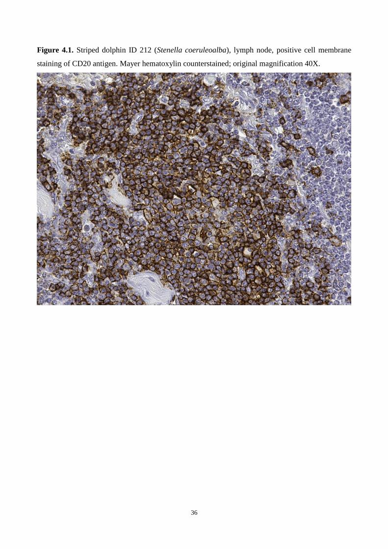
36
Figure 4.1. Striped dolphin ID 212 (Stenella coeruleoalba), lymph node, positive cell membrane
staining of CD20 antigen. Mayer hematoxylin counterstained; original magnification 40X.

37
Figure 4.2. Striped dolphin ID 212 (Stenella coeruleoalba), lymph node, positive cell membrane
staining of CD3 antigen. Mayer hematoxylin counterstained; original magnification 40X.

38
Figure 4.3. Striped dolphin ID 212 (Stenella coeruleoalba), lymph node, positive cell membrane
staining of HLA-DR antigen. Mayer hematoxylin counterstained; original magnification 40X.

39
Figure 4.4. The three well-defined bands, with molecular weight of 61, 33 and 33 kDa represent the
specific antibodies against CD3, CD20 and HLA-DR antigen, respectively.* CD3, CD20 and HLA-
DR antigens bands were identified in lymph node tissue of men (Hu), striped dolphin (ST) and
bottlenose dolphin (TT).
*: Hu = lymph node tissue of men; ST = lymph node tissue of striped dolphin (Stenella
coeruleoalba); TT = lymph node tissue of bottlenose dolphin (Tursiops truncatus).

40
4.2 DOLPHIN MORBILLIVIRUS
4.2.1 Immunohistochemical (IHC) analyses
Circulating monocytes and tissue macrophages from the fin whale’s (BP#2) brain and thymus were
positively stained by the hyperimmune rabbit anti-Rinderpest Virus (RPV) antiserum (Institute for
Animal Health, Pirbright, Surrey, England, UK) (Figure 4.5).
IHC evidence of DMV nucleoprotein (N) antigen (murine monoclonal anti CDV antibody) was
observed in macrophages and follicular dendritic-like cells from the spleen’s white pulp in the
youngest female (PM#1), as well as in monocytes circulating within splenic blood vessels from the
same animal (Figure 4.6).
4.2.2 Extraction methods
In this study, RNA extractions were performed by two different protocols (PureLink RNA Mini Kit
and Trizol). Each of the 2 extraction methods yielded sufficient RNA amounts for obtaining
positive results in a nested RT-PCR for DMV. However, the RNA extracted by the TRIzol Reagent
constantly had the highest yield (400-4000 ng/microL), while the RNA extracted by the PureLink
RNA Mini Kit had the lowest one (15-80 ng/microL), but the highest A260/A280 ratio for almost
all samples. After the first PCR step, the PCR products from samples extracted using the PureLink
RNA Mini Kit showed, with all the sets of primers and for all the samples, a more intense signal
after separation by agarose gel electrophoresis.
4.2.3 Nested PCR
Nested PCR was performed on all tissue samples under investigation (Table 3.4). DMV-11 primers
pair was used for the first step of PCR and DMV-13 for the second round (Table 3.5 and Figure
4.7). A sample was considered positive for DMV when it generated PCR products of the expected
size of 200 bp and sequencing of the obtained fragments demonstrated that those PCR products
shared 100% sequence homology with DMV (GenBank Acc. No. AJ608288). The DMV 200 bp
sequence isolated from the sperm whales was registered in GenBank (Acc. No. KU886570).
As shown in Figure 4.8, positive results were obtained after application of the nested RT-PCR
protocol herein reported on the 21 tissue samples under investigation (Table 4.3): Tursiops
truncatus#1 (TT#1) brain; Phoca vitulina#1 (PV#1) lung, brain and lymph node; Balaenoptera
physalus#1 (BP#1) lung and lymph node; BP#2 lung, brain and spleen; Stenella coeruleoalba#1
(SC#1) lung and lymph node; Physeter microcephalus#1 (PM#1) brain and spleen; PM#2 lung;

41
PM#3 lung and kidney. The same data were obtained using both RNAs extracted with PureLink
RNA Mini Kit (Ambion, Thermo Scientific) and with TRIzol Reagent (Ambion, Thermo
Scientific).
In order to prove the increased sensitivity of this new technique, we performed an analysis of low-
titer dilution series of a positive sample (TT#1 brain tissue) using the nested RT-PCR primers
herein investigated and DMV-N1 and DMV-P2 primers, as previously reported by Bellière et al.
(2011) and by Mazzariol et al. (2016) (product size:1358 bp). To ensure that PCR products were
authentic and not the result of cross-contamination, the number of amplification cycles was reduced
to 25 for the second nested PCR step and for the conventional PCR. In conventional PCR, the
cDNA lowest detection limit in agarose gel electrophoresis was defined as 1:200 dilution, while in
the second step of nested PCR the lowest detection limit was 1:400000 dilution (Figure 4.9). Direct
sequencing of 1:5000, 1:50000 and 1:100000 dilution of nested PCR second step products was
performed to confirm their identity to DMV genome sequences.
4.2.4 Complete DMV genome study
DMV genome was detected in brain, lung and spleen of the newborn fin whale (BP#2, Table 3.4).
Sequences of 5 lung, 4 cerebral and 5 spleen plasmidic colonies were analyzed. The conventional
RT-PCR technique associated with viral cloning using plasmid vector allowed the identification of
the entire viral genome. The sequence of each single gene was deposited in Genbank as shown
below: complete N gene (1573 bp) provisional Acc. No. KU977449, complete P/V/C gene (1521
bp) provisional Acc, No. KU977450, complete M gene: (1008 bp) provisional Acc. No. KU977451,
complete F gene (1659 bp) provisional Acc. No. KU977452 and complete H gene (1814 bp)
provisional Acc. No. KU977453.
Afterwards each sequence was compared to the DMV complete genome isolate in the 1990’s
outbreak in a bottlenose dolphin (Acc. No. AJ608288): these fragments showed a sequence
homology between 99.10% and 99.80%.
4.2.5 Secondary structure prediction for DMV complete genome study
PSIPRED was used to predict the secondary structure of gene N, gene M, gene H, gene F and gene
P/V/C reported in literature (GenBank accession number AJ608288) and of the corresponding
sequences identified in our sample.
The observed secondary structure were the same for gene M and gene H: the secondary sequence
structures of the two proteins show a high degree of similarity in terms of helices and β-sheets in

42
our sequences as in reported ones. On the contrary the gene secondary structure gene F, gene N and
gene P/V/C show difference between our and predicted sequences.
Gene F of our fin whale showed a greater amount of helices in comparison with the gene F reported
in literature, in particular from residues 70 and 100 of the protein.
Gene N of our fin whale showed a high degree of similarity in terms of helices and β-sheets from
the reported gene F. Moreover, fin whale gene F showed differences in particular in the disposition
of boundary amino-acids.
Gene P/V/C of our fin whale showed a greater amount of helices in comparison with the gene F
reported in literature, in particular from residues 185 to 275 of the protein. Moreover the sequence
was different also for the amount of beta-sheets and for the disposition of boundary amino-acids.
4.2.6 Nucleotide and amino-acid sequence comparison for DMV complete genome study
Differences nucleotide and amino-acid pairwise raw distance between fin whales and bottlenose
dolphin strain are reported in Table 4.4. Briefly, the genetic and amino-acidic distance were 0.01and
0.009 for F gene, 0.002 and 0.006 for M gene, 0.009 and 0.008 for N gene, 0.007 and 0.01 for the
P/V/C gene and 0.006 and 0.003 for the H gene. Five out 16 substitution were non-synonymous in
the F gene, 2 out 2 in the M gene, 4 out 14 in the N gene and 1 out 9 in the H gene.
Reliable (i.e. experimentally determined) structural templates were identified for the M,N,H and F
proteins and consequently the tertiary/quaternary structure was predicted only for these proteins.
Particularly, based on the available templates, the following regions of each protein were
consistently modeled: F protein (aa 26-487), M protein (aa 20-326), N protein (aa 3-401) and H
protein (157-603).
Non-synonymous changes were located in nucleus of the H (amino-acid 451) and M (amino-acids
59-190) proteins while were exposed on the protein surface of the F protein (amino-acids 52,
151,154 and 161) or were part of its signal peptide (amino-acid 3) (Figure 4.10). The only amino-
acid change modeled in the N protein (amino-acid 21) was located in the inner part of the capsid
(Figure 4.10). Unfortunately, amino-acidic changes located in position 436, 462 and 509 of the
same protein were located in regions that could not be confidently modeled by homology modeling.
Table 4.3. Positive results obtained after application of the nested RT-PCR protocol reported on the
21 tissue samples under investigation (Table 3.4).

43
Animal
number Species Tissue
Previous
diagnostic
tecnique
Positivity
(Sequenced:
Y/N)
Positivity to
nested RT-
PCR
TT#1 Tursiops truncatus
Lung
RT-PCR
(Di Guardo et
al., 2013)
Negative Negative
Brain
RT-PCR
(Di Guardo et
al., 2013)
Positive
(N) Positive
Spleen
RT-PCR
(Di Guardo et
al., 2013)
Negative Negative
Lymph
node
RT-PCR
(Di Guardo et
al., 2013)
Negative Negative
PV#1 Phoca vitulina
Lung
RT-PCR
(Mazzariol et
al., 2013)
Positive
(Y) Positive
Brain
RT-PCR
(Mazzariol et
al., 2013)
Positive
(Y) Positive
Lymph
node
RT-PCR
(Mazzariol et
al., 2013)
Positive
(Y) Positive
BP#1 Balaenoptera
physalus
Lung
RT-PCR
(Di Guardo et
al., 2013)
Negative Positive
Spleen
RT-PCR
(Di Guardo et
al., 2013)
Positive
(N) Negative
Lymph
node
RT-PCR
(Di Guardo et
al., 2013)
Positive
(N) Positive

44
BP#2 Balaenoptera
physalus
Lung
RT-PCR and
Cloning
(Mazzariol et
al., 2016)
Positive
(Y) Positive
Brain
RT-PCR and
Cloning
(Mazzariol et
al., 2016)
Positive
(Y) Positive
Spleen
RT-PCR and
Cloning
(Mazzariol et
al., 2016)
Positive
(N) Positive
SC#1 Stenella coeruleoalba
Lung ND ND Positive
Lymph
node ND ND Positive
PM#1 Physeter
macrocephalus
Lung ND ND Negative
Brain ND ND Positive
Spleen ND ND Positive
PM#2 Physeter
macrocephalus Lung ND ND Positive
PM#3 Physeter
macrocephalus
Lung ND ND Positive
Kidney ND ND Positive
Table 4.4. Differences nucleotide and amino-acid variations in each DMV single gene between fin
whale (Balaenoptera physalus) and bottlenose dolphin (Tursiops truncatus) strain (GenBank Acc.
No. 608288). Nucleotide
bases in
AJ608288
Nucleotide bases in
fin whale
AA in
AJ608288 AA in fin whale AA position
GENE N
ATT ACT Ile Thr 21
GTA GTG Val Val 36
TAT TAC Tyr Tyr 63
GAG GAA Glu Glu 151
GAA GAG Glu Glu 161
GCT GCC Ala Ala 184
CTA TTA Leu Leu 271

45
TCT TCC Ser Ser 298
ATT ATC Ile Ile 397
GCA ACA Ala Thr 437
ACA GCA Thr Ala 462
CGT CAT Arg His 509
CAC CAT His His 514
GAT GAC Asp Asp 520
GENE P/V/C
GAG GAA Glu Glu 103
GCG GAG Ala Glu 119
GGC GGT Gly Gly 134
AAT AGT Asn Ser 163
CTA TTA Leu Leu 210
AAG AAA Lys Lys 211
ATG CTG Met Leu 313
CCC CCT Pro Pro 396
CGA CGG Arg Arg 479
GENE M
TTA GTA Leu Val 59
TTA TCA Leu Ser 191
GENE F
GCC GCT Ala Ala 2
GCT TCT Ala Ser 3
ACA ACG Thr Thr 40
AAC AGC Asn Ser 52
GTC GTT Val Val 89
AAT AGT Asn Ser 151
ACA GCA Thr Ala 154
CAA CGA Gln Arg 162
CCA CCG Pro Pro 190
TTG TTA Leu Leu 204
ATC ATA Ile Ile 328
GTG GTA Val Val 342
AAA AAG Lys Lys 412
CTG CTA Leu Leu 512
GTA GTG Val Val 514
GENE H
GGA GGG Gly Gly 57
CTT CTC Leu Leu 74
AAT AAC Asn Asn 77
GTT GCT Val Ala 164
CGT CGA Arg Arg 262
GGG GGA Gly Gly 324
GCC ACC Ala Thr 451

46
CTT CTC Leu Leu 510
GTT GTC Val Val 535
Figure 4.5. Mayer hematoxylin counterstained tissue samples from the newborn fin whale
(Balaenoptera physalus, BP#2). A) Brain tissue showing positive immunostaining for Morbillivirus
sp. antigen in macrophages in the meningeal space. B) Thymus showing positive immunostaining
for Morbillivirus sp. antigen in thymocytes and macrophages. For both samples, Morbillivirus sp.
was detected by IHC analysis, using a rabbit hyperimmune anti–rinderpest virus serum (provided by
Pirbright Institute, Pirbright, UK). Original magnification ×40.

47
Figure 4.6. Sperm whale (Physeter macrocephalus; PM#3). Spleen. Positive immunostaining for
morbilliviral antigen is shown in monocytes within vascular lumina and in follicular dendritic-like
cells in the splenic white pulp. IHC for Morbillivirus with a murine monoclonal antibody against
canine distemper virus (CDV) nucleoprotein (VMRD Inc., Pullman, WA, USA). Mayer’s
hematoxylin counterstain. Final magnification 20x.

48
Figure 4.7. Nested RT-PCR for DMV. RNA was extracted using the PureLink RNA Mini Kit. The
cDNAs were prepared using DMV2 primer pair. Agarose gel (a) and electropherogram results (b).
(a) M: DNA marker, 100 bp (DNA Ladder, Invitrogen); DMV-11F and DMV-11R primer pairs
(Lanes 1 and 2) and second round of nested PCR using DMV-13F and DMV-13R primer pairs
(Lanes 4 and 5). Tursiops truncatus#1 (TT#1) lung sample (Lanes 1 and 4) and Physeter
macrocephalus#1 (PM#1) spleen sample (Lanes 2 and 5). Lanes 3 and 6: PCR negative controls.
(b) Electropherogram obtained from the PCR product obtained in line 5. Sequencing of the 200 bp
fragment obtained from PM#1 spleen using DMV-13F and DMV-13R primer pairs in the second
step of nested PCR. Genbank Acc. No. KU886570.

49
Figure 4.8. Agarose gel results of the second step of the nested PCR for CeMV using the DMV-
13F and DMV-13R primer pairs on all the samples in Table 3.4.
RNA from all tissues was extracted using PureLink RNA Mini Kit (Ambion, Thermo Scientific).
M: DNA marker, 100 bp (DNA Ladder, Invitrogen); Lanes 1 and 2: Stenella coeruleoalba#1
(SC#1) lung and lymph node samples, respectively; Lanes 3, 4, 5 and 6: Tursiops truncatus#1
(TT#1) lung, brain, spleen and lymph node samples, respectively; Lanes 7, 8 and 9: Phoca
vitulina#1 (PV#1) lung, brain, and lymph node samples, respectively; Lanes 10, 11 and 12:
Balaenoptera physalus#1 (BP#1) lung, spleen and lymph node samples, respectively. Lanes 13, 14
and 15: BP#2 lung, brain and spleen samples, respectively; Lanes 16, 17 and 18: Physeter
macrocephalus#1 (PM#1) lung, brain and spleen samples, respectively; Lane 19, PM#2 lung
sample; Lanes 20 and 21: PM#3 lung and kidney samples; Lane 22: PCR negative control.

50
Figure 4.9. A) Agarose gel electrophoresis results for the conventional PCR protocol using primers
DMV-N1 and DMV-P2 (Bellière et al., 2011; Mazzariol et al., 2016). The product size is 1358 bp.
B and C) Agarose gel electrophoresis results for the second step of the nested RT-PCR protocol
using primers DMV-13F and DMV-13R. The product size is 200 bp.

51
Figure 4.10. DMV homology modeling. Non-synonymous changes were exposed on the protein
surface of the F protein (amino-acids 52) and the only amino-acid change modeled in the N protein
(amino-acid 21) was located in the inner part of the capsid.

52

53
5. DISCUSSION
5.1 IMMUNE SYSTEM
Monoclonal antibodies which identify subpopulations of lymphoid organs’ leucocytes are essential
to investigate the role of these cells in the pathogenesis and regulation of diseases. Although cross-
reactivity of antibodies between phylogenetically distant species has sometimes been observed
(Jacobson et al., 1993; Jaber et al., 2013), a better understanding of immune function of cetaceans
could be possible only using antibody validated for the species under study.
Cross-reaction of antibodies against human, bovine, mouse and ovine surface antigens such as
HLA-DR, CD2, CD4, B cells and TCR has been reported on peripheral blood leukocytes of beluga
whale (Delphinapterus leucas) and bottlenose dolphin (De Guise at al., 2002; Romano et al., 1992).
Different monoclonal antibodies to lymphocyte surface antigens for cetaceans were also used in
several research to study the subpopulations expression in lymph nodes (De Guise et al., 2002;
Jaber et al., 2003): however these markers were considerate working on marine mammals just
considering the positive immunolabelling of the cells and the similar pattern observed in the species
of origin.
The cross-reactivity in striped dolphin (Stenella coeruleoalba) and bottlenose dolphin (Tursiops
truncatus) of the monoclonal anti-human antibodies against respectively T and B cells surface and
major histocompatibility complex were certified for the first time in this study. In fact, to confirm
the specificity of anti-human antibodies used, the molecular weight of the surface proteins was
evaluated and tested by Western Blotting, besides of the pattern of immunoreaction. Tested
antibodies resulted to be suitable for immunohistochemical studies in formalin-fixed, paraffin-
embedded tissues of striped dolphins and bottlenose dolphins. As expected the anti-human HLA-
DR reacted strongly with cells of varying morphology that had characteristic of macrophages and
dendritic-like cells of striped dolphin and bottlenose dolphin, whereas a huge number of
lymphocytes were labelled by this antibody. The anti-CD3 marker positively stained T-cell zones,
localized in the germinal center, in the mantle and marginal zone of lymph nodes sections, while
anti-CD20 antibody stained B-cell in the para-cortical zone.
Furthermore, the selected commercial antibodies can be valuable tools also because they work on
formalin-fixed, paraffin-embedded sample and not only on fresh or snap frozen tissues (Shirai et al.,
1998; De Giuse et al., 2002).

54
Following the antibodies validation, albeit the small number of samples, a counting of leukocytes
subsets was performed on immunostained tissues to understand the possible role of independent
variables on lymphocytic cells subpopulations composition. Statistical analysis does not show
statistically significant difference, except for the expression of anti-CD20 positive staining cells (B
lymphocytes) in normal and hypoplastic lymph nodes, whose p-value was very close to the value
considered indicative of a strong association. Analyzing the dolphins in the two groups we can
realize that the 40% of the animals presenting hypoplastic lymphatic tissues are individuals with
ongoing infections while the normal histology group include only animals apparently healthy.
Even if the number of animals included in the study is low due to the eligibility for storage
characteristics of the tissue or the completeness of the medical history, we can speculate that, as
already reported in the literature for some marine mammals’ infections such as those from DMV
(Van Bressem et al., 2014), the amount of lymphocytes to decrease as a result of immune depletion
caused by the pathogens themselves. Morbillivirus infections have long been known to result in
host immune suppression (Appel et al., 1982; Griffin et al., 1994). Recent studies suggest similar
effects in naturally infected, wild bottlenose dolphins as part of capture-release studies for health
assessment (Bossart et al., 2011). The author and his working group reported a significant decrease
in mitogen-induced T cell proliferation along with an increase in lysozyme concentrations and a
marginally significant increase in monocyte phagocytosis, along with a marginally significant
decrease in the numbers of CD4+ T lymphocytes in bottlenose dolphin that had antibody titers ≥ 1:8
against morbillivirus, suggestive of previous exposure to the virus, compared to animals with lower
or no titers. They found no effects on neutrophil phagocytosis. While the timing of the morbillivirus
infection (active infection, chronic infection, resolved past infection) cannot be determined from
titers alone, it is clear that there is an association between modulation of immune functions and
previous exposure to morbillivirus infection in bottlenose dolphin, as observed in other species
(Van Bressem et al., 2014). Therefore the data presented in literature seem to contrast those we
encountered: a decrease of B lymphocytes instead of T lymphocytes. The researcher attention must
be directed to the characterization of the T cell subsets in order to understand the subpopulation
expression, possibly according to the timing of the infection. Therefore further studies are needed to
understand the effective role of DMV as an immune suppression agent particularly in T cell
populations validating and using CD4 and CD8 antibodies, marker of T helper cells and T cytotoxic
cells respectively.
In addition to infectious agents, lymphoid depletion could be caused by environmental
contaminants in cetaceans. Lymphoid organs hypoplasia is primarily associated with elevated
55
polybrominated diphenyl ether (PBDE) levels, while there is no correlation with the health status
and nutritional state in bycaught animals, supporting the hypothesis of a contaminant-induced
immune deficiency (Beineke et al., 2005). Additional toxicants, such as methylmercury and heavy
metals are also suspected to negatively influence the immune system and increase disease
susceptibility of harbor porpoises (Jepson et al., 1999; Siebert et al., 1999). The immunotoxic effect
of several xenobiotics on cetacean blood leukocytes at concentrations equivalent to those observed
in wildlife marine mammal populations has been verified in vitro. Noteworthy, DDT and non-
coplanar PCB congeners inhibited spontaneous and mitogen-induced proliferation of beluga whale
lymphoid cells, while coplanar (dioxin-like) PCB congeners and TCDD failed to modulate
leukocyte function (De Guise et al., 1998). Further in vitro experiments confirmed their inhibitory
effect on phagocytosis of neutrophils and monocytes of bottlenose dolphins and beluga whales. The
dominating effect of non-coplanar PCB congeners is suggestive of a modulation of the leukocyte
function in an aryl hydrocarbon receptor independent manner in these marine mammals (Levin et
al., 2004). Similarly, mitogen-induced T and B cell proliferation is mainly modulated by non-
coplanar PCB congeners, as investigated in killer whales, beluga whales, bottlenose dolphins and
pilot whales, respectively (Mori et al., 2008).
In vitro experiments confirmed the negative influence of heavy metals on bottlenose dolphin
leukocytes: mercury and cadmium decrease cell viability, phagocytosis and proliferation of these
cells. In addition, they start lymphocyte apoptosis in concentrations equivalent to those reported in
free-ranging cetaceans (Camara Pellisso et al., 2008). Similarly, in vitro exposure to mercury
chloride and cadmium chloride decreases beluga whale splenocyte and thymocyte proliferation (De
Guise et al., 1996).
Several studies on rodents indicate a potential immunotoxic effect of various marine xenobiotics,
but metabolism and detoxification most likely differ between rodents and cetaceans (Levin et al.,
2007; Mori et al., 2008). Further research in marine mammals has to be focused on evaluate if the
biological activity of marine xenobiotics could cause a contaminant-induced hypocellularity with
reduced numbers of CD4+ T helper cells or a reduced number of CD8+ cytotoxic T cells and an
inhibited splenic humoral immune response such as lipophilic contaminants, as occurred in rodents
(Beineke et al., 2010).
Recently, few research reported the use of lymphocytic antigen surface antibodies to characterized
immunonocytologic features of a primary uterine T-cell lymphoma with multiple metastases in an
adult female Atlantic spotted dolphin (Stenella frontalis) (Delgado et al., 2015), and to determine
the immunophenotype of cellular inflammatory infiltrates in chronic cholangitis in 6 common

56
dolphins (Delphinus delphi) (Jaber et al., 2013). As reported before, the use of these antibodies was
not validated by Western Blot.
On the contrary, the monoclonal anti-human antibodies validated for striped dolphin and bottlenose
dolphin in this study could be useful as a IHC set to characterize the inflammatory infiltrate in
different infection, such as Toxoplasma gondi. Indeed Toxoplasma gondii is a protozoan
opportunistic pathogen for cetaceans and it has been recently reported in the brain of several striped
dolphins with extensive meningoencephalitis lesions, all of which found stranded between 2007 and
2008 on the Ligurian Sea coast of Italy (Di Guardo et al., 2010), as well as in the brain of striped
dolphins and bottlenose dolphins beached off the Italian Tuscany coast between 2006 and 2008
(Pretti et al., 2010). The study of the different lymphocytic subpopulation could be helpful to
comprehend how different species presented in the Mediterranean basin, such as bottlenose dolphin
and striped dolphin, respond to the pathogen depending on the environmental factor they are
exposed and if the parasite stimulate a different inflammatory process.
57
5.2 DOLPHIN MORBILLIVIRUS
5.2.1 Nested PCR tecnique
RT-PCR is a valuable technique that is increasingly being used for the diagnosis of animal diseases
caused by RNA viruses (Shin et al., 2004). In the present study, a more sensitive biomolecular
technique based on conventional RT-PCR, the nested RT-PCR, has been implemented to detect
DMV in badly preserved tissues from aquatic mammals.
An absolute prerequisite for the performance of RT-PCR is an efficient RNA extraction method.
Currently, there are numerous protocols which can be used to isolate and purify RNA, although
there are a few published studies comparing extraction methods for samples of animal origin (Deng
et al., 2005). In our study, in which we compared two viral RNA extraction methods, PureLink
RNA Mini Kit and TRIzol Reagent, we did not detect any relevant differences between the results
obtained with the two aforementioned methods. However, RNAs extracted by the TRIzol Reagent
constantly had the highest yield, while the extracts obtained by the PureLink RNA Mini Kit showed
the highest A260/A280 ratio for almost all samples. In addition, after the first PCR step, the PCR
products from samples extracted using the PureLink RNA Mini Kit showed a more intense signal
following separation by agarose gel electrophoresis.
On the basis of these results, we can conclude that the two methods of RNA extraction are equally
useful for the detection of CeMV in badly preserved aquatic mammal carcasses as well as in
stranded whales.
For this reason, we focused our attention on the amplification method and we chose nested RT-PCR
in order to detect CeMV even in poorly preserved samples.
The first set of primers selected in this study (DMV-11) was previously used to detect a 612 bp
sequence of a conserved region of the DMV H gene (Mazzariol et al., 2016). The second set of
primers (DMV-13) was designed on a short, specific, conserved region of the DMV H gene; this
region was chosen to reduce the intraspecific variations among the different CeMV isolates hitherto
characterized (Van Bressem et al., 2014).
We analyzed tissues from animals which had been already investigated in previous studies by
molecular and/or immunohistochemical (IHC) analyses (Di Guardo et al., 2013; Mazzariol et al.,
2013; Mazzariol et al., 2016), in order to test the specificity of the proposed nested RT-PCR method
for CeMV detection. In these investigations, viral genome fragments were not always suitable for
sequencing (Di Guardo et al., 2013), so an alternative approach was developed to obtain DNA
fragments for sequencing studies (Mazzariol et al., 2013; Mazzariol et al., 2016). The results herein

58
obtained using nested RT-PCR are in agreement with those previously reported, except for the
negativity observed in the BP#1 spleen. Notwithstanding what above, all the other tissues from
BP#1 tested positive, so that we believe that two alternate hypotheses could be drawn in order to
explain the aforementioned discrepancy. Indeed, while it seems plausible that such "negativity" may
be due to an unsatisfactory preservation status of the spleen from BP#1, causing an intense viral
RNA degradation, it might be additionally speculated that the previously reported positivity could
have originated from RT-PCR nonspecific results, provided that the amplified genome fragment
was not sequenced.
Furthermore, this technique allowed detection of DMV genome in animals not yet reported as
giving positive results, both in small odontocetes, such as striped dolphin SC#1, where tissue
degradation is very fast, and in stranded whales, such as sperm whales PM#1, PM#2 and PM#3, in
which post mortem autolytic processes are increased by the size of the carcass, with subsequent
rapid degradation of the RNA viral genome.
Due to the proven efficiency of the nested RT-PCR technique herein described, we believe this new
method could be useful both to detect the presence of DMV infection in aquatic mammal tissues
harbouring low viral loads and to easily sequence small amounts of viral genome, as also confirmed
by previously described nested RT-PCR protocols (Shin et al., 2004; Sierra et al., 2014).
Furthermore, this method could provide a valuable option for detecting DMV during the initial and
final stages of the viral infection in susceptible host tissues, where morbilliviruses have been
reported to be present in low amounts (Ludlow et al., 2015). In addition, the nested RT-PCR
protocol herein reported may also turn out to be useful both in “retrospective” investigations on
aquatic mammal specimens previously assessed as being Morbillivirus-negative (or whose
Morbillivirus infection status has not been determined, as in the case of tissue bank specimens, for
instance) and in “controversial” positive cases, as reported in recent mortality outbreaks occurring
worldwide (Rubio-Guerri et al., 2013; Casalone et al., 2014; Stephens et al., 2014). In these recent
events, indeed, the morphopathological lesions’ patterns did not always correspond to the
“classical” disease phenotype described in the literature (i.e. subacute-to-chronic
broncho/bronchiolo-interstitial pneumonia with “Warthin-Finkeldey type” multinucleate syncytia,
along with subacute-to-chronic, non-suppurative meningoencephalitis). Moreover, morbilliviral
infection was not always detected by means of the “canonical” laboratory diagnosis tests, such as
IHC and conventional RT-PCR analyses.
In conclusion, the nested RT-PCR protocol presented may be considered a sensitive and rather
cheap biomolecular technique, which is able to identify DMV-positive samples from aquatic
mammals also when poorly preserved.

59
Despite these promising results, further studies should be conducted in order to increase the
efficiency of this nested PCR technique, as well as in order to understand if the same method could
be also useful to detect CeMV through the amplification of other evolutionarily conserved viral
genes, such as the P and the nucleocapsid protein (N) genes.
5.2.2 DMV in fin whales
The results of direct (IHC and biomolecular) and indirect (serologic) testing provide evidence of
DMV infection or exposure in 5 (55%) of 9 fin whales that were found stranded along the
Mediterranean Coast during 2011–2013. These 5 infected whales correspond to 21.7% of the 23
whales stranded along the Italian coastline during 2006–2014. The other 4 examined whales showed
no evidence of morbillivirus infection. The range of DMV-susceptible host species has
progressively expanded (Van Bressem et al., 2014), as highlighted by the recent report of DMV
infection in a captive harbour seal (Phoca vitulina) during the 2011 outbreak (Mazzariol et al.,
2013). This expansion, combined with spread of DMV through the transplacental route, resulting in
virus colonization of the thymus in fetuses, could represent DMV survival strategies among
cetacean populations. In addition, our data argue in favor of an epidemic cluster of fatal DMV
among the Mediterranean fin whales population, even though, on the basis of the amino acid
sequence of the SLAM/CD150 viral receptor, this species is not included among those susceptible
to DMV epidemics (Van Bressem et al., 2014; Ohishi et al., 2010).
Although the single amino acid substitution, p.Ala451Thr, did not cause substantial variations in the
structure of H antigen, the effect of the variation on protein functions is unclear. Recent studies
showed that similar amino acid changes could affect virulence and infectivity of different canine
distemper virus (family Paramyxoviridae, genus Morbillivirus) strains, but such changes are often
neutralized by compensatory mutations that preserve the biologic activity of H protein (Satter et al.,
2014). Furthermore, despite the high sequence homology observed between N, P, and H genes of
the DMV strain identified in the newborn fin whale and in the isolates recovered from DMV-
affected cetaceans during the 1990–1992 and the 2006–2007 epidemics (GenBank accession no.
AJ608288), we cannot exclude that more prominent differences occurred in virus genes encoding
for both structural and nonstructural proteins responsible for virulence and pathogenicity (e.g.,
P/V/C and fusion genes) (Van Bressem et al., 2014); the simultaneous occurrence of primary
structure differences, if any, in the SLAM/CD150 receptor should also be taken into account
(Ohishi et al., 2010). In conclusion, although further studies are needed to elucidate the complex
virus–host interaction dynamics and the putative influence exerted by environmental factors, DMV

60
should be regarded as one of the major threats for the conservation of fin whales within the
Mediterranean Sea.
5.2.3 DMV in sperm whale
The positive sequences obtained from the 4 sperm whales’ tissues were analyzed by means of an
hoc computer program and the H consensus fragment obtained from all the positive samples
showed 100% sequence homology with the corresponding DMV genome sequence (GenBank Acc.
No. AJ608288). Simultaneous immunohistochemical evidence of DMV nucleoprotein (N) antigen
was additionally observed in macrophages and follicular dendritic-like cells from the spleen’s white
pulp in the youngest female (#PM1), as well as in monocytes circulating within splenic blood
vessels from the same animal.
Despite no “classical” DMV-related pathological changes being observed during post mortem
investigations3,4, viral circulation was strongly suspected in at least in 2 out of the 3 DMV-infected
sperm whales based on IHC (#PM1) and biomolecular (#PM1, #PM2 and #PM3) findings. In this
respect, the consistent morbilliviral antigen’s immunolabeling both in circulating monocytes and in
splenic follicular-like dendritic cells, support the hypothesis that DMV infection was in an early
developmental stage (De Swart et al., 2007). Experimental studies conducted on similar
morbilliviruses show that during this period, even if no severe clinical signs may be expected, a
“general discomfort condition”, secondary to a viremic circulation could be suspected (De Vries et
al., 2012; Stein et al., 2008).
While DMV infection has been often associated to mass mortalities during epidemic outbreaks (De
Swart et al., 2007), it has been seldom described in single mass stranding events, not only in sperm
whales but, more in general, in cetaceans. In this respect, while sperm whales' susceptibility to
Cetacean Morbillivirus (CeMV) has been recently documented, alongside with the likely occurring
materno-fetal transfer of the virus (West et al., 2015), the biomolecular evidence of DMV infection
obtained in the fetal sperm whale herein investigated strongly supports the hypothesis of a
transplacentally acquired infection in this animal.
Although no clear-cut evidence exists that DMV was the primary cause of the sperm whales’ mass
stranding reported herein, a huge body of scientific literature is available to support the primary
pathogenicity of Morbillivirus genus members for their mammalian hosts, including aquatic
mammals (Van Bressem et al., 2014). Despite previous studies suggest chemical pollutants as one
of the relevant causative factor in determining stranding and subsequently death of the 3 stranded
cetaceans under study (Squadrone et al., 2015), the present investigation underline a possible role of

61
DMV infection which could have determined their entrance northward into the Adriatic Sea, which
is known to be a "cul-de-sac" for this species (Mazzariol et al., 2011).
In conclusion, while it is easier to support the direct involvement of CeMV “clade” members such
as DMV to the several epidemic outbreaks occurred in the last 25-30 years among free-ranging
cetaceans worldwide (Van Bressem et al., 2014), their role, as well as other factors, in single mass
stranding events cannot be defined precisely since biological, ecological and environmental co-
factors should be also investigated by using a multidisciplinary study effort (Mazzariol and Di
Guardo, 2016).

62

63
6. GENERAL CONCLUSIONS AND FUTURE PERSPECTIVES
The recent discoveries of several new morbilli-related viruses in bats (Drexler et al., 2012), as well
a potentially novel feline morbillivirus representing a basal divergence in the genus (Woo et al.,
2012), are likely to lead to understanding of the evolution of morbilliviruses. Morvilliviruses are a
growing concern because of their ability to infect multiple species as recently reported for canine
distemper virus infected Amour tiger (Panthera tigris altaica) and lion (Panthera leo) populations
(Seimon et al., 2013; Viana et al., 2015) probably caused by a spill-over of CDV from domestic dog
associated with severe decline in wild carnivores worldwide and mass dog vaccination.
The single mutations herein described in the DMV genomic sequence and the recent results of
diagnostic surveillance performed by public veterinary Institutions involved in the Italian Stranding
networks, which confirm that 19% of cetaceans found dead along the Italian coastline were
molecularly positive for DMV in 2015 without any evidences of ongoing epidemic event (Italian
Diagnostic report on stranded cetaceans - 2015), argue in favor of an endemic circulation of DMV
among Mediterranean cetaceans resulting in increased infectious pressure.
DMV epidemiology depends upon several factors, such as its virulence and herd immunity
population density (Beineke et al. 2015). The viral strains that were responsible for the strandings
herein described exhibit a marked genetic relatedness with those which have determined large mass
mortalities in early nineties and in 2006 (Van Bressem et al., 2014). This observation, supports
simultaneously a prolonged DMV circulation in the Western Mediterranean as well as the
hypothesis of an inadequate level of anti-viral immunity in cetaceans inhabiting this area.
During endemic circulation of virus belonging to this genus, periodic and self-limiting disease
outbreaks may occur, due to a decreased level of the population’s antiviral immunity, as occurred in
2011 (Di Guardo et al., 2014) and 2013 (Casalone et al., 2014).
Of great interest are the documented cases of DMV infection in the fin whale (Balaenoptera
physalus) newborn (Mazzariol et al., 2016) and in the sperm whale (Physeter macrocephalus)
foetus, which confirms the possibility of vertical transmission. The sperm whale mother was also
DMV-infected, was part of a DMV-infected, female sperm whales’ pod. To our knowledge, this
should be one of the few documented cases of DMV involvement in a cetacean mass stranding and
the only reported example of DMV involvement in a sperm whales’ mass stranding worldwide.
In these news species vertical transmission is hypothesized as the likely route of entry as well as the
possible survival strategy of the virus. (West et al., 2015; Mazzariol et al., 2016). The involvement

64
of newborns, young and pregnant females, could represent a serious menace for the conservation of
these already threatened species. This potential “infection strategy” could provide the virus with a
stronger pathogenicity, which could be further enhanced by the immune suppression para-
physiologically such as occurring during pregnancy (Sykes et al., 2012). In this respect, the recently
described biomolecular and immunohistochemical evidence of DMV in the thymus of the newborn
fin whale (Mazzariol et al., 2016) provides additional support to the aforementioned hypothesis,
given that an immunotolerance-like condition against the virus presumably developed in this whale
(Weissman, 2016).
The changing infection process of DMV toward its “traditional” and its “new” hosts could be
explained both by a population’s immunity enhancement resulting from prolonged DMV exposure
(Profeta et al., 2015) and by the evolving lesions’ pattern.
While the viral “infectious pressure” may be reasonably expected to increase under similar
conditions, a simultaneous hosts’ immune response impairment, either age or pregnancy (having
many of the recent cases of DMV infection been diagnosed in newborns, calves or pregnant
females) or otherwise pollutant-related, may facilitate cross-species infection. In fact, it is unknown
whether the high tissue levels of immunotoxic chemical substances commonly found in
Mediterranean cetaceans (Panti et al., 2011) are responsible for the lack of protective immunity
against one or more of the CeMV strains circulating for years in this region (Di Guardo et al.,
2013).
The density of the recipient host population is important in the transmission of any transferred virus
(Parrish et al., 2008), but in the last years, monitoring programs dedicated to estimates cetaceans
abundance in the Mediterranean basin denounced a decreased presence of striped dolphins (Stenella
coeruleoalba) in the Thyrrenian Sea (Panigada et al., 2011). The lower density of striped dolphins
and bottlenose dolphins (Tursiops truncatus), considered the classic hosts for DMV (Sato et al.,
2012), could have play a role in the cross-species infection, along with other potential factors as an
intermediate and amplifier hosts with the critical role of bringing DMV in close contact with
alternative hosts, as well as factors which change the geographical distribution of the species or
decrease the behavioral separation In fact, the rate and intensity of contact may be relevant as well
as the evolutionary relatedness (Parrish et al., 2008). These new species, similarly to wild
carnivores for CDV, could act as reservoir being a possible cause of spillback events involving
naive population of classic hosts and supporting the presence and circulation of the virus in this
limited basin (Beineke et al, 2015; Mazzariol et al., 2016).
Further research on the challenging topic of morbilliviral infections in cetaceans and, more in
general, in aquatic mammals is warranted. To this aim, identification of the SLAM/CD150 cell

65
receptor in several cetacean species (Ohishi et al., 2010; Shimizu et al., 2013) represents a major
step in our understanding of the pathogenesis of DMV infection, especially with regard to
susceptibility and transmission to non-classical hosts, such as pinnipeds and fin whales (Mazzariol
et al., 2013; Mazzariol et al., 2016). Further studies should confirm whether the SLAM/CD150 cell
receptor is indeed the primary immune receptor for CeMV, as is the case for other studied
morbilliviruses, and should look for the nectin4 epithelial cell receptor and other cell receptors in
cetaceans. It will be interesting also quantify the expression of SLAM/CD150 to understand if in the
animal affected by DMV the virus causes an over-expression of its receptor.
Further studies are also warranted to delineate the host responses to CeMV strains and lineages, and
the factors that determine the outcome of infection in cetaceans. Mathematical models should be
developed to examine the long-term dynamic consequences of the epidemics on cetaceans
populations and to predict the risk of epidemics (Van Bressem et al., 2014).
Further analyzes will be also aimed at analyzing the relationship between CD4+ and CD8+
lymphocytic cells in order to understand the effect of the DMV, important immunosuppressant
agent, on T cell populations. Finally, it could be interesting compare the data obtained on the
immune system with those obtained from the same samples as regards the search for environmental
pollutants.
The study of the different lymphocytic subpopulation expression in DMV affected animals using
monoclonal validated antibodies, together with an integrative approach that includes
epidemiological parameters, life history of the affected species and environmental parameters
should provide a better and more complete picture of the ecology and evolution of DMV, and, more
in general, of CeMV.

66

67
7. REFERENCES
Aguilar A., Raga J.A. The striped dolphin epizootic in the Mediterranean Sea. Ambio. 1993, 22:
524–528.
Aguilar A., Borrell A. Abnormally high polychlorinated biphenyl levels in striped dolphins
(Stenella coeruleoalba) affected by the 1990–1992 Mediterranean epizootic. Sci. Total Environ.
1994, 154: 237–247.
Almberg E.S., Cross P.C., Smith D.W. Persistence of canine distemper virus in the Greater
Yellowstone ecosystem’s carnivore community. Ecol. Appl. 2010, 7: 2058–2074.
Appel M.J., Shek W.R., Summers B.A. Lymphocyte-mediated immune cytotoxicity in dogs
infected with virulent canine distemper virus. Infect. Immun.1982, 37: 592–600.
Avila M., Khosravi M., Alves L., Ader-Ebert N., Bringolf F., Zurbriggen A., Plemper R.K., Platteta
P. Canine distemper virus envelope protein interactions modulated by hydrophobic residues in the
fusion protein globular head. J. Virol. 2015, 89:1445–1451.
Banyard A.C., Grant R.J., Romero C.H., Barrett T. Sequence of the nucleocapsid gene and genome
and antigenome promoters for an isolate of porpoise morbillivirus. Virus Res. 2008, 132: 213–219.
Barrett T., Visser I.K.G., Mamaev L., Goatley L., van Bressem M.-F., Osterhaus A.D.M.E. Dolphin
and porpoise morbilliviruses are genetically distinct from phocine distemper virus. Virology. 1993,
193: 1010–1012.
Barrett T. Morbillivirus infections, with special emphasis on morbilliviruses of carnivores. Vet.
Microbiol.1999, 69: 3–13.
Beineke A., Siebert U., Wunschmann A., Stott J.L., Prengel I., Kremmer E., Baumgartner W.
Immunohistochemical investigation of the cross-reactivity of selected cell markers from various
species for characterization of lymphatic tissues in the harbour porpoise (Phocoena phocoena). J.
Comp. Pathol. 2001, 5: 311–317.

68
Beineke A., Siebert U., McLachlan M., Bruhn R., Thron K., Failing K., Muller G., Baumgartner W.
Investigations of the potential influence of environmental contaminants on the thymus and spleen of
harbor porpoises (Phocoena phocoena). Environ. Sci. Technol. 2005, 39: 3933–3938.
Beineke A., Siebert U., Wohlsein P., Baumga W. Immunology of whales and dolphins. Veterinary
Immunology and Immunopathology. 2010, 133: 81-94.
Beineke A., Baumgartner W., Wohlsein P., Cross-species transmission of canine distemper virus—
an update. One Health. 2015; 1: 49-59.
Bellière E.N., Esperón F., Fernández A., Arbelo M., Muñoz M.J., Sánchez-Vizcaíno J.M.
Phylogenetic analysis of a new Cetacean morbillivirus from a short-finned pilot whale stranded in
the Canary Islands. Research in veterinary science. 2011, 90: 324-328.
Bellière E.N., Esperón F., Sánchez-Vizcaíno J.M. Genetic comparison among dolphin morbillivirus
in the 1990-1992 and 2006-2008 Mediterranean outbreaks. Infection, Genetics and Evolution. 2011,
11: 1913-1920.
Bieringer M., Han J.W., Kendl S., Khosravi M., Plattet P., Schneider-Schaulies J. Experimental
adaptation of wild-type canine distemper virus (CDV) to the human entry receptor CD150. PLoS
One. 2013, 8: e57488
Black F. Epidemiology of Paramyxoviridae. In The Paramyxoviruses; Kingsburry D.W., Ed.
Plenum Press: New York, NY, USA, 1991, 509–536.
Bolt G., Blixenkrone-Möller M., Gottschalk E., Wishaupt R.G.A., Welsh M.J., Earle P.J.A., Rima
B.K. Nucleotide and deduced amino acid sequences of the matrix (M) and fusion (F) protein genes
of cetacean morbilliviruses isolated from a porpoise and a dolphin. Virus Res. 1994,34: 291–304.
Bossart G.D., Reif J.S., Schaefer A.M., Goldstein J., Fair P., Saliki J.T. Morbillivirus infection in
free-ranging Atlantic bottlenose dolphins (Tursiops truncatus) from the Southeastern United States:
seroepidemiologic and pathologic evidence of subclinical infection. Vet. Microb. 2010, 143: 160-
166.

69
Bossart G.D., Romano T.A., Peden-Adams M.M., Schaefer A., McCulloch S., Goldstein J.D., Rice
C.D., Saliki J.T., Fair P.A., Reif J.S. Clinico-immunopathologic findings in atlantic bottlenose
dolphins (Tursiops truncatus) with positive cetacean morbillivirus antibody titers. Dis. Aquat.
Organ. 2011,97: 103–112.
Buonocore F., Randelli E., Casani D., Guerra L., Picchietti S., Costantini S., Facchiano A.M., Zou
J., et al. A CD4 homologue in sea bass (Dicentrarchus labrax): molecular characterization and
structural analysis. Molecular immunology. 2008, 45: 3168-3177.
Camara Pellisso S., Munoz M.J., Carballo M., Sanchez-Vizcaıno J.M. Determination of the
immunotoxic potential of heavy metals on the functional activity of bottlenose dolphin leukocytes
in vitro. Vet. Immunol. Immunopathol. 2008, 121: 189–198.
Casalone C., Mazzariol S., Pautasso A., di Guardo G., di Nocera F., Lucifora G., Ligios C., Franco
A., Fichi G., Cocumelli C. et al. Cetacean strandings in Italy: An unusual mortality event along the
Tyrrhenian Sea coast in 2013. Dis. Aquat. Org. 2014, 109: 81–86.
Cebrian D. The striped dolphin (Stenella coeruleoalba) epizootic in Greece, 1991–1992. Biological
conservation. 1995, 74: 143-145.
Cowan D., Smith T. Morphology of the lymphoid organs of the bottlenose dolphin, Tursiops
truncatus. Journal of anatomy. 1999, 194: 505-517.
De Guise S., Martineau D., Beland P., Fournier M. Possible mechanisms of action of environmental
contaminants on St. Lawrence beluga whales (Delphinapterus leucas). Environ. Health Perspect.
1995, 103: 73–77.
De Guise S., Bernier J., Martineau D., Beland P., Fournier M. Effects of in vitro exposure of beluga
whale splenocytes and thymocytes to heavy metals. Environ. Toxicol. Chem. 1996, 15: 1357–1364.
De Guise S., Martineau D., Beland P., Fournier M. Effects of in vitro exposure of beluga whale
leukocytes to selected organochlorines. J. Toxicol. Environ. Health. 1998, 55: 479–493.

70
De Guise S., Erickson K., Blanchard M., Dimolfetto L., Lepper H.D., Wang J., Stott J.L., Ferrick
D.A. Monoclonal antibodies to lymphocyte surface antigens for cetacean homologues to CD2,
CD19 and CD21. Vet. Immunol. Immunopathol. 2002, 4: 209–221.
De Oliveira e Silva F.M., Luz Carvalho V., Placido Guimaraes J., Vergara-Parente J.E., Oliveira de
Meirelles A.C., Marmontel M., Miglino M.A. Acessory spleen in cetaceans and its relevance as a
secondary lymphoid organ. Zoomorphology. 2014, 133: 343-350.
De Swart R.L., Ludlow M., De Witte L., Yanagi Y., Van Amerongen G., McQuaid S. Yüksel S.,
Geijtenbeek T.B.H., Duprex W.P., Osterhaus A.D.M. Predominant infection of CD150+
lymphocytes and dendritic cells during measles virus infection of macaques. PLoS pathogens. 2007,
3: e178.
Di Guardo G., Agrimi U., Morelli L., Cardeti G., Terracciano G., Kennedy S. Post mortem
investigations on cetaceans found stranded on the coasts of Italy between 1990 and 1993. Vet. Rec.
1995, 136: 439–442.
Di Guardo G., Marruchella G., Agrimi U., Kennedy S. Morbillivirus infections in aquatic
mammals: a brief overview. Journal of Veterinary Medicine. 2005, 52: 88–93.
Di Guardo G., Mazzariol S., Fernandez A. Biologically threatened dolphins and whales.
Environmental microbiology. 2011: 13:2833-2834.
Di Guardo G. Morbillivirus-host interaction: lesson from acquatic mammals. Front. Microbiol.
2012, 3: e432.
Di Guardo G., di Francesco C.E., Eleni C., Cocumelli C., Scholl F., Casalone C., Peletto S.,
Mignone W., Tittarelli C., di Nocera F. et al. Morbillivirus infection in cetaceans stranded along the
Italian coastline: pathological, immunohistochemical and biomolecular findings. Res. Vet. Sci.
2013, 94: 132–137.
Di Guardo G., Mazzariol S. Dolphin Morbillivirus: a lethal but valuable infection model. Emerg
Microbes Infect. 2013, 2: e74.

71
Di Guardo G., Mazzariol S. Cetacean Morbillivirus-associated pathology: knowns and unknowns.
Frontiers in Microbiology. 2016, 7:1-5.
Díaz-Delgado J., Sierra E., Arbelo M., Suárez-Bonnet A., Suárez-Santana C., Grau-Bassas E.,
Fernández A. Primary Uterine T-cell Lymphoma with Metastasis in an Atlantic Spotted Dolphin
(Stenella frontalis), Canary Islands, Spain. Journal of Wildlife Diseases. 2015, 51: 538-541.
Dobson A., Holdo R.M., Holt R.D. Rinderpest. In Encyclopedia of Biological Invasions,
Simberloff, D., Rejmánek M., Eds. University of California Press: Berkeley, CA, USA, 1991: 597–
604.
Domingo M., Ferrer L., Pumarola M., Marco A., Plana J., Kennedy S., McAliskey M., Rima B.K.
Morbillivirus in dolphins. Nature. 1990, 348: 21.
Domingo M., Visa J., Pumarola M., Marco A., Ferrer L., Rabanal R., Kennedy S. Pathologic and
immunocytochemical studies of morbillivirus infection in striped dolphins (Stenella
coeruleoalba).Vet. Pathol. 1992, 29: 1–10.
Domingo M., Vilafranca M., Visa J., Prats N., Trudgett A., Visser I. Evidence for chronic
morbillivirus infection in the Mediterranean striped dolphin (Stenella coeruleoalba). Vet. Microbiol.
1995, 44: 229–239.
Drexler J.F., Corman V.M., Müller M.A., Maganga G.D., Vallo P., Binger T., Gloza-Rausch F.,
Cottontail V.M., Rasche A., Yordanov S. et al. Bats host major mammalian paramyxoviruses. Nat.
Commun. 2012, 3: e796.
Duignan P.J., House C., Geraci J.R., Duffy N., Rima B.K., Walsh M.T., Early G., St D.J., Sadove
S., Koopman H., Rhinehart H. Morbillivirus infection in cetaceans of the western Atlantic.
Veterinary Microbiology. 1995, 44: 241-249.
Fernández A., Esperón F., Herraéz P., Espinosa de los Monteros A., Clavel C., Bernabé A.,
Sanchez-Vizcaino M., Verborgh P., DeStephanis R., Toledano F. et al. Morbillivirus and pilot
whale deaths, Mediterranean Sea. Emerg. Infect. Dis. 2008, 14, 792–794.

72
Fossi M.C., Casini S., Marsili L. Potential toxicological hazard due to endocrine-disrupting
chemicals on Mediterranean top predators: State of art, gender differences and methodological
tools. Environ. Res. 2007, 104: 174–182.
Fossi M.C., Panti C., Guerranti C., Coppola D., Giannetti M., Marsili L., Minutoli R. Are baleen
whales exposed to the threat of microplastics? A case study of the Mediterranean fin whale
(Balaenoptera physalus). Marine pollution bulletin. 2012, 64: 2374-2379.
Frisk A.L., König M., Moritz A., Baumgärtner W. Detection of canine distemper virus
nucleoprotein RNA by reverse transcription-PCR using serum, whole blood, and cerebrospinal fluid
from dogs with distemper. Journal of clinical microbiology. 1999, 37: 3634-3643.
Garibyan L., Avashia N. Polymerase Chain Reaction. Journal of Investigate Dermatology. 2013,
133: e6.
Garrigue C., Oremus M., Dodemont R., Bustamante P., Kwiatek O., Libeau G., Lockyer C., Vivier
J.C., Dalebout M.L. A mass stranding of seven Longman ’ s beaked whales (Indopacetus pacificus)
in New Caledonia , South Pacific. Marine Mammal Science. 2015, 1-27.
Geraci J.R., Lounsbury V.J. Marine Mammals Ashore: A Field Guide for Strandings. National
Aquarium in Baltimore. 2005.
Gilbert M., Miquelle D.G., Goodrich J.M., Reeve R., Cleaveland S., Matthews L., Joly D.O.
Estimating the Potential Impact of Canine Distemper Virus on the Amur Tiger Population
(Panthera tigris altaica) in Russia. PLoS ONE. 2014, 9: e110811.
Gilbert M., Soutyrina S.V., Seryodkin I.V., Sulikhan N., Uphyrkina O.V., Goncharuk M., Matthews
L., Cleaveland S., Miquelle D.G. Canine distemper virus as a threat to wild tigers in Russia and
across their range. Integrative Zoology. 2015, 10:329-343.
Grant R.J., Banyard A.C., Barrett T., Saliki J.T., Romero C.H. Real-time RT-PCR assays for the
rapid and differential detection of dolphin and porpoise morbilliviruses. J. Virol. Methods. 2009,
156: 117–123.

73
Griffin D.E., Ward B.J., Esolen L.M. Pathogenesis of measles virus infection: An hypothesis for
altered immune responses. J. Infect. Dis. 1994, 170: S24–S31.
Groch K.R., Colosio A.C.; Marcondes M.C.; Zucca D., Díaz-Delgado J., Niemeyer C.,Marigo
J.,Brandão P.E., Fernández A., Luiz Catão-Dias J. Novel cetacean morbillivirus in Guiana dolphin,
Brazil. Emerg. Infect. Dis. 2014, 20: 511–513.
Hall A.J. Morbilliviruses in marine mammals. Trends Microbiol. 1995, 3: 4–9.
Hashiguchi T., Ose T., Kubota M., Maita N., Kamishikiryo J., Maenaka K., Yanagi K. Structure of
the measles virus hemagglutinin bound to its cellular receptor SLAM. Nature structural &
Molecular Biology. 2011, 18: 135-142.
Headley S.A., Amude A.M., Alfieri A.F., Bracarense A.P., Alfieri A.A., Summers B.A. Molecular
detection of Canine distemper virus and the immunohistochemical characterization of the
neurologic lesions in naturally occurring old dog encephalitis. J. Vet. Diagn. Invest. 2009, 21: 588–
597.
Italian Diagnostic report on stranded cetaceans - 2015.
http://www.izsto.it/images/CREDIMA/Report_spiaggiamenti_2015.pdf
Jaber J.R., Fernandez A., Herraez P., Espinosa de los Monteros A., Ramirez G.A., Garcia P.M.,
Fernandez T. Arbelo M., Perez J. Cross-reactivity of human and bovine antibodies in striped
dolphin paraffin was-embedded tissues. Veterinary immunology and immunopathology. 2003, 96:
65-72.
Jaber J.R., Pérez J., Zafra R., Herráez P., Rodríguez F., Arbelo M., Espinosa de los Monteros A.,
Fernández A. Cross-reactivity of anti-human, anti-porcine and anti-bovine cytokine antibodies with
cetacean tissues. Journal of Comparative Pathology. 2010, 143: 45-51.
Jacob J.M., West K.L., Levine G., Sanchez S., Jensen B.A. Initial characterization of novel beaked
whale morbillivirus in Hawaiian cetaceans. Dis Aquat Organ. 2016,117:215-27.

74
Jepson P.D., Bennett P.M., Allchin C.R., Law R.J., Kuiken T., Baker J.R., Rogan E., Kirkwood J.K.
Investigating potential associations between chronic exposure to polychlorinated biphenyls and
infectious disease mortality in harbour porpoises from England and Wales. Sci. Total Environ.
1999: 243/244: 339–348.
Jepson P.D., Deaville R., Barber J.L. Agiular A., Borell A., Murphy S., Barry J., Brownlow A.,
Barnett J. et al. PCB pollution continues to impact populations of orcas and other dolphins in
European Waters. Scientific Report. 2016, 6: 18573.
Kennedy S., Smyth J.A., Cush P.F., McCullough S.J., Allan G.M., McQuaid S. Viral distemper
now found in porpoises. Nature. 1988, 336: 21.
Kennedy S., Smyth J.A., Cush P.F., McAliskey M., McCullough S.J., Rima B.K. Histopathologic
and immunocytochemical studies of distemper in harbor porpoises. Vet. Pathol. 1991, 28: 1–7.
Kerdiles Y.M., Cherif B., Marie J.C., Tremillon N., Blanquier B., Libeau G., Diallo A., Wild T.F.
Villiers M.B. Immunomodulatory properties of Morbillivirus nucleoproteins. Viral Immunology.
2006, 19: 324-334.
Kumar N., Maherchandani S., Kashyap S.K., Singh S.V., Sharma S., Chaubey K.K., Ly H. Peste
des petits ruminants virus infection of small ruminants: A comprehensive review. Vet. Med. Int.
2014, 6: 2287–2327.
Levin M., Morsey B., Mori C., De Guise S. Specific non-coplanar PCB-mediated modulation of
bottlenose dolphin and beluga whale phagocytosis upon in vitro exposure. J. Toxicol. Environ.
Health. 2004, 67: 1517–1535.
Lin W.H., Kouyos R.D., Adams R.J., Grenfell B.T., Griffin D.E. Prolonged persistence of measles
virus RNA is characteristic of primary infection dynamics. Proc. Natl. Acad. Sci. 2012,109: 14989–
14994
Lipscomb T.P., Schulman F.Y., Moffett D., Kennedy S. Morbilliviral disease in Atlantic bottlenose
dolphins (Tursiops truncatus) from the 1987–1988 epizootic. J. Wildl. Dis. 1994, 30: 567–557.

75
Ludlow M., McQuaid S., Milner D., de Swart R.L., Duprex W.P. Pathological consequences of
systemic measles virus infection. J. Pathol. 2015, 235: 253–265.
Mazzariol S., Marcer F., Mignone W., Serracca L., Goria M., Marsili L., di Guardo G., Casalone C.
Dolphin Morbillivirus and Toxoplasma gondii coinfection in a Mediterranean fin whale
(Balaenoptera physalus). BMC Vet. Res. 2012, 8: e20.
Mazzariol S., Peletto S., Mondin A., Centelleghe C., di Guardo G., di Francesco C.E., Casalone C.,
Acutis P.L. Dolphin morbillivirus infection in a captive harbor seal (Phoca vitulina). J. Clin.
Microbiol. 2013, 51: 708–711.
Mazzariol S., Centelleghe C., Beffagna G., Povinelli M., Terracciano G., Cocumelli C., Pintore A.,
Denurra D., Casalone C., Pautasso A., Di Francesco C.E. and Di Guardo G. Mediterranean fin
whales (Balaenoptera physalus) threatened by dolphin morbillivirus. Emerging Infectious Diseases.
2016, 22(2).
McCullough S.J., McNeilly F., Allan G.M., Kennedy S., Smyth J.A., Cosby S.L., McQuaid S.,
Rima B.K. Isolation and characterisation of a porpoise morbillivirus. Arch. Virol. 1991, 118: 247–
252.
Melia M.M., Earle J.P., Abdullah H., Reaney K., Tangy F., Cosby S.L. Use of SLAM and PVRL4
and identification of pro-HB-EGF as cell entry receptors for wild type phocine distemper virus.
PLoS One. 2014, 9: e106281.
Mori C., Morsey B., Levin M., Nambiar P.R., De Guise S. Immunomodulatory effects of in vitro
exposure to organochlorines on T-cell proliferation in marine mammals and mice. J. Toxicol.
Environ. Health. 2006, 69: 283–302.
Mori C., Morsey B., Levin M., Gorton T.S., De Guise S., 2008. Effects of organochlorines,
individually and in mixtures, on B-cell proliferation in marine mammals and mice. J. Toxicol.
Environ. Health. 2008, 71: 266–275.
Ohishi K., Ando A., Suzuki R., Takishita K., Kawato M., Katsumata E., Ohtsu D., Okutsu K.,
Tokutake K., et al. Host–virus specificity of morbilliviruses predicted by structural modeling of the

76
marine mammal SLAM, a receptor. Comparative immunology, microbiology and infectious
diseases. 2010, 33: 277-241.
Ohishi K., Suzuki R., Maruyama,T. Host-virus specificity of the Morbillivirus receptor, SLAM, in
marine mammals: risk assessment of infection based on three-dimensional models. InNew
Approaches to the Study of Marine Mammals; Romero A., Keith E.O., Eds. InTech: Rijeka, Croatia,
2012: 183–204.
Osterhaus A.D.M.E., de Swart R.L., Vos, H.W., Ross P.S., Kenter M.J.H., Barrett T. Morbillivirus
infections of aquatic mammals: Newly identified members of the genus. Vet. Microb. 1995, 44:
219–227.
Panigada S., Lauriano G., Burt L., Pierantonio N., Donovan G. Monitoring winter and summer
abundance of cetaceans in the Pelagos sanctuary (Northwestern Mediterranean Sea) through aerial
surveys. PLoS ONE. 2011; 6: e22878.
Panti C., Spinsanti G., Marsili L., Casini S., Frati F., Fossi M.C. Ecotoxicological diagnosis of
striped dolphin (Stenella coeruleoalba) from the Mediterranean basin by skin biopsy and gene
expression approach. Ecotoxicology. 2011, 20: 1791-1800.
Parrish C.R., Holmes E.C., Morens D.M., Park E.C., Burke D.S., Calisher C.H., Laughlin C.A., Saif
L.J., Daszak P. Cross-species virus transmission and the emergence of new epidemic diseases.
Microbiol Mol Biol Rev. 2008, 72:457-70.
Profeta F., Di Francesco C.E., Marsilio F., Mignone W., Di Nocera F., De Carlo E., Lucifora G.,
Pietroluongo G., Baffoni M., Cocumelli C., Eleni C., Terracciano G., Ferri N., Di Francesco G.,
Casaolone C., Pautasso A., Mazzariol S., Centelleghe C. and Di Guardo G. Retrospective
seroepidemiological investigations against Morbillivirus , Toxoplasma gondii and Brucella spp . in
cetaceans stranded along the Italian coastline (1998–2014). Research in Veterinary Science. 2015,
101: 89–92.
Raga J.A., Banyard A., Domingo M., Corteyn M., Van Bressem M-F., Fernández M., Aznar F.J.,
Barrett T. Dolphin morbillivirus epizootic resurgence, Mediterranean Sea. Emerg. Infect. Dis.
2008,14: 471–473.

77
Rima B.K., Collin A.M., Earle J.A. Completion of the sequence of a cetacean morbillivirus and
comparative analysis of the complete genome sequences of four morbilliviruses. Virus Genes. 2005,
30: 113–119.
Rubio-Guerri C., Melero M., Rivera-Arroyo B., Bellière E.N., Crespo J.L., García-Párraga D.,
Esperón F., Sánchez-Vizcaíno J.M. Simultaneous diagnosis of Cetacean morbillivirus infection in
dolphins stranded in the Spanish Mediterranean sea in 2011 using a novel Universal Probe Library
(UPL) RT-PCR
assay. Vet. Microbiol. 2013,165:109–114.
Saliki J.T., Lehenbauer T.W. Monoclonal antibody-based competitive enzyme-linked
immunosorbentassay for detection of morbillivirus antibody in marine mammal sera. J. Clin.
Microbiol. 2001, 39: 1877–1881.
Sato H., Yoneda M., Honda T. and Kai C. Morbillivirus receptors and tropism : multiple pathways
for infection. Frontiers in Microbiology. 2012, 3: 1–9.
Seimon T.A., Miquelle D.G., Chang T.Y., Newton A.L., Korotkova I., Ivanchuk G., Lyubchenko
E., Tupikov A., Slabe E. and McAloose D. Canine distemper visrus: an emerging disease in wild
endangered Amur tiger (Panthera tigris altaica). mBio. 2013, 4: e00410-13.
Shimizu Y., Ohishi K., Suzuki R., Tajima Y., Yamada T., Kakizoe Y., Bando T., Fujise Y., Taru
H., Murayama T. et al. Amino acid sequence variations of signaling lymphocyte activation
molecule and mortality caused by morbillivirus infection in cetaceans. Microbiol. Immunol.2013,
57: 624–632.
Shin Y., Cho K., Cho H., Kang S., Kim H., Kim Y, Park H., Parka N. Comparison of one-step RT-
PCR and a nested PCR for the detection of canine distemper virus in clinical samples. Australian
Veterinary Journal. 2004, 82: 83-86.
Siebert U., Joiris C., Holsbeek L., Benke H., Failing K., Frese K., Petzinger E. Potential relation
between mercury concentrations and necropsy findings in cetaceans from German waters of the
North and Baltic Seas. Mar. Pollut. Bull. 1999, 38: 285–295.

78
Sierra E., Sánchez S., Saliki J.T., Blas-Machado U., Arbelo M., Zucca D., Fernández A.
Retrospective study of etiologic agents associated with nonsuppurative meningoencephalitis in
stranded cetaceans in the Canary Islands. J. Clin. Microbiol. 2014, 52: 2390–2397.
Soto S., Alba A., Ganges L., Vidal E., Raga J.A., Alegre F., González B., Medina P., Zorrilla I.,
Martínez J. Post-epizootic chronic dolphin morbillivirus infection in Mediterranean striped dolphins
(Stenella coeruleoalba). Dis. Aquat. Organ. 2011, 96: 187–194.
Soto S., Gonzalez B., Willoughby K., Maley M., Olivera A., Kennedy S., Marco A., Domingo M.
Systemic herpesvirus and morbillivirus co-infection in a striped dolphin (Stenella coeruleoalba). J.
Comp. Pathol. 2012, 146: 269–273.
Squadrone S., Chiaravalle E., Gavinelli S., Monaco G., Rizzi M., Abete M.C. Analysis of mercury
and methylmercury concentrations, and selenium: mercury molar ratios for a toxicological
assessment of sperm whales (Physeter macrocephalus) in the most recent stranding event along the
Adriatic coast (Southern Italy, Mediterranean Sea). Chemosphere. 2015, 138: 633-641.
Stephens. N., Duignan P.J., Wang J., Bingham J., Finn H., Bejder L., Patterson A.P., Holyoake C.
Cetacean morbillivirus in coastal Indo-Pacific bottlenose dolphins, Western Australia. Emerg.
Infect. Dis. 2014, 20:666–670.
Stone B.M., Blyde D.J., Saliki J.T., Blas-Machado U., Bingham J., Hyatt A., Wang J., Payne J.,
Crameri S. Fatal cetacean morbillivirus infection in an Australian offshore bottlenose dolphin
(Tursiops truncatus). Aust. Vet. J. 2011,89: 452–457.
Sykes L.,MacIntyre D.A.,Yap X.J.,Teoh T.G., Bennett P.R. The Th1:Th2 dichotomy of pregnancy
and preterm labour. Mediators Inflamm. 2012, 967629.
Tatsuo H., Ono N., Yanagi Y. Morbilliviruses use signaling lymphocyte activation molecules
(CD150) as cellular receptors. J. Virol. 2001, 75: 5842–5850.

79
Taubenberger J.K., Tsai M.M., Atkin T.J., Fanning T.G., Krafft A.E., Moeller R.B., Kodsi S.E.,
Mense M.G., Lipscomb T.P. Molecular genetic evidence of a novel morbillivirus in a long-finned
pilot whale (Globicephala melas). Emerg. Infect. Dis. 2000, 6: 42–45.
Van Bressem M.F., van Waerebeek K., Fleming M., Barrett T. Serological evidence of
morbillivirus infection in small cetaceans from the Southeast Pacific. Vet. Microb. 1998, 59: 89–98.
Van Bressem M.F., van Waerebeek K., Raga J.A. A review of virus infections of cetaceans and the
potential impact of morbilliviruses, poxviruses and papillomaviruses on host population dynamics.
Dis. Aquat. Organ. 1999, 38: 53–65.
Van Bressem M.F., van Waerebeek K., Jepson P.D., Raga J.A., Duignan P.J., Nielsen O., di
Beneditto A.P., Siciliano S., Ramos R., Kant W. et al. An insight into the epidemiology of dolphin
morbillivirus worldwide. Vet. Microb. 2001, 81: 287–304.
Van Bressem M.F., Duignan P.J., Banyard A., Barbieri M., Colegrove K.M., De Guise S., Di
Guardo G., Dobson A., Domingo M., et al. Cetacean Morbillivirus: current knowledge and future
directions. Viruses. 2014, 5145-5181.
Viana M., Cleaveland S., Matthiopoulos J., Halliday J., Packer C., Craft M.E., Hampson K.,
Czupryna A., Dobson A.P., Dubovi E.J., Ernest E., Fyumagwa R., Hoare R., Hopcraft J.G.C.,
Horton D.L., Kaare M.T., Kanellos T., Lankester F., Mentzel C., Mlengeya T., Mzimbiri I.,
Takahashi E., Willett B., Haydon D.T. and Lembo T. Dynamics of a morbillivirus at the domestic-
wildlife interface: canine distemper virus in domestic dogs and lions. PNAS. 2015, 111: 1464-1496.
Weissman, I. How one thing led to another. Annu. Rev. Immunol. 2016, 34: 1-30.
Wenzlow N., Plattet P., Wittek R., Zurbriggen A., Grone A. Immunohistochemical demonstration
of the putative canine distemper virus receptor CD150 in dogs with and without distemper. Vet.
Pathol. 2007, 44: 943-948.
West K.L., Sanchez S., Rotstein D., Robertson K.M., Dennison S., Levine G., Davis N., Schofield
D., Potter C.W. and Jensen B. A Longman’s beaked whale (Indopacetus pacificus) strands in Maui,

80
Hawaii, with first case of morbillivirus in the central Pacific. Marine Mammal Science. 2013, 29:
767–776.
West K.L., Levine G., Jacob J., Jensen B., Sanchez S., Colegrove K., Rotstein D. Coinfection and
vertical transmission of Brucella and Morbillivirus in a neonatal sperm whale (Physeter
macrocephalus) in Hawaii, USA. Journal of Wildlife Diseases. 2015, 5: 227-232.
Wild T.F., Malvoisin E., Buckland R. Measles virus: both the haemagglutinin and fusion
glycoproteins are required for fusion. J. Gen. Virol. 1991, 72: 439–442.
Wohlsein P., Puff C., Kreutzer M., Siebert U., Baumgartner W. Distemper in a dolphin. Emerg.
Infect. Dis. 2007, 13: 1959–1961.
Woo P.C., Lau S.K., Wong B.H., Fan R.Y., Wong A.Y., Zhang A.J., Wu Y., Choi G.K., Li K.S.,
Hui J. et al. Feline morbillivirus, a previously undescribed paramyxovirus associated with
tubulointerstitial nephritis in domestic cats. Proc. Natl. Acad. Sci. 2012, 109: 5435–5440.

81
8. LIST OF ORIGINAL PUBLICATION
Centelleghe C, Beffagna G, Zanetti R, Zappulli V, Di Guardo G, Mazzariol S. Molecular analysis
of dolphin morbillivirus: a new sensitive detection method based on nested RT-PCR. Journal of
Virological Methods. 2016 May, 235:85-91.
Mazzariol S, Centelleghe C, Beffagna G, Povinelli M, Terracciano G, Cocumelli C, Pintore A,
Denurra D, Casalone C, Pautasso A, Di Francesco CE, Di Guardo G. Dolphin Morbillivirus, a
major threat for Mediterranean fin whales (Balaenoptera physalus). Emerging Infectious Diseases.
2016 Feb, 22:N°2
Mazzariol S, Cozzi B, Centelleghe C. 2015. Spiaggiamento dei cetacei, manuale di gestione. Ed.
The coffee house art&media. Milano.
Mazzariol S, Cozzi B, Centelleghe C. 2015. Handbook for cetacean’s strandings. Ed. The coffee
house art&media. Milano.
Banzato T, Gelain ME, Aresu L, Centelleghe C, Benali SL,Zotti A. Quantitative analysis of
ultrasonographic images and citology in relation to histopathology of canine and feline liver: an ex-
vivo study. Research in Veterinary Science. 2015 Dec,103:164-9.
Profeta F, Di Francesco CE, Marsilio F, Mignone W, Di Nocera F, De Carlo E, Lucifora G,
Pietroluongo G, Baffoni M, Cocumelli C, Eleni C, Terracciano G, Ferri N, Di Francesco G,
Casalone C, Pautasso A, Mazzariol S, Centelleghe C, Di Guardo G. Retrospective
seroepidemiological investigations against Morbillivirus, Toxoplasma gondii and Brucella spp. in
cetaceans stranded along the Italian coastline (1998-2014). Research in Veterinary Science. 2015
Aug,101:89-92.
Zotti A, Banzato T, Gelain ME, Centelleghe C, Vaccaro C, Aresu L. Correlation of renal
histopathology with renal echogenicity in dogs and cats: an ex-vivo quantitative study. BMC
Veterinary Research. 2015 Apr, 11:99.

82
Di Guardo G, Falconi A, Di Francesco A, Mazzariol S, Centelleghe C, Casalone C, Pautasso A,
Cocumelli C, Eleni C, Petrella A, Di Francesco CE, Sabatucci A, Leonardi L, Serroni A, Marsili L,
Storelli MM, Giacominelli-Stuffler R. Western blot expression of 5-lipoxygenase in the brain from
striped dolphins (Stenella coeruleoalba) and bottlenose dolphins (Tursiops truncatus) with or
without encephalitis/meningo-encephalitis of infectious nature. Journal Biological Regulators and
Homeostatic Agents. 2015 Jan-Mar, 29(1):245-50.
Mazzariol S, Peletto S, Mondin A, Centelleghe C, Di Guardo G, Di Francesco CE, Casalone C,
P.L. Acutis PL. Dolphin Morbillivirus Infection in a Captive Harbor Seal (Phoca vitulina). Journal
of Clinical Microbiology. 2013; 51(2): 708.

83
9. SCIENTIFIC CONTRIBUTIONS TO CONGRESSES
Oral presentation: Capture myopathy in 16 cetaceans stranded along the Italian coastline between
2008 and 2014. 29th European Cetacean Society Conference, 23-25 Marzo 2015;St. Julians
(Malta).
Molecular detection of dolphin morbillivirus: a new fast and sensitive detection system based on
nested RT-PCR. 30th European Cetacean Society Conference, 14-16 Marzo 2016; Funchal
(Madeira).
The brain of large whales. 21th Biennial Conference on the Biology of Marine Mammals. 11-18
Dicembre 2015; San Francisco (CA).
Cutaneous macroscopic and microscopic lesions caused by Pennella sp., Crassicauda sp.,
Phyllobothrium delphini in Cetaceans. 28th Annual Congress of the European Society and College
of Veterinary Dermatology, 24-26 Settembre 2015; Cracovia (Polonia).
Cross-reactivity evaluation of commercial antibodies against leukocyte sub-populations in striped
dolphins (Stenella coeruleoalba). V Convegno Nazionale della Ricerca nei Parchi – L’Uomo per gli
Animali, 10-12 Ottobre 2014; Bussolengo (Verona).
Post-mortem findings in three under human care giraffes (Giraffe camelopardalis). V Convegno
Nazionale della Ricerca nei Parchi – L’uomo per gli Animali, 10-12 Ottobre 2014; Bussolengo
(Verona).
Striped dolphins’ (Stenella coeruleoalba) unusual mortality in the Adriatic sea. 27th European
Cetacean Society Conference, 8-10 Aprile 2013; Setubal (Lisbona).
Post-mortem findings in stranded Mediterranean whales. 27th European Cetacean Society
Conference, 8-10 Aprile 2013; Setubal (Lisbona).