Embed Size (px)
Citation preview
Università degli Studi di Trieste
PhD program in MOLECULAR BIOMEDICINE
PhD Thesis
Use of Tn5-transposon mutagenesisfor the identification of potential novel interactors
of antimicrobial peptides in E. coli
Karol Bociek
XXII ciclo – Anno Accademico 2008/2009
PhD THESIS SUPERVISOR
Prof. Renato GennaroUniversity of Trieste, Department of Life SciencesVia Giorgieri, 1 – 34127 Trieste, [email protected]
TUTOR Marco Scocchi, PhDUniversity of Trieste, Department of Life SciencesVia Giorgieri, 1 – 34127 Trieste, [email protected]
EXTERNAL ADVISOR
Vittorio Venturi, PhDInternational Centre for Genetic Engineering and Biotechnology (ICGEB)Padriciano, 99 – 34012 Trieste, [email protected]
PhD COURSE COORDINATOR
Prof. Giannino Del SalUniversity of Trieste, Department of Life SciencesVia Giorgieri, 1 – 34127 Trieste, [email protected]
PhD EXAM COMMISSION
Prof. Enrico ToninUniversity of Trieste, Department of Life SciencesVia Fleming, 22 – 34127 Trieste, [email protected]
Prof. Maria Luisa MangoniUniversità di Roma “La Sapienza”, Dip. di Scienze BiochimicheVia degli Apuli, 9 – 00185 Roma, [email protected]
Prof. Alessandro PiniUniversità di Siena, Dip. di Biologia MolecolareVia Fiorentina, 1 – 53100 Siena, [email protected]
Prof. Gudmund Skjåk-BrækNorwegian University of Science and Technology (NTNU),Dept. of BiotechnologyN-7491 Trondheim, [email protected]
Prof. Gaio ParadossiUniversità di Roma “Tor Vergata”, Dip. di Scienze e Tecnologie ChimicheVia della Ricerca Scientifica snc – 00133 Roma, [email protected]
Table of Contents
ABSTRACT..................................................................................................................8
RIASSUNTO................................................................................................................9
LIST OF PUBLICATIONS.........................................................................................10
LIST OF ABBREVIATIONS......................................................................................11
INTRODUCTION......................................................................................................121. Immunity and the immune system.....................................................................12
1.1. Immunity as a key feature for survival.......................................................121.2. Two types of immune response..................................................................121.3. Innate immunity..........................................................................................12
1.3.1. Epithelia..............................................................................................131.3.2. Complement........................................................................................131.3.3. Phagocytes and NK cells....................................................................141.3.4. Cytokines in innate immunity.............................................................16
1.4. Adaptive immunity.....................................................................................161.4.1. Cell-mediated immunity.....................................................................171.4.2. Humoral immunity..............................................................................171.4.3. Antigen presenting cells (APCs).........................................................181.4.4. Cytokines............................................................................................19
2. Antimicrobial peptides.......................................................................................202.1. Classification of AMPs...............................................................................202.2. Host Defence Peptides................................................................................232.3. Major physical properties of AMPs............................................................23
2.3.1. Charge.................................................................................................232.3.2. Amphipathicity...................................................................................24
2.4. Modes of action..........................................................................................242.4.1. Membrane permeabilisation...............................................................242.4.2. Cell penetration and intracellular interactions....................................26
2.5. Mechanisms of resistance in bacteria.........................................................272.5.1. Extracellular mechanisms...................................................................282.5.2. Intracellular mechanisms....................................................................29
3. Mammalian HDPs..............................................................................................293.1. Defensins....................................................................................................29
3.1.1. Human α-defensins.............................................................................303.1.2. Human β-defensins.............................................................................30
3.2. Cathelicidins...............................................................................................314. The human cathelicidin......................................................................................33
4.1. Genetics, expression and processing..........................................................334.2. Structural properties...................................................................................34
4.2.1. Conformation......................................................................................344.2.2. Charge and amphipathicity.................................................................36
4.3. Physiology..................................................................................................37
4/111
4.3.1. Direct and indirect antimicrobial activity...........................................374.3.2. Immune system stimulation................................................................374.3.3. Cytotoxicity........................................................................................38
4.4. Resistance in bacteria.................................................................................385. Bacterial LPS.....................................................................................................39
5.1. Structure.....................................................................................................395.1.1. Lipid A................................................................................................405.1.2. Core oligosaccharide..........................................................................415.1.3. O-Antigen...........................................................................................41
5.2. Genetics of LPS synthesis..........................................................................415.3. Physiological effects of LPS......................................................................42
6. Antimicrobial peptides as therapeutics...............................................................436.1. Existing approaches....................................................................................436.2. Surpassing the in vivo limitations...............................................................446.3. Structural studies and target determination................................................44
7. Summary............................................................................................................45
AIMS OF THE STUDY.............................................................................................47
MATERIALS AND METHODS................................................................................481. Bacterial cultures................................................................................................48
1.1. Microbiological media................................................................................481.2. Growth conditions and bacteria storage.....................................................49
2. Bacterial strains, plasmids and oligonucleotides...............................................493. Mutant library production..................................................................................50
3.1. Preparation of competent cells...................................................................503.2. Transformation with the transposon complex............................................513.3. Extraction of genomic DNA of the mutants...............................................513.4. Confirmation of a random transposon insertion.........................................52
3.4.1. Genomic DNA digestion.....................................................................523.4.2. Electrophoresis and transfer onto a membrane...................................523.4.3. Southern blot.......................................................................................52
4. Peptide preparation.............................................................................................534.1. Synthesis and purification..........................................................................534.2. Fluorescent labelling of LL-37...................................................................544.3. Peptide quantification.................................................................................54
5. Determination of the minimal inhibitory concentration.....................................556. Selection of resistant mutants.............................................................................557. Identification of the mutated genes in B7R mutants..........................................56
7.1. Preparation of the cloning vector...............................................................567.2. Construction of genomic libraries of the mutants......................................577.3. Screening of the libraries and insert sequencing........................................57
8. Identification of the mutated genes in HCR mutants.........................................57
5/111
8.1. Preliminary identification...........................................................................578.2. Determination of mutation sites in 6 mutants.............................................588.3. PCR identification of the remaining mutants.............................................58
9. RT-PCR analysis.................................................................................................5810. Trans-complementation of the waaY mutation................................................5911. Antimicrobial activity assays............................................................................60
11.1. Growth kinetics assay...............................................................................6011.2. Killing kinetics assay................................................................................60
12. Flow cytometry assays.....................................................................................61
RESULTS....................................................................................................................631. Mutant library production and verification........................................................63
1.1. Estimated number of clones.......................................................................631.2. Selection of the restriction enzyme to digest mutant gDNA......................631.3. Verification of the random insertion of Tn5...............................................63
2. Selection, identification and characterisation of Bac7(1-16)-resistant mutants.642.1. Selection and identification of the resistant clones....................................642.2. Growth kinetics of 5 different clones.........................................................65
3. Selection, identification and characterisation of LL-37-resistant mutants.........663.1. Selection and isolation of mutant clones resistant to LL-37......................663.2. Analysis of the hybridisation profiles of the twenty isolated clones..........663.3. Each hybridisation profile corresponded to a different interrupted gene...673.4. Confirmation of waaY interruption in the other selected clones................683.5. Growth kinetics of 6 different clones.........................................................693.6. The second selection and characterisation of 8 of 24 new resistant clones703.7. Significance of the prevalence of waaY-mutants........................................71
4. Resistance in HCR1 was caused by the inactivation of waaY...........................714.1. The downstream genes were not inhibited by Tn5 insertion......................714.2. Trans-complementation of the mutation restored the wild-type phenotype...........................................................................................................................734.3. HCR1 was killed less efficiently by LL-37 than HB101 and HCR1C.......74
5. LL-37 bound and permeabilised HCR1 cells with lower efficiency..................756. HCR1 was not less susceptible to the orangutan orthologue of LL-37..............76
DISCUSSION.............................................................................................................791. Mutagenesis using Tn5 complex........................................................................792. Test selection with Bac7(1-16)...........................................................................81
2.1. Identified resistant clones...........................................................................812.2. Assessment of the resistance......................................................................83
3. Application of the method in search of bacterial interactors of LL-37..............843.1. Establishing the proper peptide concentration for plate selection..............85
6/111
3.2. Identified resistant clones...........................................................................863.3. Assessment of the resistance......................................................................883.4. Inactivation of the waaY gene led to a decreased bactericidal activity of LL-37 against HCR1.........................................................................................883.5. The role of waaY in the properties of LPS.................................................893.6.Binding to and permeabilisation of HCR1 cells by LL-37..........................903.7. Inactivation of waaY did not increase the resistance to ppyLL-37 and other AMPs in HCR1..................................................................................................923.8. Unidentified resistant clones from the second selection............................93
CONCLUSIONS.........................................................................................................94
ACKNOWLEDGEMENTS........................................................................................98
REFERENCES...........................................................................................................99
7/111
ABSTRACT
ABSTRACT
Antimicrobial peptides (AMPs) participate in the immunity of both animals
and plants. In mammals, the most important AMP families are defensins and
cathelicidins. The only human cathelicidin, LL-37, is a 37-residue, α-helical, cationic
peptide with a direct membranolytic antibacterial activity and various
immunomodulatory activities.
The aim of our research was to establish a procedure based on an E. coli
knock-out mutant library in the search for mutations conferring altered susceptibility
to AMPs. The library was created by random insertions of Tn5 transposon into the
bacterial genome. The selection of low-susceptibility mutants and their identification
was first tested with a Bac7-derived peptide, for which at least one interactor had
been found. The procedure was then applied for the identification of mutations that
modulate the susceptibility of E. coli cells to LL-37 with the aim of dissecting the
steps of the peptide's mode of action. The differences between the wild-type and the
mutant phenotypes were characterised using assays determining the bacterial culture
growth inhibition and killing, as well as cell binding and membrane permeabilisation.
In the majority of the isolated Bac7-resistant mutants, Tn5 was inserted in the
sbmA gene, confirming previous findings that showed an essential role for the SbmA
protein in the internalisation of Bac7, allowing it to exert its antimicrobial activity.
Tn5 insertion in the gene waaY, identified in 15 out of 20 resistant mutants
selected with LL-37, lowered the peptide’s ability to inhibit bacterial growth and to
kill cells, decreased peptide binding to the bacterial surface and membrane
permeabilisation. However, it did not alter the susceptibility to other AMPs. WaaY
encodes a specific kinase that phosphorylates the Hep II residue in the core region of
bacterial lipopolysaccharide. The inactivation of this enzyme determines a decreased
negative charge of the outer membrane, which in turn causes a decrease in the
peptide’s binding to the cell surface and in its antibacterial activity. The results reveal
a putative LPS-binding site for LL-37 and stress the importance of its initial binding
to the cell surface for antimicrobial efficacy.
The established procedure utilising Tn5 mutants in the search for bacterial
mutations conferring resistance to AMPs, proved efficient and opened a previously
unreported branch of research on the mode of action of the human cathelicidin.
8/111
RIASSUNTO
RIASSUNTO
I peptidi antimicrobici (AMP) fanno parte dell'immunità innata sia degli
animali che delle piante. Le famiglie di AMP più importanti nei mammiferi sono le
defensine e le catelicidine. L'unica catelicidina presente nell'uomo, LL-37, è un
peptide lineare e cationico di 37 residui con una conformazione ad α-elica, che
esercita un’azione microbicida diretta tramite permeabilizzazione di membrana. In
aggiunta, il peptide è anche dotato di una serie di funzioni immunomodulatorie.
Lo scopo del lavoro è stato quello di mettere a punto una procedura che utilizza
una libreria di mutanti knock-out di E. coli, ottenuta mediante inserzioni casuali del
trasposone Tn5 nel genoma batterico, per cercare mutazioni che rendano i batteri
resistenti agli AMP. La bontà del metodo è stata prima verificata selezionando e
identificando mutanti resistenti al frammento 1-16 del peptide Bac7. Una volta
ottenuti i risultati attesi, la procedura è stata applicata all'identificazione di mutazioni
che modulino la suscettibilità di E. coli a LL-37, con lo scopo di meglio
comprenderne il meccanismo d'azione. I fenotipi wild-type e mutante sono stati
caratterizzati usando saggi d'inibizione della crescita batterica, di killing e di capacità
del peptide di legarsi ai batteri e di permeabilizzarli.
Nella maggioranza dei mutanti resistenti al peptide Bac7 isolati, Tn5 ha
inattivato il gene sbmA, confermando risultati precedenti che avevano portato a
dimostrare l'importanza della proteina SbmA per l'internalizzazione di Bac7.
Nel caso di LL-37, in 15 dei 20 mutanti resistenti selezionati il trasposone ha
inattivato il gene waaY, che codifica per una chinasi specifica per la fosforilazione
del residuo Hep II della regione core del lipopolisaccaride batterico. La mancata
fosforilazione nei mutanti selezionati riduce la carica negativa della superficie
batterica, che, a sua volta, diminuisce l'inibizione della crescita, il killing, il legame e
la permeabilizzazione delle cellule da LL-37, ma non da altri peptidi, suggerendo che
la modificazione abbia alterato specificamente l'attività di LL-37, rivelando un suo
putativo sito di legame all’LPS e sottolineando l'importanza del legame iniziale alla
superficie cellulare per l'efficacia battericida del peptide.
La procedura messa a punto per la ricerca di mutazioni che inducono resistenza
agli AMP è risultata efficiente ed ha indicato una nuova via per far luce sulla
sequenza di eventi responsabile del meccanismo d'azione della catelicidina umana.
9/111
LIST OF PUBLICATIONS
LIST OF PUBLICATIONS
Included in the thesis:
• BOCIEK K, FERLUGA S, BENINCASA M, MARDIROSSIAN M, GENNARO R, SCOCCHI M.
Reduced sensitivity of E. coli to the human antimicrobial peptide LL-37 on
inactivation of the waaY gene responsible for the phosphorylation of LPS heptose
II. Submitted to Mol. Microbiol.
• SCOCCHI M, PALLAVICINI A, SALGARO R, BOCIEK K, GENNARO R. The salmonid
cathelicidins: a gene family with highly varied C-terminal antimicrobial domains.
Comp Biochem Physiol B Biochem Mol Biol. 2009 Apr; 152(4):376-81
(http://www.ncbi.nlm.nih.gov/pubmed/19168146)
Not included in the thesis:
-
10/111
LIST OF ABBREVIATIONS
LIST OF ABBREVIATIONS
AMPs – antimicrobial peptides
APCs – antigen-presenting cells
ATR-FTIR – attenuated total reflectance-Fourier transform infrared spectroscopy
CD – circular dichroism
cDNA – complementary DNA
CF – cystic fibrosis
CFU – colony-forming units
CLD – cathelin-like domain
CPS – capsule polysaccharide
DC – dendritic cells
dsRNA – double-stranded RNA
FDCs – follicular dendritic cells
gDNA – genomic DNA
hBDs – human β-defensins
HDPs – host-defence peptides
HNPs – human neutrophil peptides (human α-defensins)
LB – Luria-Bertani (microbiological medium)
LPS – lipopolysaccharide
MAC – membrane attacking complex
MAMPs – microbe-associated molecular patterns
MH - Mueller-Hinton (microbiological medium)
MHC – major histocompatibility complex
MIC – minimal inhibitory concentration
MW – molecular weight
NMR – nuclear magnetic resonance
OD – optical density
OM – outer membrane
PAMPs – pathogen-associated molecular patterns
PBS – phosphate-buffered saline (microbiological medium)
PCR – polymerase chain reaction
PRPs – proline-rich peptides
ROS – reactive oxygen species
RPM – revolutions per minute
RT-PCR – reverse-transcription polymerase chain reaction
TCR – T-cell receptor
TLR – Toll-like receptor
11/111
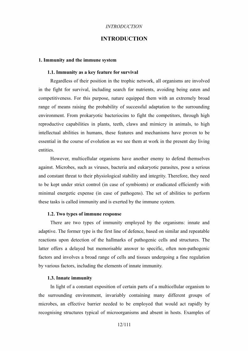
INTRODUCTION
INTRODUCTION
1. Immunity and the immune system
1.1. Immunity as a key feature for survival
Regardless of their position in the trophic network, all organisms are involved
in the fight for survival, including search for nutrients, avoiding being eaten and
competitiveness. For this purpose, nature equipped them with an extremely broad
range of means raising the probability of successful adaptation to the surrounding
environment. From prokaryotic bacteriocins to fight the competitors, through high
reproductive capabilities in plants, teeth, claws and mimicry in animals, to high
intellectual abilities in humans, these features and mechanisms have proven to be
essential in the course of evolution as we see them at work in the present day living
entities.
However, multicellular organisms have another enemy to defend themselves
against. Microbes, such as viruses, bacteria and eukaryotic parasites, pose a serious
and constant threat to their physiological stability and integrity. Therefore, they need
to be kept under strict control (in case of symbionts) or eradicated efficiently with
minimal energetic expense (in case of pathogens). The set of abilities to perform
these tasks is called immunity and is exerted by the immune system.
1.2. Two types of immune response
There are two types of immunity employed by the organisms: innate and
adaptive. The former type is the first line of defence, based on similar and repeatable
reactions upon detection of the hallmarks of pathogenic cells and structures. The
latter offers a delayed but memorisable answer to specific, often non-pathogenic
factors and involves a broad range of cells and tissues undergoing a fine regulation
by various factors, including the elements of innate immunity.
1.3. Innate immunity
In light of a constant exposition of certain parts of a multicellular organism to
the surrounding environment, invariably containing many different groups of
microbes, an effective barrier needed to be employed that would act rapidly by
recognising structures typical of microorganisms and absent in hosts. Examples of
12/111
INTRODUCTION
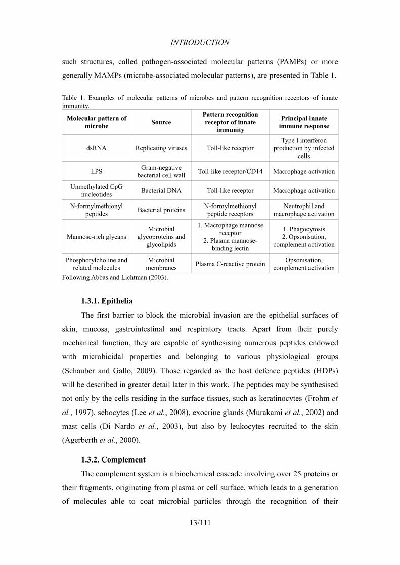
such structures, called pathogen-associated molecular patterns (PAMPs) or more
generally MAMPs (microbe-associated molecular patterns), are presented in Table 1.
Table 1: Examples of molecular patterns of microbes and pattern recognition receptors of innate immunity.
Molecular pattern of microbe Source
Pattern recognition receptor of innate
immunity
Principal innate immune response
dsRNA Replicating viruses Toll-like receptorType I interferon
production by infected cells
LPS Gram-negative bacterial cell wall Toll-like receptor/CD14 Macrophage activation
Unmethylated CpG nucleotides Bacterial DNA Toll-like receptor Macrophage activation
N-formylmethionyl peptides Bacterial proteins N-formylmethionyl
peptide receptorsNeutrophil and
macrophage activation
Mannose-rich glycansMicrobial
glycoproteins and glycolipids
1. Macrophage mannose receptor
2. Plasma mannose-binding lectin
1. Phagocytosis2. Opsonisation,
complement activation
Phosphorylcholine and related molecules
Microbial membranes Plasma C-reactive protein Opsonisation,
complement activationFollowing Abbas and Lichtman (2003).
1.3.1. Epithelia
The first barrier to block the microbial invasion are the epithelial surfaces of
skin, mucosa, gastrointestinal and respiratory tracts. Apart from their purely
mechanical function, they are capable of synthesising numerous peptides endowed
with microbicidal properties and belonging to various physiological groups
(Schauber and Gallo, 2009). Those regarded as the host defence peptides (HDPs)
will be described in greater detail later in this work. The peptides may be synthesised
not only by the cells residing in the surface tissues, such as keratinocytes (Frohm et
al., 1997), sebocytes (Lee et al., 2008), exocrine glands (Murakami et al., 2002) and
mast cells (Di Nardo et al., 2003), but also by leukocytes recruited to the skin
(Agerberth et al., 2000).
1.3.2. Complement
The complement system is a biochemical cascade involving over 25 proteins or
their fragments, originating from plasma or cell surface, which leads to a generation
of molecules able to coat microbial particles through the recognition of their
13/111
INTRODUCTION
characteristic structures (see Tab. 1). Among three ways of cascade activation, the
alternative pathway and the mannan-binding lectin pathway are considered as
effectors of innate immunity (the third, classical pathway is triggered by antigen-
antibody complexes). The microbe coating by the elements of complement, chiefly
C3b and C5b (originating from C3 and C5 precursors, respectively), is called
opsonisation and significantly promotes phagocytosis of the coated particles. The
elements C3a and C5a (of the same origin as C3b and C5b, respectively) are able to
activate neutrophils and mast cells, the former of which participate in innate
immunity. Finally, several elements in the late steps of complement activation form a
membrane attack complex (MAC) that perforates microbial membranes leading to
cell death (Abbas et al., 2007; Markiewski and Lambris, 2007).
1.3.3. Phagocytes and NK cells
The two main types of cells involved in innate immunity act in different ways.
Neutrophils and macrophages act upon activation by performing phagocytosis of
invading microbes and/or host cells infected by viruses. The activation is a reaction
to certain cytokines, secreted by cells participating in both types of immune
response, and to the microbes themselves. NKs carry out a degranulation to kill
microbes and do not need activation to exert their activity.
1.3.3.1 Neutrophils and macrophages
Human neutrophils (polymorphonuclear leukocytes) carry two major types of
intracellular granules, specific and azurophilic, that contain proteases and other
antimicrobial agents, such as antimicrobial peptides. Their life span is only
approx. 6 h and after that time of circulation in blood they undergo apoptosis.
Macrophages, originating from monocytes, are mononuclear phagocytes
containing numerous lysosomes and phagocytic vacuoles. They are evolutionarily the
oldest and the most common effectors of innate immunity, possessing the ability to
survive for longer times and to differentiate at the site of inflammation.
Recruitment of phagocytes starts with an increased expression of selectins on
the surface of blood vessel endothelium near the infection site, that is stimulated by
cytokines. This causes the phagocytes to “roll” and gather on the endothelium
surface as their adhesion molecules interact with selectin. Then, the cell affinity
increases thanks to the interaction of phagocytic integrin with its ligands on the
14/111
INTRODUCTION
vessel surface. Finally, chemokines stimulate the cells to migrate through the
endothelium towards the infected tissue, following the increasing chemokine
concentration. Such accumulation of leukocytes is a hallmark of inflammation.
Phagocytosis of a microbe is possible after its positive recognition by several
types of phagocytic surface receptors. They may recognise microbe-specific patterns
through direct contact (see Tab. 1) or bind to different types of opsonins coating a
hostile cell, such as some elements of the complement system (mainly C3b and C4b),
lectins and, most of all, IgG antibodies that induce a particularly high level of
response and, as a product of B lymphocytes, are one of the ways of employment of
innate immunity effectors by the elements of adaptive immunity.
Ingested cells/particles are destroyed by several mechanisms upon the fusion of
a newly created phagosome with lysosomes containing antimicrobial agents,
including proteolytic enzymes (elastase, cathepsin G), several types of reactive
oxygen species (ROS) and nitric oxide (Abbas et al., 2007; Hume, 2006; Segal,
2005).
1.3.3.2 Natural killer cells (NK)
The origin of NK cells is of lymphoid lineage but the name is taken from their
characteristic ability to kill target cells with no need for external activating factors.
Their action is regulated by simultaneously generated signals from activating and
inhibiting receptors. The activating receptors bind various antigens expressed on the
surface of cells infected with an intracellular pathogen or subjected to tumoral
transformation. The inhibitory receptors are able to recognise class I antigens of the
major histocompatibility complex (MHC I) that are a sort of an ID tag carried by all
nucleated host cells and this ability allows NKs to keep healthy cells intact. This
feature is also useful for elimination of cancer cells with reduced MHC I expression,
which would be “invisible” for other control factors like T cells.
The cytotoxic activity is exerted by the release of the content of intracellular
granules. Several factors released towards the target cell cause its apoptosis, among
which perforins realise membrane perforation and granzymes decompose caspases,
essential factors for cell cycle progression, which leads to cell apoptosis.
Another important function of NKs is the release of IFNγ that promotes the
activation of macrophages (Abbas et al., 2007; Topham and Hewitt, 2009).
15/111
INTRODUCTION
1.3.4. Cytokines in innate immunity
Cytokines are proteins produced by the cells involved in both types of
immunity that allow the interaction between these cells and constitute a locally and
systemically active set of stimuli. They influence the release of other cytokines, may
share the sites of synthesis and function (effect), and their production is regulated by
positive and negative feedbacks, as well as by a variable expression of cytokine
receptors on the effector cells.
Innate immunity utilises several types of those interactors to modify the
activity of both branches of the immune system. The cell type most commonly
functioning as a source of cytokines are macrophages, which makes them a universal
stimulator of the immune response.
The two principal innate immunity cytokines are TNF-α (tumor necrosis
factor-α) and IL-1 (interleukin-1), sharing the function of activation of inflammatory
processes in endothelia, generation of fever in hypothalamus and induction of
synthesis of acute phase proteins in liver. TNF-α alone additionally activates
neutrophils, increases fat catabolism in muscles and induces apoptosis in many cell
types.
Chemokines, as already mentioned, function as phagocyte activators and
attractants of these cells (chemotaxis) to the infection site. Interferon-α and -β (IFN)
are important factors in blocking viral infections by attenuating viral DNA/RNA
expression and replication and by inducing MHC I-mediated presentation of viral
antigens on the surface of the infected cells.
Other cytokines from the interleukin group synthesised by macrophages are
enhancers of the adaptive immune response (Abbas et al., 2007; Hopkins, 2003).
1.4. Adaptive immunity
The range of antigens recognised by the elements of adaptive immunity is
broader than that of innate immunity and expands also to those of non-pathogenic
origin. A level of diversity this high is obtained by somatic mutations leading to the
generation of extremely variable cell receptors and antibodies. The system is also
able to memorise certain antigens and target them immediately after their
reappearance with a higher level of response due to the clonal expansion of antigen-
specific lymphocytes. These properties make this highly developed and
16/111
INTRODUCTION
phylogenetically newer type of immunity the vertebrates' most important mechanism
to cope with microbe invasion. Being a very complex system and not the principal
subject of this thesis, the elements of adaptive immunity will only be described to an
extent necessary for the understanding of further parts of this section.
1.4.1. Cell-mediated immunity
T lymphocytes are the effectors of cell-mediated adaptive immune response.
The group contains several cell types differing in function and identified by different
surface antigens. The so called naive lymphocytes are functionally inactive cells with
hypervariable antigen receptors (T cell receptors, TCRs) that circulate in blood and
may be activated in lymphoid tissues. After the recognition by a naive lymphocyte of
a specific antigen presented by an antigen-presenting cell (see 1.4.3.), the T cell
becomes activated, proliferates and differentiates. Thus formed effector cell returns
into the circulation.
The most important T cell group is that of the CD4+ T lymphocytes (Th, helper)
as they are involved in universal stimulation of the immune system. Upon a contact
with its specific antigen, either presented directly by a microbe or by a macrophage
that ingested the microbe, Th lymphocyte secretes a series of cytokines intensifying
phagocytosis, antibody production by B cells (see 1.4.2.) and the cytotoxic effect of
the CD8+ lymphocytes (Tc, cytotoxic).
The latter cells act on specific targets endowed with the recognised MHC class
I antigen. After activation, they are able to kill infected host cells through a
mechanism similar to that of NK cells or by binding the target cell's Fas receptor that
initiates the process of apoptosis. They are an important factor in blocking the spread
of viruses and intracellular parasites.
Memory T cells are responsible for maintaining the recognition of antigens
years after the primary exposure. They may be derived from both of the
aforementioned T cell types and their maintenance in tissues depends on a local
production of specific cytokines (see 1.4.4.) (Abbas et al., 2007; Abbas et al., 1996;
Barry and Bleackley, 2002).
1.4.2. Humoral immunity
This type of activity is based on antibodies and B lymphocytes that produce
them. Antibodies (immunoglobulins, Ig) are proteins with hypervariable regions,
17/111
INTRODUCTION
generated by somatic gene rearrangement in B cells, able to bind to their antigens
with a very high specificity and functioning as opsonins and markers for target
destruction by phagocytes, but also as B cell receptors (BCRs).
Naive B lymphocytes carrying a variably rearranged BCR reside in a lymphoid
organ and are constantly exposed to antigen presentation, either by freely circulating
soluble particles or by specialised cells, APCs (see 1.4.3.). On the recognition of a
compatible antigen and direct interaction with an activated Th cell, the B lymphocyte
is also activated, proliferates and differentiates into a plasma cell secreting copies of
an antibody capable of binding the presented antigen. Antibody gene rearrangement
does not stop at that point but continues in order to select the cells producing
antibodies with a higher, fine-tuned affinity to the given antigen (affinity maturation).
Another process, called isotype switching, leads to Ig heavy chain modifications
determining different types of antibodies, typical for various stages and sites of the
humoral immune response.
Some plasma cells may survive for longer periods in lymphoid organs in a
quiescent state. These so called memory cells enable a much faster and more efficient
production of high affinity antibodies after a repeated infection with the same
microbe.
Immunoglobulins bear an important function of stimulating the response of
innate immunity. Fc receptors on different types of leukocytes bind to the heavy
chains of the antibodies in complex with the antigen derived from targeted cells and
particles. This induces a variety of phagocytic, cytotoxic and stimulatory reactions,
depending on the effector type. Some products of complement activation can
covalently bind the opsonising Ig's and help to isolate and deactivate potential threats
(Abbas et al., 2007; Carter, 2006).
1.4.3. Antigen presenting cells (APCs)
APCs are necessary for targeting CD4+ T lymphocytes and partially B
lymphocytes against the antigens originating from cells and tissues to be sought and
destroyed. They ingest peptide/protein antigens, process them intracellularly and
expose their fragments as a part of MHC class II antigens that present the molecular
pattern to the immunity effectors.
The most common and most specialised APC type are the dendritic cells
18/111
INTRODUCTION
residing in tissues usually affected and colonised by intruders. They have the ability
to ingest not only microbes, but also host cells infected by a virus or those
malignantly transformed. Furthermore, they present the latter antigens using MHC
class I structures (cross-presentation) to enable the activation of CD8+ T cells. The
presentation occurs in the lymphoid organs and, in case of B cells, is continued by
the so called FDCs (follicular dendritic cells) as the affinity maturation occurs.
Other cell types capable of presenting target antigens to the immunity effectors
are macrophages and B lymphocytes. The former cell population expresses the
antigens of ingested microbes to Th cells, which in turn secrete IFN-γ that increases
the rate of phagocytosis and antigen presentation in macrophages. B cells present the
captured soluble antigens and their presentation is necessary for Th-dependent
antibody production (Abbas et al., 2007; Chen and Jensen, 2008; Hume, 2008;
Stockwin et al., 2000).
1.4.4. Cytokines
Cytokines utilised by adaptive immunity have usually local effect and may
remain undetectable in collected blood samples. Their principal source are T
lymphocytes.
Interleukin-2 (IL-2), previously known as T cell growth factor, is the most
versatile pro-proliferative factor for T, B and NK cells. In T cells, it also stimulates
the production of other cytokines and increases pro-apoptotic activity of Tc.
Interferon-γ mainly increases the expression of MHC antigens of both classes
and activates macrophages. It also switches the produced antibody isotype to IgG,
promoting opsonisation and complement activation.
IL-4 and -5 are secreted by Th2 cell subset. IL-5 increases the production and
activates eosinophils, leukocytes necessary for the eradication of parasitic worms.
One of the IL-4's functions is the inhibition of macrophage activation by IFN-γ.
Transforming growth factor-β (TGF-β) is a common anti-proliferative factor of
leukocytes that stimulates collagen synthesis in fibroblasts and tissue repair (Abbas
et al., 2007; Hopkins, 2003).
19/111
INTRODUCTION
2. Antimicrobial peptides
Antimicrobial peptides (AMPs) are an important component of innate
immunity. They have been found in plants and in vertebrate and invertebrate animals,
which indicates that they are an evolutionarily ancient mechanism of defence. They
are endowed with antibacterial, antifungal, antiviral and anticancer properties, but
can have a broad range of physiological effects on the host organism as well. Their
microbicidal activity is exerted through membrane permeabilisation and/or inhibition
of intracellular targets (Andreu and Rivas, 1998). More than 1,000 of them have been
described so far and they can be looked up in several online databases, e.g.:
http://www.bbcm.univ.trieste.it/~tossi/pag1.htm (Tossi A et al.)
http://aps.unmc.edu/AP/main.php (Wang et al., 2009).
The classic definition described them as cationic, mostly linear and
amphipathic polypeptides composed of 10-100 amino acids with a molecular weight
usually not exceeding 10 kDa. More recent findings made this definition somewhat
obsolete by including larger polypeptides or anionic AMPs.
As for the sites of production and secretion, in plants the peptides have been
found mainly in seeds and leaves. In invertebrates they are present in hemolymph,
both free and inside the granules of hemocytes (phagocytes), and are secreted by
epithelia of various location. Vertebrates synthesise AMPs in epithelia and leukocytes
(Ganz and Lehrer, 1999). The latter issue will be described in greater detail further in
this section. Many other peptides and protein fragments possess antimicrobial
properties, such as some cytokines and chemokines, neuropeptides and peptide
hormones (Brogden, 2005).
2.1. Classification of AMPs
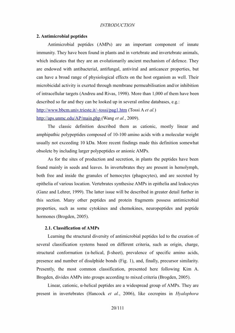
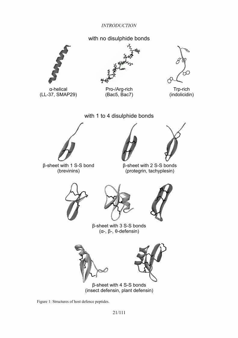
Learning the structural diversity of antimicrobial peptides led to the creation of
several classification systems based on different criteria, such as origin, charge,
structural conformation (α-helical, β-sheet), prevalence of specific amino acids,
presence and number of disulphide bonds (Fig. 1), and, finally, precursor similarity.
Presently, the most common classification, presented here following Kim A.
Brogden, divides AMPs into groups according to mixed criteria (Brogden, 2005).
Linear, cationic, α-helical peptides are a widespread group of AMPs. They are
present in invertebrates (Hancock et al., 2006), like cecropins in Hyalophora
20/111
INTRODUCTION
21/111
Figure 1: Structures of host defence peptides.
α-helical(LL-37, SMAP29)
Pro-/Arg-rich(Bac5, Bac7)
Trp-rich(indolicidin)
with no disulphide bonds
with 1 to 4 disulphide bonds
β-sheet with 1 S-S bond(brevinins)
β-sheet with 2 S-S bonds(protegrin, tachyplesin)
β-sheet with 3 S-S bonds(α-, β-, θ-defensin)
β-sheet with 4 S-S bonds(insect defensin, plant defensin)
INTRODUCTION
cecropia moth (Steiner et al., 1981), andropin in Drosophila (Samakovlis et al.,
1991) and melittin in honeybee (Dufourcq and Faucon, 1977), and in vertebrates, like
pleurocidin in the winter flounder (Cole et al., 1997), magainins in Xenopus frogs
(Zasloff, 1987), cathelicidin-BF in Bungarus snake (Wang et al., 2008), myeloid
antimicrobial peptides in cattle [BMAPs, (Skerlavaj et al., 1996)], pigs [PMAPs,
(Storici et al., 1994)] and sheep [SMAP-29, (Skerlavaj et al., 1999)], and finally
CAP18 in various mammals, such as rabbit (Larrick et al., 1993) and primates
(Zelezetsky et al., 2006), including humans (Agerberth et al., 1995).
Similarly ubiquitous group are cationic peptides rich in a particular amino acid
or a group of amino acids, such as the Pro-rich apidaecin and abaecin in honeybee
(Casteels et al., 1989; Casteels et al., 1990), drosocin in Drosophila (Bulet et al.,
1993), the Bac family in ruminants (Gennaro et al., 1989; Shamova et al., 1999) and
PR-39 in pigs (Agerberth et al., 1991), the Pro/Phe-rich porcine prophenin (Harwig
et al., 1995), the His-rich human histatins (Kavanagh and Dowd, 2004; Oppenheim
et al., 1986), the Trp-rich indolicidin (Selsted et al., 1992) and, recently deduced
from genomes and cDNA, Ser/Gly-rich cathelicidins in salmonids (Scocchi et al.,
2009b).
Another important group included in this classification are anionic and cationic
peptides containing disulphide bridges formed by cysteine residues. There may be
one S-S bond [in brevinins, (Morikawa et al., 1992)] two bonds [in porcine protegrin
(Kokryakov et al., 1993) and tachyplesin in horseshoe crab (Nakamura et al., 1988)],
or three bonds, as in the family of defensins present in vertebrate species (Lehrer and
Ganz, 2002) and insects (Lambert et al., 1989) (more details on human defensins
later in this chapter). More than three bonds are present in antifungal plant defensins
(Thomma et al., 2002) and in a structurally similar peptide named drosomycin from
Drosophila (Fehlbaum et al., 1994).
Finally, there is a group of anionic peptides, such as maximin H5 (Lai et al.,
2002), several Glu/Asp-rich anionic peptides in sheep (Brogden et al., 1997) and
human dermcidin (Schittek et al., 2001), which seem to exert their microbicidal
activity as secondary to different principal activities, and their detailed characteristics
are still to be elucidated (Harris et al., 2009).
A separate group consists of antimicrobial peptides derived from fragmented
22/111
INTRODUCTION
proteins or of antimicrobial domains of larger proteins. The examples include
lactoferricin [a fragment of lactoferrin cleaved by pepsin (Gifford et al., 2005)],
casein-derived casocidin from bovine milk (Zucht et al., 1995), or proteolytic
fragments of ovalbumin (Pellegrini et al., 2004).
2.2. Host Defence Peptides
The term Host Defence Peptides (HDPs) is often used as a synonym of
antimicrobial peptides but some authors consider HDPs as a subdivision of AMPs
that includes only gene-encoded peptides whose only known function is the
involvement in the immune response, both by direct microbicidal action and by
stimulation of the immune system. Apart from some of the aforementioned examples,
such as anionic peptides or antimicrobial fragments of proteins, all the peptides
further considered in this thesis are HDPs according to the proposed definition and
both terms will be used as synonyms.
2.3. Major physical properties of AMPs
2.3.1. Charge
AMPs are mostly cationic, with the net charge ranging between +2 and +18,
and it is one of their key features necessary for prokaryotic cell selectivity. As
opposed to zwitterionic and neutral phosphatidylcholine (PC) which is the main
ingredient of eukaryotic membranes, prokaryotic membranes contain acidic
phospholipids like phosphatidylglycerol (PG), phosphatidylserine (PS) or cardiolipin
(CL) and additional superficial anionic compounds, such as LPS and
teichoic/teichuronic acid in Gram-negative and Gram-positive bacteria, respectively.
Thus, the content of such anions allows the electrostatic interaction with cationic
AMPs (Yount et al., 2006). It has been shown that an increase of a peptide molecule's
net charge leads to a more efficient killing of bacteria, but after exceeding a certain
threshold, it reduces the efficiency and the microbial selectivity, inducing
physiologically undesired effects such as hemolysis (Dathe et al., 2001). Anionic
peptides endowed with bactericidal activity require metal ions, such as zinc, to kill
bacteria. This suggests that the ions play a key role as mediators of the peptides'
mode of action (Brogden et al., 1996; Harris et al., 2009).
23/111
INTRODUCTION
2.3.2. Amphipathicity
An unequal distribution of hydrophobic and hydrophilic amino acids
(amphipathicity) is important for the interaction between an AMP molecule in
solution and the surface of a microbe. It is a feature of most AMPs, including those
with α-helical and β-sheet structures. In α-helices, the optimal periodicity of the
amphipathicity for a selective antimicrobial action requires the presence of a
hydrophobic residue every 3 to 4 amino acids and is then supported with electrostatic
attraction between the peptide and the bacterial membrane. β-sheet peptides often
form polar and non-polar surfaces thanks to their higher rigidity provided by S-S
bridges. Amphipathicity of AMPs, just like their charge, has to be balanced to avoid
undesirable effects, since a greater concentration of hydrophobic residues may lead
to permeabilisation of neutrally charged host cells. In fact, overall hydrophobicity,
usually around 50%, determines the ability to enter and permeabilise the lipid bilayer
and may induce host cell lysis as well when too high (Yeaman and Yount, 2003).
2.4. Modes of action
As for their mode of action, AMPs are generally divided into the groups of
“lytic” and “non-lytic” peptides. The former group, composed mostly of α-helical
peptides, primarily exerts membrane-permeabilising activity leading to the loss of
ions, dissipation of the membrane potential, irreversible changes in the internal cell
environment, eventual cell death and lysis. The latter group, while presenting weaker
permeabilising activity, is rapidly internalised into the target cells and induces cell
death through the inhibition of intracellular processes.
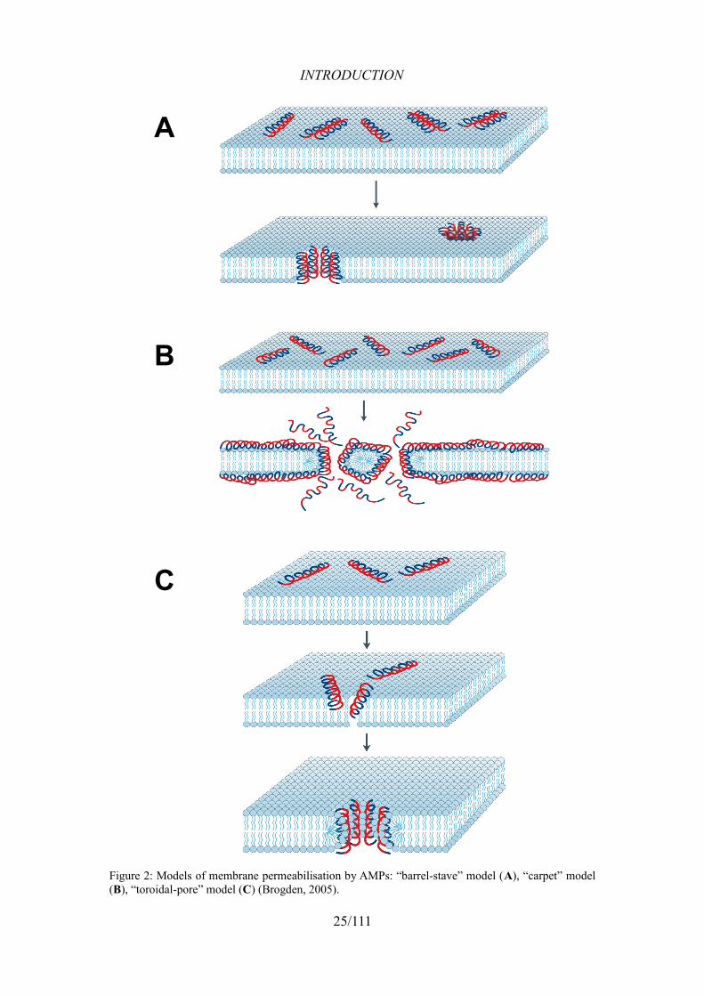
2.4.1. Membrane permeabilisation
The first of the three currently supported membrane permeabilisation models is
the “barrel-stave” model (Fig. 2A). It proposes a pore formation by a bundle of
peptide molecules spanning the lipid bilayer and lining the hydrophobic core like
barrel-staves. Even though it was historically the first model proposed, it is currently
known to be true only in the case of alamethicin (Yang et al., 2001).
The “carpet” model (Fig. 2B) was first described for dermaseptins (Pouny et
al., 1992) and then for several other AMP families, e.g. cecropins. Membrane
permeabilisation in this model occurs in a detergent-like manner, leading to the
formation of micelles. A peptide molecule is orientated parallelly to the membrane,
24/111
INTRODUCTION
25/111
Figure 2: Models of membrane permeabilisation by AMPs: “barrel-stave” model (A), “carpet” model (B), “toroidal-pore” model (C) (Brogden, 2005).
A
B
C
INTRODUCTION
does not get completely inserted into the hydrophobic core and does not contact the
hydrophilic parts of other peptide molecules. After an electrostatically driven
interaction with the polar phospholipid head groups, the peptide places its
hydrophobic face towards the fatty acid chains and the hydrophilic face towards the
surface. Once the threshold concentration is reached, a disintegration occurs by
disrupting the local bilayer curvature and during that process transient pores may be
formed (Shai, 2002).
In the third model called the “toroidal pore” model (Fig. 2C), peptide
molecules form pores in the lipid bilayer as they do in the “barrel-stave” model.
However, in this case they do not come into contact with the hydrophobic core, but
rather form a continuous bend lined throughout its surface with the lipid head groups
tilted to connect both leaflets of the membrane (Brogden, 2005). This model has been
described e.g. for magainins, melittin and protegrins (Yang et al., 2001).
2.4.2. Cell penetration and intracellular interactions
2.4.2.1 Internalisation
Some AMPs that do not present highly membranolytic properties can act
intracellularly. The internalisation of peptide molecules may occur via several
mechanisms.
For instance, a transient permeabilisation effect allows the crossing of the
membrane by the honeybee AMP hymenoptaecin, but with no long-lasting
membrane damage and no synergy with the effects of the lytic peptides' mode of
action (Casteels et al., 1993).
A type of cross-membrane passage using a stereospecific mechanism involving
a transporter-mediated internalisation has been proposed for two Pro-/Arg-rich
AMPs, active only against Gram-negative bacteria: apidaecin (Castle et al., 1999)
and Bac7 (Podda et al., 2006). For the latter, an ABC transporter employment was
proposed, since a mutation in the gene encoding SbmA, a putative element of an
ABC transporter system, leads to a significant reduction in the uptake of Bac7 and
other Pro-rich peptides by E. coli cells (Mattiuzzo et al., 2007).
Histatin-5, one of histidine-rich AMPs endowed with antifungal activity, binds
a protein located in the fungal cell envelope, Ssa1/2, that functions as a peptide
receptor and may be involved in the internalisation of histatin-5 (Kavanagh and
26/111
INTRODUCTION
Dowd, 2004; Li et al., 2003).
2.4.2.2 Intracellular killing
Until now, three major target mechanisms have been found to be interfered by
the cell penetrating AMPs.
Inhibition of cell division and induction of filamentation (abnormal cell
elongation) have been observed in several Gram-negative strains after the treatment
with PR-39 (Boman et al., 1993) and indolicidin (Subbalakshmi and Sitaram, 1998).
This is the effect of inhibition at various stages of DNA, RNA and protein synthesis.
This action is exerted by such peptides like α-helical pleurocidin and dermaseptin,
the aforementioned PR-39 and indolicidin, and human defensin 1 (HNP-1, described
further in this chapter) (Brogden, 2005).
Disruption of respiratory mechanisms leading to the generation of reactive
oxygen species is another killing mechanism, employed by histatins against fungi
(Kavanagh and Dowd, 2004) and by microcin J25, an antibacterial peptide
synthesised by E. coli, that also presents RNA polymerase inhibition in Gram-
negative bacteria (Bellomio et al., 2007).
Third significant target is the chaperone protein DnaK, that is bound and
inhibited by Pro-rich peptides, such as drosocin, pyrrhocoricin, apidaecin (Otvos et
al., 2000) and Bac7. However, the in vitro interaction of the latter peptide with DnaK
does not translate into an altered susceptibility of a dnaK-knock out strain of E. coli
to the peptide (Scocchi et al., 2009a).
2.5. Mechanisms of resistance in bacteria
In the course of evolution microorganisms have developed strategies to evade
or compensate the antimicrobial activity of HDPs. They can be exerted
extracellularly, to destroy AMP molecules or prevent their initial interaction with the
microbial surfaces, or intracellularly, usually to transport the internalised peptides
outside. Of course, the modifications are made to a limited extent in order not to be
detrimental for the microbe (Yount et al., 2006).
27/111

INTRODUCTION
2.5.1. Extracellular mechanisms
Proteolysis is one of several ways in which bacteria attempt to protect
themselves from the microbicidal activity of AMPs. A well-known example is the
outer membrane endopeptidase PgtE in Salmonella, similar to OmpT (Stumpe et al.,
1998) and protease VII in E. coli and Pla in Yersinia. PgtE is posttranscriptionally
regulated by the PhoP/PhoQ regulon, which also controls other resistance
mechanisms (see later). PgtE does not confer resistance to β-sheet peptides with S-S
bonds because of their rigid conformation (Guina et al., 2000). Other proteases
targeting AMPs are expressed in such bacterial species as S. aureus, E. coli,
P. aeruginosa (Yount et al., 2006).
External surface modifications affect the initial interaction between the peptide
and the bacterial cell. A highly electronegative capsule exopolysaccharide, e.g.
alginic acid in P. aeruginosa (Friedrich et al., 1999) or CPS (capsule polysaccharide)
in Klebsiella pneumoniae (Campos et al., 2004), binds avidly to cationic AMPs and
depletes them from the medium, protecting vital cellular targets. Tightly packed LPS
(see 5.) is also considered a mean of protection from AMPs in Gram-negative
bacteria and has demonstrated to be impermeable to unmodified dodecapeptide
(Papo and Shai, 2005).
Cell wall- or outer membrane-associated mechanisms are the key AMP-
countermeasures in Gram-positive and Gram-negative bacteria, respectively. In S.
aureus, the Dlt operon catalyses attachment of D-alanine onto teichoic acid. This
leads to a decreased anionicity of the cell wall and consequently obstructs the
attachment of cationic AMP molecules (Collins et al., 2002). The PhoP/PhoQ system
in Salmonella is responsible for upregulation of a series of active responses to AMPs
(Gunn and Miller, 1996), such as the aforementioned PgtE localisation on the outer
membrane or various lipid A modifications: palmitoylation (Guo et al., 1998),
deacylation (Trent et al., 2001) and aminoarabinose modification (Gunn et al., 1998),
that make the outer membrane less anionic and less accessible to AMPs, but also
downregulate the host signalling and PAMP recognition through TLRs (Kawasaki et
al., 2004).
The least known resistance mechanisms are those altering the plasma
membrane, of which the most commonly described is the MprF-mediated system in
28/111
INTRODUCTION
S. aureus (with homologues in several other species) leading to the substitution of
standard anionic phospholipids with cationic lipids containing ornithine (Yount et al.,
2006).
2.5.2. Intracellular mechanisms
The intracellular mechanisms of resistance generally concentrate on removal of
the internalised drugs from the cell by energy-dependent efflux pumps. These have
been described in many microbial species (Lawrence and Barrett, 1998). Several
pumps responsible for the removal of AMPs are known, such as a universal Mtr
system in Neisseria, QacA in S. aureus, or ABC-transporters in fungi (Yount et al.,
2006), and they may be an element of integrated systems involving several aspects of
AMP resistance (Tzeng et al., 2005).
3. Mammalian HDPs
The number of genes encoding antimicrobial peptides in mammals varies.
Therefore, the number and prevalence of certain peptide families in different genera
are also varied. What is common for this class of vertebrates is the presence and role
of defensins and cathelicidins as the principal HDPs.
3.1. Defensins
Mammalian defensins vary in sequence, but all share the presence of six
cysteine residues whose spacing and connectivity through disulphide bonds allows
the division into the two main classes, α- and β-defensins, and a small class of the
circular θ-defensins (Lehrer and Ganz, 2002). The three-dimensional structure
consists of a triple-stranded antiparallel β-sheet and a short N-terminal α-helix (the
latter absent in bovine defensins). In humans and some rodents both classes have
been found, in rabbits and guinea pigs only α-defensins are present, while in cattle,
sheep and pigs only the β-defensin class has been reported (Schneider et al., 2005).
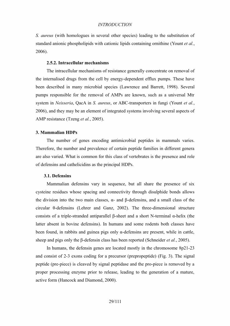
In humans, the defensin genes are located mostly in the chromosome 8p21-23
and consist of 2-3 exons coding for a precursor (prepropeptide) (Fig. 3). The signal
peptide (pre-piece) is cleaved by signal peptidase and the pro-piece is removed by a
proper processing enzyme prior to release, leading to the generation of a mature,
active form (Hancock and Diamond, 2000).
29/111
INTRODUCTION
3.1.1. Human α-defensins
According to Schneider et al., five α-defensin genes and six α-defensin
molecules are known (HNP-1 and -2 derive from the same precursor). They are
present predominantly in azurophilic granules of neutrophils, where they represent
5-7% of total protein content, and in Paneth cells, secretory cells in the epithelium of
the small intestine. Their main role seems to be the killing of ingested microbes
inside the phagolysosomes of neutrophils, but also the control of intestinal bacteria
populations. HD-5 has a particularly broad range of tissue presence, being produced
by epithelia of the female reproductive tract. The human α-defensins display
antibacterial (both against Gram-positive and Gram-negative bacteria) and antifungal
activity (Schneider et al., 2005).
3.1.2. Human β-defensins
The genes for β-defensins show a much higher level of variability, with only
four products isolated from tissues (hBD-1 through -4), but with many more
identified transcripts giving rise to theoretically functional peptides or inactive
products of pseudogenes (hBD-5 through -33) (Pazgier et al., 2006). Most of these
transcripts are encoded by genes located on the chromosomes 6 and 20, outside the
first identified location (8p21-23) of human β-defensins (Crovella et al., 2005).
As for the expression sites, they include epithelial surfaces of respiratory,
gastrointestinal and urogenital tracts, comprising exocrine glands. hBD-3 has also
30/111
Figure 3: Gene structure of human α-defensins (A) and β-defensins (B) (Hancock and Diamond, 2000, modified).
A
B
INTRODUCTION
been detected in skeletal muscles, heart, skin and neutrophils. The transcripts of
further β-defensins have been identified predominantly in testis and epididymis
(Pazgier et al., 2006).
Among the best-known β-defensins (hBD-1 to -4), hBD-3 presents the widest
target spectrum, comprising Gram-positive and Gram-negative bacteria, fungi
(Harder et al., 2001; Joly et al., 2004) and even enveloped HIV-1 virus (Chang and
Klotman, 2004; Quiñones-Mateu et al., 2003), whereas hBD-1 and -2 are active
primarily against Gram-negative bacteria strains. hBD-3 is also apparently not as
salt-sensitive as hBD-1 and -2, being able to exert its microbicidal activity under
physiological or pathologically hypertonic conditions (such as lungs of CF patients)
(Harder et al., 2001).
The antimicrobial activity of human β-defensins is only one of their properties,
as they are also powerful immunomodulators. They possess strong chemotactic
activity towards dendritic cells (see 1.4.3.) and memory T-cells (see 1.4.1.) exerted
by binding to the chemokine CCR6 receptor (Yang et al., 1999). This process also
induces DC maturation, antigen uptake and presentation, and other adaptive immune
responses (Lillard et al., 1999; Yang et al., 2004). Interestingly, it has been shown
that artificial hBD-3 variants with reorganised disulphide bonds had similar
bactericidal activity, but impaired chemotactic properties. The latter activity was
completely absent in analogues with no S-S bridges, suggesting the importance of
three-dimensional structure for chemotaxis (Wu et al., 2003). There are several other
putative roles in which hBDs might be involved, including keratinocyte
differentiation, tumour development and tissue remodelling (Pazgier et al., 2006).
3.2. Cathelicidins
The term “cathelicidins” was first proposed by Zanetti et al. in 1995 to describe
a non-homogeneous group of antimicrobial peptides, including e.g. PMAP-36,
CAP18, human FALL-39 (later renamed to LL-37) and bactenecins, identified in
mammalian myeloid cells and sharing a conserved preproregion in their precursor
sequence (Zanetti et al., 1995). In the recent years, several cathelicidins were
predicted from genomic/cDNA or purified from fish and snake (Chang et al., 2005;
Chang et al., 2006; Maier et al., 2008; Scocchi et al., 2009b; Wang et al., 2008).
Cathelicidins are evolutionarily related, despite a great variety of sequences of
31/111
INTRODUCTION
the mature peptides (Tomasinsig and Zanetti, 2005). The pro-piece contains a domain
that is highly similar to a porcine cathepsin inhibitor named cathelin (cathelin-like
domain, CLD). The recombinant CLD of LL-37 inhibits cathepsin L, a cysteine
protease (Zaiou et al., 2003), but surprisingly, a partial deletion of a loop in a CLD
derived from the porcine protegrin-3 resulted in its activating effect on cathepsin L
(Zhu et al., 2008). There has been a positive selection of certain regions of CLD-
encoding sequence in the course of evolution, which led to those differences and
suggests a functional divergence of this fragment across different species (Zhu,
2008).
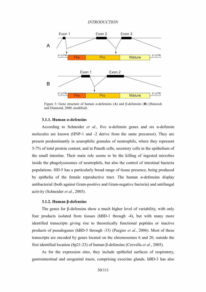
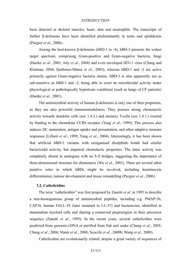
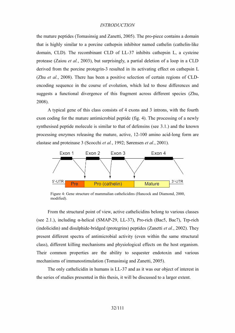
A typical gene of this class consists of 4 exons and 3 introns, with the fourth
exon coding for the mature antimicrobial peptide (fig. 4). The processing of a newly
synthesised peptide molecule is similar to that of defensins (see 3.1.) and the known
processing enzymes releasing the mature, active, 12-100 amino acid-long form are
elastase and proteinase 3 (Scocchi et al., 1992; Sørensen et al., 2001).
From the structural point of view, active cathelicidins belong to various classes
(see 2.1.), including α-helical (SMAP-29, LL-37), Pro-rich (Bac5, Bac7), Trp-rich
(indolicidin) and disulphide-bridged (protegrins) peptides (Zanetti et al., 2002). They
present different spectra of antimicrobial activity (even within the same structural
class), different killing mechanisms and physiological effects on the host organism.
Their common properties are the ability to sequester endotoxin and various
mechanisms of immunostimulation (Tomasinsig and Zanetti, 2005).
The only cathelicidin in humans is LL-37 and as it was our object of interest in
the series of studies presented in this thesis, it will be discussed to a larger extent.
32/111
Figure 4: Gene structure of mammalian cathelicidins (Hancock and Diamond, 2000, modified).
INTRODUCTION
4. The human cathelicidin
4.1. Genetics, expression and processing
LL-37, previously known as FALL-39, was initially predicted from a myeloid
cDNA library and synthesised as FA-LL-37 endowed with antimicrobial activity
(Agerberth et al., 1995). The following year, it was extracted from granulocytes and
the correct, 37 amino acid-long molecule sequence was confirmed
(LLGDFFRKSKEKIGKEFKRIVQRIKDFLRNLVPRTES) (Gudmundsson et al.,
1996). It is encoded by the human CAP18 gene (hCAP18 or CAMP), located on the
chromosome 3p21.3 and endowed with the standard 4 exon/3 intron structure, with
the active C-terminal fragment comprised in the 4th exon, along with six additional
N-terminal amino acids (Larrick et al., 1996).
The expression of the hCAP18 gene was first detected in neutrophils (Larrick
et al., 1995), but it has later been described in the following cell types, tissues and
body fluids (Dürr et al., 2006):
• leukocytes
• bone marrow
(myelocytes, metamyelocytes)
• skin of newborn infants
• numerous squamous epithelia
• nail
• sweat
• wound fluid, blister fluid
• ocular surface epithelia
• synovial membranes
• nasal mucosa
• lung epithelia
• developing lung
• bronchoalveolar lavage fluid
• salivary glands
• saliva
• gingiva
• colon epithelium and mucosa
• testis
• epididymis epithelium
• spermatozoa
• seminal plasma
• vernix caseosa
• amniotic fluid
The hCAP-18 protein, precursor of LL-37, stored in the specific granules of
neutrophils (see 1.3.3.1), is cleaved on cell activation by proteinase 3, one of the
serine proteases of azurophilic granules (Sørensen et al., 2001). Surprisingly, the
cathelin-like domain that seemed a collateral product of this cleavage, serving solely
33/111
INTRODUCTION
as a protector of the host tissues from the cytotoxic activity of LL-37 (see later),
turned out to possess some antimicrobial activity itself, showing how the evolution
maximised the usefulness of the whole translation product (Zaiou et al., 2003).
Despite being the only human cathelicidin, LL-37 can generate several other
derivative peptides with antimicrobial properties, created through cleavage
performed by proteases present locally in many tissues. They may have enhanced
microbicidal effects but reduced immunostimulatory capabilities (see later), which is
a proof of a posttranslational regulation of the broad range of LL-37's physiological
functions (Burton and Steel, 2009).
The role of factors inducing the expression of the cathelicidin is under constant
investigation. An important role has been assigned to vitamin D, produced in skin
upon UV exposure, whose biologically active form (1,25VitD3) has multiple binding
sites in the hCAP-18 promoter sequence. Increased bactericidal activity of
macrophages against M. tuberculosis has been linked to 1,25VitD3-dependent
induction of LL-37 expression. Other factors playing a role in its modification are
commensal and pathogenic bacteria in the intestine, as butyrate produced by many
symbionts increasing the expression of the peptide, and the MxiE virulence regulator
in Shigella pathogens, which leads to its downregulation (Nijnik and Hancock,
2009).
Variations in the production of the cathelicidin have been reported in numerous
diseases. Upregulation occurs in skin inflammatory disorders (e.g. psiorasis), in
gastric epithelia during H. pylori infection, bronchoalveolar lavage fluid-associated
disorders (e.g. cystic fibrosis) and several others. Downregulation has been observed
in atopic dermatitis, enteric infections and in neutrophils during acute leukaemia. A
complete absence of LL-37 in granulocytes and saliva has been observed in patients
with morbus Kostmann (Dürr et al., 2006).
4.2. Structural properties
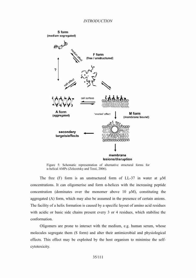
4.2.1. Conformation
LL-37 is a linear molecule which assumes an α-helical conformation under
physiological conditions. In general, it can occur in 4 different structural forms
(Fig. 5).
34/111
INTRODUCTION
The free (F) form is an unstructured form of LL-37 in water at µM
concentrations. It can oligomerise and form α-helices with the increasing peptide
concentration (dominates over the monomer above 10 µM), constituting the
aggregated (A) form, which may also be assumed in the presence of certain anions.
The facility of a helix formation is caused by a specific layout of amino acid residues
with acidic or basic side chains present every 3 or 4 residues, which stabilise the
conformation.
Oligomers are prone to interact with the medium, e.g. human serum, whose
molecules segregate them (S form) and alter their antimicrobial and physiological
effects. This effect may be exploited by the host organism to minimise the self-
cytotoxicity.
35/111
Figure 5: Schematic representation of alternative structural forms forα-helical AMPs (Zelezetsky and Tossi, 2006).
INTRODUCTION
Lastly, the membrane bound (M) form also depends on the molecule's
amphipathicity, as the hydrophobic face comes into contact with and immerses into
the lipid bilayer, and the polar side faces the solvent (Johansson et al., 1998;
Zelezetsky and Tossi, 2006). It has been recently found, that in this state the peptide
adopts a helix-break-helix conformation, with both the N- and C-termini unstructured
(Porcelli et al., 2008).
A comparison of the tendency to assume F- and A-forms by LL-37 and its
orthologues in primates, and their affinity to anionic and zwitterionic membranes,
suggest a preferential reduction of the antimicrobial activity in the human
cathelicidin (more prone to aggregate), but also a simultaneous enhancement of its
binding capabilities to various host cells (Morgera et al., 2009).
4.2.2. Charge and amphipathicity
Both of these properties have a substantial impact on the antimicrobial
efficiency of AMPs (see 2.3.). In case of LL-37, 16 of 37 residues are charged at
physiological pH, with 11 positive and 5 negative charges resulting in the net charge
of +6. This enables electrostatic interactions with anionic microbial surfaces and
decreases the affinity to neutral host cells.
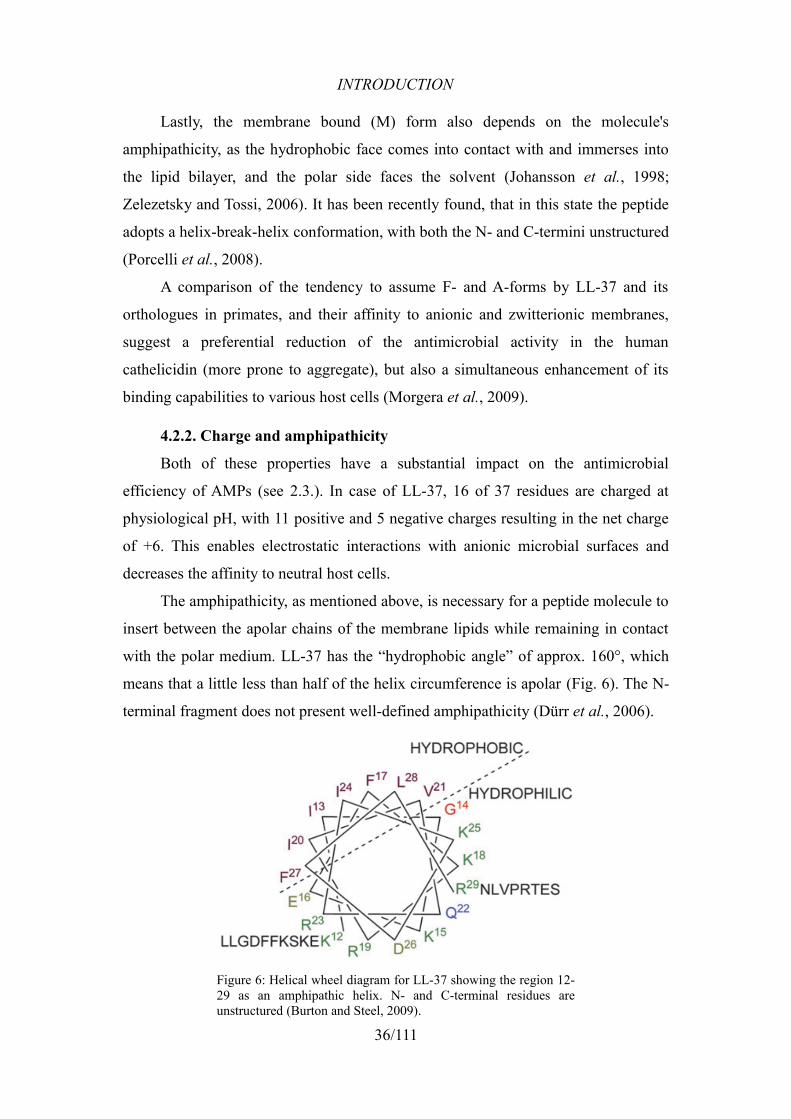
The amphipathicity, as mentioned above, is necessary for a peptide molecule to
insert between the apolar chains of the membrane lipids while remaining in contact
with the polar medium. LL-37 has the “hydrophobic angle” of approx. 160°, which
means that a little less than half of the helix circumference is apolar (Fig. 6). The N-
terminal fragment does not present well-defined amphipathicity (Dürr et al., 2006).
36/111
Figure 6: Helical wheel diagram for LL-37 showing the region 12-29 as an amphipathic helix. N- and C-terminal residues are unstructured (Burton and Steel, 2009).
INTRODUCTION
4.3. Physiology
4.3.1. Direct and indirect antimicrobial activity
The human cathelicidin is active against a broad range of microorganisms. Its
minimal inhibitory concentrations (MICs) have been established against many
species of Gram-positive and Gram-negative bacteria, Spirochetes and yeasts. As an
effective lytic peptide (see 2.4.1.), LL-37 performs a rapid permeabilisation of
bacterial cells. Two modes of membrane perforation have been proposed as the most
probable for this peptide. First, the carpet model of a detergent-like disintegration of
membranes was suggested (Oren et al., 1999). It was later opposed by the toroidal-
pore model, denying the detergent-like fragmentation of the lysed membrane
(Henzler Wildman et al., 2003). Some recent results indicate that the exact
mechanism may depend on the lipidic content of the membrane (Porcelli et al., 2008)
and differs even between the orthologues of LL-37 in primates (Morgera et al.,
2009). In the fungal membranes, LL-37 forms large pores allowing the efflux of
molecules of up to 40 kDa (den Hertog et al., 2005).
An obstruction of biofilm formation by P. aeruginosa has been recorded at
concentrations lower than bactericidal, thus the antimicrobial efficiency seems to be
supported by the inhibition of a potential defence mechanism in bacteria (Overhage
et al., 2008).
4.3.2. Immune system stimulation
Other-than-microbicidal activities of hCAP-18/LL-37 were noticed quite early
and by far have become the fastest growing branch of knowledge on this peptide.
Binding and neutralisation of bacterial LPS, one of the most potent stimuli of
the immune system, was among the first physiologically important features described
(Larrick et al., 1995). Sequestering of LPS leads to a significant decrease in
LPS-induced TNF-α and nitric oxide release by macrophages (see 1.3.4.) (Zughaier
et al., 2005). LL-37 can attack free aggregated LPS, but can also compete with LPS
bound to its co-receptor (CD14) present on the surface of macrophages, inducing
dissociation of LPS complexes and thus attenuating cytokine release by those cells
(Rosenfeld et al., 2006).
As other important HDPs, LL-37 possesses chemotactic properties that are not
affected by the presence of serum. It chemoattracts both T-lymphocytes and
37/111
INTRODUCTION
neutrophils (see 1.3.3.1), the latter of which form a sort of positive feedback by
releasing more cathelicidin on stimulation. The peptide has also been reported to
induce the maturation of dendritic cells (see 1.4.3.) and degranulation of mast cells
(that also produce LL-37). Chemotactic effects occur via interaction with at least
three types of receptors, either by a direct activation or by transactivation (Bowdish
et al., 2005; Dürr et al., 2006).
LL-37 has been indicated as an important factor in wound healing, either by
being a product of dividing epithelial cells, an inducer of migration of keratinocytes
or a putative growth factor/signalling molecule in epithelia. Angiogenic properties
have also been observed by applying LL-37 in both in vivo and in vitro experiments.
One of the cathelicidin-binding receptors has shown to be a mediator of
neovascularisation (Dürr et al., 2006).
4.3.3. Cytotoxicity
Some undesired negative effects of the generally cytotoxic properties of the
human cathelicidin have been observed. A considerable level of haemolytic activity
has been reported by Oren et al. which may be explained by putative importance of
hydrophobic interaction with otherwise zwitterionic (uncharged) membranes of
erythrocytes (Oren et al., 1999). What is more, a moderate dose of LL-37
administered to LPS-treated rats relieved the effect of sepsis, whereas a higher dose
decreased the survival rate which might have been caused by the cytotoxic effects of
the peptide (Fukumoto et al., 2005). According to a recent publication, evolutionary
changes in the sequence of the unique cathelicidin in primates may have emphasised
the ability to form aggregates and shifted the role of the peptide to that of an
immunomodulator (Morgera et al., 2009).
4.4. Resistance in bacteria
Several existing ways of bacterial defence from HDPs have confirmed to be
effective against LL-37. The most common mechanism of resistance is a proteolytic
degradation of peptide molecules by extracellularly secreted proteases (see 2.5.1.).
Among four enzymes of common pathogens reported by Schmidtchen et al. (2002),
three of them belong to the M4 peptidase family and possess similar specificities:
P. aeruginosa LL-37-specific elastase, Enterococcus faecalis gelatinase and the 50
kDa metalloproteinase of Proteus mirabilis. Streptococcus pyogenes also degraded
38/111
INTRODUCTION
the LL-37 cathelicidin (Schmidtchen et al., 2002). In S. aureus, two enzymes have
been tested for the efficiency of LL-37 degradation: a metalloproteinase (aureolysin)
and a glutamylendopeptidase (V8 protease). The former cleaves three peptidic bonds
in the peptide molecule causing its complete deactivation, whereas the latter cleaves
only the Glu16-Phe17 bond and the so formed C-terminal retains much of the
original peptide's activity (Sieprawska-Lupa et al., 2004).
Haemophilus influenzae, a resident pathogen of the upper respiratory tract,
expresses an unusual structure, phosphorylcholine (ChoP), which is exposed to the
exterior on over 90% of LPS molecules. It reduces the anionicity of the outer
membrane and thus helps to mimic the zwitterionic eukaryotic membranes, which
leads to a 1,000-fold increased survival of the bacteria treated with LL-37 (Lysenko
et al., 2000).
5. Bacterial LPS
Lipopolysaccharide (LPS) is an essential ingredient of the outer membrane
(OM) in Gram-negative bacteria, exposed towards the cell exterior, providing
mechanical and chemical protection to the cell, stabilising the overall membrane
structure and possessing a unique set of biological properties. Although there are
many variations in its content and in intramolecular connections of its ingredients,
the type of LPS molecule found and described in enterobacteria is considered the
“classical” one and will be discussed below.
5.1. Structure
LPS is a 10-20 kDa molecule composed of three main parts joined together in a
chain, starting in the outer leaflet of the OM and finishing with an externally exposed
saccharidic polymer (Fig. 7) (Magalhães et al., 2007). It is anionic and amphiphilic,
may form aggregates of varied morphology and is often firmly associated with
proteins and lipids of the OM (Wilkinson, 1996).
39/111
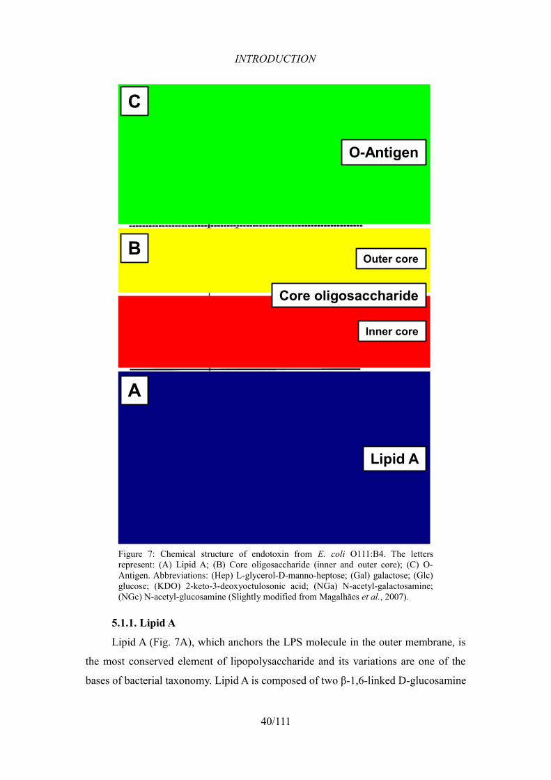
INTRODUCTION
5.1.1. Lipid A
Lipid A (Fig. 7A), which anchors the LPS molecule in the outer membrane, is
the most conserved element of lipopolysaccharide and its variations are one of the
bases of bacterial taxonomy. Lipid A is composed of two β-1,6-linked D-glucosamine
40/111
Figure 7: Chemical structure of endotoxin from E. coli O111:B4. The letters represent: (A) Lipid A; (B) Core oligosaccharide (inner and outer core); (C) O-Antigen. Abbreviations: (Hep) L-glycerol-D-manno-heptose; (Gal) galactose; (Glc) glucose; (KDO) 2-keto-3-deoxyoctulosonic acid; (NGa) N-acetyl-galactosamine; (NGc) N-acetyl-glucosamine (Slightly modified from Magalhães et al., 2007).
B
C
A
Lipid A
Core oligosaccharide
Inner core
Outer core
O-Antigen
INTRODUCTION
(GlcN) residues, covalently linked to four 3-hydroxy-acyl substituents with 12-16
carbon atoms via amide and ester bonds (Magalhães et al., 2007). The species-
specificity of lipid A is characterised by polar appendages to the phosphate residues,
present at the 1st carbon of GlcN I and the 4th carbon of GlcN II, and by the number,
identity and location of further fatty acyl groups linked to the four 3-hydroxy-acyl
residues, the latter of which are represented in enterobacteria by (R)-3-
hydroxytetradecanoic acid (Wilkinson, 1996).
5.1.2. Core oligosaccharide
Core oligosaccharide (Fig. 7B) can be divided into two separate subregions:
inner and outer core.
The inner core is composed of 2 or 3 residues of 2-keto-3-deoxyoctulosonic
acid (KDO), the third of which may be substituted by another monosaccharide e.g.
galactose, and of three L-glycerol-D-manno-heptose (Hep) residues, two of which
are phosphorylated (Hep I and Hep II).
The outer core presents some variability, even between different strains of the
same species. In E. coli, there are five types of the outer core, named K-12 and R1
through R4, differing in the content and connectivities of 5 monosaccharidic residues
(e.g. glucose, galactose) and in the ligation site of the O-polysaccharide (Raetz and
Whitfield, 2002).
5.1.3. O-Antigen
The O-Antigen (or O-Polysaccharide) (Fig. 7C) is a chain of 4-40
oligosaccharides containing 3-8 monomers, mostly glucose and galactose
(Magalhães et al., 2007). This neutral polysaccharide, covalently attached to the LPS
core, presents a high degree of variability between bacterial strains and species and is
highly antigenic. Its uniqueness also makes it an important identifier of bacterial
strains (Wilkinson, 1996).
5.2. Genetics of LPS synthesis
Since the subject of the research presented in this thesis largely regards LPS
genetics, some information on the genes and their products involved in the synthesis
of this compound will be presented.
The formation of lipid A relies on a series of independent lpx genes. The main
41/111
INTRODUCTION
molecule is formed in a sequence catalysed by the LpxA-LpxC-LpxD-LpxB-LpxK
enzyme cascade. Then, WaaA attaches two Kdo residues of the inner core to GlcN II,
and finally LpxL and LpxM add acyl side-chains to those already present at the GlcN
II (Raetz and Whitfield, 2002).
The assembly of the core oligosaccharide depends on the genes from the waa
region. The attachment of the first two Kdo residues, performed by WaaA, is
followed by addition of Hep I and Hep II by WaaC and WaaF, respectively (Gronow
et al., 2000). The third Kdo residue is present only in some core types (K12 and R2)
and is added by the product of the waaZ gene, present only in the strains carrying
those core types (Frirdich et al., 2003). Inner core synthesis is finally completed by
three enzymes: WaaP that phosphorylates Hep I, WaaQ that adds Hep III as a lateral
residue to Hep II, and WaaY that phosphorylates Hep II (Yethon et al., 1998). The
formation of the outer core is initiated by the addition of a glucose (Glc I) to Hep II
of the inner core by a conserved glucosyltransferase WaaG. The genes of the
transferases adding subsequent monosaccharidic residues of the outer core vary
according to the core type (waaB, waaO, waaR, waaU in K-12), but still share a high
homology and are found in the same loci of the enterobacterial genome (Raetz and
Whitfield, 2002).
The O-Antigen biosynthesis depends on the rfb gene cluster that is thought to
have been acquired by E. coli through a horizontal gene transfer, since it shares some
segment similarity with that of the Shigella genus (Stevenson et al., 1994). This
polymorphism is reflected in extremely varied forms of O-antigen. The synthesised
O-Antigen is joined with the Lipid A core fragment by the WaaL ligase (Whitfield et
al., 1997).
5.3. Physiological effects of LPS
Being the most external interface, LPS plays an important role in the
interactions between Gram-negative bacteria and the environment. It is liberated by
the cells at all cell-cycle stages and after cell death, which combined with its extreme
thermal stability makes LPS an efficient pathogenicity factor (Magalhães et al.,
2007).
Lipid A is the major agent responsible for the rapid and massive immune
response to free LPS molecules that got in contact with immunity effectors. The
42/111
INTRODUCTION
molecules are first bound by an acute phase protein called LPS-binding protein
(LBP) and the complex is then recognised by one of the Toll-like receptors, TLR4,
along with its co-receptor CD14, which initiates signalling cascades that induce the
expression of proinflammatory cytokines and adhesion molecules in endothelial
cells, and the activation of macrophages (see 1.3.3.). This parallel reaction leads to
the clinical state of sepsis and may be life-threatening due to such pathological
symptoms as hypotension, endothelial barrier dysfunction, excessive leukocyte
adhesion and disseminated intravascular coagulation (Dauphinee and Karsan, 2006).
Highly specific O-Antigen, the outer-most bacterial cell structure, is
considerably immunogenic, allowing a precisely targeted control of symbionts, e.g.
those inhabiting the intestine, performed by adaptive immunity. Conversely, a contact
with an unknown serotype upon infection may result in a disease state (Nagy and Pál,
2008).
6. Antimicrobial peptides as therapeutics
6.1. Existing approaches
Numerous clinical trials have been applied to three best-studied examples of
peptides intended to be used as topically applied agents against local, superficial
bacterial infections.
Pexiganan, a 22-amino acid synthetic analogue of magainin II, has been
verified as an efficient antibacterial agent in vitro (Fuchs et al., 1998), but it was only
moderately successful, and comparable to an orally administered antibiotic, as a
cream applied on mildly infected diabetic foot ulcers. However, one of the
advantages of this application was no generation of resistance in treated pathogens
(Lipsky et al., 2008). The effect was not sufficient enough for pexiganan to be
approved by FDA.
In phase III studies of the efficiency of iseganan, an analogue of protegrin-1,
the peptide did not prevent the ventilator-associated pneumonia (VAP) in ventilated
patients (Kollef et al., 2006) and did not reduce stomatitis or ulcerative oral
mucositis in patients receiving stomatotoxic therapy (Giles et al., 2004), even though
previous studies showed a 1,000-fold reduction of microflora associated with the
latter disease in healthy patients (Mosca et al., 2000). Clinical trials were stopped in
43/111
INTRODUCTION
2004 due to elevated occurrence of VAP and higher mortality in patients, and the
development of iseganan as an anti-infective agents was abolished (McPhee and
Hancock, 2005).
The third and, by now, the most promising attempt is omiganan, an indolicidin
analogue, rapidly eradicating in vitro bacteria and fungi most often causing catheter-
related bloodstream infections and various cutaneous infections (Fritsche et al.,
2008a; Fritsche et al., 2008b; Sader et al., 2004). This peptide is now awaiting
registration.
6.2. Surpassing the in vivo limitations
Antimicrobial activity of the peptides in vivo is hindered by several factors,
such as sequestration by plasma content or degradation by proteases, produced either
by the host or by the pathogens.
Numerous strategies have been developed to modify peptide structure in order
to make them less susceptible to degradation. These include size reduction,
modification of termini (acetylation, amidation), incorporation of non-natural amino
acids (e.g. D-amino acids), amino acid swapping within the protease cleavage sites
and finally cyclization of peptide molecules
To ameliorate the bioavailability and delivery rates, peptide molecules can be
coupled with protective structures, such as polyethylene glycol (PEG) moieties,
liposomes, or even nanotechnology-generated “smart” polymeric microspheres,
releasing the drug only under certain physiological conditions (Oyston et al., 2009).
6.3. Structural studies and target determination
The knowledge necessary to appoint novel drug candidates among AMPs may
be obtained using different approaches.
Structural studies, concerning physicochemical properties of the peptides that
influence their interaction with membranes or other model systems (e.g. cell wall
elements), provide us with essential information on the mode of action and on
possible modifications to increase the microbicidal efficacy of AMPs. Structural
definition of the peptides can be obtained by using such techniques as NMR, ATR-
FTIR, CD, neutron scattering and atomic force spectroscopy, offering different levels
of resolution. The effect on living cells may be analysed through flow cytometry and
visualised using electron and confocal microscopy. Finally, the stages of peptide-cell
44/111
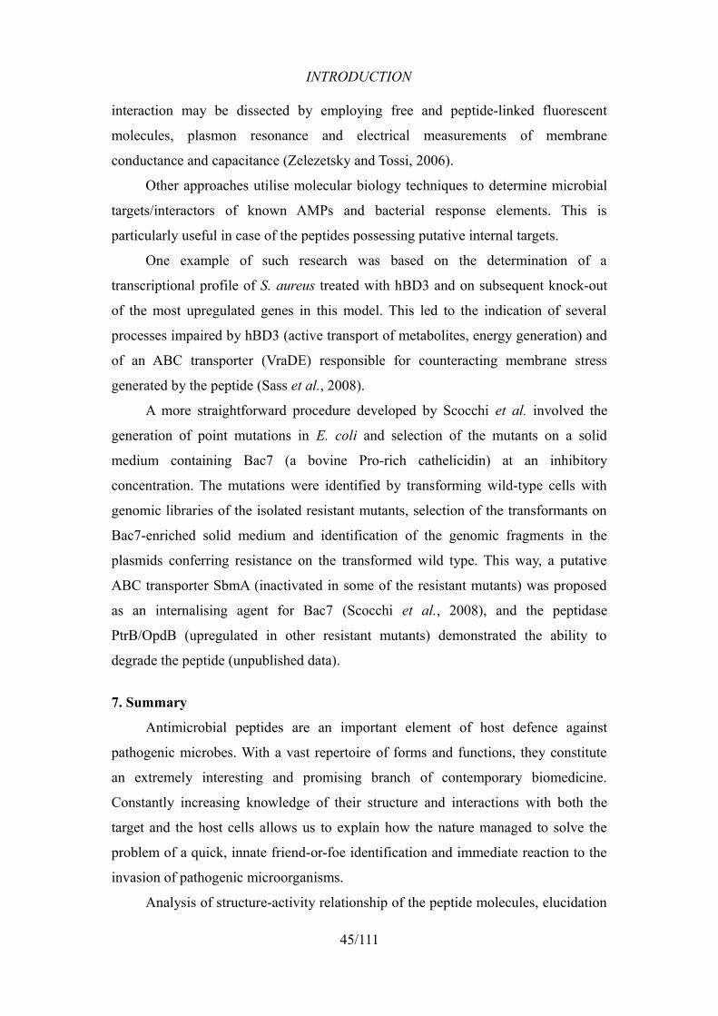
INTRODUCTION
interaction may be dissected by employing free and peptide-linked fluorescent
molecules, plasmon resonance and electrical measurements of membrane
conductance and capacitance (Zelezetsky and Tossi, 2006).
Other approaches utilise molecular biology techniques to determine microbial
targets/interactors of known AMPs and bacterial response elements. This is
particularly useful in case of the peptides possessing putative internal targets.
One example of such research was based on the determination of a
transcriptional profile of S. aureus treated with hBD3 and on subsequent knock-out
of the most upregulated genes in this model. This led to the indication of several
processes impaired by hBD3 (active transport of metabolites, energy generation) and
of an ABC transporter (VraDE) responsible for counteracting membrane stress
generated by the peptide (Sass et al., 2008).
A more straightforward procedure developed by Scocchi et al. involved the
generation of point mutations in E. coli and selection of the mutants on a solid
medium containing Bac7 (a bovine Pro-rich cathelicidin) at an inhibitory
concentration. The mutations were identified by transforming wild-type cells with
genomic libraries of the isolated resistant mutants, selection of the transformants on
Bac7-enriched solid medium and identification of the genomic fragments in the
plasmids conferring resistance on the transformed wild type. This way, a putative
ABC transporter SbmA (inactivated in some of the resistant mutants) was proposed
as an internalising agent for Bac7 (Scocchi et al., 2008), and the peptidase
PtrB/OpdB (upregulated in other resistant mutants) demonstrated the ability to
degrade the peptide (unpublished data).
7. Summary
Antimicrobial peptides are an important element of host defence against
pathogenic microbes. With a vast repertoire of forms and functions, they constitute
an extremely interesting and promising branch of contemporary biomedicine.
Constantly increasing knowledge of their structure and interactions with both the
target and the host cells allows us to explain how the nature managed to solve the
problem of a quick, innate friend-or-foe identification and immediate reaction to the
invasion of pathogenic microorganisms.
Analysis of structure-activity relationship of the peptide molecules, elucidation
45/111
INTRODUCTION
of their modes of action and identification of potential target structures in microbes
may help to create new weapons against the emerging drug-resistant infections
plaguing the modern medicine.
LL-37 could be considered a candidate for a novel antimicrobial agent due to
its broad spectrum of activity. Gaining knowledge of its interactors and potential
resistance mechanisms in bacteria might be a useful addition to the structural studies,
for the purpose of engineering a degradation- and segregation-resistant molecule
with a sufficient level of microbicidal activity, reduced host cytotoxicity and a
desired level of immunomodulating activity.
46/111
AIMS OF THE STUDY
AIMS OF THE STUDY
We attempted to adapt and improve a commercially available procedure of
generating a library of E. coli knock-out mutants, carrying random single insertions
of the Tn5 transposon in their genomes, for the search of those mutations that confer
on bacteria a decreased susceptibility to antimicrobial peptides. This procedure was
meant to be a high-throughput screening method, developed in order to appoint novel
bacterial interactors of AMPs and expand our knowledge on their modes of action,
potentially useful for drug design. The method might be expanded to over 60
bacterial species, applicable with Tn5 mutagenesis, whose majority constitutes
concerns in the ongoing war against pathogenic infections.
Once established, the method was then applied for the elucidation of the
mechanism of action of the human cathelicidin LL-37 against E. coli cells, so as to
indicate the weak points in bacterial physiology that have not changed over the
evolution and allow this antimicrobial peptide to act. Such information might then be
used for targeting those points in the generation of novel peptide-based antibiotics.
47/111
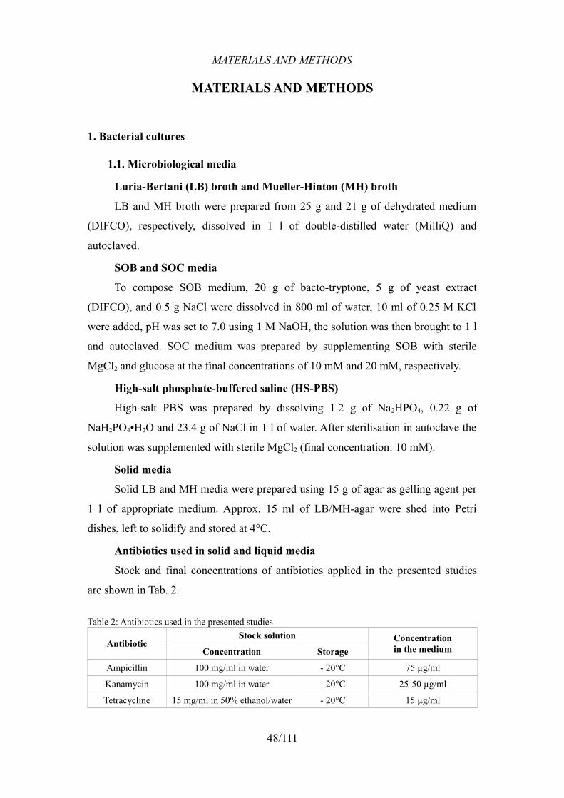
MATERIALS AND METHODS
MATERIALS AND METHODS
1. Bacterial cultures
1.1. Microbiological media
Luria-Bertani (LB) broth and Mueller-Hinton (MH) broth
LB and MH broth were prepared from 25 g and 21 g of dehydrated medium
(DIFCO), respectively, dissolved in 1 l of double-distilled water (MilliQ) and
autoclaved.
SOB and SOC media
To compose SOB medium, 20 g of bacto-tryptone, 5 g of yeast extract
(DIFCO), and 0.5 g NaCl were dissolved in 800 ml of water, 10 ml of 0.25 M KCl
were added, pH was set to 7.0 using 1 M NaOH, the solution was then brought to 1 l
and autoclaved. SOC medium was prepared by supplementing SOB with sterile
MgCl2 and glucose at the final concentrations of 10 mM and 20 mM, respectively.
High-salt phosphate-buffered saline (HS-PBS)
High-salt PBS was prepared by dissolving 1.2 g of Na2HPO4, 0.22 g of
NaH2PO4•H2O and 23.4 g of NaCl in 1 l of water. After sterilisation in autoclave the
solution was supplemented with sterile MgCl2 (final concentration: 10 mM).
Solid media
Solid LB and MH media were prepared using 15 g of agar as gelling agent per
1 l of appropriate medium. Approx. 15 ml of LB/MH-agar were shed into Petri
dishes, left to solidify and stored at 4°C.
Antibiotics used in solid and liquid media
Stock and final concentrations of antibiotics applied in the presented studies
are shown in Tab. 2.
Table 2: Antibiotics used in the presented studies
AntibioticStock solution Concentration
in the mediumConcentration Storage
Ampicillin 100 mg/ml in water - 20°C 75 µg/ml
Kanamycin 100 mg/ml in water - 20°C 25-50 µg/ml
Tetracycline 15 mg/ml in 50% ethanol/water - 20°C 15 µg/ml
48/111
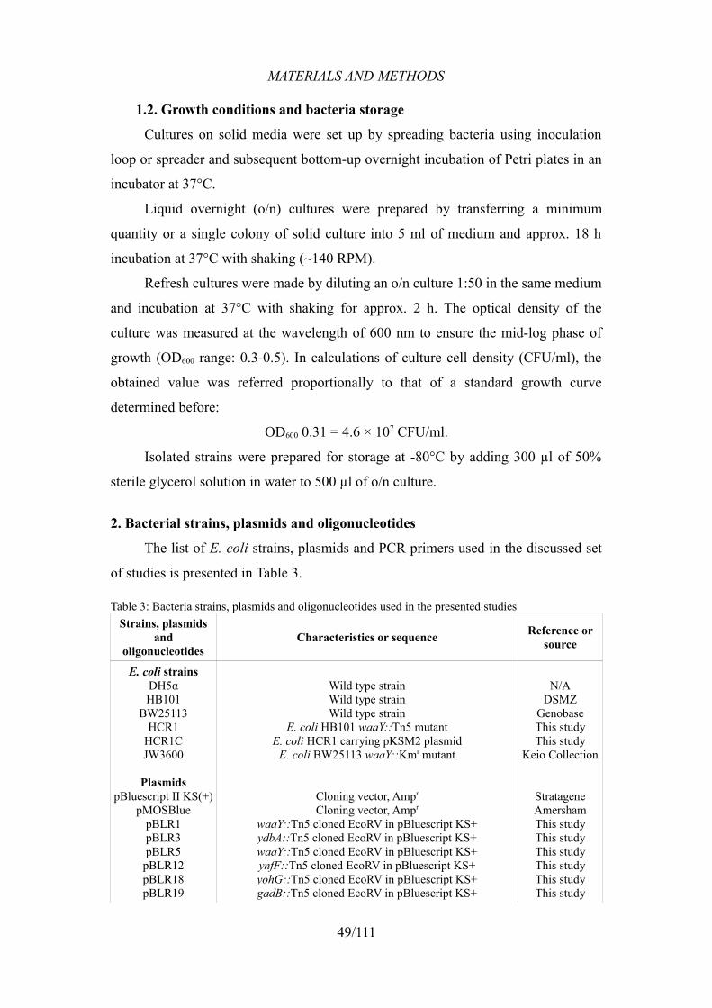
MATERIALS AND METHODS
1.2. Growth conditions and bacteria storage
Cultures on solid media were set up by spreading bacteria using inoculation
loop or spreader and subsequent bottom-up overnight incubation of Petri plates in an
incubator at 37°C.
Liquid overnight (o/n) cultures were prepared by transferring a minimum
quantity or a single colony of solid culture into 5 ml of medium and approx. 18 h
incubation at 37°C with shaking (~140 RPM).
Refresh cultures were made by diluting an o/n culture 1:50 in the same medium
and incubation at 37°C with shaking for approx. 2 h. The optical density of the
culture was measured at the wavelength of 600 nm to ensure the mid-log phase of
growth (OD600 range: 0.3-0.5). In calculations of culture cell density (CFU/ml), the
obtained value was referred proportionally to that of a standard growth curve
determined before:
OD600 0.31 = 4.6 × 10⁷ CFU/ml.
Isolated strains were prepared for storage at -80°C by adding 300 µl of 50%
sterile glycerol solution in water to 500 µl of o/n culture.
2. Bacterial strains, plasmids and oligonucleotides
The list of E. coli strains, plasmids and PCR primers used in the discussed set
of studies is presented in Table 3.
Table 3: Bacteria strains, plasmids and oligonucleotides used in the presented studiesStrains, plasmids
and oligonucleotides
Characteristics or sequence Reference or source
E. coli strainsDH5αHB101
BW25113HCR1
HCR1CJW3600
PlasmidspBluescript II KS(+)
pMOSBluepBLR1pBLR3pBLR5pBLR12pBLR18pBLR19
Wild type strainWild type strainWild type strain
E. coli HB101 waaY::Tn5 mutantE. coli HCR1 carrying pKSM2 plasmid
E. coli BW25113 waaY::Kmr mutant
Cloning vector, Ampr
Cloning vector, Ampr
waaY::Tn5 cloned EcoRV in pBluescript KS+ydbA::Tn5 cloned EcoRV in pBluescript KS+waaY::Tn5 cloned EcoRV in pBluescript KS+ynfF::Tn5 cloned EcoRV in pBluescript KS+yohG::Tn5 cloned EcoRV in pBluescript KS+gadB::Tn5 cloned EcoRV in pBluescript KS+
N/ADSMZ
GenobaseThis studyThis study
Keio Collection
StratageneAmershamThis studyThis studyThis studyThis studyThis studyThis study
49/111
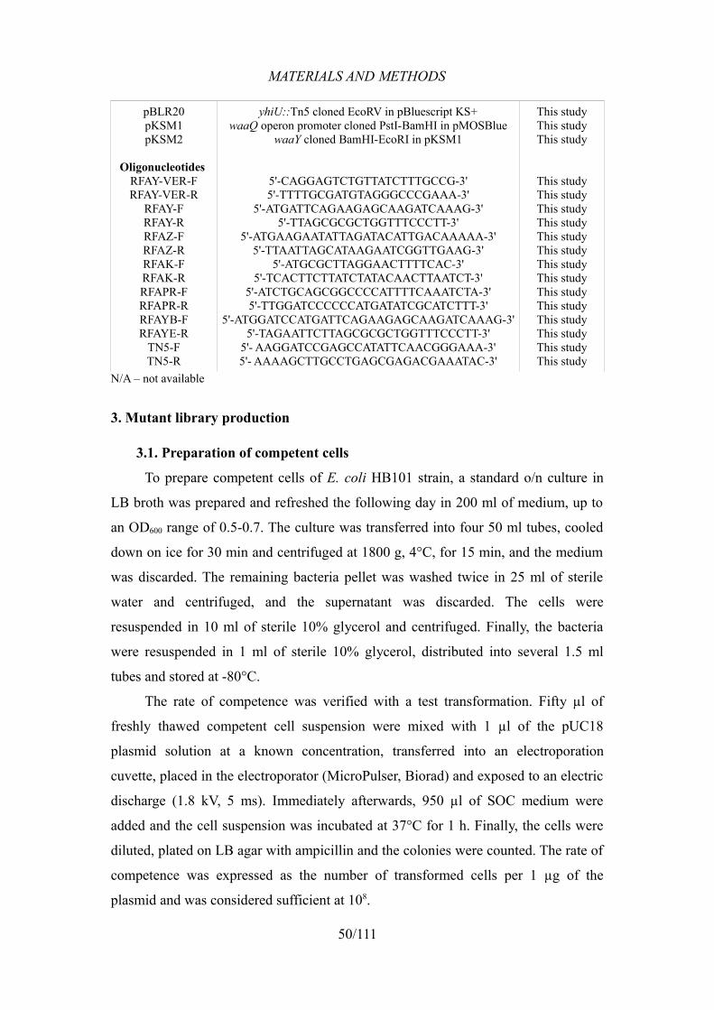
MATERIALS AND METHODS
pBLR20pKSM1pKSM2
Oligonucleotides RFAY-VER-FRFAY-VER-R
RFAY-FRFAY-RRFAZ-FRFAZ-RRFAK-FRFAK-RRFAPR-FRFAPR-RRFAYB-FRFAYE-R
TN5-FTN5-R
yhiU::Tn5 cloned EcoRV in pBluescript KS+waaQ operon promoter cloned PstI-BamHI in pMOSBlue
waaY cloned BamHI-EcoRI in pKSM1
5'-CAGGAGTCTGTTATCTTTGCCG-3'5'-TTTTGCGATGTAGGGCCCGAAA-3'
5'-ATGATTCAGAAGAGCAAGATCAAAG-3'5'-TTAGCGCGCTGGTTTCCCTT-3'
5'-ATGAAGAATATTAGATACATTGACAAAAA-3'5'-TTAATTAGCATAAGAATCGGTTGAAG-3'
5'-ATGCGCTTAGGAACTTTTCAC-3'5'-TCACTTCTTATCTATACAACTTAATCT-3'
5'-ATCTGCAGCGGCCCCATTTTCAAATCTA-3'5'-TTGGATCCCCCCATGATATCGCATCTTT-3'
5'-ATGGATCCATGATTCAGAAGAGCAAGATCAAAG-3'5'-TAGAATTCTTAGCGCGCTGGTTTCCCTT-3'
5'- AAGGATCCGAGCCATATTCAACGGGAAA-3'5'- AAAAGCTTGCCTGAGCGAGACGAAATAC-3'
This studyThis studyThis study
This studyThis studyThis studyThis studyThis studyThis studyThis studyThis studyThis studyThis studyThis studyThis studyThis studyThis study
N/A – not available
3. Mutant library production
3.1. Preparation of competent cells
To prepare competent cells of E. coli HB101 strain, a standard o/n culture in
LB broth was prepared and refreshed the following day in 200 ml of medium, up to
an OD600 range of 0.5-0.7. The culture was transferred into four 50 ml tubes, cooled
down on ice for 30 min and centrifuged at 1800 g, 4°C, for 15 min, and the medium
was discarded. The remaining bacteria pellet was washed twice in 25 ml of sterile
water and centrifuged, and the supernatant was discarded. The cells were
resuspended in 10 ml of sterile 10% glycerol and centrifuged. Finally, the bacteria
were resuspended in 1 ml of sterile 10% glycerol, distributed into several 1.5 ml
tubes and stored at -80°C.
The rate of competence was verified with a test transformation. Fifty µl of
freshly thawed competent cell suspension were mixed with 1 µl of the pUC18
plasmid solution at a known concentration, transferred into an electroporation
cuvette, placed in the electroporator (MicroPulser, Biorad) and exposed to an electric
discharge (1.8 kV, 5 ms). Immediately afterwards, 950 µl of SOC medium were
added and the cell suspension was incubated at 37°C for 1 h. Finally, the cells were
diluted, plated on LB agar with ampicillin and the colonies were counted. The rate of
competence was expressed as the number of transformed cells per 1 µg of the
plasmid and was considered sufficient at 108.
50/111
MATERIALS AND METHODS
3.2. Transformation with the transposon complex
Mutant cells derived from the HB101 strain were produced through a random
insertion of Tn5 transposon into the bacteria genome. Competent cells were
electroporated as described above with 1 µl of EZ-Tn5™ <KAN-2>Tnp
Transposome solution (Epicentre Biotechnologies), composed of a transposase
enzyme and the Tn5 transposon containing the Tn903 kanamycin resistance gene.
The transformants were plated on 9 LB agar plates (~100 µl/plate) containing
kanamycin (25 µg/ml). One additional plate with 1:10 dilution of the bacterial
suspension was prepared to enable a proper colony count. The whole procedure was
repeated 8 times and mutants from all the plates were collected, pooled, and several
stocks were prepared for -80°C storage.
3.3. Extraction of genomic DNA of the mutants
Genomic DNA (gDNA) from 10 random mutant colonies, cultured o/n in 10 ml
LB + 50 µg/ml kanamycin, was extracted using the sarkosyl-pronase lysis method
(Birnboim, 1983). Briefly, the cells were pelleted and resuspended in 400 µl of
1× TE buffer, 500 µl of 2% sarkosyl in 1× TE and 100 µl pronase E (5 mg/ml,
Sigma), and incubated at 37°C until the solution became clear. Then, 10 µl of RNase
A (10 mg/ml, Sigma) were added and the incubation was continued for another 1 h.
Afterwards, an equal volume of phenol/chloroform/isoamyl alcohol mix (25:24:1)
was added, mixed thoroughly and centrifuged using a Biofuge Pico microcentrifuge
(Heraeus) at 5,000 RPM for 5 min. The upper phase was collected and transferred
into a new tube, and an equal volume of chloroform was added, followed by another
centrifugation and transfer of the upper phase into a new tube. Subsequently, a
solution of 5 M NaCl was added to reach the final concentration of 0.2 M, which was
followed by the addition of isopropanol (1:1). The precipitated DNA was transferred
with a pipette tip into a tube containing 70% ethanol and centrifuged (13,000 RPM,
5 min). The supernatant was discarded and the extracted DNA was dried and
resuspended in water. The concentration and purity of DNA was estimated
spectrophotometrically by measuring the absorbance ratios at 230/260/280 nm.
Solutions and reagents
1× TE buffer: 10 mM Tris-HCl, 1.0 mM EDTA, pH 8.0.
51/111
MATERIALS AND METHODS
3.4. Confirmation of a random transposon insertion
To verify a correct and random insertion of Tn5 into the genome of HB101,
a Southern blot of digested and separated gDNA fragments was prepared.
3.4.1. Genomic DNA digestion
Four restriction enzymes with no cleavage sites inside the transposon sequence
(EcoRI, EcoRV, NcoI, KpnI) were tested to select the most evenly distributed
digestion profile of gDNA. The digestions were carried out for 6 h at 37°C with the
use of 40 µg of gDNA and 20 U of each enzyme (all from New England Biolabs).
EcoRV was selected for further procedures.
3.4.2. Electrophoresis and transfer onto a membrane
The fragments of digested genome were separated through electrophoresis on
0.8% agarose gel (50 V, 3 h). The gel was then photographed on a UV
transilluminator with a fluorescent ruler and subjected to three 30 min long washes:
depurination, denaturation and neutralisation, performed in appropriate solutions (see
below).
Subsequently, DNA fragments were transferred to an Amersham Hybond-N+
nylon membrane (GE Healthcare) in 20× SSC buffer. The transfer was set up as a
“sandwich” composed of 4 layers of 3 mm chromatography paper (Whatman), the
agarose gel facing downwards, the nylon membrane, another 3 layers of
chromatography paper and ~100 layers of plain paper towel. After an approx. 18 h
transfer, the membrane was incubated at 80°C for 2 h, for the purpose of fixing of the
transferred DNA, and stored in a chromatography paper envelope at 4°C.
Solutions and reagents
Depurination solution: 0.13 M HCl;
Denaturation solution: 1.5 M NaCl, 0.5 M NaOH;
Neutralisation solution: 1.5 M NaCl, 0.5 M Tris-Cl, pH 7.5;
20× SSC buffer: 0.34 M trisodium citrate, 3 M NaCl.
3.4.3. Southern blot
The subsequent Southern blot assay was carried out using the DIG High Prime
DNA Labeling and Detection Starter Kit II (Roche), based on digoxigenin and
AP-conjugated anti-DIG-antibody. A 467 bp long Tn5 fragment produced by PCR,
52/111
MATERIALS AND METHODS
used as a probe, was labelled following the producer's indications and the labelling
efficiency was then tested as described in the manual.
The following hybridisation procedure consisted of a 30 min pre-hybridisation
of the membrane in DIG Easy Hyb solution (supplied with the kit) pre-heated to
65°C, denaturation of the probe by boiling for 5 min, and its addition to the pre-
hybridisation solution. DNA was hybridised overnight in a closed glass container at
65°C with constant agitation. Afterwards, the probe solution was removed, stored at
-20°C and re-denatured by boiling before every further use. The membrane was
subjected to two stringency washes, the first of which in 2× SSC, 0.1% SDS at room
temperature for 2 × 5 min, and the second one in 0.5× SSC, 0.1% SDS at 65°C for
2 × 15 min, both with shaking.
To perform the immunological detection, the membrane was first incubated at
room temperature (RT) for 5 min in washing buffer, washed for 30 min in blocking
solution, incubated for 30 min with the secondary antibody solution, washed twice in
washing buffer for 15 min and equilibrated in detection buffer for 5 min. Finally, the
membrane was damped with the chemiluminescent substrate for alkaline phosphatase
(CSPD, supplied with kit), wrapped in a plastic pellicle, incubated for 5 min at RT
and exposed to a photographic film (X-OMAT LS, Kodak) in an exposition cassette
(Hypercassette, Amersham) for approx. 30 min. The film was then processed using
GBX developer and fixing solutions (Kodak).
Solutions and reagents
Washing buffer: 0.1 M maleic acid; 0.15 M NaCl, pH7.5; 0.3% (v/v) Tween 20;
Maleic acid buffer: 0.1 M maleic acid; 0.15 M NaCl; pH 7.5;
Blocking solution: 10× blocking solution (supplied) 1:10 in maleic acid buffer;
Secondary antibody solution: 75 mU/ml Anti-DIG-AP (supplied) in block. sol.;
Detection buffer: 0.1 M Tris-Cl; 0.1 M NaCl; pH 9.5.
4. Peptide preparation
4.1. Synthesis and purification
The peptides Bac7(1-16) and ppyLL-37 were generously supplied by the group
of prof. Alessandro Tossi (University of Trieste).
LL-37 was synthesised on 2-chlorotrityl chloride-PS resin using the Fmoc/tBu
53/111
MATERIALS AND METHODS
technology of Solid Phase Peptide Synthesis (SPPS) on a Pioneer™ peptide
synthesiser (PE Biosystems). The synthesised peptide was cleaved from the resin by
incubation for 4 h on a shaker in the following mixture: 87% trifluoroacetic acid
(TFA), 8% 3,6-dioxa-1,8-octane-dithiol (DODT), 2% triisopropylsilane (TIPS),
3% water. The cleaved peptide was washed several times with tert-butyl methyl ether
(TMBE) and dried with N2.
The purification was performed using an XTerra™ RP18 7 µm (19-300 mm)
column attached to a Gilson system of Reversed-Phase High-Performance Liquid
Chromatography (RP-HPLC) managed by Unipoint 3.0 software. The applied
gradient was set to reach 50% of acetonitrile in water from the initial 30% in 50 min.
Subsequently, the collected fractions were analysed with an esquire6000™
mass-spectrometer (Bruker Daltonics) and those of high purity were pooled and
lyophilised. In order to remove the possible TFA residues, the purified LL-37 was
resuspended in 10 mM HCl and lyophilised once again, which was followed by the
final resuspension in water.
4.2. Fluorescent labelling of LL-37
For the peptide-cell binding assay (see 12.), LL-37 was fluorescently labelled
via linkage of the thiol-reactive fluorescent dye BODIPY® FL N-(2-aminoethyl)
maleimide (Invitrogen) to an ad hoc added C-terminal cysteine residue. Peptide
labelling was carried out in 10 mM sodium-phosphate buffer, pH 7.4, containing
30% of acetonitrile. In brief, 2 ml of a 1 mg/ml solution of the fluorescent dye
dissolved in acetonitrile were added dropwise to 18 ml of a 0.1 mg/ml peptide
solution under nitrogen bubbling. After 2 h of incubation at RT in the dark with
stirring, a new aliquot of 2 mg of the peptide was added to the same solution and left
overnight in the dark at 4°C with stirring. The excess of thiol-reactive reagent was
then scavenged by the addition of 10-fold excess of cysteine. After 30 min of
incubation at RT with stirring, the labelled peptide was purified using the procedures
described above.
4.3. Peptide quantification
To estimate peptide concentration, the Waddell assay was used (Waddell,
1956). The analysis was conducted in a quartz cuvette (path length 10 mm; vol. 1 ml)
by adding 2.5 µl of peptide solution to the blank (1 ml of 1/300 N NaOH), reading
54/111
MATERIALS AND METHODS
OD at the wavelengths of 215 nm and 225 nm, subtracting OD225 from OD215 and
multiplying the difference by the dilution factor (400). The addition of another 2.5 µl
of peptide solution was followed by the same procedure (multiplier: 200) and the two
results were then normalised and averaged. A previously synthesised antimicrobial
peptide ppyLL-37 was used as a reference. Each estimation was performed in
duplicate and averaged. The values calculated for the evaluated peptide were referred
to those of the reference peptide with a known concentration, expressed in mg/ml.
5. Determination of the minimal inhibitory concentration
In order to determine the lowest peptide concentration that inhibits the growth
of the tested strains (Minimal Inhibitory Concentration, MIC), the described standard
o/n and refresh cultures in MH broth were set up to obtain mid-log phase cells,
subsequently diluted to 5×10⁵ CFU/ml. The accuracy of the dilution was verified by
plating 50 µl of the 10-2 and 10-3 dilutions, followed by colony count after o/n
incubation of the plates at 37°C.
The peptide was diluted serially in the wells of a microtiter plate in 50 µl of
MH broth, and then 50 µl of previously prepared bacteria suspensions were added to
the wells, thus diluting the peptide and the bacteria to half of their initial
concentrations in each well. The whole plate was covered with a sterile adhesive
plastic film to minimise the evaporation and incubated for 18 h at 37°C. The lowest
peptide concentration that inhibited any visible growth of the bacteria was considered
the MIC.
6. Selection of resistant mutants
To isolate the Tn5-mutants that potentially acquired resistance to Bac7(1-16)
and LL-37, we chose a straightforward selection of the mutant library on solid media
containing the peptide of interest. A cube of ~8 mm3 of frozen glycerol stock was
suspended in 15 ml of MH or LB broth and incubated for ~4 h at 37°C with shaking.
Plates with 15 ml of MH agar were prepared with the desired quantities of
LL-37 dissolved in the medium while still liquid, and the final concentrations of 1, 5,
10, 20 and 30 µM LL-37 were obtained. Bac7(1-16) was incorporated into LB agar
at the final concentration of 5 µM.
Bacteria cultures were diluted in their respective liquid media to obtain the
55/111
MATERIALS AND METHODS
density of 106 CFU/ml and 100 µl of the diluted cultures were spread over the agar
plates enriched with the appropriate peptide, resulting in the final number of approx.
105 cells per plate. Among the colonies that grew on the plates, only the ones with no
background (small, closely surrounding colonies) were chosen and re-plated on LB
agar with kanamycin. Glycerol stocks of each mutant clone were prepared from these
plates and stored at -80°C.
The mutants that survived the selection with Bac7(1-16) were named B7R
(Bac7-Resistant) and numbered consecutively.
To verify the results of the selection with LL-37, selected strains were tested
for resistance on a microtiter plate in 50 µl of MH broth with the addition of 5 µM
LL-37, along with the wild-type strain as a reference. The bacteria were inoculated
from previously prepared plates of LB agar with kanamycin, using the tip of a sterile
toothpick. The verification was considered valid when the strain growth after an
overnight incubation was clearly visible. The strains that passed the verification were
named HCR (Human Cathelicidin-Resistant) and numbered consecutively.
7. Identification of the mutated genes in B7R mutants
Identification of mutation sites was carried out by cloning of the genomic
fragments containing Tn5 from the isolated resistant clones (B7R1-11), and
subsequent sequencing of the cloned gDNA fragments surrounding the transposon.
7.1. Preparation of the cloning vector
The plasmid pBluescript II KS+ was extracted from previously transformed
E. coli HB101 using the illustra™ plasmidPrep Mini Spin Kit (GE Healthcare) and
quantified spectrophotometrically.
Approx. 12 µg of the plasmidic DNA was digested with 20 U of EcoRV (New
England Biolabs) for 6 h at 37°C. The whole digestion mix was then loaded onto a
0.8% agarose gel and separated by electrophoresis (3 h, 50 V, 10-20 mA), in parallel
with a MW marker (SharpMass 1 kb DNA ladder, EuroClone). The gel band
containing the linearised plasmid was excised and the DNA was eluted using
GenElute™ Gel Extraction Kit (Sigma). As the digestion produced blunt ends, the
linear pBluescript II KS+ was subjected to a two-step dephosphorylation, with 20 U
of Calf Intestinal Phosphatase (CIP, New England Biolabs) initially added to the
56/111
MATERIALS AND METHODS
reaction mix and 10 U of CIP added after 2 h at 37°C for another 1 h of incubation.
Finally, the prepared vector was purified using QuickClean 5M PCR Purification Kit
(GenScript Corp.).
7.2. Construction of genomic libraries of the mutants
The following step was ligation of the vector with the genomic fragments of
the mutant clones, obtained by extracting and digesting gDNA (see 3.3. and 3.4.1.).
The ligation was conducted in the total vol. of 15 µl using 10× T4 ligase buffer
(containing 600 µM ATP) and 10 U of T4 ligase (USB). The quantity of the vector
(~100 ng) and the gDNA fragments used were set to obtain the molar ratio of 1:5,
respectively. The calculation of molarity of the insert was based on an estimated
medium size of the gDNA fragments produced by EcoRV. The mix was incubated for
18 h at 16°C.
7.3. Screening of the libraries and insert sequencing
Four µl of each ligation mix were used to transform E. coli DH5α cells by
electroporation (see 3.1.). The transformants were plated on LB agar with ampicillin
and 50 µg/ml kanamycin, so as to allow the growth of only those cells that assumed
the plasmid containing gDNA fragment that carried Tn5 with its Kmr gene. The
extracted plasmids were sequenced (BMR Genomics, Padova, Italy) using KAN-2
FP-1 and RP-1 primers (supplied with EZ-Tn5™ kit) that allowed the sequencing of
the regions located outside the transposon, which indicated the interrupted genes.
The obtained sequences were analysed with BLAST (Altschul et al., 1997) and
elaborated with BioEdit software (Hall, 1999) to precisely determine the location of
Tn5 in the mutant genomes.
8. Identification of the mutated genes in HCR mutants
8.1. Preliminary identification
To perform a preliminary identification of the mutants, the isolated resistant
clones (HCR1-20) were cultured, their gDNA was extracted, digested with EcoRV
restriction enzyme and Southern-blotted using the previously described procedures
(see 3.4.).
57/111
MATERIALS AND METHODS
8.2. Determination of mutation sites in 6 mutants
Identification of mutation sites was performed by cloning of the genomic
fragments containing Tn5 from 7 mutant clones, that presented different
hybridisation profiles, into pBluescript II KS+, followed by the sequencing of the
inserts, as described in Paragraph 7. The sequenced plasmids containing the
fragments that carried Tn5 were named pBLR1, 3, 5, 12, 18, 19 and 20, according to
their respective clone numbers (Tab. 3).
8.3. PCR identification of the remaining mutants
In the mutant clones that were not sequenced, the transposon insertion was
revealed by PCR amplification of the waaY (rfaY) gene, interrupted in the sequenced
mutant presenting the most prevalent hybridisation profile.
Reaction mix (50 µl) was composed of:
• 10 µl of 5× GoTaq buffer (“Green”, containing electrophoresis loading buffer);• 4 µl of 25 mM MgCl2;• 0.5 µl of GoTaq DNA polymerase (all three from Promega);• 1 µl of 10 mM dNTPs;• 5 µl of each of 5 µM primers: RFAY-VER-F and RFAY-VER-R;• 2 µl of gDNA template (0.5-1 µg);• water to volume.
The amplification was carried out on a EuroClone One Advanced thermocycler
using the following programme: pre-denaturation (94°C, 5 min), 35 amplification
cycles (94°C, 1 min; 55°C, 1 min; 72°C, 2 min), final elongation (72°C, 5 min). The
products were separated by electrophoresis on 0.8% agarose gel (50 V, 1 h) and
visualised on a UV transilluminator.
9. RT-PCR analysis
E. coli HB101 and HCR1 strains were cultured until an OD600 of 0.4 was
reached. Total RNA was extracted from bacterial cells, lysed by several freeze/thaw
cycles, using the TRIzol Plus RNA Purification Kit (Invitrogen) and any residual
genomic DNA was removed by RQ1 RNase-Free DNase (Promega) (1 U per 1 µg of
RNA).
cDNA was synthesised from approx. 1 µg of isolated RNA, using 0.5 µg of
random hexamer primers (Promega) and 200 U of M-MLV reverse transcriptase
(USB), in the presence of 40 U of RNasin Plus RNase Inhibitor (Promega). The 1 h
58/111
MATERIALS AND METHODS
long reaction was conducted at 42°C.
PCR based on the cDNA template was performed using RFAY-F/RFAY-R
primers to amplify the waaY (rfaY) gene, RFAZ-F/RFAZ-R primers to amplify waaZ
(rfaZ), and RFAK-F/RFAK-R primers to amplify waaU (rfaK) (Tab. 3), in the
following reaction mix:
• 10 µl of 5× GoTaq buffer;• 1 µl of 100 mM MgSO4;• 0.5 µl of GoTaq DNA polymerase;• 1 µl of 10 mM dNTPs;• 0.5 µl of each of 100 µM primer pairs;• 6 µl of cDNA template;• water to volume.
The amplification programme was composed of: pre-denaturation (94°C,
5 min), 30 amplification cycles (94°C, 30 s; 55°C, 40 s; 72°C, 40 s) and the final
elongation (72°C, 7 min). The products were separated electrophoretically and
visualised on a UV transilluminator.
10. Trans-complementation of the waaY mutation
To complement the waaY mutation, the HCR1 mutant strain was transformed
with the pKSM2 plasmid, based on pMOSBlue, carrying a functional copy of waaY
downstream of waaQ operon promoter (Tab. 3).
The waaY gene was amplified from E. coli HB101 gDNA using RFAYB-F and
RFAYE-R primers, that added BamHI-EcoRI restriction sites to the amplified
fragment, in the following reaction mix:
• 10 µl of 5× GoTaq buffer;• 1 µl of 100 mM MgSO4;• 0.5 µl of Vent DNA polymerase;• 2 µl of 10 mM dNTPs;• 1 µl of each of 100 µM primers;• 1 µl of gDNA template (approx. 500 ng);• water to volume,
using the following PCR programme: pre-denaturation (94°C, 5 min), 30
amplification cycles (94°C, 30 s; 58°C, 40 s; 72°C, 40 s), final elongation (72°C,
7 min). The product was digested with EcoRI and BamHI to create sticky ends and
ligated with pMOSBlue linearised with the same pair of enzymes, creating the
pKSM1 plasmid.
59/111
MATERIALS AND METHODS
The waaQ operon promoter (Clementz, 1992) was then amplified from HB101
gDNA using RFAPR-F and RFAPR-R primers, that added PstI-BamHI restriction
sites to the amplified fragment, in a reaction mix as shown above, using the
following PCR programme: pre-denaturation (94°C, 5 min), 30 amplification cycles
(94°C, 30 s; 60°C, 30 s; 72°C, 30 s), final elongation (72°C, 7 min). The product was
digested with PstI and BamHI, to create sticky ends, and ligated with pKSM1
linearised with the same pair of enzymes, creating the pKSM2 plasmid, which was
eventually sequenced to ensure the correctness of the construct.
pKSM2, containing the waaQ promoter and the waaY gene between PstI and
EcoRI restriction sites, was then used for the transformation of competent HCR1
cells by electroporation (see 3.1.).
11. Antimicrobial activity assays
11.1. Growth kinetics assay
To determine the growth inhibition of the bacteria exposed to various peptide
concentrations, we chose an OD-based approach of measuring the cell density
changes over the incubation time on a microtiter plate.
Mid-log phase bacteria cultures were diluted in MH broth to
2 × 10⁶ CFU/ml and 100 µl of each dilution were added to 100 µl of peptide
solutions, previously prepared on a microtiter plate, or to 100 µl of medium with no
peptide, as a control. Thus prepared solutions of 106 CFU/ml, with the peptide at half
of the original concentration, were placed inside a plate reader (Tecan Sunrise,
Switzerland) and incubated at 37°C throughout the assay with intermittent shaking.
The OD change was measured at the wavelength of 620 nm with 10-minute intervals
for 4 h (25 cycles). The data from the experiments, performed in triplicate, were
collected by Magellan 4 software (Tecan) and averaged.
11.2. Killing kinetics assay
The effective killing rate of LL-37 was expressed by the reduction of the
number of CFU after a 90 min incubation with 2 µM peptide.
Mid-log phase bacteria cultures were diluted in MH broth to
2 × 10⁶ CFU/ml, and 200 µl of the dilutions were added to 200 µl of previously
prepared 4 µM peptide solution and to 200 µl of medium with no peptide, as a
60/111

MATERIALS AND METHODS
control. Thus prepared solutions of 106 CFU/ml without the peptide and with 2 µM
LL-37 were incubated at 37°C for 90 min with shaking. The initial bacteria
suspensions were serially diluted, and 50 µl of 10-3 and 10-4 dilutions were plated in
duplicate on MH agar.
After the 90 min incubation, bacteria suspensions were immediately diluted in
high-salt PBS. Fifty µl of 100 through 10-2 dilutions of the peptide-treated bacteria
and 50 µl of 10-3 and 10-4 dilutions of the untreated bacteria were plated in duplicate
on MH agar. The obtained data, expressed in CFU/ml, were averaged within each
duplicate. Significance of the results was evaluated with the unpaired t-test,
performed using GraphPad QuickCalcs software, and the values of P<0.05 were
considered statistically significant.
12. Flow cytometry assays
Binding of BODIPY-labelled LL-37 to E. coli cells was determined by flow
cytometry using a Cytomics FC 500 instrument (Beckman-Coulter, Inc.) equipped
with an argon laser (488 nm, 5 mW) and a photomultiplier tube fluorescence detector
for green (525 nm) or red (610 nm) filtered light. All detectors were set to the
logarithmic amplification. Optical and electronic noise were eliminated by setting the
electronic gating threshold on the forward-scattering detector, while the data flow
rate was kept below 300 events per second to avoid cell coincidence. At least 10,000
events were acquired for each sample.
Cultures of mid-log phase bacteria were harvested, brought to 106 CFU/ml in a
diluted medium (20% MH broth in sodium phosphate buffer), incubated with 0.1 μM
and 0.25 μM LL-37-BODIPY® at 37°C for 10 min and analysed immediately. All
experiments were conducted in triplicates and the data were expressed as Mean
Fluorescence Intensity (MFI) ± S.D. Significance of the results was evaluated with
the unpaired t-test, performed using GraphPad QuickCalcs software, and the values
of P<0.05 were considered statistically significant.
Permeabilisation of the cytoplasmic membrane of E. coli was assessed by flow
cytometry, measuring the uptake of propidium iodide (PI) into dividing cells, diluted
to 106 CFU/ml. Fluorescence of the internalised PI was measured after 15, 30, 45 and
60 min of cell incubation with 0.25 μM and 0.5 μM LL-37 at 37°C, as previously
described (Podda et al., 2006). Permeabilisation data were expressed as a percentage
61/111
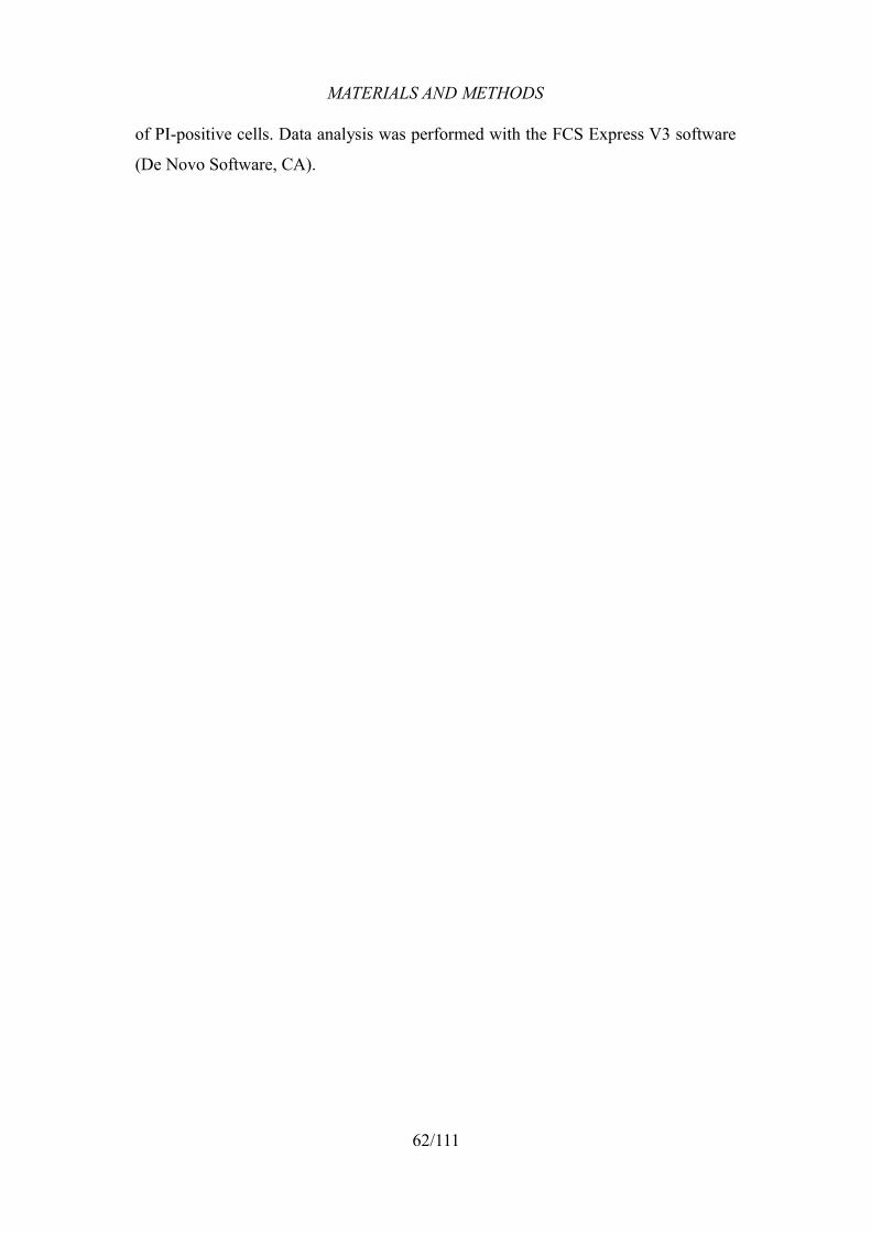
MATERIALS AND METHODS
of PI-positive cells. Data analysis was performed with the FCS Express V3 software
(De Novo Software, CA).
62/111
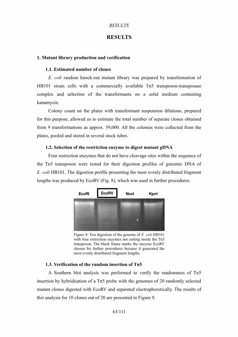
RESULTS
RESULTS
1. Mutant library production and verification
1.1. Estimated number of clones
E. coli random knock-out mutant library was prepared by transformation of
HB101 strain cells with a commercially available Tn5 transposon-transposase
complex and selection of the transformants on a solid medium containing
kanamycin.
Colony count on the plates with transformant suspension dilutions, prepared
for this purpose, allowed us to estimate the total number of separate clones obtained
from 9 transformations as approx. 59,000. All the colonies were collected from the
plates, pooled and stored in several stock tubes.
1.2. Selection of the restriction enzyme to digest mutant gDNA
Four restriction enzymes that do not have cleavage sites within the sequence of
the Tn5 transposon were tested for their digestion profiles of genomic DNA of
E. coli HB101. The digestion profile presenting the most evenly distributed fragment
lengths was produced by EcoRV (Fig. 8), which was used in further procedures.
1.3. Verification of the random insertion of Tn5
A Southern blot analysis was performed to verify the randomness of Tn5
insertion by hybridisation of a Tn5 probe with the genomes of 20 randomly selected
mutant clones digested with EcoRV and separated electrophoretically. The results of
this analysis for 10 clones out of 20 are presented in Figure 9.
63/111
Figure 8: Test digestion of the genome of E. coli HB101 with four restriction enzymes not cutting inside the Tn5 transposon. The black frame marks the enzyme EcoRV chosen for further procedures because it generated the most evenly distributed fragment lengths.
EcoRI EcoRV NcoI KpnI
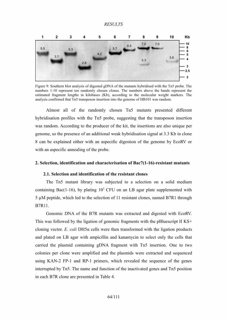
RESULTS
Almost all of the randomly chosen Tn5 mutants presented different
hybridisation profiles with the Tn5 probe, suggesting that the transposon insertion
was random. According to the producer of the kit, the insertions are also unique per
genome, so the presence of an additional weak hybridisation signal at 3.3 Kb in clone
8 can be explained either with an aspecific digestion of the genome by EcoRV or
with an aspecific annealing of the probe.
2. Selection, identification and characterisation of Bac7(1-16)-resistant mutants
2.1. Selection and identification of the resistant clones
The Tn5 mutant library was subjected to a selection on a solid medium
containing Bac(1-16), by plating 105 CFU on an LB agar plate supplemented with
5 µM peptide, which led to the selection of 11 resistant clones, named B7R1 through
B7R11.
Genomic DNA of the B7R mutants was extracted and digested with EcoRV.
This was followed by the ligation of genomic fragments with the pBluescript II KS+
cloning vector. E. coli DH5α cells were then transformed with the ligation products
and plated on LB agar with ampicillin and kanamycin to select only the cells that
carried the plasmid containing gDNA fragment with Tn5 insertion. One to two
colonies per clone were amplified and the plasmids were extracted and sequenced
using KAN-2 FP-1 and RP-1 primers, which revealed the sequence of the genes
interrupted by Tn5. The name and function of the inactivated genes and Tn5 position
in each B7R clone are presented in Table 4.
64/111
Figure 9: Southern blot analysis of digested gDNA of the mutants hybridised with the Tn5 probe. The numbers 1-10 represent ten randomly chosen clones. The numbers above the bands represent the estimated fragment lengths in kilobases (Kb), according to the molecular weight markers. The analysis confirmed that Tn5 transposon insertion into the genome of HB101 was random.
5.5
3.7
5.3
2.8
4.2
5.76.5 7.0 7.0
3.63.3
1 2 3 4 5 6 7 8 9 10108654
3
2
2.5
Kb
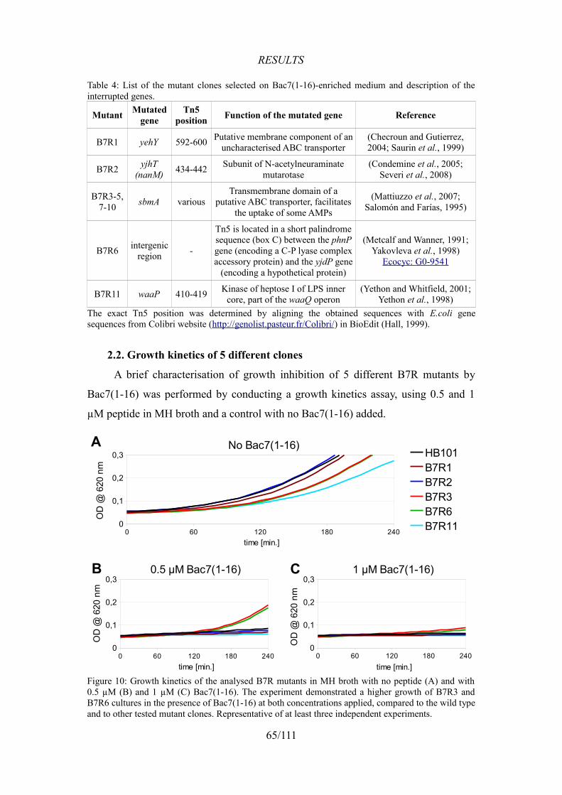
RESULTS
Table 4: List of the mutant clones selected on Bac7(1-16)-enriched medium and description of the interrupted genes.
Mutant Mutated gene
Tn5 position Function of the mutated gene Reference
B7R1 yehY 592-600 Putative membrane component of an uncharacterised ABC transporter
(Checroun and Gutierrez, 2004; Saurin et al., 1999)
B7R2 yjhT (nanM) 434-442 Subunit of N-acetylneuraminate
mutarotase(Condemine et al., 2005;
Severi et al., 2008)
B7R3-5, 7-10 sbmA various
Transmembrane domain of a putative ABC transporter, facilitates
the uptake of some AMPs
(Mattiuzzo et al., 2007; Salomón and Farías, 1995)
B7R6 intergenic region -
Tn5 is located in a short palindrome sequence (box C) between the phnP gene (encoding a C-P lyase complex accessory protein) and the yjdP gene
(encoding a hypothetical protein)
(Metcalf and Wanner, 1991; Yakovleva et al., 1998)
Ecocyc: G0-9541
B7R11 waaP 410-419 Kinase of heptose I of LPS inner core, part of the waaQ operon
(Yethon and Whitfield, 2001; Yethon et al., 1998)
The exact Tn5 position was determined by aligning the obtained sequences with E.coli gene sequences from Colibri website (http://genolist.pasteur.fr/Colibri/) in BioEdit (Hall, 1999).
2.2. Growth kinetics of 5 different clones
A brief characterisation of growth inhibition of 5 different B7R mutants by
Bac7(1-16) was performed by conducting a growth kinetics assay, using 0.5 and 1
µM peptide in MH broth and a control with no Bac7(1-16) added.
65/111
Figure 10: Growth kinetics of the analysed B7R mutants in MH broth with no peptide (A) and with0.5 µM (B) and 1 µM (C) Bac7(1-16). The experiment demonstrated a higher growth of B7R3 and B7R6 cultures in the presence of Bac7(1-16) at both concentrations applied, compared to the wild type and to other tested mutant clones. Representative of at least three independent experiments.
0 60 120 180 2400
0,1
0,2
0,3No Bac7(1-16)
HB101B7R1B7R2B7R3B7R6B7R11
time [min.]
OD
@ 6
20 n
m
0 60 120 180 2400
0,1
0,2
0,30.5 µM Bac7(1-16)
time [min.]
OD
@ 6
20 n
m
0 60 120 180 2400
0,1
0,2
0,31 µM Bac7(1-16)
time [min.]
OD
@ 6
20 n
m
A
B C
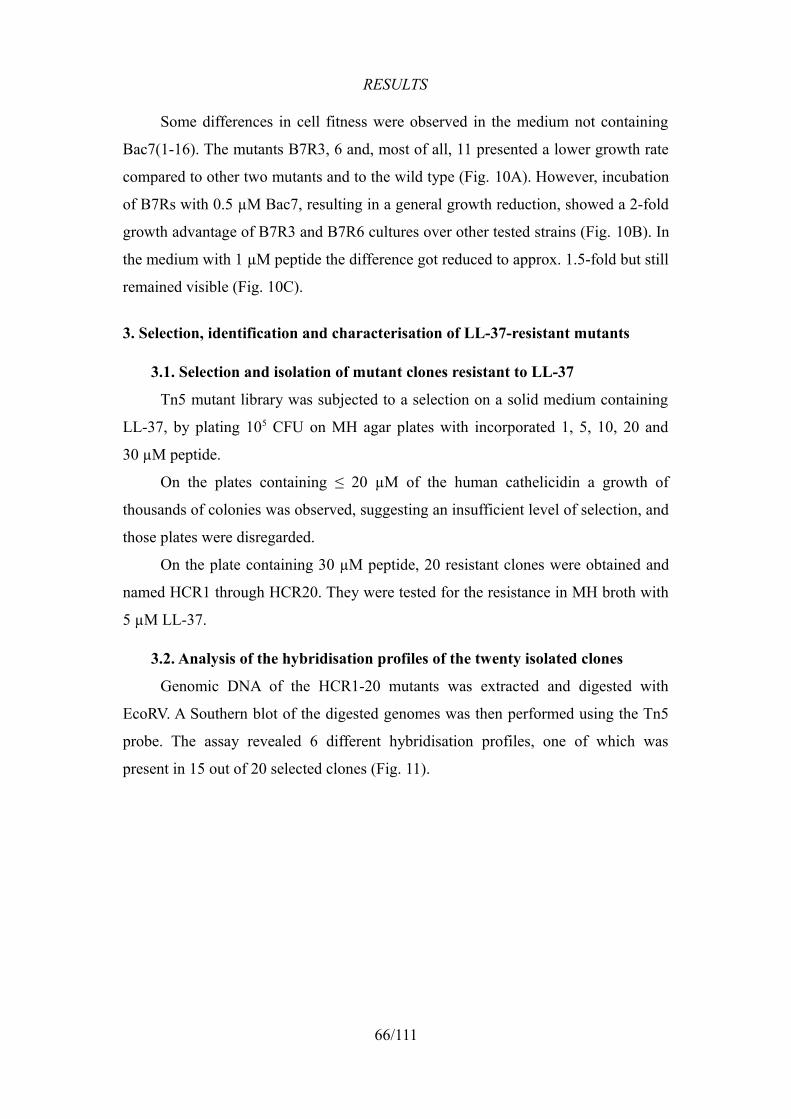
RESULTS
Some differences in cell fitness were observed in the medium not containing
Bac7(1-16). The mutants B7R3, 6 and, most of all, 11 presented a lower growth rate
compared to other two mutants and to the wild type (Fig. 10A). However, incubation
of B7Rs with 0.5 µM Bac7, resulting in a general growth reduction, showed a 2-fold
growth advantage of B7R3 and B7R6 cultures over other tested strains (Fig. 10B). In
the medium with 1 µM peptide the difference got reduced to approx. 1.5-fold but still
remained visible (Fig. 10C).
3. Selection, identification and characterisation of LL-37-resistant mutants
3.1. Selection and isolation of mutant clones resistant to LL-37
Tn5 mutant library was subjected to a selection on a solid medium containing
LL-37, by plating 105 CFU on MH agar plates with incorporated 1, 5, 10, 20 and
30 µM peptide.
On the plates containing ≤ 20 µM of the human cathelicidin a growth of
thousands of colonies was observed, suggesting an insufficient level of selection, and
those plates were disregarded.
On the plate containing 30 µM peptide, 20 resistant clones were obtained and
named HCR1 through HCR20. They were tested for the resistance in MH broth with
5 µM LL-37.
3.2. Analysis of the hybridisation profiles of the twenty isolated clones
Genomic DNA of the HCR1-20 mutants was extracted and digested with
EcoRV. A Southern blot of the digested genomes was then performed using the Tn5
probe. The assay revealed 6 different hybridisation profiles, one of which was
present in 15 out of 20 selected clones (Fig. 11).
66/111
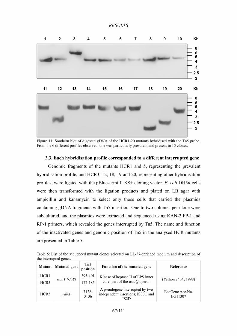
RESULTS
3.3. Each hybridisation profile corresponded to a different interrupted gene
Genomic fragments of the mutants HCR1 and 5, representing the prevalent
hybridisation profile, and HCR3, 12, 18, 19 and 20, representing other hybridisation
profiles, were ligated with the pBluescript II KS+ cloning vector. E. coli DH5α cells
were then transformed with the ligation products and plated on LB agar with
ampicillin and kanamycin to select only those cells that carried the plasmids
containing gDNA fragments with Tn5 insertion. One to two colonies per clone were
subcultured, and the plasmids were extracted and sequenced using KAN-2 FP-1 and
RP-1 primers, which revealed the genes interrupted by Tn5. The name and function
of the inactivated genes and genomic position of Tn5 in the analysed HCR mutants
are presented in Table 5.
Table 5: List of the sequenced mutant clones selected on LL-37-enriched medium and description of the interrupted genes.
Mutant Mutated gene Tn5 position Function of the mutated gene Reference
HCR1waaY (rfaY)
393-401 Kinase of heptose II of LPS inner core, part of the waaQ operon (Yethon et al., 1998)
HCR5 177-185
HCR3 ydbA 3128-3136
A pseudogene interrupted by two independent insertions, IS30C and
IS2D
EcoGene Acc.No.EG11307
67/111
Figure 11: Southern blot of digested gDNA of the HCR1-20 mutants hybridised with the Tn5 probe. From the 6 different profiles observed, one was particularly prevalent and present in 15 clones.
1 2 3 4 5 6 7 8 9 10
86543
22.5
Kb
11 12 13 14 15 16 17 18 19 20
86543
22.5
Kb
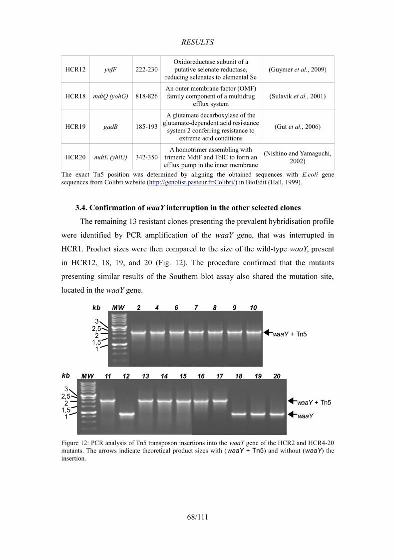
RESULTS
HCR12 ynfF 222-230Oxidoreductase subunit of a putative selenate reductase,
reducing selenates to elemental Se(Guymer et al., 2009)
HCR18 mdtQ (yohG) 818-826An outer membrane factor (OMF) family component of a multidrug
efflux system(Sulavik et al., 2001)
HCR19 gadB 185-193
A glutamate decarboxylase of the glutamate-dependent acid resistance
system 2 conferring resistance to extreme acid conditions
(Gut et al., 2006)
HCR20 mdtE (yhiU) 342-350A homotrimer assembling with
trimeric MdtF and TolC to form an efflux pump in the inner membrane
(Nishino and Yamaguchi, 2002)
The exact Tn5 position was determined by aligning the obtained sequences with E.coli gene sequences from Colibri website (http://genolist.pasteur.fr/Colibri/) in BioEdit (Hall, 1999).
3.4. Confirmation of waaY interruption in the other selected clones
The remaining 13 resistant clones presenting the prevalent hybridisation profile
were identified by PCR amplification of the waaY gene, that was interrupted in
HCR1. Product sizes were then compared to the size of the wild-type waaY, present
in HCR12, 18, 19, and 20 (Fig. 12). The procedure confirmed that the mutants
presenting similar results of the Southern blot assay also shared the mutation site,
located in the waaY gene.
68/111
Figure 12: PCR analysis of Tn5 transposon insertions into the waaY gene of the HCR2 and HCR4-20 mutants. The arrows indicate theoretical product sizes with (waaY + Tn5) and without (waaY) the insertion.
waaY + Tn5
waaY
11 12 13 14 15 16 17 18 19 20MW
32,52
1,51
kb
waaY + Tn5
32,52
1,51
2 4 6 7 8 9 10MWkb
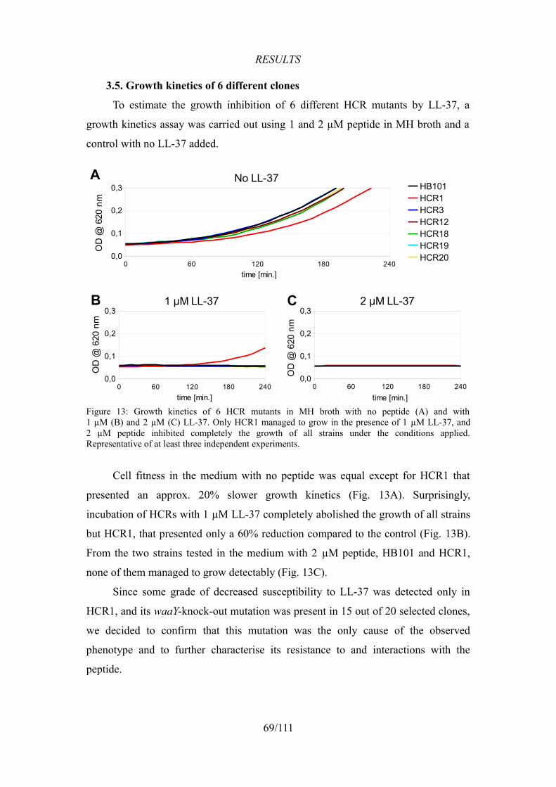
RESULTS
3.5. Growth kinetics of 6 different clones
To estimate the growth inhibition of 6 different HCR mutants by LL-37, a
growth kinetics assay was carried out using 1 and 2 µM peptide in MH broth and a
control with no LL-37 added.
Cell fitness in the medium with no peptide was equal except for HCR1 that
presented an approx. 20% slower growth kinetics (Fig. 13A). Surprisingly,
incubation of HCRs with 1 µM LL-37 completely abolished the growth of all strains
but HCR1, that presented only a 60% reduction compared to the control (Fig. 13B).
From the two strains tested in the medium with 2 µM peptide, HB101 and HCR1,
none of them managed to grow detectably (Fig. 13C).
Since some grade of decreased susceptibility to LL-37 was detected only in
HCR1, and its waaY-knock-out mutation was present in 15 out of 20 selected clones,
we decided to confirm that this mutation was the only cause of the observed
phenotype and to further characterise its resistance to and interactions with the
peptide.
69/111
Figure 13: Growth kinetics of 6 HCR mutants in MH broth with no peptide (A) and with1 µM (B) and 2 µM (C) LL-37. Only HCR1 managed to grow in the presence of 1 µM LL-37, and2 µM peptide inhibited completely the growth of all strains under the conditions applied. Representative of at least three independent experiments.
0 60 120 180 2400,0
0,1
0,2
0,3No LL-37
HB101HCR1HCR3HCR12HCR18HCR19HCR20
time [min.]
OD
@ 6
20 n
m
0 60 120 180 2400,0
0,1
0,2
0,31 µM LL-37
time [min.]
OD
@ 6
20 n
m
0 60 120 180 2400,0
0,1
0,2
0,32 µM LL-37
time [min.]
OD
@ 6
20 n
m
A
B C
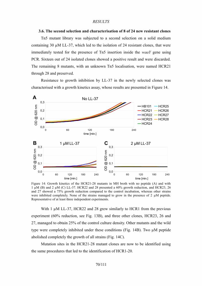
RESULTS
3.6. The second selection and characterisation of 8 of 24 new resistant clones
Tn5 mutant library was subjected to a second selection on a solid medium
containing 30 µM LL-37, which led to the isolation of 24 resistant clones, that were
immediately tested for the presence of Tn5 insertion inside the waaY gene using
PCR. Sixteen out of 24 isolated clones showed a positive result and were discarded.
The remaining 8 mutants, with an unknown Tn5 localisation, were named HCR21
through 28 and preserved.
Resistance to growth inhibition by LL-37 in the newly selected clones was
characterised with a growth kinetics assay, whose results are presented in Figure 14.
With 1 µM LL-37, HCR22 and 28 grew similarly to HCR1 from the previous
experiment (60% reduction, see Fig. 13B), and three other clones, HCR23, 26 and
27, managed to obtain 25% of the control culture density. Other mutants and the wild
type were completely inhibited under these conditions (Fig. 14B). Two µM peptide
abolished completely the growth of all strains (Fig. 14C).
Mutation sites in the HCR21-28 mutant clones are now to be identified using
the same procedures that led to the identification of HCR1-20.
70/111
Figure 14: Growth kinetics of the HCR21-28 mutants in MH broth with no peptide (A) and with1 µM (B) and 2 µM (C) LL-37. HCR22 and 28 presented a 60% growth reduction, and HCR23, 26 and 27 showed a 75% growth reduction compared to the control incubation, whereas other strains were inhibited completely. None of the strains managed to grow in the presence of 2 µM peptide. Representative of at least three independent experiments.
0 60 120 180 2400,0
0,1
0,2
0,3No LL-37
HB101HCR21HCR22HCR23HCR24
HCR25HCR26HCR27HCR28
time [min.]
OD
@ 6
20 n
m
A
0 60 120 180 2400,0
0,1
0,2
0,31 µM LL-37
time [min.]
OD
@ 6
20 n
m
B
0 60 120 180 2400,0
0,1
0,2
0,32 µM LL-37
time [min.]
OD
@ 6
20 n
m
C
RESULTS
3.7. Significance of the prevalence of waaY-mutants
A PCR amplification of the waaY gene was performed in 27 random clones
from the Tn5 mutant library. None of the electrophoretically separated PCR products
carried the Tn5 insertion, as the size of all products was equal to that of the wild-type
gene. This confirmed that the prevalence of waaY-mutants isolated in both selections
with LL-37 (75% and 67% of all selected clones in the first and the second selection,
respectively) was not a result of coincidence or preferential transposon insertion.
Different Tn5 positions in the two sequenced waaY mutants, HCR1 and HCR5,
support this assumption.
4. Resistance in HCR1 was caused by the inactivation of waaY
4.1. The downstream genes were not inhibited by Tn5 insertion
waaY (rfaY) is the third-to-last gene of the waaQ (rfaQ) operon (Fig. 15).
Although there is a putative promoter sequence before the two downstream genes,
waaZ (rfaZ) and waaU, it seemed correct to verify the possibility of inactivation of
those genes by Tn5 insertion. For this purpose, an RT-PCR analysis of all three genes
was conducted in both HB101 and HCR1.
71/111
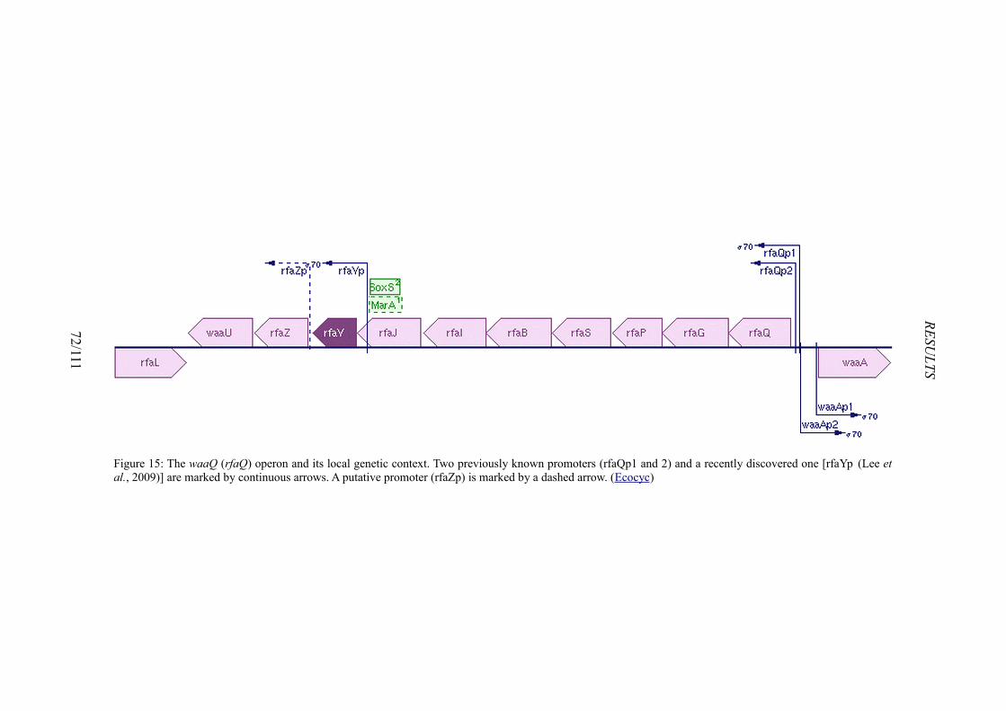
72/111
RESULTS
Figure 15: The waaQ (rfaQ) operon and its local genetic context. Two previously known promoters (rfaQp1 and 2) and a recently discovered one [rfaYp (Lee et al., 2009)] are marked by continuous arrows. A putative promoter (rfaZp) is marked by a dashed arrow. (Ecocyc)
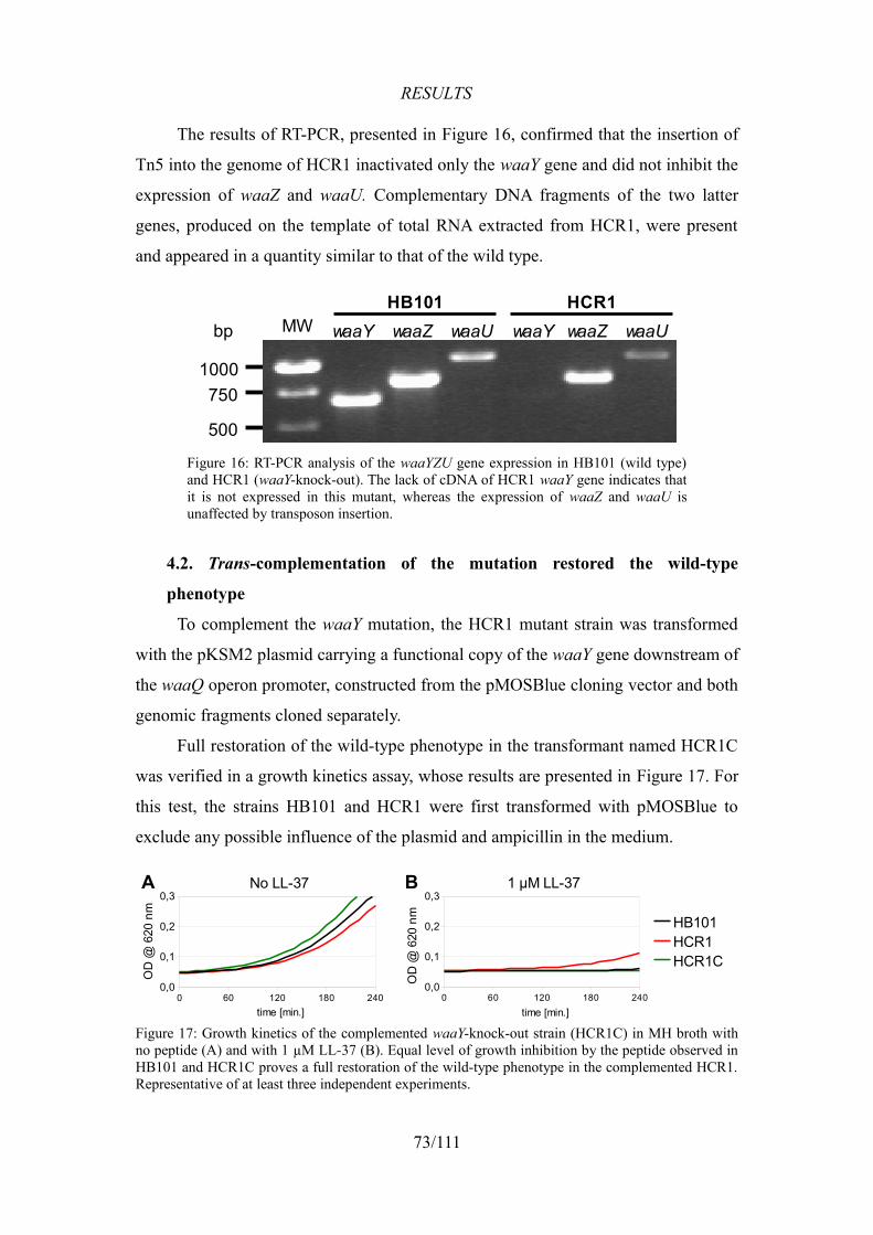
RESULTS
The results of RT-PCR, presented in Figure 16, confirmed that the insertion of
Tn5 into the genome of HCR1 inactivated only the waaY gene and did not inhibit the
expression of waaZ and waaU. Complementary DNA fragments of the two latter
genes, produced on the template of total RNA extracted from HCR1, were present
and appeared in a quantity similar to that of the wild type.
4.2. Trans-complementation of the mutation restored the wild-type
phenotype
To complement the waaY mutation, the HCR1 mutant strain was transformed
with the pKSM2 plasmid carrying a functional copy of the waaY gene downstream of
the waaQ operon promoter, constructed from the pMOSBlue cloning vector and both
genomic fragments cloned separately.
Full restoration of the wild-type phenotype in the transformant named HCR1C
was verified in a growth kinetics assay, whose results are presented in Figure 17. For
this test, the strains HB101 and HCR1 were first transformed with pMOSBlue to
exclude any possible influence of the plasmid and ampicillin in the medium.
73/111
Figure 16: RT-PCR analysis of the waaYZU gene expression in HB101 (wild type) and HCR1 (waaY-knock-out). The lack of cDNA of HCR1 waaY gene indicates that it is not expressed in this mutant, whereas the expression of waaZ and waaU is unaffected by transposon insertion.
Figure 17: Growth kinetics of the complemented waaY-knock-out strain (HCR1C) in MH broth with no peptide (A) and with 1 µM LL-37 (B). Equal level of growth inhibition by the peptide observed in HB101 and HCR1C proves a full restoration of the wild-type phenotype in the complemented HCR1. Representative of at least three independent experiments.
MW waaY waaZ waaU waaY waaZ waaU
1000750
500
HB101 HCR1bp
0 60 120 180 2400,0
0,1
0,2
0,3No LL-37
time [min.]
OD
@ 6
20 n
m
0 60 120 180 2400,0
0,1
0,2
0,31 µM LL-37
HB101HCR1HCR1C
time [min.]
OD
@ 6
20 n
m
A B
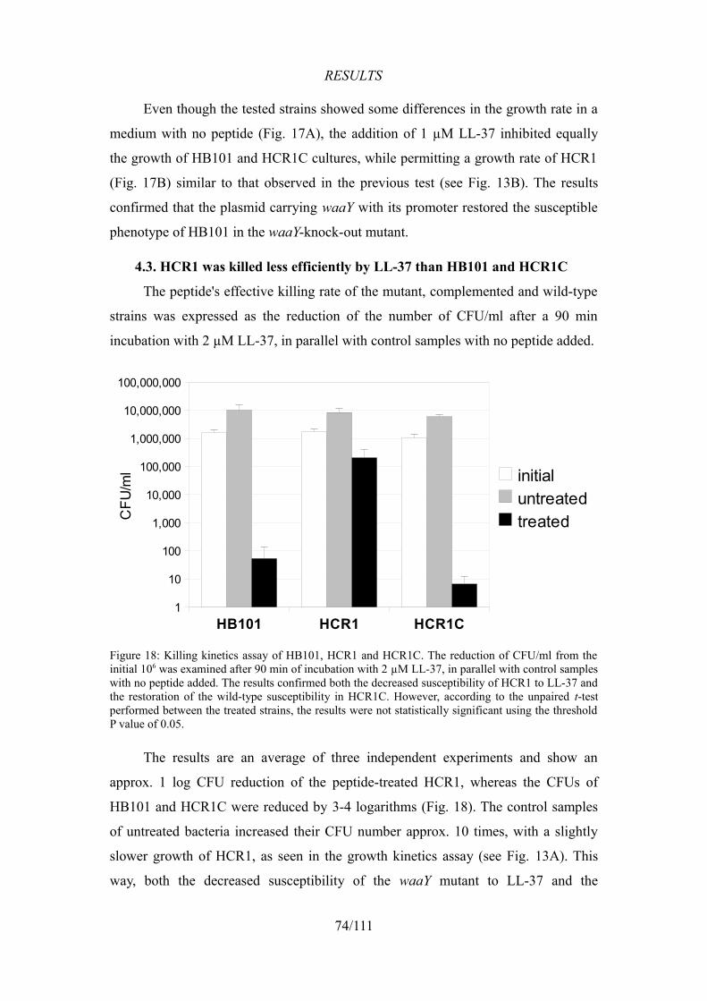
RESULTS
Even though the tested strains showed some differences in the growth rate in a
medium with no peptide (Fig. 17A), the addition of 1 µM LL-37 inhibited equally
the growth of HB101 and HCR1C cultures, while permitting a growth rate of HCR1
(Fig. 17B) similar to that observed in the previous test (see Fig. 13B). The results
confirmed that the plasmid carrying waaY with its promoter restored the susceptible
phenotype of HB101 in the waaY-knock-out mutant.
4.3. HCR1 was killed less efficiently by LL-37 than HB101 and HCR1C
The peptide's effective killing rate of the mutant, complemented and wild-type
strains was expressed as the reduction of the number of CFU/ml after a 90 min
incubation with 2 µM LL-37, in parallel with control samples with no peptide added.
The results are an average of three independent experiments and show an
approx. 1 log CFU reduction of the peptide-treated HCR1, whereas the CFUs of
HB101 and HCR1C were reduced by 3-4 logarithms (Fig. 18). The control samples
of untreated bacteria increased their CFU number approx. 10 times, with a slightly
slower growth of HCR1, as seen in the growth kinetics assay (see Fig. 13A). This
way, both the decreased susceptibility of the waaY mutant to LL-37 and the
74/111
Figure 18: Killing kinetics assay of HB101, HCR1 and HCR1C. The reduction of CFU/ml from the initial 106 was examined after 90 min of incubation with 2 µM LL-37, in parallel with control samples with no peptide added. The results confirmed both the decreased susceptibility of HCR1 to LL-37 and the restoration of the wild-type susceptibility in HCR1C. However, according to the unpaired t-test performed between the treated strains, the results were not statistically significant using the threshold P value of 0.05.
HB101 HCR1 HCR1C1
10
100
1,000
10,000
100,000
1,000,000
10,000,000
100,000,000
initialuntreatedtreatedC
FU/m
l
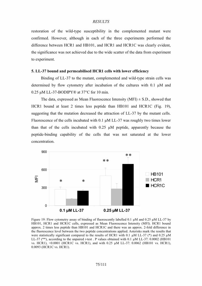
RESULTS
restoration of the wild-type susceptibility in the complemented mutant were
confirmed. However, although in each of the three experiments performed the
difference between HCR1 and HB101, and HCR1 and HCR1C was clearly evident,
the significance was not achieved due to the wide scatter of the data from experiment
to experiment.
5. LL-37 bound and permeabilised HCR1 cells with lower efficiency
Binding of LL-37 to the mutant, complemented and wild-type strain cells was
determined by flow cytometry after incubation of the cultures with 0.1 μM and
0.25 μM LL-37-BODIPY® at 37°C for 10 min.
The data, expressed as Mean Fluorescence Intensity (MFI) ± S.D., showed that
HCR1 bound at least 2 times less peptide than HB101 and HCR1C (Fig. 19),
suggesting that the mutation decreased the attraction of LL-37 by the mutant cells.
Fluorescence of the cells incubated with 0.1 μM LL-37 was roughly two times lower
than that of the cells incubated with 0.25 μM peptide, apparently because the
peptide-binding capability of the cells that was not saturated at the lower
concentration.
75/111
Figure 19: Flow cytometry assay of binding of fluorescently labelled 0.1 µM and 0.25 µM LL-37 by HB101, HCR1 and HCR1C cells, expressed as Mean Fluorescence Intensity (MFI). HCR1 bound approx. 2 times less peptide than HB101 and HCR1C and there was an approx. 2-fold difference in the fluorescence level between the two peptide concentrations applied. Asterisks mark the results that were statistically significant compared to the results of HCR1 with 0.1 µM LL-37 (*) and 0.25 µM LL-37 (**), according to the unpaired t-test . P values obtained with 0.1 µM LL-37: 0.0002 (HB101 vs. HCR1), <0.0001 (HCR1C vs. HCR1), and with 0.25 µM LL-37: 0.0062 (HB101 vs. HCR1), 0.0093 (HCR1C vs. HCR1).
0.1 µM LL-37 0.25 µM LL-370
300
600
900
HB101HCR1HCR1C
MFI
* *
****
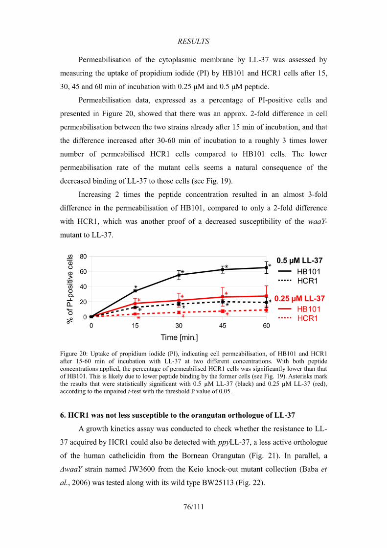
RESULTS
Permeabilisation of the cytoplasmic membrane by LL-37 was assessed by
measuring the uptake of propidium iodide (PI) by HB101 and HCR1 cells after 15,
30, 45 and 60 min of incubation with 0.25 μM and 0.5 μM peptide.
Permeabilisation data, expressed as a percentage of PI-positive cells and
presented in Figure 20, showed that there was an approx. 2-fold difference in cell
permeabilisation between the two strains already after 15 min of incubation, and that
the difference increased after 30-60 min of incubation to a roughly 3 times lower
number of permeabilised HCR1 cells compared to HB101 cells. The lower
permeabilisation rate of the mutant cells seems a natural consequence of the
decreased binding of LL-37 to those cells (see Fig. 19).
Increasing 2 times the peptide concentration resulted in an almost 3-fold
difference in the permeabilisation of HB101, compared to only a 2-fold difference
with HCR1, which was another proof of a decreased susceptibility of the waaY-
mutant to LL-37.
6. HCR1 was not less susceptible to the orangutan orthologue of LL-37
A growth kinetics assay was conducted to check whether the resistance to LL-
37 acquired by HCR1 could also be detected with ppyLL-37, a less active orthologue
of the human cathelicidin from the Bornean Orangutan (Fig. 21). In parallel, a
ΔwaaY strain named JW3600 from the Keio knock-out mutant collection (Baba et
al., 2006) was tested along with its wild type BW25113 (Fig. 22).
76/111
Figure 20: Uptake of propidium iodide (PI), indicating cell permeabilisation, of HB101 and HCR1 after 15-60 min of incubation with LL-37 at two different concentrations. With both peptide concentrations applied, the percentage of permeabilised HCR1 cells was significantly lower than that of HB101. This is likely due to lower peptide binding by the former cells (see Fig. 19). Asterisks mark the results that were statistically significant with 0.5 µM LL-37 (black) and 0.25 µM LL-37 (red), according to the unpaired t-test with the threshold P value of 0.05.
0 15 30 45 600
20
40
60
80
Time [min.]
% o
f PI-p
ositi
ve c
ells
HB101HCR1
HB101HCR1
0.25 µM LL-37
0.5 µM LL-37
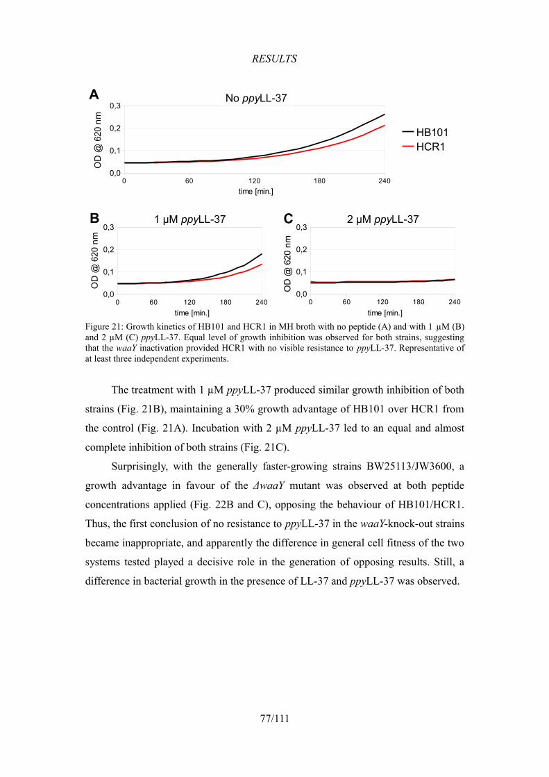
RESULTS
The treatment with 1 µM ppyLL-37 produced similar growth inhibition of both
strains (Fig. 21B), maintaining a 30% growth advantage of HB101 over HCR1 from
the control (Fig. 21A). Incubation with 2 µM ppyLL-37 led to an equal and almost
complete inhibition of both strains (Fig. 21C).
Surprisingly, with the generally faster-growing strains BW25113/JW3600, a
growth advantage in favour of the ΔwaaY mutant was observed at both peptide
concentrations applied (Fig. 22B and C), opposing the behaviour of HB101/HCR1.
Thus, the first conclusion of no resistance to ppyLL-37 in the waaY-knock-out strains
became inappropriate, and apparently the difference in general cell fitness of the two
systems tested played a decisive role in the generation of opposing results. Still, a
difference in bacterial growth in the presence of LL-37 and ppyLL-37 was observed.
77/111
Figure 21: Growth kinetics of HB101 and HCR1 in MH broth with no peptide (A) and with 1 µM (B) and 2 µM (C) ppyLL-37. Equal level of growth inhibition was observed for both strains, suggesting that the waaY inactivation provided HCR1 with no visible resistance to ppyLL-37. Representative of at least three independent experiments.
0 60 120 180 2400,0
0,1
0,2
0,3No ppyLL-37
HB101HCR1
time [min.]
OD
@ 6
20 n
m
0 60 120 180 2400,0
0,1
0,2
0,31 µM ppyLL-37
time [min.]
OD
@ 6
20 n
m
0 60 120 180 2400,0
0,1
0,2
0,32 µM ppyLL-37
time [min.]
OD
@ 6
20 n
m
A
B C
No ppyLL-37
1 µM ppyLL-37 2 µM ppyLL-37
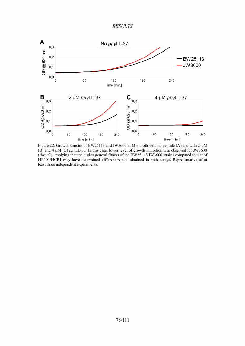
RESULTS
78/111
Figure 22: Growth kinetics of BW25113 and JW3600 in MH broth with no peptide (A) and with 2 µM (B) and 4 µM (C) ppyLL-37. In this case, lower level of growth inhibition was observed for JW3600 (ΔwaaY), implying that the higher general fitness of the BW25113/JW3600 strains compared to that of HB101/HCR1 may have determined different results obtained in both assays. Representative of at least three independent experiments.
0 60 120 180 2400,0
0,1
0,2
0,3No ppyLL-37
BW25113JW3600
time [min.]
OD
@ 6
20 n
m
0 60 120 180 2400,0
0,1
0,2
0,32 µM ppyLL-37
time [min.]
OD
@ 6
20 n
m
0 60 120 180 2400,0
0,1
0,2
0,34 µM ppyLL-37
time [min.]
OD
@ 6
20 n
m
A
B C
No ppyLL-37
2 µM ppyLL-37 4 µM ppyLL-37
DISCUSSION
DISCUSSION
1. Mutagenesis using Tn5 complex
E. coli HB101 strain was chosen for mutagenesis because it is highly
susceptible to AMPs and can be transformed with a high efficiency. The strain is a
hybrid between E. coli K12 strain AB266 (98%) and E.coli B, and the most
important characteristic of this strain is the inactivated recombinase A (RecA),
making it a stable carrier of artificially introduced DNA, though it also possesses
impaired DNA repair ability and high UV sensitivity (openwetware.org).
A library of mutants was produced using the EZ-Tn5™ <KAN-2> Tnp
Transposome kit (Epicentre Biotechnologies). Mutations were induced in the
bacterial genome by insertions of Tn5 transposon. The insertions were random and
single per genome, as verified by Southern blot analysis (see Fig. 9, p. 64).
The best-known genome of E. coli is that of the K-12 substr. MG1655 strain. It
contains 4,603 known genes, coding for 4,415 proteins and 188 non-coding RNAs.
Additionally, there are 115 pseudogenes (ecocyc.org, Keseler et al., 2009). Efficiency
calculations of the number of mutants generated with the total amount of the
transposon-transposase complex available indicated that approx. 59,000 clones were
generated. Therefore, having obtained a clone number exceeding the number of
genes almost 13 times, we can predict a very high probability of interruption of every
single gene in the mutant library.
Electroporations of competent cells, with an efficiency of >108 transformants
per µg of DNA, yielded almost 0.6 x 10⁵ mutant clones, using 0.2 µg of Tn5
complex. This efficiency is in close agreement with that indicated by the kit supplier
(>105 kanamycin-resistant CFUs/µg).
The stability of Tn5 transposition and the ease of subsequent mutant
identification are the most important features of the method presented in this thesis.
In order to identify the mutation sites, we decided not to follow the producer's
instructions suggesting direct sequencing on the template of genomic DNA. Instead,
we digested mutant genomes leaving the transposon intact and cloned the fragments,
selecting the subsequently transformed bacteria with ampicillin and kanamycin
which allowed us to distinguish the cloned fragments interrupted by Tn5. Although
79/111
DISCUSSION
this step consumed a few days, it facilitated significantly the sequencing of the
fragment, with no resequencing required in any case.
Another essential advantage of Tn5 mutagenesis is the stability of the insert.
No kanamycin was used with the solid media containing antimicrobial peptides in
the selection procedures. This way, potential interference between the antibiotic
molecules and AMPs was avoided, which was particularly useful in light of the
peptides' sensitivity to the medium ingredients (discussed later). Furthermore,
kanamycin concentration was decreased from 50 to 25 µg/ml in the refresh cultures,
since it was observed that the culture growth was slowed down with the higher
antibiotic concentration (results not shown). This permitted an equal growth of the
mutant and the wild-type cultures with no detectable problems with maintaining the
mutant phenotype.
An alternative method of generating random mutations in bacteria is based on
the mutagenic activity of agents such as N-methyl-N'-nitro-N-nitrosoguanidine
(MNNG). This is an alkylating agent causing methylation of guanine and inducing
the transition G:C→A:T, thus creating point mutations. It has been reported to
generate tumoral transformation in many organisms and there is also some evidence
of tumorigenesis in humans (IARC, 1987). MNNG as an experimental bacterial
mutagen has been described by Miller (1992) and applied in a previous series of
experiments in our laboratory (Mattiuzzo et al., 2007). The whole procedure has
been described by Scocchi et al. (2008).
One of the advantages of this approach is that the generation of point mutations
results in a broader range of effects. In fact, while insertional mutations only knock-
out single genes, the application of MNNG might alter the gene in many ways, for
instance, by single amino acid substitutions or by modifying the levels of protein
expression through mutations in the promoter region.
On the other hand, the disadvantages seem to be more important. Firstly, the
toxicity of the mutagenic compound poses a serious health threat not only to the
person conducting the experiments, but also to other operators of the microbiological
hood. Environmental safety raises difficulties in the disposal and storage of the waste
generated in such experiments. Secondly, the number of mutations generated this
way needs to be monitored and optimised to avoid creating mutants with several
80/111
DISCUSSION
mutations, since the identification of cumulative effects may require unacceptable
amounts of time and work. Finally, determining the mutation site requires isolation
of the mutated fragment e.g. by creating a genomic library and transforming the wild
type in order to induce the mutant phenotype, or by transforming the mutant with a
wild-type genomic library to complement a recessive mutation. Both procedures
require a reselection using the selecting agent (in this case an antimicrobial peptide),
which increases the costs and may be time-consuming.
It is important to note, that the range of genes mutated by transposon insertion
is limited to those not essential for the cell to survive and divide. The cell carrying
mutation in a housekeeping gene die and are not included in the final pool of
mutants.
2. Test selection with Bac7(1-16)
A selection of Tn5 mutants on a solid medium was first performed with
Bac7(1-16), an active fragment of Bac7. This was performed to utilise the newly
established procedure under previously applied conditions in order to verify if similar
results could be obtained. Furthermore, the test was conducted to confirm the
findings of Mattiuzzo et al. (2007) and possibly to appoint other potential interactors
of Bac7. Because of the fact that the new method was capable of delivering more of
easily identifiable gene candidates at a time, 5 µM Bac7 was used as the selective
peptide concentration, which was half of the peptide quantity used in the previous
experiment (10 µM). The purpose of this was to find the genes whose mutation
would provide a lower grade of resistance, but which may still be involved in the
mode of action of Bac7.
2.1. Identified resistant clones
The selection led to the isolation of 11 clones (Fig. 23), named B7R1-11, that
were identified using the described procedure.
81/111
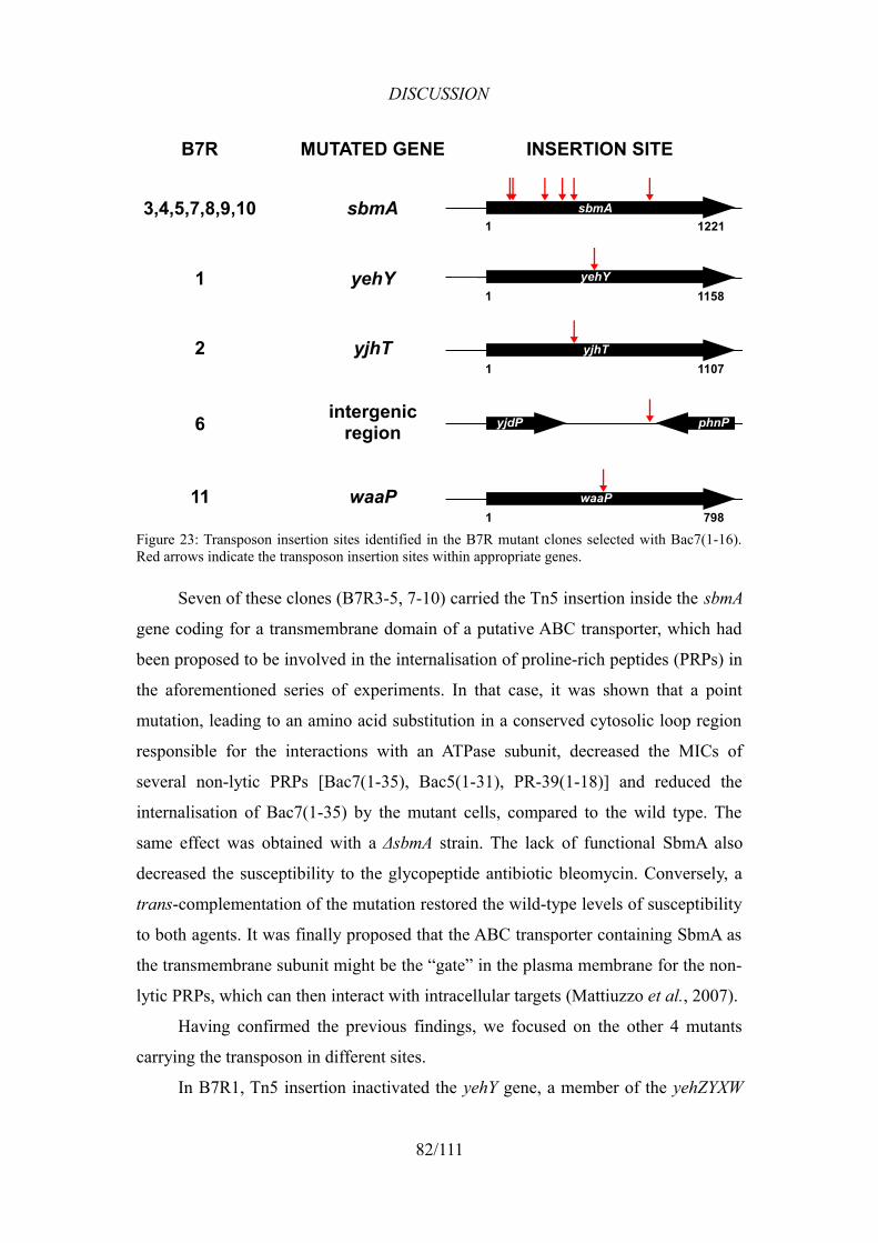
DISCUSSION
Seven of these clones (B7R3-5, 7-10) carried the Tn5 insertion inside the sbmA
gene coding for a transmembrane domain of a putative ABC transporter, which had
been proposed to be involved in the internalisation of proline-rich peptides (PRPs) in
the aforementioned series of experiments. In that case, it was shown that a point
mutation, leading to an amino acid substitution in a conserved cytosolic loop region
responsible for the interactions with an ATPase subunit, decreased the MICs of
several non-lytic PRPs [Bac7(1-35), Bac5(1-31), PR-39(1-18)] and reduced the
internalisation of Bac7(1-35) by the mutant cells, compared to the wild type. The
same effect was obtained with a ΔsbmA strain. The lack of functional SbmA also
decreased the susceptibility to the glycopeptide antibiotic bleomycin. Conversely, a
trans-complementation of the mutation restored the wild-type levels of susceptibility
to both agents. It was finally proposed that the ABC transporter containing SbmA as
the transmembrane subunit might be the “gate” in the plasma membrane for the non-
lytic PRPs, which can then interact with intracellular targets (Mattiuzzo et al., 2007).
Having confirmed the previous findings, we focused on the other 4 mutants
carrying the transposon in different sites.
In B7R1, Tn5 insertion inactivated the yehY gene, a member of the yehZYXW
82/111
Figure 23: Transposon insertion sites identified in the B7R mutant clones selected with Bac7(1-16). Red arrows indicate the transposon insertion sites within appropriate genes.
sbmA1221 1
3,4,5,7,8,9,10 sbmA
B7R MUTATED GENE INSERTION SITE
yehY1158 1
1 yehY
yjhT1107 1
2 yjhT
yjdP phnP6 intergenicregion
waaP798 1
11 waaP
DISCUSSION
operon. The operon encodes a putative ATP-dependent osmoprotectant uptake system
from the ABC transporter family. YehX is an ATP-binding component, YehW and
YehY are membrane components, and YehZ, previously called OsmF, is a
periplasmic binding protein. The transcription of yehZYXW is induced by osmotic
shock and entry into the stationary phase (Checroun and Gutierrez, 2004).
Transposon insertion in B7R2 was located in the yjhT gene, also known as
nanM, which is the second element of the nanC-namM-yjhS transcription unit
(Condemine et al., 2005). YjhT is a mutarotase of N-acetylneuraminate, allowing an
efficient use of the compound as the sole source of carbon (Severi et al., 2008).
The B7R6 clone carried Tn5 in an intergenic sequence, directly after the phnP
gene (not interrupting its stop codon) and 148 bp after the yjdP gene. The former is
the last member of a 14-gene-long operon (phnCDEFGHIJKLMNOP) responsible
for the use of phosphonates and phosphites as phosphorus sources and induced by
phosphate starvation. The phnP gene codes for an accessory protein to the carbon-
phosphorus lyase complex (Metcalf and Wanner, 1993; Metcalf and Wanner, 1991;
Yakovleva et al., 1998). The yjdP gene (erroneously called phnQ in the past) encodes
a hypothetical, uncharacterised protein and is positioned in a direction opposite to
that of phnP. The interrupted region contains a BoxC Composite Element (BOCE)
with two palindromic sequences called boxC (Bergler et al., 1992), whose role is not
yet known.
Lastly, in B7R11 the insertion was detected in the waaP gene, coding for a
kinase phosphorylating the first heptose of the inner core of LPS, as described before
(see Introduction 5.2.). The role of this gene in the susceptibility to AMPs, which
underlines an interesting link between the results of the selections with Bac7(1-16)
and LL-37, will be discussed in detail later in this section.
2.2. Assessment of the resistance
Growth kinetics assay was chosen as a reference method for preliminary
estimation of the level of resistance, because it allows a real-time visualisation of the
differences in growth velocity between multiple strains in several copies (triplicate in
this case) at a time. It also makes it possible to distinguish small variations of the
sensitivity that would otherwise remain undetectable, e.g. in a MIC test, or would
require low-throughput, time-consuming and laborious procedures, as in the killing
83/111
DISCUSSION
kinetics assay. Obviously, it does not substitute the latter experimental techniques, it
rather provides a method of selection of what to proceed with first.
The comparison of the growth kinetics of five B7R mutants representing each
of the identified interruptions (Fig. 10, p. 65), in the presence of Bac7(1-16), showed
that only the sbmA-mutant (B7R3) and, unexpectedly, the intergenic region-mutant
(B7R6) presented a growth superior to that of the wild type (HB101), whereas other
tested clones behaved similarly to HB101 with both applied concentrations of
Bac7(1-16). It is also interesting, that the two mutants grew more slowly than the
wild type in the control incubation with no peptide. Additionally, the B7R11 clone
was characterised by a particularly low fitness under all conditions applied.
A visible difference in growth between B7R3 and HB101 was expected in light
of the previously discussed results by Mattiuzzo et al. (2007). However, the fact that
all the other isolated mutants, except one, presented no difference to the wild type
cannot be easily interpreted. It can be explained either by the failure of the growth
kinetics assay to detect lower resistance levels, or by growth on solid medium of
some non-resistant clones in the process of selection. Whichever the case may be, the
results of the experiment strongly confirm that SbmA plays a key role in the peptide's
mechanism of action.
The observed decreased level of sensitivity in B7R6 is even harder to interpret,
given that no gene is known to be localised between the phnP and yjdP genes.
Transcriptional analysis of the flanking region could shed light on whether the
transposon insertion has either inactivated an unknown transcription unit, or impaired
a presently unknown function of one of the two boxC palindromic sequences present
in this intergenic region.
Since the purpose of this selection was to test the method of identification of
potential AMP interactors, no subsequent experiments were performed with the B7R
mutant clones. Instead, the same procedure was applied in a broader series of
experiments in search of E. coli mutations conferring resistance to the human
cathelicidin LL-37. This peptide shows in fact a different mode of action, a broader
antimicrobial spectrum and high relevance for the human physiology.
84/111
DISCUSSION
3. Application of the method in search of bacterial interactors of LL-37
3.1. Establishing the proper peptide concentration for plate selection
One of the biggest problems regarding the use of antimicrobial peptides in the
microbiological assays is their sensitivity to the composition of the growth medium,
mainly salts, that modify the peptides' conformation (see Introduction 4.2.1.) and
thus alter their ability to bind bacterial cells and to exert their bactericidal activity.
Moreover, the minimal inhibitory concentrations of AMPs on solid agar media are
usually higher than those obtained in liquid media with the same composition. For
example, in an unpublished series of experiments aimed at selecting mutants resistant
to the human β-defensin 3, according to Scocchi et al. (2008), it was impossible to
obtain repeatable results in the selection of peptide-resistant mutant cells. The
number of grown colonies was very high, suggesting that the selection did not occur
because the peptide was poorly active in the solid medium, even though a diluted
minimal medium was used (25% M9) and agar was substituted with ultra-pure
agarose as a solidifying agent.
As there was a possibility of experiencing such problems also with the human
cathelicidin, a broad range of peptide concentrations was used in the selection of LL-
37-resistant mutants on a solid medium. A hundred thousand cells from the Tn5
mutant library were spread on MH agar media supplemented with 1, 5, 10, 20 and
30 µM LL-37. The lowest applied concentration was equal to the MIC in liquid MH
broth (result not shown). As expected, 1 µM concentration, similarly to three higher
concentrations applied (5, 10 and 20 µM), led to no or insufficient reduction in the
CFU number. Only the highest (30 µM) peptide concentration produced a low
number of colonies (20), which indicated the desirable grade of selection with
respect to the control with the wild-type strain. A schematic presentation of the
selection results varying according to the peptide concentration is shown in
Figure 24.
85/111
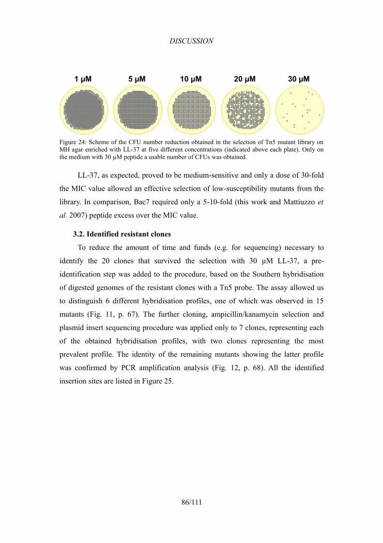
DISCUSSION
LL-37, as expected, proved to be medium-sensitive and only a dose of 30-fold
the MIC value allowed an effective selection of low-susceptibility mutants from the
library. In comparison, Bac7 required only a 5-10-fold (this work and Mattiuzzo et
al. 2007) peptide excess over the MIC value.
3.2. Identified resistant clones
To reduce the amount of time and funds (e.g. for sequencing) necessary to
identify the 20 clones that survived the selection with 30 µM LL-37, a pre-
identification step was added to the procedure, based on the Southern hybridisation
of digested genomes of the resistant clones with a Tn5 probe. The assay allowed us
to distinguish 6 different hybridisation profiles, one of which was observed in 15
mutants (Fig. 11, p. 67). The further cloning, ampicillin/kanamycin selection and
plasmid insert sequencing procedure was applied only to 7 clones, representing each
of the obtained hybridisation profiles, with two clones representing the most
prevalent profile. The identity of the remaining mutants showing the latter profile
was confirmed by PCR amplification analysis (Fig. 12, p. 68). All the identified
insertion sites are listed in Figure 25.
86/111
Figure 24: Scheme of the CFU number reduction obtained in the selection of Tn5 mutant library on MH agar enriched with LL-37 at five different concentrations (indicated above each plate). Only on the medium with 30 µM peptide a usable number of CFUs was obtained.
1 µM 5 µM 10 µM 20 µM 30 µM
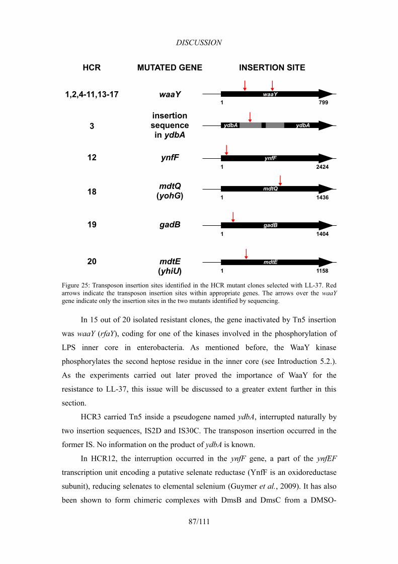
DISCUSSION
In 15 out of 20 isolated resistant clones, the gene inactivated by Tn5 insertion
was waaY (rfaY), coding for one of the kinases involved in the phosphorylation of
LPS inner core in enterobacteria. As mentioned before, the WaaY kinase
phosphorylates the second heptose residue in the inner core (see Introduction 5.2.).
As the experiments carried out later proved the importance of WaaY for the
resistance to LL-37, this issue will be discussed to a greater extent further in this
section.
HCR3 carried Tn5 inside a pseudogene named ydbA, interrupted naturally by
two insertion sequences, IS2D and IS30C. The transposon insertion occurred in the
former IS. No information on the product of ydbA is known.
In HCR12, the interruption occurred in the ynfF gene, a part of the ynfEF
transcription unit encoding a putative selenate reductase (YnfF is an oxidoreductase
subunit), reducing selenates to elemental selenium (Guymer et al., 2009). It has also
been shown to form chimeric complexes with DmsB and DmsC from a DMSO-
87/111
Figure 25: Transposon insertion sites identified in the HCR mutant clones selected with LL-37. Red arrows indicate the transposon insertion sites within appropriate genes. The arrows over the waaY gene indicate only the insertion sites in the two mutants identified by sequencing.
waaY799 1
1,2,4-11,13-17 waaY
HCR MUTATED GENE INSERTION SITE
ydbA ydbA3insertionsequencein ydbA
ynfF2424 1
12 ynfF
18 mdtQ(yohG)
gadB1404 1
19 gadB
mdtQ14361
mdtE1158 1
20 mdtE(yhiU)
DISCUSSION
reductase heterotrimer because of its similarity to the natural complex-former, DmsA
(Lubitz and Weiner, 2003).
Transposon insertion carried by HCR18 was located in the mdtQ (yohG) gene
identified to be an outer membrane factor (OMF) family component of a multidrug
efflux system, and its deletion from the E. coli W3110 strain caused increased
susceptibility to some antibiotics (Sulavik et al., 2001).
In the HCR19 mutant, the transposon was found inside the gadB gene, whose
product is a subunit of a glutamate decarboxylase involved in the Glu-dependent acid
resistance system 2 (AR2), which protects the cell from acidic conditions in the
stomach after activation by chloride ions (Gut et al., 2006), and from weak acids
formed in the carbohydrate fermentation during anaerobic phosphate starvation
(Moreau, 2007). It is regulated antagonistically by two proteins, H-NS and GadX
(Giangrossi et al., 2005).
The last resistant clone, HCR20, carried the mutation in the mdtE (yhiU) gene,
which produces a subunit of the MdtEF multidrug transporter, regulated by the EvgA
regulator (Nishino and Yamaguchi, 2002). Recently, it has been discovered that it is
activated by the GadX regulator, whose overexpression led to an increased resistance
to several antibiotics and SDS (Nishino et al., 2008). This effect of regulation
indicates a link between the aforementioned acid resistance system with GadB
involved and the drug efflux pump with MdtE.
Again, the isolated clones carried the mutations in genes of various functions
and many of them were apparently not directly related to the known activity of LL-
37. It was thus essential to assess their resistance using the growth kinetics assay,
previously chosen as a reference method, to exclude any chance of survival of the
selection by non-resistant clones.
3.3. Assessment of the resistance
Growth kinetics of six HCR mutants in the presence of LL-37, each
representing a different interrupted gene (Fig. 13, p. 69), showed that only the waaY-
mutant (HCR1) was able to growth in the presence of peptide concentrations that
were inhibitory for the wild type HB101 strain. All the other mutant clones behaved
in a manner similar to HB101. Again, it is not clear whether the isolation of the other
5 mutants with different interrupted genes was purely coincidental, or the method we
88/111
DISCUSSION
chose for a preliminary resistance characterisation (growth kinetics assay) had an
insufficient resolution to visualise lower levels of resistance. However, since the only
isolated mutant clone showing any noticeable level of resistance carried the Tn5
insertion in the waaY gene, interrupted in 15 out of 20 mutant clones, further assays
were performed only with HCR1 and with HB101 as a reference. The role of waaY
in E. coli physiology, discussed briefly in the introduction (5.2.) and more
completely in this section, indicated its possible involvement in the interaction
between bacterial cell surface and LL-37.
3.4. Inactivation of the waaY gene led to a decreased bactericidal activity of
LL-37 against HCR1
To verify whether the inserted transposon inactivated only waaY, or also its
downstream genes in the waaQ operon, waaZ and waaU, an RT-PCR analysis of the
three genes was carried out (see Results 4.1.). The results undoubtedly confirmed
that the transposon inhibited solely the proper transcription of waaY, leaving
unaltered the expression of the two downstream genes.
Furthermore, to complement the mutation, we constructed a plasmid containing
an intact copy of waaY and a 300 bp sequence of the waaQ operon promoter, so as to
restore in HCR1 the expression of the gene under the control of its natural regulators.
Mutant cells transformed with this plasmid (HCR1C) showed a complete restoration
of the wild-type phenotype, demonstrating a level of sensitivity to LL-37 equal to
that of HB101 under the same conditions (see Fig. 17, p. 73).
Having proved the involvement of waaY in the decreased susceptibility to the
peptide, we carried out killing kinetics assays to test the extent of increased
resistance to LL-37. Indeed, the CFU number of the peptide-treated mutant strain
was 100-1000 times higher than that of HB101 and HCR1C (see Fig. 18, p. 74).
These results indicate that the mutated waaY leads to a decreased killing efficiency of
the human cathelicidin against E. coli.
3.5. The role of waaY in the properties of LPS
As already mentioned, the waaY (rfaY) gene is the third-to-last gene of the
waaQGPSBORYZU (or rfaQGPSBIJYZwaaU) transcription unit (see Fig. 15, p. 72),
which, in the MG1655 strain, is located approximately in the position of 3.806-3.796
Mb (Keseler et al., 2009). The operon consists of 10 genes, whose products are
89/111
DISCUSSION
responsible for the biosynthesis of the outer core oligosaccharide and modification of
the inner core of LPS. In particular, the waaP, waaQ and waaY genes are involved in
the proper formation of the inner core moiety, composed mainly of heptose residues.
It has been shown that single mutations of each of these three genes cause
modifications of the inner core structure. The proposed sequence of events during the
addition of the lateral residues of the inner core is as follows (Fig. 26):
1) phosphorylation of HepI by WaaP; 2) addition of HepIII residue to HepII by
WaaQ; 3) phosphorylation of HepII by WaaY (Yethon et al., 1998). The absence of
each of these enzymes disables the further steps. In particular, mutation of the waaP
gene leads to a series of morphological and physiological alterations known as the
“rough” phenotype and is responsible for an increased susceptibility to some
antibiotics in Salmonella and loss of virulence in vivo (Yethon et al., 2000b). The
waaY product has been described as the last factor in the sequence and its activity is
no prerequisite to other reactions (Yethon et al., 1998). Therefore, the only effect of
inactivating the waaY gene is the lack of a phosphate residue in HepII without
alteration of the rest of the LPS molecule. The modification removes one negative
charge from the molecule's net charge, which might decrease the electrostatic
interactions between LPS and peptide molecules. This hypothesis was verified by
flow cytometry assays.
90/111

DISCUSSION
3.6.Binding to and permeabilisation of HCR1 cells by LL-37
Binding of LL-37 to the mutant, complemented and wild-type strain cells was
determined by flow cytometry after culture incubation with LL-37-BODIPY®. As
expected, HCR1 bound at least 2 times less peptide than HB101 and HCR1C (see
Fig. 19, p. 75), confirming the hypothesis of a lower attraction between the peptide
and the mutant cells. As a consequence of the lower binding, also the percentage of
91/111
Figure 26: Structure of the LPS from E. coli strains with an R1-type core. Core residues are designated by sugar abbreviation and number to facilitate identification. Abbreviations are as follows: GlcN, D-glucosamine; Hep, L-glycero-D-manno-heptose; P, phosphate; EtNP, 2-aminoethylphosphate; Glc, D-glucose; Gal, D-galactose. The names of the genes encoding core glycosyltransferases and phosphotransferases indicate the bonds catalysed by each enzyme. The lateral residues of the inner core and the enzymes involved in the sequence of their addition are marked with red fields and arrows, respectively (modified from Yethon et al., 2000a).
1)
2)
3)
DISCUSSION
permeabilisation of the HCR1 cells by LL-37 was 2-3 times lower compared to that
of the HB101 cells (see Fig. 20, p. 76).
The assays suggested an explanation for the decreased susceptibility of HCR1
to the peptide. Reduced peptide binding to the external surface of the outer
membrane, likely due to a decreased negative net charge of the LPS molecules,
diminished to a similar extent the percentage of cell permeabilisation, allowing a
higher number of the mutant cells to survive.
The overall effect of modifications in the LPS molecule determines a mild
increase of resistance to AMPs, such as that observed in the waaY deletion mutants.
On the contrary, some mutations, such as deletion of the waaP gene, whose product
adds a phosphate group to the Hep I residue, may increase the susceptibility to
bactericidal compounds such as novobiocin and SDS (Yethon et al., 1998). This
effect is due to the fact that these mutants show a “deep-rough” phenotype, which
leads to a serious reduction of the LPS molecule length due to the lack of lateral
residues in the inner core and, in turn, to failure in the addition of the O-antigen.
These “deep rough” mutants have a less stable outer membrane that can be easily
penetrated by antibacterial agents. In this respect, we tested the activity of LL-37
against eight knock-out E. coli mutants from the Keio collection (Baba et al., 2006),
individually lacking each of the eight waaQGPSBORY genes. In these experiments,
we confirmed the increased sensitivity to SDS in two of them: ΔwaaG and ΔwaaP,
and additionally we found that the ΔwaaP mutant is hypersensitive to LL-37 (results
not shown). This suggests that there are two parts of the LPS molecule particularly
important for membrane stability and for permeability of the OM to antimicrobial
agents: the O-antigen together with the outer core, whose assembly is initiated by
WaaG, and the lateral residues of the inner core, whose addition depends on WaaP at
the beginning of the aforementioned WaaP-WaaQ-WaaY cascade. Therefore, we
infer that the cells with a mutation in waaP carry a form of LPS whose grade of
interaction with the peptide may be even lower than that of the waaY mutants
because of an additional loss of negative charge, however, the increased instability of
their outer membrane overcomes the benefit of a lower peptide binding, and leads to
a higher susceptibility. This observation is also supported by the lower fitness
showed in a growth kinetics assay by the B7R11 mutant in which Tn5 is inserted in
92/111
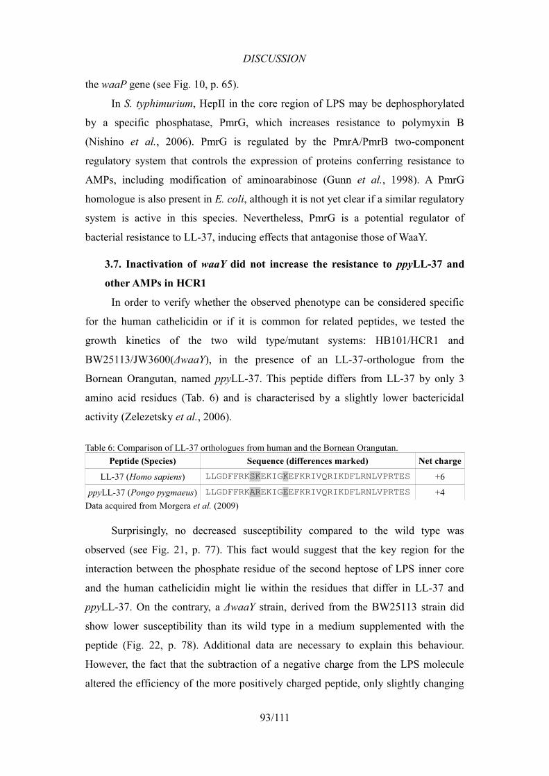
DISCUSSION
the waaP gene (see Fig. 10, p. 65).
In S. typhimurium, HepII in the core region of LPS may be dephosphorylated
by a specific phosphatase, PmrG, which increases resistance to polymyxin B
(Nishino et al., 2006). PmrG is regulated by the PmrA/PmrB two-component
regulatory system that controls the expression of proteins conferring resistance to
AMPs, including modification of aminoarabinose (Gunn et al., 1998). A PmrG
homologue is also present in E. coli, although it is not yet clear if a similar regulatory
system is active in this species. Nevertheless, PmrG is a potential regulator of
bacterial resistance to LL-37, inducing effects that antagonise those of WaaY.
3.7. Inactivation of waaY did not increase the resistance to ppyLL-37 and
other AMPs in HCR1
In order to verify whether the observed phenotype can be considered specific
for the human cathelicidin or if it is common for related peptides, we tested the
growth kinetics of the two wild type/mutant systems: HB101/HCR1 and
BW25113/JW3600(ΔwaaY), in the presence of an LL-37-orthologue from the
Bornean Orangutan, named ppyLL-37. This peptide differs from LL-37 by only 3
amino acid residues (Tab. 6) and is characterised by a slightly lower bactericidal
activity (Zelezetsky et al., 2006).
Table 6: Comparison of LL-37 orthologues from human and the Bornean Orangutan.Peptide (Species) Sequence (differences marked) Net charge
LL-37 (Homo sapiens) LLGDFFRKSKEKIGKEFKRIVQRIKDFLRNLVPRTES +6
ppyLL-37 (Pongo pygmaeus) LLGDFFRKAREKIGEEFKRIVQRIKDFLRNLVPRTES +4Data acquired from Morgera et al. (2009)
Surprisingly, no decreased susceptibility compared to the wild type was
observed (see Fig. 21, p. 77). This fact would suggest that the key region for the
interaction between the phosphate residue of the second heptose of LPS inner core
and the human cathelicidin might lie within the residues that differ in LL-37 and
ppyLL-37. On the contrary, a ΔwaaY strain, derived from the BW25113 strain did
show lower susceptibility than its wild type in a medium supplemented with the
peptide (Fig. 22, p. 78). Additional data are necessary to explain this behaviour.
However, the fact that the subtraction of a negative charge from the LPS molecule
altered the efficiency of the more positively charged peptide, only slightly changing
93/111
DISCUSSION
the efficiency of the less charged one, implies a considerable specificity of the LL-
37-LPS interaction, perhaps more dependent on the peptide's amino acid composition
and layout, than on the net charge itself. This hypothesis is supported by the fact that
HCR1 showed no visible resistance to other AMPs, such as α-helical BMAP-27,
BMAP-28, SMAP-29, and proline-rich Bac7(1-16) and Bac7(1-35) (results not
shown). Moreover, Rosenfeld et al. suggested that the antimicrobial activity of LL-
37 is not correlated with its ability to neutralise LPS, and that different properties
may be responsible for each of the activities (Rosenfeld et al., 2006, reviewed by
Rosenfeld and Shai, 2006).
3.8. Unidentified resistant clones from the second selection
Repeated selection of the mutant library allowed us to isolate 24 new clones
able to grow on a medium with 30 µM LL-37. Among these clones, 16 were
identified as waaY-mutants, whereas 8 did not carry the transposon in that gene (PCR
analysis, results not shown). Two of those 8 clones grew similarly to HCR1 in the
presence of LL-37, and three others showed a lower level of resistance but still
higher than the wild type (Fig. 14, p. 70). Isolation of the new set of non-waaY-
mutants indicates that the selection may have to be performed more than one time to
identify all the possible mutations conferring resistance to a certain AMP. The eight
newly isolated mutants, and particularly those presenting some decrease in growth
inhibition by LL-37, have now to be identified and their study might help to further
elucidate how the human cathelicidin interacts with the enterobacterial cell.
Nevertheless, in light of the fact that no waaY-mutant was detected among 27
randomly chosen clones from the mutant library (PCR analysis, results not shown),
the prevalence of the waaY mutation in the selected clones observed in both
selections indicates the key role of this mutation in the generation of
an LL-37-resistant phenotype. Therefore, it was correctly considered the main reason
for continuing the subsequent experimental work in that direction.
94/111
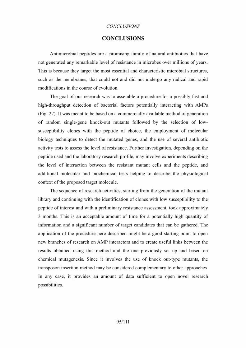
CONCLUSIONS
CONCLUSIONS
Antimicrobial peptides are a promising family of natural antibiotics that have
not generated any remarkable level of resistance in microbes over millions of years.
This is because they target the most essential and characteristic microbial structures,
such as the membranes, that could not and did not undergo any radical and rapid
modifications in the course of evolution.
The goal of our research was to assemble a procedure for a possibly fast and
high-throughput detection of bacterial factors potentially interacting with AMPs
(Fig. 27). It was meant to be based on a commercially available method of generation
of random single-gene knock-out mutants followed by the selection of low-
susceptibility clones with the peptide of choice, the employment of molecular
biology techniques to detect the mutated genes, and the use of several antibiotic
activity tests to assess the level of resistance. Further investigation, depending on the
peptide used and the laboratory research profile, may involve experiments describing
the level of interaction between the resistant mutant cells and the peptide, and
additional molecular and biochemical tests helping to describe the physiological
context of the proposed target molecule.
The sequence of research activities, starting from the generation of the mutant
library and continuing with the identification of clones with low susceptibility to the
peptide of interest and with a preliminary resistance assessment, took approximately
3 months. This is an acceptable amount of time for a potentially high quantity of
information and a significant number of target candidates that can be gathered. The
application of the procedure here described might be a good starting point to open
new branches of research on AMP interactors and to create useful links between the
results obtained using this method and the one previously set up and based on
chemical mutagenesis. Since it involves the use of knock out-type mutants, the
transposon insertion method may be considered complementary to other approaches.
In any case, it provides an amount of data sufficient to open novel research
possibilities.
95/111
CONCLUSIONS
96/111
Figure 27: Scheme of the established procedure for searching potential AMP interactors in bacteria.
+EZ-Tn5™
Transposome E. coli HB101
Creation of the bank of single-gene knock-out mutants by random insertion of the Tn5 transposon into the genome (using the Epicentre EZ-Tn5™ Transposome).
Selection of the mutant bank on a solid medium supplemented with the peptide.
gDNA digestion and sub-cloning. Subsequent selection of plasmids on ampicillin/kanamycin and sequencing.
gDNA
EcoRV
+Amp/Kan
Identification of the mutations
Growth inhibition and cell killing assays, followed by optional analysis of peptide-cell interaction.
Growth kinetics
HB101 HCR1 HCR1C1
10
100
1 000
10 000
100 000
1 000 000
10 000 000
100 000 000
initialuntreatedtreatedC
FU
/ml Killing
kinetics
Peptide-cell interaction assays
0.1 µM LL-37 0.25 µM LL-370
300
600
900
HB101HCR1HCR1C
MFI
* *
**
0 15 30 45 600
20
40
60
80
Time [min.]
% of
PI-p
ositiv
e cell
s HB101HCR1
HB101HCR1
0.25 µM LL-37
0.5 µM LL-37
Characterisation of the resistance
Further molecular and biochemical analyses.
Mutant bank production
Selection
CONCLUSIONS
If a high number of clones is to be identified, the identification procedure can
be sped up by dividing the obtained mutants into groups presenting similar Southern-
hybridisation profiles that, with a high probability, share the same mutation site. The
possibility of identification through PCR product analysis drastically increases the
productivity of the method and allows to grade the mutations that influence
resistance to the applied peptide to a different extent.
Last but not least, the use of Tn5 mutagenesis in the discovery of novel AMP
targets only requires basic molecular biology techniques, such as cell electroporation,
digestion of DNA and its ligation with a vector, and therefore requires neither high
expenses nor any rare skills of the research staff. However, it later opens a vast range
of possible directions to take for elucidation of the obtained data, and helps to
employ laboratory's full methodical potential.
Application of the established procedure allowed us to confirm the findings
from previous experiments performed with the Pro-rich Bac7 and to begin with a
new field of investigation with the human cathelicidin LL-37.
In the presented work, we confirmed the previous results emphasising the
importance of a putative ABC transporter protein, SbmA, for the proper functioning
of Bac7: a point mutation (Mattiuzzo et al., 2007) or interruption (this work) of the
sbmA gene induced a resistant phenotype for this peptide. Furthermore, we found
another site in the bacterial genome whose interruption conferred on E. coli some
degree of resistance to Bac7(1-16). Interestingly, it is an intergenic region containing
a palindromic sequence of unknown function. Even though at present it may be
difficult to explain, this result can be a starting point to a series of experiments
exploring the role of these palindromic sequences.
Our main object of interest and the main purpose of constructing the mutant
library was the search for factors involved in LL-37 activity in E. coli. The
experiments allowed us to identify the bacterial gene waaY whose inactivation
resulted in a decreased susceptibility to the human cathelicidin. Comparison of this
result with published data permits to hypothesise that the phosphorylation of one of
the residues of the LPS inner core may be an important factor in the interaction
between the peptide and bacterial outer membrane. This behaviour was observed
97/111
CONCLUSIONS
only with the human cathelicidin, while no resistance to other AMPs, both α-helical
and Pro-rich, was detected. Thus, we may have discovered a moiety of LPS
specifically responsible for the interactions with LL-37.
The second screening allowed the selection of clones with a level of resistance
similar to or slightly lower than that of HCR1. Regardless of which genes have been
isolated in the newly identified clones, the novel selection did not reproduce exactly
the result of the first one, although the prevalence of waaY-mutants was confirmed. It
is thus suggested to repeat the selection process since a single gene may not represent
the whole view except in the case of prevalent mutations.
The search of structures responsible for microbial susceptibility to
antimicrobial peptides complements the investigation of the AMPs' structure-activity
relationship. Our procedure permits a high-performance and relatively rapid
identification of bacterial interactors that can later help to explain the mode of action
of AMPs of interest, so as to facilitate the generation of novel antibiotic drugs to treat
the emerging multi-resistant infections.
98/111

ACKNOWLEDGEMENTS
ACKNOWLEDGEMENTS
I would like to thank anyone who made possible this PhD programme.
Vorrei ringraziare tutte le persone che mi hanno accompagnato e aiutato
durante i tre anni del mio dottorato di ricerca:
Il Prof. Renato Gennaro, per la possibilità di partecipazione alla ricerca del suo
gruppo e per aiuto di qualunque tipo che potevo sempre chiedere.
Il Dott. Marco Scocchi, per la sua disponibilità e pazienza a spiegarmi le cose nel
mio periodo iniziale.
La Dott.ssa Sara Ferluga, per il suo sorriso e per avermi portato la luce nel periodo di
buio, che è ovviamente tutto il know-how, senza il quale non sarebbe stato
possibile realizzare questa tesi.
Il Dott. Giuseppe Cannone e il Dott. Mario Mardirossian, i miei interni, per il loro
aiuto e lavorare gomito al gomito con me, grazie ai quali non mi è mai venuto
il dubbio sull'esistenza dello scopo del nostro tormento. :)
La Dott.ssa Monica Benincasa, per la sua accuratezza, professionalità e disponibilità
per organizzare tutte le cose che creano “la vita del gruppo”.
Le persone riportate sotto, per aver creato un gruppo modello in cui ho potuto
rimanere un vero “me stesso” e sentirmi sempre rispettato ed apprezzato.
Nina AntchevaCristian De GobbaMariangela FaudaleMilena GuidaAnnalisa IngegneriVincenzo LucaMarianna Lucafò
Francesca MorgeraChiara PelilloGiulia RuntiТатьяна СтоиловаAlex TossiDaniela Xhindoli
Tutto il personale degli altri gruppi del ex-BBCM per aver creato un'atmosfera
amichevole.
Zo, za to, że była tu ze mną duchem i/lub ciałem i kibicowała mi w rozterkach nt.
celowości pracy w tym „zakładzie”. :)
Rodzicom, za opiekę i sfinansowanie całej mojej dotychczasowej nauki, bez której
nie byłbym tym, kim jestem.
DZIĘKUJĘ!!!
99/111
REFERENCES
REFERENCES
Abbas, AK. and Lichtman, AH. (2003). Cellular and Molecular Immunology (5th Edition). Philadelphia: Saunders. ISBN 0721600085.
Abbas, AK., Lichtman, AH. and Pillai, S. (2007). Cellular and Molecular Immunology (6th Edition). Philadelphia: Saunders. ISBN 1416031227.
Abbas, AK., Murphy, KM. and Sher, A. (1996). Functional diversity of helper T lymphocytes. Nature 383, 787-793.
Agerberth, B., Charo, J., Werr, J. et al. (2000). The human antimicrobial and chemotactic peptides LL-37 and alpha-defensins are expressed by specific lymphocyte and monocyte populations. Blood 96, 3086-3093.
Agerberth, B., Gunne, H., Odeberg, J. et al. (1995). FALL-39, a putative human peptide antibiotic, is cysteine-free and expressed in bone marrow and testis. Proc. Natl. Acad. Sci. U.S.A. 92, 195-199.
Agerberth, B., Lee, JY., Bergman, T. et al. (1991). Amino acid sequence of PR-39. Isolation from pig intestine of a new member of the family of proline-arginine-rich antibacterial peptides. Eur. J. Biochem. 202, 849-854.
Altschul, SF., Madden, TL., Schäffer, AA. et al. (1997). Gapped BLAST and PSI-BLAST: a new generation of protein database search programs. Nucleic Acids Res. 25, 3389-3402.
Andreu, D. and Rivas, L. (1998). Animal antimicrobial peptides: an overview. Biopolymers 47, 415-433.
Baba, T., Ara, T., Hasegawa, M. et al. (2006). Construction of Escherichia coli K-12 in-frame, single-gene knockout mutants: the Keio collection. Mol. Syst. Biol. 2, 2006.0008.
Barry, M. and Bleackley, RC. (2002). Cytotoxic T lymphocytes: all roads lead to death. Nat. Rev. Immunol. 2, 401-409.
Bellomio, A., Vincent, PA., de Arcuri, BF. et al. (2007). Microcin J25 has dual and independent mechanisms of action in Escherichia coli: RNA polymerase inhibition and increased superoxide production. J. Bacteriol. 189, 4180-4186.
Bergler, H., Högenauer, G. and Turnowsky, F. (1992). Sequences of the envM gene and of two mutated alleles in Escherichia coli. J. Gen. Microbiol. 138, 2093-2100.
Birnboim, HC. (1983). A rapid alkaline extraction method for the isolation of plasmid DNA. Meth. Enzymol. 100, 243-255.
Boman, HG., Agerberth, B. and Boman, A. (1993). Mechanisms of action on Escherichia coli of cecropin P1 and PR-39, two antibacterial peptides from pig intestine. Infect. Immun. 61, 2978-2984.
Bowdish, DME., Davidson, DJ., Lau, YE. et al. (2005). Impact of LL-37 on anti-infective immunity. J. Leukoc. Biol. 77, 451-459.
Brogden, KA. (2005). Antimicrobial peptides: pore formers or metabolic inhibitors in bacteria?. Nat. Rev. Microbiol. 3, 238-250.
Brogden, KA., Ackermann, M. and Huttner, KM. (1997). Small, anionic, and charge-neutralizing propeptide fragments of zymogens are antimicrobial. Antimicrob.
100/111
REFERENCES
Agents Chemother. 41, 1615-1617.Brogden, KA., De Lucca, AJ., Bland, J. et al. (1996). Isolation of an ovine
pulmonary surfactant-associated anionic peptide bactericidal for Pasteurella haemolytica. Proc. Natl. Acad. Sci. U.S.A. 93, 412-416.
Bulet, P., Dimarcq, JL., Hetru, C. et al. (1993). A novel inducible antibacterial peptide of Drosophila carries an O-glycosylated substitution. J. Biol. Chem. 268, 14893-14897.
Burton, MF. and Steel, PG. (2009). The chemistry and biology of LL-37. Nat Prod Rep 26, 1572-1584.
Campos, MA., Vargas, MA., Regueiro, V. et al. (2004). Capsule polysaccharide mediates bacterial resistance to antimicrobial peptides. Infect. Immun. 72, 7107-7114.
Carter, RH. (2006). B cells in health and disease. Mayo Clin. Proc. 81, 377-384.Casteels, P., Ampe, C., Jacobs, F. et al. (1993). Functional and chemical
characterization of Hymenoptaecin, an antibacterial polypeptide that is infection-inducible in the honeybee (Apis mellifera). J. Biol. Chem. 268, 7044-7054.
Casteels, P., Ampe, C., Jacobs, F. et al. (1989). Apidaecins: antibacterial peptides from honeybees. EMBO J. 8, 2387-2391.
Casteels, P., Ampe, C., Riviere, L. et al. (1990). Isolation and characterization of abaecin, a major antibacterial response peptide in the honeybee (Apis mellifera). Eur. J. Biochem. 187, 381-386.
Castle, M., Nazarian, A., Yi, SS. et al. (1999). Lethal effects of apidaecin on Escherichia coli involve sequential molecular interactions with diverse targets. J. Biol. Chem. 274, 32555-32564.
Chang, C., Pleguezuelos, O., Zhang, Y. et al. (2005). Identification of a novel cathelicidin gene in the rainbow trout, Oncorhynchus mykiss. Infect. Immun. 73, 5053-5064.
Chang, C., Zhang, Y., Zou, J. et al. (2006). Two cathelicidin genes are present in both rainbow trout (Oncorhynchus mykiss) and atlantic salmon (Salmo salar). Antimicrob. Agents Chemother. 50, 185-195.
Chang, TL. and Klotman, ME. (2004). Defensins: natural anti-HIV peptides. AIDS Rev 6, 161-168.
Checroun, C. and Gutierrez, C. (2004). Sigma(s)-dependent regulation of yehZYXW, which encodes a putative osmoprotectant ABC transporter of Escherichia coli. FEMS Microbiol. Lett. 236, 221-226.
Chen, X. and Jensen, PE. (2008). The role of B lymphocytes as antigen-presenting cells. Arch. Immunol. Ther. Exp. (Warsz.) 56, 77-83.
Clementz, T. (1992). The gene coding for 3-deoxy-manno-octulosonic acid transferase and the rfaQ gene are transcribed from divergently arranged promoters in Escherichia coli. J. Bacteriol. 174, 7750-7756.
Cole, AM., Weis, P. and Diamond, G. (1997). Isolation and characterization of pleurocidin, an antimicrobial peptide in the skin secretions of winter flounder. J. Biol. Chem. 272, 12008-12013.
Collins, LV., Kristian, SA., Weidenmaier, C. et al. (2002). Staphylococcus aureus
101/111
REFERENCES
strains lacking D-alanine modifications of teichoic acids are highly susceptible to human neutrophil killing and are virulence attenuated in mice. J. Infect. Dis. 186, 214-219.
Condemine, G., Berrier, C., Plumbridge, J. et al. (2005). Function and expression of an N-acetylneuraminic acid-inducible outer membrane channel in Escherichia coli. J. Bacteriol. 187, 1959-1965.
Crovella, S., Antcheva, N., Zelezetsky, I. et al. (2005). Primate beta-defensins--structure, function and evolution. Curr. Protein Pept. Sci. 6, 7-21.
Dathe, M., Nikolenko, H., Meyer, J. et al. (2001). Optimization of the antimicrobial activity of magainin peptides by modification of charge. FEBS Lett. 501, 146-150.
Dauphinee, SM. and Karsan, A. (2006). Lipopolysaccharide signaling in endothelial cells. Lab. Invest. 86, 9-22.
den Hertog, AL., van Marle, J., van Veen, HA. et al. (2005). Candidacidal effects of two antimicrobial peptides: histatin 5 causes small membrane defects, but LL-37 causes massive disruption of the cell membrane. Biochem. J. 388, 689-695.
Di Nardo, A., Vitiello, A. and Gallo, RL. (2003). Cutting edge: mast cell antimicrobial activity is mediated by expression of cathelicidin antimicrobial peptide. J. Immunol. 170, 2274-2278.
Dufourcq, J. and Faucon, JF. (1977). Intrinsic fluorescence study of lipid-protein interactions in membrane models. Binding of melittin, an amphipathic peptide, to phospholipid vesicles. Biochim. Biophys. Acta 467, 1-11.
Dürr, UHN., Sudheendra, US. and Ramamoorthy, A. (2006). LL-37, the only human member of the cathelicidin family of antimicrobial peptides. Biochim. Biophys. Acta 1758, 1408-1425.
Fehlbaum, P., Bulet, P., Michaut, L. et al. (1994). Insect immunity. Septic injury of Drosophila induces the synthesis of a potent antifungal peptide with sequence homology to plant antifungal peptides. J. Biol. Chem. 269, 33159-33163.
Friedrich, C., Scott, MG., Karunaratne, N. et al. (1999). Salt-resistant alpha-helical cationic antimicrobial peptides. Antimicrob. Agents Chemother. 43, 1542-1548.
Frirdich, E., Lindner, B., Holst, O. et al. (2003). Overexpression of the waaZ gene leads to modification of the structure of the inner core region of Escherichia coli lipopolysaccharide, truncation of the outer core, and reduction of the amount of O polysaccharide on the cell surface. J. Bacteriol. 185, 1659-1671.
Fritsche, TR., Rhomberg, PR., Sader, HS. et al. (2008a). Antimicrobial activity of omiganan pentahydrochloride against contemporary fungal pathogens responsible for catheter-associated infections. Antimicrob. Agents Chemother. 52, 1187-1189.
Fritsche, TR., Rhomberg, PR., Sader, HS. et al. (2008b). Antimicrobial activity of omiganan pentahydrochloride tested against contemporary bacterial pathogens commonly responsible for catheter-associated infections. J. Antimicrob. Chemother. 61, 1092-1098.
Frohm, M., Agerberth, B., Ahangari, G. et al. (1997). The expression of the gene coding for the antibacterial peptide LL-37 is induced in human keratinocytes
102/111
REFERENCES
during inflammatory disorders. J. Biol. Chem. 272, 15258-15263.Fuchs, PC., Barry, AL. and Brown, SD. (1998). In vitro antimicrobial activity of
MSI-78, a magainin analog. Antimicrob. Agents Chemother. 42, 1213-1216.Fukumoto, K., Nagaoka, I., Yamataka, A. et al. (2005). Effect of antibacterial
cathelicidin peptide CAP18/LL-37 on sepsis in neonatal rats. Pediatr. Surg. Int. 21, 20-24.
Ganz, T. and Lehrer, RI. (1999). Antibiotic peptides from higher eukaryotes: biology and applications. Mol Med Today 5, 292-297.
Gennaro, R., Skerlavaj, B. and Romeo, D. (1989). Purification, composition, and activity of two bactenecins, antibacterial peptides of bovine neutrophils. Infect. Immun. 57, 3142-3146.
Giangrossi, M., Zattoni, S., Tramonti, A. et al. (2005). Antagonistic role of H-NS and GadX in the regulation of the glutamate decarboxylase-dependent acid resistance system in Escherichia coli. J. Biol. Chem. 280, 21498-21505.
Gifford, JL., Hunter, HN. and Vogel, HJ. (2005). Lactoferricin: a lactoferrin-derived peptide with antimicrobial, antiviral, antitumor and immunological properties. Cell. Mol. Life Sci. 62, 2588-2598.
Giles, FJ., Rodriguez, R., Weisdorf, D. et al. (2004). A phase III, randomized, double-blind, placebo-controlled, study of iseganan for the reduction of stomatitis in patients receiving stomatotoxic chemotherapy. Leuk. Res. 28, 559-565.
Gronow, S., Brabetz, W. and Brade, H. (2000). Comparative functional characterization in vitro of heptosyltransferase I (WaaC) and II (WaaF) from Escherichia coli. Eur. J. Biochem. 267, 6602-6611.
Gudmundsson, GH., Agerberth, B., Odeberg, J. et al. (1996). The human gene FALL39 and processing of the cathelin precursor to the antibacterial peptide LL-37 in granulocytes. Eur. J. Biochem. 238, 325-332.
Guina, T., Yi, EC., Wang, H. et al. (2000). A PhoP-regulated outer membrane protease of Salmonella enterica serovar typhimurium promotes resistance to alpha-helical antimicrobial peptides. J. Bacteriol. 182, 4077-4086.
Gunn, JS. and Miller, SI. (1996). PhoP-PhoQ activates transcription of pmrAB, encoding a two-component regulatory system involved in Salmonella typhimurium antimicrobial peptide resistance. J. Bacteriol. 178, 6857-6864.
Gunn, JS., Lim, KB., Krueger, J. et al. (1998). PmrA-PmrB-regulated genes necessary for 4-aminoarabinose lipid A modification and polymyxin resistance. Mol. Microbiol. 27, 1171-1182.
Guo, L., Lim, KB., Poduje, CM. et al. (1998). Lipid A acylation and bacterial resistance against vertebrate antimicrobial peptides. Cell 95, 189-198.
Gut, H., Pennacchietti, E., John, RA. et al. (2006). Escherichia coli acid resistance: pH-sensing, activation by chloride and autoinhibition in GadB. EMBO J. 25, 2643-2651.
Guymer, D., Maillard, J. and Sargent, F. (2009). A genetic analysis of in vivo selenate reduction by Salmonella enterica serovar Typhimurium LT2 and Escherichia coli K12. Arch. Microbiol. 191, 519-528.
Hall, TA. (1999). BioEdit: a user-friendly biological sequence alignment editor and
103/111
REFERENCES
analysis program for Windows 95/98/NT.. Nucl Acids Symp Ser 41, 95-98.Hancock, RE. and Diamond, G. (2000). The role of cationic antimicrobial peptides in
innate host defences. Trends Microbiol. 8, 402-410.Hancock, REW., Brown, KL. and Mookherjee, N. (2006). Host defence peptides
from invertebrates--emerging antimicrobial strategies. Immunobiology 211, 315-322.
Harder, J., Bartels, J., Christophers, E. et al. (2001). Isolation and characterization of human beta -defensin-3, a novel human inducible peptide antibiotic. J. Biol. Chem. 276, 5707-5713.
Harris, F., Dennison, SR. and Phoenix, DA. (2009). Anionic antimicrobial peptides from eukaryotic organisms. Curr. Protein Pept. Sci. 10, 585-606.
Harwig, SS., Kokryakov, VN., Swiderek, KM. et al. (1995). Prophenin-1, an exceptionally proline-rich antimicrobial peptide from porcine leukocytes. FEBS Lett. 362, 65-69.
Henzler Wildman, KA., Lee, D. and Ramamoorthy, A. (2003). Mechanism of lipid bilayer disruption by the human antimicrobial peptide, LL-37. Biochemistry 42, 6545-6558.
Hopkins, SJ. (2003). The pathophysiological role of cytokines. Leg Med (Tokyo) 5 Suppl 1, S45-57.
Hume, DA. (2008). Macrophages as APC and the dendritic cell myth. J. Immunol. 181, 5829-5835.
Hume, DA. (2006). The mononuclear phagocyte system. Curr. Opin. Immunol. 18, 49-53.
IARC (1987). N-METHYL-N'-NITRO-N-NITROSOGUANIDINE (MNNG) (Group 2A). IARC Monographs on the Evaluation of Carcinogenic Risks to Humans, Overall Evaluations of Carcinogenicity: An Updating of IARC Monographs, Volumes 1 to 42 Supplement 7, 248.
Johansson, J., Gudmundsson, GH., Rottenberg, ME. et al. (1998). Conformation-dependent antibacterial activity of the naturally occurring human peptide LL-37. J. Biol. Chem. 273, 3718-3724.
Joly, S., Maze, C., McCray, PBJ. et al. (2004). Human beta-defensins 2 and 3 demonstrate strain-selective activity against oral microorganisms. J. Clin. Microbiol. 42, 1024-1029.
Kavanagh, K. and Dowd, S. (2004). Histatins: antimicrobial peptides with therapeutic potential. J. Pharm. Pharmacol. 56, 285-289.
Kawasaki, K., Ernst, RK. and Miller, SI. (2004). Deacylation and palmitoylation of lipid A by Salmonellae outer membrane enzymes modulate host signaling through Toll-like receptor 4. J. Endotoxin Res. 10, 439-444.
Keseler, IM., Bonavides-Martínez, C., Collado-Vides, J. et al. (2009). EcoCyc: a comprehensive view of Escherichia coli biology. Nucleic Acids Res. 37, D464-70.
Kokryakov, VN., Harwig, SS., Panyutich, EA. et al. (1993). Protegrins: leukocyte antimicrobial peptides that combine features of corticostatic defensins and tachyplesins. FEBS Lett. 327, 231-236.
Kollef, M., Pittet, D., Sánchez García, M. et al. (2006). A randomized double-blind
104/111
REFERENCES
trial of iseganan in prevention of ventilator-associated pneumonia. Am. J. Respir. Crit. Care Med. 173, 91-97.
Lai, R., Liu, H., Hui Lee, W. et al. (2002). An anionic antimicrobial peptide from toad Bombina maxima. Biochem. Biophys. Res. Commun. 295, 796-799.
Lambert, J., Keppi, E., Dimarcq, JL. et al. (1989). Insect immunity: isolation from immune blood of the dipteran Phormia terranovae of two insect antibacterial peptides with sequence homology to rabbit lung macrophage bactericidal peptides. Proc. Natl. Acad. Sci. U.S.A. 86, 262-266.
Larrick, JW., Hirata, M., Balint, RF. et al. (1995). Human CAP18: a novel antimicrobial lipopolysaccharide-binding protein. Infect. Immun. 63, 1291-1297.
Larrick, JW., Hirata, M., Shimomoura, Y. et al. (1993). Antimicrobial activity of rabbit CAP18-derived peptides. Antimicrob. Agents Chemother. 37, 2534-2539.
Larrick, JW., Lee, J., Ma, S. et al. (1996). Structural, functional analysis and localization of the human CAP18 gene. FEBS Lett. 398, 74-80.
Lawrence, LE. and Barrett, JF. (1998). Efflux pumps in bacteria: overview, clinical relevance, and potential pharmaceutical target. Expert Opin Investig Drugs 7, 199-217.
Lee, D., Yamasaki, K., Rudsil, J. et al. (2008). Sebocytes express functional cathelicidin antimicrobial peptides and can act to kill propionibacterium acnes. J. Invest. Dermatol. 128, 1863-1866.
Lee, J., Lee, K., Yeo, W. et al. (2009). SoxRS-mediated lipopolysaccharide modification enhances resistance against multiple drugs in Escherichia coli. J. Bacteriol. 191, 4441-4450.
Lehrer, RI. and Ganz, T. (2002). Defensins of vertebrate animals. Curr. Opin. Immunol. 14, 96-102.
Li, XS., Reddy, MS., Baev, D. et al. (2003). Candida albicans Ssa1/2p is the cell envelope binding protein for human salivary histatin 5. J. Biol. Chem. 278, 28553-28561.
Lillard, JWJ., Boyaka, PN., Chertov, O. et al. (1999). Mechanisms for induction of acquired host immunity by neutrophil peptide defensins. Proc. Natl. Acad. Sci. U.S.A. 96, 651-656.
Lipsky, BA., Holroyd, KJ. and Zasloff, M. (2008). Topical versus systemic antimicrobial therapy for treating mildly infected diabetic foot ulcers: a randomized, controlled, double-blinded, multicenter trial of pexiganan cream. Clin. Infect. Dis. 47, 1537-1545.
Lubitz, SP. and Weiner, JH. (2003). The Escherichia coli ynfEFGHI operon encodes polypeptides which are paralogues of dimethyl sulfoxide reductase (DmsABC). Arch. Biochem. Biophys. 418, 205-216.
Lysenko, ES., Gould, J., Bals, R. et al. (2000). Bacterial phosphorylcholine decreases susceptibility to the antimicrobial peptide LL-37/hCAP18 expressed in the upper respiratory tract. Infect. Immun. 68, 1664-1671.
Magalhães, PO., Lopes, AM., Mazzola, PG. et al. (2007). Methods of endotoxin removal from biological preparations: a review. J Pharm Pharm Sci 10, 388-
105/111
REFERENCES
404.Maier, VH., Dorn, KV., Gudmundsdottir, BK. et al. (2008). Characterisation of
cathelicidin gene family members in divergent fish species. Mol. Immunol. 45, 3723-3730.
Markiewski, MM. and Lambris, JD. (2007). The role of complement in inflammatory diseases from behind the scenes into the spotlight. Am. J. Pathol. 171, 715-727.
Mattiuzzo, M., Bandiera, A., Gennaro, R. et al. (2007). Role of the Escherichia coli SbmA in the antimicrobial activity of proline-rich peptides. Mol. Microbiol. 66, 151-163.
McPhee, JB. and Hancock, REW. (2005). Function and therapeutic potential of host defence peptides. J. Pept. Sci. 11, 677-687.
Metcalf, WW. and Wanner, BL. (1993). Evidence for a fourteen-gene, phnC to phnP locus for phosphonate metabolism in Escherichia coli. Gene 129, 27-32.
Metcalf, WW. and Wanner, BL. (1991). Involvement of the Escherichia coli phn (psiD) gene cluster in assimilation of phosphorus in the form of phosphonates, phosphite, Pi esters, and Pi. J. Bacteriol. 173, 587-600.
Miller, JH. (1992). A short course in bacterial genetics: a laboratory manual and handbook for Escherichia coli and related bacteria. Plainview, N.Y. : Cold Spring Harbor Laboratory Press. ISBN 0879693495.
Moreau, PL. (2007). The lysine decarboxylase CadA protects Escherichia coli starved of phosphate against fermentation acids. J. Bacteriol. 189, 2249-2261.
Morgera, F., Vaccari, L., Antcheva, N. et al. (2009). Primate cathelicidin orthologues display different structures and membrane interactions. Biochem. J. 417, 727-735.
Morikawa, N., Hagiwara, K. and Nakajima, T. (1992). Brevinin-1 and -2, unique antimicrobial peptides from the skin of the frog, Rana brevipoda porsa. Biochem. Biophys. Res. Commun. 189, 184-190.
Mosca, DA., Hurst, MA., So, W. et al. (2000). IB-367, a protegrin peptide with in vitro and in vivo activities against the microflora associated with oral mucositis. Antimicrob. Agents Chemother. 44, 1803-1808.
Murakami, M., Ohtake, T., Dorschner, RA. et al. (2002). Cathelicidin anti-microbial peptide expression in sweat, an innate defense system for the skin. J. Invest. Dermatol. 119, 1090-1095.
Nagy, G. and Pál, T. (2008). Lipopolysaccharide: a tool and target in enterobacterial vaccine development. Biol. Chem. 389, 513-520.
Nakamura, T., Furunaka, H., Miyata, T. et al. (1988). Tachyplesin, a class of antimicrobial peptide from the hemocytes of the horseshoe crab (Tachypleus tridentatus). Isolation and chemical structure. J. Biol. Chem. 263, 16709-16713.
Nijnik, A. and Hancock, REW. (2009). The roles of cathelicidin LL-37 in immune defences and novel clinical applications. Curr. Opin. Hematol. 16, 41-47.
Nishino, K. and Yamaguchi, A. (2002). EvgA of the two-component signal transduction system modulates production of the yhiUV multidrug transporter in Escherichia coli. J. Bacteriol. 184, 2319-2323.
Nishino, K., Hsu, F., Turk, J. et al. (2006). Identification of the lipopolysaccharide
106/111
REFERENCES
modifications controlled by the Salmonella PmrA/PmrB system mediating resistance to Fe(III) and Al(III). Mol. Microbiol. 61, 645-654.
Nishino, K., Senda, Y. and Yamaguchi, A. (2008). The AraC-family regulator GadX enhances multidrug resistance in Escherichia coli by activating expression of mdtEF multidrug efflux genes. J. Infect. Chemother. 14, 23-29.
Oppenheim, FG., Yang, YC., Diamond, RD. et al. (1986). The primary structure and functional characterization of the neutral histidine-rich polypeptide from human parotid secretion. J. Biol. Chem. 261, 1177-1182.
Oren, Z., Lerman, JC., Gudmundsson, GH. et al. (1999). Structure and organization of the human antimicrobial peptide LL-37 in phospholipid membranes: relevance to the molecular basis for its non-cell-selective activity. Biochem. J. 341 ( Pt 3), 501-513.
Otvos, LJ., O, I., Rogers, ME. et al. (2000). Interaction between heat shock proteins and antimicrobial peptides. Biochemistry 39, 14150-14159.
Overhage, J., Campisano, A., Bains, M. et al. (2008). Human host defense peptide LL-37 prevents bacterial biofilm formation. Infect. Immun. 76, 4176-4182.
Oyston, PCF., Fox, MA., Richards, SJ. et al. (2009). Novel peptide therapeutics for treatment of infections. J. Med. Microbiol. 58, 977-987.
Papo, N. and Shai, Y. (2005). A molecular mechanism for lipopolysaccharide protection of Gram-negative bacteria from antimicrobial peptides. J. Biol. Chem. 280, 10378-10387.
Pazgier, M., Hoover, DM., Yang, D. et al. (2006). Human beta-defensins. Cell. Mol. Life Sci. 63, 1294-1313.
Pellegrini, A., Hülsmeier, AJ., Hunziker, P. et al. (2004). Proteolytic fragments of ovalbumin display antimicrobial activity. Biochim. Biophys. Acta 1672, 76-85.
Podda, E., Benincasa, M., Pacor, S. et al. (2006). Dual mode of action of Bac7, a proline-rich antibacterial peptide. Biochim. Biophys. Acta 1760, 1732-1740.
Porcelli, F., Verardi, R., Shi, L. et al. (2008). NMR structure of the cathelicidin-derived human antimicrobial peptide LL-37 in dodecylphosphocholine micelles. Biochemistry 47, 5565-5572.
Pouny, Y., Rapaport, D., Mor, A. et al. (1992). Interaction of antimicrobial dermaseptin and its fluorescently labeled analogues with phospholipid membranes. Biochemistry 31, 12416-12423.
Quiñones-Mateu, ME., Lederman, MM., Feng, Z. et al. (2003). Human epithelial beta-defensins 2 and 3 inhibit HIV-1 replication. AIDS 17, F39-48.
Raetz, CRH. and Whitfield, C. (2002). Lipopolysaccharide endotoxins. Annu. Rev. Biochem. 71, 635-700.
Rosenfeld, Y. and Shai, Y. (2006). Lipopolysaccharide (Endotoxin)-host defense antibacterial peptides interactions: role in bacterial resistance and prevention of sepsis. Biochim. Biophys. Acta 1758, 1513-1522.
Rosenfeld, Y., Papo, N. and Shai, Y. (2006). Endotoxin (lipopolysaccharide) neutralization by innate immunity host-defense peptides. Peptide properties and plausible modes of action. J. Biol. Chem. 281, 1636-1643.
Sader, HS., Fedler, KA., Rennie, RP. et al. (2004). Omiganan pentahydrochloride (MBI 226), a topical 12-amino-acid cationic peptide: spectrum of antimicrobial
107/111
REFERENCES
activity and measurements of bactericidal activity. Antimicrob. Agents Chemother. 48, 3112-3118.
Salomón, RA. and Farías, RN. (1995). The peptide antibiotic microcin 25 is imported through the TonB pathway and the SbmA protein. J. Bacteriol. 177, 3323-3325.
Samakovlis, C., Kylsten, P., Kimbrell, DA. et al. (1991). The andropin gene and its product, a male-specific antibacterial peptide in Drosophila melanogaster. EMBO J. 10, 163-169.
Sass, V., Pag, U., Tossi, A. et al. (2008). Mode of action of human beta-defensin 3 against Staphylococcus aureus and transcriptional analysis of responses to defensin challenge. Int. J. Med. Microbiol. , .
Saurin, W., Hofnung, M. and Dassa, E. (1999). Getting in or out: early segregation between importers and exporters in the evolution of ATP-binding cassette (ABC) transporters. J. Mol. Evol. 48, 22-41.
Schauber, J. and Gallo, RL. (2009). Antimicrobial peptides and the skin immune defense system. J. Allergy Clin. Immunol. 124, R13-8.
Schittek, B., Hipfel, R., Sauer, B. et al. (2001). Dermcidin: a novel human antibiotic peptide secreted by sweat glands. Nat. Immunol. 2, 1133-1137.
Schmidtchen, A., Frick, I., Andersson, E. et al. (2002). Proteinases of common pathogenic bacteria degrade and inactivate the antibacterial peptide LL-37. Mol. Microbiol. 46, 157-168.
Schneider, JJ., Unholzer, A., Schaller, M. et al. (2005). Human defensins. J Mol Med 83, 587-595.
Scocchi, M., Lüthy, C., Decarli, P. et al. (2009a). The Proline-rich Antibacterial Peptide Bac7 Binds to and Inhibits in vitro the Molecular Chaperone DnaK. Int J Pept Res Ther 15, 147-155.
Scocchi, M., Mattiuzzo, M., Benincasa, M. et al. (2008). Investigating the mode of action of proline-rich antimicrobial peptides using a genetic approach: a tool to identify new bacterial targets amenable to the design of novel antibiotics. Methods Mol. Biol. 494, 161-176.
Scocchi, M., Pallavicini, A., Salgaro, R. et al. (2009b). The salmonid cathelicidins: a gene family with highly varied C-terminal antimicrobial domains. Comp. Biochem. Physiol. B, Biochem. Mol. Biol. 152, 376-381.
Scocchi, M., Skerlavaj, B., Romeo, D. et al. (1992). Proteolytic cleavage by neutrophil elastase converts inactive storage proforms to antibacterial bactenecins. Eur. J. Biochem. 209, 589-595.
Segal, AW. (2005). How neutrophils kill microbes. Annu. Rev. Immunol. 23, 197-223.
Selsted, ME., Novotny, MJ., Morris, WL. et al. (1992). Indolicidin, a novel bactericidal tridecapeptide amide from neutrophils. J. Biol. Chem. 267, 4292-4295.
Severi, E., Müller, A., Potts, JR. et al. (2008). Sialic acid mutarotation is catalyzed by the Escherichia coli beta-propeller protein YjhT. J. Biol. Chem. 283, 4841-4849.
Shai, Y. (2002). Mode of action of membrane active antimicrobial peptides. Biopolymers 66, 236-248.
108/111
REFERENCES
Shamova, O., Brogden, KA., Zhao, C. et al. (1999). Purification and properties of proline-rich antimicrobial peptides from sheep and goat leukocytes. Infect. Immun. 67, 4106-4111.
Sieprawska-Lupa, M., Mydel, P., Krawczyk, K. et al. (2004). Degradation of human antimicrobial peptide LL-37 by Staphylococcus aureus-derived proteinases. Antimicrob. Agents Chemother. 48, 4673-4679.
Skerlavaj, B., Benincasa, M., Risso, A. et al. (1999). SMAP-29: a potent antibacterial and antifungal peptide from sheep leukocytes. FEBS Lett. 463, 58-62.
Skerlavaj, B., Gennaro, R., Bagella, L. et al. (1996). Biological characterization of two novel cathelicidin-derived peptides and identification of structural requirements for their antimicrobial and cell lytic activities. J. Biol. Chem. 271, 28375-28381.
Sørensen, OE., Follin, P., Johnsen, AH. et al. (2001). Human cathelicidin, hCAP-18, is processed to the antimicrobial peptide LL-37 by extracellular cleavage with proteinase 3. Blood 97, 3951-3959.
Steiner, H., Hultmark, D., Engström, A. et al. (1981). Sequence and specificity of two antibacterial proteins involved in insect immunity. Nature 292, 246-248.
Stevenson, G., Neal, B., Liu, D. et al. (1994). Structure of the O antigen of Escherichia coli K-12 and the sequence of its rfb gene cluster. J. Bacteriol. 176, 4144-4156.
Stockwin, LH., McGonagle, D., Martin, IG. et al. (2000). Dendritic cells: immunological sentinels with a central role in health and disease. Immunol. Cell Biol. 78, 91-102.
Storici, P., Scocchi, M., Tossi, A. et al. (1994). Chemical synthesis and biological activity of a novel antibacterial peptide deduced from a pig myeloid cDNA. FEBS Lett. 337, 303-307.
Stumpe, S., Schmid, R., Stephens, DL. et al. (1998). Identification of OmpT as the protease that hydrolyzes the antimicrobial peptide protamine before it enters growing cells of Escherichia coli. J. Bacteriol. 180, 4002-4006.
Subbalakshmi, C. and Sitaram, N. (1998). Mechanism of antimicrobial action of indolicidin. FEMS Microbiol. Lett. 160, 91-96.
Sulavik, MC., Houseweart, C., Cramer, C. et al. (2001). Antibiotic susceptibility profiles of Escherichia coli strains lacking multidrug efflux pump genes. Antimicrob. Agents Chemother. 45, 1126-1136.
Thomma, BPHJ., Cammue, BPA. and Thevissen, K. (2002). Plant defensins. Planta 216, 193-202.
Tomasinsig, L. and Zanetti, M. (2005). The cathelicidins--structure, function and evolution. Curr. Protein Pept. Sci. 6, 23-34.
Topham, NJ. and Hewitt, EW. (2009). Natural killer cell cytotoxicity: how do they pull the trigger?. Immunology 128, 7-15.
Tossi A, Mitaritonna N, Tarantino, C. et al. Antimicrobial Sequences Database. http://www.bbcm.univ.trieste.it/~tossi/pag1.htm
Trent, MS., Pabich, W., Raetz, CR. et al. (2001). A PhoP/PhoQ-induced Lipase (PagL) that catalyzes 3-O-deacylation of lipid A precursors in membranes of Salmonella typhimurium. J. Biol. Chem. 276, 9083-9092.
109/111
REFERENCES
Tzeng, Y., Ambrose, KD., Zughaier, S. et al. (2005). Cationic antimicrobial peptide resistance in Neisseria meningitidis. J. Bacteriol. 187, 5387-5396.
Waddell, WJ. (1956). A simple ultraviolet spectrophotometric method for the determination of protein. J. Lab. Clin. Med. 48, 311-314.
Wang, G., Li, X. and Wang, Z. (2009). APD2: the updated antimicrobial peptide database and its application in peptide design. Nucleic Acids Res. 37, D933-7.
Wang, Y., Hong, J., Liu, X. et al. (2008). Snake cathelicidin from Bungarus fasciatus is a potent peptide antibiotics. PLoS ONE 3, e3217.
Whitfield, C., Amor, PA. and Köplin, R. (1997). Modulation of the surface architecture of gram-negative bacteria by the action of surface polymer:lipid A-core ligase and by determinants of polymer chain length. Mol. Microbiol. 23, 629-638.
Wilkinson, SG. (1996). Bacterial lipopolysaccharides--themes and variations. Prog. Lipid Res. 35, 283-343.
Wu, Z., Hoover, DM., Yang, D. et al. (2003). Engineering disulfide bridges to dissect antimicrobial and chemotactic activities of human beta-defensin 3. Proc. Natl. Acad. Sci. U.S.A. 100, 8880-8885.
Yakovleva, GM., Kim, SK. and Wanner, BL. (1998). Phosphate-independent expression of the carbon-phosphorus lyase activity of Escherichia coli. Appl. Microbiol. Biotechnol. 49, 573-578.
Yang, D., Biragyn, A., Hoover, DM. et al. (2004). Multiple roles of antimicrobial defensins, cathelicidins, and eosinophil-derived neurotoxin in host defense. Annu. Rev. Immunol. 22, 181-215.
Yang, D., Chertov, O., Bykovskaia, SN. et al. (1999). Beta-defensins: linking innate and adaptive immunity through dendritic and T cell CCR6. Science 286, 525-528.
Yang, L., Harroun, TA., Weiss, TM. et al. (2001). Barrel-stave model or toroidal model? A case study on melittin pores. Biophys. J. 81, 1475-1485.
Yeaman, MR. and Yount, NY. (2003). Mechanisms of antimicrobial peptide action and resistance. Pharmacol. Rev. 55, 27-55.
Yethon, JA. and Whitfield, C. (2001). Purification and characterization of WaaP from Escherichia coli, a lipopolysaccharide kinase essential for outer membrane stability. J. Biol. Chem. 276, 5498-5504.
Yethon, JA., Gunn, JS., Ernst, RK. et al. (2000b). Salmonella enterica serovar typhimurium waaP mutants show increased susceptibility to polymyxin and loss of virulence in vivo. Infect. Immun. 68, 4485-4491.
Yethon, JA., Heinrichs, DE., Monteiro, MA. et al. (1998). Involvement of waaY, waaQ, and waaP in the modification of Escherichia coli lipopolysaccharide and their role in the formation of a stable outer membrane. J. Biol. Chem. 273, 26310-26316.
Yethon, JA., Vinogradov, E., Perry, MB. et al. (2000a). Mutation of the lipopolysaccharide core glycosyltransferase encoded by waaG destabilizes the outer membrane of Escherichia coli by interfering with core phosphorylation. J. Bacteriol. 182, 5620-5623.
Yount, NY., Bayer, AS., Xiong, YQ. et al. (2006). Advances in antimicrobial peptide
110/111
REFERENCES
immunobiology. Biopolymers 84, 435-458.Zaiou, M., Nizet, V. and Gallo, RL. (2003). Antimicrobial and protease inhibitory
functions of the human cathelicidin (hCAP18/LL-37) prosequence. J. Invest. Dermatol. 120, 810-816.
Zanetti, M., Gennaro, R. and Romeo, D. (1995). Cathelicidins: a novel protein family with a common proregion and a variable C-terminal antimicrobial domain. FEBS Lett. 374, 1-5.
Zanetti, M., Gennaro, R., Skerlavaj, B. et al. (2002). Cathelicidin peptides as candidates for a novel class of antimicrobials. Curr. Pharm. Des. 8, 779-793.
Zasloff, M. (1987). Magainins, a class of antimicrobial peptides from Xenopus skin: isolation, characterization of two active forms, and partial cDNA sequence of a precursor. Proc. Natl. Acad. Sci. U.S.A. 84, 5449-5453.
Zelezetsky, I. and Tossi, A. (2006). Alpha-helical antimicrobial peptides--using a sequence template to guide structure-activity relationship studies. Biochim. Biophys. Acta 1758, 1436-1449.
Zelezetsky, I., Pontillo, A., Puzzi, L. et al. (2006). Evolution of the primate cathelicidin. Correlation between structural variations and antimicrobial activity. J. Biol. Chem. 281, 19861-19871.
Zhu, S. (2008). Positive selection targeting the cathelin-like domain of the antimicrobial cathelicidin family. Cell. Mol. Life Sci. 65, 1285-1294.
Zhu, S., Wei, L., Yamasaki, K. et al. (2008). Activation of cathepsin L by the cathelin-like domain of protegrin-3. Mol. Immunol. 45, 2531-2536.
Zucht, HD., Raida, M., Adermann, K. et al. (1995). Casocidin-I: a casein-alpha s2 derived peptide exhibits antibacterial activity. FEBS Lett. 372, 185-188.
Zughaier, SM., Shafer, WM. and Stephens, DS. (2005). Antimicrobial peptides and endotoxin inhibit cytokine and nitric oxide release but amplify respiratory burst response in human and murine macrophages. Cell. Microbiol. 7, 1251-1262.
111/111