Embed Size (px)
Citation preview
UNIVERSITÀ DEGLI STUDI DI UDINE
CORSO DI DOTTORATO DI RICERCA IN SCIENZE E TECNOLOGIE CLINICHE CICLO XXVII
TESI DI DOTTORATO DI RICERCA
EXOSOMES RELEASED BY GLIOBLASTOMA STEM CELLS
ACTIVATE ASTROCYTES AND INDUCE THE ACQUISITION
OF A NEURAL STEM CELL POTENTIAL BY GLIAL CELLS
ANNO ACCADEMICO 2014
RELATORE:
Prof. Giacinto SCOLES
DOTTORANDO:
Dott. Damiano Mangoni
CORRELATORE:
Prof. Carlo Alberto Beltrami
CORRELATORE:
Dott.ssa Daniela Cesselli
INDEX
ABSTRACT .......................................................................................................................................... i
LIST OF ABBREVIATIONS .............................................................................................................. ii
1. INTRODUCTION ........................................................................................................................ 1
1.1 ANATOMY OF THE GLIA ................................................................................................................ 1
1.2 GLIOMAS............................................................................................................................................ 4
1.2.1. Epidemiology and etiology of gliomas ............................................................................................ 4
1.2.2. Glioma classification ....................................................................................................................... 5
1.2.3. Low-Grade Gliomas ........................................................................................................................ 7
1.2.4. High-Grade Gliomas........................................................................................................................ 8
1.3 THE HETEROGENEITY OF GLIOMA CELLS .............................................................................. 12
1.3.1. Cancer Stem Cells: from the “Stochastic Model” to the “Hierarchical Model” ............................ 12
1.3.2. Glioma-Initiating Stem Cells ........................................................................................................ 15
1.3.3. Expression of radial glia markers in gliomas ................................................................................ 18
1.4 THE TUMOR MICROENVIRONMENT ......................................................................................... 20
1.4.1. Tumor-Associated Fibroblasts ....................................................................................................... 20
1.4.2. The brain tumor microenvironment ............................................................................................... 22
1.5 EXOSOMES ...................................................................................................................................... 27
1.5.1. Biogenesis and secretion ............................................................................................................... 27
1.5.2. Exosome components .................................................................................................................... 30
1.5.3. The role of exosomes in the tumor microenvironment .................................................................. 31
2. AIM OF THE STUDY ................................................................................................................ 34
3. MATERIALS AND METHODS ................................................................................................ 36
3.1 Cells.................................................................................................................................................... 36
3.2 Exosomes purification ........................................................................................................................ 37
3.3 Exosomes characterization ................................................................................................................. 37
3.3.1 Dimensional analysis .................................................................................................................. 37
3.3.2 Flow cytometry ........................................................................................................................... 37
3.4 Exosomes uptake by astrocytes .......................................................................................................... 38
3.5 Conditioning media and cultures ........................................................................................................ 39
3.6 Morphological analysis ...................................................................................................................... 39
3.7 Growth curve ...................................................................................................................................... 39
3.8 Scratch assay ...................................................................................................................................... 40
3.9 Soft agar assay .................................................................................................................................... 40
3.10 Reverse transcriptase quantitative PCR analysis.............................................................................. 41
3.11 Neurosphere formation assay ........................................................................................................... 41
3.12 Induction of in vitro neural differentiation ....................................................................................... 42
3.13 Radial glia cell outgrowth assay ....................................................................................................... 43
3.14 Migration of newborn neurons along the radial process .................................................................. 43
3.15 Immunofluorescence ........................................................................................................................ 43
3.16 Statistics ........................................................................................................................................... 44
4. RESULTS ................................................................................................................................... 45
4.1 GSC release exosomes that are internalized by astrocytes ................................................................. 45
4.2 Exosomes released by glioma cells induce in normal human astrocytes an activated phenotype ..... 46
4.3 Only exosomes released by GSC promote the acquisition of a neural stem-like potential by
astrocytes ..................................................................................................................................................... 53
4.4 Astrospheres are able to differentiate into functional radial glia cells ............................................... 56
5. DISCUSSION ............................................................................................................................. 60
6. REFERENCES............................................................................................................................ 64
PUBLICATIONS ............................................................................................................................... 79
a. Publications regarding the PhD Thesis ................................................................................................. 79
b. Other publications ................................................................................................................................ 79
i
ABSTRACT
Cellular heterogeneity and plasticity is a hallmark of glioblastoma multiforme and the role played
by the surrounding microenvironment is considered to be crucial for the tumor progression. Recent
findings suggest that the glioblastoma microenvironment is populated by astrocytes that undergo a
process of activation acquiring properties characterizing reactive gliosis, along with the acquisition
or re-activation of a stem cell potential. However, communication processes occurring among the
different cell types populating the glioblastoma microenvironment are largely unclear. We
evaluated the possibility that exosomes produced by glioblastoma cells could modulate the
biological properties of normal astrocytes inducing an activated phenotype and stem-like features.
The results obtained indicate that glioblastoma cells, both derived from a commercial cell line or
primary cultured cells, are capable of inducing the activation of astrocytes by means of exosomes
release. Indeed, after being grown in the presence of these latter, astrocytes acquire morphological,
functional and phenotypic features distinguishing the phenotype of reactive astrocyte. However,
only exosomes released by glioblastoma stem cells (GSC) promote the de-differentiation of normal
human astrocytes, increasing their growth efficiency as spherical aggregates in suspension. These
neurospheres, here called “astrospheres”, possess neural stem cell-like features, being able to
express neural stem cell markers, and to differentiate into neuronal, glial and oligodendroglial
lineages. Moreover, “astrospheres” obtained from astrocytes grown in culture in the presence of
exosomes released by glioblastoma stem cells were able to spontaneously differentiate into
functional radial glia-like cells, characterized by the expression of both neuroepithelial and
astroglial markers and by the ability to support the migration of immature neurons, thus suggesting
a possible mechanism through which glioblastoma cells infiltrate the brain parenchyma.
ii
LIST OF ABBREVIATIONS
ASTRO/A: Astrocytes
AM: Astrocyte Medium
CSC: Cancer Stem Cells
EXO: Exosomes
GBM: Primary cultured glioblastoma cells
GSC: Primary cultured glioblastoma stem cells
LGG: Low-Grade Gliomas
HGG: High-Grade Gliomas
MVs: Microvesicles
NSC: Neural Stem Cells
RGC: Radial Glial Cells
TAF: Tumor-Associated Fibroblasts
1
1. INTRODUCTION
1.1 ANATOMY OF THE GLIA
Glioma is the general term used to describe any tumor that arises from the supportive tissue of the
brain. It is called glioma because it arises from glial cells (also called neuroglia).
Glial cells have many functions. They provide mechanical support to neurons and, because of their
non-conducting nature, act as insulators between neurons and prevent neuronal impulses from
spreading in unwanted directions. They can repair damaged areas of the nervous tissue by
proliferation (gliosis), as they form a glial scar tissue by filling the gaps left by degenerated
neurons, and they can remove foreign material and cell debris by phagocytosis. Glial cells help in
neuronal functions by maintaining a suitable metabolic and ionic environment for neurons, and can
take up and store neurotransmitters released by neighboring synapses.
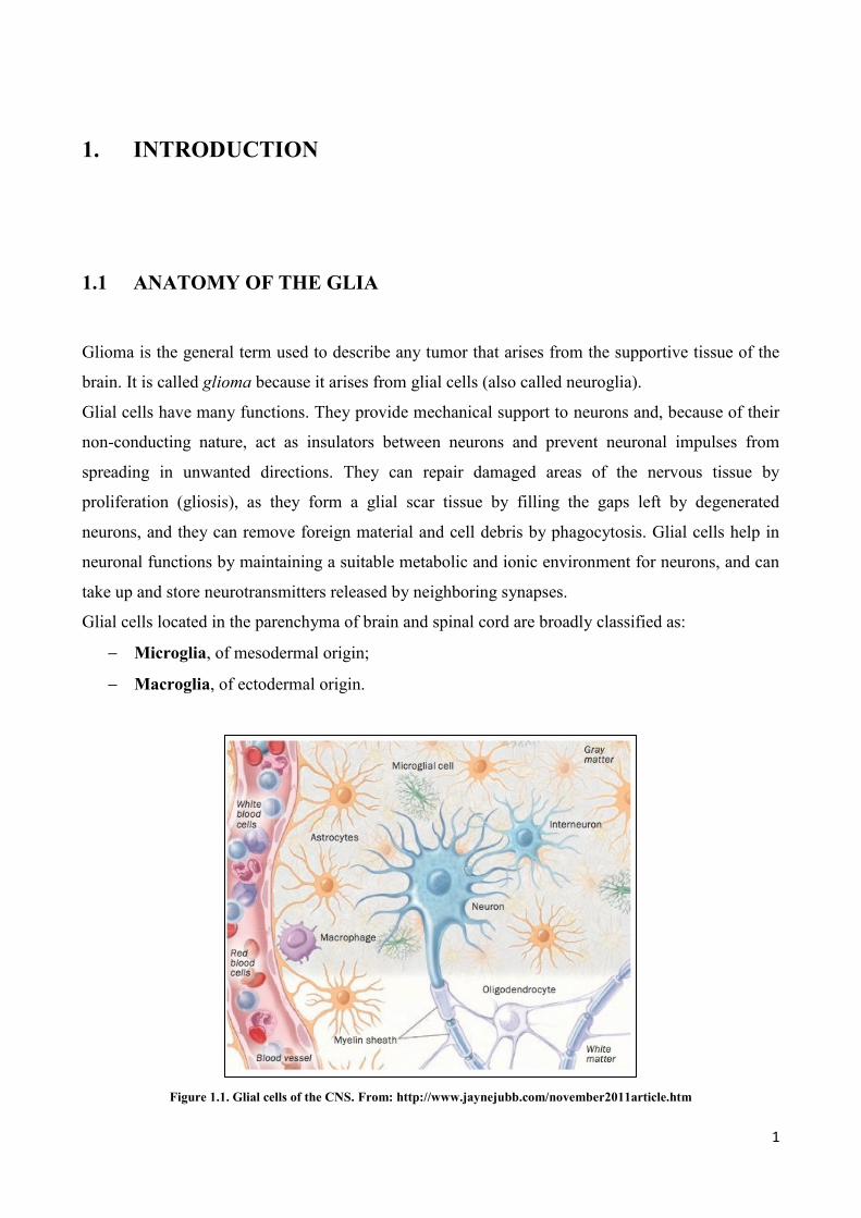
Glial cells located in the parenchyma of brain and spinal cord are broadly classified as:
Microglia, of mesodermal origin;
Macroglia, of ectodermal origin.
Figure 1.1. Glial cells of the CNS. From: http://www.jaynejubb.com/november2011article.htm
2
Microglia. These are the smallest of glial cells and are characterized by a flattened cell body with a
few short, fine processes. They are often associated with capillaries and are considered phagocytic
in nature. Microglial cells are possibly derived from circulating monocytes, which migrate into the
Central Nervous System (CNS) during the late fetal and early postnatal life. However, the
developmental origin of microglia remains debatable (1), and the two major views affirm that they
derive either from neuroepithelial cells (2, 3) or from hematopoietic cells (4, 5).
Macroglia. It’s mainly composed by astroglia and oligodendroglia, and it is generally considered
developmentally distinct from microglia, being derived from neuroectoderm (6).
Astrocytes
Astrocytes have been divided in two main subtypes, protoplasmic and fibrous, on the basis of
differences of their cellular morphology and anatomy location (7):
Protoplasmic astrocytes are found throughout all gray matter, and as first demonstrated using
classical silver impregnation techniques, exhibit the morphology of several stem branches that give
rise to many finely branched processes in a uniform globoid distribution;
Fibrous astrocytes are found throughout all white matter and exhibit a morphology characterized by
many long fiber-like processes. Classical and modern neuroanatomical studies also indicate that
both astrocytes subtypes make extensive contacts with blood vessels.
Oligodendrocytes
The term oligodendroglia was introduced by Del Rio-Hortega and collaborators to describe those
neuroglial cells that, in material stained by metallic impregnation techniques, showed few processes
(8). The oligodendrocyte is mainly a myelin-forming cell, but there are also satellite
oligodendrocytes that may not be directly connected to the myelin sheath (9). Satellite
oligodendrocytes are perineuronal and may serve to regulate the microenvironment around neurons
(10, 11). A number of features consistently distinguish oligodendrocytes from astrocytes (11), in
particular their smaller size, the greater density of both the cytoplasm and nucleus (with dense
chromatin), the absence of intermediate filaments (fibrils) and glycogen in the cytoplasm, and the
presence of a large number of microtubules (25 nm in diameter) in their processes that may be
involved in their stability (12). An oligodendrocyte extends many processes, each of which contacts
and repeatedly envelopes a stretch of axon with subsequent condensation of this multispiral
membrane-forming myelin (11, 13).
3
Glioblast
Glioblasts are stem cells able to differentiate into macroglial cells. They are particularly numerous
beneath the ependyma (14).
Ependymal cells
These neuronal supporting cells form the epithelial lining of the brain ventricles and of the central
canal of the spinal cord. Ependymal cells also give rise to the epithelial layer that surrounds the
choroid plexus, a network of blood vessels located in the walls of the lateral ventricles. Ependymal
cells share, with all other neuroglial cells, a neuroectodermic origin (15).
4
1.2 GLIOMAS
As previously mentioned, gliomas, which comprise astrocytic, oligodendrocytic and ependymal
lesions, are the most frequent primary intracranial tumors. This section will focus on glioma
epidemiology, etiology, classification, histopathology, diagnosis and therapy.
1.2.1. Epidemiology and etiology of gliomas
Gliomas of astrocytic, oligodendroglial and ependymal origin account for more than 70% of all
brain tumors (16, 17). The most frequent (65%) and malignant histological type is the glioblastoma
multiforme (16, 17). Since the introduction of computerized tomography and magnetic resonance
imaging, the incidence rates of brain tumors have been rather stable, with a tendency of higher rates
in highly developed, industrialized countries. Some reports indicate that Caucasians have higher
incidence than black or Asians populations, but to some extent, this may reflect socio-economic
differences and under-ascertainment in some regions, rather a significant difference in genetic
susceptibility.
In Italy, is reported an incidence of 5-10 cases every 100.000 people, equally divided in both sex,
even though most malignant forms are usually revealed in the male sex (16, 17). In the pediatric
population, gliomas represent, after hematopoietic neoplasia, the most common class of tumors,
with an incidence of 1-1,5 cases every 100.000 children aged from 0 to 14 years (16, 17).
With the exception of pilocytic astrocytomas, the prognosis of glioma patients is still poor. Less
than 3% of glioblastoma patients are still alive at 5 years after diagnosis, higher age being the most
significant predictor of poor outcome.
The etiology of gliomas is still not completely understood. However, different genetic elements
seem to be implied as predisposing factors for the development of these pathologies. In particular, it
has been reported the importance of p53 mutations (TP53) in low-grade gliomas (LGG) and
secondary glioblastomas derived therefrom. Approximatively 60% of mutations are located in the
hot spot codons 248 and 273 and the majority of these are G:C-->AT transitions at CpG sites. TP53
mutations are significantly more frequent in low-grade astrocytomas with promoter methylation of
the O(6)-methylguanine-DNA methyltransferase repair gene, suggesting that, in addition to
deamination of 5-methylcytosine, exogenous or endogenous alkylation in the O(6) position of
5
guanine may contribute to the formation of these mutations. Conversely, the loss of heterozygosity
(LOH) of chromosome 19 has been described in anaplastic astrocytomas (16, 17).
Yet, some epidemiologic studies underline the significant association between some hereditary
syndromes (such as type 1 and 2 neurofibromatosis and Li Fraumeni Syndrome) and the
development of astrocytoma (16, 17).
Today, the only environmental factor unequivocally associated with an increased risk of brain
tumors is the exposure to ionizing radiations, including therapeutic X-irradiation. In particular,
children treated with X-irradiation for acute lymphoblastic leukemia show a significantly elevated
risk of developing gliomas and primitive neuroectodermal tumors, often within 10 years of therapy.
Other studies, although not conclusive, suggest cerebral traumas, nitrosamine rich food ingestion ad
electromagnetic fields exposure (cell phones predominantly) as possible predisposing factors (16,
17).
The association between occupations carcinogens and glioma occurrence has been object of
different studies, demonstrating an elevated risk of astrocytomas in electric and electronic
employees, proportional to the time of exposure. An increased risk has been found even in workers
who have been exposed to organic chemicals in chemical and oil industries. Other possible
carcinogens factors are tetrachloride carbon, tetrachloroethylene, trichloroethylene, but mostly
chloride methylene (16, 17).
1.2.2. Glioma classification
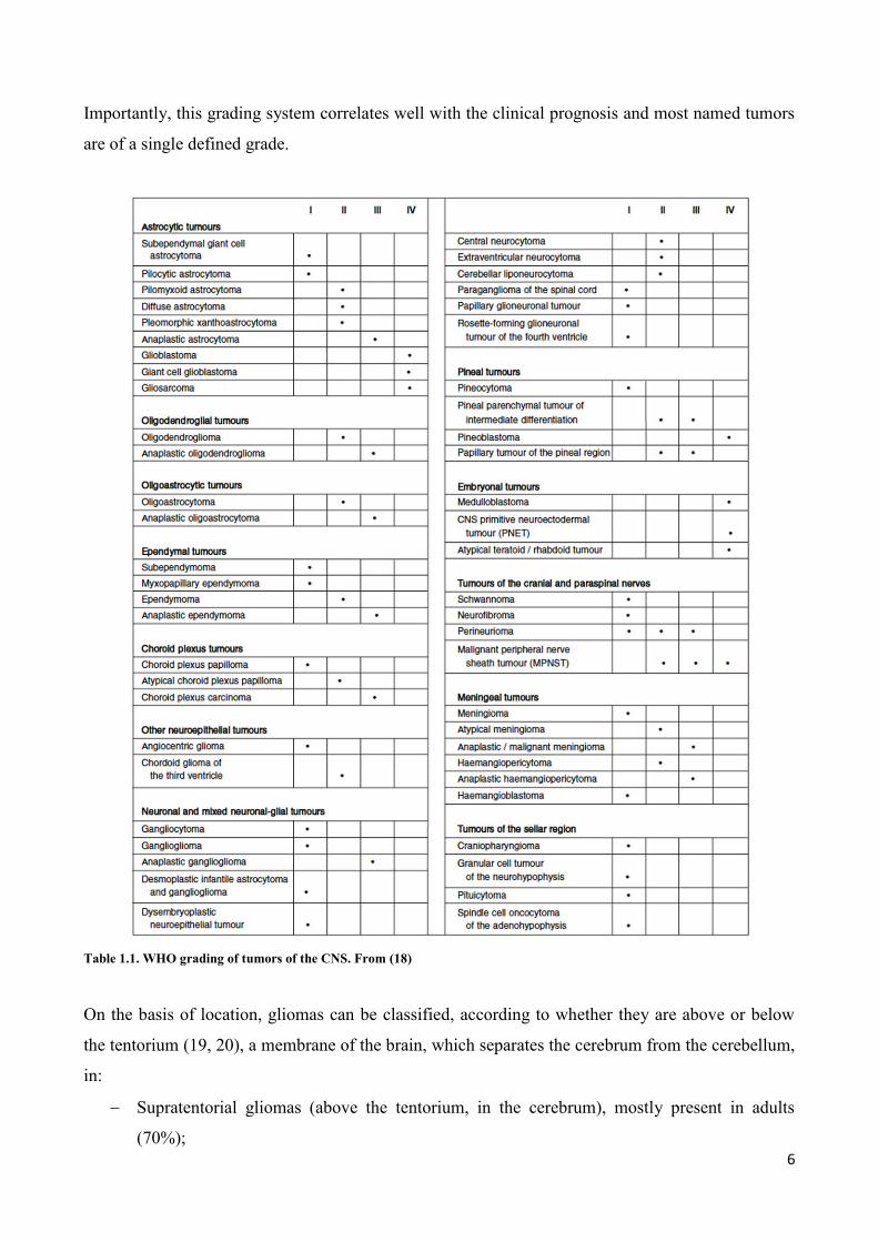
The WHO classification, which incorporates the criteria of the St.Anne/Mayo, is usually
recommended as a recent and updated international standard for classifying and grading gliomas
(Table 1.1) (18).
This classification is based on the premise that each type of tumor results from the abnormal growth
a specific cell type and includes criteria for their grading.
Therefore, according to WHO, gliomas are:
Divided on the basis of morphological criteria (cell shape, cytoplasmic appearance and the
character of the nuclei) into astrocytomas, oligodendrogliomas and oligoastrocytomas;
Classified into 4 grades, according to their increasing malignancy, from grade I to grade IV.
The grade is based on histological criteria such as cellularity, nuclear atypia, vascularization
and necrosis.
6
Importantly, this grading system correlates well with the clinical prognosis and most named tumors
are of a single defined grade.
Table 1.1. WHO grading of tumors of the CNS. From (18)
On the basis of location, gliomas can be classified, according to whether they are above or below
the tentorium (19, 20), a membrane of the brain, which separates the cerebrum from the cerebellum,
in:
Supratentorial gliomas (above the tentorium, in the cerebrum), mostly present in adults
(70%);
7
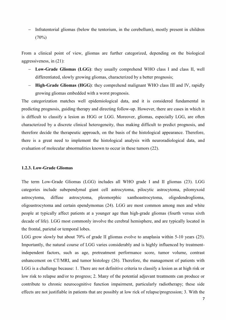
Infratentorial gliomas (below the tentorium, in the cerebellum), mostly present in children
(70%)
From a clinical point of view, gliomas are further categorized, depending on the biological
aggressiveness, in (21):
Low-Grade Gliomas (LGG): they usually comprehend WHO class I and class II, well
differentiated, slowly growing gliomas, characterized by a better prognosis;
High-Grade Gliomas (HGG): they comprehend malignant WHO class III and IV, rapidly
growing gliomas embedded with a worst prognosis.
The categorization matches well epidemiological data, and it is considered fundamental in
predicting prognosis, guiding therapy and directing follow-up. However, there are cases in which it
is difficult to classify a lesion as HGG or LGG. Moreover, gliomas, especially LGG, are often
characterized by a discrete clinical heterogeneity, thus making difficult to predict prognosis, and
therefore decide the therapeutic approach, on the basis of the histological appearance. Therefore,
there is a great need to implement the histological analysis with neuroradiological data, and
evaluation of molecular abnormalities known to occur in these tumors (22).
1.2.3. Low-Grade Gliomas
The term Low-Grade Gliomas (LGG) includes all WHO grade I and II gliomas (23). LGG
categories include subependymal giant cell astrocytoma, pilocytic astrocytoma, pilomyxoid
astrocytoma, diffuse astrocytoma, pleomorphic xanthoastrocytoma, oligodendroglioma,
oligoastrocytoma and certain ependymomas (24). LGG are most common among men and white
people at typically affect patients at a younger age than high-grade gliomas (fourth versus sixth
decade of life). LGG most commonly involve the cerebral hemisphere, and are typically located in
the frontal, parietal or temporal lobes.
LGG grow slowly but about 70% of grade II gliomas evolve to anaplasia within 5-10 years (25).
Importantly, the natural course of LGG varies considerably and is highly influenced by treatment-
independent factors, such as age, pretreatment performance score, tumor volume, contrast
enhancement on CT/MRI, and tumor histology (26). Therefore, the management of patients with
LGG is a challenge because: 1. There are not definitive criteria to classify a lesion as at high risk or
low risk to relapse and/or to progress; 2. Many of the potential adjuvant treatments can produce or
contribute to chronic neurocognitive function impairment, particularly radiotherapy; these side
effects are not justifiable in patients that are possibly at low rick of relapse/progression; 3. With the
8
exception of temozolomide (TMZ), current therapies are mainly designed according to previously
tested molecules against other types of cancer; in fact, novel drugs specifically designed to target
LGG are not yet available. Researches focused on other cancer types are currently exploiting these
issues taking advantage of: 1. Wide genome analysis; 2. Drug discovery approach; 3. Identification
of putative novel therapeutic targets within the tumor, such as tumor-initiating cells, tumor-
associated fibroblasts and infiltrating Mesenchymal Stem Cells (MSC).
For LGG all of these topics have been only incompletely explored. While a comprehensive genomic
characterization defining human glioblastoma genes and core pathways is available (26-31), this
extensive analysis is missing for LGG. What we know is that, genetically, the vast majority of LGG
are mutated in IDH1, frequently deleted in 1p19q (oligodendroglioma) or mutated in p53
(astrocytomas) (22, 32, 33). The IDH1 mutation is inversely correlated with grade, tightly
associated with a 1p19q co-deleted genotype and a MGMT methylated status by mutually exclusive
with Epidermal Growth Factor Receptor (EGFR) amplification and loss of chromosome 10 (32, 33).
Moreover, abnormalities in the PTEN tumor-suppressor gene and the BRAF oncogene are under
investigation (24).
1.2.4. High-Grade Gliomas
High-Grade Gliomas (HGG) comprise glioblastoma (WHO grade IV), anaplastic astrocytoma
(WHO grade III), mixed anaplastic oligoastrocytoma (WHO grade III) and anaplastic
oligodendroglioma (WHO grade III) (34).
Although anaplastic astrocytoma can be diagnosed as a de novo tumor, in the majority of patients
(50-75%) it represent a progression of pre-existing diffuse astrocytomas. This latter form affects
mostly patients 35-55 years old, and it has an overall survival of nearly 36 months. The malignant
progression of a diffuse astrocytoma, usually monitored by an enhancing in the contrast in Magnetic
Resonance Imaging (MRI), is detected in a 5-10% of the cases. Surgery is usually the initial
therapeutic approach, but considering the infiltrative nature of anaplastic gliomas, it is difficult to
completely eradicate the tumor. With respect to LGG, anaplastic gliomas are characterized by an
increased cellular proliferation, nuclear pleomorphism, mitosis, glomerular endothelial
proliferation, and necrosis.
From a genetic point of view, the mutation of TP53 is frequently found in anaplastic gliomas that
evolve from LGG. To confirm the role of TP53 in the glioma’s evolution, it’s emphasized that 90%
of patients with this protein mutation in the tumor relapse, already presented TP53 mutation in the
primitive LGG.
9
Nevertheless, in anaplastic tumor there are many other abnormal conditions like deletion of the
protein p16 (in 30% of the patients), alteration in the expression of the protein RB (in the 25% if the
cases), co-deletion chromosome 1p19q (in 15% of the cases), mutation in IDH1/2, as well as the
tumor-suppressor gene PTEN (15%), and finally the amplification of EGFR in 10% of the patients.
Besides that, it has also been reported an increased loss of heterozygosity (LOH) regarding, in
particular, the chromosome 10p (30-60%) and 19 (40%) as well as chromosome 22 (30%) along
with the deletion of chromosome 6 (33%).
The high incidence of the TP53 mutation and the elevated frequency of chromosomal aberrations
compared to LGG, indicates that anaplastic astrocytomas are the transitional form between LGG
and secondary glioblastoma multiforme. Nevertheless, the pathogenesis of primary anaplastic
astrocytomas is still unknown.
Glioblastoma multiforme is the most common and aggressive form of malignant astrocytoma and
can arise de novo or from pre-existing lower grade tumors (18). The incidence of glioblastoma in
Italy is of 2-3 new cases every 100.000 people per year (23). Glioblastoma can occur at any age but
is more likely to develop in older people (median age 53 years). It is predominantly found in male.
Although in adults glioblastoma develops mainly in the subcortical region, in the cerebral
hemisphere and, especially, in the temporal lobe, in children it develops mostly in the cerebral trunk
of the brain (35). It is generally associated with a poor prognosis (mean survival 11 months), yet
individual patient survivals may vary.
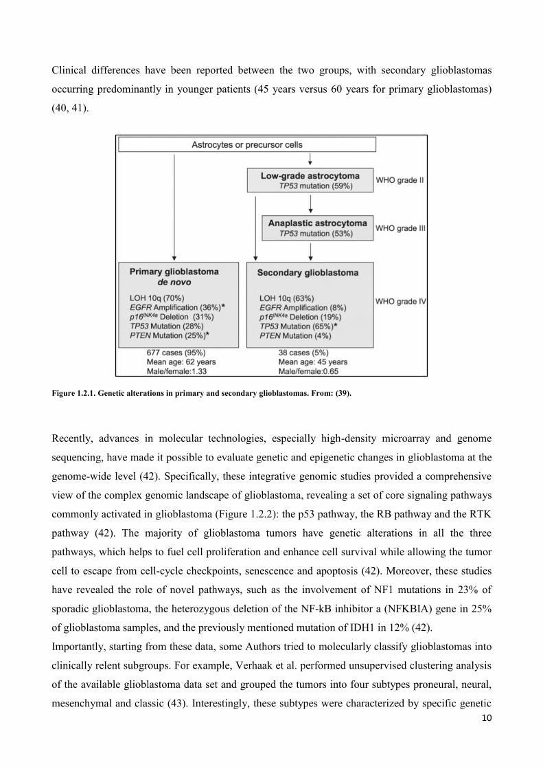
Historically, glioblastoma have been classified into two groups: “primary” and “secondary”
glioblastoma (36). The vast majority of glioblastoma (approximately 90%) develop rapidly de novo
in elder patients, without clinical or histological evidence of a less malignant precursor lesion
(primary glioblastoma) (37). Secondary glioblastoma progress from low-grade diffuse astrocytomas
or anaplastic astrocytomas. They manifest in younger patients, have a lesser degree of necrosis, are
preferentially located in the frontal lobe and carry a significantly better prognosis. Histologically,
primary and secondary glioblastomas are largely indistinguishable, but they differ in their genetic
and epigenetic profile (Figure 1.2.1). Decisive genetic signpost of secondary glioblastomas are
IDH1 mutations (38), that are absent in primary glioblastomas and which are associated with a
hypermethylation phenotype (37). IDH1 mutations are the earliest detectable genetic alterations in
precursor low-grade diffuse astrocytomas and in oligodendrogliomas, indicating that these tumors
are derived from neural precursor cells that differ from those of primary glioblastomas (37).
According to Yan and collaborators, the mutation of IDH1 promote the progression of low-grade
gliomas into high-grade (39).
10
Clinical differences have been reported between the two groups, with secondary glioblastomas
occurring predominantly in younger patients (45 years versus 60 years for primary glioblastomas)
(40, 41).
Figure 1.2.1. Genetic alterations in primary and secondary glioblastomas. From: (39).
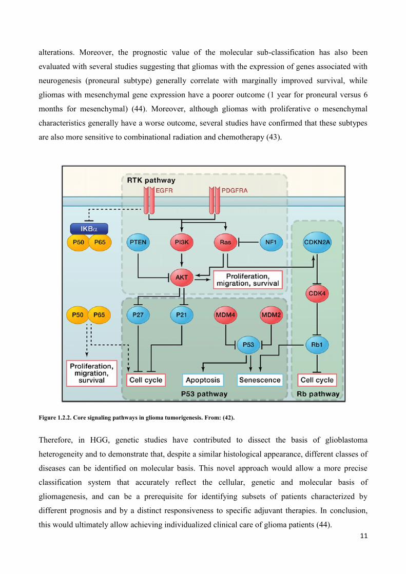
Recently, advances in molecular technologies, especially high-density microarray and genome
sequencing, have made it possible to evaluate genetic and epigenetic changes in glioblastoma at the
genome-wide level (42). Specifically, these integrative genomic studies provided a comprehensive
view of the complex genomic landscape of glioblastoma, revealing a set of core signaling pathways
commonly activated in glioblastoma (Figure 1.2.2): the p53 pathway, the RB pathway and the RTK
pathway (42). The majority of glioblastoma tumors have genetic alterations in all the three
pathways, which helps to fuel cell proliferation and enhance cell survival while allowing the tumor
cell to escape from cell-cycle checkpoints, senescence and apoptosis (42). Moreover, these studies
have revealed the role of novel pathways, such as the involvement of NF1 mutations in 23% of
sporadic glioblastoma, the heterozygous deletion of the NF-kB inhibitor a (NFKBIA) gene in 25%
of glioblastoma samples, and the previously mentioned mutation of IDH1 in 12% (42).
Importantly, starting from these data, some Authors tried to molecularly classify glioblastomas into
clinically relent subgroups. For example, Verhaak et al. performed unsupervised clustering analysis
of the available glioblastoma data set and grouped the tumors into four subtypes proneural, neural,
mesenchymal and classic (43). Interestingly, these subtypes were characterized by specific genetic
11
alterations. Moreover, the prognostic value of the molecular sub-classification has also been
evaluated with several studies suggesting that gliomas with the expression of genes associated with
neurogenesis (proneural subtype) generally correlate with marginally improved survival, while
gliomas with mesenchymal gene expression have a poorer outcome (1 year for proneural versus 6
months for mesenchymal) (44). Moreover, although gliomas with proliferative o mesenchymal
characteristics generally have a worse outcome, several studies have confirmed that these subtypes
are also more sensitive to combinational radiation and chemotherapy (43).
Figure 1.2.2. Core signaling pathways in glioma tumorigenesis. From: (42).
Therefore, in HGG, genetic studies have contributed to dissect the basis of glioblastoma
heterogeneity and to demonstrate that, despite a similar histological appearance, different classes of
diseases can be identified on molecular basis. This novel approach would allow a more precise
classification system that accurately reflect the cellular, genetic and molecular basis of
gliomagenesis, and can be a prerequisite for identifying subsets of patients characterized by
different prognosis and by a distinct responsiveness to specific adjuvant therapies. In conclusion,
this would ultimately allow achieving individualized clinical care of glioma patients (44).
12
1.3 THE HETEROGENEITY OF GLIOMA CELLS
Phenotypic and functional heterogeneity arise among cancer cells within the same tumor as a
consequence of genetic alterations, environmental differences and reversible changes in cell
properties. Some cancer cells also contain a hierarchy in which tumorigenic cancer stem cells
differentiate into non-tumorigenic progeny.
High-grade brain tumors are heterogeneous with respect to the composition of bona fide tumor cells
and with respect to a range of intermingling parenchymal cells. Glioblastoma harbor multiple cell
types, some with increasing tumorigenicity and stem-like properties (45).
1.3.1. Cancer Stem Cells: from the “Stochastic Model” to the “Hierarchical Model”
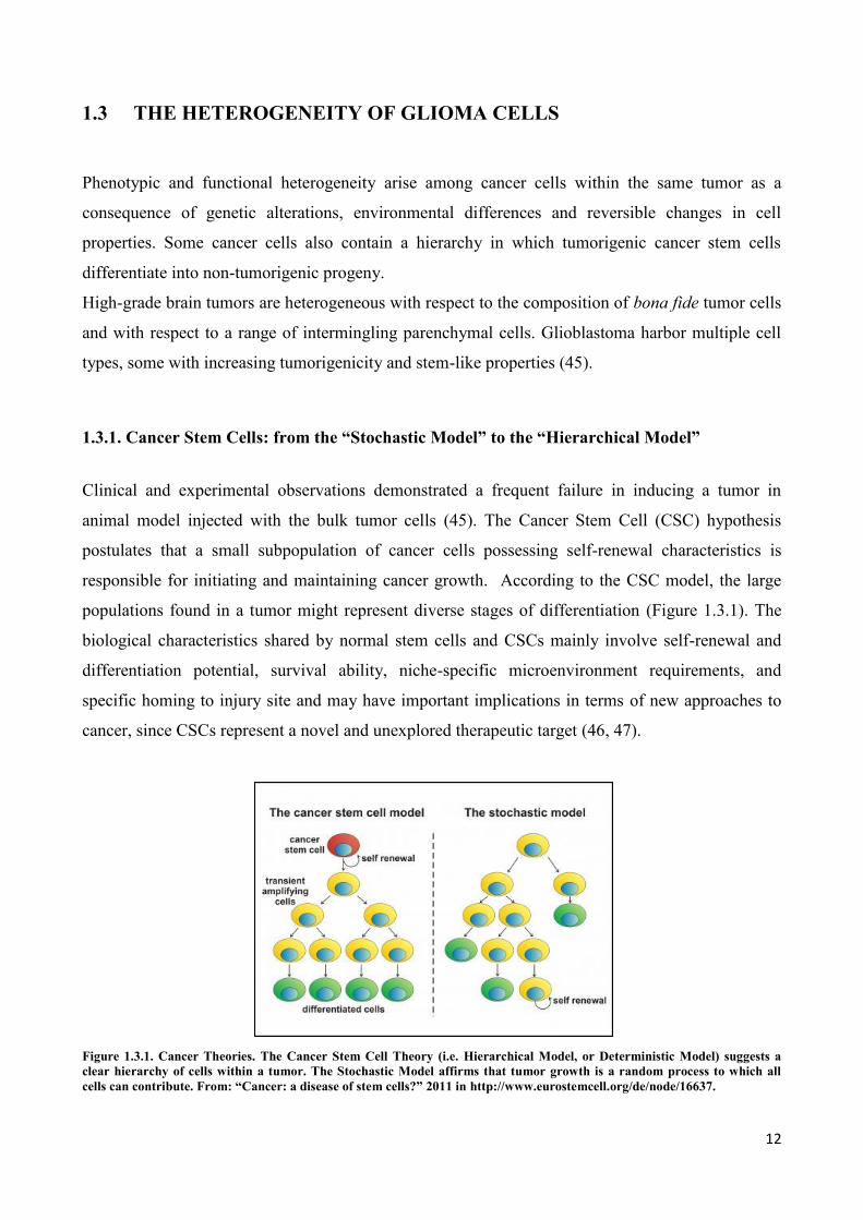
Clinical and experimental observations demonstrated a frequent failure in inducing a tumor in
animal model injected with the bulk tumor cells (45). The Cancer Stem Cell (CSC) hypothesis
postulates that a small subpopulation of cancer cells possessing self-renewal characteristics is
responsible for initiating and maintaining cancer growth. According to the CSC model, the large
populations found in a tumor might represent diverse stages of differentiation (Figure 1.3.1). The
biological characteristics shared by normal stem cells and CSCs mainly involve self-renewal and
differentiation potential, survival ability, niche-specific microenvironment requirements, and
specific homing to injury site and may have important implications in terms of new approaches to
cancer, since CSCs represent a novel and unexplored therapeutic target (46, 47).
Figure 1.3.1. Cancer Theories. The Cancer Stem Cell Theory (i.e. Hierarchical Model, or Deterministic Model) suggests a
clear hierarchy of cells within a tumor. The Stochastic Model affirms that tumor growth is a random process to which all
cells can contribute. From: “Cancer: a disease of stem cells?” 2011 in http://www.eurostemcell.org/de/node/16637.
13
Bonnet and Dick gave the first demonstration of the existence of CSCs in 1997 in human acute
myeloid leukemia (48). Authors demonstrated that a subpopulation of leukemia cells that were
CD34+/CD38
-, were able to self-renewal and to generate, when injected in NOD-SCID mice (non-
obese and diabetic - severe and combined immunodeficient systems), a myeloid acute leukemia
phenotypically identical to the starting tumor. Authors named these cell “tumor-initiating cells”
(TICs) and demonstrated that the phenotype of these cells was very similar to the ones described for
normal hematopoietic stem cells, suggesting that tumor and normal tissue are governed by a similar
hierarchical model.
Therefore, cells inside the tumor bulk are not all the same in terms of self-renewal, proliferating and
differentiation ability, and only the very primitive cells are characterized by the attitude to initiate
and maintain the tumor and can be responsible of recurrences and metastasis (49).
This leukemia model represented the paradigm for the subsequent studies focused on solid tumors,
such as mammary carcinoma (50), glioma (51, 52), melanoma (53), thyroid cancer (54), lung cancer
and gastroenteric tumors (55).
Solid tumors resulted to be more complex as they contain not only tumor cells, but also stromal
cells, which interactions can change the first one’s properties.
Notably, CSC share several stem cell related features with non-neoplastic stem cells, especially the
ones related to self-renewal (56).
In fact, pathways related to Wnt, Sonic Hedgehog and Notch, which normally regulate the cell self-
renewal, are present and up-regulated in several cancers (57).
Importantly, stem cells, whether they are normal or neoplastic, are usually characterized by a major
resistance to common therapies due to the up-regulation of drug-resistance pathways (45, 58).
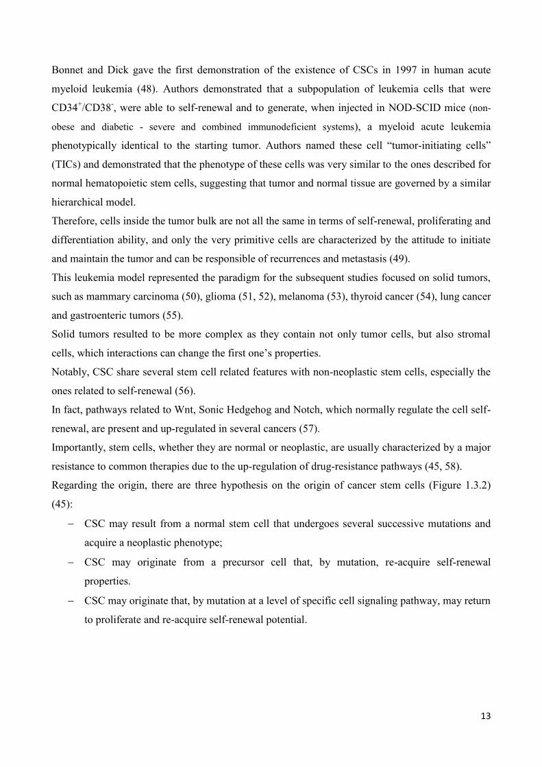
Regarding the origin, there are three hypothesis on the origin of cancer stem cells (Figure 1.3.2)
(45):
CSC may result from a normal stem cell that undergoes several successive mutations and
acquire a neoplastic phenotype;
CSC may originate from a precursor cell that, by mutation, re-acquire self-renewal
properties.
CSC may originate that, by mutation at a level of specific cell signaling pathway, may return
to proliferate and re-acquire self-renewal potential.
14
Figure 1.3.2. The origin of Cancer Stem Cells. The cancer stem cell may appear after mutation in specific stem cells or early
stem cell progenitors. It is also possible that cancer stem cells can be derived from differentiated cells. There might be
numerous factors in the host microenvironment that trigger the initial steps of tumor formation. From: Bjerkvig et al. The
origin of the cancer stem cell: controversies and new insights. 2005 Nature Reviews Cancer 5, 899-904; doi:10.1038/nrc1740.
Recently, the stem cell theory for cancer has been questioned. Specifically, Quintana showed that
modified xenotransplantation assay conditions, including the use of more immunocompromised
NOD-SCID interleukin-2 receptor gamma chain null (IL2Rg(-/-)) mice, could highly increase the
detection of tumorigenic melanoma cells, suggesting that, at least in some cancer, tumorigenic cells
are quite frequent (59).
One of the possible explanation for this results is the fact that the tumor microenvironment (stromal
fibroblasts, adipocytes and endothelial cells, as well as the extracellular matrix) and the immune
system play an important role in cancer progression (60, 61). Consequently, the xenograft model
could not offer an appropriate microenvironment for the growth of human tumors, because of the
differences between mice and humans and the lack of an intact immune system. These limitations
can be important when evaluating the tumor-initiating capacity of human cancer cells. Thus, it is
possible that the sub-population of cells that appeared non tumorigenic in a NOD-SCID mouse
model might actually be tumorigenic in the presence of the appropriate microenvironment. In other
words, tumor cells may be functionally homogeneous, with heterogeneous potential arising as a
consequence of extrinsic cues or of their lack.
15
Another important aspect is to understand how cancer stem cells are able to resist to common
therapies and treatments, and it would be clinically useful to assess the survival based on the
number of CSCs and not on the residual tumor mass (58).
In conclusion, the CSC paradigm refers to the ability of a subpopulation of cancer cells to initiate
tumorigenesis by undergoing self-renewal and differentiation, like normal stem cells, whereas the
remain majority of cells are more differentiated and lack of these properties. This concept, although
debated, has opened new horizons for understanding the biology of cancer and for identifying new
therapeutic modalities apt to eliminate cells responsible for tumor self-renewal.
1.3.2. Glioma-Initiating Stem Cells
The bulk of malignant cells in glioblastoma is generated by rare fractions of self-renewing,
multipotent tumor-initiating cells (51, 52), responsible for tumor growth and recurrence and
resistance to chemo- and radiotherapies (62). Glioma Stem Cells (GSCs) generate tumors with the
cardinal features of the glioblastoma from which they are derived, including an infiltrative
phenotype and histopathological feature such as hypercellularity, pseudopalisading necrosis, and
angiogenesis.
The first report of cells with stem-like properties in brain tumors was provided by Ignatova at al.,
where surgical specimens of glioblastoma multiforme were shown to have clonogenic
neurospheres-forming cells that expressed both neuronal and glial markers upon differentiation
(63). According to Vescovi’s definition (64), brain tumor cells could be considered as stem cells if
they show:
Cancer-initiating ability upon orthotopic implantation;
Extensive self-renewal capacity either ex vivo or in vivo;
Karyotypic or genetic alterations;
Aberrant differentiation properties;
Capacity to generate non-differentiated end cells;
Multi-lineage differentiation capacity.
Subsequently, Dirks et al. demonstrated CSC in brain tumors by transplantation of CD133+ or
CD133- cell populations into immunodeficient mouse. With as few as 100 CD133
+ cells from the
primary tumor, a new phenocopy of the tumor could be created in the transplanted mice, whereas
unsorted or CD133- primary tumor cells were unable to cause de novo tumor generation. As part of
16
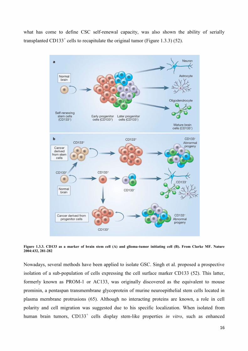
what has come to define CSC self-renewal capacity, was also shown the ability of serially
transplanted CD133+ cells to recapitulate the original tumor (Figure 1.3.3) (52).
Figure 1.3.3. CD133 as a marker of brain stem cell (A) and glioma-tumor initiating cell (B). From Clarke MF. Nature
2004:432, 281-282
Nowadays, several methods have been applied to isolate GSC. Singh et al. proposed a prospective
isolation of a sub-population of cells expressing the cell surface marker CD133 (52). This latter,
formerly known as PROM-1 or AC133, was originally discovered as the equivalent to mouse
prominin, a pentaspan transmembrane glycoprotein of murine neuroepithelial stem cells located in
plasma membrane protrusions (65). Although no interacting proteins are known, a role in cell
polarity and cell migration was suggested due to his specific localization. When isolated from
human brain tumors, CD133+ cells display stem-like properties in vitro, such as enhanced
17
proliferation ability, self-renewal, differentiation and neurosphere-like growth (52). Indeed, CD133+
cells are preferentially resistant to chemotherapeutic agents and radiation and express high levels of
ABC-transporter BCRP1, (O)6-methylguanine DNA methyltransferase and negative regulators of
apoptosis (62, 66). However, recent reports indicate that this initially proposed model might
represent an over-simplification and stem-cell specificity of the epitope detected by the antibody
AC133 has been questioned (67, 68). Thus, stem cell-specific markers other than CD133, as
CD15/SSEA-1 and integrin α-6, have been described, but there is not yet consensus for optimal
markers for GSC in glioblastoma (69-71).
Another method utilized to isolate glioma stem cells relies on the ability of these tumor-initiating
cells to grow as neurospheres. This assay was originally optimized for the isolation of normal stem
cells from healthy brain tissue (72). Authors demonstrated that brain tumor cells were able to
produce proliferating neurospheres that could be passaged at clonal density and differentiated into
cells of both neuronal and glial lineage. However, with respect to neural stem cells, tumor
neurospheres were characterized by an impaired differentiation capacity mainly favoring the
differentiation along the phenotype of the tumor of origin (52) or characterized by generation of
cells co-expressing glial and neuronal markers (63). Importantly, neurospheres expressed many
genes characteristic of NSC-derived spheres, such as CD133 (73).
Subsequently, Galli et al demonstrated that glioblastoma-derived neurospheres were characterized
by genetic aberration and were able, once injected both subcutaneously and orthotopically into
immunocompromised animal, to generate tumor xenograft histologically resembling the original
tumor. Importantly, tumor xenograft could be serially transplanted thus confirming the in vivo self-
renewal and tumorigenic capacity of neurospheres (85). Many papers have subsequently confirmed
the results obtained by Galli (83) and the superiority of the serum-free culture-method above the
standard serum-supplemented culture conditions (102).
Most importantly, in vivo studies have shown that neurosphere formation is a significant predictor
of clinical outcome in glioma patients, independent of Ki67 proliferation index, and is a robust,
independent predictor of glioma tumor progression (74).
Unfortunately, both methods present some drawbacks, making not feasible, to date, to apply high-
throughput in vitro analyses to study GSC biology and to search for compounds that selectively kill
cancer stem cells without killing the normal cells of the CNS. In fact, freshly isolated CD133
positive cells represent a small, although variable, fraction of the tumor cells and the efficiency of
neurosphere assays for producing GSC lines is considered rather low (67). Moreover, neurospheres
can be effectively obtained only from 30-50% of glioblastoma, but not from adult low-grade
gliomas (51, 73). Unfortunately, these methods are mostly effective in glioblastoma and
18
isolated/obtained cells cannot always be analyzed by high-throughput methods thus making more
difficult their study.
In order to overcome limitations of the neurosphere-based culture of GSC, Pollard et al. recently
reported a methodology for deriving and expanding glioma neural stem cell lines in adhesion onto a
laminin coating (75). Adherent cultured GSC are characterized by better features compared to
neurosphere cultured cells: they can be isolated from a higher number of tumor samples, can be
expanded with higher efficiency in order to obtain a higher yield, possess a lesser propensity to the
spontaneously differentiation due to accessibility of growth factors by inner cell mass of
neurosphere and maintain the ability to be tumorigenic in orthotopic transplantation and are suitable
for genetic and drug screenings.
1.3.3. Expression of radial glia markers in gliomas
Numerous studies suggest that astrocytomas may arise from pre-astrocytic transitional cells or
multipotent neural stem cells (76), and the observation that glioblastoma tissues are characterized
by the expression of molecular markers distinguishing the early stages of development, has attracted
much interest. An example is provided by the expression in gliomas of radial glial cells markers,
such as fatty acid-binding protein 7 (FABP-7).
Radial glial cells (RGC) play at least two crucial roles in the cortical development: neuronal
production in the ventricular zone, and the subsequent guidance of the neuronal migration toward
the cerebral cortex (77). FABP-7, also known as brain lipid binding protein (BLBP), is a member of
the FABP family, consisting of structurally related proteins that have specific cell, tissue, and
development patterns of expression. In the brain tissue, FABP7 gene is expressed more abundantly
at an immature stage of the brain than after maturation (78). Potential FABP-7 binding partners are
decosahexaenoic (DHA), oleic, linoleic and elaidic acid (79). Generally, FABP proteins are
involved in the uptake and intracellular trafficking of fatty acids, bile acids and retinoids, as well as
in cell signaling, gene transcription, cell growth, and differentiation. In radial glial cells (RGC),
FABP-7 plays a role in the establishment of the radial glia system required for neuronal migration
(80).
As reported by Liang et al, the nuclear expression of the radial glial marker FABP-7 is associated
with poor survival in patients with glioblastoma (81). Although FABP-7 is a cytoplasmic protein,
its varying subcellular location between nucleus and cytoplasm has been reported in developing
brain (82), glioma cells (83) and glioblastoma specimens (84). In addition, Taylor et al found that
19
all ependymoma-derived tumor spheres displayed a CD133+/Nestin+/FABP-7+ immunophenotype
similar to that of RGC, suggesting that these cells may be the cells of origin of ependymomas (85).
However, since the increased FABP-7 expression was also found in glia following nerve injury
(84), it is not yet clear whether the re-expression of radial glial markers occurs only in glioma cells,
or may be even in the stromal cells.
20
1.4 THE TUMOR MICROENVIRONMENT
In addition to malignant cancer cells, tumors contain a variety of different stromal cells that
constitute the so-called tumor microenvironment. Cancer cells and resident or infiltrating stromal
cells are involved in heterotypic interactions with one another. Some of these cell types provide
crucial support for tumor growth, are responsible for inducing angiogenesis, avoiding immune
response against tumor cells, deregulating cellular energetics or inducing invasion and metastasis
(86). Thus, this tumor-associated stroma is not a passive spectator of tumor growth, but it plays an
active role in tumorigenesis and cancer progression (87).
1.4.1. Tumor-Associated Fibroblasts
Nowadays, the tumor microenvironment has been studied mainly in solid tumors of epithelial origin
and Authors focused their attention on Cancer Associated Fibroblasts (CAF) or Tumor Associated
Fibroblasts (TAF) that represent the most abundant cell type in the tumor stroma (88-90).
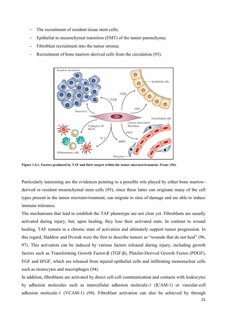
Five aspects can define TAF (Figure 1.4.1):
Expression of markers such as Fibroblast Activating Protein (FAP), which is selectively
expressed on TAF, and Fibroblasts Specific Protein (FSP), that is normally expressed by
Mesenchymal Stem Cells (MSC);
Expression of proteins involved in processes of invasion and remodeling of surrounding
stroma such Matrix metalloproteinases (MPPs), stromelysin-1 (SL-1), thrombospondin-1
(TSP-1) and Tenascin-C (TN-C);
Expression of proteins associated with neovascularization such as Smooth Muscle Actin
(SMA), desmin, Stromal-cell Derived Factor-1 (SDF-1) and Vascular Endothelial Growth
Factor (VEGF);
Production of growth factors such as basic Fibroblast Growth Factor (bFGF), Epidermal
Growth Factor (EGF), Insulin-like Growth Factor (IGF), Interleukin-6 (IL-6) and
Hepatocyte Growth Factor (HGF), many of which can also be derived from carcinoma cells;
Secretion of chemokines, such as Monocyte Chemotactic Protein 1 (MCP1) (91).
Although the biological origin of TAF is still undetermined, data in the literature currently support
four possible origins (92):
21
The recruitment of resident tissue stem cells;
Epithelial to mesenchymal transition (EMT) of the tumor parenchyma;
Fibroblast recruitment into the tumor stroma;
Recruitment of bone marrow-derived cells from the circulation (93).
Figure 1.4.1. Factors produced by TAF and their targets within the tumor microenvironment. From: (94).
Particularly interesting are the evidences pointing to a possible role played by either bone marrow-
derived or resident mesenchymal stem cells (95), since these latter can originate many of the cell
types present in the tumor microenvironment, can migrate to sites of damage and are able to induce
immune tolerance.
The mechanisms that lead to establish the TAF phenotype are not clear yet. Fibroblasts are usually
activated during injury, but, upon healing, they lose their activated state. In contrast to wound
healing, TAF remain in a chronic state of activation and ultimately support tumor progression. In
this regard, Haddow and Dvorak were the first to describe tumors as “wounds that do not heal” (96,
97). This activation can be induced by various factors released during injury, including growth
factors such as Transforming Growth Factor-β (TGF-β), Platelet-Derived Growth Factor (PDGF),
EGF and bFGF, which are released from injured epithelial cells and infiltrating mononuclear cells
such as monocytes and macrophages (94).
In addition, fibroblasts are activated by direct cell-cell communication and contacts with leukocytes
by adhesion molecules such as intercellular adhesion molecule-1 (ICAM-1) or vascular-cell
adhesion molecule-1 (VCAM-1) (94). Fibroblast activation can also be achieved by through
22
reactive oxygen species, complement factor C1 and or altered extracellular matrix (ECM)
composition (94). Moreover, during tumorigenesis, following loss of basement membrane integrity,
tumor cells invade into the underlying connective tissue and directly interact with local
mesenchymal fibroblasts.
Importantly, the mechanisms that determine a permanent activation of fibroblasts in tumors are still
unknown, although epigenetic mechanisms have been hypothesized (88, 90). The essential role of
TAF in the tumor progression suggested the possibility to envision TAF as possible novel
therapeutic target. The advantages of using the stroma as a therapeutic target comprises that these
cells are no genetically unstable as cancer cells and they are less likely to develop resistance to
treatment and to therapy (98).
An interesting approach is to inhibit TAF by blocking molecules responsible for their activation.
For example, inhibition of TGF-β by specific molecules seems to reduce the incidence of
metastases (99). Another possible target is FAP, thought to be involved in the control of fibroblast
growth or epithelial-mesenchymal interactions during development, tissue repair and epithelial
carcinogenesis (94). Further studies are suggested the use of Bevacizumab, a recombinant
humanized anti-VEGF monoclonal antibody, able to inhibit tumor angiogenesis (100).
In any case, it is very important to notice that these targeted therapies are aimed at targeting only
the stromal component of the tumor and have not direct effect on the other components of cancer. A
rational approach could be combining “stromal therapy” with a cytotoxic approach against tumor
cells.
1.4.2. The brain tumor microenvironment
The composition of the tumor microenvironment varies depending on the tumor site. The brain, in
particular, consists of numerous specialized cell types such as microglia, astrocytes and brain
endothelial cells. In addition to these brain-resident cells, brain tumor have also been shown to be
infiltrated by different populations of bone marrow-derived circulating cells (Figure 1.4.2) (101).
However, the role of different cell types that constitutes the tumor microenvironment in the
progression of gliomas is only poorly understood.
23
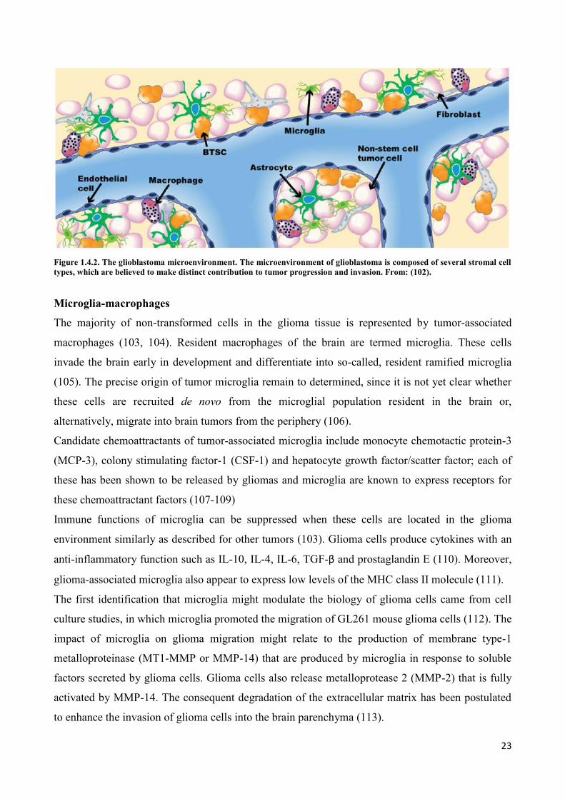
Figure 1.4.2. The glioblastoma microenvironment. The microenvironment of glioblastoma is composed of several stromal cell
types, which are believed to make distinct contribution to tumor progression and invasion. From: (102).
Microglia-macrophages
The majority of non-transformed cells in the glioma tissue is represented by tumor-associated
macrophages (103, 104). Resident macrophages of the brain are termed microglia. These cells
invade the brain early in development and differentiate into so-called, resident ramified microglia
(105). The precise origin of tumor microglia remain to determined, since it is not yet clear whether
these cells are recruited de novo from the microglial population resident in the brain or,
alternatively, migrate into brain tumors from the periphery (106).
Candidate chemoattractants of tumor-associated microglia include monocyte chemotactic protein-3
(MCP-3), colony stimulating factor-1 (CSF-1) and hepatocyte growth factor/scatter factor; each of
these has been shown to be released by gliomas and microglia are known to express receptors for
these chemoattractant factors (107-109)
Immune functions of microglia can be suppressed when these cells are located in the glioma
environment similarly as described for other tumors (103). Glioma cells produce cytokines with an
anti-inflammatory function such as IL-10, IL-4, IL-6, TGF-β and prostaglandin E (110). Moreover,
glioma-associated microglia also appear to express low levels of the MHC class II molecule (111).
The first identification that microglia might modulate the biology of glioma cells came from cell
culture studies, in which microglia promoted the migration of GL261 mouse glioma cells (112). The
impact of microglia on glioma migration might relate to the production of membrane type-1
metalloproteinase (MT1-MMP or MMP-14) that are produced by microglia in response to soluble
factors secreted by glioma cells. Glioma cells also release metalloprotease 2 (MMP-2) that is fully
activated by MMP-14. The consequent degradation of the extracellular matrix has been postulated
to enhance the invasion of glioma cells into the brain parenchyma (113).
24
Endothelial cell, pericytes and the perivascular niche
The tumor vasculature contributes an additional large compliment of non-malignant cells to
gliomas. Gliomas are highly vascularized tumors and emerging evidences indicate that endothelial
cells, pericytes and astrocytes that form the neurovascular unit function to support tumor
progression (102). This microanatomical structure has received increasing attention in the last few
years following the discovery that the perivascular niche (PVN) is the stem cell niche in both
normal and malignant brain tissue.
The vasculature in gliomas is characterized by hyperplasia and microvascular proliferation (MVP)
that are histological hallmarks of high-grade gliomas (114). The presence of MVP is associated
with tumor transition to a malignant phenotype. Microvascular proliferating structures have been
shown to derive from hyper-proliferative activity in a combination of endothelial, pericytic and
vascular smooth muscle cells (115). MVP structures represent regions of angiogenesis, which is
critical for tumor progression and is particularly important for brain tumors, especially malignant
gliomas that are among the most vascularized/angiogenic tumors. These angiogenic regions, of
which the perivascular niche is a part, have emerged as important locations responsible for the
maintenance of glioma stem cell (GSC) populations (116).
GSC are intimately associated with the vasculature across multiple brain tumor types. Endothelial-
derived factors also maintain self-renewal properties of glioblastoma stem cells in vitro. When
cultured with primary endothelial cells, GSC preferentially associated with them(117).
The development of the brain tumor vascular niche environment requires the recruitment of bone
marrow-derived cells, and some of these include: 1. Endothelial progenitors that mature into
endothelia of the vascular lumen; 2. Pericyte progenitor cells that envelope the vasculature and
mature into pericytes and vascular smooth muscle cells; and 3. CD45+ vascular-associated cells,
some of which give rise to perivascular macrophages and microglia (118).
In the context of tumor development, recruitment of pericytes and vascular smooth muscle cells to
the tumor is critical for structural stability of the vascular niche and for survival of tumor-
endothelial cells (119).
Fibroblasts and mesenchymal cells
Activated stromal fibroblasts have been identified by several reports to play a key role in the
induction of angiogenesis and metastasis in melanoma and other solid tumors (120, 121). A similar
role within the glioma microenvironment has also been suggested. Co-culture of brain-derived
25
human fibroblasts with glioblastoma cells induced production and activation of MMP2, and its
activators MT1-MMP and MT2-MMP, that are involved in the progression of gliomas (122).
Recently, our group identified a novel population of mesenchymal stem cells which can be isolated
from human low-grade and high-grade gliomas. This cells, called Glioma-Associated Stem Cells
(GASC), although devoid genetic alterations of the tumor of origin, are characterized by stem cell
properties, aberrant growth features, a gene expression profile typical of tumor-supporting cells and
are able to enhance in vitro the aggressiveness of glioblastoma initiating stem cells by means of
exosome release (123).
Astrocytes
Astrocytes are often identified by their expression of Glial Fibrillary Acidic Protein (GFAP), which
increase in response to injury. Astrocytes were originally thought to play a passive role in the brain,
but are now known to play critical functions in supporting the integrity of the blood brain barrier
(BBB) (124), in the transmission of neural impulses and in maintaining the biochemical
homeostasis of the brain tissue (125). In addition, astrocytes contribute to the brain’s response to
injuries. When injury occurs, astrocytes become reactive, change their morphology, proliferate and
migrate to the area of injury (126).
In addition to reactive astrocytes, adult Neural Stem Cells (NSC) also express GFAP (127). These
cells share ultrastructural and protein expression characteristics with mature astrocytes (128) and
reside in two neurogenic niches within the adult brain: the sub-ventricular zone of the lateral
ventricles and the sub-granular zone in the dentate gyrus of the hippocampus (129). Within these
regions, neural stem cells reside adjacent to blood vessels, and these adult astrocyte-like stem cells
proliferate slowly and are thought to divide asymmetrically to give rise to progenitor cells that
proliferate rapidly and symmetrically to produce terminal cell types such as neurons and glia (130).
Recent studies have highlighted the plasticity of astrocytes with regard to their functions in injury
and as stem cells. In addition to shared markers and ultrastructural characteristics between reactive
astrocytes and adult NSC, recent works have demonstrated that reactive astrocytes derived from an
injury setting possess characteristics specific to an adult NSC, including the expression of NSC
markers as well as the ability to proliferate and generate neurospheres (124, 131-133). This studies
suggest a novel role for astrocytes in the context of injury and demonstrate the plasticity of reactive
astrocytes.
The role of astrocytes cam vary significantly among gliomas. Astrocytes are closely associated with
the vascular endothelium and they secrete a number of neurotrophic factors, including transforming
growth factor-α (TGF-α), CXCL-12, SIP and GDNF, that have been described as capable of driving
26
the invasive properties of glioblastoma cells (134). Reports suggest that glioma-induced remodeling
within the tumor microenvironment through disruption of the BBB facilitates tumor invasion and
involve abnormal astrocyte-endothelial interaction (135).
Tumor-astrocyte interactions are an important mechanism by which glioma cells facilitate tumor
invasion. An increase in MMP2 production following cross-communication between co-cultured
astrocytes and glioblastoma cells, gas been associated with an enhancement of the invasive capacity
of glioblastoma cells through production of proMMP. This effect was mediated by the urokinase-
type plasminogen activator (uPA)-plasmin cascade. Astrocytes produced significant amount of
proMMP2, the inactive form of the matrix metalloproteinase MMP2, as well as uPA and its
receptor uPAR, both required for plasminogen cleavage. Plasminogen provided from glioblastoma
cells were cleaved to produce plasmin, which cleaved proMMP2 to its active form MMP2.
Furthermore, analysis of resected human glioblastoma specimens revealed elevated levels of
plasminogen, suggesting that a similar mechanism may exist in vivo (136).
Astrocytes also mediate activation of developmental pathways like the Sonic Hedgehog (SHH)
pathway, which is known to play an important role in GSC self-renewal and growth of gliomas.
SHH signaling was shown to be activated in gliomas and was correlated with increasing grade
(137).
27
1.5 EXOSOMES
Many kinds of cells shed small vesicles that play a key role in the intercellular communication.
Three major kind of vesicles have been described so far: microvesicles (100 nm to 1 µm in
diameter), which directly bud from the plasma membrane, apoptotic blebs (50-500 nm), which are
released by cells undergoing apoptosis, and exosomes (30-150 nm), released via exocytosis from
multivesicular bodies (MVBs) of the late endosome (138, 139).
Following the initial description in reticulocytes (140), several cell types have described to release
exosomes in the extracellular medium in vitro, such as hematopoietic cells (B-, T-, dendritic and
mast cells and platelets), epithelial cells, neural cells (oligodendrocytes, neurons, microglia and
Schwann cells), adipocytes, fibroblasts, stem cells and many types of tumor cells. Of relevance,
exosome are found in vivo in several biological fluids, such as saliva, urine, plasma, seminal fluid,
amniotic fluid, ascites, bronchoalveolar lavage fluid, synovial fluid, breast milk and cerebrospinal
fluid (CSF) (141).
1.5.1. Biogenesis and secretion
Exosomes originate in the intraluminal vesicles (ILVs) of MVBs, which fuse with the cell
membrane in an exocytic manner (142): as late endosomes bud off parts of their limiting membrane
into the lumen of late endosomes, vesicles are formed inside the lumen (ILVs) originating this
particular type of late endosome, which accumulates hundreds of ILVs and is termed MVB
(Figure1.5.1).
Initially, it was thought that MVBs could just fuse with lysosomes to degrade their intraluminal
content. This process was considered to be important to remove transmembrane proteins as well as
for the recycling system of cellular membranes (143, 144). Later it was demonstrated that in
reticulocytes undergoing maturation into red blood cells, MVBs can also fuse with the plasma
membrane to release their ILVs into the extracellular space, and the term “exosomes” was proposed
to identify these extracellular intra-endosomal vesicles (140).
28
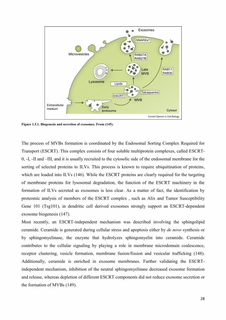
Figure 1.5.1. Biogenesis and secretion of exosomes. From (145).
The process of MVBs formation is coordinated by the Endosomal Sorting Complex Required for
Transport (ESCRT). This complex consists of four soluble multiprotein complexes, called ESCRT-
0, -I, -II and –III, and it is usually recruited to the cytosolic side of the endosomal membrane for the
sorting of selected proteins to ILVs. This process is known to require ubiquitination of proteins,
which are loaded into ILVs (146). While the ESCRT proteins are clearly required for the targeting
of membrane proteins for lysosomal degradation, the function of the ESCRT machinery in the
formation of ILVs secreted as exosomes is less clear. As a matter of fact, the identification by
proteomic analysis of members of the ESCRT complex , such as Alix and Tumor Susceptibility
Gene 101 (Tsg101), in dendritic cell derived exosomes strongly support an ESCRT-dependent
exosome biogenesis (147).
Most recently, an ESCRT-independent mechanism was described involving the sphingolipid
ceramide. Ceramide is generated during cellular stress and apoptosis either by de novo synthesis or
by sphingomyelinase, the enzyme that hydrolyzes sphingomyelin into ceramide. Ceramide
contributes to the cellular signaling by playing a role in membrane microdomain coalescence,
receptor clustering, vesicle formation, membrane fusion/fission and vesicular trafficking (148).
Additionally, ceramide is enriched in exosome membranes. Further validating the ESCRT-
independent mechanism, inhibition of the neutral sphingomyelinase decreased exosome formation
and release, whereas depletion of different ESCRT components did not reduce exosome secretion or
the formation of MVBs (149).
29
Interestingly, exosomes produced by the ESCRT-independent pathway are enriched in tetraspanins,
which are transmembrane proteins that may also be involved in endosomal sorting pathways (150).
Based on these observations, ESCRT-dependent sorting mechanisms may target proteins loaded
into ILVs for lysosomal degradation, whereas ESCRT-independent sorting mechanisms may target
ILVs for secretion. However, this process is much more complex and may depend on cell type,
nature of the cargo or other stimuli.
Exosomes are secreted by the fusion of MVBs with the plasma membrane and released into the
extracellular space. Exosome secretion is not considered a random event, but rather a highly
controlled process. Although largely is still under investigation, this process is thought to be
coordinated through the transport and fusion of MVBs with the plasma membrane by the
microtubule and actin cytoskeleton, t- and v-SNAREs and Rab GTPases (151). Rab GTPases are
ubiquitously expressed proteins that are responsible for the coordination of vesicle trafficking
events (152). For example, over-expression of Rab11 has been shown to stimulate exocytosis and
Rab27a and Rab27b control different steps of exosome secretion pathway (153, 154).
Exosome secretion may be enhanced by many different conditions. For example, has been
demonstrated that cancer cells cultured in hypoxic or low pH conditions showed an enhanced
release of exosomes (155).
In order for secreted exosomes to exert any biological function, they must be adsorbed by and
deliver their contents to a recipient target cell. However, the specific targeting of exosomes to target
cell and how this process unfolds in normal physiology or in the diseased state is not well
understood. This process must critically depend on the specific adhesion molecules, integrins and
antigenic factors expressed on the exosome, as well as the receptor or other docking molecules
found on the surface of target cells. For example, exosomes from T-, B- and dendritic cells were
shown to communicate with antigen presenting cells by transferring their content in a n
unidirectional manner and modulating gene expression in the recipient cell (156).
Presumably, any cell capable of endocytosis or phagocytosis may participate in the uptake of
exosomes. Indeed, many studies have documented that exosomes can be internalized by recipient
cells through many processes, such as receptor-mediated endocytosis (both clathrin- or dynamin-
dependent), phagocytosis and pinocytosis (157, 158).
However, the mechanisms of uptake of exosomes by recipient cells have not yet been completely
clarified.
30
1.5.2. Exosome components
Apart from their morphological and physical properties, exosomes are usually identified on the
bases of their unique protein and lipid composition. Because of their endosomal origin, exosomes
contain proteins involved in membrane transport and fusion (e.g. Rab GTPases, annexins, flotillins),
MVB biogenesis (e.g. Alix, Tsg101) or protein families mostly associated with lipid microdomains,
such as integrins and tetraspanins (e.g. CD9, CD63, CD81, CD82) (142).
According to proteomic studies, exosome protein composition is different in membrane vesicles
released by different cell types (147). Specifically, exosome protein content includes both
conserved proteins, i.e. identified in almost all exosomes despite their origin, and cell-type specific
proteins. Among conserved proteins, the heat shock protein cognate 70 kDa protein (hsp70) and the
tetraspanin CD63, are the most frequently identified (141). Moreover, other frequently found
proteins are linked to the cytoskeleton (β-actin, myosin, tubulin) or to metabolism (e.g.
glyceraldehyde 3-phosphate dehydrogenase, GAPDH). In agreement to their role as antigen
presenting vesicles, many exosomes contain major histocompatibility complex (MHC) class I and II
molecules. Of interest, exosomes contain also protein involved in cell signaling pathways, like β-
catenin, Wnt5b or the Notch ligand, Delta-like 4, and mediators of intercellular cell signaling, like
IL-1β, TNF-α and TGF-β (159).
As for proteins, the lipid composition of exosomes is distinct from that of the cell of origin, but is
anyway characteristic of the cell type. Lipid composition analyses have been performed of
exosomes derived from hematopoietic cells (141), oligodendrocytes (93) and melanoma cells (160).
Internal membranes of MVBs are enriched in lipids, such as lysobisphosphatidic acid (LBPA),
which plays an important role in exosome biogenesis. Moreover, exosomal membranes are often
enriched in lipids usually associated with lipid rafts, such as cholesterol, sphingolipids, ceramide
and glycerosphingolpids with long and saturated fatty-acyl chains (149, 161).
A major breakthrough in the field has been the discovery that exosomes contain nucleic acids, such
as mRNAs and microRNAs (162, 163). As mRNAs were also shown to be translated in target cells,
this was the first evidence suggesting cell-to-cell communication by exosome-mediated transfer of
genetic information. Exosome-transferred miRNAs were also suggested to be functional in target
cells, as miRNAs from T-cell exosomes caused inhibition of target genes in dendritic cells (156).
Not all mRNAs present in a cell are included in exosomes, so there is a specific targeting of
mRNAs into exosomes, whose mechanisms are unknown. Recently, it was also reported that neural
cells (164) and myoblasts (165) release exosomes carrying mitochondrial DNA (mtDNA). The role
31
of mtDNA is unknown, although it has been suggested that it could reach the cytosol of the target
cells and be imported into the mitochondria.
1.5.3. The role of exosomes in the tumor microenvironment
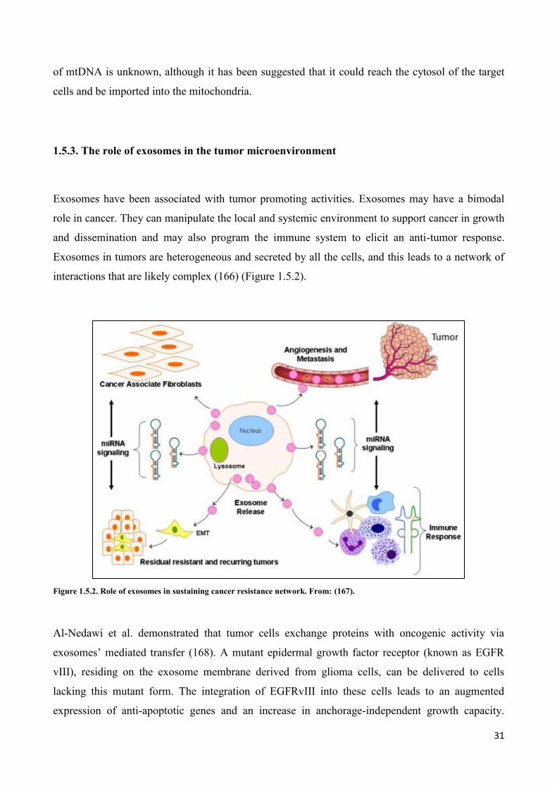
Exosomes have been associated with tumor promoting activities. Exosomes may have a bimodal
role in cancer. They can manipulate the local and systemic environment to support cancer in growth
and dissemination and may also program the immune system to elicit an anti-tumor response.
Exosomes in tumors are heterogeneous and secreted by all the cells, and this leads to a network of
interactions that are likely complex (166) (Figure 1.5.2).
Figure 1.5.2. Role of exosomes in sustaining cancer resistance network. From: (167).
Al-Nedawi et al. demonstrated that tumor cells exchange proteins with oncogenic activity via
exosomes’ mediated transfer (168). A mutant epidermal growth factor receptor (known as EGFR
vIII), residing on the exosome membrane derived from glioma cells, can be delivered to cells
lacking this mutant form. The integration of EGFRvIII into these cells leads to an augmented
expression of anti-apoptotic genes and an increase in anchorage-independent growth capacity.
32
Colon cancer cell harboring only mutant KRAS alleles are capable of releasing exosomes with
mutant KRAS protein. Colon cancer cells expressing only the wild-type KRAS can internalize this
mutant form. The exosomes’ mediated exchange of mutant KRAS induces cell growth and
tumorigenicity (169).
Tumor-derived exosomes can also modulate the tumor microenvironment and angiogenesis. For
example, exosomes from prostate cancer cells and mesothelioma cell lines contain TGF-β1 protein,
which is transferred to recipient cells in a biologically active form. In vitro experiments have shown
that TGF-β1 expressing exosomes can trigger the differentiation of fibroblasts to myofibroblasts
(170). Myofibroblasts are a key source of matrix-remodeling proteins within the tumor
microenvironment and participate in tumor angiogenesis (171).
Recently, our group reported that exosome-mediate communication between cancer cells and
stromal cell is bidirectional. In particular, mesenchymal stem cells isolated from human gliomas
(called Glioma-Associated Stem Cells, GASC) can modulate the aggressiveness of glioblastoma
initiating cells and glioblastoma cell lines by releasing exosomes (123).
Furthermore, cancer cells transfer membrane-bound EGFR to endothelial cells via exosomes. This
transfer activates the autocrine VEGF/VEGFR-2 pathway in endothelial cells and likely support
tumor angiogenesis (172). Moreover, melanoma-derived exosomes can deliver microRNA-9 (miR-
9) into endothelial cells, promoting endothelial cell migration in vitro and tumor angiogenesis in
vivo (173).
Several studies have shown that TD-exosomes can alter the extracellular matrix through secretion
of matrix metalloproteinases (MMPs) or activators of MMPs, such as heat shock proteins. MMPs
are zinc-dependent plasma membrane endo-peptidases that can degrade extracellular matrix
proteins, such as collagen, fibronectin, proteoglycans and laminins. In fibrosarcoma and melanoma
cells, it was shown that MT1-MMP was secreted in exosomes and could activate pro-MMP-2 and
degrade collagen and gelatin (174). Other studies have demonstrated that heat shock proteins, such
as hsp90, are also secreted via exosomes and can activate MMP-2 to enhance invasion of cancer
cells (175).
Numerous reports indicate that exosome-mediated communication between tumor cells and the
immune system is involved in recruiting pro-tumorigenic immune cells. For example, the blockade
of exosome secretion by inhibiting Rab27a is associated with a decreased mobilization of
neutrophils, which results in a reduced primary tumor growth and lung metastasis (176). Moreover,
miRNAs in lung cancer released exosomes can silence the transcripts associated with Toll-like
Receptor (TLR) family in macrophages. This mechanism stimulates macrophages to secrete pro-
inflammatory cytokines, which support enhanced tumor dissemination (177). Cancer cells are also
33
able of inhibiting anti-tumor functions of the host immune system via an exosome-induced
signaling. Chalmin et al. showed that tumor-derived exosomes activate myeloid-derived suppressor
cells (MDSC), which exert immunosuppressive functions in cancer by suppressing the T-cell
response (178, 179).
Tumor cells release large amount of microvesicles and the number of circulating microvesicles is
increased in patients with cancer and correlates with poor prognosis (180). Thus, circulating
exosomes may be potential biomarkers to predict cancer burden at an early stage and impact
personalized cancer care. For example, a clinical subtype of glioblastoma contains EGFRvIII
mutant/variant (163). The expression of this EGFRvIII mutant/variant is decisive in determining the
therapeutic approach for such patients (181). In this regard, Skog and al. have demonstrated that
circulating serum exosomes are positive for this mutant/variant EGFRvIII when the parental
glioblastoma cells also expressed it (163). Therefore, determining EGFR status from a small serum
sample derived exosomes instead of a need for primary tumor biopsy that involved invasive brain
surgery, is of immense benefit for patients. Such application for circulating serum exosomes in
tumor diagnosis have been described for colorectal cancer (182), ovarian cancer (183) and
melanoma (184). Silva et al. have reported that high levels of exosomes in plasma of patients with
colorectal cancer was significantly associated with poor differentiated tumors and with decreased
overall survival (182). Patients with ovarian cancer exhibit significantly increased levels of serum
exosomes compared to benign disease or healthy controls. A profile between exosome-derived
miRNAs and corresponding tumor-derived miRNAs revealed a dysregulated expression of 3 out of
218 detectable miRNAs, suggesting a specific selection of miRNAs in tumor-derived exosomes
(183).
Apart from serum, exosomes can be detected in further body fluid such as urine or saliva and
provide non-invasive diagnostic markers for cancer (185, 186). Taken together, exosome-derived
proteins and RNAs could serve as a useful tool to classify patients into different risk categories o
enable a more tailored cancer therapy.
In addition, since cancer cells modify the local and systemic environment by releasing exosomes to
support tumor progression and metastasis, impairing the secretion of exosomes by cancer cells may
constitute a potential target for cancer therapy.
34
2. AIM OF THE STUDY
Gliomas are the most frequent tumors of the central nervous system. High-grade gliomas (HGG) are
associated with disproportionately high morbidity and mortality and, despite an optimal treatment,
the median survival is only 12 to 15 months for patients with glioblastoma multiforme. With respect
to HGG, low-grade gliomas (LGG) grow slowly, but about 70% of grade II LGG evolve to
anaplasia within 5-10 years (25).
Glioblastoma multiforme is the most common and malignant form of human glioma; it accounts for
60% of all glial tumors and occurs at a frequency of 5 cases per 100.000 people (187). Although
molecular markers for glioblastoma multiforme have helped to identify patients responsive to
current therapies (188), the overall survival of responsive patients has not changed substantially in
the last two decades (189).
The aggressiveness of HGG is not only due to the high rate of tumor growth, but also to its
particularly invasive nature, which allows the active egress of glioma cells from the tumor mass into
the surrounding normal brain tissue (190). Therefore, invasive gliomas are essentially beyond the
reach of current surgical therapies, which are localized to the tumor and immediate peri-tumor
environment. However, glioblastoma multiforme cell invasion is not random; transformed glial
cells preferentially invade along anatomic features such as myelinated axons, vascular basement
membranes, and the subependyma (191), which suggest that they interact with specific structures in
their immediate microenvironment.
Indeed, mounting evidence indicates that normal peritumoral cells plays active roles in tumor
progression (98, 192) and a considerable interest is focused on the tumor-associated stroma. In fact,
in addition to cancer cells, tumor lesions contain a mixture of different stromal cells, such as
endothelial cells that constitute blood vessels, inflammatory cells that infiltrate the neoplastic tissue
from the blood stream and resident stromal cells (95, 193). These stroma components have been
studied essentially in solid tumors of epithelial origin, where Cancer Associated Fibroblasts (CAF)
represent the most abundant cell type (194). Regarding gliomas, xenotransplantation experiments
employing human glioma cell line have suggested that astrocytes in the vicinity can be activated
and facilitate tumor invasiveness (102). The glial response to injuries, including the presence of a
tumor mass, consists of multiple and complex changes in morphology, gene expression and
function, a process referred to as “astrogliosis” (124). In addition, genetic fate mapping studies have
35
shown that reactive glia could acquire a stem cell potential outside the two major neural stem cell
niches in adult mammalians (129, 133).
Thus, studying the tumor-stroma interplay could be an important opportunity for identifying key
factors that mediate the activation of stromal cells toward a phenotype able to support tumor growth
and molecular mechanisms driving tumor progression and anaplastic transformation. At this regard,
recent studies suggest that the release of microvesicles could have an active role in communication
processes occurring among different cell types populating the tumor microenvironment (163, 195,
196). However, little is known about this in gliomas.
In particular, our group has recently demonstrated that exosomes secreted by non-tumorigenic
mesenchymal stromal cells isolated from human gliomas (Glioma-Associated Stem Cell, GASC)
were able to increase the aggressiveness of glioma initiating stem cells in vitro (123). However,
there is little information in the literature about the effects produced by glioma cells-derived
microvesicles on the surrounding microenvironment.
Therefore, in order to shed light on communication mechanisms established between glioma cells
and stromal cells within the tumor microenvironment, the work has been focused on the effects
produced by exosomes released by different types of glioblastoma cells on astrocytes.
In particular, specific aims of this study were:
1. To verify if astrocytes grown in culture in the presence of exosomes deriving from a
glioblastoma commercial cell line (U87-MG), primary isolated glioblastoma cells, and
primary isolated glioblastoma stem cells, could acquire phenotypic and functional features
of the reactive astrocytes populating the glioma microenvironment;
2. To verify whether exosomes produced by different glioblastoma cells could be able in vitro
to induce a re-programming of glial cells by the acquisition of a stem cell potential;
3. To assess whether neural stem cells obtained by astrocytes conditioned with exosomes
derived from glioblastoma cells could be differentiated in functional radial glia-like cells.
36
3. MATERIALS AND METHODS
3.1 Cells
Human glioblastoma samples were collected by the Neurosurgery Department of the Azienda Ospedaliero
Universitaria of Udine, after informed consent was obtained, in accordance with the Declaration of Helsinki,
and with approval by the Independent Ethics Committee of the University-Hospital of Udine. Glioblastomas
were histopathologically evaluated by expert Pathologist following WHO criteria.
Glioblastoma stem cells (GSC) were isolated as described in (75, 123). Briefly, cells less than 40
μm in diameter were seeded at a density of 2x10⁴ cells/cm
2 onto laminin coated dishes in a growing
medium composed as follows: Neurobasal-A medium (Gibco), 2 mM L-glutamine (Sigma-Aldrich),
1X N2 supplement (Gibco), 25 µg/mL Insulin, Penicillin-streptomycin (Sigma-Aldrich), 100
µg/mL human apo-trasferrin (Sigma-Aldrich), 1X B-27 supplement (Gibco), 20 ng/mL h-FGF-
basic (Peprotech), 20 ng/mL h-EGF (Peprotech).GSC were cultured in an incubator at 37°C,
5%O2/5%CO2. Half of the medium was replaced with fresh one every 4 days. Once cells reached
70-80% of confluence, they were detached by TrypLE Express (Invitrogen) and re-plated at a
density of 3-4x10³/cm
2.
Glioblastoma primary cells (GBM) were isolated from glioblastoma samples and cultured in
DMEM-low glucose (Gibco) supplemented with 10% fetal bovine serum (FBS; Euroclone)
previously ultracentrifuged at 100,000 x g for 4 hours at 4°C (uFBS) and 1X Penicillin-
streptomycin (Sigma-Aldrich).
Commercially available U87-MG glioblastoma cells (purchased from American Type Culture
Collection) were cultured in DMEM-low glucose (Gibco) supplemented with 10% uFBS and 1X
Penicillin-streptomycin (Sigma-Aldrich).
Commercially available Wi38 human normal lung fibroblasts (gently provided from Prof. C.
Schneider) were cultured in DMEM-low glucose (Gibco) supplemented with 10% uFBS and 1X
Penicillin-streptomycin (Sigma-Aldrich).
Cell culture supernatants from GSC (n=3), GBM (n=3), U87-MG and Wi38 cells were collected
during the exponential growth phase for exosomes purification (see 3.2).
Human normal astrocyte from cerebral cortex (ScienCell Research Laboratories) were cultured onto
0,1% poly-L-lysine (Sigma-Aldrich) coated-dishes in Astrocyte Medium (AM; ScienCell Research
Laboratories) supplemented with 2% uFBS, 1X Astrocyte Growth Supplement (ScienCell Research
Laboratories) and 1X Penicillin-streptomycin (ScienCell Research Laboratories) at 37°C, 5% CO2.
37
3.2 Exosomes purification
Exosomes were purified from the collected cell culture supernatants by precipitation with
ExoQuick-TC (System Biosciences), according to the manufacturer’s protocol with minor
modifications. Briefly, cell culture supernatants were harvested, centrifuged at 3,000 x g for 15
minutes to remove cells and cell debris and filtered through a 0,2 µm mesh filter to remove particles
larger than 200 nm in diameter. Clarified supernatants were concentrated in their vesicular fraction
by ultrafiltration with a centrifugal filter unit (cut-off 100 kDa, Amicon Ultra-15, Millipore) at
4,500 x g for 30 minutes. Concentrated supernatants were washed three times with D-PBS
(Dulbecco’s Phosphate Buffered Solution, Life Technologies) by ultrafiltration, mixed with
ExoQuick-TC (5:1 v/v) and incubated over-night at 4°C. The mixture was centrifuged at 1,500 x g
for 30 minutes and exosome pellet resuspended in 100 µL of D-PBS or dH2O, according to
downstream applications.
Exosomal protein yield was determined for each sample by Bradford assay (Bio-Rad).
3.3 Exosomes characterization
3.3.1 Dimensional analysis
Exosome dimension and concentration were assessed using a NanoSight LM10 instrument
(NanoSight) in collaboration with Prof. Benedetta Bussolatti (University of Turin). NTA
(Nanoparticle Track Analysis) software suite was utilized to calculate particles diameter and to
construct the frequency distribution graph.
3.3.2 Flow cytometry
Purified exosomes were incubated with 4 µm aldehyde/sulfate latex beads (Invitrogen, Molecular
Probes) in 1 ml of PBS and left shaking over night at room temperature. Solution were then added
with 300 mL of 1 M glycine (Sigma-Aldrich) and, after 30 minutes, washed twice with PBS
38
containing 0.5% bovine serum albumin (Sigma-Aldrich) (PBS/BSA 0.5%). Exosomes conjugated
with beads were finally incubated 20 minutes at room temperature with PE-labeled antibodies
directed against CD63 and CD9 tetraspanins (Becton Dickinson). After two washes with PBS/BSA
0.5%, pellets obtained were re-suspended in 500 µL of PBS, and acquired by FACSCalibur and
analyzed by Summit software (Beckman Coulter). PE-isotype-matched antibodies were used as a
negative control.
3.4 Exosomes uptake by astrocytes
The interaction between astrocytes and exosomes secreted by glioma cells was investigated by
evaluating the ability of astrocytes to internalize GSC-deriving exosomes. Briefly, 100 µg of
purified exosomes resuspended in D-PBS were labeled with 5 µM Vybrant-DiD (Invitrogen) for 30
minutes at 37°C. Labeled exosomes (exo-DiD) were purified by precipitation with ExoQuick-TC
(see 3.2) and resuspended in Astrocyte Medium (AM) at the concentration of 10 µg/mL.
Astrocytes were seeded onto 0,1% poly-L-lysine coated-coverslips (Corning) and cultured in AM.
Upon reaching 70-80% cell confluence, the culture medium was replaced with fresh new AM
containing exo-DiD and cells incubated for 4 hours at 37°C/5% CO2. Astrocytes conditioned by
exo-DiD were fixed in 4% buffered paraformaldehyde (PFA, Sigma-Aldrich) for 20 minutes at
room temperature (RT) and permeabilized with 0,1% Triton X-100 (Sigma-Aldrich) in PBS for 15
minutes at RT. Primary antibody incubation was performed for 2 hours at 37°C to detect early-
endosome antigen 1 (EEA-1; Abcam, 1:100), while Rhodamine Phalloidin (Molecular Probe,
Invitrogen, 1:400) has been incubated for 1 hour at 37°C to detect actin filaments. To detect the
primary antibody, A488-labeled secondary antibody (Molecular Probe, Invitrogen, 1:800) was
employed. Vectashield (Vector) added with 1 µg/mL DAPI (Sigma-Aldrich) was used as mounting
medium. Epifluorescence images were obtained utilizing a live cell imaging dedicated system
consisting of a Leica DMI6000B microscope connected to a Leica DFC350FX camera (Leica
Microsystems) equipped with a 5X objective (numerical aperture: 0.12) or a 40X oil immersion
objective (numerical aperture: 1.25). Adobe Photoshop software was utilized to compose, overlay
the images and to adjust contrast (Adobe).
39
3.5 Conditioning media and cultures
Exosomes purified from GSC (n=3), GBM (n=3), U87-MG and Wi38 cell culture supernatants were
resuspended in AM supplemented with 2% uFBS (uAM) at the concentration of 10 µg/mL.
Human astrocytes were seeded onto 0,1% poly-L-lysine coated 6-cm Petri dishes at a cell density of
5,000 cells/cm2 and cultured in the presence of the different conditioned uAM (3 mL/6-cm Petri
dish) for 5 days at 37°C/5% CO2. Cell culture supernatant was replaced with fresh new conditioned
medium every 3 days. When astrocytes reached 75-80% cell confluence, they were either
morphologically analyzed (see 3.6) ore detached and assayed as described below (see 3.7-3.11).
Astrocytes cultured in uAM not added with exosomes have been utilized as negative control.
3.6 Morphological analysis
Astrocytes, grown in the presence of Wi38-, U87-MG, GMB- and GSC-exosomes for 5 days, were
incubated with FITC labeled lectin from Triticum vulgaris (Sigma-Aldrich, 1:1000) in uAM for 30
minutes at 37°C/5% CO2. After incubation, cell culture medium was discarded, cells washed twice
with Hank’s Balanced Salt Solution (HBSS; Sigma-Aldrich) and new fresh uAM was added.
Astrocytes grown in uAM devoid of exosomes were utilized as a control. Epifluorescence images
were obtained utilizing a live cell imaging dedicated system consisting of a Leica DMI6000B
microscope connected to a Leica DFC350FX camera (Leica Microsystems) and equipped with a
10X objective (numerical aperture: 0.25). The cell surface, the number of processes per cell and the
length of the processes have been performed by Image-J.
3.7 Growth curve
Astrocytes deriving from the different experimental conditions (+Wi38-, +U87-MG-, +GBM-, and
+GSC-exosomes) and from the negative control were plated at a cell density of 1,000 cells/cm2 into
96-well BD high-content imaging plates (BD Falcon) and cell number was determined after staining
of nuclei with 0,5 µM Hoechst for 20 minutes at 37°C. Cell culture supernatant was replaced with
fresh new medium every 3 days. Cells were counted at day 0, 1, 2, 3, 4, 5, 6 and 7 after plating.
40
Triplicate wells were used at each time point. Population doubling time was calculated during the
log-phase of the growth.
3.8 Scratch assay
Astrocytes conditioned or not by Wi38-, U87-MG-, GBM- and GSC-derived exosomes were plated
at a cell density of 5,000 cells/cm2 into a 6-well plate. At confluence, the cell monolayer was
scraped in a straight line with a p10 pipette tip. Cellular debris was removed by washing with HBSS
(Sigma-Aldrich) and the culture medium was replaced with fresh one. Images of at least 10
“scratches” for experimental conditions were acquired, at time intervals of 4 hours, by a Leica DMI
6000B microscope connected to a Leica DFC350FX camera (5X objective, numerical aperture:
0.12). Images were then compared and quantified by Image-J in order to calculate the rate of cell
migration (197).
3.9 Soft agar assay
The ability of astrocytes (conditioned or not by Wi38-, U87-MG-, GBM- and GSC-derived
exosomes) to grow in an anchorage-independent way has been evaluated by the soft agar assay.
Briefly, 5x10⁴ cells cultured in the different experimental conditions were resuspended in 2 mL of
0,25% agar supplemented with complete uAM or conditioned uAM and seeded in triplicates in 6-
well plate containing a 1% agar base layer. Plates were incubated at 37°C in a humid atmosphere of
5% CO2 for four weeks, with periodic additions of 1 mL of (conditioned) medium. To evaluate the
adhesion independent growth of astrocytes conditioned or not by exosomes, soft agars were
analyzed 1 day and 1, 2, 3 and 4 weeks after cell seeding. Specifically, a specific volume of the soft
agar was analyzed acquiring a z-stack of phase contrast images with a live cell imaging dedicated
system consisting of a Leica DMI 6000B microscope connected to a Leica DFC350FX camera
(10X objective, numerical aperture 0.25). Once obtained, by using the LAS-AF software (Leica
Microsystems), the sum image of the phase contrast channel, the number of seeded cells (day 1) and
the number of colonies with a diameter greater than 60 microns (week 4) were determined. For each
41
replicate, a volume of 500μm3 has been sampled. Results were expressed as number of colonies per
1x10³seeded cells and colony diameter (µm).
3.10 Reverse transcriptase quantitative PCR analysis
Total RNA was extracted from astrocytes conditioned for 5 days by Wi38-, U87-MG-, GBM- and
GSC-exosomes using the TRIzol Reagent (Invitrogen). Total RNA extracted from unconditioned
astrocytes was used as a control. Genomic DNA was removed through DNaseI digestion (Qiagen).
Total RNA was reverse-transcribed into cDNA using a SuperScript III cDNA synthesis kit
(Invitrogen). The cDNA products were used for quantitative real-time PCR with the ready-to-use
SYBR Green Master Mix gene expression kit (Bio-Rad). Real-time PCR was performed in 96-well
plates with a LightCycler 480 system (Roche). The PCR conditions were as follow: 95°C for 3
minutes; 45 cycles at 95°C for 10 seconds, 60°C for 20 seconds and 72°C for 60 seconds. Primer
pairs were the following: GFAP (glial fibrillary acidic protein) NCBI reference sequence
NM_002055.4 primer FW 5’GGCAGAAGCTCCAGGATGAA3’ and primer RW
5’CAGGGTGGCTTCATCTGCTT3’ (product length 89 bp); nestin NCBI reference sequence
NM_006617.1 FW 5’CTCAGCTTTCAGGACCCCAAG3’ and primer RW
5’AGCAAAGATCCAAGACGCCG3’ (product length 74 bp); vimentin NBCI reference sequence
NM_003380 primer FW 5’GGCTCGTCACCTTCGTGAAT3’ and primer RW
5’GCAGAGAAATCCTGCTCTCCT3’ (product length 113 bp); GAPDH (glyceraldehyde-3-
phosphate dehydrogenase) NCBI reference sequence NM_001256799.1 primer FW
5’GTCTCCTCTGACTTCAACAGCG3’ and primer RW 5’ACCACCCTGTTGCTGTAGCCAA3’
(product length 131 bp). Gene expression changes were calculated using a Ct method with
statistical analysis (198).
3.11 Neurosphere formation assay
The ability of astrocytes (conditioned or not by Wi38-, U87-MG-, GBM- and GSC-derived
exosomes) to grow as spherical aggregates in neural stem cell medium has been evaluated by the
42
neurosphere formation assay (199). Briefly, cells from each experimental condition were detached
and resuspended at the concentration of 2x10⁴ cells/mL in neural stem cell medium containing
Neurobasal-A medium (Gibco), 2 mM L-glutamine (Sigma-Aldrich), 1X B-27 supplement (Gibco),
20 ng/mL h-FGF-basic (Peprotech), 20 ng/mL h-EGF (Peprotech). 500 µL of cell suspension
(1x10⁴ cells) were seeded into a 24-well plate. Plates were incubated at 37°C in a humid atmosphere
of 5% CO2/5% O2 for 1 week. To evaluate the formation of neurospheres, phase contrast images
were acquired with a live cell imaging dedicated system consisting of a Leica DMI 6000B
microscope connected to a Leica DFC350FX camera (5X objective, numerical aperture 0.12). The
number of neurospheres with a diameter greater than 60 µm and the dimension of neurospheres
were determined by Image-J.
Furthermore, in order to determine the ability of self-renewal, the neurospheres formed after 1 week
were disaggregated in a single cell suspension, resuspended in neural stem cell medium at the
concentration of 2x10⁴ cells/mL and 500 µL seeded into a MW-24. This operation was repeated
once a week for a total of 4 weeks. The number of neurospheres with a diameter greater than 60 µm
and the dimension of neurospheres were determined every 7 days by Image-J. Three independent
experiments were performed in triplicates and analyzed. Results were expressed as number of
neurospheres per 1x10³ seeded cells and as spheroid diameter (µm).
3.12 Induction of in vitro neural differentiation
Neurospheres obtained as in 3.11 were differentiated and analyzed as in (200). Briefly,
neurospheres were dissociatated in a single cell suspension and 3x10³ cells/cm
2 were plated in
DMEM high-glucose (Invitrogen), 10% FBS (Euroclone). After 24 h, the medium was replaced
with DMEM high-glucose (Invitrogen), 10% FBS (Euroclone), 1X B-27 Supplement (Gibco), 10
ng/ml EGF (Peprotech EC) and 20 ng/ml bFGF (Peprotech EC). After 5 days, cells were washed
and incubated for 5-10 days in DMEM containing 5 g/ml insulin, 200 M indomethacin and 0.5
mM IBMX (all from Sigma-Aldrich), in the absence of FBS. At the end of the treatment, cells were
fixed with 4% buffered Paraformaldehyde, and assessed for the expression of neuronal (β3-tubulin,
MAP-2), glial (GFAP) and oligodendroglial (O4) markers. The number of positive cells for the
differentiation markers was determined.
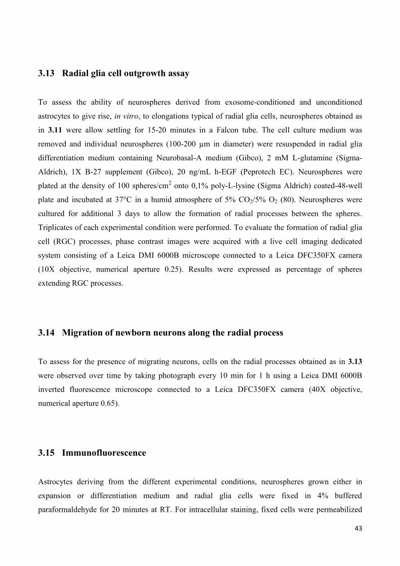
43
3.13 Radial glia cell outgrowth assay
To assess the ability of neurospheres derived from exosome-conditioned and unconditioned
astrocytes to give rise, in vitro, to elongations typical of radial glia cells, neurospheres obtained as
in 3.11 were allow settling for 15-20 minutes in a Falcon tube. The cell culture medium was
removed and individual neurospheres (100-200 µm in diameter) were resuspended in radial glia
differentiation medium containing Neurobasal-A medium (Gibco), 2 mM L-glutamine (Sigma-
Aldrich), 1X B-27 supplement (Gibco), 20 ng/mL h-EGF (Peprotech EC). Neurospheres were
plated at the density of 100 spheres/cm2 onto 0,1% poly-L-lysine (Sigma Aldrich) coated-48-well
plate and incubated at 37°C in a humid atmosphere of 5% CO2/5% O2 (80). Neurospheres were
cultured for additional 3 days to allow the formation of radial processes between the spheres.
Triplicates of each experimental condition were performed. To evaluate the formation of radial glia
cell (RGC) processes, phase contrast images were acquired with a live cell imaging dedicated
system consisting of a Leica DMI 6000B microscope connected to a Leica DFC350FX camera
(10X objective, numerical aperture 0.25). Results were expressed as percentage of spheres
extending RGC processes.
3.14 Migration of newborn neurons along the radial process
To assess for the presence of migrating neurons, cells on the radial processes obtained as in 3.13
were observed over time by taking photograph every 10 min for 1 h using a Leica DMI 6000B
inverted fluorescence microscope connected to a Leica DFC350FX camera (40X objective,
numerical aperture 0.65).
3.15 Immunofluorescence
Astrocytes deriving from the different experimental conditions, neurospheres grown either in
expansion or differentiation medium and radial glia cells were fixed in 4% buffered
paraformaldehyde for 20 minutes at RT. For intracellular staining, fixed cells were permeabilized
44
for 10 minutes at RT with 0,1% Triton X-100 (Sigma-Aldrich) before exposing them to primary
antibodies. Primary antibody incubation was performed over-night at 4°C to detect: CD133
(Milteny, 1:100), CD44 (NeoMarkers, 1:100), FABP-7 (Cell Signaling, 1:1000), GD3 (Abcam,
1:100), GFAP (Dako, 1:50), Ki67 (Leica, 1:100), MAP-2 (Millipore, 1:1000), N-Cadherin (Dako,
1:100), Nestin (Millipore, 1:100), NeuN (Millipore, 1:100), Numb (Cell Signaling, 1.100), O4
(Millipore, 1:100), Sox-2 (Millipore, 1:500), s100β (Abcam, 1:100), Tenascin C (Abcam, 1:10), β3-
tubulin (Tuj1, Abcam, 1:1000), Vimentin (Dako, 1:50). To detect primary antibodies, A488 and
A555 dyes labeled secondary antibodies, diluted 1:800, were employed (Molecular Probe,
Invitrogen). Finally, Vectashield (Vector) added with 1 μg/ml DAPI (Sigma-Aldrich) was used as
mounting medium. Epifluorescence images were obtained utilizing a live cell imaging dedicated
system consisting of a Leica DMI 6000B microscope connected to a Leica DFC350FX camera
(Leica Microsystems) and equipped with a 63X oil immersion objective (numerical aperture: 1.40),
a 40X oil immersion objective (numerical aperture: 1.25), a 20X objective (numerical aperture:
1.00) or a 10X objective (numerical aperture: 0.25) Adobe Photoshop software was utilized to
compose, overlay the images and to adjust contrast (Adobe).
3.16 Statistics
Characteristics of the study population are described using means ± s.d. or median and 25th
-75th
percentiles for continuous variables and percentages for categorical variables. Data were tested for
normal distribution using the Kolmogorov-Smirnov test. T-test or Mann-Whitney test, as
appropriate, was used to compare continuous variables between two groups. Effects of exosomes
and supernatant on different cell types were performed, as appropriate, by repeated measurements
one-way Anova followed by Bonferroni post-test (Prism, version 4.0c) or Kruskal-Wallis followed
by Dunn’s post-test. P values less than 0.05 were considered significant.
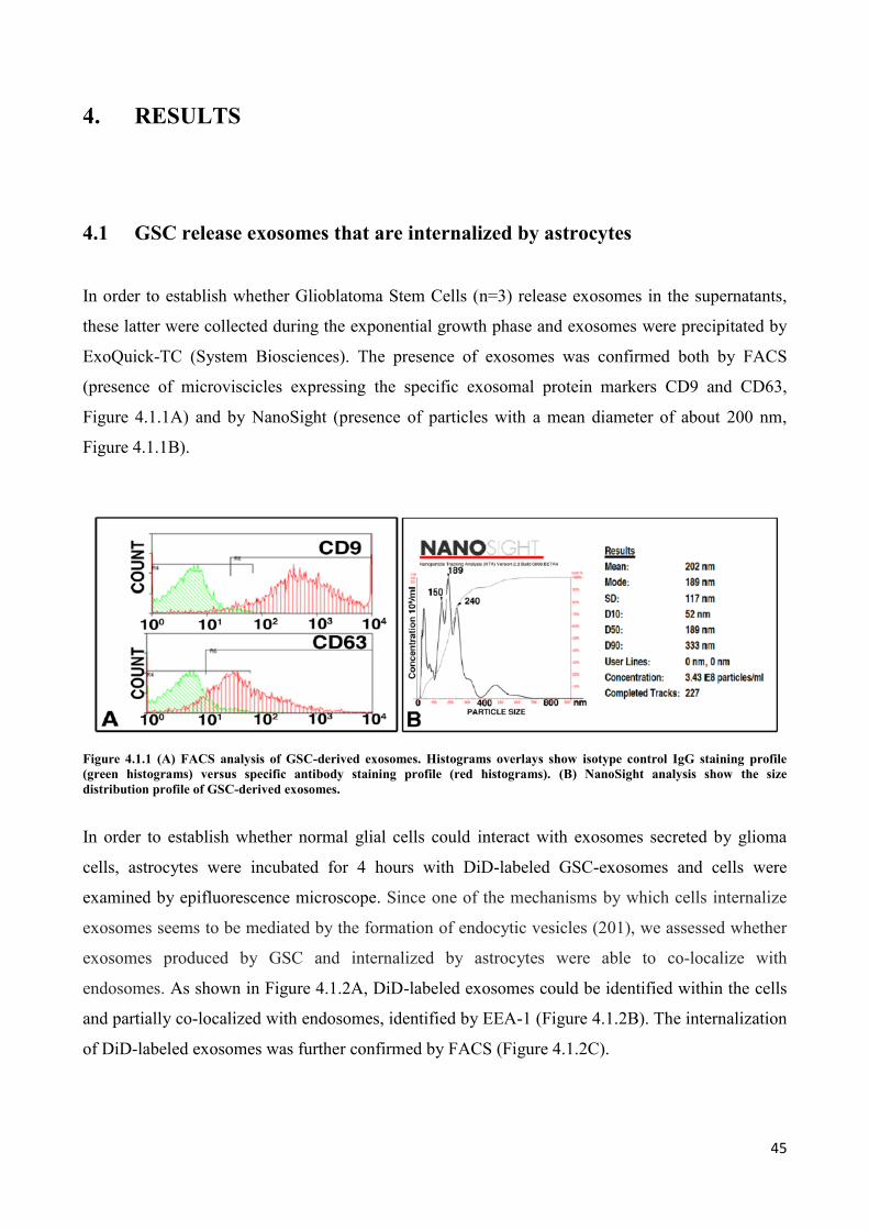
45
4. RESULTS
4.1 GSC release exosomes that are internalized by astrocytes
In order to establish whether Glioblatoma Stem Cells (n=3) release exosomes in the supernatants,
these latter were collected during the exponential growth phase and exosomes were precipitated by
ExoQuick-TC (System Biosciences). The presence of exosomes was confirmed both by FACS
(presence of microviscicles expressing the specific exosomal protein markers CD9 and CD63,
Figure 4.1.1A) and by NanoSight (presence of particles with a mean diameter of about 200 nm,
Figure 4.1.1B).
Figure 4.1.1 (A) FACS analysis of GSC-derived exosomes. Histograms overlays show isotype control IgG staining profile
(green histograms) versus specific antibody staining profile (red histograms). (B) NanoSight analysis show the size
distribution profile of GSC-derived exosomes.
In order to establish whether normal glial cells could interact with exosomes secreted by glioma
cells, astrocytes were incubated for 4 hours with DiD-labeled GSC-exosomes and cells were
examined by epifluorescence microscope. Since one of the mechanisms by which cells internalize
exosomes seems to be mediated by the formation of endocytic vesicles (201), we assessed whether
exosomes produced by GSC and internalized by astrocytes were able to co-localize with
endosomes. As shown in Figure 4.1.2A, DiD-labeled exosomes could be identified within the cells
and partially co-localized with endosomes, identified by EEA-1 (Figure 4.1.2B). The internalization
of DiD-labeled exosomes was further confirmed by FACS (Figure 4.1.2C).
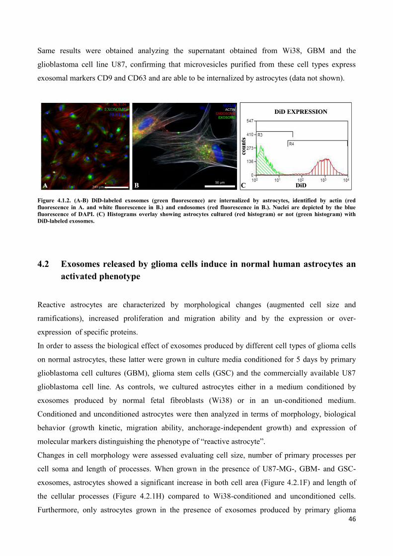
46
Same results were obtained analyzing the supernatant obtained from Wi38, GBM and the
glioblastoma cell line U87, confirming that microvesicles purified from these cell types express
exosomal markers CD9 and CD63 and are able to be internalized by astrocytes (data not shown).
Figure 4.1.2. (A-B) DiD-labeled exosomes (green fluorescence) are internalized by astrocytes, identified by actin (red
fluorescence in A. and white fluorescence in B.) and endosomes (red fluorescence in B.). Nuclei are depicted by the blue
fluorescence of DAPI. (C) Histograms overlay showing astrocytes cultured (red histogram) or not (green histogram) with
DiD-labeled exosomes.
4.2 Exosomes released by glioma cells induce in normal human astrocytes an
activated phenotype
Reactive astrocytes are characterized by morphological changes (augmented cell size and
ramifications), increased proliferation and migration ability and by the expression or over-
expression of specific proteins.
In order to assess the biological effect of exosomes produced by different cell types of glioma cells
on normal astrocytes, these latter were grown in culture media conditioned for 5 days by primary
glioblastoma cell cultures (GBM), glioma stem cells (GSC) and the commercially available U87
glioblastoma cell line. As controls, we cultured astrocytes either in a medium conditioned by
exosomes produced by normal fetal fibroblasts (Wi38) or in an un-conditioned medium.
Conditioned and unconditioned astrocytes were then analyzed in terms of morphology, biological
behavior (growth kinetic, migration ability, anchorage-independent growth) and expression of
molecular markers distinguishing the phenotype of “reactive astrocyte”.
Changes in cell morphology were assessed evaluating cell size, number of primary processes per
cell soma and length of processes. When grown in the presence of U87-MG-, GBM- and GSC-
exosomes, astrocytes showed a significant increase in both cell area (Figure 4.2.1F) and length of
the cellular processes (Figure 4.2.1H) compared to Wi38-conditioned and unconditioned cells.
Furthermore, only astrocytes grown in the presence of exosomes produced by primary glioma
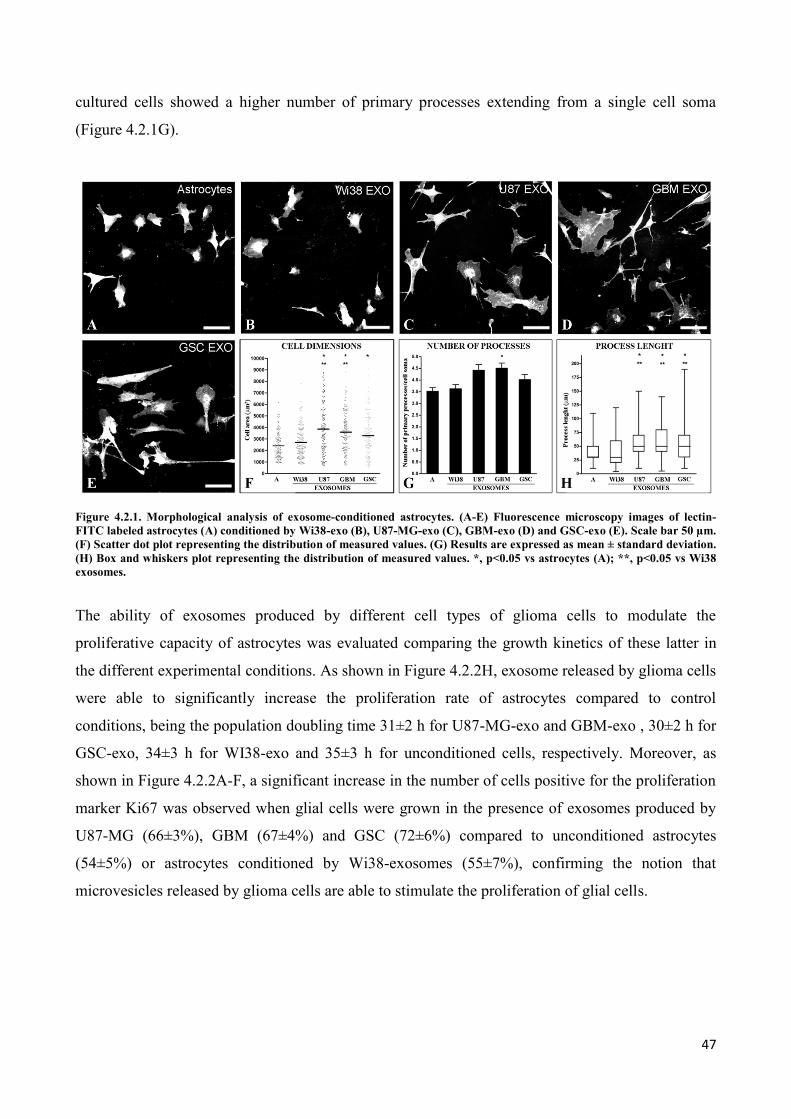
47
cultured cells showed a higher number of primary processes extending from a single cell soma
(Figure 4.2.1G).
Figure 4.2.1. Morphological analysis of exosome-conditioned astrocytes. (A-E) Fluorescence microscopy images of lectin-
FITC labeled astrocytes (A) conditioned by Wi38-exo (B), U87-MG-exo (C), GBM-exo (D) and GSC-exo (E). Scale bar 50 µm.
(F) Scatter dot plot representing the distribution of measured values. (G) Results are expressed as mean ± standard deviation.
(H) Box and whiskers plot representing the distribution of measured values. *, p<0.05 vs astrocytes (A); **, p<0.05 vs Wi38
exosomes.
The ability of exosomes produced by different cell types of glioma cells to modulate the
proliferative capacity of astrocytes was evaluated comparing the growth kinetics of these latter in
the different experimental conditions. As shown in Figure 4.2.2H, exosome released by glioma cells
were able to significantly increase the proliferation rate of astrocytes compared to control
conditions, being the population doubling time 31±2 h for U87-MG-exo and GBM-exo , 30±2 h for
GSC-exo, 34±3 h for WI38-exo and 35±3 h for unconditioned cells, respectively. Moreover, as
shown in Figure 4.2.2A-F, a significant increase in the number of cells positive for the proliferation
marker Ki67 was observed when glial cells were grown in the presence of exosomes produced by
U87-MG (66±3%), GBM (67±4%) and GSC (72±6%) compared to unconditioned astrocytes
(54±5%) or astrocytes conditioned by Wi38-exosomes (55±7%), confirming the notion that
microvesicles released by glioma cells are able to stimulate the proliferation of glial cells.
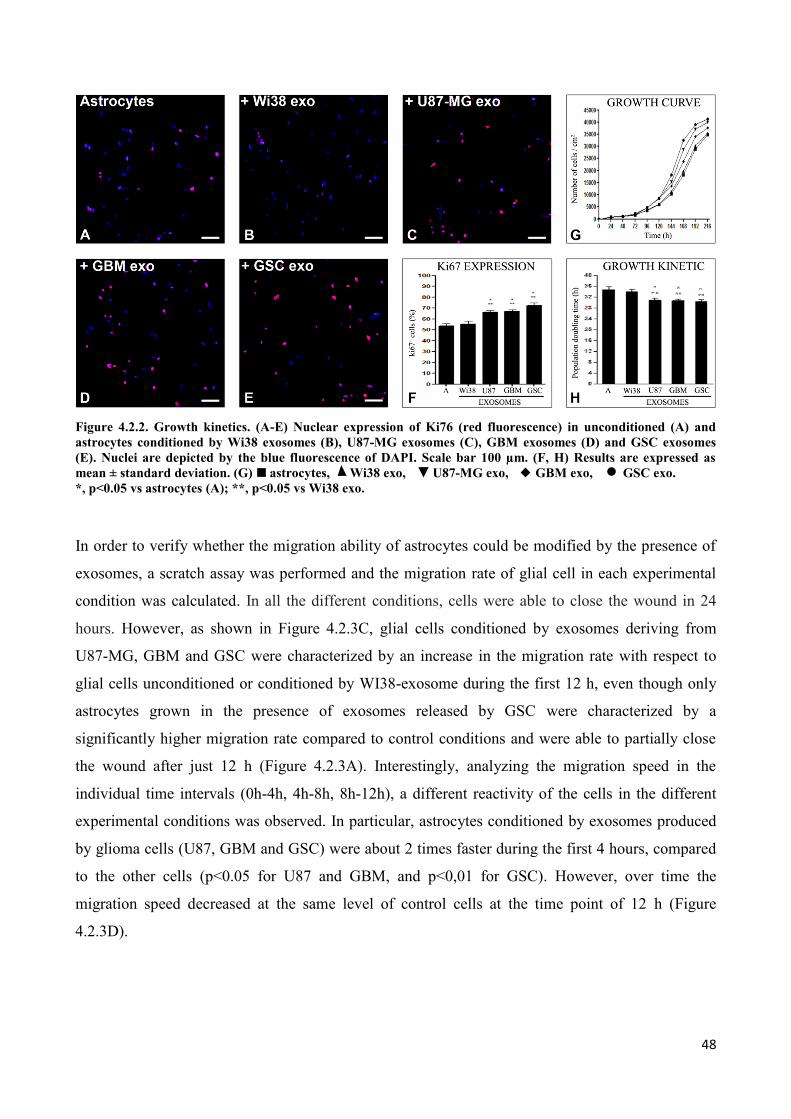
48
Figure 4.2.2. Growth kinetics. (A-E) Nuclear expression of Ki76 (red fluorescence) in unconditioned (A) and
astrocytes conditioned by Wi38 exosomes (B), U87-MG exosomes (C), GBM exosomes (D) and GSC exosomes
(E). Nuclei are depicted by the blue fluorescence of DAPI. Scale bar 100 µm. (F, H) Results are expressed as
mean ± standard deviation. (G) astrocytes, Wi38 exo, U87-MG exo, GBM exo, GSC exo.
*, p<0.05 vs astrocytes (A); **, p<0.05 vs Wi38 exo.
In order to verify whether the migration ability of astrocytes could be modified by the presence of
exosomes, a scratch assay was performed and the migration rate of glial cell in each experimental
condition was calculated. In all the different conditions, cells were able to close the wound in 24
hours. However, as shown in Figure 4.2.3C, glial cells conditioned by exosomes deriving from
U87-MG, GBM and GSC were characterized by an increase in the migration rate with respect to
glial cells unconditioned or conditioned by WI38-exosome during the first 12 h, even though only
astrocytes grown in the presence of exosomes released by GSC were characterized by a
significantly higher migration rate compared to control conditions and were able to partially close
the wound after just 12 h (Figure 4.2.3A). Interestingly, analyzing the migration speed in the
individual time intervals (0h-4h, 4h-8h, 8h-12h), a different reactivity of the cells in the different
experimental conditions was observed. In particular, astrocytes conditioned by exosomes produced
by glioma cells (U87, GBM and GSC) were about 2 times faster during the first 4 hours, compared
to the other cells (p<0.05 for U87 and GBM, and p<0,01 for GSC). However, over time the
migration speed decreased at the same level of control cells at the time point of 12 h (Figure
4.2.3D).
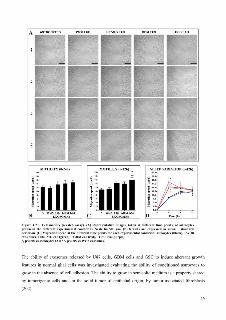
49
Figure 4.2.3. Cell motility (scratch assay). (A) Representative images, taken at different time points, of astrocytes
grown in the different experimental conditions. Scale ba 500 µm. (B) Results are expressed as mean ± standard
deviation. (C) Migration speed at the different time points for each experimental condition: astrocytes (black), +Wi38
exo (blue), +U87-MG exo (green), +GBM exo (red), +GSC exo (purple).
*, p<0.05 vs astrocytes (A); **, p<0.05 vs Wi38 exosomes.
The ability of exosomes released by U87 cells, GBM cells and GSC to induce aberrant growth
features in normal glial cells was investigated evaluating the ability of conditioned astrocytes to
grow in the absence of cell adhesion. The ability to grow in semisolid medium is a property shared
by tumorigenic cells and, in the solid tumor of epithelial origin, by tumor-associated fibroblasts
(202).
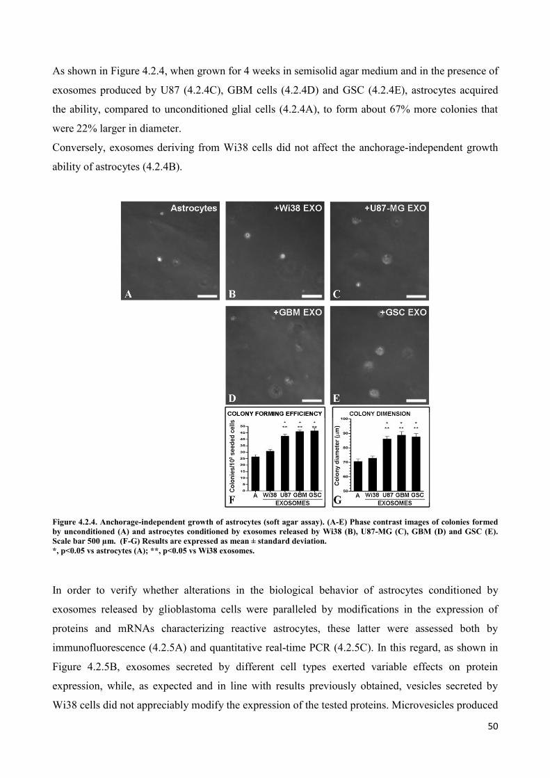
50
As shown in Figure 4.2.4, when grown for 4 weeks in semisolid agar medium and in the presence of
exosomes produced by U87 (4.2.4C), GBM cells (4.2.4D) and GSC (4.2.4E), astrocytes acquired
the ability, compared to unconditioned glial cells (4.2.4A), to form about 67% more colonies that
were 22% larger in diameter.
Conversely, exosomes deriving from Wi38 cells did not affect the anchorage-independent growth
ability of astrocytes (4.2.4B).
Figure 4.2.4. Anchorage-independent growth of astrocytes (soft agar assay). (A-E) Phase contrast images of colonies formed
by unconditioned (A) and astrocytes conditioned by exosomes released by Wi38 (B), U87-MG (C), GBM (D) and GSC (E).
Scale bar 500 µm. (F-G) Results are expressed as mean ± standard deviation.
*, p<0.05 vs astrocytes (A); **, p<0.05 vs Wi38 exosomes.
In order to verify whether alterations in the biological behavior of astrocytes conditioned by
exosomes released by glioblastoma cells were paralleled by modifications in the expression of
proteins and mRNAs characterizing reactive astrocytes, these latter were assessed both by
immunofluorescence (4.2.5A) and quantitative real-time PCR (4.2.5C). In this regard, as shown in
Figure 4.2.5B, exosomes secreted by different cell types exerted variable effects on protein
expression, while, as expected and in line with results previously obtained, vesicles secreted by
Wi38 cells did not appreciably modify the expression of the tested proteins. Microvesicles produced
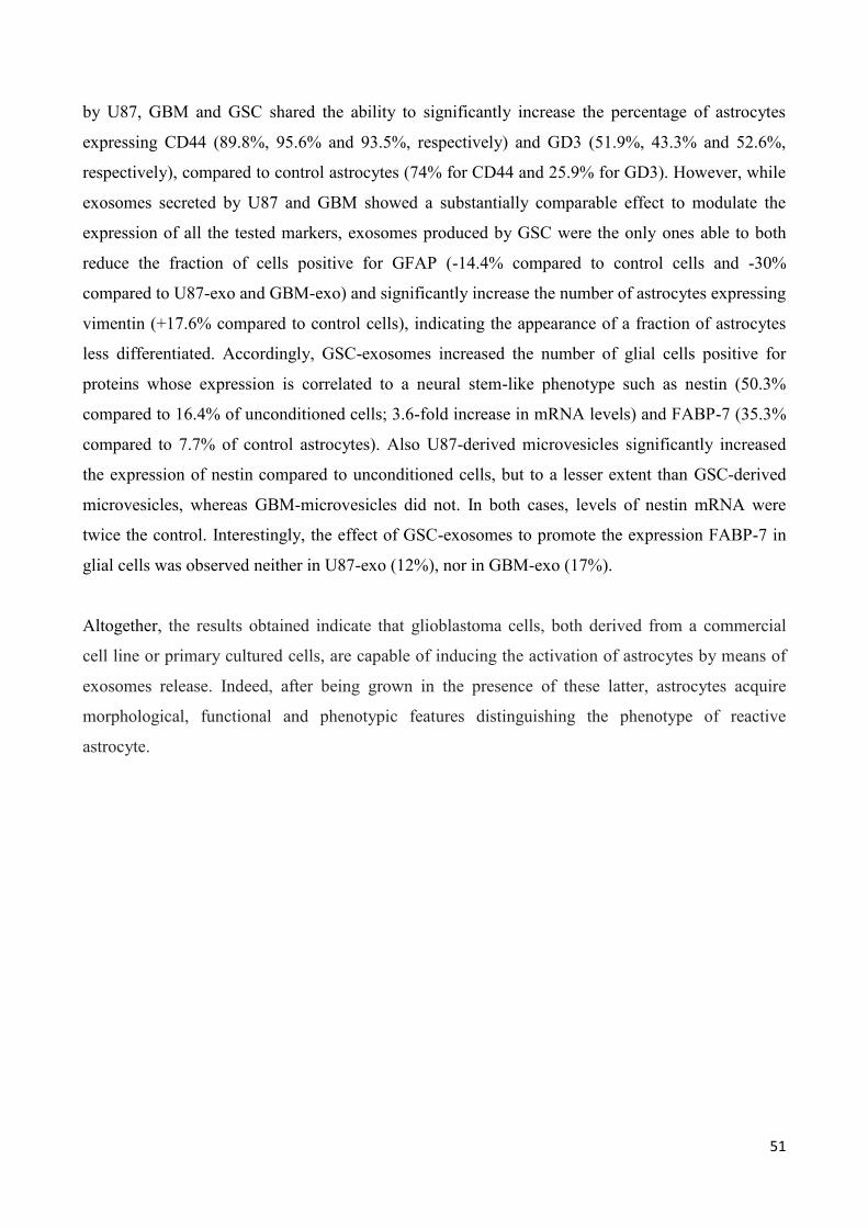
51
by U87, GBM and GSC shared the ability to significantly increase the percentage of astrocytes
expressing CD44 (89.8%, 95.6% and 93.5%, respectively) and GD3 (51.9%, 43.3% and 52.6%,
respectively), compared to control astrocytes (74% for CD44 and 25.9% for GD3). However, while
exosomes secreted by U87 and GBM showed a substantially comparable effect to modulate the
expression of all the tested markers, exosomes produced by GSC were the only ones able to both
reduce the fraction of cells positive for GFAP (-14.4% compared to control cells and -30%
compared to U87-exo and GBM-exo) and significantly increase the number of astrocytes expressing
vimentin (+17.6% compared to control cells), indicating the appearance of a fraction of astrocytes
less differentiated. Accordingly, GSC-exosomes increased the number of glial cells positive for
proteins whose expression is correlated to a neural stem-like phenotype such as nestin (50.3%
compared to 16.4% of unconditioned cells; 3.6-fold increase in mRNA levels) and FABP-7 (35.3%
compared to 7.7% of control astrocytes). Also U87-derived microvesicles significantly increased
the expression of nestin compared to unconditioned cells, but to a lesser extent than GSC-derived
microvesicles, whereas GBM-microvesicles did not. In both cases, levels of nestin mRNA were
twice the control. Interestingly, the effect of GSC-exosomes to promote the expression FABP-7 in
glial cells was observed neither in U87-exo (12%), nor in GBM-exo (17%).
Altogether, the results obtained indicate that glioblastoma cells, both derived from a commercial
cell line or primary cultured cells, are capable of inducing the activation of astrocytes by means of
exosomes release. Indeed, after being grown in the presence of these latter, astrocytes acquire
morphological, functional and phenotypic features distinguishing the phenotype of reactive
astrocyte.
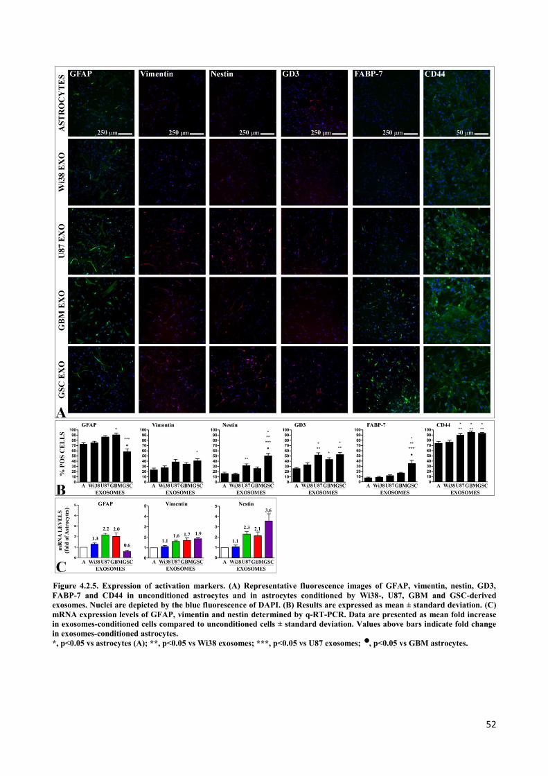
52
Figure 4.2.5. Expression of activation markers. (A) Representative fluorescence images of GFAP, vimentin, nestin, GD3,
FABP-7 and CD44 in unconditioned astrocytes and in astrocytes conditioned by Wi38-, U87, GBM and GSC-derived
exosomes. Nuclei are depicted by the blue fluorescence of DAPI. (B) Results are expressed as mean ± standard deviation. (C)
mRNA expression levels of GFAP, vimentin and nestin determined by q-RT-PCR. Data are presented as mean fold increase
in exosomes-conditioned cells compared to unconditioned cells ± standard deviation. Values above bars indicate fold change
in exosomes-conditioned astrocytes.
*, p<0.05 vs astrocytes (A); **, p<0.05 vs Wi38 exosomes; ***, p<0.05 vs U87 exosomes; , p<0.05 vs GBM astrocytes.
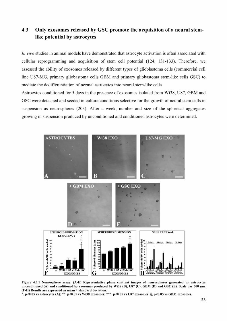
53
4.3 Only exosomes released by GSC promote the acquisition of a neural stem-
like potential by astrocytes
In vivo studies in animal models have demonstrated that astrocyte activation is often associated with
cellular reprogramming and acquisition of stem cell potential (124, 131-133). Therefore, we
assessed the ability of exosomes released by different types of glioblastoma cells (commercial cell
line U87-MG, primary gliobastoma cells GBM and primary gliobastoma stem-like cells GSC) to
mediate the dedifferentiation of normal astrocytes into neural stem-like cells.
Astrocytes conditioned for 5 days in the presence of exosomes isolated from Wi38, U87, GBM and
GSC were detached and seeded in culture conditions selective for the growth of neural stem cells in
suspension as neurospheres (203). After a week, number and size of the spherical aggregates
growing in suspension produced by unconditioned and conditioned astrocytes were determined.
Figure 4.3.1 Neurosphere assay. (A-E) Representative phase contrast images of neurospheres generated by astrocytes
unconditioned (A) and conditioned by exosomes produced by Wi38 (B), U87 (C), GBM (D) and GSC (E). Scale bar 500 µm.
(F-H) Results are expressed as mean ± standard deviation.
*, p<0.05 vs astrocytes (A); **, p<0.05 vs Wi38 exosomes; ***, p<0.05 vs U87 exosomes; §, p<0.05 vs GBM exosomes.
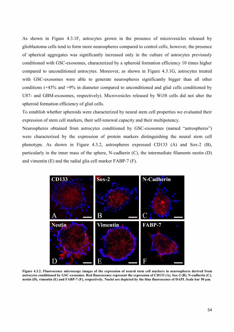
54
As shown in Figure 4.3.1F, astrocytes grown in the presence of microvesicles released by
glioblastoma cells tend to form more neurospheres compared to control cells, however, the presence
of spherical aggregates was significantly increased only in the culture of astrocytes previously
conditioned with GSC-exosomes, characterized by a spheroid formation efficiency 10 times higher
compared to unconditioned astrocytes. Moreover, as shown in Figure 4.3.1G, astrocytes treated
with GSC-exosomes were able to generate neurospheres significantly bigger than all other
conditions (+43% and +9% in diameter compared to unconditioned and glial cells conditioned by
U87- and GBM-exosomes, respectively). Microvesicles released by Wi38 cells did not alter the
spheroid formation efficiency of glial cells.
To establish whether spheroids were characterized by neural stem cell properties we evaluated their
expression of stem cell markers, their self-renewal capacity and their multipotency.
Neurospheres obtained from astrocytes conditioned by GSC-exosomes (named “astrospheres”)
were characterized by the expression of protein markers distinguishing the neural stem cell
phenotype. As shown in Figure 4.3.2, astrospheres expressed CD133 (A) and Sox-2 (B),
particularly in the inner mass of the sphere, N-cadherin (C), the intermediate filaments nestin (D)
and vimentin (E) and the radial glia cell marker FABP-7 (F).
Figure 4.3.2. Fluorescence microscopy images of the expression of neural stem cell markers in neurospheres derived from
astrocytes conditioned by GSC-exosomes. Red fluorescence represent the expression of CD133 (A), Sox-2 (B), N-cadherin (C),
nestin (D), vimentin (E) and FABP-7 (F), respectively. Nuclei are depicted by the blue fluorescence of DAPI. Scale bar 50 µm.
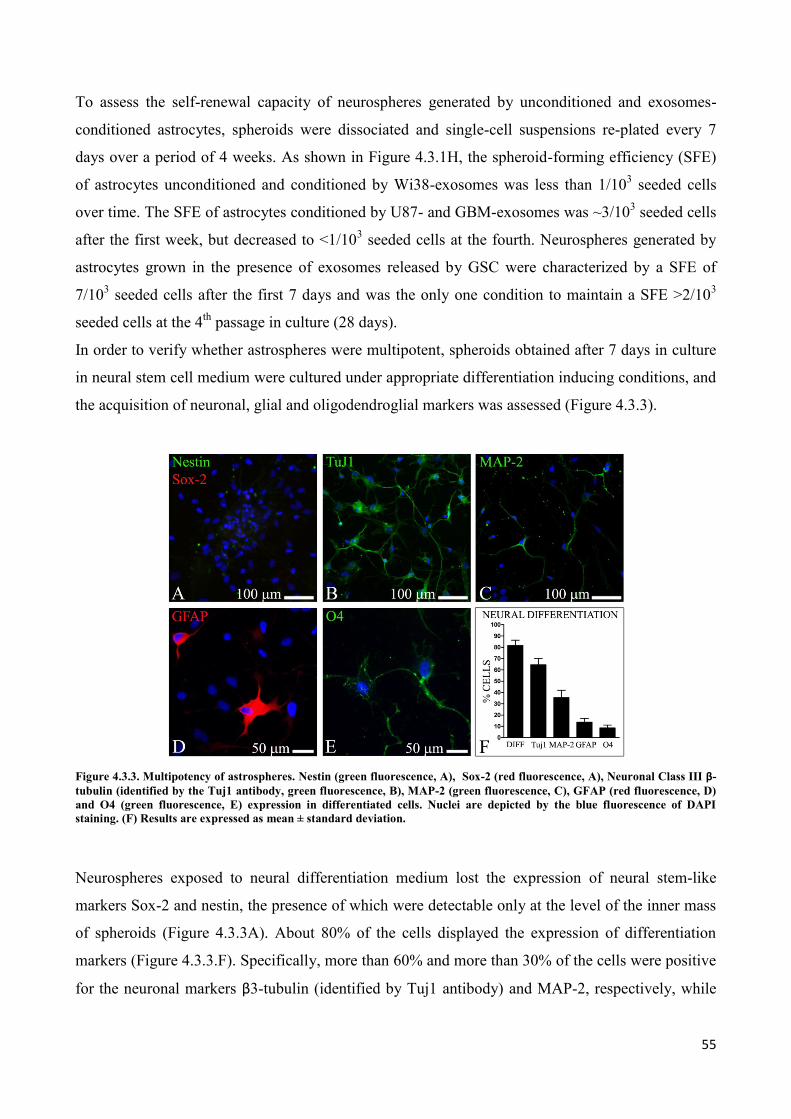
55
To assess the self-renewal capacity of neurospheres generated by unconditioned and exosomes-
conditioned astrocytes, spheroids were dissociated and single-cell suspensions re-plated every 7
days over a period of 4 weeks. As shown in Figure 4.3.1H, the spheroid-forming efficiency (SFE)
of astrocytes unconditioned and conditioned by Wi38-exosomes was less than 1/103 seeded cells
over time. The SFE of astrocytes conditioned by U87- and GBM-exosomes was ~3/103 seeded cells
after the first week, but decreased to <1/103 seeded cells at the fourth. Neurospheres generated by
astrocytes grown in the presence of exosomes released by GSC were characterized by a SFE of
7/103 seeded cells after the first 7 days and was the only one condition to maintain a SFE >2/10
3
seeded cells at the 4th
passage in culture (28 days).
In order to verify whether astrospheres were multipotent, spheroids obtained after 7 days in culture
in neural stem cell medium were cultured under appropriate differentiation inducing conditions, and
the acquisition of neuronal, glial and oligodendroglial markers was assessed (Figure 4.3.3).
Figure 4.3.3. Multipotency of astrospheres. Nestin (green fluorescence, A), Sox-2 (red fluorescence, A), Neuronal Class III β-
tubulin (identified by the Tuj1 antibody, green fluorescence, B), MAP-2 (green fluorescence, C), GFAP (red fluorescence, D)
and O4 (green fluorescence, E) expression in differentiated cells. Nuclei are depicted by the blue fluorescence of DAPI
staining. (F) Results are expressed as mean ± standard deviation.
Neurospheres exposed to neural differentiation medium lost the expression of neural stem-like
markers Sox-2 and nestin, the presence of which were detectable only at the level of the inner mass
of spheroids (Figure 4.3.3A). About 80% of the cells displayed the expression of differentiation
markers (Figure 4.3.3.F). Specifically, more than 60% and more than 30% of the cells were positive
for the neuronal markers β3-tubulin (identified by Tuj1 antibody) and MAP-2, respectively, while
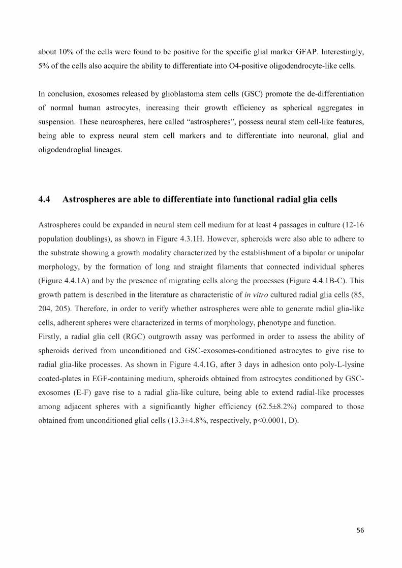
56
about 10% of the cells were found to be positive for the specific glial marker GFAP. Interestingly,
5% of the cells also acquire the ability to differentiate into O4-positive oligodendrocyte-like cells.
In conclusion, exosomes released by glioblastoma stem cells (GSC) promote the de-differentiation
of normal human astrocytes, increasing their growth efficiency as spherical aggregates in
suspension. These neurospheres, here called “astrospheres”, possess neural stem cell-like features,
being able to express neural stem cell markers and to differentiate into neuronal, glial and
oligodendroglial lineages.
4.4 Astrospheres are able to differentiate into functional radial glia cells
Astrospheres could be expanded in neural stem cell medium for at least 4 passages in culture (12-16
population doublings), as shown in Figure 4.3.1H. However, spheroids were also able to adhere to
the substrate showing a growth modality characterized by the establishment of a bipolar or unipolar
morphology, by the formation of long and straight filaments that connected individual spheres
(Figure 4.4.1A) and by the presence of migrating cells along the processes (Figure 4.4.1B-C). This
growth pattern is described in the literature as characteristic of in vitro cultured radial glia cells (85,
204, 205). Therefore, in order to verify whether astrospheres were able to generate radial glia-like
cells, adherent spheres were characterized in terms of morphology, phenotype and function.
Firstly, a radial glia cell (RGC) outgrowth assay was performed in order to assess the ability of
spheroids derived from unconditioned and GSC-exosomes-conditioned astrocytes to give rise to
radial glia-like processes. As shown in Figure 4.4.1G, after 3 days in adhesion onto poly-L-lysine
coated-plates in EGF-containing medium, spheroids obtained from astrocytes conditioned by GSC-
exosomes (E-F) gave rise to a radial glia-like culture, being able to extend radial-like processes
among adjacent spheres with a significantly higher efficiency (62.5±8.2%) compared to those
obtained from unconditioned glial cells (13.3±4.8%, respectively, p<0.0001, D).
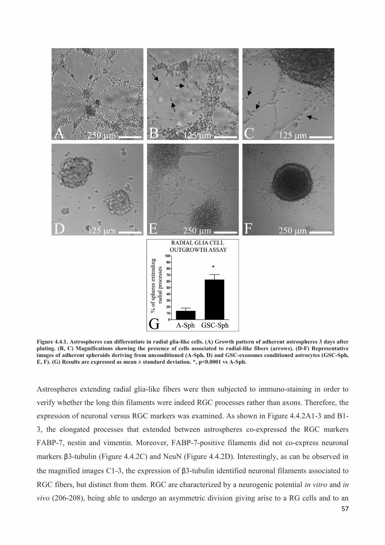
57
Figure 4.4.1. Astrospheres can differentiate in radial glia-like cells. (A) Growth pattern of adherent astrospheres 3 days after
plating. (B, C) Magnifications showing the presence of cells associated to radial-like fibers (arrows). (D-F) Representative
images of adherent spheroids deriving from unconditioned (A-Sph, D) and GSC-exosomes conditioned astrocytes (GSC-Sph,
E, F). (G) Results are expressed as mean ± standard deviation. *, p<0.0001 vs A-Sph.
Astrospheres extending radial glia-like fibers were then subjected to immuno-staining in order to
verify whether the long thin filaments were indeed RGC processes rather than axons. Therefore, the
expression of neuronal versus RGC markers was examined. As shown in Figure 4.4.2A1-3 and B1-
3, the elongated processes that extended between astrospheres co-expressed the RGC markers
FABP-7, nestin and vimentin. Moreover, FABP-7-positive filaments did not co-express neuronal
markers β3-tubulin (Figure 4.4.2C) and NeuN (Figure 4.4.2D). Interestingly, as can be observed in
the magnified images C1-3, the expression of β3-tubulin identified neuronal filaments associated to
RGC fibers, but distinct from them. RGC are characterized by a neurogenic potential in vitro and in
vivo (206-208), being able to undergo an asymmetric division giving arise to a RG cells and to an
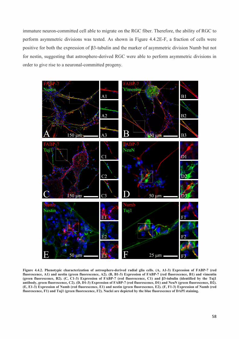
58
immature neuron-committed cell able to migrate on the RGC fiber. Therefore, the ability of RGC to
perform asymmetric divisions was tested. As shown in Figure 4.4.2E-F, a fraction of cells were
positive for both the expression of β3-tubulin and the marker of asymmetric division Numb but not
for nestin, suggesting that astrosphere-derived RGC were able to perform asymmetric divisions in
order to give rise to a neuronal-committed progeny.
Figure 4.4.2. Phenotypic characterization of astrosphere-derived radial glia cells. (A, A1-3) Expression of FABP-7 (red
fluorescence, A1) and nestin (green fluorescence, A2). (B, B1-3) Expression of FABP-7 (red fluorescence, B1) and vimentin
(green fluorescence, B2). (C, C1-3) Expression of FABP-7 (red fluorescence, C1) and β3-tubulin (identified by the Tuj1
antibody, green fluorescence, C2). (D, D1-3) Expression of FABP-7 (red fluorescence, D1) and NeuN (green fluorescence, D2).
(E, E1-3) Expression of Numb (red fluorescence, E1) and nestin (green fluorescence, E2). (F, F1-3) Expression of Numb (red
fluorescence, F1) and Tuj1 (green fluorescence, F2). Nuclei are depicted by the blue fluorescence of DAPI staining.
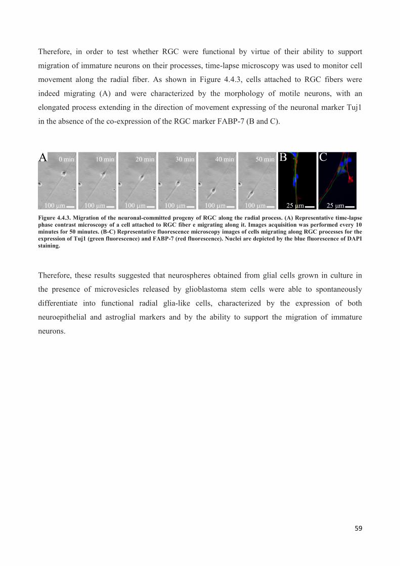
59
Therefore, in order to test whether RGC were functional by virtue of their ability to support
migration of immature neurons on their processes, time-lapse microscopy was used to monitor cell
movement along the radial fiber. As shown in Figure 4.4.3, cells attached to RGC fibers were
indeed migrating (A) and were characterized by the morphology of motile neurons, with an
elongated process extending in the direction of movement expressing of the neuronal marker Tuj1
in the absence of the co-expression of the RGC marker FABP-7 (B and C).
Figure 4.4.3. Migration of the neuronal-committed progeny of RGC along the radial process. (A) Representative time-lapse
phase contrast microscopy of a cell attached to RGC fiber e migrating along it. Images acquisition was performed every 10
minutes for 50 minutes. (B-C) Representative fluorescence microscopy images of cells migrating along RGC processes for the
expression of Tuj1 (green fluorescence) and FABP-7 (red fluorescence). Nuclei are depicted by the blue fluorescence of DAPI
staining.
Therefore, these results suggested that neurospheres obtained from glial cells grown in culture in
the presence of microvesicles released by glioblastoma stem cells were able to spontaneously
differentiate into functional radial glia-like cells, characterized by the expression of both
neuroepithelial and astroglial markers and by the ability to support the migration of immature
neurons.
60
5. DISCUSSION
The importance of the microenvironment in tumor progression has been extensively reported in the
last few years and the active role of microenvironment in supporting tumorigenesis, invasion and
metastasis is widely recognized.
The tumor microenvironment in the brain consist of many different cell types that involve brain-
resident and brain-infiltrating cells. In this context, endothelial cells that constitute the
microvascular blood vessels, microglia-macrophages, and astrocytes have received the most
attention so far. One of the current challenges in understanding the role of the tumor
microenvironment is poor definition of different cell populations. For example, new phenotypes or
subtypes of myeloid cells have been described in recent years, and further cellular subtypes are
expected to emerge in the future. As for astrocytes, has been shown that they differ in various brain
locations and their phenotype changes upon the exposure of different stimuli.
Astrocytes are glial cells that become activated in response to different central nervous system
(CNS) injuries (124, 209-211). Astrocyte activation is known as reactive gliosis and is characterized
by cellular hypertrophy and changes in gene expression patterns, including the up-regulation of the
Glial Fibrillary Acidic Protein (GFAP) and the acquisition of stem-like potential (129, 133, 212).
After a CNS insult, reactive astrocytes form a scar tissue surrounding the damaged area and this has
been shown to be essential for the local containment of inflammation and for the repair of the
demarcated tissue (212). Reactive astrocytes have been frequently observed in the vicinity of
primary and metastatic brain tumors in animal model, as well in human patients (213, 214). In vitro
studies demonstrated that a variety of factors released by astrocytes can support the growth of
primary and metastatic brain tumor cells, including the secretion of substances that promote cancer
cell proliferation and invasion, suppression of adaptive immune responses through the repression of
microglia activation and induction of T-cell apoptosis, and protection of cancer cells from apoptosis
through direct cell-cell interactions (215-218). However, little is known about molecular
mechanisms responsible for astrocyte activation, in particular in the context of brain tumors, and the
complex role of microenvironment in the glioma progression is still poor understood, as well as the
interactions occurring between cancer cells and stromal cells in the brain.
A continuous cross-talk between glioma cells and stroma is required for the development of a
permissive environment, able to promote the effective tumor growth and dissemination. Emerging
evidence suggest that extracellular microvesicles released by cancer cells may play an important
and active role in local and systemic cell-cell communication in cancer (219). In particular, a lot of
61
interest has been directed to exosomes (166). Exosomes are small membrane vesicles of endocytic
origin secreted by most cell types. They can contain a large number of biologically active
molecules, such as proteins, mRNAs, microRNAs and DNA fragments, and are shuttled from a
donor cell to a recipient cell (162, 163). Cancer-derived exosomes may contribute to the recruitment
and reprogramming of the tumor microenvironment to form a pro-tumorigenic and permissive soil
able to stimulate tumor growth and dissemination and by eliciting an anti-tumor response. Several
studies in the last decade demonstrated that exosomes can mediate the transfer of oncogenic
proteins between cancer cells, modulate the tumor microenvironment by stromal cells activation,
extracellular matrix remodeling and angiogenesis stimulation, and the immune system response
against tumor antigens (168-170, 173). Recently, our group has identified a novel class of
mesenchymal stromal cell in human gliomas. This cells, although devoid genetic alterations of the
tumor of origin, are characterized by stem cell properties, aberrant growth features, a gene
expression profile typical of tumor-supporting cells and are able to enhance in vitro the
aggressiveness of glioblastoma initiating stem cells by means of exosome release, thus supporting
the notion that microvesicles released by stromal activated cells can have an active role in tumor
progression (123).
Therefore, since the mechanisms that mediate glial cells activation in response to the presence of a
tumor mass have not yet been characterized, we decided to verify if the release of microvesicles by
glioma cells could represent a key factor able to promote astrocyte activation. In particular, in order
to take into account the heterogeneity of tumor cells and to evaluate whether different glioma cell
models could induce different outcomes, we compare the effects produced on glial cells by
exosomes obtained from a commercial cell line of gliobastoma (U87-MG) and from primary
cultured glioblastoma cells (GBM) and glioblastoma stem cells (GSC).
Numerous experimental evidences suggest that microvesicles released by glioblastoma cells are
able to support tumor growth by means of angiogenesis stimulation and the formation of a hypoxic
microenvironment (163, 220, 221). However, studies regarding microvesicles released by
glioblastoma stem cells are not yet been reported. Therefore, we first demonstrated that the latters
are able to secrete exosomes. In analogy to what reported in literature, exosomes purified from GSC
tend to be larger than those produced by non-cancer cells (222). In fact, vesicles purified from
culture supernatants of GSC possess an average size of about 190 nm in diameter and expressed
tetraspanins CD9 and CD63, commonly recognized exosomal markers. Furthermore, GSC-
exosomes can be internalized by normal human astrocytes and internalized vesicles partially co-
localize with endosomes, thus suggesting an uptake modality mediated by the formation of
endocytic vesicles.
62
Astrocytes grown in the presence of exosomes purified from different cell types of glioma cells
assume morphological and phenotypical characteristics of reactive astrocytes. In fact, only after 5
days in conditioned culture medium, glial cells are characterized by an increased cell size and
number and dimension of cellular processes. Furthermore, exosome-conditioned astrocytes showed
an increased proliferation and migration rate and acquired the ability to form colonies in semisolid
culture medium, an aberrant growth feature shared by transformed and tumor-associated cells (202).
These alterations of the biological properties of glial cells were also associated with modifications
of astrocyte phenotype. Exosome-conditioned astrocytes were characterized by an increased
expression of GFAP, vimentin, GD3 and CD44. These results are in agreement with what has been
observed about the reactive astrocyte phenotype (209, 212, 223, 224). As demonstrated by Katz,
CD44 and tenascin-C are expressed in glioblastoma tissues at a higher level in the perivascular
astrocytes when compared to peritumoral astrocytes, suggesting subtle differences between the two
populations of non-neoplastic tumor-associated cells (225). Interestingly, exosomes purified from
different types of glioblastoma cells produced different effects on glial cells. Indeed, while
microvesicles obtained from U87 and GBM cells showed a very similar behavior in stimulating an
activated phenotype of astrocytes, exosomes derived from GSC were the only ones able to decrease
the expression of GFAP and, in the same time, increase nestin and FABP- 7 levels in glial cells, two
proteins that characterize neural stem cell and radial glia phenotypes, respectively (226, 227).
In the adult cerebral cortex mature astrocytes lack expression of nestin and vimentin, decrease
GFAP levels and do not proliferate. However, astrocytes exposed to injury return to express these
proteins, proliferate and may resume properties of glia present at earlier developmental stages (127,
228, 229). Therefore, we decided to verify whether exosomes secreted by glioblastoma cells were
able to mediate de-differentiation effects on glial cells. Among all the experimental conditions
tested, only exosomes produced by GSC were able to significantly increase the propensity of
astrocytes to grow as spheroids in neural stem cell medium. Neurospheres obtained possessed self-
renewal ability, expressed neural stem cell markers CD133, Sox-2, nestin, vimentin and N-cadherin
and could be induced to differentiate in vitro in neurons, astrocytes and oligodendrocytes. These
results are very important because: 1. provide a mechanism by which glioblastoma cells can
modulate the behavior of glial cells by inducing a “reactive-astrocyte-like” phenotype through the
release of microvesicles, and 2. represent the first experimental evidence of the fact that exosomes
can act as mediators of cellular de-differentiation processes. Yang et al. has recently demonstrated
that β-catenin signaling could be the molecular event that initiate astrocyte activation, and that the
dysregulation of the β-catenin signaling pathway could be responsible for the transformation of
normal glial cells and the formation of an astrocytoma (230). However, leading events that induce
63
astrocyte activation in brain tumors still remain unclear, as well as relationships existing between
activated stromal cells and tumor cells in gliomas.
Furthermore, neurospheres obtained by GSC-exosomes-conditioned astrocytes (which we called
“astrospheres”), once let adhere, showed a greater ability to give rise to cultures of functional radial
glia cells (RGC) compared to those obtained from normal unconditioned astrocytes. RGC were
characterized by the presence of long and straight filaments extending between adjacent spheres and
by the expression of FABP-7, nestin and vimentin, and were able to support the migration of
immature newborn neuronal-committed cells along the radial process. In fact, these migrating cells
expressed neuronal markers in the absence of the expression of radial glia markers. During brain
development, FABP-7 is expressed in radial glial cells, where it plays a role in the establishment
and maintenance of the radial glial fiber system that guides immature migrating neurons to their
final destination (82, 231). Radial glial cells give rise to GFAP-expressing astrocytes once neuronal
migration is completed and can also display properties of precursor cells, generating both neurons
and glial cells in vitro (232). Moreover, a role as neural progenitors or neural stem cell has been
proposed for radial glial cells (233). Interestingly, the radial glia marker FABP-7 is expressed by a
subset of malignant glioma cell lines established from high-grade astrocytomas, and it’s associated
with an increased cellular motility and invasive ability. In human glioma tissues, FABP-7
expression is correlated with patient’s prognosis (83, 234). However, although the expression
markers of radial glia markers in gliomas has so far been associated only to tumor cells, the results
obtained in this work show that also glial cells could re-express neural stem cells- or radial glial
cell-features when they grow in the presence of exosomes released by GSC.
In conclusion, in this work we propose a mechanism for astrocyte activation in the context of brain
tumors based on the release of microvesicles by glioblastoma cells. In addition, we demonstrate that
exosomes secreted by glioblastoma initiating stem cells possess a potent effect on the acquisition of
stem-like features by glial cells, which can also spontaneously differentiate in functional radial glia,
a particular phenotype characteristic of the earlier developmental stages, whose expression is
correlated with a migratory and invasive phenotype of glioma cells. Taken together, these results
suggest that microvesicles released by glioblastoma stem cells possess a molecular content able to
profoundly modify the homeostasis of brain tissue inducing cell reprogramming processes and
contributing to anaplasia and heterogeneity of the neoplastic tissue. Studying the molecular cargo of
exosomes could be important to understand the tumor biology, by the identification of molecular
factors which may possess a key role in activating stromal cells toward a tumor-supporting
phenotype.
64
6. REFERENCES
1. Theele DP & Streit WJ (1993) A chronicle of microglial ontogeny. Glia 7(1):5-8.
2. Lewis PD (1968) The fate of the subependymal cell in the adult rat brain, with a note on the
origin of microglia. Brain : a journal of neurology 91(4):721-736.
3. Neuhaus J & Fedoroff S (1994) Development of microglia in mouse neopallial cell cultures.
Glia 11(1):11-17.
4. Perry VH & Gordon S (1988) Macrophages and microglia in the nervous system. Trends in
neurosciences 11(6):273-277.
5. Ling EA & Wong WC (1993) The origin and nature of ramified and amoeboid microglia: a
historical review and current concepts. Glia 7(1):9-18.
6. Eglitis MA & Mezey E (1997) Hematopoietic cells differentiate into both microglia and
macroglia in the brains of adult mice. Proceedings of the National Academy of Sciences of
the United States of America 94(8):4080-4085.
7. Foran DR & Peterson AC (1992) Myelin acquisition in the central nervous system of the
mouse revealed by an MBP-Lac Z transgene. The Journal of neuroscience : the official
journal of the Society for Neuroscience 12(12):4890-4897.
8. Del Rio-Hortega P (2012) Studies on neuroglia: Glia with very few processes
(oligodendroglia) by PA-o del RA-o-Hortega. 1921. Clinical neuropathology 31(6):440-
459.
9. Dawson MR, Levine JM, & Reynolds R (2000) NG2-expressing cells in the central nervous
system: are they oligodendroglial progenitors? Journal of neuroscience research 61(5):471-
479.
10. Bradl M & Lassmann H (2010) Oligodendrocytes: biology and pathology. Acta
neuropathologica 119(1):37-53.
11. Baumann N & Pham-Dinh D (2001) Biology of oligodendrocyte and myelin in the
mammalian central nervous system. Physiological reviews 81(2):871-927.
12. Lunn KF, Baas PW, & Duncan ID (1997) Microtubule organization and stability in the
oligodendrocyte. The Journal of neuroscience : the official journal of the Society for
Neuroscience 17(13):4921-4932.
13. Roussel G, Delaunoy JP, Nussbaum JL, & Mandel P (1979) Demonstration of a specific
localization of carbonic anhydrase C in the glial cells of rat CNS by an
immunohistochemical method. Brain research 160(1):47-55.
14. Kaur C, Hao AJ, Wu CH, & Ling EA (2001) Origin of microglia. Microscopy research and
technique 54(1):2-9.
15. Sugita Y, Tokunaga O, Terasaki M, Morimatsu M, & Shigemori M (2003) Epithelial
differentiation in medulloblastoma: comparison with other embryonal tumors of
neuroectodermal origin. Pathology international 53(12):858-864.
65
16. Ohgaki H (2009) Epidemiology of brain tumors. Methods in molecular biology 472:323-
342.
17. Ohgaki H & Kleihues P (2005) Epidemiology and etiology of gliomas. Acta
neuropathologica 109(1):93-108.
18. Louis DN, et al. (2007) The 2007 WHO classification of tumours of the central nervous
system. Acta neuropathologica 114(2):97-109.
19. Jiang Y & Uhrbom L (2012) On the origin of glioma. Upsala journal of medical sciences
117(2):113-121.
20. Lindberg N, Kastemar M, Olofsson T, Smits A, & Uhrbom L (2009) Oligodendrocyte
progenitor cells can act as cell of origin for experimental glioma. Oncogene 28(23):2266-
2275.
21. Furnari FB, et al. (2007) Malignant astrocytic glioma: genetics, biology, and paths to
treatment. Genes & development 21(21):2683-2710.
22. Weiler M & Wick W (2012) Molecular predictors of outcome in low-grade glioma. Current
opinion in neurology 25(6):767-773.
23. Pouratian N & Schiff D (2010) Management of low-grade glioma. Current neurology and
neuroscience reports 10(3):224-231.
24. Bourne TD & Schiff D (2010) Update on molecular findings, management and outcome in
low-grade gliomas. Nature reviews. Neurology 6(12):695-701.
25. Soffietti R, et al. (2010) Guidelines on management of low-grade gliomas: report of an
EFNS-EANO Task Force. European journal of neurology : the official journal of the
European Federation of Neurological Societies 17(9):1124-1133.
26. Cancer Genome Atlas Research N (2008) Comprehensive genomic characterization defines
human glioblastoma genes and core pathways. Nature 455(7216):1061-1068.
27. Mischel PS, et al. (2003) Identification of molecular subtypes of glioblastoma by gene
expression profiling. Oncogene 22(15):2361-2373.
28. Louis DN (2006) Molecular pathology of malignant gliomas. Annual review of pathology
1:97-117.
29. Maher EA, et al. (2006) Marked genomic differences characterize primary and secondary
glioblastoma subtypes and identify two distinct molecular and clinical secondary
glioblastoma entities. Cancer research 66(23):11502-11513.
30. Phillips HS, et al. (2006) Molecular subclasses of high-grade glioma predict prognosis,
delineate a pattern of disease progression, and resemble stages in neurogenesis. Cancer cell
9(3):157-173.
31. Parsons DW, et al. (2008) An integrated genomic analysis of human glioblastoma
multiforme. Science 321(5897):1807-1812.
32. Ducray F, El Hallani S, & Idbaih A (2009) Diagnostic and prognostic markers in gliomas.
Current opinion in oncology 21(6):537-542.
66
33. Sanson M, et al. (2009) Isocitrate dehydrogenase 1 codon 132 mutation is an important
prognostic biomarker in gliomas. Journal of clinical oncology : official journal of the
American Society of Clinical Oncology 27(25):4150-4154.
34. Stupp R, Tonn JC, Brada M, Pentheroudakis G, & Group EGW (2010) High-grade
malignant glioma: ESMO Clinical Practice Guidelines for diagnosis, treatment and follow-
up. Annals of oncology : official journal of the European Society for Medical Oncology /
ESMO 21 Suppl 5:v190-193.
35. Templeton A, et al. (2008) Extraneural spread of glioblastoma--report of two cases.
Onkologie 31(4):192-194.
36. Ria F, et al. (2001) The level of manganese superoxide dismutase content is an independent
prognostic factor for glioblastoma. Biological mechanisms and clinical implications. British
journal of cancer 84(4):529-534.
37. Ohgaki H & Kleihues P (2013) The definition of primary and secondary glioblastoma.
Clinical cancer research : an official journal of the American Association for Cancer
Research 19(4):764-772.
38. Smeitink J (2010) Metabolism, gliomas, and IDH1. The New England journal of medicine
362(12):1144-1145.
39. Yan H, et al. (2009) IDH1 and IDH2 mutations in gliomas. The New England journal of
medicine 360(8):765-773.
40. Kleihues P & Ohgaki H (1999) Primary and secondary glioblastomas: from concept to
clinical diagnosis. Neuro-oncology 1(1):44-51.
41. Ohgaki H, et al. (2004) Genetic pathways to glioblastoma: a population-based study. Cancer
research 64(19):6892-6899.
42. Chen J, McKay RM, & Parada LF (2012) Malignant glioma: lessons from genomics, mouse
models, and stem cells. Cell 149(1):36-47.
43. Verhaak RG, et al. (2010) Integrated genomic analysis identifies clinically relevant subtypes
of glioblastoma characterized by abnormalities in PDGFRA, IDH1, EGFR, and NF1.
Cancer cell 17(1):98-110.
44. Vitucci M, Hayes DN, & Miller CR (2011) Gene expression profiling of gliomas: merging
genomic and histopathological classification for personalised therapy. British journal of
cancer 104(4):545-553.
45. Reya T, Morrison SJ, Clarke MF, & Weissman IL (2001) Stem cells, cancer, and cancer
stem cells. Nature 414(6859):105-111.
46. Brescia P, Richichi C, & Pelicci G (2012) Current strategies for identification of glioma
stem cells: adequate or unsatisfactory? Journal of oncology 2012:376894.
47. Altaner C (2008) Glioblastoma and stem cells. Neoplasma 55(5):369-374.
48. Bonnet D & Dick JE (1997) Human acute myeloid leukemia is organized as a hierarchy that
originates from a primitive hematopoietic cell. Nature medicine 3(7):730-737.
67
49. Clarke MF & Fuller M (2006) Stem cells and cancer: two faces of eve. Cell 124(6):1111-
1115.
50. Al-Hajj M, Wicha MS, Benito-Hernandez A, Morrison SJ, & Clarke MF (2003) Prospective
identification of tumorigenic breast cancer cells. Proceedings of the National Academy of
Sciences of the United States of America 100(7):3983-3988.
51. Galli R, et al. (2004) Isolation and characterization of tumorigenic, stem-like neural
precursors from human glioblastoma. Cancer research 64(19):7011-7021.
52. Singh SK, et al. (2004) Identification of human brain tumour initiating cells. Nature
432(7015):396-401.
53. Fang D, et al. (2005) A tumorigenic subpopulation with stem cell properties in melanomas.
Cancer research 65(20):9328-9337.
54. Zhang P, Zuo H, Ozaki T, Nakagomi N, & Kakudo K (2006) Cancer stem cell hypothesis in
thyroid cancer. Pathology international 56(9):485-489.
55. Alamgeer M, Peacock CD, Matsui W, Ganju V, & Watkins DN (2013) Cancer stem cells in
lung cancer: Evidence and controversies. Respirology 18(5):757-764.
56. Scaffidi P & Misteli T (2011) In vitro generation of human cells with cancer stem cell
properties. Nature cell biology 13(9):1051-1061.
57. Zhou BB, et al. (2009) Tumour-initiating cells: challenges and opportunities for anticancer
drug discovery. Nature reviews. Drug discovery 8(10):806-823.
58. Jordan CT, Guzman ML, & Noble M (2006) Cancer stem cells. The New England journal of
medicine 355(12):1253-1261.
59. Quintana E, et al. (2008) Efficient tumour formation by single human melanoma cells.
Nature 456(7222):593-598.
60. Bissell MJ & Labarge MA (2005) Context, tissue plasticity, and cancer: are tumor stem cells
also regulated by the microenvironment? Cancer cell 7(1):17-23.
61. Mantovani A (2009) Cancer: Inflaming metastasis. Nature 457(7225):36-37.
62. Bao S, et al. (2006) Glioma stem cells promote radioresistance by preferential activation of
the DNA damage response. Nature 444(7120):756-760.
63. Ignatova TN, et al. (2002) Human cortical glial tumors contain neural stem-like cells
expressing astroglial and neuronal markers in vitro. Glia 39(3):193-206.
64. Vescovi AL, Galli R, & Reynolds BA (2006) Brain tumour stem cells. Nature reviews.
Cancer 6(6):425-436.
65. Zeppernick F, et al. (2008) Stem cell marker CD133 affects clinical outcome in glioma
patients. Clinical cancer research : an official journal of the American Association for
Cancer Research 14(1):123-129.
66. Bleau AM, et al. (2009) PTEN/PI3K/Akt pathway regulates the side population phenotype
and ABCG2 activity in glioma tumor stem-like cells. Cell stem cell 4(3):226-235.
68
67. Wan F, et al. (2010) The utility and limitations of neurosphere assay, CD133
immunophenotyping and side population assay in glioma stem cell research. Brain
pathology 20(5):877-889.
68. Wang J, et al. (2008) CD133 negative glioma cells form tumors in nude rats and give rise to
CD133 positive cells. International journal of cancer. Journal international du cancer
122(4):761-768.
69. Lathia JD, et al. (2010) Integrin alpha 6 regulates glioblastoma stem cells. Cell stem cell
6(5):421-432.
70. Read TA, et al. (2009) Identification of CD15 as a marker for tumor-propagating cells in a
mouse model of medulloblastoma. Cancer cell 15(2):135-147.
71. Son MJ, Woolard K, Nam DH, Lee J, & Fine HA (2009) SSEA-1 is an enrichment marker
for tumor-initiating cells in human glioblastoma. Cell stem cell 4(5):440-452.
72. Uchida N, et al. (2000) Direct isolation of human central nervous system stem cells.
Proceedings of the National Academy of Sciences of the United States of America
97(26):14720-14725.
73. Hemmati HD, et al. (2003) Cancerous stem cells can arise from pediatric brain tumors.
Proceedings of the National Academy of Sciences of the United States of America
100(25):15178-15183.
74. Laks DR, et al. (2009) Neurosphere formation is an independent predictor of clinical
outcome in malignant glioma. Stem cells 27(4):980-987.
75. Pollard SM, et al. (2009) Glioma stem cell lines expanded in adherent culture have tumor-
specific phenotypes and are suitable for chemical and genetic screens. Cell stem cell
4(6):568-580.
76. Sanai N, Alvarez-Buylla A, & Berger MS (2005) Neural stem cells and the origin of
gliomas. The New England journal of medicine 353(8):811-822.
77. Weissman T, Noctor SC, Clinton BK, Honig LS, & Kriegstein AR (2003) Neurogenic radial
glial cells in reptile, rodent and human: from mitosis to migration. Cerebral cortex
13(6):550-559.
78. Shimizu F, Watanabe TK, Shinomiya H, Nakamura Y, & Fujiwara T (1997) Isolation and
expression of a cDNA for human brain fatty acid-binding protein (B-FABP). Biochimica et
biophysica acta 1354(1):24-28.
79. Oeemig JS, et al. (2009) Backbone and sidechain 1H, 13C and 15N resonance assignments
of the human brain-type fatty acid binding protein (FABP7) in its apo form and the holo
forms binding to DHA, oleic acid, linoleic acid and elaidic acid. Biomolecular NMR
assignments 3(1):89-93.
80. Gregg C & Weiss S (2003) Generation of functional radial glial cells by embryonic and
adult forebrain neural stem cells. The Journal of neuroscience : the official journal of the
Society for Neuroscience 23(37):11587-11601.
81. Liang Y, Bollen AW, Aldape KD, & Gupta N (2006) Nuclear FABP7 immunoreactivity is
preferentially expressed in infiltrative glioma and is associated with poor prognosis in
EGFR-overexpressing glioblastoma. BMC cancer 6:97.
69
82. Feng L, Hatten ME, & Heintz N (1994) Brain lipid-binding protein (BLBP): a novel
signaling system in the developing mammalian CNS. Neuron 12(4):895-908.
83. Godbout R, Bisgrove DA, Shkolny D, & Day RS, 3rd (1998) Correlation of B-FABP and
GFAP expression in malignant glioma. Oncogene 16(15):1955-1962.
84. Miller SJ, et al. (2003) Brain lipid binding protein in axon-Schwann cell interactions and
peripheral nerve tumorigenesis. Molecular and cellular biology 23(6):2213-2224.
85. Taylor MD, et al. (2005) Radial glia cells are candidate stem cells of ependymoma. Cancer
cell 8(4):323-335.
86. Junttila MR & de Sauvage FJ (2013) Influence of tumour micro-environment heterogeneity
on therapeutic response. Nature 501(7467):346-354.
87. Whiteside TL (2008) The tumor microenvironment and its role in promoting tumor growth.
Oncogene 27(45):5904-5912.
88. Pietras K & Ostman A (2010) Hallmarks of cancer: interactions with the tumor stroma.
Experimental cell research 316(8):1324-1331.
89. Polyak K & Kalluri R (2010) The role of the microenvironment in mammary gland
development and cancer. Cold Spring Harbor perspectives in biology 2(11):a003244.
90. Rasanen K & Vaheri A (2010) Activation of fibroblasts in cancer stroma. Experimental cell
research 316(17):2713-2722.
91. Spaeth EL, et al. (2009) Mesenchymal stem cell transition to tumor-associated fibroblasts
contributes to fibrovascular network expansion and tumor progression. PloS one 4(4):e4992.
92. Wels J, Kaplan RN, Rafii S, & Lyden D (2008) Migratory neighbors and distant invaders:
tumor-associated niche cells. Genes & development 22(5):559-574.
93. Udagawa T, Puder M, Wood M, Schaefer BC, & D'Amato RJ (2006) Analysis of tumor-
associated stromal cells using SCID GFP transgenic mice: contribution of local and bone
marrow-derived host cells. FASEB journal : official publication of the Federation of
American Societies for Experimental Biology 20(1):95-102.
94. Kalluri R & Zeisberg M (2006) Fibroblasts in cancer. Nature reviews. Cancer 6(5):392-401.
95. Paunescu V, et al. (2011) Tumour-associated fibroblasts and mesenchymal stem cells: more
similarities than differences. Journal of cellular and molecular medicine 15(3):635-646.
96. Dvorak HF (1986) Tumors: wounds that do not heal. Similarities between tumor stroma
generation and wound healing. The New England journal of medicine 315(26):1650-1659.
97. Haddow A (1972) Molecular repair, wound healing, and carcinogenesis: tumor production a
possible overhealing? Advances in cancer research 16:181-234.
98. Mueller MM & Fusenig NE (2004) Friends or foes - bipolar effects of the tumour stroma in
cancer. Nature reviews. Cancer 4(11):839-849.
99. Bhowmick NA, et al. (2004) TGF-beta signaling in fibroblasts modulates the oncogenic
potential of adjacent epithelia. Science 303(5659):848-851.
70
100. Kabbinavar FF, et al. (2005) Combined analysis of efficacy: the addition of bevacizumab to
fluorouracil/leucovorin improves survival for patients with metastatic colorectal cancer.
Journal of clinical oncology : official journal of the American Society of Clinical Oncology
23(16):3706-3712.
101. Lorger M (2012) Tumor microenvironment in the brain. Cancers 4(1):218-243.
102. Charles NA, Holland EC, Gilbertson R, Glass R, & Kettenmann H (2011) The brain tumor
microenvironment. Glia 59(8):1169-1180.
103. Graeber MB, Scheithauer BW, & Kreutzberg GW (2002) Microglia in brain tumors. Glia
40(2):252-259.
104. Watters JJ, Schartner JM, & Badie B (2005) Microglia function in brain tumors. Journal of
neuroscience research 81(3):447-455.
105. Hanisch UK & Kettenmann H (2007) Microglia: active sensor and versatile effector cells in
the normal and pathologic brain. Nature neuroscience 10(11):1387-1394.
106. Badie B & Schartner JM (2000) Flow cytometric characterization of tumor-associated
macrophages in experimental gliomas. Neurosurgery 46(4):957-961; discussion 961-952.
107. Alterman RL & Stanley ER (1994) Colony stimulating factor-1 expression in human
glioma. Molecular and chemical neuropathology / sponsored by the International Society
for Neurochemistry and the World Federation of Neurology and research groups on
neurochemistry and cerebrospinal fluid 21(2-3):177-188.
108. Okada M, et al. (2009) Tumor-associated macrophage/microglia infiltration in human
gliomas is correlated with MCP-3, but not MCP-1. International journal of oncology
34(6):1621-1627.
109. Suzuki Y, Funakoshi H, Machide M, Matsumoto K, & Nakamura T (2008) Regulation of
cell migration and cytokine production by HGF-like protein (HLP) / macrophage
stimulating protein (MSP) in primary microglia. Biomedical research 29(2):77-84.
110. Suzumura A, Sawada M, Yamamoto H, & Marunouchi T (1993) Transforming growth
factor-beta suppresses activation and proliferation of microglia in vitro. Journal of
immunology 151(4):2150-2158.
111. Badie B, Bartley B, & Schartner J (2002) Differential expression of MHC class II and B7
costimulatory molecules by microglia in rodent gliomas. Journal of neuroimmunology
133(1-2):39-45.
112. Bettinger I, Thanos S, & Paulus W (2002) Microglia promote glioma migration. Acta
neuropathologica 103(4):351-355.
113. Markovic DS, Glass R, Synowitz M, Rooijen N, & Kettenmann H (2005) Microglia
stimulate the invasiveness of glioma cells by increasing the activity of metalloprotease-2.
Journal of neuropathology and experimental neurology 64(9):754-762.
114. Wen PY & Kesari S (2008) Malignant gliomas in adults. The New England journal of
medicine 359(5):492-507.
115. Wesseling P, et al. (1995) Early and extensive contribution of pericytes/vascular smooth
muscle cells to microvascular proliferation in glioblastoma multiforme: an immuno-light
71
and immuno-electron microscopic study. Journal of neuropathology and experimental
neurology 54(3):304-310.
116. Calabrese C, et al. (2007) A perivascular niche for brain tumor stem cells. Cancer cell
11(1):69-82.
117. Folkins C, et al. (2007) Anticancer therapies combining antiangiogenic and tumor cell
cytotoxic effects reduce the tumor stem-like cell fraction in glioma xenograft tumors.
Cancer research 67(8):3560-3564.
118. Cleaver O & Melton DA (2003) Endothelial signaling during development. Nature medicine
9(6):661-668.
119. Song S, Ewald AJ, Stallcup W, Werb Z, & Bergers G (2005) PDGFRbeta+ perivascular
progenitor cells in tumours regulate pericyte differentiation and vascular survival. Nature
cell biology 7(9):870-879.
120. Gallagher PG, et al. (2005) Gene expression profiling reveals cross-talk between melanoma
and fibroblasts: implications for host-tumor interactions in metastasis. Cancer research
65(10):4134-4146.
121. Hwang RF, et al. (2008) Cancer-associated stromal fibroblasts promote pancreatic tumor
progression. Cancer research 68(3):918-926.
122. Sameshima T, et al. (2000) Glioma cell extracellular matrix metalloproteinase inducer
(EMMPRIN) (CD147) stimulates production of membrane-type matrix metalloproteinases
and activated gelatinase A in co-cultures with brain-derived fibroblasts. Cancer letters
157(2):177-184.
123. Bourkoula E, et al. (2014) Glioma-associated stem cells: a novel class of tumor-supporting
cells able to predict prognosis of human low-grade gliomas. Stem cells 32(5):1239-1253.
124. Sofroniew MV & Vinters HV (2010) Astrocytes: biology and pathology. Acta
neuropathologica 119(1):7-35.
125. Barker AJ & Ullian EM (2008) New roles for astrocytes in developing synaptic circuits.
Communicative & integrative biology 1(2):207-211.
126. Laird MD, Vender JR, & Dhandapani KM (2008) Opposing roles for reactive astrocytes
following traumatic brain injury. Neuro-Signals 16(2-3):154-164.
127. Doetsch F, Caille I, Lim DA, Garcia-Verdugo JM, & Alvarez-Buylla A (1999)
Subventricular zone astrocytes are neural stem cells in the adult mammalian brain. Cell
97(6):703-716.
128. Lim DA, Huang YC, & Alvarez-Buylla A (2007) The adult neural stem cell niche: lessons
for future neural cell replacement strategies. Neurosurgery clinics of North America
18(1):81-92, ix.
129. Robel S, Berninger B, & Gotz M (2011) The stem cell potential of glia: lessons from
reactive gliosis. Nature reviews. Neuroscience 12(2):88-104.
130. Garcia AD, Doan NB, Imura T, Bush TG, & Sofroniew MV (2004) GFAP-expressing
progenitors are the principal source of constitutive neurogenesis in adult mouse forebrain.
Nature neuroscience 7(11):1233-1241.
72
131. Sofroniew MV (2009) Molecular dissection of reactive astrogliosis and glial scar formation.
Trends in neurosciences 32(12):638-647.
132. Lang B, et al. (2004) Astrocytes in injured adult rat spinal cord may acquire the potential of
neural stem cells. Neuroscience 128(4):775-783.
133. Buffo A, et al. (2008) Origin and progeny of reactive gliosis: A source of multipotent cells
in the injured brain. Proceedings of the National Academy of Sciences of the United States of
America 105(9):3581-3586.
134. Hoelzinger DB, Demuth T, & Berens ME (2007) Autocrine factors that sustain glioma
invasion and paracrine biology in the brain microenvironment. Journal of the National
Cancer Institute 99(21):1583-1593.
135. Lee J, et al. (2009) Glioma-induced remodeling of the neurovascular unit. Brain research
1288:125-134.
136. Le DM, et al. (2003) Exploitation of astrocytes by glioma cells to facilitate invasiveness: a
mechanism involving matrix metalloproteinase-2 and the urokinase-type plasminogen
activator-plasmin cascade. The Journal of neuroscience : the official journal of the Society
for Neuroscience 23(10):4034-4043.
137. Clement V, Sanchez P, de Tribolet N, Radovanovic I, & Ruiz i Altaba A (2007)
HEDGEHOG-GLI1 signaling regulates human glioma growth, cancer stem cell self-
renewal, and tumorigenicity. Current biology : CB 17(2):165-172.
138. Dragovic RA, et al. (2011) Sizing and phenotyping of cellular vesicles using Nanoparticle
Tracking Analysis. Nanomedicine : nanotechnology, biology, and medicine 7(6):780-788.
139. Kalra H, et al. (2012) Vesiclepedia: a compendium for extracellular vesicles with
continuous community annotation. PLoS biology 10(12):e1001450.
140. Johnstone RM, Adam M, Hammond JR, Orr L, & Turbide C (1987) Vesicle formation
during reticulocyte maturation. Association of plasma membrane activities with released
vesicles (exosomes). The Journal of biological chemistry 262(19):9412-9420.
141. Mathivanan S & Simpson RJ (2009) ExoCarta: A compendium of exosomal proteins and
RNA. Proteomics 9(21):4997-5000.
142. Simons M & Raposo G (2009) Exosomes--vesicular carriers for intercellular
communication. Current opinion in cell biology 21(4):575-581.
143. Futter CE, Pearse A, Hewlett LJ, & Hopkins CR (1996) Multivesicular endosomes
containing internalized EGF-EGF receptor complexes mature and then fuse directly with
lysosomes. The Journal of cell biology 132(6):1011-1023.
144. Mullock BM, Bright NA, Fearon CW, Gray SR, & Luzio JP (1998) Fusion of lysosomes
with late endosomes produces a hybrid organelle of intermediate density and is NSF
dependent. The Journal of cell biology 140(3):591-601.
145. Kowal J, Tkach M, & Thery C (2014) Biogenesis and secretion of exosomes. Current
opinion in cell biology 29:116-125.
146. Katzmann DJ, Odorizzi G, & Emr SD (2002) Receptor downregulation and multivesicular-
body sorting. Nature reviews. Molecular cell biology 3(12):893-905.
73
147. Thery C, et al. (2001) Proteomic analysis of dendritic cell-derived exosomes: a secreted
subcellular compartment distinct from apoptotic vesicles. Journal of immunology
166(12):7309-7318.
148. van Blitterswijk WJ, van der Luit AH, Veldman RJ, Verheij M, & Borst J (2003) Ceramide:
second messenger or modulator of membrane structure and dynamics? The Biochemical
journal 369(Pt 2):199-211.
149. Trajkovic K, et al. (2008) Ceramide triggers budding of exosome vesicles into
multivesicular endosomes. Science 319(5867):1244-1247.
150. van Niel G, et al. (2011) The tetraspanin CD63 regulates ESCRT-independent and -
dependent endosomal sorting during melanogenesis. Developmental cell 21(4):708-721.
151. Rodriguez-Boulan E, Kreitzer G, & Musch A (2005) Organization of vesicular trafficking in
epithelia. Nature reviews. Molecular cell biology 6(3):233-247.
152. Hutagalung AH & Novick PJ (2011) Role of Rab GTPases in membrane traffic and cell
physiology. Physiological reviews 91(1):119-149.
153. Savina A, Vidal M, & Colombo MI (2002) The exosome pathway in K562 cells is regulated
by Rab11. Journal of cell science 115(Pt 12):2505-2515.
154. Ostrowski M, et al. (2010) Rab27a and Rab27b control different steps of the exosome
secretion pathway. Nature cell biology 12(1):19-30; sup pp 11-13.
155. King HW, Michael MZ, & Gleadle JM (2012) Hypoxic enhancement of exosome release by
breast cancer cells. BMC cancer 12:421.
156. Mittelbrunn M, et al. (2011) Unidirectional transfer of microRNA-loaded exosomes from T
cells to antigen-presenting cells. Nature communications 2:282.
157. Escrevente C, Keller S, Altevogt P, & Costa J (2011) Interaction and uptake of exosomes by
ovarian cancer cells. BMC cancer 11:108.
158. Feng D, et al. (2010) Cellular internalization of exosomes occurs through phagocytosis.
Traffic 11(5):675-687.
159. Simpson RJ, Lim JW, Moritz RL, & Mathivanan S (2009) Exosomes: proteomic insights
and diagnostic potential. Expert review of proteomics 6(3):267-283.
160. Parolini I, et al. (2009) Microenvironmental pH is a key factor for exosome traffic in tumor
cells. The Journal of biological chemistry 284(49):34211-34222.
161. Chevallier J, et al. (2008) Lysobisphosphatidic acid controls endosomal cholesterol levels.
The Journal of biological chemistry 283(41):27871-27880.
162. Valadi H, et al. (2007) Exosome-mediated transfer of mRNAs and microRNAs is a novel
mechanism of genetic exchange between cells. Nature cell biology 9(6):654-659.
163. Skog J, et al. (2008) Glioblastoma microvesicles transport RNA and proteins that promote
tumour growth and provide diagnostic biomarkers. Nature cell biology 10(12):1470-1476.
164. Guescini M, Genedani S, Stocchi V, & Agnati LF (2010) Astrocytes and Glioblastoma cells
release exosomes carrying mtDNA. Journal of neural transmission 117(1):1-4.

74
165. Guescini M, et al. (2010) C2C12 myoblasts release micro-vesicles containing mtDNA and
proteins involved in signal transduction. Experimental cell research 316(12):1977-1984.
166. Kahlert C & Kalluri R (2013) Exosomes in tumor microenvironment influence cancer
progression and metastasis. Journal of molecular medicine 91(4):431-437.
167. Azmi AS, Bao B, & Sarkar FH (2013) Exosomes in cancer development, metastasis, and
drug resistance: a comprehensive review. Cancer metastasis reviews 32(3-4):623-642.
168. Al-Nedawi K, et al. (2008) Intercellular transfer of the oncogenic receptor EGFRvIII by
microvesicles derived from tumour cells. Nature cell biology 10(5):619-624.
169. Demory Beckler M, et al. (2013) Proteomic analysis of exosomes from mutant KRAS colon
cancer cells identifies intercellular transfer of mutant KRAS. Molecular & cellular
proteomics : MCP 12(2):343-355.
170. Webber J, Steadman R, Mason MD, Tabi Z, & Clayton A (2010) Cancer exosomes trigger
fibroblast to myofibroblast differentiation. Cancer research 70(23):9621-9630.
171. Vong S & Kalluri R (2011) The role of stromal myofibroblast and extracellular matrix in
tumor angiogenesis. Genes & cancer 2(12):1139-1145.
172. Al-Nedawi K, Meehan B, Kerbel RS, Allison AC, & Rak J (2009) Endothelial expression of
autocrine VEGF upon the uptake of tumor-derived microvesicles containing oncogenic
EGFR. Proceedings of the National Academy of Sciences of the United States of America
106(10):3794-3799.
173. Zhuang G, et al. (2012) Tumour-secreted miR-9 promotes endothelial cell migration and
angiogenesis by activating the JAK-STAT pathway. The EMBO journal 31(17):3513-3523.
174. Hakulinen J, Sankkila L, Sugiyama N, Lehti K, & Keski-Oja J (2008) Secretion of active
membrane type 1 matrix metalloproteinase (MMP-14) into extracellular space in
microvesicular exosomes. Journal of cellular biochemistry 105(5):1211-1218.
175. McCready J, Sims JD, Chan D, & Jay DG (2010) Secretion of extracellular hsp90alpha via
exosomes increases cancer cell motility: a role for plasminogen activation. BMC cancer
10:294.
176. Bobrie A, et al. (2012) Rab27a supports exosome-dependent and -independent mechanisms
that modify the tumor microenvironment and can promote tumor progression. Cancer
research 72(19):4920-4930.
177. Fabbri M, et al. (2012) MicroRNAs bind to Toll-like receptors to induce prometastatic
inflammatory response. Proceedings of the National Academy of Sciences of the United
States of America 109(31):E2110-2116.
178. Chalmin F, et al. (2010) Membrane-associated Hsp72 from tumor-derived exosomes
mediates STAT3-dependent immunosuppressive function of mouse and human myeloid-
derived suppressor cells. The Journal of clinical investigation 120(2):457-471.
179. Nagaraj S & Gabrilovich DI (2012) Regulation of suppressive function of myeloid-derived
suppressor cells by CD4+ T cells. Seminars in cancer biology 22(4):282-288.
75
180. Kim HK, et al. (2003) Elevated levels of circulating platelet microparticles, VEGF, IL-6 and
RANTES in patients with gastric cancer: possible role of a metastasis predictor. European
journal of cancer 39(2):184-191.
181. Mellinghoff IK, et al. (2005) Molecular determinants of the response of glioblastomas to
EGFR kinase inhibitors. The New England journal of medicine 353(19):2012-2024.
182. Silva J, et al. (2012) Analysis of exosome release and its prognostic value in human
colorectal cancer. Genes, chromosomes & cancer 51(4):409-418.
183. Taylor DD & Gercel-Taylor C (2008) MicroRNA signatures of tumor-derived exosomes as
diagnostic biomarkers of ovarian cancer. Gynecologic oncology 110(1):13-21.
184. Peinado H, et al. (2012) Melanoma exosomes educate bone marrow progenitor cells toward
a pro-metastatic phenotype through MET. Nature medicine 18(6):883-891.
185. Gallo A, Tandon M, Alevizos I, & Illei GG (2012) The majority of microRNAs detectable
in serum and saliva is concentrated in exosomes. PloS one 7(3):e30679.
186. Keller S, Ridinger J, Rupp AK, Janssen JW, & Altevogt P (2011) Body fluid derived
exosomes as a novel template for clinical diagnostics. Journal of translational medicine
9:86.
187. McCarthy BJ, Kruchko C, & Central Brain Tumor Registry of the United S (2005)
Consensus conference on cancer registration of brain and central nervous system tumors.
Neuro-oncology 7(2):196-201.
188. Hegi ME, et al. (2005) MGMT gene silencing and benefit from temozolomide in
glioblastoma. The New England journal of medicine 352(10):997-1003.
189. Demuth T & Berens ME (2004) Molecular mechanisms of glioma cell migration and
invasion. Journal of neuro-oncology 70(2):217-228.
190. Molina JR, Hayashi Y, Stephens C, & Georgescu MM (2010) Invasive glioblastoma cells
acquire stemness and increased Akt activation. Neoplasia 12(6):453-463.
191. Scherer HJ (1940) A Critical Review: The Pathology of Cerebral Gliomas. Journal of
neurology and psychiatry 3(2):147-177.
192. Pollard JW (2004) Tumour-educated macrophages promote tumour progression and
metastasis. Nature reviews. Cancer 4(1):71-78.
193. Cesselli D, et al. (2011) Role of tumor associated fibroblasts in human liver regeneration,
cirrhosis, and cancer. International journal of hepatology 2011:120925.
194. Franco OE, Shaw AK, Strand DW, & Hayward SW (2010) Cancer associated fibroblasts in
cancer pathogenesis. Seminars in cell & developmental biology 21(1):33-39.
195. Rana S, Malinowska K, & Zoller M (2013) Exosomal tumor microRNA modulates
premetastatic organ cells. Neoplasia 15(3):281-295.
196. Boelens MC, et al. (2014) Exosome transfer from stromal to breast cancer cells regulates
therapy resistance pathways. Cell 159(3):499-513.
197. Liang CC, Park AY, & Guan JL (2007) In vitro scratch assay: a convenient and inexpensive
method for analysis of cell migration in vitro. Nature protocols 2(2):329-333.
76
198. Livak KJ & Schmittgen TD (2001) Analysis of relative gene expression data using real-time
quantitative PCR and the 2(-Delta Delta C(T)) Method. Methods 25(4):402-408.
199. Galli R (2013) The neurosphere assay applied to neural stem cells and cancer stem cells.
Methods in molecular biology 986:267-277.
200. Beltrami AP, et al. (2007) Multipotent cells can be generated in vitro from several adult
human organs (heart, liver, and bone marrow). Blood 110(9):3438-3446.
201. Raposo G & Stoorvogel W (2013) Extracellular vesicles: exosomes, microvesicles, and
friends. The Journal of cell biology 200(4):373-383.
202. San Francisco IF, DeWolf WC, Peehl DM, & Olumi AF (2004) Expression of transforming
growth factor-beta 1 and growth in soft agar differentiate prostate carcinoma-associated
fibroblasts from normal prostate fibroblasts. International journal of cancer. Journal
international du cancer 112(2):213-218.
203. Chen F, Qiu J, Wang J, Liu S, & Mi W (2006) [Isolation and culture of neural stem cell
from rat olfactory epithelium]. Lin chuang er bi yan hou ke za zhi = Journal of clinical
otorhinolaryngology 20(14):656-659.
204. Nat R, et al. (2007) Neurogenic neuroepithelial and radial glial cells generated from six
human embryonic stem cell lines in serum-free suspension and adherent cultures. Glia
55(4):385-399.
205. Marko K, et al. (2011) Isolation of radial glia-like neural stem cells from fetal and adult
mouse forebrain via selective adhesion to a novel adhesive peptide-conjugate. PloS one
6(12):e28538.
206. Englund C, et al. (2005) Pax6, Tbr2, and Tbr1 are expressed sequentially by radial glia,
intermediate progenitor cells, and postmitotic neurons in developing neocortex. The Journal
of neuroscience : the official journal of the Society for Neuroscience 25(1):247-251.
207. Noctor SC, Flint AC, Weissman TA, Dammerman RS, & Kriegstein AR (2001) Neurons
derived from radial glial cells establish radial units in neocortex. Nature 409(6821):714-720.
208. Heins N, et al. (2002) Glial cells generate neurons: the role of the transcription factor Pax6.
Nature neuroscience 5(4):308-315.
209. Sofroniew MV (2005) Reactive astrocytes in neural repair and protection. The
Neuroscientist : a review journal bringing neurobiology, neurology and psychiatry
11(5):400-407.
210. Fontana A, Fierz W, & Wekerle H (1984) Astrocytes present myelin basic protein to
encephalitogenic T-cell lines. Nature 307(5948):273-276.
211. Yong VW, Yong FP, Ruijs TC, Antel JP, & Kim SU (1991) Expression and modulation of
HLA-DR on cultured human adult astrocytes. Journal of neuropathology and experimental
neurology 50(1):16-28.
212. Wilhelmsson U, et al. (2006) Redefining the concept of reactive astrocytes as cells that
remain within their unique domains upon reaction to injury. Proceedings of the National
Academy of Sciences of the United States of America 103(46):17513-17518.
77
213. Nicolson GL, et al. (1996) Brain metastasis: role of trophic, autocrine, and paracrine factors
in tumor invasion and colonization of the central nervous system. Current topics in
microbiology and immunology 213 ( Pt 2):89-115.
214. Lorger M & Felding-Habermann B (2010) Capturing changes in the brain
microenvironment during initial steps of breast cancer brain metastasis. The American
journal of pathology 176(6):2958-2971.
215. Sierra A, et al. (1997) Astrocyte-derived cytokines contribute to the metastatic brain
specificity of breast cancer cells. Laboratory investigation; a journal of technical methods
and pathology 77(4):357-368.
216. Langley RR, et al. (2009) Generation of an immortalized astrocyte cell line from H-2Kb-
tsA58 mice to study the role of astrocytes in brain metastasis. International journal of
oncology 35(4):665-672.
217. Marchetti D, Li J, & Shen R (2000) Astrocytes contribute to the brain-metastatic specificity
of melanoma cells by producing heparanase. Cancer research 60(17):4767-4770.
218. Bechmann I, et al. (2002) Astrocyte-induced T cell elimination is CD95 ligand dependent.
Journal of neuroimmunology 132(1-2):60-65.
219. D'Souza-Schorey C & Clancy JW (2012) Tumor-derived microvesicles: shedding light on
novel microenvironment modulators and prospective cancer biomarkers. Genes &
development 26(12):1287-1299.
220. Svensson KJ, et al. (2011) Hypoxia triggers a proangiogenic pathway involving cancer cell
microvesicles and PAR-2-mediated heparin-binding EGF signaling in endothelial cells.
Proceedings of the National Academy of Sciences of the United States of America
108(32):13147-13152.
221. Bastida E, Ordinas A, Escolar G, & Jamieson GA (1984) Tissue factor in microvesicles shed
from U87MG human glioblastoma cells induces coagulation, platelet aggregation, and
thrombogenesis. Blood 64(1):177-184.
222. Morello M, et al. (2013) Large oncosomes mediate intercellular transfer of functional
microRNA. Cell cycle 12(22):3526-3536.
223. Ridet JL, Malhotra SK, Privat A, & Gage FH (1997) Reactive astrocytes: cellular and
molecular cues to biological function. Trends in neurosciences 20(12):570-577.
224. Buffo A, et al. (2005) Expression pattern of the transcription factor Olig2 in response to
brain injuries: implications for neuronal repair. Proceedings of the National Academy of
Sciences of the United States of America 102(50):18183-18188.
225. Katz AM, et al. (2012) Astrocyte-specific expression patterns associated with the PDGF-
induced glioma microenvironment. PloS one 7(2):e32453.
226. Park D, et al. (2010) Nestin is required for the proper self-renewal of neural stem cells. Stem
cells 28(12):2162-2171.
227. Elkabetz Y, et al. (2008) Human ES cell-derived neural rosettes reveal a functionally
distinct early neural stem cell stage. Genes & development 22(2):152-165.
78
228. Seri B, Garcia-Verdugo JM, McEwen BS, & Alvarez-Buylla A (2001) Astrocytes give rise
to new neurons in the adult mammalian hippocampus. The Journal of neuroscience : the
official journal of the Society for Neuroscience 21(18):7153-7160.
229. Ganat YM, et al. (2006) Early postnatal astroglial cells produce multilineage precursors and
neural stem cells in vivo. The Journal of neuroscience : the official journal of the Society for
Neuroscience 26(33):8609-8621.
230. Yang C, et al. (2012) beta-Catenin signaling initiates the activation of astrocytes and its
dysregulation contributes to the pathogenesis of astrocytomas. Proceedings of the National
Academy of Sciences of the United States of America 109(18):6963-6968.
231. Kurtz A, et al. (1994) The expression pattern of a novel gene encoding brain-fatty acid
binding protein correlates with neuronal and glial cell development. Development
120(9):2637-2649.
232. Schmechel DE & Rakic P (1979) A Golgi study of radial glial cells in developing monkey
telencephalon: morphogenesis and transformation into astrocytes. Anatomy and embryology
156(2):115-152.
233. Goldman S (2003) Glia as neural progenitor cells. Trends in neurosciences 26(11):590-596.
234. Mita R, et al. (2007) B-FABP-expressing radial glial cells: the malignant glioma cell of
origin? Neoplasia 9(9):734-744.
79
PUBLICATIONS
a. Publications regarding the PhD Thesis
Bourkoula E, Mangoni D, Ius T, Pucer A, Isola M, Musiello D, Marzinotto S, Toffoletto B,
Sorrentino M, Palma A, Caponnetto F, Gregoraci G, Vindigni M, Pizzolitto S, Falconieri G, De
Maglio G, Pecile V, Ruaro ME, Gri G, Parisse P, Casalis L, Scoles G, Skrap M, Beltrami CA,
Beltrami AP, Cesselli D. Glioma-associated stem cells: a novel class of tumor-supporting cells able
to predict prognosis of human low-grade gliomas. Stem Cells. 2014 May;32(5):1239-53. doi:
10.1002/stem.1605.
b. Other publications
Domenis R, Lazzaro L, Calabrese S, Mangoni D, Gallelli A, Bourkoula E, Manini I, Bergamin N,
Toffoletto B, Beltrami CA, Beltrami AP, Cesselli D, Parodi PC. Adipose tissue derived stem cells:
in vitro and in vivo analysis of a standard- and three commercially available Cell-Assisted
Lipotransfer techniques. Stem Cell Research and Therapy. 2014 December, Stem Cell Research and
Therapy, accepted.