Embed Size (px)
Citation preview
UNIVERSITAT DE BARCELONA
FACULTAT D’ODONTOLOGIA
Treball de Final de Grau
Quality of life after upper third
molar removal.
Vanesa Avellaneda Gimeno
1 de juliol matí (9:00)
INDEX 1. ABSTRACT
1.1. Spanish version……………………………………………………………………………………………1 1.2. English version…………………………………………………………………………………………….2
2. INTRODUCTION…………………………………………………………………………………………...…...3 3. MATERIALS AND METHODS…………………………………………………….…………………….…..5 4. RESULTS………………..…………………………………………………………………………………………...7 5. DISCUSSION……………………………………………………………………………………….……….…….18 6. CONCLUSIONS
6.1. English version………………………………………………………………………………….…..…..21 6.2. Spanish version……………………………………………………………………………..………..…22
7. BIBLIOGRAPHY………………………………………………………………………………………..……….23 8. ANNEX
8.1. Patient information………………………………………………………………………………...…25 8.2. Informed consent ………………………………………………………………………………..…...26 8.3. Questionnaire of QoL……………………………….….………………………………………….…27 8.4. Visual Analogue Scale for pain……………………………………………………………….…..29 8.5. Questionnaire of suture ……………………………….………………………………………...…30
1
1.- ABSTRACT
1.2.- Spanish version
Objetivo: El Objetivo principal del estudio fue evaluar la calidad de vida (CdV) y el
grado de satisfacción de los pacientes que se sometieron a extracciones de terceros
molares superiores bajo anestesia local. El segundo objetivo fue describir la evolución
del dolor descrita por los pacientes mediante la escala analógica visual durante los
siete días postoperatorios y correlacionarlo con los factores pre- e intraoperatorios.
Diseño del estudio: Estudio longitudinal prospectivo. Treinta y siete pacientes
recibieron el cuestionario sobre aislamiento social, aislamiento laboral, habilidad para
comer, modificaciones en la dieta, habilidad para hablar, dificultades para
dormir, apariencia física, dolor e incomodidad al retirar la sutura y satisfacción con el
tratamiento pasados 7 días desde la extracción. Mediante la escala analógica visual
(VAS) de 100mm de longitud, los pacientes marcaron cada día, durante siete días, cuál
fue su dolor promedio.
Resultados: Treinta pacientes rellenaron correctamente los cuestionarios. Las
puntuaciones de dolor reportadas después de 7 días mostraron una reducción
progresiva y lineal del dolor. Durante el segundo y tercer día fue cuando hubo una
reducción significativamente mayor del dolor. Se observó una asociación positiva
entre técnica quirúrgica, grado de erupción, patología previa en el molar,
complicaciones durante la intervención y mayores niveles de dolor postoperatorio. La
inflamación fue el cambio más importante reportado por los pacientes.
Conclusión: La extracción del tercer molar superior afectó significativamente la calidad
de vida y de relación de los pacientes sobre todo durante los 2 primeros días.
2
1.2. - English version
Objective: The aim of this study was to evaluate the quality of life (QoL) and degree of
satisfaction of patients undergoing extraction of an upper third molar under local
anesthesia. A second objective was to describe the evolution of self-reported
pain measured in a visual analogue scale (VAS) in the seven days after surgery and the
relationship with pre- and intraoperative factors.
Study design: Prospective longitudinal study. Thirty-seven patients received the
questionnaire assessing social isolation, working isolation, eating ability, diet
modifications, speaking ability, sleep impartment, physical appearance, discomfort at
suture removal and overall satisfaction on day 7 after surgery. A 100-mm visual
analogue scale (VAS) of pain was scored by the patients every day from extraction until
day 7.
Results: thirty patients filled the questionnaire correctly. The VAS score for pain across
the 7 days showed a progressive reduction in pain intensity with a clear linear
pattern. Day 2 and day 3 patients experienced a statistically significant reduction of
pain. A positive association was observed among surgical technique, eruption molar
side with previous pathology, intraoperative complications and higher postoperative
pain levels. Swelling was the most important change reported.
Conclusions: Upper third molar removal affects patient quality of life and social
relationship significantly, particularly during the first two days after extraction.
3
2.-INTRODUCTION
The term quality of live (QoL) describes a multidimensional
concept concerning the ability of the patients to carry out their daily activities. 1, 2, 3
QOL is a concept difficult to be assessed considering that the result might have
differences depending on the perception of the treated person. However, the
questionnaires to assess QoL are designed to measure the quality, the effectiveness
and the efficiency of the treatment methods as well as physical, psychological and
social consequences of patients with the different health states. 2
Most people require the extraction of the third molar at some time in life mostly due
to pain, tooth decay or periodontal disease. Therefore, third molar extraction is still
one of the most frequent interventions in oral surgery. 2, 3, 4
Pericoronitis is the most frequent indication for the extraction of third
molars.5 Furthermore, there are other indications, such as preventive or prophylactic
indications (which take into account possible future complications), infection,
orthodontic, restorative or prosthetic reasons or the presence of
concomitant pathology. 6
The current opinion about personal state of health after the extraction of third molars
is influenced by a set of variables: age, gender, degree of eruption, bone retention ,
previous symptoms, or surgical technique, surgeon’s experience and tooth position. 1
The upper third molar extraction can be surgical or nonsurgical, may require tooth
sectioning or not and may be performed using different flap designs. There are also
different postoperative antibiotic regimens. 6
Due to the many indications to extraction, the third molar extraction is the most
common surgery in the oral cavity. Currently, there is a great availability of scientific
publications describing the various treatment options, as their extraction or control
only, although there is not a consensus treatment yet.
As in any surgery, the surgical approach for the extraction of the upper third molar
produces damage on the patient's tissues. 7, 8 It has an impact both at local and
4
systemic level that deteriorates the QoL of the patient. There is little information
related to the extension of the damage and therefore patients and clinicians can only
rely on clinical experience to predict. People who undergo surgical treatment for
extraction of third molars suffer alterations in their daily routine, mainly produced by
pain and inflammation. 7, 9, 10
However, every day there are more studies evaluating the influence on the QoL of
patients during the postoperative period after undergoing different treatments of oral
surgery. 1, 2, 8, 11 To date, there is no information on the impact on QoL in the
postoperative course after extraction of the upper third molars.
The main objective of this report was to measure the QoL for the first 7 days after the
removal of a third upper molar, using a previously validated questionnaire.
The secondary objective was to measure the degree of satisfaction in the outpatients
undergoing the extraction of the third upper molars using a validated questionnaire, to
assess the evolution of the postoperative pain during the first 7 days after the
extraction and to describe the need for analgesic consumption.
5
3. - MATERIAL AND METHODS
Patients who had an appointment to extract an upper third molar at the Master of Oral
Surgery and Orofacial Implantology of the University of Barcelona were recruited for
the study.
Exclusion criteria were: systemic diseases ( ASA III or higher) that contraindicate
surgery or alter the wound healing , patients with antibiotic premedication or under
any pharmacological treatment that interferes with the wound healing , patients with
contraindications of the extraction, patients who are on chronic NSAID therapy ,
patients in whom is contraindicated the administration of medication or the local
anesthesia of the study protocol, patients who are not able to understand the visual
analogue scales or the questions related to the QOL. Inclusion criteria were: patients
older than 18 years old and patients in whom extraction of third molars is indicated.
The study protocol was approved by the Institutional Review Board (Comitè Ètica
d’Investigació Clínica HOU-UB). Patients signed an informed consent for the
participation in this study.
The variables registered were : age , gender, the side of the third molar, eruption
status, bone retention, previous symptoms, bone removal, tooth sectioning,
experience of the surgeon, and intraoperative complications such as mucosal tear,
tuberosity fracture or oroantral communication. The average pain for each day on a
10 cm visual analogue scale (VAS) and the number of analgesic or nSAID pills taken
was collected.
The same day of the surgery, the patient received the questionnaires and the visual
analogue scales and was instructed about how and when to fill it.
Seven days after surgery the patient attended the clinic for suture removal. At this
time, the completed questionnaire with information about pain and postoperative
quality of life was collected and another questionnaire was in situ filled by the patient.
The data were processed using IBM SPSS version 22.0 (IBM Corp; Armonk, NY, USA).
6
T tests were used in order to compare the duration of the changes (in days) depending
on gender and occupation. However, when the comparison was set with the degree of
surgical difficulty, one-way ANOVA for test were used.
The association of pain with genders was measured by χ²-test by Pearson. The pain
VAS scores were analyzed by analysis of variance (ANOVA) for repeated measures with
Greenhouse-Geisser correction.
7
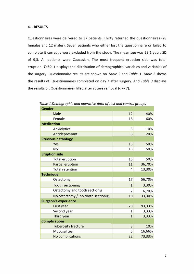
4. - RESULTS
Questionnaires were delivered to 37 patients. Thirty returned the questionnaires (28
females and 12 males). Seven patients who either lost the questionnaire or failed to
complete it correctly were excluded from the study. The mean age was 29,1 years SD
of 9,3. All patients were Caucasian. The most frequent eruption side was total
eruption. Table 1 displays the distribution of demographical variables and variables of
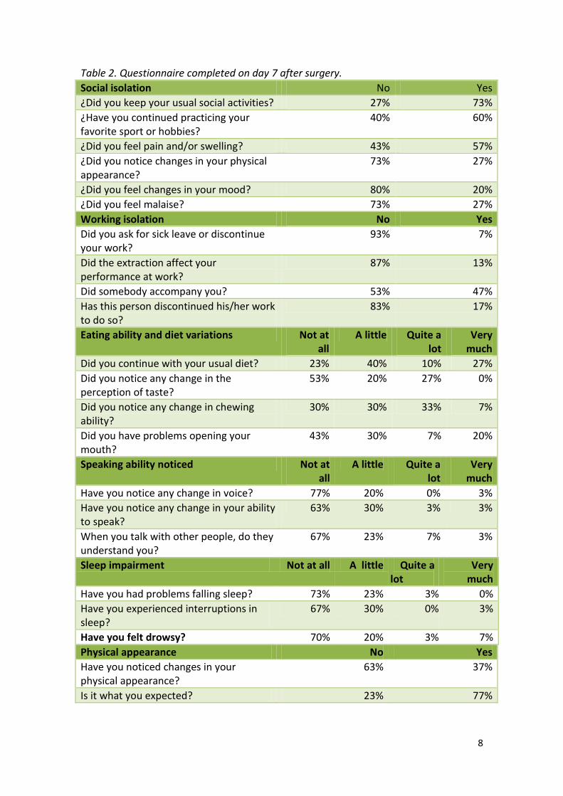
the surgery. Questionnaire results are shown on Table 2 and Table 3. Table 2 shows
the results of: Questionnaires completed on day 7 after surgery. And Table 3 displays
the results of: Questionnaires filled after suture removal (day 7).
Table 1.Demographic and operative data of test and control groups
Gender Male 12 40%
Female 18 60%
Medication Anxiolytics 3 10%
Antidepressant 6 20%
Previous pathology Yes 15 50%
No 15 50%
Eruption side Total eruption 15 50%
Partial eruption 11 36,70%
Total retention 4 13,30%
Technique Ostectomy 17 56,70%
Tooth sectioning 1 3,30%
Ostectomy and tooth sectionig 2 6,70%
No ostectomy / no tooth sectionig 10 33,30%
Surgeon's experience First year 28 93,33%
Second year 1 3,33%
Third year 1 3,33%
Complications Tuberosity fracture 3 10%
Mucosal tear 5 16,66%
No complications 22 73,33%
8
Table 2. Questionnaire completed on day 7 after surgery.
Social isolation No Yes
¿Did you keep your usual social activities? 27% 73%
¿Have you continued practicing your favorite sport or hobbies?
40% 60%
¿Did you feel pain and/or swelling? 43% 57%
¿Did you notice changes in your physical appearance?
73% 27%
¿Did you feel changes in your mood? 80% 20%
¿Did you feel malaise? 73% 27%
Working isolation No Yes
Did you ask for sick leave or discontinue your work?
93% 7%
Did the extraction affect your performance at work?
87% 13%
Did somebody accompany you? 53% 47%
Has this person discontinued his/her work to do so?
83% 17%
Eating ability and diet variations Not at all
A little Quite a lot
Very much
Did you continue with your usual diet? 23% 40% 10% 27%
Did you notice any change in the perception of taste?
53% 20% 27% 0%
Did you notice any change in chewing ability?
30% 30% 33% 7%
Did you have problems opening your mouth?
43% 30% 7% 20%
Speaking ability noticed Not at all
A little Quite a lot
Very much
Have you notice any change in voice? 77% 20% 0% 3%
Have you notice any change in your ability to speak?
63% 30% 3% 3%
When you talk with other people, do they understand you?
67% 23% 7% 3%
Sleep impairment Not at all A little Quite a lot
Very much
Have you had problems falling sleep? 73% 23% 3% 0%
Have you experienced interruptions in sleep?
67% 30% 0% 3%
Have you felt drowsy? 70% 20% 3% 7%
Physical appearance No Yes
Have you noticed changes in your physical appearance?
63% 37%
Is it what you expected? 23% 77%
9
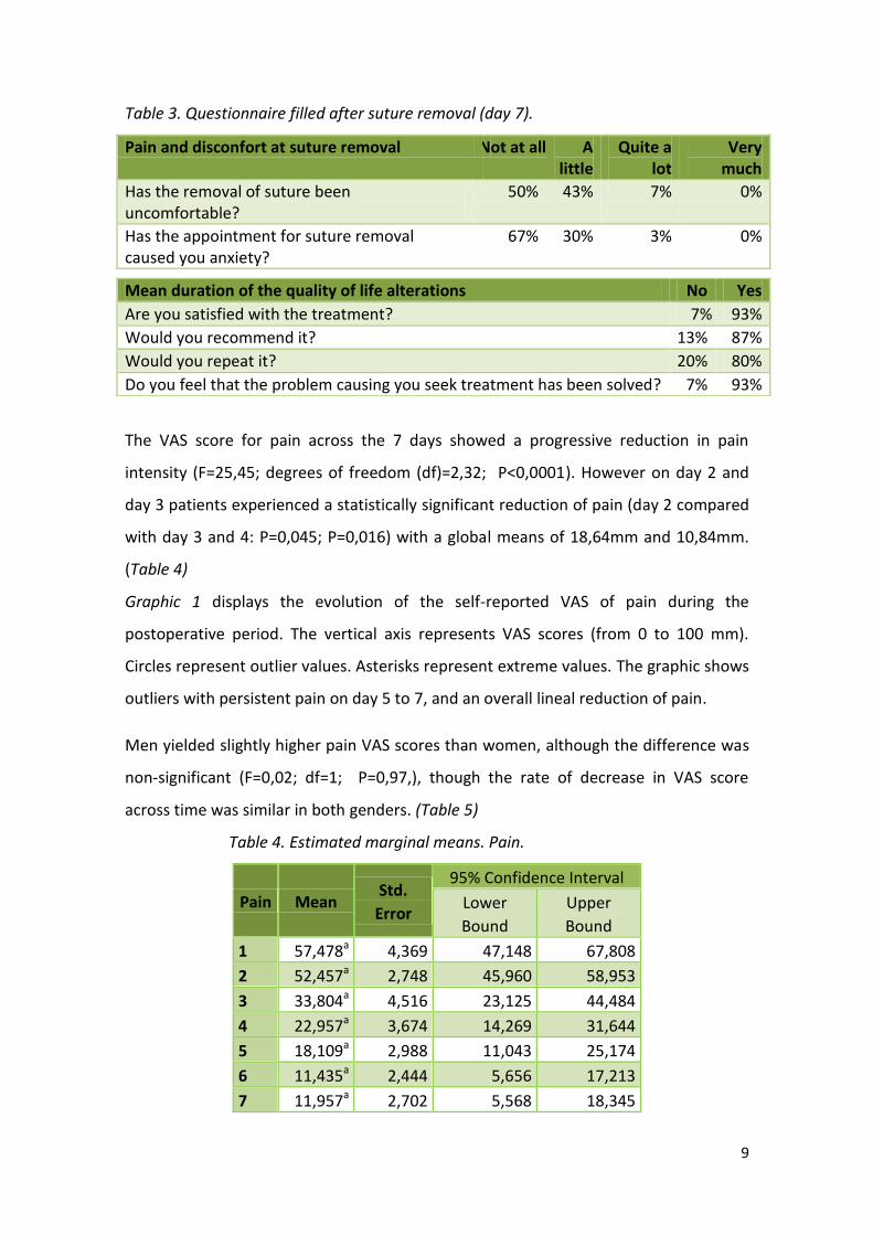
Table 3. Questionnaire filled after suture removal (day 7).
Pain and disconfort at suture removal Not at all A little
Quite a lot
Very much
Has the removal of suture been uncomfortable?
50% 43% 7% 0%
Has the appointment for suture removal caused you anxiety?
67% 30% 3% 0%
The VAS score for pain across the 7 days showed a progressive reduction in pain
intensity (F=25,45; degrees of freedom (df)=2,32; P<0,0001). However on day 2 and
day 3 patients experienced a statistically significant reduction of pain (day 2 compared
with day 3 and 4: P=0,045; P=0,016) with a global means of 18,64mm and 10,84mm.
(Table 4)
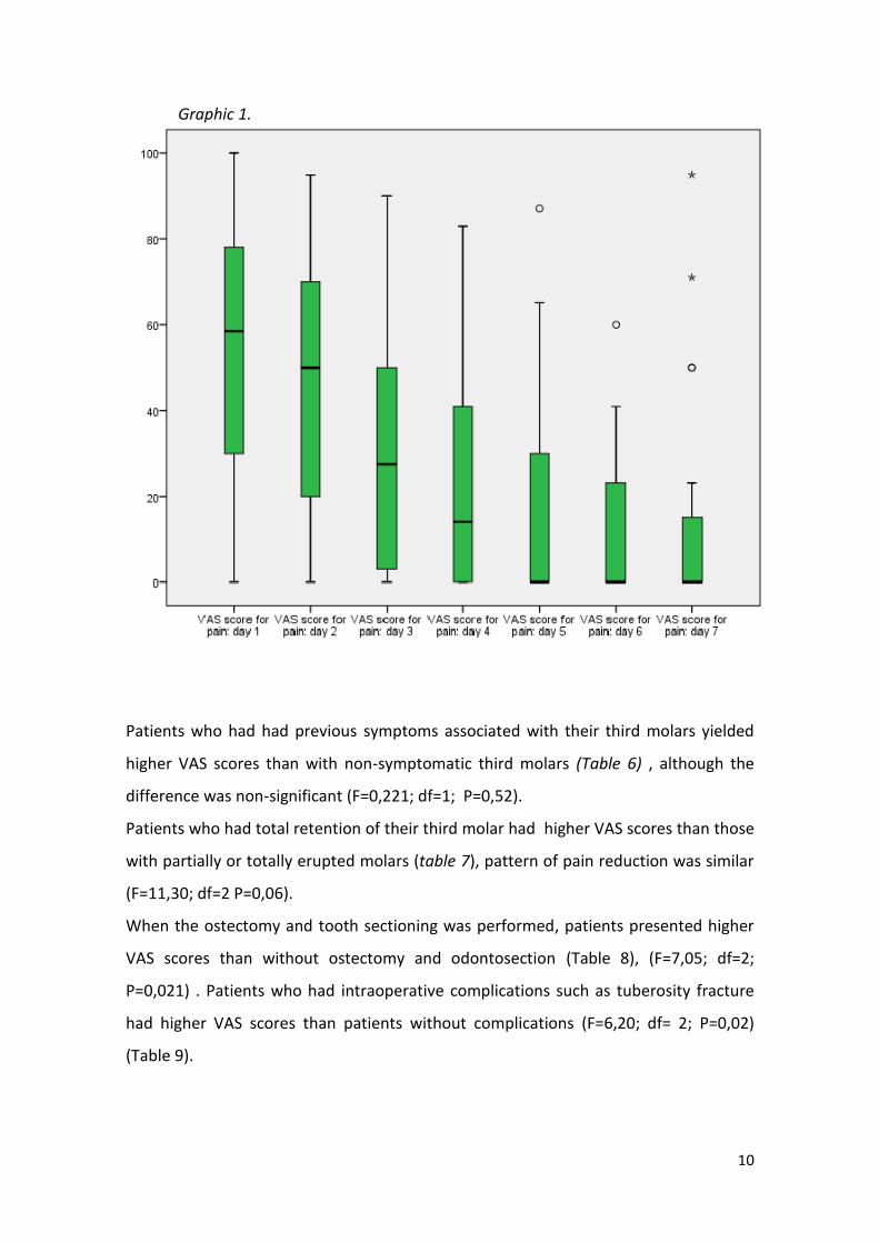
Graphic 1 displays the evolution of the self-reported VAS of pain during the
postoperative period. The vertical axis represents VAS scores (from 0 to 100 mm).
Circles represent outlier values. Asterisks represent extreme values. The graphic shows
outliers with persistent pain on day 5 to 7, and an overall lineal reduction of pain.
Men yielded slightly higher pain VAS scores than women, although the difference was
non-significant (F=0,02; df=1; P=0,97,), though the rate of decrease in VAS score
across time was similar in both genders. (Table 5)
Table 4. Estimated marginal means. Pain.
Pain Mean Std.
Error
95% Confidence Interval
Lower
Bound
Upper
Bound
1 57,478a 4,369 47,148 67,808
2 52,457a 2,748 45,960 58,953
3 33,804a 4,516 23,125 44,484
4 22,957a 3,674 14,269 31,644
5 18,109a 2,988 11,043 25,174
6 11,435a 2,444 5,656 17,213
7 11,957a 2,702 5,568 18,345
Mean duration of the quality of life alterations No Yes
Are you satisfied with the treatment? 7% 93%
Would you recommend it? 13% 87%
Would you repeat it? 20% 80%
Do you feel that the problem causing you seek treatment has been solved? 7% 93%
10
Graphic 1.
Patients who had had previous symptoms associated with their third molars yielded
higher VAS scores than with non-symptomatic third molars (Table 6) , although the
difference was non-significant (F=0,221; df=1; P=0,52).
Patients who had total retention of their third molar had higher VAS scores than those
with partially or totally erupted molars (table 7), pattern of pain reduction was similar
(F=11,30; df=2 P=0,06).
When the ostectomy and tooth sectioning was performed, patients presented higher
VAS scores than without ostectomy and odontosection (Table 8), (F=7,05; df=2;
P=0,021) . Patients who had intraoperative complications such as tuberosity fracture
had higher VAS scores than patients without complications (F=6,20; df= 2; P=0,02)
(Table 9).
11
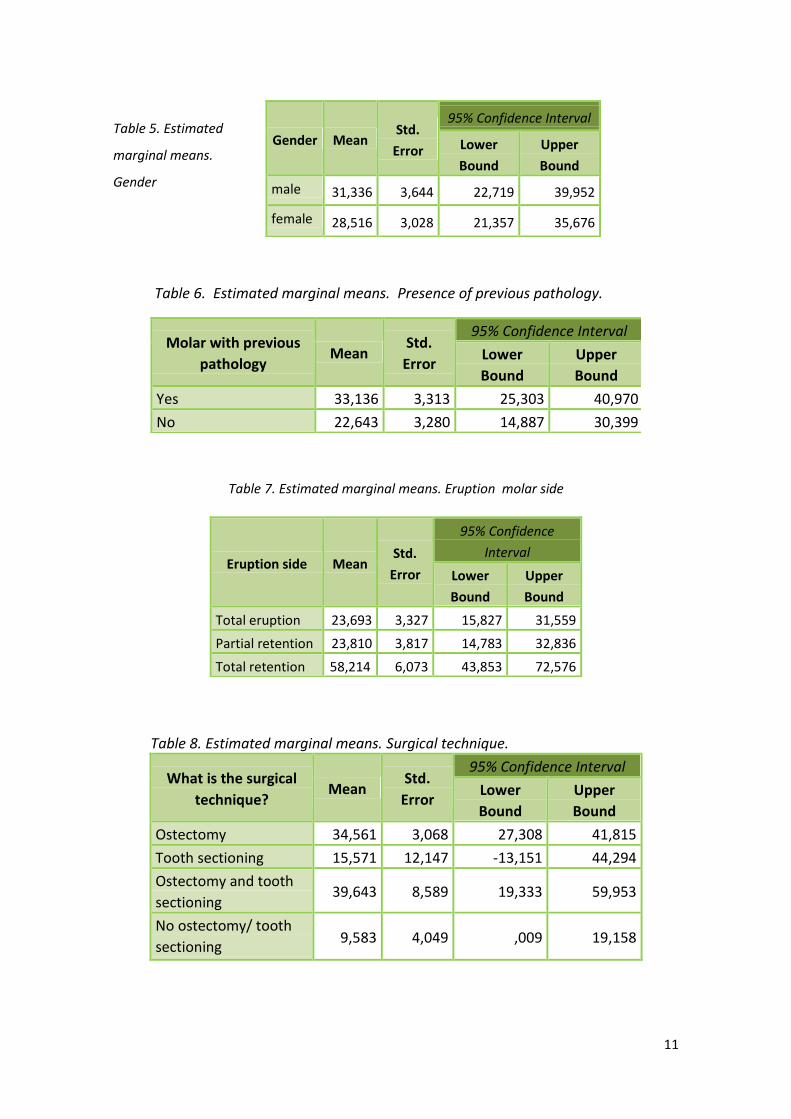
Table 5. Estimated
marginal means.
Gender
Table 6. Estimated marginal means. Presence of previous pathology.
Molar with previous
pathology Mean
Std.
Error
95% Confidence Interval
Lower
Bound
Upper
Bound
Yes 33,136 3,313 25,303 40,970
No 22,643 3,280 14,887 30,399
Table 7. Estimated marginal means. Eruption molar side
Table 8. Estimated marginal means. Surgical technique.
Eruption side Mean Std.
Error
95% Confidence
Interval
Lower
Bound
Upper
Bound
Total eruption 23,693 3,327 15,827 31,559
Partial retention 23,810 3,817 14,783 32,836
Total retention 58,214 6,073 43,853 72,576
What is the surgical
technique? Mean
Std.
Error
95% Confidence Interval
Lower
Bound
Upper
Bound
Ostectomy 34,561 3,068 27,308 41,815
Tooth sectioning 15,571 12,147 -13,151 44,294
Ostectomy and tooth
sectioning 39,643 8,589 19,333 59,953
No ostectomy/ tooth
sectioning 9,583 4,049 ,009 19,158
Gender Mean Std.
Error
95% Confidence Interval
Lower
Bound
Upper
Bound
male 31,336 3,644 22,719 39,952
female 28,516 3,028 21,357 35,676
12
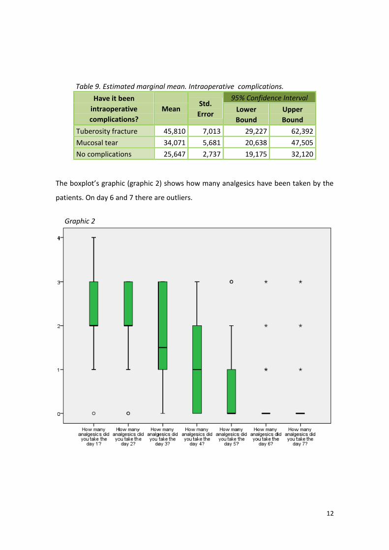
The boxplot’s graphic (graphic 2) shows how many analgesics have been taken by the
patients. On day 6 and 7 there are outliers.
Graphic 2
Table 9. Estimated marginal mean. Intraoperative complications.
Have it been
intraoperative
complications?
Mean Std.
Error
95% Confidence Interval
Lower
Bound
Upper
Bound
Tuberosity fracture 45,810 7,013 29,227 62,392
Mucosal tear 34,071 5,681 20,638 47,505
No complications 25,647 2,737 19,175 32,120
13
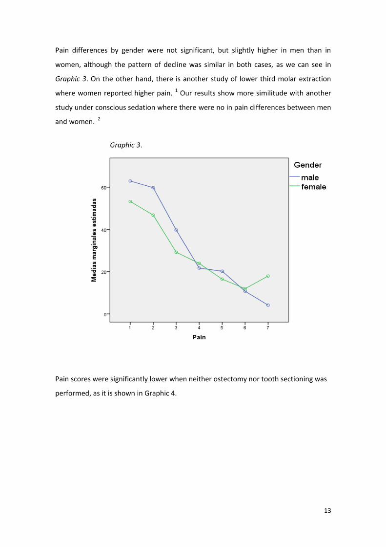
Pain differences by gender were not significant, but slightly higher in men than in
women, although the pattern of decline was similar in both cases, as we can see in
Graphic 3. On the other hand, there is another study of lower third molar extraction
where women reported higher pain. 1 Our results show more similitude with another
study under conscious sedation where there were no in pain differences between men
and women. 2
Graphic 3.
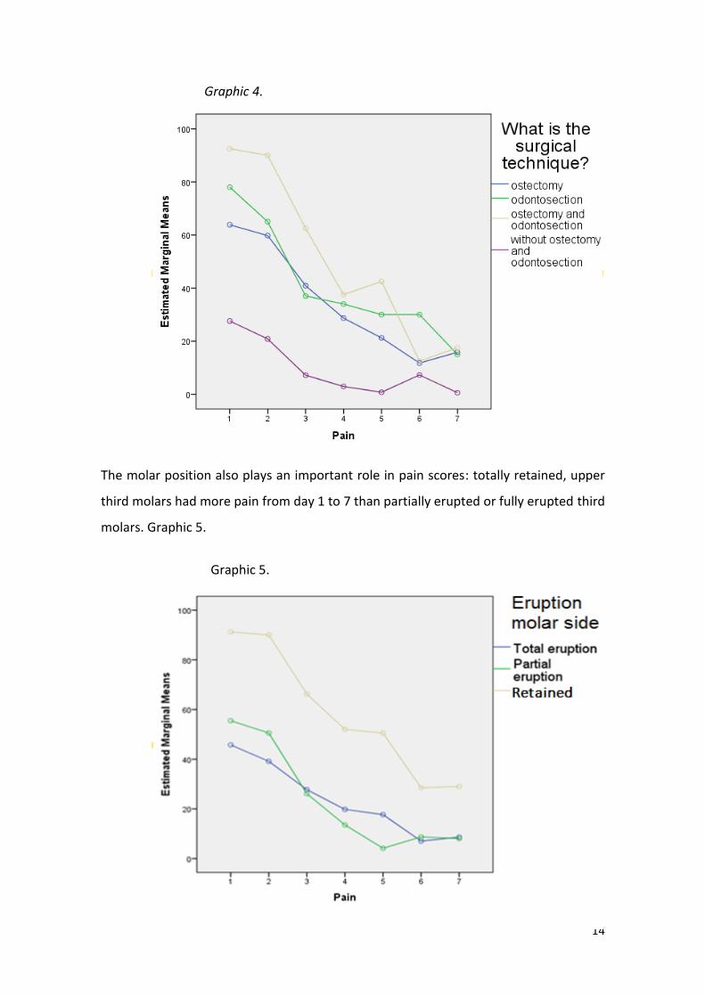
Pain scores were significantly lower when neither ostectomy nor tooth sectioning was
performed, as it is shown in Graphic 4.
14
Graphic 4.
The molar position also plays an important role in pain scores: totally retained, upper
third molars had more pain from day 1 to 7 than partially erupted or fully erupted third
molars. Graphic 5.
Graphic 5.
15
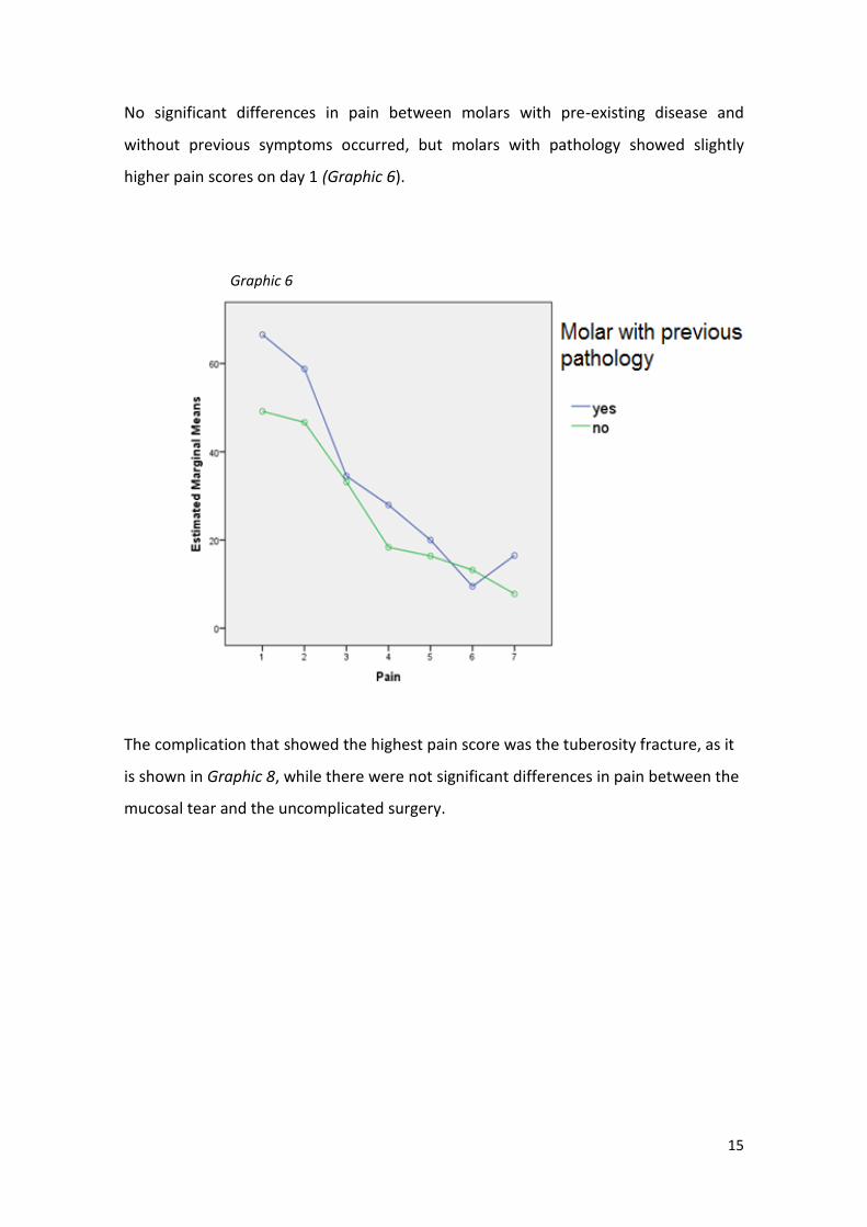
No significant differences in pain between molars with pre-existing disease and
without previous symptoms occurred, but molars with pathology showed slightly
higher pain scores on day 1 (Graphic 6).
Graphic 6
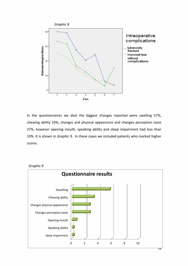
The complication that showed the highest pain score was the tuberosity fracture, as it
is shown in Graphic 8, while there were not significant differences in pain between the
mucosal tear and the uncomplicated surgery.
16
0 2 4 6 8 10
sleep impairment
Speaking ability
Opening mouth
Changes perception taste
Changes physical appearance
Chewing ability
Sweelling
Questionnaire results
Graphic 8
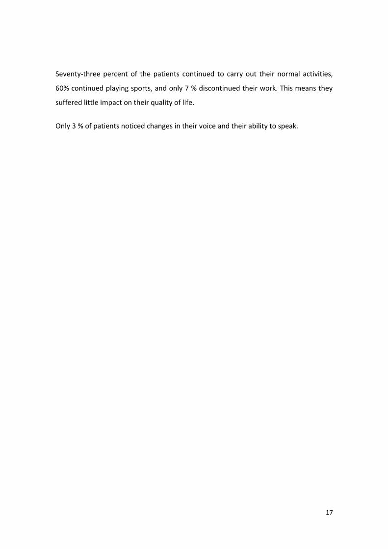
In the questionnaires we deal the biggest changes reported were swelling 57%,
chewing ability 33%, changes and physical appearance and changes perception taste
27%, however opening mouth, speaking ability and sleep impairment had less than
10%. It is shown in Graphic 9. In these cases we included patients who marked higher
scores.
Graphic 9
17
Seventy-three percent of the patients continued to carry out their normal activities,
60% continued playing sports, and only 7 % discontinued their work. This means they
suffered little impact on their quality of life.
Only 3 % of patients noticed changes in their voice and their ability to speak.
18
5. – DISCUSSION
One of the main problems of this study was to recruit patients undergoing only
extraction of upper third molars. Usually, in order to increase the patient comfort and
reduce the number of visits, patients underwent extraction of both third molars of the
same side, which was an exclusion criterion.
A limitation of the present study is that data are subjective, based only on the patient
perception. However, this is a constant limitation in studies of QoL. Another limitation
of the study is that the questionnaire only collected information corresponding to the
first 7 days after surgery; consequently, only the short-term evolution of the changes
in QoL could be assessed. Therefore, possible long-term complications could not be
evaluated, which might be interesting especially in extractions with intraoperative
complications. There are studies reporting late infections after the extraction of third
molars, although they are usually related to lower third molars. 14
Pain seemed to lineally decrease and practically disappeared on day 4. Other studies
about lower third molar extraction showed that pain is more persistent, and
disappears around day 6 or 7. 12
Despite the fact that the VAS score for pain across the 7 days showed a progressive
reduction in pain intensity, the outliers on day 6 and day 7 might be caused by the
anxiety of return to the hospital to remove the suture. This might be an explication of
the higher score of pain around day 6 and day 7.15
It is interesting to compare our pain scores with Colorado - Bonnin et al, and with
Sancho - Puchades considering that extractions were performed at the same
institution, with the same surgical technique and the same anesthetic technique.
However, in the first study, only lower third were extracted under local anesthesia and
in the second one the four wisdom teeth were extracted under conscious sedation.
Pain scores observed in our study were slightly lower on days 4, 5, 6 and 7, comparing
with Colorado - Bonnin et al and Sancho - Puchades, but during the first three days
were similar. 1, 2
19
Unlike the study of mandibular third molars where there is a constant higher pain in
pathological molars. 1 In our study population, only 50% of upper third molars had
previous symptoms, all the same Raymond et al study reported 54% of molars with
previous symptoms. 3
It was not possible to analyze pain scores based on the experience of the professional
considering that 93 % of surgeries were conducted by first course students. However
this is a variable of great interest where some studies show a smaller number of
complications with more experimented professionals. 3, 16
Therefore the biggest drawbacks were inflammation and discomfort related to the
chewing ability, from 10% to 30 %. As a result, the quality of life is basically affected by
eating difficulties. These figures are considerably higher when removing the 4 third
molars at the same time 57.1%. 2 This shows that isolated extractions have a faster
recovery of the ability to eat.
Most of the patients continued to carry out their normal activities. They suffered little
impact on their quality of life.
Regarding changes in chewing ability and the problems of opening the mouth in our
results 7% and 20% respectively reported this inability, however upon lower third
molar removal up to 80% of patients reported changes in chewing ability. 1 When 4
third molars are extracted under conscious sedation 50% of patients reported trismus.
Therefore, for upper third molar extraction there is less chance of developing trismus
or masticatory problems, probably related to a shorter operating time and a local
inflammation that affects less muscles related to masticatory function such as
masseters and pterygoids. 17
About 3 % of patients noticed changes in their voice and their ability to speak. We
found great differences when extraction of the 4 wisdom teeth is performed
simultaneously with 10 % of change in their voice and a 60% of difficulties to be
understood other people. 2 In the extraction of mandibular third molars 20% perceived
changes in their voice, while 57 % had problems to be understood.
20
A greater ability to speak might be related to the lower prevalence of trismus and
ability to open the mouth, with better pronunciation.
In any case talking problems are secondary in other studies. 10, 3 For the patient, it
might be of great importance if their working life requires public speaking. However,
the upper third molar extraction reported very few cases of impossibility of speech.
Between 20% and 27 % of patients showed an alteration in taste perception for the
first 7 days. Similar results were found in the study of Colorado Bonnin et al, between
15 and 27.5 % quite a lot and a little respectively. In the case of lower molars this could
be related with lingual nerve function or use of chlorhexidine mouthrinses.
In upper molar extractions rinses with chlorhexidine digluconate might play an
important role in the perception of flavor, especially increasing the threshold for salty
taste. 1, 4, 13
The McGrath cohort study confirms that there is a deterioration in the quality of life in
short term, but better oral health in long term, especially observed in cases of previous
pericoronitis.11 Our study confirms that the quality of life is slightly affected in short
term.
If we analyze the level of satisfaction we note that 93 % of patients are satisfied and
believe that their problem is solved. Most studies show similar satisfaction rates above
90 %. 5, 2,1,10, 3
A variable that we did not take into account, but could be interesting, is the tobacco
use, which we believe that could affect postoperative pain levels and the degree of
inflammation, especially in the extractions that require more aggressive surgical
techniques.
21
6.- CONCLUSIONS
6.1.-Spanish version
La mayoría de pacientes están satisfechos con el tratamiento y creen que su problema
ha sido resuelto. Aunque hay un pequeño porcentaje que no lo repetiría, ni lo
recomendaría.
Durante el día 1 y el día 2 las puntuaciones de dolor son similares a las de otros
estudios sobre extracción de cordales inferiores y sobre extracción de los cuatro
cordales, podemos considerar que se trata de un postoperatorio más corto respecto
de las otras intervenciones.
No hay diferencias significativas en las puntuaciones del dolor en función del género.
Sin embargo sí que hay diferencias de dolor con la variable técnica quirúrgica, posición
del molar, molares con patología previa y complicaciones intraoperatorias.
Respectivamente se han encontrado mayores puntuaciones de dolor en intervenciones
con ostectomía y odontosección, en cordales incluidos, y en intervenciones con
complicaciones intraoperatorias.
El mayor inconveniente que encontraron los pacientes fue la inflamación y las
dificultades masticatorias.
Consideramos que la intervención ha tenido pocas repercusiones a nivel de su calidad
de vida, ya que la mayoría han continuado con sus actividades habituales.
El análisis sobre las repercusiones de las intervenciones quirúrgicas en la calidad de
vida de los pacientes es importante para una evaluación preoperatoria óptima y
conocimiento de las indicaciones adecuadas después de la cirugía. Además, permite al
cirujano dar expectativas realistas a los pacientes sobre los días postoperatorios y
dejar que el paciente elija el mejor momento para someterse al procedimiento,
tratando de minimizar las interferencias con la vida cotidiana.
22
6.2.- English version
Most patients are satisfied with the treatment and believe that their problem is
resolved. Although, there is a small percentage of patients that would not repeat
either recommend it.
During day 1 and day 2 pain scores are similar to other studies of mandibular third
molars extraction and 4 wisdom teeth extraction. However, postoperative pain
disappears faster compared to these other surgeries.
There are not significant differences in pain scores by gender. However, there are
differences of pain with the variable surgical technique, molar with previous pathology
eruption position, and intraoperative complications. In addition, pain scores are
increased in interventions with osteotomy and tooth section, in included wisdom
teeth, wisdom teeth with previous pathology and extractions with intraoperative
complications.
The biggest drawback that patients found was inflammation and chewing difficulties.
We believe that the intervention had little impact in their quality of life, considering
that most continued their normal activities.
The analysis of the repercussions of the surgical interventions on patients’ QoL is
important for an optimal preoperative assessment and development of appropriate
indications after the surgery. Furthermore, it enables the surgeon to give the patient
realistic expectancies of the postoperative days, and helps the patient choose the best
moment to undergo the procedure, trying to minimize mayor interferences with
everyday life.
23
7.- BIBLIOGRAPHY
1.- Colorado-Bonnin M, Valmaseda-Castellon E, Berini-Aytes L, Gay-Escoda C. Quality of
life following lower third molar removal. Int J oral Maxillofac Surg. 2006; 35:343-47.
2.- Sancho-Puchades M, Valmaseda-Castellon E, Berini-Aytes L, Gay-Escoda C. Quality
of life following lower third molar removal under conscisious sedation. Med Oral Patol
Oral Cir Bucal. 2002; 17:994-99.
3.-Savin J, Ogden GR. Third molar surgery- a preliminary reporto n aspects affecting
quality of life in the early postoperative period. Br J Oral Maxillofac Surg 1997; 35:246-
53.
4.- Shafer DM, Frank ME, Gent JF. Fischer ME. Gustatory function after third molar
extraction. Oral Surg Oral Med Oral Pathol Oral Radiol Endod 1999; 87:419-28.
5.- Lopes V, Mumenya R, Feinmann C, Harris M. Third molar surgery an audit of
indications for surgery, post-operative complaints and patient satisfaction. Br J Oral
Maxillofac Surg 1995; 33: 33-35.
6. Gay Escoda C, Piñera Penalva M, Valmaseda Castellon E. Cordales incluidos. En: Gay
Escoda C, Berini Aytés L. Tratado de Cirugía Bucal. Madrid: Ediciones Ergon; 2004. P
387-459.
7.- Ogden Gr, Bissias E, Ruta DA, ogston S. Quality of life following third molar removal:
a patient versus professional perspective. Br Dent J. 2003; 194: 265-68.
8.- Grossi Gb, Marioana C, Garramone RA, Borgonovo A, Creminelli L, Santoro F.
Assessing postoperative discomfort after third molar surgery: a prospective study. J
Oral Maxillofac Surg. 2007; 65: 901-17
9.-Coulthard P, Pleuvry B, Dobson M, Price M. Behavioural measurement of
postoperative pain after oral surgery. Br J Oral Maxillofac Surg 2000; 38:127-131.
10.-White RP, Shugars DA, Shafer DM, Laskin DM, Buckley MJ, Phillips C. Recovery after
third molar surgery: clinical and health-related quality of life outcomes. J Oral
Maxillofac Surg 2003; 61: 535-44
11.-McGrath C, Comfort MB, Lo EC, Luo Y. Can third molar surgery improve quality of
life? A 6-month cohort study. J Oral Maxillofac Surg 2003; 61:759-63.
12.- Deepti C, Rehan HS, Mehra P,Changes in quality of life after surgical removal of impacted mandibular third molar teeth. J Maxillofac Oral Surg. 2009; 8:257-60
13.-Helms JA, Della-Fera MA ,Mott AE, Frank ME. Effects of clorhexidine on human
taste perception . Arch Oral Biol. 1995; 40:913-20
24
14.- Figueiredo R, Valmaseda-Castellón E, Berini-Aytés L, Gay-Escoda C. Delayed-Onset Infections After Lower Third Molar Extraction: A Case-Control Study. 2007; 65:97-102.
15.- Gorczyca R, Filip R, Walczak E. Psychological Aspects of Pain. Ann Agric Environ
Med. 2013; 1:23–27.
16.- Blondeau F, Nach GD Extraction of Impacted Mandibular Third Molars:
Postoperative Complications and Their Risk Factors. 2007; 73:325-27.
17.- Okeson Jeffrey P. Etiología de los trastornos funcionales del sistema masticatorio.
En: Okeson Jeffrey P. Tratamiento de oclusión y afecciones temporomandibulares.
Madrid: Edición. Ed. Mosby Co; 2003. P.149-90.
25
8.- ANNEX
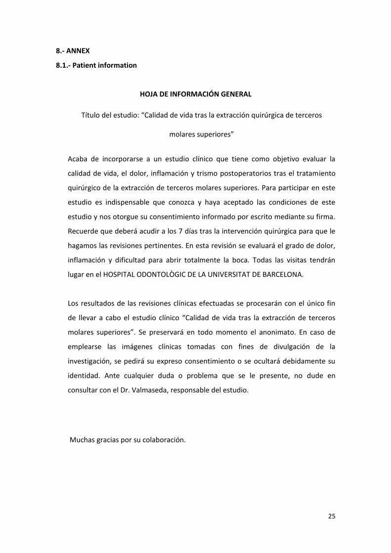
8.1.- Patient information
HOJA DE INFORMACIÓN GENERAL
Título del estudio: “Calidad de vida tras la extracción quirúrgica de terceros
molares superiores”
Acaba de incorporarse a un estudio clínico que tiene como objetivo evaluar la
calidad de vida, el dolor, inflamación y trismo postoperatorios tras el tratamiento
quirúrgico de la extracción de terceros molares superiores. Para participar en este
estudio es indispensable que conozca y haya aceptado las condiciones de este
estudio y nos otorgue su consentimiento informado por escrito mediante su firma.
Recuerde que deberá acudir a los 7 días tras la intervención quirúrgica para que le
hagamos las revisiones pertinentes. En esta revisión se evaluará el grado de dolor,
inflamación y dificultad para abrir totalmente la boca. Todas las visitas tendrán
lugar en el HOSPITAL ODONTOLÒGIC DE LA UNIVERSITAT DE BARCELONA.
Los resultados de las revisiones clínicas efectuadas se procesarán con el único fin
de llevar a cabo el estudio clínico “Calidad de vida tras la extracción de terceros
molares superiores”. Se preservará en todo momento el anonimato. En caso de
emplearse las imágenes clínicas tomadas con fines de divulgación de la
investigación, se pedirá su expreso consentimiento o se ocultará debidamente su
identidad. Ante cualquier duda o problema que se le presente, no dude en
consultar con el Dr. Valmaseda, responsable del estudio.
Muchas gracias por su colaboración.
26
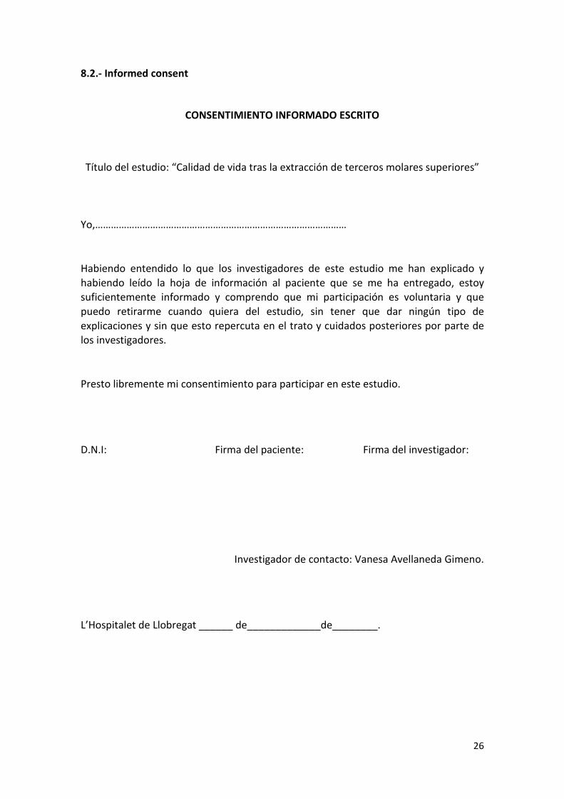
8.2.- Informed consent
CONSENTIMIENTO INFORMADO ESCRITO
Título del estudio: “Calidad de vida tras la extracción de terceros molares superiores”
Yo,……………………………………………………………………………………
Habiendo entendido lo que los investigadores de este estudio me han explicado y
habiendo leído la hoja de información al paciente que se me ha entregado, estoy suficientemente informado y comprendo que mi participación es voluntaria y que
puedo retirarme cuando quiera del estudio, sin tener que dar ningún tipo de
explicaciones y sin que esto repercuta en el trato y cuidados posteriores por parte de los investigadores.
Presto libremente mi consentimiento para participar en este estudio.
D.N.I: Firma del paciente: Firma del investigador:
Investigador de contacto: Vanesa Avellaneda Gimeno.
L’Hospitalet de Llobregat ______ de_____________de________.
27
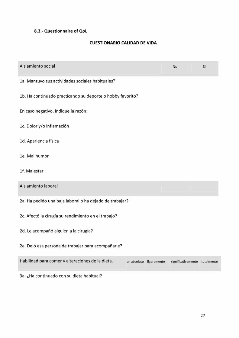
8.3.- Questionnaire of QoL
CUESTIONARIO CALIDAD DE VIDA
Aislamiento social No Sí
1a. Mantuvo sus actividades sociales habituales?
1b. Ha continuado practicando su deporte o hobby favorito?
En caso negativo, indique la razón:
1c. Dolor y/o inflamación
1d. Apariencia física
1e. Mal humor
1f. Malestar
Aislamiento laboral
2a. Ha pedido una baja laboral o ha dejado de trabajar?
2c. Afectó la cirugía su rendimiento en el trabajo?
2d. Le acompañó alguien a la cirugía?
2e. Dejó esa persona de trabajar para acompañarle?
Habilidad para comer y alteraciones de la dieta. en absoluto ligeramente significativamente totalmente
3a. ¿Ha continuado con su dieta habitual?
28
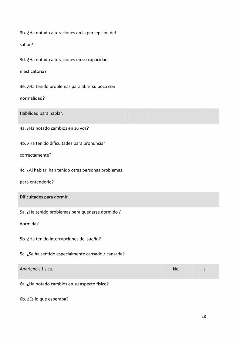
3b. ¿Ha notado alteraciones en la percepción del
sabor?
3d. ¿Ha notado alteraciones en su capacidad
masticatoria?
3e. ¿Ha tenido problemas para abrir su boca con
normalidad?
Habilidad para hablar.
4a. ¿Ha notado cambios en su voz?
4b. ¿Ha tenido dificultades para pronunciar
correctamente?
4c. ¿Al hablar, han tenido otras personas problemas
para entenderle?
Dificultades para dormir.
5a. ¿Ha tenido problemas para quedarse dormido /
dormida?
5b. ¿Ha tenido interrupciones del sueño?
5c. ¿Se ha sentido especialmente cansado / cansada?
Apariencia física. No si
6a. ¿Ha notado cambios en su aspecto físico?
6b. ¿Es lo que esperaba?
29
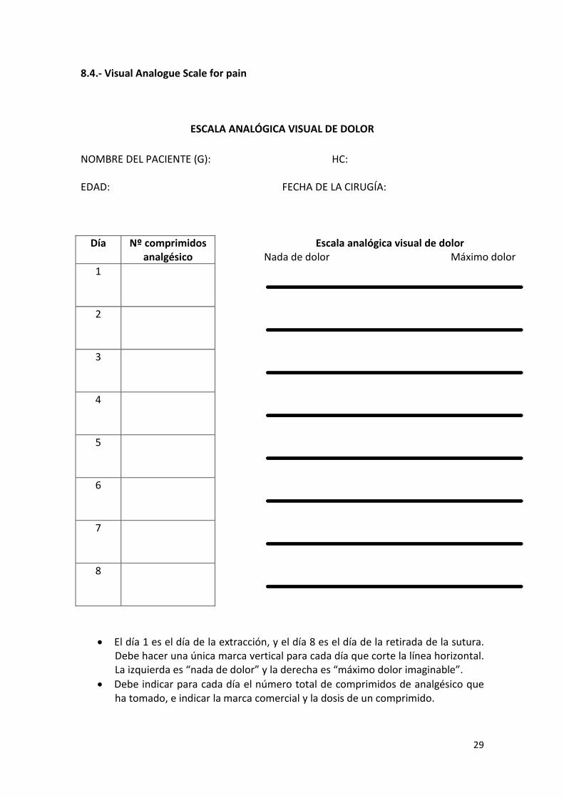
8.4.- Visual Analogue Scale for pain
ESCALA ANALÓGICA VISUAL DE DOLOR
NOMBRE DEL PACIENTE (G): HC: EDAD: FECHA DE LA CIRUGÍA:
Día Nº comprimidos analgésico
Escala analógica visual de dolor Nada de dolor Máximo dolor
1
2
3
4
5
6
7
8
El día 1 es el día de la extracción, y el día 8 es el día de la retirada de la sutura. Debe hacer una única marca vertical para cada día que corte la línea horizontal. La izquierda es “nada de dolor” y la derecha es “máximo dolor imaginable”.
Debe indicar para cada día el número total de comprimidos de analgésico que ha tomado, e indicar la marca comercial y la dosis de un comprimido.
30
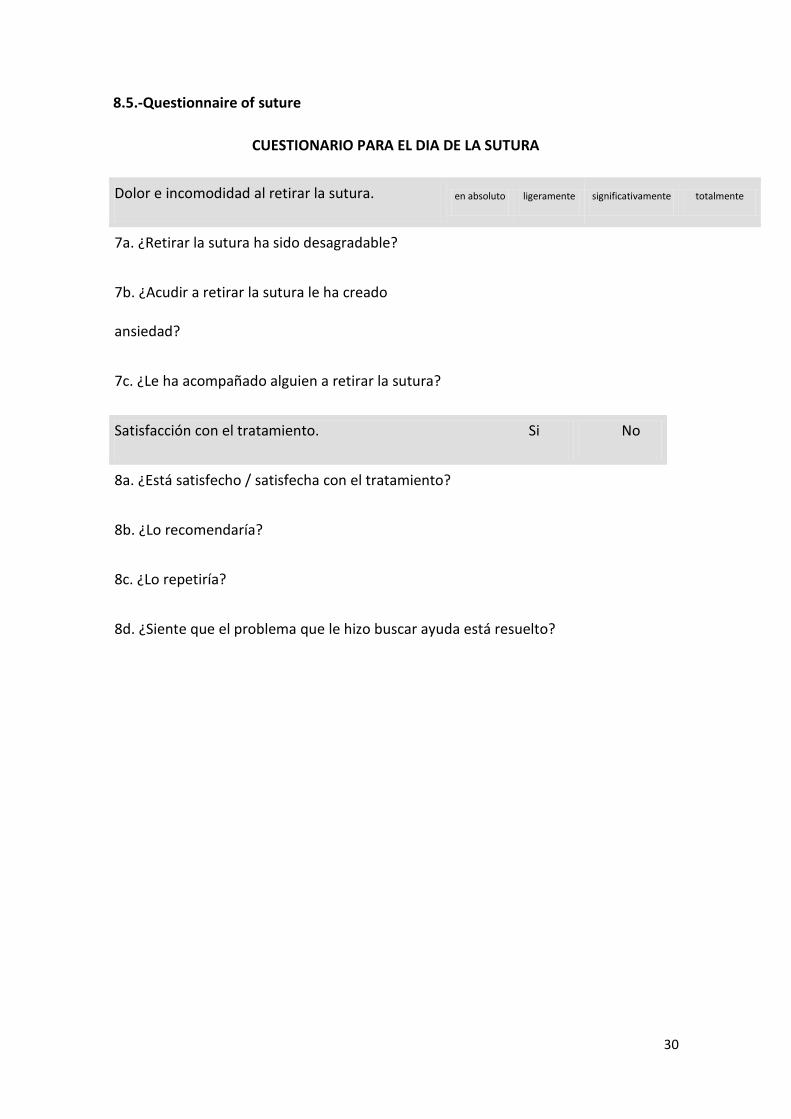
8.5.-Questionnaire of suture
CUESTIONARIO PARA EL DIA DE LA SUTURA
Dolor e incomodidad al retirar la sutura. en absoluto ligeramente significativamente totalmente
7a. ¿Retirar la sutura ha sido desagradable?
7b. ¿Acudir a retirar la sutura le ha creado
ansiedad?
7c. ¿Le ha acompañado alguien a retirar la sutura?
Satisfacción con el tratamiento. Si No
8a. ¿Está satisfecho / satisfecha con el tratamiento?
8b. ¿Lo recomendaría?
8c. ¿Lo repetiría?
8d. ¿Siente que el problema que le hizo buscar ayuda está resuelto?

31