Embed Size (px)
Citation preview
PHYSICOCHEMICAL AND ANTIBACTERIAL PROPERTIES OF COTTON FABRIC TREATED WITH CHITOSAN–TRIPOLYPHOSPHATE NANOPARTICLES
AND LAURIC ACID
SITI NUR HANA BINTI MAMAT
UNIVERSITI TEKNOLOGI MALAYSIA
PHYSICOCHEMICAL AND ANTIBACTERIAL PROPERTIES OF COTTON
FABRIC TREATED WITH CHITOSAN –TRIPOLYPHOSPHATE
NANOPARTICLES AND LAURIC ACID
SITI NUR HANA BINTI MAMAT
A thesis submitted in fulfillment of the
requirements for the award of the degree of
Master of Engineering (Bioprocess)
Faculty of Chemical and Energy Engineering
Universiti Teknologi Malaysia
OCTOBER 2016
iii
To my beloved mother and father……….
iv
ACKNOWLEDGEMENTS
I am heartily thankful to my supervisor, Dr. Eraricar Salleh, my beloved
parents, Hanawiah Sehab and Mamat Bakar, whose encouragement, critics, guidance
and support from the initial to the final level enable me to develop an understanding
of the subject. Without their continued support and interest, this proposal would not
have been the same as presented here. This thesis would not have been possible
unless the faculty able to provide the equipments and material throughout this
project. I am indebted to my colleagues; Mohd Harfiz, Norhana, Norhayati,
Nozieana, Norul Fatiha, laboratory mates and Faculty of Chemical and Energy
Engineering postgraduates office staff for support and guidance while doing this
project. I would like to thank to thank my family members, especially my parents,
my siblings, my uncle and my aunty for supporting and encouraging me to persue
this master degree. Lastly, I offer my regards and blessing to all of those who
supported me in my respect during the completion of the project.
v
ABSTRACT
Today, antibacterial treatment has become a prerequisite for textile goods
used in hospitals, hotels, sports and personal care industries. Cotton is one of the
most important natural fibers that is used extensively in textile industry. In this work,
properties of cotton fabric treated with chitosan-tripolyphosphate nanoparticles
(CNPs) and lauric acid (LA) was studied. CNPs were prepared based on the ionic
gelation of chitosan with tripolyphosphate anions. The pretreatment process of
cotton fabric was performed using sodium hydroxide. Afterwards, the CNPs solution
with the addition of LA was applied to the treated cotton fabric to give wider
antibacterial effects. The physicochemical properties of the CNPs and treated cotton
fabric were determined by size distribution, Fourier transform infrared analysis and
field emission scanning electron microscopy analysis. The average size of CNPs
obtained was 179.1 ± 82.45 nm at concentration of 0.1% chitosan (w/v). The
morphology surface of CNPs revealed the particles were spherical and almost
uniform. CNPs solution with the addition of 15% (w/v) LA was formulated to treat
the cotton fabric by soaking the cotton fabric in the solution while stirring for two
hours at 60 °C. The antibacterial activity of treated cotton fabric with CNPs and LA
against Bacillus subtilis (Gram-positive bacteria) and Escherichia coli (Gram-
negative bacteria) was evaluated by using agar diffusion and liquid culture test
methods. Cotton fabric treated with CNPs and LA showed a good ability to inhibit
bacteria reproduction with diameter of zone of inhibition of 2.03±0.09 cm for
Escherichia coli and 2.27±0.07 cm for Bacillus subtilis. Results showed that CNPs
incorporated with LA gave better inhibition toward tested bacteria compared to
CNPs solely, with differences as much as 30% for Gram-negative bacteria and 71%
for Gram-positive bacteria.
vi
ABSTRAK
Sekarang, rawatan anti-bakteria telah menjadi pra-syarat utama bagi barangan
tekstil yang digunakan di hospital, hotel, sukan dan industri penjagaan diri. Kapas
adalah merupakan salah satu daripada gentian semulajadi paling penting yang
digunakan secara meluas dalam industri tekstil. Dalam hasil kerja ini, sifat-sifat kain
kapas yang dirawat dengan nanopartikel kitosan-tripolifosfat (CNPs) dan asid laurik
(LA) telah dikaji. CNPs disediakan berasaskan gelatin ionik kitosan dengan anion
tripolifosfat (TPP). Proses prarawatan kain kapas telah dilakukan menggunakan
natrium hidroksida. Selepas itu, larutan CNPs dengan penambahan LA telah
digunakan untuk kain kapas terawat untuk memberi kesan anti-bakteria yang lebih
meluas. Sifat-sifat fizikokimia daripada CNPs dan kain kapas terawat ditentukan
dengan taburan saiz, analisis inframerah jelmaan Fourier dan analisis mikroskop
elektron imbasan pancaran medan. Saiz purata CNPs diperoleh ialah 179.10 ± 82.45
nm pada kepekatan 0.1% chitosan (w/v). Permukaan morfologi CNPs menunjukkan
zarah yang terhasil adalah berbentuk sfera dan hampir seragam. Larutan CNPs
dengan penambahan 15% (w/v) LA telah diformulasikan untuk merawat kain kapas
dengan merendam kain kapas di dalam larutan tersebut dan dikacau selama dua jam
pada suhu 60 °C. Aktiviti anti-bakteria kain kapas yang dirawat dengan CNPs dan
LA terhadap Bacillus subtilis (bakteria Gram-positif) dan Escherichia coli (bakteria
Gram-negatif) telah diukur dengan menggunakan kaedah penyebaran agar dan ujian
kultur cecair. Kain kapas dirawat dengan CNPs dan LA menunjukkan keupayaan
yang baik untuk merencat pembiakan bakteria dengan zon perencatan sebanyak 2.03
± 0.09 cm untuk Escherichia coli dan 2.2 ± 0.07 cm untuk Bacillus subtilis.
Keputusan menunjukkan bahawa CNPs yang digabungkan dengan LA memberikan
perencatan yang lebih baik terhadap bakteria diuji berbanding dengan CNPs tanpa
LA dengan perbezaan sebanyak 30% untuk bakteria Gram-negatif dan 71% untuk
bakteria Gram-positif.
vii
TABLE OF CONTENTS
CHAPTER TITLE PAGE
DECLARATION ii
DEDICATION iii
ACKNOWLEDGEMENTS iv
ABSTRACT v
ABSTRAK vi
TABLE OF CONTENTS vii
LIST OF TABLES x
LIST OF FIGURES xi
LIST OF SYMBOLS xiii
LIST OF ABBREVIATIONS xiv
LIST OF APPENDICES xv
1 INTRODUCTION 1
1.1 Background of Study 1
1.2 Problem Statements 3
1.3 Objective 5
1.4 Scope of the Study 5
1.5 Significance of the Study 6
1.6 Outline of the Thesis 6
2 LITERATURE REVIEW 8
2.1 Textile
2.1.1 Cotton Fabric
8
9
viii
2.1.2 Antimicrobial Textile
2.1.3 Antimicrobial Treatment
2.1.3.1 Metal Salts and Organometallics
(Mercury/Silver/Tin/Zinc/Various metal)
2.1.3.2 Halogenated Phenols (Triclosan)
2.1.3.3 Quartenary Ammonium Compound
(QACs)
2.1.3.4 Polyhexamethylene biguanide (PHMB)
2.1.3.5 Chitosan
2.1.3.6 N-halamines
10
11
13
14
15
16
17
18
2.2 Chitosan as Antimicrobial Agent 19
2.3 Production of Chitosan 24
2.4 Incorporation of Nanoparticles in Textiles 27
2.5 Effects of Chemical Nanoparticles in Textiles 29
2.6 Chitosan Nanoparticles (CNPs)
2.7 Ionotropic Gelation Method
32
35
2.8 Lauric Acid 38
2.9 Antimicrobial Activity Test
2.9.1 Bacilus subtilis
2.9.2 Escheria coli
39
39
40
3 METHODOLOGY 42
3.1 Introduction 42
3.2 Materials 44
3.3 Pretreatment 44
3.4 Preparation of Chitosan Stock Solution 45
3.5 Preparation of Chitosan-TPP Nanoparticles 45
3.6 Characterization of Chitosan Nanoparticles
3.6.1 Nanoparticles Size and Size Distribution
3.6.2 Morphology Study of the Nanoparticles
3.6.3 Nanoparticles Chemical Analysis
46
46
46
47
3.7 Preparation of Chitosan Nanoparticles-Lauric Acid
Antimicrobial Solution
47
3.8 Testing Antimicrobial Activity in Solution 48
ix
3.9 Treatment of Cotton Fabric with Chitosan Nanoparticles-
Lauric Acid Solution
49
3.10 Characterization of Cotton Fabric Treated with
Chitosan Nanoparticles-Lauric Acid
3.10.1 Morphology Study of Cotton Fabric Treated with
Chitosan Nanoparticles-Lauric Acid
3.10.2 Chemical Analysis of Cotton Fabric Treated with
Chitosan Nanoparticles –Lauric Acid
50
50
50
3.11 Antibacterial Properties of Treated Cotton Fabric
3.11.1 Agar Diffusion Method
3.11.2 Enumeration (liquid culture test)
3.12 Statistical Analysis
50
50
51
51
3.13 Methodology Summary 51
4 RESULTS AND DISCUSSIONS 52
4.2 Introduction to Formation of Chitosan-Tripolyphosphate
Nanoparticles
52
4.2 Characterization of Chitosan Nanoparticles
4.2.1 Particles and Size Distribution of Chitosan
Nanoparticles
4.2.2 Morphology of Chitosan Nanoparticles
4.2.3 Chemical Composition of Treated Chitosan
Nanoparticles
54
54
56
58
4.3 Antibacterial Activity of Solution 60
4.4 Characterization Cotton Fabric Treated with Chitosan
Nanoparticles and Lauric Acid
4.4.1 Morphology of Cotton Fabric Treated with
Chitosan Nanoparticles and Lauric Acid
4.4.2 Chemical Composition of Treated Cotton Fabric
63
63
65
4.5 Antimicrobial Properties of Cotton Fabric Treated with
Chitosan Nanoparticles and Lauric Acid
4.5.1 Agar Diffusion Test
4.5.2 Enumeration (Liquid Culture Test)
67
67
69
5 CONCLUSION AND RECOMMENDATIONS 71
5.1 Introduction 71
5.2 Conclusion 71
x
5.3 Recommendation for Further Study 73
REFERENCES 75-85
APPENDICES A-F 86-109
x
LIST OF TABLES
TABLE NO TITLE PAGE
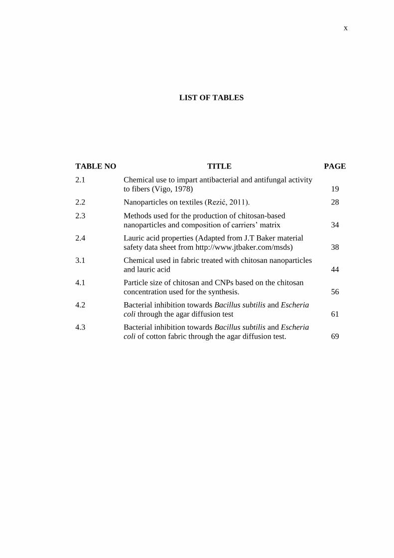
2.1 Chemical use to impart antibacterial and antifungal activity
to fibers (Vigo, 1978)
19
2.2 Nanoparticles on textiles (Rezić, 2011). 28
2.3 Methods used for the production of chitosan-based
nanoparticles and composition of carriers’ matrix
34
2.4 Lauric acid properties (Adapted from J.T Baker material
safety data sheet from http://www.jtbaker.com/msds)
38
3.1 Chemical used in fabric treated with chitosan nanoparticles
and lauric acid
44
4.1 Particle size of chitosan and CNPs based on the chitosan
concentration used for the synthesis.
56
4.2 Bacterial inhibition towards Bacillus subtilis and Escheria
coli through the agar diffusion test
61
4.3 Bacterial inhibition towards Bacillus subtilis and Escheria
coli of cotton fabric through the agar diffusion test.
69
xi
LIST OF FIGURES
FIGURE NO TITLE PAGE
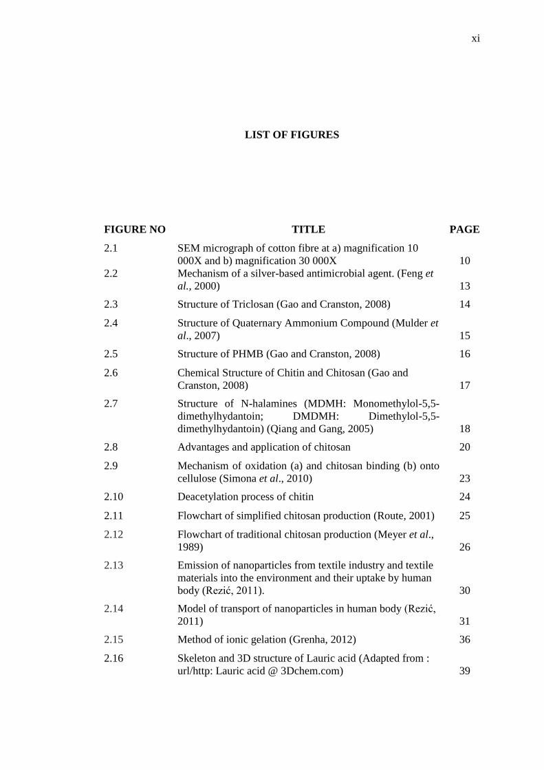
2.1 SEM micrograph of cotton fibre at a) magnification 10
000X and b) magnification 30 000X
10
2.2 Mechanism of a silver-based antimicrobial agent. (Feng et
al., 2000)
13
2.3 Structure of Triclosan (Gao and Cranston, 2008) 14
2.4 Structure of Quaternary Ammonium Compound (Mulder et
al., 2007)
15
2.5 Structure of PHMB (Gao and Cranston, 2008) 16
2.6 Chemical Structure of Chitin and Chitosan (Gao and
Cranston, 2008)
17
2.7 Structure of N-halamines (MDMH: Monomethylol-5,5-
dimethylhydantoin; DMDMH: Dimethylol-5,5-
dimethylhydantoin) (Qiang and Gang, 2005)
18
2.8 Advantages and application of chitosan 20
2.9 Mechanism of oxidation (a) and chitosan binding (b) onto
cellulose (Simona et al., 2010)
23
2.10 Deacetylation process of chitin 24
2.11 Flowchart of simplified chitosan production (Route, 2001) 25
2.12 Flowchart of traditional chitosan production (Meyer et al.,
1989)
26
2.13 Emission of nanoparticles from textile industry and textile
materials into the environment and their uptake by human
body (Rezić, 2011).
30
2.14 Model of transport of nanoparticles in human body (Rezić,
2011)
31
2.15 Method of ionic gelation (Grenha, 2012) 36
2.16 Skeleton and 3D structure of Lauric acid (Adapted from :
url/http: Lauric acid @ 3Dchem.com)
39
xii
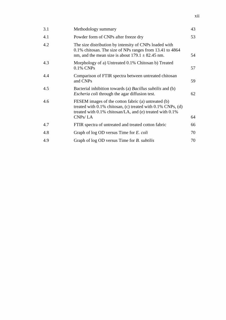
3.1 Methodology summary 43
4.1 Powder form of CNPs after freeze dry 53
4.2 The size distribution by intensity of CNPs loaded with
0.1% chitosan. The size of NPs ranges from 13.41 to 4864
nm, and the mean size is about 179.1 ± 82.45 nm.
54
4.3 Morphology of a) Untreated 0.1% Chitosan b) Treated
0.1% CNPs
57
4.4 Comparison of FTIR spectra between untreated chitosan
and CNPs
59
4.5 Bacterial inhibition towards (a) Bacillus subtilis and (b)
Escheria coli through the agar diffusion test.
62
4.6 FESEM images of the cotton fabric (a) untreated (b)
treated with 0.1% chitosan, (c) treated with 0.1% CNPs, (d)
treated with 0.1% chitosan/LA, and (e) treated with 0.1%
CNPs/ LA
64
4.7 FTIR spectra of untreated and treated cotton fabric 66
4.8 Graph of log OD versus Time for E. coli 70
4.9 Graph of log OD versus Time for B. subtilis 70
xiii
LIST OF SYMBOLS
ml Mililiter
µl Microliter
g Gram
°C Degree celcius
hr Hour
kV Kilo volt
wt Weight
rpm Revolution per minute
v/v Volume per volume
w/v Weight per volume
xiv
LIST OF ABBREVIATIONS
AATCC American Association of Textile Chemists and Colorists
AM Antimicrobial
AgNPs Silver nanoparticles
CAGR Compound Annual Growth Rate
CDC Centers for Disease Control and Prevention
CFU Colony Forming Unit
CNPs Chitosan nanoparticles
CNTs Carbon nanotubes
DMDMH Dimethylol-5-5-dimethylhydontoin
DNA Deoxyribonucleic acid
FESEM Field Emission Scanning Electron Microscopy
FTIR Fourier Transform Infrared Spectroscopy
HAIs Healthcare Associated Infections
HHS Health and Human Services
KBr Potassium Bromide
LA Lauric Acid
MDMH Monomethylol-5-5-dimethylhydontoin
MRSA Methicillin-resistant Staphylococcus aureus
NMA-HTCC O-acrylamidomethyl-N-[(2-hydroxy-3-
trimethylammonium)propyl] chitosan derivative
NPs Nanoparticles
TPP Pentasodium triphosphate/ tripolyphosphate
TNTs Titania nanotubes
UV-vis Ultraviolet visible
WHO World Health Organization
xv
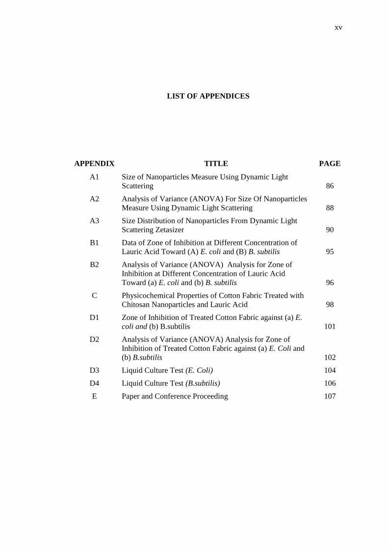
LIST OF APPENDICES
APPENDIX TITLE PAGE
A1 Size of Nanoparticles Measure Using Dynamic Light
Scattering
86
A2 Analysis of Variance (ANOVA) For Size Of Nanoparticles
Measure Using Dynamic Light Scattering
88
A3 Size Distribution of Nanoparticles From Dynamic Light
Scattering Zetasizer
90
B1 Data of Zone of Inhibition at Different Concentration of
Lauric Acid Toward (A) E. coli and (B) B. subtilis
95
B2 Analysis of Variance (ANOVA) Analysis for Zone of
Inhibition at Different Concentration of Lauric Acid
Toward (a) E. coli and (b) B. subtilis
96
C Physicochemical Properties of Cotton Fabric Treated with
Chitosan Nanoparticles and Lauric Acid
98
D1 Zone of Inhibition of Treated Cotton Fabric against (a) E.
coli and (b) B.subtilis
101
D2 Analysis of Variance (ANOVA) Analysis for Zone of
Inhibition of Treated Cotton Fabric against (a) E. Coli and
(b) B.subtilis
102
D3 Liquid Culture Test (E. Coli) 104
D4 Liquid Culture Test (B.subtilis) 106
E Paper and Conference Proceeding 107

1
CHAPTER 1
INTRODUCTION
1.1 Background of Study
Over the last twenty years, intensive research into new material and
procedures, that would assure permanent bioactive effects together with complete
safety for the people has been done due to the increase in the number of microbially
caused diseases and hospital infections (Chung et al., 1998; Liu et al., 2000; Lee at
al., 1999). There are commonly found microorganism in textile materials likes
mould, germs, Gram-positive and Gram-negative bacteria that have occupied every
habitat on earth from geothermal vents to the coldest Arctic ice and play both
beneficial and harmful roles in human lives.
According to the Centers for Disease Control and Prevention (CDC, USA,
2011), there are approximately 722,000 Healthcare Associated Infections (HAIs) and
75,000 associated deaths occur each year on account of infection-causing bacteria.
About 85% of all invasive methicillin-resistant Staphylococcus aureus (MRSA)
infections were associated with health care (Klevens et al., 2007). In 2005, there
were about 94,360 people infected with serious MRSA infection in the United States
and 18,650 people died.
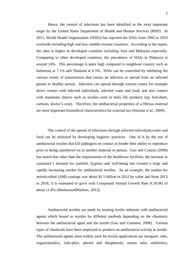
2
Hence, the control of infections has been identified as the most important
target by the United States Department of Health and Human Services (HHS). In
2011, World Health Organization (WHO) has reported the HAIs from 1995 to 2010
worlwide including high and low/ middle-income countries. According to the report,
the rates is higher in developed countries including Asia and Malaysia especially.
Comparing to other developed countries, the prevalence of HAIs in Malaysia is
around 14%. This percentage is quite high compared to neighbour country such as
Indonesia at 7.1% and Thailand at 6.5%. HAIs can be controlled by inhibiting the
various routes of transmission that causes an infection to spread from an infected
person to healthy person. Infection can spread through various routes for example
direct contact with infected individuals, infected water and food, and also contact
with inanimate objects such as textiles used in daily life products (eg: bed-sheet,
curtains, doctor’s coat). Therefore, the antibacterial properties of a fibrous material
are most important biomedical characteristics for external use (Simona et al., 2000).
The control of the spread of infections through infected individuals,water and
food can be achieved by developing hygienic practices. One of it by the use of
antibacterial textiles that kill pathogens on contact or hinder their ability to reproduce
prior to being transferred on to another material or person. Gao and Craston (2008)
has stated that other than the requirements of the healthcare facilities, the increase in
consumer’s demand for comfort, hygiene and well-being has created a large and
rapidly increasing market for antibacterial textiles. As an example, the market for
antimicrobial (AM) coatings was about $1.5 billion in 2012 by value and from 2013
to 2018, it is estimated to grow with Compound Annual Growth Rate (CAGR) of
about 11.8% (MarketsandMarkets, 2013).
Antibacterial textiles are made by treating textile substrate with antibacterial
agents which bound to textiles by different methods depending on the chemistry
between the antibacterial agent and the textile (Gao and Cranston, 2008). Various
types of chemicals have been employed to produce an antibacterial activity in textile.
The antibacterial agents most widely used for textile applications are inorganic salts,
organometalics, iodo-phor, phenol and thiophenols, onium salts, antibiotics,
3
heterocyclics with anionic groups, nitro compounds, ureas and related compounds,
formaldehyde derivatives and amines (Lim and Hudson, 2004a; Lim and Hudson,
2004b). Most of these chemicals are toxic to human and cannot easily degrade in
nature. These antibacterial agents have been studied independently and have been
proven to possess effective antibacterial ability.
The above factors have led to high interest in researching and developing
technology of natural antibacterial agent. One of the commonly used natural AM
agent is chitosan, a β-(1,4)-linked polysaccharide of D-glucosamine, is a deacetylated
form of chitin, the second most abundant natural polymer in the world (Ökem, 2003;
Sashiwa et al, 2004). Furthermore, in textile technology application, it is important
to adjoin some additional features in the natural biopolymer. Thus, various additives
such as lauric acid are incorporated into the chitosan solution to enhanced the
antibacterial activity of the treated fabric.
1.2 Problem Statements
Nowadays, the protection against infection of harmful and pathogenic
microorganism is strengthened. This is due to the facts that a safe, hygiene and
comfortable living environment turns to be important. Hence, the demand for the
healthcare textiles and medical textiles as ever increasing. Comparing to man-made
fibers, natural textiles, especially made from cellulose and protein fibers are often
considered to be more vulnerable towards microbe attack due to characteristics of
hydrophilic porous structure and moisture transport. Therefore, to prevent the
growth of bacteria using antibacterial agent becoming a standard finishing for textile
goods.
4
In textiles these can be achieved by treating the fabrics with silver salts,
quartenary ammonium chloride, metals, aromatic, halogen compounds, etc (Kenawy
et al., 2007; Takai et al., 2002). However, most of these antibacterial agents are
toxic and biocidal. Due to its non-toxicity and antibacterial properties, chitosan was
selected to be incorporated into fabric fiber as AM agent. According to Simona et al.
(2000), chitosan antibacterial activity is assigned on its amino groups, in diluted
acids from ammonium salts and the manipulation of chitosan’s binding strategies
onto cellulose surface. Based on the previous study on the AM textiles, most of the
research focused on synthesizing and evaluating uniquely distinct antibacterial agents
on different textile substrates with the aim of proving their effectiveness against
various microbes (Lim and Hudson, 2004; Qi et al., 2004; Ye et al., 2005; Demir et
al., 2010; Hebeish et al., 2013).
Chitosan nanoparticles (CNPs) is one of the alternatives that was investigated
for its AM properties in textile applications. According to Hebeish et al., (2013), the
nano form of chitosan is highly active due to very high surface area to volume ratio
and expected to have desirable bioactivity even at very low concentration. Lauric
acid (LA) is a crystalline fatty acid which has been shown to have AM effects
towards Gram-positive bacteria and yeast (Beuchat and Golden, 1989; Kabara,
1993). LA will be added to widen the spectrum activity of antibacterial mechanism
towards Gram-positive and Gram-negative bacteria.
In general, this study focused on the enhancement of antibacterial textiles in
terms of increase the fabric safety, increase natural antibacterial effectiveness
towards microbes and reducing the usage of chemical antibacterial agents by
replacing natural sources that are non-toxic and safe for the public usage.
5
1.3 Objectives of the Study
The main objective of this study is to develop antibacterial cotton fabric
incorporated with chitosan nanoparticles (CNPs) and lauric acid to enhance physical
and antibacterial properties of cotton fabric. The specific objectives of this study are:
1. To investigate the effect of chitosan concentration on the formation of
CNPs.
2. To determine the effect of LA concentration on antibacterial properties.
3. To characterize physical and antibacterial properties of treated cotton
fabric.
1.4 Scope of the Study
In order to achieve the objectives of this study, the work perform included all
the following scope.
1. The nano-sized particles of chitosan was extracted via ionotropic gelation
method basing on the reaction between the cationic amino groups of
chitosan and Tripolyphosphate (TPP).
2. Five different concentrations of chitosan from 0.1% to 0.5% (w/v) were
used to obtain the smallest nano size of particles.
3. An antibacterial solution was formulated by adding CNPs into the solution
with different LA ratio (0, 7,9,12, and 15 wt % of chitosan weight).
4. Formulation that give best antibacterial properties for CNPs-LA was
selected to be incorporated into fabric as antibacterial agent.
5. Physical properties of cotton fabric treated into CNPs-LA were analyzed
by surface structure and particles morphology.
6
6. Antibacterial study of the cotton fabric against Gram-positive (B. subtilis)
and Gram-negative (E. coli) bacteria were evaluated by agar diffusion test
and liquid culture test
1.5 Significance of the Study
Changing of human lifestyle nowadays, led to safe, healthy and comfortable
living environment keeps demanding. It is very important to keep strengthened the
protection from the infection of pathogenic microorganisms. Therefore, demand for
the healthcare and medical textiles keep increasing. Until now, a number of
chemical have been employed to impart the antibacterial activity of the textiles that
most of these chemical are toxic to human and cannot easily degrade in the nature.
The textile industry continuesly looking for the eco-friendly process that can be
carried out without toxic textile chemicals. Hence, the use of natural source of AM
agent like chitosan has been introduced to replace those chemical AM agent. The
incorporation of CNPs will increase the AM properties of the textiles. Addition of
LA will widen the AM activity of the textile.
1.6 Outline of the Thesis
This thesis consists of 5 Chapters. Chapter 1 introduces the introduction of
the research, significance of the study, research problem, the objective and the scope
of the study. Chapter 2 presents the literature review of the CNPs, antibacterial
fabrics, antibacterial agents used in fabric treatments, nanoparticles (NPs) in textiles
and ionotropic gelation method used to produced CNPs. Chapter 3 provides a
detailed methodology of this research to achieve the targeted objectives. Results and
7
discussion shows in Chapter 4 on physical properties and antibacterial activity of
CNPs and cotton fabric treated with CNPs solution. Effects of adding LA into the
solution also discussed in this chapter. Finally chapter 5 summarises the findings of
this study and suggestions for further work.
75
REFERENCES
Agrawal P., Strijkers G.J. and Nicolay K. (2010). Chitosan-based systems for
molecular imaging. Adv Drug Deliv Rev, 62, 42-58.
Ahamed, M., Karns, M., Goodson, M., Rowe, J., Hussain, S. M., Schlager, J. J. and
Hong, Y.(2008). DNA damage response to different surface chemistry of
silver nanoparticles in mammalian cells. Toxicology and Applied
Pharmacology, 233(3), 404-410.
Ahamed, M., Posgai, R., Gorey, T. J., Nielsen, M., Hussain, S. M. and Rowe, J.
J.(2010). Silver nanoparticles induced heat shock protein 70, oxidative stress
and apoptosis in Drosophila melanogaster. Toxicology and Applied
Pharmacology, 242(3), 263-269.
Ahmed A. Tayel, Shaaban H. Moussa, Wael F. El-Tras, Nihal M. Elguindy and
Klaus Opwis (2011). Antimicrobial textile treated with chitosan from
aspergillus niger mycelial waste. International Journal of Biological
Macromolecules. Elsevier. 49 ,241–245
Ali Hebeish, S. Sharaf and A. Farouk (2013). Utilization of chitosan nanoparticles as
a green finish in multifunctionalization of cotton textile. International
Journal of Biological Macromolecules, 60, 10-17.
Al-Qadi S, Grenha A and Remuñán-López C. (2011). Chitosan and its derivatives as
nanocarriers for siRNA delivery. J Drug Deliv Sci Technol.
Anahita R. S., Nahid H. N., and Azadesh B. (2014). Antibacterial finishing of cotton
fabric via the chitosan /TPP self-assembled nano layer. Fibers and Polymers,
15 (9), 1908-1914.
Ash Demir, Buket Arik, Esen Ozdogan and Necdet Seventekin (2010). A new
application method of chitosan for improved antimicrobial activity on wool
fabrics pretreated by different ways. Fibers and Polymers, 11 (3), 351-356.
76
Banerjee T, Mitra S, Singh AK, Sharma R.K. and Maitra A. (2002). Preparation,
characterization and biodistribution of ultrafine chitosan nanoparticles. Int J
Pharm, 243, 93-105.
Bayat A, Larijani B, Ahmadian S, Junginger HE and Rafiee-Tehrani M. (2008).
Preparation and characterization of insulin nanoparticles using chitosan and
its quaternized derivatives. Nanomed Nanotechnol Biol Med, 4, 115-120.
Berger, J., Reist, M., Mayer, J., Felt, O., Peppas, N. A. and Gurny R., (2004).
Structure and interactions in covalently and ionically crosslinked chitosan
hydrogels for biomedical applications. European Journal of Pharmaceutics
and Biopharmaceutics, 57 (1), 19-34.
Blackburn, R. S., Harvey, A. L., Kettle, L., Payne, J. D. and Russell, S. J.(2006).
Sorption of poly (hexamethylenebiguanide) on cellulose: mechanism of
binding and molecular recognition, Langmuir, 22, 5636–5644.
Calvo, P., C.Remunan-Lopez, J.L.Vila-Jato, and M.J.Alonso (1997). Novel
hydrophilic chitosan-polyethelene oxide nanoparticles as protein carriers.
Journal of Applied Polymer Science, 63, 125-132.
Casals, E., Vazquez-Campos, S., Bastus, N. G. and Puntes, V.(2008). Distribution
and potential toxicity of engineered inorganic nanoaprticles and carbon
nanostructures in biological systems. Analytical Chemistry, 27(8), 672-683.
Chaudury, A and Das, S. (2011). Recent advancement of chitosan-based
nanoparticles for oral controlled delivery of insulin and other therapeutic
agents. AAPS PharmSciTech, 12, 10-20.
Choi, O., Yu, C. P., Fernandez, G. E. and Hu, Z.(2010). Interactions of nanosilver
with Escherichia coli cells in planktonic and biofilm cultures. Water
Research, 44 (20), 6095-6103.
Chung, Y. S, Lee, K.K and Kim, J.W. (1998). Durable press and antimicrobial
finishing of cotton fabrics with a citric acid and chitosan treatment. Textile
Research Journal, 68 (10), 722-755.
Dastjerdi, R. and Montazer, M. (2010). A review on the application of inorganic
nano-structures materials in the modification of textiles: Focus on anti-
microbial properties. Colloids and Surfaces B: Biointerfaces, 79, 5-18.
Diana, A., Miquel, G. and Keiko, S. (2009). Cross-linking chitosan into UV-
irradiated cellulose fibers for the preparation of antimicrobial-finished
textiles. Carbohydrate Polymers, 77, 536-543.
77
de Campos A, Sanchez. A. and Alonso M.J. (2001). Chitosan nanoparticles: a new
vehicle for the improvement of the delivery of drugs to the ocular surface.
Application to cyclosporin A. Int J Pharm, 224,159-168.
Deepti Gupta and Adane Haile (2006). Multifunctional properties of cotton fabric
treated with chitosan and carboxymethyl chitosan. Carbohydrate Polymers,
69, 164-171.
Demir, M.M, Altın, B. and Özçelik, S. (2010). Composites of Reactive Silica
Nanoparticles and Poly (glycidyl methacrylate) with Linear and Crosslinked
Chains by in situ Bulk Polymerization. Composite Interfaces 17 (9), 831-844.
Duceppe N and Tabrizian M. (2010). Advances in using chitosan-based
nanoparticles for in vitro and in vivo drug and gene delivery. Expert Opin
Drug Deliv, 7, 1191-1207.
El-Shabouri, M. H. (2002). Positively charged nanoparticles for improving the oral
bioavailability of cyclosporin-A. Int J Pharm, 249, 101-108.
El-Tahlawy, K. F., El-Bendary, M. A., Elhandawy, A.G., and Hudson, S. M. (2005).
The antimicrobial activity of cotton fabric treated with different crosslinking
agents and chitosan. Carbohydrate Polymers, 60, 421-430.
Enig, M. G. (1998). Lauric oils as Antimicrobial Agents: Theory of Effect,
Scientific Rationale and Dietary Applications as Adjunct Nutritional Support
for HIV-Infected Individuals. In Watson, R. R. (Ed). Nutrients and Foods in
AIDS (pp. 81-97). Boca Raton: CRC Press.
E.S.K. Tang, M. Huang and L.Y. Lim, (2003). Ultrasonication of chitosan and
chitosan nanoparticles Journal of Pharmaceutics, 265, 103-114.
Fan W, Yan W, Xu Z and Ni H. (2012). Formation mechanism of monodisperse, low
molecular weight chitosan nanoparticles by ionic gelation technique. Colloid
Surf B-Biointerfaces, 90, 21-27.
Feng, Q.L., Wu, J., Chen, G.Q., Cui, F.Z., Kim, T. N. and Kim, J.O. (2000). A
mechanistic study of the antibacterial effect of silver ions on Escherichia coli
and Staphylococcus aureus. Biomedical Material Research, 52, 662-668.
Feng, P., Weagant, S.D. (ret), Grant, M.A. (dec) and Burkhardt, W. (2002).
Enumeration of Escheria coli and the coliform bacteria. U.S Food and Drug
Administration.
Fletcher, R. D., Albers, A. C., Albertson, J. N. and Kabara, J. J. (1985). effects of
monoglycerides on mycoplasma pneumoniae growth. In Kabara, J.J. (Ed.).
78
The Pharmacological Effect of Lipids II. Champaign Illinois: American Oil
Chemists' Society, 59-63.
Freedonia group, http://www.freedoniagroup.com/Disinfectant-And-Antimicrobial-
Chemicals.html, accessed on November 2014.
Gao, Y. and Cranston, R. (2008). Recent advances in antimicrobial treatments of
textiles. Textile Research Journal, 78, 60-72.
doi:10.1177/0040517507082332
Grenha, A. (2012). Chitosan nanoparticles: a survey of preparation methods. Journal
of Drug Targetting. 20(4), 291-300.
G. Skjak-Braek, T. Anthonsen and P.A. Sandford (1989). Chitin and chitosan.
sources, chemistry, biochemistry. physical properties and applications.
Elsevier Applied Science, London/New York.
Giri Joshi, Hohyun Lee, Yucheng Lan, Xiaowei Wang, Gaohua Zhu, Dezhi Wang,
Ryan W Gould, Diana C Cuff, Ming Y Tang, Mildred S Dresselhaus, Gang
Chen and Zhifeng Ren. (2008). Enhanced thermoelectric figure-of-merit in
nanostructured p-type silicon germanium bulk alloys. Nano letters 8 (12),
4670-4674.
Hagens, W. I., Oomen, A. G., de Jong, W. H., Cassee, F. R., and Sips, A. J. A.
M.(2007). What do we (need to) know about the kinetic properties of
nanoparticles in the body?. Regulatory Toxicology and Pharmacology, 49 (3),
217-229.
Hong-liang Zhang, Si-hui Wu, Yi Tao, Lin-quan Zang and Zheng-quan Su (2009).
Preparation and characterization of water soluble chitosan nanoparticles as
protein delivery system. Journal of Nanomaterials.
Doi:10.1155/2010/898910.
Iva Rezić (2011). Determination of engineered nanoparticles on textiles and in
textiles wastewaters. Trend in Analytical Chemistry, 30(7).
Jay, J. M. (2000). Modern Food Microbiology.(6th ed). USA: Aspen
Publishers, Inc. 343-351.
Jia, H., Zhu, G. and Wang, P., Catalytic behaviors of enzymes attached to
nanoparticles; the effect of particle mobility. Biotechnol. Bioeng., 84, 406-
414.
79
Kabara, J. J. (1978). Fatty acids and derivatives as antimicrobial agents - a review.
In Kabara, J. J. (Ed). The Pharmacological Effect of Lipids.. Champaign
Illinois: American Oil Chemists' Society. 1-14.
Kabara, J. J. (1985). Inhibition of Staphylococcus aureaus. In Kabara, J.J. (Ed). The
Pharmacological Effect of Lipids II. Champaign Illinois: American Oil
Chemists' Society.. 71-75.
Kaihara S, Suzuki Y. and Fujimoto K. (2011). In situ synthesis of polysaccharide
nanoparticles via polyion complex of carboxymethyl cellulose and chitosan.
Colloid Surf B Biointerfaces, 85, 343-348.
Kenawy, E.R., Worley, S.D. and Broughton, R. (2007). The chemistry and
applications of antimicrobial polymers: A state-of-the-art-review.
Biomacromolecules, 8(5), 1359-1384.
Kim,H. W., Kim, B. R. and Rhee, Y. H. (2010). Imparting durable antimicrobial
properties to cotton fabric using alginate-quaternary ammonium complex
nanoparticles. Carbohydrate Polymers, 79 (4), 1057-1062.
Klevens, R. M., Morrison, M. A., Nadle, J., Petit, S., Gershman, K., Ray, S.,
Harrison, L.H.,Lynfield, R., Dumyati, G., Townes, J. M., Craig, A. S., Zell,
E. R., Fosheim, G. E., McDougal, L. K., Carey, R. B. and Fridkin, S. K.
(2007). Invasive methicillin-resistance Staphylococcus aureus infections in
United States. Journal of the American Medical Association 2007; 298
(15):1763-1771.
Kut D., Orhan, M., Gunesoglu, C. and Ozakin, C. (2005). Effects of environmental
conditions on the antimicrobial activity of treated cotton knits. AATCC
Review, 5,25-28.
Lee, S., Cho, J.S. and Cho, G. (1999). Antimicrobial and blood repellent finishes for
cotton and nonwoven fabrics based in chitosan and fluoropolymers. Textile
Res. Journal, 69 (2), 104-112.
Li-Ming Zhao, Lu-E Shi, Zhi-Liang Zhang, Jian-Min Chen, Dong-Dong Shi, Jie
Yang and Zhen-Xing Tang (2011). Preparation and application of chitosan
naoparticles and nanofibers. Brazilian Journal of Chemical Engineering, 28
(3), 353-362.
Li, Q., Dunn, E.T., Grandmaison, E.W. and Goosen, M.F. in: M.F. Goosen (Ed.)
(1997). Application of chitin and chitosan. Technomic Publishing,
Lancaster, PA, 3–29.
80
Lim, S., H. and Hudson, M. (2003). Review of chitosan and its derivatives as
antimicrobial agents and their uses as textile chemicals. Journal of
Macromolecular Science, C43(2), 223- 269.
Lim, S. H. and Hudson, M, (2004a). Application of a fiber-reactive chitosan
derivative to cotton fabric as an antimicrobial textile finish. Carbohydrate
Polymer, 56, 227-234.
Lim, S. H. and Hudson, M, (2004b). Synthesis and antimicrobial activity of a water
soluble chitosan derivative with a fiber-reactive group. Carbohydrate
Research, 339, 313-319.
Lin Y-H, Mi F-L, Chen C-T, Chang W-C, Peng S-F, Liang H-F. and Sung H-W.
(2007). Preparation and characterisation of nanoparticles shelled with
chitosan for oral insulin delivery. Biomacromolecules, 8, 146-152.
Liu, X.F., Guan, Y.L., Yang, D.Z., Li, Z. and Yao, K.D., (2000). Antibacterial action
of chitosan and carboxymethylated chitosan. Journal of Applied Polymers,
70, 166-173.
Manchanda R, and Nimesh R. (2010). Controlled size chitosan nanoparticles as an
efficient, biocompatible oligonucleotides delivery system. J Appl Polym Sci,
118, 2071-2077.
Madigan, M. and Martinko, J. (2005). Brock Biology of Microorganisms. Upper
Saddle River, New Jersey: Pearson Prentice Hall.
Malek, J. R. and Speier, J. L. (1982). Development of organosilicone antimicrobial
agent for the treatment of surfaces. Journal of Coated Fabrics, 12, 38-46.
MarketsandMarkets.com, (2013). Antimicrobial coating market by type (silver,
copper & others), application (indoor air/HVAC, medical, mold remediation,
building & construction, food & beverages, textile & others) & geography (
North america, europe, asia-pacific, and ROW)- global trends and forecasts to
2018. www.marketsandmarkets.com/Market-Reports/antimicrobial-coatings-
market-1297.html. Excess on December 2015.
Mitra S, Gaur U, Ghosh P.C. and Maitra A.N. (2001). Tumor targeted delivery of
encapsulated dextrandoxorubicin conjugate using chitosan nanoparticles as
carriers. J Control Rel, 74, 317-323.
Mulder, G. D., Cavorsi, J. P. and Lee, D. K. (2007) Polyhexamethylenebiguanide
(PHMB): An addendum to current topical antimicrobials. Wounds, 19(7),
173-182.
81
Muzzarelli, R. A. A (1977). Chitin - Handbook of Fungal Biotechnology. Pergamon
Press: Oxford, U.K., 566.
N.A. Ivanova and A.B.Philipchenko (2012). Superhydrophobic chitosan-based
coatings for textile processing. Applied Surface Science, 263, 783-787.
Nakashima, T., Sakagami,Y., Ito, H. and Matsuo, M. (2001). Antibacterial activity of
cellulose fabrics modified with metallic salts. Textile Research Journal,
71(8), 688-694.
Nakano, M. M. and Zuber, P. (1998). Anaerobic Growth of a "strict aerobe"
(Bacillus Subtilis)". Annual Review of Microbiology. 52: 165-90.
National Center for Health Statistics. Health, United States, (2011): With Special
Feature on Socioeconomic Status and Health. Hyattsville, MD. 2012.
Ohya, Y., Shiratani, M., Kobayashi, H.and Ouchi, T. (1994). Release behaviour of
50fluroroucil from chitosan-gel nenosphere immobilizing 5-fluorouracil
coated with polysaccharides and their cell specific cytotoxicity. Pure Appl
Chem. A31, 629-642.
O.L. Shanmugasundaram (2006). Chitosan coated cotton yarn and its effect on
antimicrobial activity. Journal of Textile and Apparel, Technology and
Management, 5 (3).
Ökem,T. (2003). Surface treatment of cotton fabric with chitosan. Coloration
Technology, 119, 241-246.
Pochanavanich, P. and Suntornsuk, W. (2002). Fungal chitosan productionm and its
characterization. Lett. Appl. Microbiol. 35, 17-21.
P. Sandford, in: G. Skjak-Braek, T. Anthonsen, P.A. Sandford (Eds.) (1989). Chitin
and chitosan. sources, chemistry, biochemistry, physical properties and
application. Elsevier Applied Science, London/New York, pp. 51–69.
Pavlidou, V., (2005). New multifunctional textiles: antimicrobial treatments.
“Proceedings of the Intelligent Textile Structures—Application, Production
and Testing International Workshop”, Thessaloniki, Greece
http://centrum.vslib.cz/centrum/itsapt/greece2005.html (accessed June
2015).
Purwar, R., and Joshi, M., (2004). Recent developments in antimicrobial finishing of
textiles—a review, AATCC Review, 4, 22–26
Qi, L., Xu, Z., Jiang, X., Hu, C., and Zou, X., (2004). Preparation and antibacterial
activity of chitosan nanoparticles. Carbohydrate Research, 339, 2693-2700.
82
Rajesh D. A., Anand, S.C.; Kenndy, J.F.; Miraftab, M. and Rajendran, S.; (2006).
Role of advanced textile materials in healthcare, Medical Textiles and
Biomaterials for Healthcare, Woodhead Publ Ltd, ISBN 1-85573-683-7,
Cambridge, England, 90-98.
Rahman, M. F., Wang, J., Patterson, T. A., Saini, U. T., Robinson, B. L., Newport,
G. D., Murdock, R. C., Schlager, J. J., Hussain, S. M. and Ali, S. F. (2009).
Expression of genes related to oxidative stress in the mouse brain after
exposure to silver-25 nanoparticles. Toxicology Letters, 187 (1), 15-21.
Ruparelia, J. P., Chatterjee, A. K., Duttagupta, S. P. and Mukherji, S. (2008). Strain
specificity in antimicrobial activity of silver and copper nanoparticles. Acta
Biomaterialia, 4 (3), 707-716.
Sang-Hoon Lim and Samuel M. Hudson (2003). Review of chitosan and its
derivatives as antimicrobial agents and their uses as textiles chemical’s.
Journal of Macromolecular Science, 43, 223-269.
Sang-Hoon Lim and Samuel M. Hudson (2004). Application of a fiber-reactive
chitosan derivative to cotton fabric as an antimicrobial textile finish.
Carbohydrate Polymers, 56, 227-234.
Sandford PA (1989). Chitin and chitosan. London and NY: Elsevier Science
Publisher, 51-69.
Sarmento, B., Martins, S., Ribeiro, A., Veiga, F., Neufeld, R. and Ferreira, D.
(2006a). Development and comparison of different nanoparticulate
polyelectrolyte complexes as insulin carriers. Int JPeptide Res Ther, 12, 131-
138.
Sarmento, B., Ribeiro, A., Veiga, F. and Ferreira, D. (2006b). Development and
characterization of new insulin containing polysaccharide nanoparticles.
Colloid Surf B - Biointerfaces, 53, 193-202.
Sashiwa H, and Aiba S. (2004). Chemically modified chitin and chitosan as
biomaterials. Progress in Polymer Science, 29, 887-908
Shin, Y., and Min, K. (1996). Antimicrobial finishing of cotton fabrics with chitosan
(I)- Effect of degree of deacetylation on the antimicrobial property. Journal
of Korean Fiber Society, 33, 487-491.
Shin, Y., Yoo, D.I. and Jang, J.(2000). Molecular weight effect on antimicrobial
activity of chitosan treated cotton fabrics. Journal of Applied Polymer
Science ,80, 2495-2501.
83
Shin, Y., Yoo D.I. and Jang J. (2001).Molecular Weight Effect on Antimicrobial
Activity of Chitosan Treated Cotton Fabric. Application Polymer Science,
80, 2495.
Shindler, W. D. and Hauser, P.J. (2004). Antimicrobial finishes. Chemical finishing
of textiles. Woodhead Publishing Limited. Cambridge England:. 165-174.
Shubayev, V. I., Pisanic II, T. R., Jin, S. (2009). Magnetic nanoparticles for
theragnostics. Advanced Drug Delivery Review, 61, 469-477.
Simona Strnad, Olivera Šauperl and Lidija Fras-Zemljič. (2000) Cellulose fibres
functionalised by chitosan: characterization and application. Biopolymers,
Magdy Elnashar (Ed.), ISBN:978-953-307-109-1, In-Tech.
Simoncic, B. and Tomsic, B. (2010). Structures of novel antimicrobial agents for
textiles- A review. Textile Research Journal, 0 (0), 1-17.
Doi:10.1177/0040517510363193.
Songjiang, Z. and Lixiang, W. (2009). Amyloid-Beta associated with chitosan nano-
carrier has favorable immunogenicity and permeates the BBB. AAPS
PharmsciTech, 10, 900-905.
Suédina M. L. S., Carla, R. C. Braga, Marcus V. L., Fook, Claudia M.O. Raposo,
Laura H. Carvalho and Eduardo L. Canedo (2012). Application of infrared
spectroscopy to analysis of chitosan/clay nanocomposites. Infrared
Spectroscopy-Material Sciences, Engineering and Technology, InTech.
ISBN 978-953-51-0537-4
Takai, K., Ohtsuka, T., Senda, Y., Nakao, M., Yamamoto, K.,and Matsuoka, J.,
(2002). Antibacterial properties of antimicrobial-finished textile products.
Microbiology and Immunology, 46 (2), 75-81.
Tang, Z.X., Qian, J. Q. and Xi, L.E. (2007). Preparation of chitosan nanoparticles as
carrier for immobilized enzyme. Appl Biochem Biotechnol, 136, 77-96.
Teijeiro-Osorio D, Remuñan-López C, Alonso MJ. (2009). New generation of hybrid
poly/oligosaccharide nanoparticles as carriers for the nasal delivery of
macromolecules. Biomacromolecules, 10, 243-249.
Terbojevich M. and Muzzarelli RAA. (2009). Chitosan. In: Phillips GO, Williams P,
eds. Handbook of hydrocolloids. Cambridge: Woodhead Publishing Ltd.,
367-378.
84
Teng, W. L.; Khor, E.; Tan, T. K.; Lim, L. Y. and Tan, S. C. (2001). Concurrent
production of chitin from shrimp shell and fungi. Carbohydr. Research., 332,
305-316.
Textiles Inteligence. (2004). Antimicrobial fabrics help fight war against germs.
www.textilesintelligence.com/til/press.cfm?prid=325. Access on June 2015.
Tokumitsu, H., Ichikawa, H., Fukumori, Y. and Block, L.H. (1999a). Preparation of
gadopentetic acidloaded chitosan microparticles for gadolinium neutron-
capture therapy of cancer by a novel emulsion-droplet coalescence technique.
Chem Pharm Bull, 47, 838-842.
Tokumitsu, H., Ichikawa, H. and Fukumori, Y. (1999b). Chitosan-gadopentetic acid
complexnanoparticles for gadolinium neutron capture therapy of cancer:
preparation by novel emulsiondroplet coalescence technique and
characterization. Pharm Res, 16, 1830-1835.
Vigo, T. L. (1978). Antibacterial fibers. Academic Press, Inc. ISBN 0-12-599750-7
Vigo, T.L. Lewin, M. and Sello, S.B. (Eds.) (1983). Handbook of fiber science and
technology, vol. II, Marcel Dekker, New York, 19, 367–426.
Wang, X., Chi, N. and Tang, X. (2008). Preparation of estradiol chitosan
nanoparticles for improving nasal absorption and brain targeting. Eur J
Pharm Biopharm, 70, 735-740.
Westers, L., Westers, H., and Wim, J. Q., (2004). Bacillus subtilis as cell factory for
pharmaceutical proteins: a biotechnological approach to optimize the host
organism. Molecular Cell Research, 1694 (1), 299-310.
White, W. C. and Monticello, R. A. (2002). Antimicrobial performance of medical
textiles. Presented at IFAI Expo, Charlotte, NC.
Williams, J. F., HaloSource, V., and Cho, U (2005). Antimicrobial functions for
synthetic fibers: recent developments, AATCC Review, 5, 17–21.
World Health organization (WHO). Report on the burden of endemic health Care-
associated infection worldwide. Switzerland, ISBN 978-92-4-150150-7.
Ye, W., Leung, M. F., John, X., Kwong, T. L., Daniel, K. L. L. and Li, P. (2005).
Novel core-shell particles with poly(n-butyl acrylate) cores and chitosan
shells as an antibacterial coating for textiles. Polymer, 46, 10538-10543.
Y.H.Kim, H.M. Choi, and J.H. Yoon (1998). Synthesis of a quarternary ammonium
derivative of chitosan and its application to a cotton antimicrobial finish.
Textile Research Journal, 68 (6), 428-434.
85
Young, D.H., Kohle, H. and Kauss, H. (1982). Effect of chitosan on membrane
permeability of suspension-cultured Glycine max and Phaseolus vulgaris
Cells. Plant Physiology, 70,1449-1454.
Zhang, X., Pei, X., Zhang, J., and Wang, Q. (2009). Effects of carbon fiber surface
treatment on the friction and wear behavior of 2D woven carbon
fabric/phenolic composits. Colloids and Surfaces A: Physicochemical Eng
Aspects, 339, 7-12.