Embed Size (px)
Citation preview
IN VITRO ASSESSMENT OF COAGULATION
ACTIVITIES IN HUMAN PLASMA TREATED WITH
AJWA DATES (PHOENIX DACTYLIFERA L.)
EXTRACTS
By
AINUL MARDHIYAH BINTI ZAKARIA
Dissertation Submitted In Partial Fulfilment
Of The Requirements For The Degree Of
Master Of Science (Transfusion Science)
UNIVERSITI SAINS MALAYSIA
AUGUST 2015
i
DECLARATION
I hereby declare that this dissertation is based on my original work. I also declare that
this dissertation has not previously or concurrently submitted by any other master
student at USM or other institutions.
AUGUST 2015 ……………………………………….
AINUL MARDHIYAH BINTI ZAKARIA
900430-08-6486
PIPM0074/14
ii
ACKNOWLEDGEMENTS
In the name of Allah, the Exalted, Most Benevolent. Peace of Allah be upon the
Prophet Muhammad, his family, companions and to the righteous.
All Praise to Allah, with His Blessings, Merciful and Tenderness, I am able to
complete my research and dissertation.
First and foremost, I would like to extend my utmost gratitude towards my
supervisor, Dr. Abdul Rahim Hussein for his endless support, guidance, consideration
and advice. Greatest appreciation goes to my co-supervisor, Dr. Lim Vuanghao for his
support and encouragement. Also, my appreciation goes to my Head of Cluster, Dr.
Badrul Hisham Yahaya for his guidance and advice throughout the completion of this
research project as a whole.
Special thanks go to the Director of Advanced Medical and Dental Institute
(AMDI), Professor Abd Aziz Tajuddin, all lectures, Madam Srly Saman, Mr Ahmad
Firdaus Abdul Hadi, Mr Nizuwan Azman, Mr Mohd Najwan Mohd Nasir, Miss Faizatul
Syima Abd Manaf, Madam Rohani Bakar, Madam Nur Shahida Rusli, Miss Erma and
everybody involved directly or indirectly during the progress of the study.
My thanks also goes to my fantastic course-mates; Wan Norshazwani Wan
Shaffee, Nurul Munira Yahya, Kong Hung Chuo, Oor Vasi a/p Samynazan, Mageswary
a/p Nageswaran and Dr. Muhammad Aslam Farooqui for their excellent cooperation,
support and understanding.
Last but not least, thanks to my beloved family for their supplication, love and
warmth.
Thank you.
iii
CONTENTS
LIST OF TABLES vi
LIST OF FIGURES vii
LIST OF ABBREVIATIONS viii
ABSTRAK x
ABSTRACT xi
CHAPTER 1: INTRODUCTION 1
1.1 Literature review 1
1.1.1 Haemostasis 1
1.1.1.1 Definition 1
1.1.1.2 Coagulation in Haemostasis 2
1.1.1.3 Coagulation Pathway 3
1.1.1.4 Haemostatic Screening Assay 4
1.1.1.5 Disorders of Defective Haemostasis 6
1.1.1.6 Anti-coagulant Drugs 9
1.1.2 Dates 10
1.1.2.1 Overview 10
1.1.2.2 Dates Rich in Salicylic Acid 11
1.1.2.3 Dates and Haemostatic Activities 12
1.1.3 Salicylic Acid 13
1.1.3.1 Salicylic Acid in Human 13
1.1.3.2 Salicylic Acid in Foods and Dates 13
1.1.3.3 Mechanism of Action 15
1.2 Problem Statement / Research Question 16
1.3 Research Objectives 18
iv
1.3.1 General 18
1.3.2 Specific 18
1.4 Research Hypothesis 18
1.5 Rationale of Study / Expected Outcome 18
CHAPTER 2: METHODOLOGY 19
2.1 Study Design 19
2.2 Extraction 19
2.2.1 Ajwa Dates Collection 19
2.2.2 Ethanol Extraction 20
2.2.3 Aqueous Extraction 20
2.2.4 Yield Percentage (%) Calculation 21
2.3 GC-MS analysis 21
2.3.1 Crude Extracts Derivatization 21
2.3.2 Analysis of Compounds 21
2.4 Assessment of Coagulation Activities 22
2.4.1 Sample Size Calculation 22
2.4.2 Donor Recruitments 22
2.4.3 Blood Collection 23
2.4.4 Platelet Count 23
2.4.5 Measurement of Coagulation Factors 23
2.4.6 Sample Disposal 24
2.5 Statistical Analysis 24
CHAPTER 3: RESULTS 26
3.1 Extracts Yielding 26
3.2 Platelets Count 26
3.3 Prothrombin Time (PT) 26
3.3.1 Ethanol Extract 26
v
3.3.2 Aqueous Extract 27
3.4 Activated Partial Thromboplastin Time (APTT) 27
3.4.1 Ethanol Extract 27
3.4.2 Aqueous Extract 29
3.5 Thrombin Time (TT) 29
3.5.1 Ethanol Extract 29
3.5.2 Aqueous Extract 30
3.6 Gas chromatography-mass spectrometry (GC-MS) 33
CHAPTER 4: DISCUSSION AND LIMITATIONS 36
CHAPTER 5: CONCLUSION AND RECOMMENDATIONS 45
REFERENCES 47
APPENDIX 52
vi
LIST OF TABLES
Tables Titles Pages
Table 1.1 Coagulation Factors and Inhibitors 6
Table 1.2 Coagulation disorders derived from congenital or acquired
abnormalities
8
Table 3.1
Clotting time for coagulation parameters when treated with
Ajwa dates ethanol and aqueous extracts at concentration 0.1
g/mL(I), 0.5 g/mL(II) and 1.0 g/mL(III)
32
Table 3.2 Compounds in Ajwa dates ethanol extracts by GCMS at
1mg/mL 33
Table 3.3
Compounds in Ajwa dates aqueous extracts by GCMS at
1mg/mL 33
vii
LIST OF FIGURES
Figures Titles Pages
Figure 1.1 The more in-depth version of the coagulation cascade 7
Figure 1.2 Skeletal formulas of acetylsalicylic acid and salicylic acid 15
Figure 2.1 Experimental study design of the research 25
Figure 3.1 PT clotting time when treated with Ajwa dates ethanol and
aqueous extracts at concentration 0.1 g/mL(I), 0.5g/mL(II)
and 1.0 g/mL(III)
28
Figure 3.2 APTT clotting time when treated with Ajwa dates ethanol
and aqueous extracts at concentration 0.1 g/mL(I), 0.5
g/mL(II) and 1.0 g/mL(III)
30
Figure 3.3 TT clotting time when treated with Ajwa dates ethanol and
aqueous extracts at concentration 0.1 g/mL(I), 0.5 g/mL(II)
and 1.0 g/mL(III)
31
Figure 3.4
Total Ion Chromatogram (TIC) of Ajwa dates ethanol
extracts based on the compound abundance against the
retention time (tR)
34
Figure 3.5
Total Ion Chromatogram (TIC) of Ajwa dates aqueous
extracts based on the compound abundance against the
retention time (tR)
35
viii
LIST OF ABBREVIATIONS
% Percent
°C Degree Celcius
> More than
< Less than
g Gravity
α Alpha
g Grams
σ Sigma
δ Delta
γ Gamma
kg Kilogram
µL Microlitre
s Seconds
○ Outliers
* Extreme Outliers
a Activated
mg Milligram
h Hours
min Minutes
mL Millilitre
RT Room Temperature
PPP Platelet-Poor Plasma
SPSS Statistical Package for the Social Sciences
TMB Tetramethylbenzidine
tR Retention Time
v/v Volume per Volume
VKA Vitamin K Antagonist
ix
GC-MS Gas Chromatography Mass Spectrometry
ET/EtOH Ethanol
AQ Aqueous
HREC Human Research Ethics Committee
AMDI Advanced Medical and Dental Institute
PDN National Blood Centre
BSTFA N,O-bis(trimethylsilyl) trifluoroacetamide
TMCS Trimethylchlorosilane
RPM Rotation per Minute
VTE Venous Thromboembolism
PT Prothrombin Time
APTT Activated Partial Thromboplastin Time
TT Thrombin Time
HPLC High Performance Liquid Chromatography
F Factor
TFPI Tissue Factor Pathway Inhibitor
HMWK High Molecular Weight Kininogen
CBC Complete Blood Count
DIC Disseminated Intravascular Coagulation
EDTA Ethylene-Diamine Tetraacetic Acid
TIC Total Ion Chromatogram
MI Myocardial Infarction
CAM Complementary and Alternative Medicine
RI Retention Time Index
RBC Red Blood Cell
Hb Hemoglobin
PCV Pack Cell Volume
x
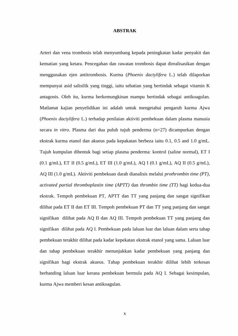
ABSTRAK
Arteri dan vena trombosis telah menyumbang kepada peningkatan kadar penyakit dan
kematian yang ketara. Pencegahan dan rawatan trombosis dapat direalisasikan dengan
menggunakan ejen antitrombosis. Kurma (Phoenix dactylifera L.) telah dilaporkan
mempunyai asid salisilik yang tinggi, iaitu sebatian yang bertindak sebagai vitamin K
antagonis. Oleh itu, kurma berkemungkinan mampu bertindak sebagai antikoagulan.
Matlamat kajian penyelidikan ini adalah untuk mengetahui pengaruh kurma Ajwa
(Phoenix dactylifera L.) terhadap penilaian aktiviti pembekuan dalam plasma manusia
secara in vitro. Plasma dari dua puluh tujuh penderma (n=27) dicampurkan dengan
ekstrak kurma etanol dan akueus pada kepakatan berbeza iaitu 0.1, 0.5 and 1.0 g/mL.
Tujuh kumpulan dibentuk bagi setiap plasma penderma: kontrol (saline normal), ET I
(0.1 g/mL), ET II (0.5 g/mL), ET III (1.0 g/mL), AQ I (0.1 g/mL), AQ II (0.5 g/mL),
AQ III (1.0 g/mL). Aktiviti pembekuan darah dianalisis melalui prothrombin time (PT),
activated partial thromboplastin time (APTT) dan thrombin time (TT) bagi kedua-dua
ekstrak. Tempoh pembekuan PT, APTT dan TT yang panjang dan sangat signifikan
dilihat pada ET II dan ET III. Tempoh pembekuan PT dan TT yang panjang dan sangat
signifikan dilihat pada AQ II dan AQ III. Tempoh pembekuan TT yang panjang dan
signifikan dilihat pada AQ I. Pembekuan pada laluan luar dan laluan dalam serta tahap
pembekuan terakhir dilihat pada kadar kepekatan ekstrak etanol yang sama. Laluan luar
dan tahap pembekuan terakhir menunjukkan kadar pembekuan yang panjang dan
signifikan bagi ekstrak akueus. Tahap pembekuan terakhir dilihat lebih terkesan
berbanding laluan luar kerana pembekuan bermula pada AQ I. Sebagai kesimpulan,
kurma Ajwa memberi kesan antikoagulan.
xi
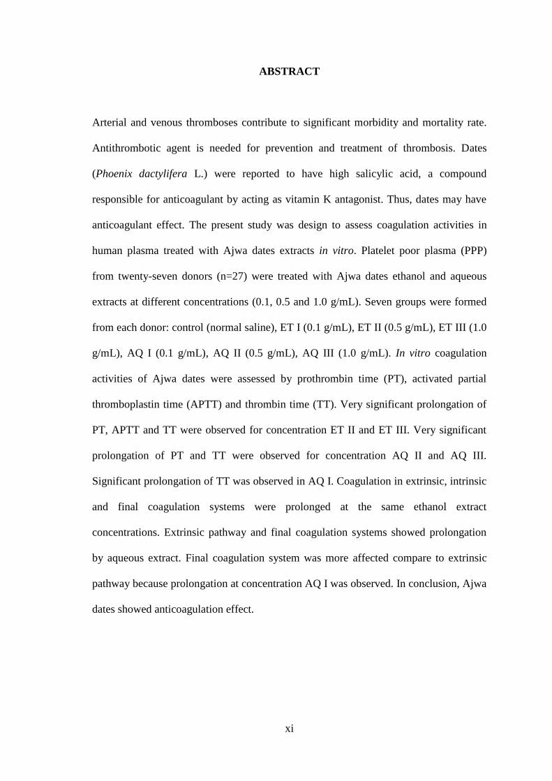
ABSTRACT
Arterial and venous thromboses contribute to significant morbidity and mortality rate.
Antithrombotic agent is needed for prevention and treatment of thrombosis. Dates
(Phoenix dactylifera L.) were reported to have high salicylic acid, a compound
responsible for anticoagulant by acting as vitamin K antagonist. Thus, dates may have
anticoagulant effect. The present study was design to assess coagulation activities in
human plasma treated with Ajwa dates extracts in vitro. Platelet poor plasma (PPP)
from twenty-seven donors (n=27) were treated with Ajwa dates ethanol and aqueous
extracts at different concentrations (0.1, 0.5 and 1.0 g/mL). Seven groups were formed
from each donor: control (normal saline), ET I (0.1 g/mL), ET II (0.5 g/mL), ET III (1.0
g/mL), AQ I (0.1 g/mL), AQ II (0.5 g/mL), AQ III (1.0 g/mL). In vitro coagulation
activities of Ajwa dates were assessed by prothrombin time (PT), activated partial
thromboplastin time (APTT) and thrombin time (TT). Very significant prolongation of
PT, APTT and TT were observed for concentration ET II and ET III. Very significant
prolongation of PT and TT were observed for concentration AQ II and AQ III.
Significant prolongation of TT was observed in AQ I. Coagulation in extrinsic, intrinsic
and final coagulation systems were prolonged at the same ethanol extract
concentrations. Extrinsic pathway and final coagulation systems showed prolongation
by aqueous extract. Final coagulation system was more affected compare to extrinsic
pathway because prolongation at concentration AQ I was observed. In conclusion, Ajwa
dates showed anticoagulation effect.
1
CHAPTER I
INTRODUCTION
1.1 Literature Review
1.1.1 Haemostasis
1.1.1.1 Definition
Haemostasis is a dynamic, ordered and well-regulated body process of blood fluids
maintenance, vascular damage repairing and bleeding arrest (Zehnder 2012, p. 601).
Haemostasis aims to prevent intravascular blood clot formation and to stop blood loss from
blood vessel (Estridge & Reynolds 2008). Abnormality in normal haemostasis will disrupt
the balance maintained in haemostasis mechanism and thus giving rise to two extreme and
distinct conditions; obstructive intravascular clotting (thrombosis) or excessive bleedings
(haemorrhage) (Zehnder 2012, p. 601; Sood, 2010). Haemostasis is governed tightly to
prevent thrombosis and/or haemorrhage by substantial negative feedback mechanism of the
endothelium and plasma (Shaz et al. 2013) as well as the delicate balance in procoagulant,
anticoagulant and fibrinolysis process in haemostasis mechanism (Hoffbrand, Moss &
Pettit 2006). Haemostasis involved five components that define haemostasis in an orderly
events starting from blood vessels, platelets, coagulation factors, coagulation inhibitors and
lastly fibrinolysis (Estridge & Reynolds 2008; Hoffbrand, Moss & Pettit 2006).
Haemostasis is a multistep process that is divided into two; primary and secondary (Fowler
& Perry 2015; Longo 2013). Primary haemostasis involved the action of the first two
components of haemostasis, blood vessels and platelets while secondary haemostasis refers
2
to the action of coagulation factors and inhibitors as well as fibrinolysis (Fowler & Perry
2015). Contrary to Fowler & Perry (2015) statement, Longo (2013) stated that primary
haemostasis describes the coagulation system while secondary haemostasis describes the
other factors that contribute to haemostasis for example the blood vessel and platelets.
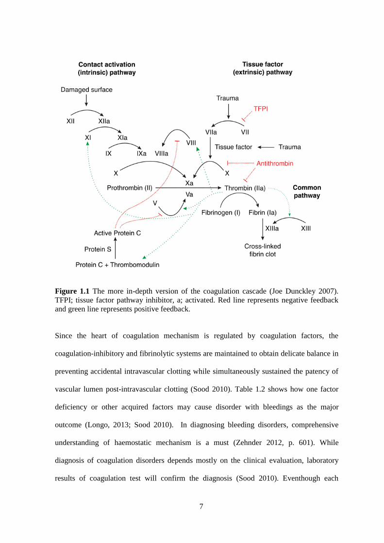
1.1.1.2 Coagulation in haemostasis
Coagulation is one of the components of haemostasis that follows the establishment of
blood vessel and platelets action during bleeding (Figure 1.1). When bleeding occurs,
damaged blood vessel and the regional small arteries and arterioles will constrict to reduce
blood flow; thereby allowing contact activation of platelets and coagulation factors
(Hoffbrand, Moss & Pettit 2006). If bleeding is too severe and critical, the cut edges of the
bleeding site will retract and compressed by contracted skeletal muscles. During this
retraction and contraction process, small capillary vessels will seal themselves when the
cut edges touch to one another (Estridge & Reynolds 2008). Then, platelets circulating in
the blood stream will adhere to the collagen of the exposed connective tissue and caused
platelets to activate by releasing substances from their granules (Estridge & Reynolds
2008; Hoffbrand, Moss & Pettit 2006). Adhesion of platelets will be followed by platelet
aggregation and subsequently blood coagulation in an extremely rapid manner depending
on von Willebrand factor, fibronectin, divalent cations, calcium, magnesium and
glycoprotein IIb/ IIIa complex in platelet membrane (Sood 2010). Coagulation of blood is
determined by coagulation factors and inhibitors (Table 1.1). Coagulation factors include
plasma proteins factor I till factor XIII (except ionized calcium; previously known as
Factor IV) (Estridge & Reynolds 2008; Hoffbrand et al. 2006). These proteins are produce
in the liver and remains in the blood in inactive form. However, when blood vessel is
damaged, haemostasis signals the activation of coagulation factors, leading to the
3
formation of fibrin clot (Estridge & Reynolds 2008). On the other hand, coagulation
inhibitors include tissue factor pathway inhibitor (TFPI), heparin cofactor II, α2-
Macroglobulins, α2-antiplasmin, C1 esterase inhibitor and α1-antitrypsin (Hoffbrand, Moss
& Pettit 2006). When fibrin clot is formed, bleeding will slowly stop as the site of injury is
healing. At this point, fibrinolysis is needed to dissolve fibrin clot by the action of
plasminogen and plasmin. Activation of plasminogen to plasmin is stimulated from injured
vessel wall or from tissues (Hoffbrand, Moss & Pettit 2006). To sum it all, primary
haemostasis takes place when sub-endothelial of collagen is exposed; signalling the
initiation of platelet adhesion, granule secretion and initial aggregation. Following primary
haemostasis, thrombin generation and fibrin clot formation marked the hallmark of
secondary haemostasis (Fowler & Perry 2015).
1.1.1.3 Coagulation pathway
Coagulation process is best understood through coagulation pathway. Complex interaction
between coagulation factors and inhibitors is shown through three pathways; intrinsic,
extrinsic and common (Figure 1.1). Coagulation cascade describe the action of each factors
in contributing to the final formation of fibrin clot while preventing thrombosis formation
(Table 1.1 and Figure 1.1). In this cascade, the common pathway is activated by either the
extrinsic or intrinsic pathway or by interaction between these two pathways (Estridge &
Reynolds 2008). Tissue factor or alternatively known as FIII or thromboplastin does not
present in the circulating blood, hence the name extrinsic pathway (Estridge & Reynolds
2008). Initiation of extrinsic pathway by TPFI occurs during vessel damages (trauma)
(Estridge & Reynolds 2008). With the presence of calcium, FVII will be activated and
subsequently activating FX (Estridge & Reynolds 2008). Intrinsic pathway is named
because the factors involved in this pathway are circulating in the blood (Estridge &
4
Reynolds 2008). Intrinsic pathway is initiated when FXII or alternatively known as contact
factor is activated by damaged surface (Estridge & Reynolds 2008). Activated FXII will
then subsequently activate FXI and the subsequent activated FXI will activate FIX
(Estridge & Reynolds 2008). Activated FIX will participate in the activation of FX that
will convert prothrombin to thrombin (Estridge & Reynolds 2008). In common pathway,
fibrinogen will be converted to fibrin by thrombin. With the aid of activated FXIII, fibrin
clot will form and stabilized when fibrin cross-linked each other (Estridge & Reynolds
2008; Hoffbrand, Moss & Pettit 2006).
1.1.1.4 Haemostatic screening assay
Defects in haemostasis may arise from vascular disorders, thrombocytopenia or platelet
function disorder as well as problems in blood coagulation. Due to these reasons, screening
test for blood coagulation disorders are platelet count, thrombin time (TT), prothrombin
time (PT), activated partial thromboplastin time (APTT) and fibrinogen quantitation
(Estridge & Reynolds 2008; Fowler & Perry 2015; Hoffbrand, Moss & Pettit 2006). With
regard to assessment of coagulation activity; PT, APTT and TT are used to evaluate
common and extrinsic pathway; common and intrinsic pathway; and final coagulation
stage respectively (Estridge & Reynolds 2008; Fowler & Perry 2015; Hoffbrand, Moss &
Pettit 2006; Torres-Urrutia et al. 2013). Battery of test is compulsory when assessing
coagulation abnormalities because no single test is capable to sufficiently detect such
abnormalities (Sood 2010). PT, APTT and TT served as initial screening for clotting
factor activity (Longo 2013) and also regarded as functional tests because they monitored
the formation of clot. PT measures fibrinogen (Hoffbrand, Moss & Pettit 2006), FII, FV,
FVII and FX while APTT measures FX, FV, FII, fibrinogen (Hoffbrand, Moss & Pettit
2006), FVIII, FIX and FXI (Fowler & Perry 2015; Hoffbrand, Moss & Pettit 2006).
5
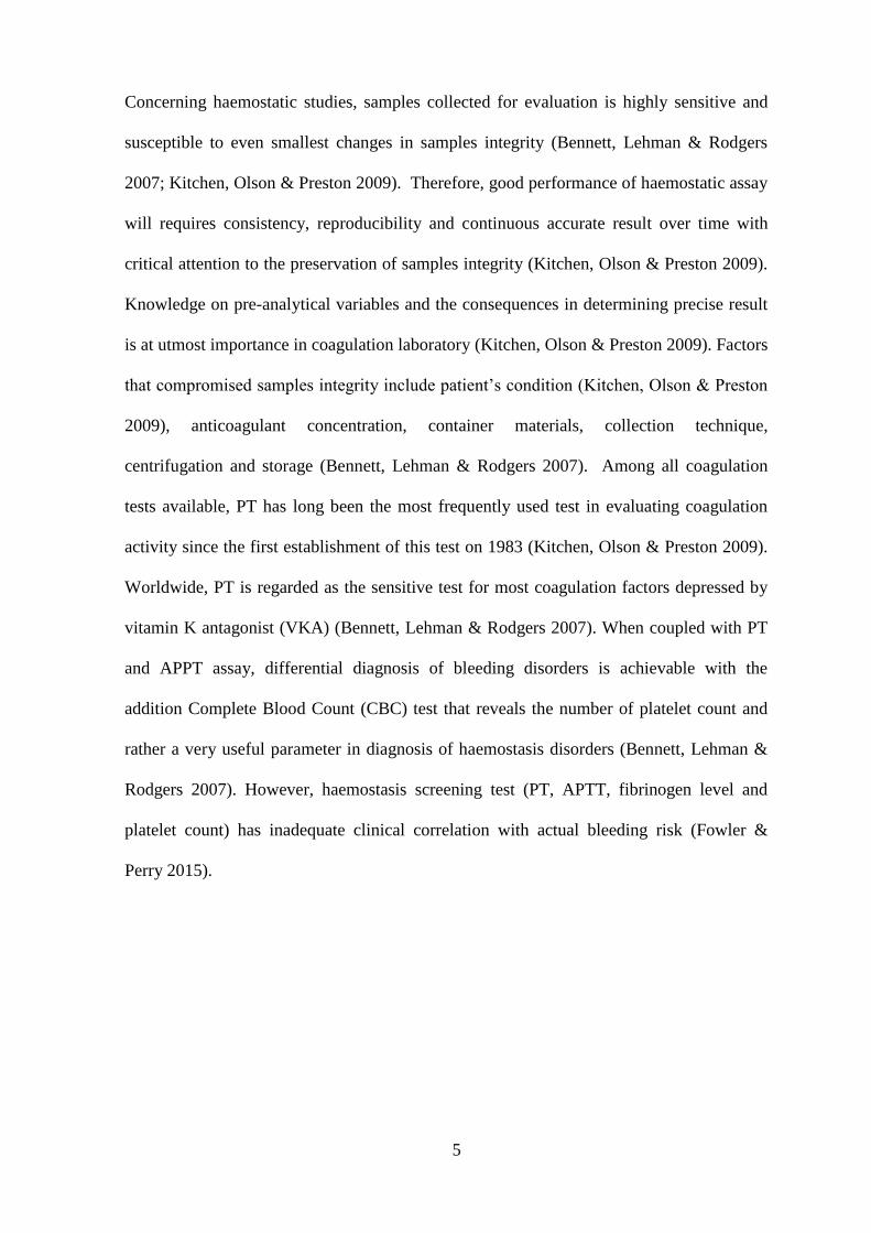
Concerning haemostatic studies, samples collected for evaluation is highly sensitive and
susceptible to even smallest changes in samples integrity (Bennett, Lehman & Rodgers
2007; Kitchen, Olson & Preston 2009). Therefore, good performance of haemostatic assay
will requires consistency, reproducibility and continuous accurate result over time with
critical attention to the preservation of samples integrity (Kitchen, Olson & Preston 2009).
Knowledge on pre-analytical variables and the consequences in determining precise result
is at utmost importance in coagulation laboratory (Kitchen, Olson & Preston 2009). Factors
that compromised samples integrity include patient’s condition (Kitchen, Olson & Preston
2009), anticoagulant concentration, container materials, collection technique,
centrifugation and storage (Bennett, Lehman & Rodgers 2007). Among all coagulation
tests available, PT has long been the most frequently used test in evaluating coagulation
activity since the first establishment of this test on 1983 (Kitchen, Olson & Preston 2009).
Worldwide, PT is regarded as the sensitive test for most coagulation factors depressed by
vitamin K antagonist (VKA) (Bennett, Lehman & Rodgers 2007). When coupled with PT
and APPT assay, differential diagnosis of bleeding disorders is achievable with the
addition Complete Blood Count (CBC) test that reveals the number of platelet count and
rather a very useful parameter in diagnosis of haemostasis disorders (Bennett, Lehman &
Rodgers 2007). However, haemostasis screening test (PT, APTT, fibrinogen level and
platelet count) has inadequate clinical correlation with actual bleeding risk (Fowler &
Perry 2015).
6
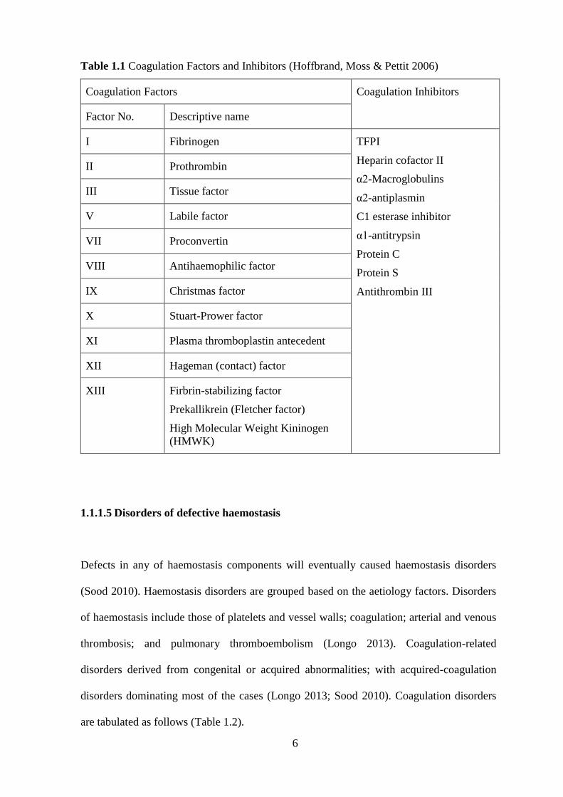
Table 1.1 Coagulation Factors and Inhibitors (Hoffbrand, Moss & Pettit 2006)
Coagulation Factors Coagulation Inhibitors
Factor No. Descriptive name
I Fibrinogen TFPI
Heparin cofactor II
α2-Macroglobulins
α2-antiplasmin
C1 esterase inhibitor
α1-antitrypsin
Protein C
Protein S
Antithrombin III
II Prothrombin
III Tissue factor
V Labile factor
VII Proconvertin
VIII Antihaemophilic factor
IX Christmas factor
X Stuart-Prower factor
XI Plasma thromboplastin antecedent
XII Hageman (contact) factor
XIII Firbrin-stabilizing factor
Prekallikrein (Fletcher factor)
High Molecular Weight Kininogen
(HMWK)
1.1.1.5 Disorders of defective haemostasis
Defects in any of haemostasis components will eventually caused haemostasis disorders
(Sood 2010). Haemostasis disorders are grouped based on the aetiology factors. Disorders
of haemostasis include those of platelets and vessel walls; coagulation; arterial and venous
thrombosis; and pulmonary thromboembolism (Longo 2013). Coagulation-related
disorders derived from congenital or acquired abnormalities; with acquired-coagulation
disorders dominating most of the cases (Longo 2013; Sood 2010). Coagulation disorders
are tabulated as follows (Table 1.2).
7
Figure 1.1 The more in-depth version of the coagulation cascade (Joe Dunckley 2007).
TFPI; tissue factor pathway inhibitor, a; activated. Red line represents negative feedback
and green line represents positive feedback.
Since the heart of coagulation mechanism is regulated by coagulation factors, the
coagulation-inhibitory and fibrinolytic systems are maintained to obtain delicate balance in
preventing accidental intravascular clotting while simultaneously sustained the patency of
vascular lumen post-intravascular clotting (Sood 2010). Table 1.2 shows how one factor
deficiency or other acquired factors may cause disorder with bleedings as the major
outcome (Longo, 2013; Sood 2010). In diagnosing bleeding disorders, comprehensive
understanding of haemostatic mechanism is a must (Zehnder 2012, p. 601). While
diagnosis of coagulation disorders depends mostly on the clinical evaluation, laboratory
results of coagulation test will confirm the diagnosis (Sood 2010). Eventhough each
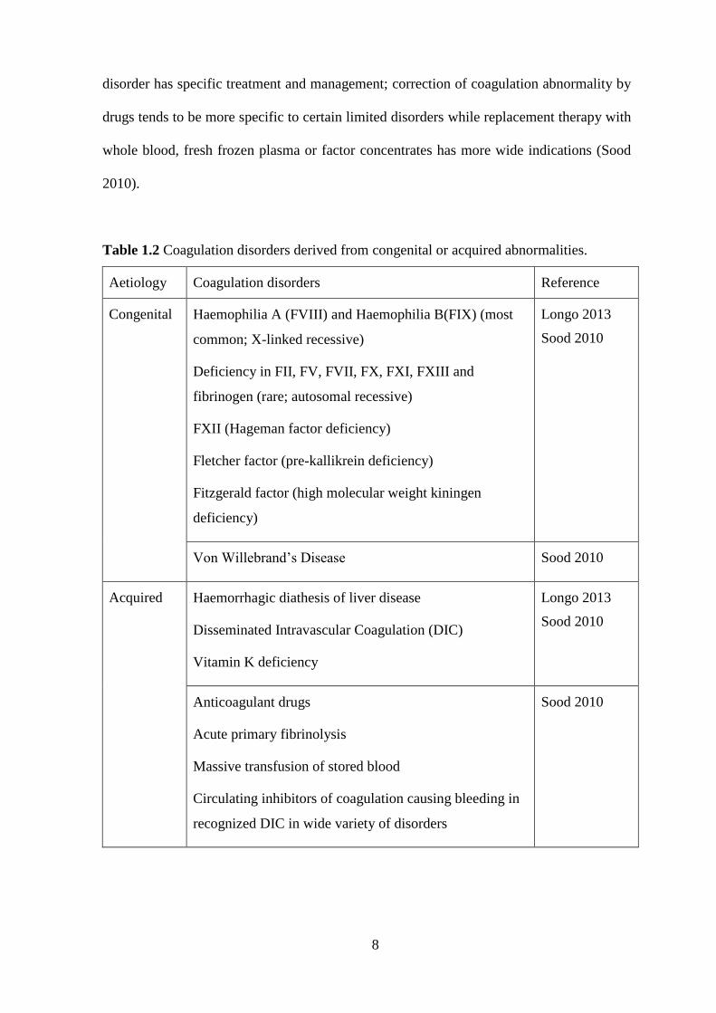
8
disorder has specific treatment and management; correction of coagulation abnormality by
drugs tends to be more specific to certain limited disorders while replacement therapy with
whole blood, fresh frozen plasma or factor concentrates has more wide indications (Sood
2010).
Table 1.2 Coagulation disorders derived from congenital or acquired abnormalities.
Aetiology Coagulation disorders Reference
Congenital Haemophilia A (FVIII) and Haemophilia B(FIX) (most
common; X-linked recessive)
Deficiency in FII, FV, FVII, FX, FXI, FXIII and
fibrinogen (rare; autosomal recessive)
FXII (Hageman factor deficiency)
Fletcher factor (pre-kallikrein deficiency)
Fitzgerald factor (high molecular weight kiningen
deficiency)
Longo 2013
Sood 2010
Von Willebrand’s Disease Sood 2010
Acquired Haemorrhagic diathesis of liver disease
Disseminated Intravascular Coagulation (DIC)
Vitamin K deficiency
Longo 2013
Sood 2010
Anticoagulant drugs
Acute primary fibrinolysis
Massive transfusion of stored blood
Circulating inhibitors of coagulation causing bleeding in
recognized DIC in wide variety of disorders
Sood 2010
9
1.1.1.6 Anti-coagulant drugs
Imbalanced haemostatic activity may resulted in thrombosis or bleeding (Sood 2010;
Zehnder 2012, p. 601). Thrombosis formation in blood vessel is harmful when blood
coagulation cascade is unchecked by inoperative coagulation inhibitor, blood flow and
fibrinolysis (Hoffbrand, Moss & Pettit 2006). Significant morbidity and mortality events
accounted by arterial and venous thromboses (Weitz 2013, p. 277). Arterial thrombosis
commonly causes acute myocardial infarction (MI), ischemic stroke and limb gangrene,
which can progress to fatal pulmonary embolism and postphlebitic when deep-vein
thrombosis occurs (Weitz 2013, p. 277). Venous thrombosis is rare but occurs at vascular
disruption site, post-surgical trauma and indwelling venous catheters (Weitz 2013, p. 277).
Prevention and treatment of thrombosis can be realized by the use of antithrombotic agents
that are grouped into three; antiplatelets, anticoagulants and fibrinolytic drugs (Weitz 2013,
p. 277). Antithrombotic agents have been used in clinical settings for more than 50 years
and becoming one of the most frequently prescribed medications (Patriquin & Crowther
2013, p.477). Antiplatelet agents include aspirin, thienopyridines, dipyridamole,
glycoprotein IIB/IIIA receptor antagonist; anticoagulant agents include heparin, low-
molecular-weight heparin, fondaparinux, warfarin, dabigatran, rivaroxaban, apixaban; and
fabrinolytic agents include streptokinase, anisteplase, urokinase, alteplase, tenecteplase and
reteplase (Weitz 2013, p. 277). Antithrombotic agents can be taken orally or parenterally
for various indications (Patriquin & Crowther 2013, p.477). Antiplatelets along with
anticoagulants and fibrinolytic agents are mostly used to inhibit or treat arterial thrombosis
since the pathophysiology of arterial thrombosis comprises of high amount of platelets due
to the fleece-injured arteries. On the other hand, anticoagulants alone are the backbone for
prevention and treatment of venous thromboembolism (VTE) due to the fact that fibrin
predominantly contributes to such condition while fibrinolytic drugs served as therapeutic
10
agent for patients with VTE (Weitz 2013, p. 277). The main goal of anticoagulant therapy
is to prevent unwanted clot formation in patient with artificial heart valve, phlebitasis,
circulatory problems and joint replacement (Estridge & Reynolds 2008). Heparin and
warfarin are the infamous antithrombotic agents most frequently used in clinical settings
(Patriquin & Crowther 2013, p.477). However, current anticoagulant therapy demonstrated
the use of recently-developed multiple novel agents such as dabigatran, rivaroxaban and
apixaban that tackles issues associated with traditional agents; for example dosing
variability, difficult monitoring, drug-drug and drug-environment interactions (Patriquin &
Crowther 2013, p.477). Data on adverse drugs events showed warfarin and antiplatelets
agents as one of the main factors contributing to emergency hospitalization in older
patients (Budnitz et al. 2011). Anticoagulant therapy requires meticulous and details
monitoring in providing the utmost therapeutic benefits but maintaining safe balance
between preventing thromboembolic events and side effects (Patriquin & Crowther 2013,
p.477). Two serious issues associated with antithrombotic therapy that often being brought
up in medicolegal proceedings are failures to prescribe anticoagulants when clinically
indicated and insufficient monitoring once therapy started (Patriquin & Crowther 2013,
p.477).
1.1.2 Dates
1.1.2.1 Overview
Dates (Phoenix dactylifera L.) belongs to the Arecaceae family and has been cultivated for
over 6000 years ago (Alkaabi et al. 2011) and the most common and vital fruit crop in
Middle East (Mohamed et al. 2014). Dates are best categorized into the stage of ripening;
Kamiri (stage 1), Khalal (stage 2), Rutab (stage 3) and Tamar (stage 4) (Alkaabi et al.
11
2011). There are various types of dates; Ajwa, Munifi, Hilali, Ruthana, Khodry, Khalas,
Sukkary, Sefri and Segae with each of them have roles in diverse disease prevention
(Rahmani et al 2014). In terms on nutrients, dates have high carbohydrate content (70–
80%), fat (0.20–0.50%), protein (2.30–5.60%), dietary fiber (6.40–11.50%), minerals
(0.10–916 mg/100 g dry weight), and vitamins (C, B1, B2, B3 and A) with very little or no
starch (Mohamed et al. 2014). The phytochemical constituents of dates include alkaloids,
flavonoids, steroids, tannins, estertepens, vitamins and phenolic acids (Onuh et al. 2012).
The health benefits derived from dates are diverse and expanding including
antihyperlipidemic, anticancer, gastroprotective, hepatoprotective, nephroprotective
properties (Mohamed et al. 2014); anti-oxidant, anti-inflammatory, anti-bacterial (Rahmani
et al 2014); and used for anaemia, stroke and tooth ache treatment, weight-gain and anti-
aging (Onuh et al. 2012). Onto the medicinal value of specific type of dates; Ajwa, the
Saudi Arabia and/or Madinah Al-Munawara-cultivated dates posed significant value in
several types of diseases cure as well as protective effect in hepatic toxicity (Rahmani et al
2014).
1.1.2.2 Dates rich in salicylic acid
Dates have very high concentration of salicylic acid, which is more than 1 mg/100g (What
is salicylic acid 2014). To our knowledge, there is one available scientific study that
reported the salicylic acid content in dates (Swain, Dutton & Truswell 1985). Swain &
Dutton (1985) studied the concentration of salicylic acid using High Performance Liquid
Chromatography (HLPC) on 333 foods. Swain, Dutton & Truswell (1985) reported that
fresh dates have 3.75 mg/100g salicylic acid while dried dates have 4.50 mg/100g salicylic
acid. Salicylic acid is the compound that has the ability to block the action of vitamin K
during coagulation pathway activation and act as a vitamin K antagonist (Roncaglioni et al.
12
1988). Previously, oral anticoagulant therapy was synonym to oral Vitamin K antagonist
(VKA) especially in North America where warfarin is referred as VKA (Patriquin &
Crowther 2013, p.477). This marked the importance of VKA action as an anticoagulant.
Thus, dates may have anticoagulant effect. In an animal study, salicylates at moderate dose
may prevent thrombus formation while limiting bleeding complications (Roncaglioni et al.
1998). Roncaglioni and team demonstrated that salicylates have anticoagulant effect
comparable to warfarin when the animals administered with 175 mg/kg salicylates were
compared to animal administered with 0.1-0.2 mg/kg warfarin.
1.1.2.3 Dates and hemostatic activities
To our knowledge, there is no scientific report on the relationship between dates and
haemostatic activities. On a closely related study, Onuh et al. (2012) had reported a study
on the effect of dates extracts on peripheral blood parameters using animal model. Their
study concluded a dosage dependent significant increase in absolute values, red blood cell
(RBC), Hemoglobin (Hb), Pack Cell Volume (PCV), reticulocytes and platelets. In relation
to haemostatic activities, platelets count were seen significantly increased in rats treated
with both ethanolic and aqueous crude dates extracts for a continuous 112 days. Increment
in platelet counts suggests pro-coagulant effect on haemostatic activity. While there is no
available scientific data on the correlation between dates and haemostatic activities, many
scientific data has been published on antithrombotic effects of fruits, vegetables and spices
as published by Torres-Urrutia et al. (2013).
13
1.1.3 Salicylic acid
1.1.3.1 Salicylic acid in human
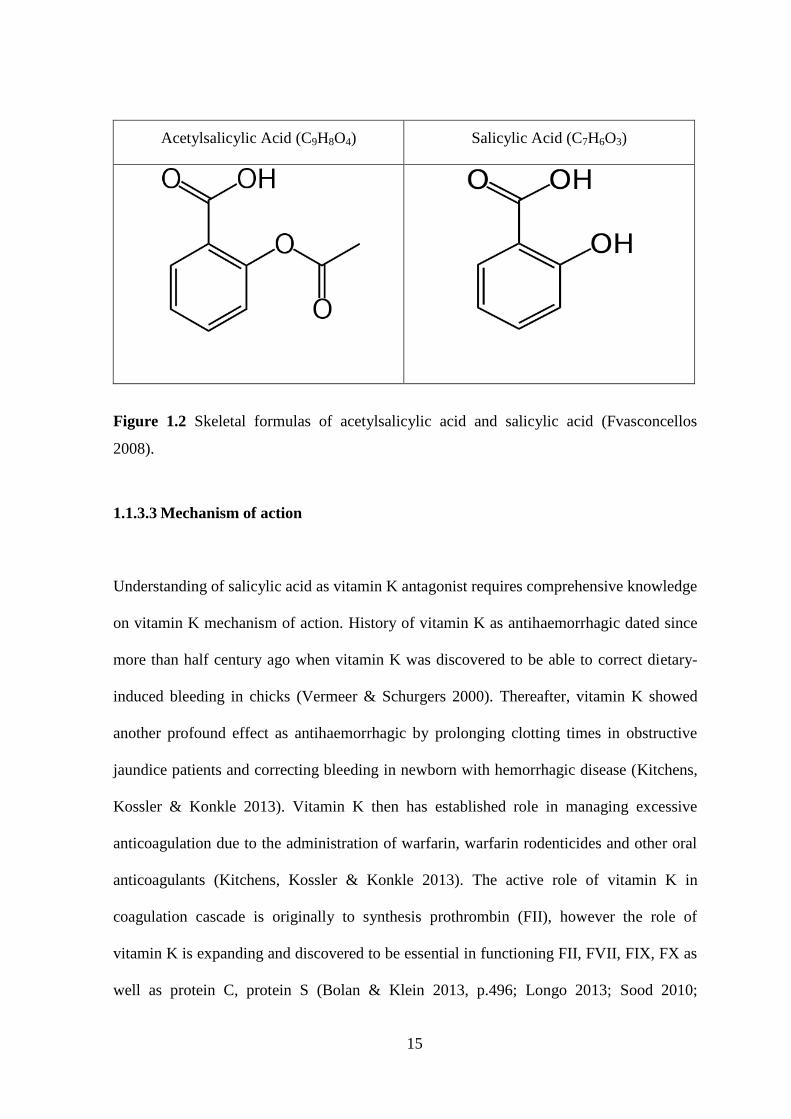
Salicylic acid (2-hydroxybenzoic acid) is produced when acetylsalicylic acid (2-
acetoxybenzoic acid) hydrolized (Figure 1.2). In human liver and blood, aspirin
(acetylsalicylic acid) will be immediately converted to salicylic acid once ingested into the
body (Duthie et al. 2005). Salicylic acid bound to plasma proteins and circulated in the
blood and distributed to all tissues in the body (Duthie et al. 2005) especially synovial
cavity, central nervous system and saliva (Hare, Woodside & Young 2003). Apart from
serum, salicylic acid may be found in the urine. A study found that diet rich in salicylic
acid by vegetarians surprisingly showed overlapping serum and urinary salicylates
concentration in comparison to those taking low-dose aspirin; indicating prominent
absorption of salicylic acid from ingested fruits and vegetables (Battezzati 2006; Duthie et
al. 2005). Aspirin (acetylsalicylic acid) is analgesic, anti-thrombotic and anti-inflammatory
(Scotter et al. 2007). Other than possible antithrombotic effect, acetylsalicylates may
benefit human health by preventing colon cancer, pregnancy induced pre-eclampsy and
worked as prophylactic agent against coronary heart disease at concentration as low as 30
mg/day (Venema et al. 1996).
1.1.3.2 Salicylic acid in foods and dates
Salicylic acid presents in many foods including fruits, vegetables, herbs, spices and vary in
concentration (Battezzati 2006). Data on the salicylates content of foods are scarce and
contradictory (Scotter 2007; Venema et al. 1996). On a study reported by Venema et al.
(1996), low salicylic acid and no acetylsalicylic acid was detected in foods thought to have
14
high salicylates as published in the literature by fellow researchers. Salicyclic acid in food
is often associated as natural blood thinner referring to food that has the ability to reduce
blood. Salicylic acid has been reported to act as vitamin-K antagonist by lowering plasma
levels of vitamin-K dependent clotting factors as well as inhibiting vitamin-K
carboxylation in the liver (Roncaglioni et al. 1988). Therefore, a diet rich in acetylsalicylic
acid should have an antithrombotic effect (Venema et al. 1996). Since dates have been
claimed to have very high salicylic acid, it may suggest that dates might influenced
haemostatic activity. However, low level of salicylates in normal diet probably insufficient
to give physiological effect in vivo (Venema et al. 1996) and subsequently affect disease
risk (Janssen et al. 1997). Determination of salicylic acid in foods (Swain, Dutton &
Truswell 1985; Venema et al. 1996) and soups (Baxter et al. 2001) implied the use of
HPLC in earlier studies. HPLC was also used in the determination of salicylates in
biological samples such as serum (Blacklock et al. 2001), urinary salicylates (Janssen et al.
1996) and plasma (Dadgar et al. 1985). Application of gas-chromatography mass
spectrometry (GCMS) was later applied in the determination of salicylic acids in plants
(Deng et al. 2003; Engelberth et al. 2003) and human serum (Battezzati 2006). Scotter et
al. (2007) came with an optimised method of salicylic acid determination in 76 foods using
GCMS. A highly specific, rapid and cost effective method was developed for
quantification of salicylic acid and acetylsalicylic acid (Scotter et al. 2007). GCMS is
preferable because HPLC is lengthy and likely to be affected with interference especially
when UV detection method is used (Scotter et al. 2007). Eventhough HPLC with
fluorescence detection method is able to successfully determined salicylic acid in plants;
samples need to undergo complex purification procedure to isolate salicylic acid from
other fluorescent compounds of the same plants (Engelberth et al. 2003). Thus, GCMS
provide a dependable method for separating, identifying and quantifying salicylic acid
from samples that has undergo carboxylic acids derivatization (Engelberth et al. 2003).
15
Acetylsalicylic Acid (C9H8O4) Salicylic Acid (C7H6O3)
Figure 1.2 Skeletal formulas of acetylsalicylic acid and salicylic acid (Fvasconcellos
2008).
1.1.3.3 Mechanism of action
Understanding of salicylic acid as vitamin K antagonist requires comprehensive knowledge
on vitamin K mechanism of action. History of vitamin K as antihaemorrhagic dated since
more than half century ago when vitamin K was discovered to be able to correct dietary-
induced bleeding in chicks (Vermeer & Schurgers 2000). Thereafter, vitamin K showed
another profound effect as antihaemorrhagic by prolonging clotting times in obstructive
jaundice patients and correcting bleeding in newborn with hemorrhagic disease (Kitchens,
Kossler & Konkle 2013). Vitamin K then has established role in managing excessive
anticoagulation due to the administration of warfarin, warfarin rodenticides and other oral
anticoagulants (Kitchens, Kossler & Konkle 2013). The active role of vitamin K in
coagulation cascade is originally to synthesis prothrombin (FII), however the role of
vitamin K is expanding and discovered to be essential in functioning FII, FVII, FIX, FX as
well as protein C, protein S (Bolan & Klein 2013, p.496; Longo 2013; Sood 2010;
16
Vermeer & Schurgers 2000), protein Z (Vermeer & Schurgers 2000) and osteocalcin
(Bolan & Klein 2013, p.496; Vermeer & Schurgers 2000). Two enzymes involved during
metabolism and regeneration of vitamin K are γ-glutamylcarboxylase and epoxide
reductase (Bolan & Klein 2013, p.496; Longo 2013). Vitamin K is needed as cofactor for
γ-glutamylcarboxylase during formation of γ-carboxyglutamic acid residues on coagulation
proteins (Bolan & Klein 2013, p.496; Longo 2013). In turn, γ-glutamylcarboxylase will
catalyze epoxide reductase for regeneration of reduced vitamin K (Longo 2013). In
coagulation cascade, vitamin K- dependent coagulation proteins bind to the calcium and
phospholipids of activated platelets with the help of γ-carboxyglutamate (Gla) residues and
eventually improve thrombin formation by speeding up the reaction rate at several orders
of magnitude (Vermeer & Schurgers 2000). Since Gla has stable divalent anionic charges,
interaction of Gla and calcium ions allow localization of clotting factors to take place as
well as building up internal calcium channels (Bolan & Klein 2013, p.496). Warfarin act as
VKA by blocking the action of epoxide reductase and thus inhibit vitamin K effect (Longo
2013). Worldwide, VKA is used for the treatment of patient with developing thrombosis
risk to inhibit blood coagulation (Vermeer & Schurgers 2000). In clinical practice, marked
correction of PT test within 6-24 hours post-parenteral vitamin K administration confirmed
the deficiency of vitamin K (Sood 2010).
1.2 Problem Statement / Research Question
Dates were reported to have high concentration of salicylic acid. Salicylic acid is the
compound that has the ability to block the action of vitamin K during coagulation pathway
activation and act as a vitamin K antagonist (Roncaglioni et al. 1988). Thus, dates may
have anti-coagulation effect. However, an in vivo study by Onuh et al. (2012) on the effects
of dates extract on blood parameters showed significant increment of platelets count;
17
which may suggest pro-coagulant activity of dates. In addition, literatures have reported
that dates reduce postpartum haemorrhage (Khadeem et al. 2007); suggesting dates having
antihaemorrhagic activity. In view of antiplatelet and anticoagulant drugs; aspirin is the
infamous and mostly used antiplatelet drug (Weitz 2013, p. 277) while warfarin is the most
widely used oral anticoagulant in the world (Krynetskiy & McDonnel 2007; Patriquin &
Crowther 2013, p. 477). However, some conventional antiplatelet and anticoagulant drugs
are associated with adverse events and therapeutic failure. Side-effects of anticoagulant
drugs include internal bleeding, prolonged bleeding time, palpitation, gastrointestinal
symptoms and haemorrhage. Aspirin along with other oral anti-platelet drugs account for
13.3% while warfarin accounts for 33.3% emergency hospitalization for adverse drug
events in older American (Budnitz et al. 2011). At present time, no ideal anticoagulant
drugs have ever existed since all anticoagulants and fibrinolytic will increase bleeding risk
(Zehnder 2012, p. 601). These two claims of dates having high salicylic acid and the
medicinal value to reduce postpartum haemorrhage as well as significant platelet count
increment seem to contradict to each other. Therefore, the studies of coagulation activities
of dates as well as analysis of chemical composition of dates are of great interest. Natural
products are beneficial remedy because they are cheap and readily available without
complication and side effects (Rahmani et al. 2014). General supportive measure is needed
in coagulation disorders (Shaz et al. 2013). Since salicylates enhanced oral anticoagulant
therapy (Sood 2010), the study of dates as a dietary supplement for patient with
coagulation problem is highly attentive. On another side, supplements have potential of
altering haemostatis (Markham & Dog 2013, p. 595); therefore, knowledge on dates as
pro-coagulant or anti-coagulant food is beneficial in improving human health with
coagulation-related problem.
18
1.3 Research Objectives
1.3.1 General
To assess coagulation activities in human plasma treated with Ajwa dates (Phoenix
dactylifera L.) extracts in vitro.
1.3.2 Specific
1. To determine coagulation activities in human plasma treated with Ajwa dates (Phoenix
dactylifera L.) extracts at different concentration (0.1, 0.5 and 1.0 g/mL).
2. To screen chemical compounds of Ajwa dates (Phoenix dactylifera L.) qualitatively
using gas chromatography-mass spectrometry (GCMS) from ethanol and aqueous
extracts.
1.4 Research Hypothesis
H0: Ajwa dates (Phoenix dactylifera L.) do not have coagulation effect
HA: Ajwa dates (Phoenix dactylifera L.) have coagulation effect
1.5 Rationale of Study / Expected Outcome
1. This research concerned about the potential role of dates as anti-coagulant.
2. This research focused on the improvement of human health by introducing dates as pro-
or anti-coagulant food.
19
Chapter II
METHODOLOGY
2.1 Study design
This research employed experimental study design with control and treatment groups.
Dates were extracted with single extraction method using ethanol and aqueous. Venous
blood was withdrawn from twenty-seven donors (n=27) to be collected into K2-EDTA
and trisodium-citrate tubes for platelet and coagulation studies respectively. Platelet
poor plasma (PPP) were treated with Ajwa dates ethanol and aqueous extracts at
different concentrations (0.1, 0.5 and 1.0 g/mL). Seven groups were formed from each
donor: control (normal saline), ET I (0.1 g/mL), ET II (0.5 g/mL), ET III (1.0 g/mL),
AQ I (0.1 g/mL), AQ II (0.5 g/mL), AQ III (1.0 g/mL). In vitro coagulation activities of
Ajwa dates were assessed by prothrombin time (PT), activated partial thromboplastin
time (APTT) and thrombin time (TT). Data was analyzed statistically using SPSS
Version 22.0 (Figure 2.1). Following extraction, both ethanol and aqueous extracts were
analyzed qualitatively using Gas Chromatography – Mass Spectrometry (GCMS).
2.2 Extraction
2.2.1 Ajwa Dates Collection
This method of dates collection was adapted from Biglari et al. (2007) with few
modifications. Ajwa dates were purchased from local distribution centre (Yusuf
Taiyoob Sdn. Bhd., Pulau Pinang, Malaysia). Dates weighed about 4–8g per fruit and
20
for each extraction, approximately 100 g (~16 dates) of dates were used. Dates were
properly selected in terms of size, colour, ripening stage and quality. The edible part of
dates (100 g) were pitted and oven-dried for 7 days.
2.2.2 Ethanol Extraction
Pitted dates (100 g) were dry-blended with an analytical mill (IKA, A11 Basic).
Grounded dates were extracted two times with 200 ml 99.7%, ethanol (QreC) at room
temperature (20°C, 1H) using ultrasonic cleaner (WiseClean, WUC-A10H). The
extracts were centrifuged (6000rpm, 10min) and filtered. The supernatant was
concentrated under reduced pressure (40°C, 3H) using a rotary evaporator (EYELA,
N1100) to obtain dates ethanolic crude extracts. The crude extracts were kept at 4°C
until used for analysis.
2.2.3 Aqueous Extraction
This method of aqueous extraction was adapted from Vayalil (2002) with few
modifications. Grounded dates were extracted two times with 200ml distilled water at
room temperature (20°C, 1H) using ultrasonication (WiseClean, WUC-A10H). The
extracts were centrifuged (6000rpm, 10min) and filtered. The supernatant was collected
and lyophilized using freeze dryer (Christ, Alpha 1-4 LSC) to obtain aqueous crude
extracts. The crude extracts were kept at 4°C until used for analysis.
21
2.2.4 Yield Percentage (%) Calculation
The percentage of extraction yield was calculated using the following formula (Zhang,
Bi & Liu 2007).
Extraction yield (%) = Weight of dried extract × 100
Weight of original sample
2.3 GC-MS analysis
2.3.1 Crude Extracts Derivatization
This method of chemical derivatization was adapted from Scotter et al. (2007) with few
modifications. A 1 mL aliquot of crude extract solution (1 mg/mL) was transferred to a
2 ml glass vial and solvent was removed under a stream of N2 on an evaporator system
(Glas-Col) at 35°C. The vial was removed as soon as the residue solvent dried. A 200
µl of BSTFA/TMCS derivatising agent (Supelco) was added to the vial. The vial was
capped immediately and heated at 60°C for 1 H with occasional swirling. The solution
was cooled to room temperature prior to analysis by GC–MS (Agilent, 7890A).
2.3.2 Analysis of Compounds
This method of GC-MS analysis was adapted from Deng et al. (2003). Analysis was
done using HP-5ms capillary column (30m, 0.25mm, 0.25µm) equipped with MSD
5975C detector and spitless injection system. Helium gas (99.999%) was used as a
carrier gas at a flow rate of 1 mL/min. The oven temperature was set at 100°C for 2 min
then programmed at 15° C/min to 300°C which was maintained for 10 min. The results
22
were analyzed qualitatively in full-scan acquisition mode with a mass range of 45–500
a.m.u. The compounds were identified using NIST database. Identification was made by
comparing mass spectra of each compound with the compound in NIST database for
matching quality.
2.4 Assessment of Coagulation Activities
2.4.1 Sample Size Calculation
By using Power and Sample software (Dupont & Plummer 1997) which based on paired
t-test formula; result showed as follows: α = 0.05, δ= 0.345 (detectable difference at
60%), σ= 0.575 (based on coagulation activity result (Torres-Urrutia et al. 2011), power
= 0.8 (80%), n = 27 (plus 10% drop out rate) for subject group.
2.4.2 Donor Recruitments
Twenty-seven donors (n=27) were recruited at Clinical Trial Complex, Advanced
Medical and Dental Institute (AMDI), USM. All donors were informed about the
objective of this study and had signed informed consent form prior to blood collection.
The protocol was authorized by Human Research Ethics Committee (HREC), USM.
Inclusion criteria include all the criteria for blood donation as stated by National Blood
Centre (PDN). Inclusion criteria includes male or female; age range between 18-60
years old; healthy and free from clinical disorders; body weight of more than 45 kg; had
taken food before donating blood; not pregnant, not breast-feeding, menstrual period on
day-5 onwards and do not involved in any risk activities. Exclusion criteria include had
23
taken antiplatelets/anticoagulant/fibrinolytic drugs, vitamin/mineral supplements or
medication for at least 7 days before recruitment.
2.4.3 Blood Collection
For platelet count, 3 mL blood was collected in K2-ethylene-diamine tetraacetic acid
(EDTA). For coagulation studies, 6 mL blood was collected in trisodium citrate tube
(3.2%) in a 9:1 ratio. Blood was centrifuged (6000rpm, 10min) to obtain PPP (platelet-
poor plasma) and analyzed immediately.
2.4.4 Platelet Count
Platelet count was determined using automated hematology analyzer (Sysmex, KX-21).
The result of platelet count was expressed in number of platelets per microlitre of blood.
2.4.5 Measurement of coagulation factors
This method of anticoagulant activity study was adapted from Karim, Noor & Seman
(2013). PT, APTT and TT tests were conducted using an automated coagulation
analyzer (Diagnostica Stago, STA Compact). The PPP was pre-incubated (37°C, 7min)
with crude extract solutions at 1:1 (v/v) with concentration 0.1 g/mL, 0.5 g/mL and 1
g/mL. The results of coagulation tests were expressed in seconds (s).
24
2.4.6 Sample Disposal
Excess samples from this research were not used for other reasons and were destroyed
with the consent from Human Research Ethics Committee USM (HREC).
2.5 Statistical Analysis
Statistical analysis was performed using SPSS Version 22.0 (IBM Corp.). The data was
expressed as median with minimum and maximum range of duplicate determination.
Kruskal-Wallis with Bonferroni correction was used to compare significance difference
between control and each treatment group. Values were considered significantly
different when p<0.05 and very significantly different when p<0.001.