1
1285 to the treatment of bleeding varices. Vasopressin raises L.E.S.P. and is already used in the treatment of bleeding varices. Metoclopramide and bethanechol, which are used in the treatment of gastro-oesophageal reflux, also raise L.E.S.P.; so do gastrin, motilin, prostaglandin, and coffee. IS Falls in L.E.S.P. occur after alcohol, cigarette smoking, chocolates, fatty meals, benzodiazepines, mor- phine, -glucagon, cholecystokinin, and secretin. IS The use of agents which raise L.E.s.P. and the avoidance of agents which lower L.E.s.P. may be valuable in the treat- ment of oesophageal varices. FURTHER RESEARCH Further oesophagographic, endoscopic, or portagraphic stu- dies of the relation between L.E.s.P. and oesophageal varices are necessary. When varices are seen on conventional oesopha- gography their size should be reduced after the use of L.E.S.P.-increasing agents. Fibreoptic endoscopy may be in- accurate because inflation of the lumen may reduce the size of the varices. Portagraphy seems to be the best technique. Con- trast material can be injected through a transhepatically intro- duced catheter placed in the portal vein, and oesophageal var- ices visualised. After a rise in L.E.S.P. the flow of contrast material into the varices should be smaller or absent. I thank Prof. H. Baden and Dr B. Andersen for their help. Methods and Devices UNIVERSITY COLLEGE HOSPITAL MUSCLE-BIOPSY NEEDLE ARCHIE YOUNG* C. M. WILES R. H. T. EDWARDS Department of Human Metabolism, University College Hospital Medical School, London INTEREST in needle biopsy of muscle for the diagnosis of neuromuscular disorders has steadily increased since we de- scribed the technique in this journal.l 2 At the IVth Inter- national Congress on Neuromuscular Diseases we reported our experience of 490 such biopsies;3 in discussion it became apparent that some workers had found difficulty using the needle originally introduced by Bergstrom.4 We describe a modification which overcomes the main problem experienced with Bergstrom’s needle-i.e., the small size of the sample. This modified needle is very satisfactory in clinical practice when muscle samples are required for chemical analysis, histo- chemistry, and electron miscroscopy. 5 The main part of the modified needle (see accompanying figure) is controlled by two finger rings, while the inner cutting cylinder is operated by means of a thumb ring. The operator has single-handed control of the biopsy needle; this allows him to use his other hand to place the needle in the muscle and to press tissue into the needle window. Incomplete opening of the window or premature withdrawal of the needle before completion of the cutting movement may result in a small specimen. Premature withdrawal also pulls and tears the muscle and/or fascia and thus increases the dis- comfort of the patient. These problems need not arise with the modified version of the needle. As with Bergstrom’s needle it is essential to keep the cutting edge sharp. The U.C.H. muscle biopsy needle (outside diameter 4.5 mm) is available from the Surgical Division of Needle Industries Ltd., P.O. Box 3, Redditch, Worcestershire. *Present address: Oxford Rehabilitation Research Unit, Nuffield Orthopædic Centre, Headington, Oxford. REFERENCES 1. Olsson, R. Digestion, 1972, 6, 65. 2. Brik, I., Palmer, E. D. Archs intern. Med. 1964, 113, 501. 3. Clark, G. A.J. Physiol. 1928, 66, 274. 4. Millette, B., Huet, P. M., Lavoie, P., Viallet, A. Gastroenterology, 1975, 69, 6. 5. Nusbaum, M., Conn, H. O. ibid. p. 263. 6. Rabøl, A., Juhl, E., Schmidt, A., Winkler, K. Digestion, 1976, 14, 285. 7. van Thiel, D. H., Stremple, J. F. Gastroenterology, 1977, 72, 842. 8. Hogan, N. J., Andrade, S. R. W., Winship, D. H. J. appl. Physiol. 1972, 32, 755. 9. Dagradi, A. E. Am. J. Gastroent. 1972, 57, 520. 10. Aronsen, K. F., Nylander, G. Acta chir. scand. 1966, 131, 443. 11. Boesby, S., Pedersen, S. A. Scand. J. Gastroent. 1974, 9, 587. 12. Nelson, S. Am. J. Roentg. 1957, 77, 599. 13. Lind, J. S., Crispin, J. S., Mclver, D. K. Can. J. Physiol. Pharmac. 1968, 46, 233. 14. Biancani, P., Zabinski, M. P., Behar, J. J. clin. Invest. 1975, 56, 476. 15. Pope, Ch. E. Gastroenterology, 1976, 70, 445. 16. Pruszynski, B. Polski. Przegl. radiol. 1969, 33, 73. 17. Dalinka, M. K., Smith, E. H., Wolfe, R. D., Goldenberg, D., Langdon, D. E. Radiology, 1972, 102, 281. 18. Ghahremani, G. G., Port, R. B., Winans, Ch. S., Williams, J. R. Dig. Dis. 1972, 17, 703. 19. Uthgenannt, H., Strömlid, A., Zwad, H. D. Fortschr. Röntg. 1973, 119, 10. 20. Chung-I, L. Am. J. Roentg. 1974, 121, 232. 21. Novak, D. Fortschr. Röntg. 1975, 123, 409. 22. Waldram, R., Nunnerley, H., Davis, M., Laws, J. W., Williams, R. Clin. Radiol. 1977, 28, 137. 23. Eckardt, V. F., Grace, N. D., Kantrowitz, P. A. Gastroenterology, 1976, 71, 185. 24. Orloff, M. J., Halasz, N. A., Lipman, Ch., Schwabe, A. D., Thompson, J. C., Weidner, W. A. Ann. intern. Med. 1967, 66, 165. U.C.H. muscle-biopsy needle. (A) Needles as supplied complete, (B) outer needle, (C) inner cutting cylinder, (D) rod used only to remove specimen from inner cutting cylinder-i.e., not used when biopsy taken. REFERENCES 1. Edwards, R. H. T. Lancet, 1971, ii, 593. 2. Edwards, R. H. T., Maunder, C., Lewis, P. D., Pearse, A. G. E. ibid. 1973, ii, 1070. 3. Edwards, R. H. T., Maunder, C. A., Round, J. M., Wiles, C. M., Young, A. 4th International Congress on Neuromuscular Diseases. Abstr 453, Montreal, 1978. 4. Bergström, J. Scand. J. clin. Lab. Invest. 1962, 14, suppl. 68. 5. Edwards, R. H. T., Maunder, C. Hosp. Update, 1977, 3, 569.

UNIVERSITY COLLEGE HOSPITAL MUSCLE-BIOPSY NEEDLE

  • Upload
    rht

  • View
    213

  • Download
    0

Embed Size (px)

Citation preview

1285

to the treatment of bleeding varices. Vasopressin raisesL.E.S.P. and is already used in the treatment of bleedingvarices. Metoclopramide and bethanechol, which are

used in the treatment of gastro-oesophageal reflux, alsoraise L.E.S.P.; so do gastrin, motilin, prostaglandin, andcoffee. IS Falls in L.E.S.P. occur after alcohol, cigarettesmoking, chocolates, fatty meals, benzodiazepines, mor-phine, -glucagon, cholecystokinin, and secretin. IS Theuse of agents which raise L.E.s.P. and the avoidance of

agents which lower L.E.s.P. may be valuable in the treat-ment of oesophageal varices.

FURTHER RESEARCH

Further oesophagographic, endoscopic, or portagraphic stu-dies of the relation between L.E.s.P. and oesophageal varicesare necessary. When varices are seen on conventional oesopha-gography their size should be reduced after the use ofL.E.S.P.-increasing agents. Fibreoptic endoscopy may be in-accurate because inflation of the lumen may reduce the size ofthe varices. Portagraphy seems to be the best technique. Con-trast material can be injected through a transhepatically intro-duced catheter placed in the portal vein, and oesophageal var-ices visualised. After a rise in L.E.S.P. the flow of contrastmaterial into the varices should be smaller or absent.

I thank Prof. H. Baden and Dr B. Andersen for their help.

Methods and Devices

UNIVERSITY COLLEGE HOSPITALMUSCLE-BIOPSY NEEDLE

ARCHIE YOUNG* C. M. WILESR. H. T. EDWARDS

Department of Human Metabolism, University CollegeHospital Medical School, London

INTEREST in needle biopsy of muscle for the diagnosis ofneuromuscular disorders has steadily increased since we de-scribed the technique in this journal.l 2 At the IVth Inter-national Congress on Neuromuscular Diseases we reported ourexperience of 490 such biopsies;3 in discussion it became

apparent that some workers had found difficulty using theneedle originally introduced by Bergstrom.4 We describe amodification which overcomes the main problem experiencedwith Bergstrom’s needle-i.e., the small size of the sample.This modified needle is very satisfactory in clinical practicewhen muscle samples are required for chemical analysis, histo-chemistry, and electron miscroscopy. 5The main part of the modified needle (see accompanying

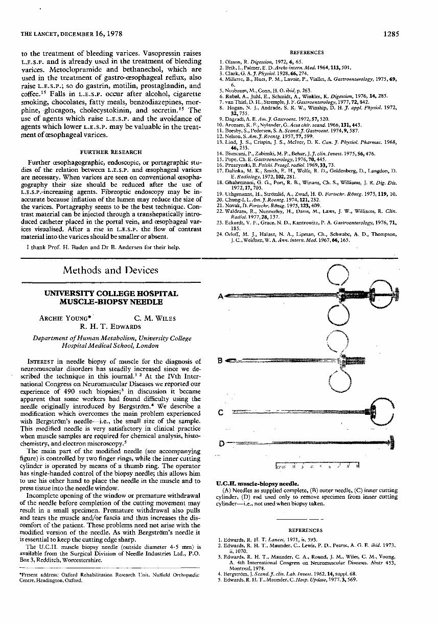
figure) is controlled by two finger rings, while the inner cuttingcylinder is operated by means of a thumb ring. The operatorhas single-handed control of the biopsy needle; this allows himto use his other hand to place the needle in the muscle and topress tissue into the needle window.

Incomplete opening of the window or premature withdrawalof the needle before completion of the cutting movement mayresult in a small specimen. Premature withdrawal also pullsand tears the muscle and/or fascia and thus increases the dis-comfort of the patient. These problems need not arise with themodified version of the needle. As with Bergstrom’s needle itis essential to keep the cutting edge sharp.The U.C.H. muscle biopsy needle (outside diameter 4.5 mm) is

available from the Surgical Division of Needle Industries Ltd., P.O.Box 3, Redditch, Worcestershire.

*Present address: Oxford Rehabilitation Research Unit, Nuffield OrthopædicCentre, Headington, Oxford.

REFERENCES

1. Olsson, R. Digestion, 1972, 6, 65.2. Brik, I., Palmer, E. D. Archs intern. Med. 1964, 113, 501.3. Clark, G. A.J. Physiol. 1928, 66, 274.4. Millette, B., Huet, P. M., Lavoie, P., Viallet, A. Gastroenterology, 1975, 69,

6.5. Nusbaum, M., Conn, H. O. ibid. p. 263.6. Rabøl, A., Juhl, E., Schmidt, A., Winkler, K. Digestion, 1976, 14, 285.7. van Thiel, D. H., Stremple, J. F. Gastroenterology, 1977, 72, 842.8. Hogan, N. J., Andrade, S. R. W., Winship, D. H. J. appl. Physiol. 1972,

32, 755.9. Dagradi, A. E. Am. J. Gastroent. 1972, 57, 520.

10. Aronsen, K. F., Nylander, G. Acta chir. scand. 1966, 131, 443.11. Boesby, S., Pedersen, S. A. Scand. J. Gastroent. 1974, 9, 587.12. Nelson, S. Am. J. Roentg. 1957, 77, 599.13. Lind, J. S., Crispin, J. S., Mclver, D. K. Can. J. Physiol. Pharmac. 1968,

46, 233.14. Biancani, P., Zabinski, M. P., Behar, J. J. clin. Invest. 1975, 56, 476.15. Pope, Ch. E. Gastroenterology, 1976, 70, 445.16. Pruszynski, B. Polski. Przegl. radiol. 1969, 33, 73.17. Dalinka, M. K., Smith, E. H., Wolfe, R. D., Goldenberg, D., Langdon, D.

E. Radiology, 1972, 102, 281.18. Ghahremani, G. G., Port, R. B., Winans, Ch. S., Williams, J. R. Dig. Dis.

1972, 17, 703.19. Uthgenannt, H., Strömlid, A., Zwad, H. D. Fortschr. Röntg. 1973, 119, 10.20. Chung-I, L. Am. J. Roentg. 1974, 121, 232.21. Novak, D. Fortschr. Röntg. 1975, 123, 409.22. Waldram, R., Nunnerley, H., Davis, M., Laws, J. W., Williams, R. Clin.

Radiol. 1977, 28, 137.23. Eckardt, V. F., Grace, N. D., Kantrowitz, P. A. Gastroenterology, 1976, 71,

185.24. Orloff, M. J., Halasz, N. A., Lipman, Ch., Schwabe, A. D., Thompson,

J. C., Weidner, W. A. Ann. intern. Med. 1967, 66, 165.

U.C.H. muscle-biopsy needle.(A) Needles as supplied complete, (B) outer needle, (C) inner cutting

cylinder, (D) rod used only to remove specimen from inner cuttingcylinder-i.e., not used when biopsy taken.

REFERENCES

1. Edwards, R. H. T. Lancet, 1971, ii, 593.2. Edwards, R. H. T., Maunder, C., Lewis, P. D., Pearse, A. G. E. ibid. 1973,

ii, 1070.3. Edwards, R. H. T., Maunder, C. A., Round, J. M., Wiles, C. M., Young,

A. 4th International Congress on Neuromuscular Diseases. Abstr 453,Montreal, 1978.

4. Bergström, J. Scand. J. clin. Lab. Invest. 1962, 14, suppl. 68.5. Edwards, R. H. T., Maunder, C. Hosp. Update, 1977, 3, 569.