Embed Size (px)
Citation preview
MOLECULAR AND FUNCTIONAL CHARACTERISATION
OF LINEAR UBIQUITINATION IN THE LIVER
A THESIS
SUBMITTED TO
UNIVERSITY COLLEGE LONDON (UCL)
IN FULFILMENT OF THE REQUIREMENTS
FOR THE DEGREE OF
DOCTOR OF PHILOSOPHY
Yutaka Shimizu
July 2016
2
Declaration
I, Yutaka Shimizu, hereby confirm that the research work presented in this thesis is
original and my own. Where information has been derived from other sources, I
confirm that this has been indicated in the thesis.
July 2016
Yutaka Shimizu

3
Abstract
Linear ubiquitination is one of the key post-translational modifications that regulate
immune signalling pathways and cell death. The only known enzyme complex
capable of forming linear ubiquitin chains de novo is the linear ubiquitin chain
assembly complex (LUBAC), and its catalytic core component is HOIP. To
understand the underlying mechanisms of liver inflammation and associated
carcinogenesis, the physiological role of LUBAC in the liver parenchyma was
investigated. Here I report that HOIP deficiency in liver parenchymal cells triggered
spontaneous cell death and inflammation in murine livers at early stages and
tumourigenesis later in life. HOIP-deficient livers displayed increased cell death,
including apoptosis, regeneration and immune cell infiltration. TNF-induced NF-κB
activation was attenuated in HOIP-deficient primary hepatocytes and they were more
susceptible to TNF-induced cell death independently of their impaired gene-
activatory capacity. Unexpectedly, however, liver damage observed in HOIP-
deficient livers was TNFR1-independent as co-deletion of TNFR1 did not ameliorate
cell death in HOIP-deficient livers whilst mitigating their inflammation. In accordance
with increased cell death, HOIP-deficient hepatocytes displayed enhanced formation
of a death-inducing signalling complex containing FADD, RIPK1, caspase-8 and c-
FLIP. This elevated signalling complex formation in HOIP-deficient hepatocytes was
independent of lowered gene-activatory capacity and the presence of TNFR1.
Moreover, combined ablation of systemic caspase-8 and MLKL completely
prevented liver damage due to loss of HOIP, whereas MLKL deficiency did not have
a beneficial effect. This demonstrates a crucial role for aberrant cell death in HOIP-
deficient livers. Collectively, these results identify LUBAC as a previously
unrecognised tumour suppressor, which acts by restraining TNFR1-independent
FADD-RIPK1-caspase-8 complex formation and caspase-8-dependent apoptosis,
regardless of its gene-regulatory function, in hepatocytes to prevent liver damage
and inflammation.
4
Table of contents
DECLARATION .................................................................................................... 2
ABSTRACT .......................................................................................................... 3
TABLE OF CONTENTS ........................................................................................ 4
TABLE OF FIGURES ........................................................................................... 9
ABBREVIATIONS............................................................................................... 11
CHAPTER 1
1 INTRODUCTION .............................................................................................. 16
1.1 Inflammation and liver cancer ......................................................................... 16
1.1.1 Inflammation ............................................................................................. 16
1.1.2 Inflammation and cancer........................................................................... 18
1.1.3 Liver cancer .............................................................................................. 20
1.1.4 Mouse models of autochthonous hepatocellular carcinoma ..................... 20
1.2 NF-κB and cell death signalling in liver disease .............................................. 23
1.2.1 NF-κB signalling ........................................................................................ 23
1.2.2 NF-κB activating modules ......................................................................... 25
1.2.3 Cell death pathways ................................................................................. 29
1.2.4 Crosstalk between NF-κB signalling and cell death pathways .................. 33

5
1.2.5 Implication of NF-κB and cell death pathways in mouse models of liver
diseases ............................................................................................................ 34
1.3 Linear ubiquitination ........................................................................................ 36
1.3.1 Ubiquitination ............................................................................................ 36
1.3.2 Linear ubiquitin and LUBAC ...................................................................... 37
1.3.3 In-vivo function of linear ubiquitination ...................................................... 39
1.3.4 LUBAC substrates and regulation of signalling ......................................... 40
1.3.5 Deubiquitinases for linear chains .............................................................. 41
1.3.6 Linear ubiquitin-binding proteins ............................................................... 44
1.4 Aims of the project .......................................................................................... 48
CHAPTER 2
2 MATERIAL AND METHODS ........................................................................... 49
2.1 Material ........................................................................................................... 49
2.1.1 Chemical and reagents ............................................................................. 49
2.1.2 Inhibitors ................................................................................................... 49
2.1.3 Buffers and solutions ................................................................................ 49
2.1.4 Biological agents ....................................................................................... 50
2.1.6 Cell culture media and supplements ......................................................... 52
2.1.7 Ready-to-use kits and solutions ................................................................ 53
2.1.8 Consumables ............................................................................................ 53
2.1.9 Instruments ............................................................................................... 54
2.1.10 Software .................................................................................................. 55

6
2.2 Cell biological methods ................................................................................... 55
2.2.1 Isolation and culture of mouse primary hepatocytes ................................. 55
2.2.2 Cell viability assay .................................................................................... 56
2.3 Biochemical methods ...................................................................................... 56
2.3.1 iz-TRAIL production from E.coli ................................................................ 56
2.3.2 Preparation of cell lysates from primary cells for western blot .................. 56
2.3.3 Liver lysate preparation for western blot ................................................... 57
2.3.4 Determination of protein concentration ..................................................... 57
2.3.5 Immunoprecipitation ................................................................................. 57
2.3.6 Polyacrylamide gel electrophoresis .......................................................... 57
2.3.7 Western blotting ........................................................................................ 58
2.3.8 Stripping of Western blot membranes ....................................................... 58
2.3.9 Tissue RNA extraction and RT-qPCR ....................................................... 58
2.4 Animal studies ................................................................................................. 59
2.4.1 Liver-specific HOIP knockout mice ........................................................... 59
2.4.2 Crossings .................................................................................................. 59
2.4.3 Genotyping ............................................................................................... 59
2.4.4 Dissection and fixation of tissues for histology .......................................... 60
2.4.5 Histological sections ................................................................................. 60
2.4.6 Immunohistochemistry .............................................................................. 61
2.4.7 Flow cytometric analysis of liver immune cells .......................................... 61
7
2.4.8 Serum analysis ......................................................................................... 61
CHAPTER 3
3 RESULTS ........................................................................................................ 62
3.1 Characterisation of liver-specific HOIP-deficient mice .................................... 62
3.1.1 Generation of liver-specific HOIP-deficient mice ....................................... 62
3.1.2 HOIP deficiency in the liver parenchyma results in spontaneous liver
tumour formation ................................................................................................ 66
3.1.3 Inflammation emerges at early stages of life in HOIP-deficient livers ....... 68
3.1.4 HOIP deficiency renders hepatocytes to cell death which precedes the
emergence of inflammation in mice ................................................................... 73
3.1.5 Summary .................................................................................................. 79
3.2 Dissecting the role of cell death pathways in HOIP deficiency-induced liver
damage ................................................................................................................. 80
3.2.1 The role of TNFR1 signalling in HOIP-deficient livers ............................... 80
3.2.2 Formation of cell death-inducing signalling complex ................................. 85
3.2.3 Genetic deletion of MLKL and caspase-8 affects the early pathology of the
HOIP-deficient livers .......................................................................................... 88
3.2.4 Summary .................................................................................................. 92
CHAPTER 4
4 DISCUSSION ................................................................................................... 93
4.1 Impact of HOIP deletion in liver parenchyma .................................................. 93
4.1.1 Cre expression, loss of HOIP and cell death ............................................ 93
4.1.2 Cell death precedes inflammation ............................................................. 95
8
4.2 Tumourigenic process ..................................................................................... 96
4.2.1 Pathological aspects ................................................................................. 96
4.2.2 DNA damage and selection ...................................................................... 98
4.2.3 Pro-tumourigenic signalling pathways ...................................................... 99
4.3 Role of TNFR1 signalling .............................................................................. 101
4.4 How does LUBAC deficiency kill hepatocytes? ............................................. 102
4.4.1 RIPK1 ..................................................................................................... 103
4.4.2 cIAP1/2 ................................................................................................... 104
4.4.3 Necroptosis ............................................................................................. 105
4.4.4 LUBAC substrates .................................................................................. 106
4.4.5 LUBAC and linear chain associated proteins .......................................... 107
4.5 Summary and outlook ................................................................................... 110
ACKNOWLEDGEMENTS ................................................................................. 112
BIBLIOGRAPHY ............................................................................................... 114
APPENDIX ........................................................................................................ 132
9
Table of Figures
Figure 1.1 Simplified scheme of canonical and non-canonical NF-κB pathways ...... 24
Figure 1.2 Schematic representation of TNFR1 signalling ....................................... 26
Figure 1.3 TNF-induced formation of cell death-inducing complexes. ...................... 32
Figure 1.4 LUBAC-mediated signalling. ................................................................... 43
Figure 3.1 HOIP liver-specific deletion reduces the protein levels of LUBAC
components in hepatocytes. ..................................................................................... 63
Figure 3.2 Expression of HOIP in mouse spleen and liver. ...................................... 64
Figure 3.3 Survival curves of Hoipflox and HoipΔhep mice. ......................................... 64
Figure 3.4 HoipΔhep mice spontaneously develop liver tumours at 18 months of age 67
Figure 3.5 HoipΔhep mice spontaneously develop steatosis, cystic lesions and fibrotic
lesions at 18 months of age ..................................................................................... 68
Figure 3.6 Quantification of hepatic leukocyte in Hoipflox and HoipΔhep mice at an
early stage ................................................................................................................ 69
Figure 3.7 Relative quantification of hepatic cytokine/chemokine transcript levels in
Hoipflox and HoipΔhep mice at an early stage ............................................................. 69
Figure 3.8 Characterisation of immune infiltrates at the peak of early inflammation in
HoipΔhep livers ........................................................................................................... 71
Figure 3.9 HoipΔhep livers have increased proliferating cells at an early stage ......... 72
Figure 3.10 HoipΔhep livers displayed elevated DNA damage at an early stage ....... 73
Figure 3.11 HoipΔhep mice showed higher levels of serum ALT ................................ 74
Figure 3.12 HoipΔhep mice suffer from transient liver damage .................................. 75
Figure 3.13 HoipΔhep livers suffer from increased cell death ..................................... 77
Figure 3.14 Caspase activation occurs in HOIP-deleted hepatocytes ...................... 77
Figure 3.15 Levels of major pro-apoptotic and anti-apoptotic proteins in HOIP-
deleted hepatocytes are unaltered ........................................................................... 78
Figure 3.16 TNF-induced canonical NF-κB activation is impaired in HOIP-deficient
hepatocytes .............................................................................................................. 80

10
Figure 3.17 HOIP-deficient hepatocytes are sensitised to TNF-induced cell death .. 81
Figure 3.18 Depletion of TNFR1 does not ameliorate liver damage in HoipΔhep mice
................................................................................................................................. 82
Figure 3.19 Abrogation of TNFR1 does not reduce cell death in HOIP-deficient livers
................................................................................................................................. 82
Figure 3.20 TNFR1 ablation ameliorates inflammation in HOIP-deficient livers ....... 83
Figure 3.21 TNFR1 deletion mitigates DNA damage in HOIP-deficient livers .......... 84
Figure 3.22 HOIP-deficient hepatocytes are sensitive to CD95L- and Poly(I:C)-
induced cell death .................................................................................................... 85
Figure 3.23 HOIP-deficient hepatocytes display increased apoptosis-inducing
signalling complex formation independently of TNFR1 signalling and loss of gene
activation .................................................................................................................. 86
Figure 3.24 Apoptosis-inducing signalling complex in HOIP-deficient hepatocytes is
formed independently of RIPK1 kinase activity ........................................................ 87
Figure 3.25 Caspase-8 heterozygosity does not alleviate liver damage of HoipΔhep
mice .......................................................................................................................... 88
Figure 3.26 Caspase-8 heterozygosity reduces apoptosis and cell death in HoipΔhep
livers ......................................................................................................................... 89
Figure 3.27 Caspase-8 heterozygosity does not ameliorate leukocyte infiltration into
HoipΔhep livers ........................................................................................................... 89
Figure 3.28 MLKL deletion and caspase-8 heterozygosity do not ameliorate liver
damage in HoipΔhep mice, whereas co-ablation of MLKL and caspase-8 rescues it . 91
Figure 4.1 Model of kinetics of HOIP deletion and dead cell number in HoipΔhep livers
................................................................................................................................. 94
Figure 4.2 Schematic of the potential consequences of HOIP inactivation. ........... 109
Figure 4.3 Schematic representation of the molecular function of HOIP in
hepatocytes ............................................................................................................ 110
11
Abbreviations
A Alanine
aa Amino acid
ABIN A20 binding inhibitor of NF-κB
ALT alanine aminotransferase
AMP adenosin-monophosphate
AP-1 activator protein 1
Apaf-1 apoptotic protease activating factor 1
APC antigen presenting cell
ATP adenosin-triphosphate
BAFF B-cell-activating factor
Bak BCL2 antagonist/killer
Bax BCL2 associated X protein
BCA bicinchoninic acid
Bcl-XL B-cell lymphoma extra large
BCR B cell receptor
C cysteine
CAD Caspase-activated DNase
CARD caspase activation and recruitment domain
CD cluster of differentiation
CD40L CD40 ligand
CD-HFD choline-deficient high-fat diet
c-FLIP cellular FLICE-inhibitory protein
cIAP cellular inhibitor of apoptosis protein
12
CYLD cylindromatosis
DAI DNA-dependent activator of IFN-regulatory factors
DAMP danger-associated molecular pattern
DC dendritic cell
DD death domain
DED death effector domain
DEN diethylnitrosamine
DISC death-inducing signalling complex
DMEM Dulbecco's modified Eagle's medium
ER endoplasmic reticulum
FACS fluorescence-activated cell sorter
FADD Fas-associated protein with death domain
Fc fragment crystallizable
FoxP3 forkhead box P3
G glycine
GalN D-galactosamine
HECT homologous to the E6AP carboxyl terminus
HEK human embryonic kidney
HET heterozygosity
HFD high-fat diet
HOIL-1 heme-oxidized IRP2 ubiquitin ligase-1
HOIP HOIL-1L interacting protein
HRP horse radish peroxidase
ICAD inhibitor of CAD
IFN interferon
IκB inhibitory κB
13
IL interleukin
IRAK interleukin-1 receptor-associated kinase
JNK Jun N-terminal kinase
KO knockout
LIGHT homologous to lymphotoxin, exhibits inducible expression and competes with HSV glycoprotein D for binding to herpesvirus entry mediator, a receptor expressed on T lymphocytes
LPC liver parenchymal cell
LPS lipopolysaccharide
LT lymphotoxin
LUBAC linear ubiquitin chain assembly complex
Lys lysine
MAPK mitogen-activated protein kinase
MEF mouse embyonic fibroblast
Met methionine
MHC major histocompatibility complex
MnSOD manganese superoxide dismutase
mRNA messenger ribonucleic acid
NAFLD non-alcoholic fatty liver disease
NEMO NF-κB essential modulator
NF-κB nuclear factor-κB
NK natural killer
NOD nucleotide-binding oligomerization domain-containing protein
NZF Npl4 zinc finger
PAMP Pathogen-associated molecular pattern
PARP poly ADP ribose polymerase
14
PI3K phosphoinositide 3-kinase
PIM PUB-interacting motif
PUB putative protein-protein interaction domain
RBR RING-between-RING
RHIM RIP homotypic interaction motif
RIPK receptor-interacting protein kinase
ROS reactive oxygen species
RT-qPCR reverse transcription-quantitative polymerase chain reaction
SM Smac mimetic
SOD superoxide dismutase
SPATA2 spermatogenesis-associated protein 2
TAB transforming growth factor β-activated kinase 1 binding protein
TAK1 transforming growth factor β-activated kinase 1
TGFβ transforming growth factor β
TH T helper
TLR Toll-like receptor
TNF tumour necrosis factor
TNF-RSC tumour necrosis factor-receptor signalling complex
TRADD TNFR1-associated death domain
TRAF TNF-receptor associated factor
Treg regulatory T cell
TRIF TIR-domain-containing adapter-inducing interferon-β
TrCP transducin repeat-containing protein
UBA ubiquitin-associated
UBAN ubiquitin binding in ABIN and NEMO
15
UBL ubiquitin-like
vICA viral inhibitor of caspase-8-induced apoptosis
WT wild-type
XIAP X-linked inhibitor of apoptosis
ZF zinc-finger

16
Chapter 1
1 Introduction
1.1 Inflammation and liver cancer
1.1.1 Inflammation
Inflammation is a physiological response to pathogen infection and tissue damage.
Inflammation has been classically characterised by five features: heat, pain, redness,
swelling and loss of function originating from the description in Latin: calor, dolor,
rubor, tumor and functio laesa (Karin and Clevers, 2016). The inflamed site is
characterised by vasodilation is mediated by histamine and nitric oxide, which
increase blood flow and trigger heat sensation and redness. Additionally, an
adhesive capacity of the blood vessels and vascular permeability are altered,
allowing leukocyte migration to the infected area, which causes tissue swelling and
oedema, frequently also imparing local tissue functionality. Lastly, pain can occur
due to stimulation of nerve endings with inflammatory mediators.
The innate immune system detects pathogens as the first line of defence (Alberts,
2002). It recognises pathogens by means of surface-expressed pattern recognition
receptors, which bind to highly conserved pathogenic structures, pathogen-
associated molecular patterns (PAMPs). When innate immune cells are activated by
pathogen recognition, they secrete inflammatory mediators, such as cytokines and
chemokines, which alter the behaviour of other cells to trigger inflammatory
responses to pathogens. Chemokines can attract other immune cells
(chemoattraction), and activate them to reinforce the immune response (Janeway
and Medzhitov, 2002). Cytokines do not facilitate chemotaxis unlike chemokines, but
they promote transcription of genes which facilitate the inflammatory response.
An initial step to eliminate incoming pathogens is their engulfment by phagocytes.
The majority of myeloid cells, including monocytes, macrophages, neutrophils,
dendritic cells and mast cells are capable of phagocytosing pathogens and attacking
them with ROS (Aderem and Underhill, 1998). Furthermore, the complement system
can be activated by immunoglobulin bound to the surface of pathogens or lectin
17
molecules on pathogens themselves to be phagocytosed or lysed (Medzhitov and
Janeway, 1999). Upon activiation, the complement system creates a pore-forming
complex on recognized pathogens to eliminate them.
NK cells are another innate immune cell type capable of recognising pathogen-
infected cells. NK cells kill target cells by releasing perforin and granzymes upon
activation, which in turn create a pore in the plasma membrane of infected cells and
activate caspases thereof, respectively (Vivier et al., 2008).
Additionally, phagocytes are crucial for relaying signals from the innate immune
system to the adaptive immune system via antigen presentation. Phagocytosed or
pinocytosed pathogens are digested in these cells. Pathogen-derived peptides are
presented to naïve T cells by MHC molecules to activate adaptive immunity
response. Both antigen presentation and co-stimulation are central for T cell
activation to enable naïve T cells to proliferate and differentiate into effector, helper
and regulatory T cells (Janeway et al., 2001). Peripheral naïve T cells mainly
consist of CD4 and CD8 positive cell subsets. CD4+ T cells activated by MHC class
II-peptide complexes differentiate largely into helper T cells such as TH1, TH2, or
TH17 and inducible Treg cell subsets. The differentiation of naïve T cells is
orchestrated by an array of cytokines, chemokines and growth factors produced by
APCs and surrounding cells. CD8+ T cells, by contrast, are activated by MHC class I-
peptide complexes on APCs. The naïve CD8+ T cells then differentiate into cytotoxic
T cells that can recognise the foreign antigen peptides presented on MHC class I
molecules in infected cells. The killing mechanisms of cytotoxic T cells upon target
recognition are similar to NK cells, again involving granzymes and perforin.
B cells are triggered upon encountering their antigens from infected cells or APCs
which bind to their receptor (BCR). B cells can digest antigens intracellularly and
present them to T cells with corresponding T cell receptors (TCRs) via MHC class II
molecules. Subsequently activated T cells secrete cytokines that activate B cells to
elicit antibody production against their antigens. Secreted antibodies bind to antigens,
allowing recognition and subsequent elimination by cells expressing Fc receptors,
such as phagocytes and NK cells, to recognise and eliminate those (Janeway et al.,
2001).

18
While activated innate immune cells and T cells produce pro-inflammatory cytokines,
they also secrete anti-inflammatory mediators, including IL-10 and TGF-β to regulate
inflammation. In addition, T cells are regulated by co-inhibitory receptors such as
CTLA-4 and PD-1. Among T cells, Tregs play a crucial role in suppression of
inflammation. Mutation in the FoxP3 gene, which is a transcription factor required for
regulatory T cell induction, is causative for immunodysregulation polyendocrinopathy
enteropathy X-linked syndrome (IPEX), which is an autoimmune disease (van der
Vliet and Nieuwenhuis, 2007). This evidence indicates that suppressive immune
signalling is essential to control inflammation.
1.1.2 Inflammation and cancer
It has been argued that inflammation is strongly associated with cancer. Research
showed that infections are linked to 15-20% of all cancer-related deaths (Balkwill and
Mantovani, 2001). For instance, Helicobacter pylori infection is a major cause of
stomach cancers. Also, patients with chronic inflammation carry a higher risk of
tumour malignancy (Balkwill and Mantovani, 2001). Accordingly, recent large
epidemiological studies implicate that non-steroidal anti-inflammatory drugs
(NSAIDs) could help to circumvent tumour development (Rothwell et al., 2012; Chan
and Ladabaum, 2015).
In late 19th century, Rudolf Virchow observed that leukocytes are present in
neoplastic tissues and suggested a connection between inflammation and cancer
(Balkwill and Mantovani, 2001). The tumour microenvironment includes leukocytes in
the tumour-supporting stroma as well as intra-tumour areas. Inflammatory cells and
mediators participate in tumour growth, progression, metastasis, and
immunosuppression. The majority of tumour-associated leukocytes are tumour-
associated macrophages (TAM), dendritic cells, and lymphocytes (Mantovani et al.,
2002). Pro-inflammatory cytokines from tumour-infiltrating immune cells and tumour
cells themselves contribute to tumour progression by causing DNA damage,
stimulating growth, subverting antitumour immunity, and enhancing invasion
(Hanahan and Weinberg, 2011).
On the other hand, the immune system is capable of detecting tumour cells and
specifically killing them by the action of cytotoxic T cells and NK cells. Interestingly,
19
tumour cells originating from immunodeficient mice are incapable of colonising and
growing in syngeneic immunocompetent mice, whilst cancer cells from
immunocompetent mice are capable of doing so in both hosts. This evidence
suggested that, in immunocompetent mice, immunogenic cells presenting non-self
antigens are removed by the immune system so that only less immunogenic cells
can thereafter survive (Kim et al., 2007). By contrast, since immunodeficient mice fail
to reject immunogenic pre-cancerous cells, these cells cannot survive
immunocompetent environment after transplantation (Teng et al., 2008). The
concept of constant removal of tumourigenic cells is termed cancer
immunosurveillance, or immunoediting (Dunn et al., 2004; Hanahan and Weinberg,
2011).
Thus, developed cancers often evade the destruction by immune cells by being less
immunogenic or protected from the recognition by cytotoxic T cells (Hanahan and
Weinberg, 2011). The recent success of clinical immune checkpoint blockade, which
targets co-inhibitory molecules such as CTLA-4 and PD-1, proves that T cell
functions in human cancers are indeed impaired by these co-inhibitory molecules
(Sharma and Allison, 2015). Ligands of PD-1 can be expressed by stromal cells as
well as tumour cells to dampen T cell activation. Moreover, the presence of Tregs,
TH2 and TH17 cells in tumours is suggested to promote subversion of the immune
system to block antitumour immunity, for example, by skewing TAM to M2
macrophages, which produce immunosuppressive cytokines IL-10 and TGFβ
(Mantovani and Allavena, 2015).
In addition to the response to invading pathogen and regulation on tumour
development, inflammation also plays a central role in wound healing and tissue
regeneration (Karin and Clevers, 2016). Upon tissue injury or infection, damaged
cells release PAMPs and danger-associated molecular patterns (DAMPs), as well as
ROS. Thus tissue damage activates multiple signalling pathways in the surrounding
cells via recognition of PAMPs and DAMPs, leading to the production of
inflammatory mediators. Inflammatory cytokines, in turn, stimulate the growth of stem
cells and facilitate turn-over of blood vessels and fibroblasts. Also, they can induce
dedifferentiation of tissue cells, which results in the expansion of regenerative
precursor cells.
20
1.1.3 Liver cancer
The major types of human primary liver tumour are hepatocellular carcinoma (HCC)
and cholangiocarcinoma (CCA), where HCC is the most common type of liver cancer.
HCC is the fifth most common cancer in men and seventh most common in women
(Marquardt et al., 2015). The leading cause for HCC is hepatotropic virus infection in
sub-Saharan Africa and Asia, as well as alcohol abuse worldwide. In addition, in
Western countries, diabetes and obesity are increasingly associated with HCC
without a history of cirrhosis. HCC is considered to be derived from mature
hepatocytes, or liver progenitor cells (Marquardt et al., 2015).
Infection with hepatotropic viruses, notably HBV or HCV, is found in the large
majority of patients with HCC (El-Serag, 2011). This viral infection causes chronic
inflammation and subsequent cirrhosis. Some viral proteins are also capable of
transforming hepatocytes to be pre-tumourigenic by blocking p53 protein, which is a
tumour suppressor gene crucial for maintaining genomic stability (Wang et al., 1994;
Ueda et al., 1995).
The most common frequently mutated oncogene in HCC is CTNNB1 encoding β-
catenin, whilst the most commonly mutated tumour suppressor gene is TP53 which
encodes p53 protein (Kan et al., 2013). In addition, genetic alterations leading to
HCC are related to oncogenic networks, including canonical Wnt signalling (eg
amplifications in FZD6 and RSPO2, mutations in CTNNB1, AXIN1, APC), the JAK-
STAT pathway (eg amplification in IL6R, JAK1 mutation), chromatin modification (eg
SWI/SNF mutation), apoptotic pathways (eg deletions in TNFRSF10A/B, CASP3)
and the PI3K-AKT-mTOR pathways (eg PTEN, RPS6KA3 mutations) (Kan et al.,
2013; Marquardt et al., 2015).
1.1.4 Mouse models of autochthonous hepatocellular carcinoma
In order to understand the nature of liver cancer development, researchers
generated several mouse models that can mimic human HCC. The models can be
classified according to the trigger of tumourigenesis: 1) viral gene transgenics, 2)
chemicals and 3) gene editing (Heindryckx et al., 2009; Weber et al., 2011).
21
1.1.4.1 Viral protein transgenics
The most common cause of HCC is viral hepatitis-induced carcinogenesis. In the
case of HBV infection, the replication of HBV inside human hepatocytes can cause
transformation of hepatocytes and cell death.
The replication of HBV requires the function of the HBX, which a viral protein is
essential for HBV infection. HBX alters cell signalling in multiple aspects to transform
hepatocytes into pre-tumourigenic cells (Murakami et al., 2001). First, HBX can
serve as a coactivator of transcription by binding to transcriptional activators, CREB
and NF-κB (Su et al., 1996). HBX also activates oncogenic signalling such as Src
and Ras pathways (Doria et al., 1995; Bouchard et al., 2001). Furthermore, HBX
inhibits the function of p53 protein by direct interaction (Wang et al., 1994; Ueda et
al., 1995). Ectopic expression of the HBX gene in mice triggers an alteration of
hepatocytes, resulting in HCC formation at 13 months of age (Kim et al., 1991).
When combined with the overexpression of the proto-oncogene c-myc, liver
carcinogenesis by HBX is accelerated by promoting transformation of hepatocytes
(Teradillos et al., 1997).
In addition, a transgene of the HBV envelope protein induces hepatocyte cell death
in mice due to the accumulation of viral surface protein HBsAg in the ER (Chisari et
al., 1989). Mice transgenic for the HBV envelope protein develop inflammation and
regenerative hyperplasia, which eventually leads to carcinogenesis at 15 months of
age (Chisari et al., 1989).
1.1.4.2 Chemically-induced and diet-induced liver carcinogenesis
DEN is the most commonly used carcinogen to induce liver cancer in rodents. DEN
is oxidised and activated in hepatocytes by cytochrome P450 to function as a DNA-
alkylating agent, and it also causes ROS production (Heindryckx et al., 2009).
Previous research demonstrated that DEN-induced tumours have close gene
expression signatures to those of HCC patients with poor survival (Lee et al., 2004).
A single injection of DEN into male mice at 2 weeks of age is sufficient to trigger
tumourigenesis. Of note, as mice ages, they are more resistant to DEN-induced
carcinogenesis (Lee et al., 1998). Male mice are more prone to bear DEN-induced

22
tumours as compared to females due to higher MyD88-dependent IL-6 production
(Naugler et al., 2007).
Dietary change can be also a model of liver tumourigenesis. For instance, choline-
deficient diet induces severe steatosis in murine livers. Dietary choline deficiency
combined with a high level of fat intake (CD-HFD) induces steatosis, oxidative stress,
hepatocyte ballooning and HCC in C57BL/6 mice, whilst HFD alone does not induce
carcinogenesis (Wolf et al., 2014). CD-HFD-induced steatohepatitis and HCC is
promoted by CD8+ T cells and NKT cells (Wolf et al., 2014). Moreover, HFD
treatment combined with DEN injection is another model of steatohepatitis-
associated hepatocarcinogenesis (Park et al., 2010).
1.1.4.3 Gene deletion and editing
Mdr2 knockout mice are often employed to mimic inflammation-associated HCC
formation. Mdr2 belongs to the ABC transporter family and is involved in the
transport of phospholipids and cholesterol into bile. Mdr2 knockout mice develop
cholestasis and severe biliary fibrosis prior to carcinogenesis (Popov et al., 2005).
Defective bile secretion leads to cholangitis and fibrosis, which results in
tumourigenesis.
Tyler Jacks and colleagues reported that concomitant deletion of PTEN and p53 in
hepatocytes, using hydrodynamic injection and CRISPR/Cas9 system, results in
rapid tumour development (Xue et al., 2014). They also succeeded in introducing
oncogenic PTEN and β-catenin mutations in the liver with the same method. As
CRISPR/Cas9 allows multiplexed gene deletion and editing, it is a feasible tool to
mimic mutagenesis of multiple genes, thereby enabling better modeling of HCC
found in human patients (Weber et al., 2015). Therefore, in parallel to classical
methods of tumour initiation and promotion with chemicals, emerging genome editing
technology will most likely be abundantly employed in liver cancer studies.
23
1.2 NF-κB and cell death signalling in liver disease
1.2.1 NF-κB signalling
NF-κB is a family of transcription factors that are activated by various immune
stimulations and induce expression of pro-survival genes, pro-inflammatory
cytokines and immunoregulatory proteins (Sen and Baltimore, 1986; Oeckinghaus
and Ghosh, 2009). The NF-κB family consists of five proteins, RelA/p65, RelB, c-Rel,
NF-κB1/p105 and NF-κB2/p100. These proteins are divided into two classes. Class 1
NF-κB proteins are p105 and p100, which are initially produced as precursor proteins
and processed into p50 and p52, respectively, by the ubiquitin-proteasome system
(Liang et al., 2006). Processing of p105 occurs constitutively whereas that of p50 is
tightly regulated by phosphorylation. Although p50 and p52 have DNA-binding and
dimerisation domains, they depend on additional factors for activation of gene
transcription (Ghosh et al., 1998). These class 2 NF-κB proteins, RelA, RelB and c-
Rel harbour transactivation domains in their C-termini and DNA-binding and
dimerisation domains in their N-termini. RelA, RelB and c-Rel activate target gene
transcription by forming homodimers or heterodimers with p50 or p52 (Ghosh et al.,
1998).
NF-κB activity is tightly regulated and normally suppressed by IκB family proteins.
IκB proteins contain ankyrin repeats which bind to the DNA-binding and dimerisation
domain of NF-κB proteins to prevent their activity. The best characterised IκB is IκBα,
which constitutively binds to the NF-κB-activating heterodimeric RelA-p50 complex
(Ghosh and Baltimore, 1990). Upon NF-κB-activating stimulation, IκBα is subjected
to degradation by the ubiquitin-proteasome system, which in turn releases RelA-p50
to activate gene transcription (Yaron et al., 1998; Fuchs et al., 1999). Also, precursor
forms of p105 and p100 are considered to be inhibitory as their C-terminal ankyrin
repeats can serve as a function of IκB proteins. These inhibitory domains of p105
and p100 are cleaved off and degraded during processing (Sun, 2011).

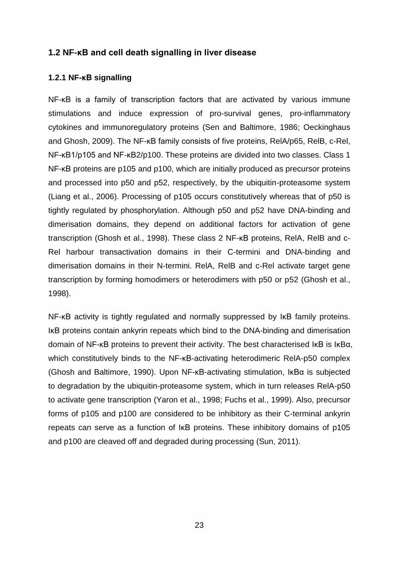
24
Figure 1.1 Simplified scheme of canonical and non-canonical NF-κB pathways
The IκB kinase (IKK) complex plays a critical role in activation of NF-κB (Häcker and
Karin, 2006). The IKK complex initially activates NF-κB signalling by phosphorylating
IκB proteins. In canonical NF-κB signalling, the IKK complex is comprised of two
kinases IKK1/IKKα and IKK2/IKKβ and a scaffold protein NEMO/IKKγ (Mercurio et
al., 1997; Rothwarf et al., 1998; Yamaoka et al., 1998). IKK1 and IKK2 have kinase
domains in their N-terminus and bind to NEMO via C-terminal NEMO-binding
domains. In canonical NF-κB signalling, which is activated by stimuli such as TNF,
IL-1 or LPS, the IKK complex is activated by the recruitment to the corresponding
receptor complexes. IKK2 is sufficient and essential for the IKK complex to
phosphorylate IκBα in a NEMO-dependent manner and triggers its proteasomal
degradation (Oeckinghaus and Ghosh, 2009) (Figure 1.1, left panel).
In non-canonical NF-κB signalling, which is activated by subsets of TNF superfamily
receptors including CD40, lymphotoxin beta receptor and BAFFR, NF-κB-inducing
kinase (NIK) plays a central role in activating NF-κB. Upon receptor oligomerisation,
NIK is stabilised and phosphorylates IKK1 (Senftleben et al., 2001; Liao et al., 2004).
25
IKK1 homodimer subsequently phosphorylates p100, which results in degradation of
their inhibitory C-terminal ankyrin repeats by SCFβTrCP ubiquitin ligase complex and
processing into p52 (Coope et al., 2002; Liang et al., 2006). p52 forms heterodimers
with RelB to activate target gene transcription (Bonizzi and Karin., 2004) (Figure 1.1,
right panel). Whilst canonical NF-κB activation is rapid and transient, non-canonical
NF-κB activation is slower and persistent and exerts specific functions depending on
cell types and activating receptors (Sun, 2011).
1.2.2 NF-κB activating modules
NF-κB is activated upon diverse immune stimulation, such as cytokines (eg TNF and
IL-1), PAMPs (eg LPS and viral DNA/RNA) and immunomodulatory receptors (eg
CD40, TCR and BCR). As explained in the previous section, NF-κB activation
depends on whether IKK is activated. Here I describe how IKK and NF-κB are
activated by coordinated signalling cascades from representative immune receptors.
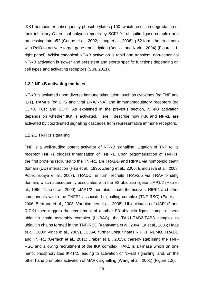
1.2.2.1 TNFR1 signalling
TNF is a well-studied potent activator of NF-κB signalling. Ligation of TNF to its
receptor TNFR1 triggers trimerisation of TNFR1. Upon oligomerisation of TNFR1,
the first proteins recruited to the TNFR1 are TRADD and RIPK1 via homotypic death
domain (DD) interaction (Hsu et al., 1995; Zheng et al., 2006; Ermolaeva et al., 2008;
Pobezinskaya et al., 2008). TRADD, in turn, recruits TRAF2/5 via TRAF binding
domain, which subsequently associates with the E3 ubiquitin ligase cIAP1/2 (Hsu et
al., 1996; Tsao et al., 2000). cIAP1/2 then ubiquitinate themselves, RIPK1 and other
components within the TNFR1-associated signalling complex (TNF-RSC) (Ea et al.,
2006; Bertrand et al., 2008; Varfolomeev et al., 2008). Ubiquitination of cIAP1/2 and
RIPK1 then triggers the recruitment of another E3 ubiquitin ligase complex linear
ubiquitin chain assembly complex (LUBAC), the TAK1-TAB2-TAB3 complex to
ubiquitin chains formed in the TNF-RSC (Kanayama et al., 2004; Ea et al., 2006; Haas
et al., 2009; Vince et al., 2009). LUBAC further ubiquitinates RIPK1, NEMO, TRADD
and TNFR1 (Gerlach et al., 2011; Draber et al., 2015), thereby stabilising the TNF-
RSC and allowing recruitment of the IKK complex. TAK1 is a kinase which on one
hand, phosphorylates IKK1/2, leading to activation of NF-κB signalling, and, on the
other hand promotes activation of MAPK signalling (Wang et al., 2001) (Figure 1.2).

26
Figure 1.2 Schematic representation of TNFR1 signalling
1.2.2.2 IL-1/TLR signalling
IL-1R and TLRs activate downstream signalling in a similar manner, as they share
homology in intracellular Toll/IL-1R (TIR) domains for the recruitment of respective
signalling complexes. Upon binding of the respective ligand, the receptors undergo
dimerisation and conformational change that allows recruitment of downstream
signalling components (Akira and Takeda, 2004). MyD88 is one of the first adaptors
recruited to IL-1R/TLR via TIR domain interaction. MyD88 is universally engaged in
IL-1R and all TLR signalling except for TLR3 signalling (Kawai and Akira 2010). The
N-terminal DD of MyD88 serves as an association platform for kinase IRAK1/2/4
through a homotypic interaction between their DDs. IRAK4 is considered to be the
most important proximal kinase (O’Neill et al., 2003). IRAK4-mediated IRAK1
activation leads to interaction with the E3 ligase TRAF6, which in turn ubiquitinates
itself and IRAK1, allowing the recruitment and activation of the TAK1-TAB2-TAB3

27
complex mediated by the ubiquitin-binding capability of TAB2/3 (Ninomiya-Tsuji et al.,
1999; Ishitani et al, 2003; Kanayama et al., 2004).
1.2.2.3 NOD2 and RLR signalling
Other PAMP receptors including NOD2 and RIG-I/MDA5 can also activate canonical
NF-κB signalling. NOD2 receptors bind to peptidoglycan dipeptide, muramyl
dipeptide (MDP), (Girardin et al, 2003; Inohara et al, 2003). The NOD2 receptor
recruits RIPK2, XIAP and cIAP1/2 upon activation and ubiquitination of RIPK2 by
IAPs leads to the subsequent recruitment of the TAK1-TAB2/3 complex and the IKK
complex to trigger NF-κB signalling (Inohara et al., 2000; Strober et al., 2006;
Bertrand et al., 2009). In addition, LUBAC is also engaged in ubiquitination of RIPK2
to facilitate NF-κB signalling (Damgaard et al., 2012).
RIG-I and MDA5 are receptors for foreign DNA derived from infectious viruses. Upon
recognition of cytosolic DNA, they bind to the adaptor protein MAVS on the outer
mitochondrial membrane, thereby triggering oligomerisation of MAVS (Takeuchi and
Akira, 2008). Activated MAVS recruits TRAF proteins, which in turn become a
platform for recruitment of the TAK1-TAB2/3 complex and the IKK complex.
1.2.2.4 TCR/BCR signalling
In addition to innate immune signalling pathways, adaptive immune receptors are
also activators of NF-κB signalling. Upon activation both TCR and BCR recruit the
CBM complex consisting of CARD11/CARMA1, BCL10 and MALT1 upon
engagement of receptor-proximal adaptors and kinases (Thorne et al., 2004; Beyeart,
2014). In the CBM complex, BCL10 is ubiquitinated in Lys63-linked and Met1-linked
manners by a concerted action of the E3 ligases, cIAP1/2 and LUBAC, respectively
(Dubois et al., 2014; Yang et al., 2014; Yang et al., 2016). Ubiquitination of BCL10 is
critical for the recruitment of NEMO by its binding capacity to polyubiquitin, which in
turn activates the IKK complex in T-cell and B-cell receptor signalling (Wu et al.,
2008).
28
1.2.2.5 Non-canonical NF-κB activators
Non-canonical NF-κB activators are members of TNF superfamily receptors,
including lymphotoxin beta receptor (LTβR), CD40, BAFFR (Häcker and Karin, 2006).
LTβR is mainly expressed on lymphoid cells and epithelial cells. There are two
ligands for LTβR: lymphotoxins and LIGHT, cytokines mainly produced by T cells.
Membrane-anchored LTβ forms heterotrimers with soluble LTα (LTα1β2 and LTα2β1).
LTβR activates non-canonical NF-κB signalling as well as canonical NF-κB signalling
(Coope et al, 2002). LTβR signalling is central for lymphoid organogenesis (Sun et
al., 2012). CD40 is expressed on APCs and activated B cells, and its ligand CD40L
is predominantly expressed on activated T cells (Elgueta et al., 2009). CD40 is a vital
co-stimulatory molecule in the immune synapse formation upon antigen presentation.
CD40 signalling modulates B-cell functions, such as germinal centre formation and
isotype class switching (Ma and Clark, 2009). CD40L-CD40 also elicits activation of
both canonical and non-canonical NF-κB pathways (Coope et al, 2002). BAFFR is
exclusively expressed on B cells and is involved in maturation of peripheral B cells
(Mackay and Schneider, 2009). Unlike CD40, BAFFR specifically activates non-
canonical NF-κB and is hardly capable of activating canonical NF-κB signalling
(Claudio et al, 2002). BAFFR-mediated activation of non-canonical NF-κB promotes
B-cell survival via induction of Bcl-2 and Bcl-XL (Mackay and Schneider, 2009).
These recepetors contain TRAF-binding motif to recruit TRAF3, which leads to the
subsequent degradation of TRAF2 and TRAF3, which is mediated by cIAP1/2
(Vallabhapurapu et al, 2008; Zarnegar et al, 2008). Degradation of TRAF2/3 thereby
leads to stabilisation of NIK (Sun, 2011).
It is suggested that the non-canonical pathway activation requires de novo synthesis
of NIK as well as degradation of TRAF2 or TRAF3 (Sun, 2011). Normally, the level of
NIK protein is maintained at an extremely low level due to its constant degradation
by the ubiquitin-proteasome system (Liao et al., 2004). This degradation of NIK is
orchestrated by TRAF3, TRAF2 and cIAP1/2, with TRAF3 being central to the
inhibition of NIK (Liao et al., 2004; Vallabhapurapu et al, 2008; Zarnegar et al, 2008).
TRAF3 is bound to the N-terminus of NIK and targets NIK for constant ubiquitination
in a Lys48-linked manner by the E3 ubiquitin ligase cIAP1/2. Loss of TRAF2 or
TRAF3, or degradation of cIAP1/2 by Smac mimetic compounds causes
29
accumulation of NIK and aberrant p100 processing, leading to non-canonical NF-κB
activation (Vallabhapurapu et al, 2008; Zarnegar et al, 2008).
1.2.3 Cell death pathways
1.2.3.1 Apoptosis
The best characterised programmed cell death is apoptosis, executed by a cascade
of activated caspases, resulting in DNA fragmentation and cell blebbing. Apoptotic
pathways were initially investigated in C. elegans employing forward genetic
methods. Thereby mutants that do not undergo cell death increase cell numbers in
their bodies (Ellis et al., 1991). Apoptosis is crucial in morphogenesis as well as
tissue homeostasis. In apoptotic cell death, executioner caspases cleave multiple
cytoskeletal proteins including actin and nuclear lamins, which leads to the loss of
cellular structure and blebbing. In addition, they cleave inhibitor of caspase-activated
DNase (ICAD) to release and activate DNase CAD, resulting in fragmentation of
nuclear DNA (Fischer et al., 2003).
There are two classical routes of interconnected apoptotic pathways: the intrinsic
and the extrinsic pathway (Scaffidi et al., 1998; Jost et al., 2009). The intrinsic
pathway is mediated by the mitochondria, whereas the extrinsic pathway is triggered
by receptors for apoptosis-inducing ligands. Intrinsic death pathway is activated by
cellular stresses such as DNA damage and oxidative stress. Under such stresses,
activation of pro-apoptotic Bcl-2 family member proteins, Bax and Bak, leads to their
oligomerisation and insertion to mitochondrial outer membranes, leading to
mitochondrial outer membrane permeabilisation (MOMP) (Tait and Green, 2013).
MOMP triggers the release of mitochondrial contents into the cytosol. A key
mitochondrial protein initiating apoptosis is cytochrome c, which is a component of
the electron transport chain in the mitochondria. Released cytochrome c binds to
Apaf-1 and provokes its conformational change, which leads to the formation of a
heptameric complex of Apaf-1-cytochrome c, referred to as apoptosome (Li et al.,
1997). The apoptosome also contains procaspase-9 which is recruited to Apaf-1 by
CARD domain interaction between these proteins (Riedl et al., 2007). Dimerisation of
pro-caspase-9 at the apoptosome results in cleavage and activation of caspase-9.
Activated caspase-9 subsequently cleaves and activates the executioner caspase-3
30
and -7 (Tait and Green, 2013). In addition to cytochrome c, the mitochondrial
proteins Smac/Diablo and HtrA2/Omi promote apoptosis execution by inhibiting anti-
apoptotic IAPs, which normally bind to executioner caspases and suppress their
activation (Jost et al., 2009).
The extrinsic apoptosis pathway is activated by TNF superfamily receptors, referred
to as death receptors (DRs), such as TNF-R1, DR3/TRAMP, TRAIL-R1/DR4, TRAIL-
R2/DR5 (in H. sapiens; M. musculus has only one TRAIL-R), CD95/Fas, and DR6
(Walczak, 2013). In the cases of TRAIL-Rs and CD95, upon ligation by their
respective ligands, these receptors trimerise (oligomerise) and their intracellular DDs
recruit FADD as an adaptor protein. In turn, FADD recruits caspase-8 via DED
interaction and subsequently activates caspase-8 by forming the DISC (Kischkel et
al., 1995; Boldin et al., 1996; Muzio et al., 1996; Bodmer et al., 2000; Kischkel et al.,
2000; Sprick et al., 2000). Caspase-8 activated at DRs cleaves executioner
caspases, leading to apoptosis of stimulated cells. Caspase-8 activity is controlled by
a negative regulator c-FLIP, which harbours DED domains on its N-terminus and a
caspase-like domain on its C-terminus which does not have proteolytic activity. c-
FLIP forms heterodimers with caspase-8 and hence restricts its apoptotic function
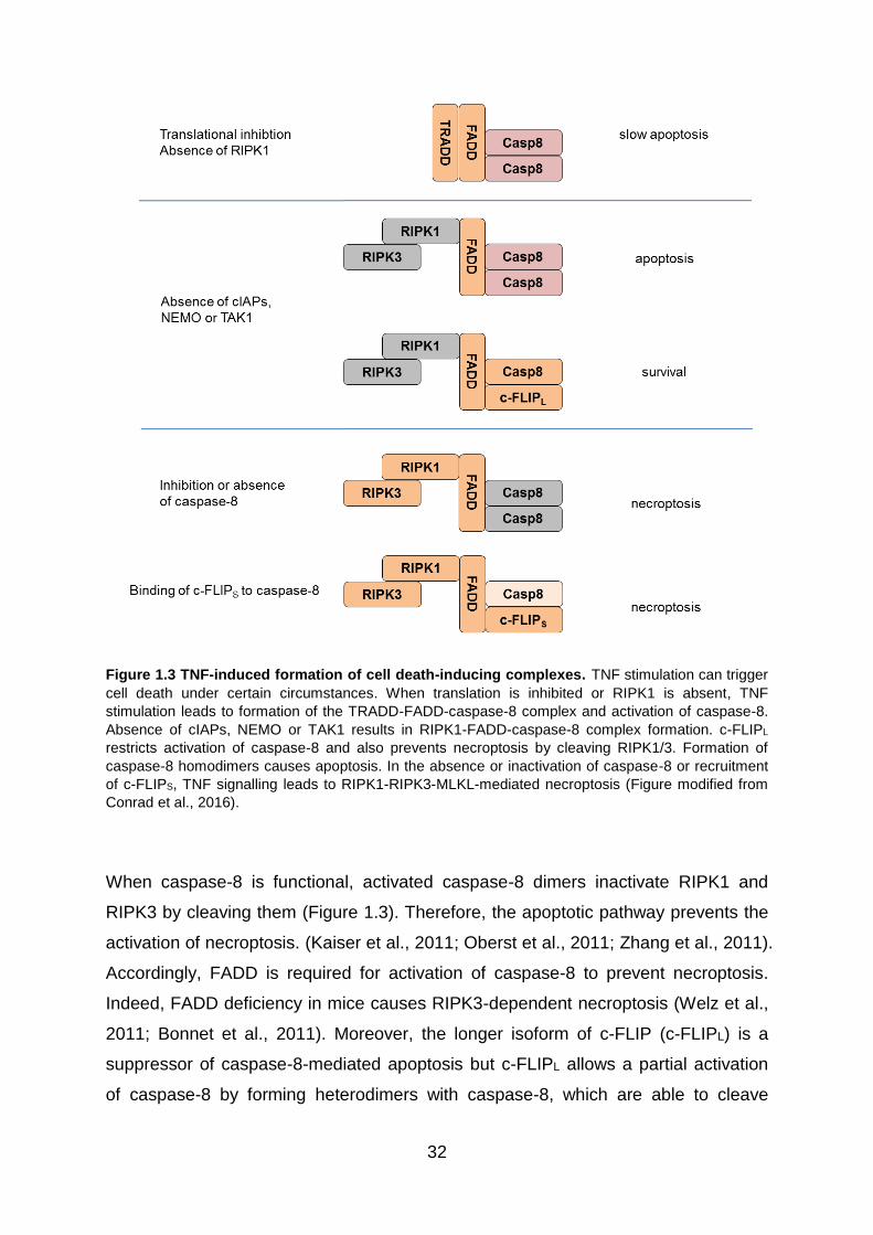
(Irmler et al., 1997; Oberst et al., 2011; Dillon et al., 2012) (Figure 1.3).
The extrinsic cell death pathway is connected to the mitochondrial apoptosis
pathway. The pro-apoptotic BH3-only family protein Bid is cleaved by caspase-8 to
initiate the mitochondrial pathway. Truncated Bid (tBid) translocates to the
mitochondria, where it activated Bax and/or Bak to induce MOMP (Wei et al., 2000).
Whilst the primary function of TNF is to promote gene activation, in certain contexts
TNF can induce cell death. When the TNF-RSC is not properly assembled or gene
activation is not optimally executed, a part of the TNF-RSC is released into the
cytosol, including TRADD and RIPK1, and nucleates a secondary cytosolic complex,
termed complex II, together with FADD and pro-caspase-8, which results in
activation of the initiator caspase-8 and subsequent apoptosis (Micheau and
Tschopp, 2003, Conrad et al., 2016).
31
1.2.3.2 Necroptosis
When the TNF-RSC formation is compromised or caspase-8 is inhibited or lacking in
cells, TNF executes another type of cell death, termed programmed necrosis or
necroptosis. This emerging type of cell death was shown to be non-apoptotic and
caspase-independent (Wang et al., 2005). Despite this interesting finding, a
physiological role of necroptosis was unclear and questioned for several years.
Recent studies clarified that viral proteins such as vICA protein of CMV blocks
caspase-8 when the virus infect cells in order to avoid apoptosis of host cells.
However, infected cells can still undergo necroptosis (Mocarski, 2015). This
suggests that cells can execute necroptosis as an alternative cell death mechanism
when caspases are inhibited. In addition, human biopsies of patients with ischemia-
reperfusion injury and DILI display a sign of necroptosis in biopsies (Linkermann et
al., 2013, Wang et al., 2014). Thus viral infections and tissue injury may involve
necroptosis in some cases.
Mechanistically, necroptosis is orchestrated by the RIP kinases, RIPK1 and RIPK3,
and the pseudo-kinase MLKL (Sun et al., 2012). When RIPK3 is activated by the
RHIM domain interaction with RIPK1 and the kinase activity of RIPK1, RIPK3
autophosphorylates itself and phosphorylates MLKL. Phosphorylation of MLKL, in
turn, results in a conformational change and translocation of MLKL to the plasma
membrane (Dondelinger et al., 2014; Cai et al., 2014). Pore-forming MLKL interacts
with phospholipids in the plasma membrane, leading to perforation and death of the
cells.
32
Figure 1.3 TNF-induced formation of cell death-inducing complexes. TNF stimulation can trigger
cell death under certain circumstances. When translation is inhibited or RIPK1 is absent, TNF
stimulation leads to formation of the TRADD-FADD-caspase-8 complex and activation of caspase-8.
Absence of cIAPs, NEMO or TAK1 results in RIPK1-FADD-caspase-8 complex formation. c-FLIPL
restricts activation of caspase-8 and also prevents necroptosis by cleaving RIPK1/3. Formation of
caspase-8 homodimers causes apoptosis. In the absence or inactivation of caspase-8 or recruitment
of c-FLIPS, TNF signalling leads to RIPK1-RIPK3-MLKL-mediated necroptosis (Figure modified from
Conrad et al., 2016).
When caspase-8 is functional, activated caspase-8 dimers inactivate RIPK1 and
RIPK3 by cleaving them (Figure 1.3). Therefore, the apoptotic pathway prevents the
activation of necroptosis. (Kaiser et al., 2011; Oberst et al., 2011; Zhang et al., 2011).
Accordingly, FADD is required for activation of caspase-8 to prevent necroptosis.
Indeed, FADD deficiency in mice causes RIPK3-dependent necroptosis (Welz et al.,
2011; Bonnet et al., 2011). Moreover, the longer isoform of c-FLIP (c-FLIPL) is a
suppressor of caspase-8-mediated apoptosis but c-FLIPL allows a partial activation
of caspase-8 by forming heterodimers with caspase-8, which are able to cleave

33
RIPK1 and RIPK3 to prevent cells from necroptotic death (Oberst et al., 2011)
(Figure 1.3). By contrast, the shorter isoform of c-FLIP (c-FLIPS) rather promotes
formation of the death-inducing complex and subsequent necroptosis (Feoktistova et
al., 2011). In addition, RIPK3 activity is also negatively controlled by the
phosphatase Ppm1b through dephosphorylation (Chen et al., 2015).
An additional RHIM-containing protein, TRIF is also capable of activating RIPK3,
independently of RIPK1 (Mocarski et al., 2012; Dillon et al., 2014). TRIF is an
adaptor protein of TLR3 and TLR4 signalling, and has been shown to mediate cell
death upon TLR3 and TLR4 activation. Another RHIM-containing protein DAI is a
cytosolic double-stranded DNA sensor, which is able to interact with RIPK1 and
RIPK3 via RHIM domain upon viral infection (Upton et al., 2012).
1.2.4 Crosstalk between NF-κB signalling and cell death pathways
The transcpritional activity of NF-κB induces the expression of target genes including
a wide range of pro-survival proteins which antagonise the action of cell death
pathway (Kucharczak et al., 2003). This includes, for instance, cIAP1/2 and XIAP, c-
FLIP and Bcl-2 family member proteins, such as Bcl-2, Bcl-XL and A1. Also, it
upregulates anti-oxidative protein and SODs, which quench ROS to prevent cell
death mediated by oxidative stress in the liver (Kondylis et al., 2015).
Additionally, recent studies demonstrated that NF-κB pathway components suppress
cell death independently of NF-κB's transcriptional activation. Indeed, IKK1/2 and
NEMO have an additional role in inhibiting RIPK1-dependent cell death in addition to
their role in activating NF-κB (Dondelinger et al., 2015; Kondylis et al., 2015; Vlantis
et al., 2016). Mechanistically, the IKK complex appears to phosphorylate RIPK1
which prevents it from forming a death-inducing complex (Dondelinger et al., 2015).
Also, RelA/p65 is capable of suppressing RIPK1-dependent apoptosis of intestinal
Paneth cells and death of mouse embryos (Vlantis et al., 2016). Yet, it is still not
entirely clear how RelA inhibits RIPK1-dependent cell death.
34
1.2.5 Implication of NF-κB and cell death pathways in mouse models of liver
diseases
Cell death is one of the central players in inflammation. Previous studies highlighted
the significance of protection against cell death in order to maintain tissue
homeostasis. For instance, genetic deletions of anti-apoptotic proteins Mcl-1 and
Bcl-XL in liver parenchymal cells results in hepatocyte apoptosis and subsequent
HCC formation (Weber et al. 2009, Hikita et al. 2012). These reports underpin the
importance of apoptosis regulation to prevent liver inflammation and subsequent
tumourigenesis.
The fact that NF-κB is a pivotal player of inflammatory signalling has led scientists to
investigate the role of NF-κB signalling in liver cancer. The first study of NF-κB
signalling in liver carcinogenesis employed Mdr2 knockout mice (Pikarsky et al,
2004). In this model, suppression of canonical NF-κB signalling by hepatocyte-
specific expression of IκB super-repressor inhibits the progression to a malignant
tumour via downregulation of anti-apoptotic factors (Pikarsky et al, 2004). In Mdr2
knockout mice, NF-κB activation was mediated by TNF produced by adjacent
endothelial cells and immune cells. Consequently anti-TNF therapy also inhibits
tumour progression (Pikarsky et al, 2004). Therefore, at least in Mdr2 knockout mice,
NF-κB signalling is a tumour-promoting factor. Furthermore, truncation of a tumour
suppressor and negative regulator of NF-κB signalling CYLD in hepatocytes also
leads to periportal hepatocyte apoptosis, hepatitis, hepatomegaly, fibrosis and
cancer (Nikolaou et al, 2012), whereas systemic CYLD deletion apparently does not
recapitulate this phenotype (Massoumi et al., 2006; Reiley et al., 2006; Zhang et al.,
2006).
Lymphotoxins and their receptor LTβR are upregulated in patients suffering from
viral hepatitis or HCC. Ectopic expression of LTα and LTβ specifically in hepatocytes
(AlbLTαβ mice) leads to chronic hepatitis from 6 months of age and HCC at 12
months (Haybaeck et al., 2009). Hepatitis and cancer development in AlbLTαβ mice
were ameliorated by ablation of mature T and B cells by crossing to Rag1KO mice.
This indicates that lymphocytes play a major role in promoting hepatitis triggered by
lymphotoxins. More importantly, the pathology is also prevented by deletion of IKK2
in hepatocytes. This evidence suggests that canonical NF-κB signalling in

35
hepatocytes, activated by lymphotoxins, is crucial for creating an inflammatory
environment by producing chemokines including CCL2 and CXCL1 (Haybaeck et al.,
2009, Wolf et al, 2010).
On the contrary to the studies demonstrating the pro-tumourigenic role of NF-κB,
genetic models have illustrated the protective role of NF-κB against liver
carcinogenesis. First, mice devoid of IKK2 specifically in hepatocytes are more
susceptible to DEN-induced liver tumourigenesis (Maeda et al., 2005). IKK2 in
hepatocytes suppresses DNA damage by a reduction in hepatic ROS via NF-κB
activation (Maeda et al., 2005). Moreover, liver parenchymal deletion of NEMO
triggers spontaneous hepatocyte apoptosis, fibrosis, steatohepatitis leading to
eventual HCC formation (Luedde et al., 2007). This phenotype in NEMO-deficient
livers is reversed by constitutive activation of IKK2 in hepatocytes (Kondylis et al.,
2015). Furthermore, TAK1 deletion in hepatocytes results in early tumourigenesis
and death of the animals (Betterman et al., 2010). Thus, these results demonstrate
that NF-κB signalling is required for protection against liver carcinogenesis. It
appears that optimal control of NF-κB activation is crucial for maintaining liver
homeostasis as the abrogation of both activator and inhibitor of NF-κB can be a
cause of liver inflammation and cancer.
Of note, deletion of NF-κB signalling components not only impairs NF-κB activation
but also activates cell death pathways. Pasparakis and colleagues demonstrated
that concomitant ablation of all the three NF-κB transcription factors, RelA, RelB and
c-Rel, in mouse livers does not recapitulate the tumourigenic phenotype seen in
NEMO-deficient livers, although it causes mild liver damage (Kondylis et al., 2015).
RIPK1-dependent apoptosis plays a central role in initiating the inflammation and
promotes carcinogenesis as co-ablation of RIPK1 and TRADD or inactivation of
RIPK1 kinase activity prevents hepatocyte apoptosis and HCC in NEMO-deficient
livers (Kondylis et al., 2015). Furthermore, caspase-8 deletion in hepatocytes also
suppressed TAK1 deletion-mediated hepatocyte death and carcinogenesis (Vucur et
al, 2013). This evidence suggests that apoptosis is a major driver of hepatitis-driven
oncogenesis in the liver devoid of NEMO or TAK1.
36
1.3 Linear ubiquitination
1.3.1 Ubiquitination
Ubiquitination is one of the post-translational protein modifications where one or
more ubiquitin molecules are attached to a target protein. Ubiquitin is an
evolutionarily conserved small protein, consisting of 76 amino acids (8.6 KDa)
(Hershko and Ciechanover, 1998). There are four genes encoding ubiquitin in
mammals. Ubiquitin proteins are generated as polyubiquitin, where ubiquitin moieties
are fused in a head-to-tail configuration, or expressed as fusion proteins UbL40 and
UbS27, a ubiquitin molecule conjugated to a ribosomal protein L40 and S27,
respectively. These polyubiquitin precursors are cleaved by deubiquitinases to
create a pool of free ubiquitin molecules (Kimura and Tanaka, 2010).
Ubiquitination is coordinated by sequential actions of three classes of enzymes:
ubiquitin-activating enzymes (E1), ubiquitin-conjugating enzymes (E2) and ubiquitin
ligating enzymes (E3) (Hershko and Ciechanover, 1998). First, E1 activates ubiquitin
in an ATP-dependent fashion to form an intermediate ubiquitin adenylate, followed
by generation of a thioester bond between the C-terminal glycine residue of a
ubiquitin and a catalytic cysteine residue of an E1 enzyme. The activated ubiquitin is
subsequently transferred to a cysteine residue in the active site of E2, which is a
ubiquitin-carrier protein. In the last step, an E3 ligase catalyses the attachment of
ubiquitin to target proteins. H. sapiens bears only two E1 enzymes, approximately 40
E2 enzymes and over 600 E3 ligases (Glickman and Ciechanover, 2002).
Ubiquitin can be conjugated together to form polyubiquitin, which adds a layer of
complexity to this protein modification. Ubiquitin has seven lysine residues (Lys6,
Lys11, Lys27, Lys29, Lys33, Lys48, and Lys63), and all of the lysines can be
ubiquitinated. This type of ubiquitination results in the formation of an isopeptide
bond between the carboxyl group of the C-terminal glycine of a ubiquitin and the ε-
amino group of a lysine residue of another ubiquitin moiety (Kulathu and Komander,
2012). Additionally, an unconventional linkage was discovered and reported in 2006.
The N-terminal methionine (Met1) of a ubiquitin moiety was found to be capable of
mediating the di-ubiquitin linkage (Kirisako et al., 2006), where this peptide bond
37
generates the same polyubiquitins translated as ubiquitin precursors. Thus,
depending on the lysine or methionine residue engaged in an inter-ubiquitin bond,
two ubiquitin moieties can be tied with eight different possible di-ubiquitin linkages.
Each type of di-ubiquitin linkages confers distinct conformation of polyubiquitin. Lys6-,
Lys11- and Lys48-linked di-ubiquitins take packed structures due to intramolecular
interactions, whereas Lys63- and Met1-linked di-ubiquitins display rather stretched
structures (Kulathu and Komander, 2012). The divergent topologies of each linkage
mediate individual biological outcomes as they determine which ubiquitin receptors
are bound to which linkage type and to which signalling complex (Shimizu et al.,
2015). For instance, Lys48-linked polyubiquitin mediates the degradation of
substrates, whereas Lys63- and Met1- linked polyubiquitin modulates intracellular
signalling pathways (Kulathu and Komander, 2012).
More recently, ubiquitin modifications are perceived to be far more complex. A
substrate protein is not necessarily ubiquitinated with a single type of linkages. It can
be a mixture of multiple linkage types; for instance, Lys63- and Met1-linked
polyubiquitin coexists on the same signalling molecule in TLR signalling, hence
creating hybrid ubiquitin chains (Emmerich et al., 2013; Emmerich et al., 2016).
Furthermore, ubiquitin can be phosphorylated or acetylated. Proteomics studies
identified several potential phosphorylation sites and acetylation sites in ubiquitin
(Swatek et al., 2016). Whilst the role of ubiquitin acetylation still remains elusive,
scientists have started to understand the role of ubiquitin phosphorylation. Ubiquitin
is phosphorylated at Ser65 by PINK1 on depolarised mitochondria, and
subsequently phosphorylated ubiquitin activates ubiquitin E3 ligase Parkin (Koyano
et al., 2014; Kane et al., 2014).
1.3.2 Linear ubiquitin and LUBAC
Among the eight ubiquitin linkage types, Met1-linked, or linear ubiquitination is an
emerging type of non-proteolytic ubiquitin modification (Kulathu et al., 2012). Linear
ubiquitination was first identified to be engaged in IL-1 and TNF signalling where it
regulates NF-κB and MAPK signalling (Tokunaga et al., 2009; Haas et al., 2009).
Linear ubiquitin is present in diverse innate and adaptive immune signalling

38
pathways and regulates their outputs, which is covered in more detail in the review I
wrote during the PhD programme (Shimizu et al., 2015).
LUBAC is the only enzyme complex identified to date that generates the linear di-
ubiquitin linkage de novo. LUBAC consists of HOIP, HOIL-1 and SHARPIN, where
HOIP is the catalytic core component of LUBAC. HOIP and HOIL-1 are RBR E3
ubiquitin ligases, but HOIL-1 does not appear to have an E3 activity within LUBAC in
cells, as the expression of the catalytically inactive mutant of HOIL-1 does not affect
linear ubiquitin synthesis (Kirisako et al., 2006). Yet, HOIP alone is not sufficient to
produce linear ubiquitin and it requires at least one of the other LUBAC components,
HOIL-1 or SHARPIN, to do so efficiently (Gerlach et al., 2011, Ikeda et al., 2011,
Tokunaga et al., 2011). This means that HOIP has a self-inhibitory regulation which
is neutralised by binding to HOIL-1 or SHARPIN. HOIP interacts with the UBL
domain of HOIL-1 and SHARPIN via its UBA domain (Kirisako et al., 2006;
Tokunaga et al., 2011).
The C-terminal RBR domain and linear ubiquitin chain determining domain (LDD) of
HOIP are the minimal catalytic core capable of forming Met1-linked di-ubiquitin
linkage (Smit et al., 2012, Stieglitz et al., 2013). HOIP acts as a RING/HECT hybrid
E3 ligase, similar to other members of the RBR E3 proteins including Parkin
(Stieglitz et al., 2012). As a first step, HOIP binds to ubiquitin-loaded E2 via its
RING1 domain so that E2 and E3 catalytic centres are aligned for ubiquitin transfer
(Lechtenberg et al., 2016). Ubiquitin is subsequently transferred from E2 to HOIP
RING2 domain. Next, in the proximity of the catalytic cysteine, a histidine residue in
RING2 acts as a basic residue to activate the ε-amino group of Met1. Activated Met1
creates the thioester bond with the C-terminus of the proximal ubiquitin which is
attached to catalytic cysteine (Stieglitz et al., 2013).
Interestingly, a most recent study reported an allosteric regulation on HOIP by an
additional ubiquitin molecule, which interacts with the third ubiquitin binding region
(UBR3) of HOIP within its RBR domain (Lechtenberg et al., 2016). This allosteric
regulation promotes a conformational change in HOIP's RBR domain to
accommodate an activated ubiquitin, and thereby enables linear di-ubiquitin linkage
formation. In accordance, the regulation via UBR3 is required for activation of HOIP
E3 activity and NF-κB activating capacity.
39
In TNF signalling, where LUBAC was first identified in a signalling complex, LUBAC
is recruited to the TNF-RSC depending on the E3 activity of cIAP1/2 and
ubiquitinates RIPK1, NEMO, TRADD and TNFR1 (Haas et al., 2009; Gerlach et al.,
2011; Draber et al., 2015) (also see Figure 1.2). LUBAC not only regulates NF-κB
and MAPK signalling but also, more importantly, prevents TNF-induced cell death
(Haas et al., 2009; Gerlach et al., 2011; Ikeda et al., 2011; Peltzer et al., 2014).
1.3.3 In-vivo function of linear ubiquitination
The role of linear ubiquitin in animal models has been extensively examined using
chronic proliferative dermatitis mice (C57BL/KaLawRij, referred to as cpdm mice).
Cpdm mice harbour a single-base pair deletion in the first exon in the Sharpin gene
leading to complete loss of the SHARPIN protein (Seymour et al, 2007). Cpdm mice
develop chronic proliferative dermatitis and multi-organ inflammation, such as in the
liver, intestine and joint, at 3-4 weeks of age (HogenEsch et al., 1992). In addition,
cpdm mice display immune aberrations such as loss of Peyer's patches and
disorganisation of lymphoid architecture (HogenEsch et al., 1999). The loss of
SHARPIN is causative for both inflammatory and lymphoid phenotypes (Seymour et
al, 2007).
Researchers investigated the role of inflammatory mediators in cpdm mice to
understand how cpdm mice develop spontaneous inflammation. However,
lymphocyte or eosinophil depletion is unable to ameliorate inflammation. By contrast,
cpdm skin transplantation to nude mice transferred the donor's skin pathology to the
recipient (Gijbels et al., 1995), which suggested that the aetiology of the dermatitis is
intrinsic in the cpdm skin.
Strikingly, genetic ablation of TNF or TNFR1 rescues the inflammatory phenotype in
cpdm mice; however, their lymphoid aberration is unaltered by TNF or TNFR1
deletion (Gerlach et al., 2011; Kumari et al., 2014). TNF-induced cell death is
causative for cpdm phenotype, as genetically demonstrated by the following studies;
co-deletion of RIPK3 and heterozygosity of caspase-8 (Rickard et al., 2014),
epidermal ablation of FADD with systemic deletion of RIPK3 (Kumari et al., 2014),
and RIPK1 kinase inactivation (Berger et al., 2014) corrected the cpdm phenotypes,
including lymphoid distortion. These studies together illustrated that the inflammatory

40
and immune phenotype is mediated by cell death triggered by SHARPIN deficiency,
which firmly support the notion that cell death is a leading cause of inflammation.
In contrast to cpdm mice, deletion of the catalytic component of LUBAC, HOIP, leads
to embryonic lethality at mid-gestation due to aberrant endothelial cell death (Peltzer
et al., 2014). This cell death is rescued by TNFR1 deletion, suggesting that TNFR1-
mediated cell death is responsible for mid-gestational death of the animals, although
it does not rescue their death at late gestation. Mutant mice expressing catalytically
inactive HOIP also suffer from embryonic lethality at a similar stage to HOIP-deficient
embryos (Emmerich et al., 2013). Our group also found that HOIL-1 knockout mice
manifest an equivalent phenotype to HOIP knockout animals (Peltzer et al.,
manuscript in preparation), whilst HOIL-1 knockout mice were previously reported to
be viable by another group (Tokunaga et al., 2009). HOIL-1-deficient mice made by
Tokunaga et al. exhibit amylopectin-like deposits in the myocardium but show
minimal signs of hyper-inflammation (MacDuff et al., 2015). Moreover, loss-of-
function mutations of HOIL-1 and HOIP genes in human are causative for
autoinflammation, immunodeficiency, amylopectinosis, and lymphangiectasia
(Boisson et al., 2012; Boisson et al., 2015).
1.3.4 LUBAC substrates and regulation of signalling
Recent studies identified several targets of linear ubiquitination in multiple immune
signalling pathways. Among them, TNFR1 and NOD2 signalling pathways are best
characterised. In TNFR1 signalling, RIPK1 and NEMO are identified to be linearly
ubiquitinated upon stimulation by mass-spec analysis (Gerlach et al., 2011).
Furthermore, affinity purification assay for linear ubiquitin unravelled that TNFR1 and
TRADD are additional substrates of LUBAC (Draber et al., 2015).
In NOD2 signalling, RIPK2 is the only protein that is linearly ubiquitinated and
predominantly ubiquitinated upon MDP stimulation, as defined by mass-spec
analysis using stable isotope labelling with amino acids in cell culture (SILAC)
approach (Fiil et al., 2013). Functionally, linear ubiquitination of RIPK2 is required for
optimal NF-κB and MAPK signalling outputs by recruiting IKK complex.
41
1.3.5 Deubiquitinases for linear ubiquitin chains
Deubiquitinases (DUBs) remove ubiquitin from substrates and process ubiquitin
precursors after translation of initial polyubiquitin (Komander, 2009). Functions of
DUBs are characterised by which ubiquitin chain they hydrolyse. Biological roles of
DUBs on non-degradative ubiquitin modifications, including on Met1-mediated
linkages, is at present not entirely clear. Interestingly, two DUBs, OTULIN and CYLD
are capable of cleaving Met1-linked ubiquitin, and they have recently been shown to
be a part of a complex with LUBAC (Figure 1.4) (Takiuchi et al., 2014). Of note,
LUBAC-OTULIN and LUBAC-CYLD complexes exist in a mutually exclusive fashion
(Draber et al., 2015).
1.3.5.1 OTULIN
OTULIN, or FAM105B, is a member of the OTU family of DUBs (Keusekotten et al.,
2013). While the other 14 annotated OTU DUBs do not cleave Met1-linked
diubiquitin, OTULIN is highly specific for cleaving linear ubiquitin linkage as
compared to Lys63-linked di-ubiquitin (more than 100-fold) and it retains its
specificity even at a very high concentration (Keusekotten et al., 2013; Rivkin et al.,
2013; Hospenthal et al., 2015). OTULIN interacts with the PUB domain of HOIP via
its PIM motif, which is regulated by phosphorylation of Tyr56 in OTULIN. This
phosphorylation prevents the binding of OTULIN to HOIP (Schaeffer et al., 2014,
Elliot et al., 2014). However, it is unknown which kinase and phosphatase control
phosphorylation state of the tyrosine residue of OTULIN.
In line with a role for OTULIN in counteracting linear ubiquitination, OTULIN
deficiency in cells greatly enhances the amount of linear ubiquitin in the cytosol
(Draber et al., 2015). The role of OTULIN in TNF and NOD2 signalling has been
controversial. Whilst a study identified OTULIN as a component of the TNF-RSC by
mass spectrometry analysis (Wagner et al., 2016), our laboratory reported that
OTULIN is not recruited to TNF- and NOD2- signalling complexes and does not
drastically affect linear ubiquitination in these complexes (Draber et al., 2015).
Furthermore, other groups claimed that OTULIN overexpression delays LUBAC-
mediated NF-κB and JNK activation upon TNF, whereas the inducible knockdown of
OTULIN increases these signalling outputs (Keusekotten et al., 2013; Fiil et al.,

42
2013). It appears that the interaction of OTULIN within those complexes is transient
and unstable and therefore in some cases OTULIN does not drastically affect the
signalling outcome of those signalling when expressed at physiological levels.
Loss-of-function mutations of Otulin/Fam105b gene in mice are causative for the
gumby phenotype, which manifests reduced craniofacial vasculature and embryonic
lethality at mid-gestation (Rivkin et al., 2013). Gumby mice exhibit abnormal vessel
branching in the head and trunk, whereas the major vessels develop normally.
OTULIN is shown to bind to dishevelled 2(DVL2), a Wnt signalling adaptor,
suggesting that linear ubiquitination could be involved in canonical Wnt signalling
and mediates angiogenesis during development (Rivkin et al., 2013). Yet, the
mechanism as to how LUBAC and OTULIN would regulate Wnt signalling remains
enigmatic.
1.3.5.2 CYLD
Cylindromatosis (CYLD) is a tumour suppressor belonging to the USP family of
DUBs. CYLD preferentially cleaves Lys63- and Met1- ubiquitin linkages. Mutations in
Cyld gene are responsible for familial cylindromatosis (Bignell et al., 2000), where
the majority of mutations are clustered around the catalytic USP domain. Loss of
CYLD leads to aberrantly increased activation of NF-κB (Sun, 2010). Loss of CYLD
function in mice is associated with the formation of inflammation-associated cancers
including colorectal, skin and liver cancer (Massoumi et al., 2006; Zhang et al., 2006;
Nikolaou et al., 2012). Furthermore, CYLD is downregulated in multiple cancers such
as colon and liver cancer in human (Pannem et al., 2014). CYLD plays a role in
various immune signalling pathways and hydrolyses ubiquitin chains on several
signalling components and thereby shut off signal transduction.
CYLD is also important for cell death induction via TNFR1 as it has been shown to
be required for the induction of necroptosis by facilitating the formation of complex II
of TNFR1 signalling. Activated caspase-8 can cleave CYLD which results in its
degradation and prevention of CYLD-dependent necroptosis (O’Donnell et al., 2011).
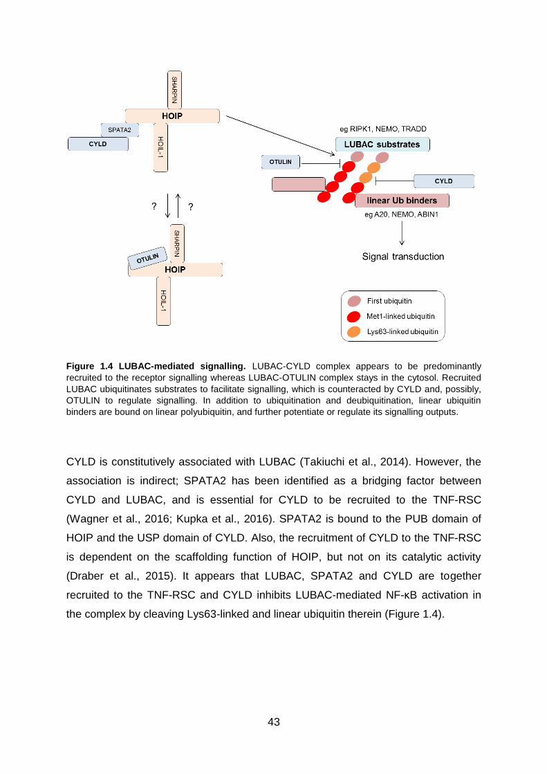
43
Figure 1.4 LUBAC-mediated signalling. LUBAC-CYLD complex appears to be predominantly
recruited to the receptor signalling whereas LUBAC-OTULIN complex stays in the cytosol. Recruited
LUBAC ubiquitinates substrates to facilitate signalling, which is counteracted by CYLD and, possibly,
OTULIN to regulate signalling. In addition to ubiquitination and deubiquitination, linear ubiquitin
binders are bound on linear polyubiquitin, and further potentiate or regulate its signalling outputs.
CYLD is constitutively associated with LUBAC (Takiuchi et al., 2014). However, the
association is indirect; SPATA2 has been identified as a bridging factor between
CYLD and LUBAC, and is essential for CYLD to be recruited to the TNF-RSC
(Wagner et al., 2016; Kupka et al., 2016). SPATA2 is bound to the PUB domain of
HOIP and the USP domain of CYLD. Also, the recruitment of CYLD to the TNF-RSC
is dependent on the scaffolding function of HOIP, but not on its catalytic activity
(Draber et al., 2015). It appears that LUBAC, SPATA2 and CYLD are together
recruited to the TNF-RSC and CYLD inhibits LUBAC-mediated NF-κB activation in
the complex by cleaving Lys63-linked and linear ubiquitin therein (Figure 1.4).
44
1.3.6 Linear ubiquitin-binding proteins
LUBAC regulates the function of its substrates by ubiquitination, but ubiquitins must
be recognised by other proteins to fully exert its function to signal to the downstream.
Figure 1.4 indicates how LUBAC can mediate the signalling by ubiquitination, and
ubiquitin is 'read' by ubiquitin receptors, or binders. Within ubiquitin receptors, most
of the ubiquitin binding domains interact specifically with particular types of ubiquitin
linkages. Therefore these binders are recruited to specific ubiquitin linkages on
particular targets in signalling complexes (Shimizu et al., 2015).
Studies have shown that the ubiquitin binding domains in NEMO, ABIN-1 and A20
bind preferentially to linear ubiquitin for Met1-linked polyubiquitin. It is crucial for
understanding the biological role of linear ubiquitination by reviewing the nature of
these proteins.
1.3.6.1 NEMO
The adaptor protein NEMO/IKKγ is engaged in the IKK complex with the kinase
heterodimer IKKα/β, which phosphorylates IκBα and activates canonical NF-κB
signalling. NEMO harbours a C-terminal ubiquitin recognition motif, the UBAN motif.
The UBAN motif specifically recognises linear ubiquitin over Lys48- and Lys63-linked
polyubiquitin (Rahighi et al., 2009). NEMO mutations are causative for incontinentia
pigmenti and anhidrotic ectodermal dysplasia with immunodeficiency (EDA-ID)
(Aradhya et al., 2001; Döffinger et al., 2001). Noteworthy, mutations in the UBAN
domain of NEMO are linked to EDA-ID (Döffinger et al., 2001; Hubeau et al., 2011).
A point mutation in its UBAN motif hampers the binding to polyubiquitins, particularly
linear chains, and thereby dampens IKK activation and increases susceptibility to
TNF-induced cell death, despite an unaltered folding of NEMO protein (Wu et al.,
2008; Rahighi et al., 2009; Hubeau et al., 2011; O'Donnell et al., 2012).
Loss of NEMO in different tissues results in severe inflammation. NEMO deletion in
skin epithelium leads to perinatal lethality preceded by dermatitis, which is
reminiscent of incontinentia pigmenti phenotype in humans (Nenci et al., 2006). Also,
the intestinal ablation of NEMO results in severe spontaneous colitis (Nenci et al.,
2007). In both cases, TNF drives the inflammation and, at least in the latter,
45
inflammation due to NEMO deficiency was driven by RIPK1-dependent cell death
(Vlantis et al., 2016). By contrast, LPC-specific deletion of NEMO leads to TNF-
independent steatohepatitis and liver carcinogenesis (Luedde et al., 2007; Ehlken et
al., 2014). NEMO together with IKKα/β suppresses activation of RIPK1 to undergo
cell death (Dondelinger et al., 2015). Thus, the recognition of linear ubiquitin is
essential for NEMO to optimally activate NF-κB signalling and prevent aberrant cell
death.
1.3.6.2 ABIN-1
ABIN-1 harbours a C-terminal UBAN motif, similar to NEMO, which preferentially
binds to linear ubiquitin as compared to Lys63-linked chains (Nanda et al., 2011).
ABIN-1 suppresses NF-κB activation in TNFR1 and TLR signalling by binding to
NEMO (Mauro et al., 2006). ABIN-1 restricts TNF-induced cell death by binding to
ubiquitin chains (Oshima et al., 2009).
Deletion of ABIN-1 in mice causes lethality late during embryonic development. Its
lethality is rescued by concomitant TNF ablation, and TNF-induced cell death
appears to be accountable for the cause of death of ABIN-1-deficient embryos
(Oshima et al., 2009). A missense mutation in its UBAN domain leads to loss of
binding capacity to Met1- and Lys63-linked ubiquitins and cells expressing this
mutant fail to protect against TNF-induced cell death (Oshima et al., 2009; Nanda et
al., 2011). The knock-in mice of UBAN mutant of ABIN-1 spontaneously develop
immune defects including lymphadenopathy and autoimmunity. Interestingly, these
immune abnormalities are corrected by MyD88 deficiency (Nanda et al., 2011). Yet,
the UBAN mutant mice did not phenocopy the embryonic lethality of ABIN-1-deficient
mice, suggesting that ABIN-1 has other regulatory domains to modulate immune
signalling.
1.3.6.3 A20
A20 (also known as TNFAIP3) has an OTU domain at the N-terminus and seven ZF
domains at the C-terminus (Ma et al., 2012). The function of A20’s DUB activity has
been controversial. A20 was initially thought to act as a suppressor of NF-κB by
cleaving Lys63-linked ubiquitin linkages (Wertz et al., 2004, Wertz et al., 2015).
46
However, other studies indicated that A20 is incapable of hydrolysing Lys63-linked
ubiquitin linkages (Lin et al., 2008; Mevissen et al., 2013; Ritorto et al., 2014).
Following studies showed that A20 binds to linear ubiquitin and stabilises it and that,
on the other hand, the presence of linear ubiquitin prevents the DUB activity of A20
(Draber et al., 2015; Wertz et al., 2015).
A20 inhibits activation of NF-κB in various immune signalling including TNFR1, CD40,
IL-1R/TLR (Wertz et al., 2004; Turer et al., 2008; Hitotsumatsu et al., 2008, Chu et
al., 2011). A20-deficient mice develop multi-organ inflammation in a MyD88-
dependent manner, and suffer from lethality within two weeks of birth (Lee et al.,
2000; Turer et al., 2008). A20-deficient cells show TNF-induced NF-κB
hyperactivation and also increased cell death. However, inactivation of A20’s DUB
activity in the OTU domain does not elicit spontaneous inflammation in mice, and
neither is it required for prevention of TNF-induced cell death (De et al., 2014;
Draber et al., 2015). The inactive mutant only marginally impacts NF-κB activation in
macrophages and dendritic cells (De et al., 2014). The OTU domain appears to
regulate ubiquitination of TNFR1 and contribute to the inhibition of inflammation, but
the precise mechanism is unclear (Wertz et al., 2015).
A20 has two ubiquitin binding sites in its C-terminus, the ZF4 and ZF7 domains. The
ZF4 motif is the preferential binding site to Lys63-linked, as compared to Lys11-,
Lys48- or Met1-linked polyubiquitin (Bosanac et al., 2010). Mutation of ZF4 in mice
does not affect their development (Lu et al., 2013), although ubiquitin binding of ZF4
is required for attenuation of TNFR1 signalling (Bosanac et al., 2010; Lu et al., 2013;
Wertz et al., 2015). In contrast to ZF4, the ZF7 domain specifically binds to linear
ubiquitin and is essential for recruitment of A20 to the TNF-RSC, depending on the
presence of linear polyubiquitin (Tokunaga et al., 2013; Draber et al., 2015). The ZF7
motif directly inhibits TAK1-dependent activation of NF-κB signalling by binding to
polyubiquitin chains (Skaug et al., 2013, Verhelst et al., 2013, Tokunaga et al., 2013).
Also, TNF-induced cell death was suppressed by linear ubiquitin binding of ZF7
(Draber et al., 2015). It appears that concerted actions of the OTU, ZF4 and ZF7
domains are essential for the anti-inflammatory function of A20 as none of the
mutants of single domains fully recapitulates the phenotype of systemic A20
deficiency.

47

48
1.4 Aims of the project
Our laboratory and others have investigated the role of LUBAC and established that
linear ubiquitination regulates NF-κB signalling as well as MAPK signalling and
prohibits TNF-induced cell death. Furthermore, perturbation of TNFR1 signalling is
strongly associated with liver pathogenesis in mice.
The aim of this research is to elucidate the role of linear ubiquitination in the liver
parenchyma by observing how linear ubiquitination or its loss impacts inflammation
and cell death in the development of hepatitis, and possibly, liver cancer formation.
The major research questions are:
1. Does perturbation of linear ubiquitination in hepatocytes exacerbate or prevent the
induction of hepatitis and/or HCC in vivo? If so, what is the mechanism engaged?
2. How does perturbation of linear ubiquitination in hepatocytes alter their response
to TNF and other death receptor ligands, such as TRAIL and CD95L, in terms of
gene activation and cell death induction?
To answer these questions, I have utilised a mouse model relying on HOIP
deficiency in liver parenchymal cells. On the basis of this model, I have investigated
the role of linear ubiquitination in regulating cell death and inflammation in the liver
and how this affects liver carcinogenesis with the aim to broaden our understanding
of hepatic diseases.
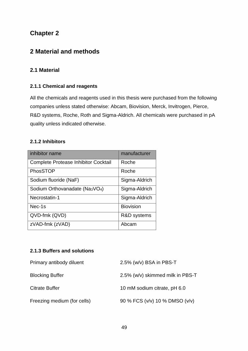
49
Chapter 2
2 Material and methods
2.1 Material
2.1.1 Chemical and reagents
All the chemicals and reagents used in this thesis were purchased from the following
companies unless stated otherwise: Abcam, Biovision, Merck, Invitrogen, Pierce,
R&D systems, Roche, Roth and Sigma-Aldrich. All chemicals were purchased in pA
quality unless indicated otherwise.
2.1.2 Inhibitors
inhibitor name manufacturer
Complete Protease Inhibitor Cocktail Roche
PhosSTOP Roche
Sodium fluoride (NaF) Sigma-Aldrich
Sodium Orthovanadate (Na3VO4) Sigma-Aldrich
Necrostatin-1 Sigma-Aldrich
Nec-1s Biovision
QVD-fmk (QVD) R&D systems
zVAD-fmk (zVAD) Abcam
2.1.3 Buffers and solutions
Primary antibody diluent 2.5% (w/v) BSA in PBS-T
Blocking Buffer 2.5% (w/v) skimmed milk in PBS-T
Citrate Buffer 10 mM sodium citrate, pH 6.0
Freezing medium (for cells) 90 % FCS (v/v) 10 % DMSO (v/v)

50
IP-lysis Buffer 30 mM Tris-Base, 120 mM NaCl, 2 mM
EDTA, 2 mM KCl, 10 % Glycerol (v/v), 1 %
Triton X-100 (v/v)
PBS 137 mM NaCl, 8.1 mM Na2HPO4, 2.7 mM
KCl, 1.5 mM KH2PO4
PBS-T PBS, 0.05% (v/v) Tween-20
Liver perfusion buffer NaCl 8 g/L, KCl 0.2 g/L, Na2HPO4 33 mg/L,
HEPES 2.38 g/L, pH 7.65
RIPA buffer: 50 mM Tris HCl pH 8.0, 150 mM NaCl, 1%
NP-40, 0.5% sodium deoxycholate, 0.1%
SDS
Stripping buffer 50 mM glycine, pH 2.3
TBS: 150 mM NaCl, 20 mM Tris, pH7.6
TEX buffer: 10 mM Tris, 1 mM EDTA,
0.1% Triton X-100 pH 8.0
Transfer buffer: 25 mM bicine, 25 mM Tris, 1 mM EDTA,
10% Methanol (v/v), pH 7.2
Tris-EDTA buffer: 10 mM Tris, 1 mM EDTA, pH 8.0
2.1.4 Biological agents
agent source
His-TNF previously produced in E.coli and purified in our laboratory
(kindly provided by Dr Peter Draber)
iz-TRAIL (mouse) produced in E.coli and purified
CD95L-Fc previously produced in HEK293T cells (kindly provided by
Mr Torsten Hartwig)

51
2.1.5 Antibodies
Unconjugated primary antibody for western blot
antibody manufacturer catalogue
number host/isotype
Bax Santa Cruz sc-7480 mouse IgG2b
Bcl-2 Santa Cruz sc-7382 mouse IgG1
Bcl-xL Cell signaling 2764 rabbit
Bid R&D systems AF860 goat
caspase-8 Enzo Life Sciences ALX-804-447 rat
c-FLIP Adipogen AG-20B-0005 rat
cIAP1/2 R&D systems 315301 mouse IgG2a
cleaved caspase-3 Cell Signaling 9661 rabbit
cleaved caspase-8 Cell Signaling 9429 rabbit
ERK1/2 Cell Signaling 9102 rabbit
FADD Assay Design AAM-121 mouse IgG1
GAPDH Abcam ab8245 mouse IgG1
HOIL-1 in-house - mouse IgG2a
HOIP in-house - rabbit
IB Cell Signaling 9242 rabbit
JNK Cell Signaling 9258 rabbit
Mcl-1 Cell signaling 94296 rabbit
phospho-ERK Cell Signaling 9101 rabbit
phospho-IκBα Cell Signaling 9246 mouse IgG1
phospho-JNK Cell Signaling 4671 rabbit
RIPK1 BD Biosciences 610459 mouse IgG2a
RIPK3 Enzo Life Sciences ADI-905-242 rabbit
SHARPIN Proteintech 14626-1-AP rabbit
XIAP Cell Signaling 2042 rabbit
Unconjugated primary antibody for immunoprecipitation
antibody manufacturer catalogue
number isotype
FADD Santa Cruz sc-6036 goat
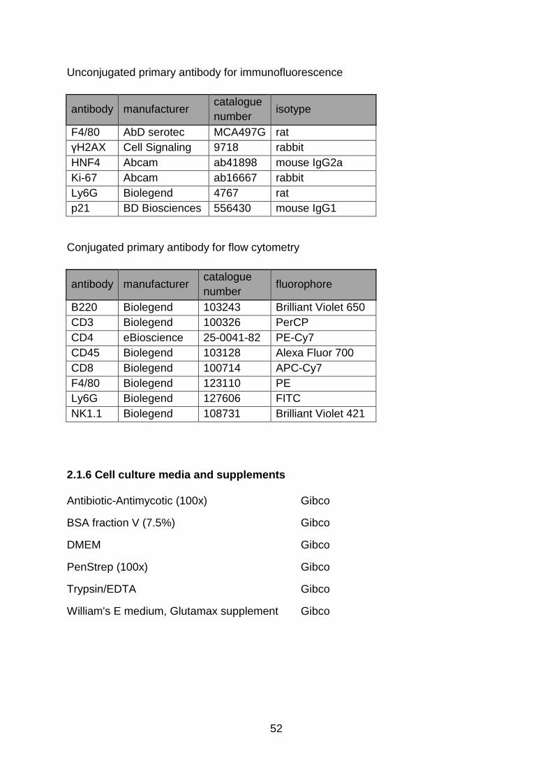
52
Unconjugated primary antibody for immunofluorescence
antibody manufacturer catalogue
number isotype
F4/80 AbD serotec MCA497G rat
γH2AX Cell Signaling 9718 rabbit
HNF4 Abcam ab41898 mouse IgG2a
Ki-67 Abcam ab16667 rabbit
Ly6G Biolegend 4767 rat
p21 BD Biosciences 556430 mouse IgG1
Conjugated primary antibody for flow cytometry
antibody manufacturer catalogue
number fluorophore
B220 Biolegend 103243 Brilliant Violet 650
CD3 Biolegend 100326 PerCP
CD4 eBioscience 25-0041-82 PE-Cy7
CD45 Biolegend 103128 Alexa Fluor 700
CD8 Biolegend 100714 APC-Cy7
F4/80 Biolegend 123110 PE
Ly6G Biolegend 127606 FITC
NK1.1 Biolegend 108731 Brilliant Violet 421
2.1.6 Cell culture media and supplements
Antibiotic-Antimycotic (100x) Gibco
BSA fraction V (7.5%) Gibco
DMEM Gibco
PenStrep (100x) Gibco
Trypsin/EDTA Gibco
William's E medium, Glutamax supplement Gibco
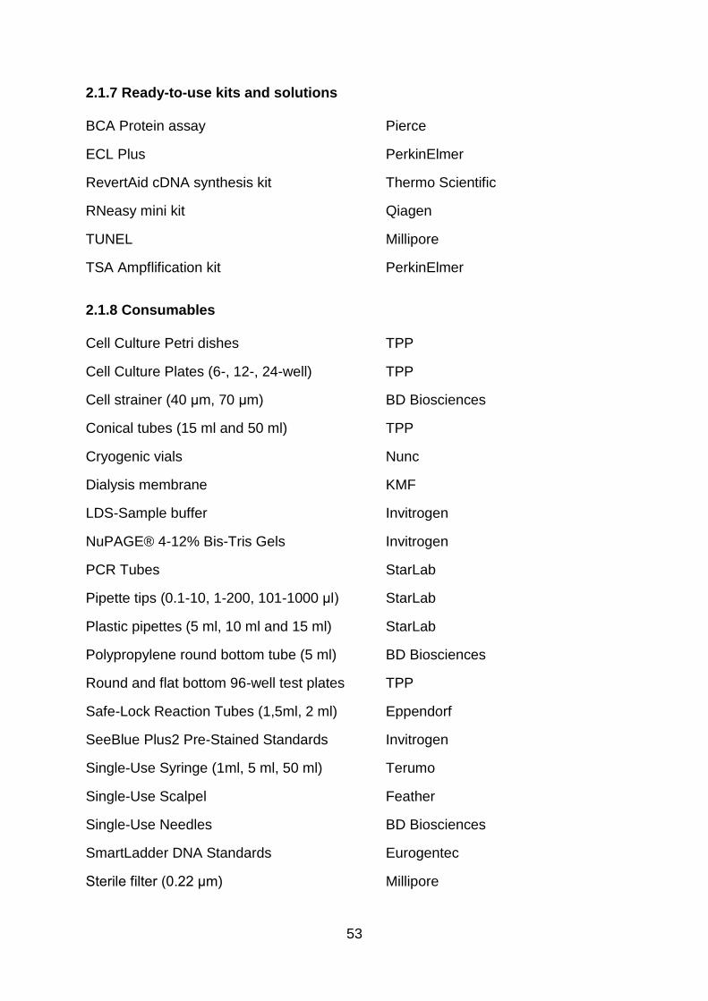
53
2.1.7 Ready-to-use kits and solutions
BCA Protein assay Pierce
ECL Plus PerkinElmer
RevertAid cDNA synthesis kit Thermo Scientific
RNeasy mini kit Qiagen
TUNEL Millipore
TSA Ampflification kit PerkinElmer
2.1.8 Consumables
Cell Culture Petri dishes TPP
Cell Culture Plates (6-, 12-, 24-well) TPP
Cell strainer (40 μm, 70 μm) BD Biosciences
Conical tubes (15 ml and 50 ml) TPP
Cryogenic vials Nunc
Dialysis membrane KMF
LDS-Sample buffer Invitrogen
NuPAGE® 4-12% Bis-Tris Gels Invitrogen
PCR Tubes StarLab
Pipette tips (0.1-10, 1-200, 101-1000 μl) StarLab
Plastic pipettes (5 ml, 10 ml and 15 ml) StarLab
Polypropylene round bottom tube (5 ml) BD Biosciences
Round and flat bottom 96-well test plates TPP
Safe-Lock Reaction Tubes (1,5ml, 2 ml) Eppendorf
SeeBlue Plus2 Pre-Stained Standards Invitrogen
Single-Use Syringe (1ml, 5 ml, 50 ml) Terumo
Single-Use Scalpel Feather
Single-Use Needles BD Biosciences
SmartLadder DNA Standards Eurogentec
Sterile filter (0.22 μm) Millipore

54
Tissue Culture flasks (75, 150 cm2) TPP
Whatman paper Schleicher&Schuell
X-Ray film VWR
2.1.9 Instruments
Accuri C6 BD Biosciences
Blotting equipment Invitrogen
Digital Camera Olympus
Evos FL Life Technologies
Fortessa LSR II BD Biosciences
Freezer -20°C Liebherr
Freezer -80°C Forma Scientific
Gene Amplification PCR system 9700 Applied Bioscience
Light Microscope Zeiss
LaboStar™ TWF Water Purification Evoqua
Microwave AEG
Mithras Berthold
Multiskan Ascent Thermo Labsystems
NanoDrop Spectrophotometer ND-1000 NanoDrop Technologies
pH Meter Mettler
Pipetboy Integra Bioscience and StarLab
Pipettes (10 μl, 100 μl, 200 μl, 1 ml) Gilson and Eppendorf
Power Supply for agarose gels BioRad
Table Centrifuge Biofuge Heraeus
Thermomixer compact Eppendorf
Vortex Heidolph
X-Ray film Developer KONICA

55
2.1.10 Software
Adobe Photoshop CS6 Adobe
Adobe Illustrator CS6 Adobe
ApE Wayne Davis (University of Utah)
Ascent Software Version 2.6 Thermo Labsystems
EndNote Thomson Reuters
FACSDiva BD Biosciences
FlowJo 7.6.5 TriStar
GraphPad Prism 6 GraphPad Software
ImageJ NIH
Microsoft Excel 2010 Microsoft
Microsoft Powerpoint 2010 Microsoft
Microsoft Word 2010 Microsoft
ND1000 V3.3.0 NanoDrop Technology
2.2 Cell biological methods
2.2.1 Isolation and culture of mouse primary hepatocytes
Primary hepatocytes were isolated from 7-12 week-old mice. First, mice were
anaesthetised with ketamine and xylazine. Mice were cannulated via the inferior
vena cava and subsequently the portal vein was incised. Mouse livers were perfused
with warm perfusion buffer supplemented with 0.5 mM EDTA for 7 minutes. The
Second perfusion with warm perfusion buffer plus 0.5 mg/ml collagenase (Sigma,
C5138) and 10 mM CaCl2 was subsequently performed for 3 minutes. Digested
livers were isolated in warm hepatocyte culture medium (William's E medium
Glutamax supplement with 1x Antibiotic-antimycotic, 0.1 % (w/v) BSA fraction V and
25 nM dexamethasone (Sigma)) with 10% FCS. Liver lobules were opened with
forceps and scissors and vigorously shaken in the medium to release cells. Cells
were passed through 70 um nylon mesh and spun at 29×g for 2 minutes at room
temperature. Cell pellets were washed twice with hepatocyte culture medium with

56
FCS. Live cell numbers were counted by means of trypan blue exclusion. Isolated
hepatocytes were plated on plates coated with collagen type I (Sigma, C3867) in
hepatocyte culture medium with 10% FCS at 37ºC in humidified 5% CO2 chamber.
The culture media were replaced 3-4 hours after plating with hepatocyte culture
medium without FCS.
2.2.2 Cell viability assay
Culture media were removed and primary hepatocytes were lysed in diluted Cell
Titer Glo solution (1:6 in PBS) (Promega). Cell lysates were incubated on a shaker
for 10 minutes, and lysates were transferred to opaque 96-well plates for
measurement. Luminescence reflecting the ATP level in cells was quantified with
Mithras (Berthold).
2.3 Biochemical methods
2.3.1 iz-TRAIL production from E.coli
Endotoxin-free isoleucine zipper-tagged murine TRAIL (izTRAIL) was purified from
Rosetta strain of E.coli transformed with plasmid encoding izTRAIL as described by
Ganten et al. (Ganten et al., 2005). E.coli lysate were subjected to a sequential
purification employing AKTA micro system with hydroxyapatite column and Ni-NTA
column to obtain concentrated fraction. Purified fractions were evaluated by means
of UV absorbance, Bradford protein assay and SDS-PAGE. Level of contaminating
LPS was examined by LAL assay.
2.3.2 Preparation of cell lysates from primary cells for western blot
Cells were lysed with ice-cold IP lysis buffer supplemented with Complete protease
inhibitor cocktail (the solution prepared by dissolving a tablet in water, and added to
lysis buffer in 1:50), 10 mM NaF and 1mM Na3VO4. Lysates were spun at 13300 rpm
at 4°C for 15 minutes to remove insoluble fraction. Supernatants were mixed with 4x
LDS sample buffer with 50 mM DTT (3:1) and boiled at 92°C for 5 minutes for PAGE.
57
2.3.3 Liver lysate preparation for western blot
Livers were dissected from 4 week-old mice, snap-frozen in liquid nitrogen and
stored at -80ºC until the following manipulation. Frozen livers were processed with a
mortar and a pestle pre-chilled with liquid nitrogen until tissues became a fine
powder. Processed livers were incubated in ice-cold RIPA buffer with Complete
protease inhibitor cocktail solution (1:50) and PhosSTOP soliution (1:50) at 4ºC.
Lysates were vortexed briefly and sonicated for 20 seconds four times, with one-
minute intervals on ice between each sonication. Lysates were incubated with
continuous inversion for another 10 minutes. Samples were spun at 15000 g at 4ºC
for 15 minutes to remove insoluble fraction.
2.3.4 Determination of protein concentration
Protein concentrations of liver lysates were determined by BCA protein assay
(Pierce) with BSA as a protein standard. RIPA buffer was added to each sample to
adjust protein concentration.
2.3.5 Immunoprecipitation
For immunoprecipitation for FADD-associated complex, cell lysates were diluted in
IP lysis buffer. Lysates were pre-cleared with Sepharose CL-4B (Sigma) for 2 hours.
Protein G-coupled sepharose (GE) was blocked with 1% BSA in IP lysis buffer for 1
hour and washed extensively with IP lysis buffer prior to incubation. Pre-cleared
lysates were incubated overnight with blocked Protein G-coupled sepharose and
anti-FADD (M-19) antibody or isotype control, normal goat IgG (Santa Cruz), at 4ºC.
Beads were washed 5 times with ice-cold IP lysis buffer and vortexed and proteins
were eluted in 1x LDS buffer at 92ºC.
2.3.6 Polyacrylamide gel electrophoresis
Denatured lysates were separated on 4-12% Bis-Tris NuPAGE gel (Invitrogen) with
MOPS buffer at 80 V for 15 min and at 180 V for 50 min.
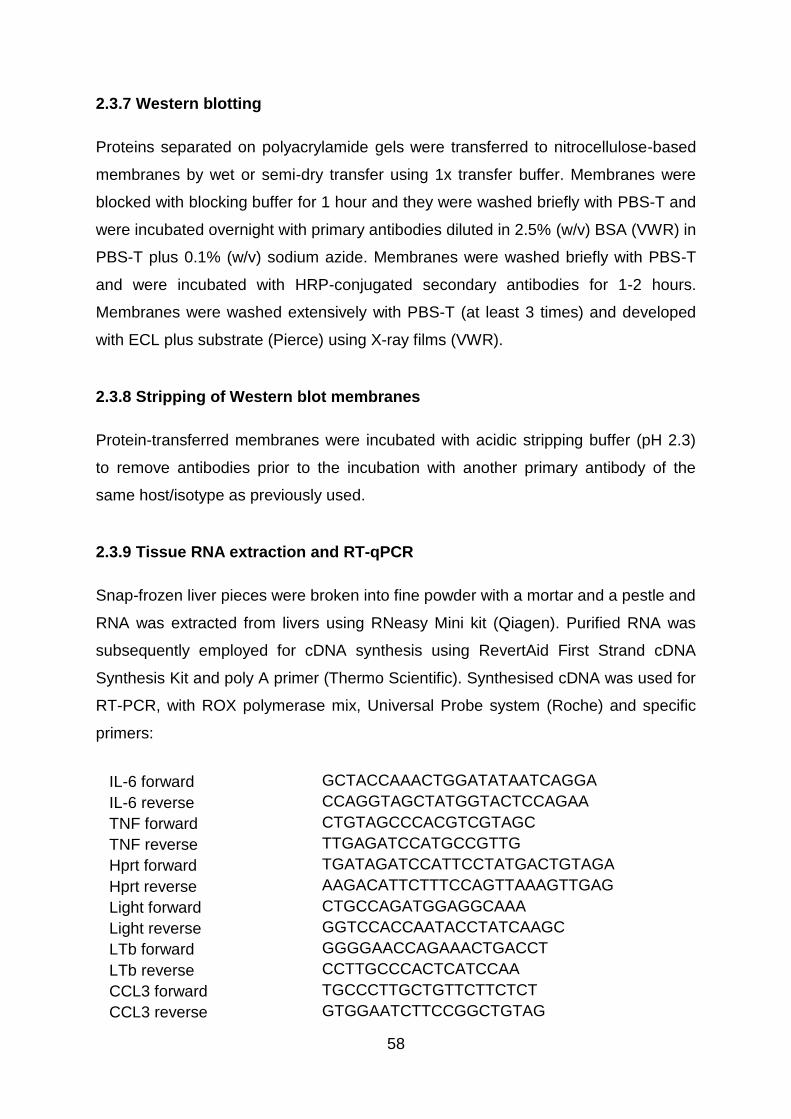
58
2.3.7 Western blotting
Proteins separated on polyacrylamide gels were transferred to nitrocellulose-based
membranes by wet or semi-dry transfer using 1x transfer buffer. Membranes were
blocked with blocking buffer for 1 hour and they were washed briefly with PBS-T and
were incubated overnight with primary antibodies diluted in 2.5% (w/v) BSA (VWR) in
PBS-T plus 0.1% (w/v) sodium azide. Membranes were washed briefly with PBS-T
and were incubated with HRP-conjugated secondary antibodies for 1-2 hours.
Membranes were washed extensively with PBS-T (at least 3 times) and developed
with ECL plus substrate (Pierce) using X-ray films (VWR).
2.3.8 Stripping of Western blot membranes
Protein-transferred membranes were incubated with acidic stripping buffer (pH 2.3)
to remove antibodies prior to the incubation with another primary antibody of the
same host/isotype as previously used.
2.3.9 Tissue RNA extraction and RT-qPCR
Snap-frozen liver pieces were broken into fine powder with a mortar and a pestle and
RNA was extracted from livers using RNeasy Mini kit (Qiagen). Purified RNA was
subsequently employed for cDNA synthesis using RevertAid First Strand cDNA
Synthesis Kit and poly A primer (Thermo Scientific). Synthesised cDNA was used for
RT-PCR, with ROX polymerase mix, Universal Probe system (Roche) and specific
primers:
IL-6 forward GCTACCAAACTGGATATAATCAGGA
IL-6 reverse CCAGGTAGCTATGGTACTCCAGAA
TNF forward CTGTAGCCCACGTCGTAGC
TNF reverse TTGAGATCCATGCCGTTG
Hprt forward TGATAGATCCATTCCTATGACTGTAGA
Hprt reverse AAGACATTCTTTCCAGTTAAAGTTGAG
Light forward CTGCCAGATGGAGGCAAA
Light reverse GGTCCACCAATACCTATCAAGC
LTb forward GGGGAACCAGAAACTGACCT
LTb reverse CCTTGCCCACTCATCCAA
CCL3 forward TGCCCTTGCTGTTCTTCTCT
CCL3 reverse GTGGAATCTTCCGGCTGTAG

59
CXCL1 forward GACTCCAGCCACACTCCAAC
CXCL1 reverse TGACAGCGCAGCTCATTG
2.4 Animal studies
All the animal studies stated are conducted according to the appropriate licence
under Animals (Scientific Procedures) Act 1986.
2.4.1 Liver-specific HOIP knockout mice
HOIP-floxed mice were generated at the Walter and Eliza Hall (Melbourne, Australia)
(Peltzer et al., 2014). HOIP-floxed mice were subsequently crossed to Alb-Cre mice
(purchased from Jackson laboratories) for analysis. Both strains had and were
maintained in C57BL/6 background. Mice were maintained in Charles River (Margate,
UK) or UCL Biological Service Unit (London, UK) under the specific pathogen-free
condition and 12-hour dark/light cycle, and fed ad libitum with standard chow.
2.4.2 Crossings
TNFR1 knockout mice (Pfeffer et al., 1993) were obtained from Jackson laboratories.
They were derived from mixed 129S2/Sv and C57BL/6 background but were
maintained in C57BL/6 background. Caspase-8 knockout mice were kindly provided
by Dr William Kaiser, which were maintained in C57BL/6 background. MLKL
knockout mice were generated in C57BL/6 background in our laboratory using
transcription activator-like effector nuclease (TALEN) designed by Mr Sebastian
Kupka. Liver-specific HOIP knockout mice were crossed to these mice for analysis.
2.4.3 Genotyping
Mice were sampled at 2-3 weeks of age for genotyping. Ear or tail samples were
lysed in genotyping lysis buffer at 95°C for 30 min, and neutralised with an equal
amount of genotyping neutralising buffer. Samples were preserved at 4°C until use.

60
PCR mix was prepared with 5 Prime master mix or Dream Taq (Thermo Fisher
Scientific) and appropriate sets of primers:
HOIP forward CTCATTTGAATTCTATGATGC
HOIP reverse GAAACAGGGACCAGGAGT
Cre forward TTGCCCCTGTTTCACTATCCAG
Cre reverse TGCTGTTTCACTGGTTATGCGG
TNFR1 WT TGTGAAAAGGGCACCTTTACGGC
TNFR1 common GGCTGCAGTCCACGCACTGG
TNFR1 mutant ATTCGCCAATGACAAGACGCTGG
Casp8 WT forward GTAGAAATGGAGAAGAGGACCATG
Casp8 KO forward TTGAGAACAAGACCTGGGGACTG
Casp8 reverse GGATGTCCAGGAAAAGATTTGTGTC
MLKL common forward CAAGGAAAGAAGAACCTGCCCGA
MLKL WT reverse GGATGCTGGCTGGCTGACA
MLKL KO reverse CCTGCTGCCATCTGAAACGG
2.4.4 Dissection and fixation of tissues for histology
Mice were euthanised and whole livers were resected with forceps and scissors.
Tissues were cut into no more than 2 cm pieces, and pieces of livers were fixed in
10% neutral-buffered formalin (Sigma). The rest of the tissues were snap-frozen in
liquid nitrogen for further analysis. Formalin was replaced with 70% ethanol to avoid
over-fixation before embedding.
2.4.5 Histological sections
Formalin-fixed tissues were embedded in paraffin blocks by sequential incubation
with ethanol, histoclear and paraffin and sliced into thin sections in 4 μm thickness.
Hematoxylin and eosin (H&E) staining was performed to observe tissue structure.
Tissue embedding and sectioning were performed by Ms Lorraine Lawrence (NHLI,
Imperial College London).

61
2.4.6 Immunohistochemistry
Paraffin sections were dewaxed by sequential treatment with histoclear, 100%
industrial methylated spirit (IMS), 90% IMS, 75% IMS, 50 % IMS and distilled water.
For antigen retrieval, heat-induced epitope retrieval with citrate buffer was used for
Ki-67 and cleaved caspase-3 or Tris-EDTA buffer for Ly6G and γH2AX staining.
Proteinase K (Promega) 10 μg/mL diluted in TEX buffer was used for demasking
F4/80 antigen. Tyramide signal amplification (TSA) system (Perkin Elmer) was
utilised to enhance signals. To detect cell death, terminal deoxynucleotidyl
transferase dUTP nick end labelling (TUNEL) was performed using ApopTag In situ
detection kit (Millipore).
2.4.7 Flow cytometric analysis of liver immune cells
Livers were processed in PBS with syringe plungers and the cell suspension was
filtered with 70-μm cell strainer (BD). Cells were washed with PBS and red blood
cells were removed using RBC lysis buffer (R&D). Cells were incubated with anti-
mouse CD16/CD32 and Fixable Viability Dye eFluor 660 (eBioscience) for 30 min
prior to incubation with antibodies. Cells were subsequently stained with fluorophore-
coupled antibodies for 1 hour. Cells were fixed with fixation/permeablization
concentrate (eBioscience) diluted 1:4 in fixation/permeablization diluent
(eBioscience) until the day of measurement and were washed with PBS shortly
before measurement. For absolute quantification, 10 μL of 123count eBeads
(eBioscience) were added to each cell suspension, and cells were measured on
Fortessa LSR II (BD), followed by analysis using FlowJo 7.6.5 (Tristar).
2.4.8 Serum analysis
Blood was withdrawn from the submandibular vein using a lancet and collected into
Microtainer tube with separation gels (BD). Sampled blood was kept on ice and spun
to isolate serum. Isolated sera were diluted in 0.9 % (v/w) sodium chloride and 30 μL
of diluted sera were placed on Reflotron ALT test strip (Roche), which subsequently
measured on Reflovet (Roche).

62
Chapter 3
3 Results
3.1 Characterisation of liver-specific HOIP-deficient mice
3.1.1 Generation of liver-specific HOIP-deficient mice
Systemic deficiency in the catalytic component of LUBAC, HOIP, in mice causes
embryonic lethality at mid-gestation (Peltzer et al., 2014). At this stage, the
developing livers accommodate the precursor of mature hepatocytes and bile duct
cells, hepatoblasts (Zorn, 2008). Due to the death of the animals and immaturity of
liver tissue at this embryonic stage, it is not possible to understand the role of linear
ubiquitination in the mature liver cells from the study of HOIP-deficient embryos.
To overcome this issue and specifically investigate the role of linear ubiquitination in
mature hepatocytes, our laboratory generated mice with liver parenchyma-specific
deletion of HOIP by crossing Hoipflox mice with transgenic mice expressing Cre
recombinase under the regulation of the serum albumin promoter (Alb-Cre mice)
(Postic et al., 1999). Mice deficient for HOIP in the liver, which we named HoipΔhep
mice, showed a reduction in HOIP expression levels in whole liver cell lysates to
approximately half of the control mice (Figure 3.1A). This partial penetrance of Cre-
mediated deletion can be explained by the expression of HOIP protein in non-
parenchymal cells. Thus, to specifically analyse the level of HOIP expression in
hepatocytes, primary hepatocytes were isolated by means of two-step perfusion. In
contrast to whole liver lysates, hepatocytes isolated from HoipΔhep animals displayed
a drastic reduction in the level of HOIP protein (Figure 3.1B). HOIP-deficient
hepatocytes also displayed a slight decline in the levels of the other LUBAC
components HOIL-1 and SHARPIN, which is in agreement with previous studies
showing that loss of HOIP destabilises HOIL-1 and SHARPIN in MEFs, keratinocytes
and cancer cell lines (Peltzer et al., 2014; Draber et al., 2015; Taraborrelli et al.,
submitted).
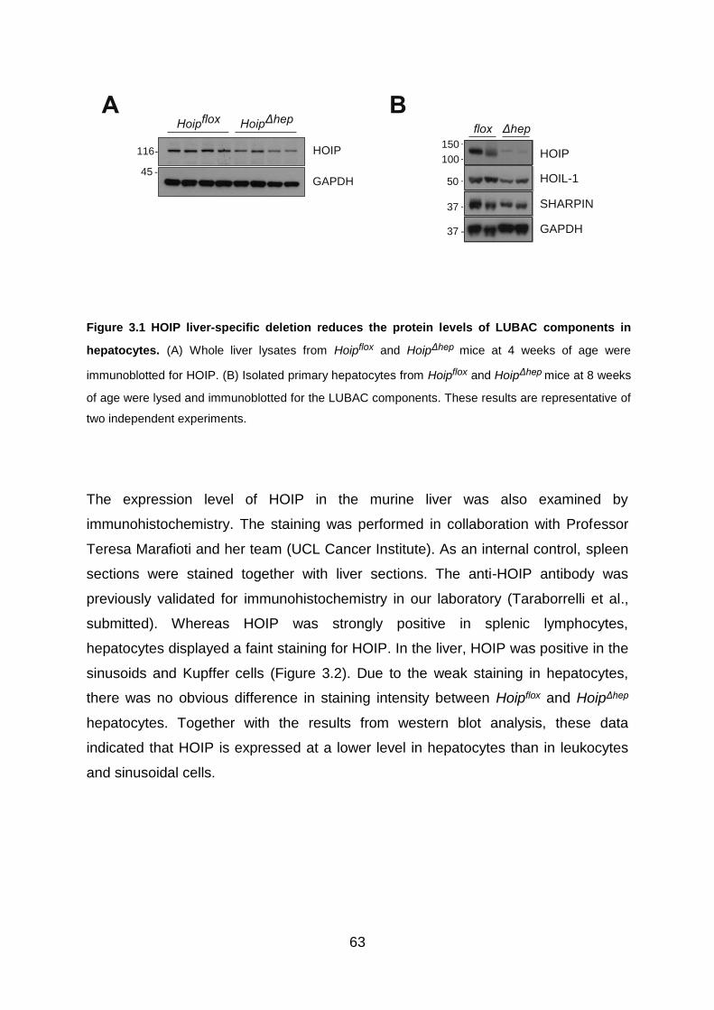
63
Figure 3.1 HOIP liver-specific deletion reduces the protein levels of LUBAC components in
hepatocytes. (A) Whole liver lysates from Hoipflox and HoipΔhep mice at 4 weeks of age were
immunoblotted for HOIP. (B) Isolated primary hepatocytes from Hoipflox and HoipΔhep mice at 8 weeks
of age were lysed and immunoblotted for the LUBAC components. These results are representative of
two independent experiments.
The expression level of HOIP in the murine liver was also examined by
immunohistochemistry. The staining was performed in collaboration with Professor
Teresa Marafioti and her team (UCL Cancer Institute). As an internal control, spleen
sections were stained together with liver sections. The anti-HOIP antibody was
previously validated for immunohistochemistry in our laboratory (Taraborrelli et al.,
submitted). Whereas HOIP was strongly positive in splenic lymphocytes,
hepatocytes displayed a faint staining for HOIP. In the liver, HOIP was positive in the
sinusoids and Kupffer cells (Figure 3.2). Due to the weak staining in hepatocytes,
there was no obvious difference in staining intensity between Hoipflox and HoipΔhep
hepatocytes. Together with the results from western blot analysis, these data
indicated that HOIP is expressed at a lower level in hepatocytes than in leukocytes
and sinusoidal cells.
100 µm
spleen
liver
Hoipflox HoipΔhep
116 -
45 -
HOIP
GAPDH
Hoipflox HoipΔhepB
HOIL-1
HOIP
50 -
150 -
100 -
SHARPIN
GAPDH
flox Δhep
37 -
37 -
A
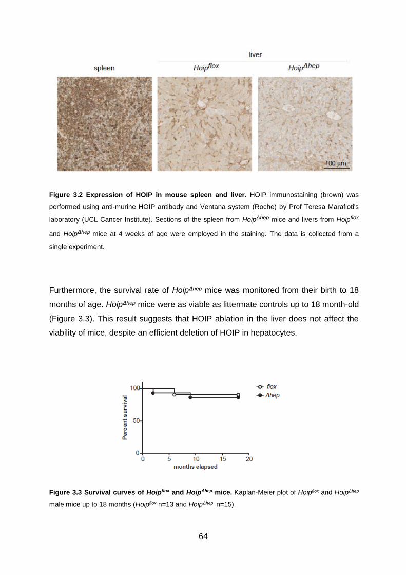
64
Figure 3.2 Expression of HOIP in mouse spleen and liver. HOIP immunostaining (brown) was
performed using anti-murine HOIP antibody and Ventana system (Roche) by Prof Teresa Marafioti's
laboratory (UCL Cancer Institute). Sections of the spleen from HoipΔhep mice and livers from Hoipflox
and HoipΔhep mice at 4 weeks of age were employed in the staining. The data is collected from a
single experiment.
Furthermore, the survival rate of HoipΔhep mice was monitored from their birth to 18
months of age. HoipΔhep mice were as viable as littermate controls up to 18 month-old
(Figure 3.3). This result suggests that HOIP ablation in the liver does not affect the
viability of mice, despite an efficient deletion of HOIP in hepatocytes.
Figure 3.3 Survival curves of Hoipflox and HoipΔhep mice. Kaplan-Meier plot of Hoipflox and HoipΔhep
male mice up to 18 months (Hoipflox n=13 and HoipΔhep n=15).

65
Hepatic abrogation of the anti-apoptotic protein c-FLIP using Alfp-Cre leads to
perinatal lethality whilst its deletion by Alb-Cre does not (Piao et al, 2012). Given that
Alfp-Cre contains an additional enhancer of the Afp gene in its regulatory sequence
and that deletion by Alfp-Cre occurs earlier than by Alb-Cre, Alfp-Cre could cause a
more severe phenotype than Alb-Cre (Postic et al., 1999; Kellendonk et al., 2000).
Consequently, HOIP deletion in hepatocytes using Alfp-Cre could results in
premature lethality. To test this, HOIP was deleted in LPCs using Alfp-Cre. For this
purpose, Hoipflox/+ Alfp-Cre male mice were crossed to Hoipflox/flox female mice.
Hoipflox/flox Alfp-Cre mice were born approximately at Mendelian ratio and were viable
at least up to a weaning age (2-3 weeks of age) (Table 3.1). Nevertheless, it is yet to
be analysed whether they would show impaired survival at a later stage in life. In sum,
the two Cre driver lines demonstrated that ablation of HOIP in LPCs does not seem
to affect the viability of mice.
Table 3.1 Alfp-Cre-mediated HOIP deletion in liver parenchyma does not cause perinatal
lethality
66
3.1.2 HOIP deficiency in the liver parenchyma results in spontaneous liver
tumour formation
Disruption of the NF-κB signalling and cell death pathway in hepatocytes can lead to
pathogenesis such as hepatitis and fibrosis as well as liver tumourigenesis at a later
stage of life (Luedde et al., 2007; Betterman et al., 2010; Weber et al., 2010).
However, these genetic manipulations do not lead to lethality of the animals in most
of the cases, except for c-FLIP deletion, which causes perinatal death using Alfp-Cre
(Piao et al., 2012). Although HoipΔhep mice displayed an unaltered survival rate,
HOIP deletion could be pathogenic at a later stage in life. Therefore, the livers from
HoipΔhep mice were analysed at 18 months of age.
Strikingly, the large majority of male HoipΔhep mice at 18 months of age exhibited
macroscopic nodules in the liver. At this stage, control mice did not show any overt
aberration in the liver (Figure 3.4A). The size, number and severity of macroscopic
lesions appearing in HoipΔhep livers were variable. Four mice out of 13 showed mild
phenotype with smaller lesions (lesion diameter < 2 mm). Another five mice
developed a moderate phenotype with multiple lesions and nodules (including
maximal lesion diameter = 2-5 mm). Lastly, the other three mice suffered from
severe pathology with large nodules (diameter > 5 mm) together with cystic lesions
(Figure 3.4A). HCC formation was observed in over a half (5 out of 8) of the HoipΔhep
animals displaying moderate and severe pathologies (Figure 3.4B, bar graph). The
other mice that did not have HCC displayed pre-cancerous anisokaryosis or
inflammatory foci (Figure 3.4B, middle pictures).
Furthermore, HoipΔhep livers exhibited focal lipid accumulation, which suggests that
HoipΔhep mice develop steatosis (Figure 3.5). In addition, Sirius red-positive mild
fibrotic lesions were present at peri-tumour areas (Figure 3.5). Taken together, HOIP
deletion in liver parenchyma causes liver tumourigenesis and steatosis.
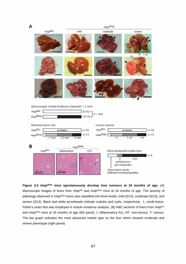
67
Figure 3.4 HoipΔhep mice spontaneously develop liver tumours at 18 months of age. (A)
Macroscopic images of livers from Hoipflox and HoipΔhep mice at 18 months of age. The severity of
pathology observed in HoipΔhep livers was classified into three levels: mild (5/13), moderate (5/13), and
severe (3/13). Black and white arrowheads indicate nodules and cysts, respectively. L: small lesion.
Fisher's exact test was employed in nodule incidence analysis. (B) H&E sections of livers from Hoipflox
and HoipΔhep mice at 18 months of age (left panel). I: inflammatory foci, NT: non-tumour, T: tumour.
The bar graph indicates the most advanced nodule type on the liver which showed moderate and
severe phenotype (right panel).
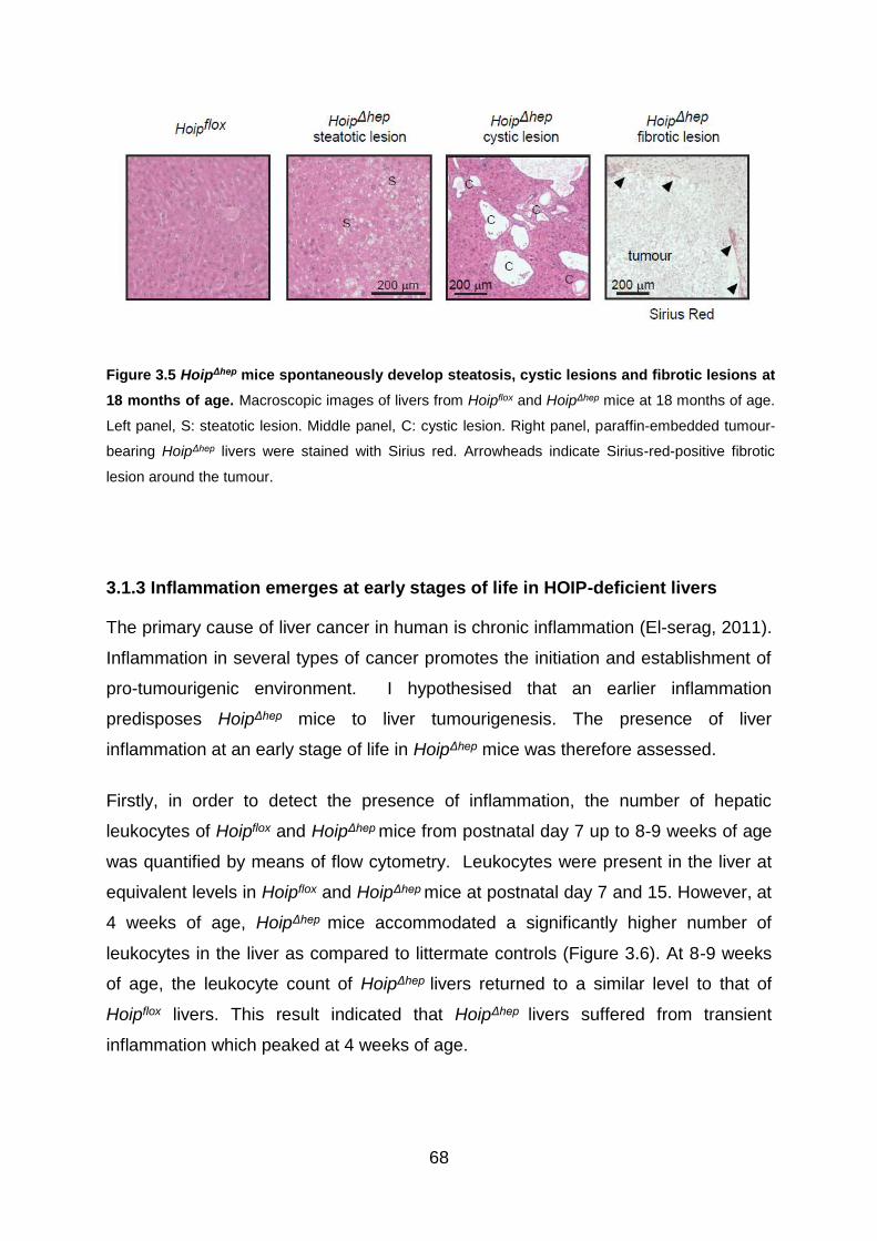
68
Figure 3.5 HoipΔhep mice spontaneously develop steatosis, cystic lesions and fibrotic lesions at
18 months of age. Macroscopic images of livers from Hoipflox and HoipΔhep mice at 18 months of age.
Left panel, S: steatotic lesion. Middle panel, C: cystic lesion. Right panel, paraffin-embedded tumour-
bearing HoipΔhep livers were stained with Sirius red. Arrowheads indicate Sirius-red-positive fibrotic
lesion around the tumour.
3.1.3 Inflammation emerges at early stages of life in HOIP-deficient livers
The primary cause of liver cancer in human is chronic inflammation (El-serag, 2011).
Inflammation in several types of cancer promotes the initiation and establishment of
pro-tumourigenic environment. I hypothesised that an earlier inflammation
predisposes HoipΔhep mice to liver tumourigenesis. The presence of liver
inflammation at an early stage of life in HoipΔhep mice was therefore assessed.
Firstly, in order to detect the presence of inflammation, the number of hepatic
leukocytes of Hoipflox and HoipΔhep mice from postnatal day 7 up to 8-9 weeks of age
was quantified by means of flow cytometry. Leukocytes were present in the liver at
equivalent levels in Hoipflox and HoipΔhep mice at postnatal day 7 and 15. However, at
4 weeks of age, HoipΔhep mice accommodated a significantly higher number of
leukocytes in the liver as compared to littermate controls (Figure 3.6). At 8-9 weeks
of age, the leukocyte count of HoipΔhep livers returned to a similar level to that of
Hoipflox livers. This result indicated that HoipΔhep livers suffered from transient
inflammation which peaked at 4 weeks of age.
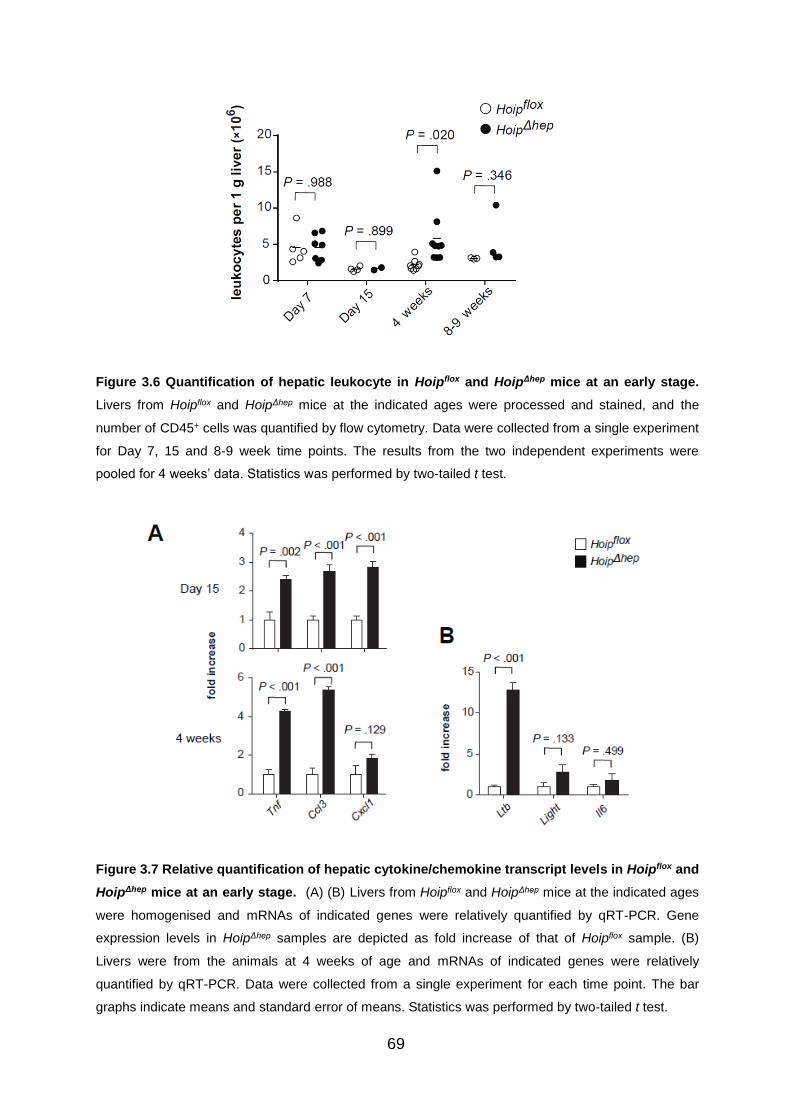
69
Figure 3.6 Quantification of hepatic leukocyte in Hoipflox and HoipΔhep mice at an early stage.
Livers from Hoipflox and HoipΔhep mice at the indicated ages were processed and stained, and the
number of CD45+ cells was quantified by flow cytometry. Data were collected from a single experiment
for Day 7, 15 and 8-9 week time points. The results from the two independent experiments were
pooled for 4 weeks’ data. Statistics was performed by two-tailed t test.
Figure 3.7 Relative quantification of hepatic cytokine/chemokine transcript levels in Hoipflox and
HoipΔhep mice at an early stage. (A) (B) Livers from Hoipflox and HoipΔhep mice at the indicated ages
were homogenised and mRNAs of indicated genes were relatively quantified by qRT-PCR. Gene
expression levels in HoipΔhep samples are depicted as fold increase of that of Hoipflox sample. (B)
Livers were from the animals at 4 weeks of age and mRNAs of indicated genes were relatively
quantified by qRT-PCR. Data were collected from a single experiment for each time point. The bar
graphs indicate means and standard error of means. Statistics was performed by two-tailed t test.
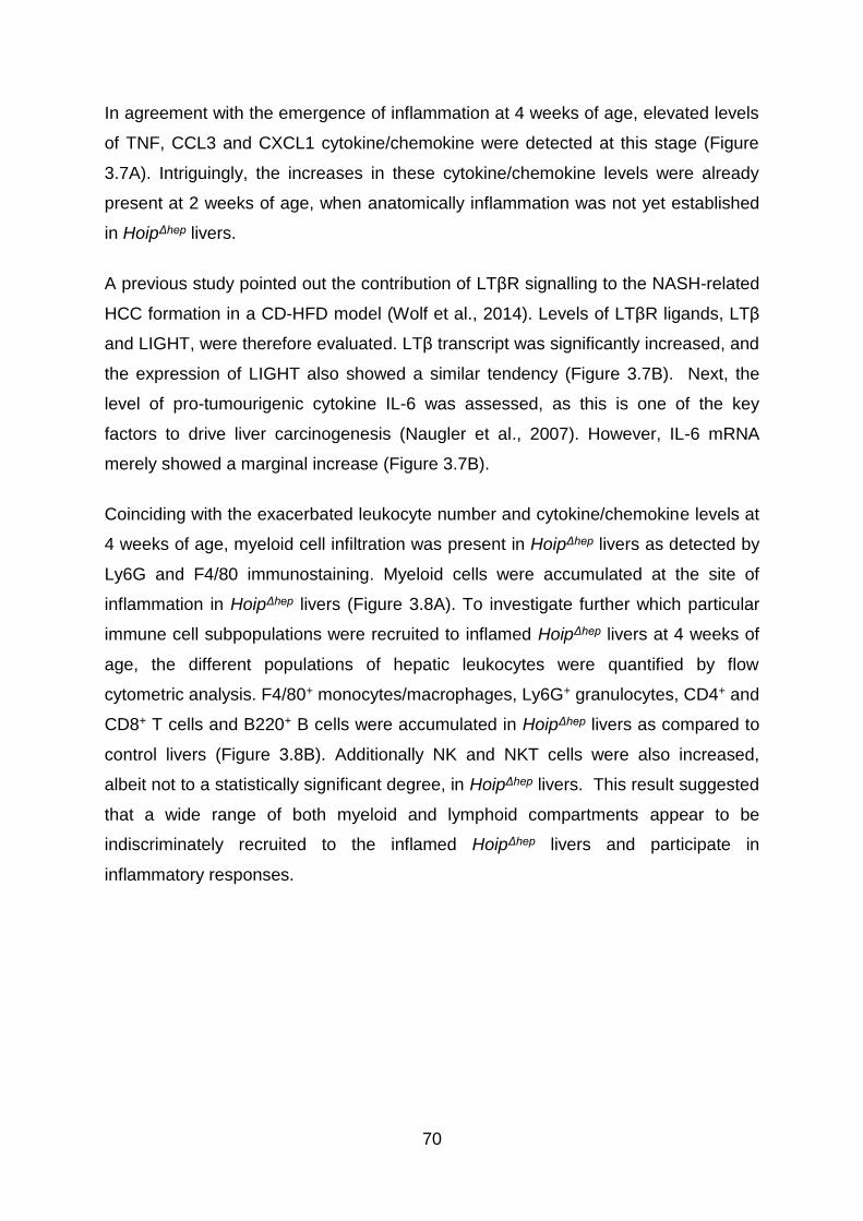
70
In agreement with the emergence of inflammation at 4 weeks of age, elevated levels
of TNF, CCL3 and CXCL1 cytokine/chemokine were detected at this stage (Figure
3.7A). Intriguingly, the increases in these cytokine/chemokine levels were already
present at 2 weeks of age, when anatomically inflammation was not yet established
in HoipΔhep livers.
A previous study pointed out the contribution of LTβR signalling to the NASH-related
HCC formation in a CD-HFD model (Wolf et al., 2014). Levels of LTβR ligands, LTβ
and LIGHT, were therefore evaluated. LTβ transcript was significantly increased, and
the expression of LIGHT also showed a similar tendency (Figure 3.7B). Next, the
level of pro-tumourigenic cytokine IL-6 was assessed, as this is one of the key
factors to drive liver carcinogenesis (Naugler et al., 2007). However, IL-6 mRNA
merely showed a marginal increase (Figure 3.7B).
Coinciding with the exacerbated leukocyte number and cytokine/chemokine levels at
4 weeks of age, myeloid cell infiltration was present in HoipΔhep livers as detected by
Ly6G and F4/80 immunostaining. Myeloid cells were accumulated at the site of
inflammation in HoipΔhep livers (Figure 3.8A). To investigate further which particular
immune cell subpopulations were recruited to inflamed HoipΔhep livers at 4 weeks of
age, the different populations of hepatic leukocytes were quantified by flow
cytometric analysis. F4/80+ monocytes/macrophages, Ly6G+ granulocytes, CD4+ and
CD8+ T cells and B220+ B cells were accumulated in HoipΔhep livers as compared to
control livers (Figure 3.8B). Additionally NK and NKT cells were also increased,
albeit not to a statistically significant degree, in HoipΔhep livers. This result suggested
that a wide range of both myeloid and lymphoid compartments appear to be
indiscriminately recruited to the inflamed HoipΔhep livers and participate in
inflammatory responses.
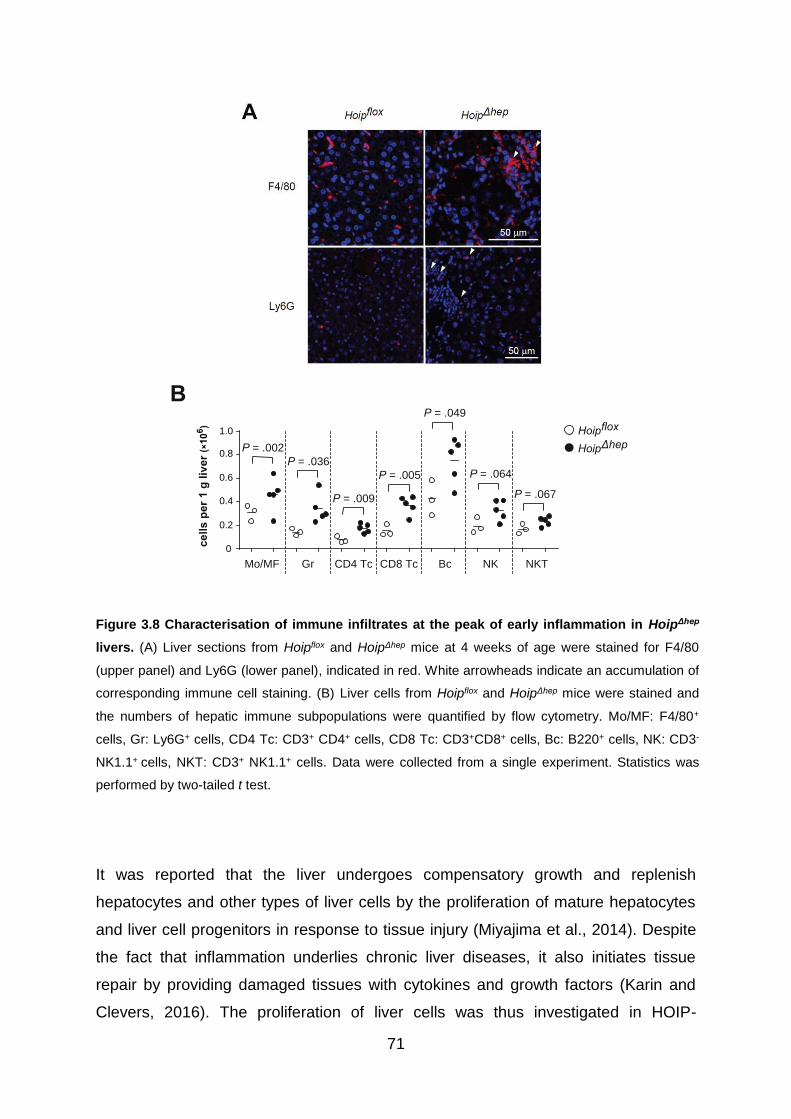
71
Figure 3.8 Characterisation of immune infiltrates at the peak of early inflammation in HoipΔhep
livers. (A) Liver sections from Hoipflox and HoipΔhep mice at 4 weeks of age were stained for F4/80
(upper panel) and Ly6G (lower panel), indicated in red. White arrowheads indicate an accumulation of
corresponding immune cell staining. (B) Liver cells from Hoipflox and HoipΔhep mice were stained and
the numbers of hepatic immune subpopulations were quantified by flow cytometry. Mo/MF: F4/80+
cells, Gr: Ly6G+ cells, CD4 Tc: CD3+ CD4+ cells, CD8 Tc: CD3+CD8+ cells, Bc: B220+ cells, NK: CD3-
NK1.1+ cells, NKT: CD3+ NK1.1+ cells. Data were collected from a single experiment. Statistics was
performed by two-tailed t test.
It was reported that the liver undergoes compensatory growth and replenish
hepatocytes and other types of liver cells by the proliferation of mature hepatocytes
and liver cell progenitors in response to tissue injury (Miyajima et al., 2014). Despite
the fact that inflammation underlies chronic liver diseases, it also initiates tissue
repair by providing damaged tissues with cytokines and growth factors (Karin and
Clevers, 2016). The proliferation of liver cells was thus investigated in HOIP-
Hoipflox
HoipΔhep
B
Mo/MF Gr CD4 Tc CD8 Tc Bc NK NKT
0
0.8
1.0
0.6
0.4
0.2
ce
lls
pe
r 1
g l
ive
r (×
10
6)
P = .049
P = .064P = .036
P = .009
P = .005
P = .002
P = .067
A
Ly6G
Hoipflox HoipΔhep
F4/80
50 µm
50 µm

72
proficient and -deficient livers. Indeed, as detected by Ki-67 expression, a higher
number of proliferating liver cells was observed in HoipΔhep mice at 2-4 weeks of age
as compared to littermate controls. Therefore, HOIP deletion leads to the initiation of
liver regeneration, consistent with the presence of inflammation (Figure 3.9).
Figure 3.9 HoipΔhep livers have increased proliferating cells at an early stage. Liver sections from
Hoipflox and HoipΔhep mice at the indicated ages were stained for Ki-67. The right graph shows the
quantification of Ki-67+ cells per optical area (magnification: 400x). Data were collected from a single
experiment. Statistics was performed by two-tailed t test.
In addition, inflammation can also lead to DNA damage by producing reactive
oxygen species (ROS) which is associated with inflammation-associated liver
carcinogenesis (Maeda et al., 2005; Ohnishi et al., 2013). To determine whether
there is underlying DNA damage response in HOIP-deficient hepatocytes, liver
sections from 4 week-old HoipΔhep and control mice were stained for phosphorylated
histone H2AX (γH2AX), a marker for DNA damage. Intriguingly, HoipΔhep livers
presented increased γH2AX+ foci, which is suggestive of increased DNA damage in
HoipΔhep livers (Figure 3.10). In summary, deficiency in HOIP in liver parenchyma
leads to transient inflammation at early stages of life, accompanied by proliferation of
regenerative liver cells and a concurrent accumulation of DNA damage.
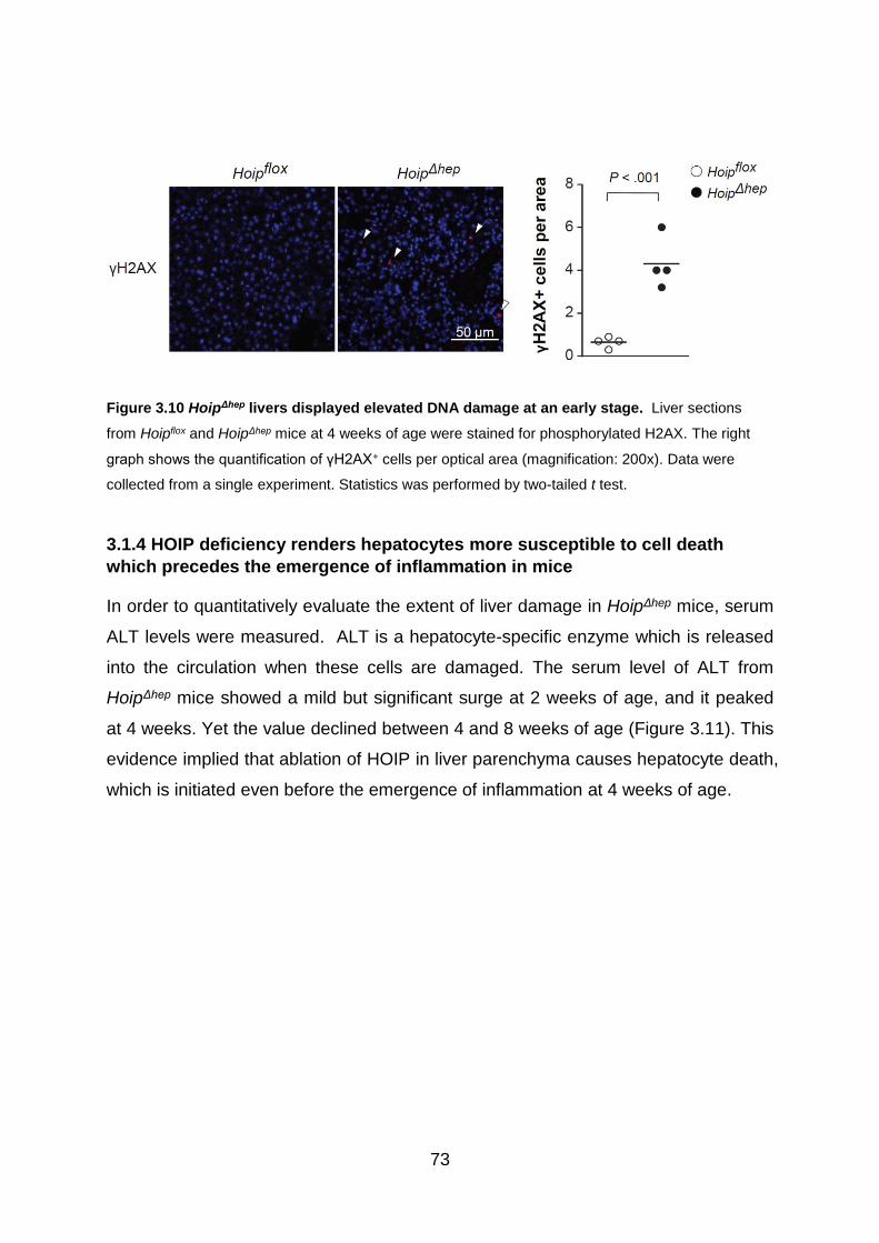
73
Figure 3.10 HoipΔhep livers displayed elevated DNA damage at an early stage. Liver sections
from Hoipflox and HoipΔhep mice at 4 weeks of age were stained for phosphorylated H2AX. The right
graph shows the quantification of γH2AX+ cells per optical area (magnification: 200x). Data were
collected from a single experiment. Statistics was performed by two-tailed t test.
3.1.4 HOIP deficiency renders hepatocytes more susceptible to cell death
which precedes the emergence of inflammation in mice
In order to quantitatively evaluate the extent of liver damage in HoipΔhep mice, serum
ALT levels were measured. ALT is a hepatocyte-specific enzyme which is released
into the circulation when these cells are damaged. The serum level of ALT from
HoipΔhep mice showed a mild but significant surge at 2 weeks of age, and it peaked
at 4 weeks. Yet the value declined between 4 and 8 weeks of age (Figure 3.11). This
evidence implied that ablation of HOIP in liver parenchyma causes hepatocyte death,
which is initiated even before the emergence of inflammation at 4 weeks of age.
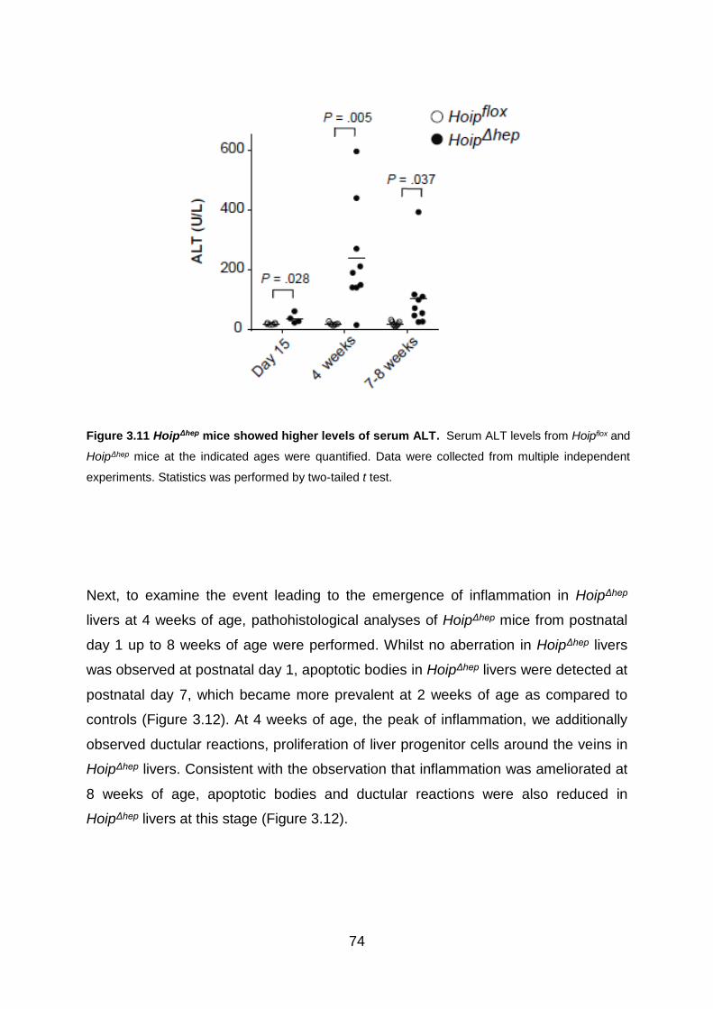
74
Figure 3.11 HoipΔhep mice showed higher levels of serum ALT. Serum ALT levels from Hoipflox and
HoipΔhep mice at the indicated ages were quantified. Data were collected from multiple independent
experiments. Statistics was performed by two-tailed t test.
Next, to examine the event leading to the emergence of inflammation in HoipΔhep
livers at 4 weeks of age, pathohistological analyses of HoipΔhep mice from postnatal
day 1 up to 8 weeks of age were performed. Whilst no aberration in HoipΔhep livers
was observed at postnatal day 1, apoptotic bodies in HoipΔhep livers were detected at
postnatal day 7, which became more prevalent at 2 weeks of age as compared to
controls (Figure 3.12). At 4 weeks of age, the peak of inflammation, we additionally
observed ductular reactions, proliferation of liver progenitor cells around the veins in
HoipΔhep livers. Consistent with the observation that inflammation was ameliorated at
8 weeks of age, apoptotic bodies and ductular reactions were also reduced in
HoipΔhep livers at this stage (Figure 3.12).
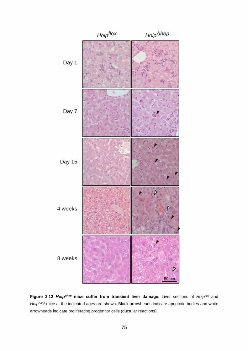
75
Figure 3.12 HoipΔhep mice suffer from transient liver damage. Liver sections of Hoipflox and
HoipΔhep mice at the indicated ages are shown. Black arrowheads indicate apoptotic bodies and white
arrowheads indicate proliferating progenitor cells (ductular reactions).
Day 1
Day 7
Day 15
4 weeks
Hoipflox HoipΔhep
8 weeks
50 μm
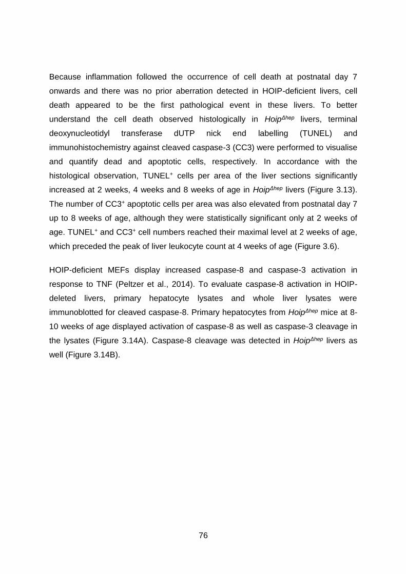
76
Because inflammation followed the occurrence of cell death at postnatal day 7
onwards and there was no prior aberration detected in HOIP-deficient livers, cell
death appeared to be the first pathological event in these livers. To better
understand the cell death observed histologically in HoipΔhep livers, terminal
deoxynucleotidyl transferase dUTP nick end labelling (TUNEL) and
immunohistochemistry against cleaved caspase-3 (CC3) were performed to visualise
and quantify dead and apoptotic cells, respectively. In accordance with the
histological observation, TUNEL+ cells per area of the liver sections significantly
increased at 2 weeks, 4 weeks and 8 weeks of age in HoipΔhep livers (Figure 3.13).
The number of CC3+ apoptotic cells per area was also elevated from postnatal day 7
up to 8 weeks of age, although they were statistically significant only at 2 weeks of
age. TUNEL+ and CC3+ cell numbers reached their maximal level at 2 weeks of age,
which preceded the peak of liver leukocyte count at 4 weeks of age (Figure 3.6).
HOIP-deficient MEFs display increased caspase-8 and caspase-3 activation in
response to TNF (Peltzer et al., 2014). To evaluate caspase-8 activation in HOIP-
deleted livers, primary hepatocyte lysates and whole liver lysates were
immunoblotted for cleaved caspase-8. Primary hepatocytes from HoipΔhep mice at 8-
10 weeks of age displayed activation of caspase-8 as well as caspase-3 cleavage in
the lysates (Figure 3.14A). Caspase-8 cleavage was detected in HoipΔhep livers as
well (Figure 3.14B).
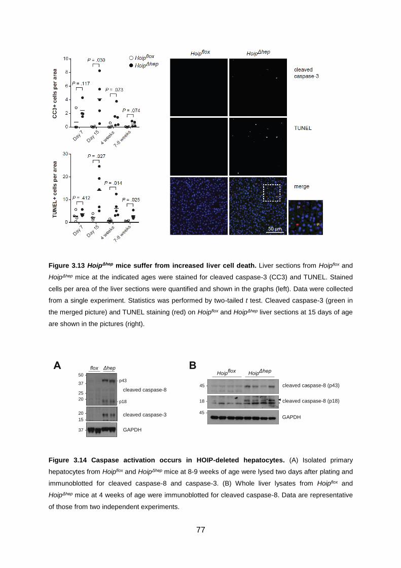
77
Figure 3.13 HoipΔhep mice suffer from increased liver cell death. Liver sections from Hoipflox and
HoipΔhep mice at the indicated ages were stained for cleaved caspase-3 (CC3) and TUNEL. Stained
cells per area of the liver sections were quantified and shown in the graphs (left). Data were collected
from a single experiment. Statistics was performed by two-tailed t test. Cleaved caspase-3 (green in
the merged picture) and TUNEL staining (red) on Hoipflox and HoipΔhep liver sections at 15 days of age
are shown in the pictures (right).
Figure 3.14 Caspase activation occurs in HOIP-deleted hepatocytes. (A) Isolated primary
hepatocytes from Hoipflox and HoipΔhep mice at 8-9 weeks of age were lysed two days after plating and
immunoblotted for cleaved caspase-8 and caspase-3. (B) Whole liver lysates from Hoipflox and
HoipΔhep mice at 4 weeks of age were immunoblotted for cleaved caspase-8. Data are representative
of those from two independent experiments.
cleaved caspase-3
37 -
50 -
cleaved caspase-8
20 -
25 -
20 -
15 -
flox Δhep
GAPDH
-
- p18
p43
37 -
-45 -
18 -
45 -
*
cleaved caspase-8 (p43)
cleaved caspase-8 (p18)
GAPDH
Hoipflox HoipΔhepBA
Δhep
Bcl-xL
pro-apoptotic
anti-apoptotic
Mcl-1
50 -XIAP
flox
Bid20 -
15 -
c-FLIP50 -
Bax20 -
Bcl-225 -
-
cIAP1/275 -
25 -
GAPDH37 -
25 -
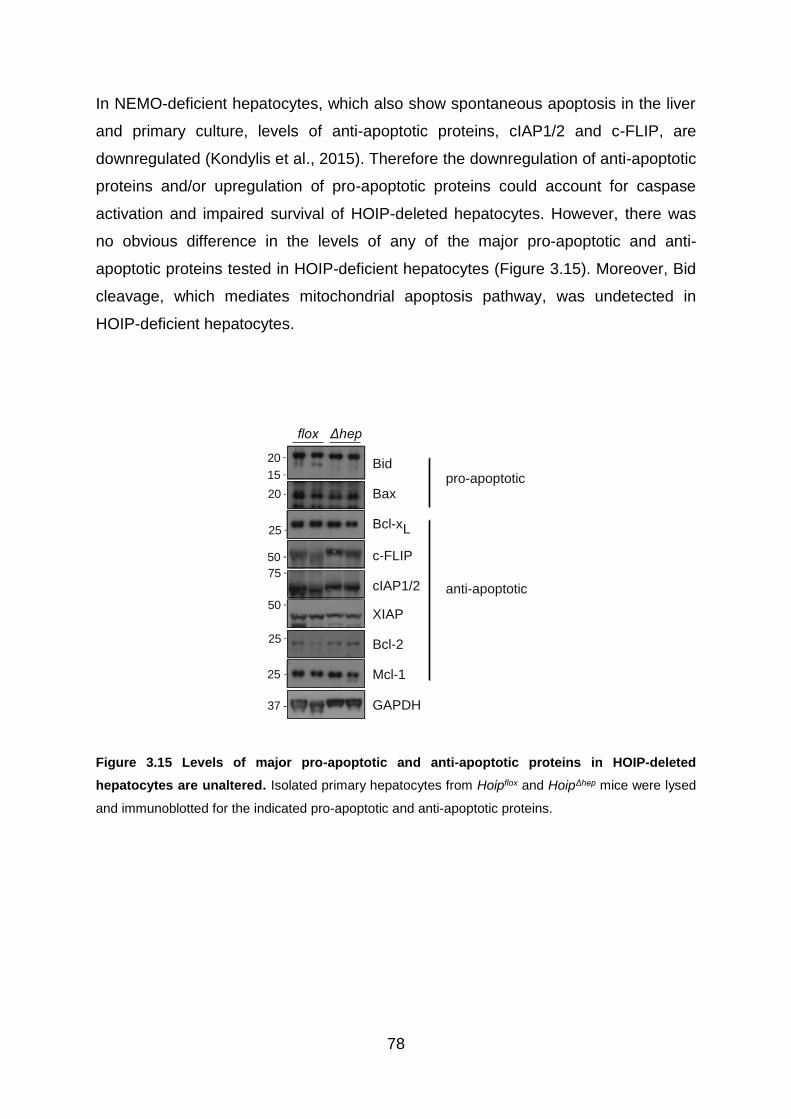
78
In NEMO-deficient hepatocytes, which also show spontaneous apoptosis in the liver
and primary culture, levels of anti-apoptotic proteins, cIAP1/2 and c-FLIP, are
downregulated (Kondylis et al., 2015). Therefore the downregulation of anti-apoptotic
proteins and/or upregulation of pro-apoptotic proteins could account for caspase
activation and impaired survival of HOIP-deleted hepatocytes. However, there was
no obvious difference in the levels of any of the major pro-apoptotic and anti-
apoptotic proteins tested in HOIP-deficient hepatocytes (Figure 3.15). Moreover, Bid
cleavage, which mediates mitochondrial apoptosis pathway, was undetected in
HOIP-deficient hepatocytes.
Figure 3.15 Levels of major pro-apoptotic and anti-apoptotic proteins in HOIP-deleted
hepatocytes are unaltered. Isolated primary hepatocytes from Hoipflox and HoipΔhep mice were lysed
and immunoblotted for the indicated pro-apoptotic and anti-apoptotic proteins.
cleaved caspase-3
37 -
50 -
cleaved caspase-8
20 -
25 -
20 -
15 -
flox Δhep
GAPDH
-
- p18
p43
37 -
-45 -
18 -
45 -
*
cleaved caspase-8 (p43)
cleaved caspase-8 (p18)
GAPDH
Hoipflox HoipΔhepBA
Δhep
Bcl-xL
pro-apoptotic
anti-apoptotic
Mcl-1
50 -XIAP
flox
Bid20 -
15 -
c-FLIP50 -
Bax20 -
Bcl-225 -
-
cIAP1/275 -
25 -
GAPDH37 -
25 -
79
3.1.5 Summary
The biological effect of LUBAC deficiency in the liver parenchyma was evaluated in
this chapter by creating LPC-specific HOIP knockout mice, HoipΔhep mice. HOIP
deletion in LPCs, utilising Alb-Cre mice, resulted in an efficient deletion of the protein.
Although there was no impairment in viability up to 18 months of age, the majority of
HoipΔhep mice developed macroscopic nodules and lesions in the liver, which
included malignant tumour at this age. At the perinatal stage, HoipΔhep mice began to
suffer from hepatocyte apoptosis which is exacerbated up to 2-4 weeks of age. The
cell death of hepatocytes in HoipΔhep mice was followed by the emergence of
inflammation. At this stage, leukocyte infiltration and the levels of inflammatory
mediators were increased, where a wide range of myeloid and lymphoid cells were
recruited to HoipΔhep liver and engaged in their hepatitis. In line with the aggravated
cell death and inflammation, liver cell proliferation occurred to compensate the
damage and increased DNA damage response was detected in HoipΔhep mice.
HOIP-deficient primary hepatocytes underwent apoptosis characterised by the
activation of caspase-8 and caspase-3. These results suggest that LUBAC
deficiency in LPCs is detrimental as it provokes hepatocyte death, notably apoptosis,
and inflammation. These early events associated with the late tumourigenesis in the
liver.
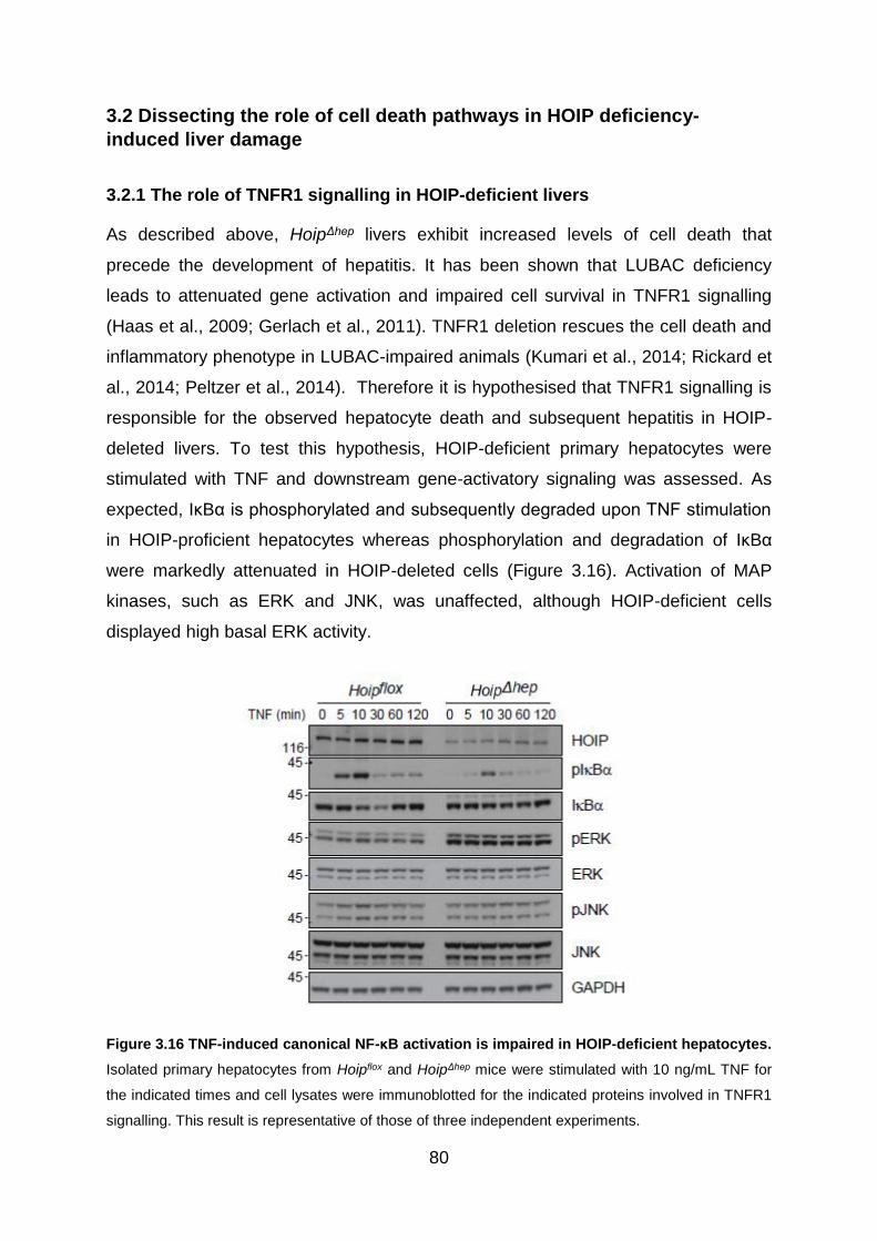
80
3.2 Dissecting the role of cell death pathways in HOIP deficiency-
induced liver damage
3.2.1 The role of TNFR1 signalling in HOIP-deficient livers
As described above, HoipΔhep livers exhibit increased levels of cell death that
precede the development of hepatitis. It has been shown that LUBAC deficiency
leads to attenuated gene activation and impaired cell survival in TNFR1 signalling
(Haas et al., 2009; Gerlach et al., 2011). TNFR1 deletion rescues the cell death and
inflammatory phenotype in LUBAC-impaired animals (Kumari et al., 2014; Rickard et
al., 2014; Peltzer et al., 2014). Therefore it is hypothesised that TNFR1 signalling is
responsible for the observed hepatocyte death and subsequent hepatitis in HOIP-
deleted livers. To test this hypothesis, HOIP-deficient primary hepatocytes were
stimulated with TNF and downstream gene-activatory signaling was assessed. As
expected, IκBα is phosphorylated and subsequently degraded upon TNF stimulation
in HOIP-proficient hepatocytes whereas phosphorylation and degradation of IκBα
were markedly attenuated in HOIP-deleted cells (Figure 3.16). Activation of MAP
kinases, such as ERK and JNK, was unaffected, although HOIP-deficient cells
displayed high basal ERK activity.
Figure 3.16 TNF-induced canonical NF-κB activation is impaired in HOIP-deficient hepatocytes.
Isolated primary hepatocytes from Hoipflox and HoipΔhep mice were stimulated with 10 ng/mL TNF for
the indicated times and cell lysates were immunoblotted for the indicated proteins involved in TNFR1
signalling. This result is representative of those of three independent experiments.
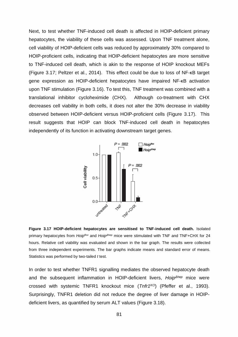
81
Next, to test whether TNF-induced cell death is affected in HOIP-deficient primary
hepatocytes, the viability of these cells was assessed. Upon TNF treatment alone,
cell viability of HOIP-deficient cells was reduced by approximately 30% compared to
HOIP-proficient cells, indicating that HOIP-deficient hepatocytes are more sensitive
to TNF-induced cell death, which is akin to the response of HOIP knockout MEFs
(Figure 3.17; Peltzer et al., 2014). This effect could be due to loss of NF-κB target
gene expression as HOIP-deficient hepatocytes have impaired NF-κB activation
upon TNF stimulation (Figure 3.16). To test this, TNF treatment was combined with a
translational inhibitor cycloheximide (CHX). Although co-treatment with CHX
decreases cell viability in both cells, it does not alter the 30% decrease in viability
observed between HOIP-deficient versus HOIP-proficient cells (Figure 3.17). This
result suggests that HOIP can block TNF-induced cell death in hepatocytes
independently of its function in activating downstream target genes.
Figure 3.17 HOIP-deficient hepatocytes are sensitised to TNF-induced cell death. Isolated
primary hepatocytes from Hoipflox and HoipΔhep mice were stimulated with TNF and TNF+CHX for 24
hours. Relative cell viability was evaluated and shown in the bar graph. The results were collected
from three independent experiments. The bar graphs indicate means and standard error of means.
Statistics was performed by two-tailed t test.
In order to test whether TNFR1 signalling mediates the observed hepatocyte death
and the subsequent inflammation in HOIP-deficient livers, HoipΔhep mice were
crossed with systemic TNFR1 knockout mice (Tnfr1KO) (Pfeffer et al., 1993).
Surprisingly, TNFR1 deletion did not reduce the degree of liver damage in HOIP-
deficient livers, as quantified by serum ALT values (Figure 3.18).
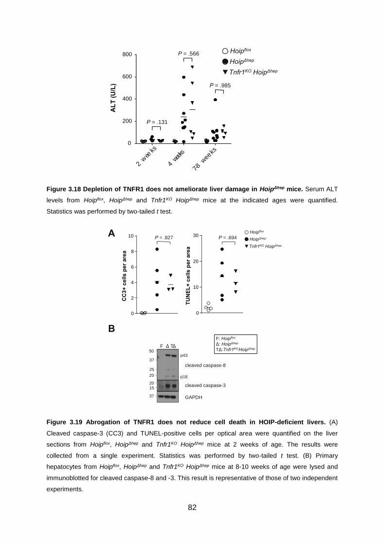
82
Figure 3.18 Depletion of TNFR1 does not ameliorate liver damage in HoipΔhep mice. Serum ALT
levels from Hoipflox, HoipΔhep and Tnfr1KO HoipΔhep mice at the indicated ages were quantified.
Statistics was performed by two-tailed t test.
Figure 3.19 Abrogation of TNFR1 does not reduce cell death in HOIP-deficient livers. (A)
Cleaved caspase-3 (CC3) and TUNEL-positive cells per optical area were quantified on the liver
sections from Hoipflox, HoipΔhep and Tnfr1KO HoipΔhep mice at 2 weeks of age. The results were
collected from a single experiment. Statistics was performed by two-tailed t test. (B) Primary
hepatocytes from Hoipflox, HoipΔhep and Tnfr1KO HoipΔhep mice at 8-10 weeks of age were lysed and
immunoblotted for cleaved caspase-8 and -3. This result is representative of those of two independent
experiments.
AL
T (
U/L
)
0
200
400
600
800
Tnfr1KO HoipΔhep
HoipΔhep
Hoipflox
2 w
eeks
4 weeks
7-8 w
eeks
P = .131
P = .566
P = .985
GAPDH
37 -
25 -
50 -
20 -
20 -
15 -
37 -
F Δ TΔ
A
B
cleaved caspase-8
cleaved caspase-3
-
- p18
p43
F: Hoipflox
Δ: HoipΔhep
TΔ:Tnfr1KOHoipΔhep
TU
NE
L+
ce
lls
pe
r a
rea
0
10
20
30 P = .694
CC
3+
ce
lls
pe
r a
rea
0
2
4
6
8
10 P = .827
Tnfr1KO HoipΔhep
HoipΔhep
Hoipflox

83
In line with the extent of liver damage, the numbers of apoptotic cells and/or dead
cells per area in HoipΔhep livers also remain unaffected by deletion of TNFR1 (Figure
3.19A). Furthermore, isolated primary hepatocytes from Tnfr1KO HoipΔhep mice
displayed similar activation of caspase-8 and caspase-3 to HOIP-deficient cells
(Figure 3.19B).
Intriguingly, even though TNFR1 deletion did not rescue the death and caspase
activation in HOIP-deficient hepatocytes, leukocyte recruitment to HOIP-deficient
livers was markedly attenuated in Tnfr1KO HoipΔhep mice reaching similar levels to
HOIP-proficient livers (Figure 3.20, left panel). Additionally, mRNA levels of TNF
and CCL3 were strongly decreased in Tnfr1KO HoipΔhep livers (Figure 3.20, right
panel). This suggests that TNFR1 ablation significantly alleviates the inflammatory
phenotype triggered by HOIP deficiency in liver parenchyma. Since the inflammation
in HOIP-deficient livers was resolved by TNFR1 deletion, it was hypothesised that
the subsequent DNA damage caused by inflammation was ameliorated as well.
Indeed, a significant reduction in the number of γH2AX+ cells in Tnfr1KO HoipΔhep
livers was observed as compared to HoipΔhep livers (Figure 3.21). Collectively, these
results show that, regardless of the TNFR1-independent cell death of HOIP-deficient
hepatocytes, TNFR1 signalling mediates the inflammation in HOIP-deficient livers.
Figure 3.20 TNFR1 ablation ameliorates inflammation in HOIP-deficient livers. Leukocyte count
per gramme of the liver in Hoipflox, HoipΔhep and Tnfr1KO HoipΔhep mice at 4 weeks of age were
quantified (left panel). The levels of Tnf and Ccl3 transcripts were quantified from Hoipflox, HoipΔhep
and Tnfr1KO HoipΔhep livers by qRT-PCR (right panel). The levels were normalised to those from
Hoipflox livers. The results for leukocyte count were collected and pooled from two independent
experiments. The results for cytokine were collected from a single experiment. The bar graphs
indicate means and standard error of means. Statistics was performed by two-tailed t test.
leu
ko
cyte
sp
er
1g
liver
0
5.0×10⁶
1.0×10⁷
1.5×10⁷
2.0×10⁷P = .037
fold
in
du
cti
on
TnfCcl3
0
2
4
6
P = .005
P = .001
Tnfr1KO HoipΔhep
HoipΔhep
Hoipflox

84
Figure 3.21 TNFR1 deletion mitigates DNA damage in HOIP-deficient livers. Liver sections from
Hoipflox, HoipΔhep and Tnfr1KO HoipΔhep mice at 4 weeks of age were stained for phosphorylated H2AX.
The right graph shows the quantification of γH2AX+ cells per optical area (magnification: 200x). The
results were collected from a single experiment. Statistics was performed by two-tailed t test.
Because TNFR1 ablation did not rescue liver damage in HOIP-deficient livers
(Figure 3.19), death-inducing extracellular ligands, other than TNFR1 ligands such
as TNF and LTα, could be responsible for the cell death observed in HOIP-deficient
livers. Therefore, the sensitivity of HOIP-proficient and -deficient hepatocytes to
TRAIL and CD95L-induced cell death was evaluated. Treatment with soluble TRAIL
failed to induce cell death in both cells, with no obvious sensitisation due to HOIP
deficiency. It was already reported that soluble TRAIL fails to induce cell death in
primary mouse hepatocytes in previous studies (Walczak et al., 1999; Corazza et al.,
2006). In contrast to TRAIL, CD95L-induced cell death was observed in both HOIP-
proficient and -deficient hepatocytes (Figure 3.22). Interestingly, HOIP-deficient
hepatocytes were more sensitive to CD95L-induced cell death than HOIP-proficient
cells (Figure 3.22).
In addition to death receptor signalling, TLR3 has recently been described to induce
cell death via recruiting the cell death-inducing signalling platform (Feoktistova et al.,
2011). In line, HOIP-deficient hepatocytes are also sensitised to cell death induced
by the TLR3 ligand poly(I:C).
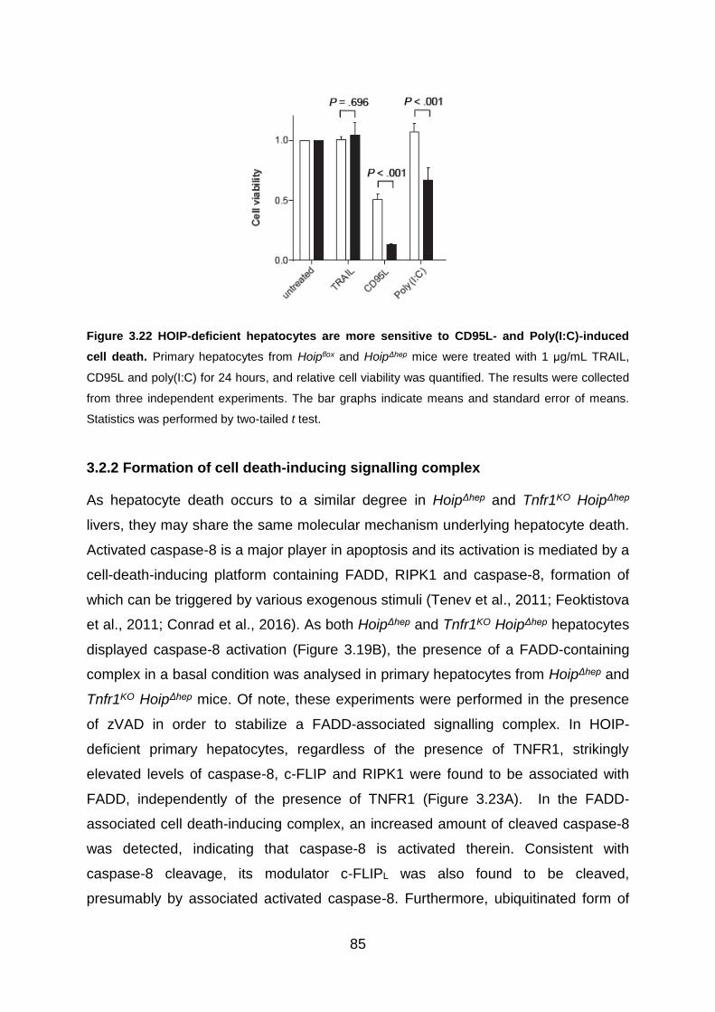
85
Figure 3.22 HOIP-deficient hepatocytes are more sensitive to CD95L- and Poly(I:C)-induced
cell death. Primary hepatocytes from Hoipflox and HoipΔhep mice were treated with 1 μg/mL TRAIL,
CD95L and poly(I:C) for 24 hours, and relative cell viability was quantified. The results were collected
from three independent experiments. The bar graphs indicate means and standard error of means.
Statistics was performed by two-tailed t test.
3.2.2 Formation of cell death-inducing signalling complex
As hepatocyte death occurs to a similar degree in HoipΔhep and Tnfr1KO HoipΔhep
livers, they may share the same molecular mechanism underlying hepatocyte death.
Activated caspase-8 is a major player in apoptosis and its activation is mediated by a
cell-death-inducing platform containing FADD, RIPK1 and caspase-8, formation of
which can be triggered by various exogenous stimuli (Tenev et al., 2011; Feoktistova
et al., 2011; Conrad et al., 2016). As both HoipΔhep and Tnfr1KO HoipΔhep hepatocytes
displayed caspase-8 activation (Figure 3.19B), the presence of a FADD-containing
complex in a basal condition was analysed in primary hepatocytes from HoipΔhep and
Tnfr1KO HoipΔhep mice. Of note, these experiments were performed in the presence
of zVAD in order to stabilize a FADD-associated signalling complex. In HOIP-
deficient primary hepatocytes, regardless of the presence of TNFR1, strikingly
elevated levels of caspase-8, c-FLIP and RIPK1 were found to be associated with
FADD, independently of the presence of TNFR1 (Figure 3.23A). In the FADD-
associated cell death-inducing complex, an increased amount of cleaved caspase-8
was detected, indicating that caspase-8 is activated therein. Consistent with
caspase-8 cleavage, its modulator c-FLIPL was also found to be cleaved,
presumably by associated activated caspase-8. Furthermore, ubiquitinated form of
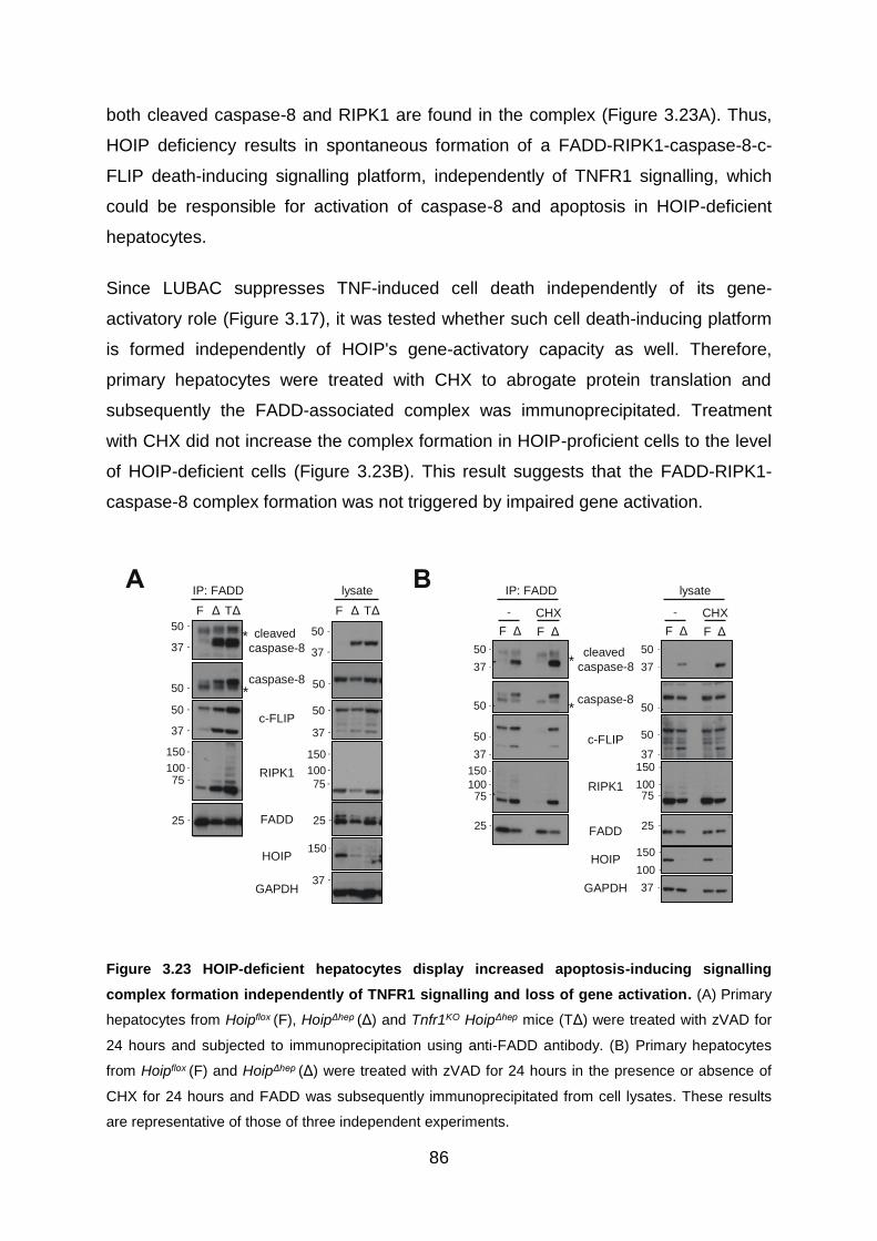
86
both cleaved caspase-8 and RIPK1 are found in the complex (Figure 3.23A). Thus,
HOIP deficiency results in spontaneous formation of a FADD-RIPK1-caspase-8-c-
FLIP death-inducing signalling platform, independently of TNFR1 signalling, which
could be responsible for activation of caspase-8 and apoptosis in HOIP-deficient
hepatocytes.
Since LUBAC suppresses TNF-induced cell death independently of its gene-
activatory role (Figure 3.17), it was tested whether such cell death-inducing platform
is formed independently of HOIP's gene-activatory capacity as well. Therefore,
primary hepatocytes were treated with CHX to abrogate protein translation and
subsequently the FADD-associated complex was immunoprecipitated. Treatment
with CHX did not increase the complex formation in HOIP-proficient cells to the level
of HOIP-deficient cells (Figure 3.23B). This result suggests that the FADD-RIPK1-
caspase-8 complex formation was not triggered by impaired gene activation.
Figure 3.23 HOIP-deficient hepatocytes display increased apoptosis-inducing signalling
complex formation independently of TNFR1 signalling and loss of gene activation. (A) Primary
hepatocytes from Hoipflox (F), HoipΔhep (Δ) and Tnfr1KO HoipΔhep mice (TΔ) were treated with zVAD for
24 hours and subjected to immunoprecipitation using anti-FADD antibody. (B) Primary hepatocytes
from Hoipflox (F) and HoipΔhep (Δ) were treated with zVAD for 24 hours in the presence or absence of
CHX for 24 hours and FADD was subsequently immunoprecipitated from cell lysates. These results
are representative of those of three independent experiments.
A B
c-FLIP
RIPK1
caspase-8
FADD
HOIP
GAPDH
50 -
37 -
50 -
150 -
100 -
75 -
50 -
37 -
25 -
50 -
37 -
50 -
150 -
100 -75 -
50 -
37 -
25 -
150 -
100 -
37 -
F Δ F Δ
- CHX
F Δ F Δ
- CHX
IP: FADD lysate
cleaved
caspase-8
F Δ TΔ
50 -
37 -
50 -
150 -
100 -
75 -
50 -
37 -
25 -
c-FLIP
50 -
37 -
50 -
37 -
RIPK1
FADD
GAPDH
HOIP
50 -
150 -
25 -
37 -
cleaved
caspase-8
caspase-8
75 -
F Δ TΔ
IP: FADD lysate
150 -
100 -
**
*
*
45 -cleaved Casp-8 (p45)
FADD25 -
116 -
RIPK1IP: FADD
FADDLysate
flox Δhep
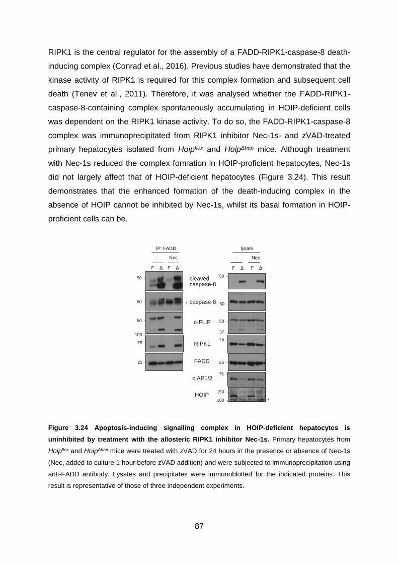
87
RIPK1 is the central regulator for the assembly of a FADD-RIPK1-caspase-8 death-
inducing complex (Conrad et al., 2016). Previous studies have demonstrated that the
kinase activity of RIPK1 is required for this complex formation and subsequent cell
death (Tenev et al., 2011). Therefore, it was analysed whether the FADD-RIPK1-
caspase-8-containing complex spontaneously accumulating in HOIP-deficient cells
was dependent on the RIPK1 kinase activity. To do so, the FADD-RIPK1-caspase-8
complex was immunoprecipitated from RIPK1 inhibitor Nec-1s- and zVAD-treated
primary hepatocytes isolated from Hoipflox and HoipΔhep mice. Although treatment
with Nec-1s reduced the complex formation in HOIP-proficient hepatocytes, Nec-1s
did not largely affect that of HOIP-deficient hepatocytes (Figure 3.24). This result
demonstrates that the enhanced formation of the death-inducing complex in the
absence of HOIP cannot be inhibited by Nec-1s, whilst its basal formation in HOIP-
proficient cells can be.
Figure 3.24 Apoptosis-inducing signalling complex in HOIP-deficient hepatocytes is
uninhibited by treatment with the allosteric RIPK1 inhibitor Nec-1s. Primary hepatocytes from
Hoipflox and HoipΔhep mice were treated with zVAD for 24 hours in the presence or absence of Nec-1s
(Nec, added to culture 1 hour before zVAD addition) and were subjected to immunoprecipitation using
anti-FADD antibody. Lysates and precipitates were immunoblotted for the indicated proteins. This
result is representative of those of three independent experiments.
supplemental or take out
- Nec1s did not block complex formation
IP: FADD
F Δ
cleavedcaspase-8
caspase-8
cIAP1/2
- Nec
c-FLIP
F Δ F Δ F Δ
RIPK1
FADD
50 -
50 -
50 -
*
100 -
75 -
50 -
50 -
50 -
37 -
75 -
- Nec
HOIP*
150 -
100 -
75 -
25 - 25 -
lysate
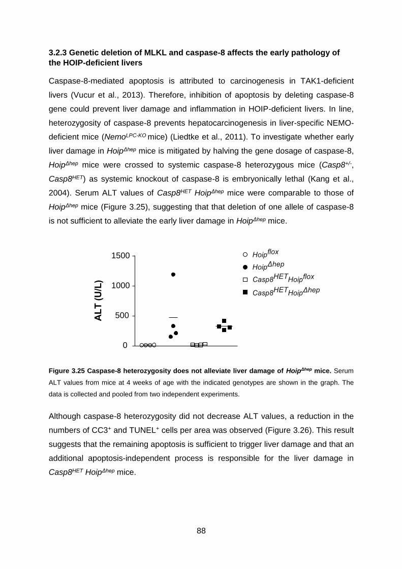
88
3.2.3 Genetic deletion of MLKL and caspase-8 affects the early pathology of
the HOIP-deficient livers
Caspase-8-mediated apoptosis is attributed to carcinogenesis in TAK1-deficient
livers (Vucur et al., 2013). Therefore, inhibition of apoptosis by deleting caspase-8
gene could prevent liver damage and inflammation in HOIP-deficient livers. In line,
heterozygosity of caspase-8 prevents hepatocarcinogenesis in liver-specific NEMO-
deficient mice (NemoLPC-KO mice) (Liedtke et al., 2011). To investigate whether early
liver damage in HoipΔhep mice is mitigated by halving the gene dosage of caspase-8,
HoipΔhep mice were crossed to systemic caspase-8 heterozygous mice (Casp8+/-,
Casp8HET) as systemic knockout of caspase-8 is embryonically lethal (Kang et al.,
2004). Serum ALT values of Casp8HET HoipΔhep mice were comparable to those of
HoipΔhep mice (Figure 3.25), suggesting that that deletion of one allele of caspase-8
is not sufficient to alleviate the early liver damage in HoipΔhep mice.
Figure 3.25 Caspase-8 heterozygosity does not alleviate liver damage of HoipΔhep mice. Serum
ALT values from mice at 4 weeks of age with the indicated genotypes are shown in the graph. The
data is collected and pooled from two independent experiments.
Although caspase-8 heterozygosity did not decrease ALT values, a reduction in the
numbers of CC3+ and TUNEL+ cells per area was observed (Figure 3.26). This result
suggests that the remaining apoptosis is sufficient to trigger liver damage and that an
additional apoptosis-independent process is responsible for the liver damage in
Casp8HET HoipΔhep mice.
0
500
1000
1500
AL
T (
U/L
)
Hoipflox
HoipΔhep
Casp8HETHoipΔhep
Casp8HETHoipflox

89
Figure 3.26 Caspase-8 heterozygosity reduces apoptosis in HoipΔhep livers. The numbers of
CC3+ cells and TUNEL+ cells per area in livers from HoipΔhep, Casp8HET Hoipflox and Casp8HET HoipΔhep
mice at 2 weeks of age are shown in the graphs. The data is collected from a single experiment.
Despite the decrease in apoptotic cells in the liver, the percentage of hepatic
leukocytes in Casp8HET HoipΔhep mice was elevated to an equivalent extent as
HoipΔhep mice at the peak of inflammation (Figure 3.27). This result indicates that
inflammation was not ameliorated by partial loss of caspase-8.
Figure 3.27 Caspase-8 heterozygosity does not ameliorate leukocyte infiltration into HoipΔhep
livers. Percentage of leukocytes present in the livers of Hoipflox, HoipΔhep, Casp8HET Hoipflox and
Casp8HET HoipΔhep mice at 4 weeks of age are plotted in the graph. The data is collected and pooled
from two independent experiments.
0
5
10
15C
C3+
cells p
er
are
a
0
5
10
15
20
25
TU
NE
L+
cells p
er
are
a HoipΔhep
Casp8HETHoipΔhep
Casp8HETHoipflox
0
5
10
15
20
25
leu
ko
cyte
s (
%)
Hoipflox
HoipΔhep
Casp8HETHoipΔhep
Casp8HETHoipflox
90
Heterozygosity of caspase-8 did not rescue liver damage evaluated by serum ALT in
HOIP-deficient livers, whereas TUNEL+ or CC3+ cells therein were reduced, which
suggests the presence of non-apoptotic cell death. Blockade of caspase-8 activity is
a trigger to induce RIPK3/MLKL-dependent necroptosis (Conrad et al., 2016). Thus,
heterozygosity of caspase-8 could also sensitise hepatocytes to necroptosis by
lowering the amount of caspase-8 in cells. It has been shown that necroptotic cells
can be negative for TUNEL (Carneiro et al., 2009), and therefore TUNEL performed
in HOIP-deficient livers could have overlooked the presence of necroptotic cells
therein.
Pathology triggered by SHARPIN deficiency was completely reverted by RIPK3
deletion and caspase-8 heterozygosity (Rickard et al., 2014), indicating the crucial
role of RIPK3 in the cpdm phenotype. However, RIPK3 deletion in HOIP-floxed
animals cannot be performed by conventional crossings due to the genetic proximity
of Ripk3 and Hoip loci (Peltzer et al., 2014). It has been shown that the embryonic
lethality of caspase-8 knockout mice can be circumvented by co-deleting necroptosis
executer MLKL, although the double knockout mice develop lymphadenopathy at a
few months of age (Etemadi et al., 2015). Therefore, Casp8HET HoipΔhep mice were
crossed to MLKL knockout (MlklKO) mice to evaluate the contribution of necroptosis
in the phenotype. Deletion of MLKL in addition to caspase-8 heterozygosity did not
reduce ALT values in HoipΔhep mice (Figure 3.28). This result suggests that one
allele of caspase-8 is sufficient to initiate apoptosis in MlklKO Casp8HET HoipΔhep livers,
as apoptotic bodies are present in these livers (Figure 3.28).
Strikingly, MlklKO Casp8KO HoipΔhep mice showed serum ALT values comparable to
those of Hoipflox mice, indicating that the co-deletion of MLKL and caspase-8
completely prevents liver damage induced by HOIP deficiency (Figure 3.28).
Accordingly, no cell death were observed in histological analysis of MlklKO Casp8KO
HoipΔhep livers.
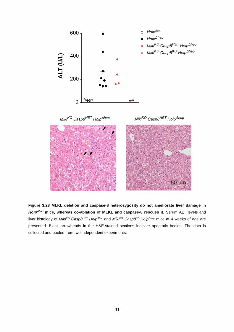
91
Figure 3.28 MLKL deletion and caspase-8 heterozygosity do not ameliorate liver damage in
HoipΔhep mice, whereas co-ablation of MLKL and caspase-8 rescues it. Serum ALT levels and
liver histology of MlklKO Casp8HET HoipΔhep and MlklKO Casp8KO HoipΔhep mice at 4 weeks of age are
presented. Black arrowheads in the H&E-stained sections indicate apoptotic bodies. The data is
collected and pooled from two independent experiments.
AL
T (
U/L
)
Hoipflox
HoipΔhep
MlklKO Casp8HET HoipΔhep
MlklKO Casp8KO HoipΔhep
50 μm
MlklKO Casp8HET HoipΔhepMlklKO Casp8HET HoipΔhep
0
200
400
600

92
3.2.4 Summary
In this subchapter, the contributions of the TNFR1 signalling, as well as the
involvement of apoptotic and necroptotic pathways to the pathogenesis of HoipΔhep
mice were examined in order to decipher the molecular mechanisms causing liver
upon HOIP deficiency in LPCs. In summary, HOIP-deficient hepatocytes were
sensitised to TNF-induced cell death and displayed impaired NF-κB activation in
response to TNF. Unexpectedly, systemic TNFR1 ablation did not affect the liver
damage and cell death. Double TNFR1- and HOIP-deficient hepatocytes underwent
apoptosis via caspase-8 and caspase-3 activation to a similar extent to HOIP-
deficient cells. However, Tnfr1KO HoipΔhep exhibited much less severe inflammation
as compared to HoipΔhep mice.
In addition, the involvement of apoptotic and necroptotic pathways in the observed
phenotype was assessed. Halving the gene dosage of caspase-8 did not largely
ameliorate liver damage or inflammation. Also, depletion of the necroptosis executer
MLKL on top of heterozygosity of caspase-8 also failed to mitigate liver damage. By
contrast, complete deletion of both caspase-8 and MLKL abolished liver damage in
HoipΔhep mice. Taken together, these results illustrated that TNFR1-independent but
caspase-8 dependent apoptosis plays a central role in the development of liver
damage in HoipΔhep mice.

93
Chapter 4
4 Discussion
Chronic inflammation predisposes the liver to cancer (El-serag, 2011). Disturbance
of NF-κB signalling and/or cell death pathway may result in severe pathology in
epithelial tissues (Pasparakis, 2012). Dysregulation of TNFR1 signalling and NF-κB
signalling components has been associated with liver pathogenesis and
tumourigenesis in several mouse models (Luedde et al., 2007; Bettermann et al.,
2010; Inokuchi et al., 2010; Nikolaou et al., 2012; Kondylis et al., 2015). However,
the role of LUBAC and linear ubiquitination in hepatic cells has been poorly
understood. In this thesis, mice specifically lacking HOIP in liver parenchyma were
generated and the physiological role of LUBAC in hepatocytes was examined.
LUBAC deficiency in liver parenchymal cells leads to hepatocyte apoptosis and
inflammation in the liver at an earlier stage and promotes tumour formation. The
mechanisms by which LUBAC deficiency results in cell death, hepatitis and eventual
oncogenesis is discussed in this chapter.
4.1 Impact of HOIP deletion in liver parenchyma
4.1.1 Cre expression, loss of HOIP and cell death
Alb-Cre expression starts around embryonic day 14.5, and Cre gradually excises
loxP-flanked genomic DNA until 6-7 weeks of age, when gene deletion is completed
in approximately 90% of the alleles (Postic et al, 1999; Weisend et al, 2009).
Embryonic livers at mid-gestation accommodate hepatoblasts and a large amount of
hematopoietic progenitor cells. As hepatoblasts differentiate into mature hepatocytes
during late gestation, hepatocytes begin to express albumin. In correlation with the
increased level of serum albumin protein, Cre proteins are expressed in hepatocytes
and excise loxP-flanked DNA.
In line with these reports, Alb-Cre-mediated HOIP deletion was efficiently executed
in hepatocytes by 7-9 weeks of age in HoipΔhep mice. Due to the low sensitivity of
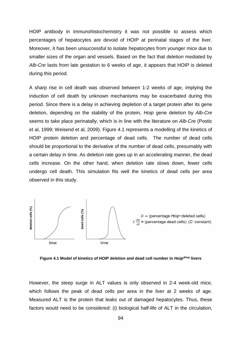
94
HOIP antibody in immunohistochemistry it was not possible to assess which
percentages of hepatocytes are devoid of HOIP at perinatal stages of the liver.
Moreover, it has been unsuccessful to isolate hepatocytes from younger mice due to
smaller sizes of the organ and vessels. Based on the fact that deletion mediated by
Alb-Cre lasts from late gestation to 6 weeks of age, it appears that HOIP is deleted
during this period.
A sharp rise in cell death was observed between 1-2 weeks of age, implying the
induction of cell death by unknown mechanisms may be exacerbated during this
period. Since there is a delay in achieving depletion of a target protein after its gene
deletion, depending on the stability of the protein, Hoip gene deletion by Alb-Cre
seems to take place perinatally, which is in line with the literature on Alb-Cre (Postic
et al, 1999; Weisend et al, 2009). Figure 4.1 represents a modelling of the kinetics of
HOIP protein deletion and percentage of dead cells. The number of dead cells
should be proportional to the derivative of the number of dead cells, presumably with
a certain delay in time. As deletion rate goes up in an accelerating manner, the dead
cells increase. On the other hand, when deletion rate slows down, fewer cells
undergo cell death. This simulation fits well the kinetics of dead cells per area
observed in this study.
Figure 4.1 Model of kinetics of HOIP deletion and dead cell number in HoipΔhep livers
However, the steep surge in ALT values is only observed in 2-4 week-old mice,
which follows the peak of dead cells per area in the liver at 2 weeks of age.
Measured ALT is the protein that leaks out of damaged hepatocytes. Thus, these
factors would need to be considered: (i) biological half-life of ALT in the circulation,

95
and (ii) liver size and growth during this period. Because ALT could remain in the
blood for a certain time after its release, this might cause a delay in the increase in
ALT values. Finally, the size of the liver significantly expands during the first month
of life in mice as they rapidly gain body weight. The drastic increase in the liver size
might account for the delay in the surge of the total number of dead cells as
compared to the number of the dead cells per area.
In the case of c-FLIP deletion in the liver, the two Cre drivers targeting LPCs has
been shown to give rise to distinct phenotypes. While deletion with Alb-Cre causes
only liver damage with elevated ALT, modification with Alfp-Cre confers perinatal
lethality to mice (Piao et al., 2012). Scientists who established Alfp-Cre mice noticed
that albumin expression was also regulated by an enhancer of alpha-fetoprotein
(Afp) genes, which is located next to Alb gene (Kellendonk et al., 2000). By
combining the Alb gene regulator with Afp gene enhancer, Alfp-Cre achieves an
efficient induction of Cre from embryonic day 10.5, which is earlier than the initiation
of Cre expression by Alb-cre. It is not entirely clear when Alfp-Cre achieves full
penetrance of gene deletion during development, but c-FLIP deletion with Alfp-Cre
demonstrated an efficient depletion of the protein already at postnatal day 1 (Piao et
al., 2012). Although an earlier efficient deletion of HOIP at the perinatal stage is
expected with Alfp-Cre, Hoipflox/flox Alfp-Cre mice were not lethal up to a weaning age.
It will be intriguing to investigate whether there is any difference between phenotypes
caused by gene deletion with Alb-Cre and Alfp-Cre at later stages of life.
4.1.2 Cell death precedes inflammation
The emergence of apoptotic bodies in HoipΔhep mice was observed as early as
postnatal day 7, which clearly precedes that of inflammation at 4 weeks of age. Initial
apoptosis appears to trigger an early induction of cytokines at 2 weeks of age.
Histologically, at 2 weeks of age, some apoptotic cells are approached by immune
cells (Figure 3.12), which implies that leukocytes react to the cytokines and DAMPs
produced by dead cells in HOIP-deficient livers. This process seems exacerbated
around 4 weeks of age, when a wide range of immune cells is recruited to the site of
inflammation in HOIP-deficient livers. Furthermore, the apparent resolution of
inflammation coincided with the sharp decline in the number of dead cells in the liver.
96
Given that cell death due to loss of LUBAC is the primary driver of inflammation
therein, it is highly likely that inflammation is ameliorated because its source, cell
death, is reduced.
This observation is in line with accumulating evidence indicating that cell death is a
driver of inflammation in various in-vitro and in-vivo studies. For instance, in LPC-
specific truncated CYLD knock-in mice, the first abnormality detected is increased
cell death from day 10 onwards, which precedes the induction of Tnf mRNA and
immune cell activation at day 35 (Nikolaou et al., 2012). Thus, it appears that cell
death drives production of inflammatory cytokines, which in turn renders cells prone
to undergo cell death further. This is in accordance with the notion that regulated cell
death participates in the initiation and amplification of inflammation (Pasparakis and
Vandenabeele, 2015).
4.2 Tumourigenic process
4.2.1 Pathological aspects
The initial liver cell death in HoipΔhep mice at postnatal day 7 was not accompanied
by an inflammatory feature at all. The initial aberration observed, cell death, is
reminiscent of a histological feature of patients suffering from drug-induced liver
injury (DILI). The most common cause of DILI is an overdose of paracetamol
(acetaminophen, N-acetyl-para-aminophenol, or APAP) due to its availability and
popularity (Tajiri and Shimizu, 2008). However, in contrast to the excessive
apoptosis in HOIP-deficient livers, a major characteristic of DILI is hepatocyte
necrosis. The mechanism by which APAP induces cell death is not entirely clear.
APAP is converted to a reactive metabolite (N-acetyl-p-benzoquinone imine, NAP-
QI) in hepatocytes. Upon APAP overdose, accumulated NAP-QI depletes the
reducing capacity of glutathione. Currently the only effective intervention for DILI is
the antioxidant N-acetylcysteine (NAC), which promotes the recovery of the level of
glutathione in the liver after APAP overdose. NAC counteracts the oxidising action of
NAP-QI toward glutathione to facilitate its regeneration. ROS production is
associated with carcinogenesis as it causes immune activation and provokes DNA
97
damage in stromal cells (Ohnishi et al., 2013). In livers devoid of IKK2 or NEMO, a
reduced level of superoxide dismutases (SODs), which are enzymes quenching
ROS, seems to exacerbate inflammation (Maeda et al, 2005; Vlantis et al, 2015).
The expression level of SODs is upregulated by constitutive activation of canonical
NF-κB by expressing an active form of IKK2 (Vlantis et al., 2015). Therefore, it will be
interesting to pursue whether liver damage and tumourigenesis triggered by HOIP
deletion would be ameliorated by treatment with antioxidants or upregulation of
SODs.
HOIP-deficient livers seem to lack a robust fibrogenesis, which is often seen in
human patients suffering from liver cancer and NEMO or TAK1 deficiency in murine
livers (Luedde et al., 2007; Betterman et al., 2010; El-serag, 2011). However, it has
been reported that HCV-associated cancer can evolve without cirrhosis (De Mitri et
al., 1995). Also there are increasing numbers of liver cancer patients without a prior
history of liver cirrhosis, which appears to be linked to NAFLD, particularly in the
Western world (Rahman et al., 2013). Insulin resistance is linked to fat accumulation
in the liver. Excessive visceral fat deposition promotes the release of pro-
inflammatory cytokines. Hyperglycemia caused by insulin resistance leads to
increased oxidative stress which also promotes inflammation. Interestingly, in a
mouse model, DEN and HFD-induced hepatocarcinogenesis is promoted by
inflammatory cytokines IL-6 and TNF (Park et al., 2010).
As the accumulation of lipid droplets was observed in HOIP-deficient livers at 18
months of age, it is reasonable to speculate that HoipΔhep mice suffer from NAFLD-
related symptoms. The mechanism as to how cell death-driven inflammation driven
by cell death leads to accumulation of fatty acids in hepatocytes needs to be
addressed. In a NASH mouse model employing CD-HFD, C57BL/6 mice develop
CD8+ T cell- and NKT cell-driven steatohepatitis (Wolf et al., 2014). Deletion of the
TNF superfamily member LIGHT or its receptor LTβR prevents the development of
steatohepatitis and HCC in mice. LIGHT effectively upregulates fatty acid intake in
hepatocytes (Wolf et al., 2014), and LIGHT-mediated dyslipidemia is dependent on
LTβR (James et al., 2007). In humans, serum LIGHT is increased in NAFLD patients
(Otterdal et al., 2015). Light mRNA level has a tendency to increase at the peak of
inflammation in HoipΔhep livers. Moreover, the other LTβR ligand LTβ was highly

98
upregulated in HoipΔhep livers. Therefore, it would be tempting to investigate the role
of LTβR and its ligands in HoipΔhep livers.
There is no evidence, to date, that loss of LUBAC occurs in human patients suffering
from liver diseases. In mouse models of NASH, methionine- and choline-deficient
diet (MCD) induces downregulation of LUBAC components and impairment in
LUBAC formation (Matsunaga et al., 2015). Yet it remains unsolved whether LUBAC
downregulation is responsible for MCD-induced liver damage and steatohepatitis. It
will be intriguing to investigate whether hepatotropic virus carriers, ASH, NASH and
HCC patients displays any change in the protein levels of LUBAC components. If this
is the case, sustaining LUBAC function might be a promising strategy for a novel
therapy in such patients.
4.2.2 DNA damage and selection
After hepatitis flares in HoipΔhep mice at 4 weeks of age, the inflammation is
ameliorated, suggesting that inflammation is transient and not sustained. Is this mere
transient inflammation sufficient to account for the eventual tumourigenesis? In the
case of a chemically-induced liver cancer model, a single injection of carcinogen
DEN in C57BL/6 mice at early age induces liver cancer later in life (Maeda et al.,
2005), which means that mutagenesis at an early stage is sufficient to trigger
carcinogenesis even without chronic inflammation. According to the ‘Big Bang’ model
of cancer evolution, most of the intra-tumour heterogeneity is derived from early
genetic alteration, rather than later clonal expansions (Sottoriva et al., 2015). This
model also indicates that some tumour subclones can acquire malignant
characteristics at a very early stage of carcinogenesis. If this model applies to
HoipΔhep livers, their early transient inflammation can be a decisive factor in
determining the mutagenic signature of hepatocytes which later give rise to a tumour.
An increased DNA damage response was detected in HoipΔhep livers at the peak of
inflammation (Figure 3.10), which could explain, at least in part, the process of
oncogenesis therein. The extent of DNA damage appears to be correlated with that
of inflammation since Tnrf1KO HoipΔhep mice had less γH2AX+ cells with reduced
inflammation as compared to HoipΔhep mice (Figure 3.21). It will be interesting to
99
know if this decreased inflammation correlates with less severe tumourigenesis at a
later stage.
It is still unclear what genes are affected and mutated in HoipΔhep mice. It certainly is
an interesting question to test whether HoipΔhep livers develop well-known oncogenic
mutations in their genome, for instance Tp53 and Ctnnb1, at different ages until
tumours arise in HoipΔhep mice. Nodules from Tak1LPC-KO and NemoLPC-KO mice
exhibited upregulation of proliferation and cell cycle genes (Betterman et al., 2010).
The rate of gene alteration in Tak1LPC-KO livers is clearly correlated with the degree of
apoptosis and inflammation, as caspase-8 deletion abolishes most of it (Vucur et al.,
2013).
Hepatocytes devoid of HOIP are prone to undergo cell death. Despite the efficient
deletion of HOIP, the vast majority of HOIP-deficient hepatocytes are still alive. This
means that a substantial portion of HOIP-deficient hepatocytes are able to survive
the environment. It is also possible that they adapted to the change in LUBAC levels
by upregulating some pro-survival factors unexamined here, which in turn could
contribute to oncogenesis.
4.2.3 Pro-tumourigenic signalling pathways
IL-6 is one of the most prominent pro-tumourigenic cytokines, notably in liver cancer
(He et al., 2010). IL-6 activates transcription factor STAT3, which is a transcription
activator of pro-survival genes and frequently mutated in human cancer. In murine
liver cancer models, IL-6 promotes chemically-induced liver carcinogenesis and, in
preneoplastic lesions, liver cancer progenitor cells grow depending on autocrine IL-6
signalling (Naugler et al., 2007; He et al., 2013). At the peak of early inflammation in
HoipΔhep mice, no significant increase in the level of Il6 mRNA was observed (Figure
3.7). However, the future work will investigate the contribution of IL-6 in
tumourigenesis in HoipΔhep mice by evaluating the level of IL-6 at 18 months of age
at which the majority of the animals developed nodules. In addition, STAT3
activation in the pre-cancerous stage will be also examined to determine whether
STAT3 activation is involved in the oncogenesis.

100
Disruption of NF-κB signalling components in LPCs often results in hyperactivation of
JNK (Vucur et al, 2013; Nikolaou et al, 2012; Betterman et al., 2010). Deletion of
JNK1 diminishes the susceptibility of Ikk2Δhep mice to chemically-induced
carcinogenesis (Sakurai et al., 2006). Furthermore, abrogation of TAK1 and NEMO
in LPCs also leads to oncogenesis with JNK hyperactivation (Luedde et al., 2007;
Betterman et al., 2010; Inokuchi et al., 2010). JNK activation in Ikk2Δhep and
NemoLPC-KO livers is reduced by treatment with an antioxidant, suggesting JNK
activation is mediated by ROS in these contexts. In NemoLPC-KO livers, the JNK
pathway appears to be activated by TAK1. Aberrant TAK1 activation is also present
in LPC-specific truncated CYLD knock-in livers, where CYLD truncation causes
accumulation of Lys63-linked ubiquitin chains on TAK1. In fact, ablation of TAK1 in
the CYLD-mutant livers results in amelioration of periportal cell death and fibrosis,
although LPC-specific CYLD-mutant TAK1-deficient mice suffer from carcinogenesis,
similar to Tak1LPC-KO mice (Nikolaou et al., 2012). Thus, the JNK pathway is engaged
in the promotion of inflammation, and compensatory proliferation of hepatocytes. Yet,
how NF-κB inhibition leads to JNK activation is not entirely clear. In HOIP-deficient
hepatocytes, the basal level of JNK activation in primary hepatocytes is unaltered
(Figure 3.16). Therefore, analysis of the TAK1-JNK signalling in the inflamed
HoipΔhep livers will be needed to clarify whether the TAK1-JNK axis promotes cell
death, inflammation and regeneration of LUBAC-deficient hepatocytes.
Hyperactivation of the canonical Wnt pathway is one of the common characteristics
of human liver cancer by mutation of signalling components, such as β-catenin and
Axin (Kan et al., 2013). The role of LUBAC in Wnt signalling is yet unclear. Only a
few studies to date investigated its role and claimed that LUBAC has a suppressive
role in Wnt signalling. OTULIN interacts with Wnt signalling adaptor DVL2 and
augments Wnt signalling output (Rivkin et al., 2013). By contrast, overexpression of
HOIP and HOIL-1 suppresses Wnt signalling. OTULIN counteracts LUBAC to
promote the signalling via binding to the PUB domain of HOIP (Takiuchi et al., 2014).
As the Wnt pathway is crucial for determining liver zonation (Planas-Paz et al.,
2016), disruption of Wnt pathway by LUBAC deficiency could impair liver
homeostasis and regeneration upon liver damage.
101
4.3 Role of TNFR1 signalling
TNF-induced cell death is causative for the inflammatory phenotype of cpdm mice
(Gerlach et al., 2011; Rickard et al., 2014; Kumari et al., 2014). Increased liver cell
apoptosis in cpdm mice is reduced by genetic deletion of TNFR1 in the whole body
(Kumari et al., 2014). Unexpectedly however, apoptosis and TUNEL-positive cell
death in HOIP-deficient livers are independent of the presence of TNFR1.
In the case of epidermal keratinocytes in cpdm mice, TNFR1 deletion in the
epidermis is sufficient to inhibit dermatitis (Kumari et al., 2014). As keratinocytes are
a potent source of TNF, cell-autonomous TNF-TNFR1 pathway appears to be
causative for keratinocyte death and skin inflammation in cpdm mice. In contrast,
TNFR1 ablation did not prevent basal cell death in HOIP-deficient hepatocytes
although, in vitro, they were sensitised to TNF-induced cell death. This result clearly
indicates that TNF does not account for hepatocyte death in vivo upon HOIP deletion.
Moreover, an increased level of TNF does not seem to amplify the death of
hepatocytes, since there is no difference in serum ALT values between HoipΔhep
mice and Tnfr1KO HoipΔhep mice whereas hepatic TNF level was much less in the
latter. It remains enigmatic whether the death of LUBAC-deficient hepatocytes
requires some ligands, or whether it occurs spontaneously.
Interestingly, despite the fact that TNFR1 was not involved in hepatocyte death,
TNFR1 deletion ameliorated inflammation of HOIP-deficient livers, as characterised
by the decrease in leukocyte number and cytokine production. Since Tnfr1KO
HoipΔhep mice have a similar degree of cell death, the process after cell death such
as the recognition of cell death, reaction to dead cells and/or amplification of
inflammation must be hampered by systemic abrogation of TNFR1. These processes
are predominantly mediated by non-parenchymal cells. For instance, TNFR1
signalling is involved in activation of APCs in response to inflammatory cytokines
(Calzascia et al., 2007). TNF is a potent activator of myeloid cells and TNF inhibits
the expression of arginase, which plays an anti-inflammatory role by depriving
immune cells of their nutrient, arginine. Therefore, pro-inflammatory functions of
immune cells could be hampered by TNFR1 abrogation.
102
In addition to immune cells, it was considered that TNFR1 signalling in non-
parenchymal and non-hematopoietic cells could participate in inflammation in
NemoLPC-KO mice (Ehlken et al., 2015). For example, recruitment of leukocytes from
blood is regulated by immune cells and sinusoidal endothelial cells (Shetty et al.,
2008). Endothelial cells play a crucial role in retaining immune cells in the tissue by
expressing adhesion molecules. Notably ICAM-1 and VCAM-1 are adhesion
molecules upregulated by TNF-mediated NF-κB activation (Pober and Sessa, 2007).
Some chemokines recruiting lymphocytes, notably CCR5 ligands CCL3-5, are highly
expressed in human liver endothelial cells (Shetty et al., 2008). These chemokines
are induced by NF-κB signalling and also secreted from hepatocytes and hepatic
stellate cells, which can together exacerbate inflammation.
Moreover, TNF is not the only ligand for TNFR1. Another ligand for TNFR1
characterised to date is LTα. LTα activates NF-κB signalling and induce cell death in
HOIP-deficient cells, just as TNF (Peltzer et al., 2014). TNFR1 ablation rescues the
lethality of HOIP-deficient mouse embryos to a larger degree than TNF knockout.
Moreover, TNF knockout in the whole body does not mitigate cell death,
inflammation and carcinogenesis in NemoLPC-KO livers (Ehlken et al., 2015), whereas
systemic TNFR1 knockout does (Cubero et al., 2014). Therefore, non-TNF TNFR1
ligand, presumably LTα, may also be involved in the pathogenesis.
4.4 How does LUBAC deficiency kill hepatocytes?
Since TNFR1 abrogation in HoipΔhep mice did not alleviate the aberrant cell death in
the liver, TNFR1-independent pathways are responsible for the death induction.
Homozygous caspase-8 deletion completely prevented the liver damage in MlklKO
HoipΔhep (Figure 3.28), whereas MLKL did not rescue it, which suggests caspase-8
dependent apoptosis is the primary cause of liver damage in HoipΔhep livers. In this
subchapter, I will discuss which pathways might be involved in activation of caspase-
8 and subsequent inflammation of LUBAC-deficient livers.
103
4.4.1 RIPK1
In a search for the molecular mechanism of activation of caspase-8 in HoipΔhep livers,
the FADD-RIPK1-caspase-8 complex formation was noticed, and this formation was
independent of the defect in gene activation. Several studies demonstrated that the
RIPK1 kinase activity is required for the formation of the FADD-RIPK1-caspase-8
complex, where cIAP1/2 depletion, TAK1 inhibition and NEMO depletion induce such
complex (Tenev et al., 2011; Dondelinger et al., 2013; Dondelinger et al., 2015;
Conrad et al., 2016). By contrast, upon TLR3 activation and cIAP depletion, the
RIPK1 kinase activity is dispensable to induce cell death, whereas RIPK1
knockdown prevents cell death (Feoktistova et al., 2011). This study indicates that
involvement of the kinase activity of RIPK1 in activation of the FADD-RIPK1-
caspase-8 complex may be context-dependent. In the case of HoipΔhep livers, the
FADD-RIPK1-caspase-8 complex formation was not inhibited by the allosteric RIPK1
kinase inhibitor Nec-1s, which suggested that activation of RIPK1 is not required for
it, or that RIPK1 activation may have already taken place at the time of the treatment,
which rendered the inhibitor unable to inhibit the complex formation. Also, in this
thesis, it is not addressed whether RIPK1 inhibition interferes with other processes of
liver pathogenesis. To answer this question, RIPK1 inhibition in HoipΔhep mice needs
to be performed in vivo, either by treatment with RIPK1 inhibitor or by crossing those
mice with RIPK1 kinase-inactive mice.
RIPK1 is one of the prominent targets of linear ubiquitination in the TNF-RSC, where
RIPK1 is ubiquitinated by cIAP1/2 and LUBAC in a Lys63-linked and Met1-linked
manner, respectively (Haas et al., 2009; Gerlach et al., 2011; Draber et al., 2015).
RIPK1 is also linearly ubiquitinated in the necrosome upon TNF/SM/zVAD
stimulation (de Almagro et al., 2015). However, it is unclear whether ubiquitination of
RIPK1 occurs at the TNFR1 signalling, in cytosolic compartments before the
necrosome formation, or after it. HOIP silencing did not affect TNF/SM/zVAD-
induced necrosome formation or cell death (de Almagro et al., 2015), which implies
linear ubiquitination is dispensable for necrosome formation and function upon
TNF/SM/zVAD stimulation. This research claimed that LUBAC can ubiquitinate
RIPK1 in necrosome in SM-treated cells, where cIAP1 is absent but cIAP2 may still
be present (de Almagro et al., 2015). In multiple immune signalling pathways, linear
104
ubiquitin is conjugated to Lys63-linked ubiquitin chains, but not directly on its
substrates (Emmerich et al., 2013; Emmerich et al., 2016). Also, cIAP1/2 are often
required for the recruitment of LUBAC to receptor signalling complexes, such as
those of TNF, CD40 and TCR. In HOIP-deficient livers, RIPK1 is ubiquitinated,
particularly the fraction recruited to FADD-caspase-8-c-FLIP complex. It is not known
with which ubiquitin linkage types RIPK1 is modified in the complex. Of note, linear
ubiquitin was not detected in the complex in both HOIP-proficient and -deficient
hepatocytes. Therefore it is possible that linear ubiquitination of RIPK1 is essential
for prevention of the FADD-RIPK1-caspase-8 complex, and thereby protection of
hepatocytes against apoptosis.
4.4.2 cIAP1/2
Loss of IAP proteins triggered by SM induces the formation of a large protein
complex consisting of RIPK1, FADD, caspase-8/10 and c-FLIP, named ripoptosome,
formed independently of TNF, TRAIL and CD95L (Tenev et al., 2011; Feoktistova et
al., 2011). Importantly, ripoptosome formation is reminiscnent of the complex formed
upon HOIP deficiency in hepatocytes. However, basal IAP loss was not detected in
HOIP-deficient hepatocytes, unlike NEMO deficiency where it reduces the levels of
cIAP1/2 (Kondylis et al., 2015). It is not entirely clear how NEMO ablation impairs the
expression levels of cIAP1/2, but it was suggested that loss of NF-κB signalling could
be the cause of their downregulation. Although HOIP deficiency in hepatocytes also
results in lowered canonical NF-κB signalling, at least in response to exogenous TNF,
it does not seem to affect cIAP levels in the basal condition. Whilst NEMO deficiency
completely abrogates canonical NF-κB signalling, there seems to be a little residual
canonical NF-κB activation in the absence of HOIP, which could suffice to maintain
the 'safe' expression levels of cIAP1/2. However, when HOIP-deficient primary
hepatocytes were treated with caspase inhibitor zVAD, the levels of cIAP1/2 were
reduced as compared to HOIP-proficient cells (Figure 3.24). Thus, there may be a
mechanism to regulate cIAP1/2 expression in a caspase activity-dependent manner.
In response to TNF, cIAP1 depletion by treatment with SM is sufficient to induce
FADD-RIPK1-caspase-8 complex in the presence of LUBAC in the cytosol (Tenev et
al., 2011; Feoktistova et al., 2011). In TNFR1 signalling, and also in CD40 and TCR
105
signalling, cIAP1/2 are recruited prior to LUBAC to these receptor signalling
complexes, and cIAP ablation by treatment with SM prevents LUBAC recruitment. It
is not yet clear whether the increased FADD-RIPK1-caspase-8 formation upon IAP
loss is dependent on the defect in LUBAC recruitment. To evaluate cIAP1/2's role in
inhibition of apoptosis in hepatocytes, immunoprecipitation against FADD could be
performed with lysates of cells treated with SM or knockdown of cIAP1/2. It is an
interesting question to ask whether or not cIAP1/2 have an additional contribution in
suppressing apoptosis, as compared to HOIP.
4.4.3 Necroptosis
In human NASH patients, RIPK3 is overexpressed in hepatocytes (Gautheron et al.,
2014). In mouse NASH model employing methionine-choline-deficient diet (MCD) or
in Tak1LPC-KO mice, RIPK3 mediates liver damage, hepatocyte growth and liver
inflammation in the absence of caspase-8 (Vucur et al., 2013; Gautheron et al.,
2014). These processes require the JNK pathway. However, it is not unequivocal
whether these functions of RIPK3 are dependent on necroptotic regulation, as RIPK3
has other functions such as regualtion of inflammasome activation (Kang et al.,
2013). It will be intriguing to know whether MLKL ablation has the same effect as
RIPK3 deficiency in MCD model and Tak1LPC-KO mice.
Recent reports suggest that necroptotic cells are immunogenic. Vaccination of
necroptotic tumour cells into the flank of mice is sufficient to induce antitumour
immunity and reject tumour cells (Aaes et al., 2016). Necroptotic cells are capable of
activating CD8+ T cells via cross-presentation by DCs. Necroptosis induces DC
activation and cross-priming of CD8+ T cells more efficiently than apoptotic cells
(Yatim et al., 2015). RIPK3 oligomerisation via RHIM domains leads to RIPK1-
dependent NF-κB-mediated gene activation and the immunogenicity of necroptotic
cell death requires NF-κB signalling (Yatim et al., 2015). Therefore, if there are
necroptotic cells in the liver, they could amplify the inflammation.
Caspase-8 deletion in parenchyma leads to mild necrosis in the liver (Vucur et al.,
2013). This is in contrast with the severe phenotype of caspase-8 ablation in the skin
and intestinal epithelium, where it leads to dermatitis and ileitis, respectively, driven
by RIPK3-mediated necroptosis (Kovalenko et al., 2009; Gunther et al., 2011;
106
Weinlich et al., 2013). Moreover, RIPK1 deletion leads to uncontrolled RIPK3
activation in a kinase-independent fashion (Dannappel et al., 2014; Dillon et al.,
2014; Rickard et al., 2014). Thus, deregulation of TNFR1 signalling by LUBAC
deficiency could promote necroptosis. Indeed, the skin in cpdm mice bears
necroptotic cells as evaluated by phospho-MLKL staining and electron microscopy
(Shimizu et al., 2016).
Currently, there is no decisive marker for necroptotic death in the field, although
some scientists adopted phosphorylation of MLKL as a necroptosis marker (Wang et
al., 2014; Shimizu et al., 2016). Necroptotic cells can be TUNEL-negative as some
reports highlighted the features of necroptotic death. For instance, cells infected with
Shigella die by necroptosis, where cells are negative for TUNEL (Carneiro et al.,
2009). Thus, it is possible that TUNEL staining did not fully detect necroptosis in
HoipΔhep livers.
Yet in HOIP-deficient liver and hepatocytes, phosphorylation of MLKL was not
detected in the lysates. This does not exclude the possibility of necroptosis in HOIP-
deficient livers. Yet, considering that substantial activation of caspase-8 is seen in
HOIP-deficient hepatocytes, caspase-8 is most likely active enough to prevent
RIPK1 and RIPK3 from executing necroptosis. MLKL is currently considered to
execute necroptosis downstream of RIPK3 (Peltzer et al., 2014; Conrad et al., 2016).
Moreover, the locus of Ripk3 gene is in the proximity of Hoip gene, which does not
allow us to create RIPK3-deficient HOIP-floxed mice by mating the two strains. Thus,
instead of RIPK3 deficiency, MLKL knockout mice was employed in this study. MLKL
deletion in Casp8HET HoipΔhep did not ameliorate liver injury, indicating that MLKL-
mediated necroptosis is unlikely to be a major player in the pathogenesis of HoipΔhep
livers.
4.4.4 LUBAC substrates
Aside from RIPK1, the known substrates of LUBAC in TNFR1 signalling are TNFR1,
TRADD and NEMO (Gerlach et al., 2011; Draber et al., 2015). Because caspase-8
activation in HOIP-deficient hepatocytes is independent of TNFR1, ubiquitination of
TNFR1 is highly unlikely to be involved in the cell death induction. Secondly, TRADD
can be a part of cell death-inducing complex in TNF signalling when gene activation

107
is impaired or in the absence of RIPK1 (Figure 1.4). TRADD contributes to the 'slow'
apoptotic process as compared to FADD-RIPK1-caspase-8, which mediates 'fast'
apoptosis in NemoLPC-KO livers when RIPK1 is absent (Kondylis et al., 2015). In
HOIP-deficient hepatocytes, TRADD was not detected in the FADD-associated
complex although the band of TRADD could have been overshadowed by an intense
signal of the immunoglobulin derived from anti-FADD antibody used in
immunoprecipitation, due to their proximity in the molecular size. Although the role of
linear ubiquitination on TRADD is entirely unknown at present, in the absence of
LUBAC, TRADD might mediate apoptotic signalling.
NEMO is not only a linear ubiquitin binding protein but also the first-described
substrate for LUBAC (Tokunaga et al., 2009; Gerlach et al., 2011). NEMO
ubiquitination is described to be required for activation of canonical NF-κB pathway
(Tokunaga et al., 2009; Jun et al., 2013). It is still unclear whether NEMO
ubiquitination, in contrast to ubiquitin binding via its UBAN domain, is required for
inhibition of apoptosis.
Ikeda and colleagues reported at a conference that FADD can be linearly
ubiquitinated in TNFR1 signalling (Ikeda, European Workshop of Cell Death, 2014),
primarily based on in-vitro ubiquitination studies. FADD ubiquitination in the FADD-
RIPK1-caspase-8 complex was not detected in HOIP-deficient hepatocytes. FADD
appears to be the core component of the caspase-8 activating complex in HOIP-
deficient hepatocytes. NEMO-deficient hepatocytes undergo apoptosis by activating
caspase-8 via FADD, as FADD deletion significantly reduced their apoptosis, where
FADD plays a central role in their cell death by forming the FADD-RIPK1-caspase-8
complex (Luedde et al., 2007; Kondylis et al., 2015). Thus, it may be hypothesised
that concomitant FADD deletion could abolish caspase-8 activation and, hence,
apoptosis of HOIP-deficient hepatocytes.
4.4.5 LUBAC and linear chain associated proteins
LUBAC is constitutively associated with two DUBs, OTULIN and CYLD. These DUBs
are associated with HOIP in two distinct pools (Draber et al., 2015; Hrdinka et al.,
2016). While the recruitment of OTULIN to TNFR1 or NOD2 signalling complex is
controversial, CYLD is recruited to these receptor complexes, depending on HOIP's
108
presence but independently of HOIP’s catalytic activity (Draber et al., 2015). In
addition to the role as a suppressor of NF-κB signalling, CYLD promotes necrosome
formation by cleaving ubiquitin chains on RIPK1 and thereby necroptosis (O'Donnell
et al., 2011; Moquin et al., 2013).
HOIP deficiency might affect CYLD’s function, its proper recruitment and/or
intracellular localisation. As CYLD truncation in the liver leads to hepatocyte
apoptosis (Nikolaou et al., 2012), which is akin to the phenotype of HOIP deficiency,
the pro-apoptotic effect of LUBAC deficiency in hepatocytes might be attributable to
deregulation of CYLD. It has been shown that SPATA2 mediates binding between
HOIP and CYLD, and regulates NF-κB activation and TNF-induced necroptosis
(Wagner et al., 2016; Kupka et al., in press). However, at the moment it is completely
unknown whether this function of SPATA2 on signalling and cell death is entirely
dependent on its binding to CYLD.
A20 recruitment to the TNF-RSC is dependent on the binding to linear ubiquitin and
HOIP’s catalytic activity (Tokunaga et al., 2013; Draber et al., 2015). A20 serves as
an inhibitor of TNF-induced apoptosis (Lee et al., 2000). Particularly mutation of ZF7
of A20, which lacks its binding site to linear ubiquitin, sensitises cells to TNF-induced
cell death (Draber et al., 2015; Yamaguchi et al., 2015). Although A20 deletion in
LPCs does not trigger spontaneous liver damage, A20LPC-KO mice are more sensitive
to TNF-induced hepatitis, which is dependent on FADD-mediated apoptosis
(Catrysse et al., 2016). Also, A20 counteracts NASH by attenuating accumulation of
fatty acids in hepatocytes (Ai et al., 2015). Thus, loss of HOIP could disrupt A20
recruitment to signalling complexes which are mediated by linear ubiquitination, and
increase susceptibility to cell death and fat deposition.
NEMO recruitment to the TNF-RSC is also LUBAC-dependent (Haas et al., 2009).
NEMO deficiency in LPCs leads to much more severe pathology in the liver, as
compared to HoipΔhep. Therefore it is unlikely that LUBAC deficiency results in
complete loss of function of NEMO. Nevertheless, some of the functions of NEMO
might be impaired by LUBAC deficiency because NEMO may not be properly
recruited to certain complexes. In addition to A20 and NEMO, functions of other
linear ubiquitin binding proteins might be disrupted by LUBAC deficiency in
hepatocytes. For instance, the recruitment of UBAN-carrying protein ABIN-1 could

109
be impaired as well. ABIN-1 inhibits FADD-caspase-8 association and protects
hepatocytes from TNF-induced cell death depending on the presence of the UBAN
domain (Oshima et al., 2009).
Taken together, HOIP abrogation could promote cell death via mislocating LUBAC-
and linear ubiquitin-associated factors. It will be tempting to dissect the catalytic
activity and the scaffolding function of HOIP in the liver pathogenesis by employing
the catalytically inactive mutant of HOIP. Inactive HOIP mutant abrogates linear
polyubiquitin production and would affect proper localisation of linear ubiquitin
binding proteins in the signalling as previously shown in TNF signalling, while
maintaining the recruitment of LUBAC-associated proteins such as SPATA2-CYLD
and/or OTULIN intact (Figure 4.2).
Figure 4.2 Schematic of the potential consequences of HOIP inactivation. Inactivation of HOIP
leads to the loss of linear ubiquitin linkage on its substrates. This impairs the recruitment of complex
components; for instance, linear ubiquitin receptors are not recruited to LUBAC substrates in the
corresponding signalling complexes, which would cause sub-optimal activation of signalling and
aberrant regulation of cell death.
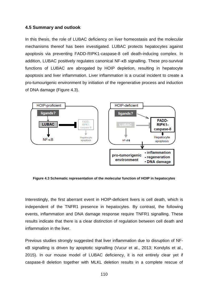
110
4.5 Summary and outlook
In this thesis, the role of LUBAC deficiency on liver homeostasis and the molecular
mechanisms thereof has been investigated. LUBAC protects hepatocytes against
apoptosis via preventing FADD-RIPK1-caspase-8 cell death-inducing complex. In
addition, LUBAC positively regulates canonical NF-κB signalling. These pro-survival
functions of LUBAC are abrogated by HOIP depletion, resulting in hepatocyte
apoptosis and liver inflammation. Liver inflammation is a crucial incident to create a
pro-tumourigenic environment by initiation of the regenerative process and induction
of DNA damage (Figure 4.3).
Figure 4.3 Schematic representation of the molecular function of HOIP in hepatocytes
Interestingly, the first aberrant event in HOIP-deficient livers is cell death, which is
independent of the TNFR1 presence in hepatocytes. By contrast, the following
events, inflammation and DNA damage response require TNFR1 signalling. These
results indicate that there is a clear distinction of regulation between cell death and
inflammation in the liver.
Previous studies strongly suggested that liver inflammation due to disruption of NF-
κB signalling is driven by apoptotic signalling (Vucur et al., 2013; Kondylis et al.,
2015). In our mouse model of LUBAC deficiency, it is not entirely clear yet if
caspase-8 deletion together with MLKL deletion results in a complete rescue of

111
inflammation and carcinogenesis, which requires further clarification. The obstacle
for examining MlklKO Casp8HET HoipΔhep mice is their early development of
lymphadenopathy around 2-3 months of age. To circumvent this problem,
hematopoietic cells in MlklKO Casp8KO HoipΔhep mice could be reconstituted with
MlklKO Casp8HET or wild-type bone marrow cells after irradiation. Alternatively, it is
possible to employ caspase-8 floxed strain, instead of systemic caspase-8 knockout
mice, to avoid its deletion in non-LPCs, particularly in the immune system.
The precise mechanism of caspase-8 activation in HOIP-deficient hepatocytes
remains still elusive. How is the FADD-RIPK1-caspase-8 complex formed in HOIP-
deficient hepatocytes? Does this require some exogenous stimuli? Or the mere
LUBAC deficiency is sufficient to trigger this? The potential involvement of some
death receptor signalling can be tested by employing, for instance, TRAIL-R and
CD95 knockout animals. TLR signalling can be evaluated by crossing with MyD88
knockout mice except for TLR3 signalling, which can be addressed by TLR3 or TRIF
deficiency.
Furthermore, how LUBAC is involved in the prevention of the complex formation is
still a conundrum. Is linear ubiquitination of the complex component(s) sufficient for
the inhibition, or is the scaffolding function of HOIP, for instance binding to OTULIN
and/or SPATA2-CYLD complex, required for it? To answer these questions, a further
dissection of the functions of HOIP would be essential. If the loss of linear
ubiquitination is solely accountable for the phenotype, there must be one or multiple
LUBAC substrates that is linearly ubiquitinated in hepatocytes under steady state
and keeps FADD, RIPK1 and caspase-8 in check to prevent them to be assembled
and activated.

112
Acknowledgements
First of all, I would like to express my gratitude to my supervisor Professor Henning Walczak
for accepting me and providing me with a very stimulating environment in his laboratory. I
would like to thank Japan student service organization (JASSO) for a generous three-year
funding for my PhD study.
Furthermore, I would like to thank:
Professor Mala Maini and Dr Pablo Rodriguez-Viciana for being wonderful PhD upgrade
assessors and giving me advice on the directions of my project;
Professor Sibylle Mittnacht and Professor Pascal Meier for being the examiners for the viva
voce and correcting PhD thesis;
Ms Lorraine Lawrence for excellent histology service and training;
Dr Samira Alliouachene and Dr Benoit Bilanges for instructing hepatocyte isolation
techniques;
Dr Alison Winstanley for giving me histopathological insights;
Mr Signore Massimo for mouse transgenic services;
Mr Paul Levy, Ms Toni Clark, Mr Mark White and Mr Peter Ward for taking care of our
mouse colonies.
Also, I would like to thank former and current lab members for having me and working
together in the lab:
Dr Nieves Peltzer for giving me guidance throughout my PhD, correcting my thesis and
many documents and being a cheerful desk neighbour;
Dr Alexandra Sevko for teaching me immunological experiments, broadening my insights on
immunology and correcting my thesis;
Dr Elodie Lafont for being a fun bench neighbour, having chit-chats from science to our life,
all the original jingles, French lessons and correcting my manuscripts and thesis;
Dr Maurice Darding and Ms Lucia Taraborrelli for being amazing teammates of in-vivo
LUBAC team and correction of my thesis;
Ms Aida Sarr for all the appreciated genotyping works and laughs;
Ms Helena Draberova for our complex mouse colony management;

113
Dr Maureen Galmes-Spit for having fun working together on the intestine project and
correction of my thesis;
Dr Peter Draber and Mr Torsten Hartwig for being fanatic training buddies and correction of
my thesis;
Dr Katarzyna Wojdyla for being a kind desk neighbour and running buddy;
Dr Silvia Surinova, Dr Diego de Miguel Samaniego, Mr Sebastian Kupka and Ms Itziar Areso
for correction of my thesis and companionship;
Dr Eva Rieser, Dr Silvia von Karstedt, Dr Antonella Montinaro, Dr Johannes Lemke, Dr
Charahzade Kantari-Mimoun, Ms Julia Zinngrebe and Mr Matthias Reichert for being lovely
lab mates;
Dr Nessa Adams, Ms Silvia Bianco, Ms Celeste Aouilk-Jaiyeola, Ms Ieva Songailaite and Ms
Roxanne Payne for helping me on administrations.
Last but not least, I would like to thank my family and friends:
My grandparents, parents, my cousins and brother for unconditional love and support from
abroad;
Wataru Ohira, Rie Yamazaki and Ángel Andreu for great friendship, love and support during
and after living together in London.
Thank you very much and arigato,
Yutaka Shimizu

114
Bibliography
Aaes, T.L., Kaczmarek, A., Delvaeye, T., De Craene, B., De Koker, S., Heyndrickx, L., Delrue, I.,
Taminau, J., Wiernicki, B., De Groote, P., et al. (2016). Vaccination with Necroptotic Cancer Cells
Induces Efficient Anti-tumor Immunity. Cell reports 15, 274-287.
Aderem, A., and Underhill, D.M. (1999). Mechanisms of phagocytosis in macrophages. Annual review
of immunology 17, 593-623.
Ai, L., Xu, Q., Wu, C., Wang, X., Chen, Z., Su, D., Jiang, X., Xu, A., Lin, Q., and Fan, Z. (2015). A20
Attenuates FFAs-induced Lipid Accumulation in Nonalcoholic Steatohepatitis. International journal of
biological sciences 11, 1436-1446.
Alberts B, Johnson A, Lewis J, et al. (2002). Molecular Biology of the Cell. 4th edition.
Akira, S., and Takeda, K. (2004). Toll-like receptor signalling. Nature reviews Immunology 4, 499-511.
Aradhya, S., Courtois, G., Rajkovic, A., Lewis, R.A., Levy, M., Israel, A., and Nelson, D.L. (2001).
Atypical forms of incontinentia pigmenti in male individuals result from mutations of a cytosine tract in
exon 10 of NEMO (IKK-gamma). American journal of human genetics 68, 765-771.
Balkwill, F., and Mantovani, A. (2001). Inflammation and cancer: back to Virchow? Lancet 357, 539-
545.
Berger, S.B., Kasparcova, V., Hoffman, S., Swift, B., Dare, L., Schaeffer, M., Capriotti, C., Cook, M.,
Finger, J., Hughes-Earle, A., et al. (2014). Cutting Edge: RIP1 kinase activity is dispensable for
normal development but is a key regulator of inflammation in SHARPIN-deficient mice. J Immunol 192,
5476-5480.
Bertrand, M.J., Doiron, K., Labbe, K., Korneluk, R.G., Barker, P.A., and Saleh, M. (2009). Cellular
inhibitors of apoptosis cIAP1 and cIAP2 are required for innate immunity signaling by the pattern
recognition receptors NOD1 and NOD2. Immunity 30, 789-801.
Bertrand, M.J., Milutinovic, S., Dickson, K.M., Ho, W.C., Boudreault, A., Durkin, J., Gillard, J.W.,
Jaquith, J.B., Morris, S.J., and Barker, P.A. (2008). cIAP1 and cIAP2 facilitate cancer cell survival by
functioning as E3 ligases that promote RIP1 ubiquitination. Molecular cell 30, 689-700.
Bettermann, K., Vucur, M., Haybaeck, J., Koppe, C., Janssen, J., Heymann, F., Weber, A.,
Weiskirchen, R., Liedtke, C., Gassler, N., et al. (2010). TAK1 suppresses a NEMO-dependent but NF-
kappaB-independent pathway to liver cancer. Cancer cell 17, 481-496.
Beyaert, R. (2014). An E3 ubiquitin ligase-independent role of LUBAC. Blood 123, 2131-2133.
Bignell, G.R., Warren, W., Seal, S., Takahashi, M., Rapley, E., Barfoot, R., Green, H., Brown, C.,
Biggs, P.J., Lakhani, S.R., et al. (2000). Identification of the familial cylindromatosis tumour-
suppressor gene. Nature genetics 25, 160-165.
Bodmer, J.L., Holler, N., Reynard, S., Vinciguerra, P., Schneider, P., Juo, P., Blenis, J., and Tschopp,
J. (2000). TRAIL receptor-2 signals apoptosis through FADD and caspase-8. Nature cell biology 2,
241-243.
115
Boisson, B., Laplantine, E., Dobbs, K., Cobat, A., Tarantino, N., Hazen, M., Lidov, H.G., Hopkins, G.,
Du, L., Belkadi, A., et al. (2015). Human HOIP and LUBAC deficiency underlies autoinflammation,
immunodeficiency, amylopectinosis, and lymphangiectasia. The Journal of experimental medicine 212,
939-951.
Boisson, B., Laplantine, E., Prando, C., Giliani, S., Israelsson, E., Xu, Z., Abhyankar, A., Israel, L.,
Trevejo-Nunez, G., Bogunovic, D., et al. (2012). Immunodeficiency, autoinflammation and
amylopectinosis in humans with inherited HOIL-1 and LUBAC deficiency. Nature immunology 13,
1178-1186.
Boldin, M.P., Goncharov, T.M., Goltsev, Y.V., and Wallach, D. (1996). Involvement of MACH, a novel
MORT1/FADD-interacting protease, in Fas/APO-1- and TNF receptor-induced cell death. Cell 85,
803-815.
Bonizzi, G., and Karin, M. (2004). The two NF-kappaB activation pathways and their role in innate and
adaptive immunity. Trends in immunology 25, 280-288.
Bonnet, M.C., Preukschat, D., Welz, P.S., van Loo, G., Ermolaeva, M.A., Bloch, W., Haase, I., and
Pasparakis, M. (2011). The adaptor protein FADD protects epidermal keratinocytes from necroptosis
in vivo and prevents skin inflammation. Immunity 35, 572-582.
Bosanac, I., Wertz, I.E., Pan, B., Yu, C., Kusam, S., Lam, C., Phu, L., Phung, Q., Maurer, B., Arnott,
D., et al. (2010). Ubiquitin binding to A20 ZnF4 is required for modulation of NF-kappaB signaling.
Molecular cell 40, 548-557.
Bouchard, M.J., Wang, L.H., and Schneider, R.J. (2001). Calcium signaling by HBx protein in hepatitis
B virus DNA replication. Science 294, 2376-2378.
Callahan, J.A., Hammer, G.E., Agelides, A., Duong, B.H., Oshima, S., North, J., Advincula, R., Shifrin,
N., Truong, H.A., Paw, J., et al. (2013). Cutting edge: ABIN-1 protects against psoriasis by restricting
MyD88 signals in dendritic cells. J Immunol 191, 535-539.
Calzascia, T., Pellegrini, M., Hall, H., Sabbagh, L., Ono, N., Elford, A.R., Mak, T.W., and Ohashi, P.S.
(2007). TNF-alpha is critical for antitumor but not antiviral T cell immunity in mice. The Journal of
clinical investigation 117, 3833-3845.
Carneiro, L.A., Travassos, L.H., Soares, F., Tattoli, I., Magalhaes, J.G., Bozza, M.T., Plotkowski, M.C.,
Sansonetti, P.J., Molkentin, J.D., Philpott, D.J., et al. (2009). Shigella induces mitochondrial
dysfunction and cell death in nonmyleoid cells. Cell host & microbe 5, 123-136.
Catrysse, L., Farhang Ghahremani, M., Vereecke, L., Youssef, S.A., Mc Guire, C., Sze, M., Weber, A.,
Heikenwalder, M., de Bruin, A., Beyaert, R., et al. (2016). A20 prevents chronic liver inflammation and
cancer by protecting hepatocytes from death. Cell death & disease 7, e2250.
Chan, A.T., and Ladabaum, U. (2016). Where Do We Stand With Aspirin for the Prevention of
Colorectal Cancer? The USPSTF Recommendations. Gastroenterology 150, 14-18.
Chen, W., Wu, J., Li, L., Zhang, Z., Ren, J., Liang, Y., Chen, F., Yang, C., Zhou, Z., Su, S.S., et al.
(2015). Ppm1b negatively regulates necroptosis through dephosphorylating Rip3. Nature cell biology
17, 434-444.
Chisari, F.V., Klopchin, K., Moriyama, T., Pasquinelli, C., Dunsford, H.A., Sell, S., Pinkert, C.A.,
Brinster, R.L., and Palmiter, R.D. (1989). Molecular pathogenesis of hepatocellular carcinoma in
hepatitis B virus transgenic mice. Cell 59, 1145-1156.
116
Chu, Y., Vahl, J.C., Kumar, D., Heger, K., Bertossi, A., Wojtowicz, E., Soberon, V., Schenten, D.,
Mack, B., Reutelshofer, M., et al. (2011). B cells lacking the tumor suppressor TNFAIP3/A20 display
impaired differentiation and hyperactivation and cause inflammation and autoimmunity in aged mice.
Blood 117, 2227-2236.
Claudio, E., Brown, K., Park, S., Wang, H., and Siebenlist, U. (2002). BAFF-induced NEMO-
independent processing of NF-kappa B2 in maturing B cells. Nature immunology 3, 958-965.
Coope, H.J., Atkinson, P.G., Huhse, B., Belich, M., Janzen, J., Holman, M.J., Klaus, G.G., Johnston,
L.H., and Ley, S.C. (2002). CD40 regulates the processing of NF-kappaB2 p100 to p52. The EMBO
journal 21, 5375-5385.
Corazza, N., Jakob, S., Schaer, C., Frese, S., Keogh, A., Stroka, D., Kassahn, D., Torgler, R., Mueller,
C., Schneider, P., et al. (2006). TRAIL receptor-mediated JNK activation and Bim phosphorylation
critically regulate Fas-mediated liver damage and lethality. The Journal of clinical investigation 116,
2493-2499.
Damgaard, R.B., Nachbur, U., Yabal, M., Wong, W.W., Fiil, B.K., Kastirr, M., Rieser, E., Rickard, J.A.,
Bankovacki, A., Peschel, C., et al. (2012). The ubiquitin ligase XIAP recruits LUBAC for NOD2
signaling in inflammation and innate immunity. Molecular cell 46, 746-758.
Dannappel, M., Vlantis, K., Kumari, S., Polykratis, A., Kim, C., Wachsmuth, L., Eftychi, C., Lin, J.,
Corona, T., Hermance, N., et al. (2014). RIPK1 maintains epithelial homeostasis by inhibiting
apoptosis and necroptosis. Nature 513, 90-94.
De, A., Dainichi, T., Rathinam, C.V., and Ghosh, S. (2014). The deubiquitinase activity of A20 is
dispensable for NF-kappaB signaling. EMBO reports 15, 775-783.
de Almagro, M.C., Goncharov, T., Newton, K., and Vucic, D. (2015). Cellular IAP proteins and LUBAC
differentially regulate necrosome-associated RIP1 ubiquitination. Cell death & disease 6, e1800.
De Mitri, M.S., Poussin, K., Baccarini, P., Pontisso, P., D'Errico, A., Simon, N., Grigioni, W., Alberti, A.,
Beaugrand, M., Pisi, E., et al. (1995). HCV-associated liver cancer without cirrhosis. Lancet 345, 413-
415.
Dillon, C.P., Weinlich, R., Rodriguez, D.A., Cripps, J.G., Quarato, G., Gurung, P., Verbist, K.C.,
Brewer, T.L., Llambi, F., Gong, Y.N., et al. (2014). RIPK1 blocks early postnatal lethality mediated by
caspase-8 and RIPK3. Cell 157, 1189-1202.
Doffinger, R., Smahi, A., Bessia, C., Geissmann, F., Feinberg, J., Durandy, A., Bodemer, C., Kenwrick,
S., Dupuis-Girod, S., Blanche, S., et al. (2001). X-linked anhidrotic ectodermal dysplasia with
immunodeficiency is caused by impaired NF-kappaB signaling. Nature genetics 27, 277-285.
Dondelinger, Y., Declercq, W., Montessuit, S., Roelandt, R., Goncalves, A., Bruggeman, I., Hulpiau,
P., Weber, K., Sehon, C.A., Marquis, R.W., et al. (2014). MLKL compromises plasma membrane
integrity by binding to phosphatidylinositol phosphates. Cell reports 7, 971-981.
Dondelinger, Y., Jouan-Lanhouet, S., Divert, T., Theatre, E., Bertin, J., Gough, P.J., Giansanti, P.,
Heck, A.J., Dejardin, E., Vandenabeele, P., et al. (2015). NF-kappaB-Independent Role of
IKKalpha/IKKbeta in Preventing RIPK1 Kinase-Dependent Apoptotic and Necroptotic Cell Death
during TNF Signaling. Molecular cell 60, 63-76.
117
Doria, M., Klein, N., Lucito, R., and Schneider, R.J. (1995). The hepatitis B virus HBx protein is a dual
specificity cytoplasmic activator of Ras and nuclear activator of transcription factors. The EMBO
journal 14, 4747-4757.
Draber, P., Kupka, S., Reichert, M., Draberova, H., Lafont, E., de Miguel, D., Spilgies, L., Surinova, S.,
Taraborrelli, L., Hartwig, T., et al. (2015). LUBAC-Recruited CYLD and A20 Regulate Gene Activation
and Cell Death by Exerting Opposing Effects on Linear Ubiquitin in Signaling Complexes. Cell reports
13, 2258-2272.
Dubois, S.M., Alexia, C., Wu, Y., Leclair, H.M., Leveau, C., Schol, E., Fest, T., Tarte, K., Chen, Z.J.,
Gavard, J., et al. (2014). A catalytic-independent role for the LUBAC in NF-kappaB activation upon
antigen receptor engagement and in lymphoma cells. Blood 123, 2199-2203.
Dunn, G.P., Old, L.J., and Schreiber, R.D. (2004). The immunobiology of cancer immunosurveillance
and immunoediting. Immunity 21, 137-148.
Ea, C.K., Deng, L., Xia, Z.P., Pineda, G., and Chen, Z.J. (2006). Activation of IKK by TNFalpha
requires site-specific ubiquitination of RIP1 and polyubiquitin binding by NEMO. Molecular cell 22,
245-257.
Ehlken, H., Krishna-Subramanian, S., Ochoa-Callejero, L., Kondylis, V., Nadi, N.E., Straub, B.K.,
Schirmacher, P., Walczak, H., Kollias, G., and Pasparakis, M. (2014). Death receptor-independent
FADD signalling triggers hepatitis and hepatocellular carcinoma in mice with liver parenchymal cell-
specific NEMO knockout. Cell death and differentiation 21, 1721-1732.
Elgueta, R., Benson, M.J., de Vries, V.C., Wasiuk, A., Guo, Y., and Noelle, R.J. (2009). Molecular
mechanism and function of CD40/CD40L engagement in the immune system. Immunological reviews
229, 152-172.
Elgueta, R., Benson, M.J., de Vries, V.C., Wasiuk, A., Guo, Y., and Noelle, R.J. (2009). Molecular
mechanism and function of CD40/CD40L engagement in the immune system. Immunological reviews
229, 152-172.
Elliott, P.R., Nielsen, S.V., Marco-Casanova, P., Fiil, B.K., Keusekotten, K., Mailand, N., Freund, S.M.,
Gyrd-Hansen, M., and Komander, D. (2014). Molecular basis and regulation of OTULIN-LUBAC
interaction. Molecular cell 54, 335-348.
Ellis, R.E., Yuan, J.Y., and Horvitz, H.R. (1991). Mechanisms and functions of cell death. Annual
review of cell biology 7, 663-698.
El-Serag, H.B. (2011). Hepatocellular carcinoma. The New England journal of medicine 365, 1118-
1127.
Emmerich, C.H., Bakshi, S., Kelsall, I.R., Ortiz-Guerrero, J., Shpiro, N., and Cohen, P. (2016).
Lys63/Met1-hybrid ubiquitin chains are commonly formed during the activation of innate immune
signalling. Biochemical and biophysical research communications 474, 452-461.
Emmerich, C.H., Ordureau, A., Strickson, S., Arthur, J.S., Pedrioli, P.G., Komander, D., and Cohen, P.
(2013). Activation of the canonical IKK complex by K63/M1-linked hybrid ubiquitin chains.
Proceedings of the National Academy of Sciences of the United States of America 110, 15247-15252.
Ermolaeva, M.A., Michallet, M.C., Papadopoulou, N., Utermohlen, O., Kranidioti, K., Kollias, G.,
Tschopp, J., and Pasparakis, M. (2008). Function of TRADD in tumor necrosis factor receptor 1
signaling and in TRIF-dependent inflammatory responses. Nature immunology 9, 1037-1046.

118
Etemadi, N., Chopin, M., Anderton, H., Tanzer, M.C., Rickard, J.A., Abeysekera, W., Hall, C., Spall,
S.K., Wang, B., Xiong, Y., et al. (2015). TRAF2 regulates TNF and NF-kappaB signalling to suppress
apoptosis and skin inflammation independently of Sphingosine kinase 1. eLife 4.
Fiil, B.K., Damgaard, R.B., Wagner, S.A., Keusekotten, K., Fritsch, M., Bekker-Jensen, S., Mailand, N.,
Choudhary, C., Komander, D., and Gyrd-Hansen, M. (2013). OTULIN restricts Met1-linked
ubiquitination to control innate immune signaling. Molecular cell 50, 818-830.
Fischer, U., Janicke, R.U., and Schulze-Osthoff, K. (2003). Many cuts to ruin: a comprehensive
update of caspase substrates. Cell death and differentiation 10, 76-100.
Fuchs, S.Y., Chen, A., Xiong, Y., Pan, Z.Q., and Ronai, Z. (1999). HOS, a human homolog of Slimb,
forms an SCF complex with Skp1 and Cullin1 and targets the phosphorylation-dependent degradation
of IkappaB and beta-catenin. Oncogene 18, 2039-2046.
Ganten, T.M., Koschny, R., Haas, T.L., Sykora, J., Li-Weber, M., Herzer, K., and Walczak, H. (2005).
Proteasome inhibition sensitizes hepatocellular carcinoma cells, but not human hepatocytes, to TRAIL.
Hepatology 42, 588-597.
Gautheron, J., Vucur, M., Reisinger, F., Cardenas, D.V., Roderburg, C., Koppe, C., Kreggenwinkel, K.,
Schneider, A.T., Bartneck, M., Neumann, U.P., et al. (2014). A positive feedback loop between RIP3
and JNK controls non-alcoholic steatohepatitis. EMBO molecular medicine 6, 1062-1074.
Gerlach, B., Cordier, S.M., Schmukle, A.C., Emmerich, C.H., Rieser, E., Haas, T.L., Webb, A.I.,
Rickard, J.A., Anderton, H., Wong, W.W., et al. (2011). Linear ubiquitination prevents inflammation
and regulates immune signalling. Nature 471, 591-596.
Ghosh, S., and Baltimore, D. (1990). Activation in vitro of NF-kappa B by phosphorylation of its
inhibitor I kappa B. Nature 344, 678-682.
Ghosh, S., May, M.J., and Kopp, E.B. (1998). NF-kappa B and Rel proteins: evolutionarily conserved
mediators of immune responses. Annual review of immunology 16, 225-260.
Gijbels, M.J., HogenEsch, H., Bruijnzeel, P.L., Elliott, G.R., and Zurcher, C. (1995). Maintenance of
donor phenotype after full-thickness skin transplantation from mice with chronic proliferative dermatitis
(cpdm/cpdm) to C57BL/Ka and nude mice and vice versa. The Journal of investigative dermatology
105, 769-773.
Girardin, S.E., Boneca, I.G., Viala, J., Chamaillard, M., Labigne, A., Thomas, G., Philpott, D.J., and
Sansonetti, P.J. (2003). Nod2 is a general sensor of peptidoglycan through muramyl dipeptide (MDP)
detection. The Journal of biological chemistry 278, 8869-8872.
Glickman, M.H., and Ciechanover, A. (2002). The ubiquitin-proteasome proteolytic pathway:
destruction for the sake of construction. Physiological reviews 82, 373-428.
Gunther, C., Martini, E., Wittkopf, N., Amann, K., Weigmann, B., Neumann, H., Waldner, M.J., Hedrick,
S.M., Tenzer, S., Neurath, M.F., et al. (2011). Caspase-8 regulates TNF-alpha-induced epithelial
necroptosis and terminal ileitis. Nature 477, 335-339.
Haas, T.L., Emmerich, C.H., Gerlach, B., Schmukle, A.C., Cordier, S.M., Rieser, E., Feltham, R.,
Vince, J., Warnken, U., Wenger, T., et al. (2009). Recruitment of the linear ubiquitin chain assembly
119
complex stabilizes the TNF-R1 signaling complex and is required for TNF-mediated gene induction.
Molecular cell 36, 831-844.
Haybaeck, J., Zeller, N., Wolf, M.J., Weber, A., Wagner, U., Kurrer, M.O., Bremer, J., Iezzi, G., Graf,
R., Clavien, P.A., et al. (2009). A lymphotoxin-driven pathway to hepatocellular carcinoma. Cancer
cell 16, 295-308.
He, G., Dhar, D., Nakagawa, H., Font-Burgada, J., Ogata, H., Jiang, Y., Shalapour, S., Seki, E., Yost,
S.E., Jepsen, K., et al. (2013). Identification of liver cancer progenitors whose malignant progression
depends on autocrine IL-6 signaling. Cell 155, 384-396.
He, G., Yu, G.Y., Temkin, V., Ogata, H., Kuntzen, C., Sakurai, T., Sieghart, W., Peck-Radosavljevic,
M., Leffert, H.L., and Karin, M. (2010). Hepatocyte IKKbeta/NF-kappaB inhibits tumor promotion and
progression by preventing oxidative stress-driven STAT3 activation. Cancer cell 17, 286-297.
Heindryckx, F., Colle, I., and Van Vlierberghe, H. (2009). Experimental mouse models for
hepatocellular carcinoma research. International journal of experimental pathology 90, 367-386.
Hershko, A., and Ciechanover, A. (1998). The ubiquitin system. Annual review of biochemistry 67,
425-479.
Hikita, H., Kodama, T., Shimizu, S., Li, W., Shigekawa, M., Tanaka, S., Hosui, A., Miyagi, T., Tatsumi,
T., Kanto, T., et al. (2012). Bak deficiency inhibits liver carcinogenesis: a causal link between
apoptosis and carcinogenesis. Journal of hepatology 57, 92-100.
Hitotsumatsu, O., Ahmad, R.C., Tavares, R., Wang, M., Philpott, D., Turer, E.E., Lee, B.L., Shiffin, N.,
Advincula, R., Malynn, B.A., et al. (2008). The ubiquitin-editing enzyme A20 restricts nucleotide-
binding oligomerization domain containing 2-triggered signals. Immunity 28, 381-390.
HogenEsch, H., Gijbels, M.J., Offerman, E., van Hooft, J., van Bekkum, D.W., and Zurcher, C. (1993).
A spontaneous mutation characterized by chronic proliferative dermatitis in C57BL mice. The
American journal of pathology 143, 972-982.
HogenEsch, H., Janke, S., Boggess, D., and Sundberg, J.P. (1999). Absence of Peyer's patches and
abnormal lymphoid architecture in chronic proliferative dermatitis (cpdm/cpdm) mice. J Immunol 162,
3890-3896.
Hospenthal, M.K., Mevissen, T.E., and Komander, D. (2015). Deubiquitinase-based analysis of
ubiquitin chain architecture using Ubiquitin Chain Restriction (UbiCRest). Nature protocols 10, 349-
361.
Hsu, H., Shu, H.B., Pan, M.G., and Goeddel, D.V. (1996). TRADD-TRAF2 and TRADD-FADD
interactions define two distinct TNF receptor 1 signal transduction pathways. Cell 84, 299-308.
Hsu, H., Xiong, J., and Goeddel, D.V. (1995). The TNF receptor 1-associated protein TRADD signals
cell death and NF-kappa B activation. Cell 81, 495-504.
Hubeau, M., Ngadjeua, F., Puel, A., Israel, L., Feinberg, J., Chrabieh, M., Belani, K., Bodemer, C.,
Fabre, I., Plebani, A., et al. (2011). New mechanism of X-linked anhidrotic ectodermal dysplasia with
immunodeficiency: impairment of ubiquitin binding despite normal folding of NEMO protein. Blood 118,
926-935.

120
Ikeda, F., Deribe, Y.L., Skanland, S.S., Stieglitz, B., Grabbe, C., Franz-Wachtel, M., van Wijk, S.J.,
Goswami, P., Nagy, V., Terzic, J., et al. (2011). SHARPIN forms a linear ubiquitin ligase complex
regulating NF-kappaB activity and apoptosis. Nature 471, 637-641.
Inohara, N., Koseki, T., Lin, J., del Peso, L., Lucas, P.C., Chen, F.F., Ogura, Y., and Nunez, G. (2000).
An induced proximity model for NF-kappa B activation in the Nod1/RICK and RIP signaling pathways.
The Journal of biological chemistry 275, 27823-27831.
Inohara, N., Ogura, Y., Fontalba, A., Gutierrez, O., Pons, F., Crespo, J., Fukase, K., Inamura, S.,
Kusumoto, S., Hashimoto, M., et al. (2003). Host recognition of bacterial muramyl dipeptide mediated
through NOD2. Implications for Crohn's disease. The Journal of biological chemistry 278, 5509-5512.
Inokuchi, S., Aoyama, T., Miura, K., Osterreicher, C.H., Kodama, Y., Miyai, K., Akira, S., Brenner,
D.A., and Seki, E. (2010). Disruption of TAK1 in hepatocytes causes hepatic injury, inflammation,
fibrosis, and carcinogenesis. Proceedings of the National Academy of Sciences of the United States
of America 107, 844-849.
Irmler, M., Thome, M., Hahne, M., Schneider, P., Hofmann, K., Steiner, V., Bodmer, J.L., Schroter, M.,
Burns, K., Mattmann, C., et al. (1997). Inhibition of death receptor signals by cellular FLIP. Nature 388,
190-195.
Janeway, C.A., Jr., and Medzhitov, R. (2002). Innate immune recognition. Annual review of
immunology 20, 197-216.
Jost, P.J., Grabow, S., Gray, D., McKenzie, M.D., Nachbur, U., Huang, D.C., Bouillet, P., Thomas,
H.E., Borner, C., Silke, J., et al. (2009). XIAP discriminates between type I and type II FAS-induced
apoptosis. Nature 460, 1035-1039.
Kaiser, W.J., Upton, J.W., Long, A.B., Livingston-Rosanoff, D., Daley-Bauer, L.P., Hakem, R.,
Caspary, T., and Mocarski, E.S. (2011). RIP3 mediates the embryonic lethality of caspase-8-deficient
mice. Nature 471, 368-372.
Kan, Z., Zheng, H., Liu, X., Li, S., Barber, T.D., Gong, Z., Gao, H., Hao, K., Willard, M.D., Xu, J., et al.
(2013). Whole-genome sequencing identifies recurrent mutations in hepatocellular carcinoma.
Genome research 23, 1422-1433.
Kanayama, A., Seth, R.B., Sun, L., Ea, C.K., Hong, M., Shaito, A., Chiu, Y.H., Deng, L., and Chen,
Z.J. (2004). TAB2 and TAB3 activate the NF-kappaB pathway through binding to polyubiquitin chains.
Molecular cell 15, 535-548.
Kane, L.A., Lazarou, M., Fogel, A.I., Li, Y., Yamano, K., Sarraf, S.A., Banerjee, S., and Youle, R.J.
(2014). PINK1 phosphorylates ubiquitin to activate Parkin E3 ubiquitin ligase activity. The Journal of
cell biology 205, 143-153.
Kang, T.B., Ben-Moshe, T., Varfolomeev, E.E., Pewzner-Jung, Y., Yogev, N., Jurewicz, A., Waisman,
A., Brenner, O., Haffner, R., Gustafsson, E., et al. (2004). Caspase-8 serves both apoptotic and
nonapoptotic roles. J Immunol 173, 2976-2984.
Kang, T.B., Yang, S.H., Toth, B., Kovalenko, A., and Wallach, D. (2013). Caspase-8 blocks kinase
RIPK3-mediated activation of the NLRP3 inflammasome. Immunity 38, 27-40.
Karin, M., and Clevers, H. (2016). Reparative inflammation takes charge of tissue regeneration.
Nature 529, 307-315.
121
Kawai, T., and Akira, S. (2010). The role of pattern-recognition receptors in innate immunity: update
on Toll-like receptors. Nature immunology 11, 373-384.
Kellendonk, C., Opherk, C., Anlag, K., Schutz, G., and Tronche, F. (2000). Hepatocyte-specific
expression of Cre recombinase. Genesis 26, 151-153.
Keusekotten, K., Elliott, P.R., Glockner, L., Fiil, B.K., Damgaard, R.B., Kulathu, Y., Wauer, T.,
Hospenthal, M.K., Gyrd-Hansen, M., Krappmann, D., et al. (2013). OTULIN antagonizes LUBAC
signaling by specifically hydrolyzing Met1-linked polyubiquitin. Cell 153, 1312-1326.
Kim, C.M., Koike, K., Saito, I., Miyamura, T., and Jay, G. (1991). HBx gene of hepatitis B virus
induces liver cancer in transgenic mice. Nature 351, 317-320.
Kim, R., Emi, M., and Tanabe, K. (2007). Cancer immunoediting from immune surveillance to immune
escape. Immunology 121, 1-14.
Kimura, Y., and Tanaka, K. (2010). Regulatory mechanisms involved in the control of ubiquitin
homeostasis. Journal of biochemistry 147, 793-798.
Kirisako, T., Kamei, K., Murata, S., Kato, M., Fukumoto, H., Kanie, M., Sano, S., Tokunaga, F.,
Tanaka, K., and Iwai, K. (2006). A ubiquitin ligase complex assembles linear polyubiquitin chains. The
EMBO journal 25, 4877-4887.
Kischkel, F.C., Hellbardt, S., Behrmann, I., Germer, M., Pawlita, M., Krammer, P.H., and Peter, M.E.
(1995). Cytotoxicity-dependent APO-1 (Fas/CD95)-associated proteins form a death-inducing
signaling complex (DISC) with the receptor. The EMBO journal 14, 5579-5588.
Kischkel, F.C., Lawrence, D.A., Chuntharapai, A., Schow, P., Kim, K.J., and Ashkenazi, A. (2000).
Apo2L/TRAIL-dependent recruitment of endogenous FADD and caspase-8 to death receptors 4 and 5.
Immunity 12, 611-620.
Komander, D., Clague, M.J., and Urbe, S. (2009). Breaking the chains: structure and function of the
deubiquitinases. Nature reviews Molecular cell biology 10, 550-563.
Kondylis, V., Polykratis, A., Ehlken, H., Ochoa-Callejero, L., Straub, B.K., Krishna-Subramanian, S.,
Van, T.M., Curth, H.M., Heise, N., Weih, F., et al. (2015). NEMO Prevents Steatohepatitis and
Hepatocellular Carcinoma by Inhibiting RIPK1 Kinase Activity-Mediated Hepatocyte Apoptosis.
Cancer cell 28, 582-598.
Kovalenko, A., Kim, J.C., Kang, T.B., Rajput, A., Bogdanov, K., Dittrich-Breiholz, O., Kracht, M.,
Brenner, O., and Wallach, D. (2009). Caspase-8 deficiency in epidermal keratinocytes triggers an
inflammatory skin disease. The Journal of experimental medicine 206, 2161-2177.
Koyano, F., Okatsu, K., Kosako, H., Tamura, Y., Go, E., Kimura, M., Kimura, Y., Tsuchiya, H.,
Yoshihara, H., Hirokawa, T., et al. (2014). Ubiquitin is phosphorylated by PINK1 to activate parkin.
Nature 510, 162-166.
Kucharczak, J., Simmons, M.J., Fan, Y., and Gelinas, C. (2003). To be, or not to be: NF-kappaB is
the answer--role of Rel/NF-kappaB in the regulation of apoptosis. Oncogene 22, 8961-8982.
Kulathu, Y., and Komander, D. (2012). Atypical ubiquitylation - the unexplored world of polyubiquitin
beyond Lys48 and Lys63 linkages. Nature reviews Molecular cell biology 13, 508-523.
122
Kumari, S., Redouane, Y., Lopez-Mosqueda, J., Shiraishi, R., Romanowska, M., Lutzmayer, S.,
Kuiper, J., Martinez, C., Dikic, I., Pasparakis, M., et al. (2014). Sharpin prevents skin inflammation by
inhibiting TNFR1-induced keratinocyte apoptosis. eLife 3.
Lechtenberg, B.C., Rajput, A., Sanishvili, R., Dobaczewska, M.K., Ware, C.F., Mace, P.D., and Riedl,
S.J. (2016). Structure of a HOIP/E2~ubiquitin complex reveals RBR E3 ligase mechanism and
regulation. Nature 529, 546-550.
Lee, E.G., Boone, D.L., Chai, S., Libby, S.L., Chien, M., Lodolce, J.P., and Ma, A. (2000). Failure to
regulate TNF-induced NF-kappaB and cell death responses in A20-deficient mice. Science 289,
2350-2354.
Lee, G.H., Ooasa, T., and Osanai, M. (1998). Mechanism of the paradoxical, inhibitory effect of
phenobarbital on hepatocarcinogenesis initiated in infant B6C3F1 mice with diethylnitrosamine.
Cancer research 58, 1665-1669.
Lee, J.S., Chu, I.S., Mikaelyan, A., Calvisi, D.F., Heo, J., Reddy, J.K., and Thorgeirsson, S.S. (2004).
Application of comparative functional genomics to identify best-fit mouse models to study human
cancer. Nature genetics 36, 1306-1311.
Li, P., Nijhawan, D., Budihardjo, I., Srinivasula, S.M., Ahmad, M., Alnemri, E.S., and Wang, X. (1997).
Cytochrome c and dATP-dependent formation of Apaf-1/caspase-9 complex initiates an apoptotic
protease cascade. Cell 91, 479-489.
Liang, C., Zhang, M., and Sun, S.C. (2006). beta-TrCP binding and processing of NF-kappaB2/p100
involve its phosphorylation at serines 866 and 870. Cellular signalling 18, 1309-1317.
Liao, G., Zhang, M., Harhaj, E.W., and Sun, S.C. (2004). Regulation of the NF-kappaB-inducing
kinase by tumor necrosis factor receptor-associated factor 3-induced degradation. The Journal of
biological chemistry 279, 26243-26250.
Liedtke, C., Bangen, J.M., Freimuth, J., Beraza, N., Lambertz, D., Cubero, F.J., Hatting, M., Karlmark,
K.R., Streetz, K.L., Krombach, G.A., et al. (2011). Loss of caspase-8 protects mice against
inflammation-related hepatocarcinogenesis but induces non-apoptotic liver injury. Gastroenterology
141, 2176-2187.
Lin, S.C., Chung, J.Y., Lamothe, B., Rajashankar, K., Lu, M., Lo, Y.C., Lam, A.Y., Darnay, B.G., and
Wu, H. (2008). Molecular basis for the unique deubiquitinating activity of the NF-kappaB inhibitor A20.
Journal of molecular biology 376, 526-540.
Linkermann, A., Brasen, J.H., Darding, M., Jin, M.K., Sanz, A.B., Heller, J.O., De Zen, F., Weinlich, R.,
Ortiz, A., Walczak, H., et al. (2013). Two independent pathways of regulated necrosis mediate
ischemia-reperfusion injury. Proceedings of the National Academy of Sciences of the United States of
America 110, 12024-12029.
Lo, J.C., Wang, Y., Tumanov, A.V., Bamji, M., Yao, Z., Reardon, C.A., Getz, G.S., and Fu, Y.X.
(2007). Lymphotoxin beta receptor-dependent control of lipid homeostasis. Science 316, 285-288.
Lu, T.T., Onizawa, M., Hammer, G.E., Turer, E.E., Yin, Q., Damko, E., Agelidis, A., Shifrin, N.,
Advincula, R., Barrera, J., et al. (2013). Dimerization and ubiquitin mediated recruitment of A20, a
complex deubiquitinating enzyme. Immunity 38, 896-905.
123
Luedde, T., Beraza, N., Kotsikoris, V., van Loo, G., Nenci, A., De Vos, R., Roskams, T., Trautwein, C.,
and Pasparakis, M. (2007). Deletion of NEMO/IKKgamma in liver parenchymal cells causes
steatohepatitis and hepatocellular carcinoma. Cancer cell 11, 119-132.
Ma, A., and Malynn, B.A. (2012). A20: linking a complex regulator of ubiquitylation to immunity and
human disease. Nature reviews Immunology 12, 774-785.
Ma, D.Y., and Clark, E.A. (2009). The role of CD40 and CD154/CD40L in dendritic cells. Seminars in
immunology 21, 265-272.
Mackay, F., and Schneider, P. (2009). Cracking the BAFF code. Nature reviews Immunology 9, 491-
502.
Maeda, S., Kamata, H., Luo, J.L., Leffert, H., and Karin, M. (2005). IKKbeta couples hepatocyte death
to cytokine-driven compensatory proliferation that promotes chemical hepatocarcinogenesis. Cell 121,
977-990.
Mantovani, A., and Allavena, P. (2015). The interaction of anticancer therapies with tumor-associated
macrophages. The Journal of experimental medicine 212, 435-445.
Mantovani, A., Sozzani, S., Locati, M., Allavena, P., and Sica, A. (2002). Macrophage polarization:
tumor-associated macrophages as a paradigm for polarized M2 mononuclear phagocytes. Trends in
immunology 23, 549-555.
Marquardt, J.U., Andersen, J.B., and Thorgeirsson, S.S. (2015). Functional and genetic
deconstruction of the cellular origin in liver cancer. Nature reviews Cancer 15, 653-667.
Massoumi, R., Chmielarska, K., Hennecke, K., Pfeifer, A., and Fassler, R. (2006). Cyld inhibits tumor
cell proliferation by blocking Bcl-3-dependent NF-kappaB signaling. Cell 125, 665-677.
Matsunaga, Y., Nakatsu, Y., Fukushima, T., Okubo, H., Iwashita, M., Sakoda, H., Fujishiro, M.,
Yamamotoya, T., Kushiyama, A., Takahashi, S., et al. (2015). LUBAC Formation Is Impaired in the
Livers of Mice with MCD-Dependent Nonalcoholic Steatohepatitis. Mediators of inflammation 2015,
125380.
Mauro, C., Pacifico, F., Lavorgna, A., Mellone, S., Iannetti, A., Acquaviva, R., Formisano, S., Vito, P.,
and Leonardi, A. (2006). ABIN-1 binds to NEMO/IKKgamma and co-operates with A20 in inhibiting
NF-kappaB. The Journal of biological chemistry 281, 18482-18488.
Medzhitov, R., and Janeway, C., Jr. (2000). Innate immunity. The New England journal of medicine
343, 338-344.
Mercurio, F., Zhu, H., Murray, B.W., Shevchenko, A., Bennett, B.L., Li, J., Young, D.B., Barbosa, M.,
Mann, M., Manning, A., et al. (1997). IKK-1 and IKK-2: cytokine-activated IkappaB kinases essential
for NF-kappaB activation. Science 278, 860-866.
Mevissen, T.E., Hospenthal, M.K., Geurink, P.P., Elliott, P.R., Akutsu, M., Arnaudo, N., Ekkebus, R.,
Kulathu, Y., Wauer, T., El Oualid, F., et al. (2013). OTU deubiquitinases reveal mechanisms of
linkage specificity and enable ubiquitin chain restriction analysis. Cell 154, 169-184.
Micheau, O., and Tschopp, J. (2003). Induction of TNF receptor I-mediated apoptosis via two
sequential signaling complexes. Cell 114, 181-190.

124
Miyajima, A., Tanaka, M., and Itoh, T. (2014). Stem/progenitor cells in liver development, homeostasis,
regeneration, and reprogramming. Cell stem cell 14, 561-574.
Mocarski, E.S., Guo, H., and Kaiser, W.J. (2015). Necroptosis: The Trojan horse in cell autonomous
antiviral host defense. Virology 479-480, 160-166.
Mocarski, E.S., Upton, J.W., and Kaiser, W.J. (2012). Viral infection and the evolution of caspase 8-
regulated apoptotic and necrotic death pathways. Nature reviews Immunology 12, 79-88.
Moquin, D.M., McQuade, T., and Chan, F.K. (2013). CYLD deubiquitinates RIP1 in the TNFalpha-
induced necrosome to facilitate kinase activation and programmed necrosis. PloS one 8, e76841.
Murakami, S. (2001). Hepatitis B virus X protein: a multifunctional viral regulator. Journal of
gastroenterology 36, 651-660.
Muzio, M., Chinnaiyan, A.M., Kischkel, F.C., O'Rourke, K., Shevchenko, A., Ni, J., Scaffidi, C., Bretz,
J.D., Zhang, M., Gentz, R., et al. (1996). FLICE, a novel FADD-homologous ICE/CED-3-like protease,
is recruited to the CD95 (Fas/APO-1) death--inducing signaling complex. Cell 85, 817-827.
Nanda, S.K., Venigalla, R.K., Ordureau, A., Patterson-Kane, J.C., Powell, D.W., Toth, R., Arthur, J.S.,
and Cohen, P. (2011). Polyubiquitin binding to ABIN1 is required to prevent autoimmunity. The
Journal of experimental medicine 208, 1215-1228.
Naugler, W.E., Sakurai, T., Kim, S., Maeda, S., Kim, K., Elsharkawy, A.M., and Karin, M. (2007).
Gender disparity in liver cancer due to sex differences in MyD88-dependent IL-6 production. Science
317, 121-124.
Nenci, A., Becker, C., Wullaert, A., Gareus, R., van Loo, G., Danese, S., Huth, M., Nikolaev, A.,
Neufert, C., Madison, B., et al. (2007). Epithelial NEMO links innate immunity to chronic intestinal
inflammation. Nature 446, 557-561.
Nenci, A., Huth, M., Funteh, A., Schmidt-Supprian, M., Bloch, W., Metzger, D., Chambon, P.,
Rajewsky, K., Krieg, T., Haase, I., et al. (2006). Skin lesion development in a mouse model of
incontinentia pigmenti is triggered by NEMO deficiency in epidermal keratinocytes and requires TNF
signaling. Human molecular genetics 15, 531-542.
Nikolaou, K., Tsagaratou, A., Eftychi, C., Kollias, G., Mosialos, G., and Talianidis, I. (2012).
Inactivation of the deubiquitinase CYLD in hepatocytes causes apoptosis, inflammation, fibrosis, and
cancer. Cancer cell 21, 738-750.
Ninomiya-Tsuji, J., Kishimoto, K., Hiyama, A., Inoue, J., Cao, Z., and Matsumoto, K. (1999). The
kinase TAK1 can activate the NIK-I kappaB as well as the MAP kinase cascade in the IL-1 signalling
pathway. Nature 398, 252-256.
Oberst, A., Dillon, C.P., Weinlich, R., McCormick, L.L., Fitzgerald, P., Pop, C., Hakem, R., Salvesen,
G.S., and Green, D.R. (2011). Catalytic activity of the caspase-8-FLIP(L) complex inhibits RIPK3-
dependent necrosis. Nature 471, 363-367.
O'Donnell, M.A., Hase, H., Legarda, D., and Ting, A.T. (2012). NEMO inhibits programmed necrosis
in an NFkappaB-independent manner by restraining RIP1. PloS one 7, e41238.
O'Donnell, M.A., Perez-Jimenez, E., Oberst, A., Ng, A., Massoumi, R., Xavier, R., Green, D.R., and
Ting, A.T. (2011). Caspase 8 inhibits programmed necrosis by processing CYLD. Nature cell biology
13, 1437-1442.
125
Oeckinghaus, A., and Ghosh, S. (2009). The NF-kappaB family of transcription factors and its
regulation. Cold Spring Harbor perspectives in biology 1, a000034.
Ohnishi, S., Ma, N., Thanan, R., Pinlaor, S., Hammam, O., Murata, M., and Kawanishi, S. (2013).
DNA damage in inflammation-related carcinogenesis and cancer stem cells. Oxidative medicine and
cellular longevity 2013, 387014.
O'Neill, L.A., Dunne, A., Edjeback, M., Gray, P., Jefferies, C., and Wietek, C. (2003). Mal and MyD88:
adapter proteins involved in signal transduction by Toll-like receptors. Journal of endotoxin research 9,
55-59.
Oshima, S., Turer, E.E., Callahan, J.A., Chai, S., Advincula, R., Barrera, J., Shifrin, N., Lee, B.,
Benedict Yen, T.S., Woo, T., et al. (2009). ABIN-1 is a ubiquitin sensor that restricts cell death and
sustains embryonic development. Nature 457, 906-909.
Otterdal, K., Haukeland, J.W., Yndestad, A., Dahl, T.B., Holm, S., Segers, F.M., Gladhaug, I.P.,
Konopski, Z., Damas, J.K., Halvorsen, B., et al. (2015). Increased Serum Levels of LIGHT/TNFSF14
in Nonalcoholic Fatty Liver Disease: Possible Role in Hepatic Inflammation. Clinical and translational
gastroenterology 6, e95.
Pannem, R.R., Dorn, C., Ahlqvist, K., Bosserhoff, A.K., Hellerbrand, C., and Massoumi, R. (2014).
CYLD controls c-MYC expression through the JNK-dependent signaling pathway in hepatocellular
carcinoma. Carcinogenesis 35, 461-468.
Park, E.J., Lee, J.H., Yu, G.Y., He, G., Ali, S.R., Holzer, R.G., Osterreicher, C.H., Takahashi, H., and
Karin, M. (2010). Dietary and genetic obesity promote liver inflammation and tumorigenesis by
enhancing IL-6 and TNF expression. Cell 140, 197-208.
Pasparakis, M. (2012). Role of NF-kappaB in epithelial biology. Immunological reviews 246, 346-358.
Pasparakis, M., and Vandenabeele, P. (2015). Necroptosis and its role in inflammation. Nature 517,
311-320.
Peltzer, N., Rieser, E., Taraborrelli, L., Draber, P., Darding, M., Pernaute, B., Shimizu, Y., Sarr, A.,
Draberova, H., Montinaro, A., et al. (2014). HOIP deficiency causes embryonic lethality by aberrant
TNFR1-mediated endothelial cell death. Cell reports 9, 153-165.
Pfeffer, K., Matsuyama, T., Kundig, T.M., Wakeham, A., Kishihara, K., Shahinian, A., Wiegmann, K.,
Ohashi, P.S., Kronke, M., and Mak, T.W. (1993). Mice deficient for the 55 kd tumor necrosis factor
receptor are resistant to endotoxic shock, yet succumb to L. monocytogenes infection. Cell 73, 457-
467.
Planas-Paz, L., Orsini, V., Boulter, L., Calabrese, D., Pikiolek, M., Nigsch, F., Xie, Y., Roma, G.,
Donovan, A., Marti, P., et al. (2016). The RSPO-LGR4/5-ZNRF3/RNF43 module controls liver
zonation and size. Nature cell biology 18, 467-479.
Pober, J.S., and Sessa, W.C. (2007). Evolving functions of endothelial cells in inflammation. Nature
reviews Immunology 7, 803-815.
Pobezinskaya, Y.L., Kim, Y.S., Choksi, S., Morgan, M.J., Li, T., Liu, C., and Liu, Z. (2008). The
function of TRADD in signaling through tumor necrosis factor receptor 1 and TRIF-dependent Toll-like
receptors. Nature immunology 9, 1047-1054.
126
Popov, Y., Patsenker, E., Fickert, P., Trauner, M., and Schuppan, D. (2005). Mdr2 (Abcb4)-/- mice
spontaneously develop severe biliary fibrosis via massive dysregulation of pro- and antifibrogenic
genes. Journal of hepatology 43, 1045-1054.
Postic, C., Shiota, M., Niswender, K.D., Jetton, T.L., Chen, Y., Moates, J.M., Shelton, K.D., Lindner, J.,
Cherrington, A.D., and Magnuson, M.A. (1999). Dual roles for glucokinase in glucose homeostasis as
determined by liver and pancreatic beta cell-specific gene knock-outs using Cre recombinase. The
Journal of biological chemistry 274, 305-315.
Rahighi, S., Ikeda, F., Kawasaki, M., Akutsu, M., Suzuki, N., Kato, R., Kensche, T., Uejima, T., Bloor,
S., Komander, D., et al. (2009). Specific recognition of linear ubiquitin chains by NEMO is important
for NF-kappaB activation. Cell 136, 1098-1109.
Rahman, R., Hammoud, G.M., Almashhrawi, A.A., Ahmed, K.T., and Ibdah, J.A. (2013). Primary
hepatocellular carcinoma and metabolic syndrome: An update. World journal of gastrointestinal
oncology 5, 186-194.
Reiley, W.W., Zhang, M., Jin, W., Losiewicz, M., Donohue, K.B., Norbury, C.C., and Sun, S.C. (2006).
Regulation of T cell development by the deubiquitinating enzyme CYLD. Nature immunology 7, 411-
417.
Rickard, J.A., Anderton, H., Etemadi, N., Nachbur, U., Darding, M., Peltzer, N., Lalaoui, N., Lawlor,
K.E., Vanyai, H., Hall, C., et al. (2014). TNFR1-dependent cell death drives inflammation in Sharpin-
deficient mice. eLife 3.
Riedl, S.J., and Salvesen, G.S. (2007). The apoptosome: signalling platform of cell death. Nature
reviews Molecular cell biology 8, 405-413.
Ritorto, M.S., Ewan, R., Perez-Oliva, A.B., Knebel, A., Buhrlage, S.J., Wightman, M., Kelly, S.M.,
Wood, N.T., Virdee, S., Gray, N.S., et al. (2014). Screening of DUB activity and specificity by MALDI-
TOF mass spectrometry. Nature communications 5, 4763.
Rivkin, E., Almeida, S.M., Ceccarelli, D.F., Juang, Y.C., MacLean, T.A., Srikumar, T., Huang, H.,
Dunham, W.H., Fukumura, R., Xie, G., et al. (2013). The linear ubiquitin-specific deubiquitinase
gumby regulates angiogenesis. Nature 498, 318-324.
Rothwarf, D.M., Zandi, E., Natoli, G., and Karin, M. (1998). IKK-gamma is an essential regulatory
subunit of the IkappaB kinase complex. Nature 395, 297-300.
Rothwell, P.M., Wilson, M., Price, J.F., Belch, J.F., Meade, T.W., and Mehta, Z. (2012). Effect of daily
aspirin on risk of cancer metastasis: a study of incident cancers during randomised controlled trials.
Lancet 379, 1591-1601.
Sakurai, T., Maeda, S., Chang, L., and Karin, M. (2006). Loss of hepatic NF-kappa B activity
enhances chemical hepatocarcinogenesis through sustained c-Jun N-terminal kinase 1 activation.
Proceedings of the National Academy of Sciences of the United States of America 103, 10544-10551.
Scaffidi, C., Fulda, S., Srinivasan, A., Friesen, C., Li, F., Tomaselli, K.J., Debatin, K.M., Krammer,
P.H., and Peter, M.E. (1998). Two CD95 (APO-1/Fas) signaling pathways. The EMBO journal 17,
1675-1687.
Schaeffer, V., Akutsu, M., Olma, M.H., Gomes, L.C., Kawasaki, M., and Dikic, I. (2014). Binding of
OTULIN to the PUB domain of HOIP controls NF-kappaB signaling. Molecular cell 54, 349-361.
127
Schneider, P., Thome, M., Burns, K., Bodmer, J.L., Hofmann, K., Kataoka, T., Holler, N., and Tschopp,
J. (1997). TRAIL receptors 1 (DR4) and 2 (DR5) signal FADD-dependent apoptosis and activate NF-
kappaB. Immunity 7, 831-836.
Sen, R., and Baltimore, D. (1986). Inducibility of kappa immunoglobulin enhancer-binding protein Nf-
kappa B by a posttranslational mechanism. Cell 47, 921-928.
Senftleben, U., Cao, Y., Xiao, G., Greten, F.R., Krahn, G., Bonizzi, G., Chen, Y., Hu, Y., Fong, A.,
Sun, S.C., et al. (2001). Activation by IKKalpha of a second, evolutionary conserved, NF-kappa B
signaling pathway. Science 293, 1495-1499.
Seymour, R.E., Hasham, M.G., Cox, G.A., Shultz, L.D., Hogenesch, H., Roopenian, D.C., and
Sundberg, J.P. (2007). Spontaneous mutations in the mouse Sharpin gene result in multiorgan
inflammation, immune system dysregulation and dermatitis. Genes and immunity 8, 416-421.
Sharma, P., and Allison, J.P. (2015). Immune checkpoint targeting in cancer therapy: toward
combination strategies with curative potential. Cell 161, 205-214.
Shetty, S., Lalor, P.F., and Adams, D.H. (2008). Lymphocyte recruitment to the liver: molecular
insights into the pathogenesis of liver injury and hepatitis. Toxicology 254, 136-146.
Shimizu, S., Fujita, H., Sasaki, Y., Tsuruyama, T., Fukuda, K., and Iwai, K. (2016). Differential
Involvement of the Npl4 Zinc Finger Domains of SHARPIN and HOIL-1L in Linear Ubiquitin Chain
Assembly Complex-Mediated Cell Death Protection. Molecular and cellular biology 36, 1569-1583.
Shimizu, Y., Taraborrelli, L., and Walczak, H. (2015). Linear ubiquitination in immunity. Immunological
reviews 266, 190-207.
Skaug, B., Chen, J., Du, F., He, J., Ma, A., and Chen, Z.J. (2011). Direct, noncatalytic mechanism of
IKK inhibition by A20. Molecular cell 44, 559-571.
Smit, J.J., Monteferrario, D., Noordermeer, S.M., van Dijk, W.J., van der Reijden, B.A., and Sixma,
T.K. (2012). The E3 ligase HOIP specifies linear ubiquitin chain assembly through its RING-IBR-RING
domain and the unique LDD extension. The EMBO journal 31, 3833-3844.
Sottoriva, A., Kang, H., Ma, Z., Graham, T.A., Salomon, M.P., Zhao, J., Marjoram, P., Siegmund, K.,
Press, M.F., Shibata, D., et al. (2015). A Big Bang model of human colorectal tumor growth. Nature
genetics 47, 209-216.
Sprick, M.R., Weigand, M.A., Rieser, E., Rauch, C.T., Juo, P., Blenis, J., Krammer, P.H., and Walczak,
H. (2000). FADD/MORT1 and caspase-8 are recruited to TRAIL receptors 1 and 2 and are essential
for apoptosis mediated by TRAIL receptor 2. Immunity 12, 599-609.
Stieglitz, B., Rana, R.R., Koliopoulos, M.G., Morris-Davies, A.C., Schaeffer, V., Christodoulou, E.,
Howell, S., Brown, N.R., Dikic, I., and Rittinger, K. (2013). Structural basis for ligase-specific
conjugation of linear ubiquitin chains by HOIP. Nature 503, 422-426.
Su, F., and Schneider, R.J. (1996). Hepatitis B virus HBx protein activates transcription factor NF-
kappaB by acting on multiple cytoplasmic inhibitors of rel-related proteins. Journal of virology 70,
4558-4566.
Sun, S.C. (2010). CYLD: a tumor suppressor deubiquitinase regulating NF-kappaB activation and
diverse biological processes. Cell death and differentiation 17, 25-34.

128
Sun, S.C. (2011). Non-canonical NF-kappaB signaling pathway. Cell research 21, 71-85.
Tait, S.W., and Green, D.R. (2013). Mitochondrial regulation of cell death. Cold Spring Harbor
perspectives in biology 5.
Tajiri, K., and Shimizu, Y. (2008). Practical guidelines for diagnosis and early management of drug-
induced liver injury. World journal of gastroenterology 14, 6774-6785.
Takeuchi, O., and Akira, S. (2008). MDA5/RIG-I and virus recognition. Current opinion in immunology
20, 17-22.
Takiuchi, T., Nakagawa, T., Tamiya, H., Fujita, H., Sasaki, Y., Saeki, Y., Takeda, H., Sawasaki, T.,
Buchberger, A., Kimura, T., et al. (2014). Suppression of LUBAC-mediated linear ubiquitination by a
specific interaction between LUBAC and the deubiquitinases CYLD and OTULIN. Genes to cells :
devoted to molecular & cellular mechanisms 19, 254-272.
Teng, M.W., Swann, J.B., Koebel, C.M., Schreiber, R.D., and Smyth, M.J. (2008). Immune-mediated
dormancy: an equilibrium with cancer. Journal of leukocyte biology 84, 988-993.
Terradillos, O., Billet, O., Renard, C.A., Levy, R., Molina, T., Briand, P., and Buendia, M.A. (1997).
The hepatitis B virus X gene potentiates c-myc-induced liver oncogenesis in transgenic mice.
Oncogene 14, 395-404.
Thome, M. (2004). CARMA1, BCL-10 and MALT1 in lymphocyte development and activation. Nature
reviews Immunology 4, 348-359.
Tokunaga, F., Nakagawa, T., Nakahara, M., Saeki, Y., Taniguchi, M., Sakata, S., Tanaka, K., Nakano,
H., and Iwai, K. (2011). SHARPIN is a component of the NF-kappaB-activating linear ubiquitin chain
assembly complex. Nature 471, 633-636.
Tokunaga, F., Nishimasu, H., Ishitani, R., Goto, E., Noguchi, T., Mio, K., Kamei, K., Ma, A., Iwai, K.,
and Nureki, O. (2012). Specific recognition of linear polyubiquitin by A20 zinc finger 7 is involved in
NF-kappaB regulation. The EMBO journal 31, 3856-3870.
Tokunaga, F., Sakata, S., Saeki, Y., Satomi, Y., Kirisako, T., Kamei, K., Nakagawa, T., Kato, M.,
Murata, S., Yamaoka, S., et al. (2009). Involvement of linear polyubiquitylation of NEMO in NF-
kappaB activation. Nature cell biology 11, 123-132.
Tsao, D.H., McDonagh, T., Telliez, J.B., Hsu, S., Malakian, K., Xu, G.Y., and Lin, L.L. (2000). Solution
structure of N-TRADD and characterization of the interaction of N-TRADD and C-TRAF2, a key step
in the TNFR1 signaling pathway. Molecular cell 5, 1051-1057.
Turer, E.E., Tavares, R.M., Mortier, E., Hitotsumatsu, O., Advincula, R., Lee, B., Shifrin, N., Malynn,
B.A., and Ma, A. (2008). Homeostatic MyD88-dependent signals cause lethal inflamMation in the
absence of A20. The Journal of experimental medicine 205, 451-464.
Ueda, H., Ullrich, S.J., Gangemi, J.D., Kappel, C.A., Ngo, L., Feitelson, M.A., and Jay, G. (1995).
Functional inactivation but not structural mutation of p53 causes liver cancer. Nature genetics 9, 41-
47.
Upton, J.W., Kaiser, W.J., and Mocarski, E.S. (2012). DAI/ZBP1/DLM-1 complexes with RIP3 to
mediate virus-induced programmed necrosis that is targeted by murine cytomegalovirus vIRA. Cell
host & microbe 11, 290-297.

129
Vallabhapurapu, S., Matsuzawa, A., Zhang, W., Tseng, P.H., Keats, J.J., Wang, H., Vignali, D.A.,
Bergsagel, P.L., and Karin, M. (2008). Nonredundant and complementary functions of TRAF2 and
TRAF3 in a ubiquitination cascade that activates NIK-dependent alternative NF-kappaB signaling.
Nature immunology 9, 1364-1370.
van der Vliet, H.J., and Nieuwenhuis, E.E. (2007). IPEX as a result of mutations in FOXP3. Clinical &
developmental immunology 2007, 89017.
Varfolomeev, E., Goncharov, T., Fedorova, A.V., Dynek, J.N., Zobel, K., Deshayes, K., Fairbrother,
W.J., and Vucic, D. (2008). c-IAP1 and c-IAP2 are critical mediators of tumor necrosis factor alpha
(TNFalpha)-induced NF-kappaB activation. The Journal of biological chemistry 283, 24295-24299.
Verhelst, K., Carpentier, I., Kreike, M., Meloni, L., Verstrepen, L., Kensche, T., Dikic, I., and Beyaert,
R. (2012). A20 inhibits LUBAC-mediated NF-kappaB activation by binding linear polyubiquitin chains
via its zinc finger 7. The EMBO journal 31, 3845-3855.
Vince, J.E., Pantaki, D., Feltham, R., Mace, P.D., Cordier, S.M., Schmukle, A.C., Davidson, A.J.,
Callus, B.A., Wong, W.W., Gentle, I.E., et al. (2009). TRAF2 must bind to cellular inhibitors of
apoptosis for tumor necrosis factor (tnf) to efficiently activate nf-{kappa}b and to prevent tnf-induced
apoptosis. The Journal of biological chemistry 284, 35906-35915.
Vivier, E., Tomasello, E., Baratin, M., Walzer, T., and Ugolini, S. (2008). Functions of natural killer
cells. Nature immunology 9, 503-510.
Vlantis, K., Wullaert, A., Polykratis, A., Kondylis, V., Dannappel, M., Schwarzer, R., Welz, P., Corona,
T., Walczak, H., Weih, F., et al. (2016). NEMO Prevents RIP Kinase 1-Mediated Epithelial Cell Death
and Chronic Intestinal Inflammation by NF-kappaB-Dependent and -Independent Functions. Immunity
44, 553-567.
Vucur, M., Reisinger, F., Gautheron, J., Janssen, J., Roderburg, C., Cardenas, D.V., Kreggenwinkel,
K., Koppe, C., Hammerich, L., Hakem, R., et al. (2013). RIP3 inhibits inflammatory
hepatocarcinogenesis but promotes cholestasis by controlling caspase-8- and JNK-dependent
compensatory cell proliferation. Cell reports 4, 776-790.
Wagner, S.A., Satpathy, S., Beli, P., and Choudhary, C. (2016). SPATA2 links CYLD to the TNF-
alpha receptor signaling complex and modulates the receptor signaling outcomes. The EMBO journal.
Walczak, H. (2013). Death receptor-ligand systems in cancer, cell death, and inflammation. Cold
Spring Harbor perspectives in biology 5, a008698.
Walczak, H., Miller, R.E., Ariail, K., Gliniak, B., Griffith, T.S., Kubin, M., Chin, W., Jones, J., Woodward,
A., Le, T., et al. (1999). Tumoricidal activity of tumor necrosis factor-related apoptosis-inducing ligand
in vivo. Nature medicine 5, 157-163.
Wang, C., Deng, L., Hong, M., Akkaraju, G.R., Inoue, J., and Chen, Z.J. (2001). TAK1 is a ubiquitin-
dependent kinase of MKK and IKK. Nature 412, 346-351.
Wang, H., Sun, L., Su, L., Rizo, J., Liu, L., Wang, L.F., Wang, F.S., and Wang, X. (2014). Mixed
lineage kinase domain-like protein MLKL causes necrotic membrane disruption upon phosphorylation
by RIP3. Molecular cell 54, 133-146.
Wang, L., Du, F., and Wang, X. (2008). TNF-alpha induces two distinct caspase-8 activation
pathways. Cell 133, 693-703.
130
Wang, X.W., Forrester, K., Yeh, H., Feitelson, M.A., Gu, J.R., and Harris, C.C. (1994). Hepatitis B
virus X protein inhibits p53 sequence-specific DNA binding, transcriptional activity, and association
with transcription factor ERCC3. Proceedings of the National Academy of Sciences of the United
States of America 91, 2230-2234.
Weber, A., Boege, Y., Reisinger, F., and Heikenwalder, M. (2011). Chronic liver inflammation and
hepatocellular carcinoma: persistence matters. Swiss medical weekly 141, w13197.
Weber, J., Ollinger, R., Friedrich, M., Ehmer, U., Barenboim, M., Steiger, K., Heid, I., Mueller, S.,
Maresch, R., Engleitner, T., et al. (2015). CRISPR/Cas9 somatic multiplex-mutagenesis for high-
throughput functional cancer genomics in mice. Proceedings of the National Academy of Sciences of
the United States of America 112, 13982-13987.
Wei, M.C., Lindsten, T., Mootha, V.K., Weiler, S., Gross, A., Ashiya, M., Thompson, C.B., and
Korsmeyer, S.J. (2000). tBID, a membrane-targeted death ligand, oligomerizes BAK to release
cytochrome c. Genes & development 14, 2060-2071.
Weinlich, R., Oberst, A., Dillon, C.P., Janke, L.J., Milasta, S., Lukens, J.R., Rodriguez, D.A., Gurung,
P., Savage, C., Kanneganti, T.D., et al. (2013). Protective roles for caspase-8 and cFLIP in adult
homeostasis. Cell reports 5, 340-348.
Weisend, C.M., Kundert, J.A., Suvorova, E.S., Prigge, J.R., and Schmidt, E.E. (2009). Cre activity in
fetal albCre mouse hepatocytes: Utility for developmental studies. Genesis 47, 789-792.
Welz, P.S., Wullaert, A., Vlantis, K., Kondylis, V., Fernandez-Majada, V., Ermolaeva, M., Kirsch, P.,
Sterner-Kock, A., van Loo, G., and Pasparakis, M. (2011). FADD prevents RIP3-mediated epithelial
cell necrosis and chronic intestinal inflammation. Nature 477, 330-334.
Wertz, I.E., Newton, K., Seshasayee, D., Kusam, S., Lam, C., Zhang, J., Popovych, N., Helgason, E.,
Schoeffler, A., Jeet, S., et al. (2015). Phosphorylation and linear ubiquitin direct A20 inhibition of
inflammation. Nature 528, 370-375.
Wertz, I.E., O'Rourke, K.M., Zhou, H., Eby, M., Aravind, L., Seshagiri, S., Wu, P., Wiesmann, C.,
Baker, R., Boone, D.L., et al. (2004). De-ubiquitination and ubiquitin ligase domains of A20
downregulate NF-kappaB signalling. Nature 430, 694-699.
Wolf, M.J., Adili, A., Piotrowitz, K., Abdullah, Z., Boege, Y., Stemmer, K., Ringelhan, M., Simonavicius,
N., Egger, M., Wohlleber, D., et al. (2014). Metabolic activation of intrahepatic CD8+ T cells and NKT
cells causes nonalcoholic steatohepatitis and liver cancer via cross-talk with hepatocytes. Cancer cell
26, 549-564.
Wolf, M.J., Seleznik, G.M., Zeller, N., and Heikenwalder, M. (2010). The unexpected role of
lymphotoxin beta receptor signaling in carcinogenesis: from lymphoid tissue formation to liver and
prostate cancer development. Oncogene 29, 5006-5018.
Wu, C.J., and Ashwell, J.D. (2008). NEMO recognition of ubiquitinated Bcl10 is required for T cell
receptor-mediated NF-kappaB activation. Proceedings of the National Academy of Sciences of the
United States of America 105, 3023-3028.
Xue, W., Chen, S., Yin, H., Tammela, T., Papagiannakopoulos, T., Joshi, N.S., Cai, W., Yang, G.,
Bronson, R., Crowley, D.G., et al. (2014). CRISPR-mediated direct mutation of cancer genes in the
mouse liver. Nature 514, 380-384.

131
Yamaguchi, N. (2015). The seventh zinc finger motif of A20 is required for the suppression of TNF-
alpha-induced apoptosis. FEBS letters 589, 1369-1375.
Yamaoka, S., Courtois, G., Bessia, C., Whiteside, S.T., Weil, R., Agou, F., Kirk, H.E., Kay, R.J., and
Israel, A. (1998). Complementation cloning of NEMO, a component of the IkappaB kinase complex
essential for NF-kappaB activation. Cell 93, 1231-1240.
Yang, Y., Kelly, P., Shaffer, A.L., 3rd, Schmitz, R., Yoo, H.M., Liu, X., Huang da, W., Webster, D.,
Young, R.M., Nakagawa, M., et al. (2016). Targeting Non-proteolytic Protein Ubiquitination for the
Treatment of Diffuse Large B Cell Lymphoma. Cancer cell 29, 494-507.
Yang, Y., Schmitz, R., Mitala, J., Whiting, A., Xiao, W., Ceribelli, M., Wright, G.W., Zhao, H., Xu, W.,
Rosenwald, A., et al. (2014). Essential role of the linear ubiquitin chain assembly complex in
lymphoma revealed by rare germline polymorphisms. Cancer discovery 4, 480-493.
Yatim, N., Jusforgues-Saklani, H., Orozco, S., Schulz, O., Barreira da Silva, R., Reis e Sousa, C.,
Green, D.R., Oberst, A., and Albert, M.L. (2015). RIPK1 and NF-kappaB signaling in dying cells
determines cross-priming of CD8(+) T cells. Science 350, 328-334.
Yaron, A., Hatzubai, A., Davis, M., Lavon, I., Amit, S., Manning, A.M., Andersen, J.S., Mann, M.,
Mercurio, F., and Ben-Neriah, Y. (1998). Identification of the receptor component of the
IkappaBalpha-ubiquitin ligase. Nature 396, 590-594.
Zarnegar, B.J., Wang, Y., Mahoney, D.J., Dempsey, P.W., Cheung, H.H., He, J., Shiba, T., Yang, X.,
Yeh, W.C., Mak, T.W., et al. (2008). Noncanonical NF-kappaB activation requires coordinated
assembly of a regulatory complex of the adaptors cIAP1, cIAP2, TRAF2 and TRAF3 and the kinase
NIK. Nature immunology 9, 1371-1378.
Zhang, H., Zhou, X., McQuade, T., Li, J., Chan, F.K., and Zhang, J. (2011). Functional
complementation between FADD and RIP1 in embryos and lymphocytes. Nature 471, 373-376.
Zhang, J., Stirling, B., Temmerman, S.T., Ma, C.A., Fuss, I.J., Derry, J.M., and Jain, A. (2006).
Impaired regulation of NF-kappaB and increased susceptibility to colitis-associated tumorigenesis in
CYLD-deficient mice. The Journal of clinical investigation 116, 3042-3049.
Zheng, L., Bidere, N., Staudt, D., Cubre, A., Orenstein, J., Chan, F.K., and Lenardo, M. (2006).
Competitive control of independent programs of tumor necrosis factor receptor-induced cell death by
TRADD and RIP1. Molecular and cellular biology 26, 3505-3513.
Zorn, A.M. (2008). Liver development. In StemBook (Cambridge (MA)).

132
Appendix
List of publications during the PhD programme
Shimizu, Y., Peltzer, N., Lafont, E., Sevko, A., Sarr, A., Draberova, H., and Walczak,
H. The linear ubiquitin chain assembly complex acts as a liver tumor suppressor and
inhibits hepatocyte apoptosis and hepatitis. Hepatology, in press (manuscript
accepted on 18 January 2017).
Shimizu, Y.*, Taraborrelli, L.*, and Walczak, H. (2015). Linear ubiquitination in
immunity. Immunological reviews 266, 190-207. (*equal contribution)
Peltzer, N., Rieser, E., Taraborrelli, L., Draber, P., Darding, M., Pernaute, B.,
Shimizu, Y., Sarr, A., Draberova, H., Montinaro, A., et al. (2014). HOIP deficiency
causes embryonic lethality by aberrant TNFR1-mediated endothelial cell death. Cell
reports 9, 153-165.