Embed Size (px)
Citation preview

University of Groningen
Felodipine in congestive heart failure pharmacokinetic, pharmacodynamics, hemodynamicand clinical aspectsDunselman, Peter Henricus Johannes Marie
IMPORTANT NOTE: You are advised to consult the publisher's version (publisher's PDF) if you wish to cite fromit. Please check the document version below.
Document VersionPublisher's PDF, also known as Version of record
Publication date:1989
Link to publication in University of Groningen/UMCG research database
Citation for published version (APA):Dunselman, P. H. J. M. (1989). Felodipine in congestive heart failure pharmacokinetic, pharmacodynamics,hemodynamic and clinical aspects. [S.n.].
CopyrightOther than for strictly personal use, it is not permitted to download or to forward/distribute the text or part of it without the consent of theauthor(s) and/or copyright holder(s), unless the work is under an open content license (like Creative Commons).
The publication may also be distributed here under the terms of Article 25fa of the Dutch Copyright Act, indicated by the “Taverne” license.More information can be found on the University of Groningen website: https://www.rug.nl/library/open-access/self-archiving-pure/taverne-amendment.
Take-down policyIf you believe that this document breaches copyright please contact us providing details, and we will remove access to the work immediatelyand investigate your claim.
Downloaded from the University of Groningen/UMCG research database (Pure): http://www.rug.nl/research/portal. For technical reasons thenumber of authors shown on this cover page is limited to 10 maximum.
Download date: 30-12-2021


FELODIPINE IN CONGESTIVE HEART FAILURE
Pharmacokinetic, Pharmacodynamic, Hemodynamic and Clinical Aspects

CIP-gegevens Koninklijke Bibliotheek, Den Haag
Dunselman Peter Henricus Johannes Marie
Felodipine in congestive heart failure: pharmacokinetic , pharmacodynamic , hemodynamic and clinical aspects I CS.l.: s .n .J .-Ill . Proefschrift Groningen. - met lit. opg. -Met samenvatting in het Nederlands. ISBN 90-9002787-4 SISO 605.12 UDC 615 .22:616. 12(043.3) Trefw.: calciumantagonisten; cardiologie. _
Copyright 1989 by Peter H.J .M. Dunselman. All rights reserved. No part of this book may be used or reproduced in any manner whatsoever without written permission except in the case of brief quotations embodied in articles and reviews.

STELLING EN
1. Voor een adequate inschatting van de ernst van pompfunctiestoornissen van het hart is meting van de maximale zuurstofopname tijdens inspanning noodzakelijk.
2 . Invasief en niet invasief onderzoek van de pompfunctie van het hart tijdens rust heeft een beperkte waarde voor de diagnostiek van hartfalen in de individuele patient , en is nauwelijks zinvol bij de selectie van patienten voor wetenschappelijke studies.
3. In de bijsluiter van vasodilaterende geneesmiddelen met het indicatiegebied hartfalen dient te warden gewezen op de mogelijkheid dat de farmacokinetiek van het geneesmiddel kan veranderen tijdens de behandeling, als gevolg van de werkzaamheid van het geneesmiddel .
4. Aangezien de handhaving van een adequate perfusie van de weefsels niet gestuurd wordt door modulering van de vaatweerstand of het hartminuutvolume dient bij de acute toediening van een vasodilaterend geneesmiddel het farmacodynamisch profiel eerst en vooral te warden geanalyseerd middels de relatie tussen plasma spiegels en arteriele bloeddruk.
5. Het is noodzakelijk om in de post-doctorale fase van de medische opleiding een volwaardig coassistentschap klinische farmacologie in te brengen, teneinde de patient te behoeden voor iatrogeen farmacotherapeutisch letsel .
6. Het voorschrijven van digitalis aan patienten met decompensatio cordis , sinus ritme en een normale boezemfunctie was gebaseerd op een combinatie van oude, klinische traditie en een groat aantal irrelevante studies.
7. Een zorgvuldige anamnese, adequate fysische diagnostiek , gevolgd door een beleid dat eerst en vooral gekenmerkt wordt door voorzichtig niets doen geeft meer inzicht in de pathofysiologie van hartfalen dan de ,,medicine a la mitrailleuse" die door de moderne farmacotherapie wordt opgedrongen.
8. Een aggressieve benadering van het ziektebeeld hartfalen lijkt gerechtvaardigd aangezien pappen en nathouden onvermijdelijk leidt tot gewichtsvermeerdering en oedemen.
9. De nederlandse medici dienen de internationale nomenclatuur te volgen door het verwarrende ,,decompensatio cordis" te vervangen door ,,hartfalen".
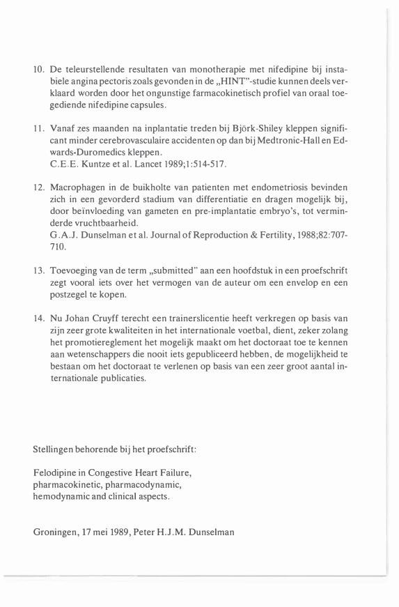
10. De teleurstellende resultaten van monotherapie met nifedipine bij instabiele angina pectoris zoals gevonden in de ,,HINT"-studie kunnen deels verklaard warden door het ongunstige farmacokinetisch profiel van oraal toegediende nifedipine capsules.
11 . Vanaf zes maanden na inplantatie treden bij Bjork-Shiley kleppen significant minder cerebrovasculaire accidenten op dan bij Medtronic-Hall en Edwards-Duromedics kleppen . C .E .E. Kuntze et al. Lancet 1 989;1 :51 4-51 7.
1 2. Macrophagen in de buikholte van patienten met endometriosis bevinden zich in een gevorderd stadium van differentiatie en dragen mogelijk bij , door bei"nvloeding van gameten en pre-implantatie embryo's , tot verminderde vruchtbaarheid. G. A .J. Dunselman et al. Journal of Reproduction & Fertility , 1988; 82: 707-710.
13 . Toevoeging van de term ,,submitted" aan een hoofdstuk in een proefschrift zegt vooral iets over het vermogen van de auteur om een envelop en een postzegel te kopen.
1 4. Nu Johan Cruyff terecht een trainerslicentie heeft verkregen op basis van zijn zeer grate kwaliteiten in het internationale voetbal, dient, zeker zolang het promotiereglement het mogelijk maakt om het doctoraat toe te kennen aan wetenschappers die nooit iets gepubliceerd hebben, de mogelijkheid te bestaan om het doctoraat te verlenen op basis van een zeer groat aantal internationale publicaties.
Stellingen behorende bij het proefschrift:
Felodipine in Congestive Heart Failure, pharmacokinetic, pharmacodynamic, hemodynamic and clinical aspects.
Groningen, 17 mei 1989, Peter H.J.M. Dunselman

Rijksuniven,itcit Groningen
Felodipine in Congestive Heart Failure Pharmacokinetic, Pharmacodynamic,
Hemodynamic and Clinical Aspects
Proefschrift
tcr verkrijging van het doctoraat in de
Gcnccskundc aan de Rijksunivcrsitcit
Groningcn op gezag van de Rector Magnificus
Dr. L.J. Engels in het openbaar le verdedigen
op woensdag 17 mci 1989 des namiddags le
2.45 uur precies
door
Peter Henricus Johannes Marie Dunselman
geborcn tc Helmond
1989 Drukkcrij van Denderen B.V.
Groningen

Promotoren : Prof. Dr. K.I . Lie Prof. Dr. H. Wesseling
Referenten: Dr. A.H.J . Scaf Dr. J .P.M. Hamer
Promotiecommissie: Prof. P.G. Hugenholtz Prof. Dr. D .K.F. Meijer Prof. Dr. W.D. Reitsma


If people bring so much courage to this world, the world has to kill them to break them, so of course it kills them. The world breaks everyone and afterwards many are strong at the broken places. But those that will not break it kills. It kills the very good, and the very gentle, and the very brave impartially. If you are none of these you can be sure it will kill you too, but there will be no special hurry.
Ernest Hemingway

I am indebted to the 50 patients with congestive heart failure, whose physical characteristics are described in this book.
To Anniek, and our children, Joline, Bob and Astrid

Author's Note
As a young student I had the opportunity to work in the heart failure team of the departments of thoracic surgery and cardiology, University of Groningen. My mentors, Dr.J.J . Bredee, Dr.J.W. Viersma and Drs .G.J . Kootstra were devoted to the treatment of cardiogenic shock after myocardial infarction, and the "stone-heart syndrome", after cardiac surgery. We build our own cardiac output and endocardial viability computers with the experience and skill of J. van der Zee (Department of Medical Physics) and A. Bergstra (Department of Physiology) . In these years my interest in the pathophysiology of the circulation grew deeper, resulting in the wish to become a cardiologist.
I was trained in Internal Medicine by Prof.Dr. A.E.C. Saleh , St.Elisabeth Hospital Curacao, Dutch Antilles. I had to return to the University of Groningen, to become a cardiologist, and was welcomed back into the Department by Dr.J .C.A. Hoorntje and Dr.J .H. Kingma. Even after six years, I know that I can not be objective about these men. It has been a rewarding experience to work with them, and I suppose that the department had not seen their likes before.
Dr.Kingma introduced me in clinical pharmacology, Drs .F.D.M. Haagen and Dr.Hoorntje in invasive cardiology. Dr.J .P .M. Hamer was a fascinating teacher in all aspects of non-invasive cardiology. Prof.Dr.E . van der Wall should be mentioned especially, for his compassion for the cardiac patient and his superb qualities in bedside teaching made him a legendary figure . In 1983 , Prof.Dr. K.I . Lie became Head of the Department of Cardiology. He gave me every possibility to initiate research programs in congestive heart failure. He advised me to analyze the new dihydropyridine drug felodipine in heart failure patients, stimulated me to write this thesis, and supported my ambition to set up a cardiopulmonary exercise test laboratory. I am grateful that our personal and scientific contacts have persisted after my transfer to Breda.
During the years, the cooperation between the Departments of Cardiology and Clinical Pharmacology has resulted in a rather unique relationship. This thesis demonstrates the close contact between cardiologists and clinical pharmacologists at the University of Groningen . Every chapter has benefited from the scrutiny and critical comments of Prof.Dr.H . Wesseling. Dr. A.H.J . Scaf analyzed the pharmacokinetic data with his sophisticated computer programs and simulations.
Dr. B. Edgar, clinical pharmacologist from the Swedish Astra (Hassle) Company, who analyzed pharmacokinetics and pharmacodynamics of felodipine in other patient groups, enabled me to present the results in a broader context. Drs. C.E.E. Kuntze, a wizard in medical statistics and computer technology, analyzed the data . Drs. A. van Bruggen assisted at the catheterization procedures and cardiopulmonary exercise tests. Jacob Pleiter, the artist and photogra-

pher of the department of Clinical Pharmacology, made all figures, cheerfully accepting that the deadline for everything is yesterday.
My collegues from the Departments of Cardiology of the Ignatius Hospital and Thoracic Surgery of the Klokkenberg, Breda, supported my ambition to publish and present the studies described in this book.
The pleasant cooperation with Dr.E. Dawson from Astra Holland goes back to the MIAMI study and is likely to continue for years to come. The contributions from Mrs.Gunilla Flygt and Prof.Or.D . Elmfeldt from Astra Hassle Sweden are gratefully acknowledged.
I thank my parents for the example presented to their children, that everyone is gifted for something, and that this thing, at whatever cost, should be attained.

Parts of this study have been or will be published as:
Chapter 2 American Heart Journal 1 988; 11 6: 14 75- 14 82 (full article) . European Journal of Nuclear Medicine 1 989 (abstract) . Nederlands tijdschrift voor geneeskunde 1 988; 13 2:893 (short communication).
Chapter 3 British Journal of Clinical Pharmacology 1989 (full article) .
Chapter4 Journal of Cardiovascular Pharmacology 1 989 (full article) . Cardiovascular Drugs and Therapy 1987;1 :258 (abstract) .
Chapter 5 The Journal of Clinical Pharmacology 1989 (full article) . Cardiovascular Drugs and Therapy 1987;1 :286 (abstract) .
Chapter 6 European Journal of Clinical Pharmacology 1988; 35 :4 61-4 65 ( fu II article) . Cardiovascular Drugs and Therapy 1987; 1 :231 (abstract) . Pharmaceutisch Weekblad Scientific Edition 1988 (short communication) Nederlands tijdschrift voor geneeskunde 1988; 132: 520 (short communication).
Chapter? European Heart Journal 1 989 (full article). Circulation 1987; 7611 :710 (abstract AHA) . Drugs 1987; 34 (suppl .3) : 79-80 (short communication) .
Chapter 8 European Heart Journal 1989 (abstract) .

CONTENTS
Chapter 1 Introduction 1 . 1 Aetiology of cardiac failure 1 .2 Definition of congestive heart failure 1 . 3 Rationale for vasodilator therapy 1 .4 Efficacy of vasodilator therapy in
congestive heart failure 1 .4 . 1 . Nitrates 1.4 .2 Prazosin 1 .4 .3 . Hydralazine 1 .4 .4 . ACE inhibitors 1 .4 .5 . Dihydropyridines
2 The Anatomy of a Heart Failure Study
2 .1 Methodological Criteria 2.2 Cross-over versus Parallel Design 2.3 Concomitant Medication 2.4 Assessment of the Severity of Heart Failure 2.4 . 1 New York Heart Association Classification 2.4 .2 Physical examination 2.4 .3 �eft ventricular ejection fraction 2 .4 .4 Chest X-ray 2.4 .5 Echocardiography and Systolic Time Intervals 2.4 .6 Cardiac catheterization 2.4.7 The Cardio Pulmonary Exercise Test
2 .5 Duration of a Congestive Heart Failure study
2.6 Clinical Pharmacology
3 Felodipine, a new dihydropyridine
3 . 1 Cardiovascular actions of felodipine 3.2 Felodipine in congestive heart failure 3 .3 Efficacy of felodipine in other circulatory diseases 3 .3 . 1 Essential hypertension 3 .3 .2 Coronary artery disease
4 Aims of the study
1
7
17
20

Chapter 2 Value of New York Heart Association 27 Classification , Radionuclide Ventriculography and Cardiopulmonary Exercise Tests for Selection of Patients for Congestive Heart Failure Studies
Chapter 3 Pharmacokinetics of Felodipine after 41 Intravenous and Chronic Oral Administration in Patients with Congestive Heart Failure
Chapter4 The Plasma Concentration-Effect Relationship of 55 Felodipine Intravenously in Patients with Congestive Heart Failure
Chapters Oral Pharmacokinetics of Felodipine in Patients 67 with Heart Failure: Variable Prediction Using Intravenous Data
Chapter 6 Digoxin - Felodipine Interaction in Patients 79 with Congestive Heart Failure
Chapter? Efficacy of Felodipine in Patients with 89 Congestive Heart Failure
Chapters Different results after long-term treatment 109 with Felodipine and Enalapril in Cardiopulmonary exercise tests in Patients with Congestive Heart Failure
Chapter 9 Epilogue 121
Summary 123
Samenvatting 127
Appendix

CHAPTER!
Introduction
1 . 1 Aetiology of cardiac failure
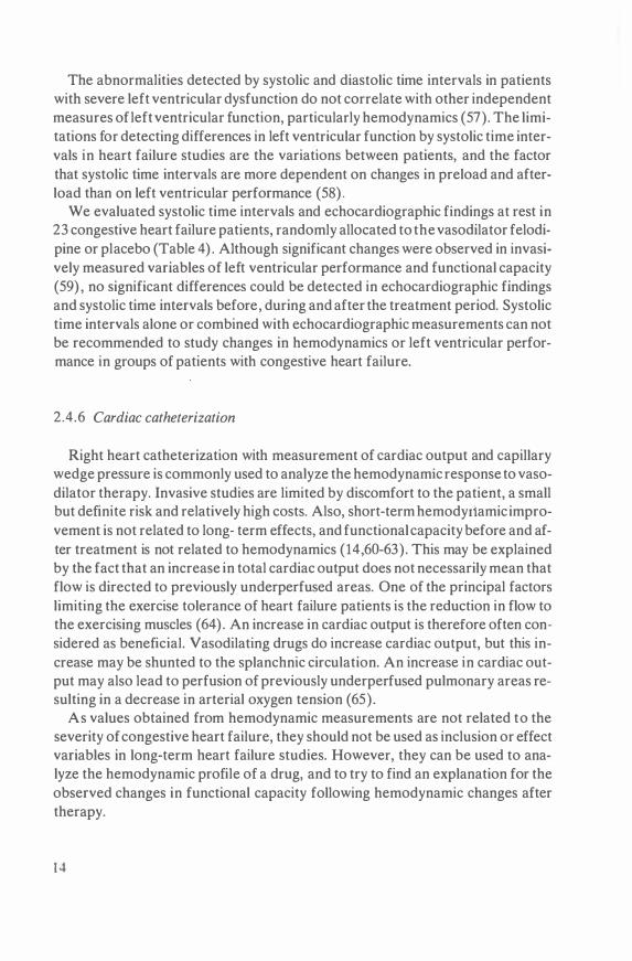
Cardiac failure can be defined as a condition that results from the inability of the heart to pump blood at a rate commensurate with the requirements of the metabolizing tissues. The aetiology of cardiac failure is diverse {Table 1) . However, the most important cause is an abnormality of systolic cardiac function, leading to the chronic disease state congestive heart failure.
Table I Aetiology of Cardiac Failure
A Myocardial Failure
1) Coronary Artery Disease - acute myocardial infarction - chronic failure due to one or more infarctions - chronic failure due to myocardial ischemia
2) Hypertrophic Cardiomyopathy - hypertension - genetic
3) Restrictive cardiomyopathy - amyloidosis - sarcoidosis
4) Dilating cardiomyopathy - alcohol - thiamine deficiency - myocarditis - genetic?
5) Valvular Heart Disease
1 .2 Definition of Congestive Heart Failure
B Circulatory Failure
l) low output state - volume depletion
2) high output state - anemia - sepsis - thyreotoxicosis - Paget Disease
3) restricted cardiac filling - pericardia( disease
Congestive heart failure is a clinical syndrome characterized by a limitation of exercise tolerance typified by dyspnea and/or fatigue , attributed to an abnormality of cardiac function, secondary to alterations in cardiovascular transport and/ or in cardiac filling.

Most pharmacological studies in congestive heart failure investigated patients with documented systolic function abnormalities. An overview of the therapeutic possibilities of vasodilating drugs in these patients is presented.
1. 3 Rationale for vasodilator therapy
Although the syndrome of congestive heart failure is best described by a limitation of exercise tolerance, the use of vasodilators for the treatment of patients with congestive heart failure is founded on hemodynamic characteristics, high ventricular filling pressures, low cardiac output and increased systemic vascular resistance. Vasodilators increase stroke volume by reducing resistance to left ventricular ejection. This may lead to a decrease of end diastolic ventricular volume and pressure . Furthermore, increase in venous capacitance by venodilation will ameliorate symptoms of congestion. In patients with chronic heart failure who are optimally treated with diuretics, persistent symptoms are unlikely to improve with an increase in diuretic dose, because fluid compartments have already been normalized. Therapy should then be directed to an increase of skeletal muscle blood flow (1) .
Vasodilator therapy is usually started when symptoms of heart failure persist despite treatment with digoxin and diuretics. All the relevant studies with vasodilators have been performed in patients whose symptoms of heart failure could not adequately be managed with digoxin and diuretics. There has never been a controlled trial large enough to help us decide whether digoxin, diuretics or vasodilators should be used as the initial therapy in heart failure.
Digoxin therapy in heart failure is a time honoured approach, although under severe discussion. More than 200 years have passed since William Withering wrote his classic about the beneficial effects of the drug derived from the foxglove. Scientific discussions about this therapeutic approach started immediately and are a continuing story ever since (2-5) . The use of digoxin in heart failure is based on a combination of old clinical tradition and a large number of clinically
Table 2 Mechanisms of Action of Vasodilating Drugs
Alpha-adrenergic blockers, Direct Vasodilators
Angiotensin Converting Enzyme (ACE) Inhibitors
Calcium Channel blockers
2
Prazosin Hydralazine Minoxidil Nitrates Captopril, Enalapril
Dihydropyridines

irrelevant studies (6) . In a review of clinical studies with digoxin in patients with sinus rhythm it was concluded that only three studies addressed the efficacy of digoxin in a correct way; one found digoxin of benefit, the other two the converse (7) .
Vasodilators can be classified according to their mechanism of action (table 2) , or to their peripheral site of action (table 3) . Both classifications are in fact simplifications. They may be useful as a pharmacological description , but they do not provide therapeutic guidelines. The clinical response to a vasodilator agent can not be predicted by the assessment of circulating levels of catecholamines or plasma renin activity before treatment , nor by the analysis of hemodynamic subsets during cardiac catheterization. The impressive clinical presentation of the patient reflects the compensatory responses ( vasoconstriction, fluid retention) to the underlying myocardial dysfunction, but fails to give a direct insight in the pathophysiological background. Since the results of hemodynamic or neurohumoral profiling contribute little to the physicians decision to start with a certain vasodilating drug, the approach in pharmaceutical research has often been to try to invent an agent that may result in therapeutic success and cause few adverse reactions . This approach has lead to the development of a variety of vasodilating drugs - and to numerous clinical studies.
Table 3 Peripheral Site of Action of Vasodilating Drugs
Arteriolar
Venous
Balanced
Hydralazine, Minoxidil, Dihydropyridines
Nitrates
Captopril, Enalapril Prazosin
The prognosis of patients with congestive heart failure is gloomy despite "optimal" medical management. The probability of death within four years after the diagnosis is made was 52% for men and 34% for women in the patients analyzed in the Framingham study (8) . Patients with NYHA class III congestive heart failure have a yearly mortality of 40% (9) . The ultimate cause of death is almost equally divided between sudden death caused by a lethal arrhythmia and progressive cardiac failure. Prognosis is related to the severity of left ventricular dysfunction as determined by left ventricular stroke work index (10) and left ventricular ejection fraction (11) , to plasma norepinephrine levels (12) and serum sodium levels (13), and to exercise capacity (14,15).
3

One should realise that a beneficial change in one of these parameters does not automatically lead to an actual reduction in mortality . Ejection fraction , for instance, has to be considered as a risk marker, not a risk factor, as long as it has not been demonstrated that an increase in ejection fraction induced by therapy results in a reduction of mortality.
A moderate reduction in risk markers has been demonstrated in studies with converting-enzyme inhibitors. Two studies, in which vasodilators were used in comparison with placebo, have reported a reduction in mortality in the treated patient groups (16,17). The influence of converting- enzyme inhibitors on the neurohumoral systems, peripheral vasculature, serum sodium concentration and exercise capacity may partly explain the reduction in mortality that was demonstrated in the CONSENSUS-study (17).
The converting enzyme inhibitor enalapril and the combination of the arterial vasodilating agent hydralazine with the long acting nitrate isosorbide-dinitrate, have proven their efficacy in the reduction of mortality but they have side effects. Even when precautions are taken to minimize the occurrence of these side effects, they are poorly tolerated by about one fourth to one third of congestive heart failure patients (18) .
The question a physician should ask now is no longer whether vasodilators are indicated to relieve the symptoms, but whether the side effects of a specific vasodilating agent may result in a contraindication to its use in a certain patient. The considerations mentioned above emphasize the need to continue with controlled trials with new generations of vasodilating drugs .
1 .4 Efficacy of vasodilator therapy in congestive heart failure
1.4. l. Nitrates
The major action of nitrates in congestive heart failure is the reduction of ventricular filling pressure or preload, by venodilation. In 1974, Cohn et al reported on the beneficial effects of nitrate therapy in a patient with coronary artery disease and cardiogenic shock (19). Double- blind, randomized clinical trials described long term hemodynamic and clinical improvement in patients with severe heart failure (20,21 ) . Nitrates in combination with arterial vasodilating agents (hydralazine) is commonly used in the United States, as its efficacy in relief of symptoms and reduction of mortality has been demonstrated (16) . Nitrates might have particular appeal in patients with underlying coronary artery disease. Transdermal applications should be interrupted for at least 8 hours to avoid development of nitrate tolerance. However, the adverse reactions (headaches and flushes) , together with the demonstrated tolerance during prolonged therapy
4

and the methodological flaws in published studies have resulted in discussions about the long term efficacy of nitrates (7) .
1 .4 .2 Prazosin
Prazosin is an alpha-adrenergic blocker with both arteriolar and venous vasodilatory properties. In contrast to an early report of benefit (22) , what might have been the result of an increase in concomitantly given diuretic therapy, other double blind clinical trials have not shown prazosin to be superior to placebo. Tolerance develops rapidly and becomes irreversible in up to 50% of patients (23) . Inhibition of the alpha adrenergic system by prazosin leads to a reduction in systemic vascular resistance. However, plasma renin activity, aldosterone and noradrenaline concentrations increase with development of fluid retention. This explains the lack of clinical benefit after long term treatment (24 ,25) .
Prazosin might be effective i n hypertension but has to be considered obsolete now for the treatment of congestive heart failure.
1 .4 .3 Hydralazine
Hydralazine is a direct arteriolar vasodilator. Long -term efficacy at high doses , in combination with oral nitrates, has been demonstrated (16) . However, adverse effects, such as gastrointestinal distress (nausea, vomiting) and the development of systemic lupus erythematosus usually limit long term treatment with high doses. The drug is not effective at well tolerated low doses (26) . It leads to tolerance after long-term therapy at higher dosage (27) .
1 .4 .4 A CE inhibitors
The Angiotensin Converting Enzyme (ACE) inhibitors are now generally considered to be the most effective drugs for the treatment of congestive heart failure. The mechanisms by which converting enzyme inhibitors maintain improvement during long-term therapy include persistent beneficial hemodynamic effects on afterload and preload, persistent suppression of plasma aldosterone, interference with the effects of angiotensin-11 on the kidney and a direct effect of the accumulated angiotensin-1 on the heart. Their efficacy has been demonstrated in many reports showing an improvement in hemodynamic indexes as well as clinical symptoms, exercise duration, quality of life, and life expectancy in severe heart failure (17,28-35) .
5

While there is no doubt about the efficacy of ACE inhibition in congestive heart failure, there is serious concern with respect to unwanted side effects. The magnitude and duration of hypotension , induced by ACE-inhibitors, may be detrimental to the cerebral and renal circulation (36) . The beneficial increase in urinary excretion of sodium by suppression of the production of angiotensin-11 disappears if renal perfusion pressure falls (37) . Thus, the benefits of ACE-inhibition in congestive heart failure are not achieved without risk : nearly 50% of the patients develop some untoward effect of therapy (38) . In addition to hypotension, renal insufficiency, hyperkalemia, drug rashes, neutropenia and loss of taste may be caused by ACE-therapy. There is, of course , always a price to pay : if a drug is really effective in interfering with the complex homeostatic functions of the renin-angiotensin system, it will also lead to interference with the well adapted role of the renin-angiotensin system in maintaining circulatory homeostasis in sodium depleted states (39,4 0) .
1 .4 .5 Dihydropyridines
The calcium antagonists of the dihydropyridine group, with the archetype nifedipine as the most studied drug, exert a significant arterial vasodilating effect. The setback of nifedipine is its direct negative inotropic effect on cardiac muscle. This may be partly offset by the sympathetic stimulation that accompanies arterial vasodilation, but it may also become clinically important in patient with severely impaired left ventricular dysfunction (4 1 ,4 2) .
The place of the calcium channel blockers i n the treatment of heart failure is not yet clear, but their capacity to both dilate coronary arteries and improve myocardial relaxation appears to make them particularly attractive in the treatment of patients with heart failure when myocardial ischemia or impaired relaxation (or both) have a contributory role ( 43) .
Since all calcium antagonists available now (verapamil , diltiazem and nifedipine) have minor to mild negative inotropic effects, it seems unlikely that these drugs can seriously challenge vasodilators with no direct negative inotropic effects.
New dihydropyridines, with less negative inotropic effects due to a highly selective action on smooth muscle have been developed recently. These drugs are studied in patients with heart failure, and the characteristics of one of these drugs , felodipine, will be discussed in depth in the following chapters .
6

2 The Anatomy of a Heart Failure Study
2 . 1 Methodological Criteria
The quality of a clinical study in congestive heart failure depends on the methodology of the study. There are four basic criteria to be considered (7):
a. The existence of treatment and control groups.
A patient with congestive heart failure has good days and bad days. The influence of, for instance, the weather contributes to his state of wellbeing. If an uncontrolled study with a vasodilating drug is started in January, and the clinical outcome is evaluated in June, positive results may be caused by other factors then the treatment itself. The placebo effect, the tender care by the doctor, the mutual desire to ameliorate the patients condition, the hemodynamic alterations induced by cardiac catheterization itself, the training effect of repeated exercise tests, all ask for a control group, treated with placebo, or with a drug whose therapeutic effects are known.
b. The existence of a random allocation to treatment groups.
Many characteristics of a patient are not included in the base line characteristics, so an investigator who can influence the allocation may introduce a bias into the study, which may not be reflected by the statistical analysis of differences at baseline. Skewed distributions of interfering variables should be avoided by a strict adherence to random allocation of patients to treatment groups.
c. The existence of blindness towards treatment allocation, towards the data of
clinical follow up, and clinical outcome.
The mea�urement and registration of data during the study must be performed without knowledge of treatment schedules. The randomization code may be broken only after the last patient has completed the study, and only after all data of all patients have been recorded. This methodological criterion is difficult to oblige to: a clinical study in patients with congestive heart failure takes some time, and the inclusion of all patients into the study is not done at the same time. As a consequence, one patient may have completed the study period, while others are still in the study. If the code is broken for an individual patient, to be able to prescribe optimal open treatment after the double blind period, a bias will be introduced, because the investigator will be influenced by his knowledge about the study data of that particular patient. To circumvent this problem, amends have to be made before the study starts.
7

d. The existence of a definition of clinically important outcomes before the start
of the study.
The most important clinical outcome in heart failure studies is increase in life expectancy. But this is not always a feasible outcome , despite the high mortality of the studied population. We must therefore also look at other measures of outcome. Another goal is the improvement in quality of life, of functional status, e.g. , the reduction of symptoms of heart failure. The assessment of severity of congestive heart failure and the value of different clinical parameters are discussed in section 1 .2 .4 and Chapter 2 of this book.
2.2 Cross-over versus Parallel Design
A cross-over clinical trial is a trial in which the effects of different treatments are compared on the same subject during different treatment periods. After the first treatment is withdrawn, the symptom treated has to return during a washout or placebo phase, after which the second treatment period starts.
A parallel study compares two groups of patients, whose baseline characteristics are the same, randomly allocated to one of the treatment groups.
In theory, there are a few advantages of a cross-over design in clinical trials ( 44 ) . A comparison of treatments on the same subject is expected to be of more value than a comparison between subjects. From a statistical point of view, less patients are needed for a cross-over trial. There are some disadvantages too. One has to disentangle treatment effects from both time and carry over effects from the previous treatment period. This is a major disadvantage of cross-over studies in heart failure. Congestive heart failure is a progressive disease, so patients may enter the second period in a worse condition . It is difficult to decide on the duration of the wash out period between the two treatment periods, because more often than not, the effect of treatment of the first period can not be related to pharmacokinetic or pharmacodynamic parameters. The response to a treatment during the second period should nevertheless not be influenced by the treatment given during the first period. Even when the wash-out period seems to be completely effective, the physiological or psychological state induced by the first treatment may outlast the presence of the drug in the body, making the patients no longer comparable in their clinical state at the start of the second treatment period.
Therefore , to choose a cross-over is to take a chance. If the results of the crossover trial demonstrate a definite interaction between treatments and periods, then the chance has not come off, and one is obliged to discuss the results, and draw the conclusions on the first period alone. Then, of course, the cross-over

study has to be analyzed as a parallel group study, but all too often the size of the study population, chosen with a cross-over study in mind, is too small to make that possible (44).
Therefore , in long term studies with patients with congestive heart failure, the wisest approach is a parallel design study, with use of independent groups t-test analyzing the difference scores between baseline and after treatment in and between actively treated and placebo groups.
2.3 Concomitant Medication
Another problem is the use of other medication than the active/placebo medication . The most rigid answer to this problem is to demand a standard medication during the whole study period, provided that the run-in period has demonstrated that a stable condition during "optimal" (digoxin and diuretic) therapy has been reached. A clinical necessary deviation from this approach during the study may then be used as a separate variable to demonstrate the efficacy of one of the therapy schedules. Increases or decreases in concomitant therapy may influence the efficacy of the tested drug. For instance, congestive heart failure patients treated with vasodilating agents like minoxidil , hydralazine or prazosin (all drugs known for their ability to increase fluid retention) , may benefit from a concomitant increase of diuretic treatment. If an increase in diuretic treatment is only observed in the patient group treated with placebo, the analytical problems are markedly reduced, although observed differences in hemodynamic data (ventricular filling pressures , cardiac output, arterial pressures and derived variables) may still be influenced by changes in concomitant therapy.
There is no easy answer to this problem, so one should give special attention to every study in which differences in concomitant therapy are mentioned between patient groups, whether this reaches statistical significance or not.
2.4 Assessment of the Severity of Heart Failure
The primary goals of a therapeutic agent for the treatment of congestive heart failure are prolongation of life, and reduction in symptoms of dyspnea and fatigue at rest or during exercise. While it is easy to count the number of patients who are alive after a treatment period, it is difficult to assess and to quantify the severity of heart failure.
9

2.4.1 New York Heart Association Classification
The patients functional state is commonly classified in the 4 categories of the New York Heart Association (NYHA) Classification (4 5) . This classification is widely used and rather simple. However, it is not sensitive enough to detect small changes in functional capacity. It is also subject to interobserver variability, being a subjective interpretation of the narrated symptoms ( 46) . It has nevertheless been demonstrated that the NYHA classification correlates rather well with exercise capacity, though not well enough to be used as the only entry criterion in heart failure studies ( 47) .
2.4.2 Physical examination
The best way to assess changes in fluid retention is the straight-forward measurement of body weight provided that diurectic therapy is unaltered (48) . Fluid retention can also be assessed from jugular venous distension , the degree of hepatomegaly, of peripheral edema, and the presence of pulmonary rales. Signs and symptoms of heart failure, as a weak pulse, a third or fourth heart sound, or mitral insufficiency are not closely related to the severity of heart failure. They are also subject to interobserver variability, and thereby not very useful in the assessment of efficacy of therapy.
2.4.3 Left ventricular ejection fraction
The ejection fraction of the left ventricle is used as an indirect determinant of left ventricular performance and thus of myocardial failure. The assessment can be performed by noninvasive techniques including echocardiography and radionuclide ventriculography.
The ejection fraction can not be used to assess the severity of congestive heart failure ( 47 ,49) . Ejection fraction of the left ventricle quantifies myocardial failure, while chronic congestive heart failure should be quantified by measurements reflecting the primary disorder, the inability of the heart to pump blood at a rate commensurate to the metabolic needs of the oxygen consuming tissues . The lack of correlation between ejection fraction of the left ventricle and exercise duration (fig 1) and maximal oxygen uptake (fig 2) in patients with congestive heart failure NYHA class II and III demonstrates that left ventricular performance has little to do with exercise capacity ( 47) . Marked improvement in functional capacity may be associated with a lack of improvement or even deterioration in the ejection fraction (50). This reflects the complex interplay of central and peripheral mechanisms of heart failure.
10

n = 50 r = 0,007
1200 p = 0,96 0 0 0 • 0 0
1000 0 00 0 0 0 0 0
� BOO 0 • •o z • • • 0 • ;::: • <( 600 • • 0 • • a: • • • ::::> • • D 0 w • I • Vl 400 0 •o• u a: • w • 0 x w • 200 • •
0 5 10 15 20 25 30 35 40
Figure 1 % LEFT VENTRICULAR EJECTION FRACTION
Scatterdiagram of left ventricular ejection fraction versus exercise duration in SO patients with congestive heart failure NYHA II (squares) and I I I (dots), demonstrating the lack of relationship between left ventricular performance and functional capacity.
25
20
c e ci. 15 -"' ' 'E )( c E ..... §; 10
5
Figure 2
n :50 r = 0,018 p = 0,89
•
5
0
•
10
0
• • •
•
•
0 0
0
• • •
15 20
0 0 0
0
0
•
• •
•
0
•
25
0
0 0
•
30
0
•
0
0 0
0
0 0 • • • •
• • •
• • 0
35 % LEFT VENTRICULAR EJECTION FRACTION
40
Scatterdiagram of left ventricular ejection fraction versus maximal oxygen uptake (V02max) in SO patients with congestive heart failure NYHA II (squares) and III (dots), demonstrating the lack of relationship between left ventricular performance and aerobic capacity.
11

Nevertheless, the left ventricular ejection fraction is an important parameter in congestive heart failure studies : it is a major predictor of mortality (11) , and it differentiates between circulatory and cardiac failure in patients with signs and symptoms of heart failure (47) .
2.4.4 Chest X-ray
A chest x-ray is part of the routine investigation of a patient with heart failure. It is undoubtedly useful for diagnostic purposes in the individual patient.
In three heart failure studies, however, either a poor or no correlation between cardiothoracic ratio, pulmonary congestion and functional capacity has been demonstrated (47,51,52) . From these studies it can be concluded that one can abstain from chest x-rays at the inclusion, the follow up and analysis of clinical outcome in heart failure studies.
2.4 .5 Echocardiography and Systolic Time Intervals
A noninvasive technique that quantifies the degree of left ventricular impairment would, irrespective of the lack of correlation between left ventricular function and functional capacity, improve the evaluation of patients with congestive heart failure.
Echocardiography provides a non-invasive and harmless method for the evaluation of left ventricular function, using measurements of the internal diameter of the left ventricle during systole and diastole, of wall thickness and wall motion. Ejection fraction, mean velocity of circumferential fiber shortening and fractional shortening can be calculated from echocardiographic recordings. Echocardiographic measurements of cardiac output correlate poorly with invasively measured cardiac output (53) . The quantitative assessment of left ventricular dysfunction by echocardiography may be ameliorated by measurements of flow velocity and cardiac output with Doppler ultrasound techniques (54) . The measurement of ejection fraction is often performed with M-mode echocardiography. A number of drawbacks renders this technique unsatisfactory in many situations. M-mode echocardiograms, being one-dimensional, lack spatial information crucial in discriminating between global and regional changes. As a result not the whole left ventricle , but only a small region based on two specific measurement points of the interventricular septum and posterior wall is analyzed (55). When the ejection fraction is calculated using both long and short axis views, the results are more reliable (56).
12

Echocardiographic analysis is useful in individual patients with symptoms of heart failure, to analyze whether complaints of dyspnea and fatigue are based on left ventricular dysfunction . However, a left ventricle with a poor contraction pattern does not necessarily lead to signs and symptoms of clinical heart failure. Also in the presence of normal left ventricular diameters symptoms of heart failure are possible.
Because of the difference between myocardial function and clinical condition, echocardiographic criteria for the clinical diagnosis congestive heart failure are difficult to establish.
Table 4 Echocardiographic Measurements & Systolic Time Intervals before and after felodipine therapy in congestive heart failure
WeekO Week4 Week8
Echocardiography LVEDD (mm)
p 61 ± 9 61 ± 1 1 61 ± 9 F 64 ± 8 65 ± 1 1 62 ± 1 1
LVESD(mm) p 51 ± 1 1 5 1 ± 12 51 ± 1 1 F 55 ± 10 55 ± 10 5 1 ± 1 1
F.S. (%) p 17 ± 8 17 ± 8 17 ± 8 F 16 ± 8 15 ± 4 17 ± 6
Systolic Time Intervals
QIIAc (msec) p 535 ±26 532 ± 32 527 ± 30 F 528± 32 527 ± 34 541 ± 33
PEPc(msec) p 163 ± 31 159 ± 33 160 ± 29 F 152 ± 25 146 ± 24 149 ± 22
ETI (msec) p 369 ± 21 375 ± 23 366 ± 24 F 376± 22 381 ± 25 389 ± 17
PEP/ET p .56 ± .2 .54 ± . 2 .52 ± . 1 F .52 ± . 1 .48 ± . 1 . 48 ± . 1
P = Placebo, F = Felodipine. L VEDD = Left Ventricular End Diastolic Diameter; LVESD = Left Ventricular End Systolic Diameter; F.S. = Fractional Shortening of the left ventricle; QIIAc = time between onset of ORS-complex and aortic closure sound, corrected for heart rate; ETI: Ejection Time Index; PEP/ET: Pre Ejection Period divided by Ejection Time. Alie values are expressed as mean ± SD. No significant differences before, and after 4 and 8 weeks therapy, between group comparison of changes.
13
The abnormalities detected by systolic and diastolic time intervals in patients with severe left ventricular dysfunction do not correlate with other independent measures of left ventricular function, particularly hemodynamics (57) . The limitations for detecting differences in left ventricular function by systolic time intervals in heart failure studies are the variations between patients, and the factor that systolic time intervals are more dependent on changes in preload and afterload than on left ventricular performance (58) .
We evaluated systolic time intervals and echocardiographic findings at rest in 23 congestive heart failure patients, randomly allocated to the vasodilator felodipine or placebo (Table 4) . Although significant changes were observed in invasively measured variables of left ventricular performance and functional capacity (59) , no significant differences could be detected in echocardiographic findings and systolic time intervals before, during and after the treatment period. Systolic time intervals alone or combined with echocardiographic measurements can not be recommended to study changes in hemodynamics or left ventricular performance in groups of patients with congestive heart failure.
2.4.6 Cardiac catheterization
Right heart catheterization with measurement of cardiac output and capillary wedge pressure is commonly used to analyze the hemodynamic response to vasodilator therapy. Invasive studies are limited by discomfort to the patient, a small but definite risk and relatively high costs. Also, short-term hemodyrlamic improvement is not related to long- term effects, and functional capacity before and after treatment is not related to hemodynamics (14,60-63) . This may be explained by the fact that an increase in total cardiac output does not necessarily mean that flow is directed to previously underperfused areas. One of the principal factors limiting the exercise tolerance of heart failure patients is the reduction in flow to the exercising muscles (64) . An increase in cardiac output is therefore often considered as beneficial. Vasodilating drugs do increase cardiac output, but this increase may be shunted to the splanchnic circulation. An increase in cardiac output may also lead to perfusion of previously underperfused pulmonary areas resulting in a decrease in arterial oxygen tension (65) .
As values obtained from hemodynamic measurements are not related to the severity of congestive heart failure, they should not be used as inclusion or effect variables in long-term heart failure studies. However, they can be used to analyze the hemodynamic profile of a drug, and to try to find an explanation for the observed changes in functional capacity following hemodynamic changes after therapy.
1 4

2.4. 7 The Cardio Pulmonary Exercise Test
One of the cardinal manifestations of congestive heart failure is exercise intolerance. The exercise capacity is limited by fatigue and/or dyspnea, due to a reduced oxygen delivery to working muscles, and this limitation resides at least in part in the periphery (66) . Oxygen supply is dependent on cardiac output, pulmonary function , and hemoglobin content, whereas oxygen extraction relies primarily on the metabolic capacity of skeletal muscle, its ability to vasodilate and extract oxygen (50) . Objective assessment of the severity of congestive heart failure is afforded, and possible by determination of maximal oxygen uptake during exercise (67) . According to the Fick principle, oxygen uptake is the product of cardiac output and arteriovenous oxygen content difference. Oxygen uptake is measured during gradually increasing treadmill exercise. The patient breathes through a mouthpiece containing a low- resistance, non-rebreathing valve. Total minute ventilation, fraction of oxygen in the expired gas, and fraction of carbon dioxide in the expired gas are directly measured. From these data oxygen uptake, carbon dioxide production and other variables are derived. Maximal oxygen uptake (V021n .. ): this is defined as an unchanging oxygen uptake despite maintenance of a higher work load. Respiratory quotient: the amount of carbon dioxide produced divided by oxygen consumed. Anaerobic treshold: marks the sharp increase in carbon dioxide production, total minute volume and lactate levels at approximately 70% of maximal oxygen uptake.
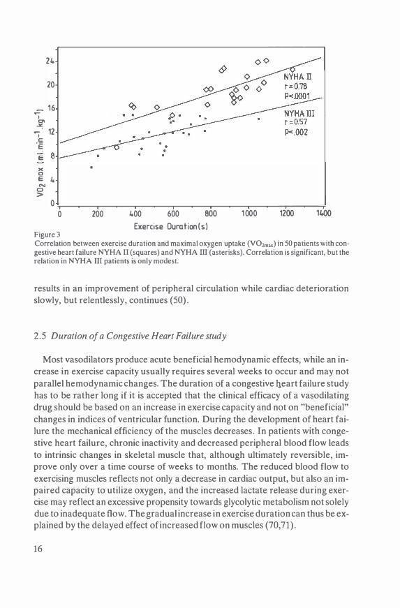
Exercise duration is greatly influenced by patient and physician motivation, and training effects. A significant correlation between exercise duration and V02max was demonstrated in NYHA II and III congestive heart failure patients, but their relation is modest (fig 3). If respiratory gas exchange is not measured a notable improvement in exercise duration may mimick an amelioration in the patients condition following the institution of placebo therapy (26,67) . Measurements of oxygen uptake and respiratory quotient at submaximal exercise levels
can also be used to analyze the result of vasodilator therapy. Normal daily activities of heart failure patients do not require exercise at maximal capacity, and severely ill patients are often unable to reach a true plateau in V02max (68) .
Determination of V02max represents the best currently available objective measurement of functional capacity in heart failure. The V02max measurement is also a sensitive index for assessing the efficacy of drug therapy (48,69) , and it may differentiate a cardiac from a pulmonary limitation to exercise. Whereas cardiac output limits maximal oxygen uptake in normal subjects, the V02max of patients with heart failure reflects both the status of the peripheral circulation and the cardiac output. Palliation of heart failure by vasodilator therapy often
1 5
24
20
- 16 ...... I °' �
12 ...... I c: ·e E B >< •
Cl E 4 N 0 >
0
0 200 400 600 BOO 1 000 1200 1400
Exercise Duration{s) Figure 3 Correlation between exercise duration and maximal oxygen uptake (V02max) in 50 patients with congestive heart failure NYHA II (squares) and NYHA III (asterisks). Correlation is significant, but the relation in NYHA III patients is only modest.
results in an improvement of peripheral circulation while cardiac deterioration slowly, but relentlessly, continues (50) .
2 .5 Duration of a Congestive Heart Failure study
Most vasodilators produce acute beneficial hemodynamic effects, while an increase in exercise capacity usually requires several weeks to occur and may not parallel hemodynamic changes. The duration of a congestive qeart failure study has to be rather long if it is accepted that the clinical efficacy of a vasodilating drug should be based on an increase in exercise capacity and not on "beneficial" changes in indices of ventricular function. During the development of heart failure the mechanical efficiency of the muscles decreases . In patients with congestive heart failure, chronic inactivity and decreased peripheral blood flow leads to intrinsic changes in skeletal muscle that, although ultimately reversible, improve only over a time course of weeks to months. The reduced blood flow to exercising muscles reflects not only a decrease in cardiac output, but also an impaired capacity to utilize oxygen, and the increased lactate release during exercise may reflect an excessive propensity towards glycolytic metabolism not solely due to inadequate flow. The gradual increase in exercise duration can thus be explained by the delayed effect of increased flow on muscles (70,71) .
16

A clinical study in patients with congestive heart failure should have an active treatment period of at least 6 weeks (29,72,73).
2 .6 Clinical Pharmacology
The elevated filling pressures leading to congestion, and the reduced cardiac output, both typical for congestive heart failure , may have consequences for the pharmacokinetic profile of a drug used in heart failure patients.
Pharmacokinetic analysis of a drug describes the effect that the body has on the drug. The pharmacokinetics are influenced by age, and by disease states. Hepatic, gastrointestinal and renal congestion, and hypoperfusion of these organ systems may alter pharmacokinetics. Digoxin, concomitant therapy in many patients with congestive heart failure, interacts with a number of drugs, including vasodilating drugs. Adverse drug reactions may be due to both pharmacokinetic changes and drug interactions .
Therefore, in the clinical analysis of a new vasodilating drug it is necessary to characterize its pharmacological actions (74) . A descriptive analysis of the pharmacokinetic model, absorption, distribution and elimination data, pharmacodynamic analysis with dose-response studies, time-effect relations, and interaction studies must be obtained.
Pharmacokinetic, pharmacodynamic and digoxin-interaction studies with the vasodilator agent felodipine in congestive heart failure are described in chapter 3,4,5 and 6 of this book.
3 Felodipine, a new dihydropyridine
3 . 1 Cardiovascular actions of felodipine
Felodipine , generic name of the dihydropyridine 4-(2,3 dichlorophenyl)-1 ,4-dihydro-2,6-dimethyl-3-ethoxycarbonyl-5- methoxycarbonylpyridine is developed by Astra Hassle Laboratories, Sweden. The aim was to find a drug that inhibits transmembrane calcium influx and selectively relaxes myogenically active vascular smooth muscle without causing a negative inotropic myocardial effect.
A significant smooth muscle selectivity was demonstrated in the portal vein/ papillary muscle model (75) . The predictive value of this model must be validated in vivo, although it is difficult to reliably measure direct effects of vasoactive agents on cardiac contractility in vivo. The drop in peripheral resistance invariably leads to reflex sympathetic stimulation of the heart, masking a possible negative myocardial inotropic effect . The absence of decreases in contractility and
17

myocardial wall thickness parameters after both intravenous and intracoronary administration of felodipine in pigs indicates that felodipine exhibits no negative inotropic action on the myocardium (76).
In patients with coronary artery disease negative inotropic effects could not be demonstrated (77, 78). After acute administration of felodipine in patients with left ventricular dysfunction maximum dP/dt remained unchanged and maximum dP/dt/P increased (79) . The alterations in the circulation induced by vasodilators in heart failure patients differ markedly over time. Acute administration leads to immediate central changes, while chronic administration leads to peripheral changes in the skeletal muscles (50) .
A possible negative inotropic effect after long term treatment in patients with left ventricular dysfunction has not been analyzed as yet. However, there is no evidence to refute the conclusion that felodipine indeed selectively inhibits vascular smooth muscle without causing negative inotropic effects in vivo (80) .
3.2 Felodipine in congestive heart failure
A number of open studies of the effects of felodipine in congestive heart failure have been reported. Acute administration of felodipine resulted in beneficial changes in invasively measured hemodynamic variables. Cardiac output, coronary sinus flow, and dP/dt/P increased, while capillary wedge pressure and systemic vascular resistance decreased (81-83) .
Chronic oral therapy during 4 weeks in 10 patients with congestive heart failure showed an increase in exercise duration and a decrease of capillary wedge pressure during exercise (84) .
Apart from the studies described in the following chapters, 2 other double blind placebo controlled studies were reported (85 ,86) . Both studies had a crossover design , active treatment periods 3 weeks. An increase in cardiac output at rest and during exercise, a decrease in vascular resistance and po change (86) or a decrease in heart rate (85) were observed. Left and right ventricular filling pressures were not significantly different at rest, but decreased during exercise. Measurements of oxygen uptake were not performed. Subjective symptom scores and exercise duration did not improve. The disparity between these results and those obtained in open studies underlines the importancy of controlled randomized trials. The disparity in changes in filling pressures (observed in open studies, not observed in controlled studies) can be explained by the observation that filling pressures also tend to decrease during placebo treatment (54,87) . The lack of symptomatic improvement in these studies can be explained by the study design, cross- over with only short periods (3 weeks) on active treatment, and a wash-out period of only 1 week (44,59) .
1 8

The results of a parallel design double blind placebo controlled study with a duration of 8 weeks are presented in chapter 7.
Controlled clinical studies with felodipine in comparison with other vasodilating drugs have not been published as yet. The different characteristics of felodipine and enalapril are described in chapter 8 of this book, using the best diagnostic procedure in heart failure research available, the cardio pulmonary exercise test.
3.3 Efficacy of felodipine in other circulatory diseases
3 .3 . 1 Essential hypertension
Several studies in essential hypertension have demonstrated the efficacy of felodipine. The influence of acute and chronic treatment with felodipine on arterial blood pressure revealed a persistent anti-hypertensive effect during 9 hours after a single oral dose. The beneficial effect was maintained during 6 weeks chronic oral treatment and followed by a return of blood pressure to control level after withdrawal of felodipine. The baroreflex mediated increase in heart rate was reset after one week treatment (88) . Comparable results were reported during an 8 week open study in patients with moderate hypertension. Again, there was no increase in heart rate despite enhanced activity of the sympathetic nervous system, as documented by an increased plasma renin activity (89) . An increase in both total body flow and renal plasma flow, while glomerular filtration rate remained unchanged, was documented in 12 patients with moderate hypertension (90) .
In a double blind study in 100 patients, felodipine, in combination with a beta blocker, appeared to be more effective than triple therapy consisting of a beta blocker, hydralazine and hydrochlorothiazide (91 ) . In a double blind, randomized parallel group study of 100 patients with moderately severe essential hypertension despite beta blockade, concomitant treatment with felodipine was more effective than concomitant treatment with prazosin (92) .
Another double blind, placebo controlled study in hypertensive patients revealed that long-term (8 weeks) administration of felodipine resulted in a reduction of blood pressure in spite of increased levels of noradrenaline, adrenaline and angiotensin II levels. This supports the concept that vasoconstriction is dependent on calcium influx, and that treatment with felodipine results in a reduction of pressoramine effects on the end organ (93) . Felodipine leads to an increase in natriuresis in hypertensive patients. This is probably caused by a direct effect on tubular reabsorption (94,95) .
1 9

All studies reported the occurrence of dose dependent side effects, such as precapillary ankle edema without weight increase, flushes and headaches.
3 .3 .2 Coronary artery disease
Hemodynamic effects of felodipine 10 mg orally were evaluated during cardiac catheterization in 22 patients with coronary artery disease (96) . Clinical and statistical significant changes in arterial blood pressure ( - 16%), cardiac index ( + 35% ) , stroke volume index ( + 12%) and heart rate ( + 16%) were recorded. The hemodynamic changes persisted when heart rate was kept constant by atrial pacing. In another invasive study with felodipine intravenously in 11 patients with coronary artery disease, similar hemodynamic effects were found (97) . Analysis of left ventricular performance showed no decrease in left ventricular circumferential fiber shortening, peak dP/dt or peak dP/dt/P at fixed heart rates in patients with coronary artery disease (77,78,98) . The hemodynamic effects during exercise were evaluated in an open study with 8 patients with symptomatic exertional angina. Felodipine prevented angina! complaints, increased exercise capacity with 20% , and lowered systemic vascular resistance and capillary wedge pressure during exercise. These beneficial effects were confirmed in a double blind placebo controlled study, where felodipine was added to beta blockers in angina! patients (99) .
4. Aims of the study
In summary, the aims of the study described in the following chapters were to investigate the pharmacokinetic, pharmacodynamic, hemodynamic and clinical aspects of felodipine administered intravenously and orally to patients with moderate to severe congestive heart failure:
- to analyze the value of various assessments of the severity of congestive heart failure, with special attention to the selection procedure of patients for heart failure studies (Chapter 2) .
- to analyze the pharmacokinetics of felodipine in congestive heart failure, the possible relationship between flow variables and pharmacokinetics, and to compare the pharmacokinetic data with those from different patient groups (Chapter 3).
- to analyze the plasma concentration-effect relationship of felodipine in patients with congestive heart failure, with special attention to changes in effect over time, and to gain a deeper insight in the overall relationship between plasma concentrations of vasodilating drugs and various hemodynamic effects (Chapter 4).
20
- to analyze the (im)possibility to predict oral steady state pharmacokinetics of felodipine after long term treatment from intravenous data before treatment (Chapter 5) .
- to analyze the possible interaction between felodipine and digoxin in patients with congestive heart failure (Chapter 6) .
- to analyze the efficacy of felodipine for the treatment of congestive heart failure in a randomized, placebo controlled, double blind study (Chapter 7) .
- to analyze possible differences in the results of cardio pulmonary exercise tests between felodipine and enalapril, two vasodilating drugs with a different hemodynamic profile (Chapter 8) .
References
1 . Poole-Wilson P A,Buller NP. Causes of symptoms in chronic congestive heart failure and implications for treatment.AJC 1988;62:31A-34A.
2. Johnson GD,McDevitt DG. ls maintenance digoxin necessary in patients with sinus rhythm? Lancet 1979;1 :567-570.
3. Aeg JL,Gottlieb SH,Lakatta GH. Is digoxin really important in treatment of compensated heart failure? Am J Med 1982;73:244-250.
4. Arnold SB,Byrd RC,Meister W. Long-term digitalis therapy improved left ventricular function in heart failure.N Engl J Med 1982;306:699-705.
5. Murray RG,Tweddel AC,Martin W,Pearson D,Hutton I ,Lawrie TDV. Evaluation of digitalis in cardiac failure. Br Med J 1982;284: 1526-1528.
6. Selzer A: Digitalis in cardiac failure.Arch Intern Med 1981 ;14 1 : 188-192. 7. Guyatt GH. The treatment of heart failure.Drugs 1986;32:538-568. 8. McKee PA,Castelli WP,McNamara PM,Kannel WB.The natural history of congestive heart
failure:the Framingham Study.N Engl J Med 1971 ;285 : 1441-1446. 9. Franciosa JA,Wilen M,Ziesche S,Cohn JN.Survival in men with severe chronic left ventricular
failure due to either coronary heart disease or idiopathic dilated cardiomyopathy.Am J Cardiol 1983;51 :831-836.
10. Creager MD,Faxon DP,Halperin JL.Determinants of clinical response and survival in patients with congestive heart failure treated with captopril .Am Heart J 1982; 104: 1 147-54.
1 1 . Califf RM,Bounous P ,Harell FE. The prognosis in the presence of coronary artery disease. ln:Braunwald E ed. Congestive heart failure:current research and clinical applications.New York:Grune and Stratton ,1982:31-40.
12. Cohn JN,Levine TB,Olivari MT.Plasma norepinephine as a guide to prognosis in patients with congestive heart failure.N Engl J Med 1984;31 1 : 819-823.
13. Lee WH,Packer M.Prognostic importance of serum sodium concentration and its modification by converting-enzyme inhibition in patients with severe chronic heart failure.Circulation 1986;73:257-267.
14. Szlachcic J ,Massie BM,Kramer BL,Topic N ,Tubau J.Correlates and prognostic implication of exercise capacity in chronic congestive heart failure.Am J Cardiol 1985;55: 1037-1042.
15 . Likoff MJ ,Chandler SL, Kay HR.Clinical determinants of mortality in chronic congestive heart failure secondary to idiopathic dilated or to ischemic cardiomyopathy .Am J Cardiol 1987;59:634-638.
16. Cohn JN,Archibald D,Ziesche S ,Franciosa JA,Harston WE,Tristani FE:Effect of vasodilator therapy on mortality in chronic congestive heart failure:results of a Veterans Administration Cooperative Study(V-HeFT).N Engl J Med 1986; 314: 1547.
21

17. The CONSENSUS Trial Study Group: Effects of enalapril on mortality in severe congestive heart failure.N Engl J Med 1987;316:1429-1435.
18. Packer M. Do vasodilators prolong life in heart failure? New Engl J Med 1987;316:1471-1473. 19. Cohn JN,Mathew KJ,Franciosa JA,Snow JA:Chronic vasodilator therapy in the management
of cardiogenic shock and intractable left ventricular failure.Ann Intern Med 1974;81 :777-780. 20. Leier CV,Huss P,Magorien RD,Unverferth DV: Improved exercise capacity and differing arte
rial and venous tolerance during chronic isosorbide dinitrate therapy for congestive heart failure. Circulation 1983;677:817-822.
21 . Franciosa JA,Nordstrom LA,Cohn JN: Nitrate therapy for congestive heart failure.JAMA 1978;240:443-446.
22. Colucci WS, Wynne J ,Holman BL,Braunwald E:Long-term therapy of heart failure with prazosin: a randomized double- blind trial.Am J Cardiol 1980;45:337-344.
23. Packer M: Vasodilator and inotropic therapy for severe chronic heart failure:passion and skepticism.JACC 1983;2:841- 852.
24. Bayliss J ,Norell MS,Canepa-Anson R,Reid C,Poole-Wilson P ,Sutton G: Clinical importance of the renin-angiotensin system in chronic heart failure: double blind comparison of captopril and prazosin.BMJ 1985;290:1861-1865.
25. Packer M, Medina N, Yushak M. Comparative hemodynamic and clinical effects of long term treatment with prazosin and captopril for severe chronic congestive heart failure secondary to coronary artery disease or idiopathic dilated cardiomyopathy. Am J Cardiol 1986;57: 1323-1327.
26. Franciosa JA,Weber KT,Levine TB:Hydralazine in the long- term treatment of chronic heart failure:lack of difference from placebo.Am Heart J 1982; 104:587.
27. Packer M ,Meller J ,Medina N ,Yushak M,Gorlin R:Hemodynamic characterization of tolerance to long-term hydralazine therapy in severe chronic heart failure .N Engl J Med 37 1982;306:57· 62.
28. Dzau VJ, Colucci WS, Williams G H ,Curfman G ,Meggs L: Sustained effectiveness of converting enzyme inhibition in patients with severe congestive heart failure.N Engl J Med 1980;302: 1373-1379.
29. Captopril Multicenter Research Group: A placebo- controlled trial of captopril in refractory chronic congestive heart failure.JACC 1983;Vol 2.no 4:755-763.
30. Chatterjee K, Parmley WW, Cohn JN, Levine TB : Captopril Multicenter Research group: a cooperative multicenter study of captopril in congestive heart failure: hemodynamic effects and long-term response.Am Heart J 1985; 1 10:439-447.
3 1 . Kramer BL,Massie BM ,Topic N :Controlled trial of captopril in chronic heart failure:a rest and exercise hemodynamic study.Circulation 1983;67:807-816.
32. Franciosa JA,Wilen MM,Jordan RA: Effects of enalapril,a new angiotensin-converting enzyme inhibitor in a controlled trial in heart failure. J Am Coll Cardiol 1985;5:101-107.
33. Cleland JGF, Dargie HJ, Hodsman GP, Ball SG, Robertson JIS, Morton JJ, East BW, Robertson I, Murray GD, Gillen G. Captopril in heart failure;a double blind controlled trial .Br Heart J 1984;52:530-535.
34. Sharpe DN,Murphy J,Coxon R,Hannan SF:Enalapril in patients with chronic heart failure:a placebo-controlled, randomized, double-blind study. Circulation 1984;70:271-278.
35. Creager MA,Massie BM,Faxon DP,Friedman SD,Kramer BL,Weiner DA,Ryan TJ,Topic N,Melidossian CD:Acute and long term effects of enalapril on the cardiovascular response to exercise and exercise tolerance in patients with congestive heart failure.JACC 1985;6: 163-170.
36. Cleland JGF, Dargie HJ, Gillen G, Robertson I, East BW, Ball SG, Morton JJ, Robertson JIS. Captopril in heart failure: a double blind study of the effects on renal function .J Cardiov Pharmacol 1986;8:700-706.
37. Packer M ,Lee WH,Yushak M and Medina N:Comparison of captopril and enalapril in patients with severe chronic heart failure.NEJM 1986;315:847-853.
38. Packer M,Kessler PD, Gottlieb SS.Adverse effects of converting-enzyme inhibition in patients with severe congestive heart failure:pathofysiology and management.Postgrad Med J 1986;62 suppl 1 : 179-182.
39. Packer M. ls the renin-angiotensin system really unnecessary in patients with severe chronic heart failure: the price we pay for interfering with evolution.JACC 1985 ;vol 6: 171-173.
22

40. Packer M ,Lee WH,Medina N, Yushak M, Kessler P. Functional renal insufficiency during longterm therapy with captopril and ealapril in severe chronic failure.Ann Int Med 1 987; 106:346-354.
41 . Elkayam U,Weber L,Torkan B,McKay CR,Rahimtoola SH. Comparison of hemodynamic responses to nifedipine and nitroprusside in severe chronic congestive heart failure.Am J Cardiol 1984;53: 1321-5.
42. Fifer MA,Colucci WS,Lorell BH,Jaski BE,Barry WH. lnotropic,vascular and neuroendocrine effects of nifedipine in heart failure:comparison with nitroprusside.JACC 1985;5:731-737.
43. Braunwald E:Mechanism of action of calcium-channel- blocking agents.N Engl J Med 1982;307 : 1618-1626.
44. Hills M, Armitage P.The two-period cross-over clinical trial .Br J Clin Pharmac 1979;8:7-20. 45. The Criteria Committee of the New York Heart Association: Diseases of the Heart and Blood
Vessels. In:Nomenclature and Criteria for Diagnosis 7th ed. Boston,Litt-le,Brown&Co, 1973;286.
46. Goldman L,Hasmoto B,Cook F,Lascalzo A.Comparative reproducibility and validity of systems for assessing cardiovascular functional class.Circulation 1981 ;64:1227-34.
47. Dunselman PHJM,Kuntze CEE,van Bruggen A,Beekhuis H,Piers B,Scaf AHJ,Wesseling H,Lie Kl .Value of New York Heart Association classification.radionuclide ventriculography ,and cardiopulmonary exercise tests for selection of patients for congestive heart failure studies.Am Heart J 1988;1 16: 1475- 1482.
48. Packer M. How should we judge the efficacy of drug therapy in patients with chronic congestive heart failure? The insights of six blind men.JACC 1987;9:433-8.
49. Franciosa JA,Park M ,Levine B. Lack of correlation between exercise capacity and indices of resting left ventricular performance in heart failure.Am J Cardiol 1981 ;47:33-39.
50. Mancini DM,Le Jemtel TH,Factor S,Sonnenblick EH. Central and peripheral components of cardiac failure.Am J Med 1986;80(suppl 2B)2-13.
51 . Engler R,Ray R,Higgins CB.Clinical assessment and follow up of functional capacity in patients with chronic congestive cardiomyopathy.Am J Cardiol 1982;49:1 832-37.
52. Francis GS,Goldsmith SR,Cohn JN. Relationship of exercise capacity to resting left ventricular performance and basal plasma norepinephrine levels in patients with congestive heart failure.Am Heart J 1982;104:725-732.
53. Kronik G ,Slany J ,Mosslacher H.Comparative value of eight M-mode echocardiographic formulas for determining left ventricular stroke volume. Circulation 1979;60: 1308-16.
54. Gardin JM,Iseri L T,Elkayam U ,To bis J ,Childs W ,Burn CS,Henry WL.Evaluation of dilated cardiomyopathy by pulsed Doppler echocardiography.Am Heart J 1983; 106:1057-65.
55. Weiss JL. Evaluation of ventricular size, shape and function by echocardiography.In:Braunwald E ed.Congestive heart failure:current research and clinical applications.New York:Grune and Stratton, 1982 : 197-219.
56. Teichholz LE,Kreulin T,Herman MV,Gorlm R.Problems in echocardiographic volume determinations:echocardiographic- angiographic correlations in the presence or absence of asynergy .Am J Cardiol 1976;37:7
57. Haq A,Rakowski H,Baigrie R,McLaughlin P,Burns R,Tihal H,Hilton D,Feiglin D.Vasodilator therapy in refractory congestive heart failure:a comparative analysis of hemodynamic and noninvasive studies .Am J Cardiol 1983;49:439-444.
58 Rahko PS ,Shaver JA,Salerni R, Uretsky B. Noninvasive evaluation of systolic and diastolic function in severe congestive heart failure secondary to coronary artery disease or idiopathic dilated cardiomyopathy.Am J Cardiol 1986;57: 1 315-22.
59. Dunselman PHJM,Kuntze CEE,van Bruggen A,Hamer JPM,Scaf AHJ ,Wesseling H,Lie Kl.Efficacy of felodipine in congestive heart failure. Eur Heart J 1989;in press.
60. Franciosa JA,Zeische S,Wilen M .Functional capacity of patients with chronic left ventricular failure.Am J Med 1979;6:460-466.
6 1 . Benge W,Litchfield RL,Marcus ML.Exercise capacity in patients with severe left ventricular dysfunction. Circulation 1980;61 :955-959.
23

62. Higginbotham MD.Morris KG,Conn EH,Coleman RE,Cobb FR.Determinants of variable exercise performance among patients with severe left ventricular dysfunction.Am J Cardiol 1983;51 :51-60.
63. Franciosa JA,Park M,Levine B .Lack of correlation between exercise capacity and indices ofresting left ventricular performance in heart failure.Am J Cardiol 1981 ;47:33-39.
64. Wilson JR,Ferraro N .Exercise intolerance in patients with chronic heart failure: relation to oxygen transport and ventilatory abnormalities.Am J Cardiol 1983;51 : 1358-63.
65. Vrobel TR, Cohn JN. Comparative hemodynamic effects of converting enzyme inhibitor and sodium nitroprusside in severe heart failure.Am J Cardiol 1980;45:331-36.
66. Wilson JR,Martin JL,Schwartz D,Ferraro N .Exercise intolerance in patients with chronic heart failure:role of impaired nutritive flow to skeletal muscle.Circulation 1984;69: 1079-1087.
67. Weber K,Janicki J .Cardiopulmonary exercise testing for evaluation of chronic cardiac failure.Am J Cardiol 1985;55:22A-31A.
68. Lipkin DP,Bayliss J ,Poole-Wilson PA.The ability of a submaximal exercise test to predict maximal exercise capacity in patients with heart failure.Eur Heart J 1985;6:829-833.
69. Weber KT,Kinasewitz GT,Janicki JS,Fishman AP.Oxygen utilization and ventilation during exercise in patients with chronic cardiac failure.Circulation 1982;65:1213-23.
70. Wilson JR,Ferraro N ,Wiener DH.Effect of the sympathetic nervous system on limb circulation and metabolism during exercise in patients with heart failure.Circulation 1985;72:72-81 .
71 . Massie BM: Exercise tolerance in congestive heart failure Role of cardiac function,peripheral blood flow, and muscle metabolism and effect of treatment. AJM 1988:84 suppl 3A;75- 82.
72. Rubin S,Chatterjee K ,Parmley WW. Metabolic assessment of exercise in chronic heart failure patients treated with short-term vasodilators.Circulation 1983;67:817-822.
73. Franciosa JA,Goldsmith SR,Cohn JN. Contrasting immediate and long-term effects of isosor-bide dinitrate on exlercise capacity in congestive heart failure. Am J Med 1980;69:559-566.
74. Shammas FV ,Dickstein K.Clinical pharmacokinetics in heart failure.Drugs 1988;15:94-113 . 75 . Ljung B .Vascular selectivity of felodipine.Drugs 1985;29(suppl 2):46-58. 76. Verdouw PD, Wolffenbuttel BHR,Scheffer MG. Cardiovascular actions of the calmodulin inhi
bitor felodipine.Arch Pharmacol 1983;323:350-354. 77. Sheridan DJ ,Culling W.Acute haemodynamic effects of felodipine in patients with coronary ar
tery disease.Drugs 1985 ;29(Suppl .2):87-92. 78. Culling W,Ruttley MSM,Sheridan DJ.Acute hemodynamic effects of felodipine during beta
blockade in patients with coronary artery disease.Br Heart J 1984;52:431-434. 79. Timmis AD,Campbell S ,Monaghan MJ,Walker L,Jewitt DE.Acute haemodynamic and meta
bolic effects of felodipine in congestive heart failure.Br Heart J 1984;51:445-451 . 80. Ljung B . Vascular selectivity of felodipine.Drugs 1985;29(Suppl.2) :46-58. 8 1 . Emanuelsson H,Hjalmarson A,Holmberg S,Waagstein F.Acute haemodynamic effects of felo
dipine in congestive heart failure. Eur J Clin Pharmacol 1985;28:489-493. 82. Timmis AD,Smyth P ,Kenny JF,Campbell S,Jewitt DE.Effect of vasodilator treatment with fe
lodipine on haemodynamic responses to treadmill exercise in congestive heart failure.Br Heart J 1984;52:314-320.
83. Tweddel AC,Hutton l .Felodipine in ventricular dysfunction.Eur Heart J 1986;7:54-60. 84. Timmis AD,Smyth P ,Kenny JF,Campbell S,Jewitt DE.Effects of vasodilator treatment with fe
lodipine on hemodynamic responses to treadmill testing in congestive heart failure.Br Heart J 1984;52:314-320.
85. Kassis E ,Amtorp O,Waldorff S,Fritz-Hansen P.Efficacy of felodipine in chronic congestive heart failure:a placebo controlled haemodynamic study at rest and during exercise and orthostatic stress .Br Heart J 1987;58:505-1 1 .
86. Tan LB ,Murray RG,Littler WA. Felodipine i n patients with chronic heart failure:discrepant haemodynamic and clinical effects.Br Heart J 1987;58: 122-8.
87. Packer M,Medina N,Yushak M.Hemodynamic changes mimicking a vasodilator drug response in the absence of drug therapy after right heart catheterization in patients with chronic heart failure.Circulation 1985;71:761-766.

88. Smith SA,Mace PJE,Littler WA.Felodipine,bloodpressure and certain cardiovascular reflexes in hypertensive men.Hypertension 1986;8: 1 1 72-1 178.
89. Katzman PL,Hulthen UL,Hokfelt B.The effect of 8 weeks treatment with the calcium antagonist felodipine on blood pressure,heart rate, working capacity,plasma renin activity,plasma angiotensin 11,urinary catecholamines and aldosterone in patients with essential hypertension.Br J Clin Pharmac 1986;21 :633-640.
90. Andersson K,Granerus G,Hedner T,Wysocki M.Systemic and renal hemodynamic effects of single oral doses of felodipine in patients with refractory hypertension receiving chronic therapy with beta-blockers and diuretics .] Cardiov Pharmacol 1985;7:544-549.
91 . The Swedish Multicentre Study Group.Can standard triple treatment of hypertension be replaced by the combination of felodipine and a betablocker? Journal of Hypertension 47 1986;4(suppl.5}:S446-S447.
92. Jackson B,Morgan TO,Gibson J ,Anderson A.Felodipine versus prazosin as an addition to a beta-blocker in the treatment of essential hypertension. Drugs 1987;34(suppl .3): 109-1 19.
93. Katzman PL,Hulthen UL,Hokfelt B.Catecholamines,renin- angiotensin-aldosterone,and cardiovascular response during exercise following acute and long-term calcium antagonism with felodipine in essential hypertension.] Cardiov Pharmacol 1987; 10 :439-444.
94. Di Bona GF,Sawin LL.Renal tubular site of action of felodipine.J Pharm Exp Ther 1984;228:420-424.
95. Edgar B ,Bengtsson B ,Elmfeldt D,Lundborg P,Nyberg G.Acute diuretic/natriuretic properties of felodipine in man.Drugs 1985;29(Suppl.2} : 176- 184.
96. Tweddel AC,Johnsson G ,Pringle TH.Murray RG ,Hutton I.The systemic and coronary haemodynamic effects of felodipine in patients with coronary heart disease. Eur Heart J 1983;4:699-705.
97. Emanuelsson H,Holmberg S.No adverse effects from high doses of felodipine to patients with coronary artery disease.Clinical Cardiology 1985;8:329-336.
98. Freedman DD,Waters DD. "Second Generation" dihydropyridine calcium antagonists.Drugs 1987 ;34:578-598.
99. Sheridan JV,Thomas P,Sheridan DJ.Felodipine in ischemic heart disease.Drugs 1987;34 (Suppl.3):71-78.
25


CHAPTER 2
Value of New York Heart Association Classification, Radionuclide Ventriculography and Cardiopulmonary Exercise Tests for Selection of Patients for Congestive
Heart Failure Studies
Peter H .J .M. Dunselman, MD, C.Erik E. Kuntze, MD, Auke van Bruggen, MD, Henk Beekhuis, MD, Bert Piers, MD, A.H.J . Scaf, PhD , H. Wesseling,
MD, and Kong Ing Lie , MD
Departments of Cardiology and Pharmacology I Clinical Pharmacology, University of Groningen, The Netherlands
American Heart Journal 1988, 1 16: 1475-1482
SUMMARY
To evaluate the necessity of an objective cardiopulmonary exercise test in the selection procedure of patients for heart failure studies, 50 patients with congestive heart failure New York Heart Association (NYHA) class II and III and a left ventricular ejection fraction < 40% were subjected to a cardiopulmonary exercise test. The results of the exercise test were compared with the independent clinical estimation of NYHA class and data of measurements obtained at rest. The NYHA classification separated the patients with a mild to moderate impairment of aerobic capacity from patients with a moderate to severe impairment of aerobic capacity very well , but 32 % of the NYHA II patients had a near normal aerobic capacity, and 29 % of the NYHA III patients a very severe impairment of aerobic capacity, more compatible with NYHA class IV. Only data from exercise measurements showed differences between groups. The determination of maximal aerobic capacity for an objective assessment of patients performance at exercise is necessary for a proper selection procedure of patients for heart failure studies.
27

Congestive heart failure (CHF) is commonly defined as the condition in which the heart is unable to provide the metabolizing tissues with oxygen and substrate that are commensurate with their aerobic requirements during physical activity (1) . Patients with CHF have an inadequate increase in cardiac output during exercise that limits maximal oxygen uptake and thereby exercise capacity (2-8) . The assessment of the severity of CHF by the New York Heart Association (NYHA) classification (9) acknowledges the assumption that the severity of CHF is related to exercise capacity. However, the NYHA classification is based on information through patient interview, and thereby not an objective, but a subjective appraisal of physical activity. Various parameters of left ventricular dysfunction at rest are used to quantify the seriousness of the disease state, but the problem is that parameters such as ejection fraction, cardio thoracic ratio and invasive hemodynamic data are all poor predictors of exercise tolerance (5,7,8,10-13) which makes their use in the assessment of the severity of CHF dubious if the definition of CHF as mentioned is accepted.
The cardiopulmonary exercise test is a procedure that permits an objective assessment of CHF (1 ,2) . The maximal oxygen uptake and anaerobic treshold are objective quantities, because they eliminate patient and physician bias in assessing the functional capacity of a given patient (21) .
Although this method has been described extensively i n recent years (2 ,10,14-21) , many clinical studies with CHF patients are still conducted - and published - without an objective parameter of CHF, so inclusion into studies is often based only on NYHA classification and traditional parameters of left ventricular function at rest. Reasons for the obvious reluctance to accept the guide lines of many researchers in this field might be that measurement of aerobic capacity is considered to be cumbersome in general and especially difficult in patients with CHF. Another reason might be that many investigators consider the combination of NYHA classification and documented left ventricular dysfunction to be sufficient for a proper assessment of the severity of CHF. This study evaluates an objective parameter of CHF, the cardiopulmonary exercise test, in patients who are eligible for CHF studies by their history, NYHA classification and radionuclide ventriculography. If a close correlation would be found between NYHA classes and maximal oxygen uptake in patients with low ejection fractions, one might abstain from a cardiopulmonary exercise test. On the other hand, if a substantial number of patients with a history of CHF, left ventricular dysfunction and a normal to near normal cardiopulmonary exercise test could be identified, the concept that the NYHA classification combined with an objective parameter of left ventricular dysfunction is sufficient in the selection procedure of patients for clinical CHF studies has to be challenged.
28

METHODS
Patients
This study is a prospective analysis of 50 patients, referred to the University Hospital of Groningen because of limitation in physical activity by dyspnea and/ or fatigue, associated with clinical signs of CHF. In all patients the underlying cardiac diagnosis was coronary artery disease as defined by the presence of abnormal q waves on the ECG and a documented myocardial infarction > 3 months before . After the first visit the patients were treated with digoxin and diuretics. No vasodilating, beta blocking or an ti arrhythmic therapy was allowed. All patients were in sinus rhythm. After 6 weeks they were classified according to NYHA functional classes (9) , by one cardiologist who was blinded towards any other investigation than patient history and physical examination. Radionuclide ventriculography at rest and chest x ray were performed at the same day. In the same week a cardiopulmonary exercise test was performed in all NYHA II and III patients with an ejection fraction<40% , who were capable of exercise to an endpoint related to CHF, dyspnea or exhaustion without symptom-limiting angina pectoris , primary pulmonary disease or claudication. Class IV patients were not studied because they were symptomatic at rest. Patients with an ejection fraction � 40% were not included to ascertain that the study group consisted of patients with a dysfunction of the heart because of a depression in myocardial contractility (21) .
Radionuclide Ventriculography
All studies were performed by means of Multiple Gated (MUGA) cardiac blood pool imaging with 350 MBq technetium-99m (Tc-99m) pertechnetate intravenously. Ventriculography was performed in supine position with a large field-to-view gamma camera (Siemens AG, Erlangen, W. Germany). The detector head was turned to anterior left oblique 35 to 45 degrees, angle dependent on the best separation of left and right ventricle, no caudal tilt. Additionally a second view in the posterior left oblique direction perpendicular to the first view and in a few occasions a third view in the anterior projection were recorded. Studies were performed 2 hours after the last meal . The computer data were analyzed with a blood pool software package (22) . The left ventricular ejection fraction was calculated by means of the dual region of interest (ROI) method with contour detection on the end- diastolic and end-systolic frame, and the variable ROI method with contour detection on every frame. Both ejection fraction va-
29

Iues did not differ more than 1 U . The normalized mean left ventricular ejection rate was calculated as the difference between the end-diastolic and end-systolic count, divided by the time from end-diastole to end-systole, and normalized for the average count over the ejection phase. Left ventricular cavity at end-diastole was graded in three scales : 1 = normal, 2 = moderately enlarged, 3 = much enlarged.
All MUGA scans were interpreted by 2 nuclear medicine specialists who were blinded to the clinical data.
Chest X-Ray
A standard posteroanterior roentgenogram was obtained in upright position at full inspiration. Cardiac size was measured as the distance between vertical lines parallel to the right and left heart borders and was divided by the widest horizontal distance from the right to left margins to obtain the cardio thoracic ratio. Pulmonary congestion was graded on a scale of 1 to 4: 1 = no pulmonary venous congestion; 2 = mild, redistribution of flow to the upper lung zones; 3 = moderate , interstitial edema proved by a peripheral haze, diminished clarity of mediumsized pulmonary vessels; and 4 = severe, evidence of alveolar edema. All chest x-rays were interpreted by one specialist in cardiovascular roentgenography who was blinded to the clinical data.
Cardio pulmonary exercise test
The exercise test was performed on a treadmill 2 hours after the last meal. The treadmill protocol consisted of stages of 3 minutes each, starting with 2 km/h , with an increase of I km/h every 3 minutes. This small increment was chosen to make it possible to obtain respiratory and hemodynamic information during several levels of exercise. For the same reason, the slope of the treadmill was maintained at zero during the whole exercise test. The presence of intrinsic pulmonary disease was excluded by spirometry by the use of flow rate, lung volume and maximal voluntary ventilation. The brachia! artery was cannulated with a 20 gauge cannula. Arterial blood pressures were continuously measured with Statham P23ID pressure transducers (Spectramed Co. , Cardiovascular Division, Oxnard, Calif.) positioned at mid-heart level, and were registered on a multichannel recorder (Elema 803, Siemens AG, Munich, W.Germany) . Mean blood pressure was obtained by electronic integration of the phasic arterial pressure tracing. In the last minute of each exercise step, arterial blood samples were
drawn and analyzed for pH, pC02, and p02, using an automatic blood gas analy-
30

zer (Corning 175, Dow Corning Corp. , Midland, Mich. ) and for 02-saturation (No. 282, Instrumentation Laboratory Inc. , Lexington, Mass . ) . Before, during and after exercise, the ECG was monitored continuously. The patient breathed via a mouth piece through a low resistance two-way valve with a dead space of 44 ml. The expiratory gases were led into a mixing chamber (volume 5 1) , from which the gas sample was drawn for analysis ( capnograph and fast paramagnetic analyzer, Gould Inc . , Recording Systems Division, Cleveland, Ohio) . Minute volume and mixed expired 02 and C02 concentration were measured in the last minute of each exercise step and at maximal exercise. The measured values were registered on a multichannel recorder and derivation of the appropriate physiological parameters was done according to the equations from Jones and Campbell (23) .
Patients were encouraged to exercise to exhaustion and the exercise test was terminated at the patients request. All patients were familiar with exercise tests, and stopped exercise because of dyspnea or fatigue, no one was limited by chest pain, arrhythmia or ECG changes. This cardiopulmonary exercise test was by nature a symptom- limited test. The accuracy of the true V02max measurement requires that a plateau in oxygen uptake can be demonstrated, despite further increments in exercise workload. Patients with congestive heart failure are often unable to achieve such a plateau in oxygen uptake during graded exercise because they are limited by symptoms of dyspnea or fatigue. Thus, V02max should be read here as the symptomatic maximum oxygen uptake, verified by an increase in the respiratory quotient by at least 0 .15 from its lowest value and an absolute value of > 1 .0 during maximal exercise, which correlates with the onset of lactate production (19) .
Statistical analysis
Differences between patient groups defined by NYHA classification or V02max were evaluated by unpaired t tests. The relation between measurements at rest and exercise and NYHA classification was examined by linear regression analysis. All data are expressed as mean ± standard deviation (S .D . ) . A P value of < 0.05 was considered statistically significant.
31

RESULTS
The clinical characteristics of all 50 patients are summarized in Table 1 .
TABLE 1 Characteristics of Entire Study Population
n = 50
Age (yr ) Sex NYHA (score) LVEF (%) Mean LVER (s 1) Max LVER (s"1) LV Cavity (score) CTR Pulm.Con. (score) V02max (ml kg" 1 min.1) Exerc.Dur.(s)
Mean + S.D.
59 ± 8 39 M , l l F 22 II , 28 III
26 ± 8 0.9 ± 0.3 1 7 ± 0.6 2.4 ± 0.6
0.50 ± 0.06 1 .6 ± 0.7
14.8 ± 4.7 668 ± 268
Range
38 - 74
6 - 39 0.4 - 1 .6 0.6 - 2.7
1 - 3 0.39 - 0.69
1 - 3 6.2 - 24.2 169 - 1238
LVEF,left ventricular ejection fraction; Mean LVER,mean left ventricular ejection rate; Max LVER,maximal left ventricular ejection rate; LV Cavity,left ventricular cavity on radioventriculography; CTR,cardio thoracic ratio; Pulm.Con. ,pulmonary congestion on chest x-ray film; V02max• maximal oxygen consumption; Exerc.Dur.=exercise duration ;NYHA,New York Heart Association.
NYHA classification.
Patients were separated into two groups based upon their NYHA classification. There were no significant differences in age and sex between the groups (NYHA II: age 57 ± 8 years, 19 men, 3 women; NYHA III: age 60 ± 7 years, 20 men, 8 women) . Measurements at rest showed no differences in radionuclide ventriculography and chest x-ray results. NYHA III patients hjld higher values for heart rate and rate pressure product at rest (Table 2) . Measurements during exercise demonstrated that NYHA III patients developed the same blood pressure but lower heart rates at maximal exercise. There was a significant difference in maximal aerobic capacity, exercise duration and increase in heart rate between the two groups (Table 3) . Because significant differences do not indicate whether NYHA classification was determined by or related to the exercise measurement results, correlation between exercise variables and NYHA classification was examined by regression analysis. NYHA classification correlated closely with V02max (r=-0 .80, P<0.0001) and to a lesser extent with exercise duration (r=-0.57, P<0.0001) .
None of the variables measured at rest showed a significant correlation with NYHA classification.
32

TABLE 2 Comparison of Measurements at Rest in NYHA II and III
Congestive Heart Failure Patients
Total NYHA II NYHA ill P value n = 50 n = 22 n = 28
LVEF(%) 26 + 8 25 ± 9 N.S. Mean LVER (s"1) 0.9 ± 0.3 0.9 ± 0.3 N.S. Max LVER (s 1) l .8 ± 0.5 1 .7 ± 0.6 N.S. L V Cavity (score) 2.4 ± 0.7 2.4 ± 0.5 N.S. CTR 0.49 ± 0.03 0.51 ± 0.06 N.S. Pulm.Con.(score) 1 .7 ± 0.8 1 .6 ± 0.6 N.S. HR rest (beats min.1) 90 -t 12 102 ± 1 6 <0.003 MAP rest (mm Hg) 95 ± 16 103 ± 18 N.S. RPP rest (b mmHg 1 0·2 min.1) 127 ± 27 152 ± 50 <0.05 VC02 rest (ml min-1) 268 ± 72 260 ± 65 N.S . VE rest ( I min"1) 12 .2 ± 4.0 12.6 ± 3.7 N.S. F rest (breath min·1) 19 ± 5 18 ± 4 N.S. V02 rest (ml kg· 1 min.1) 4.2 ± 1 . 1 4.0 ± 0.7 N.S.
Values are expressed as Mean ± S.D. HR,heart rate; MAP ,mean arterial pressure ; RPP ,rate pressure product,heart rate x systolic arterial pressure; VO�, oxygen consumption ; VC02, carbondioxide production; VE,minute ventilation; F,respiratory rate;other abbreviations as in Table 1 .
TABLE 3 Comparison of Measurements During Maximal Exercise in NYHA I I and NYHA III Congestive Heart Failure Patients
Total n = 50
HR max (beats min-1) Delta HR max-rest MAP max (mm Hg) Delta MAP max-rest RPP max (b mmHg 10·2 min.1) Delta RPP max-rest VC02max (ml min-1) VE max (I min-1) F max (breath min.1) Exercise Duration (s) V02max (ml kg·1 min.1)
NYHA II n = 22
139 ± 16 49 ± 17
107 ± 16 13 + 12
240 ± 44 1 14 ± 41
1294 ± 315 45.0 ± 14.0
33 ± 14 839 ± 253 18.9 ± 3.4
NYHA Ill n = 28
129 ± 19 26 ± 1 1
1 10 ± 19 8 ± 11
218 ± 58 66 ± 40
760 ± 250 30.3 ± 8.9
28 ± 7 533 ± 194
1 1 .6 ± 2.5
P value
<0.05 <0.0001
N.S. N.S. N.S.
<0.0002 <0.0001 <0.0001
N.S . <0.0001 <0.0001
Delta max-rest,value at maximal exercise minus value at rest ; other abbreviations as in Table 1 & 2 .
33

Cardiopulmonary exercise test
A further analysis of the relation between the NYHA classification and V02max was made by a separation of the patients based upon their V02max. Groups were made and named in accordance with the studies of Weber e .a . (1 ,2) . Group A consisted of seven patients with a V02max ;::::,: 20 ml kg-1 min-1
(mean 22.6 ± 1 .3) ; group B consisted of 13 patients with a V02max of 15 to 20 ml kg-1 min-1 (mean 18.0 ± 1 .3) ; group C consisted of 21 patients with a V02max of 10 to 15 ml kg-1 min-1 (mean 12.9 ± 1 .6) ; and group D consisted of nine patients with a V02max < 10 ml kg·1 min·1 (mean 8.6 ± 1 . 1) . A V02max ;::::,: 15 ml kg-1 min"1
is considered to be in accordance with NYHA II or less, a V02max < 15 ml kg-1
min-1 with NYHA III or more (5) . Correlation between V02max and exercise duration was close in all 50 patients together (r= 0 .80, P<0.0001 , figure 1) , and in the 2 NYHA groups: NYHA II, r=0.78, P<0.0001 and NYHA III, r=0.57, P<0.002.
In figure 1, the NYHA classification can be chosen as a separator variable, intended to distinguish between two disease states, V02max < 15 ml kg-1 min-1 , and V02max ;::::,: 15 ml kg-I min-1 •
2 4
20
- 1 6 �
I en
-:.: 12 I
c E • 0 E 8
x Cl E 4 N
a > 0
0 200
Figure 1
0 � . .... . �· ..... ,,,,.. .
. .... ...... . . . . . /
/ ,, .
400
. .
I 600 800
Exerc ise Durot ion ( s ) 1 000
r = 0. 80
P<.0001
1200 1400
Relation between V02max and exercise duration in all 50 CHF patients. NYHA class I I (n = 22) identified by squares, NYHA III (n = 28) by asterisks. Dotted line equals regression line of V02max and exercise duration in all 50 patients. Horizontal bar equals seperation line at a V02max of 15 ml kg-1
min·1 •
34

We calculated the probability that a patient has a V02max � 15 ml kg-1 min- 1 , given a NYHA classification of II . The sensitivity of the NYHA classification in our patient group consisting of 50 NYHA II and III patients, with an EF <40% , is then 100% , specificity 93 % , 7% false positives and 0% false negatives. The values of this test specificity and sensitivity found in this selected patient population with documented myocardial infarction and an EF <40% and other characteristics as given in Table 1 may not apply to a different population of CHF patients and cannot be used for the calculation of the posttest probability of disease (27) . The predictive value, e .g . the frequency of a V02max � 15 ml kg-1 min-1 in NYHA class II and III patients can not be analyzed here, since we do not have general knowledge of prevalence of the disease state in other populations than this selected study population (28) . These results only indicate that the NYHA classification was a reliable tool in the screening of CHF given the presence of disease_ Further analysis of the cardiopulmonary exercise tests (Table 4) revealed that 35% (7 of 20) of the NYHA II patients with a VOzmax � 15 ml kg-1 min-1
had a normal to near normal aerobic capacity (class A) , while 29% (8/28) of the NYHA III patients with a V02max <15 ml kg-1 min-1 had a severely impaired aerobic capacity (class D). Although the NYHA classification divided the patients well into patients with a V02max < and � 15 ml kg-1 min-1 , 34% (17 of 50) were considered to be in NYHA class II or III , while measurement of aerobic capacity at exercise showed that they were in a better, or respectively, a worse condition than expected from their subjective NYHA rating. A comparison of the results of the measurements at rest and during maximal exercise between the four groups is shown in Table 5 . Apart from rate pressure product and heart rate, no significant differences at rest were found. Data from radionuclide ventriculography and chest x ray revealed no differences in the subsequent V02 max classes. Regression analysis between values obtained at rest and the results of the cardiopulmoary exercise test failed to show significant correlations.
TABLE 4 Cross table of NYHA and V02max Classes in Congestive Heart Failure Patients
V02max
A B c D Total ;;;>20 15-20 10-15 <10
NYHA II 7 13 1 22 NYHA III 0 0 20 8 28 Total 7 13 21 9 50
Abbreviations as in table 1
35

TABLE 5 Comparison of Measurements at Rest and During Maximal Exercise in subsequent V02m"" classes in Congestive Heart Failure Patients
A ;;. 20
(n= 7)
REST Age 54 ± 8 LVEF 24 ± 6 Mcan LVER 0.8 + 0 3 Max LVER 1.6 ± 0.5 LV cavity 2.4 ± 0.5 CTR 0,48 + 0.04 Pulm.Con. I 7 ± 0.8
H R 85 ± IO MAP 96 ± 15 RPP 1 17 ± 28 vco= 272 ± 94 VE 1 1 .6 ± 4.4 F 1 8 + 7 V02 4.8 + 1 . 6
MAXIMAL EXERCISE
H R 142 ± 14 MAP 106 ± 1 5 RPP 259 ± 35 vco, 1459 ± 250 VE 48.5 ± 6.5 F 29 ± 8 Exerc. Dur. 1030 ± 133 vo, 22.6 ± 1 . 3
P value A vs B
N.S. N.S. N S. N S . N.S. N.S. N.S.
<0.05 N.S.
<0.05 N.S. N . S . N. S. N .S.
N.S. N.S. N.S. N.S. N.S, N.S.
<0.01
Y02m>x class
B 15-20
(n - 13) P value B vs C
58 ± 8 N.S. 27 ± 8 N S.
0.9 + 0.4 N.S. 1 .6 ± 0.5 N . S . 2 . 4 ± 0.7 N.S.
0.48 ± 0.04 N.S. 1 .8 ± 0.9 N.S.
96 ± 1 1 N .S. 92 ± 16 N. S
141 ± 20 N.S. 262 ± 50 N.S.
1 2 I ± 3.3 N.S. 1 9 + 5 N . S .
4 I + 0.6 N.S.
1 36 ± 14 N.S. 109 + 1 8 N.S. 234 ± 46 N.S.
1259 ± 230 <0.0001 45. 1 ± 15.5 <0.01
33 ± 1 7 N . S . 796 ± 226 <0.01
18.0 ± 1 .3
c I0-15
(n-2 1 )
59 ± 7 27 ± 9
1 .0 + 0.3 1.8 ± 0.6 2.4 ± 0.6
0.52 ± 0.07 I 6 ± 0.7
1 00 ± 1 3 I03 :!: 17 149 ± 37 280 ± 73
1 3.3 ± 4.0 17 + 5
4, 1 + 0.7
131 ± 20 1 12 + 20 2 1 8 ± 56 870 ± 276
32 3 ± 9 1 27 ± 6
590 ± 172 1 2 9 ± 1 .6
Comparison of subsequent V02max classes. Abbreviations and units as in Table l and 2
DISCUSSION
D < IO
P value (n=9) C vs D
N.S, 62 ± 6 N.S. 23 ± 6 N.S. 0 9 + 0.4 N . S . 1 .6 ± 0,5 N.S. 2 . 4 ± 0.5 N.S. 0.50 ± 0.05 N.S. 1 . 3 ± 0.5
N .S . 1 0 6 ± 2 0 N . S . 1 0 1 ± 1 7 N.S. 160 ± 72 N.S. 223 = 44 N.S. 1 1 .5 ± 3.6 N . S . 20 + 3 N.S. 3.7 :!- 0.7
N.S. 127 ± 23 N.S. 105 ± 1 6 N.S. 215 ± 65
<0.0003 542 ± 149 N.S. 26.0 ± JO. I N.S. 28 ± 8
<0.005 380 ± 158 8.6 ± I . I
Many critical reports have been published about the use of data from measurements obtained at rest in the assessment of the severity of CHF. The lack of correlation between rest and exercise measurements has been described by Franciosa et al. ,(5 , 10) and our results are in accord with their work. When drug trials are conducted to evaluate the effect of a new therapy in patients with CHF, a reliable tool for the assessment of both the patients eligibility to enter the trial and the efficacy of therapy is necessary. A beneficial effect of a drug can be masked when it is prescribed to a patient who does not need the drug and when the effect of the drug can not be properly measured. Also, after therapy, changes in variables obtained at rest are not related to changes in exercise capacity (8) .
36

Measurement of maximal aerobic capacity is an objective mean of classifying and following patients with CHF, while NYHA classification is inadequate to evaluate a patients response to therapy and to compare one patient with another (2, 15 , 18) .
Our study shows that the NYHA classification is surprisingly good in separating patients with a mild to moderate impairment of aerobic capacity (class A + B) from patients with a moderate to severe impairment (class C + D). However, the study also reveals a discrepancy in 34% (17 of 50) of the cases between subjective and objective assessment of the severity of CHF. This could have consequences for both the selection procedure and the interpretation of results of clinical trials in CHF patients. In a drug trial in NYHA class II patients with low ejection fractions, no amelioration may be found in all patients who belong to the 32% (7 of 22) of our patients that did not have objective signs of CHF. If any change is observed in the measurements obtained at rest, the relevance of such a finding is dubious, since measurements at rest are not related to the severity of CHF, and changes in these measurements are not related to changes in the objective functional classification of CHF.
When NYHA class III patients with a low EF are selected for a clinical trial, interpretation of the results will be difficult because a number of patients (29% (8 of 28) in our study) may have a disease state more serious than expected from the inclusion criteria . Patients with low EFs can have an impressive exercise capacity. Mechanisms that preserve this exercise capacity are the preservation of stroke volume by increased filling pressure, preserved chronotropic competence (e.g. the ability to increase heart rate) , increased oxygen extraction of the working muscles , augmented pulmonary lymphatic flow, and altered left ventricular compliance (7 ,8) . In patients with more severe CHF stroke volume does not change significantly during exercise and an increase in cardiac index is therefore dependent upon an increase in heart rate (8 ,24) . In our study, NYHA class II patients had a significantly higher increase in heart rate, but this was probably related to the difference in work load attained and does not necessarily reflect a difference in heart rate response. An increase in cardiac index during exercise is less dependent on an increase in heart rate in NYHA class II patients than it is with NYHA class III patients ( 4,25 ,26) . The correlation between exercise duration and V02max in NYHA III patients was significant , but the relation was only modest. Duration of exercise is widely used in clinical studies but can be inaccurate due to changes in the patients motivation (30,31) . In the subsets of patients separated by NYHA classes or V02max classes, no differences could be found in EF and other objective or subjective parameters of CHF at rest. If the EF can not be related to the severity of CHF, one has to explain why EF is so often used as an inclusion criterion in CHF studies. Patients with CHF can be separated into patients with cardiac failure and patients with circulatory failure; this circum-
37

stance often leads to patient complaints and findings at physical examination similar to those of true cardiac failure. The EF can be useful in some circumstances to separate circulatory failure based on for instance arrhythmias, constrictive pericarditis, valvular heart disease or anemia from cardiac failure due to a loss of systolic function after infarction or in cardiomyopathy (21 ,29,32) . Furthermore, left ventricular dysfunction measured by EF is a risk marker with respect to mortality in CHF, and studies that intend to analyze the influence of a drug on life expectancy may use EF to assess the severity or left ventricular dysfunction and to relate changes in EF during therapy to survival .
There are some potential limitations of this study. The patients were classified according to the New York Heart Association Classification by only one, albeit experienced, cardiologist. Interobserver variability in physicians appraisal of patients complaints was therefore not tested. NYHA class I and IV patients were not analyzed. The aim of the study was to analyze the tools which are commonly used in CHF research . NYHA class I patients are generally not considered for CHF protocols, as they are virtually without symptoms. We did not analyze class IV patients because patients with dyspnea at rest are unable to perform a conclusive and reproducible cardiopulmonary exercise test. In these patients, an estimate of maximal exercise capacity can be determined from a submaximal test by measuring V02 at the anaerobic treshold or at a respiratory quotient of 1 (30) . Patients with a left ventricular EF � 40% were not studied to confine the study population to patients with cardiac failure based on a loss of systolic function. Thus, our conclusions about the value of diagnostic procedures for the selection of patients for CHF studies must be restricted to NYHA class II and III patients with a left ventricular EF < 40% at rest. Measurements of aerobic capacity during exercise were performed without complications in all 50 patients, and thanks to the advances in respiratory measurement techniques these measurements were not at all cumbersome. The discomfort to the patient in comparison with exercise tests performed without measurement of aerobic capacity is minimized by the development of light-weight facial masks, making uncomfortable nose clamps outdated.
In conclusion, we suggest the following procedure in the selection of patients for CHF studies: 1) Detect the presence and assess the severity of symptoms by t�e subjective NYHA classification, to be sure to include only patients with symptomatic CHF. 2) Detect the presence and assess the severity of left ventricular dysfunction by measurement of left ventricular EF, to discern between circulatory and cardiac failure. 3) Detect the presence and assess the objective impairment of functional capacity by measurement of aerobic capacity during exercise.
38

References
I . Weber KT.Andrews V,Kinasewitz GT.Janicki JS.Fishman AP.Vasodilator and inotropic agents in treatment of chronic cardiac failure :clinical experience and response in exercise performance .Am Heart J 1981 ;102:569-577.
2. Weber KT,Kinasewitz GT,Janicki JS,Fishman AP.Oxygen utilization and ventilation during exercise in patients with chronic cardiac failure.Circulation 1982;65: 1213-1223.
3 . Wilson JR,Ferraro N.Exercise tolerance in patients with chronic left CHF:relation to oxygen transport and ventilatory abnormalities. Am J Cardiol 1983;5 I : 1 358-1363.
4 . Ross J jr,Gault JH ,Mason DT,Linhart W,Braunwald E.Left ventricular performance during muscular exercise in patients with and without cardiac dysfunction.Circulation 1966;34:597-608.
5. Franciosa JA,Zeische S,Wilen M.Functional capacity of patients with chronic left ventricular failure.Am J Med 1979;6:460-466.
6. Gelberg HJ,Rubin SA.Ports TA,Brundage H,Parmley WW,Chatterjee K.Detection of left ventricular functional reserve by supine exercise hemodynamics in patients with severe,chronic heart failure.Am J Cardiol 199;44: 1062-1067.
7. Benge W,Litchfield RL,Marcus ML.Exercise capacity in patients with severe left ventricular dysfunction .Circulation 1 980;6 I :955-959.
8. Higginbotham MD,Morris KG,Conn EH,Coleman RE,Cobb FR.Determinants of variable exercise performance among patients with severe left ventricular dysfunction.Am J Cardiol 1983;51 :5 1-60.
9. The Criteria Committee of the New York Heart Association: Diseases of the Heart and Blood Vessels. ln:Nomenclature and Criteria for Diagnosis, 7th ed.Boston,Litt-le ,Brown&Co, 1973 ;286.
I 0. Franciosa JA ,Park M ,Levine B. Lack of correlation between exercise capacity and indices of resting left ventricular performance in heart failure.Am J Cardiol 1981 ;47:33-39.
1 1 . Francis GS ,Goldsmith SR, Cohn JN. Relationship of exercise capacity to resting left ventricular performance and basal plasma norepinephrine levels in patients with congestive heart failure.Am Heart J 1982;104:725-732.
12 . Patterson JA ,Naughton J ,Pietras RT,Gunnar RM.Treadmill exercise in assessment of the functional capacity of patients with cardiac disease .Am J Cardiol I 972;30:757-762.
13 . Conn EH,Williams RS, Wallace AG.Exercise responses before and after physical conditioning in patients with severely depressed left ventricular function .Am J Cardiol l 982;49:296-300.
14. Franciosa JA ,Leddy CL, Wilen M,Scwartz DE. Relation between hemodynamic and ventilatory responses in determining exercise capacity in severe congestive heart failure.Am J Cardiol 1984;53: 1 27-134.
15. Franciosa JA.Exercise testing in chronic congestive heart failure.Am J Cardiol 1984;53:1447-50. 16. Wasserman K,Whipp BJ,Koyal SN,Beaver WL.Anaerobic treshold and respiratory gas ex
change during exercise .J Appl Physiol 1973;35:236-43. 17 . Astrand P ,Rodahl K.Textbook of work physiology .New York: McGraw-Hill , 1970: 160-162. 18. Matsumara N,Nishijima H,Koima S ,Hashimoto F,Minami M,Yasuda H .Determination of
anaerobic treshold for assessment of functional state in patients with chronic heart failure .Circulation 1983;68:360-367.
19. Naimark A, Wasserman K,Mcllroy MB:Continuous measurement of ventilatory exchange ratio during exercise:a test of cardiovascular function.J Appl Physiol 1964; 19:644-652.
20. Weber KT,Janicki JS.Cardiopulmonary exercise testing for evaluation of chronic cardiac failure.Am J Cardiol 1985;55:22A-31A.
2 1 . Weber K ,Janicki JS.Chronic Cardiac Failure. Jn :Cardio Pulmonary exercise testing. Philadelphia:WB Saunders Company,1986; 169.
22. Reiber JHC,Lie SP,Simoons ML,Hoek C,Gerbrands JJ,Wijns W,Bakker WH,Kooij PPM.Clinical validation of fully automated computation of ejection fraction from gated equilibrium blood-pool scintigrams.J Nucl Med 1983;24: 1099- 1 107.
39

23. Jones NL,Campbell EJM.Appendix B4. ln:Clinical Exercise Testing.Philadelphia:WB Saunders, 1982.
24. Szlachcic 1 ,Massie BM,Kramer BL,Topic N ,Tubau 1 .Correlates and prognostic implication of exercise capacity in chronic congestive heart failure .Am J Cardiol 1985;55: 1037-1042.
25. Epstein SE,Beiser GD,Stampfer M,Robinson B,Braunwald E.Characterization of the circulatory response to maximal upright exercise in normal subjects and patients with heart desease. Circulation 1 967 ;35: 1049- 1062.
26. Goldstein RE.Beiser GD.Stampfer M,Epstein SE. Impairment of autonomically mediated heart rate control in patients with cardiac dysfunction.Circ Res 1975;36:571 -578.
27. Hlatky MA,Mark DB,Harrell jr FE,Lee KL,Califf RM,Pryor DB. Rethinking sensitivity and specificity .Am J Cardiol 1987;59: 1 195-1 198.
28. Weinstein MC,Fineberg HY.The use of diagnostic information to revise probabilities .ln :Clinical Decision Analysis. Philadelphia: WB Saunders Company, 1980;75-132.
29. Kruelen TH ,Bove AA ,McDonough MT ,Sands MJJ ,Spann JF. The evaluation of left ventricular function in man.Circulation 1975 ;5 1 :677.
30. Lipkin DP ,Bayliss 1 ,Poole-Wilson PA. The ability of a submaximal exercise test to predict maximal exercise capacity in patients with heart failure. Eur Heart J 1985;6:829-833.
3 1 . Packer M.How should we judge the efficacy of drug therapy in patients with chronic congestive heart failure? The insights of six blind men.J Am Coll Card 1987;9:433- 438.
32. Katz LN, Feinberg H, Shaffer AB. Hemodynamic aspects of congestive heart failure.Circulation 1960;21 :95- 1 1 1 .
40

CHAPTER 3
Pharmacokinetics of Felodipine after Intravenous and Chronic Oral Administration in Patients with Congestive
Heart Failure
P.H.J.M. Dunselman* , B . Edgarv, A.H.J . Scaf* * , C.E.E. Kuntze* and H. Wesseling* *
Departments of Cardiology* and Pharmacology/Clinical Pharmacology* * , Groningen University, The Netherlands, Department of Clinical
Pharmacologyv, AB Hassle , Molndal, Sweden
British Journal of Clinical Pharmacology, 1 989; in press
SUMMARY
1 In a randomized, parallel , double blind study felodipine was administered to 1 1 , and placebo to 12 patients with congestive heart failure. Pharmacokinetics of felodipine were studied after acute intravenous administration and after chronic oral treatment for 8 weeks. The relationship between cardiac output and pharmacokinetics was analyzed. Pharmacokinetic data were compared with data from young healthy individuals and hypertensive patients.
2 After oral therapy, significant correlations were found between cardiac output and area under the plasma concentration time curve (AUC) and systemic bioavailability (F) . Furthermore , cardiac output before therapy was also significantly correlated with absorption characteristics. No relationship could be demonstrated between cardiac output and iv pharmacokinetics. Comparison of patients with heart failure and young healthy individuals revealed that the AUC is three times higher in heart failure patients, while Vss and the ratio of the AUC of the pyridine metabolite to that of felodipine were in the same order. Oral clearance was reduced by 50% and terminal half life was concomitantly increased. The pharmacokinetics of felodipine are similar in patients with heart failure and in elderly hypertensive patients.
3 In conclusion, an increase in (liver blood) flow during chronic oral therapy, induced by felodipine itself, leads to an increase in bioavailability and thus to higher plasma levels. This makes it necessary to start felodipine treatment at a low dosage in patients with congestive heart failure.
41

INTRODUCTION
Felodipine is a dihydropyridine with strong vasodilating action and negligible negative inotropic effects due to its selectivity for smooth muscle (Ljung,1985) . Its potency to decrease vascular resistance has been demonstrated in hypertensive patients (Leonetti et al . ,1986; Hedner et al . , 1986) , as well as in patients with congestive heart failure (Kassis et al. ,1987; Dunselman et al . , 1987) . The absorption and disposition characteristics of felodipine have been analyzed in healthy young subjects (Edgar et al. , 1985 ; Edgar et al. ,1987 I), hypertensive patients of middle age (Edgar et al. ,1987 II) and elderly hypertensive patients (Landahl et al. ,1988) . Since the systemic clearance from blood is approximately equal to liver blood flow in healthy subjects, felodipine is considered to be a high clearance drug. The primary pyridine metabolite has no vasodilating properties . The absence of felodipine in urine suggests that it is eliminated entirely by metabolism. There are no data concerning the pharmacokinetics of felodipine in patients with congestive heart failure. The pharmacokinetics of a drug in patients with heart failure may be different because of the hypoperfusion of the intestine, liver and kidneys. The aim of this study was to analyze the pharmacokinetics of felodipine in patients with congestive heart failure after intravenous administration, and after 8 weeks oral treatment, and to study the relationship between cardiac output and pharmacokinetics. The pharmacokinetic data are compared with the data of other patient groups (Edgar et al . , 1987 I; Edgar et al. , 1987 II ; Landahl et al . ,1988) .
METHODS
Patients
Twenty three patients with signs and symptoms of congestive heart failure participated in a randomized, double blind, parallel , placebo controlled study of 8 weeks after a placebo run in phase of 2 weeks. Eleven patients, 9 men and 2 women, mean age 62 ± 7 years, were treated with felodipine and 12 patients with placebo. The cause of congestive heart failure was coronary artery disease as documented by myocardial infarctions more than 3 months ago. All patients were on long term treatment (> 2 month) with digoxin and diuretics (hydrochlorothiazide 50 mg once daily, with potassium supplement if necessary) , and on a so-
42

di um chloride restricted diet of not more than 3 grams sodium chloride daily. No vasodilating, beta blocking or antiarrhythmic drugs were allowed during the study. All patients were in sinus rhythm. Digoxin therapy was titrated before the start of the study and therapeutic regimens were verified with serum digoxin concentration measurements at trough, 2 hours and 6 hours post-dosing. The long term therapy with diuretics and digoxin resulted in a circulatory condition in which no pulmonary or systemic edema was apparent. Routine liver function tests were within the normal range in all patients. Patients with a history of alcohol abuse were not admitted to the study. All patients were in New York Heart Association class III , further documented by an ejection fraction < 40% and a maximal oxygen consumption < 15 ml/kg/min, demonstrating a loss of systolic myocardial function and a severely reduced exercise capacity. Baseline characteristics of the patient population revealed no significant differences between the two treatment groups. Written informed consent was obtained from all patients. The study protocol was approved by the ethics committee of the University Hospital Groningen.
Procedures
At the end of the run in phase each patient was admitted to the hospital for a period of three days. Felodipine or placebo was administered intravenously, as a slow bolus, 1 mg over 60 minutes, during cardiac catheterization. Blood samples for determination of felodipine and its primary pyridine metabolite were collected in heparinized tubes at 0 (control) , 15 , 30, 45 , 60, 62, 66, 70, 75 , 90, 105 min, 2, 3, 4, 5 , 6, 7 and 8 hours after start of the infusion. Arterial, pulmonary artery, right atrial and capillary wedge pressures and cardiac output were measured invasively every 15 minutes after start of the infusion. Cardiac output (lxmin-1 ) was measured using the thermodilution technique, allowing a variation of < 10% in 3 subsequent measurements. The day after the invasive study chronic oral therapy was started with felodipine or placebo tablets that completely disintegrate within one hour, at an initial dosage of 5 mg b. i .d . On the next day the patient was discharged from the hospital. During the 8 weeks oral treatment period patients were seen at the outpatient department every 2 weeks. After the first visit the dosage was increased to 10 mg b. i .d . , provided that the drug was well tolerated, that standing systolic blood pressure was above 90 mmHg and that no unacceptable side effects were apparent. After 8 weeks of chronic oral therapy patients werd readmitted to the hospital. Cardiac catheterization was performed 2 hours after the morning dose. Blood samples were obtained at 0 (trough level) and 30, 60, 90, 120 min , 3 , 4, 6, 8 and 12 hours after dosage.
43

Felodipine plasma levels
Blood samples were taken from the subclavian vein through an indwelling cannula. The blood samples were centrifuged for 10 min after which plasma was removed by pipette and stored at - 20°C until analysis. Plasma samples were analyzed for felodipine and its primary pyridine metabolite by a selective, specific gas chromatographic method using electron capture detection (Ahnoff et al . , 1987) .
Pharmacokinetic analysis
The disposition constants were calculated from non-linear regression analysis of the plasma concentration-time curves using the extended least squares method (Peck et al. , 1984; Sheiner et al. , 1985). The duration of the infusion was taken into account. The area under the plasma concentration-time curve (AUC) of the i. v. dose was calculated according to (Loo, 1970) :
where /...1 and /...2 are the exponents of the computer-fitted bi- exponential function of the post infusion plasma concentrations vs time curve and C1 and C2 are the intercepts of the first and second components of the decay curves. The AUC after the oral dose was determined by the linear trapezoidal rule over a 12 h period. Plasma clearance after i .v. dosage was calculated as:
Cl =
The volume of distribution at steady state , Vss was calculated as : Vss = D x d C, x A.;-2 x AUC-
2, where C; is the intercept and A.; the coefficient re
presenting the rate constant of drug elimination (Gibaldi , 1984). F is defined as the fraction of drug reaching the systemic circulation unchan
ged. It is calculated as the ratio of AUCpo and AUC,v, corrected for dose :
F = (eq. l)
44

Under the assumption that the liver behaves as a well stirred compartment, the relationship between F, liver blood flow (Q) and the ability of the liver to remove a drug irreversibly by all pathways (CL1n1) can be written as (Wilkinson , 1975) :
F = Q{l - F} (eq. 2) CLint
or :
Q = CLm1 X F (eq.3) 1 - F
Statistical analysis
Due to the skewed distribution of the pharmacokinetic data , results are presented as medians and ranges and correlations were analyzed using the Spearman rank correlation test if appropriate. Results are also presented as mean values with standard deviation (mean ± S .D .) .
RESULTS
Intravenous Pharmacokinetics
The mean plasma concentration-time curve of the intravenous dose in the 0 to 8 h interval is given in Figure 1 . The observation period of 8 hours was based on the assumption that the plasma levels of felodipine would fall below detectable levels within that period. Analysis of the plasma concentration versus time curve revealed that the terminal phase of the intravenous study could not be measured exactly , leading to a probable underestimation of the A UC. The disposition characteristics are given in Table l . Felodipine was extensively distributed to extravascular compartments. The initially available body space was more than the total body water volume. Thereafter, substantial redistribution occurred resulting in a volume of distribution at steady state of about 8 lxkg·1 . This indicates extensive distribution into tissues, despite the extent of plasma protein binding of > 95% (Edgar,1985) .
45

Cp ( nmol/l l 30
25
20
15
10
5
0 0 1 2 3 4 5 6 7 s
time ( h) Figure 1 : Mean plasma concentrations of felodipine in 1 1 congestive heart failure patients during and after infusion of 1 mg felodipine in 60 minutes. Bars indicate standard deviation.
Table 1 . Disposition characteristics of felodipine after i. v. administration of 1 mg over a 60 min period, n = 1 1
Median 20
Range 14-35
AUC nmolxhxr1
58
42-110
62
15-123
Vz I
139
90-370
CL mlxmin-1
750
400-1020
tl/2}..2 min
174
72-570
Cmax = maximum plasma level; AUC = area under the plasma concentration - time curve; Ve = Volume of central compartment, Vz :initial distribution volume; CL = plasma clearance; tl/2 A.2 = intermediate half life, 1 - 8h after bolus infusion. 1 nMol felodipine is 384 ngll or 0.384 nglml .
Oral steady state pharmacokinetics
The dose after 8 weeks therapy was 10 mg b. i .d . in 8 patients and 5 mg b.i .d. in 3 patients. All pharmacokinetic characteristics are adjusted to a dose of 10 mg b. i .d . The mean plasma concentration-time curves of the steady state study after 8 weeks oral treatment with felodipine are given in Figure 2. In 7 of the 1 1 pa-
46

tients the pharmacokinetic analysis showed a smaller sum of squares fitting to a tri-exponential function. In the other 4 cases a bi-exponential function was better. This was probably due to a long lag time and hence the peak concentrations appeared so late that there were insufficient data to detect all descending exponentials. Mean peak and trough values in these 4 patients did not differ significantly from those measured in the 7 patients showing a tri-exponential curve. The absorption and disposition characteristics are presented in Table 2. There was a wide variation in oral Cmax' in AUCp0, and in calculated F.
Cp (nmol/l l 50
40
30
20
10
0 0
• 2 3 4 6 8 12
time (hl Figure 2 : Mean plasma concentrations of felodipine in 11 congestive heart failure patients after 8 weeks oral treatment with IO mg felodipine b.i .d. Arrow : administration of IO mg tablet of felodipine. Bars indicate standard deviation.
Table 2. Absorption and disposition characteristics of felodipine 10 mg b.i .d. at steady state after 8 weeks oral treatment, n = 1 1 .
Median
Range
Cm ax
nmolxr1
37
14-68
tmax h
1 .0
0.5-4.0
F %
25
12-74
AUCpo nmolxhxl·1
140
42-306
tl/2z h
22.7
8.7-35.4
Cmax = maximal plasma concentration ; tmax = time to maximal plasma concentration; F = systemic bioavailability; AUCpo = area under the oral plasma concentration-time curve; t l/2z = elimination half life.
47

Pharmacokinetics in relation to cardiac output
The wide variation in Cma><' AUC and F was examined by the relationship between the measured flow (cardiac output) and F, Cmax , AUCpo and AUC;v· Because liver blood flow was not measured in these patients we used cardiac output (C.O.) as the flow parameter. In congestive heart failure patients blood flow to hepatic, renal and limb regions is significantly decreased, and this decrease is proportional and linearly related to the reduction in C.O. (Leithe et al 1984) . C.O. was used instead of cardiac index since a standard dose of felodipine was
80
•
60 •
n = 1 1
r = 0,85
� 40 • p < 0,01 0 • u.. • 20
• • --
0
400 •
..... n = 1 1 I )( 300 r = 0,83 .r; • )( • p< 0,01 0 200 E c: .,,. : :t 100 '
u • ::J <(
0
I 90 • )( n=11
.r; • r=0,34 " 70 • •
0 • p>0,3 E .. c: so •
• -� • •
u 30 ::J <( �\ I I
2 4 b 8 CARDIAC OUTPUT ( ! x mi n - 1 )
Figure 3 : Cardiac output before therapy in relation to Systemic availability (F), Area Under the Curve after oral administration (AUCpo), and Area Under the Curve after intravenous administra-tion (AUC,v) in 1 1 patients with congestive heart failure.
48

given regardless of body size. C.O. at baseline was 4.7 ± l .3 lxmin-1 xm-2, after intravenous administration of felodipine 1 mg over 60 minutes it was 6.4 ± 1 .6 lxmin-1 xm-2• (C.O.iv,+36% , P < 0.0001) and after 8 weeks oral therapy 5 .4 ± 1 .4 lxmin-1 xm-2 (C.O.po , + 15% , P < 0 .001) . Regression analysis of individual data revealed no significant correlation between C.O. iv and AUCiv (r = -0. 19) , or between increase in C.O. iv and AUCiv (r = 0.04) . But there were significant correlations between C.O.po and Cmax (r = 0.83, P < 0.01 ) , between C.O.po and AUCpo (r = 0.81 , P < 0.01) and between C.O.po and F (r = 0.83 , P <0.01) .
Furthermore, cardiac output at baseline , before therapy, was also positively correlated with F after oral therapy (r = 0.85 , P < 0.01) and with AUCpo (r = 0.83 , P < 0.01), while the correlation with AUCiv was not significant (r = -0.34, P > 0.3) , Fig.3 . No relationship was found between cardiac output and the A UC-ratio for the primary pyridine metabolite to that of the parent drug. Other heamodynamic parameters, arterial , pulmonary artery, right atrial and capillary wedge pressures were not related to pharmacokinetic data.
Comparison with pharmacokinetic data from other populations
The pharmacokinetic data of this study have been compared with data from other studies. A survey of the published pharmacokinetic data in healthy subjects (Landahl et al . , 1988) , middle aged hypertensives (Edgar et al, 1987 II) and eldery hypertensives (Landahl et al . , 1988) is presented in Table 3 together with the results of this study. In this overview means and s .d. are presented to allow comparison with the available data. The same pharmaceutical formulations were used, analytical procedures were identical , and all felodipine plasma samples were analyzed in the same laboratory (Hassle, Sweden) . In all groups felodipine was absorbed rapidly from the gastrointestinal tract when given as a tablet. The time to peak concentration was about the same in patients with congestive heart failure, elderly hypertensives and young healthy individuals. The mean Cmax and AUCpo are three times higher in congestive heart failure patients if compared with young healthy individuals, and about the same in comparison with elderly hypertensive patients. The interindividual variation in Cmax and AUCpo is about three to fourfold in every patient group. The calculated F was almost twice as high in the heart failure population if compared with all other groups. Yss was not different from other patient groups. Clearance in heart failure and elderly hypertensive patients was reduced by almost 50% in association with an almost twofold increase in the terminal half life in the elderly in comparison with the young healthy individuals.
49

Table 3. Survey of Mean Pharmacokinetic Data in different patient groups and in young healthy subjects
Age (yrs)
ABSORPTION
Cmax (nmolxl"1) tmax (h) F (%) AUC (nmolxhxl-1) AUCmet/AUC
DISTRIBUTION
V,.(lxkg"1)
CLEARANCE
CL (mlxmin-1) CLoral (lxmin.1)
ELIMINATION
tl/2z (h)
Congestive Heart Failure
n = 1 1
62 = 7
38 ± 19 1 .8 ± 1 . 1 29 ± 19
170 ± 91 1 .5 ± 0.5
8.5 ± 2.4
527 ± 1 15 4.6 ± 1 . 0
22.7 ± 1 1
Elderly Hypertension
n = 1 1
74 ± 5
34 ± 14 1 .6 ± 0.9 16 ± 6
175 ± 67 1 .0 ± 0.2
8.5 ± 2.9
424 ± 172 4.1 ± 1 . 8
27.5 ± 8 .4
Middle Aged Young Healthy Hypertension subjects
n = 12 n = 12
44 ± 15 26 ± 6
22 ± 1 1 12 ± 4 1 .2 ± 0.5 2.2 ± 1 .0 12 ± 7 15 ± 9 95 ± 41 67 ± 24
1 .4 ± 0.4 1.5 ± 0.3
7.6 ± 2.8 10.3 ± 3.4
640 ± 248 934 ± 288 8.4 ± 3.7 10.3 ± 3.6
24.5 ± 7.0 13.6 ± 4.9
AUCmet/AUC = ratio between AUC of pyridine metabolite and AUC of felodipine; CL0,.1 = oral clearance; other abbreviations as in Table 1 and 2. Where necessary, corrections for iv and oral dosages to 1 mg iv and 10 mg b. i .d . respectively are made. Data are presented as mean ± S.D. (see text). Due to the skewed distributions a parametric between group analysis of data is not appropriate.
DISCUSSION
The hemodynamics of congestive heart failure can be described as peripheral hypoperfusion secondary to low cardiac output, and congestion secondary to elevated filling pressures. The vasodilating profile of felodipine in heart failure patients is best described as massive arterial vasodilation without primary effects on preload, resulting in an increase in flow and a reduction in vascular resistance without significant changes in arterial , pulmonary artery and atrial pressures (Dunselman et al. , 1987; Kassis et al. ,1987). We used cardiac output instead of liver blood flow as the flow parameter. The correlation between cardiac output and liver blood flow in congestive heart failure patients is proportionate and linear (Leithe et al 1984) and the demonstrated correlation between cardiac output and felodipine absorption kinetics suggests that felodipine disposition is (Ii-
50

ver) flow dependent. The influence of liver blood flow on absorption kinetics has been described for other dihydropyridines. Nisoldipine increases liver blood flow only after oral administration during its absorption phase, and not after intravenous administration (Meredith et al. , 1985 ; van Harten et al . , 1988) . The observations in studies with nifedipine (Feely , 1984; Kleinbloesem et al. , 1984) also demonstrate the importancy of liver blood flow with respect to absorption kinetics. The results of our study with felodipine are in close agreement with these studies. The differences in pharmacokinetic characteristics between young healthy subjects and congestive heart failure patients did not affect the plasma metabolite profile since the ratio between the AUC of the first pyridine metabolite and the AUC of felodipine, was similar in both groups , and no relation was found between cardiac output or changes in cardiac output and this metabolite ratio. This indicates that the cellular clearance function of the liver is unaffected by changes in flow. The relationship between flow, Clm1 and F (equation 3) illustrates that an increase in liver blood flow during absorption will result in an increase of F. The calculation of F by equation 1 is justified if equal elimination conditions exist during intravenous and oral administration with regard to both enzyme capacity and liver blood flow (Pond et al. ,1984) . Our results demonstrate that a change in liver blood flow during chronic oral administration of felodipine is likely. It should be realized that the felodipine induced alteration in flow may lead to a change in systemic plasma clearance. So, even though the AUCiv could have been estimated more precisely, calculation of F after oral treatment must have used the AUCiv before treatment, that in reality might have changed during therapy. We cannot predict the exact influence of these variables on the calculation of F, since the extent of increase in liver blood flow may vary between patients, but both an increase in AUC,v and liver plasma flow will result in a lower calculated F (equation 1) , similar to the F of the other patient groups in Table 3. In these hypertensive patients and young healthy individuals hypoperfusion was not an underlying pathological condition before treatment, and comparable changes in blood flow during therapy are therefore not to be expected. The high oral clearance values, being three times maximal liver blood flow, reveal that bioavailability from the gut is poor. The observed differences in Cmax. AUCpo, and clearance values in heart failure patients, if compared with young healthy individuals, cannot be explained by a more rapid absorption, since the time to reach maximum concentration was not significantly different. It has been suggested that intestinal metabolism may contribute to the low bioavailability of nifedipine (Challenor et al. ,1987; Waller et al . , 1984) . However, such a process can not entirely explain the differences in felodipine kinetics between the four groups of individuals in Table 3. Between these groups, intravenous and oral clearance show a good correlation, which suggests a decrease of both clearances with advancing age. Most likely, the large oral clearances, especially in the youn-
5 1

ger individuals, reflect the effective presystemic elimination of felodipine concentrations, i .e . a large first pass effect.
V., is independent of changes in drug elimination and thereby a more appropriate parameter to employ when different disease states are compared (Benet ,1984) . There were no significant differences in Vss between the groups. Clearance of felodipine decreases with age irrespective of the underlying condition, and elimination half life concomitantly increases. This reduction in clearance may explain the high Cmax and AUCpo in the elderly hypertensive and heart failure patients. In conclusion, the analysis of the pharmacokinetics of felodipine in congestive heart failure patients revealed that AUC is increased, elimination half life prolonged and clearance reduced in comparison with young healthy subjects and middle aged hypertensive patients. The pharmacokinetics are about the same if compared with elderly hypertensive patients. As liver blood flow may increase during therapy, bioavailability and plasma levels will increase. This relationship between flow and absorption kinetics makes it necessary to start with a low dosage of felodipine in congestive heart failure patients. The wide interindividual variation in oral Cmax and AUC requires an individual dose titration during therapy. In further research with felodipine and other potent vasodilating drugs in heart failure patients, special attention should be given to the possibility that the pharmacokinetic characteristics of a drug may change during therapy by the changes in (liver) blood flow induced by the drug itself.
Acknowledgement: this study was supported by Astra Hassle Laboratories, Sweden, who also provided the felodipine and matching placebo medication.
References
Ahnoff M,Ervik M, and Johansson L (1987). Comparison of high- selectivity gas chromatographic methods, including column switching, for the determination of felodipine in plasma.Journal of Chromatography ,294:419-427.
Benet LZ (1984) Pharmacokinetics. In: Pharmacokinetic basis for drug treatment . Eds Benet,Massaud and Gambertoglia. 14, 15.
Challenor VF, Waller DG,Renwick AG,Gruchy BS and George CS (1987). The trans-hepatic extraction of nifedipine. Br.J .Clin.Pharmacol. ,24:473-477.
Dunselman PHJM,Kuntze CEE,van Bruggen A,Hamer H,Wesseling H and Lie KI (1989) .Efficacy of felodipine in congestive heart failure.Eur Heart J ; l989 in press.
Edgar B,Regardh CG,Johnsson G,Johansson L,Lundborg P ,L6fberg I and Ronn 0 (1985). Felodipine kinetics in healthy men. Clin.Pharmacol.Ther. ,38:205-21 1 .
Edgar B,Regardh CG ,Lundborg P,Romare S,Nyberg G and Ronn 0 (1987 I ) Pharmacokinetic and pharmacodynamic studies of felodipine in healthy subjects after various single,oral and intravenous doses.Biopharm.Drug Dispos. ,88:235-248.
52

Edgar B ,Collste P ,Haglund K and Regardh CG.( 1987 II) . Pharmacokinetics and haemodynamic effects of felodipine as monotherapy in hypertensive patients. Clin. Invest .Med. 10:388- 394.
Feely J . ( 1984). Nifedipine increases and glyceryl trinitrate decreases apparent liver blood flow in normal subjects.Br.J .Clin.Pharmacol. , 17:83-85.
Gibaldi M. (1984). In: Biopharmaceutics and Clinical Pharmacokinetics. Lea & Febiger eds,Philadelphia . 17-28.
van Harten J . ,van Brummelen P. ,Zeegers RRECM, Danhof M and Breimer DD.(1988). The influence of infusion rate on the pharmacokinetics and haemodynamic effects of nisoldipine in man . Br.J .Clin. Pharmacol. ,in press.
Hedner T,Samuelsson O,Sj6gren E and Elmfeldt D. (1986). Treatment of essential hypertension with felodipine in combination with a diuretic.Eur J Clin Pharmacol ,30 : 133-139.
Kassis E, Amtorp 0, Waldorff S , and Fritz-Hansen P . ( 1987). Efficacy of felodipine in chronic congestive heart failure: a placebo controlled haemodynamic study at rest and during exercise and orthostatic stress. Br. Heart .J . ,58:505- 1 1 .
Kleinbloesem CH, van Brummelen P , Faber H, Danhof M, Vermeulen NPE and Breimer DD.(1984). Variability in nifedipine pharmacokinetics and dynamics: a new oxidation polymorphism in man.Biochem.Pharmacol . ,33:3721-3724.
Landahl S ,Edgar B ,Gabrielsson M,Larson M,Lemfeldt B,Lundborg P and Regardh CG.( 1988).Pharmacokinetics and blood pressure effects of felodipine in elderly hypertensive patients: A comparison with young healthy subjects. In:Clinical Pharmacokinetics of felodipine.Thesis,Goteborg, IV: l-21 .
Leithe ME,Margorien MD,Hermiller JB ,Unverferth DY and Leier CV ( 1984).Relationship between central hemodynamics and regional blood flow in normal subjects and in patients with congestive heart failure.Circulation 69,no 1 :57-64.
Leonetti G ,radnik R ,Terzoli L,Fruscio M and Ru po Ii L (1986). Effects of single and repeated doses of the calcium antagonist felodipine on blood pressure, renal function, electrolytes and water balance, and renin-angiotensin-aldosteron system in hypertensive patients.J Cardiov Pharmacol ,8:1243-1248.
Ljung B.(1985). Vascular selectivity of felodipine.Drugs, 29(Suppl 2):46-58.
Loo SCK and Riegelman S (1970). Assessment of pharmacokinetic constants from postinfusion bloodcurves obtained after intravenous infusion.J Pharm Sciences 59;33.
Meredith PA, Pasanini F, Elliott HL and Reid JL ( 1985).The effect of nisoldipine on apparent liver blood flow and effective renal plasma flow .Br.J. Clin.Pharmacol. ,20:235-237.
Peck CC,Beal SL,Sheiner LB and Nicholas AK (1984). Extended least squares nonlinear regression: a possible solution to the "choice of weights" problem in analysis of individual pharmacokinetic data.J. Pharmacokinet .Biopharm. , 12 :545-558.
Pond SM and Tozer TN (1984). First-pass elimination. Basic concepts and clinical consequences. Cl in. Pharmacokin. ,9: 1-25.
Sheiner LB and Beal SL (1985). Pharmacokinetic parametric estimates from several least squares procedures.Superiority of extended least squares.J .Pharmacokinet .Biopharm. , 13 : 185- 201 .
Waller DG,Renwick AG ,Gruchy BS and George CF ( 1984). The first-pass metabolism of nifedipine in man. Br.J.Clin.Pharmacol . , 18:951-954.
Wilkinson GR and Shand DG.(1975).A physiological approach to hepatic drug clearance. Clin.Pharmacol. Ther. , 18:377-390.
53


CHAPTER 4
The Plasma Concentration - Effect relationship of Felodipine Intravenously in Patients with Congestive
Heart Failure
P.H.J.M. Dunselman* , B. Edgarv , A.H.J. Scaf* * , C.E.E. Kuntze* , A van Bruggen* , K.I . Lie* and H. Wesseling* *
Departments of Cardiology* and Pharmacology/Clinical Pharmacology* * , Groningen University, The Netherlands, Department of Clinical
Pharmacology v, AB Hassle, Molndal, Sweden
Journal of Cardiovascular Pharmacology, 1989; in press
SUMMARY
The pharmacodynamics of felodipine were analyzed in patients with congestive heart failure in a randomized, double blind, placebo controlled study. Felodipine, 1 mg in 60 minutes (n = 11 ) or placebo (n < 12) was given intravenously. Hemodynamic measurements and plasma samples were obtained every 15 minutes during 2 hours. An increase in heart rate ( +8% ,P<0.01) and cardiac output ( +36% ,P<0.001) and a decrease in mean arterial pressure (-24% ,P<0.001) and systemic vascular resistance (-46% ,P<0.001) was found. Pulmonary artery, right atrial, wedge pressure and stroke work index did not change. Linear regression analysis showed a significant correlation between felodipine plasma levels and changes in heart rate (r=0.71 ,P<0.05), mean arterial pressure (r=0.94,P<0.01) , cardiac output (r=0.73,P<0.05) and systemic vascular resistance (r=0.88,P<0.01) . A strong hyperbolic correlation was demonstrated between individual plasma levels and changes in mean arterial pressure, r=O. 97 ,P<0.001 . Hysteresis analysis showed that plasma levels are directly related to the concentration at the receptor site. A clockwise hysteresis was found in heart rate, cardiac output and systemic vascular resistance, but not in mean arterial pressure . It is concluded that changes in flow and resistance are based on a physiological adjustment, a baroreflex mediated response to the vasodilation induced by felodipine, resulting in mean arterial pressures that remain closely related to felodipine plasma levels over a wide range.
55

INTRODUCTION
Earlier studies with felodipine, a dihydropyridine with a highly selective action on peripheral arterial resistance vessels and negligible negative inotropic effects (1) , demonstrated a positive correlation between plasma concentrations and hemodynamic effects (2-5) . Felodipine decreases vascular resistance in hypertensive patients (6-8) , as well as in patients with congestive heart failure (9-12) . The plasma level at which no effect is observed (Cm;n) . the maximum response (Emax) and the plasma level that corresponds with 50% of Emax (EC50) has been estimated for hypertensive patients, using changes in non invasively measured arterial blood pressure as the effect parameter (2 ,5) . Controlled pharmacodynamic studies with dihydropyridines in heart failure patients are lacking thusfar. The syndrome of congestive heart failure arises when the heart becomes chronically unable to maintain an appropriate arterial pressure . Although the function of the circulation is to perfuse the tissues, the body monitors the adequacy of its perfusion only by sensing the arterial pressure (13) . The administration of a potent arterial vasodilating drug to heart failure patients results in a condition that can be described as systolic unloading, due to a decrease in systemic vascular resistance and arterial blood pressure, leading to a baroceptor mediated reflex increase in heart rate and cardiac output (14) . For a detailed analysis of a drug in heart failure patients it is necessary to measure these hemodynamic variables. A placebo controlled study is needed because changes in hemodynamics may result from catheterization procedures and from the administration of drug vehiculum (15) . Therefore, a double blind placebo controlled study was performed in which plasma samples and hemodynamic measurements were obtained during and after infusion of felodipine and a similar placebo solution to study the plasma concentration - effect relationship in patients with congestive heart failure .
METHODS
Patients.
Twenty three patients with signs and symptoms of congestive heart failure participated in a randomized, double blind, parallel, placebo controlled study after a placebo run in phase of 2 weeks. Eleven patients were treated with felodipine and 12 patients with placebo. The cause of congestive heart failure was coronary artery disease as documented by myocardial infarctions more than 3 months ago. Patients were in New York Heart Association class III ( 16) , further documented by an ejection fraction < 40% and a maximal oxygen consumption < 15 ml/kg/
56
min. All patients were on long term treatment (> 2 month) with digoxin and diuretics (hydrochlorothiazide 50 mg once daily, with potassium suppletion if necessary) , and on a sodium chloride restricted diet of not more than 3 grams sodium chloride daily. No vasodilating, beta blocking or antiarrhythmic drugs were allowed during the study. All patients were in sinus rhythm. Digoxin therapy was titrated before the start of the study and therapeutic regimens were verified with serum digoxin concentration measurements at trough, 2 hours and 6 hours post dose levels.
Written informed consent was obtained from all patients. The study protocol was approved by the ethics committee of the University Hospital Groningen.
Pharmacodynamic study.
Patients were studied in supine position , after an overnight fast. A 7F, triple lumen, thermistor-tipped catheter (Swan Ganz, Edwards laboratories) was positioned in the pulmonary artery. Arterial blood pressure was measured with an indwelling catheter in the brachia! artery. Blood pressures were measured with Statham P23ID pressure transducers positioned at mid-heart level , and registered on a multichannel recorder (Elema 803) . Mean blood pressures were obtained by electronic integration of the phasic pressure tracing. The electrocardiogram was monitored throughout the study. Cardiac output was determined by thermodilution , using 10 ml of automatically injected cold dextrose 5% with measurement of the injectate temperature at the site of injection (Edwards COset) . Values of 3 successive measurements were accepted if they varied by less than 10% . Baseline measurements were obtained 30 minutes after completion of the catheterization procedure. Heart rate, systemic and pulmonary arterial pressures, right atrial pressure , pulmonary capillary wedge pressure, and cardiac output were recorded at 15 minutes intervals. Systemic vascular resistance (dyne.sec.cm-5) was calculated as (mean aterial pressure - right atrial pressure) x 80/ cardiac output. Felodipine or placebo was administered intravenously, 1 mg in 60 minutes. During and 60 minutes after infusion, hemodynamic measurements were performed every 15 minutes.
Blood samples for determination of felodipine plasma concentration were collected in heparinized tubes at 0 (blank) , 15 , 30 , 45, 60, 75, 90, 105min, 2, 3, 4, 5 , 6, , 7 and 8 hours after start of the infusion .
Hemodynamic effects were related to plasma concntrations first by least squares linear regression analysis. The correlation between the effect on mean arterial blood pressure, expressed as the decrease from pretreatment value, and the plasma concentration of felodipine was individually determined by non linear regression analysis of the following equation (17) :
57

E = Emax . (C - Cmin)
(equation 1 ) (EC50 - Cmin) + (C - Cmin)
where E = effect (mm Hg) , Emax = maximal effect (mm Hg) , Cmin = Cp at minimal hemodynamic effect (nmol r1) , and EC50 = Cp that corresponds to 50% of the maximal effect (nmo1 1-1 ) .
Plasma samples were analyzed for felodipine and its primary pyridine metabolite by a selective, specific gas chromatographic method using electron capture detection (18) .
Statistical analysis.
Similarity of baseline values was tested with unpaired t-tests. Between group analysis was made by comparison of the difference in changes between the placebo group and the felodipine group during treatment (gainscores) using unpaired t-tests and analysis of variance (F-test) , within group analysis was performed by paired t- tests. All data are expressed as mean ± standard deviation 7 (SD). Differences were considered significant if P was < 0.05.
Table 1 Baseline Characteristics of the study population
Number Age (yrs) Weight (kg) Male Female LVEF (%) V02max(ml kg·' min"1) H .R. (b min
. 1) MAP (mmHg) PAP mean (mmHg) PCWP (mmHg) RAP (mmHg) C.I . (1 min·1 m·2) SVI (ml b·1m·2) L VSWI (gmM M2) SVR ( dynes sec cm·5)
Felodipine
1 1 62 ± 7 74 ± 10
9 2
27 ± 10 12.3 ± 2.6
79 ± 6 93 ± 14 21 ± 9 15 ± 8 4 ± 2
2.5 ± 0.6 32 ± 6 41 ± 1 1
1624 ± 480
Placebo
12 59 ± 7 70 ± 1 1
6 6
26 ± 9 1 1 .6 ± 2 . 1
81 ± 7 93 ± 15 25 ± 13 17 ± 1 1 6 ± 3
2.5 ± 0.6 3 1 ± 10 39 ± 13
1682 ± 523
No significant differences at baseline apart from sex distribution. L VEF = Left Ventricular Ejection Fraction; V02max = maximal oxygen consumption; H .R . == Heart Rate; MAP = Mean Arterial Pressure; PAP mean = mean Pulmonary Artery Pressure; PCWP = Pulmonary Capillary Wedge Pressure; RAP = Right Atrial Pressure; C.I . = Cardiac Index; SVI = Stroke Volume Index; LVSWI =
Left Ventricular Stroke Work Index; SVR = Systemic Vascular Resistance; Values are expressed as Mean ± SD.
58

RESULTS
Baseline characteristics of the study population are presented in table 1 . The only difference between the randomized groups was a difference in sexes. The plasma concentration versus time curve of the intravenous dose in the 0 to 8 h interval is given in figure 1. The infusion of 1 mg in 60 minutes lead to a gradual increase in felodipine plasma levels (Cp) followed by a triexponential decay after the end of the infusion (19).
Cp (nmol/l l 30
25
20
15
10
5
0 0 2 3 4 5 6 7 8
time ( h l Figure 1 : Mean plasma concentrations of felodipine in congestive heart failure patients versus time during and after infusion of 1 mg felodipine in 60 minutes. Number of observations in each data point is I I .Bars denote standard deviation.
The hemodynamic changes during and after infusion of felodipine and placebo are presented in figure 2 . Hemodynamic changes during infusion are compared between groups. The difference in maximal changes between the felodipine and placebo groups revealed an increase in heart rate (HR, +8%, P < 0.01) , a decrease in mean arterial pressure (MAP, -24% , P < 0.001) , and an increase in cardiac output (CO, +36% , P < 0.001) , felodipine compared with placebo. Systemic vascular resistance (SVR) decreased with 46% (P < 0.001). The increase in cardiac output was mainly caused by increased stroke volume. Stroke volume index (SVI) increased with 27% P < 0.0001 . Pulmonary artery, right atrial and pulmonary capillary wedge pressures did not change significantly during or after infusion. Left ventricular stroke work index was unchanged reflecting the opposite changes of its determinants (MAP: - 24 % , SVI: + 27%) . The
59

H.R. 90 J b/min. 80
70
MAP 1 1 0 J mm Hg 90
70
c.o. : J I/min.
SVR 2500
J dyne.si:c. 1 500 cm 5
500
figure 2:
. . . .
•
0 30 60
�ELODIPINEi.vj
o-o PLACEBO -- FELODIPINE
. •
•
• •
90 1 20 min.
Effects of felodipine intravenously (n = 1 1) and placebo (n = 12) on heart rate (H.R.), mean arterial pressure (MAP), cardiac output (C.O.) and systemic vascular resistance (SVR). Mean values versus time. Bars denote standard deviation. •p < 0.05, . . p < 0.01 , • • •p < 0.001 .
relationship between Cp and changes in hemodynamic effects was first studied by simple linear regression analysis as an empirical description of the drug effect over the observed concentration range. Linear regression between Cp and hemodynamic values were analyzed in every individual. There was a significant correlation between Cp and HR, r = 0.71 ± 0.04 (mean ± S.D. ,n = 11) , P < 0.05 , Cp and MAP, r =0.94 ± 0.02, P < 0,01 , Cp and CO, r = 0.73 ± 0.04, P <
60

a: J: �
0, J: "E
10
8
6
4
2
0
-30
-25
5 -20
a. <( :E 4 - 1 5
- 1 0
� 0 5
r= 0.7 1
P < 0.05
r= 0.94
P < 0.0 1
/ •
1 0 1 5 20 25
Cp (nmol/I)
0 cj 4
2.5
2.0
1 .5
1 .0
0.5
0
J) -800
'E u ' � -725 tJ) ' UJ z >-8 -750
a: > (/) 4 -575
-500 � 0 5
•
r� 0 73
P < 0 05
•
•
•
• •
•
r= 0.88
P< 0.0 1
•
• •
•
•
• •
•
10 1 5 20 25
Cp (nmol/I)
figure 3: regression analysis of mean plasma concentrations of felodipine (Cp) and mean hemodynamic effects from 8 data points at 15 minutes interval after start of infusion. HR = Heart Rate, MAP = Mean Arterial Pressure, C.O. = Cardiac Output, SVR = Systemic Vascular Resistance. Number of patients (N) = 1 1 .
0.05 , and Cp and SVR, r = 0.88 ± 0.05 , P < 0.01 . The mean values are presented in figure 3. The Cp and changes in haemodynamics at 15 minutes interval during 120 minutes were further analyzed by connecting the corresponding values in the order of time (figure 4) . Such analysis may detect the occurrence of hysteresis, if the area of the loop in the graph is significantly different from zero.
A clockwise loop was detected in HR, CI and SVR, while the area of the loop of MAP was not different from zero. A plateau in effect was observed in all effect variables (figure 4) between 45 and 60 minutes after the start of infusion, when Cp increases without an increase in hemodynamic effect, suggesting that the top of the concentration effect curve is approached there. The correlation between Cp and the only hemodynamic variable that showed no hysteresis, mean arterial blood pressure, was individually determined by non linear regression analysis of equation 1 , using the decrease from the pretreatment value of MAP as the effect
61
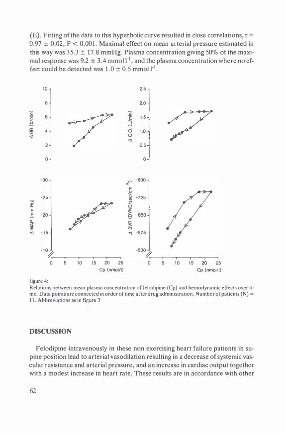
(E) . Fitting of the data to this hyperbolic curve resulted in close correlations, r =
0.97 ± 0.02, P < 0.001 . Maximal effect on mean arterial pressure estimated in this way was 35.3 ± 17.8 mmHg. Plasma concentration giving 50% of the maximal response was 9.2 ± 3 .4 mmol I-1 , and the plasma concentration where no effect could be detected was 1 . 0 ± 0.5 mmol i-1 •
10 2.5
8 2.0
c
/ '2
.E 6 .E 1 .5 ..... ..... B d a: 4 c:i 1 .0 ::c <l d
<I 2 0.5
0 0
-30 'f�
-800
E 0 ..... -25 0 -725 Q) Ci
/ � ::c UJ
E z >-5 -20 9 -650 a. a: <( > ::;; rn <I - 1 5 <I -575
- 1 0
� -500
� 0 5 10 1 5 20 25 0 5 10 1 5 20 25
Cp (nmol/IJ Cp (nmolll) figure 4: Relations between mean plasma concentration of felodipine (Cp} and hemodynamic effects over time. Data points are connected in order of time after drug administration. Number of patients (N) = 1 1 . Abbreviations as in figure 3
DISCUSSION
Felodipine intravenously in these non exercising heart failure patients in supine position lead to arterial vasodilation resulting in a decrease of systemic vascular resistance and arterial pressure, and an increase in cardiac output together with a modest increase in heart rate. These results are in accordance with other
62

studies with felodipine in heart failure patients (9, 1 1 , 12,20) . In an uncontrolled study a significant decrease in pulmonary capillary wedge pressure was demonstrated (12) . In our study this was only found in comparison with base line values, but not if compared with placebo, because a small decrease in PCWP was observed in both patient groups during the 120 minutes observation period. A significant decrease in PCWP during exercise has been demonstrated in controlled studies (9 ,20) . Reduction in left ventricular filling pressure during exercise is apparently not a primary vasodilating effect of felodipine, but secondary to improved left ventricular emptying after systolic unloading. The increase in heart rate was small (8%) , probably due to the slow rise of Cp. It has been demonstrated for nifedipine that the hemodynamic response to nifedipine is influenced by the rate of increase of its concentration in plasma (21) . Another explanation may be found in the loss of chronotropic competence which has been demonstrated in severe congestive heart failure (22,23) . Linear regression analysis first demonstrated a significant correlation between Cp and hemodynamic effects , although the plateau in the curve and the hysteresis in the time effect relationship affected the tightness of the fit, expressed by r. The close correlation between plasma concentrations and effect, after fitting of all individual data to the hyperbolic equation of Emax ( eq 1) illustrates the existence of a sigmoidal relationship (17). Hysteresis as detected by plotting of Cp against effect following its time cours will be anti clockwise if an equilibrium delay between plasma and biophase exists. This was not observed in the four plots of Cp versus change in heart rate, mean arterial pressure, cardiac output and systemic vescular resistance. Therefore, the equilibrium delay between plasma and the biophase is assumed to be negligible in comparison to drug input rate, distribution rate and elimination rate, and the concentration in the plasma compartment is assumed to be a good reflection of the concentration at the biophase, e.g. receptor site.
The occurrence of clockwise hysteresis may indicate the development of acute tolerance. The receptor based mechanism of tolerance leads to a reduced effectiveness of the same plasma level in the course of time. This has been demonstrated for beta adrenoceptor agonists and is called "downregulation" or "desensitivation" (24,25) . This process takes several hours or even days to develop, and it is therefore highly unlikely that such a mechanism caused the observed clockwise loop in heart rate, flow and resistance parameters. Apart from a change in receptor sensitivity or number, clockwise hysteresis may also be caused by the accumulation of an antagonistic metabolite. Because felodipine is extensively metabolized, the possible presence of an unbound metabolite which itself is vasoconstrictive might cause clockwise hysteresis. A pharmacological activity of metabolites has not been reported for felodipine but that does not exclude their existence (2) . Again, such a process is not likely to occur in such a short period of time. The explanation for the observed clockwise hysteresis loops in heart ra-
63

te, flow and resistance variables, and the lack of hysteresis in arterial pressure, may be the physiological adjustment of the cardiovascular system. Since the vasodilating effect of felodipine occurs mainly in the arterial bed, reflex mechanisms are likely to counteract the decreased peripheral vascular resistance (26) . The massive arterial vasodilation induced by the acute intravenous administration of felodipine is followed by an immediate, baroceptor mediated reflex which causes sympathetic stimulation or parasympathetic withdrawal (14) . This leads to arterial vasoconstriction in other vascular beds, or to a reduction of the vasodilation in all vascular beds, and to some decrease of the initially huge increase of flow.
Obviously, this process is balanced in such a way, that mean arterial pressure, being the product of resistance and flow, is closely related to Cp during the whole time course . The net results, if described in pharmacodynamic terms, are the clockwise hysteretic loops in systemic vascular resistance and cardiac output, without hysteresis in mean arterial pressure . The hysteresis loops suggest that arterial blood pressure is the most important parameter for the pharmacodynamic analysis of heart failure patients.
Vasodilating drugs are used in patients with heart failure to improve perfusion . It seems therefore logical to use flow parameters in the analysis of the efficacy of a vasodilating drug. But the adequacy of perfusion is not monitored through metabolic messengers carried from the tissues in the bloodstream, but by the sensing of the arterial pressure (13) . This physiological mechanism should be considered when the results of pharmacodynamic studies are interpreted.
Conclusion.
There is a close relationship between plasma concentrations of felodipine and hemodynamic effects in heart failure patients. The clockwise hysteresis in flow and resistance parameters is probably a reflection of the physiological adjustment of the circulation to the sudden, massive arterial vasodilation induced by felodipine intravenously, and not a sign of early acute tolerance. lfwe are to gain a deeper insight through the quantitative study of intravenously administered vasodilating drugs in congestive heart failure, then pharmacodynamics must be analyzed with the use of arterial pressure measurements, in combination with the analysis of changes in flow and resistance. Thereby an integrated approach to describe the overall relationship between plasma concentration and hemodynamic effects can be realized.
64

Acknowledgements : This study was supported by a grant from Astra Hassle Cardiovascular Sweden
References
1 . Ljung B . Vascular selectivity of felodipine.Drugs 1985;29(Suppl 2):46-58. 2. Edgar B. Clinical Pharmacokinetics of felodipine. Thesis,Goteborg 1988;39-52. 3. Emanuelsson H, Hjalmarson AA, Holmberg S, Waagstein F. Acute hemodynamic effects offe
lodipine in congestive heart failure. Eur J Clin Pharmacol 1985;28:489-93. 4. Edgar B . et al . Pharmacokinetic and pharmacodynamic studies of felodipine in healthy subjects
after various single,oral and intravenous doses.Biopharm Drug Dispos 1987; 88:235-248. 5. Edgar B,Collste P,Haglund K,Regardh CG. Pharmacokinetics and haemodynamic effects of
felodipine as monotherapy in hypertensive patients. Clin Invest Med 1987;10:388-394. 6. Leonetti G. et al .Effects of single and repeated doses of the calcium antagonist felodipine on
blood pressure, renal function, electrolytes and water balance, and renin- angiotensin-aldosteron system in hypertensive patients.] Cardiov Pharmacol 1986;8: 1243-1248.
7 . Hedner T et al. Treatment of essential hypertension with felodipine in combination with a diuretic.Eur J Clin Pharmacol 1986;30: 133-139.
8. Elmfeldt D, Hedner T. Felodipine - a new vasodilator ,in addition to beta receptor blockade in hypertension.Eur J Clin Pharmacol 193;25:571-575.
9. Kassis E, Amtorp 0. Cardiovascular and neurohumoral postural respnses and baroreceptor abnormalities during a course of adjunctive vasodilator therapy with felodipine for congestive heart failure.Circulation 1987;75: 1204-1213.
10 . Dunselman PHJM,Kuntze CEE,van Bruggen A,Hamer H,Wesseling H,Lie Kl (1987) Felodipine in heart failure:a double blind placebo controlled study in 23 patients. Circulation 76:suppl IV,abs.710.
1 1 . Kassis E , Amtorp 0, Waldorff S , Fritz-Hansen P. Efficacy of felodipine in chronic congestive heart failure: a placebo controlled haemodynamic study at rest and during exercise and orthostatic stress. Br Heart J 1987 ;58:505-1 1 .
12. Timmis AD, Smyth P , Kenny JF, Campbell S , Jewitt DE. Effects ofvasodilator treatment with felodipine on haemodynamic responses to treadmill exercise in congestive heart failure. Br Heart J 1984;52:314-20.
13. Harris P. Congestive cardiac failure: central role of the arterial blood pressure. Br Heart J 1987;58:190-203.
14. Brown AM. Receptors under pressure;an update on baroreceptors.Circ Res 1980;46: 1-10. 15. Walley TJ, Heagerty AM, Woods KL, Bing RF, Pohl JEF, Barnett DB.Br J Clin Pharmacol
1987; 23:693-701 . 16. The Criteria Committee of the New York Heart Association: Diseases of the Heart and Blood
Vessels. In:Nomenclature and Criteria for Diagnosis, 7th ed.Boston,Litt-le,Brown&Co,1973;286.
17. Holford NHG, Sheiner LB. Understanding the dose-effect relationship:Clinical application of pharmacokinetic- pharmacodynamic models. Clin Pharmacokin 1981 ;6:429-453 .
18 . Ahnoff M e t a l . Comparison of selectivity of gas chromatographic methods, including column switching for the determination of felodipine in plasma.Journal of Chromatography 1987;2945:419-427.
19. Dunselman PHJM, Edgar B, Scaf AHJ, Kuntze CEE, Wesseling H. Pharmacokinetics of felodipine after intravenous and chronic oral administration in patients with congestive heart failure.Br J Clin Pharmacol 1989, in press.
20. Tan LB, Murray RG, Littler WA. Felodipine in patients with chronic heart failure: discrepant heamodynamic and clinical effects. Br Heart J 1987;58: 122-8.
65

21 . Kleinbloesem CH, van Brummelen P, Danhof M, Faber H, Urquhart J, Breimer DD. Rate of increase in the plasma concentration of nifedipine as a major determinant of its hemodynamic effects in humans. Clin Pharmacol Tuer 1987;41 :26-30.
22. Goldstein RE, Belser GD, Stampfer M , Epstein SE. Impairment of autonomically mediated heart rate control in patients with cardiac dysfunction.Circ Res 1975;36:571-578.
23. Higginbotham MB, Morris KG, Cohn EH, Coleman RE, Cobb FR. Determinants of variable exercise performance among patients with severe left ventricular dysfunction.Am J Cardiol 1983;51 :52-60.
24. Bristow MR, Kantrowitz NE, Ginsburg R, Fowler MR. Beta adrenergic function in heart muscle disease and heart failure.J Mol Cell Cardiol 1985;17:suppl 2:41-52.
25. Francis GS, Cohn JN. The autonomic nervous system in congestive heart failure.Annu Rev Med 1986;37:235-247.
26. Zsoten TI, Church JG. Calcium antagonists:pharmacodynamic effects and mechanism of action. Drugs 1982;25:93-1 12.
66

CHAPTER S
Oral Pharmacokinetics of Felodipine in Patients with Congestive Heart Failure: Variable Prediction using
Intravenous Data
P.H.J .M. Dunselman v, A.H.J. Scaf* , and H. Wesseling*
Departments of Cardiology v and Pharmacology/Clinical Pharmacology* , Groningen University, The Netherlands
The Journal of Clinical Pharmacology, 1989; in press
SUMMARY
Peak and trough concentrations after 8 weeks oral therapy with felodipine, a vasodilating calciumantagonist of the dihydropyridine group, were predicted from intravenous pharmakocinetic data before therapy in 1 1 patients, randomly allocated to felodipine treatment 10 mg b. i .d . , during a placebo controlled study in patients with congestive heart failure . Peak concentrations were well predictable , but trough levels varied between a good agreement in some patients to a large underestimation in others. Predictability was significantly correlated with half life, plasma clearance and distribution volume of the intravenous pharmacokinetic study. After 8 weeks chronic oral therapy no significant differences could be detected between the oral pharmacokinetics of predictable (n=6) and unpredictable (n=5) patients. This demonstrates that felodipine kinetics change during felodipine treatment. Differences in the distribution of blood flow before therapy combined with an interindividual variability in blood flow response during therapy is probably responsible for the observed impossibility to calculate trough levels, and thus oral dosage schedules, from intravenous pharmacokinetic data in patients with congestive heart failure.
67

INTRODUCTION
Felodipine is a calciumantagonist of the dihydropyridine group with strong vasodilator activity and negligible negative inotropic effects due to its selectivity for smooth muscle (1) . Its efficacy in the treatment of hypertension (2-4) , and on improvement of exercise duration and quality of life in congestive heart failure (5-8) has been described. Although felodipine is efficacious in the management of heart failure, it has not yet been demonstrated to affect mortality. The pharmacokinetic characteristics of felodipine have been analyzed in healthy young subjects (9 , 10) , middle aged hypertensive patients (11) and elderly hypertensive patients (12,13) . Thorough knowledge of the relationship between intravenous dosage kinetics and multiple oral administration is an essential part of the pharmacokinetic profile of any drug. Furthermore, vasodilating drugs administered to patients with congestive heart failure may lead to improved perfusion ofhypoperfused intestine, liver and kidneys, what may result in changes in absorption and elimination characteristics during therapy.
This study reports the possibility to predict peak and trough concentrations of felodipine after chronic oral treatment from intravenous pharmacokinetic data before treatment in patients with congestive heart failure.
METHODS
Patients.
Twenty three patients (14 men, 9 women) , aged 60 ±7 years, with signs and symptoms of congestive heart failure participated in a randomized, double blind, parallel, placebo controlled study after a placebo run in phase of 2 weeks. Eleven patients were treated with felodipine and 12 patients with placebo. During the intravenous study 1 mg of felodipine or a placebo solution was administered in 60 minutes, and during chronic oral therapy 10 mg tablets or matching placebo tablets were administered b . i .d . The cause of congestive heart failure was coronary artery disease as documented by myocardial infarctions more than 3 months ago. Patients were in New York Heart Association class III (14) , with an ejection fraction (radionuclide ventriculography) < 40 (26.6 ± 9)% and a maximal oxygen consumption < 15 ( 11 .9 ± 2) ml/kg/min. Maximal oxygen consumption was measured using standard techniques (15) . All patients were on long term treatment (> 3 month) with digoxin and diuretics (hydrochlorothiazide 50 mg once daily, with potassium suppletion if necessary) , and on a sodium chloride restricted diet of not more than 3 grams sodium chloride daily. No vaso-
68

dilating, beta blocking or antiarrhythmic drugs were allowed during the study. All patients were in sinus rhythm. Routine liver function tests were within the normal range in all patients. Patients with a history of alcohol abuse were not admitted to the study. Digoxin therapy was titrated before the start of the study and therapeutic regimens were verified with serum digoxin concentration measurements at trough, 2 hours and 6 hours post dose levels ( 16) . Written informed consent was required from all patients and the protocol was approved by the Ethics Committee of the University Hospital of Groningen .
Felodipine plasma levels.
Plasma samples for determination of felodipine concentration were obtained at 0 (blank) , 15, 30, 45 , 60, 62, 66, 70, 75 , 90, 105 min , 2, 3, 4, 5 , 6, 7 and 8 hours after start of the infusion . The observation period of 8 hours was based on the assumption that the plasma levels of felodipine would reach a minimum within that period. After 8 weeks of chronic oral therapy blood samples were obtained at 0 (trough level) and 30, 60, 90, 120 min, 3 , 4, 6, 8, and 12 hours after dosage. Plasma samples were determined by GLC (17) .
Pharmacokinetic analysis.
The pharmacokinetic analysis of the plasma concentration-time relationships after the intravenous and long term oral administration was performed with an iterative peeling program (18). In the intravenous study only post infusion data were used for the fitting and the duration of infusion was taken into account . After chronic oral administration the lag time of absorption was estimated by calculation of the moment that the fitted curve was equal to trough value. In order to give a fair estimate of the primary pharmacokinetic parameters (Ci , A i , t 1n) different ways of calculation were used. The C1 were averaged on linear and logarithmic scales, and both the disposition constants (A.) and half life values (t112) were determined, while the sample t112 were also estimated from the mean disposition constants (19) . The mean values were compared with the medians. This revealed that the distributions of the respective parameters were skewed on both the linear and logarithmic scale.
Therefore the medians and range of all pharmacokinetic parameters are presented. Bioavailibility (F) was calculated by the ratio of AUC12 and AUC, where AUC12 represents the area under the curve over 12 hours after oral , and AUC the area after intravenous administration , both normalized to a dose of 1 mg.
69

The areas under the plasma concentration-time curve (AUC and AUC12) were obtained by integration from time zero to infinity and 12 hours respectively.
Plasma clearance after i . v. dosage was calculated as:
CL = Dosei.v. AUCi.v.
The volume of distribution at steady state V55 was calculated as follows:
v •• = D. L c,.A.-2. AUC2 , where Ci is the intercept and A the coefficient representing the rate constant of drug elimination.
Statistical analysis.
All data are expressed as medians and range. The Mann Whitney U test was used to analyze the differences between groups. Differences were considered significant if P was < 0.05 .
RESULTS
Intravenous pharmacokinetics.
After the end of the infusion the felodipine concentration declined tri-exponentially in 4 of the 1 1 patients. The fit to such a curve was significantly better than that to a bi-exponential one in only 2 patients, and in 1 patient no reliable
Table 1 . Pharmacokinetics of felodipine after intravenous administration in patients with congestive heart failure
Median Range Ve (I) 62 15 - 123 Vz (1) 139 90- 370 AUC(nM h-1) 58 42 - 1 10 CL {ml min-1) 750 400- 1020 t112 {h) 2 .9"" 1 . 2 - 9.5
Ve = volume of distribution of the central compartment; Vz = volume of distribution at steady state; AUC = Area Under the plasma concentration - time Curve; CL = systemic plasma clearance; t1n = terminal half life.
70

estimation of A.3 could be made because its value approached zero. Therefore, the outcome of a bi-exponential analysis is presented for all patients in table 1 and figure 1 . The halflives of both phases vary over a wide range. The plasma clearance of about 750 ml min-1 demonstrates that felodipine is a high clearance drug. The volume of distribution of the central compartment was large, about 12 times plasma volume, indicating rapid tissue distribution.
Cp ( nmol / l l
50
20
10
5
2
0
Figure 1
2 3 4 5 6 8
time ( h rs)
The plasma concentration - time relationship of felodipine during and after intravenous administration of 1 mg in 60 minutes in 1 1 patients with congestive heart failure. The curve presented corresponds to the median values. Bars indicate the standard deviation on logarithmic scale.
Oral pharmacokinetics.
The pharmacokinetic analysis resulted in 7 of the 1 1 patients in a smaller sum of squares with a fit to a tri-exponential than to a bi-exponential curve . Neither peak, nor trough values in the 4 patients differed significantly from those measured in the patients with a tri-exponential curve. The absorption and disposition characteristics are presented in Table 2.
7 1

Table 2. Pharmacokinetics of felodipine during chronic oral administration in patients with congestive heart failure
Median Range
Cmax (nM) 37 14- 68 !max (h) 1 .0 0.5 - 4.0 AUC12 (nM h-1 1"1) 140 42 - 306 F (%) 25 12- 74 t112 (h) 22.7 8.7 - 35.4
Cmax = maximal plasma concentration; tmax =time to maximal plasma concentration ; AUC12 = Area Under the plasma concentration - time Curve ; F = systemic b10availability; t112 = terminal half life
Relationship between intravenous and oral pharmacokinetics.
The peak and trough concentrations during chronic oral steady state were calculated for each individual patient from the intravenous pharmacokinetic data by correcting for the administered dose and by introducing the bioavailability and the rate constant of absorption. Because in all patients only one ascending exponential was detected, the A.1 of the respective oral curves was considered to be the rate constant of absorption. The calculated and observed peak and trough concentrations are presented in table 3. These results show that the peak concentrations were well predictable. The calculated and observed trough levels vary between a good agreement in some patients to a large underestimation in others. The most important parameters that determine trough values are the intercept C2 and the corresponding t112,v. An analysis was made of a possible relationship between the ratio of the calculated and observed trough concentrations, and C2 and the corresponding t112iv· Regression analysis resulted in a significant correlation between this ratio and the intercept C2 (r = -0 .74, P < 0.01) and t112,v (r =
0.98, P < 0.001). Both plasma clearance (r = -0.65 , P < 0.05) and distribution volume at steady state (r = 0.95 , P < 0 .001) were also significantly correlated to this ratio. The AUC12 and the terminal half life after oral administration did not show any correlation with this ratio. These results demonstrate that as the second half life increases and the second intercept becomes smaller, calculated trough concentrations will approach the observed values. In most patients terminal halflife was longer after oral than after intravenous administration. Even if the longest halflife from each individual patient was used, the correlation of the ratio of calculated and observed trough levels with plasma clearance and distribution volume was still present. The patients were divided into 2 groups along the median : (1) those in whom the trough concentration could be predicted from the intravenous data (Calculated/Observed > 0.5), n = 6, (2) those in whom this could not
72

be done (Calculated/Observed � 0.5) , n = 5 . The intravenous and oral curves of the patients in whom the trough concentrations could be predicted run almost parallel , while a steep descent is observed in the patients whose intravenous pharmacokinetic data did not lead to an accurate prediction of oral trough levels (fig 2) . The AUC, from which both plasma clearance and Vss are calculated, is significantly higher in the patients in whom the trough values after chronic oral therapy could be predicted accurately (89 vs 51 nM h-1 , P < 0.02) . After 8 weeks chronic oral therapy no significant differences could be detected between the oral pharmacokinetics of these 2 groups. Felodipine increased serum digoxin levels only at high felodipine plasma levels (16) , and a relationship between predictability of oral pharmacokinetics from intravenous data and changes in serum digoxin levels could not be demonstrated.
Table 3. The calculated and the observed peak and trough concentrations during chronic oral administration of felodipine in patients with congestive heart failure
Median Range PEAK - Calculated (nM) 4.0 1 .6 - 1 1 .2 - Observed (nM) 3.8 1 .4 - 6.8 - Cale/Obs 1 . 1 0.7 - 1 .6
TROUGH - Calculated (nM) 0.22 0.0 1 - 1 .42 - Observed (nM) 0.66 0.28 - 1 .50 - Cale/Obs 0.33 0.02 - 0.94
The peak and trough levels presented here are those after a dose of 1 mg/hr.
73

nM Felod1p1ne [ p o J 50
25
!O
5 _J
25
1.0
0 5 0
nM Felod1p1ne( p o }
50
25
10
5 J 2 5
0 5 0
Figure 2
-------- nM Felod1p1ne ( 1 v I A r:
r 20
10
2
6 8 !O 16 --------- Felod1p1ne [ 1 v }
6 8 10 12 time ( hrs}
-, lL IF.
The plasma concentration - time relationship of felodipine during and after intravenous administration (dotted line) and during chronic oral administration in patients in whom it was possible (A, n = 6) and in whom it was impossible (B, n = 5) to predict the oral trough concentrations from the intravenous data.
DISCUSSION
In the intravenous study a tri-exponential plasma decay could not be detected in every patient. This can be explained by the observation that in all cases where a bi-exponential decay was found, plasma levels were too low for a reliable estimation of A.3 at the end of the measurement period. This has also been described
74

in young healthy individuals and hypertensive patients where the same analytical problems were met (13). The analysis of the oral data resulted in a tri- exponential curve in 7 patients. In the other 4 only two exponential terms could be detected . In these cases the peak concentration appeared so late, due to slow absorption or a long lag time, that too few points were left to determine the last descending part of the curve. Hence, a tri-exponential curve may give the best representation of felodipine kinetics in congestive heart failure. If the same distribution and elimination processes underly both the intravenous and oral pharmacokinetics, then it should be possible to predict peak and trough concentrations after chronic oral treatment from intravenous pharmacokinetics by introducing the bioavailability and rate constant of absorption, corrected for the administered dose.
While trough values could be predicted rather well from the intravenous kinetics in some patients, they could not be predicted in others. In most patients terminal halflife was longer after oral than after intravenous administration, but the poor predictability of trough values did not disappear when the longest halflife irrespective of route of administration was used in each individual patient. The differences during the i . v. study and the similarity after 8 weeks oral treatment indicate that felodipine kinetics change during and probably as a consequence of felodipine treatment.
The pathophysiology involved in congestive heart failure can be described as peripheral hypoperfusion and congestion. Felodipine is a potent drug, leading to massive arterial vasodilation, resulting in an increase in total body flow in the studied patients (6) . It should be noted that the unpredictable patients had the largest clearances and that clearance in high clearance drugs like felodipine is related to liver blood flow. A difference in the distribution of total body flow in the individual patients may be responsible for the observed difference in predictability of oral pharmacokinetics. The characteristic vasoconstriction in heart failure patients might not be evenly distributed over all vascular beds in every individual patient. Although central hemodynamics are generally correlated with regional blood flow there can be great variations in individual blood flow values, reflexing the complex nature of regional blood flow regulation (20) . One could suggest that the vasodilation induced by felodipine therapy may have lead to a redistribution of flow at the expense of liver blood flow. An interindividual variability in liver blood flow response after oral therapy with dihydropyridines has been found (21) . Nevertheless, it is highly unlikely that a potent vasodilating high clearance drug as felodipine would actually decrease liver blood flow. Most likely, treatment with oral felodipine during 8 weeks dilated all vascular beds, including those in the liver. However, in some of these patients, e .g. those in whom trough levels at steady state could not be predicted from i .v . data, a relatively small increase in liver blood flow may have been accompanied by a much more
75

pronounced vasodilation in other vascular beds (20). This illustrates the importance of a combined analysis of felodipine kinetics and changes in flow patterns after prolonged administration. Our results demonstrate that oral dosage schedules for chronic treatment with dihydropyridines in patients with congestive heart failure can not be based on intravenous kinetics. In further research with felodipine and other vasodilating drugs in heart failure patients, special attention should be given to the possibility that pharmacokinetic characteristics of a drug may change during therapy by changes in (liver) blood flow induced by the drug itself. Studies using multicompartment analysis of intravenous and oral pharmacokinetic data with measurement of both central and regional hemodynamics are being designed to obtain further insight into the mechanisms that are responsible for the obvious impossibility to predict oral pharmacokinetics at steady state from intravenous data.
Acknowledgement : This study was supported by a grant from Astra Hassle Laboratories, Sweden.
References
I . Ljung B . Vascular selectivity of felodipine.Drugs 1985;29(Suppl 2) :46-58. 2. Leonetti G. et al.Effects of single and repeated doses of the calcium antagonist felodipine on
blood pressure, renal function, electrolytes and water balance, and renin- angiotensin-aldosteron system in hypertensive patients .I Cardiov Pharmacol 1986;8 : 1243-1248.
3. Hedner T et al. Treatment of essential hypertension with felodipine in combination with a diuretic.Eur J Clin Pharmacol 1986;30:133-139.
4. Elmfeldt D, Hedner T. Felodipine - a new vasodilator ,in addition to beta receptor blockade in hypertension.Eur J Clin Pharmacol 1983;25:571-575.
5 . Kassis E, Amtorp 0. Cardiovascular and neurohumoral postural respnses and baroreceptor abnormalities during a course of adjunctive vasodilator therapy with felodipine for congestive heart failure.Circulation 1987;75: 1204-1213.
6. Dunselman PHJM,Kuntze CEE,van Bruggen A,Hamer H ,Wesseling H,Lie KI. Efficacy ofFelodipine in Congestive Heart Failure. Eur Heart J 1989; in press.
7. Kassis E, Amtorp 0, Waldorff S, Fritz-Hansen P. Efficacy of felodipine in chronic congestive heart failure: a placebo controlled haemodynamic study at rest and during exercise and orthostatic stress. Br Heart J 1987;58:505-1 1 .
8 . Timmis AD, Smyth P , Kenny JF, Campbell S , Jewitt DE. Effects of vasodilator treatment with felodipine on haemodynamic responses to treadmill exercise in congestive heart failure. Br Heart J 1984;52:314-20.
9. Edgar B et al. Felodipine kinetics in healthy men. Clin Pharmacol Tuer 1985;38:205-21 1 . 10. Edgar B . et al. Pharmacokinetic and pharmacodynamic studies of felodipine i n healthy subjects
after various single,oral and intravenous doses.Biopharm Drug Dispos 1987; 88:235-248. 1 1 . Edgar B ,Collste P ,Haglund K,Regardh CG. Pharmacokinetics and haemodynamic effects of fe
lodipine as monotherapy in hypertensive patients. Clin Invest Med 1987;10:388-394.
76

12. Landahl S,Edgar B ,Gabrielsson M,Larson M,Lemfeldt B ,Lundborg P,Regardh CG. Pharmacokinetics and blood pressure effects of felodipine in elderly hypertensive patients: A comparison with young healthy subjects. In:Clinical Pharmacokinetics of felodipine.Thesis,Goteborg 1988;IV: l-21 .
13. Edgar B. Clinical Pharmacokinetics of felodipine.Thesis,Goteborg 1988;39-52. 14. The Criteria Committee of the New York Heart Association: Diseases of the Heart and Blood
Vessels. In:Nomenclature and Criteria for Diagnosis, 7th ed.Boston,Litt-le,Brown&Co,1973;286.
15. Dunselman PHJM, Kuntze CEE, van Bruggen A, Beekhuis H, Piers B, Scaf AHJ, Wesseling H and Lie Kl. Value of New York Heart association classification,radionuclide ventriculography ,and cardiopulmonary exercise tests for selection of patients for congestive heart failure studies.Am Heart J 1988;1 16:1475-1482.
16. Dunselman PHJM, Scaf AHJ, Kuntze CEE, Lie KI and Wesseling H. Digoxin-felodipine interaction in patients with congestive heart failure.Eur J Clin Pharmacol 1988;35:461- 465.
17. Ahnoff M,Ervik M and Johansson L. Comparison of high- selectivity of gas chromatographic methods, including column switching for the determination of felodipine in plasma.Journal of Chromatography 1987;2945:419-427.
18 . Scaf AHJ. Pharmacokinetic analyses with RUGFIT; an interactive pharmacokinetic computer program.Biopharm & Drug Disp 1988;9:415-446.
19. Loo S.C.K. and Riegelman S. Assessment of pharmacokinetic constants from post infusion blood curves obtained after i .v . infusion. J Pharm Sci 1970;59:33.
20. Leithe ME, Margorien MD, HermillerJB, Unverferth DV and Leier CV. Relationship between central hemodynamics and regional blood flow in normal subjects and in patients with congestive heart failure. Circulation 1984;69 nol:57-64.
21 . van Harten 1. Clinical pharmacokinetics and cardiovascular effects of nisoldipine. Thesis 1988 Leiden University, The Netherlands.
77


CHAPTER 6
Digoxin - Felodipine Interaction in Patients with Congestive Heart Failure
Peter H.J .M. Dunselman M.D . , Arnold H.J . Scaf Ph.D . , C .Erik E . Kuntze M.D . , Kong Ing Lie M .D . , and H. Wesseling M.D.
Departments of Cardiology and Pharmacology I Clinical Pharmacology, University Hospital Groningen, 59 Oostersingel , 9713 EZ Groningen ,
The Netherlands
European Journal of Clinical Pharmacology, 1988; 35 :461-465 .
SUMMARY
A possible interaction between felodipine and digoxin was studied in 23 congestive heart failure before and after 8 weeks treatment with both drugs. A modest, non-significant increase in serum digoxin level 2 h post-dose ( + 15%, P =0. 13) was found in the felodipine group (n=ll) , compared with placebo (n= 12) , with no change in the trough and 6 h post-dose levels. There was a bimodal distribution of the observed changes in serum digoxin level 2 h post-dose : a significant increase (P < 0 .001) was observed only in patients with a high felodipine plasma level, which may have been caused by changes in the absorption rate in those patients. Changes in the elimination of digoxin after felodipine therapy appeared unlikely, since the trough and 6 h post-dose post-dose levels were unchanged. Analysis of the clinical characteristics, hemodynamics and laboratory values revealed no significant differences between the subgroups. The observed increase in serum digoxin warrants monitoring the trough and peak levels of digoxin in patients with congestive heart failure who are also being treated with felodipine.
79
INTRODUCTION
Felodipine is a new dihydropyridine, a calcium antagonist with strong vasodiIator activity, which is under investigation in hypertension and congestive heart failure. An interaction between calcium antagonists and digoxin in various disease states has been described (1-16) .
The pathophysiology of congestive heart failure can be described as peripheral hypoperfusion and congestion . As a result changes in gastrointestinal , hepatic and renal function may influence the absorption, distribution and elimination of drugs. There do not appear to be any data about an interaction between felodipine and digoxin in patients with heart failure. A prospective, 8 week ,doubleblind, placebo controlled, parallel study of the influence offelodipine on the serum concentration of digoxin in patients with documented congestive heart failure is reported here.
Subjects and Methods
Patients
Twenty-three patients, 14 men, 9 women, aged 60 ± 7 years, with congestive heart failure New York Heart Association class III , on chronic treatment ( >3 month) with digoxin were admitted to the study. All patients had a history of myocardial infarction, an ejection fraction (radionuclide ventriculography) <40% , and a maximal oxygen uptake (V02max) <15 ml kg·1 min·1 . Electrolytes, serum creatinine and urea were within the normal range in all patients.
Study design
All patients were on maintenance (> 2 month) therapy with digoxin (dose 0.25 - 0. 375 mg once daily ) and a diuretic (hydrochlorothiazide 50 mg once daily with potassium supplementation if necessary) . The doses were held constant throughout the study period. No inotropic, vasodilator or antiarrhythmic drugs were allowed. After a run in period of 2 weeks, cardiac catheterization was performed 2 h after the dose of digoxin . Serum digoxin was measured at trough (before dosing) , peak (2 h after dosing) and 6 h after the dose. Patients were then randomized to receive felodipine 10 mg b.d.s . (n= l l) or placebo (n= 12), in a double blind fashion. Digoxin was administered once daily, together with the morning dose of felodipine or placebo, which was given b.d .s . After 8 weeks of
80

treatment all measurements were repeated, including the serum digoxin concentrations. Plasma samples for felodipine assay were taken at 0 (trough level) , 30, 60,and 90 min . , and 2, 3 , 4, 6, 8, 12, and 24 hours after dosing. The randomization code was broken when the last patient had completed the study. Written informed consent was required from all patients and the protocol was approved by the ethics committee of the University Hospital of Groningen.
Assay methods
Serum digoxin concentrations were determined by 125 1-digoxin radioimmunoassay (Beckton-Dickinson). The mean coefficient of variation of the method was <5% . Felodipine was shown not to interfere with the determination. Plasma felodipine levels were determined by GLC (17).
Statistical analysis
Individual patient serum digoxin measurements may show variability despite a constant drug regimen and a standardized blood sampling schedule. When a potential drug interaction is evaluated in cardiac patients with possible day to day alterations in gastrointestinal absorption and in renal and non renal clearance of digoxin this variability may lead to erronous conclusions about any drug-drug interaction. In this study a control group on placebo therapy was used. The difference in changes between the 2 groups before and after treatment at 3 different post-dose levels were compared to minimize the influence of the spontaneous variation. Differences in variances where tested using the F test. Proper randomization was tested using unpaired t-tests. Differences in the changes between patient groups (felodipine and placebo) and subgroups were evaluated by unpaired t-tests. The relationship between the observed changes after treatment and plasma felodipine levels was examined by linear regression analysis. All data are expressed as mean ± standard deviation (S .D . ) . A P value < 0.05 was considered statistically significant.
RESULTS
Patient characteristics are listed in Table I . Analysis did not show any significant differences. Serum digoxin levels before and during felodipine or placebo treatment are presented in Fig. 1 . Digoxin levels in both groups were within the therapeutic range. Comparison of the differences in the changes between the placebo and felodi-
81

TABLE 1 Baseline characteristics of the study population
Age(yrs) Weight(kg) Height( cm) BSA(m2) LVEF(%) V02max(ml kg-1 min"1)
Felodipine n= l l
62 ± 7 74 ± 10
170 ± 10 1 . 85 ± 0.2
27 ± 10 12.3 ± 2.6
Placebo n= 12
59 ± 7 69 ± 12
170 ± 1 1 1 .80 ± 0.2
26 ± 9 1 1 .6 ± 2 . 1
P value
N.S . N.S . N.S . N.S . N.S . N.S.
BSA = body surface area; LVEF = ejection fraction of the left ventricle; V02max = maximal oxygen consumption. N.S . = not significant
3
2
--------, 0 2 6 TIME ( h )
Mean (±SD) digoxin serum concentrations before and after 8 weeks of therapy with felodipine (n = 1 1) and placebo (n == 12) at trough, and 2 h and 6 h post-dose. The serum digoxin at 2h in the felodipine group was increased by 15% (P = 0.13).
pine groups after 8 weeks of therapy did not reveal a significant difference (Table 2) . A modest and non-significant increase ( + 15% , P = 0. 13) was seen in the 2 h post-dose digoxin concentration in the felodipine group, compared to placebo. Although analysis of variance revealed no significant differences in the values at baseline and in the gainscores, there was a significant difference in the variance of the mean serum digoxin concentration between the felodipine and placebo groups after treatment in the trough, and 2 h and 6-h post-dose levels (F = 4.0, 4.9 and 5.3 respectively, P < 0.05 ,df 10,11 ) . Therefore, a possible relationship
82

between felodipine treatment and digoxin levels was further analyzed. The felodipine plasma levels at steady state were (Table 3) : maximal plasma level (mean ± s .d . ) 31 .5 ± 17.7 nmol .I- 1 2; plasma level 2 h post-dose: 20.2 ± 12.6 nmoI .r1 ; area under the felodipine time-concentration curve (AUC) : 170 ± 92 nmol .r1 .h.
TABLE 2 Comparison of digoxin concentrations (ng.ml-1) in the 2 treatment groups.
Trough 2 hours 6 hours
16 hours baseline after baseline after baseline after
Placebo .88 ± .26 .78 ± . 1 6 1 .68 ± .49 1 .63 ± .32 1 . 10 ± .28 1 .08 ± .17 n=l2 Felodipine 1 .00 ± .26 .98 ± .32 1 .92 ± .51 2.20 ± .71 1 .21 ± .32 1 .28 ± .39 n = l l
Baseline Pl vs F P = .30 P = .26 P = .35 Delta Pl vs F P = .48 P = . 13 P= .45
Delta = difference in serum digoxin concentration between the 2 patient groups, levels after 8 weeks therapy minus base line. Pl = placebo; F =felodipine. mean (S.D)
TABLE 3 Pharmacokinetics of felodipine and changes in digoxin serum concentration 2 h post-dose after 8 weeks of treatment with felodipine
Patient AUC f0-24h C f-max C f-2h Delta C dig-2h (nmol r1 hr) (nmol r1) (nmo1 1-1 )
13 245 52.0 36.0 14 144 42.6 20.0 15 273 68.0 48.9 16 377 34. 1 22.6 17 156 40.0 24.2 18 101 14.9 6 . 1 19 102 14.0 9.6 20 133 26.9 16.9 21 135 18.4 10.3 22 141 13.6 13.6 23 64 22.0 14.4
Mean 170 31 .5 20.2 ±S.D. 92 17.7 12.6
AUC f 0-24h = Area under the felodipine time-concentration curve; C f-max = maximal felodipine plasma concentration; C f-2h = felodipine plasma concentration 2 hours post-dose;
(ng mr1)
0.70 1 . 10 1 .00 0.80 0.70 0.00
--0.10 --0.50 --0.20 --0 . 10 --0.30
0.28 0.58
Delta C dig-2h = difference in digoxine serum concentrations 2 h post-dose, levels after 8 week therapy minus base line.
83

After oral dosing the peak plasma steady state felodipine occurred 1 .8 ± 1 . 1 h (T max) after dosing.
Regression analysis revealed no significant correlation between the change in serum digoxin concentration and the plasma felodipine measured at the trough and 6 h post-dosing.
The change in the 2 h post-dose serum digoxin showed a different and distinctly bimodal distribution . A scatter diagram of the change in digoxin concentration versus plasma felodipine level 2 h post-dose is presented in Fig. 2. Two subgroups of patients can be identified : a significant (P < 0.001) increase in serum digoxin concentration ( +0.86 q 0.18 ng .m1" 1 ) can be seen in 5 patients with a high plasma felodipine , unlike the 6 remaining patients, in whom the digoxin level either did not change or was slightly decreased (-0.20± 0 .18 ng.ml" 1 ) . Plasma felodipine concentrations i n those 2 subgroups 2 h post-dose were 30.3 ± 12.0 and 11 .8 ± 3 .9 nmol . 1" 1 respectively (P < 0.001) . Subsequent analysis of the 2 subgroups for age, sex, digoxin dose, and trough and 6 hours post-dose serum digoxin concentration, electrolytes, serum creatinine and urea gave no significant differences. Analysis of the hemodynamic, V02max and ejection fraction values at baseline and after 8 weeks treatment with felodipine also failed to reveal a difference.
First degree atrioventricular block was observed in 2 patients (16 and 18; Table 3) during felodipine therapy. One of them (No. 16) had an increase in the peak digoxin concentration ( + 0.8 ng.ml-1 ) , without any change in the trough or 6 h post-dose levels. No higher degree block or other cardiac side effect was observed. Non-cardiac side-effects of digoxin were not mentioned by any patient during active questioning on 5 subsequent visits during the 8-week study period.
+1 2 •
+1 0 •
+0.8 •
;:- +O 6
E . +0 1. CT\
..s +0.2
• •
.c 20 40 60 80 N 0-+--��-�� gi 'O u -0 2
E -0 4 Qi 0
-06
.. • •
•
Ct - 2 h (nmol t -1 )
Scatter diagram of plasma felodipine concentrations versus difference in serum digoxin concentration 2 h post-dose after 8 weeks therapy. Abbreviations as in Table 3.
84

DISCUSSION
Kinetic interactions with digoxin have been described for a variety of cardiovascular drugs. Of the calcium antagonists , verapamil increases serum digoxin levels by decreasing both renal and extra renal clearance (1-3) , the former interaction being reversible during long term treatment ( 4). After concurrent administration with diltiazem in heart failure patients, the total body clearance of digoxin was reduced without any change in renal digoxin clearance (10) . The kinetic interactions of nifedipine and digoxin are of special interest, because nifedipine, like felodipine, is a dihydropyridine . In a review of nifedipine/digoxin interaction studies, Schwartz (16) concluded that changes in renal digoxin clearance were due to changes in the renal tubular secretion of digoxin, and not to altered glomerular filtration . In the present study of felodipine, there was no significant differences between the placebo and felodipine groups in serum digoxin at trough or at 2- and 6 h post-dose. However, a bimodal distribution pattern at 2 h post-dose was observed; patients with an increased serum digoxin concentration then had significantly higher felodipine plasma levels compared to the patients with unchanged or slightly decreased digoxin concentrations. This was not explained by differences in clinical characteristics or hemodynamic and laboratory results before or after therapy. Digoxin therapy is normally monitored by measuring its trough concentration. Patients with a high peak serum level alone would not be detected if this procedure were followed in patients concomitantly treated with felodpine.
Possible mechanisms of interaction between felodipine and digoxin are of interest. Felodipine has been shown not to influence the digoxin assay method, so a laboratory artefact as the cause of the rise in the peak serum digoxin during coadministration of felodipine is highly unlikely. The kinetic mechanisms by which felodipine could increase serum digoxin are: increased absorption from the gastrointestinal tract, or reduction in the volume of distribution, or decreased renal or extrarenal clearance of digoxin. Digoxin in tablet form (Lanoxin) which was used is normally 65 - 80% absorbed (10, 18) , and drugs that are concomitantly administered may alter its absorption (19) . A difference in absorption resulting in a different time to peak might have occurred in the present patients in a way related to the concentration of felodipine. A change in the initial volume of distribution for digoxin is unlikely to occur in such a short period of time. The majority of digoxin is excreted renally, and digoxin clearance is proportional to creatinine clearance (20) . Since there were no differences in trough or 6-h postdose levels , a change in the clearance of digoxin due to felodipine administration can be ruled out. Apart from glomerular filtration, renal tubular secretion is an important mechanism of digoxin excretion, especially in patients with impaired renal blood flow, which is common in heart failure patients (21-23) . Administra-
85

tion of potent vasodilating agents leads to a temporary increase in the renal tubular secretion of digoxin in heart failure patients, with no change in glomerular filtration rate (24) . Such a mechanism could have caused accelerated elimination of digoxin in our patients with a high felodipine plasma level . The observed difference in the 2-h post-dose digoxin concentrations, and the absence of differences in the 6-h and trough levels, may reflect a change in absorption kinetics and accelarated digoxin elimination, both inducted by a high plasma felodipine level.
The pharmacodynamic consequences of an elevated serum digoxin level resulting from a kinetic interaction with felodipine are unclear. Digitalis glycosides at therapeutic levels enhance the availability of Ca+ + to myocardial contractile elements following excitation and antagonism of felodipine on digoxin inotropism is possible in theory (25). Felodipine has a more selective effect on smooth muscle cells, the degree of action on the contractility of the myocardium compared with vascular smooth muscle is about 1 : 100 (26) . The extent to which a temporarily increased serum digoxin level may be associated with augmentation of its inotropic effect is difficult to evaluate, since a higher felodipine level itself may induce such an effect (27) . The transient change in digoxin level makes it unlikely that deep compartments, including heart muscle, are involved (28) . The development of first degree atrioventricular block in 2 patients could not be related to the plasma levels of digoxin or felodipine. Non-cardiac side effects of an increased serum digoxin level were not observed, although they might occur if felodipine were added to patients already with a digoxin level in the higher range, as it is not likely that non-cardiac side-effects will be antagonized by felodipine. More pharmacokinetic studies with intensive plasma concentration monitoring are being designed to obtain further insight in the absorption, distribution and clearance characteristics of digoxin and felodipine during concomitant therapy.
Acknowledgement.
The study was supported by a grant from Astra Hassle Laboratories , Sweden, who also provided the felodipine and matching placebo medication.
References
1 . Schwartz JB ,Keefe D ,Kates RE (1982) Acute 'and chronic pharmacodynamic interaction of verapamil and digoxin in patients with atrial fibrillation.Circulation 65: 1 163-1170.
2. Pedersen KE,Dorph-Pedersen A,Hvidt S,Klitgaard NA, Nielsen- Kudsk F (1981) Digoxin-verapamil interaction.Clin Pharmacol Ther 30:31 1-316.
3. Klein HO,Lang R,Weiss E et al (1982) The influence of verapamil on digoxin concentration.Circulation 65:99-1003.
86

4. Pedersen KE,Dorph-Pedersen A,Hvidt S,Klitgaard NA,Pedersen KK (1982) The long term effect of verapamil on plasma digoxin concentration and renal digoxin clearance in healthy subjects .Eur J Clin Pharmacol 22: 123-127.
5. Doering W (1983) Effects of coadministration of verapamil and quinidine on serum digoxin concentration. Eur J Clin Pharmacol 25:517-521 .
6. Rameis H ,Mayometschnigg D ,Ganzinger W (1984) The diltiazem digoxin interaction.Clin Pharmacol Ther 36,2 : 183-189.
7 . Yoshida A,Fujita M,Kurosawa N ,Nioka M ,Shichinohe T, Arakuwa M,Fakuda R,Owada E, lto K (1984) Effects of diltiazem on plasma level and urinary excretion of digoxin in healthy subjects.Clin Pharmacol Ther 35:681-65.
8 . Schwartz J8 ,Migliore Ph.J (1984) Effects of nifedipine on serum digoxin concentrations and renal digoxin clearance.Clin Pharmacol Ther 36, 1 : 19-24.
9. Schwartz JB ,Raizner A,Akers S ( 1984) The effect of nifedipine on serum digoxin concentration in patients. Am Heart J 1077:669.
10. Kuhlmann J .Effects of nifedipine and diltiazem on plasma levels and renal excretion ofbeta-acetyldigoxin (1985) Clin Pharmacol Tuer 37: 150-156.
1 1 . De Cesaris R,Balestruzzi M,Chiarappa R,Ranieri G (1983) Increase of serum digoxin levels after combined administration of calcium antagonists and beta-methyl digoxin.G Ital Cardiol 13 : 188-191 .
12. Belz GG ,Doering W ,Munkes R,Matthews J (1983) Interaction between digoxin and calcium antagonists and antiarrhythmic drugs.Clin Pharmacol Ther 33:410-417.
13. Pedersen KE,Dorph-Pedersen A,Hvidt S,Klitgaard NA,Kjaer K,Nielsen-Kudsk F (1982) Effect of nifedipine on digoxin kinetics in healthy subjects.Clin Pharmacol Tuer 32:562.
14. Zylber-Katz E,Koren G,Levy M (1984) Pharmacokinetic study of digoxin and nifedipine coadministration(abstr} .Clin Pharmacol Ther 36: 19-24.
15 . Kirch W ,Hutt HJ ,Dylewicz P et al (1986) Dose-dependence of the nifedipine-digoxin interaction? Clin Pharmacol Tuer 39:35-39.
16 . Schwartz J8 (1985) Digoxin and nifedipine.Am J Cardiol 55:31 E-34E. 17. Ahnoff M (1984) Determination of felodipine in plasma by gas chromatography with electron
capture detection. J Pharm Biomed Anal 2:519-526. 18 . Schaumann W (1978) Beta-methyl digoxin,a new lipophilic digoxin derivative. ln:Bodem
G ,Dengler HJ eds.Cardiac Glycosides.New York:Springer Verlag,93-104. 19. Antman EM,Smith TW (1985) Drug interactions with digitalis glycosides.ln:Smith TW,ed.Di
gitalis Glycosides.Orlando:Grune & Stratton,65-82. 20. Jelliffe RW (1986) An improved method of digoxin therapy.Ann Intern Med 69:703-717. 21. Halkin H,Sheiner LB ,Peck CC,Melmon KL (1975) Determinants of the renal clearance of digo
xin. Clin Pharmacol Ther 1 :385-394. 22. Steiness E (1974) Renal tubular secretion of digoxin.Circulation 50: 103-107. 23. Rietbrock N,Woodcock BG (1981} Pharmacokinetics of digoxin and derivatives.ln:Greeff
ed.Handbook of Experimental Pharmacology.Berlin:Springer-Verlag,31-56. 24.Cogan JJ ,Humphreys MH,Carlson CJ et al (1981) Acute vasodilator therapy increases renal clea
rance of digoxin in patients with congestive heart failure.Circulation 64:973- 976. 25. Hansen PB,Buch J ,Rasmussen 0, Waldorff S,Steiness E (1984) Influence of atenolol and nifedi
pine on digoxin-induced inotropism in humans.Br J Clin Pharmacol 18:817-822. 26. Ljung B (1980) Vascular versus myocardial selectivity in vitro of H 154/82,a new dihydropyridi
ne.Blood Vessels 17(3):154. 27. Dunselman PHJM,Kuntze CEE,van Bruggen A,Hamer H,Wesseling H ,Lie KI (1987) Felodi
pine in heart failure:a double blind placebo controlled study in 23 patients. Circulation 76:suppl IV,abs.710.
28. Kramer WG ,Kobilash AJ , Lewis RP, Bathala MS, Viscont JA,Reunig RH (1979) Pharmacokinetics of digoxin: relationship between response intensity and predicted compartmental drug levels in man.J Pharmacokinet Biopharm 7: 47-61 .
87


CHAPTER 7
Efficacy of Felodipine in Congestive Heart Failure
P H J M Dunselman* , C E E Kuntze* , A van Bruggen* , J P M Hamer* , A H J Scafv , H Wesselingv , K I Lie*
*Department of Cardiology, University Hospital , 59 Oostersingel , 9713 EZ Groningen, The Netherlands, YDepartment of Pharmacology/Clinical Pharmacology, University of Groningen, 1 Bloemsingel , 9713 BZ Groningen,
The Netherlands
European Heart Journal , 1989; in press.
ABSTRACT
The efficacy of felodipine, a vasodilating calcium antagonist, was analyzed in 23 patients with congestive heart failure , New York Heart Association class III , during an 8 weeks, double blind, randomized, placebo controlled, parallel study. After felodipine exercise duration increased significantly, without changes in oxygen consumption. Heart rate, arterial pressures and rate pressure product decreased at similar submaximal exercise levels. Invasive hemodynamics before and after 8 weeks therapy revealed arterial vasodilation without reflex tachycardia and no significant reductions in right atrial , pulmonary and capillary wedge pressures. Subjective symptom scores improved and side effects were minor. Fluid retention, as assessed by body weight and ankle circumference did not occur. Felodipine has a beneficial effect in patients with moderately severe heart failure. Further research is necessary to demonstrate its long term efficacy and safety .
89
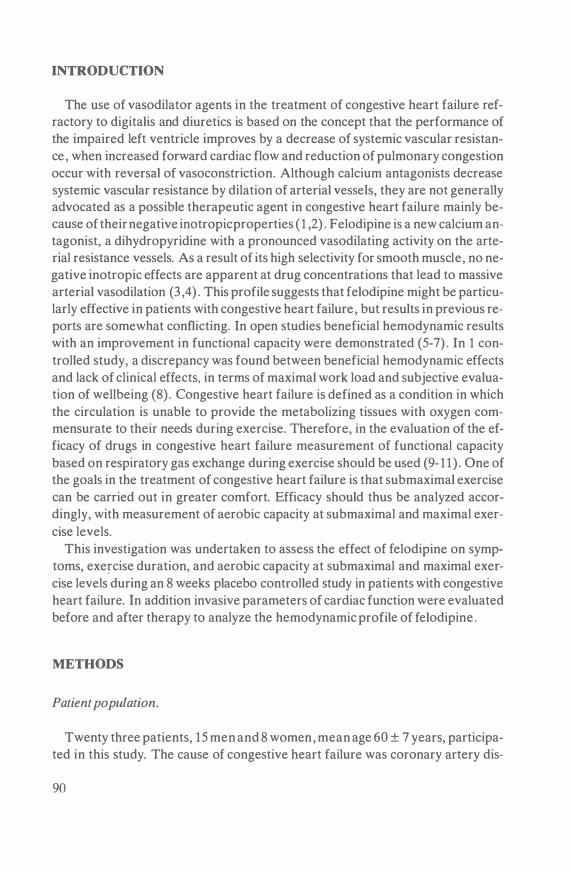
INTRODUCTION
The use of vasodilator agents in the treatment of congestive heart failure refractory to digitalis and diuretics is based on the concept that the performance of the impaired left ventricle improves by a decrease of systemic vascular resistance, when increased forward cardiac flow and reduction of pulmonary congestion occur with reversal of vasoconstriction. Although calcium antagonists decrease systemic vascular resistance by dilation of arterial vessels, they are not generally advocated as a possible therapeutic agent in congestive heart failure mainly because of their negative inotropic properties (1 ,2) . Felodipine is a new calcium antagonist, a dihydropyridine with a pronounced vasodilating activity on the arterial resistance vessels. As a result of its high selectivity for smooth muscle, no negative inotropic effects are apparent at drug concentrations that lead to massive arterial vasodilation (3 ,4) . This profile suggests that felodipine might be particularly effective in patients with congestive heart failure, but results in previous reports are somewhat conflicting. In open studies beneficial hemodynamic results with an improvement in functional capacity were demonstrated (5-7) . In 1 controlled study, a discrepancy was found between beneficial hemodynamic effects and lack of clinical effects, in terms of maximal work load and subjective evaluation of wellbeing (8) . Congestive heart failure is defined as a condition in which the circulation is unable to provide the metabolizing tissues with oxygen commensurate to their needs during exercise. Therefore, in the evaluation of the efficacy of drugs in congestive heart failure measurement of functional capacity based on respiratory gas exchange during exercise should be used (9-11) . One of the goals in the treatment of congestive heart failure is that submaximal exercise can be carried out in greater comfort. Efficacy should thus be analyzed accordingly, with measurement of aerobic capacity at submaximal and maximal exercise levels.
This investigation was undertaken to assess the effect of felodipine on symptoms, exercise duration, and aerobic capacity at submaximal and maximal exercise levels during an 8 weeks placebo controlled study in patients with congestive heart failure. In addition invasive parameters of cardiac function were evaluated before and after therapy to analyze the hemodynamic profile of felodipine .
METHODS
Patient population.
Twenty three patients, 15 men and 8 women, mean age 60 ± 7 years, participated in this study. The cause of congestive heart failure was coronary artery dis-
90

ease as documented by myocardial infarctions more than 3 months ago. Patients were classified in New York Heart Association class III (12) , further documented by an ejection fraction< 40% and a maximal oxygen consumption < 15 ml/ kg/min. Patients were in sinus rhythm and on a regimen of digitalis and diuretics during 2 month or more.
Study design.
Randomized, double blind, parallel, placebo controlled study during 8 weeks after a placebo run in phase of 2 weeks during which exercise tests and evaluation of cardiac function at rest was performed (fig. I) . At the end of the run in phase the patient was admitted to the hospital for a period of 3 days. After catheterization felodipine or placebo was given intravenously, 1 mg in 60 minutes. The day after the invasive study chronic oral therapy was started with felodipine or placebo, 5 mg b. i .d . On the next day the patient was discharged from the hospital. During the 8 weeks oral treatment period patients were seen at the outpatient department every 2 weeks. After the first visit dosage was increased to 10 mg b. i .d . , provided that the drug was well tolerated, that standing systolic blood pressure was above 90 mm Hg and that no unacceptable side effects were apparent. After 8 weeks therapy patients were readmitted to the hospital and exercise tests and
FELD DI PINE
RUN IN < I PLACEBO
���������������--i
W(CK
PHYSICAL
[XN1111A1 JON
SUBJECTIVE
ASSESSMENTS
SID( (H[CTS
LABORATORY
H£MOOYNAH I C
STUDY
[XERC ISE TEST
RAD I O NUCLIDE
VENTRI CULOGRAPHY
CHEST Jl·RAY
figure 1 .
-2
Design of study protocol
PLACEBO
0 2 4 6 8
9 1

catheterization were repeated. Written informed consent was obtained from all patients. The study protocol was approved by the ethics committee of the University Hospital Groningen.
Concomitant therapy.
Patients were on a sodium chloride restricted diet of 3 gram sodium chloride daily. Diuretic therapy consisted of hydrochlorothiazide 50 mg once daily , with potassium supplement if necessary. No vasodilating, beta blocking or antiarrhythmic drugs were allowed in 4 weeks before, and during the study. Therapeutic regimens of digoxin were verified with serum digoxin concentration measurements at trough, 2 hours and 6 hours post dose levels.
Clinical assessments.
A subjective assessment of general wellbeing was made at each visit . Patients were asked to score their perception of improvement or deterioration on a numerical scale. A scale of 1 to 7 was used : 1) markedly worse, 2) moderately worse, 3) slightly worse, 4) unchanged, 5) slightly improved, 6) moderately improved,and 7) markedly improved. The degree of dyspnea was rated on a scale from 0 = no dyspnea to 3 = marked dyspnea, and scored at each visit. Fluid retention, a known side effect of conventional vasodilators, was assessed by measurement of bodyweight and ankle circumference (10 cm above the medial malleolus) .
Hemodynamic study.
Patients were studied in supine position, after an overnight fast. A 7F, triple lumen, thermistor-tipped pulmonary arterial catheter (Swan Ganz, Edwards laboratories) was introduced. Brachia! arterial blood pressure was measured with an indwelling catheter. Blood pressures were continuously measured with Statham P23ID pressure transducers positioned at mid-heart level, and registered on a multichannel recorder (Elema 803). Mean blood pressures were obtained by electronic integration of the phasic pressure tracing. The electrocardiogram was monitored throughout the study. Cardiac output was determined by thermodilution, using 10 cc of automatically injected cold dextrose 5% with measurement of the injectate temperature at the site of injection (Edwards CO-set) .
Values of 3 successive measurements were accepted if they varied by less than 10% . Arterial and mixed venous (pulmonary) blood samples were drawn and
92

analyzed for 02 saturation (Instrumentation Lab 282) before and at 30 minutes interval during the study. Heart rate , systemic and pulmonary arterial pressures, right atrial pressure, pulmonary capillary wedge pressure, and cardiac output were recorded at 15 minutes interval . Baseline measurements were obtained 30 minutes after completion of the catheterization procedure . Felodipine or placebo was then administered by continuous infusion, 1 mg during 60 minutes. After infusion, measurements were continued during 60 minutes. After 8 weeks oral therapy patients were readmitted to the hospital. Catheterization was repeated as described, 6 hours after the morning dose of digoxin and felodipine or placebo.
Cardio pulmonary exercise test.
The presence of intrinsic pulmonary disease was excluded by spirometry using flow rate, lung volume and maximal voluntary ventilation. The exercise test was performed on a treadmill 2 hours after the latest meal. The treadmill protocol consisted of stages of 3 minutes each, starting with 2 km/h, with an increase of I km/h every 3 minutes. This small increment was chosen to obtain respiratory and hemodynamic information during several levels of exercise. For the same reason, the slope of the treadmill was maintained at zero. The brachia! artery was cannulated with a 20 gauge cannula using the seldinger technique. Arterial blood pressures were continuously measured with Statham P23ID pressure transducers positioned at mid-heart level, and registered on a multichannel recorder (Elema 803) . Mean blood pressure was obtained by electronic integration of the phasic arterial pressure tracing. Before, during and after exercise the electrocardiogram was monitored continuously. The patient breathed via a mouth piece through a low resistance 2-way valve with a dead space of 44 ml. The obtained values were written onto a multichannel recorder and derivation of the appropriate physiological parameters was done according to the equations from Jones and Campbell (13), taking into account barometer pressure, room temperature and humidity. The aerobic measurements during exercise were made during the final minute of each work load and at peak exercise when the patient signaled his inability to continue. The exercise test was terminated at the patients request. All patients stopped exercise because of the onset of fatigue or dyspnea, no one was limited by chest pain, hypotension, electrocardiogram changes or arrhythmias . The submaximal exercise level was defined for each patient as the next to highest exercise level achieved, provided that this submaximal exercise level was achieved before and after treatment. Therefore, aerobic capacity measurements at these levels were comparable despite drug intervention . All patients were accustomed to exercise tests. This cardiopulmonary exercise test was by nature a
93

symptom limited test. The accuracy of the true V02max measurement requires that a plateau in oxygen uptake can be demonstrated, despite further increments in exercise workload. Patients with congestive heart failure are often unable to achieve such a plateau in oxygen uptake during graded exercise because they are limited by symptoms of dyspnea or fatigue. Thus V02max should be read here as the symptomatic maximum oxygen uptake, verified by an increase in the respiratory quotient by at least . 15 from its lowest value, and an absolute value of > 1 .0 during maximal exercise to consider the exercise as valid with regard to maximal oxygen consumption (14) . A maximal oxygen consumption of 15 ml kg·1 min·1 or less was required, to verify that only patients with a documented impairment of functional capacity were entered into the study (10) .
Radionuclide Ventriculography.
All studies were performed using Multiple Gated (MUGA) cardiac blood pool imaging. Radiopharmaceutical : In vivo labeling of red cells with Tc99m. Intravenous injection of stannous pyrophosphate in 0.9% saline (Amersham red blood cell agent) ,and 20 minutes later 350 MBq Tc-99m pertechnetate intravenously. Studies were performed 2 hours after the latest meal. The computer data were analyzed using a blood pool software package (15). The left ventricular ejection fraction was calculated using the dual region of interest method with contour detection on the diastolic and systolic frame, and the variable region of interest method with contour detection on every frame. Both ejection fraction values did not differ more than 1 % . Every scan was interpreted by two nuclear medicine specialists who were blinded to the clinical data. An ejection fraction < 40% was required to verify that the signs and symptoms of congestive heart failure were based on a loss of systolic function (16) .
Chest X-Ray.
A standard posteroanterior roentgenogram was obtained in upright position at full inspiration. Cardiac size was measured as the distance between vertical lines parallel to the right and left heart borders and was divided by the widest horizontal distance from the right to left margins to obtain the cardio thoracic ratio.
Statistical analysis.
Similarity of baseline values was tested with unpaired t-tests. Between group analysis was made by comparison of the difference in changes between the pla-
94

cebo group and the felodipine group after treatment (gainscores) using unpaired t-tests, within group analysis was performed by paired t-tests and analysis of variance (F-test) . On an ordinal level the Fisher Exact probability test was used. All data are expressed as mean ± standard deviation (SD) . Differences were considered significant if P was < 0.05.
RESULTS
Hemodynamic studies.
The results of the study with felodipine intravenously are presented in figs 2-5 . Comparison of hemodynamic data at baseline revealed no significant differences between the groups (table 1 ) . Hemodynamic changes during infusion are compared between the groups (comparison of gainscores) . For most variables
Table 1
Number Age (yrs) Weight (kg) Male Female CTR LVEF(%) V02max(ml kg·• min.1} Exerc.Dur. (sec) H.R. (b min. 1} MAP (mmHg) PAP mean (mm Hg) PCWP (mmHg) RAP (mmHg) C.I . (I min·• m·2) SVI (ml b·1m-2) L VSWI (gmM M2) SVR ( dynes sec cm ·5) a-vDC02 (ml lOOmr')
Baseline Characteristics of the study population
Felodipine 1 1 62 ± 7 74 ± 10 9 2
.50 ± .05 27 ± 10
12.3 ± 2.6 587 ± 227
79 ± 6 93 ± 14 21 ± 9 15 ± 8 4 ± 2
2.5 ± 0.6 32 ± 6 41 ± 1 1
1624 ± 480 5 . 1 ± 1 .0
Placebo 12 59 ± 7 70 ± 1 1 6 6
.53 ± .08 26 ± 9
1 1 .6 ± 2. 1 525 ± 132 81 ± 7 93 ± 15 25 ± 13 17 ± 11 6 ± 3
2.5 ± 0.6 31 ± 10 39 ± 1 3
1682 ± 523 5.2 ± 1 .2
No significant differences at baseline apart from sex distribution. CTR = Cardio Thoracic Ratio; LVEF = Left Ventricular Ejection Fraction; VOimax = maximal oxygen consumption; Exerc .Dur. = Exercise Duration; H.R. = Heart Rate; MAP = Mean Arterial Pressure; PAP mean = mean Pulmonary Artery Pressure; PCWP = Pulmonary Capillary Wedge Pressure; RAP = Right Atrial Pressure; C.I . = Cardiac Index; SVI = Stroke Volume Index; LVSWI = Left Ventricular Stroke Work Index; SVR = Systemic Vascular Resistance; a-vDC02 = arterio venous oxygn content difference. Values are expressed as Mean ± SD.
95

maximal changes were observed after 60 minutes, at the end of the felodipine infusion. The difference in changes between the groups revealed an increase in heart rate (+8% , P < 0.01 , fig 2) , and a decrease in mean arterial pressure (-24% , P < 0.001 , fig 3). Systemic vascular resistance decreased with 46% (P < 0.001 , fig 3). The increase in cardiac output was mainly caused by increased stroke volume. Stroke volume index increased with 27% from 32 ± 6 ml at baseline to 40 ± 5 ml in the felodipine group and from 31 ± 10 to 32 ± 10 ml in the placebo group, P < 0.0001 . Pulmonary artery and right atrial pressures did not change significantly during or after infusion. A temporary and small decrease of capillary wedge pressure was observed in the felodipine group (fig 4) . Within group analysis of the wedge pressures at 15 and 30 minutes after the start of infusion revealed a significant decrease if compared with baseline, but between group comparison showed that the wedge pressure also decreased in the placebo group, and the difference in changes was not significant. Left ventricular stroke work index was unchanged because of the opposite changes of its determinants (mean arterial pressure : - 24% , stroke volume index : + 27% ). A small but significant decrease in arterial oxygen saturation was observed during infusion (fig.5) together with an increase in mixed venous saturation, resulting in a decrease of arterio venous oxygen content difference (a-vDC02 ) . Calculated oxygen consumption (V02 = cardiac output x a-vDC02) was unchanged due to the opposite changes of its determinants ( + 36% and - 32% respectively) .
M A P 110j mm Hg
90
70
figure 2.
. .
o PLACEBO • FELOOIPINE
: : i ����--r������ ----, 0 30 l/J 90 120min 8 weeks
I FELOOIP!NE ' v I after
Effects of felodipine on heart rate (H.R.) and mean arterial pressure (MAP) during intravenous infusion and after 8 weeks oral therapy. Bars denote standard deviation. •p < 0.05, ••p < 0.01 , •••p < 0.001 .
96
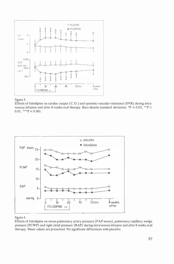
c o I /min
SVR 2500�
dyne sec 1500
c m -5
J 500
figure 3.
o PLACEBO • FELOOIPINE
i Ll�" _..._I _.__i �
0 30 60 I FELOOIPINE Iv I
90 --i
120m1n B weeks after
Effects of felodipine on cardiac output (C.O.) and systemic vascular resistance (SVR) during intravenous infusion and after 8 weeks oral therapy. Bars denote standard deviation. *P < 0.05, .. p < 0.01 , * * *P < 0.001 .
PAP mean 25
20
PCWP 1 5
10
RAP 5
mm Hg 0
figure 4.
0 30 60
FELOOlPlNE I v I
o placebo
• felodipme
90 120mm � B weeks
ofter
Effects of felodipine on mean pulmonary artery pressure (PAP mean), pulmonary capillary wedge pressure (PCWP) and right atrial pressure (RAP) during intravenous infusion and after 8 weeks oral therapy. Mean values are presented. No significant differences with placebo.
97

Sa� 100] 96
94
92
Sv02 80] 72
64
56
7
avDCai J ml/100ml:J
figure 5 .
o PLACEBO • FELODIPINE
������������- ---, 0 30 60 I FELODIPINE Iv I
90 120m1n 8 weeks after
Effects of felodipine on arterial oxygen saturation (Sa02), mixed venous oxygen saturation (Sv02), and arteriovenous oxygen content difference (a-vDC02) during intravenous infusion and after 8 weeks oral therapy. Bars denote standard deviation. •p < 0.05, ••p < 0.01, •••p < 0.001 .
After 8 weeks therapy.
Each of the 23 patients completed the 8 week study period. The hemodynamic data after 8 weeks are compared with the baseline values, between group comparison of gainscores (figs. 2-5) . There was an increase in cardiac output ( + 15% , P < 0.01) and stroke volume index ( + 12% , P < 0.05) , a decrease i n systemic vascular resistance (-21 % , P < 0.05) and in arterio venous oxygen content difference (- 14% , P < 0.01) , after felodipine therapy compared with placebo. Heart rate, arterial and pulmonary artery, capillary wedge and right atrial pressures were not statistically different from baseline values.
Cardio pulmonary exercise tests.
The results of the cardiopulmonary exercise tests are presented in table 2. Analysis of baseline values revealed no significant differences between the two
98

'° '°
Table 2 Results of Cardiopulmonary Exercise Tests
Placebo Felodipine P-value P-value n = 12 n = 1 1 baseline after
baseline after baseline after REST Heart Rate 96 ± 1 3 88 ± 1 1 107 ± 1 6 103 ± 13 . 16 .48 SAP 145 ± 50 142 ± 33 148 ± 32 130 ± 23 .89 .24 MAP 103 ± 19 97 ± 17 105 ± 19 88 ± 1 1 .92 .08 OAP 83 ± 12 80 ± 13 84 ± 14 70 ± 8 .82 .03* RPP 143 ± 63 134 ± 57 157 ± 35 135 ± 34 .49 .41 VE 11 ± 3 13 ± 4 13 ± 5 14 ± 3 . 17 .37 F 19 ± 4 19 ± 5 18 ± 5 19 ± 4 .61 .51 V02 4.0 ± .5 4.6 ± 1 . 1 4.2 ± .9 4.6 ± .9 .30 .83
SUB MAXIMAL Heart Rate 120 ± 16 1 1 6 ± 16 127 ± 2 1 1 12 ± 16 .20 .03* SAP 161 ± 29 164 ± 30 172 ± 38 156 ± 34 .45 .06 MAP 107 ± 16 106 ± 14 1 1 5 ± 19 97 ± 13 .27 .001 • OAP 82 ± 1 1 83 ± 12 86 ± 12 68 ± 6 .24 .002* RPP 194 ± 48 191 ± 48 217 ± 59 175 ± 44 . 19 .02• VE 23 ± 10 21 ± 4 25 ± 7 24 ± 5 .59 .79 F 24 ± 6 24 ± 5 28 ± 10 23 ± 6 .25 .08 V02 10.0±1 .9 9.9 ± 1 .6 1 1 . 1 ±2.2 10. 1 ±2.4 .22 .45
MAXIMAL Heart Rate 130 ± 18 129 ± 16 137 ± 17 136 ± 19 .21 .96 SAP 161 ± 31 176 ± 34 173 ± 39 169 ± 35 .45 . 12 MAP 107 ± 18 1 12 ± 16 1 16 ± 22 104 ± 15 .26 .003* OAP 79 ± 1 3 80 ± 1 1 83 ± 14 69 ± 6 .50 .03* RPP 209 ± 47 227 ± 44 236 ± 58 232 ± 57 .45 .41 VE 29 ± 9 27 ± 6 31 ± 8 36 ± 1 1 .59 .09 F 27 ± 5 26 ± 6 29 ± 9 28 ± 7 .49 .78 V02 1 1 .6±2. 1 1 1 .6±2.3 12.3±2.6 14.2±3.0 .44 .05 Exerc.Dur. 525 ± 132 555 ± 187 587 ± 227 742 ± 185 .45 .04*
P value after = Between group comparison of changes after therapy. SAP = Systolic Arterial Pressure (mmHg); MAP = Mean Arterial Pressure (mmHg) ; OAP = Diastolic Arterial Pressure (mmHg); RPP = Rate Pressure Product = Heart rate x SAP (b mmHg 10·2) ; VE = minute ventilation (I min-1) ; F = respiratory rate (breath min-1) ; V02 = oxygen consumption (ml kg-1 mm-1) ; Exerc.Dur. = Exercise Duration (sec) . Values are expressed as Mean ± SD. • = P < 0.05.

treatment groups. Between group comparison was made of the changes in both groups after 8 weeks therapy. At rest before exercise , no significant differences were seen apart from a decrease in diastolic bloodpressure in the felodipine group. Analysis of the results at submaximal exercise revealed significant decreases in heart rate, mean and diastolic arterial pressure and rate pressure product in the felodipine group compared with placebo. At maximal exercise, mean and diastolic arterial pressures were significantly decreased in the felodipine group, without differences in heart rate and rate pressure product . In the felodipine group exercise duration increased from 587 ±227 to 742 ± 185, in the placebo group from 525 ± 132 to 555 ± 187 seconds, P = 0.04 (fig.6). Maximal oxygen consumption increased from 12.3 ± 2.6 to 14.2 ± 3.0 in the felodipine group and did not change in the placebo group, 1 1 .6 ± 2. 1 , and 1 1 .6 ± 2.3 respectively, but the difference in changes between the groups did not reach significancy (P =
0.054) .
FELOOIPINE
N - 11
SECONDS
1000
800 j ~ 600
400
200
0
BASELINE
L figure 6.
EXERCISE DURATION
AFTER BASELINE
L p =004
PLACEBO
N: 12
_J AFTER
_J
Between group comparison of changes in exercise duration after 8 weeks treatment with felodipine or placebo.
1 00

Radio nuclide ventriculography.
The baseline radionuclide ejection fraction was similar in both groups (table 1) . After therapy ejection fraction was 26 ± 12% in the placebo group, and 29 ± 12% in the felodipine group. Between group comparison of the gainscores revealed no significant differences.
Chest X-ray.
No significant differences were observed in cardiothoracic ratio at base line and after therapy.
Clinical data.
Patient characteristics at baseline are given in table 1 . Eleven patients were assigned to felodipine treatment, and 12 patients to placebo. The only significant difference between the 2 patient groups was the sex distribution. In the felodipine group 9 were men and 2 women, in the placebo group 6 were men and 6 women . The initial dose of 5 mg b . i .d. could be increased to 10 mg b. i .d . in all patients after 2 weeks, but in 3 patients in the felodipine group and 1 in the placebo group the dosage was decreased after 4 weeks treatment, back to 5 mg b. i .d . (table 3) . Diuretics were temporarily increased in 1 patient of the placebo group. No other dose adjustments were made. The patients rating on the 7-graded scale demonstrated a subjective improvement after 2 weeks in the felodipine group, score 2.7 ± 0.8, compared with placebo 4 . 1 ± 2.2, P < 0.05. After 4, 6 and 8 weeks treatment the scores were 2.7 ± 1 .2 vs 4.4 ± 2.5 (P <0.05) , 2.6 ± 1 .0 vs 4.3 ± 2.0 (P < 0.01), and 2.9 ± 1 .2 vs 4.4 ± 1 .6 (P < 0.01) respectively. The subjectively assessed degree of dyspnea diminished in the felodipine group compared with placebo. By the end of the treatment period, 91 % of the patients assigned to felodipine treatment considered their dyspnea improved, compared with 41 % of the patients assigned to placebo (P < 0.01) . Body weight in the placebo group was 69.6 ± 1 1 before and 69. l ± 10 kg after treatment, in the felodipine group 74. 1 ± 10 and 73.9 ± 9 kg. The ankle circumference in the placebogroup was 23 .2 ± 2 before and 23 . 1 ± 1 cm after treatment, in the felodipine group 24.4 ± 2 and 24.9 ± 2 cm respectively. These changes were not significant.
I O I

Side effects.
Side effects as experienced by the patients are shown in Table 3. Severe symptoms lead to a dose reduction in 3 patients of the felodipine group, and in 1 patient of the placebo group. Diuretic treatment was temporarily increased in 1 patient of the placebo group. Subsequently these side effects disappeared.
Table 3 Number of Patients with Mild and Severe Side Effects During Treatment
Felodipine Placebo Symptoms mild severe mild severe Peripheral oedema 3 1 * 1 1 • Flushing 3 0 0 0 Tachycardia 1 1 * 0 0 Palpitations 0 1 * 0 0 Dizziness 1 0 1 0 Blurred Vision 1 0 0 0 Muscle weakness 1 0 2 0 Fatigue 0 0 1 0 Insomnia 0 0 3 0 Pruritus 0 0 2 0 Nausea 0 0 2 0 Conjunctivitis 0 0 1 0 Sweating 0 0 1 0
• = patients with side effects that lead to dose reductions of trial medication.
DISCUSSION
Clinical results.
The results of this study demonstrate the clinical efficacy of felodipine in patients with congestive heart failure. Subjectively, a significant improvement in general wellbeing occurred in the felodipine group compared with placebo. The clinical improvement that occurred in the patients of the felodipine group is not explained by alterations in concomitant drug therapy. The diuretic dosage was increased in one patient, of the placebo group. No other alterations were necessary. Increases in body weight and ankle circumference were not observed during and after 8 weeks of therapy.
102

Evaluation of hemodynamics at rest.
It is well established that hemodynamic data obtained at rest are not necessarily related to the severity of congestive heart failure or to the clinical response of vasodilator treatment (17-20) . An increase in cardiac output does not always lead to an improvement in exercise tolerance, and a decrease in wedge pressure not necessarily to an amelioration of dyspnea. Hence, we did not use hemodynamic data obtained at rest as inclusion criteria , and efficacy is described only in terms of objective exercise parameters and subjective symptoms. The results of the intravenous hemodynamic study are presented to give a detailed analysis of the hemodynamic profile of the drug. The pulmonary capillary wedge pressure at supine rest was rather low in both groups, especially in the light of the markedly depressed left ventricular ejection fraction .This was most probably due to the long term diuretic treatment with 50 mg hydrochlorothiazide in all patients. The hemodynamic profile of felodipine is clear : true arteriolar vasodilation leading to an increase in cardiac output and stroke volume index and to a decrease in vascular resistance and arterio venous oxygen content difference. No changes in right atrial , pulmonary artery and capillary wedge pressures, no venodilating activity. A decrease in arterial oxygen saturation was only seen during the intravenous study and is probably the result of the sudden increase in cardiac output, leading to perfusion of previously underperfused pulmonary areas (21) . An increase in heart rate and a decrease in arterial pressures was only observed during acute administration, but not after 8 weeks chronic oral therapy.
Evaluation of cardiopulmonary exercise tests.
Patients with congestive heart failure are limited during both submaximal and maximal exercise primarily by inadequate oxygen transport to working muscle (22,23) .
The results of the submaximal exercise tests are of particular interest , because of two reasons. Firstly, an important goal of therapy in congestive heart failure is to allow a patient to carry out submaximal exercise more comfortably. Secondly, the design of the submaximal exercise test enabled us to evaluate the effect of the drug at similar exercise levels. After 8 weeks therapy with felodipine, patients used the same amount of oxygen at the same submaximal exercise level, with significantly lower blood pressures, heart rate and, as a result, rate pressure product. This may point at a reduction of sympathetic activity after felodipine treatment (24) . The hemodynamic effect of felodipine at rest demonstrated a shift from pressure work to flow work. The increase in cardiac output paralelled the decrease in arterio venous oxygen content difference both during the intra-
103

venous study and after 8 weeks oral therapy, which may explain the lack of change in oxygen consumption, since oxygen consumption is the product of cardiac output and arterio venous oxygen content difference. We did not measure cardiac output and arterio venous oxygen content difference during exercise, but in other controlled studies with felodipine in congestive heart failure an increase in cardiac output during exercise has been demonstrated (8,25). One could postulate that the felodipine induced vasodilation may have occurred mostly in non exercising tissues. The increase in exercise duration demonstrates that at least a substantial part of the increase in flow was beneficial to the working muscles (26) . A definite answer can only be given by simultaneous measurement of femoral vein and pulmonary artery (mixed venous) oxygen content during both submaximal and maximal exercise. Oxygen consumption was unchanged at similar submaximal exercise levels. The almost significant increase in oxygen consumption at maximal exercise must be explained by the increase in exercise duration after felodipine treatment, and not by an increase of oxygen consumption caused by felodipine .
The placebo controlled, double blind design, makes it unlikely that the improvement in exercise capacity in the felodipine group without changes in aerobic capacity at any exercise level can be attributed to a training effect or investigator bias. There are of course some limitations of this study. An exercise assessment of stroke volume and cardiac output in association with the V02max results would have been particularly interesting. However, in earlier controlled studies (8,25) it was demonstrated that felodipine increases cardiac index and stroke volume during exercise. Heart rate and systolic blood pressure were somewhat higher in the felodipine group. Therefore one could suggest that the felodipine group had greater potential for subsequent improvement. However, the groups were well matched in all respects , statistical analyses revealed no significant differences at baseline , and between group analysis of changes demonstrated a distinct difference in changes in heart rate, arterial pressures, rate pressure product, and exercise duration. Thus, the interpretation of the results is that arterial vasodilation induced by felodipine increases cardiac output, which leads to an increase of nutritive blood flow to skeletal muscles, a decrease of pressure work at submaximal and maximal exercise levels and to an increase of exercise duration.
Exercise parameters and invasive hemodynamics at rest revealed significant changes between the treatment groups, but all non invasive parameters of left ventricular function obtained at rest failed to show differences after treatment. This discrepancy questions, once again, the usefullness of noninvasive parameters at rest in the diagnostic procedure of a disease state that is typified by symptom limited exercise . It also demonstrates the necessity to assess the efficacy of congestive heart failure treatment by exercise tests, preferably in combination with aerobic capacity measurements.
104

Side effects.
Apart from effects related to vasodilation which responded to dose reduction, felodipine treatment did not appear to cause any more unwanted effects than placebo. A number of "side effects" that did occur in the placebo group were not observed during felodipine therapy. These "side effects" may have been caused by congestive heart failure itself and may point, indirectly , to the amelioration achieved by felodipine therapy. However, a larger group of patients is required to confidently establish the incidence of side effects.
Comparison with other controlled felodipine studies in congestive heart failure.
Comparable favourable effects of felodipine have been demonstrated in a number of open studies (5-7) . Two controlled studies have been published. Tan et al (8) and Kassis et al (25) studied the effect of felodipine in heart failure patients after a treatment period of 3 weeks, using a cross over design , with a wash out period of 1 week. The dose of felodipine was the same. A comparable beneficial hemodynamic effect was observed without subjective improvement or increase in exercise capacity. Cardio pulmonary exercise tests were not performed.
The discrepancy between these results and our study may be caused by differences in study design and length of treatment. A cross over design may lead to carry over effects when the placebo wash out period is as short as 1 week, especially if a potent vasodilating drug is given to congestive heart failure patients (27) . Reduced exercise capacity is primarily due to reduced nutritive blood flow to skeletal muscles, and increased muscle flow due to vasodilation does not improve peripheral oxygen utilization immediately. A delayed effect of 2 to 4 weeks on exercise tolerance is often observed (28,29) .
In summary, our study shows that an 8 week treatment period with felodipine results in a hemodynamic change from pressure work to flow work at rest. Exercise duration increases without changes in oxygen consumption. Submaximal exercise is performed at a lower rate pressure product. Eight weeks therapy with felodipine lead to objective and subjective symptomatic improvement and side effects were minor. Felodipine has an attractive profile for the management of congestive heart failure. Further research with this drug is necessary to demonstrate long term efficacy and safety. A controlled study during a longer treatment period might be of interest, preferably in comparison with ACE inhibitors or the combination of hydralazine and nitrates, because the effectiveness of these drugs in the treatment of congestive heart failure has been demonstrated.
105

Acknowledgements
This study was supported by Astra Cardiovascular/AB Hassle, Sweden
References
1 Braunwald E. Mechanism of action of calcium-channel blocking agents. N Engl J Med 1982;307:1618-1627.
2 Josephson MA, Singh BN. Use of calcium antagonists in ventricular dysfunction. Am J Cardiol 1985;55:1 B-88B.
3 Ljung B. Vascular selectivity of felodipine. Drugs l985;29(suppl 2):46-58. 4 Culling W, Ruttley MSM, Sheridan DJ. Acute haemodynamic effects of felodipine during beta
blockade in patients with coronary artery disease. Br Heart J 1984;52:431-4. 5 Emanuelsson H, Hjalmarson AA, Holmberg S, Waagstein F. Acute hemodynamic effects of fe
lodipine in congestive heart failure. Eur J Clin Pharmacol 1985;28:489-93. 6 Tweddel AC, Hutton I. Felodipine in ventricular dysfunction. Eur Heart J 1986;7:54-60. 7 Timmis AD, Smyth P, Kenny JF, Campbell S, Jewitt DE. Effects of vasodilator treatment with
felodipine on haemodynamic responses to treadmill exercise in congestive heart failure. Br Heart J 1984;52:314-20.
8 Tan LB , Murray RG, Littler WA. Felodipine in patients with chronic heart failure:discrepant haemodynamic and clinical effects. Br Heart J 1987;58: 122-8.
9 Franciosa JA. Exercise testing in chronic congestive heart failure. Am J Cardiol 1984;53: 1447-50.
10 Weber KT, Janicki JS, McElroy PA. Determination of aerobic capacity and the severity of chronic cardiac and circulatory failure. Circulation 1987;76(suppl VI)40-45.
1 1 Lipkin DP. The role of exercise testing in chronic heart failure. Br Heart J 1987;58:559-66. 12 The Criteria Committee of the New York Heart Association: Diseases of the Heart and Blood
Vessels. In:Nomenclature and Criteria for Diagnosis 7th ed. Boston, Little, Brown&Co, 1973;286.
13 Jones NL,Campbell EJM. Clinical Exercise Testing, Appendix B4. Philadelphia: WB Saunders, 1982.
14 Naimark A, Wasserman K, Mcllroy MB. Continuous measurement ofventilatory exchange ratio during exercise. J Appl Physiol 1964;15:644.
15 Reiber JHC, Lie SP, Simoons ML, Hoek C, Gerbrands JJ, Wijns W, Bakker WH, Kooij PPM. Clinical validation of fully automated computation of ejection fraction from gated equilibrium blood-pool scintigrams. J Nucl Med 1983;24:1099- 1 107.
16 Weber KT, Janicki JS. Chronic Cardiac Failure. In:Cardio Pulmonary exercise testing. Philadelphia: WB Saunders Company, 1986;169.
17 Massie B, Ports T, Chatterjee K, et al . Long-term vasodilator therapy for heart failure: clinical response and its relationship to hemodyamic measurements. Circulation 1981 ;63:269-278.
18 Franciosa JA, Dunhman WB , Leddy CL. Hemodynamic effects of vasodilators and long-term response in heart failure. J Am Coll Cardiol 1984;3: 1521-30.
19 Massie BM, Kramer BL, Topic N. Lack of relationship between the short term hemodynamic effects of captopril and subsequent clinical response. Circulation 1984;69: 1 135-41 .
20 Szlachcic J , Massie BM, Kramer BL, Topic N , Tuban J. Correlates and prognostic implication of exercise capacity in chronic congestive heart failure. Am J Cardiol 1985;55:1037-42.
21 Vrabel TR, Cohn JN. Comparative hemodynamic effects of converting enzyme inhibitor and sodium nitroprusside in severe heart failure. Am J Cardiol 1980;45:331-36. 22 Wilson JR, Ferraro N. Exercise intolerance in patients with chronic left heart failure: relation to oxygen transport and ventilatory abnormalities. Am J Cardiol 1983;51:1358-63.
1 06

23 Wilson JR, Martin JL, Schwartz D , Ferraro N. Exercise intolerance in patients with chronic heart failure: role of impaired skeletal muscle nutritive flow. Circulation 1984;69: 1079-87.
24 McGrath BP, Arnolda LF.Enalapril reduces the catecholamine response to exercise in patients with heart failure. Eur J Clin Pharmacol 1986;30:485-487.
25 Kassis E, Amtorp 0, Waldorff S, Fritz-Hansen P. Efficacy of felodipine in chronic congestive heart failure: a placebo controlled haemodynamic study at rest and during exercise and orthostatic stress. Br Heart J 1987;58:505-1 1 .
26 Maskin CS,Forman R , Sonnenblick EH, Frishman WH, LeJemtel TH. Failure of dobutamine to increase exercise capacity despite hemodynamic improvement in severe chronic heart failure. Am J Cardiol 1983 ;5 1 : 177-82.
27 Hills M, Armitage P. The two-period cross-over clinical trial. Br J Clin Pharmac 1979;8:7-20. 28 Rubin S, Chatteree K, Parmley WW. Metabolic assessment of exercise in chronic heart failure
patients treated with short-term vasodilators. Circulation 1983;67:817-22. 29 Franciosa JA, Goldsmith SR, Cohn JN. Contrasting immediate and long-term effects of isosor
bide dinitrate on exercise capacity in congestive heart failure. Am J Med 1980;69:559-66.
107


CHAPTER S
Different results after long term treatment with Felodipine and Enalapril in Cardiopulmonary Exercise
Tests in Patients with Congestive Heart Failure
Peter H.J.M. Dunselman v, C.Erik E. Kuntze v, Auke van Bruggen v ,
Hans P.M. Hamer\ Arnold H.J. Scaf* , Harry Wesseling* and Kong Ing Lie Y
Departments of Cardiology v and Pharmacology/Clinical Pharmacology* , Groningen University, The Netherlands
ABSTRACT
We evaluated the cardiopulmonary exercise test results before and after long term (16 weeks) treatment with the dihydropyridine calcium antagonist felodipine (10 mg b. i .d . ,n=9), and the ACE inhibitor enalapril (10 mg b. i .d . ,n= ll) in 20 patients with New York Heart Association class III congestive heart failure. Baseline statistics revealed no significant differences. After 16 weeks the enalapril group showed a significant increase in exercise duration and V02max, without changes in arterial pressures and heart rate. In the felodipine group exercise duration and V02max did not change significantly, but arterial pressures and heart rate were significantly reduced at all exercise levels. Between group analysis showed a significant reduction in arterial pressures and heart rate in the felodipine group if compared with enalapri l , without differences in aerobic capacity and exercise duration . These results demonstrate that felodipine and enalapril have essentially different effects on cardiopulmonary exercise results in patients with congestive heart failure.
109
INTRODUCTION
Many reports about the benefits of vasodilator therapy in congestive heart failure have been published in recent years. The efficacy of ACE inhibitors has been documented with regard to symptoms (1 ,2) , exercise duration (3) and reduction of mortality ( 4) . The hemodynamic profile of ACE inhibitors can be described as balanced vasodilation, arterial vasodilation by inhibition of the production of the vasoconstrictor angiotensin-11 and aldosteron, while filling pressure is reduced by enhanced diuresis and systolic unloading. The calcium antagonists of the dihydropyridine group are under investigation in congestive heart failure. Studies with the archetype of these drugs , nifedipine, demonstrated that the beneficial effects of arterial vasodilation are partly counteracted by a negative inotropic effect on cardiac muscle (5,6) . Felodipine is a dihydropyridine with negligible negative inotropic effects due to its selectivity for smooth muscle (7) . The hemodynamic effect of felodipine in congestive heart failure is pure arterial vasodilation, without direct effects on the venous side (8-12) . It has been established that hemodynamic measurements do not correlate with the severity of heart failure, and that beneficial changes in hemodynamic profile after therapy are not necessarily related to an increase in exercise capacity (13-16) . The cardiopulmonary exercise test with measurement of aerobic capacity during submaximal and maximal exercise makes it possible to get an objective assessment of the efficacy of vasodilator treatment in patients with congestive heart failure (17-19) .
No data are available about possible differences in cardiopulmonary exercise tests between dihydropyridines and ACE-inhibitors, vasodilating drugs with a different hemodynamic profile. Therefore, cardiopulmonary exercise tests before and after long term treatment with felodipine and enalapril were analyzed in patients with congestive heart failure.
METHODS
All patients in this study first participated in an 8 weeks, randomized, double blind, placebo controlled study with felodipine (8) . During this double blind treatment period eleven patients were treated with felodipine and twelve with placebo. The randomization code could not be broken for an individual patient who completed the 8 week study period, before the last patient that was entered into the study completed the study period. After the 8 week study period, a decision was made by every individual patient about future therapy, from two options, open treatment with felodipine 10 mg b. i .d . , or open treatment with enalapril 10 mg b. i .d. , a registered drug with demonstrated efficacy in the treatment
1 10

of heart failure. The patients were well aware of the fact that both investigator and patient remained blinded towards the treatment allocation of the first 8 week treatment period. From the twelve patients allocated to placebo in the double blind period, 1 patient decided to "continue" with open felodipine treatment, and 1 1 patients decided to change to enalapril . From the eleven patients allocated to felodipine in the first treatment period, 9 patients decided to "continue" with open felodipine, and 2 patients decided to change to enalapril. The cardiopulmonary exercise test results of2 patient groups are compared by analysis of within and between group changes after 24 weeks versus the baseline values of the double blind study: group 1 , consisting of 9 patients, 8 weeks of double blind felodipine treatment followed by 16 weeks open felodipine treatment, and group 2, 11 patients, 8 weeks double blind placebo treatment followed by 16 weeks enalapril treatment. Double blindness towards the treatment allocation of the first period was maintained troughout the whole study. The cause of congestive heart failure was coronary artery disease as documented by myocardial infarctions more than 3 months ago. Patients were in New York Heart Association class III (20) , with an ejection fraction < 40% at supine rest (radionuclide ventriculography) and a maximal oxygen uptake < 15 ml/kg/min. Patients were in sinus rhythm, on a fixed dose of digitalis and diuretics (hydrochlorothiazide 50 mg once daily, with potassium suppletion if necessary) , and a sodium chloride restricted diet of < 3 grams sodium chloride daily , throughout the whole study. No vasodilating, beta blocking or antiarrhythmic drugs were allowed. Written informed consent was obtained from all patients. The study protocol was approved by the ethics committee of the University Hospital Groningen.
Cardiopulmonary exercise test.
The presence of intrinsic pulmonary disease was excluded by spirometry using flow rate, lung volume and maximal voluntary ventilation. The exercise test was performed on a treadmill 2 hours after the latest meal. Intra-arterial blood pressures were continuously measured and in the last minute of each exercise step arterial blood samples were drawn and analyzed for arterial blood gases. The treadmill protocol and technical procedures are previously described (21) . The submaximal exercise level was defined for each patient as the next to highest exercise level achieved, provided that this submaximal exercise level was achieved before and after treatment. The exercise test was terminated at the patients request. All patients stopped exercise because of the onset of fatigue or dyspnea, no one was limited by chest pain, hypotension, electrocardiogram changes or arrhythmias. This cardiopulmonary exercise test was by nature a symptom limited test. The accuracy of the true V02max measurement requires that a plateau
1 1 1

in oxygen uptake can be demonstrated, despite further increments in exercise workload. Patients with congestive heart failure are often unable to achieve such a plateau in oxygen uptake during graded exercise because they are limited by symptoms of dyspnea or fatigue. Thus V02max should be read here as the symptomatic maximum oxygen uptake, verified by an increase in the respiratory quotient by at least . 15 from its lowest value, and an absolute value of > 1 .0 during maximal exercise to consider the exercise as valid with regard to maximal oxygen consumption (22) . The maximal oxygen uptake of 15 ml kg-1 min"1 or less was required to verify that only patients with a documented impairment of functional capacity were entered into the study (16) . The personnel of the cardiopulmonary exercise laboratory was blinded towards treatment schedules.
Statistical analysis.
Similarity of baseline values was tested with unpaired t-tests. Between group analysis was made by comparison of the difference in changes between the groups after treatment using unpaired t-tests. Within group analysis was performed by paired t-tests and analysis of variance (F- test) . Differences were considered significant if P was < 0.05.
RESULTS
There were no differences between the groups in baseline characteristics with respect to age, ejection fraction and cardiopulmonary exercise test results. There were no inequities in the outcome of submaximal exercise duration (278 ± 128 sec. versus 290 ± 135 sec. ) between the groups, making the desired comparison of exercise test results at similar exercise levels possible (Table 1 ) .
Possible placebo or training effects were analyzed by comparison of the results from the cardiopulmonary exercise tests from the placebo group before and after the 8 weeks double blind period. No significant differences were observed (Table 2) . Within group analysis of changes after felodipine revealed a significant decrease in systolic arterial pressure (-22%, P < 0.01), mean arterial pressure (-
22% , P < 0.01), diastolic arterial pressure (-13%, P < 0.05) , and rate pressure product (-18% , P < 0.05) at rest. At submaximal exercise a significant decrease was found in heart rate (- 10% , P < 0.05) , mean arterial pressure (-18% , P < 0.01 ) , diastolic arterial pressure (-22% , P < 0.001) and rate pressure product (-
20% , P < 0.05) . This was maintained during maximal exercise with a decrease in mean arterial pressure (- 18% , P < 0.05) , diastolic arterial pressure (-22% , P <
1 12

Table 1 Cardiopulmonary Exercise Test Results at baseline Age EF V02 HR SAP MAP DAP RPP Ex.Dur.
REST FELODIPINE 63 29 4.3 104 151 107 88 157 0
±7 ±9 ± . 8 ± 15 ±35 ±20 ± 1 5 ±36
ENALAPRIL 58 27 3.9 98 154 105 85 153 0 ±7 ±9 ± .5 ± 1 3 ±33 ± 1 8 ± 1 1 ±41
SUBMAXIMAL EXERCISE
FELODIPINE 1 1 .7 124 176 1 1 8 85 219 278 ± 1 .9 ±23 ±42 20± ± 12 ±44 ± 128
ENALAPRIL 10 . 1 1 17 162 109 81 188 290 ±2.0 ± 13 ±30 ± 1 5 ± 10 ±40 ± 135
MAXIMAL EXERCISE
FELODIPINE 12.5 136 179 121 86 243 619 ±2.3 ± 1 8 ±42 ±21 ± 14 ±42 ±235
ENALAPRIL 1 1 .9 130 165 109 81 210 536 ± 1 .9 ± 16 ±30 ±16 ±12 ±41 ± 134
Comparison of baseline characteristics in the 2 treatment groups. Felodipine patients : n = 9; Enala-pril patients : n = 1 1 . Values are expressed as mean ± SD. Age (years); EF = Left Ventricular Ejec-tion Fraction (%); 02 = oxygen uptake (ml kg·1 min.1); F = respiratory rate (breath min. 1} ; HR = heart rate (beats min.1) ; SAP = systolic arterial pressure (mmHg); MAP = mean arterial pressure (mmHg); DAP = diastolic arterial pressure (mmHg); RPP = rate pressure product = HR x SAP (b mmHg 10·2) ; Ex.Dur. = exercise duration (sec). No significant differences between the groups.
0.01) and rate pressure product (-14% , P < 0.05). Within group analysis of changes after enalapril revealed no changes in heart rate and blood pressures at rest, and at submaximal and maximal exercise levels (fig. 1) . A significant increase in maximal oxygen uptake ( + 23% , P < 0.02) and maximal exercise duration ( + 33% , P < 0.02) was seen in the enalapril group, but not in the felodipine group (fig 2) . Between group comparison of changes disclosed a significantly greater decrease in arterial pressures and rate pressure product at submaximal and maximal exercise levels after felodipine than after enalapri l , without significant differences in heart rate, aerobic capacity and duration of exercise. The analysis of aerobic capacity data and arterial gas tensions revealed no differences between the 2 treatment groups (Table 3) .
1 1 3
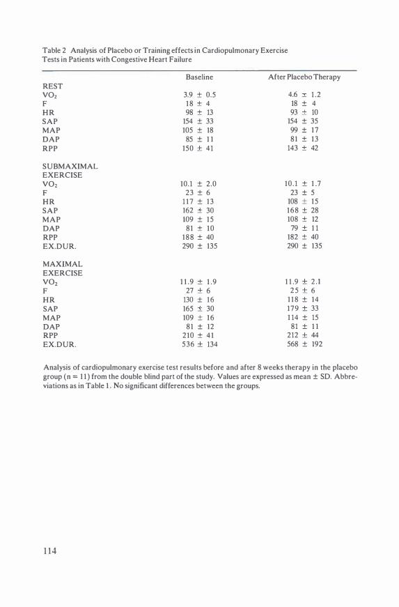
Table 2 Analysis of Placebo or Training effects in Cardiopulmonary Exercise Tests in Patients with Congestive Heart Failure
REST V02 F HR SAP MAP OAP RPP
SUB MAXIMAL EXERCISE V02 F HR SAP MAP OAP RPP EX.DUR.
MAXIMAL EXERCISE V02 F HR SAP MAP OAP RPP EX.DUR.
Baseline
3.9 ± 0.5 18 ± 4 98 ± 13
154 ± 33 105 ± 18 85 ± 1 1
150 ± 41
10.1 ± 2.0 23 ± 6
1 1 7 ± 13 162 ± 30 109 ± 15 81 ± 10
188 ± 40 290 ± 135
1 1 .9 ± 1 .9 27 ± 6
130 ± 16 165 ± 30 109 ± 16 81 ± 12
210 ± 41 536 ± 134
After Placebo Therapy
4.6 ± 1 .2 18 ± 4 93 ± 10
154 ± 35 99 ± 17 81 ± 13
143 ± 42
10 . 1 ± 1 .7 23 ± 5
108 ± 15 168 ± 28 108 ± 12 79 ± 1 1
182 ± 40 290 ± 135
1 1 .9 ± 2 . 1 25 ± 6
1 1 8 ± 14 179 ± 33 1 14 ± 15 81 ± 1 1
212 ± 44 568 ± 192
Analysis of cardiopulmonary exercise test results before and after 8 weeks therapy in the placebo group (n = 1 1 ) from the double blind part of the study. Values are expressed as mean ± SD. Abbreviations as in Table 1 . No significant differences between the groups.
1 14

125
100 D'
I E .s 75
50
figure 1
MEAN ARTE RIAL PRESSURE REST
NS . .
f:: •
••• C F c E
SUB MAX
1
c
p<0 01 . .
• j :�:
I F c E
MAX
c
p<001 . .
• l
Ill F c E
250
"' I $2 200 D'
I E E 150 I c E
..D 100
RATE PRESSURE PRODUCT REST
NS SUBMAX MAX
P<005 P <0 02
·. •
• •• • ·.·
• •• C F C E C F C E
Changes in mean arterial pressure and rate pressure product after felodipine (F) and enalapril (E) at different exercise levels. Submax = submaximal ; Max = maximal; Values presented are the means q standard error of the mean. Asterisks represent significance of changes (P < 0.05) with each drug compared to its control values (within group analysis). P values above the bars represent significance of the hemodynamic changes with enalapril compared with those produced by felodipine (between group analysis of changes versus baseline). C = control at baseline; NS =not significant.
REST NS
15
-I 10
D' --"' � I
5 l c E I 'E
0 c F ( figure 2
? E
V02 S U B MAX
c
NS . .
::: ,•, ::: .·.
iii .·. . ·. -=· �=�
F C E
l
MAX NS
. .
l l ·:·
lll ���I
C F ( E
750
500
VI -0 c 0
u 250 QJ VI
0
EXERCISE DURATION SUB MAX MAX
NS • I
l
•••• J
l ·.· ·.·
:iii �=� .· . .·.
�=� ( F c E c F ( E
Changes in oxygen uptake (V02) and exercise duration after felodipine (F) and enalapril (E) at different exercise levels. Format and abbreviations as in figure 1 .
1 15

Table 3 Aerobic Capacity & Arterial Gas Tensions during Cardiopulmonary Exercise Tests
VE p02 vco, pC02 SA02 l . min-1 KpA ml .mi� 1 KpA %
SUBMAXIMAL EXERCISE
FELODIPINE 27_8 1 1 .9 651 4.5 97 ±7.2 ± 1 .7 ± 1 14 ±0.7 ± I . I
ENALAPRIL 24.0 12.7 615 4.6 97 ±4.6 ± I . I ± 151 ±0.6 ±0.9
MAXIMAL EXERCISE
FELODIPINE 36.0 1 2 .8 946 4.4 97 ±9.7 ± 1 .6 ±272 ±0.6 ±0.9
ENALAPRIL 36.8 13.3 985 4.4 98 ±9.2 ± 1 .4 ±284 ±0.8 ±0.8
Comparison of the aerobic capacity measurements and artenal gas tensions after long term treatment. Felodipine patients : n = 9; Enalapril patients : n - 1 1 . VE = minute volume; p02 = partial pressure of oxygen in the arterial blood; VC02 = Carbond1oxide production; pC02 = partial pressure of carbondioxide in the arterial blood; SAO, = percent oxyhemoglobin saturation of arterial blood; Values are expressed as mean + SD. No significant differences between the groups.
DISCUSSION
Our findings demonstrate that the group treated with felodipine and the one given enalapril had essentially different cardiopulmonary exercise test results. Because of the allocation procedure a prudent approach towards the interpretation of the data is warranted. A comparison of the overall efficacy of these drugs in heart failure is not allowed. This study is therefore presented as a descriptive analysis of the profile of these drugs with respect to the objective cardiopulmonary exercise test. One of the goals in the treatment of heart failure is to enable the patients to carry out submaximal exercise in greater comfort. Submaximal exercise levels were defined to compare the effects of the drugs at similar exercise levels before and after therapy. The cardiac work e.g. myocardial oxygen consumption at similar submaximal exercis� levels was reduced by felodipine, but not by enalapri l . The significant and consistent decrease in both heart rate and arterial pressures at all exercise levels seems of interest, since other dihydropyridines, like nifedipine, tend to increase heart rate. If the adrenergic drive to the heart were really depressed by felodipine, probably as a result from improve-
1 1 6

ment of the circulatory state (8) , the hypothesis that the lack of negative inotropic properties of this specific dihydropyridine is of clinical significance would be reinforced.
Treatment with enalapril resulted in an increase of both exercise duration and maximal oxygen uptake, if compared with baseline values within the treatment group. In a double blind comparison of captopril and nifedipine in patients with dilated cardiomyopathy a significant improvement of exercise duration could only be demonstrated in the captopril group, but oxygen uptake was not measured (23) . The interpretation of data related to oxygen uptake is complex. V02 is the product of cardiac output and arteriovenous oxygen content difference. Enalapril and felodipine increase cardiac output at rest and during exercise (3 , 1 1 , 12) . An unchanged V02 after therapy does not mean that the circulatory state of the patient is unchanged (24) . Peripheral vasodilation in metabolically non-active vascular beds leads to an increase in cardiac output with a narrowing of the arteriovenous oxygen content difference. When blood flow to the exercising muscles is not increased, V02 remains unchanged. Thus, an increase in cardiac output by random peripheral vasodilation may not produce clinical improvement. On the other hand, if vasodilating therapy leads to an increase in both exercise duration and V02, one may assume that part of the increase in cardiac output is directed to the exercising muscles, and not shunted into a different (pulmonary, splanchnic) part of the circulation (25 ,26) . This seems to be apparent after enalapril treatment. Felodipine, so effective in increasing cardiac output (8-12) , demonstrates to have greater difficulty in showing an improvement in exercise tolerance, as has been demonstrated with other arterial vasodilators (23 ,27 ,28) . Enalapril is a balanced vasodilator, while felodipine is a pure arterial vasodilator. The impact of this difference in hemodynamic profile on cardiopulmonary exercise test results is not well established. It has been demonstrated that treatment with felodipine also results in a reduction of capillary wedge pressure during exercise, probably secondary to improved left ventricular emptying (10, 11) . More important, exercise tolerance in patients with congestive heart failure is not dependent on the ventilatory consequences of pulmonary congestion, but on the delivery of blood to exercising muscles (25 ,29,30) . The lack of differences in arterial blood gases and aerobic capacity during both submaximal and maximal exercise within and between the treatment groups further demonstrates that a difference in flow distribution is a more likely explanation for the observed differences in cardiopulmonary exercise test results than a different vasodilating profile.
We did not measure cardiac output, mixed venous and femoral vein oxygen content, or filling pressures during exercise, what might have helped in the analysis of the disparity between the cardiopulmonary exercise test results. The comparison of 2 groups that have been followed for the same period of time (24
1 17

weeks) but with a difference in duration of treatment, 16 weeks for the enalapril group and 24 weeks for the felodipine group, suggests an advantage for the felodipine group, making the observed differences in exercise capacity even more important, because vasodilator therapy results in an almost immediate improvement in hemodynamics while exercise capacity improves gradually and progressively over a period of 12 to 24 we.eks (14,25 ,26) . This late clinical response is related to an improvement in peripheral oxygen utilization that accompanies longterm therapy with vasodilator drugs (24,30-33) . The lack of differences in cardiopulmonary exercise results before and after 8 weeks treatment with placebo suggests that our results are not influenced by training effects or deterioration of the condition of the patient . Although there were no significant differences between the enalapril and felodipine groups at baseline, arterial pressures and rate pressure product were somewhat higher in the felodipine group. Therefore one could suggest that the felodipine group had greater potential for subsequent hemodynamic improvement. Alternately, oxygen uptake and exercise duration were somewhat lower in the enalapril group, making a significant improvement in these variables more likely to occur. However, the groups were well matched in all respects, the changes in arterial pressures and rate pressure product in the felodipine group and in oxygen uptake and exercise duration in the enalapril group were marked, and between group analysis of changes demonstrated a distinct difference in hemodynamic profile. It seems of interest to analyze in a long term, double blind study whether a possible difference in overall clinical efficacy of these drugs can be explained by the observed disparity in cardio pulmonary exercise test results.
References
l . Packer M. Converting-enzyme inhibition for severe chronic heart failure : views from a skep· tic.Int J Card 1985;1 1 1-120.
2. Chatterjee K, Parmley WW, Cohn JN, Levine TB: Captopril Multicenter Research Group:a cooperative multicenter study of captopril in congestive heart failure: hemodynamic effects and long-term response. Am Heart J 1985; 1 10:439-447.
3. Sharpe DN, Murphy J, Coxon R, Hannan SF: Enalapril in patients with chronic heart failure: a placebo controlled, randomized, double blind study. Circulation 1984;70:271-278.
4. The CONSENSUS Trial study group. Effects of enalapril on mortality in severe congestive heart failure: results of the Cooperative North Scandinavian Enalapril Survival study (CONSENSUS). N Engl J Med 1987;316: 1429-35.
5. Braunwald E. Mechanism of action of calciutn-channel blocking agents. N Engl J Med 1982;307:1618-1627.
6. Josephson MA, Singh BN. Use of calcium antagonists in ventricular dysfunction. Am J Cardiol 1985 ;55: 1 B-888 .
7. Ljung B. Vascular selectivity of felodipine. Drugs 1985;29(suppl 2):46-58.
1 18

8. Dunselman PHJM, Kuntze CEE, van Bruggen A, Hamer JPM, Scaf AHJ , Wesseling H, Lie Kl. Efficacy of felodipine in congestive heart failure. Eur Heart 1 1989, in press.
9. Timmis AD, Smyth P, Kenny JF, Campbell S, Jewitt DE. Effects of vasodilator treatment with felodipine on haemodynamic responses to treadmill exercise in congestive heart failure. Br Heart J 1 984;52:3 14-20.
10 . Tan LB , Murray RG, Littler WA. Felodipine in patients with chronic heart failure:discrepant haemodynamic and clinical effects. Br Heart J 1987;58: 122-8.
I 1 . Kassis E, Amtorp 0, Waldorff S, Fritz-Hansen P. Efficacy of felodipine in chronic congestive heart failure: a placebo controlled haemodynamic study at rest and during exercise and orthostatic stress. Br Heart J 1987;58:505- 1 1 .
12. Binetti G , Pancaldi S , Giovanelli N, Negroni S , Ferretti RM, Branzo A , Specchia S ,Magnani B . Hemodynamic effects of felodipine in congestive heart failure. Cardiov Drugs and Tuer 1987;1 : 161-167.
13 . Benge W, Litchfield RL, Marcus ML. Exercise capacity in patients with severe left ventricular dysfunction. Circulation 1980;61 :955-959.
14. Massie B , Ports T, Chatterjee K, et al. Long-term vasodilator therapy for heart failure:clinical response and its relationship to hemodyamic measurements. Circulation 1981 ;63:269-278.
15 . Franciosa JA, Dunhman WB, Leddy CL. Hemodynamic effects of vasodilators and long-term response in heart failure. J Am Coll Cardiol 1984;3: 1 521-30.
16. Franciosa JA, Park M, Levine B. Lack of correlation between exercise capacity and indices of resting left ventricular performance in heart failure. Am J Cardiol 1981 ;47:33-39.
17. Weber K, Janicki J. Cardiopulmonary exercise testing for evaluation of chronic cardiac failure. Am J Cardiol 1985;55:22A-31A.
18. Weber KT, Janicki S , McElroy PA. Determination of aerobic capacity and the severity of chronic cardiac and circulatory failure.Circulation 1987;76 (suppl VI)40-45.
19. Lipkin DP. The role of exercise testing in chronic heart failure. Br Heart J 1987;58:559-66. 20. The Criteria Committee of the New York Heart Association: Diseases of the Heart and Blood
Vessels. ln:Nomenclature and Criteria for Diagnosis 7th ed. Boston , Little, Brown&Co, 1973;286.
2 1 . Dunselman PHJM, Kuntze CEE, van Bruggen A, Beekhuis H, Piers B, Scaf AHJ , Wesseling H, Lie Kl. Value of New York Heart Association classification , radionuclide ventriculography, and cardiopulmonary exercise tests for selection of patients for congestive heart failure studies. Am Heart ) 1 988 ; 1 16 : 1475- 1482.
22. Naimark A, Wasserman K, Mcllroy MB. Continuous measurement of ventilatory exchange ratio during exercise. 1 Appl Physiol 1964; 15:644.
23. Agostoni PG, De Cesare N, Doria E, Polese A, Tamborini GG and Guazzi MD. Afterload reduction: a comparison of captopril and nifedipine in dilated cardiomyopathy. Br Heart 1 1986;55:391-399.
24. Maskin CS , Forman R, Sonnenblick EH. Failure of dobutamine to increase exercise capacity despite hemodynamic improvement in severe chronic heart failure. Am 1 Cardiol 1983;5 1 : 177-12 .
25. Mancini DM, Le Jemtel TH, Factor S , Sonnenblick EH. Central and peripheral components of cardiac failure. l 986;80(suppl 2B):2-13.
26. Wilson JR, Martin JL, Schwartz D, Ferraro N. Exercise intolerance in patients with chronic heart failure: role of impaired nutritive flow to skeletal muscle.Circulation 1 984;69: 1 079-87.
27. Franciosa JA, Jordan RA, Wilen MM, Leddy CL. Minoxidil in patients with chronic left heart failure: contrasting hemodynamic and clinical effects in a controlled trial. Circulation 1984;70:63-68.
28. Franciosa JA, Weber KT, Levine TB . Hydralazine in the longterm treatment of chronic heart failure: lack of difference from placebo.Am Heart J 1982;104:587-594.
29. Wilson JR, Ferraro N. Exercise tolerance in patients with chronic left CHF: relation to oxygen transport and ventilatory abnormalities. Am 1 Cardiol 1983;5 1 : 1 358-1363.
30. Franciosa JA, Goldsmith SR, Cohn JN. Contrasting immediate and long-term effects of isosorbide dinitrate on exercise capacity in congestive heart failure. Am J Med 1980;69:559-566.
119

31 . Franciosa J ,Cohn J . Immediate effects of hydralazine- isosorbide dinitrate combination on exercise capacity and exercise hemodynamics in patients with left ventricular failure Circulation 1979;59: 1085-1091 .
32. Franciosa J, Cohn J. Effect of isosorbide dinitrate on response to submaximal and maximal exercise in patients with congestive heart failure. Am J Cardiol 1979;43: 1009- 1014.
33. Massie BM, Kramer BL, Topic N. Lack of relationship between the short term hemodynamic effects of captopril and subsequent clinical response. Circulation 1984;69 : 1 135-4 1 .
1 20

CHAPTER 9
Epilogue
Felodipine has been shown to be an effective and well tolerated drug in patients with congestive heart failure . The major difference with other dihydropyridines is probably its vascular selectivity, resulting in a lack of detrimental negative inotropic effects on the cardiac muscle. The potent coronary dilator properties of felodipine with concomitant improvement in the oxygen supply/demand ratio may be of importance in congestive heart failure due to myocardial ischemia. Side effects, head ache, flushes and ankle edema, appear to be dose related and are manifestations of the drugs potent vasodilating activity.
However, the majority of data in literature are derived from a small number of studies, and more studies are needed to confirm its usefulness. It seems crucial to analyze the efficacy of felodipine in congestive heart failure in more, well designed long term clinical trials. A direct comparison of felodipine with ACE inhibitors (enalapril, captopril) , or the combination of hydralazine and nitrates, is needed to clarify its place in the long term management of congestive heart failure , with special attention to the influence of the drug on mortality.
The hemodynamic profile of felodipine intravenously in patients with congestive heart failure suggests that it may be an attractive drug to use in acute heart failure . Patients in disease states typified by extreme vasoconstriction, diastolic filling abnormalities, hypertension and circulatory shock (e.g. cardiac asthma or "asthma cardiale") may benefit from this drug. The theoretical advantage of felodipine are, as mentioned before, its lack of negative inotropism, and its metabolic (renal) inactivity. Another attractive feature is the possibility to continue with the oral compound after successful intravenous intervention. A direct comparison of felodipine with drugs like nitroprusside and nitroglycerine in acute heart failure seems therefore of interest.
1 2 1


Summary
Chapter 1 offers an introduction in which the place of vasodilator therapy in heart failure is described. A review of the relevant studies with vasodilating drugs is presented. In the second part, the methodological problems in heart failure research are analyzed. In the third part the results of published studies with felodipine in congestive heart failure are discussed, together with the results of studies in other circulatory diseases. The aims of the study published in this book are outlined in the last part of the chapter.
Chapter 2 evaluates the necessity of an objective measurement to assess the severity of congestive heart failure. It is demonstrated that the subjective appraisal of patients complaints, the New York Heart Association (NYHA) Classification separates patients with a mild to moderate impairment of functional capacity very well from patients with a moderate to severe impairment of functional capacity, but it also shows that about one third of the patients with congestive heart failure, eligible for research protocols by their history and documented left ventricular dysfunction, will be wrongly classified when an objective assessment of heart failure, the measurement of maximal oxygen uptake during a cardio pulmonary exercise test, is not performed. It is therefore concluded, that an objective assessment of patients performance at exercise is necessary for a proper selection procedure of patients for heart failure studies.
Chapter 3 describes the pharmacokinetics of felodipine after acute intravenous and chronic oral treatment, and the relationship between flow and pharmacokinetics. The data of congestive heart failure patients are compared with data from young healthy individuals and hypertensive patients. Significant correlations were found between cardiac output before therapy and felodipine absorption characteristics after therapy. An increase in flow during chronic oral therapy induced by felodipine itself may lead to an increase in bioavailability of the drug, and thereby to higher plasma levels. The pharmacokinetics of felodipine in congestive heart failure patients are almost equal to those in elderly hypertensives. Compared with young healthy individuals, oral clearance is reduced by 50% , and terminal half life is increased in the same order. It is concluded that felodipine treatment should be initiated at a low dosage in patients with congestive heart failure, that an individual dose titration is necessary, and that special attention should be given to the possibility that the pharmacokinetic characteristics of a vasodilating drug may change during therapy, as a result from changes in liver blood flow, induced by the drug itself.
Chapter 4 analyzes the pharmacodynamics of felodipine intravenously in patients with congestive heart failure. Analysis of the changes in hemodynamic variables over time demonstrated a clockwise hysteresis in heart rate, cardiac output and systemic vascular resistance, but not in mean arterial pressure. This de-
1 23

monstrates that a physiological adjustment occurs after massive vasodilation, resulting in a mean arterial pressure that remains closely related (r = 0.97 , P < 0.001) to felodipine plasma levels over a wide range. It is concluded that the observed hysteresis does not reflect the onset of early tolerance, and that in pharmacodynamic studies with vasodilating drugs pressure variables are more important than flow and resistance variables.
In Chapter 5 an analysis is made of the possibility to predict oral pharmacokinetics of felodipine at steady state after 8 weeks chronic treatment from intravenous slow bolus pharmacokinetics before oral treatment. In patients whose intravenous data resulted in well predictable oral pharmacokinetic data, predictability was significantly correlated with half life, plasma clearance and distribution volume of the intravenous study. Analysis of the data after 8 weeks chronic oral treatment revealed that no differences could be detected between the oral pharmacokinetics of predictable and unpredictable patients. This gave way to the conclusion that felodipine kinetics indeed change during, and, most likely, as a result from felodipine treatment itself.
Chapter 6 analyzes whether an interaction exists between felodipine and digoxin. Between group comparison of patients demonstrated a modest , non significant increase ( + 15%) in peak serum digoxin levels in the felodipine group, without differences in the trough and 6 hours post dose levels. Further analysis revealed a clear bimodal distribution of the observed difference in serum digoxin levels. A significant increase in serum digoxin levels (P < 0.001) was observed only in patients with high felodipine plasma levels . The lack of differences at trough and 6 hours post dose levels resulted in the conclusion that these differences were caused by changes in absorption of digoxin at high felodipine plasma levels. The observed increase in serum digoxin levels warrants monitoring of trough and peak levels of digoxin in patients with congestive heart failure, concomitantly treated with felodipine.
The beneficial clinical and hemodynamic effects of felodipine in congestive heart failure are described in Chapter 7. The results of a double blind, placebo controlled, randomized, parallel study during a treatment period of 8 weeks are presented. Between group analysis of difference in changes revealed that felodipine increased cardiac output, decreased systemic vascular resistance while heart rate remained unchanged. Exercise duration increased significantly, without significant changes in maximal oxygen uptake. Side effects were minor and appeared to be dose related.
In Chapter 8 the results of cardio pulmonary exercise tests with measurement of oxygen uptake at rest, during submaximal and maximal exercise, exercise duration and hemodynamic variables are compared in two patient groups, before and after long term (> 16 weeks) treatment with felodipine and enalapril respectively. Enalapril treatment resulted in an increase of both exercise duration and
1 24

maximal oxygen uptake, but heart rate, mean arterial pressure and rate pressure product remained unchanged at every exercise level. Felodipine treatment gave a consistent reduction in all hemodynamic variables at all exercise levels, but no significant differences in exercise duration and oxygen uptake were observed between the groups. It is concluded that the arterial vasodilating calcium antagonist felodipine and the ACE-inhibitor enalapril , with both venodilating and arterial dilating properties , lead to essential different results in cardio pulmonary exercise tests, making a direct comparison of their overall clinical efficacy in congestive heart failure even more interesting.
Chapter 9 discusses the necessity of further research with felodipine, in acute and chronic heart failure.
1 25


Sam en vatting
In hoofdstuk 1 wordt de plaats van vasodilaterende therapie bij het ziektebeeld hartfalen beschreven, met een overzicht van de relevante studies. In het tweede deel van dit hoofdstuk warden de methodologische problemen van het wetenschappelijk onderzoek bij hartfalen geanalyseerd. In het derde dee! warden de gepubliceerde resultaten van felodipine bij hartfalen en andere ziektebeelden van de circulatie besproken. De vraagstellingen van het in dit boek beschreven onderzoek warden uiteengezet in het laatste dee! van dit hoofdstuk.
In hoofdstuk 2 wordt de noodzaak van een objectieve bepaling van de ernst van het ziektebeeld hartfalen geevalueerd. Hier wordt aangetoond, dat de subjectieve waardering van de klachten van een patient, zoals gebruikt in de New York Heart Association (NYHA) classificering, een goede methode is om patienten met een geringe tot matige beperking in prestatievermogen te onderscheiden van patienten met een matige tot ernstige beperking. Het blijkt echter, dat ongeveer 1/3 van de patienten met hartfalen, die wat betreft hun anamnese en linker ventrikel functie zouden kunnen warden toegelaten tot een onderzoeksprotocol , op een foutieve wijze warden ingedeeld wanneer een objectieve bepaling van de ernst van hartfalen, de meting van de maximale zuurstofopname tijdens een cardiopulmonale inspanningstest, achterwege wordt gelaten. De conclusie is dan oak, dat voor wetenschappelijke studies een objectieve bepaling van het prestatievermogen van een patient noodzakelijk is tijdens de selectieprocedure.
In hoofdstuk 3 wordt de farmacokinetiek van felodipine na intraveneuze en orale toediening, en het verband tussen bloedstroom en farmacokinetiek beschreven . De resultaten gevonden bij patienten met hartfalen warden vergeleken met die van jonge, gezonde vrijwilligers, en van patienten met hypertensie. Een significant verband werd gevonden tussen het hart minuut volume voor behandeling met felodipine, en farmacokinetische absorptie variabelen. Dit betekent dat een stijging van het hart minuut volume gedurende langdurige behandeling met felodipine een verhoging van de biologische beschikbaarheid van felodipine, en daardoor hogere plasmaspiegels, tot gevolg kan hebben. Patienten met hartfalen hebben een farmacokinetisch profiel dat vergelijkbaar is met dat van oudere hypertensie patienten. In vergelijking met jonge, gezonde vrijwilligers is de orale klaring gehalveerd, en de halfwaarde tijd overeenkomstig verlengd. Daarom moet men bij patienten met hartfalen beginnen met een !age dosering felodipine . Bij de toepassing van vasodilaterende geneesmiddelen bij patienten met hartfalen is het belangrijk te overwegen dat de farmacokinetiek gedurende de behandeling kan veranderen, ten gevolge van een door het geneesmiddel zelf veroorzaakte verandering in de (lever) bloedstroom.
127

In hoofdstuk 4 wordt de farmacodynamiek van felodipine na intraveneuze toediening bij patienten met hartfalen beschreven. Hartfrequentie , hartminuutvolume en arteriele vaatweerstand lieten een positieve hysterese zien, maar de arteriele bloeddruk niet. Er was een significante hyperbole relatie tussen plasmaspiegels en bloeddruk (r = 0.97, P < 0.001). De conclusie is, dat de waargenomen hysterese geen weergave is van vroeg optredende tolerantie, maar van een fysiologische aanpassing van de circulatie aan de massale vaatverwijding, waarbij de arteriele bloeddruk het sturingsmechanisme vormt.
In hoofdstuk 5 wordt geanalyseerd of het mogelijk is om de orale farmacokinetiek van felodipine na 8 weken behandeling te voorspellen vanuit de intraveneuze farmacokinetische variabelen voor behandeling. De mate van voorspelbaarheid vertoonde een significante correlatie met de halfwaarde tijd, plasmaklaring en distributievolume van de intraveneuze studie. Na 8 weken orale therapie kon echter geen verschil warden gevonden in de orale farmacokinetiek van voorspelbare en onvoorspelbare patienten. Dit ondersteunt de veronderstelling, dat de kinetiek verandert gedurende, en als een gevolg van, de behandeling met felodipine.
In hoofdstuk 6 wordt de interactie tussen digoxine en felodipine bestudeerd . Een niet significante stijging in piek digoxine spiegels ( + 15%) werd gevonden in de felodipine groep, zonder verschillen in dal en 6-uur spiegels. Verdere analyse liet een duidelijke bimodale verdeling zien van de waargenomen verschillen in piek spiegels. Een significante stijging in piek digoxinespiegels werd alleen gezien bij patienten met hoge felodipine plasmaspiegels (P < 0.001) . Aangezien er geen verschillen waren in dal en 6-uurs spiegels werd geconcludeerd dat de waargenomen verschillen warden veroorzaakt door veranderingen in de absorptie van digoxine onder invloed van hoge felodipine plasma spiegels. Indien bij patienten met hartfalen, die behandeld warden met digoxine, felodipine wordt toegevoegd, moeten dal en piekspiegels van digoxine warden gecontroleerd.
Het gunstige klinische en hemodynamische effect van felodipipe bij hartfalen wordt beschreven in hoofdstuk 7. De resultaten van een dubbel blinde, placebo gecontroleerde, gerandomizeerde parallel studie met een behandelingsduur van 8 weken warden geanalyzeerd. Bestudering van de verschillen in veranderingen na behandeling laat zien dat met felodipine het hartminuut volume toeneemt, de arteriele vaatweerstand daalt, terwijl de hartfrequentie niet verandert. De inspanningsduur neemt significant toe, zonder veranderingen in maximale zuurstofopname. Bijwerkingen waren gering, en dosis afhankelijk.
In hoofdstuk 8 wordt in twee groepen patienten de zuurstof opname in rust en tijdens submaximale en maximale belasting, de inspanningsduur en hemodynamische variabelen vergeleken. De ene groep werd langdurig (> 16 weken) behandeld met enalapri l , de andere met felodipine. Na behandeling met enalapril nam zowel de inspanningsduur als de maximale zuurstofopname toe, maar hart-
1 2 8

frequentie en bloeddruk waren onveranderd. Na behandeling met felodipine was er een daling van alle hemodynamische variabelen bij iedere belasting, zonder significante verschillen in totale inspanningsduur en zuurstofopname tussen beide groepen . De conclusie is dat de arteriele vaatverwijder felodipine en enalapri l , dat zowel het veneuze als het arteriele vaatbed beinvloedt, essentieel verschillende resultaten geven bij cardiopulmonaal inspanningsonderzoek. Verder onderzoek naar het verschil in effectiviteit van deze vasodilaterende geneesmiddelen bij patienten met hartfalen lijkt aangewezen .
In hoofdstuk 9 warden de toekomstige vraagstellingen bij verder onderzoek met felodipine in acuut en chronisch hartfalen besproken.
1 29


APPENDIX
Dr A.H.J. Scaf, Department of Pharmacology/Clinical Pharmacology , University of Groningen, The Netherlands
In the thesis of Dunselman the pharmacokinetics (Chapters 3 & 5) and pharmacodynamics (Chapter 4) of felodipine are described, and in this appendix to his work possible explanations for the observed differences in Cl and Vss between the patients, for the interrelation between these parameters, and for the underestimation of the simulated trough values in the unpredictable patients are discussed.
Analysis of the intravenous (i. v .) pharmacokinetic results (Chapter 5) revealed an up to three and four fold difference between patients in clearance (Cl) and steady state volume of distribution (Vss) respectively. These parameters appeared to be interrelated (r = -0.64) , which is remarkable as these parameters are principally mutually independent. After eight weeks of oral felodipine therapy, differences in pharmacokinetic data between predictable and unpredictable patients were no longer present. In the patients with the highest Cl and smallest Vss, steady state trough levels appeared to be well predictable (Calculated/Observed > 0.5) . If one wants to explain that in the unpredictable patients (Cale/ Obs < 0.5) , experimental trough levels are higher than expected from the i .v . kinetics, then it must be assumed that Cl decreased and Vss increased during oral treatment.
A multicompartmental analysis of the data revealed that the rate of drug access to the peripheral compartments was significantly smaller in the unpredictable than in the predictable group. From the obtained parameters the Cls of the drug to the peripheral compartments were calculated by multiplying the respective rate constants with the Ve. The sum of these clearances together with the liver Cl approached cardiac output, expressed as plasma I/min. This lead to the conclusion that the rate of drug access to the tissues is strongly flow dependent and that in the unpredictable patients the peripheral resistance was more pronounced than in the predictable ones.
For further analysis, a compartmental model was constructed in which the drug exchange with the tissues was separated in a flow-dependent and a flow-independent process, by introducing extra compartments between the central and the peripheral compartments, representing the respective capillary beds. The

parameters were chosen such that the resulting plasma decay curve was comparable with the median curve of the predictable patients. This simulated curve could be transformed into one comparable with the curves of the unpredictable patients by increasing Cl and decreasing the flow through the tissues, without changing the rate of exchange between plasma in the respective capillary beds and tissues. The results of these simulations are compared with the experimental curves in Fig. l . This simulation resulted in the same Vss and a much longer elimination halflife (t112) for the unpredictable "patient" compared with the predictable "patient" . However, the part of the curve representing this long t112 falls below detectable levels of the drug. The experimental conditions were mimicked by calculating the plasma concentrations at the same sampling times as in the i. v . study and a curve was fitted to these calculated data points. This procedure resulted in a smaller volume of distribution in the unpredictable than in the predictable "patient", whereas the difference in Cl was still present.
5
0 C �nmol / I
' \
\
51 '
O S ' ' ' '
' ' ' 0 05 -t-��,--��..-----�---.'��--T
0 6 a
17 18
time I hrs) 24
0 5 ' '
' ' ' ' O�-t-��....-��,..->-��..---�-T
0 12 18 t1me l hrsl 24
Figure I . Experimental (a) and simulated (b) plasma decay curves after an intravenous bolus injection of felodipine. Predictable patients, continuous line; unpredictable patients, underbroken line.
These calculations demonstrate that the difference, experimentally found in Vss, might be due to the limitations of the experimental set up. This should make the observed interrelation between Vss and CI and the correlation between predictability and Vss questionable.
The respective simulated curves were used to simulate repeated oral administration in both the predictable and the unpredictable "patient". As the bioavai-

!ability (F) and other absorption characteristics were essentially the same in both groups, the same median value of F and absorption halflife were used in both curves. In the unpredictable "patient" the calculated trough level appeared to be 13% of the trough level in the predictable one, corresponding to the underestimation of the trough level found in the original data for the unpredictable group.
These findings may be caused by differences in the peripheral resistances between congestive heart failure patients. In the unpredictable patients a relatively high extrahepatic vascular resistance was found and a relatively high clearance, possibly due to a relatively large liver blood flow. It may be that this peripheral vasoconstriction was preexistent, or, alternatively, the resultant of early reflex mediated vasoconstriction after the initial huge vasodilation caused by felodipine i .v . . The latter hypothesis is based on the clockwise hysteresis of the vascular resistance loop found in the pharmacodynamic study (Chapter 4). Because the Vss depends on tissue plasma flow, as demonstrated in the i .v. simulation, this mechanism may explain the negative correlation between the experimental Vss and CI .
These simulations also revealed that, as in the i . v. study, the time course of the oral plasma decay curve is much steeper in the unpredictable than in the "predictable" patient. We have studied what might have happened to the oral curve of the unpredictable "patient" if during therapy either clearance or tissue plasma flow changed. If clearance decreased, AUC increased and F would have decreased during therapy, if the increased A UC would have been used for the calculation of F. Since Cl = F x intrinsic CI , we have corrected F proportional to the decrease of Cl in the simulation concerned.
The increase of tissue plasma flow in the simulation model resulted in a peak/ trough ratio of 7 .3 . The decrease of CI , with correction for the concomitant decrease of F, in a ratio of 18.5. If the peak and trough levels resulting from these calculations were considered to represent the observed experimental levels, the Cale/Obs ratio for the peak and trough levels in the former case would have been 1 .21 and 0 . 12 , and for the latter 1 .39 and 0 .34 respectively. In 4 of the 5 unpredictable patients the measured peak concentrations were indeed lower than calculated from the i .v. data. These simulations demonstrate that an increase in tissue plasma flow and a decrease in clearance during therapy can account for an underestimation of the trough levels when calculated from the i . v. data. However, it seems unlikely that a potent vasodilating and high clearance drug like felodipine would decrease hepatic clearance, because that would imply a decrease of liver blood flow. Therefore, the increased tissue flow is the more plausible explanation for higher trough levels after treatment than expected (Chapter 5) .

As mentioned above, the multicompartmental analysis indicates a more pronounced vasoconstriction during the i . v. study in peripheral extrahepatic tissues, together with a higher clearance, or larger liver blood flow, in the unpredictable than in the predictable patients. It might be assumed that felodipine induces vasodilation in all tissues, but that the amount of vasodilation in different tissues may variate. The resulting increase in Cl would give rise to a decrease of the trough levels in steady state. The effect of vasodilation outside the liver will be most pronounced in patients having the highest vascular resistance in that region.
This model implies that no direct relation exists between Cl and predictability, but that the correlation between the Cale/Obs ratio and Cl , as it was described in chapter 5, is the result of a common dependency on flow distribution in hepatic and extra-hepatic tissues.