Embed Size (px)
Citation preview
University of Groningen
Nanoscopic vibrations by bacteria adhering to surfacesSong, Lei
IMPORTANT NOTE: You are advised to consult the publisher's version (publisher's PDF) if you wish to cite fromit. Please check the document version below.
Document VersionPublisher's PDF, also known as Version of record
Publication date:2015
Link to publication in University of Groningen/UMCG research database
Citation for published version (APA):Song, L. (2015). Nanoscopic vibrations by bacteria adhering to surfaces [Groningen]: University ofGroningen
CopyrightOther than for strictly personal use, it is not permitted to download or to forward/distribute the text or part of it without the consent of theauthor(s) and/or copyright holder(s), unless the work is under an open content license (like Creative Commons).
Take-down policyIf you believe that this document breaches copyright please contact us providing details, and we will remove access to the work immediatelyand investigate your claim.
Downloaded from the University of Groningen/UMCG research database (Pure): http://www.rug.nl/research/portal. For technical reasons thenumber of authors shown on this cover page is limited to 10 maximum.
Download date: 24-06-2018
55
Chapter 4
Mouthrinses influence bond stiffness and detachment of oral bacteria
L. Song †, J. Hou †, H.C. van der Mei, D.H. Veeregowda, H.J. Busscher, J. Sjollema.
Submitted to the Journal of Dental Research, (2015)
† authors contributed equally to this work
56
ABSTRACT
Oral biofilm can never be fully removed by any oral hygiene measure. Biofilm-left-behind after brushing is often left behind on the same sites and exposed multiple times to antimicrobials from toothpastes and mouthrinses, after which its removal becomes increasingly difficult. Based on this observation, we hypothesize that oral bacteria adhering to salivary-conditioning films become more difficult to remove after exposure to antimicrobials due to stiffening of their adhesive bond. In order to verify this hypothesis, bacteria adhering to bare and saliva-coated glass were exposed to three different mouthrinses (containing: chlorhexidine-digluconate, cetylpiridinium-chloride or amine-fluoride), after which bacterial vibration spectroscopy was carried out or a liquid-air interface was passed over the adhering bacteria to stimulate their detachment. We first determined Brownian motion-induced nanoscopic vibration amplitudes of four oral streptococcal strains, reflecting their bond stiffness. Exposure to either of the selected mouthrinses yielded more positively charged bacteria by particulate microelectrophoresis, exhibiting smaller vibration amplitudes due to stiffening of their adhesive bond. Concurrently, the percentage detachment of the adhering bacteria upon the passage of a liquid-air interface decreased after exposure to mouthrinses. A buffer control left both vibration amplitudes and detachment percentages unaffected. To rule out that exposure of adhering bacteria to the mouthrinses stimulated polysaccharide production with an impact on their detachment, Fourier-transform-infrared-spectroscopy was carried out on bacteria adhering to an internal reflection element, prior to and after exposure to the antimicrobials. IR absorption band areas indicated no change in amount of polysaccharides after exposure of adhering bacteria to mouthrinses, but wave number shifts demonstrated stiffening of polysaccharides in the bond, as a result of which vibration amplitudes decreased and detachment became more difficult. These findings confirm our hypothesis that adhesive bond stiffening between oral bacteria and surfaces occurs upon exposure to oral antimicrobials leading to more difficult removal of the adhering bacteria.
57
INTRODUCTION
Despite major improvements in the design of toothbrushes and other mechanical means to remove oral biofilm and advanced antibacterial toothpaste and mouthrinse formulations, complete removal of oral biofilm is beyond reach for most people. On estimate, maximally 57% of oral biofilm is removed in a single powered brushing.1 Whereas on one hand, oral biofilm left-behind has been demonstrated to act as an intra-oral reservoir for fluorides2 and antimicrobials,3 biofilm even when comprised of dead bacteria, also serves as a substratum for new bacteria to adhere to.4 Biofilm-left-behind after brushing is often left behind on the same sites in the oral cavity and exposed multiple times to oral antimicrobials from toothpastes and mouthrinses. Staphylococcal biofilms are known to become more difficult to remove after exposure to certain antimicrobials5 and also older oral biofilms are said to be more difficult to remove.6
Both adhesion of oral biofilm to hard and soft tissues, as well as the cohesion of oral biofilm is established through attractive Lifshitz-Van der Waals forces, complemented with acid-base and electrostatic interactions, the latter implying an impact of ionic strength.7, 8 Biofilm is attached to oral surfaces through the bond of initial colonizers with the substratum surfaces. This bond is not rigid, but viscoelastic in nature which offers protection against mechanical challenges.9 The viscoelastic nature of the bond becomes evident from Brownian motion-induced nanoscopic vibrations exhibited by adhering bacteria.10 Brownian motion forces drive an adhering bacterium away from its equilibrium position after which it is pulled back by the elastic component of the bond while movement is retarded by the viscous bond component. The existence of Brownian motion-induced vibrations has been described for inert polystyrene particles11 and red blood cells12 but has been neglected hitherto for adhering bacteria. Yet, bacterial vibration spectroscopy has revealed interesting features with respect to the viscoelasticity of the adhesive bond between bacteria and surfaces. Increasing densities of fibrillar surface appendages and amounts of extra-cellular polymeric substances for instance, lead to lower vibration amplitudes, i.e. larger spring constants of the bond.10
In this paper, we hypothesize that oral bacteria adhering to salivary conditioning films become more difficult to remove after exposure to antimicrobials due to stiffening of the adhesive bond. In order to verify this hypothesis, we first aim to determine Brownian motion-induced nanoscopic vibration amplitudes, reflecting the bond elasticity, of four oral bacterial strains adhering on bare and saliva-coated glass surfaces and relate these vibration amplitudes with the percentage detachment of the adhering bacteria upon application of an externally applied force, constituted by a passing liquid-air interface.13 Adhering bacteria were exposed to three different oral mouthrinses, containing either chlorhexidine-digluconate, cetylpyridinium-chloride or amine-fluoride, after which bacterial vibration spectroscopy was carried out or a liquid-air interface was passed over the adhering bacteria to stimulate their
58
detachment. In addition, changes in bacterial cell surface charge and bond composition were measured using particulate microelectrophoresis and attenuated total reflectance Fourier transform infrared spectroscopy (ATR-FTIR), respectively.
MATERIALS & METHODS
Bacterial Strains and Culture Conditions
Four oral bacterial strains were involved in this study: Streptococcus mutans ATCC10449, Streptococcus mutans ATCC 25175, Streptococcus oralis ATCC 35037 and Streptococcus sanguinis ATCC 10556. S. mutans ATCC10449 were cultured aerobically at 37°C on brain-heart infusion broth (BHI; OXOID, Basingstoke, United Kingdom) agar plates, while S. mutans ATCC25175, S. oralis ATCC35037 and S. sanguinis ATCC 10556 bacteria were cultured on Todd Hewitt broth (THB; OXOID, Basingstoke, United Kingdom) agar plates. Single colonies were selected and used to inoculate 10 mL BHI or THB liquid medium and this pre-culture was grown for 24 h at 37°C. Each pre-culture was used to inoculate a main-culture in 190 mL medium for another 16 h under identical conditions. Bacteria were harvested by centrifugation (5000 g, 5 min, 10°C) and washed twice with buffer (0.50 mM potassium chloride, 0.02 mM potassium phosphate and 0.01 mM calcium chloride, pH 6.8). To break aggregates, bacterial suspensions were sonicated at 30 W while cooling in an ice/water bath. Finally, S. mutans ATCC10449, S. mutans ATCC 25175 and S. oralis ATCC 35037 were suspended in buffer to a concentration of 3 × 10⁸ bacteria per mL as determined using a Bürker-Türk counting chamber. S. sanguinis ATCC 10556 was suspended in a lower concentration (3 × 10⁶ bacteria per mL), because the strain adheres in much higher numbers than the other three strains selected.
Antimicrobials
Three commercially purchased mouthrinses were used, containing 2000 ppm chlorhexidine digluconate (CHX; Corsodyl® , SmithKline Beecham Consumer Brands B.V., Rijswijk, The Netherlands), 200 ppm cetylpyridinium chloride (CPC; Crest® Pro Health, Procter & Gamble, Cincinnati, USA) or 250 ppm Olaflur (AmF; Elmex® , Gaba, Lorrach, Germany).
Glass Substratum and Adsorption of a Salivary Conditioning Film
Experiments were done on glass slides (7.6 × 2.6 × 0.1 cm, Menzel-Glaser, Menzel Gmbh& Co KG, Germany) and glass slides with an adsorbed salivary conditioning film to mimic saliva-coated enamel. Enamel surfaces were impossible to use in bacterial vibration spectroscopy and although glass is slightly more hydrophilic than enamel, studies have shown that once coated with a salivary film it behaves similar with respect to interactions with oral bacteria as do saliva-coated enamel surfaces.14
59
Saliva was prepared from a stock of human whole saliva from at least 20 healthy volunteers of both genders, collected into ice-cooled beakers after stimulation by chewing Parafilm®,15 and subsequently pooled, centrifuged, dialyzed and lyophilized for storage. Prior to lyophilization, phenylmethyl sulfonyl fluoride was added to a final concentration of 1 mM as a protease inhibitor in order to reduce protein breakdown. Freeze-dried saliva (1.5 mg/mL) was dissolved in a high ionic strength buffer (50 mM potassium chloride, 2 mM potassium phosphate and 1 mM calcium chloride, pH 6.8) and will be referred to in this chapter as “saliva”. All volunteers gave their informed consent to saliva donation, in agreement with the guidelines set out by the Medical Ethical Committee at University Medical Center Groningen, Groningen, The Netherlands (letter 06-02-2009). Prior to salivary protein adsorption, glass slides were cleaned in 2% RBS (Chemical Products R. Borghgraef S.A., Brussels, Belgium) in an ultrasonic bath and rinsed with methanol and water. For experiments on saliva-coated glass surfaces, cleaned glass slides were immersed in saliva for 18 h at 4°C to create a salivary conditioning film and rinsed in buffer. All glass slides were used for experiments immediately after cleaning or formation of a salivary conditioning film.
Detachment of Adhering Bacteria
Bacterial detachment was measured in a parallel plate flow chamber (175 × 17 × 0.75 mm) with the top and bottom plate made out of glass slides positioned on the stage of a phase contrast microscope (BH2-RFCA, Olympus Optical Co.,Tokyo, Japan), equipped with a CCD camera (A101F, Basler AG, Ahrensburg, Germany) for real time observation of adhering bacteria. Before each experiment, all tubes and flow chamber were filled with buffer. After removal of all air bubbles from the tubings and flow chamber, a bacterial suspension was perfused through the system at a shear rate of 10 s-1 for 1 h at room temperature. 10 s-1 represents a moderate oral shear.16 Subsequently, medium was perfused through the system for 2 h to stimulate metabolic activity of the adhering bacteria after which buffer was perfused through the system at the same shear rate of 10 s-1 to remove all planktonic bacteria. To expose the adhering bacteria to antimicrobials, the chamber was perfused with different mouthrinses for 1 min or buffer as a control, rinsed with buffer for 15 min after which an 0.5 mL air bubble, spanning the width of the flow chamber was injected in the tube with the aid of a syringe and passed over the substratum surface along with the flow. The number of adhering bacteria just before and after air bubble passage was enumerated using a home-made image analysis program based on Matlab (Matlab, The MathWorks, Natick, MA) and used to calculate a percentage bacterial detachment.
Bacterial Vibration Spectroscopy
For bacterial vibration spectroscopy, bacteria were allowed to adhere to the bottom plate of the parallel plate flow chamber as described above, including exposure to
60
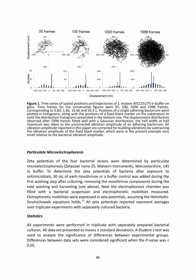
mouthrinses or the buffer control, after which flow was arrested for 15 min and bacterial vibration amplitudes were measured under stagnant conditions. Sixty consecutive images per second were recorded of adhering bacteria over a total time period of 33 s (1998 frames). Next to recording the bacterial positions, the position of a fixed black marker on the glass slide was recorded. Along the lines of equal pixel grey-values within the image of a single bacterium, a series of concentric elliptic contour lines were constructed, the center position of which was defined as the temporary position of the bacterium. The variation in positions observed over time served to analyze their trajectory and vibration amplitudes. As an example, typical vibration trajectories observed are shown in Figure 1 after 50, 100, 1000 and 1998 frames. The corresponding distributions of displacements were shown below the trajectories, the origin of which was defined as the average bacterial position observed within 1998 frames. The distribution of bacterial positions over time within 1998 frames was fitted to a Gaussian distribution. The vibration amplitude of an adhering bacterium was taken as the half width at half maximum (Figure 1) of this Gaussian distribution. Next, in order to account for possible vibrations of the building or microscope, the vibration amplitude of the fixed marker was subtracted from the uncorrected vibration amplitude calculated for adhering bacteria to yield the bacterial vibration amplitudes reported. All vibration amplitudes presented, represent the average of bacterial vibration amplitudes from three experiments with separate bacterial cultures, each comprising analysis of the vibrations of at least 10 randomly selected bacteria.
Attenuated Total Reflectance Fourier Transform Infrared Spectroscopy
ATR-FTIR spectroscopy (Cary 600 series FT-IR Spectrometer, Agilent Technologies, Santa Clara, USA) was carried out to analyze changes in chemical composition of the bond. The ATR-FTIR spectrometer used is equipped with a flow chamber, of which one wall is constituted by a Germanium internal reflection element with an angle of incidence of 45 degrees (Agilent Technologies). Bacteria were allowed to adhere to the reflection element as described above, after which absorption spectra were collected over the wavelength range of 4500 cm− 1 to 400 cm− 1. Each sample was scanned 12 times with 4 cm− 1 resolution. One series of spectra was taken 30 min after the last buffer perfusion, while another series of spectra was taken after mouthrinse exposure and buffer rinse. A background scan representing the internal reflection element in buffer was subtracted from the spectra taken for the adhering bacteria. Next, a Gaussian curve was fitted through the polysaccharide absorption band and wave number of the fitted peak was determined. The polysaccharide absorption band area was determined by an integral calculus of the area under the curve.
61
Figure 1. Time series of spatial positions and trajectories of S. mutans ATCC25175 in buffer on glass. Time frames for the consecutive figures were 50, 100, 1000 and 1998 frames, corresponding to 0.83, 1.66, 16.66 and 33.3 s. Positions of a single adhering bacterium were plotted in histograms, along with the positions of a fixed black marker on the substratum to yield the distribution histograms presented in the bottom row. The displacement distribution observed after 1998 frames fitted well with a Gaussian distribution, the half width at half maximum was taken as the uncorrected vibration amplitude of an adhering bacterium. All vibration amplitude reported in this paper are corrected for building vibrations by subtracting the vibration amplitude of the fixed black marker, which were in the present example very small relative to the bacterial vibration amplitude.
Particulate Microelectrophoresis
Zeta potentials of the four bacterial strains were determined by particulate microelectrophoresis (Zetasizer nano-ZS, Malvern Instruments, Worcestershire, UK) in buffer. To determine the zeta potentials of bacteria after exposure to antimicrobials, 30 mL of each mouthrinse or a buffer control was added during the first washing step after culturing, removing the mouthrinse components during the next washing and harvesting (see above). Next the electrophoresis chamber was filled with a bacterial suspension and electrophoretic mobilities measured. Eletrophoretic mobilities were expressed in zeta potentials, assuming the Helmholtz-Smoluchowski equations holds.17 All zeta potentials reported represent averages over triplicate experiments with separately cultured bacteria.
Statistics
All experiments were performed in triplicate with separately prepared bacterial cultures. All data are presented as means ± standard deviations. A Student t-test was used to analyze the significance of differences between experimental groups. Differences between data sets were considered significant when the P-value was < 0.05.
62
RESULTS
All bacterial strains involved in this study possessed a negative zeta potential that became more positive upon exposure to oral mouthrinse components across all four strains (Table 1). A CHX containing mouthrinse had the smallest effect on bacterial zeta potentials, while exposure to the AmF containing mouthrinse compensated the negative zeta potential most strongly, regardless of the strain considered.
Table 1. Zeta potentials of the four bacterial strains included in this study as measured in buffer (0.5 mM potassium chloride, 0.02 mM potassium phosphate and 0.01 mM calcium chloride, pH 6.8) prior to and after exposure to mouthrinses with selected oral antimicrobials. For control, bacteria were exposed to buffer. Exposure to oral antimicrobials yielded significantly less negative zeta potentials as compared with exposure to buffer for all four strains at p ˂ 0.05 (Student t-test).
Bacterial strains
Adhesion buffer
[mV]
CHX
[mV]
CPC
[mV]
AmF
[mV]
S. oralis ATCC35037 -33 ± 6 -21 ± 6 -3 ± 4 -8 ± 3
S. sanguinis ATCC10556 -21 ± 3 -12 ± 3 -5 ± 3 -5 ± 8
S. mutans ATCC25175 -35 ± 1 -23 ± 3 1 ± 8 -8 ± 3
S. mutans ATCC10449 -34 ± 3 -22 ± 2 -30 ± 2 -1 ± 6
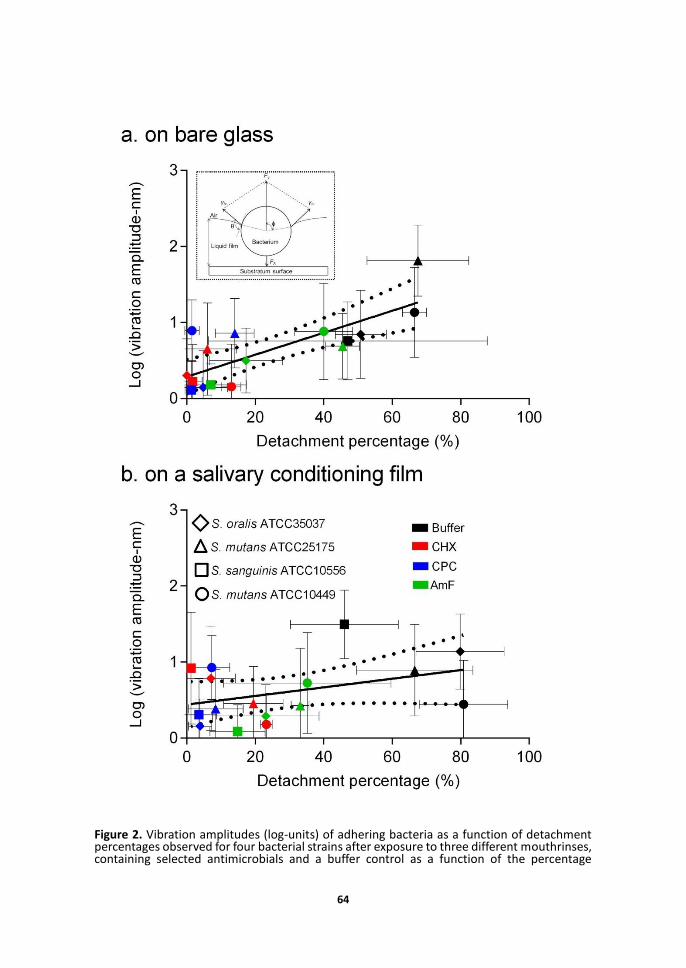
Bacteria prior to exposure to mouthrinses exhibited the largest vibration amplitudes up to 80 nm (Figure 2), while after mouthrinse exposure vibration amplitudes were generally lower than 10 nm. Exposure to mouthrinses had strong effects on bacterial detachment upon passing a liquid-air interface over the adhering bacteria, both when adhering to bare glass as well as when adhering to a salivary conditioning film. The percentage bacterial detachment increased with Brownian motion-induced bacterial vibration amplitudes for bacteria adhering to bare glass and salivary conditioning films. This indicates that Brownian motion forces assist bacterial detachment. Note that the influence of vibration amplitude on bacterial detachment was less pronounced for bacteria adhering to a salivary conditioning film than for bacteria adhering to bare glass (compare Figures 2a and 2b). LIVE-DEAD staining of adhering bacteria after exposure to mouthrinses and microscopic examination showed bacterial cell wall damage (red-staining),18 but bacteria remained microscopically intact, regardless of the strain or mouthrinse considered (data not
63
shown).
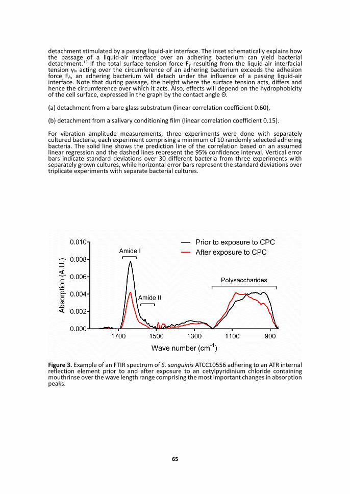
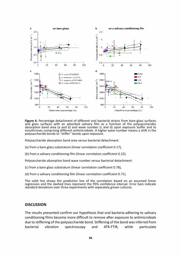
ATR-FTIR absorption spectra showed the most prominent changes upon exposure of adhering bacteria in the polysaccharide wave number region (873-1200 cm-1) and the protein wave number region including the Amide II (1498-1589 cm-1) and Amide I (1589-1700 cm-1) absorption bands (see Figure 3 for an example). Despite significant changes in Amide II and Amide I absorption bands, no consistent pattern was observed and therefore the remainder of our analysis of the ATR-FTIR spectra is confined to the polysaccharide absorption band region. Note that due to experimental restrictions, ATR-FTIR cannot be done on glass surfaces but necessarily has to be carried out on a Ge internal reflection element, while detachment percentages were determined on glass. However, both glass and Ge surfaces are negatively charged with a similar hydrophilicity19, 20 and there is no reason to assume that effects of exposure to mouthrinses will have different effects on the bond when formed with Ge or glass. Both on bare as well as on saliva-coated reflection elements, polysaccharide absorption band areas remained similar upon exposure to mouthrinses, indicating that the prevalence of polysaccharides in the interfacial region between an adhering bacterium and the substratum did not change upon mouthrinse exposure (Figures 4a and 4b). In contrast, the wave number of the polysaccharide absorption bands increased considerably by 14-56 cm-1, depending on the strain and mouthrinse used. Upward wave number shifts of IR absorption bands are indicative of increased stiffening of the molecular group under consideration. Accordingly, smaller upward shifts in wave number of the polysaccharide absorption bands correlated with higher bacterial detachment percentages (Figures 4c and 4d), indicating that less polysaccharide bond stiffening yields more detachment. Note that the correlation between wave number shift and bacterial detachment holds both for bare glass surfaces (Figure 4c) as well as for an adsorbed salivary film (Figure 4d).
64
Figure 2. Vibration amplitudes (log-units) of adhering bacteria as a function of detachment percentages observed for four bacterial strains after exposure to three different mouthrinses, containing selected antimicrobials and a buffer control as a function of the percentage
65
detachment stimulated by a passing liquid-air interface. The inset schematically explains how the passage of a liquid-air interface over an adhering bacterium can yield bacterial detachment.13 If the total surface tension force Fγ resulting from the liquid-air interfacial tension γlv acting over the circumference of an adhering bacterium exceeds the adhesion force FA, an adhering bacterium will detach under the influence of a passing liquid-air interface. Note that during passage, the height where the surface tension acts, differs and hence the circumference over which it acts. Also, effects will depend on the hydrophobicity of the cell surface, expressed in the graph by the contact angle Θ.
(a) detachment from a bare glass substratum (linear correlation coefficient 0.60),
(b) detachment from a salivary conditioning film (linear correlation coefficient 0.15).
For vibration amplitude measurements, three experiments were done with separately cultured bacteria, each experiment comprising a minimum of 10 randomly selected adhering bacteria. The solid line shows the prediction line of the correlation based on an assumed linear regression and the dashed lines represent the 95% confidence interval. Vertical error bars indicate standard deviations over 30 different bacteria from three experiments with separately grown cultures, while horizontal error bars represent the standard deviations over triplicate experiments with separate bacterial cultures.
Figure 3. Example of an FTIR spectrum of S. sanguinis ATCC10556 adhering to an ATR internal reflection element prior to and after exposure to an cetylpyridinium chloride containing mouthrinse over the wave length range comprising the most important changes in absorption peaks.
66
Figure 4. Percentage detachment of different oral bacterial strains from bare glass surfaces and glass surfaces with an adsorbed salivary film as a function of the polysaccharides absorption band area (a and b) and wave number (c and d) upon exposure buffer and to mouthrinses comprising different antimicrobials. A higher wave number means a shift in the polysaccharide bonds to “stiffer” bonds upon exposure.
Polysaccharide absorption band area versus bacterial detachment:
(a) from a bare glass substratum (linear correlation coefficient 0.17),
(b) from a salivary conditioning film (linear correlation coefficient 0.22).
Polysaccharide absorption band wave number versus bacterial detachment:
(c) from a bare glass substratum (linear correlation coefficient 0.78),
(d) from a salivary conditioning film (linear correlation coefficient 0.71).
The solid line shows the prediction line of the correlation based on an assumed linear regression and the dashed lines represent the 95% confidence interval. Error bars indicate standard deviations over three experiments with separately grown cultures.
DISCUSSION
The results presented confirm our hypothesis that oral bacteria adhering to salivary conditioning films become more difficult to remove after exposure to antimicrobials due to stiffening of the polysaccharide bond. Stiffening of the bond was inferred from bacterial vibration spectroscopy and ATR-FTIR, while particulate
67
microelectrophoresis showed that bond stiffening is caused probably by adsorption of positively charged moieties from the mouthrinses to the bacterial cell surface. We chose to use a passing liquid-air interface as a stimulus for detachment of adhering bacteria, as also occurring during non-contact powered tooth brushing,21 use of a water floss22 and as a result of naturally passing fluids and salivary flow in the oral cavity. Although amide bonds were also affected by exposure of adhering bacteria to mouthrinses, no consistent patterns in amide bond changes with respect to bacterial detachment were found. This concurs with the general notion that EPS, most notable polysaccharides govern the ease of bacterial detachment from surfaces.23-26 Enhanced EPS production by adhering staphylococci as a defense action against antibiotics was observed,27 but absence of any changes in IR absorption band areas due to mouthrinse exposure indicate no such effects for the strains used here. The bacteria cannot produce any EPS because they are not exposed to growth media. This confirms that a more difficult detachment of adhering bacteria is due to polysaccharide bond stiffening and not to increasing amounts of EPS produced upon mouthrinse exposure.
Zeta potentials as compiled in Table 1 clearly indicate that exposure to the CHX containing mouthrinse yields less negative charge compensation in the outermost bacterial surface17 than observed after exposure to CPC or AmF containing mouthrinses. CHX differs from the two cationic antimicrobials CPC and AmF in the position of the positive charge on the molecule. Both AmF and CPC have their charges at the end of the molecule, making them more stoichiometrically available than CHX which has its positive charge in the center of the molecule. Although exposure of the adhering bacteria to the mouthrinses yielded cell wall damage, often interpreted as cell death, this is of little importance in the context of the present study. Bacterial cell surfaces become more positively charged upon exposure to the mouthrinses whether killed or not (bacterial death is hard if not impossible to demonstrate anyway),28 but visibly intact, cell wall damaged bacteria remain adhering to the substratum surfaces acting as a substratum for new bacteria to adhere to.4 Being dead or alive has no impact on Brownian motion-induced vibrations, that depend solely on the thermal energy a colloidal particle possesses causing its random motion, that is restricted for adhering particles on a surface by the elasticity of the bond. Elastic forces opposing Brownian motion forces therewith control the amplitude of the vibrations which were previously found for biotic12,10 as for abiotic ones.11 It is likely that not only polysaccharides in the outermost bacterial cell surfaces are affected by exposure to mouthrinse components, but also polysaccharides in deeper cell wall layers, since ATR-FTIR with a far larger depth of information in the micro-meter range than particulate microelectrophoresis,17 showed clear effects on polysaccharide stiffness as inferred from IR wave number shifts.
68
CONCLUSIONS
In conclusion, we have demonstrated using bacterial vibration spectroscopy and ATR-FTIR that oral bacteria adhering to salivary conditioning films become more difficult to remove after exposure to antimicrobial containing mouthrinses due to stiffening of the polysaccharide bond as caused by both adsorption of positively charged moieties from the mouthrinses to the bacterial cell surface.
ACKNOWLEDGEMENTS
This work was supported by the University Medical Center Groningen and was not sponsored by any company. We thank Dr. Brandon Peterson for his helpful comments and support for this study. H.J. Busscher is also director of a consulting company, SASA BV (GN Schutterlaan 4, 9797 PC Thesinge, The Netherlands). The authors declare no potential conflicts of interest with respect to authorship and/or publication of this article. Opinions and assertions contained herein are those of the authors and are not construed as necessarily representing views of the funding organization or their respective employers.
69
REFERENCES
1. Adams, H.; Winston, M. T.; Heersink, J.; Buckingham-Meyer, K. A.; Costerton, J. W.; Stoodley, P. Development of a laboratory model to assess the removal of biofilm from interproximal spaces by powered tooth brushing. Am. J. Dent. 2002, 15 Spec No, 12B-17B.
2. Buzalaf, M. A.; Pessan, J. P.; Honorio, H. M.; Ten Cate, J. M. Mechanisms of action of fluoride for caries control. Monogr. Oral Sci. 2011, 22, 97-114.
3. Otten, M. P.; Busscher, H. J.; Abbas, F.; Van der Mei, H. C.; Van Hoogmoed, C. G. Plaque-left-behind after brushing: intra-oral reservoir for antibacterial toothpaste ingredients. Clin. Oral Investig. 2012, 16, 1435-1442.
4. Banks, M. K.; Bryers, J. D. Deposition of bacterial cells onto glass and biofilm surfaces. 1992, 6, 81-86.
5. Brindle, E. R.; Miller, D. A.; Stewart, P. S. Hydrodynamic deformation and removal of Staphylococcus epidermidis biofilms treated with urea, chlorhexidine, iron chloride, or DispersinB. Biotechnol. Bioeng. 2011, 108, 2968-2977.
6. Marsh, P. Dental plaque: biological significance of a biofilm and community life‐style. J. Clin. Periodontol. 2005, 32, 7-15.
7. Van Oss, C. J. Long-range and short-range mechanisms of hydrophobic attraction and hydrophilic repulsion in specific and aspecific interactions. J. Mol. Recogn. 2003, 16, 177-190.
8. Van Loosdrecht, M. C.; Norde, W.; Lyklema, J.; Zehnder, A. J. Hydrophobic and electrostatic parameters in bacterial adhesion. Aquat. Sci. 1990, 52, 103-114.
9. Peterson, B. W.; He, Y.; Ren, Y.; Zerdoum, A.; Libera, M. R.; Sharma, P. K.; Van Winkelhoff, A. J.; Neut, D.; Stoodley, P.; Van der Mei, H. C.; Busscher, H. J. Viscoelasticity of biofilms and their recalcitrance to mechanical and chemical challenges. FEMS Microbiol. Rev. 2015, 39, 234-245.
10. Song, L.; Sjollema, J.; Sharma, P. K.; Kaper, H. J.; Van der Mei, H. C.; Busscher, H. J. Nanoscopic vibrations of bacteria with different cell-wall properties adhering to surfaces under flow and static conditions. ACS Nano 2014, 8, 8457-8467.
11. Dabros, T.; Warszynski, P.; Van de Ven, T. Motion of latex spheres tethered to a surface. J. Colloid Interface Sci. 1994, 162, 254-256.
12. Kamiti, M.; Van de Ven, Theo GM Measurement of spring constants of polyacrylamide chains bridging particles to a solid surface. Macromolecules 1996, 29, 1191-1194.
13. Leenaars, A. In A new approach to the removal of sub-micron particles from solid (silicon) substrates; Particles on Surfaces 1; Springer: 1988; pp 361-372.
14. Hahnel, S.; Rosentritt, M.; Handel, G.; Bürgers, R. Influence of saliva substitute films on initial Streptococcus mutans adhesion to enamel and dental substrata. J. Dent. 2008, 36, 977-983.
15. Navazesh, M.; Christensen, C. M. A comparison of whole mouth resting and stimulated salivary measurement procedures. J. Dent. Res. 1982, 61, 1158-1162.
70
16. Dawes, C.; Watanabe, S.; Biglow-Lecomte, P.; Dibdin, G. H. Estimation of the velocity of the salivary film at some different locations in the mouth. J. Dent. Res. 1989, 68, 1479-1482.
17. Lyklema, J. Fundamentals of interface and colloid science: soft colloids; Academic press: 2005; Vol. 5.
18. Nocker, A.; Cheung, C.; Camper, A. K. Comparison of propidium monoazide with ethidium monoazide for differentiation of live vs. dead bacteria by selective removal of DNA from dead cells. J. Microbiol. Methods 2006, 67, 310-320.
19. Drzymala, J.; Lekki, J.; Kielkowska, M. A study of the germanium—sodium oleate flotation system. Powder Technol. 1987, 52, 251-256.
20. Zhang, F.; Kibria, M.; Cormier, K.; Howlader, M. Surface and interface characterization of sequentially plasma activated silicon, silicon dioxide and germanium wafers for low temperature bonding applications. ECS Trans. 2010, 33, 329-338.
21. Schmidt, J. C.; Zaugg, C.; Weiger, R.; Walter, C. Brushing without brushing?—a review of the efficacy of powered toothbrushes in noncontact biofilm removal. Clin. Oral Investig. 2013, 17, 687-709.
22. Rmaile, A.; Carugo, D.; Capretto, L.; Aspiras, M.; De Jager, M.; Ward, M.; Stoodley, P. Removal of interproximal dental biofilms by high-velocity water microdrops. J. Dent. Res. 2014, 93, 68-73.
23. Flemming, H.; Wingender, J. The biofilm matrix. Nature Rev. Microbiol. 2010, 8, 623-633.
24. Sutherland, I. Biofilm exopolysaccharides: a strong and sticky framework. Microbiology 2001, 147, 3-9.
25. Vu, B.; Chen, M.; Crawford, R. J.; Ivanova, E. P. Bacterial extracellular polysaccharides involved in biofilm formation. Molecules 2009, 14, 2535-2554.
26. Ryder, C.; Byrd, M.; Wozniak, D. J. Role of polysaccharides in Pseudomonas aeruginosa biofilm development. Curr. Opin. Microbiol. 2007, 10, 644-648.
27. Rachid, S.; Ohlsen, K.; Witte, W.; Hacker, J.; Ziebuhr, W. Effect of subinhibitory antibiotic concentration on polysaccharide intercellular adhesin expression in biofilm-forming Staphylococcus epidermidis. Antimicrob. Agents Chemother. 2000, 44, 3357-3363.
28. Berney, M.; Hammes, F.; Bosshard, F.; Weilenmann, H. U.; Egli, T. Assessment and interpretation of bacterial viability by using the LIVE/DEAD BacLight Kit in combination with flow cytometry. Appl. Environ. Microbiol. 2007, 73, 3283-3290.