Embed Size (px)
Citation preview

University of Illinois at Urbana-ChampaignLuthey-Schulten GroupNIH Resource for Macromolecular Modeling and BioinformaticsComputational Biophysics Workshop
Evolution of Translation
The Ribosome
VMD Developer John Stone
MultiSeq Developers Tutorial Authors
Elijah Roberts Ke ChenJohn Eargle John EargleDan Wright Tyler Earnest
Jonathan LaiZan Luthey-Schulten
April 2015
A current version of this tutorial is available athttpwwwscsillinoisedu~schultentutorialsribosome
CONTENTS 2
Contents
Introduction 3Requirements 4
1 The Ribosomal SSU and associated structures [30 minutes] 4
2 The Ribosome LSU and associated structures [30 minutes] 921 The peptidyl-transferase center 10
3 Ribosome Origins [30 minutes] 1131 Hypothesis on the evolution of the ribosome 11
4 Ribosomal signatures [60 minutes] 1241 Definition and classification of the ribosomal signatures 1442 Contribution of ribosomal signatures to phylogenetic separation 1743 Functional roles of signatures in ribosomal assembly 20
5 Kinetic Model of Ribosome assembly [30 minutes] 22
Acknowledgements 26
CONTENTS 3
Introduction
The ribosome is a large structure found in all living cells that serves as themain translation machinery of the cell Messenger RNA (mRNA) transcribedfrom the organismrsquos genome binds with the ribosome to commence translationto protein As explained in the previous tutorials [1 2 3] many other cellularcomponents including tRNA the aminoacyl-tRNA synthetases and the elonga-tion factors participate in the translation process however the ribosome is thecentral machinery that assembles a protein from a transcribed gene Solving thestructure of the ribosome was awarded the Nobel Prize in Chemistry in 2009 [4]The bacterial ribosome (70S) consists of a small (SSU or 30S) and large (LSUor 50S) subunit which bind together around a messenger RNA Each subunitis made up of rRNA and proteins the 30S subunit consists of the 16S rRNAsubunit and 21 proteins while the 50S subunit consists of the 23S rRNA sub-unit the 5S rRNA subunit and 34 proteins The lsquoSrsquo in this case refers to theSvedberg unit a measurement of sedimentation during centrifugation so thesenumbers do not necessary add up as they would if they referred to mass
The primary function of the ribosome is to translate a sequence encodedon mRNA into a protein Proteins are built as linear chains of amino acidsby adding one amino acid at a time to a growing chain The amino acid ischaracterized by two functional groups an amino group and a carboxyl group Ifthe amino group of one amino acid is brought in close proximity to the carboxylgroup of a second amino acid a peptidyl transferase (aminoacyltransferase)reaction can occur resulting in the loss of a molecule of water (one oxygenatom and one hydrogen atom from the carboxyl group and one hydrogen atomfrom the amino group) and the formation of a peptide bond between the twoamino acids As all amino acids contain one amino group and one carboxylgroup they can be joined together to form a large chain or polypeptide Asthis reaction occurs in a pocket of the LSU that is primarily rRNA the ribosomecan be thought of as an RNA enzyme or ribozyme Specifically the reactiontakes place inside the peptidyl transferase center which will be discussed inmore detail later in the tutorial
In addition to the peptidyl transferase function the ribosome also plays arole in maintaining the accuracy of translation by allowing the codon-anticodoninteraction between the bound mRNA and the tRNA carrying the next putativeamino acid to be joined to the nascent protein chain The ordering of aminoacids in a protein is directly translated from the sequence of nucleotides ofthe mRNA The translation between nucleotide codons triplets of nucleotidesand individual amino acids is known as the genetic code This translation ismediated by charged transfer RNA (tRNA) molecules Each tRNA is chargedat the acceptor stem with a specific amino acid corresponding to its anticodonlocated in the anti-codon stem by an AARS For more details on how the AARSset the genetic code by charging the tRNAs please see the tutorials on theamino-acyl tRNA synthetases [1 2] When a charged tRNA arrives at the A-site of the ribosome the anticodon loop of the tRNA is oriented to interactwith the next codon of the bound mRNA If the codon is complementary to
1 THE RIBOSOMAL SSU ANDASSOCIATED STRUCTURES [30 MINUTES]4
the anticodon it is released by its carrier molecule the elongation factor Tu(EF-Tu) If the codon is not complementary to the anticodon there is a highprobability that the tRNAEF-Tu complex will dissociate and another tRNAwill bind For more details on the behavior of the EF-Tu and tRNA please seethe tutorial on EF-Tu [3]
The ribosome contains three tRNA binding sites labeled the A-site (aminoa-cyl) P-site (peptidyl transferase reaction) and the E-site (exit) After thecharged tRNA is released into the A-site by the EF-Tu the existing nascentprotein is transfered from the tRNA in the P-site to the the amino group ofthe bound amino acid on the tRNA in the A-site extending the chain by oneresidue This reaction is catalyzed by the peptidyl tranferase activity of theribosome Elongation factor G faciliates ribosome translocation causing theA-site tRNA to move to the P-site [5] The newly vacated A-site will be freedto accept the next tRNA Because all the tRNAs are base-paired with codonsin the mRNA the movement of the tRNAs also moves the mRNA through theribosome exposing the next codon to be matched to the next aminoacylatedtRNA This repeats until a stop codon is encountered on the mRNA which isnot complementary to any tRNA but rather binds the release factors whichtrigger the release of the protein and the ultimate dissociation of the ribosomallarge and small subunits
Topics addressed in this tutorial are 1-2) structural aspects of the LSUand SSU [60 minutes] 3-4) signatures of ribosome evolution that are used toclassify organisms in the Phylogenetic Tree of Life [90 minutes] and 5) kineticmodeling of ribosome assembly [30 minutes] Intermediates in the assembly ofthe SSU are analyzed through MD simulations This tutorial will rely on thepaper Molecular Signatures of ribosomal evolution by Roberts et al [6]which we have provided for you with this tutorial This tutorial should takeapproximately three hours to complete
Requirements
MultiSeq must be correctly installed and configured before you can begin usingit to analyze the ribosome There are a few prerequisites that must be metbefore this section can be started
bull VMD 192 or later must be installed The latest version of VMD can beobtained from httpwwwksuiuceduResearchvmd
bull This tutorial requires approximately 250 MB of free space on your localhard disk
1 The Ribosomal SSU and associated structures[30 minutes]
1 Before we open a state file we need to open the Tk Console by click-ing on Extensions rarr Tk Console Now navigate to the directory TUTO-
1 THE RIBOSOMAL SSU ANDASSOCIATED STRUCTURES [30 MINUTES]5
RIAL DIR1ribosome structure Now in the VMD main window click onFile rarr Load Visualization State From the 1ribosome structure load thestate file ribosomevmd This will load the Escherichia coli 50S and 30Ssubunits containing both the rRNA and the ribosomal proteins as well asthe bound tRNAs in the A- P- and E-sites and the bound EF-Tu All ofthese structures are initially hidden with the exception of the EF-Tu andits bound aminoacyl- tRNA
2 We will first examine the overall structure of the ribosome and highlightsome of the particular features discussed in the introduction In VMDzoom in on the yellow highlighted region of the elongation factor Thisregion is known as the amino acid binding pocket where the amino acidbound to the tRNA sits as the complex migrates to the ribosome Thispart of the tRNA is known as the acceptor stem and the final threenucleotides those that sit close to the amino acid binding pocket arealways the same CCA The tip of the acceptor stem is called the CCAtail (Figure 1)
Figure 1 The tRNA bound to the elongation factor
3 Now move to the other side of the tRNA where three nucleotides havebeen highlighted in licorice representation These three nucleotides arethe anticodon of the tRNA Use the Query function of VMD to query theresname of each of these nucleotides (in the order of resid 36 35 then 34)What is the anticodon of this tRNA Given that the lsquoalphabetrsquo of RNAis A C U and G where A base pairs with U and C base pairs with Gpredict the codon to which this tRNA is bound (The lsquoresnamersquo of eachnucleotide may appear as lsquoAr Cr Ur or Grrsquo the lsquorrsquo standing for RNA)
1 THE RIBOSOMAL SSU ANDASSOCIATED STRUCTURES [30 MINUTES]6
Figure 2 The codonanticodon
4 Make sure the structure 3FIHpdb is displayed in the Selected Moleculedrop down box Now display chain X by double-clicking on it to displaythe mRNA Note the three nucleotides closest to the anticodon of thetRNA (Figure 2) These three nucleotides represent the codon of themRNA which codes for the amino acid currently bound to the tRNAUse the Query feature of VMD to determine the codon to which the tRNAis base paired The codon is read from 5rsquo to 3rsquo on the mRNA whichcorresponds to the ordering resid 19 20 21 Using the genetic code tableshown in Figure 3 determine the amino acid that is about to be added tothe growing protein
5 Now display chain V in the structure by double clicking on the title in theRepresentations window This displays the tRNA currently bound in theP-site or peptidyl transferase site for which the amino acid has alreadybeen bound to the growing protein chain Create a new representationand type the following as the selected atoms chain V and resid 34 35
36 to display the anticodon of this tRNA Color this representation byName Change the Drawing Method of this representation to be LicoriceAs you did before determine the type of this tRNA by querying the mRNAnucleotides in the order resid 16 17 18
6 Display chain W now to display the final tRNA bound to this ribosomeIt is clear this tRNA is not as associated with a corresponding codon onthe mRNA
7 Hide chain X Now in the VMD representations window choose2HGR SSU Thermophiluspdb in the Selected Molecule drop down boxThis is a different structure of the ribosomal 16S one which contains
1 THE RIBOSOMAL SSU ANDASSOCIATED STRUCTURES [30 MINUTES]7
Figure 3 The genetic code
1 THE RIBOSOMAL SSU ANDASSOCIATED STRUCTURES [30 MINUTES]8
a longer mRNA molecule We will use this structure to explore how themRNA is bound by the ribosome Display the representation chain A andresid 1535 to 1541 and the representation chain 1
8 You can see that this mRNA is in mostly the same position as the originalmRNA However this sequence is longer and contains a particular partof the mRNA known as the Shine-Dalgarno sequence This portion ofthe mRNA is what is recognized by the ribosome when the mRNA bindsand translation begins The consensus sequence of the Shine-Dalgarnosequence is AGGAGG though this differs slightly between organisms Theorange residues base pairing with the Shine-Dalgarno sequence are part ofthe 16S subunit This complementary sequence is known appropriatelyenough as the anti-Shine-Dalgarno sequence When the Shine-Dalgarnosequence and anti-Shine-Dalgarno sequence bind the initiator tRNA(N-formylmethionine) is recruited and translation begins
N-formylmethionine Translation of a protein always begins withthe start codon AUG In the genetic code from Figure 3 AUG trans-lated into methionine However this methionine derivative (fMet)with a formyl group attached is used instead for the first residue ofa protein
Figure 4 The Shine-Dalgarno sequence on the mRNA and the anti-Shine-Dalgarno sequence on the 16S rRNA
9 Now we can hide the structure 2HGR SSU Thermophiluspdb Do this bydouble clicking on the D next to the structure title in the VMD mainwindow
10 Now we will display the rRNA portion of the small subunit of the ribosomeIn the Selected Molecule drop down box select 3FIHpdb Display chain
2 THE RIBOSOME LSU ANDASSOCIATED STRUCTURES [30 MINUTES]9
A The small subunit of the bacterial ribosome contains 1540 nucleotidesAlso display the representation chain B C D E F G H I J K L M N O P QR S T U in VMD to display the small subunit proteins Together thisrRNA and 21 proteins comprise the 30S subunit of the bacterial ribosomeAlso display chain X to see how the mRNA fits into the small subunit
11 The ribosomal proteins maintain the stability of the structure particularlywith regards to ribosomal assembly and several play an important role inthe function of the ribosome Protein S4 helps to maintain translationalaccuracy mRNA has secondary structure but must be a linear chain inorder to pass through the ribosome during translation Protein S4 mayassist the mRNA in denaturing its secondary structure In the VMD rep-resentation window create a new representation Set the Selected Atomsto be chain G and the Drawing Method to be VDW Examine the locationof S4 with respect to the mRNA
12 As discussed in the introduction the nascent protein is bound to the tRNApresent in the P-site or peptidyl transferase site of the ribosome ThistRNA is currently displayed in orange in the VMD window Compare thestructure of this tRNA with the structure of the tRNA in the A-site Wehave already discussed where the amino acid binding pocket exists on theelongation factor Tu Based on the structure of the tRNA bound in theA-site find the location where the amino acid should sit on the tRNA inthe P-site (the amino acids do not exist in this structure)
13 In the VMD representations window choose the file 3FIHpdb from theSelected Molecule drop down box Create a new representation in VMDusing selected atoms chain V and resid 74 75 76 Use the drawing methodLicorice and coloring method Name Now the three final residues on thetRNA will be highlighted This is the CCA tail for the tRNA bound inthe P-site Although the crystal structure from which these coordinatesare derived did not include the amino acids bound to the tRNAs this iswhere it should be bound
2 The Ribosome LSU and associated structures[30 minutes]
1 Now hide every representation currently displayed except for any repre-sentation containing chain V Display the molecule 3FIKpdb in the Se-lected Molecule drop down box and double-click on the representation allNow the 23S rRNA subunit of the 50S ribosomal subunit is displayedNotice how the tRNA in the P-site of the ribosome reaches up into thecenter of the 23S subunit As we discussed in the introduction the nascentprotein should be currently bound to the tRNA in the P-site or peptidyltransferase site Rotate the display Can you find the lsquochannelrsquo throughthe large subunit where the nascent protein should exit
2 THE RIBOSOME LSU ANDASSOCIATED STRUCTURES [30 MINUTES]10
2 Once you think you have found the nascent protein exit channel setmolecules 3FIKpdb as Top in the main VMD window by double click-ing on the T column next to the structure name Choose Tk Console inthe VMD Extensions menu Navigate using the cd command to the 1ribo-some structure directory and type source nascent chaintcl This willdraw a sample nascent chain in the exit channel allowing you to bettervisualize how the chain will exit the ribosome as it is being synthesized
3 Ribosomal protein L11 changes conformation to allow the elongation fac-tor to bind and thus plays an important role in translation In theVMD representation window create a new representation Set the Se-lected Atoms to be chain I and the Drawing Method to be VDW Changethe molecule in the VMD representations menu Selected Molecule dropdown box to be 3FIHpdb Display representations chain A and chain Z todisplay both the small subunit and the EF-Tu Examine the location ofL11 with respect to the EF-Tu
21 The peptidyl-transferase center
There is evidence that a duplication of a more fundamental RNA structureresulted in the formation of the peptidyl transferase center where the aminoa-cyltransferase reaction to extend the nascent protein actually takes place [7 8]The PTC in its present form comprises two parts with very nearly identicalsecondary and tertiary structures These two parts are the binding sites for theCCA-3rsquo termini of the tRNA for the P- and A-sites A plausible scenario forthe evolution of the ribosome would be that one of these two CCA binding sitesduplicated resulting in the ability to bind two proto-tRNAs inclose proximityallowing a transpeptidation reaction to occur This complex would likely havebeen able to synthesize random oligopeptide sequences
1 Hide chain A Change the molecule in the VMD representations menuSelected Molecule drop down box to be 3FIKpdb Now hide the represen-tation of the entire 23S rRNA (by double-clicking on the all representation)and display the hidden representation with Selected Atoms as nucleic andchain B and (resid 2058 to 2092) You can hide the structure 3FIHpdbin the main VMD window to This will display the portion of the ribosomecalled the peptidyl transferase center or PTC Change the representationof the PTC to VDW and examine the site where the PTC surrounds thenascent chain and aminoacylated tRNA
2 Change the representation of the PTC back to NewCartoon Can you seethe symmetry suggested in the text above
3 RIBOSOME ORIGINS [30 MINUTES] 11
Figure 5 The PTC and nascent chain
3 Ribosome Origins [30 minutes]
31 Hypothesis on the evolution of the ribosome
The ribosome is largely comprised of RNA with proteins decorating the pe-riphery of the structure All organisms have a similar core structure to theirribosomes which strongly suggests that the ribosome existed prior to the differ-entiation of the three domains of life Analysis of the rRNA tertiary structurehas been used to gain insight into how the ribosome evolved before the diver-gence of the primary organismal lineages [9]
In performing this analysis on the 23S subunit Bokov and Steinberg observedthat a particular interaction motif occurred between domain V and its RNAcontacts The observed motif an A-minor interaction involves the interactionof an unpaired adenosine-rich stack with a double helix In nearly every case ofan A-minor interaction with domain V observed the double helix part of themotif was on domain V Since an RNA double helix is stable and unpaired stacksof adenosine are not it was suggested that domain V was an early segment ofthe 23S and the segments associating with it through A-minor interactions weremore recent additions This suggestion is supported by the fact that domainV contains the pepidyl-transferase center (PTC) which performs the primaryfunction of the ribosome the addition of amino acids to the nascent polypeptidechain
Bokov and Steinberg used the A-minor motif to attempt to determine theevolutionary order of assembly of the various elements of the ribosome To de-fine structurally independent elements Bokov and Steinberg required that eachelement have 3rsquo and 5rsquo ends in close proximity (suggesting an insertion thatwould not otherwise perturb the structure of the remaining portion of the ribo-some) that both sides of any double helix be contained in an element and that
4 RIBOSOMAL SIGNATURES [60 MINUTES] 12
if the element formed an A-minor interaction with the rest of the ribosome thatthe entire adenosine stack portion of the A-minor be contained in the elementand not the double helix portion Using this method 19 elements were identi-fied the deletion of which did not disturb the integrity of the remaining rRNAExcluding these ldquolayer 1rdquo elements an additional 11 elements were identifiedwhich only supported the integrity of the level 1 elements and could be consid-ered as ldquolayer 2rdquo This process was performed a total of 12 times revealing 59elements This method revealed a hierarchy of dependencies with layer 1 beingmost likely the latest additions to the 23S and each subsequent layer consistingof progressively more ancient additions to the 23S The 12 layers constituted93 of the 23S with the remaining 7 consisting of a portion surrounding andincluding the PTC
1 Relaunch VMD and Multiseq If the Tk Console is no longer open openit again by clicking on Extensions rarr Tk Console Now navigate to the di-rectory TUTORIAL DIR2ribosome evolution Now in the VMD main win-dow click on Filerarr Load Visualization State From the 2ribosome evolutionload the state file superimposedvmd This is the structure of the largesubunit (50S) from Thermus thermophius a lsquoheat-lovingrsquo bacterium orig-inally isolated from a thermal vent The silver structures consist of therRNA while the orange structures are the 50S proteins Using the VMDrepresentations menu hide the proteins in this structure by double clickingon the protein representation
2 In the VMD representations window select 1S72 LSU Marismortuipdb
from the Selected Molecule drop down box Display the hidden 23S rRNAby double clicking on the nucleic representation This is the structure ofthe 50S subunit from Haloarcula marismortui an archaeon found in theDead Sea Note the remarkable similarity of the two structures
3 Based on these two structures would you predict that the majority of theevolution of the ribosome occurred before or after the divergence of thebacterial and archaeal domains of life
4 Ribosomal signatures [60 minutes]
The term ribosomal signatures was coined by Carl Woese and used by himas one form of evidence to define and distinguish the three domains of lifeBacteria Archaea and Eucarya The universal phylogenetic tree (UPT Figure7) constructed from the 16S ribosomal RNA (rRNA) shows a so-called canonicalpattern in which all taxa group into three distinct clusters with the eucaryoticand archaeal subbranches closer to each other than to bacteria This furtherconfirms the signal we could see simply from the signatures 20 years later withthe help of the rapid growth of the massive genomic and structural data we areable to extend the signature notion and use the information it conveys to studythe evolution of cells and origin of life
4 RIBOSOMAL SIGNATURES [60 MINUTES] 13
Figure 6 50S subunit from H marismortui
Figure 7 The tree of life
4 RIBOSOMAL SIGNATURES [60 MINUTES] 14
41 Definition and classification of the ribosomal signa-tures
Ribosomal signatures are regions on the ribosome that are constant and uniqueto a particular domain of life The two general kinds of signatures are de-fined based on characteristics of the rRNA Sequence signatures are positions inrRNArsquos primary structure whose compositions remain constant in one domainof life but occur rarely in the other domains Structural signatures are regionsin its secondary andor tertiary structure that have a unique configuration in agiven domain and they could be further classified into three subtypes i) inser-tions or deletions (indels) that are characteristically present in one domain oflife but absent in another ii) regions of the rRNA in which the secondary (andtherefore tertiary) structure differs between two domains and iii) regions thatare similar in secondary structure but differ in their tertiary conformation
1 Delete all files out of the main VMD window before you move on to thenext step
2 We have prepared for you two sets of multiple sequence alignments thatwere created with MultiSeq The first set of multiple sequence alignmentsis for domain V of the 23S rRNA for bacteria and archaea We will analyzethese sequences using MultiSeq and reproduce the results of the paper byRoberts et al [6] Open MultiSeq in VMD by clicking on Extensions rarrAnalysis rarr MultiSeq
Ribosomal Domains The 23S rRNA or large subunit of the bac-terial ribosome is formed from six domains (See Figure 8 for anillustration of the domains of the 23S in a secondary structure rep-resentation) Domain V contains the peptidyl transferase centerthe enzyme lsquoactive sitersquo of the ribosome where the aminoacyltrans-ferase reaction takes place to elongate the nascent protein chain
3 Now we can identify the locations of the sequence signatures on a part ofthe 23S domain of the ribosome To do this we need to load an alignmentof several 23S rRNA sequences across the bacteria and archaea and usethis alignment to calculate the sequence signatures These signatures canthen be mapped onto the structure of 23S so we can understand the typesof places sequence signatures are found Start a new session of MultiSeqby clicking on File rarr New Session
4 We are going to load a saved state of MultiSeq Go to Filerarr Load Sessionand navigate to the directory 3ribosomal signatures Highlight the filesignaturesmultiseq and click Open This will load the MultiSeq sessionfile This contains an alignment of several archaeal and bacterial 23Ssequences for domain V of the 23S The 23S is made up of six domainsand domain V consists of the peptidyl transferase center and surroundingregions See Figure 8
4 RIBOSOMAL SIGNATURES [60 MINUTES] 15
IV
V
VI
1650
1700 1750
1800
1850
1900
1950
2000
2050
2100
2150
2200
2250
2300
2350
2400
2450
2500
2550
2600
2650
27002750
2800
2850
m2
m
3
m
5
m
6m7
m
m
m2
5m
m
-[m2G]
Secondary Structure large subunit ribosomal RNA
Escherichia coli(J01695)1cellular organisms 2Bacteria3Proteobacteria4gamma subdivision5Enterobacteriaceae and related symbionts6Enterobacteriaceae7EscherichiaNovember 1999 (cosmetic changes July 2001)Citation and related information available at httpwwwrnaicmbutexasedu
Symbols Used In This Diagram
G A
- Canonical base pair (A-U G-C)
- G-A base pair- G-U base pair
G C
G U
U U - Non-canonical base pair
Every 10th nucleotide is marked with a tickmark and every 50th nucleotide is numberedTertiary interactions with strong comparativedata are connected by solid lines
I
II
III
50
100
150
200
250
300
350
400
450
500
550
600650
700
750
800
850
900
950
1000
10501100
1150
1200
1250
1300
1350
1400
1450
1500
1550
1600
1640
2900
5rsquo 3rsquo
m1
m5
m6
GGUUAAGC
GACUAAGCGUACACGGUGGAU
G
CC C
UG G C A G U C A G A G
GC
GA
UG
AA
GG
ACG
UG
CUAAUC U
GC
GAUA
A G CGUCGGU
AAGGU
GAU A
UGA
ACC GU
UA
UAACCGGCG
AUU
UCCG A A U G
GGG
A AA
CCC A
GUGUGUU U C
GA
CA
CA
CU A
UCA
UUAACU
GA A U C
CA
UAGGUUA
AUGAG
GCGAAC C G G G GG A A C U
G A AACAUC
UAAGUA
CCCCGAGG
AA
AA
GAAAU
CA
ACCGAGAUU
CCCC CA
GUA
GC
GG
CGAG
CGA
ACG
GG
GAG
CA
GC
C
C
A
G A G CCU G A AU
C A G U G U G U G U G U U A G U GG
A A GCGUC
UGG AA
AGGCGC G
CG A
UAC
AGGG
UG
ACAGC
CCCGU
ACAC
AAAAAUGCACAUGCUG
UGAGCUCGAUGAG
UA
GGGCGGGACACGU
GGU AUCCU GUCU
GAAUA
UG
GG
GG
GAC C A
UCCUCC A A
GG
CU
AA
AUACU
CCUGACUG
ACC
GA
UAGUGAACCA
GU
ACCG
UG
A G GG
A A A GGCGAAAAGAACCCCGG
CG A G G G GA GU GAA A A A GAA CC
UGAAACCGUGUACGUACAAGCAG
UG
GG
AG
CA
CG
CUUA
GGCGUGUGACUGCG
UA C C U U UU
GUAUAAUGG
GUCAGCG
ACUU
AUAUUCUGUAGC A
AG G U U
A AC C G A
AUAGG
GGAGCC
GAAG
GGAA
ACC
GAGUCUUA
AC U G G G C G
UUA A G
UUGCAGGGUAUAGA
CC
CGAAAC
CC
GG
U
GA
UCUAGCCAUGGGC A
G G U UG A AG G U U G G G U
AA
CACUAACUGGAG
GACCGAACCG
ACUAAUG
UGAAA A AUUAG
CGGA
U GA CUUGUGGCUGG
GGGUGAA
AG GC C
AA
U C A AAC
CG
GGA
GAU A GC
U GG
UUCUCCCC
GA
AA
GCUAUU
UAGG
UA
GCGC
CU
CG
UG
AAUU
CA
UC
UC
CG
GG
GG
UA
GA
G CA
CUG
UUU
CG
GCA
AGG
GG
GU
CAUCC
CGACUUA C
CAA
CCCGAU
GCAAAC
UG C
GAAUACCGGAG
A AUG
UUA
UCACGGGAG
AC
ACACGGCGGGGCU
AA C G U C C G U C G U G
AAG
AG
GGA
AA C A
AC
CCA G A C
CGCC AGC
UAAGGUCC
CA AA G
U CAUGGU
UA
AGUGG
GA
A A CGAUGUGGGAAGGCCC
AGA
C A GCCAG
GAUGUUGGCUUA
GAA
G C AG C C A U C A U U
U A AA G
A AAG C G U
AA
UAGCUCACUGGU
CGA
GUCGGCCUGCGCG G A A
GAUGUAAC
GGG
GCUAAAC
CA
UG
CACCGAA
GCUGCGG C
AGCGACGCU U A
UG
CG
UU
GU
UG
GGUAG G G G A G
CGUUCUGUAAGCC
UGCG
A A GG
UG
UG
CU
G UGA
GG
CA
UG
CUGG
AGGUAUCAGAAG
UG CG
AAUG C U G A C
AU
AA
GU
A ACG A U A A A
GCGGGU
GA A AA
GCCCGCU C
GCC
GGAA
GACC
AAGGGUUCCUGUC
CAACGUU
AA U C G G G G C A G G
GU
GA GU CGACCCC
UAAGGC
GA
GGCCGAA
A G G CG
UAG U C
G A UG G
GA A ACAGG
UUA A U A
UU
CCUGU
ACU U G G U G U U A C U G C
G AA G G G G G
GA CGGAG
AA
GGC
UA
UGUUG
GCCGGGCGAC
GGU U G U
C C C G G UUUAAGCGUGUAGGCUGGUUUUCC
AGGCA
AA U C C G G A A A A U C
A AG G C U
G A GG C G U G
A
UGA C G A G G C A C U
AC
GGUGCUGAAGCAACA
AAU
GCCCU
GCUUC
CAG
GAAAA
GCCUCUAAGCA
UCAGGUAACAUCAAA
UCGU
ACCC
CAAAC C
G ACA
CAGGUGGUC A
G G U A G AG
AAUACCAAG
GCG C U U
GAGA
GA
A CUCGGGUG
AAGGAACUAGGCAAAAUGGUGCCGUAACUU
CG G GA G A A
G G C A CGCUGAUA
UGU
AGG
UGA
G GUCC
CU C G
CGGAU G
GA
GCUG
AA
AUCAGU C
GA AG A U A C C A G C
UGGCUGCAA
CUGU
UUAU
UA
A A AA C A
CA G
CACUGUGC
AAACACG
A AAGUGG
AC
GUAU
ACGGUGU G
AC G C C
UGCCC
G GUGCCGGA
A GGU
UAA
UU
GAUGGGGU
UA
GCG
C AAGC
GAA
GCUCUUG
AUC
GA
AGCCCCGGU A
AACGGC G
GCCG
AAC
A
AAC
GG
UC CU A
AGGU
AGCGAAAU
UCCUUGUCGGGU
AAGUUCCGACC
UGCAC
GAAUGGCG
UAAU
GAUGGCCAG
GCU
GUCUC
CACCCGAGA
CUCA G U G A A A
UUG
AA
CU
C GC U GUG AA
GA
UGCAGUGUAC C C G C G G C
AA G A C G G
AA
AG A C
CCCGUGA
ACCUU
UACUAUAGCUUGACA
CU
GAACAUUGAGCCUUGAUGU
GUA
G G A UAG G U G G
GA G
GCUU
UGA A G
UGUGGAC
GC C
AGUCUGCAU
GG
AGCC G
ACCU
UGAAAU
ACCACCC
UUUAAUGUUUGAUGUUC U A A C G U
UG A C C C G U A
AUCCGGGUUGCGGACAGU
GUCUGGUG
GGUAGU U U G
ACU
GG G G
CGGUC U
CCUCC
UAAA G A GU
AA
CGGAGGA G C A C
GA A
GGUUGGC
UA
AUCCUGG
UC
G G ACA
UCAGGA G
GU
UA GU
GC AAU
GGC
AUA
AGCCAGCUU G
AC U G C G A G C G U G
AC
GGCGCGAGCAGG
UGCG
AAAGCA
GGU
CAUA
GUG
AUCC
GGUGGU UCU
GA
AUG
GAA
GGGCCAUCGC
UCA
ACGG
AU
AAA
AGGU A
CUCCGGGG A D A
AC
AGG C GA U A C C G C C
C A AG A G UU
CAUAUC
GACGGCGGUG
UUUGGC
AC
CU
CG
AGUC
GGCUCAUCACA U C C U G G G G C U G A
AG
UAGGUCCCAA
GGGUAUGGCU
GUUCGCCAUU
UAA
A GUGGUA
CGCGA
GC
GGGUUUAGAACGUCGU
GA GA C
A GUC
GGUCCC
UAUCUGCCGUGGG
C
G
C
UG
GA
GA
AC
U GAG
GG
GGGCUGCUCC
UA GU
A CG A
GAG
GACCGGAGUGG
AC
GC
AUC A
CU
GGU G
UU
CG
GG
UU
GU
CA
UGC
CAA
UG
GC
ACUG
CC
CGGU
AGC
UAA
AU
GC
GGAAGAG
AUAAGUGCU
GAAAGC
AUC
U A AGCACGAA A CUU
GC
CC
CGAGAUGAG
UU
CU
CC
CU
GA
CC
CU
UUA
AGGGUCCUGAAG
GAA C G U U G A A G
ACGACGACG
UU
GAUAGGCCGGGUG
UG
U AAG
CGCAG
CGAUGCGUUG
AGC
UA
ACCGGUA CUA
AUG
AACCGUGAGG
CUUAACCUU
Figure 8 Secondary structures of the 23S divided into domains
4 RIBOSOMAL SIGNATURES [60 MINUTES] 16
5 Click View rarr Coloring rarr Signatures In the resulting dialog box chooseArchaea and Bacteria Click OK (It may take a minute for the signaturecalculation to complete)
6 Once the calculation completes scroll to the right in the MultiSeq windowuntil you see the alignments Note the colored columns The light bluecolumns represent nucleotides that are conserved across both bacteria andarchaea The dark blue columns represent nucleotides that are conservedwithin each domain of life but are not conserved across both domains oflife These are the sequence signatures We are going to examine one ofthem on the structure of the ribosome 23S
7 Take a look at the VMD display In the Representations menu make sure2HGQ LSU Thermophiluspdb is showing Now change the representationof chain A to be NewCartoon Finally click on the Trajectory tab and withchain A as the selected atoms click on the Set and Autoscale buttons
8 In MultiSeq click on Viewrarr Highlight Colorrarr Yellow Then click on Viewrarr Highlight Style rarr VDW This will highlight the nucleotides you selectin MultiSeq as yellow space-filling VDW spheres in the VMD display
9 Next we will load the nascent chain into this structure to easily visualizewhere the protein exits In VMD click on Extensionsrarr Tk Console Usingthe cd command navigate to the directory TUTORIAL DIR3ribosomal signaturesNow type source nascent chaintcl This will load a simple represen-tation of a protein exiting the ribosome
10 Now scroll across the MultiSeq window to column 2061 This columnshould be highlighted dark blue as a sequence signature and should beadenosine (A) for the bacteria and guanosine (G) for the archaea At thevery bottom of the MultiSeq window in the row with the title 2HGQ LSU Thermophilusclick on column 2061 to highlight it in yellow as in Figure 9
11 Now check the VMD main window Note that the highlighted nucleotidehas appeared as a yellow VDW representation right next to the proteinsitting in the ribosomal exit channel This sequence signature interacts di-rectly with the exiting protein Now load the structure antibioticspdb
from the directory 3ribosomal signatures In VMD open the represen-tations menu Make sure antibioticspdb is showing in the SelectedMolecule drop down box and make the Drawing Method VDW Color thisantibiotic Orange
12 Now take a look at the VMD display Note that the antibiotic is binding tothe exact sequence signature in the exit channel that we highlighted earlierThis antibiotic is erythromycin and it binds in the exit channel of bac-terial ribosomes blocking protein synthesis Since it binds to a sequence
4 RIBOSOMAL SIGNATURES [60 MINUTES] 17
signature this nucleotide differs between bacteria and archaeaeukaryaAs a result this antibiotic will not bind in ribosomes of other domains oflife and only the bacteria are killed by erythromycin
Figure 9 Highlight the sequence signature in MultiSeq
42 Contribution of ribosomal signatures to phylogeneticseparation
Phylogenetic separation between bacteria and archaea can be measured by thedistance between roots of the bacteria and archaea subbranches on a canonicaltree As shown in Figure 10 sequence signatures are distributed throughoutboth the 16S and 23S rRNA and they are estimated to constitute about 5 of the nucleotides in each molecule In order to quantify the contributionssignatures make to the phylogenetic separation between bacteria and archaeawe constructed trees of the 16S rRNA as well as the 23S rRNA with and withoutthose sequence signature positions The result shows that the sim 5 sequencesignatures are responsible for 42 of the distance between bacteria and archaeafor the 16S rRNA and 28 for the 23S rRNA
1 To begin analysis of the signatures in the 16S rRNA in bacteria and ar-chaea we will first load a pre-aligned set of sequences into MultiSeq
E coliT thermophilusH marismortui
III III IV V VI23S rRNA
E coliT thermophilusH marismortui
16S rRNA III III IV
Figure 10 Distribution of the presence of bacterial and archaeal signatures in90000 environmental 16S rRNA sequences [6]
4 RIBOSOMAL SIGNATURES [60 MINUTES] 18
Please start a new session of MultiSeq by clicking on the File rarr NewSession Click Yes on the prompt to clear the current session and be-gin the new session Now click on File rarr Import Data Import the file16SABfull-alignedfasta from the 3ribosomal signatures directory
2 Here we will load a premade phylogenetic tree calculated using RAxMLand examine the contribution of the sequence signatures to the differencebetween the two groups In MultiSeq click on Tools rarr Phylogenetic TreeMake sure All Sequences is selected and check the From File checkboxClick the Browse button Navigate to the directory 3ribosomal signaturesand highlight 16SABfull-alignedtre Click Open Now click OK tocreate the tree
3 To more easily visualize the separation between the domains of life wewill color the background of the tree Click on View rarr Background Colorrarr Taxonomyrarr Domain of Life Now click Viewrarr Leaf Textrarr Taxonomyrarr Domain of Life You can see that the archaea seem to be splitting thebacterial group We can correct this by rerooting the tree Click anywhereon the long line connecting the bacterial group to mark it Now select Viewrarr Reroot tree at selected point to reroot the tree
4 Now we will measure the distance between the bacterial and archaealgroups Click on the root of the bacterial subtree to highlight it with ayellow point Holding down the CTRL key (Windows) or the COMMANDkey (Mac) click on the root of the archaeal subtree Now the distancebetween the two subtrees is printed in the bottom left hand corner ofthe Tree Viewer window Note the distance which should be a fractionalnumber
5 Now we will generate a new tree one generated in the absence of thesequence signatures you generated above Move back to the MultiSeqwindow Again click on Tools rarr Phylogenetic Tree and this time load thefile 16SABnoseqsigs-alignedtre Reroot this tree as you did beforeand calculate the distance between the archaeal and bacterial subtreesNote the distance between them
6 Calculate the percentage change in distance between the bacterial andarchaeal groups with and without the sequence signatures Does thisnumber agree with the calculated value of 42 given above
In addition to the sequence alignment structure-based techniques have beenused to identify structural signatures which are structural features present inone domain of life that are not present in another Six structural signatureswere found for the 16S rRNA and 14 were found for the 23S rRNA The sametree construction procedure was applied for the structural signatures and thedecrease in the separation between the bacterial and archaeal subbranches while
4 RIBOSOMAL SIGNATURES [60 MINUTES] 19
Figure 11 Rerooted archaea and bacteria phylogenetic tree
4 RIBOSOMAL SIGNATURES [60 MINUTES] 20
structural signatures were cut out was 8 for the 16S and 16 for the 23Ssignificantly less than that for the sequence signatures However a phylogeneticapproach that explicitly includes the structural modeling of indels might be amore reliable alternative for evaluating the phylogenetic contribution of thestructural signatures Though this could only be done with the four existingstructures we have (three Bacteria Thermus thermophilus Escherichia coli andDeinococcus radiodurans and one Archaea Haloarcula marismortui) it gave anestimation of 50 reduction in distance upon removing the structural signaturesmdash a value comparable to the contributions of the sequence signatures
43 Functional roles of signatures in ribosomal assembly
Ribosomal signatures are not only molecular fossils that enable us to definetaxonomy and study the origin of life but are also important for us to studythe dynamics of ribosomal assembly in the modern cells In two consecutivepapers [6 10] we showed that h16 on the 16S rRNA and the N-terminus ofr-protein S4 are the largest bacterial structure signatures that interact witheach other using sequence and phylogeny analysis as well as structural model-ing In subsequent studies [11 12 13 14] we combined molecular dynamicssimulations single-molecule Forster resonance energy transfer (smFRET) andbiochemistry experiments to explore the dynamic landscape of these signatureregions The results (Figure 12) show that the rRNA signature adopts multi-ple metastable states prior to S4 binding and that interactions between rRNAsignature h16 and the intrinsically disordered N-terminal signature of S4 aredesigned to speed up molecular recognition during initiation of ribosomal as-sembly The two recently solved eukaryotic ribosome structures [15 16] giveus new data on these structure signatures In this section we will observe andcompare these signatures on the E coli and Saccharomyces cerevisiae (yeast)ribosomal SSU structures
II
N
I
III
FRET MD Landscape
N
III
III
N
Figure 12 All-atom MD simulations and smFRET experiments identify multi-ple metastable conformations of the native five-way junction in the 16S E coliwithout presence of S4 These states are important for the correct binding ofS4 at the onset of ribosome biogenesis
4 RIBOSOMAL SIGNATURES [60 MINUTES] 21
1 Clear all MultiSeq sessions and delete all molecules in the VMD mainwindow before you move on to the next step
2 Load the E coli ribosomal SSU structure directly from online Click FilerarrNew Molecule in the VMD main window then type 2i2p in the Filenamebox and click Load button Load the S cerevisiae SSU structure (PDBcode 3o2z) in the same way This will take a while as the each file containsover 50000 atoms After you are done you should see the structure inthe OpenGL display window and sequences of each chain loaded into theMultiSeq window
3 Notice that sequences titled ldquo302z ardquo to ldquo3o2z hrdquo under the VMD NucleicStructure group contain all question marks() This is because these chainsare unassigned secondary structure and have unnatural residues Click onany of the question marks you should see the three letter code (UNK)for its residue on the status bar at the bottom of the MultiSeq windowAs they are not relevant here you may simply delete them all There arealso question marks towards the end of sequences ldquo3o2z 1rdquo and ldquo2i2p Ardquowhich are magnesium ions and ligand osmium (III) hexammine You mayalso delete these by clicking Editrarr Enable Editingrarr Full then highlightingall the question marks you want to delete and hitting DELETE Donrsquotforget to change the editing ability to off to avoid any accidental changesto the sequences
4 In order to compare the two structures we will perform structure align-ment between the two RNA molecules Mark the 16S rRNA sequence ofE coli ldquo2i2p Ardquo and 18S rRNA sequence of S cerevisiae ldquo3o2z 1rdquo ClickTools rarr Stamp Structural Alignment In the resulting window chooseMarked Structures and keep all the default parameters Click OK
5 After the alignment is done you may color it by conservation of the struc-tures Click View rarr Coloring rarr Apply to Marked Click View rarr Coloringagain and this time choose Qres You will see that the marked sequencesas well as the structures in the OpenGL Display window are colored ac-cordingly with blue indicating conserved structures and red representingdissimilar structures (see Figure 13)
It is clear from the structural alignment that the core of SSU is conservedacross Bacteria and Eukarya especially the groove where mRNA binds and theinterface with LSU Eukaryotic extension elements in the 18S rRNA are mostlylocated on the periphery of the SSU h16 (E coli structure 2i2p chain A andresid 406 to 436 S cerevisiae structure 3o2z chain 1 and resid 477 to 512)which is located at the entrance of the mRNA on the SSU is different in thetwo structures in length secondary structure and tertiary contacts
6 Create representations for the bacterial r-protein S4 (2i2p chain D) andeukaryotic r-protein S9A (3o2z chain E) You should be able to see the
5 KINETIC MODEL OF RIBOSOME ASSEMBLY [30 MINUTES] 22
Ecolih16
Yeasth16
EcoliS4 Yeast
S4
Figure 13 Structurally aligned SSU structure of Ecoli and S cerevisiae
two proteins occupying the same binding site created by h16 h17 andh18 on the rRNA
Though the alignment was not done on S4S9A proteins directly the twoproteins are reasonably aligned as a result of the structurally aligned rRNA Thisshows the conservation of RNAprotein interactions despite of the differences inrRNA and protein sequence or structure Take a closer look at the proteinsthe C-terminus domain is conserved except for a sim40-residue insertion Unfor-tunately the N-terminus of the yeast S9A is missing in this structure To seethe differences you might turn to the newest Tetrahymena thermophila strucu-ture (PDB code 2xzm) and do the same exercise as above This structure iseven more complicated with a few more ribosomal proteins binding to the 18SrRNA The naming of r-proteins may be different in different organisms Pleaserefer to httpwwwpdborg for chain information for the structure
5 Kinetic Model of Ribosome assembly [30 min-utes]
In bacteria the biogenesis of a ribosome [19] requires a number of critical steps(1) the transcription of ribosomal RNA and r-protein mRNA from the multi-ple ribosomal operons (2) the synthesis of the r-proteins which is regulatedon the translational level based on organization of the r-protein operons in the
5 KINETIC MODEL OF RIBOSOME ASSEMBLY [30 MINUTES] 23
5 Central 3
Primary
Secondary
Tertiary
uS17uS15 uS7uS4
bS20
bS16
uS12
uS5
uS8
bS6bS18
uS11
uS13uS9 uS19
uS10 uS14
uS3uS2
bS21
Figure 14 In vitro kinetic model for 30S assembly at 15 C [17] Each noderepresents an assembly intermediate labeled according to which proteins arebound A three digit number describes the set of r-proteins bound to eachdomain (5prime- central- and 3prime- respectively) For example state 201 means thatall of the primary and secondary proteins in the 5prime and all of the primary proteinsin the 3prime are bound All remaining r-proteins are listed after the three digitnumber The edges connecting the intermediates represent the r-protein bindingreactions The width represents the total amount of intermediate converted bythat reaction and the color indicates the binding domain of that protein (5prime-redcentral-yellow and 5prime-blue) The color of each node indicates its bias toward itsuse of the two assembly pathways Green indicates that clustering of proteinbinding order trajectories have indicated that this species is more likely to takepart in the 5prime rarr central rarr 3prime pathway Predicted assembly intermediates frompulsechase qMS and cryoEM [18] are represented using rectangles
5 KINETIC MODEL OF RIBOSOME ASSEMBLY [30 MINUTES] 24
genome (3) post-transcriptional processing and modification of both the ribo-somal RNA (rRNA) and r-proteins and (4) the highly coordinated assemblyof r-proteins and rRNA towards the mature ribosomal subunits These eventsoccur in parallel throughout the cell cycle
Ribosomal assembly involves the cooperation of many of molecular compo-nents The 30S small subunit (SSU) tasked with the initial binding of mRNAand its decoding is composed of the 16S rRNA and 21 r-proteins where asthe 50S large subunit (LSU) tasked with the assembly of protein through pep-tide bond formation is composed of the 5S and 23S rRNA and 33 ribosomalproteins These 54 proteins diffuse through the cell to find the rRNA and in-termediates through parallel pathways In addition approximately 20 assemblycofactors are engaged to facilitate the process at various assembly stages Therich complexity of 30S assembly process attracted Nomura et al [20] who firstobserved how the stability of the binding of certain r-protein depend on theprior binding of other r-protein constructing a hierarchical dependency mapof the assembly process at temperatures optimal for the growth of Escherichiacoli (37 C) from equilibrium reconstitution experiments The Nomura map isshown in (Figure 14) Within each domain the primary binding r-proteins canbind directly to the rRNA while the secondary and tertiary r-proteins dependon the primary r-proteins Progress in biophysical approaches has increased ourunderstanding of in vitro ribosomal self-assembly through the protein assisteddynamics of RNA folding [21 22 14] and the kinetic cooperativity of proteinbinding [23 18 24 25 26] All of the studies suggest that assembly of the Ecoli 30S subunit proceeds through multiple parallel pathways starting with theproteins associated with the 5prime domain of the 16S rRNA binding first followedby the central domain proteins and finally the 3prime domain proteins
Using the Nomura map of thermodynamic binding dependencies and proteinincorporation kinetic data we have constructed comprehensive in vitro kineticmodels (Figure 14) that capture the topology of the protein RNA interactionnetwork and reproduce the protein binding kinetics of assembly starting fromthe bare 16S rRNA or from pre-prepared assembly intermediates [23 18] Thismodel reproduces the binding kinetics for all of the r-proteins and is consistentwith an assembly mechanism inferred from cryo-electron microscopy (cryoEM)of 30S assembly intermediates [18 24] A key prediction from this model is thepresence of two distinct assembly pathways that bifurcate from state 200 (Fig-ure 14)
In this section we will analyze the results from three different molecular dy-namics simulations to probe the structural changes near the state 200 bifurca-tion point In each MD simulation a folded 16S rRNA with different r-proteinsbound was allowed to unfold The three simulations are state201 (containingproteins S4 S17 S20 S16 and S7) state201819 (containing proteins S4S17 S20 S16 S7 S8 and S19) and state2018919 (containing proteins S4S17 S20 S16 S8 S7 S19 and S9)
1 Open a new VMD session before proceeding to the next step
2 Load the state201psf and state201dcd from the 4ribosome assembly
5 KINETIC MODEL OF RIBOSOME ASSEMBLY [30 MINUTES] 25
directory You can easily do this by clicking on the File rarr New Moleculemenu tab on the VMD Main Window A new window entitled ldquoMoleculeFile Browserrdquo should appear Please click on the Browse button (lo-cated to the right of the Filename textbox) and navigate to the 4ribo-some assembly directory Double click on the state201psf file and clickthe button ldquoLoadrdquo Repeat for state201dcd
3 In the VMD Main Window a new entry ldquostate201psfrdquo should appearThis entry should contain 37613 atoms and 34 frames (representing about80 ns from a MD simulation) Change the representation of the moleculeby clicking on the Graphicsrarr Representation menu tab on the VMD MainWindow A new window entitled ldquoGraphical Representationsrdquo should ap-pear In the middle of the ldquoGraphical Representationsrdquo a textbox withthe labels ldquoStylerdquo ldquoColorrdquo and ldquoSelectionrdquo should appear Initially inthis textbox there should be an entry labeled ldquoLinesrdquo ldquoNamesrdquo and ldquoallrdquoIf your entry looks different from this do not worry as we will be chang-ing the representation Please click on the above entry The text shouldnow be highlighted Under the ldquoDraw Stylerdquo menu tab please change theldquoColoring Methodrdquo and ldquoDrawing Methodrdquo to the following ldquoSegNamerdquoand ldquoNewCartoonrdquo respectively Click the button ldquoApplyrdquo if the repre-sentation on your VMD OpenGL Display Window did not change Runthe simulation forwards and backwards several times
4 As you run the simulation back and forth you should notice that part ofthe system unfolds We will now quantify the degree to which the systemunfolds To do this please open the TclTk console You can do thisby clicking on the Extensions rarr Tk console menu tab on the VMD MainWindow
5 Now we are going to define atom-selections for different parts of the sys-tem Figure 15a shows the different atom-selections In the Tk consoleplease type the following
set atmsel1 [atomselect top ldquonoh and resid 1060 to 1197rdquo]set atmsel2 [atomselect top ldquonoh and resid 935 to 950
1231 to 1247 to 1290 to 1380rdquo]set nf [molinfo top get numframes]set wp [open ldquodiststate201datrdquo ldquowrdquo]for set i 0 $i lt$nf incr i
$atmsel1 frame $i$atmsel2 frame $iset cAtmsel1 [measure center $atmsel1]set cAtmsel2 [measure center $atmsel2]puts $wp [veclength [vecsub $cAtmsel1 $cAtmsel2]]
close $wp
5 KINETIC MODEL OF RIBOSOME ASSEMBLY [30 MINUTES] 26
This will print out the center of mass separation for different parts of thesystem to a text file named lsquolsquodiststate201datrsquorsquo
6 Repeat for the simulations state201819 and state2018919 Makesure that you change the filename lsquolsquodiststate201datrsquorsquo lest you wantto overwrite your own files
7 Plot the center of mass separation for state201 state201819 andstate2018919 The plot should be similar to the one shown in Fig-ure 15b Your results should show that S9 prevents the system fromopening up
Figure 15 (a) Secondary structure diagram of the 3prime domain with the center ofmasses defined Center of masses are computed from the lower four-way junctionhelices h29 h30 h41-h43 (green) and the upper three-way junction helices h34-h40 (red) These centers are separated by the structural signaturemdashmarked ingray circlesmdashh33 and numerous sequence signatures [27] Nucleic acid basescircled in black make contact to S9 (lt5A) in the folded SSU (b) Time traces ofcenter of mass distances in the 3prime domain Traces show how S9 brings togetherdifferent helices in the 3prime domain
Acknowledgments
Development of this tutorial was supported by the National Institutes of Health(P41-GM104601-23 - Resource for Macromolecular Modeling and Bioinformat-
5 KINETIC MODEL OF RIBOSOME ASSEMBLY [30 MINUTES] 27
ics) National Science Foundation (MCB 12-44570 Evolution of TranslationFrom Molecules to Cells) and the Department of Energy (BER ORNL ABI)
REFERENCES 28
References
[1] L Li A Sethi and Zan Luthey-Schulten Evolution of translation Class-iaminoacyl-trna synthetases 2012
[2] P OrsquoDonoghue A Sethi B Dhaliwal A Sethi and Zan Luthey-SchultenEvolution of biomolecular structure Class-ii aminoacyl-trna synthetases2008
[3] K Chen J Eargle Z Ghaemi J Lai and Zan Luthey-Schulten Evolutionof translation ef-tu trna 2014
[4] httpwwwnobelprizeorgnobel prizeschemistrylaureates2009presshtml
[5] Peter B Moore How should we think about the ribosome Annual reviewof biophysics 411ndash19 2012
[6] E Roberts A Sethi Montoya J Woese CR and Luthey-Schulten ZMolecular signatures of ribosomal evolution PNAS 105(37)13953ndash139582008
[7] Poul Nissen Jeffrey Hansen Nenad Ban Peter B Moore and Thomas ASteitz The Structural Basis of Ribosome Activity in Peptide Bond Syn-thesis Science 289(5481)920ndash930 2000
[8] I Agmon A Bashan R Zarivach and A Yonath Symmetry at the activesite of the ribosome structural and functional implications Biol Chem386833ndash844 2005
[9] Konstantin Bokov and Sergey V Steinberg A hierarchical model for evo-lution of 23s ribosomal rna Nature 457977ndash980 2009
[10] K Chen E Roberts and Z Luthey-Schulten Horizontal gene transfer ofzinc and non-zinc forms of bacterial ribosomal protein S 4 BMC evolu-tionary biology 9(1)179 2009
[11] K Chen J Eargle K Sarkar M Gruebele and Z Luthey-Schulten Func-tional Role of Ribosomal Signatures Biophysical journal 99(12)3930ndash3940 2010
[12] K Chen J Eargle J Lai H Kim T Ha T AbeysirigunawardenaM Mayerle S Woodson and Z Luthey-Schulten Assembly of the fivewayjunction in the ribosomal small subunit using hybrid MDGo simulation1166819ndash6831 2012
[13] Jonathan Lai Ke Chen and Zaida Luthey-Schulten Structural intermedi-ates and folding events in the early assembly of the ribosomal small subunit421333513345 2013
REFERENCES 29
[14] Hajin Kim Sanjaya Abeysirigunawardena Ke Chen Megan MayerleKaushik Ragunathan Zaida Ann Luthey-Schulten Taekjip Ha and SarahWoodson Protein-guided RNA dynamics during early ribosome assemblyNature 506334ndash338 Feb 2014
[15] A Ben-Shem L Jenner G Yusupova and M Yusupov Crystal structureof the eukaryotic ribosome Science 330(6008)1203 2010
[16] J Rabl M Leibundgut SF Ataide A Haag and N Ban Crystal Struc-ture of the Eukaryotic 40S Ribosomal Subunit in Complex with InitiationFactor 1 Science 331(6018)730 2011
[17] Tyler Earnest Jonathan Lai Ke Chen Mike Hallock Jamie R Williamsonand Zan Luthey-Schulten Towards a whole-cell model of ribosome biogen-esis Kinetic modeling of ssu assembly 2015 [Submitted]
[18] AM Mulder C Yoshioka AH Beck AE Bunner RA Milligan CSPotter B Carragher and JR Williamson Visualizing Ribosome Biogen-esis Parallel Assembly Pathways for the 30S Subunit 330(6004)673ndash677October 2010
[19] Magdalena Kaczanowska and Monica Ryden-Aulin Ribosome biogenesisand the translation process in E coli 71(3)477ndash494 2007
[20] WA Held B Ballou S Mizushima and M Nomura Assembly Map-ping of 30S Ribosomal Proteins from Escherichia coli Further Studies249(10)3103ndash3111 1974
[21] T Adilakshmi P Ramaswamy and SA Woodson Protein-IndependentFolding Pathway of the 16S rRNA 5prime domain 351(3)508ndash519 August 2005
[22] Tadepalli Adilakshmi Deepti L Bellur and Sarah a Woodson ConcurrentNucleation of 16S Folding and Induced Fit in 30S Ribosome Assembly455(7217)1268ndash72 October 2008
[23] Anne E Bunner Andrea H Beck and James R Williamson Kinetic Co-operativity in Escherichia coli30S Ribosomal Subunit Reconstitution Re-veals Additional Complexity in the Assembly Landscape 107(12)5417ndash22March 2010
[24] D G Sashital C A Greeman D Lyumkis C Potter B Carragher andJ R Williamson A combined quantitative mass spectrometry and electronmicroscopy analysis of ribosomal 30s subunit assembly in E coli page eLife2014107554eLife04491 2014
[25] MWT Talkington G Siuzdak and JR Williamson An Assembly Land-scape for the 30S Ribosomal Subunit 438(7068)628ndash632 2005
[26] MT Sykes and JR Williamson A Complex Assembly Landscape for the30S Ribosomal Subunit 38197ndash215 2009
REFERENCES 30
[27] E Roberts A Sethi J Montoya CR Woese and Z Luthey-SchultenMolecular signatures of ribosomal evolution 105(37)13953ndash13958 2008
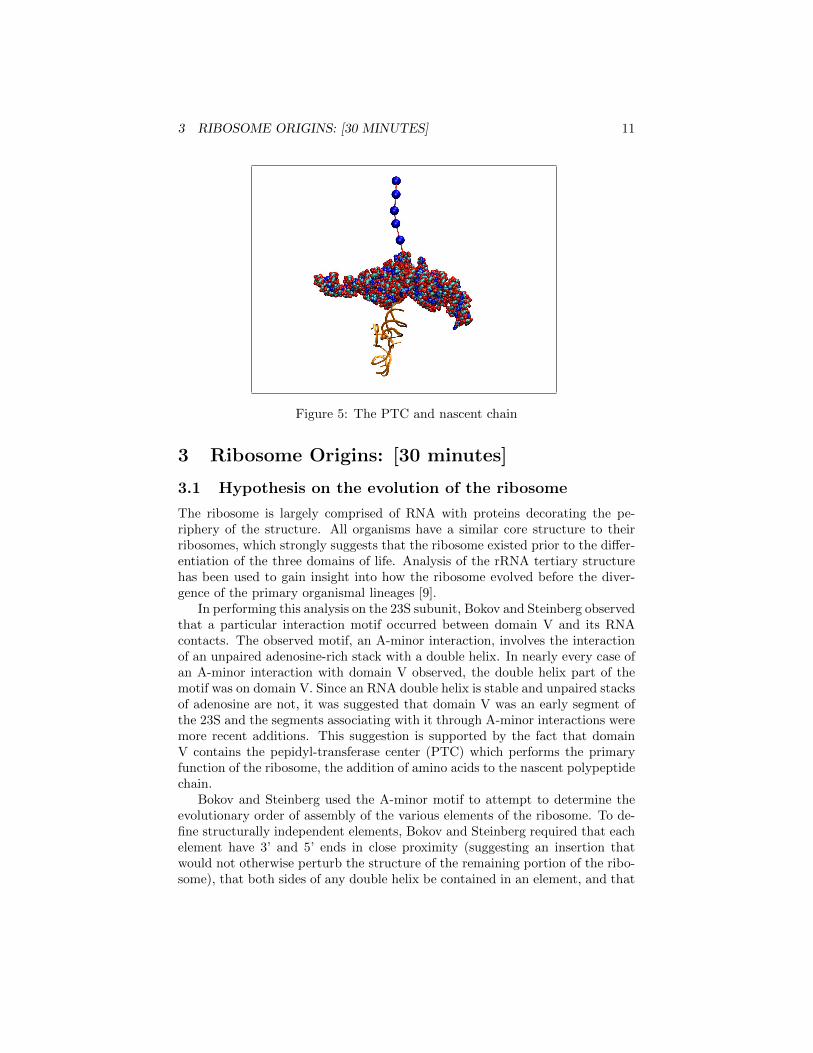
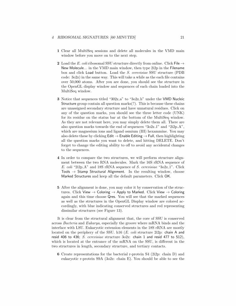
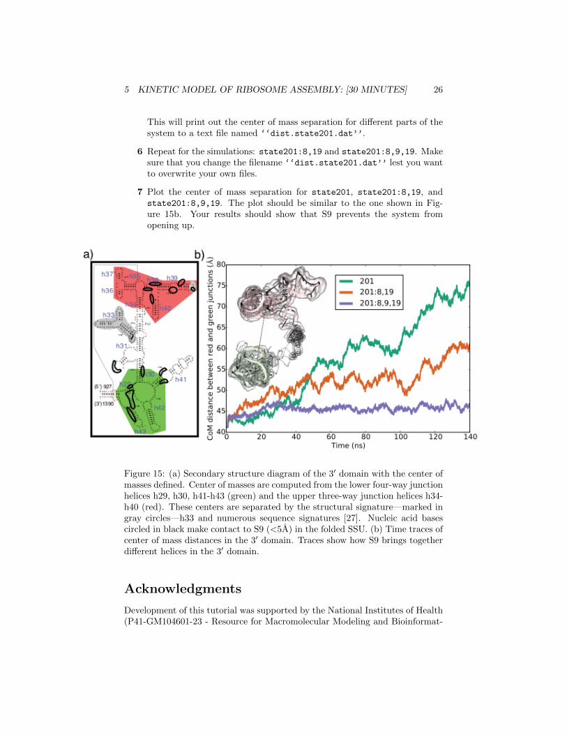

REFERENCES 30
[27] E Roberts A Sethi J Montoya CR Woese and Z Luthey-SchultenMolecular signatures of ribosomal evolution 105(37)13953ndash13958 2008


![List of Publications Professor Klaus Schulten Departments ... · Chem., NF101:371– 390, 1976. [9] Klaus Schulten. Quantum mechanical propensity rules for the transfer of angular](https://img.pdfslide.net/doc/110x75/5eccf558a55d4643151dc4b6/list-of-publications-professor-klaus-schulten-departments-chem-nf101371a.jpg)