Embed Size (px)
Citation preview
University of Khartoum
Graduate college
Medical & Health Studies Board
Histological pattern of oesophageal tumours in Sudanese patients
By Dr. Maha Mohamed Elhassan Elawad Salim
MBBS Faculty of Medicine, University of Khartoum 1995.
A thesis submitted in the partial fulfilment for the requirements of the degree of
clinical MD in Pathology August 2006
Supervisor
Dr. Ahmed Ibrahim Shumo
M.D. Associate Professor of Pathology
Faculty of Medicine and Health Sciences
International University of Africa
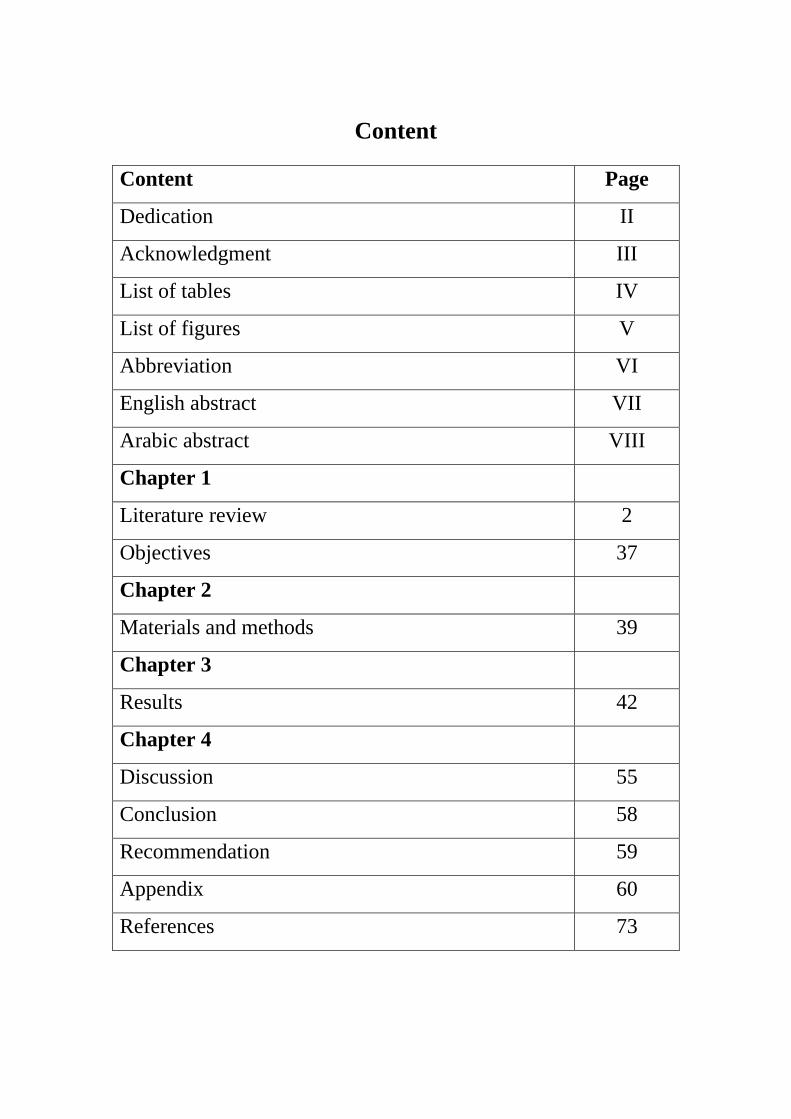
Content
Content Page
Dedication II
Acknowledgment III
List of tables IV
List of figures V
Abbreviation VI
English abstract VII
Arabic abstract VIII
Chapter 1
Literature review 2
Objectives 37
Chapter 2
Materials and methods 39
Chapter 3
Results 42
Chapter 4
Discussion 55
Conclusion 58
Recommendation 59
Appendix 60
References 73

II
Dedication
To the soul of my Father.
To my Mother for her great help.
To my husband for his appreciated help.
To my Kids: Mazin , Hind and Ahmed.
III
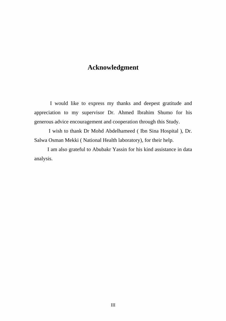
Acknowledgment
I would like to express my thanks and deepest gratitude and
appreciation to my supervisor Dr. Ahmed Ibrahim Shumo for his
generous advice encouragement and cooperation through this Study.
I wish to thank Dr Mohd Abdelhameed ( Ibn Sina Hospital ), Dr.
Salwa Osman Mekki ( National Health laboratory), for their help.
I am also grateful to Abubakr Yassin for his kind assistance in data
analysis.
IV
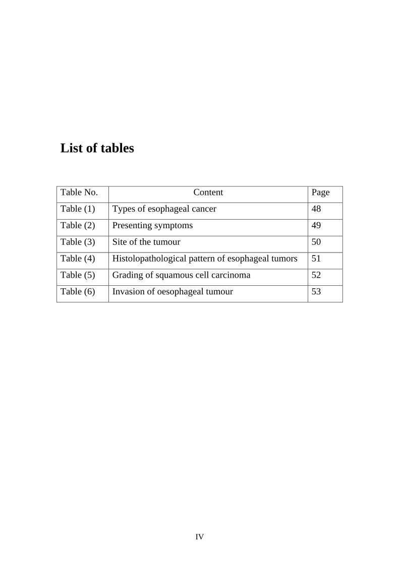
List of tables
Table No. Content Page
Table (1) Types of esophageal cancer 48
Table (2) Presenting symptoms 49
Table (3) Site of the tumour 50
Table (4) Histolopathological pattern of esophageal tumors 51
Table (5) Grading of squamous cell carcinoma 52
Table (6) Invasion of oesophageal tumour 53
V
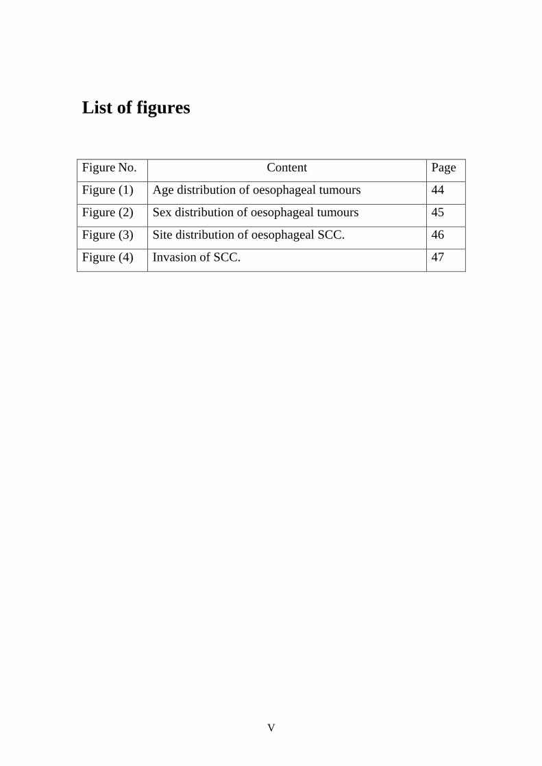
List of figures
Figure No. Content Page
Figure (1) Age distribution of oesophageal tumours 44
Figure (2) Sex distribution of oesophageal tumours 45
Figure (3) Site distribution of oesophageal SCC. 46
Figure (4) Invasion of SCC. 47
VI
Abbreviations: Abbreviation Meaning CCL cell Centrocyte-like cells CD Cluster of differentiation CT Computed tomography DNA Deoxy ribonucleic acid GERD Gastroesophageal reflux disease GISTs Gastrointestinal stromal cells HPV Human papilloma virus IUCC International union against cancer MALT Mucosa associated lymphoid tissue MCV Mean corpuscular volume MRI Magnetic resonance imaging OSCC Oesophageal squamous cell carcinoma RNA Ribonucleic acid RR Relative risk SCC Squamous cell carcinoma TNM Tumour node metastasis VIP Vasoactive intestinal polypeptide WDHA. Watery diarrhoea, hypokalaemia, achlorohydria synd. WHO World health organization
VII
Abstract
Oesophageal tumours are either benign or malignant. Benign
tumours are mostly mesenchymal in origin. With rare exceptions,
malignant oesophageal tumours arise from the epithelial layer.
This is a retrospective study conducted in Sudanese patients at Ibn
Sina hospital and NHL. The aim of the study is to determine
histopathological pattern, age and sex distribution of oesophageal
tumours.
The study comprised 102 patients of which 44 were males and 58
were females with a male to female ratio of 1:1.3. The age of all patients
ranged between 29 – 90 with a mean of 65 years.
In this study no benign tumour was identified and all the cases were
malignant tumours the histology of which was SCC and adenocarcinoma.
The SCC being predominant comprising 89.1 %, with a male to female
ratio of 1:1.6. There is striking male predominance in adenocarcinoma
(10:1)
By revising the slides: 85 were SCC (83 %) , 4 basoloid squamous
cell carcinoma (4%), 2 spindle cell carcinoma (2 %), 9 adenocarcinoma
(9%), 1 case papillary carcinoma (1 % ) , one case signet ring cell
carcinoma and 1 anaplastic carcinoma (1%).
Invasive SCC represented the majority of cases (88%) while only
12% were intraepithelial.
In conclusion benign tumours and other tumours like carcinoid,
lymphomas etc are rare. SCC is the most common oesophageal cancer in
Sudanese patients as it is the case worldwide. Intraepithelial neoplasia is
under diagnosed and this raises the importance of screening programs.
VIII
ص األطروحةملختنمو معظم األورام الحميدة من النـسيج . تنقسم أورام المرئ الي أورام حميدة و أورام خبيثة
هذه الدراسة . بينما تنمو معظم األورام الخبيثة من الخاليا المبطنة للغشاء الداخلى للمرئ . األوسط
ى ابن سيناء والمعمـل أجريت فى مستشف . الى الوراء عن أورام المرئ فى المرضى السودانيين
تهدف الدراسة الى بحث التغيرات المرضية في األنـسجة ومعرفـة . القومى الصحى بالخرطوم
.توزيع المرض فى األعمار المختلفة و عالقة نوع المرض مع الجنس
نـسبة المرضـى مـن . من النساء 58 من الرجال و 44 مريض منهم 102 تضمنت الدراسة
. سنة65 سنة و متوسط األعمار 90-29 وتراوحت األعمار بين 1.3:1 الرجال الى النساء
لم تصادف هذه الدراسة اى حالة ورم حميد، وكانت الحاالت أورام خبيثة اما ناشئة من الخاليا
أو . وتوزيع المرض شبه متماثل فى الجنـسين % 89.1المبطنة للغشاء الداخلى للمرئ وهى تمثل
). 1:10( بة الرجال تفوق نسبة النساء فى االصابة سرطان غددى و هنا نس
حالة سرطان ناشئ من الخاليا المبطنة للغشاء الداخلى للمرئ 85 بالرجوع الى الشرائح وجدت
حـاالت سـرطان 9، %)2(، حالتان سرطان مغزلـى %) 4( حاالت سرطان قاعدى 4، %) 83(
، و حالـة %)1(واحدة سـرطان حليمـى ، حالة %)1(، حالة واحدة سرطان خاتمى %)9(غددى
من حاالت السرطان الناشئ من الخاليا المبطنة % 88%). 1(واحدة سرطان متحول أو ارتدادى
.غير متغلغل% 12للغشاء الداخلى للمرئ كان سرطان متغلغل و
. خلصت الدراسة الى أن األورام الحمبدة و بعض األورام الخبيثة فى المرئ نادرة الحـدوث
ن النوع األكثر انتشارا بين المرضى السودانيين هو السرطان الناشئ مـن الخاليـا المبطنـة وأ
و أن معظـم االلحـاالت التـى تـم . للغشاء الداخلى للمرئ كما هو الحال فى غالب بلدان العـالم

IX
و أشارت الدراسة الى أهمية الفحص الطبى الدورى ). سرطان متغلغل ( تشخيصها حاالت متقدمة
. عمار التى يكثر فيها حدوث المرضخاصة لأل

1
CChhaapptteerr 11

2
(1) Introduction:
Benign tumors of the oesophagus are over shadowed
by cancer of the esophagus. However, they can be a cause of
morbidity and can lead to dysphagia and thoracic pain. The
frequency of leiomyomas which are the most common benign
tumors of the esophagus has been found to be almost 8% (1).
Carcinomas of the esophagus raised a considerable medical and
public health challenge in many parts of the world. Morphologically
and etiologically two major types are distinguished, squamous cell
carcinoma and adenocarcinoma (2). Worldwide, squamous cell
carcinoma (SCC) constitutes 90% of esophageal cancers but in
the United States there has been an exceptional increase in the
incidence of adenocarcinomas associated with Barrett esophagus
(3). Analysis of incidence rates by subsite and subtype showed an
increase in adenocarcinomas of the oesophagus and gastric
cardia, largely restricted to males. In females, the rise in incidence
of squamous cell carcinoma of the oesophagus appeared to be
more marked than the rise in adenocarcinomas (4). Most cases of
oesophageal carcinomas occur in adults older than the age of 50
with a male to female ratio of 3:1. There are striking and puzzling
differences in the geographic incidence of esophageal carcinoma.
In the United States, there are about 6 new cases per 100,000
population per year, accounting for 1% to 2% of all cancer deaths.
In regions of Asia extending from the Northern Provinces of China
to the Caspian Littoral in Iran, the prevalence is well over 100 per
100,000, and 20% of cancer deaths are caused by oesophageal
cancer (mainly squamous cell type), with females being affected
3
more often than males. These epidemiologic contrasts must
contain causative clues that remain to be deciphered (3). The
incidence in the Sudan is 1.4% of all malignant tumours. The
disease affected both sexes equally; is most common at the age of
50-69 and is commoner in patients from the North. (5).
The risk factors for squamous cell carcinomas of the
esophagus include esophageal disorders like longstanding
esophagitis, achalasia, and Plummer Vinson syndrome. Smoking
and alcohol are two important and well known risk factors.
However, different influences must underline the very high
incidence of this tumor among Moslems of Iran, who neither drink
nor smoke. Dietary factors like deficiency of vitamins and trace
metals, fungal contamination of food stuffs and high content of
nitrites, nitrosamine are invoked. Strong association with human
papilloma virus is noted in high incidence areas (3). It is suggested
that ethnicity may influence esophageal cancer histology or ethnic
background may place an individual at increased risk for certain
types of esophageal cancer (6).The role of genetic predisposition
is extremely ill-defined but the rare genetic syndrome of tylosis
(hyperkeratosis of palms and soles) carries an almost certain
probability of the development of esophageal cancer. Barret
esophagus is the only recognized precursor of esophageal
adenocarcinoma (3).
Esophageal lesions evoke a similar and remarkably limited
range of symptoms. All produce dysphagia which is attributed
either to deranged esophageal motor function or obstruction of the
lumen ( 3).
Exfoliative cytology in experienced hands is an extremely
accurate technique for the evaluation of esophageal lesions
4
particularly those of the lower third. For many years, it was clearly
superior to radiography or endoscopy. However, since the
introduction of the flexible fiberoptic endoscope, the diagnostic
accuracy of direct-vision biopsy has become as high as that of
cytology (1).
Only a small number of genotypes have been obtained on
carcinomas of the esophagus, and most of these have been
incomplete. Of the genes known to be altered in SCC of the
esophagus, TP53 is most often found to be abnormal. The
contribution of molecular studies to the prognosis of SCC of the
esophagus is not yet firmly established (7).
Radiation therapy is the most common form of treatment for
carcinomas of the upper two thirds of the esophagus and surgery
(in the form of oesophagastrectomy) is usually performed for
carcinoma of the lower third. Some authors advocated a
combination of chemotherapy and radiation therapy, with or
without surgery. In inoperable cases considerable palliation can
still be achieved by well-planned irradiation (1).
Despite progress in therapy, carcinomas of the esophagus
remain a tumor with somber prognosis. Patients often present with
relatively advanced tumor because the tumor remains
asymptomatic for a long interval. Progression of squamous cell
carcinomas of the esophagus has been recorded to take 3 to 4
years (7).
5
Literature review
(1) Anatomy and physiology of the esophagus:- The esophagus is a muscular tube, about 25 cm (10 in) long
connecting the pharynx to the stomach. It begins in the neck at
the caudal border of the cricoid cartilage. It pierces the diaphragm
at the level of the tenth thoracic vertebra, and ends at the cardiac
orifice of the stomach at the level of the eleventh thoracic vertebra.
Thus the esophagus is divided into cervical part, thoracic
part and abdominal part. Arteries supplying the esophagus are
derived from the inferior thyroid branch of the thyrocervical trunk,
form the descending thoracic aorta, from the bronchial arteries,
from the left gastric branch of the celiac artery, and from the left
inferior phrenic branch of the abdominal aorta. The veins drain
into the inferior thyroid veins, the azygos, hemiazygos and
accessory hemiazygos veins. The abdominal part drains partly
into the azygos vein and partly into the left gastric vein. The latter
vein being a tributary of the portal vein. The lymph vessels from
the upper third of the esophagus drains into the cervical nodes, the
middle third into the paraeosophageal and paratracheal
mediastinal nodes, and the lower third into the nodes around the
aorta and celiac axis.The nerve supply is derived from the vagus
and cervical sympathetic trunks (8).
Physiologically the oesophagus normally exhibits two
types of peristaltic movements: primary peristalsis and secondary
peristalsis. Primary peristalsis is simply a continuation of
swallowing. This wave passes all the way from the pharynx to the
stomach in about 8 to 10 seconds. If the primary wave fails to
move all the food that has entered the esophagus into the
6
stomach, secondary peristaltic waves result from distension of the
esophagus by the retained food, and they continue until all the
food has emptied into the stomach. These secondary waves are
initiated partly by intrinsic neural circuits in the esophageal
myenteric nervous system and partly by reflexes that are
transmitted through vagal afferent fibers from the esophagus to the
medulla and then back again to the esophagus through vagal
efferent fibers. To prevent reflux of stomach contents into the
esophagus the esophageal circular muscle functions as a lower
esophageal sphincter or gastroesophageal sphincter at the lower
end of the esophagus. Anatomically, this sphincter is no different
from the remainder of the esophagus. Physiologically, it normally
remains tonically constricted (with an intra-luminal pressure at this
point in the esophagus of about 30 mm Hg), in contrast to the mid
portion of the oesophagus between the upper and lower
sphincters, which normally remains relaxed. Another factor that
prevents reflux is a valve-like mechanism of that short portion of
the esophagus that lies immediately beneath the diaphragm before
reaching the stomach (9).
(2) Histology of the esophagus:- The histological structure of the esophagus follows the same
general pattern as the rest of the alimentary tract:-
1. The fibrous layer (serosa):- It consists of an external adventitia of irregular, dense
connective tissues containing many elastin fibers. Its fibers also
penetrate and surround the fasciculi of muscle in the deeper
layers.
2. The muscularis externa:-
7
It is 0.5-2.0 mm in thickness and consists of outer longitudinal
and inner circumferential layers of muscle fibres. In the initial
portion of the esophagus, both layers are striated muscle. In its
middle third, smooth muscle fibres begin to appear deep to the
striated muscle, and in the lower third, both layers of the
muscularis consist entirely of smooth muscle. 3. The submucosa:-
It is 400-600 µm in thickness and contains interlacing bundles of
collagen fibres, abundant elastic fibres, and many small blood
vessels. The mucosa and sub mucosa, in the un-distended
esophagus, form broad longitudinal folds that give the lumen a
highly irregular outline. 4. The mucosa:-
The esophageal mucosa is 300-500 µm thick and has an
epithelium that is a continuation of the stratified squamous
epithelium lining the oropharynx. Stratum germinativum, stratum
spinosum, and stratum corneum are identifiable, but the
thickness and degree of keratinization of the stratum corneum
vary greatly from species to species. The superficial cells retain
their nucleus and have a few keratohyalin granules in their
cytoplasm but show little evidence of heavy keratinization. Cells
of the stratum spinosum have short microplicae that project into
the intercellular spaces, and the processes of neighboring cells
are attached by prominent desmosomes. In addition to their
organelles, these cells contain bundles of intermediate filaments
and 120-150 nm vesicles that contain eccentrically placed dense
material. The base of the epithelium is quite irregular with
closely spaced deep recesses in its under surface, occupied by
papillae of the lamina propria. The cells of the stratum
8
germinativum are cuboidal and have desmosomes on their
interdiagitating lateral surfaces and hemidemosomes at their
base.
The intercellular spaces of the stratum corneum and outer
portion of the stratum spinosum contain material that gives a
strong staining reaction with Alcian blue at pH2.5 and is deeply
stained with the periodic-acid-Schiff reaction for glyconjugates.
5. Oesophageal glands:- Two kinds of esophageal glands are distinguished on the basis
of their location. (1)The superficial mucosal glands are limited to
the lamina propria and are found in limited number in the upper
oesophagus and near its junction with the stomach. They are
tortuous tubular glands lines with cuboidal or columnar epithelial
cells that resemble those of the cardiac glands of the stomach.
Owing to this resemblance, these glands are sometimes called
cardiac esophageal glands. They are few in number and are
interpreted by some as islands of ectopic gastric mucosa. Their
small ducts join a larger duct that usually opens at the tip of a
small papilla. (2) The submucosal glands are more widespread
and extend into the submucosa.They are tubuloacinar glands
arranged in small lobules that are drained by a single duct. The
acini are made up of plump cells with their nucleus compressed
to the base by droplets of mucus that occupy most of the cell
volume. A second cell type is cuboidal with a centrally placed
nucleus, a basophilic cytoplasm, and somewhat smaller and
denser secretory droplets or granules. These cells are
considered by some to be an earlier stage in the secretory cycle
of the mucous cell, but others regard them as a separate serous
cell type.
9
Like the minor gland of the oral cavity, the oesophageal
glands probably secrete, continuously maintaining a thin
lubricating layer of mucus on the surface of the epithelium, but
the rate of their secretion may be increased during the ingestion
and swallowing of food (10).
(3) Histopathology of oesophageal tumors: (A) Squamous epithelial tumors: 1- Squamous cell papilloma:- Squamous cell papilloma is rare and usually causes no
specific symptoms. It is a benign tumor composed of hyperplastic
squamous epithelium covering finger-like processes with cores
derived from the lamina propria. The polypoid lesions are smooth,
sharply demarcated and usually 5 mm or less in maximum
diameter. Rarely, giant papillomas have been reported, with sizes
up to 5 cm. Most squamous cell papillomas represent single
isolated lesions, typically located in the distal to middle third of the
oesophagus, but multiple lesions occur.
Histologically cores of firbovascular tissue are covered by
mature stratified squamous epithelium. The aetiological role of
human papillomavirus (HPV) infection has been investigated in
several studies, but the results were inconclusive. Malignant
progression to SCC is extremely rare.
2- Squamous cell carcinoma of the oesophagus - Definition:- Squamous cell carcinoma (SCC) of the esophagus is a malignant
epithelial tumor with squamous cell differentiation. Microscopically
it is characterized by keratinocyte-like-cells with intercellular
10
bridges and/or keratinization. Blacks have a fourfold to fivefold
higher rate of squamous cell carcinoma than whites, but the rate of
adenocarcinoma in blacks was 30% of the rate in whites. The
incidence of squamous cell carcinoma in black men and women
increased by approximately 30% between 1973 and 1982, and the
rate of adenocarcinoma among white men increased 74%. (11)
- Epidemiology :- Squamous cell carcinoma of the oesophagus shows great
geographical diversity in incidence, mortality and sex ratio. There
are however, several well-defined high-risk areas e.g. Normandy
and Calvados in North-West France and Northern Italy. This type
of cancer is much more frequent in Eastern countries and in many
developing countries. In both high-risk and low-risk regions, this
cancer is exceedingly rare before the age of 30 and the median
age is around 65 in both males and females. There was no
significant difference between its involvement among males and
females (12). In the past 20-30 years the incidence of squamous
cell carcinoma (SC) of the esophagus in western countries has
largely stayed constant, while that of adenocarcinoma (AC) of the
oesophagus and the cardia has risen. (13).
- Etiology:- 1- Tobacco and alcohol:- In Western countries, nearly 90% of the
risk of SCC can be attributed to tobacco and alcohol. With regard
to the consumption of tobacco, a moderate intake during a long
period carries a higher risk than a high intake during a shorter
period, whereas the reverse is true for alcohol. The oesophagus,
stomach and pancreas are primary target organs for ethanol-
related diseases. In the oesophagus and stomach, ethanol induces
motility disorders and mucosal lesions that are dose-dependent
11
and reversible under acute conditions. Chronic consumption of
alcohol causes a significant increase in the risk for squamous
carcinoma of the oesophagus. All of these effects are mainly
caused by direct contact of alcohol or its metabolite acetaldehyde
with the mucosa. Non-alcoholic components are responsible for
many effects of alcoholic beverages, including the powerful
stimulation of gastric acid secretion by beverages that are
produced by fermentation. (14), Alterations of retinoic acid
receptors protein may contribute in the development of SCC in
esophagus and that in some patients life style (e.g. smoking and
alcohol consumption) may be a critical component in the alteration
of retinoic acid receptor levels in esophagus (15). Because some
of the causes of increased mean corpuscular volume (MCV) and
oesophageal squamous cell carcinoma (OSCC) , including
alcoholism, acetaldehyde exposure, smoking, and poor nutrition
are common to both, macrocytosis has been used as a predictor of
early OSCC in alcoholics. MCV and alcohol flushing might be used
to better select candidates to screen this high-mortality-rate cancer
not only in alcoholics but also in nonalcoholic men (16).
2- Nutrition:-In high-risk areas of China, a deficiency in certain
trace elements and the consumption of pickled or mouldy foods
(which are potential sources of nitrosamines) have been
suggested . Dietary folate has been inversely related to the risk of
several cancers. Although, studies on the role of dietary folate in
oesophageal cancer are scanty, dietary folate was inversely
related to OSCC risk (17).
3- Hot beverages:- Worldwide, one of the most common risk
factors appears to be the consumption of burning-hot beverages
which cause thermal injury leading to chronic oesophagitis and
12
then to precancerous lesions. A controversial nationwide
population-based case-control study in Sweden about the relation
between hot beverage consumption and oesophageal cancer
concluded that, drinking beverages very hot did not increase the
risk for oesophageal squamous cell carcinoma, oesophageal
adenocarcinoma, or gastric cardia adenocarcinoma .(18).
4- HPV:- Conflicting reports have proposed a role for infectious
agents, including human papillomavirus (HPV) infection.
Associations between achalasia, Plummer-Vinson syndrome,
celiac disease and tylosis (focal noneepidermolytic palmoplantar
keratoderma) with oesophageal cancer have also been described.
- Localization:- Oesophageal SCC is located predominantly in the middle and the
lower third of the oesophagus, only 10-15% being situated in the
upper third. Nearly half of the SCC occurred in the middle of the
oesophagus (11)
- Macroscopy:- The gross appearance varies according to whether it is detected
in an early or an advanced stage of the disease. Among early
SCC, polypoid, plaque-like, depressed and occult lesions have
been described. For the macroscopic classification of advanced
oesophageal SCC, Ming has proposed three major patterns:
fungating, ulcerative and infiltrating. The fungating pattern is
characterized by a predominantly exophytic growth, whereas in the
ulcerative pattern, the tumor growth is predominantly intramural,
with a central ulceration and elevated ulcer edges. The infiltrative
pattern, which is the least common one, also shows a
predominantly intramural growth, but causes only a small mucosal
defect (2).
13
- Precursor lesions:-
The development of oesophageal SCC is thought to be a
multistage process which progresses from the conversion of
normal squamous epithelium to that with basal cell hyperplasia,
intraepithelial neoplasia (dysplasia and carcinoma in situ), and
finally, invasive SCC (2). Basal cell hyperplasia:-
This lesion is histologically defined as an otherwise normal
squamous epithelium with a basal zone thickness greater than
15% of total epithelial thickness, without elongation of lamina
propria papillae. In most cases, basal cell hyperplasia is an
epithelial proliferative lesion in response to oesophagitis, which is
frequently observed in high-risk populations for oesophageal
cancer (2).
Intraepithelial neoplasia:-
The lesion is about eight times more common in high cancer-
risk areas than in low-risk areas and is frequently found adjacent to
invasive SCC in oesophagectomy specimens. Morphological
features of intraepithelial neoplasia include both architectural and
cytological abnormalities. The architectural abnormality is
characterized by a disorganization of the epithelium and loss of
normal cell polarity. Cytologically, the cells exhibit irregular and
hyperchromatic nuclei, an increase in nuclear/cytoplasmic ratio
and increased mitotic activity. Dysplasia is usually graded as low
or high-grade. In low-grade dysplasia, the abnormalities are often
confined to the lower half of the epithelium, whereas in high-grade
dysplasia the abnormal cells also occur in the upper half and
exhibit a greater degree of atypia. In carcinoma in situ, the
atypical cells are present throughout the epithelium without
14
evidence of maturation at the surface of the epithelium. In a two-
tier system, severe dysplasia and carcinoma-in-situ are included
under the rubric of high-grade intraepithelial neoplasia, and may
have the same clinical implications (2).
Epidemiological follow-up studies suggest an increased risk
for the subsequent development of invasive SCC for patients with
basal cell hyperplasia (relative risk: 2.1), low-grade dysplasia (RR:
2.2), moderate-grade dysplasia (RR: 15.8), high-grade dysplasia
(RR: 72.6) and carcinoma in situ (RR: 62.5).(2)
-Tumor spread and staging:- Tumor spread:-
The most common sites of metastasis of oesophageal SCC
are the regional lymph nodes. The risk of lymph node metastasis
is about 5% in carcinomas confined to the mucosa but over 30% in
carcinomas invading the sub mucosa and over 80% in carcinomas
invading adjacent organs or tissues. Lesions of the upper third of
the oesophagus most frequently involve cervical and mediastinal
lymph nodes, whereas those of the middle third metastasise to the
mediastinal, cervical and upper gastric lymph nodes. Carcinomas
of the lower third preferentially spread to the lower mediastinal and
the abdominal lymph nodes. The most common sites of
haematogenous metastases are the lung and the liver. Less
frequently affected sites are the bones, adrenal glands and brain.
(2)
Superficial oesophageal carcinoma:- When the tumor is confined to the mucosa or the sub
mucosa, the term superficial oesophageal carcinoma is used
15
irrespective of the presence of regional lymph node metastases
(2). Intramural metastases:- A special feature of oesophageal SCC is the occurrence
of intramural metastases. These metastases are thought to result
from intramural lymphatic spread with the establishment of
secondary intramural tumor deposits. Intramural metastases are
associated with an advanced stage of disease and with shorter
survival (2). Second primary SCC:- Additionally, the occurrence of multiple independent SCC
has been described in between 14and 31% of cases, the second
cancers being mainly carcinomas in situ and superficial SCC.
Staging: For the staging of SCC, the TNM system (tumor, node,
metastasis) established by the international Union Against Cancer
(UICC) is the most widely used system. Its usefulness in the
planning of treatment and in the prediction of prognosis has been
validated (Appendix 2).
Histopathology:- Oesophageal SCC is defined as the penetration of neoplastic
squamous epithelium through the epithelial basement membrane
and extension into the lamina apropria or deeper tissue layers.
Invasion commonly starts from a carcinoma in situ with the
proliferation of rete-like projections of neoplastic epithelium that
push into the lamina propria with subsequent dissociation into
small carcinomatous cell clusters. Along with vertical tumor cell
infiltration, usually a horizontal growth undermines the adjacent
normal mucosa at the tumor periphery. The carcinoma may
already invade intramural lymphatic vessels and veins at an early
16
stage of disease. The frequency of lymphatic and blood vessel
invasion increases with increasing depth of invasion. Tumor cell in
lymphatic vessels and in blood vessels may be found
progressively several centimeters beyond the gross tumor. The
carcinoma invades the muscular layers, enters the loose fibrous
adventitia and may extend beyond the adventitia, with invasion of
adjacent organs or tissues, especially the trachea and bronchi,
eventually with the formation of oesophagotracheal or
oesophagobronchial fistulae (2).
Oesophageal SCC displays different microscopic patterns of
invasion, which are categorized as ‘expansive growth’ or
‘infiltrative growth’. The former pattern is characterized by a broad
and smooth invasion front with little or no tumor cell dissociation,
whereas the infiltrative pattern shows an irregular invasion front
and a marked tumor cell dissociation.
The degree of desmoplastic or inflammatory stromal
reaction, nuclear polymorphism and keratinization is extremely
variable. Additionally, otherwise typical oesophageal SCC may
contain small foci of glandular differentiation, indicated by the
formation of tubular glands or mucin-producing tumor cells.
Variants of squamous cell carcinoma : - Verrucous carcinoma:- This rare variant of squamous cell carcinoma is histologically
comparable to verrucous carcinomas arising at other sites. On
gross examination, its appearance is exophytic, warty, cauliflower-
like or papillary. It can be found in any part of the oesophagus.
Histologically, it is defined as a malignant papillary tumor
composed of well differentiated and keratinized squamous
epithelium with minimal cytological atypia, and pushing rather than
17
infiltrating margins. Oesophageal verrucous carcinoma grows
slowly and invades locally, with a very low metastasizing potential
(2).
- Spindle cell carcinoma This unusual malignancy is defined as a squamous cell
carcinoma with a variable sarcomatoid spindle cell component. It
is also known by a variety of other terms, including
carcinosarcoma, pseudosarcomatous squamous cell carcinoma,
polypoid carcinoma and squamous cell carcinoma with a spindle
cell component. Macroscopically, the tumor is characterized by a
polypoid growth pattern. The spindle cells may be capable of
maturation, forming bone, cartilage and skeletal muscle cells.
Alternatively, they may be more pleomorphic, resembling
malignant fibrous histocytoma. In the majority of cases a gradual
transition between carcinomatous and sarcomatous components
has been observed on the light microscopic level.
Immunohistochemical and electron microscopic studies indicate
that the sarcomatous spindle cells show various degrees of
epithelial differentiation. Therefore, the sarcomatous component
may be metaplastic. However, a recent molecular analysis of a
single case of a spindle cell carcinoma showed divergent genetic
alterations in the carcinomatous and in the sarcomatous tumor
component suggesting two independent malignant cell clones (2).
Basaloid squamous cell carcinoma:- This rare but distinct variant of oesophageal SCC appears to
be identical to the basaloid squamous cell carcinomas of the upper
aerodigestive tract. Histologically, it is composed of closely
packed cells with hyperchromatic nuclei and scant basophilic
cytoplasm, which show a solid growth pattern, small gland-like
18
spaces and foci of comedo-type necrosis. Basaloid squamous cell
carcinomas are associated with intraepithelial neoplasia, invasive
SCC or islands of squamous differentiation among the basaloid
cells. The proliferative activity is higher than in typical SCC.
However, basaloid squamous cell carcinoma is also characterized
by a high rate of apoptosis and its prognosis does not differ
significantly from that of the ordinary oesophageal SCC (2).
Grading:- Grading of oesophageal SCC is traditionally based on the
parameters of mitotic activity, anisonucleosis and degree of
differentiation (2).
Well differentiated tumors have cytological and histological
features similar to those of the normal oesophageal squamous
epithelium. In well differentiated oesophageal SCC there is a high
proportion of large differentiated, keratinocyte-like squamous cells
and a low proportion of small basal-type cells, which are located in
the periphery of the cancer cell nests. The occurrence of
keratinization has been interpreted as a sign of differentiation,
although the normal oesophageal squamous epithelium does not
keratinize (2).
Poorly differentiated tumors predominantly consist of
basal-type cells, which usually exhibit a high mitotic rate.
Moderately differentiated carcinomas, between the well
and poorly differentiated types, are the most common type,
accounting for about two-thirds of all oesophageal SCC.
Undifferentiated carcinomas are defined by a lack of
definite light microscopic features of differentiation. However,
ultrastructural or immunohistochemical investigations may disclose
19
features of squamous differentiation in a subset of light-
microscopically undifferentiated carcinomas (2).
Clinical features:- Symptoms and signs:- The most common symptoms of advanced oesophageal
cancer are dysphagia, weight loss, retrosternal or epigastric pain
and regurgitation caused by narrowing of the oesophageal lumen
by tumor growth. Superficial SCC usually has no specific
symptoms but sometimes causes a tingling sensation.
Endoscopy and vital staining:- Chromoendoscopy utilizing toluidine blue or Lugol iodine
spray may be of value. Toluidine blue, a metachromatic stain from
the thiazine group, has a particular affinity for RNA and DNA and
stains areas that are richer in nuclei than the normal mucosa.
Lugol solution reacts specifically with glycogen in the normal
squamous epithelium, whereas precancerous and cancerous
lesions, but also inflamed areas and gastric heterotopia, are not
stained. However, the superficial extension of carcinomas
confined to the mucosa can not be clearly recognized by simple
endoscopy.
Endoscopic ultrasonography:-
Endoscopic ultrasonography is used to evaluate both depth
of tumor infiltration and para-oesophageal lymph node involvement
in early and advanced stages of the disease.
-Computed tomography (CT) and magnetic resonance imaging
(MRI):-
In advanced carcinomas, CT and MRI give information on
local and systemic spread of SCC (2).
20
Treatment groups:- Following the clinical staging, patients are usually divided
into two treatment groups: those with locoregional disease in
whom the tumor is potentially curable (e.g. by surgery,
radiotherapy, multimodal therapy), and those with advanced
disease (metastases outside the regional area or invasion of the
airway) in whom only palliative treatment is indicated.
Oesophageal SCC limited to the mucosa may be treated by
endoscopic mucosal resection due to its low risk of nodal
metastasis. Endoscopic mucosal resection is also indicated for
high-grade intraepithelial neoplasia. Tumors that have invaded the
sub mucosa or those in more advanced tumor stage have more
than 30% risk of lymph node metastasis, and endoscopic therapy
is not indicated. Additionally, clinical staging is performed in order
to determine the success of treatment, e.g. following radio- and/or
chemotherapy (2).
(B) Adenocarcinoma of the oesophagus:- (1) Definition and epidemiology:-
It is malignant epithelial tumor of the oesophagus with
glandular differentiation arising predominantly from Barrett mucosa
in the lower third of the oesophagus. Infrequently, adenocrcinoma
originates from heterotopic gastric mucosa in the upper
oesophagus, or from mucosal and submucosal glands. In the mid
1990s the incidence of oesophageal adenocarcinoma has been
estimated to be between 1 and 4 per 100.000 per year in the USA
and several European countries and the incidence of oesophageal
adenocarcinoma has risen considerably, now it is equally or even
21
more prevalent than squamous cell cancers in these regions (19),
In Asia and Africa, adenocarcinoma of the oesophagus is an
uncommon finding, but increasing rates are also reported from
these areas, these include a high preponderance for the male sex
(male: female ratio 7:1) . (20), (21), a higher incidence among
whites and an average age at the time of diagnosis of around 65
years. The striking male predominance in patients with
adenocarcinoma of the oesophagus (male to female ratio of 6:1) is
not explained by known risk factor (22).
(2) Aetiology:- 1- Barrett oesophagus: Barrett's esophagus is a premalignant
condition and remains the number one risk factor for developing
adenocarcinoma. Gastro-esophageal reflux disease is a strong risk
factor for both esophageal adenocarcinoma and the precancerous
lesion Barrett's esophagus. Both of these conditions are related to
the reflux of acid and bile into the esophagus. This results in
inflammation and cell damage which initiates a sequence of events
termed the metaplasia-dysplasia sequence in which the squamous
epithelium is replaced by columnar epithelium exhibiting increasing
degrees of dysplasia and overt malignancy. Barrett's esophagus is
being better recognized in patients presenting with extra-
esophageal symptoms of gastroesophageal reflux such as chronic
cough and asthma (23 ). Barrett's esophagus is generally accepted
as a complication of chronic and severe gastroesophageal reflux
disease (GERD) (24). Experimental and clinical data indicate that
combined oesophageal exposure to gastric acid and duodenal
contents (bile acids and pancreatic enzymes) appears to be more
detrimental than isolated exposure to gastric juice or duodenal
22
contents alone. Recent reports from some surgical series further
suggest the importance of gastric and even duodenal reflux in the
etiology of esophageal metaplastic development (23 ). Combined
reflux is thought to increase cancer risk by promoting cellular
proliferation and by exposing the oesophageal epithelium to
potentially genotoxic gastro and intestinal contents e.g.
nitrosamines. However, the risk for patients with Barrett
esophagus to develop esophageal adenocarcinoma is low, and
most patients undergoing surveillance will not develop malignancy
(25). Clinically Barrett's esophagus is silent in up to 90% of cases.
Histopathologically, Barrett epithelium is characterized by two
different types of cells, i.e. goblet cells and columnar cells and has
also been termed ‘specialized’, ‘distinctive’ or Barrett metaplasia.
The goblet cells stain positively with Alcian blue at low pH (2.5).
The metaplastic epithelium has a flat or villiform surface, and is
identical to gastric intestinal metaplasia of the incomplete type
(type II or III). Rarely, foci of complete intestinal metaplasia (type
I) with absorptive cells and Paneth cells may be found. The
mucous glands beneath the surface epithelium and pits may also
contain metaplastic epithelium. Recent studies suggest that the
columnar metaplasia originates from multipotential cells located in
intrinsic oesophageal glands. Intraepithelial neoplasia in Barrett oesophagus
Generally has no distinctive gross features and is detected by
systematic sampling of a flat Barrett mucosa. The area involved is
variable, and the presence of multiple dysplastic foci is common.In
some cases, intraepithelial neoplasia presents as one of several
nodular masses resembling sessile adenomas. Here dysplastic
lesions have been considered true adenomas, with an expanding
23
but localized growth resulting in a well demarcated interface with
the surrounding tissue. Epithelial atypia in Barrett mucosa is
usually assessed according to the system devised for atypia in
ulcerative colitis, namely: negative, positive or indefinite for
intraepithelial neoplasia. If intraepithelial neoplasia is present, it
should be classified as low-grade (synonymous with mild or
moderate dysplasia) or high-grade (synonymous with severe
dysplasia and carcinoma in situ). The criteria used to grade
intraepithelial neoplasia comprise cytological and architectural
feature.
Negative for intraepithelial neoplasia :-
Usually the lamina propria of Barrett mucosa contains a mild
accompanying inflammatory infiltrate of mononuclear cells. There
may be mild reactive changes with enlarged, hyperchromatic
nuclei, prominence of nucleoli, and occasional mild stratification in
the lower portion of the glands. However, towards the surface
there is maturation of the epithelium with few or no abnormalities. Atypia indefinite for intraepithelial neoplasia Is one of the major challenges for the pathologist in Barrett
oesophagus in the differentiation of intraepithelial neoplasia from
reactive or regenerative epithelial changes. This is particularly
difficult, sometimes even impossible, if erosions or ulcerations are
present . In areas adjacent to erosions and ulcerations, the
metaplastic epithelium may display villiform hyperplasia of the
surface foveolae with cytological atypia and architectural
disturbances. These abnormalities are usually milder than those
observed in intraepithelial neoplasia. There is a normal expansion
of the basal replication zone in regenerative epithelium versus
intraepithelial neoplasia, where the proliferation shifts to more
24
superficial portions of the gland. If there is doubt as to whether
reactive and regenerative changes or intraepithelial neoplasia is
present in a biopsy, the category atypia indefinite for intraepithelial
neoplasia is appropriate and a repeat biopsy after reflux control by
medical acid suppression or anti-reflux therapy is indicated. Low-grade and high-grade intraepithelial neoplasia in Barrett
metaplastic mucosa is defined as a neoplastic process limited to
the epithelium. Its prevalence in Barrett mucosa is approximately
10% and it develops only in the intestinal type metaplastic
epithelium. Cytological abnormalities typically extend to the
surface of the mucosa. In low-grade intraepithelial neoplasia,
there is decreased mucus secretion, nuclear pseudostratification
confined to the lower half of the glandular epithelium, occasional
mitosis, mild pleomorphism, and minimal architectural changes. High-grade intraepithelial neoplasia shows marked pleomorphism
and decrease of mucus secretion, frequent mitosis, nuclear
stratification extending to the upper part of the cells and glands,
and marked architectural aberrations. The most severe
architectural changes consist of a cribriform pattern that is a
feature of high-grade intraepithelial neoplasia as long as the
basement membrane of the neoplastic glands has not been
disrupted. The diagnostic reproducibility of intraepithelial
neoplasia is far from perfect: significant interobserver variation
exists (2). 2- Tobacco:- Smoking has been identified as another major risk factor for
oesophageal adenocarcinoma and may account for as much as
40% of cases through an early stage carcinogenic effect. Placing
tobacco under the tongue or in the labiodental groove seems to be
25
associated with a high incidence of oral cancer and possibly also
of oesophageal cancer ( 5 )
3- Obesity and diabetes mellitus:- Obesity is a risk factor for adenocarcinomas of the
oesophagus and gastric cardia. Diabetes mellitus might mediate
that association, A study done by Rubenstein showed no
association between diabetes mellitus and adenocarcinoma of
oesophagus (26), (27).
4- Alcohol:- In contrast to squamous cell oesophageal carcinoma, there
is no strong relation between alcohol consumption and
adenocarcinoma of the oesophagus. (27).
5- Helicobacter pylori:- This infection does not appear to be a predisposing factor for
the development of intestinal metaplasia and adenocarcinoma in
the distal oesophagus. The relationship between H. pylori and
GERD+ Barrett's esophagus is controversial. H. pylori eradication
therapy may increase the risk for the development of
gastroesophageal reflux disease GERD, which may lead to
increase a risk factor of Barrett's esophagus and esophageal
adenocarcinoma. Accordingly, gastric H. pylori infection may even
exert a protective effect (24).
- Localization:- Adenocarcinoma may occur anywhere in a segment lined
with columnar metaplastic mucosa (Barrett oesophagus) but
develops mostly in its proximal verge. Adenocarcinoma in a short
segment of Barrett oesophagus is easily mistaken for
adenocarcinoma of the cardia. Since adenocarcinoma originating
from the distal oesophagus may infiltrate the gastric cardia and
26
carcinoma of the gastric cardia or subcardial region may grow into
the distal oesophagus these entities are frequently difficult to
discriminate. As an exception, adenocarcinoma occurs also in the
middle or proximal third of the oesphagus, in the latter usually from
a congenital islet of heterotopic columnar mucosa (that is present
in up to 10% of the population) (2).
(3) Symptoms and signs Adenocarcinoma:- Dysphagia is often the first symptom of advanced
adenocarcinoma in the oesophagus. This may be associated with
retrosternal or epigastric pain or cachexia. Endoscopically, the
early stages may be that of a small polypoid adenomatous-like
lesion, but more often it is flat, depressed, elevated or occult.
Areas with high-grade intraepithelial neoplasia are often
multicentric and occult. Therefore a systematic tissue sampling
has been recommended when no abnormality is evident
macroscopically. The usual pattern of advanced adenocarcinoma
at endoscopy is that of an axial, and often tight, stenosis in the
distal third of the oesophagus: with a polypoid tumor, bleeding
occurs at contact. Macroscopically, the majority of primary
adenocarcinomas of the oesophagus arise in the lower third of the
oesophagus within a segment of Barrett mucosa. Adjacent to the
tumor, the typical salmon-pink mucosa of Barrett oesophagus may
be evident, especially in early carcinomas. In the early stages, the
gross findings of Barrett adenocarcinoma may be subtle with
irregular mucosal bumps or small plaques. At the time of
diagnosis, most tumors are advanced with deep infiltration of the
oesophageal wall. The advanced carcinomas are predominantly
flat and ulcerated with only one third having a polypoid or fungating
27
appearance. Occasionally, multifocal tumors may be present. The
rare adenocarcinomas arising independently of Barrett
oesophagus from ectopic gastric glands and oesophageal glands
display predominantly ulceration and polypoid gross features,
respectively. These tumors are also found in the upper and middle
third of the oesophagus, but are rare (2).
(4) Histopathology of adenocarcinoma:- Adenocarcinomas arising in the setting of Barrett
oesophagus are typically papillary and/or tubular. A few tumors
are of the diffuse type and show rare glandular formations, and
sometimes signet ring cells. Differentiation may produce
endocrine cells, Paneth cells and squamous epithelium. Mucinous
adenocarcinomas i.e. tumors with more than 50% of the lesion
consisting of mucin, also occur. About Grading; most
adenocarcinomas arising from Barrett mucosa are well or
moderately differentiated, and display well formed tubular or
papillary structures. The well differentiated tumors may pose a
diagnostic problem in biopsy specimens because the infiltrating
component may be difficult to recognize as invasive since Barrett
mucosa often has irregular dispersed glands. Glandular structures
are only slightly formed in poorly differentiated adenocarcinomas
and absent in undifferentiated tumors. Adenocarcinomas spread first locally and infiltrate the oesophageal wall. Distal spread to
the stomach may occur. Extension through the oesophageal wall
into adventitial tissue, and then into adjacent organs or tissues is
similar to squamous cell carcinoma. Common sites or local spread
comprise the mediastinum, tracheobronchial tree, lung, aorta,
pericardium, heart and spine. Barrett associated adenocarcinoma
28
metastasizes to para-oesophageal and paracardial lymph nodes,
those of the lesser curvature of the stomach and the celiac nodes.
Distant metastases occur late. For staging, the TNM classification
used for SCC is applicable to Barrett adenocarcinoma and
provides prognostically significant data (2).
(5) Other carcinomas:- - Adenosquamous carcinoma:- This carcinoma has a significant squamous carcinomatous
component that is intermingled with a tubular adenocarcinoma.
- Mucoepidermoid carcinoma:- This rare carcinoma shows an intimate mixture of squamous
cells, mucus secreting cells and cells of an intermediate type.
- Adenoid cystic carcinoma:- This neoplasm is also infrequent and believed to arise, like
the mucoepidermoid variant, from oesophageal glands. Both
lesions tend to be of salivary gland type, and small tumors may be
confined to the submucosa.
(6) Prognostic factors in adenocarcinoma :- The major prognostic factors in adenocarcinoma of the
oesophagus are the depth of mural invasion and the presence or
absence of lymph node or distant metastasis. Gross features and
histological differentiation do not influence prognosis. The overall
5-year survival rate after surgery is less than 20% in most series
including a majority of advanced carcinomas. The survival rates
are better in superficial adenocarcinoma ranging from 65% to 80%
in different series.
29
Since the stage at the time of diagnosis is the most important
factor affecting outcome, endoscopic surveillance of Barrett
patients with early detection of their adenocarcinomas, results in
better prognosis in most cases (2).
(C) Endocrine tumors of the oesophagus:- Endocrine tumors of the oesophagus are rare and include
carcinoid (well differentiated endocrine neoplasm), small cell
carcinoma (poorly differentiated endocrine carcinoma) and mixed
endocrine-exocrine carcinoma. Aetiologically, patients with small
cell carcinomas often have a history of heavy smoking and one
reported case was associated with long standing achalasia. A
case of combined adenocarcinoma and carcinoid occurred in a
patient with a Barrett oesophagus. Small cell carcinoma has also
been associated with Barrett oesophagus. Carcinoid tumors are
typically located in the lower third of the oesophagus. Almost all
small cell carcinomas occur in the distal half of the oesophagus.
Clinically, dysphagia, severe weight loss and sometimes chest
pain are the main symptoms of endocrine tumors of the
oesophagus. Patients with small cell carcinomas often present at
an advanced stage. Inappropriate antidiuretic hormone syndrome
and hypercalcaemia have been reported. In addition, a case of
watery diarrhea, hypokalaemia, achlorohydria (WDHA) syndrome
due to ectopic production of VIP by a mixed-cell (squamous-small
cell) carcinoma of the oesophagus has been described.
Macroscopically, all reported oesophageal carcinoids were of
large size (from 4 to 7 cm in diameter) and infiltrated deeply the
oesophageal wall. Small cell carcinomas usually appear as
30
fungating or ulcerated masses of large size, measuring from 4 to14
cm in greatest diameter (2). Histopathologically, all carcinoids so far reported in the
literature have been described as deeply infiltrative tumors, with
high mitotic rate and metastases. Microscopically they are
composed of solid nests of tumor cells that show positive stain for
Grimelius and neuron-specific enolase, and characteristic
membrane-bound neurosecretory granules at ultrastructural
examination. Small cell carcinoma of the oesophagus is
indistinguishable from its counterpart in the lung according to
histological and immunohistochemical features as well as clinical
behaviour. The cells may be small with dark nuclei of round or
oval shape and scanty cytoplasm, or be larger with more
cytoplasm (intermediate cells) forming solid sheets and nests.
There may be foci of squamous carcinoma, adenocarcinoma,
and/or mucoepidermoid carcinoma, a finding that raises the
possibility of an origin of tumor cells from pluripotent cells present
in the squamous epithelium or ducts of the submucosal glands.
Argyrophylic granules can be demonstrated by Grimelius stain and
small dense-core granules are always detected by electron
microscopy. Mixed endocrine-exocrine carcinoma, a rare tumor
with few reported cases. There is combination of gastrointestinal
type adenocarcinoma with the trabecular-acinar component of a
carcinoid (2). Prognostic factors:-Two of three oesophageal carcinoids from
the analysis of 8305 cases of carcinoid tumors were associated
with distant metastases and one of the three reported cases died
29 months after surgery. The prognosis of small cell carcinoma of
31
the oesophagus is poor, even when the primary growth is limited.
The survival period is usually less than 6 months. Multidrug
chemotherapy may offer temporary remission (2).
(D) Lymphoma of the oesophagus:- Definition:- Primary lymphoma of the oesophagus is defined as
an extranodal lymphoma arising in the oesophagus with the bulk of
the disease localized to this site. Contiguous lymph node
involvement and distant spread may be seen but the primary
clinical presentation is in the oesophagus with therapy directed at
this site. Clinical features:- The oesophagus is the least common site of
involvement with lymphoma in the digestive tract, accounting for
less than 1% of lymphoma patients. Oesophageal involvement is
usually secondary either from the mediastinum, from nodal
disease or from a primary gastric location. Patients are frequently
male and usually over 50 years old. Tumors involving the distal
portion of the oesophagus may cause dysphagia. Histopathology:-Primary oesophageal lymphomas may be of
large B-cell type or may be low-grade B-cell MALT lymphomas.
MALT lymphomas show morphological and cytological features
common to MALT lymphomas found elsewhere in the digestive
tract. Lymphoid follicles are surrounded by a diffuse infiltrate of
centrocyte-like (CCL) cells showing a variable degree of plasma
cell differentiation. Infiltration of these cells into the overlying
epithelium is usually seen. In common with other sites in the
digestive tract, secondary involvement of the oesophagus may
occur in dissemination of any type of lymphoma. Primary
32
oesophageal T-cell lymphoma has been described but is
exceedingly rare (2).
(E) Mesenchymal tumors of the oesophagus:- Definition:-A variety of rare benign and malignant mesenchymal
tumors that arise in the oesophagus. Among these, tumors of
smooth muscle or ‘stromal’ type are most common. Epidemiology:- Sarcomas of the oesophagus accounted for 0.2% of malignant
oesophageal tumors in the United States from 1973 to 1987.
Males were more frequently affected than females by nearly 2:1.
Adults between the 6th and 8th decades are primarily affected.
Leiomyoma is the most common benign mesenchymal tumor of
the oesophagus. and constitute 0.4 - 1.5% of all tumours of this
organ (28 ), Although leiomyomas are the most common benign
tumors of the esophagus, esophageal leiomyomatosis is a rare
pathological entity, and pedunculated presentation is even
rarer(29). Esophageal leiomyoma usually originates from the
muscle layer of the esophageal wall and grows spirally around the
esophageal axis. (30). It occurs in males at twice the frequency as
females and has a median age distribution between 30 and 35
years, are most frequent in the lower oesophagus and begin as
intramural lesions. The larger tumors can extend to mediastinum
and form a predominantly mediastinal mass. Leiomyomas are
usually very slow growing and often asymptomatic. Symptomatic
tumors are usually greater than five centimeters in diameter (31).
Diffuse leiomyomatosis of the oesophagus is a rare entity among
oesophageal diseases. Histopathologically it is characterized by
33
diffuse hypertrophy of the muscular layer extending to the whole
oesophagus predominantly in the lower third, where it can result in
tumour formation. Leiomyomatosis can involve the upper part of
the stomach and is frequently associated with genital or
tracheobronchial (bronchitracheal) muscular localizations. Also, it
can be associated with Alport's syndrome in familial cases (32 ).
Leiomyomatosis forms worm-like intramural structures that may
extend into the upper portion of the stomach. Clinically, dysphagia
is the usual complaint, but many leiomyomas and a small
proportion of stromal tumors are asymptomatic and are incidentally
detected by x-ray as mediastinal masses. Since most sarcomas
project into the lumen, they are relatively easy to diagnose by
endoscopy or imaging studies. The endoscopic pattern is that of a
submucosal tumor with a swelling of a normal mucosa.
Endoscopic ultrasound helps in determining the actual size of the
tumor, its position in the oesophageal wall and its eventual position
in the mediastinum. Esophageal leiomyomatosis should be
considered in a young patient with long-standing dysphagia in
whom smooth, tapered esophageal narrowing on barium study and
circumferential esophageal wall thickening on CT scan are seen.
(33). Macroscopically Leiomyomas vary in size from a few
millimeters up to 10 cm in diameter (average 2-3 cm). They can
form sausage-like masses with a large longitudinal dimension or
dumb-bell shaped masses with circular involvement. Large
leiomyomas (over 0.5 kg) have been described. Sarcomas, most
of them representing malignant gastrointestinal stromal tumors
(GISTs), are typically multinodular or less commonly plaque-like
masses resembling sarcomas of the soft tissues. Many
oesophageal sarcomas protrude into the mediastinum.
34
Histopathologically, Leiomyoma is composed of bland spindle
cells and shows low or moderate cellularity and slight if any mitotic
activity. There may be focal nuclear atypia. The cells have
eosinophilic, fibrillary, often clumped cytoplasm. Eosinophilic
granulocytes and spherical calcifications are sometimes present.
Leiomyomas are typically globally positive for desmin and smooth,
muscle actin and are negative for CD34 and CD 117(KIT).
Leiomyomas should be removed when diagnosed, even if
asymptomatic, because malignancy cannot otherwise be excluded
and symptoms are likely to develop if treatment is delayed or
omitted (34).
Leiomyosarcoma, a malignant tumor featuring differentiated
smooth muscle cells is rare in the oesophagus. In a recent series,
such tumors comprised 4% of all combined smooth muscle and
stromal tumors. They were large tumors that presented in older
adults, and all patients died of disease. Diagnosis is based on
demonstration of smooth muscle differentiation by a-smooth
muscle actin, desmin or both, and lack of KIT expression.
Stromal tumors (GISTs) are rare in the oesophagus, and
comprise 20-30% of the combined cases of smooth muscle and
stromal tumors. Like elsewhere in the digestive system, they
predominantly occur in older adults between the 6th and 8th
decades, oesophageal stromal tumors may have a male
predominance. Most oesophageal examples are spindle cell
tumors, and a minority is epithelioid. Oesophageal GISTs are
identical with their gastric counterparts by their positivity for KIT
and CD 34, variable reactivity for smooth muscle actin and general
negativity for desmin. Most are clinically malignant, and commonly
develop liver metastases. The oesophageal tumors analyzed to
35
date have shown similar c-kit mutations (exon 11) as observed in
gastric and intestinal GISTs.
Prognosis:- The prognosis of oesophageal sarcomas, like
carcinomas, is largely dependent on the size, depth of invasion
and presence or absence of metastasis.
(F) Melanoma of the oesophagus:- Malignant melanoma in the oesophagus is much more
commonly metastatic than primary. Primary malignant melanoma
of the esophagus is rare, and its symptoms are similar to those of
squamous cell carcinoma (35), (36). Primary oesophageal
melanomas are usually polypoid and are clinically aggressive
lesions. They are believed to arise from a zone of atypical
junctional proliferation of melanocytes and such a proliferation is
often present adjacent to the invasive tumor, although it may not
be observed in advanced disease. The histology of the invasive
component is indistinguishable from cutaneous melanoma.
Growth is typically expansile rather than infiltrative.. In a case
report, melanoma was misdiagnosed in biopsy taken during
endoscopy. Final precise establishing the character of the lesion
was able during histopathological examination of the specimen
obtained during surgery. The outcome of the treatment was poor--
survival time did not exceed 14 months. Patient died because of
pulmonary metastases. (37). The clinical presentation of this
uncommon tumour is similar to esophageal carcinoma and the
preoperative diagnosis may be difficult and total esophagectomy is
the treatment of choice (38).
36
(G) Secondary tumors of the oesophagus:- Definition:- Tumors of the oesophagus that originate from but are
discontinuous with a primary tumor elsewhere in the oesophagus
or an extra-oesophageal neoplasm. Incidence:- Metastatic spread to the oesophagus is uncommon.
An unusually high frequency (6.1% of autopsy cases) was reported
from Japan. Origin of metastases:- Neoplasms of neighbouring organs such
as pharynx or gastric cardia can spread to the oesophagus via
lymphatics. Haematogenous metastases from any primary
localization may occur. Reported primary sites include thyroid,
lung, breast, skin, kidney, prostate and ovary. Localization:- The most common site of involvement is the middle
third of the oesophagus. Clinical features:- The leading symptom is dysphagia whereas
achalasia and upper gastrointestinal bleeding with anaemia are
unusual . Barium swallow examination, endoscopy, computed
tomography and magnetic resonance imaging demonstrate in most
cases a submucosal tumor, but any aspect resembling a primary
oesophageal carcinoma may be observed. Histopathological and predictive factors:- Submucosal
localizaiotn without invasion of the mucosa is characteristic for a
metastasis. Early metastases of gastric and oesphageal tumors
into the oesophagus may be local indicators of systemic spread.
The presence of metastasis in the oesphagus is a sign of poor
prognosis, but the outcome is much better when the primary tumor
growth rate is slow and when other metastases are excluded.
37
General objective: -To study oesophageal tumours in Sudanese patients. Specific Objective:
1- To identify the different histopathological patterns of oesophageal tumours in Sudanese patients.
2- To know the sex distribution among Sudanese patients with oesophageal tumours.
3- To determine the age distribution among Sudanese patients with oesophageal tumours.

38
CChhaapptteerr 22
39
Materials and method * The study design :
This is a descriptive retrospective study done in Ibn Sina
Hospital and the National Health Laboratory ( N.H.L) in
Khartoum, Sudan. Ibn Sina Hospital is a specialized hospital in
renal and gastrointestinal diseases. It receives patients from
different regions of Sudan for diagnosis and management. The
National Health Laboratory is a national reference laboratory in
Khartoum, Sudan.
* The study population :
The study include all patients with oesophageal tumours
presented to Ibn Sina hospital and the National Health
Laboratory between January 2000 through to December 2004.
* Inclusion criteria :
All patients with oesophageal tumours who underwent
endoscopic or histopathologic studies for diagnosis.
* Exclusion criteria :
40
Patients who had no histopathological slides in the
histopathology laboratory of Ibn Sina hospital and the National
Health Laboratory.
* Tools of data collection:
Data is collected in a predesigned questionnaire with detailed
history and investigation. The slides of histopathological
diagnosis for all patients were collected and revised to confirm
the diagnosis and to determine the histopathological type using
the WHO classification of oesophageal tumours ( appendix 1)
and grades the tumour using the well differentiated, moderately
differentiated and poorly differentiated grading system.
The data is electronically analysed using the computer
program SPSS. A P-value of < 0.05 is considered significant.

41
CChhaapptteerr 33
42
Results
About 102 cases of oesophageal biopsies were studied. All were
malignant. The age and sex distribution, presenting symptoms, sites,
histopathological pattern, and the behaviour of the tumour were recorded
and the following results obtained:
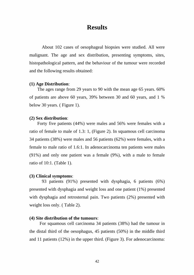
(1) Age Distribution: The ages range from 29 years to 90 with the mean age 65 years. 60%
of patients are above 60 years, 39% between 30 and 60 years, and 1 %
below 30 years. ( Figure 1).
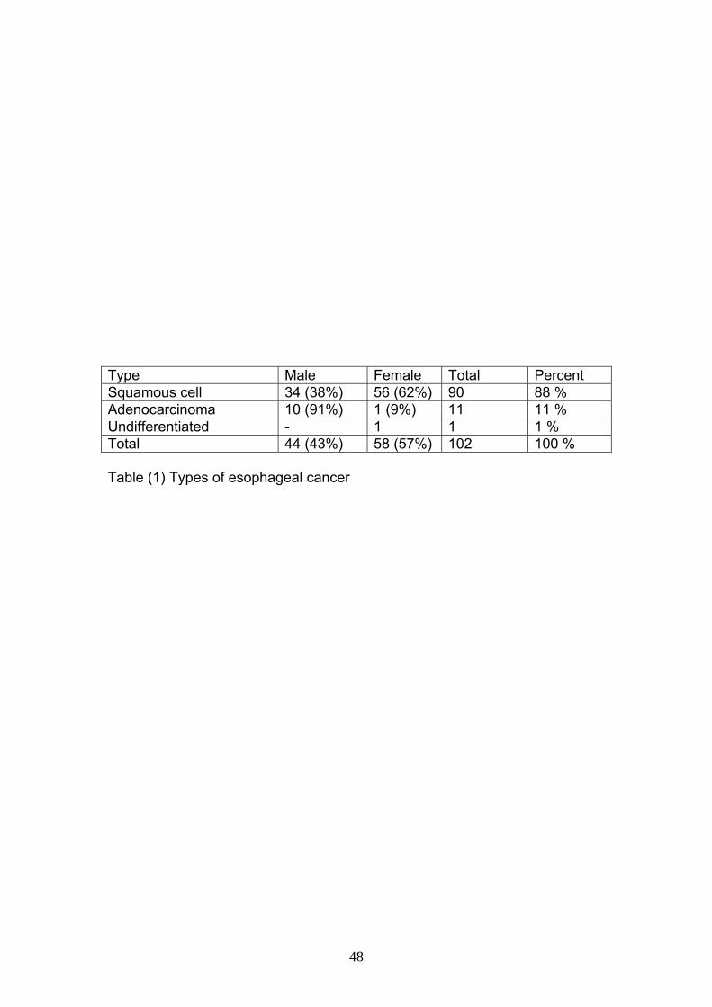
(2) Sex distribution: Forty five patients (44%) were males and 56% were females with a
ratio of female to male of 1.3: 1, (Figure 2). In squamous cell carcinoma
34 patients (38%) were males and 56 patients (62%) were females, with a
female to male ratio of 1.6:1. In adenocarcinoma ten patients were males
(91%) and only one patient was a female (9%), with a male to female
ratio of 10:1. (Table 1).
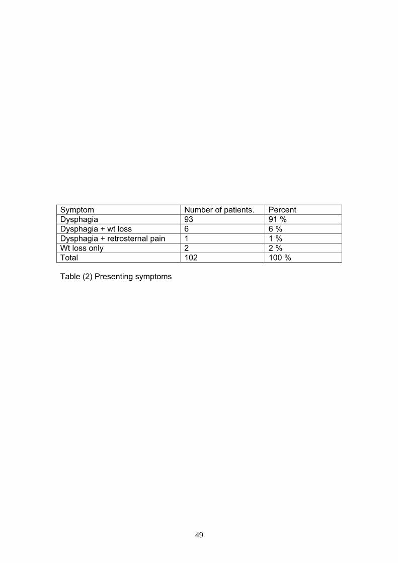
(3) Clinical symptoms: 93 patients (91%) presented with dysphagia, 6 patients (6%)
presented with dysphagia and weight loss and one patient (1%) presented
with dysphagia and retrosternal pain. Two patients (2%) presented with
weight loss only. ( Table 2).
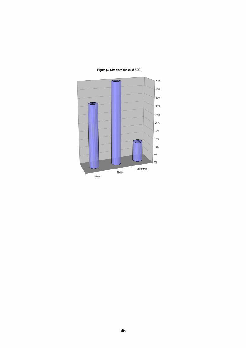
(4) Site distribution of the tumours: For squamous cell carcinoma 34 patients (38%) had the tumour in
the distal third of the oesophagus, 45 patients (50%) in the middle third
and 11 patients (12%) in the upper third. (Figure 3). For adenocarcinoma:
43
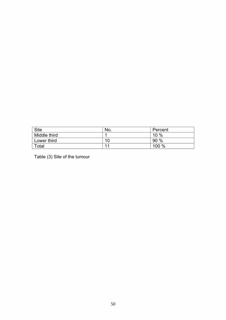
10 patients (90%) in the lower third and one patient (10%) in the middle
third. No one was found in the upper third. (Table 3).
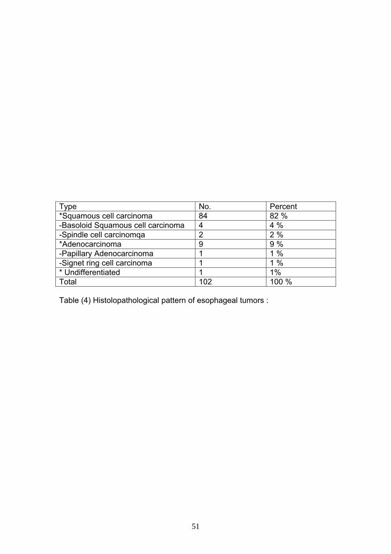
(5) Histopathological pattern: For squamous cell and its variants: 84 cases (82%) of all the
oesophageal tumours were squamous cell carcinoma, 4 cases (4%)
basaloid squamous cell carcinoma, 2 cases (2%) spindle cell carcinoma.
For adenocarcinoma and its variants: 9 cases (9%) of all the oesophageal
tumours were adenocarcinoma, one case (1%) papillary carcinoma, and
one case (1%) signet ring cell carcinoma. ( Table 4).
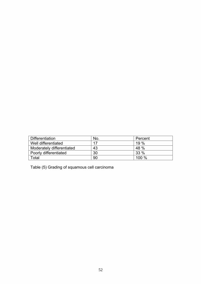
(6) Grading: For squamous cell carcinoma: moderately differentiated 43 cases
(48%), poorly differentiated 30 cases (33 %), and well differentiated 17
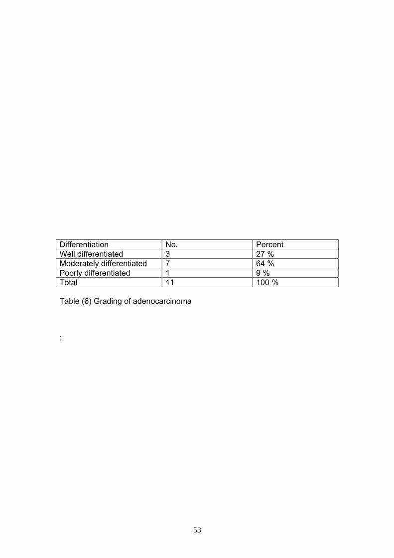
cases (19 %) . (Table 5). For adenocarcinoma moderately differentiated 7
cases (64%), poorly differentiated one case (9%), well differentiated 3
cases (27 %). (Table 6).
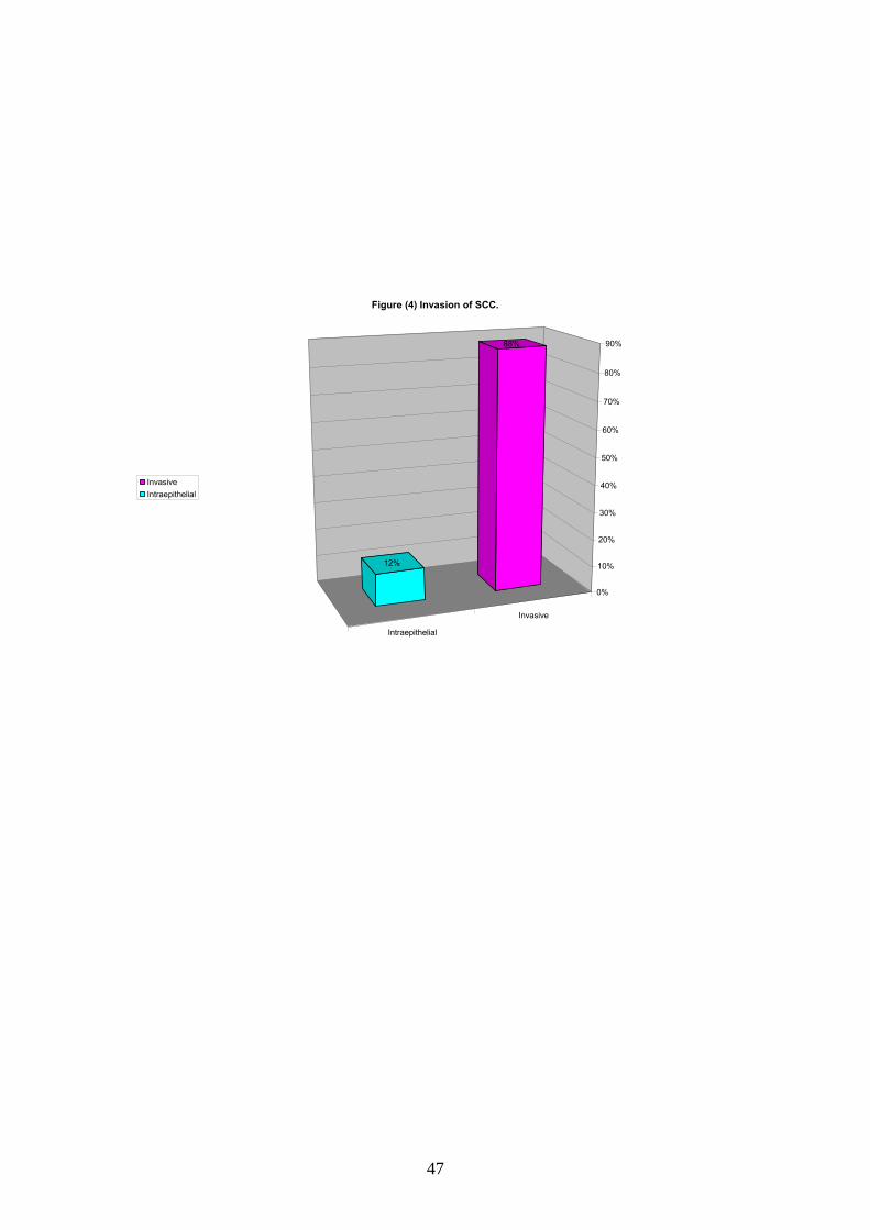
(7) Invasion of squamous cell carcinoma 80 cases (88%) were invasive, and 11 cases (12%) were
intraepithelial neoplasia. (Figure 4).
44
Figure (1) Age distibutionin oesophageal tumours in years
< 30 1%
30-5939%
60-9060%
45
Figure (2) Sex distribution in oesophageal tumours
Males44%
Females56%
MalesFemales
46
Upper thirdMiddle
Lower
12%
50%
38%
0%
5%
10%
15%
20%
25%
30%
35%
40%
45%
50%
Figure (3) Site distribution of SCC.
47
Invasive
Intraepithelial
88%
12%
0%
10%
20%
30%
40%
50%
60%
70%
80%
90%
Figure (4) Invasion of SCC.
InvasiveIntraepithelial
48
Type Male Female Total Percent Squamous cell 34 (38%) 56 (62%) 90 88 % Adenocarcinoma 10 (91%) 1 (9%) 11 11 % Undifferentiated - 1 1 1 % Total 44 (43%) 58 (57%) 102 100 %
Table (1) Types of esophageal cancer
49
Symptom Number of patients. Percent Dysphagia 93 91 % Dysphagia + wt loss 6 6 % Dysphagia + retrosternal pain 1 1 % Wt loss only 2 2 % Total 102 100 % Table (2) Presenting symptoms
50
Site No. Percent Middle third 1 10 % Lower third 10 90 % Total 11 100 % Table (3) Site of the tumour
51
Type No. Percent *Squamous cell carcinoma 84 82 % -Basoloid Squamous cell carcinoma 4 4 % -Spindle cell carcinomqa 2 2 % *Adenocarcinoma 9 9 % -Papillary Adenocarcinoma 1 1 % -Signet ring cell carcinoma 1 1 % * Undifferentiated 1 1% Total 102 100 % Table (4) Histolopathological pattern of esophageal tumors :
52
Differentiation No. Percent Well differentiated 17 19 % Moderately differentiated 43 48 % Poorly differentiated 30 33 % Total 90 100 % Table (5) Grading of squamous cell carcinoma
53
Differentiation No. Percent Well differentiated 3 27 % Moderately differentiated 7 64 % Poorly differentiated 1 9 % Total 11 100 % Table (6) Grading of adenocarcinoma :

54
CChhaapptteerr 44
55
Discussion This is a descriptive retrospective study about oesophageal
tumours in Sudanese patients. It includes 102 cases. All were found to be
malignant tumours, the histology of which is squamous cell carcinoma
and adenocarcinoma. This is consistent with the fact that benign tumours
like leiomioma and other malignant tumours like endocrine tumours and
lymphomas are rare as mentioned in the literature.
In Sudan as in many developing countries, there is at present no
reliable statistical information on absolute cancer rate. In this study, the
result is in accordance with the world wide acceptance that SCC is the
most common oesophageal cancer representing 89.1%.
Sex distribution: The male to female ratio for SCC in Sudanese patients did not
change over the last 20 years. In this study it is found to be 1:1.26
compared to an older study done in 1987 which showed a ratio of 1:1.25
(39). In Pakistan the ratio is 1:1.2 almost the same. However, studies in
Thailand and USA showed male predominance, 3.45:1 and 3:1
respectively. This puzzling epidemiologic contrast is not explained.
For adenocarcinoma there is striking male predominance, 10:1
which is a similar finding in many studies in different countries. It was
hypothesized that sex hormones could be responsible for that sex
imbalance. However, a Swedish study concluded that there is no role of
sex hormones in the aetiology of oesophageal adenocarcinoma. Another
study done in the USA showed that the ratio is 7:1 in whites and 10:1 in
blacks (11). This may suggest a genetic implication and this merit further
investigation.
56
Age distribution:
The ages of the patients range from 29-90 years, with a
mean of 65 years. The commoner age group was 60-90 years for
both SCC and adenocarcinoma, which is the same age group in
Almasri SH study in the period 1965-1974 in Sudan (5). One case
was found to be basaloid squamous cell carcinoma and it was
found in young age group (29 years). This result goes with the
international age distribution of oesophageal malignancy.
Presenting symptoms: Dysphagia was the most frequent symptom and
unfortunately a late warning one. Other symptom noticed was
weight loss. These symptoms were the same worldwide and are
the most bothering signs that lead the patient to seek medical
advice.
Site of the tumour: Fifty percent of SCC occurred in the middle third and 38% in
the lower third in accordance with many studies done in the USA,
Nigeria and other countries. The majority of adenocarcinoma
arouse in the lower third because most of which arise in Barret
oesophagus.
Histological pattern: In this study, the SCC variants basaloid cell carcinoma and
spindle cell carcinoma were commoner in females while
adenocarcinoma with its different histological patterns, namely
papillary adenocarcinoma and signet ring carcinoma were found in
males. One case was undifferentiated carcinoma and was a
female. The majority of squamous cell and adenocarcinomas were
57
moderately differentiated. However the prognostic impact of
tumour differentiation is equivocal possibly due poor
standardization of the grading system and to the high prognostic
power of tumour stage (WHO). Invasive squamous cell carcinomas
represent the majority of cases (88%) while the rest were
intraepithelial neoplasia. This implies that, the diagnosis of
oesophageal cancer is usually late and necessitates the need for
mass screening particularly for high risk patients.
58
Conclusion
1- Benign Oesophageal tumours and some malignant tumours
like endocrine tumours and lymphomas are rare in Sudan.
2- The most common type of oesophageal cancer in Sudan is
squamous cell carcinoma
3- There is striking male predominance in Sudanese patients
with adenocarcinoma.
4- The male to female ratio in Sudanese patient with squamous
cell carcinoma remained static over the last twenty years.
5- The common age group for oesophageal cancer in
Sudanese patients is 60-90 years.
59
Recommendation
It is recommended that:
(1) Establishment of screening programs for oesophageal carcinoma
for the high risk group.
(2) Further studies should be carried out to clarify exact risk factors.
(3) Detailed histopathological diagnosis and staging is needed.
(4) Provision of histopathology facilities for all regional hospitals.
(5) The statistical and registry services should be improved at both
national and regional levels.
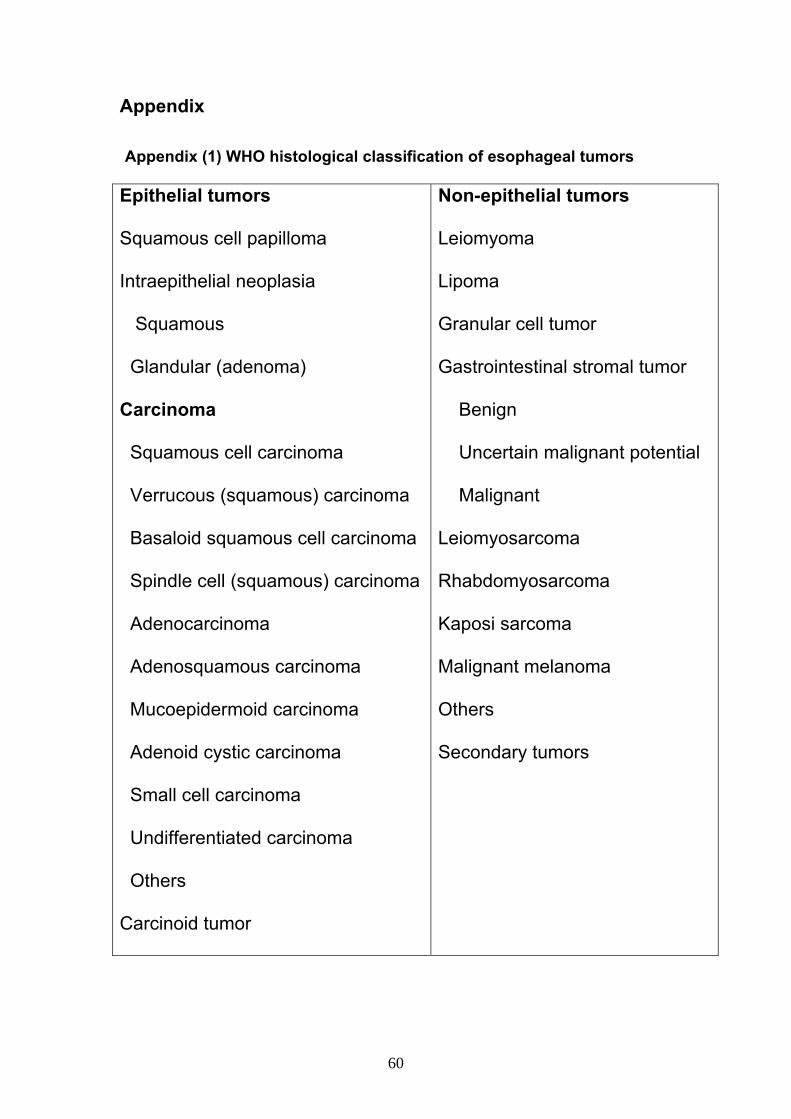
60
Appendix Appendix (1) WHO histological classification of esophageal tumors
Epithelial tumors
Squamous cell papilloma
Intraepithelial neoplasia
Squamous
Glandular (adenoma)
Carcinoma
Squamous cell carcinoma
Verrucous (squamous) carcinoma
Basaloid squamous cell carcinoma
Spindle cell (squamous) carcinoma
Adenocarcinoma
Adenosquamous carcinoma
Mucoepidermoid carcinoma
Adenoid cystic carcinoma
Small cell carcinoma
Undifferentiated carcinoma
Others
Carcinoid tumor
Non-epithelial tumors
Leiomyoma
Lipoma
Granular cell tumor
Gastrointestinal stromal tumor
Benign
Uncertain malignant potential
Malignant
Leiomyosarcoma
Rhabdomyosarcoma
Kaposi sarcoma
Malignant melanoma
Others
Secondary tumors
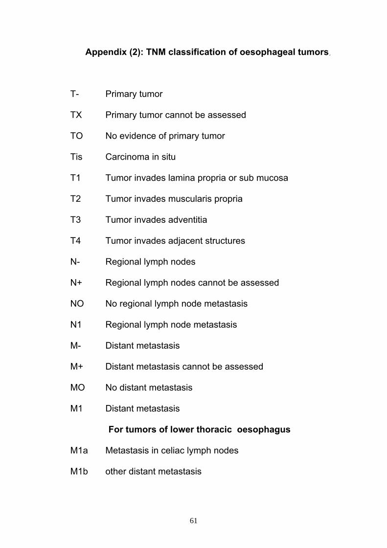
61
Appendix (2): TNM classification of oesophageal tumors ِ
T- Primary tumor
TX Primary tumor cannot be assessed
TO No evidence of primary tumor
Tis Carcinoma in situ
T1 Tumor invades lamina propria or sub mucosa
T2 Tumor invades muscularis propria
T3 Tumor invades adventitia
T4 Tumor invades adjacent structures
N- Regional lymph nodes
N+ Regional lymph nodes cannot be assessed
NO No regional lymph node metastasis
N1 Regional lymph node metastasis
M- Distant metastasis
M+ Distant metastasis cannot be assessed
MO No distant metastasis
M1 Distant metastasis
For tumors of lower thoracic oesophagus
M1a Metastasis in celiac lymph nodes
M1b other distant metastasis
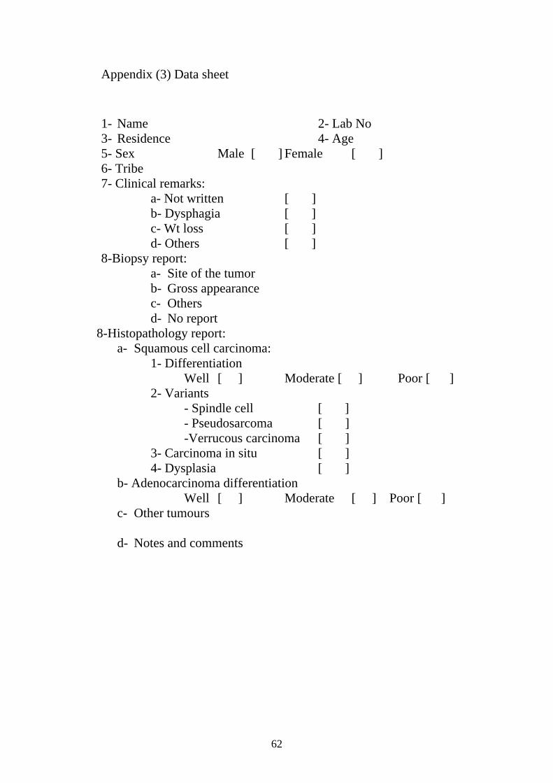
62
Appendix (3) Data sheet
1- Name 2- Lab No 3- Residence 4- Age 5- Sex Male [ ] Female [ ] 6- Tribe 7- Clinical remarks: a- Not written [ ]
b- Dysphagia [ ] c- Wt loss [ ] d- Others [ ]
8-Biopsy report: a- Site of the tumor b- Gross appearance c- Others d- No report
8-Histopathology report: a- Squamous cell carcinoma:
1- Differentiation Well [ ] Moderate [ ] Poor [ ]
2- Variants - Spindle cell [ ]
- Pseudosarcoma [ ] -Verrucous carcinoma [ ] 3- Carcinoma in situ [ ] 4- Dysplasia [ ] b- Adenocarcinoma differentiation Well [ ] Moderate [ ] Poor [ ]
c- Other tumours d- Notes and comments
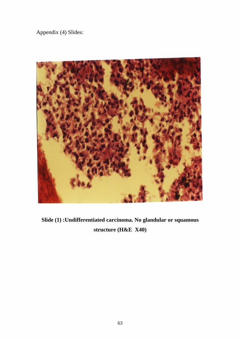
63
Appendix (4) Slides:
Slide (1) :Undifferentiated carcinoma. No glandular or squamous
structure (H&E X40)
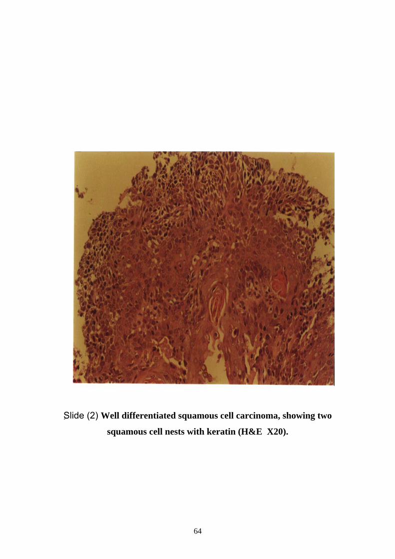
64
Sٍlide (2) Well differentiated squamous cell carcinoma, showing two
squamous cell nests with keratin (H&E X20).
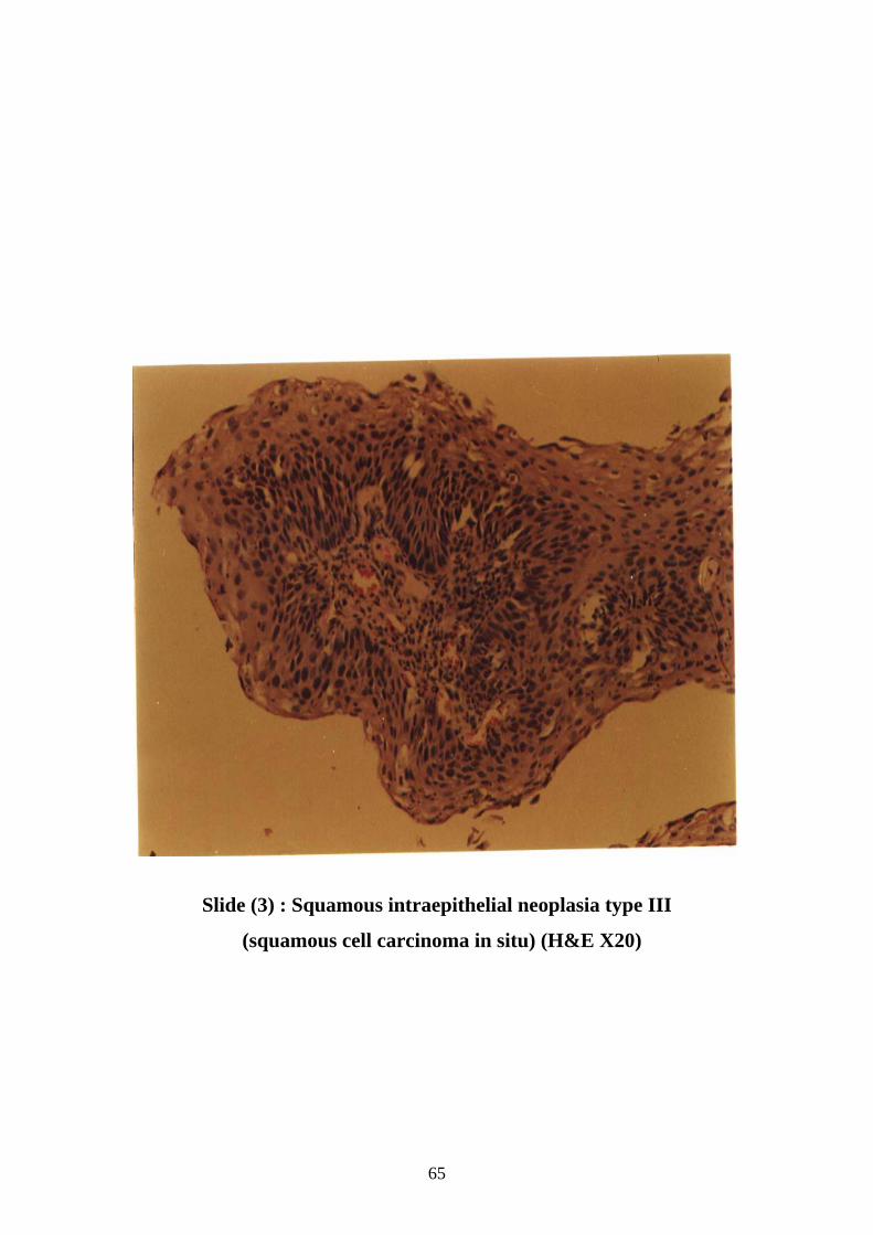
65
Slide (3) : Squamous intraepithelial neoplasia type III
(squamous cell carcinoma in situ) (H&E X20)
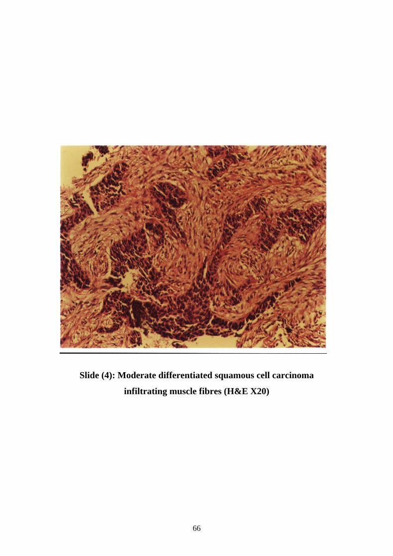
66
Slide (4): Moderate differentiated squamous cell carcinoma
infiltrating muscle fibres (H&E X20)
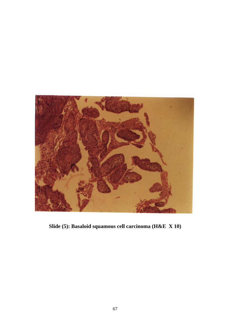
67
Slide (5): Basaloid squamous cell carcinoma (H&E X 10)
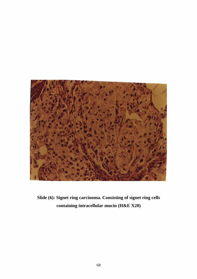
68
Slide (6): Signet ring carcinoma. Consisting of signet ring cells
containing intracellular mucin (H&E X20)
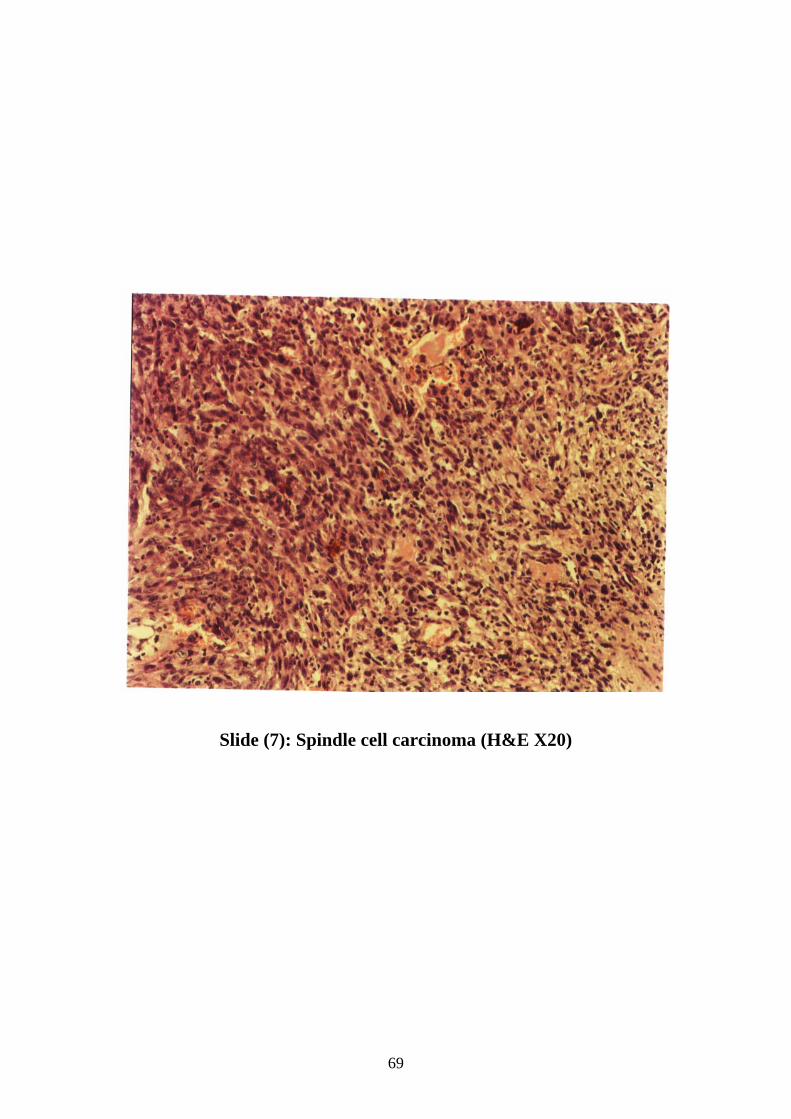
69
Slide (7): Spindle cell carcinoma (H&E X20)
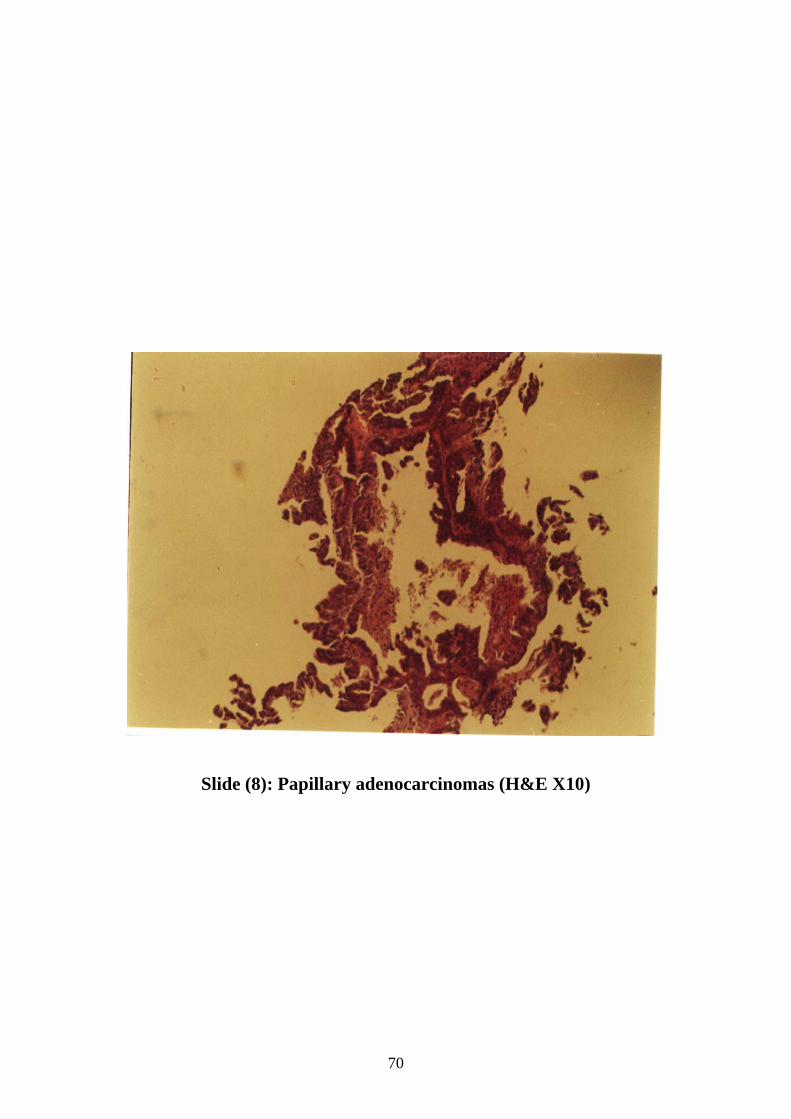
70
Slide (8): Papillary adenocarcinomas (H&E X10)
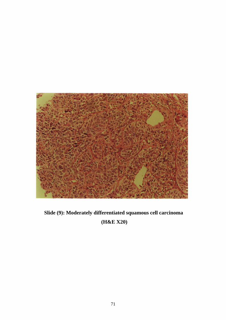
71
Slide (9): Moderately differentiated squamous cell carcinoma
(H&E X20)
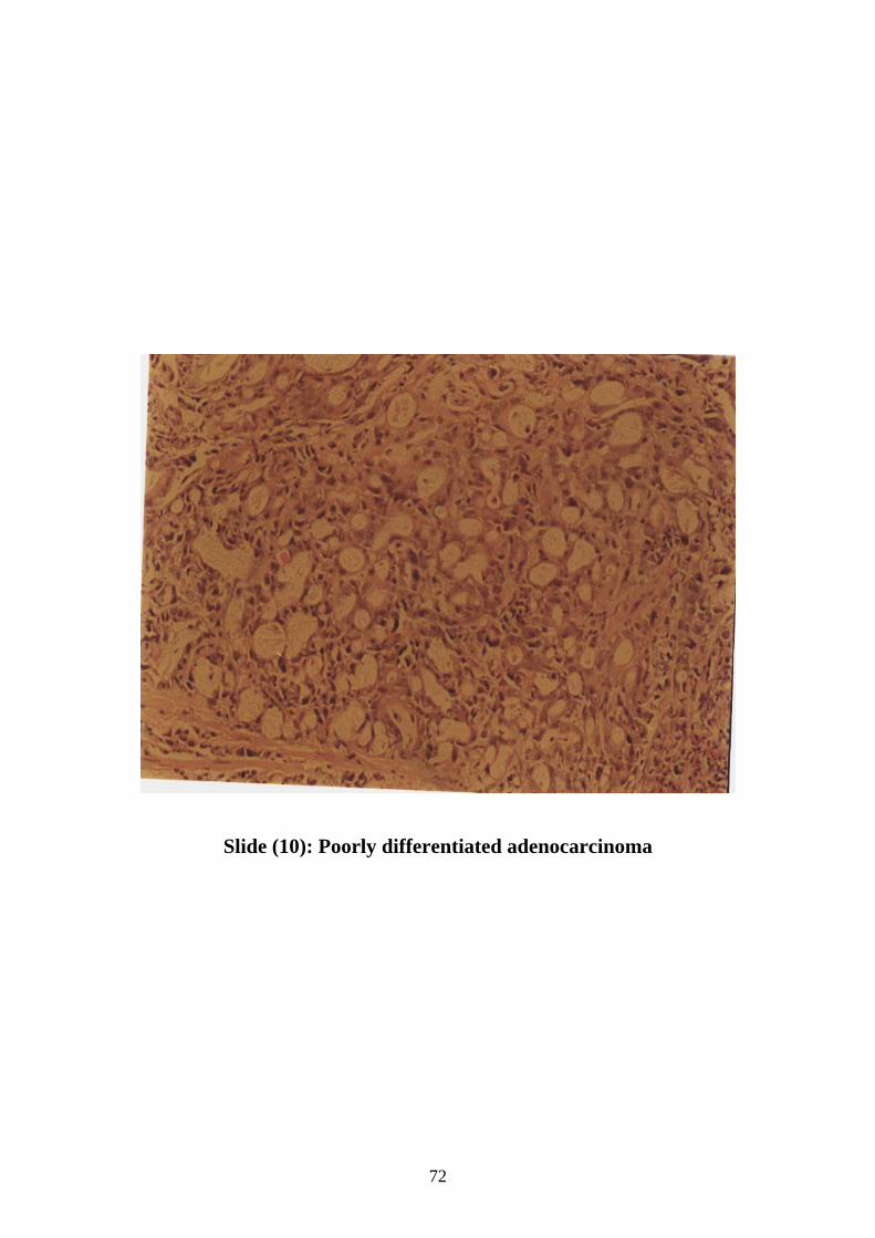
72
Slide (10): Poorly differentiated adenocarcinoma
73
References :
(1) Rosai J. Ackerman`s Surgical pathology. London; Mosby: 1996.
(2) Stanley R, Lauri A. Pathology and genetics of tumours of the
digestive system. Lyon ; WHO: 2000.
(3) Kumar V, Cotran R, Robbins SL. Robbins basic pathology.
Philadelphia ; Saunders : 2003.
(4) Wijnhoven BP, Louwman MW, Tilanus HW, Coebergh JW.
Increased incidence of adenocarcinomas at the gastro-oesophageal
junction in Dutch males since the 1990s.Eur J Gastroenterol
Hepatol 2002 Feb; 14(2):115-22.
(5) Boulos PB, El Masri SH. Carcinoma of the oesophagus in the
Sudan. Trop Geogr Med. 1977 Jun; 29(2):150-4.
(6) Vega KJ, Jamal MM. Changing pattern of esophageal cancer
incidence in New Mexico. Am J Gastroenterol 2000
Sep;95(9):2352-6.
(7) O`Leary T. Advanced diagnostic methods in pathology.
Philadelphia; Saunders : 2001 .
(8) William P, Warwick R. Gray`s anatomy. Lonon ; Churchill
Livingston : 1980.
(9) Guyton A, Hall J. Text book of medical physiology. New York;
Saunders : 2000
(10) Fawcett D. Text book of histology. New York ; Chapman
and Hall : 1994.
(11) Yang PC, Davis S. Incidence of cancer of the esophagus in
the US by histologic type. Cancer 1988 Feb 1; 61(3):612-7.
(12) Parvin S, Firouz S. A study of 415 cases of esophageal
carcinoma in northwest of Iran. Med J Malaysia 2003 Aug;
58(3):429-31.
74
(13) Bareiss D. Current epidemiology of carcinoma of the
esophagus and cardia in Germany. Dtsch Med Wochenschr 2002
Jun 21; 127(25-26):1367-74.
(14) Siegmund SV, Singer MV. Effects of alcohol on the upper
gastrointestinal tract and the pancreas--an up-to-date overview
Z Gastroenterol 2005 Aug; 43(8):723-36.
(15) Bergheim I, Wolfgarten E, Bollschweiler E, Holscher A,
Bode Ch, Parlesak A. Role of retinoic acid receptors in squamous-
cell carcinoma in human esophagus. J Carcinog 2005 Nov 8; 4:20.
(16) Yokoyama A, Yokoyama T, Kumagai Y. Mean corpuscular
volume, alcohol flushing, and the predicted risk of squamous cell
carcinoma of the esophagus in cancer-free Japanese men. Alcohol
Clin Exp Res. 2005 Oct; 29(10):1877-83.
(17) Galeone C. Folate intake and squamous-cell carcinoma of
the oesophagus in Italian and Swiss men.. Ann Oncol 2005 Dec
12; [Epub ahead of print].
(18) Terry P, Lagergren J, Wolk A, Nyren O. Drinking hot
beverages is not associated with risk of oesophageal cancers in a
Western population. Br J Cancer 2001 Jan 5; 84(1):120-1.
(19) Lakatos PL, Lakatos L, Fuszek P. Incidence and pathologic
distribution of esophageal cancers at the gastro-esophageal
junction between 1993-2003 Orv Hetil 2005 Feb 27; 146(9):411-6.
(20) Ihekwaba FN, Solanke TF. Carcinoma of the oesophagus.
Br J Surg 1984 Feb; 71(2):116-8.
(21) Parvin S, Firouz S. A study of 415 cases of esophageal
carcinoma in northwest of Iran. Med J Malaysia 2003 Aug;
58(3):429-31.
(22) Lagergren J, Nyren O. Do sex hormones play a role in the
etiology of esophageal adenocarcinoma? A new hypothesis tested
75
in a population-based cohort of prostate cancer patients. Cancer
Epidemiol Biomarkers Prev 1998 Oct; 7(10):913-5.
(23) Endo T, Sugawara N, Shinomura Y. Carcinogenesis of
Barrett's esophagus. Nippon Rinsho 2005 Aug; 63(8):1357-61.
(24) Sakata H, Fujimoto K. Barrett's esophagus and Helicobacter
pylori Nippon Rinsho 2005 Aug; 63(8):1383-6.
(25) Oberg S, Wenner J, Johansson J, Walther B, Willen R.
Barrett esophagus: risk factors for progression to dysplasia and
adenocarcinoma. Ann Surg 2005 Jul; 242(1):49-54.
(26) Rubenstein JH, Davis J, Marrero JA, Inadomi JM.
Relationship between diabetes mellitus and adenocarcinoma of the
oesophagus and gastric cardia. Aliment Pharmacol Ther 2005 Aug
1; 22(3):267-71.
(27) Lindblad M, Rodriguez LA, Lagergren J. Body mass,
tobacco and alcohol and risk of esophageal, gastric cardia, and
gastric non-cardia adenocarcinoma among men and women in a
nested case-control study. Cancer Causes Control 2005
Apr;16(3):285-94.
(28) Szumilo J, Kotarska M, Chroscicki A, Korobowicz E.
Leiomyoma of the cervical esophagus: a case report. Ann Univ
Mariae Curie Sklodowska [Med] 2003; 58(2):22-4.
(29) Kuo MJ, Yeh HZ, Chen GH, Jan YJ. Diffuse esophageal
leiomyomatosis with a pedunculated polyp. J Gastroenterol 2004
Dec; 39(12):1205-9.
(30) Kajiyama Y. Surgery for benign submucosal tumors of the
esophagus (leiomyoma and hemangioma). Nippon Geka Gakkai
Zasshi 2003 Sep; 104(9):593-6.
76
(31) Misra M, Maziak DE, Shamji FM, Michaud C, Perkins DG,
Matzinger F. Esophageal leiomyomatosis. Med Sci Monit 2003
Nov; 9(11):CS98-101.
(32) Pujol J, Pares D, Mora L, Sans M, Jaurrieta E. Diagnosis and
management of diffuse leiomyomatosis of the oesophagus. Dis
Esophagus 2000; 13(2):169-71.
(33) Cheng YL, Hsu JY, Hsu HH, Yu CP, Lee SC. Diffuse
leiomyomatosis of the esophagus. Dig Surg 2000;17(5):528-31
(34) Aurea P, Grazia M, Petrella F, Bazzocchi R. Giant
leiomyoma of the esophagus. Eur J Cardiothorac Surg 2002 Dec;
22(6):1008-10.
(35) Chalkiadakis G, Wihlm JM, Morand G, Weill-Bousson M,
Witz JP .Primary malignant melanoma of the esophagus. Ann
Thorac Surg 1985 May; 39(5):472-75.
(36) Lohmann CM, Hwu WJ, Iversen K, Jungbluth AA, Busam
KJ. Primary malignant melanoma of the oesophagus: a clinical and
pathological study with emphasis on the immunophenotype of the
tumours for melanocyte differentiation markers and cancer/testis
antigens. Melanoma Res 2003 Dec; 13(6):595-601.
(37) Dabrowski A, Skoczylas T, Misztal B, Wallner G.
Diagnostic and therapeutic difficulties in a patient with primary
malignant melanoma of the esophagus-case report. Pol Merkuriusz
Lek 2004 Oct; 17(100):378-9.
(38) Perrot M, Brundler MA, Robert J, Spiliopoulos A. Primary
malignant melanoma of the esophagus. Dis Esophagus 2000;
13(2):172-4.
(39) Abdelsamie A. A review of oesophageal cancer in Sudanese
patients. Postgraduate medical studies board, U of K. The book of
abstract 1980-2001.