Embed Size (px)
Citation preview

University of São Paulo “Luiz de Queiroz” College of Agriculture
Food structure design to modulate bioaccessibility of carotenoids from brazilian native fruits after screening of eleven non-conventional tropical fruits
Paulo Roberto de Araujo Berni
Thesis presented to obtain the degree of Doctor in Science. Area: Food Science and Technology
Piracicaba 2018

1
Paulo Roberto de Araujo Berni Bachelor of Food Science
Food structure design to modulate bioaccessibility of carotenoids from brazilian native fruits after screening of eleven non-conventional tropical fruits
Advisor: Prof. Dr. SOLANGE GUIDOLIN CANNIATTI BRAZACA
Thesis presented to obtain the degree of Doctor in Science. Area: Food Science and Technology
Piracicaba 2018

2
Dados Internacionais de Catalogação na Publicação
DIVISÃO DE BIBLIOTECA – DIBD/ESALQ/USP
Berni, Paulo Roberto de Araujo
Food structure design to modulate bioaccessibility of carotenoids from brazilian native fruits after screening of eleven non-conventional tropical fruits / Paulo Roberto de Araujo Berni. - - Piracicaba, 2018.
121p.
Tese (Doutorado) - - USP / Escola Superior de Agricultura “Luiz de Queiroz”.
1. Frutas nativas 2. Caroteno 3. Licopeno 4. Células Caco-2 5. Microemulsão 6. Bioacessibilidade 7. Biodisponibilidade I. Título

3
This thesis is dedicated to my mother and to my grandmother (in memoriam) for their examples
of faith, bravery and kindness
4
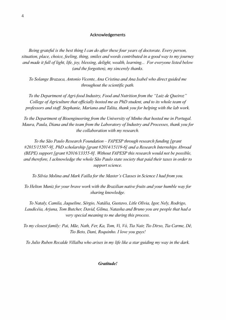
Acknowledgements
Being grateful is the best thing I can do after these four years of doctorate. Every person,
situation, place, choice, feeling, thing, smiles and words contributed in a good way to my journey
and made it full of light, life, joy, blessing, delight, wealth, learning... For everyone listed below
(and the forgotten), my sincerely thanks.
To Solange Brazaca, Antonio Vicente, Ana Cristina and Ana Isabel who direct guided me
throughout the scientific path.
To the Department of Agri-food Industry, Food and Nutrition from the “Luiz de Queiroz”
College of Agriculture that officially hosted me as PhD student, and to its whole team of
professors and staff. Stephanie, Mariana and Talita, thank you for helping with the lab work.
To the Department of Bioengineering from the University of Minho that hosted me in Portugal.
Maura, Paula, Diana and the team from the Laboratory of Industry and Processes, thank you for
the collaboration with my research.
To the São Paulo Research Foundation – FAPESP through research funding [grant
#2015/15507-9], PhD scholarship [grant #2014/15119-6] and a Research Internships Abroad
(BEPE) support [grant #2016/13355-0]. Without FAPESP this research would not be possible,
and therefore, I acknowledge the whole São Paulo state society that paid their taxes in order to
support science.
To Silvia Molina and Mark Failla for the Master’s Classes in Science I had from you.
To Helton Muniz for your brave work with the Brazilian native fruits and your humble way for
sharing knowledge.
To Nataly, Camila, Jaqueline, Sérgio, Natália, Gustavo, Litle Olívia, Igor, Nely, Rodrigo,
Laudicéia, Arjuna, Tom Butcher, David, Gilma, Natasha and Bruno you are people that had a
very special meaning to me during this process.
To my closest family: Pai, Mãe, Nath, Fer, Ka, Tom, Vi, Vó, Tia Nair, Tio Dirso, Tia Carme, Dê,
Tio Beto, Dani, Roquinho. I love you guys!
To Julio Ruben Recalde Villalba who arises in my life like a star guiding my way in the dark.
Gratitude!

5
“The great book, always open and which we should make an effort to read, is that of Nature”
Antoní Gaudí

6
Contents RESUMO .................................................................................................................................. 7
ABSTRACT .............................................................................................................................. 8
1 INTRODUCTION .................................................................................................................. 9
2 SCREENING OF ELEVEN NON-CONVENTIONAL TROPICAL FRUITS –
PROXIMAL COMPOSITION, FIBERS, ANTIOXIDANT POTENTIAL AND
CAROTENOID BIOACCESSIBILITY ............................................................................... 17
Abstract ......................................................................................................................... 17
2.1. Introduction ............................................................................................................ 18
2.2. Materials and methods ........................................................................................... 19
2.3. Results and discussion ............................................................................................ 24
2.4. Conclusions ............................................................................................................ 32
3 CAROTENOIDS FROM PITANGA (EUGENIA UNIFLORA) AND BURITI
(MAURITIA FLEXUOSA) FRUITS: PROFILE, STABILITY, BIOACCESSIBILITY
AND CELLULAR UPTAKE STUDIES BY IN VITRO DIGESTION COUPLED TO
CACO-2 CELLS CULTURE ............................................................................................... 37
Abstract ......................................................................................................................... 37
3.1. Introduction ............................................................................................................ 38
3.2. Materials and methods ........................................................................................... 40
3.3. Results and discussion ............................................................................................ 43
4 STRUCTURE DESIGN OF FRUIT MICROEMULSION DELIVERY SYSTEM FOR
CAROTENOIDS FROM PITANGA (EUGENIA UNIFLORA) AND BURITI
(MAURITIA FLEXUOSA) ...................................................................................................... 61
Abstract ......................................................................................................................... 61
4.1. Introduction ............................................................................................................ 62
4.2. Materials and methods ........................................................................................... 64
4.3. Results and discussion ............................................................................................ 69
4.4. Conclusions ............................................................................................................ 83
5 CAROTENOID BEHAVIOR OF PITANGA (EUGENIA UNIFLORA) AND BURITI
(MAURITIA FLEXUOSA) DURING MICROEMULSION PRODUCTION AND IN A
DYNAMIC GASTROINTESTINAL SYSTEM ................................................................ 89
Abstract ......................................................................................................................... 89
5.1. Introduction ............................................................................................................ 90
5.2. Materials and methods ........................................................................................... 93
5.3. Results and discussion ............................................................................................ 98
5.4. Conclusions .......................................................................................................... 114
6 FINAL CONSIDERATIONS ............................................................................................ 121

7
RESUMO
Desenho estrutural da matriz alimentar para modulação da bioacessibilidade de
carotenoides de frutas nativas do Brasil após a triagem de onze frutas tropicas não-
convencionais
O Brasil é o país detentor da maior biodiversidade do planeta e um grande produtor de
alimentos. Frutas tropicais, em especial as nativas brasileiras, podem conter quantidades
consideráveis de carotenoides que possuem ação antioxidante, anti-inflamatória, provitamina A
e anticâncer, como β-caroteno e licopeno. O desenho estrutural dos alimentos (food structure
design), visa manipular a matriz alimentar com fins específicos, por exemplo, a preservação e
manipulação da bioacessibilidade de carotenoides. Na presente tese buscou-se explorar frutas
brasileiras não-convencionais no desenvolvimento de sistemas de entrega de carotenoides. Foi
realizada uma etapa de triagem com 11 frutas, dentre as quais, 2 foram selecionadas, a pitanga
(Eugenia uniflora) e o buriti (Mauritia flexuosa), e utilizadas na produção de microemulsões.
Nesta triagem foram avaliadas a composição centesimal, o teor de fibras, o perfil de
carotenoides e a bioacessibilidade destes carotenoides. O valor nutricional das frutas
demonstrou seu potencial para utilização em produtos saudáveis, devido a seus teores eleveados
de fibras, minerais e carotenoides além do baixo teor calórico. A análise por HPLC-DAD
permitiu identificar até 14 carotenoides nas amostras das 11 frutas estudadas na triagem. O
estudo da bioacessibilidade dos carotenoides das 11 frutas demonstrou principalmente a
superioridade da bioacessibilidade de xantofilas (variando de 10 a 52 %) em relação aos
carotenos, e portanto a baixa bioacessibilidade de licopeno da pitanga (1,1%) e média
bioacessibilidade de β-caroteno do buriti (26%). Pitanga e buriti tiveram o perfil de carotenoides
detalhadamente acompanhados através da simulação in vitro da digestão associada à absorção
intestinal por culturas de células Caco-2. Neste estudo, observou-se que embora as xantofillas
sejam mais bioacessíveis, o tecido epitelial do intestino absorve preferencialmente carotenoides
provitaminicos A, como o β-caroteno e a β-criptoxantina. Para produzir as microemulsões
foram estudados processamentos (homogeneização de alta-velocidade - HSH e ultrassom - US)
em combinação com uso de surfactantes (Whey Protein Isolate e Tween 80) e adição de óleo
de milho como carreador dos carotenoides. Os experimentos mostraram que a interação entre
US e HSH é capaz de romper as paredes celulares e liberarem os carotenoides com maior
eficiência. Ficou demonstrado através de microscopia ótica e de fluorescência, tanto quanto
pela análise de carotenoides, que foi possível manipular estruturalmente a matriz alimentar
liberando os carotenoides de dentro das células vegetais e encapsulando-os dentro das gotículas
de óleo, além de aumentar sua retenção após o processamento. As microemulsões obtidas
sofreram efeito do tempo de processamento e do surfactante em relação à reologia, estrutura
final da matriz, estabilidade ao armazenamento e estabilidade dos carotenoides ao processo. Por
fim, foi utilizado um sistema dinâmico de simulação da digestão gastrointestinal para comparar
o comportamento dos carotenoides oriundos das polpas integrais das frutas e das
microemulsões selecionadas (Tween 80 a 2%, óleo de milho a 5% e processado por HSH-US 4
min-4 min). Os resultados demonstraram que foi possível aumentar a estabilidade à digestão e
a bioacessibilidade dos carotenoides totais, do licopeno e do β-caroteno.
Palavras-chave: Frutas nativas; Caroteno; Licopeno; Células Caco-2; Microemulsão;
Desenvolvimento de produtos; Bioacessibilidade; Biodisponibilidade

8
ABSTRACT
Food structure design to modulate bioaccessibility of carotenoids from brazilian native
fruits after screening of eleven non-conventional tropical fruits
Brazil is the country with the greatest biodiversity on the planet and a major producer
of food. Tropical fruits, especially natives from Brazil, may contain considerable amounts of
carotenoids that have antioxidant, anti-inflammatory, provitamin A and anticancer actions, such
as β-carotene and lycopene. The food structure design concept aims to manipulate the food
matrix for specific purposes, e.g. the preservation and manipulation of carotenoid
bioaccessibility. The aim of this thesis was to explore tropical fruits, native and exotic from
Brazil in the development of delivery systems for carotenoids. A screening step was carried out
with 11 fruits, among which 2 were selected, the pitanga (Eugenia uniflora) and buriti (Mauritia
flexuosa) fruits that were used for the production of microemulsions. At the screening were
evaluated the proximate composition, fiber contents, carotenoid profiles and bioacessibilities.
The nutritional value demonstrated that these fruits have high potential as raw-materials for
healthy products due to their high fiber, minerals and carotenoid contents in addition to low
energy value. Analysis by HPLC-DAD allowed the identification of 14 carotenoids in the 11
fruits studied for the screening. Results demonstrated the superiority of the bioaccessibility of
xanthophylls (ranging 10 % – 52 %) in relation to carotenes, and the low bioaccessibility of
lycopene from pitanga (1.1 %) and average bioaccessibility of β-carotene from buriti (26 %).
Pitanga and buriti had their carotenoid profiles analyzed and monitored throughout an in vitro
simulation of the digestion coupled with caco-2 cell cultures. Although xanthophylls are more
bioaccessible, the intestinal ephitelium absorb preferentially the provitamin A carotenoids, such
β-carotene and β-criptoxanthin. In order to produce these microemulsions, high-speed
homogenization (HSH) and ultrasound (US) were studied in combination with the use of
surfactants (Whey Protein Isolate and Tween 80), and addition of corn oil as carotenoid carrier.
The experiments have shown that the interaction of US and HSH is capable to break cell walls
and release carotenoids with higher efficiency. Optical and fluorescence microscopy, as well as
carotenoid analysis demonstrated that it was possible to manipulate the food matrix structure
releasing the carotenoids from the plant cells and encapsulating them inside the oil droplets,
what increased their retention after processing. The microemulsion were affected by time of
processing and by surfactant related to their rheology, final structure, stability of emulsion and
carotenoid stability to processing. Finally, a dynamic gastrointestinal system was used to
compare the behavior of carotenoids from whole fruit pulps and selected microemulsions (2%
Tween 80, 5% corn oil, processed by HSH-US 4 min -4 min). The results demonstrated that it
was possible to increase the stability to digestion and bioaccessibility of total carotenoids,
lycopene and β-carotene from the microemulsions.
Keywords: Native fruits; Carotene; Lycopene; Caco-2 cells; Microemulsion; Product
development; Bioaccessibility; Bioavailability

9
1 INTRODUCTION
Malnutrition and chronic diseases that are related to food habits pose one of the biggest
threats to global health and development, particularly in the developing world (WHO, 2018).
The World Health Organization (WHO) proposes that a healthy and efficient food system,
linked to healthy living habits, can significantly reduce the costs of treating chronic
degenerative and non-communicable diseases while increasing people's quality of life,
promoting productivity and generating employment and income. WHO (2018) encourage the
development, production and dissemination of food products that contribute to a healthier diet.
In this sense, intensification of fruit consumption is stimulated by consistent evidences of their
potential to provide health benefits for people. The expansion of food diversity throughout the
sustainable use of local biodiversity and traditional foods is intensively promoted by the
scientific community (Rodriguez-Amaya et al., 2008; Heywood, 2011 Hough, 2014; Santeramo
et al., 2018).
The knowledge about Brazilian biodiversity, and its use to improve people's health and
social well-being, is strategic for promoting sustainable development. The fruit production
sector has significant participation in agribusiness placing Brazil as the 3rd largest fruit producer
in the world (Bueno and Baccarin, 2012; Silva and Abud 2017). However, among the twenty
fruits most produced in 2016 (Kist et al., 2018) only three are native from Brazil, the pineapple,
passionfruit and cashew nut. Many Brazilian fruits, which are little known or studied, have large
amounts of bioactive compounds that have antioxidant, anti-inflammatory, anti-cancer
properties and other actions. With the greatest biodiversity on the planet and one of the world's
largest food producers, Brazil needs to innovate in the manufacture of food products from its
plant resources (Nogueira, 2011, Raimundo et al., 2017).
Among the abundant bioactive compounds in Brazilian fruits there are carotenoids
which are recognized as essential for a healthy life. They are found only in plants, algae and
photosynthetic bacteria, and are responsible for the colors of red, orange and yellow tones in
the great majority of foods, especially in fruits (Rodriguez-Amaya et al., 2008; Rao and Rao,
2007). Even with its numerous chemical forms, carotenoids can be divided into two large
groups: carotenes and xanthophylls. These groups are differentiated by the presence of oxygen
in the molecule, the carotenes do not have oxygen while the xanthophylls possesses (Fernández-
García, 2012; Amorim-Carrilho et al., 2004).
β-carotene, α-carotene, γ-carotene, β-cryptoxanthin and β-zeacarotene, are the most
common carotenoids that have vitamin A activity, since they can form in the body one or two

10
molecules of retinol (vitamin A) (Rodriguez-Amaya et al., 2008). Several ways of antioxidant
action were identified for carotenoids and xanthophylls, like the unique capacity to capture
oxygen singlet (Gülçin, 2012; Rao and Rao, 2007). The role of carotenoids in the prevention
and remission of certain types of cancers has been demonstrated, with emphasis on their anti-
inflammatory action and induction of physiological redox enzymes. Thus, carotenoids have
significant bioactive functions and can not be disregarded when it comes to food, health and
well-being (Miller et al., 1996; Zheng et al., 2013; Gülçin, 2012; Rodriguez-Concepcion et al.,
2018).
Because carotenoids contains a conjugated system of double bonds, they are highly
unstable, sensitive to heat, light, acids, oxygen and enzymes such as lipoxygenase, causing
changes or may lead to partial degradation of their structures. The processing of carotenoid-rich
foods can cause degradation of these compounds and impair their function. Degradation can act
in two main ways: conversion of the trans forms into cis isomers that modifies their bioactivity;
and oxidation reactions pathway that breaks down chemical structures producing low molecular
weight residues, that also interfere in their bioactivity (Saini et al., 2015; Rodriguez-Amaya,
2001).
Carotenoids have to be bioaccessible for the body prior to provide the expected health
benefits. Bioaccessibility, is defined as the portion of an ingested nutrient that will be available
to be absorbed by the intestine after digestion. Synthetically, the steps necessary for the
carotenoid bioaccessibility are: 1) release of carotenoids from the food matrix; 2) the released
carotenoids have to be incorporated to the lipid phase of the digesta still in the stomach; 3) with
the lipid droplets the carotenoids integrate the mixed micelles formed by the action of the bile
salts; 4) these mixed micelles migrate to the intestinal lumen and come into contact with the
microvilli of the enterocytes, and are absorbed by the intestinal cells (Yonekura and Nagao,
2007, Kopec and Failla, 2018).
The most common factors that negatively affect the stability of carotenoids in processed
foods are heat treatment (mainly), homogenization and particle reduction, acidity, presence of
oxygen and light. Most important factors in foods that negatively affect the carotenoid
bioaccessibility are the physicochemical structure of the food matrix, especially the presence
of fibers (dependent on the type of fiber), the chromoplast composition and structure
(crystalloid) and the absence of lipids. Moreover, since carotenoids are highly lipophilic there
are some barriers to overcome in order to efficiently incorporate them into novel functional
foods: low solubility in water; crystallization capacity; chemical instability; and low
bioaccessibility (McClements, 2015). Therefore, the processing and the food matrix from the

11
final products are fundamental for carotenoid bioaccessibility (Failla et al., 2014, Pugliese et
al., 2013; Kopec and Failla 2018).
Considering that bioaccessibility and stability of carotenoids in fresh and processed
fruits is generally low, it is possible to apply the processes and formulations to modulate it. For
example, β-carotene and lycopene had their bioaccessibility increased by using optimal
combinations of: cooking and oil incorporation; controlled heat treatment and suitable
formulation; ultrasound processing and oil addition; cooking and mixing with excipient
microemulsion (Anese et al., 2015; Berni et al., 2014; Buggenhout et al., 2012; Zhang et al.,
2015). Thus, strategic formulation of products and optimal processing may be tools to increase
carotenoid release, bioaccessibility, and consequently the action of promoting health
(Buggenthout et al., 2012). Healthier ready-to-eat and fruit-derived products could be produced
and formulated by an smart way. From the selection of the raw material combined with more
adequate and efficient thermal and/or mechanical processes, would imply in obtaining products
with better attributes for health, i.e. higher retention and bioaccessibility of the targeted
carotenoid (Buggenhout et al., 2012). The use of these techniques are called food structure
design and aims the protection, conservation, increasing bioaccessibility, and especially
boosting health benefits (McClements, 2015; McClements et al., 2015)
Many bioactive compounds from plants are highly lipophilic, such as carotenoids, which
causes many hindrance to incorporate these molecules in new functional foods, primarily low
solubility in water and chemical instability (Recharla et al., 2017). Several techniques seeks to
protect, preserve, enhance solubility, increase bioaccessibility and bioactivity of carotenoids.
The most studied and applied strategy is the encapsulation of these highly lipophilic molecules
in emulsion based systems at micro and nano scales (Recharla et al., 2017; McClements, 2015).
Successful encapsulation of purified carotenoids in micro and nano-emulsions have been done
by many research groups (Gomes et al., 2017; Davidov-Pardo et al., 2016; Gul et al., 2015;
Zhang et al., 2015; de Paz et al., 2013; Hejri et al., 2013).
At this thesis are provided efforts on developing products with structured food matrix
that modulate the stability and bioaccessibility of carotenoids from Brazilian fruits. The
research was performed in four phases: 1) screening of carotenoid-rich fruits from a set of
eleven pre-selected; 2) defining the carotenoid profile and bioaccessibility of the two chosen
fruits; 3) developing products with the two fruits by applying the food structure design
approach; 4) and evaluating the impact of processing and formulation on the carotenoid stability
and bioaccessibility.

12
At least three topics approached in this work should be highlighted. Firstly, the
exploration of eight fruits that are native from Brazilian biomes – Buriti (Mauritia flexuosa),
Pitanga (Eugenia uniflora), Araçá-boi (Eugenia Stipitata ), Seriguela (Spondias purpúrea),
Cambuiti-cipó (Sageretia elegans), Jaracatiá (Jaracatia spinosa), Capeba (Odontocarya
acuparata) and Pitangatuba (Eugenia neonitida) – and three exotic fruits that are well adapted
to Brazilian climate – Acerola (Malpighia emarginata), Dovialis (Dovyalis abyssinica) and
Abricó-da-praia (Mimusopsis comersonii). In addition, the food structure design is an
innovative approach in the food product development field, that is aligned with the current
demands of consumers for healthier food and improved quality of life. Moreover, the
methodologies applied for assessing the carotenoid bioaccessibility – i.e. in vitro digestion,
uptake by Caco-2 cells culture and dynamic gastrointestinal system – are top of the art
techniques in this research field.
This thesis is organized in four chapters, each one related to four phases of the present
research, and to the manuscripts submitted for publication. At the first chapter are presented the
nutritional value of the 11 fruits that integrated the screening and the carotenoid contents and
bioaccessibility by in vitro digestion of 9 that were not selected for the product development.
At the second chapter, pitanga and buriti are thoroughly studied regarding their carotenoid
profile, bioaccessibility and uptake by caco-2 cells culture. The third chapter presents the food
structure design of microemulsions of pitanga and buriti pulps together with oil and surfactant,
produced by highspeed homogenization and ultrasound processing. Finally, the fourth chapter
reveals how the processing and formulation impact carotenoid release, retention, encapsulation,
stability to digestion and bioaccessibility throughout the dynamic gastrointestinal system.
REFERENCES
Amorim-Carrilho, K. T., Cepeda, A., Fente, C., Regal, P. (2014) Review of methods for analysis
of carotenoids. Trac-trend Anal. Chem., 56:49–73
Anese, M., Bot, F., Panozzo, A., Mirolo, G., Lippe, G. (2015). Effect of ultrasound treatment,
oil addition and storage time on lycopene stability and in vitro bioaccessibility of tomato
pulp. Food Chem, 172:685-691
Berni, P., Chitchumroonchokchai, C., Canniatti-Brazaca, S. G., de Moura, F. F., Failla, M. L.
(2014) Impact of genotype and cooking style on the content, retention, and
bioaccessibility of β-Carotene in biofortified cassava (Manihot esculenta Crantz)
conventionally bred in Brazil. J Agric Food Chem, 62:6677−6686

13
Bueno, G., Baccarin, J. G. (2012) Participação das principais frutas brasileiras no comércio
internacional: 1997 a 2008. Rev Bras Frutic, 34(2):424-434
Buggenhout, S. V., Ahrne, L., Alminger, M., Andrys, A., Benjamin, M., Bialek, L., Cleaver, G.,
Colle, I., Langton, M., Larque, E., Lemmens, L., Lofgren, A., Lopez-Sanchez, P., Perez-
Llamas, F., Martınez-Tomas, R., Robertson, J., Schalow, S., Svelander, C., Wellner, N.,
Hendrickx, M., & Waldron, K. (2012). Structural design of natural plant-based foods to
promote nutritional quality. Trends Food Sci Technol, 24:47-59
Davidov-Pardo G, Gumus CE, McClements DJ (2016) Lutein-enriched emulsion-based
delivery systems: Influence of pH and temperature on physical and chemical stability.
Food Chem, 196:821–827
de Paz, E., Martín, A., Mateos, E., Cocero, M. J. (2013) Production of water-soluble β-carotene
micellar formulations by novel emulsion techniques. Chem Eng Process, 74:90– 96
Failla, M. L., Chitchumroonchokchai, C., Ferruzzi, M. L., Goltz, S. R., Campbell, W. (2014)
Unsaturated fatty acids promote bioaccessibility and basolateral secretion of carotenoids
and α-tocopherol. Food Func, 5:1101-1112
Fernández-García, E., Carvajal-Lérida, I., Jarén-Galán, M., Garrido-Fernández, J., Pérez-
Gálvez, A., Hornero-Méndez, D. (2012) Carotenoids bioavailability from foods: From
plant pigments to efficient biological activities. Food Res Int, 46:438-450.
Gomes, G. V. L., Sola, M. R., Marostegan, L. F. P et al. (2017) Physico-chemical stability and
in vitro digestibility of beta-carotene loaded lipid nanoparticles of cupuacu butter
(Theobroma grandiflorum) produced by the phase inversion temperature (PIT) method. J
Food Eng 192:93-102
Gul, K., Tak, A., Singh, A. K., Singh, P., Yousuf, B., Wani, A. A. (2015) Chemistry,
encapsulation, and health benefits of β-carotene - A review. Cogent Food Agric, 1:
1018696
Gülçin, I. (2012) Antioxidant activity of food constituents: an overview. Archives of
Toxicology, 86, 345-391
Hejri, A., Gharanjig, K., Khosravi, A., Hejazi, M. (2013) Effect of surfactants on kinetics of β-
carotene photodegradation in emulsions. Chem Eng Commun, 200(3):437-447
Heywood, V. H. (2011) Ethnopharmacology, food production, nutrition and biodiversity
conservation: Towards a sustainable future for indigenous peoples. J Ethnopharmacol,
137:1– 15
Hough, R. L., (2014) Biodiversity and human health: evidence for casuality? Biodivers
Conserv, 23:267-288.

14
Kist, B. B., Carvalho, C., Treichel, M., dos Santos, D. E. (2018) Anuário brasileiro da
fruticultura.
Kopec, R. E., Failla, M. L. (2018) Recent advances in the bioaccessibility and bioavailability
of carotenoids and effects of other dietary lipophiles. J Food Compost Anal, 68:16–30
McClements, D. J. (2015) Nanoscale nutrient delivery systems for food applications: improving
bioactive dispersibility, stability, and bioavailability. J Food Sci doi: 10.1111/1750-
3841.12919
McClements, D. J., Zou L., Zhang, R., Salvia-Trujillo, L., Kumosani, T., Xiao, H. (2015)
Enhancing nutraceutical performance using excipient foods: designing food structures
and compositions to increase bioavailability. Compr Rev Food Sci Food Saf, 14:824-847
Miller, N. J., Sampson, J., Candeias, L. P., Bramley, P. M., Rice-Evans, C.A. (1996) Antioxidant
activities of carotenes and xanthophylls. FEBS Letters, 384:240-242.
Nogueira, J. G. A. proposta de plano estratégico para ampliar a competitividade do setor de
frutas brasileiras no mercado internacional. Dissertação apresentada para obtenção do
título de Mestre em Ciências. Faculdade de Economia, Administração e Contabilidade de
Ribeirão Preto, USP. 2011, 165p.
Pugliese, A., Loizzo, M. R., Tundis, R., O’Callaghan, Y., Galvin, K., Menichini, F., O’Brien, N.
(2013) The effect of domestic processing on the content and bioaccessibility of
carotenoids from chili peppers (Capsicum species). Food Chem, 141:2606-2613.
Raimundo, L. M. B., Batalha, M. O., Torkomian, A. L. V. (2017) Technological dynamics of
the brazilian food and beverage industry (2000-2011). Gest Prod, 24(2):423-436.
Rao, A. V., Rao, L. G. (2007) Carotenoids and human health. Pharmacol Res, 55:207 – 216.
Recharla, N., Muhammad, R., Sanghoon, P. K., (2017) Novel technologies to enhance solubility
of food-derived bioactive compounds: A review. J Funct Foods, 39:63–73
Rodriguez-Amaya, D. B., Kimura, M., Amaya-Farfán, J. Tabela Brasileira de Composição de
Carotenoides em Alimentos: Fontes brasileiras de carotenoides. Brasília, DF: Ministério
do Meio Ambiente, Secretaria de Biodiversidade e Floresta, 2008, 100 p
Rodriguez-Concepcion, M., Avalos, J., Bonet, M. L., Boronat, A., Gomez-Gomez, L.,
Hornero-Mendez, D., Limon, M. C., Meléndez-Martínez, A. J., Olmedilla-Alonso, B.,
Palou, A., Ribot, J., Rodrigo, M. J., Zacarias, L., Zhu, C. (2018) A global perspective on
carotenoids: Metabolism, biotechnology, and benefits for nutrition and health. Prog Lipid
Res, 70:62–93

15
Santeramo, F. G., Carlucci, D., de Devitiis, B., Seccia, A., Stasia, A., Viscecchia, R., Nardone
G. (2018) Emerging trends in European food, diets and food industry. Food Res Int
104:39–47
Silva and Abud (2017) Tropical fruit pulps: processing, product standardization and main
control parameters for quality assurance. Braz arch biol technol, 60, e17160209.
http://dx.doi.org/10.1590/1678-4324-2017160209
Saini, R. K., Nile, S. H., Park, S. W. (2015) Carotenoids from fruits and vegetables: Chemistry,
analysis, occurrence, bioavailability and biological activities. Food Res Int, 76:735-750.
World Health Organizartion (2018) Time to deliver: report of the WHO Independent High-level
Commission on Noncommunicable Diseases. Geneva. Licence: CC BY-NC SA 3.0 IGO.
Yonekura, L., Nagao, A. (2007) Intestinal absorption of dietary carotenoids. Mol Nutr Food
Res, 51:107-115
Zhang, R., Zhang, Z., Zou, L., Xiao, H., Zhang, G., Decker, E. A., McClements, D. J. (2015).
Enhancing nutraceutical bioavailability from raw and cooked vegetables using excipient
emulsions: influence of lipid type on carotenoid bioaccessibility from carrots. J Agric
Food Chem, 63(48):10508–10517.
Zheng, Y. F., Bae, S. H., Kwon, M. J., Park. J. B., Choi, H. D., Shin, W. G., Bae, S. K. (2013)
Inhibitory effects of astaxanthin, b-cryptoxanthin, canthaxanthin, lutein, and zeaxanthin
on cytochrome P450 enzyme activities. Food Chem Toxicol, 59:78–85.

16

17
2 SCREENING OF ELEVEN NON-CONVENTIONAL TROPICAL FRUITS –
PROXIMAL COMPOSITION, FIBERS, ANTIOXIDANT POTENTIAL AND
CAROTENOID BIOACCESSIBILITY
Chapter partially published in:
Berni, P.; Rasera, M. L.; Campoli, S. S.; Reis, T. A. G.; Negri, T. C.; Brazaca, S. G. C. Non-
conventional tropical fruits Acerola (Malpighia emarginata), Dovialis (Dovyalis abysinica) and
Abrico-da-praia (Mimmusops commersonii) are rich sources of provitamin A with high
bioaccessibility and absorption by caco-2 cells culture. In: IUFoST 2016 Dublin - 18th World
Congress on Food Science and Technology.
Berni, P.; Negri, T. C.; Rasera, M. L.; Campoli, S. S.; Brazaca, S. G. C. Nutritional value,
dietetic fibers and antioxidant activity of eleven unknown tropical fruits from Brazil. In:
ISEKI_Food Conference 2016, Vienna.
Chapter submittted to Plant Foods for Human Nutrition journal
Abstract
Eleven non-conventional tropical fruits were evaluated regarding their nutritional value,
antioxidant potential, carotenoid contents and bioaccessibility. The fruits were chosen due to
their spread through the Brazilian territory: araçá-boi (Eugenia Stipitata), jaracatiá (Jaracatia
spinosa), cambuití (Sageretia elegans), seriguela (Spondias purpúrea), capeba (Odontocarya
acuparata), pitangatuba (Eugenia selloi), pitanga (Eugenia Uniflora), buriti (Mauritia
flexuosa), acerola (Malpighia emarginata), dovialis (Dovyalis abyssinica) and abricó-da-praia
(Mimusopsis comersonii). Results have shown that these fruits are rich in dietary fibers and
minerals, have high moisture content, and are low in proteins and lipids, excepting buriti that
have 19% of lipids. Twelve carotenoids were analyzed and results varied greatly between fruits
ranging 0.04 – 104 µg/g wet weight. Xanthophylls have great presence, being higher than
carotenes for araçá-boi, seriguela, pitangatuba and dovialis. Bioaccessibility also varied greatly
between fruits and between different carotenoids ranging 2 – 75% . Although the clear fruit
matrix effect, xanthophylls were more bioaccessible than carotenes, while lycopene and γ-
carotene presented the poorest bioaccessibility. The present study is fundamental to expand the
knowledge about the fruit properties, carotenoids bioaccessibility and potential benefits for
health, as well to preserve natural resources and promoting new fruits for human nutrition.
Keywords: Brazilian native fruit; Bioavailability; Xanthophylls; Carotenes; in vitro digestion;

18
2.1. Introduction
The great biodiversity found in South American biomes, especially the ones inside
Brazilian territory (such as Atlantic Forest, Cerrado and Amazon Forest), represents an
important source of new plants for human nutrition, new flavors and new materials for food
industry [1]. The recognition that healthy diets can effectively prevent the development of
certain diseases provided a whole new perspective for food and nutritional sciences, food
market and, mainly for people’s food habits. Many studies consistently link the intake of
carotenoids from fruits and vegetables with the improvement of the eye health, the cognitive
function, the cardiovascular health, the immune system functions, and the prevention of type 2
diabetes and some types of cancer (breast, cervical, ovarian, colorectal) [2,3]. Many Brazilian
fruits have been presenting good results when tested for these properties and bioactive
compounds (antioxidant, anti-inflammatory, anti-cancer and provitamin A) [1,4,5]. This report
resume the exploration of eleven tropical fruits that have their geographical distribution
widespread in Brazilian territory: araçá-boi (Eugenia Stipitata), jaracatiá (Jaracatia spinosa),
cambuití (Sageretia elegans), seriguela (Spondias purpúrea), capeba (Odontocarya acuparata),
pitangatuba (Eugenia selloi), pitanga (Eugenia Uniflora), buriti (Mauritia flexuosa), acerola
(Malpighia emarginata), dovialis (Dovyalis abyssinica) and abricó-da-praia (Mimusopsis
comersonii). According to the Reflora program and herbarium [6] some of these fruits are native
from Brazilian biomes (araçá-boi, jaracatiá, cambuití, capeba, pitangatuba, pitanga and buriti).
The other four fruits were introduced in Brazil and now they can naturally occur in many places,
especially at southeast and northeast regions and along to the Brazilian coast.
Seriguela, pitanga, buriti and acerola already have commercial production and
distribution in Brazil for internal consume in natura and for agroindustry processing as pulp,
extracts (for cosmetics and supplements), oil (buriti), jelly and other products. Brazil also
exports a minority of these products and fruits. There is effort for improvement of the
commercial production and agroindustry of araçá-boi, jaracatiá, and dovialis. However these
fruits are little know and integrates only local markets, ecotourism and ethnic foods. Cambuití,
capeba, pitangatuba and abricó-da-praia, despite they have high yield per plant, it is only
consumed fresh by local people or are completely ignored for consumption [7 – 9].
The research with Brazilian fruits are getting more attention as there are reported
interesting data regarding their bioactive compounds and health benefits. Previous works
reported great amounts of provitamin A carotenoids in buriti [5], identified a diversity of
carotenes (e.g. lycopene) and xanthophylls (e.g. rubixanthin) in pitanga [10] and provided
19
evidence that araçá-boi could be used as nutraceutical ingredient in the production of functional
foods due to its carotenoid pattern [11]. The genus Eugenia has been presenting many fruits
with high antioxidant activity against biological radicals and anti-inflammatory activity [1].
Acerola presents very high antioxidant activity due to its high contents of vitamin C and
polyphenols [4]. Nutritional value of tropical fruits are of great interest to local communities
that ingest these fruits regularly, e.g. buriti can be source of minerals, fibers, vitamin A and
important fat acids in the diet [12].
Carotenoids are among the most important compounds in fruits that provide the
mentioned health benefits. In plants carotenoids have primary and secondary functions – e.g.
they provide photoprotection (primary) especially in the leaves and play important role of fruit
communication attracting seed dispersing animals (secondary). So that they are responsible for
yellow to red color in fruits that also stimulate the palate in humans. Carotenes and xanthophylls
occur in the plants in several different forms: water soluble complexes with proteins; esterified
with fat acids; solubilized in lipids. The human digestion and absorption of carotenoids from
fruits suffer influence of the carotenoid species that are present, the fruit matrix, their chemical
state, the amount ingested and absorption modifiers. These factors can be studied by in vitro
methodologies (including in vitro digestion) and allows to estimate their bioaccessibility, i.e.
the amount of the carotenoid released from the food matrix during digestion and made available
for absorption [2, 3].
Therefore, this research aimed to expand the basic knowledge about the tropical fruits
from Brazil. Eleven fruits were studied regarding their proximate composition, dietary fibers,
antioxidant potential, carotenoids composition and their bioaccessibility. Especial emphasis is
given to carotenoids since these compounds in fruits are among the main providers of health
benefits.
2.2. Materials and methods
2.2.1. Fruit Samples
The eleven tropical fruits included in this study (Fig 1) were chosen due to their
availability, yield, yellow to red color of their pulps when mature, and that are native and/or
well adapted to Brazilian climate. Araçá-boi, jaracatiá, cambuití, seriguela, capeba, pitangatuba,
dovialis and abricó-da-praia were collected (around 0.5 to 2 Kg of each) at Sítio de Frutas
Raras, Campina do Monte Alegre, São Paulo, Brazil (-23.5359370, -48.5124060). Whole and
ripe fruits were collected, washed and transported in Styrofoam boxes with ice. Ripe pitanga
20
and acerola were harvested (around 3 Kg of each) from the orchard located at the Luiz de
Queiroz College of Agriculture (ESALQ), Piracicaba, São Paulo, Brazil. Buriti pulp was
produced by the co-op Sertão Veredas located in Chapada Gaúcha, north of Minas Gerais state,
Brazil. The farmers collected ripe buriti fruits, peeled them, added warm water, and crushed
them to separate the seeds and pulp. They were frozen and shipped (around 5 Kg) to Piracicaba,
São Paulo. Excepting buriti, the fruits were sanitized and pulped in the Department of Agri-
food Industry, Food and Nutrition (LAN/ESALQ). Araçá-boi, jaracatiá, seriguela, capeba,
pitangatuba and dovialis had their seeds discarded. Their pulp and peel were homogenized with
liquid nitrogen in a analytic mill (A11 Basic Mill IKA®). Cambuití was pulped in the same way,
however the seeds have not been removed, since they are too small and are ingested when they
are eaten. Abricó-da-praia had their seeds and hard skin removed, and since its pulp is very soft,
it was only mashed and stored. Whole pitanga and acerola were pulped in a juicer (Philips
Walita®) where their seeds were retained. All the samples were stored in 50 mL tubes with
blanket of nitrogen gas, sealed, packed in black bags and stored frozen at -25ºC until the
analysis.
2.2.2. Reagents
All-trans-Violaxanthin (VioX), all-trans-lutein (Lut), all-trans-α-carotene (α-C), all-
trans-β-carotene (trans-βC), all-trans-γ-carotene (trans-γ-C ) and all-trans-lycopene (Lyc)
standards for HPLC, and also the enzymes for in vitro digestion, were purchased from Sigma-
Aldrich (St Louis, MO, USA). Only chromatographic grade organic solvents were used for
carotenoid extraction and HPLC analysis (Tedia®, Fairfield, OH, USA). All other reagents were
analytical grade.

21
Figure 1 – Fruits of the study. The pictures of araçá-boi, jaracatiá, cambuití, seriguela,
dovialis, capeba, pitangatuba, acerola, pitanga and abricó-da-praia are available on-line [7] and
published with permission. Buriti picture are available online and have creative commons
license [13]

22
2.2.3. Proximate composition and dietary fibers
It was carried out classic protocols for analyzing the proximate composition. Moisture
by drying samples (105°C), ash by incinerating samples (550°C) and proteins by Kjeldhal
followed the AOAC [14] stablished protocols. Lipid content was determined by gravimetry
after solvent extraction [15]. Soluble and insoluble dietary fiber analysis was done by the
enzymatic-gravimetric method [16]. This protocol was chosen since these fruits have very low
amounts of starch and protein (< 2% wet weight). Total carbohydrates was calculated by
difference and energy content converted from the proximate analysis results following the
AOAC [14] recommendation.
2.2.4. Radical scavenger capacity
For assessment of the radical scavenger capacity it was used the protocol described in
Rufino et al. [4]. Samples were extracted with solution of MeOH 50% (60min) at room
temperature, centrifuged (24500 g for 15min) and supernatant collected. Residues were
extracted with acetone solution 70% (60 min) at room temperature and supernatants were
collected after centrifuging. Both extracts were combined and had the volume adjusted. ABTS
and DPPH assays were performed following the protocol [4]. Results were calculated using
standard curves of Trolox and data expressed in µM of Trolox equivalents/g of wet weight.
2.2.5. Carotenoid extraction and saponification
For extracting the carotenoids from fruit samples it was realized the procedures
described by Kimura et al. [17]. In summary, homogenized aliquots ( 0.2 – 1 g, depending of
the fruit color) were extracted with cold acetone and 3g of celite using an porcelain mortar and
pestle, until the samples gets colorless. The extracts were filtered under vacuum and partitioned
in separatory funnel to 15 mL of petroleum ether. Following, a saponification step was carried
out to hydrolyze the xanthophyll esters. The extracts were kept 16h in the dark mixed with a
solution of KOH (10%) in methanol containing 0.01% of BHT. The flasks received a blanket
of nitrogen gas to avoid carotenoid oxidation. Then, the extracts were washed 5 times with
distilled water, passed through funnel with Na2SO4 and then dried under nitrogen flux.
Carotenoids from micellar fraction were extracted as described in Berni et al. [18] – aliquots
of 5−10 mL were agitated 1 min in vortex with 10 mL of THF/MeOH (1:1, v/v) followed by
addition of 10 mL of hexane and then centrifugation at 800 g for 10 min at 4 °C. The upper
layer was collected and the saponification step was carried out as described above. The dried
extracts were resuspended in MeOH:MTBE (1:1, v/v), total solvent volume was dependent on
23
the carotenoid concentration (0.2 – 4 mL). Extracts were filtered (PTFE 0.22µm) before
injection into the HPLC system.
2.2.6. Carotenoid analysis by HPLC-DAD
Fruit carotenoids were analyzed by a HPLC-DAD system (Shimadzu® LC-20A
Prominence) with a polymeric YMC™ C30 column (150 mm x 4.6 mm, 5 µm particle size).
The analytic condition is described in Kimura et al. [17] consisting in: MeOH and MTBE as
mobile phase in linear gradient starting at 90:10 (MeOH:MTBE) to 40:60 in 60 min, returning
to initial conditions and kept for 15 min; flow rate was 0.8 mL/min; column temperature was
30°C; injection volume ranged 10 µL – 60 µL. Standard curves of VioX, Lut, α-C, trans-βC,
trans-γ-C and Lyc were done for carotenoid identification and quantification. For identifying
carotenoid without its respective standards we compared their diode array absorption spectra
and elution order with the certified reference material (CRM485) reported in Kimura et al. [17].
All-trans-zeaxanthin (ZeaX), all-trans-β-crytptoxanthin (βCX), 9-cis-β-carotene (9-cis-βC),
13-cis-β-carotene (13-cis-βC) and 15-cis-β-carotene (15-cis-βC) were calculated using the
trans-βC standard, since they carry the same chromophore and similar max absorption peak
(λmax). The cis-γ-carotene was calculated using the trans-γ-C curve.
2.2.7. Carotenoid bioaccessibility by in vitro digestion
In vitro digestion was carried out according to Garret, Failla and Sarama [19]. Briefly,
fruit samples were processed with a solution of basal salts (NaCl 120 mol/L, CaCl2 6 mmol/L
and KCl 5 mmol/L) in high speed homogenizer (5000 rpm, 1 min). Excepting buriti, it was
added 2% (wt/wt) of soybean oil to the fruit samples as minimum lipids needed for mixed
micelles formation. Artificial saliva containing 106 units/mL of α-amylase (Sigma® A3176) was
added (6 mL) for simulating the oral phase. The mixtures were incubated 37 °C, 10 min under
agitation (rocker shaker 120 rpm). The pH was adjusted to 2.5 and 2 mL of pepsin solution
(Sigma® P7000; 50000 units/mL) was added. Flasks were incubated for 1 h, 37 °C, 120 rpm.
The pH was raised to 6.0 to stop the gastric phase. Then, 3 mL of bile extract solution (Sigma®
B8631; 40 mg/mL), and 2 mL of pancreatin (Sigma® P1750, 4000 units/mL) and lipase (Sigma®
L3126, 1000 units/mL) solution were added. Before incubation of the intestinal phase for 2h,
37°C, pH was adjusted to 6.5. Aliquots of the digesta were centrifuged at 5000 g, 60 min, 4ºC
until aqueous phase separation. The upper phase contains the mixed micelles formed with the
bioaccessible carotenoids. The ratio between carotenoids from mixed micelles and the initial
content of the fruit pulps predicts their bioaccessibility. All procedures to avoid carotenoid

24
degradation during digestions were taken – blanket of nitrogen gas in the tubes, protection
against direct light, ice basket during the manipulation of samples and low room temperature.
Micellar phases were stored at -25°C until carotenoid analysis by HPLC-DAD.
2.2.8. Statistics
Analyses were done in triplicate and data was tested for significant differences using
ANOVA and Tukey statistics at 95% reliability (p < 0.05). Software used for the ANOVA and
Tukey tests was the Statistical Assistance software Assistat 7.7 beta (UFCG, Campo Grande,
PB, Brazil).
2.3. Results and discussion
This manuscript is part of a bigger research that aims to identify new fruits for
innovation on food product development. We were looking for fruits with high carotenoid
contents and bioaccessibility. Another characteristics of the fruit were also seek, e.g. their
nutritional composition, potential for commercial use, different taste and consumer’s interest.
So that proximate composition and antioxidant potential were assessed along with carotenoid
content and bioaccessibility. Data presented here is the first step of this research project.
The results of the proximate composition from the eleven fruits are listed in table 1. The
energy provided by 100g of each fruit ranged from 15 Kcal (araçá-boi) to 198 Kcal (buriti).
Usually fruits have very low energy value, what can be verified by the general high moisture
with low contents of lipids and sugars (Table 1). Inversely, buriti presented the highest lipid
content and lowest moisture, therefore is the most caloric fruit in this study. Araçá-boi,
pitangatuba and pitanga presented high moisture, respectively, 93.4, 93.8 and 83.5 %. These
three fruits from the same genus (Eugenia) are very appreciated for consumption due to its juicy
pulp. Jaracatiá presented the highest ash content. it was not found any data in the literature
regarding the proximate composition of this fruit. Since ash results indicates the content of
minerals, especially the ones that are micronutrients for humans, like calcium and magnesium,
it is important further studies to analyze the mineral profile of jaracatiá. The protein content
usually is low in fruits, so that the highest protein contents (around 1%) were from jaracatiá and
acerola.
Regarding soluble and insoluble fibers abricó-da-praia is the fruit that presented the
highest concentration of these components (table 1). Dietary fibers are very important for feces
formation and transit in the intestine, so that they can affect the digestion and bioaccessibility
of many nutrients. Carbohydrates results exclude the dietary fibers, so that its values represent

25
the soluble sugars of fruits. The fruits with the higher carbohydrates (table 1) are seriguela,
pitanga and abricó-da-praia, and therefore with the most sweet taste.
The radical scavenging capacity of the unconventional tropical fruits is presented in the
figure 2. Acerola presented the highest equivalents of Trolox for the DPPH assay and the second
place for the ABTS. In a study of 18 non-traditional fruits from Brazil, Rufino et al. [4], reported
that acerola is very rich in vitamin C, a strong antioxidant that acts especially in the DPPH
assay. However in their study many other Brazilian fruits – e.g. yellow mombin (Spondias
mombin), cashew apple (Anacardium accidentale), umbu (Spondias tuberosa) and açaí
(Euterpe oleracea) – presented higher antioxidant capacity than acerola due to their
polyphenolic compounds [4]. The polyphenolic composition also can explain why the cambuití
in the ABTS assay had the highest result (figure 2). This fruit is red-to-purple colored, what is
typical from fruits that are rich in anthocyanins. If assuming similarities between cambuití and
açaí, it is noteworthy that açaí had higher antioxidant potential (ABTS) than other 14 Brazilian
fruits analyzed [4]. An multivariate analysis of 44 tropical fruits (native and exotic from Brazil)
regarding their antioxidant potential resulted in a homogeneous distribution of the majority
fruits. However some fruits distanced from the main group due to specific components. Namely,
vitamin C in acerola, flavonoids in araçá (Psidium guineenses) and pitanga, and monomeric
anthocyanins in açaí [20].
Excluding acerola and cambuití due to their significative higher radical scavenging
capacity, all the other fruits remained in a similar extent for trolox equivalents in both tests,
DPPH and ABTS. The ranged observed of 1.77 – 7.8 µM trolox/g (DPPH) and 8.6 – 39 8 µM
trolox/g (ABTS) also are in agreement to the literature [4,11,20]. Many other studies have been
showing high antioxidant capacity for Brazilian native fruits and identifying some fruits as
sources of many bioactive compounds [1]. There is a need to move forward to proof the health
benefits of these fruits establishing them as functional foods and elucidating their mechanisms
of action as well the impact of processing.

26
Fru
its
En
ergy
(Kca
l/100g)
Mois
ture
1
(%)
Pro
tein
(%)
Ash
(%)
Lip
ids
(%)
Solu
ble
fib
ers
(%)
Inso
lub
le
fib
ers
(%)
Carb
oh
yd
rate
s
(%)
Ara
çá-b
oi
15.4
6
93.4
a 0.4
1c
0.2
1g
0.2
4c
0.1
0f
2.7
8e
2.9
1
Jara
cati
á 44.8
82.4
e 1.0
3a
1.7
8a
0.2
4c
0.4
5c
4.5
0d
9.6
3
Cam
buit
í 52.6
5
80.5
f 0.4
8c
0.4
8ef
0.6
9c
0.1
5ef
6.5
6c
11.1
3
Ser
iguel
a 73.9
7
77.4
g
0.4
7c
0.6
2d
0.2
6c
0.8
0b
3.0
5e
17.4
3
Cap
eba
45.2
5
88.4
c 0.2
3d
0.5
8de
0.6
8c
0.1
0f
0.3
0f
9.6
8
Pit
angat
uba
22.1
5
93.8
a 0.1
7d
0.1
0h
0.4
3c
0.0
9f
0.9
9f
4.4
0
Dovia
lis
42.0
4
88.6
c 0.1
9d
0.6
6d
0.4
2c
0.2
3def
0.5
3f
9.3
8
Ace
rola
30.9
1
91.2
b
1.0
8a
0.3
1g
0.5
3c
0.2
4def
1.1
44f
5.4
5
Abri
có-d
a-pra
ia
62.4
9
69.5
h
0.7
1b
1.2
0b
1.1
7b
0.9
9a
14.1
2a
12.2
8
Buri
ti
198.1
3
63.2
i 0.6
8b
0.9
2c
19.2
0a
0.3
9cd
9.9
4b
5.7
0
Pit
anga
54.3
2
83.5
d
0.6
2b
0.4
2f
0.4
0c
0.2
8de
2.7
1e
12.0
7
1D
ata
are
mea
n o
f th
ree
rep
lica
tes
(% o
f w
et w
eig
ht)
. A
nal
ysi
s o
f var
iance
AN
OV
A (
n=
3)
and
Tukey
tes
t fo
r m
eans
com
par
iso
n w
ere
carr
ied
out.
Dif
fere
nt
lett
ers
in t
he
colu
mn
co
rres
po
nd
to
sig
nif
ican
t d
iffe
rence
(p
<0
.05
) b
etw
een f
ruit
s.
Tab
le 1
– P
rox
imat
e co
mposi
tion a
nd d
ieta
ry f
iber
s re
sult
s fo
r th
e unco
nv
enti
onal
tro
pic
al f
ruit
s

27
Figure 2 – Radical scavenger capacity of unconventional tropical fruits. Results are
equivalents of Trolox (µM/g wet weight). Analysis of variance ANOVA (n=3) and Tukey test
for means comparison were carried out. Different letters correspond to significant difference
(p<0.05) between fruits
Figure 3 – Representative chromatograms of carotenoid extracts from unconventional
tropical fruits.
Overall observation on the carotenoid content of analyzed tropical fruits (figure 3, Table
1) demonstrates that carotenoids with provitamin A activity are the majority – trans-βC, α-C,

28
β-CX, 9-cis-βC, 13-cis-βC, 15-cis-βC. The trans-βC is present in all samples and is the main
carotenoid of 8 fruits from the 11 analyzed. Pitangatuba, seriguela and dovialis present
xanthophylls as the major carotenoids, specifically β-CX. This profile of carotenoid distribution
among tropical fruits is well demonstrated by the database of carotenoid contents published by
Dias et al. [21]. At this database, trans-βC is reported in almost all tropical and subtropical
fruits listed, followed by large presence of xanthophylls, especially β-CX and Lut, while Lyc
occurs only in some fruits [21]. Pitanga and buriti results for carotenoids analysis are not
presented in this chapter because they were selected for food product development. The study
regarding pitanga and buriti carotenoids can be detailed read on next chapter. Capeba is the fruit
presenting the higher contents of all carotenoids that were identified and calculated. Also, trans-
γ-C, cis-γ-C and Lyc were identified in capeba, these are uncommon carotenes in fruits. The
values of trans-βC found (Table 1) also coincide with the ranges presented at Dias et al.[21] for
acerola, seriguela, araçá-boi, and pitanga (despite pitanga’s data is not shown here). The other
7 fruits were not found at this database.
By searching the scientific names of jaracatiá, cambuití, capeba, pitangatuba and abricó-
da-praia it was not found results of carotenoids from these fruits. It is important to mention that
naturally occurring carotenoids have great variation between samples due to ripening, sunlight
incidence and soil composition [2]. The RAE (Table 1) was calculate according to FAO [22]
and shows that the consumption of these fruits would contribute to the vitamin A intake,
excepting cambuití and abricó-da-praia. The consumption of 100 g of Capeba would surpass
100 % of RDI (Recommended Daily Intake) of 450 µg/d RAE for children 4-6 years of age
[22].
In Table 1 there is the bioaccessible amounts of each carotenoid found in the mixed
micelles formed after in vitro digestion, and in Figure 4 there is the bioaccessibility (%), i.e. the
efficiency of micellarization of carotenoids as the ratio of the initial amounts in fresh fruit and
the bioaccessible fraction. Digested capeba presented the bioaccessible amounts of all
carotenoids higher than any other fruit in the Table 1. Among the fruits that most provide
bioaccessible carotenes the first are capeba and jaracatiá, Brazilian native fruits, followed by
seriguela and acerola, exotic fruits. Pitangatuba appears, after capeba, as a greater provider of
bioaccessible xanthophylls (the sum of β-CX, VioX, Lut and ZeaX).

29
Fru
its
tran
s-β
C¹
α-C
β
-CX
9-c
is-β
C
13
-cis
-βC
1
5-c
is-β
C
Fre
sh
Bio
ac.
Fre
sh
Bio
ac.
Fre
sh
Bio
ac.
Fre
sh
Bio
ac.
Fre
sh
Bio
ac.
Fre
sh
Bio
ac.
Ara
çá
-boi
2.1
0c
0.2
9b
0.6
5b
0.3
0c
2.4
4d
0.3
5d
0.1
8cd
0.0
4c
0.1
6c
0.0
3c
0.1
1d
0.0
5c
Jara
cati
á
10.6
b
1.9
1b
1.5
5b
0.0
9de
0.5
3ef
0.0
4e
0.3
9cd
0.0
35c
0.8
5bc
0.0
26
c 0
.63
bcd
0.0
2c
Cam
bu
ití
0.7
0c
0.0
73b
0.3
6b
nd
0.0
5e
0.0
1e
Ser
igu
ela
2.8
9c
0.8
4b
0.9
7b
0.2
3cd
5.9
4c
1.2
9c
0.0
7d
nd
0.1
3c
0.0
3c
0.0
8d
0.0
2c
Capeb
a
104.1
a 32.6
a 47.5
a 14.8
5a
13.9
9a
3.2
0a
18.3
7a
7.0
1a
12.1
a 6
.05
a 8
.81
a 3.6
9a
Pit
an
gatu
ba
3.3
5c
1.1
1b
2.4
5b
0.6
9b
4.4
8c
1.8
9b
1.8
3b
0.4
7b
1.5
6b
0.5
3b
1.2
9b
0.4
9b
Dovi
ali
s 2.4
c 0.2
5b
9.0
b
1.2
5c
0.9
c nd
0.7
bc
0.0
4c
0.3
cd
0.0
4c
Ace
rola
11.7
b
1.5
2b
1.2
4b
0.0
6e
1.9
2de
0.2
2de
0.7
3cd
nd
1.4
4b
0.0
5c
1.1
9bc
0.0
4c
Abri
có-d
a-p
raia
0.1
1c
0.0
16b
0.0
4b
0.0
09e
V
ioX
L
ut
Zea
X
tran
s-γ-C
ci
s-γ-C
L
yc
RA
E²
(µ/1
00
g)
Fre
sh
Bio
ac.
Fre
sh
Bio
ac.
Fre
sh
Bio
ac.
Fre
sh
Bio
ac.
Fre
sh
Bio
ac.
Fre
sh
Bio
ac.
Ara
çá
-boi
1.0
4b
0.2
4b
1.6
0b
0.1
5d
0.5
5b
0.2
5c
31.5
Jara
cati
á
0.3
3ef
0.0
35de
104.8
Cam
bu
ití
0.5
9de
0.1
01c
7.5
Ser
igu
ela
1.6
3b
0.8
0b
0.2
2b
n.d
.
5
4.2
Capeb
a
30.9
8a
2.0
7a
2.2
1a
1.6
4a
19.4
1a
4.9
3a
11±
0.5
0.4
2±
0.0
3
7.3
±
2.4
0.2
5±
0.0
2
6.2
±
0.5
0.1
4±
0.0
2
12
90
Pit
an
gatu
ba
4.3
6b
0.6
9b
0.8
4cd
0.4
0c
4.4
4b
1.3
7b
76.3
Dovi
ali
s
6
5.4
Ace
rola
1.0
0c
0.0
3e
235
Abri
có-d
a-p
raia
0.1
64f
0.0
66d
1.0
8
1D
ata
are
mea
n o
f th
ree
rep
lica
tes
(µg/g
of
wet
weig
ht)
. A
nal
ysi
s o
f var
iance
AN
OV
A (
n=
3)
and
Tukey t
est
for
mea
ns
com
par
iso
n w
ere
carr
ied
out.
Dif
fere
nt
lett
ers
in t
he
colu
mn
co
rres
po
nd
to
sig
nif
ican
t d
iffe
rence
(p
<0
.05
) b
etw
een f
ruit
s. n
d =
no
t d
etec
ted
. R
etin
ol
acti
vit
y e
qu
ival
ents
cal
cula
ted
acc
ord
ing t
o F
AO
[22
]
Tab
le 2
– C
arote
noid
co
mposi
tion f
ound f
or
the
unco
nven
tional
tro
pic
al f
ruit
s an
d t
he
bio
acce
ssib
le a
mounts
of
each
car
ote
noid
30
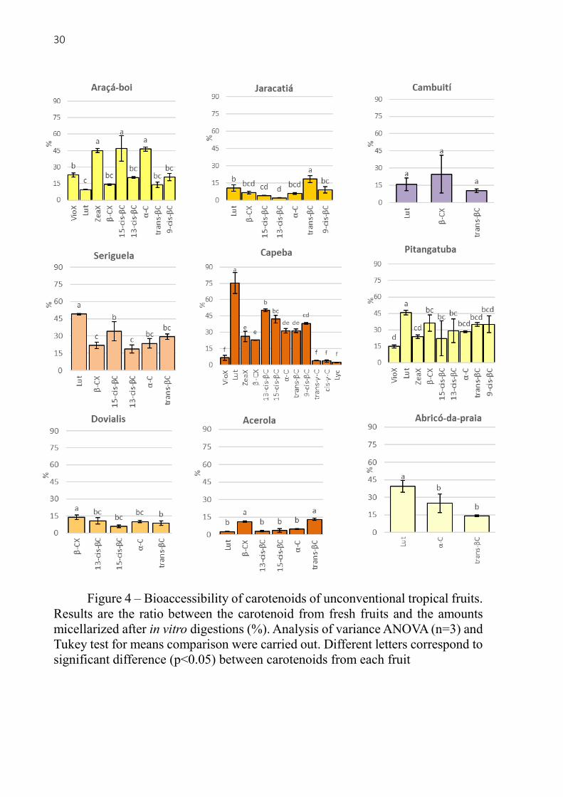
Figure 4 – Bioaccessibility of carotenoids of unconventional tropical fruits.
Results are the ratio between the carotenoid from fresh fruits and the amounts
micellarized after in vitro digestions (%). Analysis of variance ANOVA (n=3) and
Tukey test for means comparison were carried out. Different letters correspond to
significant difference (p<0.05) between carotenoids from each fruit

31
It was not found studies that determined bioaccessibility of carotenoids from these fruits.
Therefore we compared the bioaccessible amounts with other common tropical fruits. While
Capeba provide approx. 33 µg/g for trans-βC and 3 µg/g for β-CX in the bioaccessible fraction,
oranges provided 0.02 µg/g for trans-βC and 0.04 µg/g, mandarins 0.1 µg/g for trans-βC and
1.8 µg/g [23], mango 8.9 µg/g for trans-βC and papaya 2.4 for trans-βC [24]. A beverage (50:50
v/v of water and pulp) made with caja (Spondias mombim), also a brazilian native fruit from
the same genus of seriguela (Spondias purpurea), formed mixed micelles during in vitro
digestion containing: 0.06 µg/g of trans-βC, 0.06 µg/g of α-C, 0.15 µg/g of β-CX, 0.08 µg/g of
Lut and 0.03 ZeaX, excluding the esterified xanthophylls [25]. Therefore, our data of
bioaccessible amounts in the mixed micelles after in vitro digestion are slightly higher than the
found in literature for another fruits, what can be explained by the high initial content in the
fresh tropical fruits analyzed here.
The bioaccessibility of carotenoids from each fruit is presented in the figure 4. For the
araçá-boi fruit, ZeaX, 15-cis-βC and α-C were ~45% bioaccessible, being at least 25% higher
than the other carotenoids. Jaracatiá had low carotenoid bioaccessibility when compared to the
other fruits – ranging from 2% for 13-cis-βC to 18% for trans-βC. There were no significant
difference between the bioaccessibility of Lut, β-CX and trans-βC from cambuití. Seriguela
presented a range of 18% to 49% bioaccessibility, being Lut the most bioaccessible. The in vitro
digestion of capeba revealed 75% of Lut bioaccessibility followed by the cis-βC isomers (38-
50%), α-C and trans-βC (~31%), β-CX and ZeaX (22 - 26%), and finally the Lyc, trans-γ-C,
cis-γ-C and VioX had the lowest bioaccessibility (2-6%). Pitangatuba presented the lowest
bioaccessibility for VioX (15%) and the highest for Lut (45%). For dovialis the bioaccessibility
was 13% for β-CX, 8% for trans-βC followed by the other carotenes, but not significantly.
Acerola had very low bioaccessibility of its carotenoids, being β-CX and trans-βC the most
efficiently incorporated into mixed micelles, with 11% and 13% respectively. Finally, abricó-
da-praia presented Lut as the most bioaccessible (39%), α-C (24%) and trans-βC (14%). These
results are very consistent with literature for the range of bioaccessibility of several carotenoids
from many different fruits [24, 26 – 28]. Likewise, in accordance that usually xanthophylls are
more bioaccessible [30, 31] than carotenes, and that Lyc and VioX in most cases have very low
bioaccessibility [26, 28]. It is important to mention that the cis isomers of trans-βC and trans-
γ-C found in the micellar phase could be affected by the carotenoid extraction and
saponification steps, since at this stage the carotenes are already released and can suffer stress
during this long analysis period, despite all the precautions that were taken (BHT, light
protection and room temperature controlled under 25°C).

32
More examples of carotenoid bioaccessibility from tropical fruits could be mentioned.
Papaya grow in India can show bioaccessibility of 8.2% for trans-βC and 3.4% for Lyc [31] or
35% for trans-βC [24]. The differences are probably due to year and location of production,
varieties and maturity stage. Veda, Platel and Sirinivasan [24] reported the bioaccessibility of
trans-βC ranging 24% - 39% in six different varieties of mango. In vitro digestions of seven
different varieties of peppers provided results of bioaccessibility with many differences between
than, although bioaccessibility of β-CX (30% - 112%), Lut (36% – 106%) and ZeaX (34% -
106%) results were always higher than trans-βC (6% - 16%). Lycopene bioaccessibility both
for tomatoes and papaya was < 5% when analyzed by the same in vitro digestion protocol [30].
Therefore in plant foods the carotenoid bioaccessibility are very affected by food matrix and
particular characteristics, like maturity and growing conditions.
2.4. Conclusions
Eleven unconventional tropical fruits were studied as new plants for human nutrition.
Some of these fruits have their proximate composition, dietary fibers and carotenoid contents
reported for the first time. The fruits presented high contents of fibers and low energy values,
excepting buriti due to its 19% of lipids. Acerola and cambuití have much higher radical
scavenging capacity than the other fruits. Jaracatiá, seriguela, capeba, pitangatuba and acerola
highlighted due to their carotenoid diversity and concentration. The in vitro digestion results
presented variations between fruits mainly due to their food matrix, although generally
xanthophylls were more bioaccessible than carotenes. Lyc from capeba presented low
bioaccessibility as expected. The scientific knowledge together with the commercial
exploitation of tropical fruits, especially the Brazilian natives, can stimulate sustainable
development, better food habits, protection against biopiracy and the innovation in food
systems.
Acknowledgements
This work was supported by the São Paulo Research Foundation – FAPESP trough
research funding [grant #2015/15507-9] and PhD scholarship for Paulo Berni [grant
#2014/15119-6]. We thank Helton Muniz, from Sítio de Frutas Raras, for his extreme efforts
to grow and protect the Brazilian native fruits, provide samples and help with his accurate
knowledge about botany.

33
2.5. References
[1]Infante J, Rosalen PL, Lazarini JG, Franchin M, De Alencar SM (2016) Antioxidant and anti-
inflammatory activities of unexplored brazilian native fruits. PLoS ONE
https://doi.org/10.1371/journal.pone.0152974
[2]Rodriguez-Concepcion M, Avalos J, Bonet L et al (2018) A global perspective on carotenoids:
Metabolism, biotechnology, and benefits for nutrition and health. Prog Lipid Res.
https://doi.org/10.1016/j.plipres.2018.04.004
[3]Eggersdorfer M, Wyss A (2018) Carotenoids in human nutrition and health. Arch Biochem
Biophys. https://doi.org/10.1016/j.abb.2018.06.001
[4]Rufino M, Alves RE, Brito ES, Pérez-Jiménez J, Saura-Calixto F, Mancini-Filho J (2010)
Bioactive compounds and antioxidant capacities of 18 non-traditional tropical fruits from
Brazil. Food Chem. https://doi.org/10.1016/j.foodchem.2010.01.037
[5]Rosso VV, Mercadante AZ (2007) Identification and quantification of carotenoids, by HPLC-
PDA-MS/MS, from Amazonian fruits. J Agr Food Chem, 55:5062-5072.
[6]Flora do Brasil 2020 (2018). Jardim Botânico do Rio de Janeiro. http://floradobrasil.jbrj.gov.br.
Acessed 09 july 2018
[7]Muniz HJT (2008). Colecionando frutas: 100 espécies de frutas nativas e exóticas. 3. ed. Arte
& Ciência, São Paulo.
[8]Lorenzi H, Bacher LB, Lacerda MTC, Sartori SF (2006) Brazilian fruit and exotic cultivated.
Instituto Plantarum de Estudos da flora, São Paulo.
[9]Negri TC, Berni, PRA, Canniatti-Brazaca SG (2016) Nutritional value of native and exotic
fruits from Brazil. Biosaúde, 18:82-96
[10]Porcu MO, Rodriguez-Amaya DB (2008) Variation in the Carotenoid Composition of the
Lycopene-Rich Brazilian Fruit Eugenia uniflora L. Plant Foods Hum Nutr, 63:195-199
[11]Garzón AA, Narváez-Cuenca CE, Kopec RE, Barry AM, Riedl KM, Schwartz SJ (2012)
Determination of carotenoids, total phenolic content, and antioxidant activity of arazá (Eugenia
stipitata McVaugh), an Amazonian fruit. J Agric Food Chem. dx.doi.org/10.1021/jf205347f
[12]Cândido TLN, Silva MR (2017) Comparison of the physicochemical profiles of buriti from the
Brazilian Cerrado and the Amazon region. Food Sci Technol. http://dx.doi.org/10.1590/1678-
457x.32516

34
[13]CC BY 3.0 (https://creativecommons.org/licenses/by/3.0)], from Wikimedia Commons
[14]Association of official analytical chemists (1995) Official methods of analysis of the association
of official analytical chemists. 16 ed. Washington: AOAC
[15]Bligh EG, Dyer EJ (1959) A rapid method of total lipid extraction and purification. Can J
Biochem Physiol, 37:911-917
[16]Li BW, Cardozo MS (1992) Nonenzimatic-gravimetric determination of total dietary fiber in
fruits and vegetables. J AOAC Int, 75(2):372-4
[17]Kimura M, Kobori, CN, Rodriguez-Amaya DB, Nestel P (2007) Screening and HPLC methods
for carotenoids in sweet-potato, cassava and maize for plant breeding trials. Food Chem
100:1734–1746
[18]Berni P, Chitchumroonchokchai C, Canniatti-Brazaca SG, de Moura FF, Failla ML (2014)
Impact of genotype and cooking style on the content, retention, and bioaccessibility of
β‑Carotene in biofortified cassava (Manihot esculenta Crantz) conventionally bred in Brazil. J
Agric Food Chem, 62:6677−6686
[19]Garrett DA, Failla ML, Sarama RJ (1999) Development of an in vitro digestion method to assess
carotenoid bioavailability from meals. J Agric Food Chem, 47:4301-4309
[20]Stafussa AP, Maciel GM, Rampazzo V et al. (2018) Bioactive compounds of 44 traditional and
exotic Brazilian fruit pulps: phenolic compounds and antioxidant activity. Int J Food Prop.
https://doi.org/10.1080/10942912.2017.1409761
[21]Dias MG, Olmedilla-Alonso B, Hornero-Méndez D et al. (2018) Comprehensive database of
carotenoid contents in Ibero-American foods. A valuable tool in the context of functional foods
and the establishment of recommended intakes of bioactives. J Agric Food Chem.
https://doi.org/10.1021/acs.jafc.7b06148
[22]FAO/OMS. Human Vitamin and Mineral Requirements. In: Report 7th Joint FAO/OMS 491
Expert Consultation, Bangkok, Thailand. Food and Agriculture Organization, Rome, Italy, 2001
[23]Rodrigo MJ, Cilla A, Barberá R, Zacarías L (2015) Carotenoid bioaccessibility in pulp and fresh
juice from carotenoid-rich sweet oranges and mandarins. Food Funct,
https://doi.org/10.1039/c5fo00258c
35
[24]Veda S, Platel K, Sirinivasan K, (2007) Varietal differences in the bioaccessibility of β-Carotene
from mango (Mangifera indica) and papaya (Carica papaya) Fruits. J Agric Food Chem.
https://doi.org/10.1021/jf0712604
[25]Costa GA, Mercadante AZ (2018) In vitro bioaccessibility of free and esterified carotenoids in
cajá frozen pulp-based beverages. J Food Compost Anal.
http://dx.doi.org/10.1016/j.jfca.2017.02.012
[26]Schweiggert RM, Mezger D, Schimpf F, Steingass CB, Carle R (2012) Influence of chromoplast
morphology on carotenoid bioaccessibility of carrot, mango, papaya, and tomato. Food Chem,
135:2736–2742.
[27]Dhuique-Mayer C, Borel P, Reboul E, Caporiccio B, Besancon P, Amiot MJ (2007) β-
Cryptoxanthin from citrus juices: assessment of bioaccessibility using an in vitro
digestion/Caco-2 cell culture model. Br J Nutr. http://doi.org/10.1017/S0007114507670822
[28]Jeffery JL, Turner ND, King SR (2012) Carotenoid bioaccessibility from nine raw carotenoid-
storing fruits and vegetables using an in vitro model. J Sci Food Agric.
https://doi.org/10.1002/jsfa.5768
[29]O’Sullivan L, Ryan L, O’Brien N (2007) Comparison of the uptake and secretion of carotene
and xanthophyll carotenoids by Caco-2 intestinal cells. Brit J. Nutr. 98:38–44
[30]Rodrigues DB, Chitchumroonchokchai C, Mariutti LRB, Mercadante AZ, Failla ML (2017)
Comparison of two static in vitro digestion methods for screening the bioaccessibility of
carotenoids in fruits, vegetables, and animal products. J Agric Food Chem 65:11220−11228.
https://doi.org/10.1021/acs.jafc.7b04854
[31]Mashurabad PC, Palika R, Jyrwa YW, Bhaskarachary K, Pullakhandam R (2017) Dietary fat
composition, food matrix and relative polarity modulate the micellarization and intestinal
uptake of carotenoids from vegetables and fruits. J Food Sci Technol, 54(2):333–341

36

37
3 CAROTENOIDS FROM PITANGA (EUGENIA UNIFLORA) AND BURITI
(MAURITIA FLEXUOSA) FRUITS: PROFILE, STABILITY, BIOACCESSIBILITY
AND CELLULAR UPTAKE STUDIES BY IN VITRO DIGESTION COUPLED TO
CACO-2 CELLS CULTURE
Chapter submittted to Journal of Agriculural and Food Chemistry
Abstract
There were analyzed the carotenoid profile, stability during in vitro digestion,
bioaccessibility (micellarization due to bile salts) and absorption (uptake by Caco-2 cells
cultures) of pitanga and buriti fruits. Pitanga showed a more complex and diverse carotenoid
profile while buriti had a higher carotenoid concentration. In general, the xanthophylls had
lower stability during in vitro digestion than carotenes, excepting lycopene which was the least
bioaccessibile. The cis isomers of β-carotene showed an increase during simulated in vitro
digestion. Regarding bioaccessibility, violaxanthin, lutein and zeaxanthin from pitanga were
significantly more bioaccessible than other carotenes, excepting the cis-γ-carotene. Pitanga’s
lycopene and cis forms of β-carotene had the lowest bioaccessibilities. Xanthophylls and γ-
carotene forms of buriti were significantly more bioaccessible than the other carotenoids for
this fruit. The amounts of bioaccessible carotenoids shown that pitanga provides more
xanthophylls while buriti provides more amounts of carotenes. The Caco-2 cells experiment
has shown that an important fraction of xanthophylls are absorbed in their esterified forms. β-
carotene had the highest uptake by Caco-2 cells for pitanga and buriti. Therefore, β-carotene is
prevailing inside enterocytes after 4h exposition to digesta. Pitanga presented higher cellular
absorption of carotenoids than buriti (p < 0,05). This chapter evidence the essential need to
better understand the bioaccessibility of carotenoids from diverse food matrix.
Keywords: Carotenes; Xanthophylls; Eugenia uniflora; Mauritia flexuosa; Bioavailability;
Caco-2 cells

38
Graphical abstract
Highlights
- Carotenoid profiles, their bioaccessibility and intestinal uptake from pitanga and
Buriti were measured
- Xanthophylls from pitanga presented higher bioaccessibility than carotenes
- Carotenes presented higher cellular uptake from micellar fraction than xanthophylls
- Carotenoids from buriti had lower absorption by caco-2 cells than pitanga
3.1. Introduction
Food and nutritional sciences point out the health benefits of fruit bioactive compounds,
specially carotenoids. Moreover, food researchers are elucidating the health-related
mechanisms of carotenoids therewith promoting new ways of its consumption, processing and
innovation.1 Diverse antioxidant properties in carotenoids have been identified, highlighting
unique capacity to quench singlet oxygen.2 Strong epidemiological evidence show that
consumption of lycopene, α-carotene and β-carotene leads to a reduction in the risk of cancer,
such as breast and prostate tumors.3 Recently low carotenoid concentrations were identified in
older individuals and patients with cardiovascular disease, besides an widespread carotenoid
deficiency in the general population.4 Not all carotenoids act in the same way and are available
for absorption in the human digestive system. In fact, bioaccessibility is dependent on food

39
matrix, intrinsic chemical structure, and carotenoid x carotenoid interactions.5 Therefore, it is
essential to better understand carotenoid composition and bioaccessibility from new sources.
Brazil is known for its great biodiversity including several native fruits that are rich in
bioactive compounds, specially carotenoids. Despite this abundance of potential functional
fruits, there is a lack of scientific knowledge about their composition and health benefits. Some
of these Brazilian native fruits have diverse carotenoid profiles and concentrations. This is the
case of pitanga (Eugenia uniflora) and buriti (Mauritia flexuosa). Pitanga occurs naturally on
the Brazilian restinga and other coastal semi-deciduous forests, its largest farming is at
Northeast region.6 Pitanga fruit is used as juice, frozen pulp, and nectar that are rich in bioactive
carotenoids, such as lycopene and xanthophylls 7,8,9 Buriti is a palm fruit native in the Amazon
forest, and grows in Northeast, Central-west and Southeast regions of Brazil.6,10 Buriti fruit has
many interesting properties such as high content of polyphenolic and carotenoid compounds,
mainly all-trans-β-carotene. Moreover, its oil has in vitro antibacterial activity, assists in wound
healing, and is used to make cosmetics and therapeutic products.11,12,13
Bioacessible carotenoids have first to be released from the food matrix, integrated into
water soluble mixed micelles in the small intestine and absorbed by passive diffusion in
enterocytes due to its dispersion in the chyme aqueous phase.14 Many phenomena can influence
these steps. Globular shape of chromoplasts from mango and papaya presents higher carotenoid
liberation and bioaccessibility than tubular shape in tomatoes and carrots.15 Unsaturated fatty
acids promote bioaccessibility and basolateral secretion of carotenoids.16 Amount and
properties of pectin affect several events involved in carotenoid micellarization that may
increase or decrease bioaccessibility.17 Therefore, bioaccessibility and stability of carotenoids
in foods are greatly impacted by perishability, storage, preservation, and ways of consumption.
Moreover, home or industrial processing of vegetable foods play a key role in bioaccessibility
because it may break cellular structures liberating carotenoids for digestion.16,18,19 Thus, this
study contributes providing information about carotenoids bioaccessibility of these unique
fruits that helps understand carotenoid bioaccessibility in general foods.

40
3.2. Materials and methods
3.2.1. Fruit Samples
Pitanga (Eugenia uniflora) was harvested from the orchard located at Department of
Agri-food industry, food and nutrition of the Luiz de Queiroz College of Agriculture
(LAN/ESALQ), Piracicaba, São Paulo, Brazil. Red ripe pitanga was collected, selected, washed
and pulped in a juicer (Philips Walita®), then frozen and stored. Buriti (Mauritia flexuosa) pulp
was produced by the co-op Sertão Veredas located in Chapada Gaucha, north of Minas Gerais
state, Brazil. The farmers collected ripe Buriti fruits, peeled them, added warm water, and
crushed them to separate the seeds and pulp, which was frozen and shipped to Piracicaba, São
Paulo. All the samples were stored in 50 mL tubes with blanket of nitrogen gas, sealed, packed
in black bags and stored frozen at -25°C until the time of conducting the analysis. The samples
were frozen under these conditions until analyses were done. All the analytical procedures were
carried out at the Food Analyses and Nutrition laboratory, LAN/ESALQ-USP.
3.2.2. Reagents
Commercial carotenoid standards, β-apo-8’-carotenal and enzymes for in vitro digestion
were purchased from Sigma-Aldrich (St Louis, MO, USA). Specific reference numbers of each
enzyme are informed in the text. Organic solvents for extraction and HPLC analysis were
chromatographic grade (Tedia®). Caco-2 cells cultures were purchased from American Type
Culture Collection (ATCC®HTB37™). Dulbecco’s Modified Minimum Essential Medium –
DMEM, penicillin-streptomycin, fungizone, non-essential amino acids solution and L-
glutamine were purchased from GIBCO™ (Made in Brazil) while heat inactivated fetal bovine
serum was from Sigma-Aldrich (St Louis, MO, USA)
3.2.3. Carotenoid extraction and saponification
Aliquots for each sample changed according the step and appearance of carotenoid
concentration, i.e. ranged 0.1-0.2 grams of fresh fruit, 5 mL of digested food, 6 – 10 mL of
micellar fractions and cellular pellets of T25 flasks. For extracting carotenes and xanthophylls
at same time it was chosen the method described in Tibak et al.20 with modifications. Briefly,
thawed samples were extracted in 50 mL centrifuge tubes adding 15 mL of hexane, ethanol and
acetone solution (2:1:1, v/v/v) with 0.1% BHT. Tubes were mixed with a vortex and taken for
5 min in ultrasound bath and then back to the vortex for 1 minute. To the extract was added 3
mL – 5 mL of saturated salt solution and mixed with a vortex again for 30 seconds, then tubes

41
were centrifuged for 5 minutes at 800 g, 4ºC. Upper phase was collected with a pipette and
taken to dry with nitrogen gas inside the hood. For ester hydrolysis, saponification procedures
were carried out with independent extracts by combining equal volumes of methanolic KOH
solution (10 %, 0.1 % BHT) with the extracts.21 The mixtures were left overnight in dark at
room temperature. Upper phase was collected and washed three times with distilled water to
remove remaining KOH. To the final extract was added anhydrous sodium sulfate (~ 0.2 g) to
soak remained water then taken to dry with nitrogen gas in the hood. The dried extract was
dissolved in methanol and MTBE (1:1 v:v), total volume dependent on the appearance of
carotenoid concentration (0.2 – 4 mL), filtered in Millex® PTFE, 0.22 µm pore size. Finally,
vials were placed in the HPLC-DAD system for injection. β-apo-8′-carotenal was add randomly
to samples before extraction (10 µg/mL in methanol). Average recovery of β-apo-8′-carotenal
was 98.6 % ± 17.2 that allow to estimate the overall quality of carotenoid extraction and
analysis.
3.2.4. Carotenoid analysis by HPLC-DAD
Chromatographic analysis of carotenoid is presented in Kimura et al.22 and carried out
as follows. Liquid chromatographic system coupled to a diode array detector (HPLC-DAD)
Shimadzu® (LC-20A Prominence) was employed. The mobile phase consisted of methanol and
MTBE gradient starting at 90:10 (Methanol:MTBE) to 40:60 in 60 minutes, linear gradient,
returning to initial conditions and kept for 15 minutes for re-equilibration, the flow was 0.8 mL
/ min, injection volume changed according to the experimental step and carotenoid
concentration ranging 10 µL – 40 µL. The column used was the polymeric YMC™ C30 (150
mm x 4.6 mm, 5 µm particle size). Identification of carotenoids was carried out combining
commercial standards, elution order on C30 column compared to a certified reference material
(CRM485)22, diode array absorption spectra21,23 and comparisons with publications that
confirmed identity of carotenoids in HPLC-MS for the same fruits.9,12 Standard curves of all-
trans-violaxanthin, all-trans-lutein, all-trans-α-carotene, all-trans-β-carotene, all-trans-γ-
carotene and all-trans-lycopene were carried out and used for carotenoid calculation. The
respective r2 values for the standard curves were 0.9452, 0.9915, 0.9998, 0.9958, 0.9965 and
0.9949. Zeaxanthin, β-cryptoxanthin, cis and trans rubixanthin and cis isomers of β-carotene
were quantified by all-trans-β-carotene standard curve as they share same chromophore and
similar max absorption peak (λmax).
3.2.5. Carotenoid bioaccessibility by in vitro digestion
42
For the bioaccessibility analysis, an in vitro simulated digestion model was used as
described in Failla14 and Chitchumroonchokchai, Schwartz and Failla24, with slight changes.
Oral, gastric and intestinal phases of in vitro digestion are summarized next. Fruit samples (1 –
2 g) were homogenized with 10 mL of basal salts solution (BSS, NaCl 120 mol/L, CaCl2 6
mmol/L and KCl 5 mmol/L) in 50mL centrifuge tubes. The same BSS was used for volume
adjustment when needed. Because we previously determined only 0.4% (wet weight) of lipids
in pitanga pulp, by Folch et al.25 protocol, 2% (wt/wt) soybean oil was added for mixed micelles
formation, but no oil was added to the buriti (19.2% lipid) during the in vitro digestion. For the
oral phase 6 mL of previously prepared artificial saliva containing 106 units/mL of α-amylase
(Sigma® A3176) was added, and incubated at 37°C for 10 min in a rocker shaker (120 rpm).
Following, pH was adjusted to 2.5 with HCl 1 M and 2 mL of pepsin solution was added
(Sigma® P7000; 50000 units/mL in HCl 100 mM). Total volume was adjusted to 40 mL and
the tubes were incubated for 1 h, 37 °C, 120 rpm. The pH was raised to 6.0 with 1 M NaHCO3
to stop the gastric phase. Porcine bile extract solution (3 mL; Sigma® B8631; 40 mg/mL in 100
mM NaHCO3) and 2 mL of pancreatin (Sigma® P1750) and lipase (Sigma® L3126) solution at
~4000 units/mL and ~1000 units/mL in 100 mM NaHCO3, respectively, were added. Before
incubation for 2h, 37°C, pH and volume were adjusted to 6.5 and 50mL respectively. After the
intestinal phase digestion, homogenized aliquots (12 mL) were centrifuged at 5000 g, 60 min,
4ºC. The supernatant (aqueous fraction) was collected and sterile filtered (Millipore®, 0,22μm
pore size) generating the micellar fraction, i.e. bioaccessible carotenoids. The ratio between
carotenoids incorporated into mixed micelle and the determined in the fresh fruit pulp predicts
the carotenoid bioaccessibility. The remaining digesta was analized for their carotenoid
concentration. The ratio between carotenoids found in digesta and the determined in the fresh
fruit pulp represents the individual carotenoid stability. In all steps and storage samples were
covered with nitrogen gas, sealed with parafilm®, protected against direct light and high room
temperature. Samples of digested and bioaccessible fraction were stored at -26ºC until
carotenoid analysis by HPLC-DAD as described above.
3.2.6. Carotenoid uptake by Caco-2 cells culture
Caucasian colon adenocarcinoma cells (Caco-2 from American Type Culture Collection
ATCC®HTB37™) were cultivated for the evaluation of carotenoid intestinal absorption. The
procedures of cells culture were the described by Garret, Failla and Sarama (1999) and
Chitchumroonchokchai, Schwarts and Failla (2004). Passages 22-27 of Caco-2 cells were
seeded in T25 flasks (25 cm2 of area growth) at 2 – 3 x 10⁵ cells/flask. Cultures were maintained

43
at 37°C, humidified atmosphere, 95% air and 5% CO2. Dulbecco’s Modified Minimum
Essential Medium (DMEM, GIBCO™) enriched with L-glutamine (1%), non-essentials amino
acids (1%), penicillin-streptomycin (1%), fungizone (0.2%) and heat inactivated fetal bovine
serum - FBS (15%) was utilized to maintain the cells growing until the culture acquire
confluence (~6 days). After confluency FBS was reduced to 10%. Media was changed every
other day until cells differentiate in enterocytes, 10-14 days. The quality of cellular cultures
was checked visually by inverted microscope. Attachment, cell growth, absence of
contamination and number of domes were considered for controlling the viability of monolayers
that are ready for experiment. Monolayers were washed with Minimum Essential Medium
(MEM, GIBCO™) to carry out experiments. A fresh complete DMEM, pH 6.5, containing 25%
of the micellar fraction obtained from the in vitro digestions was added to the monolayers (final
volume 5 mL) and incubated for 4 hours. Thereafter, Caco-2 monolayers were washed once
with 2 mg/L of albumin in cold PBS (pH 7.0) and twice with only cold PBS (pH 7.0), harvested
with rubber scrapper, collected and centrifuged at 500 g/10 min, 4ºC. Cells pellets were covered
with nitrogen gas, sealed and stored at -26ºC until carotenoid extraction. Protein content of cell
samples was determined by a Bradford rapid assay (BIO RAD: Quick start™ Bradford) using
bovine serum albumin as a standard.
3.2.7. Statistics
Carotenoid determination was done in triplicate with homogenized fresh fruit pulp then
experiments for bioaccessibility were carried out independently also in triplicate. Data were
analyzed for significant differences using ANOVA between carotenoid species and fruit. Means
were compared using the Tukey test and significant at 95% reliability (p < 0.05). Software used
for the ANOVA and Tukey tests was the Statistical Assistance Software Assistat 7.7 beta
(UFCG, Campo Grande, PB, Brazil).
3.3. Results and discussion
3.3.1. Carotenoid compounds of pitanga and buriti
The carotenoid analysis of pitanga and buriti fruits by chromatography enabled to
separate and positively identify 14 carotenoids presented in table 1. They were identified
through a combination of commercial standards, chromatographic elution sequence in a C30
column, UV-vis spectra with the diode array detector and literature comparisons.9,12
Violaxanthin, lutein, α-carotene, all-trans-β-carotene, γ-carotene and lycopene were quantified

44
by its own standard curves while zeaxanthin, β-cryptoxanthin and rubixanthin were quantified
by all-trans-β-carotene standard curve since some carotenoids have the same chromophore in
their chemical structure and similar UV-vis spectra.21 Because accurate carotenoid
identification and calculations are a complex task, we presented only results of the carotenoids
that were confidently identified, calculated and those we could track through the in vitro
digestion experiment.
Table 1. Parameters for carotenoid identification1
Carotenoid
RT (min)
λmax
(nm)1
%
III/II 1
1. Violaxanthin 6.4 415/439/468 94
2. Lutein 11.4 421/444/472 66
3. Zeaxanthin 13.2 (423)/ 446/474 28
4. β-cryptoxanthin 19.8 (425)/450/477 29
5. 15-cis-β-carotene2+
13-cis-β-carotene2
23.2
23.4 339/(423)/448/473 11
6. α-carotene 24.7 421/445/473 65
7. all-trans-β-carotene 28.2 (425)/451/477 26
8. 9-cis-β-carotene 30.0 338/(420)/447/472 20
9. rubixanthin 35.6 436/459/490 54
10. cis-rubixanthin 36.5 355/436/459/490 38
11. γ-carotene 44.1 438/461/492 57
12. cis-γ-carotene 44.9 294/355/(438)/464/495 54
13. lycopene 60.3 445/471/503 75
a λmax and % III/II were obtained from the photodiode array detector of the peak
in their current mobile phase. b There were partial coelution of 15-cis-β-carotene
and 13-cis-β-carotene.
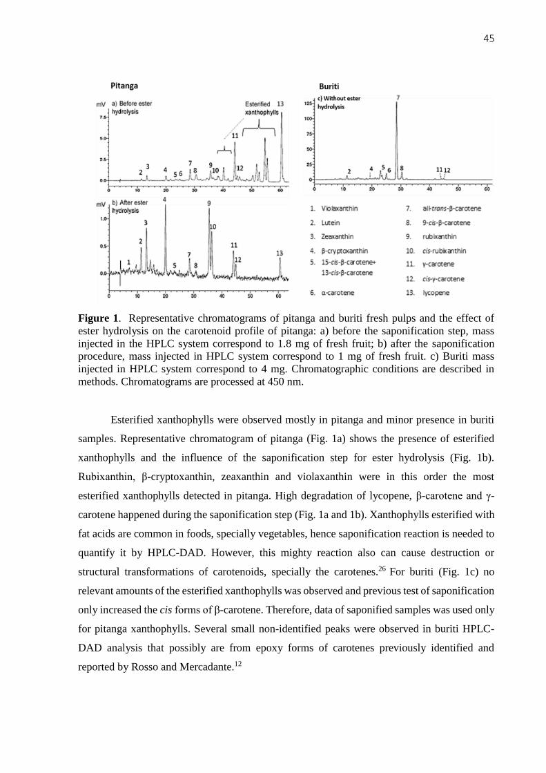
45
Figure 1. Representative chromatograms of pitanga and buriti fresh pulps and the effect of
ester hydrolysis on the carotenoid profile of pitanga: a) before the saponification step, mass
injected in the HPLC system correspond to 1.8 mg of fresh fruit; b) after the saponification
procedure, mass injected in HPLC system correspond to 1 mg of fresh fruit. c) Buriti mass
injected in HPLC system correspond to 4 mg. Chromatographic conditions are described in
methods. Chromatograms are processed at 450 nm.
Esterified xanthophylls were observed mostly in pitanga and minor presence in buriti
samples. Representative chromatogram of pitanga (Fig. 1a) shows the presence of esterified
xanthophylls and the influence of the saponification step for ester hydrolysis (Fig. 1b).
Rubixanthin, β-cryptoxanthin, zeaxanthin and violaxanthin were in this order the most
esterified xanthophylls detected in pitanga. High degradation of lycopene, β-carotene and γ-
carotene happened during the saponification step (Fig. 1a and 1b). Xanthophylls esterified with
fat acids are common in foods, specially vegetables, hence saponification reaction is needed to
quantify it by HPLC-DAD. However, this mighty reaction also can cause destruction or
structural transformations of carotenoids, specially the carotenes.26 For buriti (Fig. 1c) no
relevant amounts of the esterified xanthophylls was observed and previous test of saponification
only increased the cis forms of β-carotene. Therefore, data of saponified samples was used only
for pitanga xanthophylls. Several small non-identified peaks were observed in buriti HPLC-
DAD analysis that possibly are from epoxy forms of carotenes previously identified and
reported by Rosso and Mercadante.12

46
3.3.2. Carotenoid contents of pitanga and buriti
Table 2 presents data of carotenoid contents for both fruits. When compared with 23
vegetables that are good sources of all-trans-β-carotene listed in Fernández-García et al.27,
buriti still at least 4.9 times higher than any vegetable. More than 78% of the identified
carotenoids in buriti are the trans and cis forms of β-carotene. Buriti has 8 carotenoids having
provitamin A activity – that contains the β-ionone ring on its chemical structure28 – in very high
concentration, ranging from 19 to 434 µg/g on wet weight basis (Table 2). It also includes a
high content of lutein, 37.8 µg/g on wet weight basis (Table 2), an important component for
eyes health that may prevent macular degeneration.29 Our results are similar to others found in
literature, such 360 µg/g of all-trans-β-carotene and 82 µg/g of α-carotene presented in
Rodriguez-Amaya et al..30 The cis-β-carotene isomers naturally occurs in this fruit in high
quantity, e.g. 59 µg/g, 18.5 µg/g and 8.8 µg/g respectively for the 13-cis, 9-cis and 15-cis β-
carotene reported in Rosso and Mercadante12, and agree with our findings. However, this palm
fruit is collected inside the forests after it falls down and its edible pulp is extracted using warm
water. These procedures associated to long time of transportation and storage followed by
aggressive solvent extractions for analysis can generate the cis isomers.31 These reasons can
explain the higher amount of 9-cis and 15-cis β-carotene found in our samples.
Pitanga results present a more complex and diverse carotenoid profile and content than
buriti (Table 2 and Fig. 1). Pitanga contains lycopene as a major carotenoid that is at least twice
higher than the 6 xanthophylls it brings together, an uncommon characteristic among plant
foods (Table 2). All carotenoids found in pitanga in our study were already identified by HPLC-
MS by Azevedo-Meleiro and Rodriguez-Amaya9, excluding the minor β-carotene-5,6-epoxide
and cis-lycopene that we did not confirm present. Furthermore, the carotenoid content
determined in pitanga fruits grown in Piracicaba-SP (Table 2) match with the results reported
by Porcu and Rodriguez-Amaya23 in pitanga grown in Campinas-SP, the same climate and
mesoregion. Their study shows great variation of carotenoids contents in pitanga due to
ripeness, climate and growing region. Noteworthy, carotenoid in fruits are part of a secondary
plant metabolism that are very dynamic and dependent on plant variety, sunlight, soil, climate
and maturity.32 Range variations of carotenoid and distribution through carotenoid species
associated with food matrix plays an important role on bioaccessibility.1,5 Therefore, it is crucial
to accurately determine carotenoids composition on fresh foods before going through
bioaccessibility studies.

47
47
Table 2 – Initial carotenoid contents in fresh pulp, bioaccessible amounts of carotenoids obtained in micelle fraction after in vitro gastrointestinal
digestion and its absorption by Caco-2 human intestine cells 1
Carotenoid
Pitanga Buriti
Fresh
(µg/g)
Bioaccessible
(µg/g)
Absorbed2
(ng/mg protein/g) Fresh
(µg/g)
Bioaccessible
(µg/g)
Absorbed2
(ng/mg protein/g)
1. Violaxanthin 0.55±0.12h 0.19±0.02d nd nd nd nd
2. Lutein 2.33±0.18f 0.75±0.08c 3.99±0.15d 37.8±2.8de 19.7±0.32b 44.8±3.9b
3. Zeaxanthin 4.03±0.74e 1.11±0.02b 9.04±0.03c nd nd nd
4. β-cryptoxanthin 11.4±0.25c 1.74±0.07a 21.5±0.15b 21.5±0.91e 8.34±0.14d nd
5. 15-cis-β-carotene3 +
13-cis-β-carotene3 2.01±0.02f 0.10±0.02d nd
56.8±8.4bcd 21.6±1.03b 48.2±3.3b
42.3±2.65cd 14.3±0.30c 13.7±1.8c
6. α-carotene 2.58±0.17f 0.39±0.01d nd 65.0±5.63b 20.9±1.38b 44.3±5.9b
7. all-trans-β-carotene 3.98±0.26e 0.77±0.19c 37.9±3.4a 434±16.3a 115±4.17a 598±20a
8. 9-cis-β-carotene 1.82±0.08fg 0.14±0.07d nd 60.8±0.23bc 21.2±1.20b 56.4±1.9b
9. rubixanthin 14.2±0.37b 1.48±0.15a 23.4±2.78b nd nd nd
10. cis-rubixanthin 6.23±0.11d 0.75±0.21c 6.28±0.71cd nd nd nd
11. γ-carotene 1.42±0.17fgh 0.31±0.01d 19.6±2.3b 19.8±4,15e 8.86±0.31d nd
12. cis-γ-carotene 0.73±0.05gh 0.27±0.01d 3.59±0.48d 20.9±4.10e 9.12±0.19d nd
13. lycopene 30.8±1.03a 0.35±0.03d 2.78±0.11d nd nd nd 1Data are mean ± standard deviation of three replicates (µg/g on wet weight basis). Analysis of variance ANOVA (n=3) and Tukey test for means comparison were carried out.
Different letters in the column correspond to significant difference (p<0.05) between carotenoids levels of same fruit. 2 Results were standardized by mg of cell protein (2.8 mg of
protein per 25cm²) per g of fresh sample. 3There was partial coelution of 15-cis-β-carotene and 13-cis-β-carotene, it was possible to identify and calculate them separately only for
buriti samples.

48
3.3.3. Stability of carotenoids during simulated digestions
Figure 2 shows the carotenoid profile of pitanga in natura without saponification (2a),
the carotenoids stability after in vitro digestion (2b), the bioaccessibility (2c), i.e. carotenoids
that were released from food matrix and incorporated into mixed micelles, and the carotenoid
uptake by human intestine cells Caco-2 (2d). Comparing first and second chromatograms (Fig.
2a and 2b) may be noted that carotenoid maintained relatively stable during in vitro digestion.
Carotenoids from pitanga were statistically less stable than buriti (p < 0.05). In general,
xanthophylls were less stable than carotenes, e.g. in pitanga only 52 % of β-cryptoxanthin and
zeaxanthin remained after in vitro digestion (Fig. 3). Remaining carotenes ranged from 62 %
for lycopene in pitanga to 96 % for α-carotene in buriti. Cis isomers of β-carotene showed
slightly increase during simulated digestion – 104 % of stability for 15-cis and 13-cis β-carotene
in pitanga and 131 % of stability for 13-cis β-carotene in buriti (Fig. 3). Although, carotenoid
stability during in vitro digestion usually exceeds 75 %14, Courraud et al.33 reported all-trans-
β-carotene stability varying 31%, 100% and 40% for commercial standard, carrot juice and
spinach, respectively. Courraud et al.33 also reported that all-trans-β-carotene instability occurs
mainly in gastric phase due to its high acidity. Therefore, carotenoid can be differently affected
by the digestive environment and the food matrix. This may explain why carotenoids from
pitanga had lower stability than buriti, since pH of pitanga whole pulp was 2.8 and of buriti 4.8.
Moreover, carotenoids solubilized/emulsified in lipids are more likely protected against
degradation24 thus carotenoids from buriti were more stable during the in vitro digestion
simulations.
3.1.1. Bioaccessibility of carotenoids after in vitro digestion
Comparison of chromatograms in figures 2a and 2c shows the carotenoids efficiency of
micellarization, i. e. bioaccessibility. Xanthophylls being less hydrophobic than carotenes due
the oxygen in their chemical structure are easily incorporated into mixed micelles34, that is why
the higher peaks in the chromatogram Fig. 2c corresponds to the violaxanthin, lutein, zeaxanthin
and β-cryptoxanthin. Markedly lycopene has almost no incorporation into mixed micelles (Fig.
2c), in fact, its bioaccessibility was 1.1% (Fig. 4). Further, small peaks of esterified
xanthophylls in micellar phase (Fig. 2c) indicate that esterification seems to impair their
incorporating into mixed micelles when compared to free xanthophylls. The minor breaking of
esterification by digestive enzymes seems to be irrelevant.16,17,34
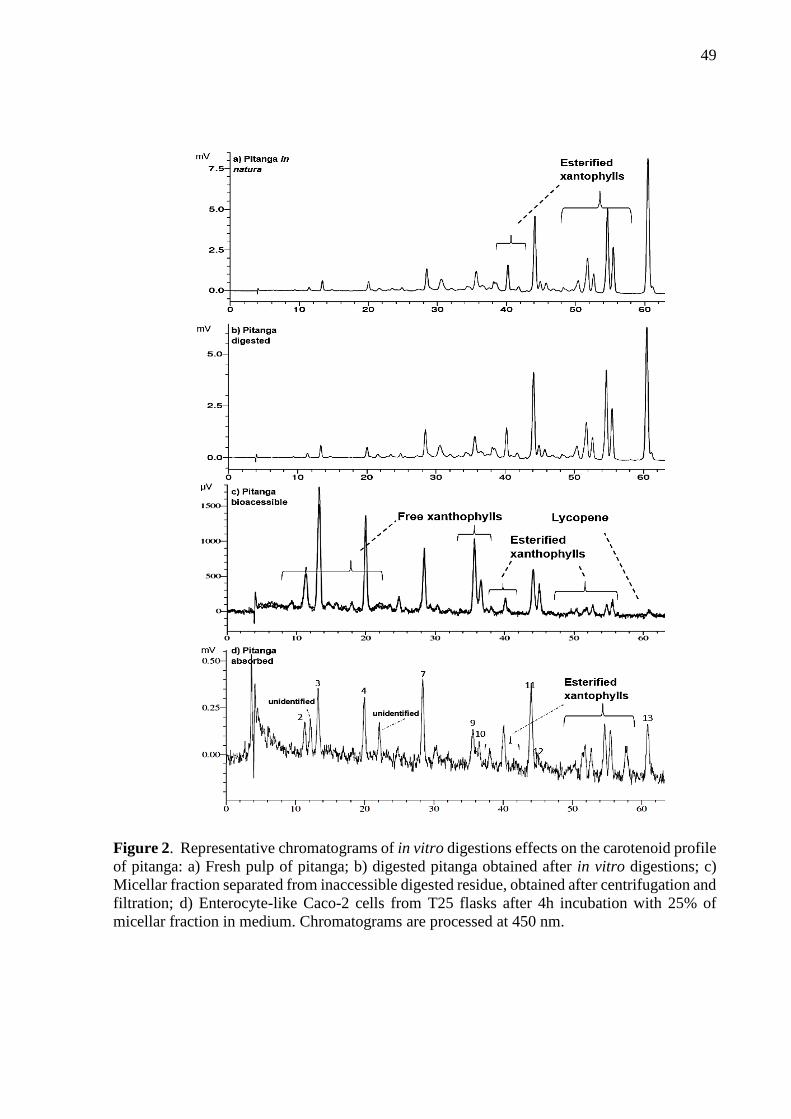
49
Figure 2. Representative chromatograms of in vitro digestions effects on the carotenoid profile
of pitanga: a) Fresh pulp of pitanga; b) digested pitanga obtained after in vitro digestions; c)
Micellar fraction separated from inaccessible digested residue, obtained after centrifugation and
filtration; d) Enterocyte-like Caco-2 cells from T25 flasks after 4h incubation with 25% of
micellar fraction in medium. Chromatograms are processed at 450 nm.

50
Figure 3. Stability of carotenoids after in vitro digestions of pitanga and buriti. Pitanga and
buriti graphic represents percentage of carotenoid that remained in digested samples (n=3) after
the analytical procedures. Analysis of variance ANOVA (n=3) and Tukey test for means
comparison were carried out. Different letters above each bar correspond to significant
difference (P<0.05) between carotenoids bioaccessibility of same fruit.
Figure 4. Bioaccessibility of carotenoids from pitanga and buriti. Data are related to percentage
of carotenoid incorporated into mixed micelles after in vitro digestions. Data are mean ±
standard deviation of three replicates. Analysis of variance ANOVA (n=3) and Tukey test for
means comparison were carried out. Different letters above each bar correspond to significant
difference (p<0.05) between carotenoids bioaccessibility of same fruit.
63
de
92
ab
52
e
52
e
10
4a
76
bcd
92
ab
97
ab
61
de 6
9cd
e 86
abc
88
abc
62
de
DIG
ESTI
VE
STA
BIL
ITY
(%)
P I T A N G A
95
b
83
b
13
1a
90
b 96
b
84
b 94
b
88
b
10
4b
10
0b
DIG
ESTI
VE
STA
BIL
ITY
(%)
B U R I T I

51
Violaxanthin, lutein and zeaxanthin from pitanga were significantly more bioaccessible
than other carotenes excepting cis-γ-carotene whereas lycopene along with cis forms of β-
carotene had lowest bioaccessibility (Fig. 4). For buriti same pattern was observed,
xanthophylls and γ-carotene forms being significantly more bioaccessible than the other present
carotenoids (Fig. 4). These evidences show that some competition may happen between
carotenoids to get into micelles during the intestinal phase of digestions.
Overall bioaccessibility of carotenoids from buriti was significant higher than pitanga
(p<0.05). Indeed, pitanga composition is much different from buriti. Pitanga has a considerable
amount of fibers, low amount of oil, high humidity and acidity while buriti is an oily palm fruit.
Presence of 19% of fat in buriti fruit against only 2.4% in pitanga – initial plus added oil for in
vitro digestions – indicates also the influence of carotenoid:lipid ratio on bioaccessibility.
Lipids in diet, mainly unsaturated fat acids, increases the micellarization and absorption of
carotenes while fat acid saturation has minimal influence on the xanthophylls bioaccessibility.16
Our results (Fig. 4) corroborate with Failla et al.16 study that reported bioaccessibilities in a
salad meal containing 3% wt/wt of different fat sources ranging 45-57% for lutein and
zeaxanthin, 9-20% for all-trans-β-carotene and 1.5-5.3% for lycopene. For the best of our
knowledge this is the first time carotenoids bioaccessibility from these unique plants was
assessed. The assessment of carotenoid bioaccessibility provide essential information about
potential health benefits of functional fruits. Usually, when any fruit is analytically proven to
contain some bioactive components, many nutritional recommendations stimulate that fruit’s
consumption. Bioaccessibility data can help professionals, companies and policymakers to
properly guide dietary changes to specific needs.
Pitanga, when consumed in natura, contains lycopene, which may prevent prostate
cancer, but β-cryptoxanthin had 5 times higher bioaccessible concentrations (Table 2). Micellar
phase of digested pitanga would first provide higher amounts of xanthophylls than the carotenes
– β-cryptoxanthin > rubixanthin > zeaxanthin > lutein > cis-rubixanthin. There is a marked
relation between carotenoid bioaccessibility and antioxidant activity. It has demonstrated that
hydroxylation and cyclization of carotenoid terminal end groups are not only responsible for
augmented bioaccessibility but also for higher antioxidant capacity against oxygen reactive
species.35,36 Sòlyom et al.35 reported antioxidant capacity of carotenoid rich micellar fraction
by Trolox equivalent antioxidant capacity (TEAC) being: rubixanthin > lycopene > β-
cryptoxanthin > γ-carotene > β-carotene. They associated rubixanthin higher TEAC values with
the twofold enhancing effect of one ψ-end and one hydroxyl group on the β-ionone ring, thus
52
being closely connected to the micelle's surface and possessing a reactive opened-chain end.
Rubixanthin’s health effects have been neglected because it is uncommon in foods. In pitanga,
it is possibly the most important functional carotenoid because of its high amount in the micellar
phase (Table 2). β-carotene and lycopene have been shown to be inversely related to the risk of
cardiovascular diseases and certain cancers whereas lutein and zeaxanthin to the disorders
related to the eye.2 β-cryptoxanthin have anti-inflammatory effects, enhance the immune system
and may reduce the risk of osteoporosis.37,38 Thereby pitanga may have much more potential as
a functional fruit due to β-cryptoxanthin, zeaxanthin and lutein than lycopene bioaccessible
amounts.
Regarding buriti, all-trans-β-carotene is the most abundant in the micellar phase. With
two β-ionone rings, all-trans-β-carotene is converted to retinol by BCO1 and BCO2 enzymes
in enterocytes twice more than other carotenes.28 Except lutein, all carotenes from buriti have
vitamin A activity, rendering buriti an interesting food for combating vitamin A deficiency in
many areas of Brazil and the world. The mean requirement ingestion of retinol equivalent (RE),
for adult women stablished by FAO39 is 300 μg RE/day. Buriti have 96.7 µg RE/g calculated
according to FAO39, that considers an averaged carotenoid bioaccessibility of 25%. However,
we presented that buriti have a range of 26 – 46 % bioaccessibility for its provitamin A
carotenoids. Not all provitamin A carotenoids absorbed by humans are converted into retinol,
i.e. some carotenoids such β-carotene and β-cryptoxanthin are absorbed and distributed by
chylomicrons intact and accumulated in many tissues providing a protective effect against
oxidative stress.40 Rodrigues et al.36 demonstrated that antioxidant capacity of carotenes against
the peroxyl radical is dependent on the opening of the β-ionone, the chromophore extension
and addition of hydroxyl groups. For example, all-trans-β-carotene have higher capacity to
scavenge the peroxyl radical than lutein but is less effective than lycopene and γ-carotene. So
that, information regarding antioxidant potential of carotenoid rich fruits should be followed by
comprehensive work about their bioaccessibility.

53
Figure 5. Carotenoid uptake by enterocyte-like Caco-2 cells culture. Data are related to the
percentage of carotenoid that was taken from micellar fraction after 4h of exposure. Data are
mean ± standard deviation of three replicates. Analysis of variance ANOVA (n=3) and Tukey
test for means comparison were carried out. Different letters above each bar correspond to
significant difference (p<0.05) between carotenoids from same fruit.
Caco-2 cells were cultivated until generating monolayers of differentiated enterocytes
(10-14 days post-confluency) and then were incubated with the micellar fraction diluted in
DMEM medium (1:4 v/v) during 4 h. Results of cellular uptake of carotenoids are presented in
percent taken from media at figure 5, and in absolute amount accumulated inside the cells at
table 2. β-carotene had the highest uptake and accumulation in the intestine cells for both fruits.
Pitanga presented an overall uptake of carotenoids higher than buriti. There is evidences that
food matrix may affect the absorption of carotenoids from foods. Previous works using the
same protocol for in vitro digestion and cellular experiment also found differences between
cassava varieties (uptake of all-trans-β-carotene ranging 10-20 %) and orange-fleshed sweet
potato varieties (ranging 10-25%).41,42 Despite the very low micellarization of lycopene, its
uptake from mixed micelles was relatively high (Fig. 5). Our data for carotenoid uptake by
caco-2 cells compared to others found in literature, is similar in the case of lycopene43,44 while
lutein and zeaxanthin values presented corroboration45 and discrepancies.46
Differences of carotenoid uptake by Caco-2 cells between green, red and yellow bell
peppers, and green and red chili peppers were also noted in a great extent for β-carotene
8,4
e
7,3
e
24
,8b 29
,7a
20
,3c
9,3
e
22
,3b
c
5,9
e
16
,0d
% U
PTA
KE
PITANGA
4,5
bc
4,6
bc
1,9
d 4,1
c
10
,4a
5,5
b
% U
PTA
KE
BURIT I

54
(ranging 1.3 – 31%) in the work of O’Sullivan et al.45. In agreement to our observations, they
detected that although lutein (a xanthophyll) was the most abundant carotenoid in the micelles,
cellular uptake of β-carotene were the highest. Therefore, interactions between carotenoids may
influence the cellular absorption. Moreover, our results indicate that all-trans-β-carotene may
have facilitated transport across the brush border. In fact, a scavenger receptor class B type I
(SR-BI) and a cluster determinant 36 (CD36) are proved to be involved in cellular uptake of β-
carotene, α-carotene and β-cryptoxanthin.47 We attribute the β-carotene higher uptake and
accumulation in enterocytes to the SR-BI effect since it is expressed by caco-2 cells.47 Thus,
our data corroborate that transporter mediated absorption in the intestine epithelium facilitates
carotenes passage, and that competition with other compounds from the food matrix may affect
carotene absorption.
Buriti presented lower carotenoid uptake than pitanga (p < 0,05, Fig. 5) that may be
explained by a saturation effect on caco-2 cells. Note that the concentration of bioaccessible
all-trans-β-carotene in the micellar phase was 150 times higher than pitanga. O’Sullivan et al.45
demonstrated this saturation effect for carotenes and xanthophylls – i.e. as much higher was the
concentration of the carotenoid in the micellar fraction, lower was the absorption by the
enterocyte-like caco-2 cells. However, despite this saturation effect carotenoid accumulated in
Caco-2 cells were higher for buriti (Table 2). Lycopene was in low concentration in the micellar
phase due to very low micellarization (1.1%), however it had a relative high uptake (16%), what
also can be explained by this concentration effect.
In summary, our work presents pitanga and buriti fruits as very rich sources of carotenoids
highly bioaccessible and consequently great potential to be recommended and commercialized
as functional fruits. Pitanga comes up as a source of xanthophylls, specifically rubixanthin,
while only a small part of lycopene is absorbed and can be used by the body. Therefore pitanga
has a potential to present antioxidant activity against physiologic reactive species of oxygen as
well may prevent macular degeneration. We suggest further studies to confirm these
functionalities. Likely, buriti is a good natural source of vitamin A due to high bioaccessible
provitamin A content. Hence, buriti may have many health benefits like improve immune
system, prevent several cancers, avoid eyes diseases and others, that also should be tested to
appraise the extent of these effects.
The assays provided evidence that the biochemistry of carotenoids and food matrix are
the main effectors of bioaccessibility. Hydroxylation and esterification of xanthophylls,
carotenes trans/cis isomerization, presence and quantity of lipids from food matrix/meal, as
well the interactions between different carotenoids are essential to explain what happens inside
55
the digestive system with carotenoids from fruits. Also, it is remarkable that beyond the simple
quantification of bioactive compounds in fruits we must know how much of that compound can
be absorbed by the body. Thus it is important to develop products and process that focus on
reducing instability through digestion and increase absorption in small intestine, like compound
delivery systems. Ultimately, pitanga and buriti should be more explored regarding their
bioactive compounds and functionalities, because they can represent a new raw-material for
development of innovative products.
Funding sources
This work was supported by the São Paulo Research Foundation – FAPESP through
research funding [grant #2015/15507-9] and PhD scholarship for Paulo Berni [grant
#2014/15119-6].
3.2. References
(1) Saini, R. K.; Nile, S. H.; Park, S. W. Carotenoids from fruits and vegetables: Chemistry,
analysis, occurrence, bioavailability and biological activities. Food Res. Int. 2015, 76, 735-
750.
(2) Rao, A. V.; Rao, L. G. Carotenoids and human health. Pharmacol. Res. 2007, 55, 207–
216.
(3) Ruiz, R. B.; Henández, P. S. Cancer chemoprevention by dietary phytochemicals:
Epidemiological evidence. Maturitas 2016, 94, 13–19.
(4) Petyaev, I. M. Lycopene deficiency in ageing and cardiovascular disease. Oxid. Med. Cell
Longev. 2016, vol. 2016, Article ID 3218605, 6 pages.
http://dx.doi.org/10.1155/2016/3218605
(5) Rodriguez-Amaya, D. B. Quantitative analysis, in vitro assessment of bioavailability and
antioxidant activity of food carotenoids—A review. J. Food Compos. Anal. 2010, 23, 726–
740.
(6) Lorenzi, H.; Bacher, L. B.; Lacerda, M. T. C.; Sartori, S. F. Frutas Brasileiras e Exóticas
Cultivadas (de Consumo in natura); São Paulo: Instituto Plantarum de Estudos da Flora;
Brazil, 2006; 640 pp.
56
(7) Bagetti, M.; Facco, E. M. P.; Rodrigues, D. B.; Vizzotto, M.; Emanuelli, T. Antioxidant
capacity and composition of pitanga seeds. Cienc. Rural 2009; 39, 2504-2510.
(8) Freitas, M. L. F.; Dutra, M. B. L; Bolini, H. M. A. Time–intensity profile of pitanga nectar
(Eugenia uniflora L.) with different sweeteners: Sweetness and bitterness. Food Sci.
Technol. Int. 2016; 22(1), 58-67.
(9) Azevedo-Meleiro, C. H.; Rodriguez-Amaya, D. B. Confirmation of the identity of the
carotenoids of tropical fruits by HPLC-DAD and HPLC-MS. J. Food Compos. Anal. 2004,
17, 385–396.
(10) Oliveira, D. M. de; Siqueira, E. P.; Nunes, Y. R. F.; Cota, B. B. Flavonoids from leaves of
Mauritia flexuosa. Rev. Bras. Farmacogn. 2013; 23, 614-620.
(11) Koolen, H. H. F.; Silva, F. M. A.; Gozzo, F. C.; Souza, A. Q. L; Souza, A. D. L.
Antioxidant, antimicrobial activities and characterization of phenolic compounds from
buriti (Mauritia flexuosa L. f.) by UPLC–ESI-MS/MS. Food Res. Int. 2013; 51, 467–473.
(12) Rosso, V. V.; Mercadante, A. Z. Identification and quantification of carotenoids, by
HPLC-PDA-MS/MS, from Amazonian fruits. J. Agr. Food Chem. 2007; 55, 5062-5072.
(13) Batista, J. S.; Olinda, R. G.; Medeiros, V. B.; Rodrigues, C. M. F.; Oliveira, A. F.; Paiva,
E. S.; Freitas, C. I. A.; Medeiros, A. C. Antibacterial and healing activities of buriti
oil Mauritia flexuosa L. Cienc. Rural 2012; 42, 136-141.
(14) Failla, M. L.; Chitchumroonchokchai, C. In vitro models as tools for screening the relative
bioavailabilities of provitamin A carotenoids in foods. Harvestplus, Technical Monograph
3 2005, 32 p.
(15) Schweiggert, R. M.; Mezger, D.; Schimpf, F.; Steingass, C. B.; Carle, R. Influence of
chromoplast morphology on carotenoid bioaccessibility of carrot, mango, papaya, and
tomato. Food Chem. 2012; 135, 2736–2742.
(16) Failla, M. L.; Chitchumroonchokchai, C.; Ferruzzi, M. L.; Goltz, S. R.; Campbell, W.
Unsaturated fatty acids promote bioaccessibility and basolateral secretion of carotenoids
and α-tocopherol. Food and Func. 2014; 5, 1101-1112.

57
(17) Cervantes-Paz, B.; Ornelas-Paz, J. J.; Pérez-Martínez, J. D.; Reyes-Hernández, J.;
Zamudio-Flores, P. B.; Rios-Velasco, C.; Ibarra-Junquera, V.; Ruiz-Cruz, S. Effect of
pectin concentration and properties on digestive events involved on micellarization of free
and esterified carotenoids. Food Hydrocolloid. 2016; 60, Supplement C, 580-588.
(18) Sensoy, I. A Review on the relationship between food structure, processing, and
bioavailability. Crit. Rev. Food Sci. 2014; 54, 902–909
(19) Dias, M. G.; Filomena, M.; Camões, G. F. C.; Oliveira, L. Carotenoid stability in fruits,
vegetables and working standards – Effect of storage temperature and time. Food Chem.
2014; 156, 37–41.
(20) Tiback, E. A.; Svelander, C. A.; Colle, I. J. P.; Altiskar, A. I.; Alminger, M. A. G.;
Hendrickx, M. E. G.; Ahrne, L. M.; Langton, M. I. B. C. Mechanical and thermal
pretreatments of crushed tomatoes: effects on consistency and in vitro accessibility of
lycopene. J. Food Sci. 2009, 63 (7), E386-E395
(21) Rodriguez-Amaya, D. B A guide to carotenoid analysis in foods. ILSI press, Washigton,
D.C, EUA, 2001, 64p.
(22) Kimura, M.; Kobori, C. N.; Rodriguez-Amaya, D. B.; Nestel, P. Screening and HPLC
methods for carotenoids in sweetpotato, cassava and maize for plant breeding trials. Food
Chem. 2007; 100, 1734–1746.
(23) Porcu, M. O.; Rodriguez-Amaya, D. B. Variation in the carotenoid composition of the
lycopene-rich Brazilian fruit Eugenia uniflora L. Plant Food Hum. Nutr. 2008; 63, 195-
199.
(24) Chitchumroonchokchai, C.; Schwartz. S. J.; Failla, M. L. Assessment of lutein
bioavailability from meals and a supplement using simulated digestion and Caco-2 human
intestinal cells. J. Nutr. 2004; 134, 2280-2286.
(25) Folch, J.; Lees, M.; Sloane, G. H. S. A simple method for the isolation and purification of
total lipids from animal tissues. J. Biol. Chem. 1957; 226 (1), 497-509.
(26) Amorim-Carrilho, K. T.; Cepeda, A.; Fente, C.; Regal, P. Review of methods for analysis
of carotenoids. Trac-trend Anal. Chem. 2014; 56, 49–73.

58
(27) Fernández-García, E.; Carvajal-Lérida, I.; Jarén-Galán, M.; Garrido-Fernández, J.; Pérez-
Gálvez, A.; Hornero-Méndez, D. Carotenoids bioavailability from foods: From plant
pigments to efficient biological activities. Food Res. Int. 2012; 46, 438-450.
(28) Harrison, E. H. (2012) Mechanisms involved in the intestinal absorption of dietary vitamin
A and provitamin A carotenoids. BBA – Mol. Cell Biol. L. 2012; 1821, 70-77
(29) Krinsky, N. I.; Landrum, J. T.; Bone, R. A. Biologic mechanisms of the protective role of
lutein and zeaxanthin in the eye. Annu. Rev. Nutr. 2003; 23, 171-201.
(30) Rodriguez-Amaya, D. B.; Kimura, M.; Amaya-Farfán, J. Tabela Brasileira de
Composição de Carotenoides em Alimentos: Fontes brasileiras de carotenoides. Brasília,
DF: Ministério do Meio Ambiente, Secretaria de Biodiversidade e Floresta, 2008; 100 p
(31) Colle, I. J. P.; Lemmens, L.; Knockaert, G.; Loey, A. V.; Hendrickx, M. Carotene
degradation and isomerization during thermal processing: A review on the kinetic aspects.
Crit. Rev. Food Sci. Nutr. 2016; 56, 1844-55.
(32) Nisar, N.; Li, L.; Lu, S.; Khin, N. C.; Pogson, B. J. Carotenoid metabolism in plants, Mol.
Plant 2015; 8, 68–82.
(33) Courraud, J.; Berger, J.; Cristol, J.; Avallone, S. Stability and bioaccessibility of different
forms of carotenoids and vitamin A during in vitro digestion. Food Chem. 2013; 136, 871-
877.
(34) Granado-Lorencio, F.; Olmedilla-Alonso, B.; Herrerro-Barbudo, C.; Pérez-Sacristán, B.;
Blanco-Navarro, I.; Blázquez-García, S.; Comparative in vitro bioaccessibility of
carotenoids from relevant contributors to carotenoid intake. J. Agric. Food Chem. 2007;
55, 6387- 6394.
(35) Sòlyom, K.; Maier, C.; Weiss, J.; Cocero, M. J.; Mato, R. B.; Carle, R.; Schweiggert, R.
Structure–response relationship of carotenoid bioaccessibility and antioxidant activity as
affected by the hydroxylation and cyclization of their terminal end groups. Food Res. Inter.
2014; 66, 107-114.

59
(36) Rodrigues, E.; Mariutti, L. R. B.; Chisté, R. C.; Mercadante, A. Z. Development of a novel
micro-assay for evaluation of peroxyl radical scavenger capacity: Application to
carotenoids and structure–activity relationship. Food Chem. 2012; 135, 2103–2111
(37) Yamaguchi, M. Role of carotenoid β-cryptoxanthin in bone homeostasis. J. Biomed. Sci.
2012; 19(1):36.
(38) Tanaka, T.; Shnimizu, M.; Moriwaki, H. Cancer chemoprevention by carotenoids.
Molecules 2012; 17, 3202-3242.
(39) Food Agriculture Organization/World Health Organization (FAO/WHO). Human Vitamin
and Mineral Requirements; Report 7th Joint FAO/WHO Expert Consultation, Bangkok;
Thailand; FAO:Rome, Italy, 2001; pp 7−25.
(40) Sugiura, M.; Ogawa, K.; Yano, M. Comparison of bioavailability between β-cryptoxanthin
and β-carotene and tissue distribution in its intact form in rats. Biosci Biotechnol Biochem
2014; 78, 307-310.
(41) Berni, P.; Chitchumroonchokchai, C.; Canniatti-Brazaca, S. G.; de Moura, F. F.; Failla, M.
L. Impact of genotype and cooking style on the content, retention, and bioaccessibility of
β‑Carotene in biofortified cassava (Manihot esculenta Crantz) conventionally bred in
Brazil. J Agric Food Chem, 2014, 62:6677−6686
(42) Berni, P.; Chitchumroonchokchai, C.; Canniatti-Brazaca, S. G.; de Moura, F. F.; Failla, M.
L. Comparison of content and in vitro bioaccessibility of provitamin A carotenoids in home
cooked and commercially processed orange fleshed sweet potato (Ipomea batatas Lam).
Plant Foods Hum Nutr, 2015. doi: 10.1007/s11130-014-0458-1
(43) Failla, M. L.; Chitchumroonchokchai, C.; Ishida, B.; In vitro micellarization and intestinal
cell uptake of cis isomers of lycopene exceed those of all-trans-lycopene. J. Nutr. 2008;
138, 482–486.
(44) Déat, E.; Blanquet-diot, S.; Jarrige, J. F.; Denis, S.; Beyssac, E.; Alric, M.; Combining the
dynamic TNO-gastrointestinal tract system with a caco-2 cell culture model: application to
the assessment of lycopene and α-tocopherol bioavailability from a whole food. J. Agric.
Food Chem. 2009, 57, 11314–11320

60
(45) O’Sullivan, L.; Ryan, L., O’Brien, N.; Comparison of the uptake and secretion of carotene
and xanthophyll carotenoids by Caco-2 intestinal cells. Brit J. Nutr. 2007, 98, 38–44
(46) Yang, C.; Fischer, M.; Kirby, C.; Liu, R.; Zhu, H.; Zhang, H.; Chen, Y.; Sun, Y.; Zhang,
L.; Tsao, R.; Bioaccessibility, cellular uptake and transport of luteins and assessment of
their antioxidant activities. Food Chemistry, 2018, 249, 66–76Yang et al. 2018
(47) Borel, P.; Lietz, G.; Gonçalves, A. et al. CD36 and SR-BI are involved in cellular uptake
of provitamin A carotenoids by caco-2 and HEK cells, and some of their genetic variants
are associated with plasma concentrations of these micronutrients in humans. J Nutr, 2013,
143(4):448–456.

61
4 STRUCTURE DESIGN OF FRUIT MICROEMULSION DELIVERY SYSTEM FOR
CAROTENOIDS FROM PITANGA (EUGENIA UNIFLORA) AND BURITI
(MAURITIA FLEXUOSA)
Chapter partially published in:
Berni, P.; Bourbon, A. I.; Pinheiro, A. C.; Vicente, A. A.; Canniatti-Brazaca, S. G. Structural
approach to design high carotene emulsions from exotic fruits Pitanga (Eugenia uniflora) and
Buriti (Mauritia flexuosa). In: The 19th Gums & Stabilisers for the Food Industry Conference:
Hydrocolloid Multifunctionality, 2017, Berlim.
Chapter submitted to jornal of Food Structure
Abstract
A structural approach was used to produce a fruit microemulsion product in order to
extract and encapsulate the naturally occurring carotenoids from buriti (Mauritia flexuosa) and
pitanga (Eugenia uniflora). Buriti is a fruit produced by an Amazonian palm tree, its pulp is
very rich in carotenoids, mainly β-carotene. Pitanga originates from the Brazilian Atlantic forest
and has high amounts of lycopene. These hydrophobic plant pigments have antioxidant, anti-
inflammatory, and anticancer activity. Their health benefits are limited by their physical
location and structure in fresh fruits and products. For the structural design of buriti and pitanga
microemulsions, fruit pulps were submitted to the following experiments: 1) experimental
design testing high-speed homogenization (HSH) and ultrasound (US) processing for carotene
release; 2) oil-in-water (O/W) microemulsion direct formation by adding corn oil, Tween 80 or
Whey Protein Isolate (WPI) to fruit pulp and processing the resulting mixtures; 3)
microstructure and rheologic study of fresh pulps and microemulsions. Main results showed
that US processing has more impact on tissue fragmentation, cell disruption and carotene
release than HSH and it is indispensable for fruit microemulsion formation. Microscopy studies
clearly elucidated that most carotenes are entrapped inside cell walls and must be released for
incorporation into lipid microcapsules. After microemulsion formation, surfactant not only
establishes bonds to the internal oil and external water, but it also interacts with the carbohydrate
from cell walls (mainly cellulose) that is in suspension, forming a gel-like structure; this was
demonstrated by Laser Scanning Confocal Microscopy (CLSM).
Keywords: Lycopene; Beta-carotene; Tropical fruits; Fruit Beverage; Smoothie; Surfactant

62
Graphical abstract
Highlights
- High-speed homogenization and ultrasounds release carotenoids from cell structures
- Processing fruits, oil and surfactant together transfer and encapsulate carotenoids
- Fruits microemulsions present gel-like networks revealead by CLSM microscopy
- Fruits matrices interact differently with the surfactant defining rheological behavior
- Tween 80 microemulsion hold out 3 weeks more than WPI without phase separation
4.1. Introduction
The emergence of new scientific knowledge, market needs and environmental demands
are pushing the food industry to very innovative approaches. Strategic formulation and
structural design are strong tools to provide the health benefits consumers most want in new
products. On this way, Buggenhout et al. (2012) asserted that integrating the effects of
processing and formulation on structure, texture, biochemistry and nutritional quality can lead
to the production of attractive, ready-to-eat products with health-promoting properties. This
kind of approach in product development has to keep the focus on functionality, e. g. bioactive

63
compounds delivery in the human body.
Fruits are increasingly known as a rich source of nutrients and bioactive compounds that
enhance wellness and are capable of preventing diseases. Some tropical fruits, and particularly,
in this case, those that are native from Brazil, contain very high amounts of carotenoids
(Rodriguez-Amaya, Kimura, Godoy & Amaya-Farfán 2008). These hydrophobic plant
pigments – e.g. β-carotene and lycopene – have antioxidant, anti-inflammatory, and anticancer
activity. In addition, several of the most abundant carotenoids in many fruits and vegetables
show provitamin A activity (e.g. β-carotene, α-carotene and β-cryptoxanthin) (Fernández-
García et al. 2012). Buriti (Mauritia flexuosa) is the fruit produced by an Amazonia palm tree.
Its pulp is very rich in carotenoids and particularly all-trans-β-carotene which represents
approximately 400 µg/g fresh weight (de Rosso & Mercadante, 2007). This is probably the
highest concentration of this provitamin A carotenoid found in fruits. Pitanga (Eugenia
uniflora), also known as Brazilian cherry, originates from Atlantic forest and has a very
diversified carotenoid profile including lycopene and rubixanthin (approx. 71 µg/g and 11 µg/g,
respectively) in ripened fruit (Porcu & Rodriguez-Amaya 2008).
Carotenoid health benefits only occur if they are available for being absorbed
throughout the human digestive system and reach biologic tissues, i.e. if their bioaccessibility
is assured. They have first to be released from the food matrix, be integrated into water-soluble
mixed micelles in the small intestine and be absorbed by passive diffusion in enterocytes (Failla
& Chitchumroonchokchai, 2005). Carotenoids bioaccessibility is highly dependent on the
physical structure of the plant as well on the interactions of carotenoids with a meal or food
matrix components, such fibers and oils. For instance, the globular shape of chromoplasts in
mango and papaya allows an easier release of carotenoids compared to tubular-shaped ones in
tomatoes and carrots (Schweiggert, Mezger, Schimpf, Steingass & Carle 2012), while
polyunsaturated long-chain fatty acids promoted the bioaccessibility of carotenoids from raw
vegetables salad (Failla, Chitchumroonchokchai, Ferruzzi, Goltz, & Campbell, 2014). The high
amount of pectin can decrease carotenoid bioaccessibility by changing the viscosity of
gastrointestinal medium, while when present in low amounts it can increase bioaccessibility
due to stimulation of bile salts binding (Cervantes-Paz et al., 2016). Industrial processing of
vegetable foods plays a key role in bioaccessibility because it may break cellular structures
releasing carotenoids for digestion (Buggenhout et al. 2012).
It is well established that the microstructural characteristics of the food matrix and the
lipids present there drive carotenoid bioaccessibility, meaning that it can be modulated by
64
processing and formulation (Lemmens et al. 2014, Sensoy, 2014). Anese et al. (2015) reported
that ultrasound (US) application on tomato pulp caused an increase in viscosity and a slight
increase in lycopene bioaccessibility while reducing initial lycopene concentration and
negatively affecting stability during storage. McClements, Decker and Weiss (2007), reported
that emulsion-based delivery systems are more suitable for lipophilic bioactive compounds,
such carotenoids. McClements and Xiao (2014), introduced the idea of excipient emulsions, an
emulsion not loaded with a bioactive itself but that will increase bioaccessibility of a lipophilic
compound when co-ingested with a vegetable source, like carotenoids from fruits.
Bioaccessibility of lycopene from tomatoes (raw, boiled and juice) was enhanced by excipient
O/W emulsions and was influenced by lipid droplet size as well by the particle size of tomatoes
plant tissue (Li et al. 2017; Salvia-Trujillo & McClements 2016). Therefore, carotenoid
entrapment within plant cell structures is a major obstacle limiting their release and subsequent
incorporation into the micelle fraction after digestion. However, after release, free carotenoids
are very exposed to degradation by external factors – such as oxygen, light and variations of
pH. Combing a process to release the great amount of carotenoids in pitanga and buriti plant
cells along with encapsulating them inside lipid droplets seemed thus to be a good strategy to
create a functional food product, such as a tropical fruit functional beverage.
Despite the knowledge that carotenoids ingestion is positively associated with reduced
risk of various chronic diseases and therefore with the quality of life, it is still lacking a variety
of functional foods as product options in the market. There are many technical challenges in the
development of health-promoting plant-based new products with high concentration and
bioaccessibility of carotenoids. Our research aimed at developing a stable microemulsion
product – similar to a smoothie – from buriti and pitanga, with the ability to protect carotenoids
and increase their bioaccessibility. The present paper presents the structural design of this
product. Our hypothesis was that producing a microemulsion-based product by directly
processing fruit pulps together with a surfactant would transfer the carotenoids from the matrix
to lipid microcapsules while stabilizing the whole system. Results also show the effect of
different types of surfactants, high-speed homogenization (HSH) and ultrasound (US) on a
matrix of the raw material and microemulsion’s behavior.
4.2. Materials and methods
4.2.1. Fruit pulps
Pitanga was grown and harvested from the orchard in a farm located at Paraibuna, São

65
Paulo, Brazil, (-23.275394, -45.423188), and commercialized by Sítio do Bello, a company
specialized in tropical fruits mainly originated from Brazil. Red ripe pitanga was harvested,
selected, sanitized and pulped using a crushing machine for pulps at Sítio do Bello. There was
no water added for processing and seeds were separated during the process. Pulp was packed
in sealed bags, frozen at -26 °C and sent in a thermal box to Piracicaba, São Paulo, Brazil. The
present lot of Buriti was grown and collected at Urucaia valley, Minas Gerais, Brazil, in the
crop of 2016. Buriti pulp was processed by the co-op Copabase located at Arinos, Minas Gerais,
Brazil and was commercialized by the co-op network Central do Cerrado. Farmers collected
ripe fruits that fall down to the ground since buriti is a palm tree and the fruits have a strong
peel protection. Fruits were soaked in clean water (3:1, fruit:water) during 12 h and pulp was
taken off by a crushing machine for pulps (Bonina®, 0.5 DF, Brazil, 0.5 mm pore size). Frozen
pulps were shipped to Piracicaba, São Paulo, Brazil by plane and stored in -80 °C freezer for
20 days. Pitanga and buriti pulps were freeze-dried at -76 °C for 3 days and packed in vacuum-
sealed bags and then shipped by plane to Braga, Portugal, where they were stored frozen at -25
°C until the time of conducting the experiments. All the experiments and analytical procedures
were carried out at the Centre of Biological Engineering, University of Minho, Braga, Portugal.
For experimental tests, dried samples (freeze dried pitanga and buriti pulp) were re-
suspended in distilled water. The whole pitanga contains 83 % of water and 0.4 % of lipids
while buriti fresh pulp contains 63 % and 18 % lipids (w/w) – moisture determined by drying
overnight at 105 °C and lipids in an automatic soxhlet equipment (Soxtec 8000, Foss,
Denmark). To compare the experimental results between pitanga and buriti, the water content
in whole pulp was standardized to 93 % that allowed to have equal final microemulsion
composition for oil, water and fruit dry matter (see microemulsion formulation below). After
this equalization, the final natural occurring oil content of whole pulp was 0.1 % and 3.64 %
(w/w) for pitanga and buriti respectively.
4.2.2. Reagents
Surfactant Tween 80 was purchased from Panreac AppliChem (Germany) and Whey
Protein Isolate (WPI) from Arla Foods (Denmark). Nile red stain (TCI, Tokyo, Japan) and
Calcofluor white Calcofluor white (Sigma-Aldrich, EUA). Corn oil (Fula®, Sovena, Portugal)
was used without further purification.
4.2.3. Microemulsion processing and formulation
In order to be encapsulated, carotenoids have first to be released from chromoplasts and

66
cellular microstructures. For this purpose, a high-speed homogenizer (Ultra-Turrax
homogenizer, T 25, Ika-Werke®, Germany) and an ultrasound processing probe (Vibra-cell™,
VCX500, Sonics®, USA) were applied. Whole pulp samples (10 mL) were processed in 50 mL
centrifuge tubes kept inside an ice-bath all the time to avoid overheating. Samples were
submitted for processing by HSH during 4 min at 10,000; 15,000 and 20,000 rpm combined
with US treatment with a 20 kHz probe and 40 % amplitude during 4 and 8 min. Additionally,
only US treatment for 4 min and only HSH (15,000 rpm/4 min) were tested.
Only food grade ingredients were used in formulations of the fruit microemulsions. Oil
in water microemulsions were produced by processing whole fruits pulp, surfactant (Tween 80
or WPI), corn oil and water in the same proportions for both fruits. The final microemulsion
product was composed by 5 % oil, 91 % water, 2 % dried pulp (of pitanga or buriti) and 2 %
surfactant of final weight. Formulation principles, oil type and proportions were based on the
work of Zhang et al. (2015) and Salvia-Trujillo & McClements (2016). Briefly, microemulsions
were made by preparing surfactant solution in distilled water (final concentration of surfactant
in microemulsion was 2% w/w) and mixing 3.2 mL of this solution with 1.5 mL re-suspended
whole pulp of each fruit in 50 mL conical tubes. Corn oil was added in exact amounts to reach
5% (w/w) of final total lipids contents in both cases, having in mind that buriti has an initial
lipids content higher than pitanga. Mixtures were then taken to HSH (15,000 rpm) and
processed immersed in ice to avoid overheating. The speed of 15,000 rpm was chosen due to
the results from carotenoid release and time processing (please see section 2.7 experiment
design). Briefly, samples were taken for US processing (20 kHz probe, 40 % amplitude), and
kept in ice during the time of US application (please see section 2.7 experiment design). Both
types of HSH and US probes were placed at the sample’s geometric center. Samples were kept
in ice in a Styrofoam box to avoid heat exchange and light.
4.2.4. Carotenoid release from the fruit matrix
In order to prove carotenoid release due to tissue disruption, processed whole pulps were
diluted 1:10 (v/v), vortexed and filtered through a 100 µm stainless steel sieve; this means that
only fragments smaller or equal to fruit cellular units were caught. Samples were analyzed
initially, i.e. in control and processed fruit pulps considered as total carotenoids, and after
filtering through a 100 µm cutoff sieve. We assumed that carotenoids in the filtrate are released
due to tissue and cell wall disruption, since celular units of pitanga and buriti ranged 50-300
µm size. The filtered fraction was assessed for turbidity and carotenoid content. Turbidity was
measured simply by diluting 1:3 (v/v) and reading absorbance at 540 nm (standard wavelength

67
for turbidity) and 450 nm (main absorption of carotenoid red color). For carotenoid assessment,
aliquots of control and processed samples before and after filtering were taken and had their
carotenoid extracted and quantified using a small scale method based on Amorim-Carrilho,
Cepeda, Fente and Regal (2014). Aliquots (50 – 300 µl) were well mixed in the vortex for 1
min with 1.5 mL of acetone/ethanol/hexane (50:25:25, v/v/v) solution containing 0.01 % of
BHT. Distilled water was added until a final volume of 2 mL and centrifuged for 10 min at
10,000 rpm. This provokes phase separation and carotenoids moved to the upper phase that was
collected. Then 1.5 mL of distilled water was carefully added to the carotenoid-rich extracts. In
case of forming an emulsion between the solvent and the aqueous phase, a pinch of sodium
chloride (≈ 20 mg) was added and the upper phase was collected again. Sodium sulfate
anhydrous (≈ 20 mg) was added to the final extract to bind water residues and the final volume
was adjusted (1 – 2 mL). The each sample was placed ina a96-well microtiter plates and its
absorbance was read by an ELISA (Synergy HT™, Biotek®, USA) reader at 450 nm, and the
β-carotene standard curve was determined.
4.2.5. Rheological analyses
Rheological shear measurements were carried out at 25 ºC, with a Hybrid rheometer
DHR-1 (TA Instruments USA), using parallel plates (40 mm diameter and 100 µm gap). Flow
curves of samples were obtained by a three-shear rate sweeps (up-down-up) program, using a
shear-rate range between 1 and 100 s-1. The three steps program was carried out in order to
eliminate time-dependence, allowing the system to reach the steady state.
4.2.6. Analyses of microstructure
Brightfield and fluorescence microscopy images were analyzed using an
epifluorescence microscope (Olympus BX51) coupled with a DP71 digital camera and three
sets of filters (DAPI – 360-370/420; FITC – 470-490/520; and TRITC – 530-550/590)
(Olympus Portugal SA, Porto, Portugal). All images were acquired using the Olympus cellSens
soſtware. For better observation aliquots were diluted 1:10 (v/v) in distilled water and 5 – 20
µL of each sample was used under the lamina coverslip. Photographs of at least 3 different areas
from every replicate were taken.
For fruits celular characterization a desktop Scanning Electron Microscope (SEM)
coupled with energy-dispersive X-ray spectroscopy (EDS) analysis (Phenom ProX, The
Netherlands) and the corresponding ProSuite software were used. Dried samples of buriti and
68
pitanga whole pulps were added to aluminium pin stubs with electrically conductive carbon
adhesive tape (PELCO Tabs™). Samples were coated with 2 nm of Au for improved
conductivity. The aluminium pin stub was then placed inside a Phenom Standard Sample Holder
(SH). The analysis was conducted at 5 kV and 3.3 spot size.
Fluorescence images were acquired using a Confocal Scanning Laser Microscope
(CLSM) (Olympus BX61, Model FluoView 1000). Calcofluor white was used for detection of
cellulose fibers in microemulsions (laser excitation line 405 nm and emission filters BA 430–
470, blue channel); for detection of encapsulated carotenoids laser excitation line 488 nm and
emissions filters BA 519–590, green channel, were used in agreement to Kilcrease et al. (2013).
Nile Red was used for detection of lipids droplets location and size (laser excitation line 559
nm and emissions filters BA 575–675, red channel). Sample aliquots (100 µL) were stained
with 10 µL of Calcofluor white and/or 10 µL of Nile Red, held for 15 min at room temperature
in the dark. After this, 5 µL of the stained aliquots were placed on microscope slides, under
coverslips, then added one drop of imersion oil (Olympus, Portugal) and evaluated by
microscope. Images were acquired with the program FV10-Ver4.1.1.5 (Olympus). Selected
images were presented either as single CLSM optical sections or maximum intensity type
constructions.
4.2.7. Experiment design and statistical analyses
Aiming at reaching the best process for tissue fragmentation and cells disruption, a
central composite non-factorial surface design was run (2 factors, 11 runs, 3 replicates at the
central point) where HSH intensity tested were 10,000; 15,000 and 20,000 rpm during 4 min
while US (20 kHz probe, 40% amplitude) processing varied from 0 (no processing), 4 and 8
min. For microemulsion processing studies, HSH time and intensity were fixed (4 min, 15000
rpm), while the US was the dependent variable in a fully random experiment. Data were
analyzed for significant differences using ANOVA, means were compared using the Tukey test
at a level of significance of 95 % (p < 0.05). Statistica 13© (Dell Inc., USA) software was used
for surface design, ANOVA and Tukey tests. Statistics results are in the Figures/Tables when
necessary and mentioned in the text when conclusions are taken.

69
4.3. Results and discussion
4.3.1. Pitanga and buriti pulp characteristics
In plants, carotenoids are located inside chromoplasts. These organelles have several
shapes and can store different carotenoid compounds depending on species and variety
(Kilcrease, Collins, Richins, Timlin, & O'Connell, 2013). Figure 1 shows general tissue and
cellular characteristics of pitanga and buriti. Scanning electron and brightfield microscopy
(Figure 1, A1 and B1) showed that buriti cell units are oval shaped – measuring around 180 µm
length and 50 µm width - very turgid and have flexible walls. Pitanga cellular units are the
opposite, they are smaller - around 50 µm size - cellular shapes are geometric, cell walls are
straight and look dryer and rigid (Figure 1, B1 and B2). By the micrographs in Figure 1, it is
possible to see the entrappent of the buriti lipids full of carotenoids (A2), shining due to auto-
fluorescence (A3) and leaking out when cell wall is broken (A1 and A2). The SEM-EDS
equipment allows to analyse the elemental composition of the sample in at a unique spot,
thereby the proportions found between the carbon (C) and oxigen (O) elements also indicates
where is the cell walls (73.8:24.4, w/w) and the lipids (86.9:13.0, w/w) poured out from cells.
Pitanga whole pulp presents many tissue fragments, as can be seen in Figure 1 (B2), that shows
fluorescence but the carotenoid location is hard to define (B3). Many studies about carotenoid
bioaccessibility demonstrated strong hindrance of carotenoid release due to chemical structures,
location, chromoplast shape and plant cell composition that would limit potential health benefits
(Schweiggert et al. 2012, Buggenhout et al. 2012). Therefore, the cell wall is the first barrier
that has to be overcome to release carotenoids and encapsulate them.

70
Figure 1. Structural characteristics of fruit pulp: Scanning Electron Microscopy (SEM) of buriti
(A1) and pitanga (B1) cellular units; Brightfield micrographs of cellular clusters presenting
entrapped carotenes, mainly yellow β-carotene (A2) and red lycopene (B2); Green Fluorescent
filter 470-490/520 indicating carotenoids presence and location (A3, B3).

71
4.3.2. Tissue disruption and carotenoid release
Microscopy analyses (Figure 2) showed that HSH and US have an impact on particle size,
distribution and cellular disruption. These results suggest that cell wall breakage with HSH
needs more intensity/time to be effective. The micrographs observed with the carotenoid release
and turbidity data (Table 1) show that HSH causes lower cellular disruption than US. Also, there
is an interaction effect between HSH and US when applied in combination. Besides, sonication
is more effective if used after HSH initial treatment due to plant tissue fragmenting and cell
wall cracking. This effect is better observed in buriti (Figure 2, A) than in pitanga fruit pulp, yet
pitanga seems to be more easily reduced to smaller fragments (Figure 2, B).
The greater the intensity of HSH applied with higher US time exposure, the greater the
turbidity (Table 1, Figure 2), that leads to smaller fragments in higher concentration, i.e.
effective cell disruption. However, total carotenoid content is reduced due to processing when
compared to control (p<0.05, Table 1) and the carotenoid release is not directly proportional to
parameters used (p<0.05). In fact, carotenoids are very unstable to light, oxygen, heat and acid
conditions, once outside cellular structures. Despite ultrasound effect of acoustic cavitation
leads to cell rupture and enhancement of plant metabolites extraction, high ultrasound
intensities can cause significant degradation of the antioxidant compounds including
carotenoids – e.g. carotenoids recovery after ultrasound treatment can range from 60 – 80 % in
microalgae (Saini & Keum, 2018). In fact, treatments for carotenoid release, and consequent
bioaccessibility increase are not always positive. Anese et al. (2015) unsuccessfully applied
ultrasound treatment to increase the bioaccessibility of lycopene from tomatoes and had a
decrease in lycopene concentration during storage because of its exposition to environmental
stresses.
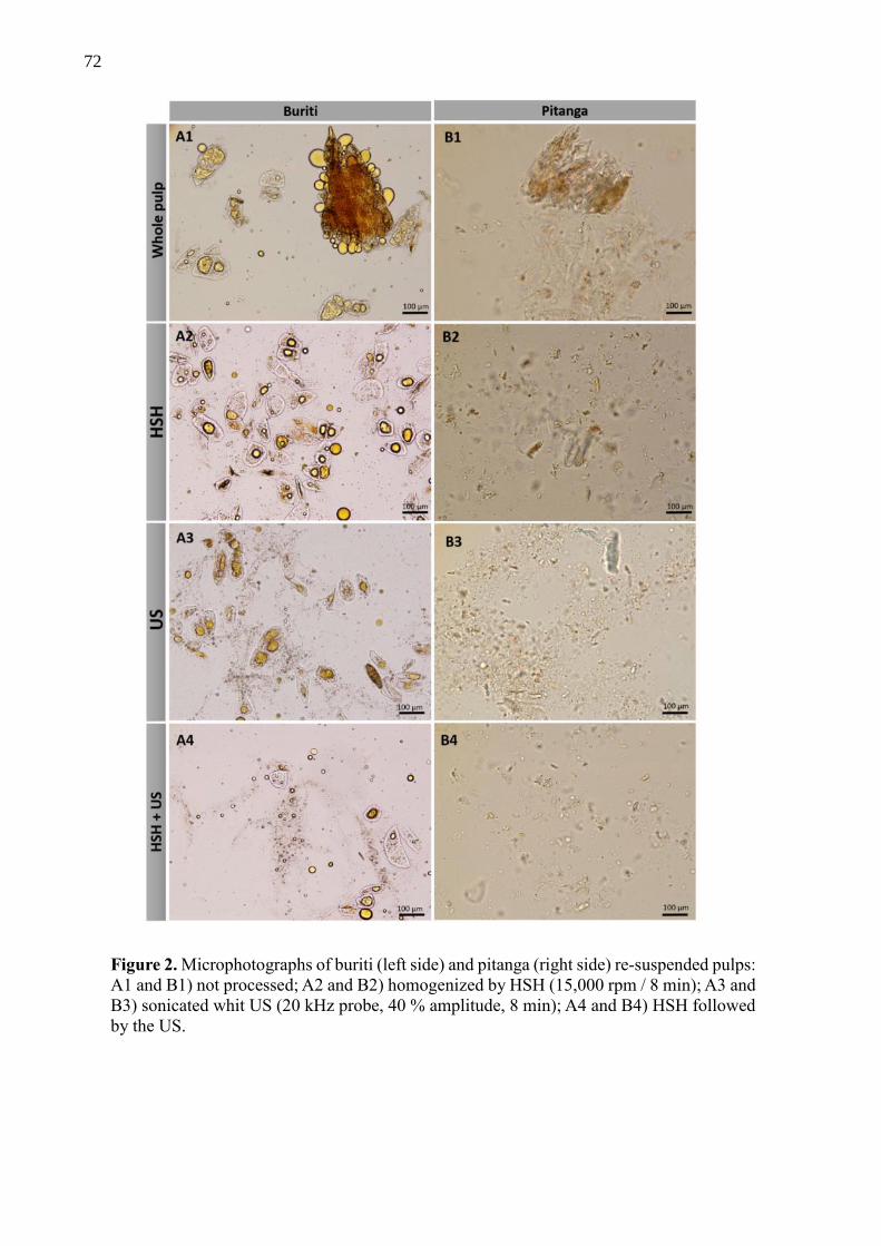
72
Figure 2. Microphotographs of buriti (left side) and pitanga (right side) re-suspended pulps:
A1 and B1) not processed; A2 and B2) homogenized by HSH (15,000 rpm / 8 min); A3 and
B3) sonicated whit US (20 kHz probe, 40 % amplitude, 8 min); A4 and B4) HSH followed
by the US.

73
Table 1 – Effect of processing on tissue fragmentation and carotenoid release1
Processing
HSH – US
Buriti Pitanga
Total
Carotenoids
(µg/mL)
Free
Carotenoids
(µg/mL)
Turbidity
at 540nm
(abs)
Turbidity
at 450nm
(abs)
Total
Carotenoids
(µg/mL)
Free
Carotenoids
(µg/mL)
Turbidity
at 540nm
(abs)
Turbidity
at 450nm
(abs)
Control (0 – 0) 253.7 ±6.6 54.7 ±1.6 1.80 2.35 59.6 ±1.1 15.9 ±0.2 0.64 0.81
10,000 – 0’ 149.7 ±4.6 43.6 ±1.0 2.07 2.79 47.4 ±1.0 18.7 ±0.4 0.93 1.19
15,000 – 0’ 105.9 ±2.0 40.6 ±0.6 2.08 2.82 50.2 ±0.4 21.7 ±0.5 0.82 1.07
20,000 – 0’ 227.2 ±9.7 54.9 ±1.2 2.11 2.89 47.1 ±0.8 22.4 ±0.2 0.98 1.30
10,000 – 4’ 209.4 ±5.6 72.2 ±1.6 2.17 2.94 55.3 ±0.6 21.3 ±0.4 0.98 1.25
15,000 – 4’ 218.3 ±6.1 81.4 ±1.7 2.28 3.09 43.0 ±0.5 20.4 ±0.5 0.99 1.31
15,000 – 4’ 188.5 ±4.2 68.4 ±1.4 2.20 2.99 54.2 ±0.3 21.2 ±1.1 1.03 1.31
15,000 – 4’ 213.7 ±6.9 84.5 ±2.9 2.20 2.98 55.7 ±0.8 21.1 ±1.2 1.09 1.44
20,000 – 4’ 174.1 ±5.0 75.7 ±1.6 2.18 2.98 52.7 ±0.6 21.5 ±0.4 1.03 1.33
10,000 – 8’ 177.3 ±3.8 84.1 ±2.0 2.13 2.93 46.8 ±0.8 21.2 ±0.2 0.97 1.25
15,000 – 8’ 115.8 ±2.3 63.9 ±1.1 2.01 2.69 49.2 ±0.5 17.9 ±0.4 0.94 1.26
20,000 – 8’ 175.4 ±3.4 74.9 ±1.7 2.12 2.88 44.7 ±0.5 23.8 ±0.1 0.89 1.20 1Processing parameters: HSH is on rpm while US intensity was 20 kHz, 40 % amplitude varying time (min). Experiment is central
composite non-factorial surface design with 3 replicates in central point. Total carotenoids were read in triplicate, data is means ±
SD. US processing effects were statistically significative for free total carotenoid (L and Q, p < 0,05) and turbidities (Q, p <
0,05) from buriti. For pitanga only US effect on turbidity was significative (Q, p < 0,05). For all variables processing was
statistically diferent (p < 0,05) from the control samples.
73
74
4.3.3. Whole pulp and microemulsion rheology
Figure 3 shows the flow curves of pitanga and buriti whole pulp, revealing that viscosity
(ƞ) decreases with the increase of shear rate (γ). In all cases, a shear-thinning behavior was
observed that may be regarded as arising from modifications in the macromolecular
organization of the solution as the shear rate changes. Pitanga whole pulp viscosity increased
with the processing treatment (independently of intensity and time applied), indicating a
structural change of samples.
In the case of buriti whole pulp, there are viscosity increments as the intensity of HSH
increases, however when combined with US the increase of viscosity is less pronounced.
Indeed, 20,000 rpm of HSH during 4 min without US processing presented the highest viscosity
for buriti whole pulp. Leverrier et al. (2016) showed that insoluble solids content and
organization in an apple model suspension have a first-order effect on the rheological behavior
of the pulp causing the non-newtonian shear-thinning characteristics observed; particle size and
shape had only a second order effect. Polysaccharides and the aqueous phase or other soluble
particles forms a gel-like network that breaks under high shear rates, and then reorganize when
flow curve cycle slow down (Leverrier, Almeida, Espinosa-Mu noz, & Cuvelier, 2016; Butler
& Snook, 2018).
Effect of surfactant on rheological behavior was observed only in buriti microemulsion,
showing that the fruit matrix determines the thixotropic behavior. The viscosity reduction
intensifies after increasing shear rate – specifically after 10 s-1 for WPI microemulsion –
representing how the microemulsion structures highly influence rheology. In complex systems,
like foods, emulsions can be stabilized by particles and form many structural arrangements
(layers, complexes, aggregates, networks) (Dickinson, 2012). Before initial shearing, the ability
of structural arrangements to start flowing depends on the potential energy barrier that keeps
particles organized. At low shear rates, the structure of the suspension resists deformation until
the destruction of internal networks is completed. In summary, the whole pulps have their
rheology strongly determined by particle interactions, mainly polysaccharides from cell walls
(like pectin and cellulose), while the microemulsions rheological properties are a consequence
of particles interaction with the emulsified oil droplets surface – i.e. of the microemulsions
structural arrangement.

75
Figure 3 – Flow curves of whole pulp (upper charts) and fruit microemulsion (bottom).
Whole pulp labels of treatments are HSH – US (rpm x 1000 – min) while labels of fruit
microemulsions are Tween 80 (T80) or WPI followed by treatment HSH – US (rpm x 1000 –
min).

76
4.3.4. Microstructure and stability of fruit microemulsions
Preliminary tests were performed for fruit microemulsion production in two steps – first
producing stable excipient O/W microemulsions followed by incorporation of pre-fragmented
fruit pulp. However, not all tests ended in homogeneous systems after mixing, some were
inneffective to form the microemulsion. Finally, the one-step direct processing of a mixture of
whole fruit pulp (2 % dry matter) mixed with a surfactant (2 % of Tween 80 or WPI) and corn
oil (5 %) in 91 % water was able to produce homogenous fruit microemulsions (Figure 5).
Experiments showed that the microstructural organization of the final product is a direct effect
of fruit type and surfactant used, while size and homogeneity are mainly influenced by
time/intensity of processing (Figure 4). Also, for control samples, Figure 4 shows that energy
input – mainly due to the US use – reduce oil droplet size from ≈ 0.9-10 µm to almost reaching
the nanoscale ≈ 0.3-1.0 µm. The fruit matrix, buriti or pitanga, interacted differently with
surfactants, forming a network organization where fruit fibers entrapped oily microcapsules. In
microemulsions many surfactants – such as caseinate, whey protein, lecithin or Tween 80 -
interact externally with matrix polysaccharides – like cellulose, pectin and fucoidan - adsorbing
to the surfaces of lipid droplets and/or altering the colloidal interactions between the lipid
droplets, causing many structural and functional effects (Chang & McClements, 2016; Hu et
al. 2016; Zhang et al. 2015).
In the case of pitanga, the final microemulsion presented a more homogenous gel-like
microstructure, while the oily microcapsules entrapped within the cellular fibers net have a
smaller size compared to buriti (Figure 4). Figure 4 also shows that there is a slight influence
of surfactant on the microstructure. WPI seems to have lower and weaker interaction with
fibers, presenting more scattering and homogeneous gel-like structures. Tween 80 presents
bigger agglomerates (and in higher numbers) than WPI, however it seems to let fewer oil
droplets free (Figure 4). For both fruits, the processing intensity only reduced particle size and
increased homogeneity while keeping the main organization (Figure 4).

77
Figure 4 – Brightfield micrographs of: A) control samples - microemulsion composed of water
93 %, corn oil 5 % and Tween 80 2 % (v/v). Magnification lens 60x; B) and C) buriti and
pitanga microemulsion, the composition described in methods. Magnification lens 10x; D) and
E) buriti microemulsion, Tween 80 and WPI as surfactants. Magnification lens 10x; F) and G)
pitanga microemulsion, Tween 80 and WPI as surfactants.

78
The surfactant interaction with fruit fibers showed to be fundamental to the final product
stability (Figure 5). In the first 24 h all fruit microemulsions were stable, but before 7 days
stored at 7-10 ºC, microemulsion instability starts to appear, first in WPI samples. Buriti
products made with WPI separated in two phases for both US (4 min) and HSH (4 min) + US
(8 min) process applied. For pitanga with WPI, all microemulsions lost stability before 7 days
of storage, independently of the process applied. Only Tween 80 microemulsions resisted until
28 days of storage at 7-10 °C, however, after that period instability signs appeared such as
creaming/oily phase separation. The conclusion is that Tween 80-based microemulsions were
more stable, independently of the fruit used. The process that presented the highest stability
after 28 days was HSH (15,000 rpm/4 min) + US (20 kHz, 40 % amplitude/4 min) for both
buriti and pitanga fruits. Instability of O/W emulsions, which includes gravitational separation,
i.e. creaming, flocculation and coalescence, all leading to phase separation (McClements &
Gumus 2016), hinders their use in food products. These problems are naturally occurring in
emulsions due to the energy loss of the systems, however, the presence of polysaccharides can
help stabilizing the whole system or, on the contrary, may induce instability, depending on the
nature of surfactant-polysaccharides interactions (Dickinson, 2013). This is the reason why we
tentatively tried to elucidate the type of interactions between the microemulsions and the fruit
matrix.

79
Figure 5 – Stability of final buriti and pitanga microemulsions stored at
7-10 °C during 28 days. Each photo is set, from left to right – 3 tubes using WPI as surfactant
and 3 tubes with Tween 80 (T80)– as follows: US processing (20 kHz, 40% amplitude/4 min);
HSH (15,000 rpm/4 min) + US (20 kHz, 40% amplitude/4 min); HSH (15,000 rpm/4 min) +
US (20 kHz, 40% amplitude/8 min)
4.3.5. Elucidation of the interaction of microemulsion with the fruit matrix
Since the main objective is to obtain a structure where the naturally occurring
carotenoids are transferred from the plant cells into the oil microcapsules formed, our
hypothesis was that cell wall disruption along with carotenoids lipophilic properties would
promote their encapsulation. Therefore, we used CLSM microscopy to reveal microstructure
and carotenoid location. Carotenoids autofluorescence can be observed by CLSM microscopy
with excitation at 488 nm and an emission filters of 515-590 nm, and micrographs indicate its
exact location inside cellular structures (Kilcrease et al., 2013). The same principle coupled to

80
Raman fluorescence spectroscopy allowed Wan Mohamad et al. (2017) to quantify in situ β-
carotene (detected at 532 nm) inside oil droplets from an O/W microemulsion stabilized by
WPI. In Figure 6 (micrographs A2, B2, C2 and D2), green autofluorescence is a strong evidence
that carotenoids from fruits migrated into oily microcapsules - stained with Nile Red (Figure 6
A3, B3, C3 and D3), confirming our hypothesis that direct processing of pitanga and buriti
microemulsions would encapsulate their natural carotenoids.
Amiri-Rigi and Abasi (2016) were able to increase lycopene extraction from tomato
products residues up to 35 % recovery applying a microemulsion-based system - saponin as
surfactant combined with ultrasound treatment; it is noteworthy that lycopene bioaccessibility
usually is very low, around 0.1-3 %, mainly because of its entrapment inside cellular structures
(Salvia-Trujillo & McClements, 2016). Zhang et al. (2016) successfully used excipient
emulsions to increase β-carotene bioaccessibility and they have shown by CLSM microscopy
that carrot naturally-occurring carotenoids migrated into the oil droplets increasing their
accessibility to the digestive system. The main physic-chemical characteristic of carotenoids is
their lipophilicity due to their structure: a long carbon chain with conjugated double bonds.
Based on our results and related literature, there is enough evidence to show that direct
production of fruit-based microemulsions readily extracts and encapsulates carotenoids from
the fruit matrix.
The observed interaction between surfactant and fruit matrix is the key to the final
product’s microstructure. In our experiments, oil microcapsules and cellulose exhibited
different interactions depending on fruit and surfactant used. Figure 6 shows the
polysaccharides net formed mainly due to cellulose fibers – marked with calcofluor white stain
– and probably pectin that is not observed in the micrographs. The net formed by oil droplets
and the fruit fibers behave as a gel-like structure being able to change the rheology and
properties of the whole system. Buriti present longer and thicker cellulose fibers than pitanga,
clearly organized in blocks of intertwined fibers. Pitanga microstructure is more spread, gel-
like, homogenous and formed with thin cellulose fibers compared to buriti. Micrographs of
buriti microemulsion with Tween 80 present less free oil droplets than the ones stabilized by
WPI. Inversely, pitanga microemulsion made with Tween 80 presents much more free oil
droplets than the WPI-based product.
Zhang et al. (2015) showed that lactoferrin and Tween 80 stabilized-microemulsions did
not interact with low methoxyl pectin at neutral pH, while sodium caseinate-stabilized
microemulsion did interact with pectin at study conditions promoting a flocculation effect.
Since the microemulsions were prepared with whole fruit pulp, they presented the acidity of the

81
original fruit. Thus the pH of pitanga and buriti microemulsions were 1.8 and 3.5, respectively,
measured by pHmeter. The surfactant aggregates with celullose (figure 6) and probably pectin
observed are gel-like structures and have some rheologic effects, like an increase in viscosity
and microemulsion stability. Other works showed microemulsions interactions with
polyshacarides (xanthan gum and locust bean gum) with microscopy photos that have an
organization of the microstructure that is similar to the one we observed in our products
(agregation, flocculation and gel-like structures), and similar rheological effects (Chung,
Degner & McClements, 2013; Wu & McClements 2015). Therefore, we may conclude that the
obtained microstructure is mostly an effect of surfactant-fibers interactions, which have a great
importance in the final functional properties, since carotenoid-rich microcapsules need to be
spread and free to be attacked by digestive enzymes for carotenoid accessibility.

82
Figure 6 – CLSM microscopy of buriti and pitanga microemulsions. Calcofluor white in
the first column marks cellulose fibers. Auto-fluorescence of carotenoids in the second
column (excitation 488 nm and emission filter BA 519–590nm). Nile Red in third column
mark lipid droplets. The last column is an overlap revealing the 3 dimensions
microstructure.

83
4.4. Conclusions
A structure design approach to encapsulate carotenoids from pitanga and buriti was
successfully applied. Both high-speed homogenization and ultrasounds were able to brake
fragments of fruit tissues and disrupt cellular walls releasing carotenoids, mainly when these
processing techniques were used in combination. However, the main results showed that
ultrasound processing has a more pronounced impact on tissue fragmentation, cell disruption
and carotene release than high-speed homogenization and therefore it is an indispensable step
for fruit microemulsion formation. Microscopy clearly shows that most carotenes are entrapped
inside cell walls and must be released for incorporation into lipid micelles. The high-speed
homogenizer and ultrasound treatments released up to 50 % of the initial amount of carotenoids.
After microemulsion formation, the surfactant does not link only to the internal oil and external
water, it also interacts with the carbohydrates from cell walls (mainly cellulose that is in
suspension), forming a gel-like structure – that was demonstrated by CLSM microscopy. Tween
80 and WPI were used as surfactants due to their food grade status and for being widely used
in the food industry. Surfactant interactions with the food matrix depend on the fruit used. In
both cases Tween 80 was more stable over time than WPI, but buriti rheology was much more
influenced by surfactant and processing than pitanga. Tween 80-stabilized microemulsions
present softer viscosity than WPI-based microemulsions for both fruits. The gel-like structure
formed is the main factor for final product stability and rheology, and may modulate carotenoid
encapsulation and release during digestion, thus influencing bioaccessibility. The obtained
buriti and pitanga microemulsions have a high potential for the development of new products
with more bioaccessible β-carotene and lycopene.
Acknowledgements
This work was supported by the São Paulo Research Foundation – FAPESP through
research funding [grant #2015/15507-9] and PhD scholarship for Paulo Berni [grant
#2014/15119-6] and a Research Internships Abroad (BEPE) support [grant #2016/13355-0] The
author Ana C. Pinheiro is recipient of a fellowship from the Portuguese Foundation for Science
and Technology (FCT) [grant SFRH/BPD/101181/2014]. Diana Vilas Boas was supported by
FCT under the scope of the strategic funding of UID/BIO/04469/2013 unit and COMPETE
2020 [POCI-01-0145-FEDER-006684]. Paula Pereira was supported by BioTecNorte operation

84
(NORTE-01-0145-FEDER-000004) funded by the European Regional Development Fund
under the scope of Norte2020.
Conflicts of interest: none
4.5. References
Amiri-Rigi, A., & Abbasi, S. (2016). Microemulsion-based lycopene extraction: Effect of
surfactants, co-surfactants and pretreatments. Food Chemistry, 197, 1002–1007.
Amorim-Carrilho, K. T., Cepeda, A., Fente, C., & Regal, P. (2014). Review of methods for
analysis of carotenoids. Trends in Analytical Chemistry, 56, 49–73.
Anese, M., Bot, F., Panozzo, A., Mirolo, G., & Lippe, G. (2015). Effect of ultrasound treatment,
oil addition and storage time on lycopene stability and in vitro bioaccessibility of tomato
pulp. Food Chemistry, 172, 685-691.
Buggenhout, S. V., Ahrne, L., Alminger, M., Andrys, A., Benjamin, M., Bialek, L., Cleaver, G.,
Colle, I., Langton, M., Larque, E., Lemmens, L., Lofgren, A., Lopez-Sanchez, P., Perez-
Llamas, F., Martınez-Tomas, R., Robertson, J., Schalow, S., Svelander, C., Wellner, N.,
Hendrickx, M., & Waldron, K. (2012). Structural design of natural plant-based foods to
promote nutritional quality. Trends in Food Science & Technology, 24, 47-59.
Butler, J. E., & Snook, B. (2018). Microstructural Dynamics and Rheology of Suspensions of
Rigid Fibers. Annual Review of Fluid Mechanics 50 (1), 299-318.
Cervantes-Paz, B., Ornelas-Paz, J. J., Pérez-Martínez, J. D., Reyes-Hernández, J., Zamudio-
Flores, P. B., Rios-Velasco, C., Ibarra-Junquera, V., & Ruiz-Cruz, S. (2016). Effect of
pectin concentration and properties on digestive events involved on micellarization of
free and esterified carotenoids. Food Hydrocolloids, 60, Supplement C, 580-588.
Chang, Y., & McClements, D. J. (2016). Influence of emulsifier type on the in vitro digestion
of fish oil-in-water emulsions in the presence of an anionic marine polysaccharide
(fucoidan): Caseinate, whey protein, lecithin, or Tween 80. Food Hydrocolloids, 61, 92-
101.
Chung, C., Degner, B., & McClements, D. J. (2013). Physicochemical characteristics of mixed
colloidal dispersions: Models for foods containing fat and starch. Food Hydrocolloids,
30, 1, 281-291
de Rosso, V. V., & Mercadante, A. Z. (2007). Identification and quantification of carotenoids,
by HPLC-PDA-MS/MS, from Amazonian fruits. Journal of Agricultural and Food
Chemistry, 55, 5062-5072.

85
Dickinson, E. (2013). Stabilizing emulsion-based colloidal structures with mixed food
ingredients. Journal of the Science of the Food and Agriculture, 93, 710–721.
Failla, M. L., & Chitchumroonchokchai, C. (2005). In vitro models as tools for screening the
relative bioavailabilities of provitamin A carotenoids in foods. Harvestplus, Technical
Monograph 3, 32 p.
Failla, M. L., Chitchumroonchokchai, C., Ferruzzi, M. L., Goltz, S. R., & Campbell, W. (2014).
Unsaturated fatty acids promote bioaccessibility and basolateral secretion of carotenoids
and α-tocopherol by Caco-2 cells. Food and Function, 5(6), 1101- 1112.
Fernández-García, E., Carvajal-Lérida, I., Jarén-Galán, M., Garrido-Fernández, J., Pérez-
Gálvez, A., & Hornero-Méndez, D. (2012). Carotenoids bioavailability from foods: From
plant pigments to efficient biological activities. Food Research International, 46(2), 438-
450.
Hu, H., Xing, L., Hu, Y, Qiao, C., Wu, T, Zhou, G., & Zhang, W. (2016). Effects of regenerated
cellulose on oil-in-water emulsions stabilized by sodium caseinate. Food Hydrocolloids,
52, 38-46.
Kilcrease, J., Collins, A. M., Richins, R. D., Timlin, J. A., & O'Connell, M. A. (2013). Multiple
microscopic approaches demonstrate linkage between chromoplast architecture and
carotenoid composition in diverse capsicum annuum fruit. Plant Journal, 76(6), 1074-
1083. https://doi.org/10.1111/tpj.1235
Lemmens, L., Colle, I., Buggenhout, S. V., Palmero, P., Loey, A. V., & Hendrickx. M. (2014).
Carotenoid bioaccessibility in fruit- and vegetable based food products as affected by
product (micro)structural characteristics and the presence of lipids: A review. Trends in
Food Science & Technology, 38, 125-135. https://doi.org/10.1016/j.tifs.2014.05.005
Leverrier, C., Almeida, G., Espinosa-Mu noz, L., & Cuvelier, G. (2016). Influence of particle
size and concentration on rheological behavior of reconstituted apple purees. Food
Biophysics, 11(3), 235-247. https://doi.org/10.1007/s11483-016-9434-7
Li, Q., Li, T., Liu, C., Dai, T., Zhang, R., Zhang, Z., & McClements, D. J. (2017). Enhancement
of carotenoid bioaccessibility from tomatoes using excipient emulsions: influence of
particle size. Food Biophysics, 12(2), 172-185. https://doi.org/10.1007/s11483-017-9474-
7
McClements, D. J., & Xiao, H. (2014). Excipient foods: designing food matrices that improve
the oral bioavailability of pharmaceuticals and nutraceuticals. Food and Function, 5,
1320-1333. https://doi.org/10.1039/c4fo00100a
86
McClements, D. J., Decker, E. A., & Weiss, J. (2007). Emulsion-based delivery systems for
lipophilic bioactive components. Journal of Food Science, 72(8), R109-R124.
https://doi.org/10.1111/j.1750-3841.2007.00507.x
Porcu, M. O., & Rodriguez-Amaya, B. D. (2008). Variation in the Carotenoid Composition of
the Lycopene-Rich Brazilian Fruit Eugenia uniflora L. Plant Foods Human Nutrition, 63,
195-199
Rodriguez-Amaya, D. B., Kimura, M., Godoy, H. T., & Amaya-Farfan, J. (2008). Updated
brazilian database on food carotenoids: Factors affecting carotenoid composition. Journal
of Food Composition and Analysis, 21(6), 445-463.
http://dx.doi.org/10.1016/j.jfca.2008.04.001
Saini, R. K., & Keum, Y. S. (2018) Carotenoid extraction methods: A review of recent
developments. Food Chemistry, 240, 90-103.
https://doi.org/10.1016/j.foodchem.2017.07.099
Saini, R. K.; Nile, S. H., & Park, S. W. (2015). Carotenoids from fruits and vegetables:
Chemistry, analysis, occurrence, bioavailability and biological activities. Food Research
international 76, 735-750.
Salvia-Trujillo, L., & McClements, D. J. (2016) Enhancement of lycopene bioaccessibility from
tomato juice using excipient emulsions: Influence of lipid droplet size. Food Chemistry
210 295–304. https://doi.org/10.1016/j.foodchem.2016.04.125
Schweiggert, R. M., Mezger, D., Schimpf, F., Steingass, C. B., & Carle, R. (2012). Influence of
chromoplast morphology on carotenoid bioaccessibility of carrot, mango, papaya, and
tomato. Food Chemistry, 135, 2736–2742.
Sensoy, I. (2014). A review on the relationship between food structure, processing, and
bioavailability. Critical Reviews in Food Science and Nutrition, 54, 902–909.
Wan Mohamad, W. A. F., Buckow, R., Augustin, M., & McNaughton, D. (2017). In situ
quantification of b-carotene partitioning in oil-in-water emulsions by confocal Raman
microscopy. Food Chemistry, 233, 197–203.
http://dx.doi.org/10.1016/j.foodchem.2017.04.086
Wu, B., & McClements, D. J. (2015) Design of reduced-fat food emulsions: Manipulating
microstructure and rheology through controlled aggregation of colloidal particles and
biopolymers. Food Research International, 76, 3, 777-786

87
Zhang, R., Zhang, Z., Zou, L., Xiao, H., Zhang, G., Decker, E. A. & McClements, D. J. (2015).
Enhancing Nutraceutical Bioavailability from Raw and Cooked Vegetables Using
Excipient Emulsions: Influence of Lipid Type on Carotenoid Bioaccessibility from
Carrots. Journal of Agricultural and Food Chemistry, 63 (48), 10508–10517.
Zhang, R., Zhang, Z., Zou, L., Xiao, H., Zhang, G., Decker, A. E., & McClements, D. J.
(2016) Enhancement of carotenoid bioaccessibility from carrots using excipient
emulsions: influence of particle size of digestible lipid droplets. Food Function, 7, 1, 93-
103.

88

89
5 CAROTENOID BEHAVIOR OF PITANGA (EUGENIA UNIFLORA) AND BURITI
(MAURITIA FLEXUOSA) DURING MICROEMULSION PRODUCTION AND IN
A DYNAMIC GASTROINTESTINAL SYSTEM
Chapter submitted to Journal of Food Science and Techonology
Abstract
Uncommon tropical fruits are emerging as raw-material for new food products with
health benefits. This work aimed to formulate and process microemulsions from pitanga
(Eugenia uniflora) and buriti (Mauritia flexuosa) fruits. Since these fruits are very rich in
carotenoids, it was sought to release, encapsulate and protect their carotenoids along with
increasing bioaccessibility, specifically of lycopene and β-carotene. Pitanga and buriti
microemulsion were produced by applying a direct processing (high speed homogenization –
HSH at 15000 rpm and Ultrasound – US with 20 kHz probe at 40 % amplitude) of the whole
pulp together with surfactant (T80 or WPI at 2%) and corn oil (5%). All treatments (HSH – US
for 0-4, 4-0, 4-4, 4-8 min-min) applied were able to increase the amount of carotenoid released.
However, the processing also decreases total amount of carotenoids in whole pulp of studied
fruits. The significant impact of processing during microemulsion production was not severe.
The overall data suggest that surfactant and oil presence during processing may protect the
carotenoids in fruits and microemulsions. Final recovery of total carotenoids, after passing the
samples through a dynamic gastrointestinal system, was higher for microemulsions than for
whole pulps. Results of total carotenoids, β-carotene and lycopene indicate that high losses
happened during jejunum and ileum phases. The present paper confirms that is possible to
increase β-carotene and lycopene bioaccessibility from fruits by direct processing
microemulsions (p<0.01). The microemulsion processing and formulation chosen was able to
improve final bioaccessibility of total carotenoids from buriti, β-carotene from pitanga and
buriti, and of lycopene from pitanga.
Keywords: Dynamic digestive system; Tropical fruits; Bioavailability; Beta-carotene;
Lycopene; Structure design

90
Graphical Abstract
Highlights
- Ultrasound is effective for carotenoid release from pitanga and buriti matrices, however
stimulate its degradation
- Surfactant protected the carotenoids against processing degradation
- Carotenoids from pitanga and buriti were efficiently encapsulated, ranging 25 – 80 %
- Microemulsion protected carotenoids against degradation and losses during digestion by
a dynamic gastrointestinal system
- Microemulsion increase total carotenoids, β-carotene and lycopene bioaccessibility up to
4 times
5.1. Introduction
There is a growing demand for plant based products that promote health and well-being.
Thereby, fruits represents the most popular option for new products due to their ‘naturally
healthy’ image and their bioactive compounds composition (e. g. carotenoids). Several fruits
are being used by functional foods industry for developing new healthy products, like
smoothies, yogurts, beverages, ice-creams and even as natural colorants. This can incorporate
new flavors as well functionalities to diet (Sun-Waterhouse 2011, Khan et al. 2013).
Carotenoids are present in many common foods and its ingestion can provide protection for the
91
eyes, improvement in vision health, promotion of the immune systems, reduction of the overall
risk of chronic diseases and avoid abortion (Saini et al. 2015).
In this sense, the biodiversity of fruits native from Brazil can be rich source of
innovation and opportunities, growth of income, value adding and scientific contribution.
Pitanga (Eugenia uniflora) belongs to the botanic family of Myrtaceae, grows on tropical and
subtropical regions, and it is originally from Brazilian restinga. The fruit is very valued due to
its strong red color, juicy pulp, unique aroma, sour and sweet taste. Northeast region of Brazil
have the largest production, being that only Pernambuco state produces annually up to 1.700
tons/year, that is basically commercialized in natura or processed as fruit pulp. Pitanga has a
very high concentration of antioxidant compounds like flavonoids, vitamin C and carotenoids
– mainly lycopene in concentration levels around 70 µg/g of fresh pulp (Negri et al. 2016; Porcu
and Rodriguez-Amaya 2008). Buriti is a palm fruit from Arecaceae family, it naturally grows
on amazônia, caatinga and cerrado biomes. Buriti fruit is very rich in lipids, around 20% of
fresh pulp, that are extracted and commercialized as crude or refined oil. Usually buriti oil is
consumed in foods or applied in cosmetics due to healing properties of the high concentration
of carotenoids, tocopherol and fat acids. Probably buriti is the plant with the highest content of
all-trans-β-carotene in the nature, reaching levels of 373 µg/g of whole pulp (de Rosso and
Mercadante 2007; Negri et al. 2016)
The preserving techniques, processing, storage and final product matrix/formulation
have great impact on stability and bioaccessibility of carotenoids, and consequently on the
intended health benefits (Dias et al. 2014; Failla et al. 2014). Carotenoid bioaccessibility is
defined as the portion that are ready for being absorbed by intestinal cells after food digestion,
i. e. carotenoids incorporated into water soluble mixed micelles produced by bile salts and lipids
(Failla and Chitchumroonchokchai 2005). Food matrix factors that are determinant for
bioaccessibility are: intracellular form of carotenoid (e.g. crystalline or micellar); kind of fiber
present (soluble or insoluble); amount and kind of lipids present (e.g. unsaturated long chain
fat acids increase bioaccessibility) (Failla and Chitchumroonchokchai 2005; Saini et al. 2015).
The presence, amount and type of lipids is crucial. Also, interactions of components originally
present in the meal or produced inside the digestive system still can influence bioaccessibility
– e.g. fibers, carotenoid x carotenoid interactions, kind of lipids ingested with the carotenoid
source (Sensoy 2014; Saini et al. 2015; Fernández-García et al. 2012).
Once carotenoids are released from plant cells they are subject to structural
modifications and degradation that can impair bioaccessibility (Yonekura and Nagao 2007).
92
Thus, every event after cells fragmentation due to processing, mastication or stomach digestion
will influence final bioaccessibility in different ways. Therefore, processing, implies changes
in the food matrix that causes two main effects: carotenoids can degrade losing their health
effect, but, in the other hand, bioaccessibility can be improved/impaired due to structural
changes. Indeed, losses of bioactive compounds and/or increase in bioaccessibility due to
processing are oftentimes reported (Pugliese et al. 2013; Dias et al. 2014; Failla et al. 2014;
Anese et al. 2015; Berni et al. 2015).
Generally, industrial processing of plant based products do not take into account the
retention and bioaccessibility of bioactive compounds. The standard processing of soups and
sauces for example treat all ingredients at same way, applying high temperature for long time
that results in decrease of nutritional, functional and sensorial quality of final products
(Buggenhout et al. 2012). Carotenoid bioaccessibility in foods is very low, but it is possible to
strategically formulate and process them in order to increase their release, bioaccessibility and
consequently their health benefits (Buggenthout et al. 2012). For instance, bioaccessibility of
β-carotene, lutein and lycopene were augmented due to cooking process as frying (Berni et al.
2014), microemulsion addition (Li et al. 2017) or optimal combination of adequate
process/formulation (Buggenhout et al. 2012), respectively. However, still exist many gaps to
be filled about this topic that represents a very promising field for research and food innovation.
Many bioactive compounds are highly lipophilic, such as carotenoids, which causes
many hindrance to incorporate these molecules in new functional foods, primarily low
solubility in water and chemical instability (Recharla et al. 2017). Several techniques combinate
structure design approaches and/or delivery systems engineering. They aims to protect,
preserve, enhance solubility, increase bioaccessibility and bioactivity of lipophilic bioactive
compounds. The most studied and applied strategy is the encapsulation of lipophilic bioactive
compounds in emulsion based systems at micro and nano scales (Recharla et al. 2017;
McClements 2015). Successful encapsulation of purified carotenoids in micro and
nanoemulsions that increase their solubility in water, stability and bioaccessibility have been
done by many research groups (Gomes et al. 2017; Davidov-Pardo et al. 2016; Gul et al. 2015;
Zhang et al. 2015; de Paz et al. 2013; Hejri et al. 2013).
Therefore, our approach attempt to manipulate structure and composition of fruit matrix
in order to encapsulate carotenoids leading to higher retention and bioaccessibility of
carotenoids. We produced a microemulsion-based product by directly processing fruit pulps of
pitanga and buriti together with a surfactant that stabilized the whole system. Our hypothesis
was that combined process and formulation would transfer the carotenoids from the matrix to

93
lipid microcapsules while protecting their carotenoids and increasing their bioaccessibility. The
present chapter focus on carotenoid contents, retention, efficiency of encapsulation and
bioaccessibility throughout a dynamic digestive system. Data also show the effect of the
surfactants Tween 80 (T80) or Whey protein isolate (WPI), and processing by high-speed
homogenization (HSH) and ultrasound (US) on carotenoid behavior over emulsion formation.
5.2. Material and Methods
5.2.1. Pitanga and buriti fruit pulp
Buriti pulp was commercialized by the co-op network Central do Cerrado, Brasilia,
Distrito Federal, Brazil. Fruits were soaked in clean water, 3 parts of water to 1 part of fruit
(v/v), during 12 hours and pulp was taken off by a crushing machine for pulps (Bonina®, 0,5
mm pore size). Pitanga pulp was acquired from the tropical fruits company Sítio do Bello,
Paraibuna, São Paulo, Brazil. Red ripe pitanga was harvested, selected, sanitized and pulped at
Sitio do Bello using a crushing machine for pulps. There was no water added and seeds were
separated during the processing of pitanga. Both fruits pulps were packed in sealed bags, frozen
at -26 °C and sent in thermal box to Piracicaba, São Paulo, Brazil, and immediately stored in -
80 °C freezer. Pulps were freeze dried at -76°C during 3 days and packed in vacuum-sealed
bags and then shipped by plane to Braga, Portugal, where they were stored frozen at -26 °C
until the time of conducting the experiments. Dried samples of lyophilized pitanga and buriti
pulp were re-suspended in distilled water until the final water content of 93 % of wet weight.
After this equalization, final natural occurring lipids contents in whole pulp were 0.1 % and
3.64 % respectively for pitanga and buriti.
5.2.2. Reagents
Commercial standards of β-carotene, lycopene and enzymes for the dynamic in vitro
digestion were purchased from Sigma-Aldrich (St Louis, MO, USA). Specific reference
numbers of each enzymes are informed in the correspondent methods. Organic solvents for
extraction and HPLC analysis were chromatographic grade (Chromasolv™, Muskegon, MI,
USA). Surfactant Tween80 (T80) was purchased from Panreac AppliChem (Germany) and
Whey Protein Isolate (WPI) from Arla Foods (Denmark). Corn oil (Fula®, Sovena, Portugal )
was used without further purification.
5.2.3. Microscopy

94
Brightfield images were analyzed using an epifluorescence microscope (Olympus
BX51) coupled with a DP71 digital camera and three sets of filters (DAPI – 360-370/420; FITC
– 470-490/520; and TRITC – 530-550/590) (Olympus Portugal SA, Porto, Portugal). All
images were acquired using the Olympus cellSens soſtware. For better observation aliquots
were diluted 1:10 (v/v) in distilled water and 5 – 20 µL of each sample was used under the
lamina coverslip. Photographs of at least 3 different areas from every replicate were taken.
Fluorescence images were acquired using a Confocal Scanning Laser Microscope
(CLSM) (Olympus BX61, Model FluoView 1000). Calcofluor white (Sigma-Aldrich, EUA)
was used for detection of cellulose fibers in microemulsions (laser excitation line 405 nm and
emission filters BA 430–470, blue channel); for detection of encapsulated carotenoids laser
excitation line 488 nm and emissions filters BA 519–590, green channel, were used in
agreement to Kilcrease et al. (2013). Nile Red (TCI, Tokyo, Japan) was used for detection of
lipids droplets location and size (laser excitation line 559 nm and emissions filters BA 575–
675, red channel). Images were acquired with the program FV10-Ver4.1.1.5 (Olympus).
5.2.4. Microemulsion formulation and processing
First step of producing the microemulsion is to release the carotenoids from fruit
chromoplasts. For this purpose, samples were submitted to processing by a high-speed
homogenization (HSH) equipment (Ultra-Turrax® homogenizer, T 25, Ika-Werke, Germany)
at 15000 rpm during 0 min and 4 min combined with ultrasound (US) treatment with a 20 kHz
probe (Vibra-Cell™, Sonics®, EUA), 40 % amplitude during 0, 4 and 8 min. The samples were
processed in 50 mL centrifuge tubes and kept inside ice-bath all the time to avoid overheating
and carotenoid degradation. To assess treatment impact, total carotenoids, as well β-carotene
and lycopene by HPLC-DAD were analyzed in whole, processed, and in filtered pulp after
processing (stainless steel sieves, particle size < 100 µm).
Microemulsion formulation was based on the work of Zhang et al. (2015) and Salvia-
Trujillo and McClements (2016). Only food grade ingredients were used in formulations of the
fruit microemulsions. Oil-in-water (O/W) microemulsions were produced by processing
together fruit pulp, surfactant (T80 or WPI), corn oil and water at equal proportions for both
fruits. Briefly, microemulsions were made by preparing surfactant solution (T80 or WPI) in
distilled water, and then mixing with whole pulp and corn oil. Final microemulsion product was
composed by 5 % corn oil, 91 % distilled water, 2 % of surfactant and 2 % of pulp (in dry basis).
Processing parameters for microemulsion and the selected one for dynamic gastrointestinal
system study were chosen based on previous data of total carotenoids, rheologic behavior, and

95
on microemulsion stability during storage, all results that compose another manuscript of our
work (unpublished). Mixtures were first processed by HSH (varying 15000 rpm during 4 min)
and immediately taken to US sonication (20 kHz probe, 40 % amplitude, varying time). Both
probes for HSH and US processing were placed at sample geometric center. Samples were kept
all the time in ice inside a Styrofoam box to avoid heat and light.
5.2.5. Carotenoid extraction for analysis
For carotenoid assessment aliquots were took and had their carotenoid extracted and
quantified using an small scale method based on Amorim-Carrilho et al. (2014) review,
described concisely as follow. Aliquots (50 – 300 µl) were vigorously mixed in vortex for 1
min with 1.5 mL of Acetone/Ethanol/Hexane (50/25/25 v/v/v) solution containing 0.01% of
BHT. Distilled water was added until 2 mL of final volume and centrifuged for 3 min at 1000
rpm. This provoke phase separation and carotenoid drives to upper phase. Carotenoid rich
extracted were collected, then 1.5 mL of distilled water was added carefully. In case of forming
an emulsion a pinch of sodium chloride (≈ 20 mg) was added and the upper phase is collected
again. Sodium sulfate anhydrous (≈ 20 mg) was added to final extracts for binding water
residues. For extraction of carotenoids from jejunum and ileum filtrates obtained after dynamic
simulation of digestion, as well the undigested residues, the same procedures were used with
the adjustment of volumes and keeping the same sample/solvent ratio. Extracts were collected
and then dried under nitrogen flow, resuspended in petroleum ether (for spectrophotometric
measurement of total carotenoids) or in methanol/MTBE (50/50 v/v) for HPLC-DAD analysis
of β-carotene and lycopene.
To evaluate retention and release of carotenoids due to processing, samples were
analyzed: initially, i.e. the control sample; in the HSH and US processed fruit pulp; and after
filtering throughout 100 µm cutoff sieve, that estimates free carotenoids. Microemulsions were
analyzed freshly and in the upper phase after centrifuging for 5 min at 8385 g, that allowed to
estimate efficiency of encapsulation, since unencapsulated carotenoids are attached to cell
fibers in pellets. Each step of dynamic digestion simulation – gastric, duodenum, jejunum
filtrate, ileum filtrate and final unfiltered residue – had the carotenoids contents measured to
evaluate stability of carotenoids to digestion and final bioaccessibility.
96
5.2.6. Carotenoid analysis by spectrophotometer and HPLC-DAD
Total carotenoids - In a 96 well microplate reader (Synergy™ HT, Bio-tek®) 300 µL of
extracts, as well β-carotene standard curves, had absorbance measured at 450 nm. Two curves
were prepared, where first ranged 0.18 - 1.42 µg/mL of β-carotene in 8 points and second ranged
2.7 – 6.3 µg/mL of β-carotene in 5 points, respectively R² was 0,9849 and 0,9635, and were
used for whole pulps, < 100 µm filtered samples, microemulsion ready and after centrifugation.
β-carotene and lycopene - Chromatographic analysis of carotenoid is presented in
Kimura et al. (2007). The HPLC-DAD system used was a Shimadzu® Nexera X2 (modules:
degasser DGU 20A 5R, pump LC-30AD, autosampler SIL-30AC, oven CTO-20AC and
detector DAD SPD-M20A) with a polymeric YMC™ C30 (150 mm x 4.6 mm, 5 µm particle
size). The mobile phase was methanol and MTBE at 90:10 (Methanol:MTBE, v/v) in linear
gradient to 40:60 in 60 min, returning to initial conditions and kept for 15 min for re-
equilibration; the oven temperature was 30º C while autosampler chamber was kept in 8º C;
flow was 0.8 mL / min and injection volume changed according to appearance of carotenoid
concentration ranging 10 µL – 40 µL. Identification of β-carotene and lycopene was based on
commercial standards, and diode array absorption spectra. Chromatograms where taken at fixed
wavelength of 450 nm for β-carotene and 470 nm for lycopene integration and calculation.
Standard curves of β-carotene (from 0.9 µg/mL to 4.5 µg/mL) and lycopene (from 1.09 µg/mL
to 2.74 µg/mL) were performed, and the respective r2 values for the standard curves were
0.9988 and 0.9918.
5.2.7. Dynamic gastrointestinal system
A dynamic gastrointestinal system equipment and protocol, described in Pinheiro et al.
(2016), was used to evaluate carotenoids stability to digestion and bioaccessibility comparing
in natura samples and microemulsions. In summary, this model simulates the main events that
occur during digestion – i.e. simulation of stomach, duodenum, jejunum and ileum digestions
by separated reactors. The temperature is maintained to 37 °C and physiological pH of each
step is controlled by addition of HCl or NaHCO3. The peristaltic movements are mimicked with
water jacket. The compartments are connected by silicone tubes and the flow throughout the
system is controlled manually using 10 mL plastic syringes. The micellar phase is separated by
filtration through a hollow-fibre device (SpectrumLabs Minikros®, M20S-100-01P, USA)
placed after jejunum reactor and another one after the ileum reactor. The micellar phase is
collected directly in a ellenmeyer setted in ice to stop further reactions. At this filtrate are the
mixed micelles containing the bioaccessibile carotenoids.

97
The system runs with initial 40 mL of samples, and the experiment was done in
triplicate. Gastric and intestinal secretions were freshly prepared and secreted inside the reactors
by syringe pumps at pre-set flow rates. Gastric enzyme solution was made by adding 0.0176 g
of pepsin (3409 U), 0.0741 g of lipase (54 U) in 100 mL of gastric salts solution (NaCl 5 g/L,
KCl 0.6 g/L and CaCl2 0.25 g/L), pH adjusted to 1.5 with HCl 1M, and flow rate was 0.33
mL/min. Duodenum secretion was made by combining 34 mL of pancreatin solution (2.8 g in
40 mL distilled water), 20 mL of small intestine salts solution (NaCl 4.8 g/L, KCl 2.2 g/L,
CaCl2 0.22 g/L and NaHCO3 1.5 g/L) and 66 mL of bile solution (4 g/100 mL of distilled water),
final pH adjusted to 7.5 with NaHCO3 1M, flow rate 0.6 mL/min. Jejunum secretion was
prepared with 40 mL of the bile solution diluted 1:10 with small intestine salts solution, flow
rate was 2.13 mL/min. Finally, in the ileum was secreted only the small intestine salts solutions
at 2 mL/min flow. The system runs 5h of each digestion. At 90 min is collected an aliquot of
the stomach, and at 120 min is collected an aliquot of the duodenum. The aliquots helps to
follow the stability to digestion. The ratio between initial amount of carotenoids and the
amounts in the micellar phases indicates the bioaccessibilities (jejunal, ileal and total). The
residue also is collected and analysed composing the results of carotenoid recovery.
5.2.8. ζ-potential
The ζ-potential of the particles in the samples as they passed through the various stages
of the dynamic gastrointestinal system were measured according to Pinheiro et al. (2013) using
a Malvern Zetasizer Nano ZS instrument (Malvern Instruments Ltd., UK). The measurement
was performed at 25 ºC assuming the Smoluchowski model and using a disposable folded
capillary cell (DTS1070), as also an automatic voltage. Zeta analysis was carried out by
zetasizer software through an Auto Mode - General Purpose model. Initial samples were diluted
1:1000 (v/v), while chyme and digesta were diluted 1:10 (v/v) in appropriate buffer solution
(pH 2 for stomach sample and pH 7 for initial and small intestine samples) at room temperature.
Each individual ζ-potential measurement was determined in triplicate.
5.2.9. Statistics
Experiment data was analyzed for significant differences using ANOVA, means were
compared using the Tukey test and significant at 95 % reliability (p < 0.05). Software used for
the surface design, ANOVA and Tukey tests was the Statistica 13© (Dell Inc.).

98
5.3. Results and discussion
5.3.1. Carotenoid release and impact of processing
For the purpose of producing a microemulsion while transferring the carotenoids from
pitanga and buriti to the oil droplets, the main obstacle is to release the carotenoids as much as
possible. An experiment was carried out to test carotenoid release and stablish the parameters
for the microemulsion production. Results of total carotenoid release and retention after
processing are presented in Figure 1. The observed behavior is that all treatments applied are
able to increase the ratio of initial carotenoid/free carotenoid, i.e. release. However, at the same
time processing decreases the total amount of carotenoids from studied fruits. All treatments
tested, especially when combined HSH-US, was successfully capable to break cells and release
its intracellular content as it can be seen by empty cells and small fragments observed in Figure
2. After testing different treatments it was choose to discard the HSH without US (T-U 4’-0’)
process because the results of carotenoids release are unexpressive, and the microemulsion
formation in further tests was not homogeneous. The US treatment at 24 kHz was able to
fragment plant tissue, break the cellular wall and release lycopene crystalloids from tomatoes –
demonstrated by light microscopy images very similar to ours – with slight decrease of lycopene
concentration (Anese et al. 2015)

99
Figure 1 – Evaluation of carotenoid release and impact of processing pitanga and buriti whole
pulps by HSH and US. Results are total carotenoids determined spectrophotometrically.
Acronyms meaning treatments HSH-US (T-U) and time of processing (min-min). Different
letters for the same series represents significant differences between treatments (p < 0.05).
Carotenoids are found in plants, and are stored inside chromoplasts that varies in shape
and size related to their carotenoid composition (Kilcrease et al. 2013; Saini et al. 2015). The
carotenoids are stored in two main forms, lipid-dissolved or liquid-crystalline, that have
influence on bioaccessibility due to their readiness to being digested and micellized during
human digestions (Schweiggert et al. 2012). Images form light microscopy on Figure 2 show
these differences in buriti (lipid-dissolved) and in pitanga (liquid-crystalline), as well the
carotenoid entrapment inside cellular structures. In some fruits like mango and papaya, β-

100
carotene and xanthophylls are in the both states, lipid-dissolved and liquid-crystalline, together
with crystalloid lycopene (not easily seen by light microscopy), all inside of globular
chromoplasts (Schweiggert et al. 2012). This seems to be the case of pitanga that is rich in
lycopene, β-carotene and xanthophylls (Porcu and Rodriguez-Amaya 2008). The entrapment of
the carotenoids is numerically perceived in Figure 1 by the data of carotenoid release from
control samples. Pitanga whole pulp has around 25 % of its carotenoid released from cellular
walls while buriti has only 15%. In the whole pulp, the free carotenoids exist due to pulp’s
taking of, and are dispersed in the aqueous fraction inside lipid droplets or as crystalloids.
Carotenoid retention are strongly affected by kind of processing, temperature, time of
exposure and intensity of the energy applied (Dias et al. 2014; Berni et al. 2015; Frias et al.
2010). The processing goal was to get the higher content of free carotenoids. On this way, only
HSH (15000 rpm, 4 min) followed by US (20 kHz, 40% amplitude, 8 min) in pitanga presented
higher quantity of free carotenoids compared to control (Figure 1). Nevertheless, the extent of
this increment is low (14 µg/mL) despite statistically significant. The food matrix have a
protection rule on retention, but once carotenoids are released they are more subject of
degradation and environmental stresses. Carail et al. (2015) investigated the kinetics of all-
trans-β-carotene degradation due to US processing (20 kHz, same used in our work), they
demonstrated that degradation is high in presence of water and oxygen, and have more influence
of time of exposure than US intensity.

101
Figure 2 – Microemulsion formation: carotenoid location in cell; carotenoid release; formed
microemulsion (T80, 4 min of HSH and 4 min of US); microemulsion (Nile red stained and
carotenoid autofluorescence) interaction with fruit cellulose (calcofluor white stained).

102
5.3.2. Microemulsion development
To produce the microemulsion it was applied a direct processing of the whole pulp
together with surfactant (T80 or WPI at 2% of final weight) and corn oil (5% of final weight).
T80 and WPI surfactants were already used to produce excipient emulsions using olive oil or
corn oil that successfully increased bioaccessibility of lycopene and β-carotene from tomato
and carrots (Salvia-Trujillo and McClements 2016; Li et al. 2017; Zhang et al. 2016). Figure 2
shows the oily microcapsules formed due to surfactant action that stabilize the system. Many
works have been demonstrated the efficacy of US to break cell walls and improve carotenoid
extraction to oil. For example, Goula et al. (2017) were able to extract up to 93.8% of total
carotenoids from pomegranate peel using US assisted extraction with sunflower oil as
extracting solvent, i. e. breaking cellular walls and transferring carotenoid to oil. Also, US is
effective tool for producing stabilized microemulsion of β-carotene in oil (de Paz et al. 2013;
Kentish and Feng, 2014).
CLSM image (figure 2) clearly show the microstructure formed. It is possible to observe
that surfactant interacts externally with tissue fragments and fibers that comes from cellular
walls. Carotenoids autofluorescence can be observed by CLSM microscopy with excitation at
488 nm and an emission filters of 515-590 nm (same we used at our analysis), and micrographs
indicate its exact location inside cellular structures (Kilcrease et al. 2013). The same principle
coupled to Raman fluorescence spectroscopy allowed Wan Mohamad et al. (2017) to quantify
in situ β-carotene (detected at 532 nm) inside oil droplets from an O/W microemulsion
stabilized by WPI. The CLSM micrograph (Figure 2) reveals this gel-like structure since the oil
droplets are stained with Nile red, that combined with green autofluorescence from carotenoids,
exhibit a green circle with an yellow interior. Calcofluor white stained the cellulose fibers from
fruit matrix, that is seen as a blue net binding the oil droplets and forms agglomerates dispersed
in the aqueous phase. This outline of fluorescence is a strong evidence that confirms our
hypothesis that direct processing of whole pulp, surfactant and oil is able to release, transfer
and encapsulate carotenoids from fruits inside oil droplets. For better understand we refer to
our parallel work (unpublished) where microstructure study is detailed discussed and reported.
Study of the interaction between microemulsions made with different surfactants (sodium
caseinate, Tween 80 or lactoferrin) with different levels of low methoxy pectin showed the
occurrence of oil droplets aggregation, flocculation and gel-like structures by CLSM images
very similar to the ones presented in this paper (Zhang et al. 2015). These gel forming capacity
due to surfactant interaction with polysaccharides, was also demonstrated in sodium caseinate
oil-in-water emulsion with cellulose, exploring the cellulose for increase whole system stability

103
(Hu et al. 2016). Micro and nano emulsions have been extensively explored as delivery systems
for carotenoids such as lutein, lycopene, β-carotene and overall total carotenoids (Davidov-
Pardo et al. 2016, McClements and Gumus 2016; Zhang et al. 2016; Salvia-Trujillo and
McClements, 2016; Liu et al. 2015). However, for the best of our knowledge, this is the first
time a microemulsion is produced by directly processing surfactant, oil and carotenoid rich fruit
pulp aiming to encapsulate their carotenoid and increase bioaccessibility of lycopene and β-
carotene.
5.3.3. Tracking the carotenoid over the microemulsion production
In this section we focus on retention and encapsulation efficiency of total carotenoids,
lycopene and β-carotene during the microemulsion processing as well as the effect of HSH-US
(T-U) treatment at different times and the differences between the surfactants used (T80 or
WPI). On Figure 3 results indicate slight degradation of total carotenoids being significative
only for pitanga WPI and buriti T80 microemulsions that were US processed for 8 min. The
overall data of total carotenoids, when compared to previous retention study (Figure 1), suggest
that surfactant and oil presence during processing may protect total carotenoids, since the
impact is much less noted. The energy input is probably catch by the emulsion formation, by
size reduction of the oil droplets and by the surfactant stabilization of the whole system avoiding
the hydroxylation, isomerization and cleavage phenomena since these main forms of carotenoid
chemical degradation are energy dependent (Carail et al. 2015; Sun et al. 2010). Further, Carail
et al. (2015) demonstrated that degradation caused by ultrasound needs that carotenoids have
contact to oxygen and/or to the radicals hydroxyl (OH•) and hydrogen peroxyl radicals (HO2•)
formed due to sonolysis of water, effects that surfactant cover may prevent. Efficiency of
encapsulation of US exclusive processing is lower than HSH combined with US in all cases,
exception for pitanga WPI microemulsion processed 4 min for HSH and for US (figure 3).
Surfactant effect is significative only for pitanga (p < 0.05), where WPI have higher efficiency
of encapsulation (figure 3).

104
Figure 3 – Effect of treatment and surfactant on total carotenoid content and its encapsulation
for pitanga and buriti fruits. Results are total carotenoids determined spectrophotometrically.
Acronyms meaning treatments HSH-US (T-U) and time of processing (min-min). Different
letters for the same series represents significant differences between treatments (p < 0.05).

105
Regarding the amount of total carotenoid emulsified there are significative differences
for pitanga T80, pitanga WPI and buriti WPI microemulsions (p < 0.05). These differences may
be related to total carotenoid degradation, i. e. similar to demonstrated at Figure 1, along with
the carotenoid encapsulation also occurs some degradation. It is noteworthy that pitanga and
buriti are very complex matrices and have a diverse profile of carotenoids (Azevedo-Meleiro
and Rodriguez-Amaya 2004; de Rosso and Mercadante, 2007) in our preliminary study of
pitanga and buriti carotenoids profile and bioaccessibility it were identified up to 14 different
carotenes and xanthophylls (chapter 3). The enclose of carotenoids into the oil droplet may not
obey a balanced incorporation, so that carotenoid x carotenoid interactions plays an important
rule. The encapsulation of carotenoids inside oil droplets can be compared to their incorporation
into the mixed micelles formed during intestinal digestion. Many reports shows greater
micellarization of xanthophylls than carotenes in vegetables due to their degree of polarity
(Petry and Mercadante, 2017; Dube et al. 2018). Also, β-carotene, lycopene, α-carotene and
lutein micellarization are affected differently by addition of unsaturated fat (higher
micellarization) or saturated fat (lower micellarization) (Mashurabad et al. 2017). Therefore, in
the case of preferable incorporation of some carotenoids, the spectrophotometric method used
to determine total carotenoids will misread interpretation of data, that is why we also used liquid
chromatography to follow singly β-carotene and lycopene (Figure 4). There is no significative
differences regarding efficiency of encapsulation of lycopene in pitanga due to treatment or
surfactant used. Degradation of lycopene was light and it was significative only for WPI T-U
4’-8’. The same was observed regarding absolute amount of encapsulated Lycopene. Total and
encapsulated amounts of β-carotene from buriti decreased for T80 T-U 4’-8’ microemulsion.
By general observation of Figure 3 and 4 it is possible to note that lycopene and β-carotene
tendency of degradation is related to intensification of the processing independently of fruit or
surfactant used.

106
Figure 4 – Effect of treatment and surfactant on pitanga’s lycopene (Lyc) and buriti’s β-
carotene (βC) contents and its encapsulation for each fruit. Results are determined by HPLC-
DAD. Acronyms meaning treatments HSH-US (T-U) and time of processing (min-min).
Different letters for the same series represents significant differences between treatments (p <
0.05).

107
Carotenes are very sensitive molecules and can easily degrade due to, light, heat, energy
input, presence of oxygen and acidity (Rodriguez-Amaya 2010). Since preliminary study of
processing the whole pulp showed great reduction for total carotenoids (Figure 1), it was
expected great reduction of β-carotene and lycopene due to microemulsion preparation.
However the significant impact of processing microemulsion was not severe (Figure 4). We
attribute this to the protection effect of surfactant, as discussed above. Accordingly, Hejri et al.
(2013) demonstrated that surfactants have a protective effect in the light stability of β-carotene
in microemulsion during storage and observed that this protection is surfactant dependent. They
also suggested that surfactants may provide another effects besides photoprotection against β-
carotene breakdown in food products. Lycopene microemulsions made with eight different
surfactants, including the T80, were processed by diverse methods (Freeze–thaw cycles,
pasteurization and short exposure to UV irradiation) and had none effect on lycopene
concentration while higher-heat shorter-time processing, and sterilization had a 25% reduction
on lycopene content (Amiri-Rigi and Abasi, 2017). These results show the importance about
carotenoid stability during microemulsion formulations and how carotenes microemulsion
present a relative high stability to processing.
Usually lycopene extraction, recovery and bioaccessibility are very low, mainly due to
its crystalloid form and high hydrophobicity. But micro and nano emulsions have been used to
increase lycopene extraction and bioaccessibility for commercial purposes. Amiri-rigi et al.
(2016) reached 36% of lycopene extraction from tomato waste using microemulsions made
with saponin and glycerol as surfactant and co-surfactant. Li et al. (2017) increased the
carotenoids bioaccessibility from tomatoes up to 5 times by mixing them with microemulsions
(2% olive oil, 1% WPI) and the authors attributes this effect to the ability of the oil droplets go
through the cellular walls and solubilize the carotenoids.
5.3.4. Carotenoid recovery after dynamic simulation of the digestion in the
gastrointestinal system
Degradation and losses of β-carotene, lutein and lycopene during static in vitro
digestions, in the presence or not of digestive enzymes as well as dietary pro-oxidants, were
minutely studied in the work of Kopec et al. (2017). They reported a decrease in the remaining
β-carotene, lutein and lycopene of approximately 40%, 40% and 20% after in vitro digestion
with digestive enzymes without pro-oxidants, respectively. The degradation reached up to 80%
of β-carotene in vitro digested with enzymes and presence of metmyoglobin (Kopec et al.
108
2017). Our results of total carotenoids, β-carotene and lycopene recovery from whole pulp and
fruit microemulsions after digestion in dynamic gastrointestinal system are presented in the
Figure 5. Whereas our samples presented high instability of carotenoids during the digestion
(ranging 8.7% - 53 % of total recovery, Figure 5) along with high variation of data, we choose
to give statistics indicating highly significative (p<0,01, ***) and significative (p<0,05, **).
Final recovery of total carotenoids from microemulsions was higher than whole pulps.
Pitanga microemulsion also presented a higher recovery of total carotenoids in stomach, i.e.
after 90 min of digestion. Recovery was calculated individually for stomach and duodenum
while final recovery is related to all digestive steps (stomach, duodenum, jejunum and ileum).
Therefore, bigger losses of total carotenoids in buriti, β-carotene and lycopene in pitanga
happened during jejunum and ileum phases. β-carotene from buriti had very low recovery,
especially in the stomach that was 3.1% and 5.4%, respectively for pulp and microemulsion.
Blanquet-Diot et al. (2009) reported recoveries for β-carotene from yellow and red tomatoes
digested in the TNO gastrointestinal tract model (TIM) of ~ 20% in the stomach and ~ 6.5 %
in the duodenum.

109
Figure 5 – Total carotenoids (TC), β-carotene (βC) and lycopene (Lyc) recovery after dynamic
gastrointestinal digestion. Recovery of stomach and duodenum were determined from an
aliquot collected at 90 min and 120 min respectively. Final recovery is the fraction between the
amounts found in jejunal filtrate, ileum filtrate and the digesta residue (unfiltered) with the
initial sample. Statistics are differences between whole pulp and microemulsion (*** = p <
0.01; ** = p < 0.05).

110
Since the oil droplets size is an important factor in microemulsions, we tentatively tried
to measure the particle size of microemulsions by light scattering diffraction (DLS), however
reliable data could not be obtained for the lipid droplets in the presence of fruit matrix. The
various size of fruit fibers, fragments and agglomerates (Figure 2) dominated the light-
scattering signal. For this reason, the DLS was used only during the digestions to follow
samples charge (ζ-potential) over the stomach, duodenum, jejunal and ileal filtrates as well as
the residual digesta (figure 6). ζ-potential followed the expected during the digestion in the
gastrointestinal simulator, keeping up with the pH from acidity in the stomach to alkalinity in
the intestinal phases (Pinheiro et al. 2013). Initial whole pulp and microemulsion diluted 1:1000
v/v presented negative ζ-potential (ranged from -15 mV to -10 mV) significantly increasing in
the stomach (-13 mV to 4.8 mV, p < 0.05) and then decreasing in the duodenum (-34 mV to -
21 mV, p < 0.05). Initially, pitanga pulp and microemulsion differed between them and from
buriti samples, that is explained by pitanga pulp and microemulsion pH (1.7 and 3.5
respectively), while buriti maintained the 3.2 pH. There were no differences between samples
inside the digestion system, excepting for pitanga pulp in the stomach that can be attributed to
high acidity of the initial sample. The charge variation can help to explain the degradation
observed for the carotenoids since as far from 0, the ζ-potential indicates intensity of reactions
that have potential to occurs (Pinheiro et al. 2013).
Figure 6 - ζ-potential values of pitanga and buriti whole pulps and microemulsions (T80
4’-4’) as they passed through the dynamic gastrointestinal system. Initial samples were diluted
1:1000 (v/v), while chyme and digesta were diluted 1:10 (v/v). Statistics are mentioned in the
text discussion.
-50
-40
-30
-20
-10
0
10
20
INITIAL STOMACH DUODENUM JEJUNUM ILEUM RESIDUE
mV
ζ-potential Pitanga pulp
Pitanga emulsion
Buriti pulp
Buriti emulsion

111
The results obtained are not only due to β-carotene degradation, but also to substantial
losses that happen inside the gastrointestinal systems. It was observed that after the whole
digestion the stomacher bags where the digestion occurs changed to light yellow color (for
buriti) and light red (for pitanga), especially in the stomach phase. Carotenes easily interacts
with plastic materials due to their hydrophobicity, mainly when they are dispersed in aqueous
solution (Rodriguez-Amaya 2001). For some buriti samples, the light yelow color appeared as
a line coincident to the digesta surface, that is an evidence samples oil after released migrated
to surface in the stomach thus allowing carotene contact with the stomacher bag. In the case of
pitanga, small red fibers were observed when cleaning the hollow-fibre device, indicating
material losses entrapped in the system. These losses contribute to the low recovery found, and
probably caused and underestimation of the bioaccessibility results despite the agreement of
our findings with others (Kopec et al. 2017; Blanquet-Diot et al. 2009). Thus, the recovery of
carotenoids after dynamic gastrointestinal digestions may be caused not only by carotenoids
degradation – i. e. oxidation, isomerization and breakdown due to pH, oxygen, enzymes and
pro-oxidants – but also by limitations of the system applied – such carotenoid adherence at
stomacher bags, entrapment of undigested fibers and photodegradation, since the unit reactors
are made with transparent glass. Our data, corroborating with literature (Kopec et al. 2017;
Blanquet-Diot et al. 2009), implicates that researchers need to increase the attention on the main
rule of carotenoid stability during human digestion and its simulation (in vitro digestions and
gastrointestinal systems).
5.3.5. Carotenoid bioaccessibility determined by the dynamic gastrointestinal system
Our hypothesis was that the direct processing of the whole pulp, surfactant and oil
together would transfer the carotenoids, specially β-carotene and lycopene, to the oil droplets
and increase their bioaccessibility. The carotenoids encapsulation into the oil droplets is
demonstrated in a parallel work (unpublished) as well as the microemulsion microstructure
exposed. The present paper confirms that is possible to increase β-carotene and lycopene
bioaccessibility (p<0.01), and results are exposed in the Figure 7. The microemulsion
processing and formulation chosen was able to improve final bioaccessibility of total
carotenoids from buriti, β-carotene from pitanga and buriti, and of lycopene from pitanga
(Figure 7). The final bioaccessibility is the sum of jejunal and ileal bioaccessibility. The
microemulsion of pitanga presented better bioaccessibility of β-carotene and lycopene in the
jejunum, while microemulsion of buriti had higher bioaccessibility of β-carotene in the ileum

112
(Figure 7). General bioaccessibility is higher in the Jejunum (p < 0,05). Whole pulp was better
than microemulsion in total carotenoids and β-carotene bioaccessibility from pitanga only in
the ileal phase.
Carotenoids bioaccessibility are very dependent of the fruit matrix, specially their
deposition form in chromoplasts, and can variate widely. β-carotene bioaccessibility reported
in Schweiggert et al. (2012) measured by the static in vitro digestion was approximately 0.5%
in carrots, 3% in tomatoes, 5% in papaya and 10% in mango. Lycopene in the same work was
~ 0.3% for tomato and papaya, and significantly increased to ~ 0.7 % for tomato by the addition
of 2.5% of sunflower oil (Schweiggert et al. 2012). Beyond the simply oil addition, excipient
microemulsions were able to increase carotenoid bioavailability from vegetables, like total
carotenoids from yellow peppers (Liu et al. 2015), total carotenoids (Li et al. 2017) and
lycopene (Salvia-Trujillo and McClements, 2016) from tomatoes, and α-carotene and β-
carotene from carrots (Zhang et al. 2016). For example, for the excipient emulsions made with
corn oil and WPI as surfactant mixed with raw carrots, the final extent of α-carotene and β-
carotene bioaccessibility was approximately 0.9, 0.8, 13 and 26 % respectively for
microemulsions made at 0, 2, 4 and 8 % of corn oil (Zhang et al. 2016). Note that these
mentioned reports assessed the carotenoid bioaccessibility by a static model of in vitro
digestion. Also the direct preparation of microemulsion using the whole pulp is a innovation
did by our group. Therefore it can explain some discrepancies in data from literature when
compared to our results.

113
Figure 7 – Total carotenoids (TC), β-carotene (βC) and lycopene (Lyc) bioaccessibility after
dynamic gastrointestinal digestion. Bioaccessibility is the fraction between the amounts found
in jejunal filtrate and ileal filtrate in relation to the amount in the initial sample. Statistics are
differences between whole pulp and microemulsion (*** = p < 0.01; ** = p < 0.05).
**
0
1
2
3
4
5
6
7
8
T O T A L J E J U N U M I L E U M
%
Pitang a (b ioac c es . , T C ) PulpMicroemulsion
**
0
1
2
3
4
5
6
7
8
T O T A L J E J U N U M I L E U M
%
Bur i t i (b ioac c es . , T C ) Pulp
Microemulsion
**
***
0
1
2
3
T O T A L J E J U N U M I L E U M
%
Bur i t i (b ioac c es . , βC ) PulpMicroemulsion
******
0,0
0,5
1,0
1,5
2,0
2,5
T O T A L J E J U N U M I L E U M
%
Pitang a (b ioac c es . , Ly c ) Pulp
microemulsion
***
******
0,0
0,5
1,0
1,5
2,0
T O T A L J E J U N U M I L E U M
%
Pitanga (b ioacces . , βc )Pulp
Microemulsion

114
Despite the positive conclusions regarding microemulsion data, some of our results are
lower than previous works found in the literature. We attribute this discrepancy primarily to
differences of static/dynamic in vitro digestion models applied, and also to the low stability and
carotenoids losses inside the gastrointestinal system. Previous research using the same dynamic
gastrointestinal system equipment that is applied in the present work found ~ 15%
bioaccessibility of β-carotene from lipid nanoparticles of cupuaçu butter (Theobroma
grandiflorum) (Gomes et al. 2017). The authors demonstrated that their lipid nanoparticles were
highly stable during storage and digestion, being that nanoparticles were very resistant to
stomach acidity, releasing β-carotene only in the duodenum. So that, they identified lower
losses of β-carotene inside the system (e.g., residues adhered to the dynamic digestion model
walls). The carotenoids degradation and losses may happen preferentially with the already
released carotenoids from the cellular structures or from microemulsion microcapsules. There
is only a few works that evaluated bioaccessibility of carotenoids microemulsions by the
dynamic gastrointestinal model. Van Loo-Bouwman et al. (2014) reported 30 and 53 % of β-
carotene bioaccessibility from a mixed diet and an oil diet, respectively, measured by the TIM-
1 equipment, but they did not compared results with other studies using the dynamic
gastrointestinal models. Bioaccessibility of egg’s xanthophylls, zeaxanthin and lutein, were
measured by the TIM equipment, that presented a range of 20 – 40 % of these compound found
in the ileal and jejunal filtrates (Nimalaratne et al. 2015). Note that carotenoids recovery in the
both studies were very high (69 – 105 %) (Van Loo-Bouwman et al. 2014; Nimalaratne et al.
2015). In summary, pitanga and buriti microemulsion processing and formulation established
in the present work were able to increase carotenoids bioaccessibility. The improvement was
higher for pitanga’s microemulsion, specially lycopene, that was 4 times higher than the whole
pulp.
5.4. Conclusion
The treatments and microemulsion formulation in this work were able to: 1) release
carotenoids from pitanga and buriti fruit matrices; 2) encapsulate the released carotenoids into
oil droplets from fruit microemulsions; 3) protect the carotenoids against degradation during
processing; 4) provide higher recovery of total carotenoids from microemulsion than from
whole pulp after passing through a dynamic gastrointestinal system; 5) and increase β-carotene
and lycopene bioaccessibility in the microemulsions. The limitations are related to the
sensitivity of the carotenoids to the extensive work – i. e. many steps from whole pulp take of,

115
drying and transportation, microemulsion fabrication, five hours being digested, extractions in
organic solvents and finally the HPLC analysis. Nevertheless, reliable data were produced to
conclude regarding carotenoid behavior during the pitanga and buriti microemulsions
processing and its bioaccessibility. For the best of our knowledge, this is the only work where
microemulsion is produced by directly processing surfactant, oil and fruit pulp rich in
carotenoid that encapsulate their carotenoid and increase their bioaccessibility, specifically
lycopene and β-carotene. The overall bioaccessibility of carotenoids in complex systems
including microemulsion still need much research. Moreover, still lack research applying
dynamic gastrointestinal models for studying carotenoids bioaccessibility in complex matrices.
On this way, our work contributes to the field of structure designing food matrices for increasing
carotenoid bioaccessibility.
Acknowledgements
This work was supported by the São Paulo Research Foundation – FAPESP through
research funding [grant #2015/15507-9] and PhD scholarship for Paulo Berni [grant
#2014/15119-6] and a Research Internships Abroad (BEPE) support [grant #2016/13355-0].
The author Ana C. Pinheiro is recipient of a fellowship from the Portuguese Foundation for
Science and Technology (FCT) [grant SFRH/BPD/101181/2014].
5.5. References
Amiri-Rigi A and Abbasi S (2017) Stability assessment of lycopene microemulsion prepared
using tomato industrial waste against various processing conditions. J Sci Food Agric 97:
4922–4928
Amiri-Rigi A, Abbasi S, Scanlon MG (2016) Enhanced lycopene extraction from tomato
industrial waste using microemulsion technique: optimization of enzymatic and
ultrasound pre-treatments. Innov Food Sci Emerg Tech 35:160–167
Amorim-Carrilho KT, Cepeda A, Fente C, Regal P (2014) Review of methods for analysis of
carotenoids. Trends Analyt Chem, 56, 49–73
116
Anese M, Bot F, Panozzo A, Mirolo G, Lippe G (2015) Effect of ultrasound treatment, oil
addition and storage time on lycopene stability and in vitro bioaccessibility of tomato
pulp. Food Chem, 172:685-691
Azevedo-Meleiro C and Rodriguez-Amaya DB (2004) Confirmation of the identity of the
carotenoids of tropical fruits by HPLC-DAD and HPLC-MS. J Food Compos Anal,
17:385–396
Berni P, Chitchumroonchokchai C, Canniatti-Brazaca SG, de Moura FF, Failla ML (2014)
Impact of genotype and cooking style on the content, retention, and bioaccessibility of
β‑Carotene in biofortified cassava (Manihot esculenta Crantz) conventionally bred in
Brazil. J Agric Food Chem, 62:6677−6686
Berni P, Chitchumroonchokchai C, Canniatti-Brazaca SG, de Moura FF, Failla ML (2015)
Comparison of content and in vitro bioaccessibility of provitamin A carotenoids in home
cooked and commercially processed orange fleshed sweet potato (Ipomea batatas Lam).
Plant Foods Hum Nutr. doi: 10.1007/s11130-014-0458-1
Blanquet-Diot S, Soufi M, Rambeau M, Rock E, Alric M (2009) Digestive stability of
xanthophylls exceeds that of carotenes as studied in a dynamic in vitro gastrointestinal
system. J Nutr 139:876–883
Buggenhout SV, Ahrne L, Alminger M et al (2012). Structural design of natural plant-based
foods to promote nutritional quality. Trends Food Sci Technol, 24:47-59
Davidov-Pardo G, Gumus CE, McClements DJ (2016) Lutein-enriched emulsion-based
delivery systems: Influence of pH and temperature on physical and chemical stability.
Food Chem, 196:821–827
de Paz E, Martín A, Mateos E, Cocero MJ (2013) Production of water-soluble β-carotene
micellar formulations by novel emulsion techniques. Chem Eng Process, 74:90– 96
de Rosso VV, Mercadante AZ (2007) Identification and quantification of carotenoids, by
HPLC-PDA-MS/MS, from Amazonian fruits. J Agric Food Chem, 55:5062-5072
Dias MG, Filomena M, Camões GFC, Oliveira L (2014) Carotenoid stability in fruits,
vegetables and working standards – Effect of storage temperature and time. Food Chem,
156:37–41
117
Dube N, Mashurabad PC, Hossain F et al (2018) β-Carotene bioaccessibility from biofortified
maize (Zea mays) is related to its density and is negatively influenced by lutein and
zeaxanthin. Food Funct., 9:379-388
Failla ML and Chitchumroonchokchai C (2005) In vitro models as tools for screening the
relative bioavailabilities of provitamin A carotenoids in foods. Harvestplus, Technical
Monograph 3, 32 p.
Failla, M. L., Chitchumroonchokchai, C., Ferruzzi, M. L., Goltz, S. R., Campbell, W. (2014).
Unsaturated fatty acids promote bioaccessibility and basolateral secretion of carotenoids
and α-tocopherol by Caco-2 cells. Food and Function, 5(6), 1101-12.
Fernández-García E, Carvajal-Lérida I, Jarén-Galán M et al (2012) Carotenoids bioavailability
from foods: From plant pigments to efficient biological activities. Food Res Int,
46(2):438-450
Frias J, Peñas E, Ullate M, Vidal-Valverde C (2010) Influence of Drying by Convective Air
Dryer or Power Ultrasound on the Vitamin C and β-Carotene Content of Carrots. J Agric
Food Chem, 58 (19):10539–10544
Gomes GVL, Sola MR, Marostegan LFP et al (2017) Physico-chemical stability and in vitro
digestibility of beta-carotene loaded lipid nanoparticles of cupuacu butter (Theobroma
grandiflorum) produced by the phase inversion temperature (PIT) method. J Food Eng
192:93-102
Gul K, Tak A, Singh AK, Singh P, Yousuf B, Wani AA (2015) Chemistry, encapsulation, and
health benefits of β-carotene - A review. Cogent Food Agric, 1: 1018696
McClements DJ and Gumus CE (2016) Natural emulsifiers — biosurfactants, phospholipids,
biopolymers, and colloidal particles: Molecular and physicochemical basis of functional
performance. Adv Colloid Interface Sci, 234:3-26
Hejri A, Gharanjig K, Khosravi A, Hejazi M (2013) Effect of surfactants on kinetics of β-
carotene photodegradation in emulsions. Chem Eng Commun, 200(3):437-447
Hu H, Xing L, Hu Y, Qiao C, Wu T, Zhou G, Zhang W (2016). Effects of regenerated cellulose
on oil-in-water emulsions stabilized by sodium caseinate. Food Hydrocoll, 52:38-46

118
Kentish S, Feng H (2014) Applications of power ultrasound in food processing. Annu Rev Food
Sci Technol, 5:263–84
Khan RS, Grigor J, Winger R, Win A (2013) Functional food product development –
Opportunities and challenges for food manufacturers. Trends Food Sci Technol, 30:27-
37
Kilcrease J, Collins AM, Richins RD, Timlin JA, O'Connell MA (2013) Multiple microscopic
approaches demonstrate linkage between chromoplast architecture and carotenoid
composition in diverse capsicum annum fruit. Plant J 76(6):1074-1083
Kimura M, Kobori CN, Rodriguez-Amaya DB, Nestel P (2007) Screening and HPLC methods
for carotenoids in sweet potato, cassava and maize for plant breeding trials. Food Chem
100:1734–1746
Kopec RE, Gleize B, Borel P, Desmarchelierd C, Caris-Veyrat C (2017) Are lutein, lycopene,
and β-carotene lost through the digestive process? Food Funct, 8:1494–1503
Li Q, Li T, Liu C, Dai T, Zhang R, Zhang Z, McClemnets DJ (2017) Enhancement of carotenoid
bioaccessibility from tomatoes using excipient emulsions: Influence of particle size. Food
Biophys. doi: 10.1007/s11483-017-9474-7
Liu X, Bi J, Xiao H, McClements DJ (2015) Increasing carotenoid bioaccessibility from yellow
peppers using excipient emulsions: impact of lipid type and thermal processing. J Agric
Food Chem, 63:8534−8543
Van Loo-Bouwman CA, Naber THJ, Minekus M, van Breemen RB, Hulshof PJM, Schaafsma
G (2014) Food matrix effects on bioaccessibility of β‑carotene can be measured in an in
vitro gastrointestinal model. J Agric Food Chem, 62:950−955
Mashurabad PC, Palika R, Jyrwa YW, Bhaskarachary K, Pullakhandam R (2017) Dietary fat
composition, food matrix and relative polarity modulate the micellarization and intestinal
uptake of carotenoids from vegetables and fruits. J Food Sci Technol, 54(2):333–341
McClements DJ (2015) Nanoscale nutrient delivery systems for food applications: improving
bioactive dispersibility, stability, and bioavailability. J Food Sci. doi: 10.1111/1750-
3841.12919
Negri TC, Berni PRA, Canniatti-Brazaca SGC (2016) Nutritional value of native and exotic
fruits from Brazil. Biosaúde, 8:82-96

119
Nimalaratne C, Savard P, Gauthier SF, Schieber A, Wu J (2015) Bioaccessibility and digestive
stability of carotenoids in cooked eggs studied using a dynamic in vitro gastrointestinal
model. J Agric Food Chem, 63:2956−2962
Petry FC and Mercadante AZ (2017) Impact of in vitro digestion phases on the stability and
bioaccessibility of carotenoids and their esters in mandarin pulps. Food Funct.
8(11):3951-3963.
Pinheiro AC, Lad M, Silva HD, Coimbra MA, Boland M, Vicente AA, (2013) Unravelling the
behaviour of curcumin nanoemulsions during in vitro digestion: effect of the surface
charge. Soft Matter 9:3147
Porcu MO, Rodriguez-Amaya DB (2008) Variation in the carotenoid composition of the
lycopene-rich brazilian fruit Eugenia uniflora L. Plant Food Hum Nutr, 63:195-199
Pugliese A, O’Callaghan Y, Tundis R, Galvin K, Menichini F, O’Brien N, Loizzo MR, (2013)
In vitro investigation of the bioaccessibility of carotenoids from raw, frozen and boiled
red chili peppers (Capsicum annuum). Eur J Nutr, 53:501–510
Recharla N, Muhammad R, Sanghoon PK, (2017) Novel technologies to enhance solubility of
food-derived bioactive compounds: A review. J Funct Foods, 39:63–73
Rodriguez-Amaya DB (2001) A guide to carotenoid analysis in foods. ILSI press, Washington,
D.C, EUA, 64p.
Rodriguez-Amaya DB (2010) Quantitative analysis, in vitro assessment of bioavailability and
antioxidant activity of food carotenoids—A review. J Food Comp Anal, 23:726–740.
Saini RK, Nile SH, Park SW (2015) Carotenoids from fruits and vegetables: chemistry,
analysis, occurrence, bioavailability and biological activities. Food Res int 76:735-750.
Salvia-Trujillo L and McClements DJ (2016) Enhancement of lycopene bioaccessibility from
tomato juice using excipient emulsions: Influence of lipid droplet size. Food Chem
210:295–304
Schweiggert RM, Mezger D, Schimpf F, Steingass CB, Carle R (2012) Influence of
chromoplast morphology on carotenoid bioaccessibility of carrot, mango, papaya, and
tomato. Food Chem, 135:2736–2742.
Sensoy I (2014) A Review on the Relationship Between Food Structure, Processing, and
Bioavailability. Crit Rev Food Sci Nutr, 54:902–909

120
Sun Y, Mab G, Ye X, Kakuda Y, Meng R (2010) Stability of all-trans-b-carotene under
ultrasound treatment in a model system: Effects of different factors, kinetics and newly
formed compounds. Ultrason Sonochem 17:654–661
Sun-Waterhouse D (2011) The development of fruit‐based functional foods targeting the health
and wellness market: a review. Int J Food Sci Technol, 46: 899-920
Wan Mohamad WAF, Buckow R, Augustin M, McNaughton D (2017). In situ quantification
of β-carotene partitioning in oil-in-water emulsions by confocal Raman microscopy. Food
Chem, 233:197–203
Yonekura L and Nagao A (2007) Intestinal absorption of dietary carotenoids. Mol Nutr Food
Res, 51:107-115
Zhang R, Zhang Z, Zou L (2016) Impact of lipid content on the ability of excipient emulsions
to increase carotenoid bioaccessibility from natural sources (raw and cooked carrots).
Food Biophys, 11:71–80
Zhang R, Zhang Z, Zhang H, Decker EA, McClements DJ (2015) Influence of emulsifier type
on gastrointestinal fate of oil-in-water emulsions containing anionic dietary fiber (pectin).
Food Hydrocoll, 45:175-185

121
6 FINAL CONSIDERATIONS
This research had an intention to improve the knowledge of non-conventional tropical
fruits, especially the ones that are native from Brazilian biomes. Regarding this objective this
thesis provide many data that can stimulate the interests of researchers, industry, food
technologist, nutritionists and consumers on the studied fruits, specially pitanga and buriti.
Further studies should be conducted to boost the sustainable use of Brazilian native fruits.
The structure design approach showed to be an effective tool in the development of fruit
microemulsion with increased carotenoid bioaccessibility. However there is a vast field to be
explored at this topic in order to address more positive properties to final products. Along with
this approach, microscopy strategies emerges with great potential for studying food structure in
relation to chemical and nutritional impact.
The microemulsion produced had their structure revealed, what evidence the key role
played by carbohydrates and surfactant interactions and their influence on bioaccessibility. On
this way, despite the microemulsion that was developed increase stability and bioaccessibility
of carotenoids from pitanga and buriti, a more detailed and extensive work is needed to explore
and explain the potentials and constraints of fruit microemulsified products.
The methods used in the present thesis for inferring the carotenoids behavior during the
human digestion are until now among the best techniques available – in vitro digestion, human
intestinal cells cultures and dynamic simulators of digestive systems. However, these
techniques still have many limitations that may have influenced the assumptions concluded,
despite our efforts and care of data interpretation.
Pitanga and buriti fruits have great commercial potential as raw materials for new
products. They may introduce to consumers new textures, flavors and options among healthy
food. The present research had shown only one possibility of product that takes advantage from
pitanga’s and buriti’s carotenoid profiles, contents and fruit matrices. Many other options of
processing and formulations could be tested and applied with the main highlight: these fruits
are very rich sources of diverse carotenoids with recognized properties for improving
consumers health.