Embed Size (px)
Citation preview
BMJ Case Reports 2012; doi:10.1136/bcr.04.2011.4140 1 of 5
BACKGROUND Infective endocarditis is not a common entity, yet it remains a severe and potentially lethal disease. 1
In the clinical case presented, we underline the diagnos-tic diffi culty, despite the high level of clinical suspicion, in accordance with the great capacity of Actinobacillus actino-mycetemcomitans for systemic embolism. Aside the aetio-logic agent of endocarditis, in this case, being infrequent we stress the rare association with hypertrophic cardiomy-opathy and the organs targeted by systemic embolisation, as well as the need of a multi-disciplinary approach to deal with the systemic involvement.
CASE PRESENTATION A 59-year-old Caucasian man with a history of fever for 2 weeks was admitted in the Internal Medicine Ward. The fever had maximal temperatures of 38ºC, two peaks per day, mostly in the afternoon, and a good response to anti-pyretics; it was associated with diaphoresis, anorexia, 3 kg weight loss, ocular redness with blurred vision in the left eye, progressive infl ammatory lumbar pain (with a need of gait aids) and worsening rectal bleeding. In that length of time, he was diagnosed with anterior uveitis and began topical therapy with steroids and anticholinergic drugs. Of note, he had a history of dental surgery 3 weeks before admission to the hospital.
The patient had a known history of arterial hyperten-sion, obstructive hypertrophic cardiomyopathy (1995), ischaemic stroke without neurological sequelae (2006), cholecystectomy for biliary lithiasis (1992), lumbar disc herniation and haemorrhoids (waiting surgical correction).
On clinical examination, the patient presented with skin and mucosal pallor, left eye redness and multiple dental cavities; cardiac auscultation with rhythmical heart sounds and a systolic murmur, III/VI in the mitral area and another
one, II/VI, in the aortic area; the Lasègue test was posi-tive on both sides with painful lumbar spinous processes palpation.
The initial investigation revealed normocytic normoc-romic anaemia (haemoglobin of 12 g/dl), raised infl am-matory parameters, acute renal failure and active urinary sediment ( table 1 ). The ECG showed sinus rhythm (HR: 110 bpm) with voltage criteria for left ventricle hypertro-phy. The chest radiography was unremarkable.
Fever of unknown origin was assumed, with the most likely infectious causes being acute spondylodiscitis, endo-carditis, brucellosis or tuberculosis; an autoimmune aeti-ology, like systemic vasculitis or spondyloartropathy with infl ammatory bowel disease, were also considered.
Serologic, immunologic, blood and urine cultures were negative. The transthoracic echocardiogram did not reveal vegetations and the x-ray and CT study of the lumbar spine and sacro-iliac joints, as well as the thoracoabdomi-nopelvic CT were unremarkable.
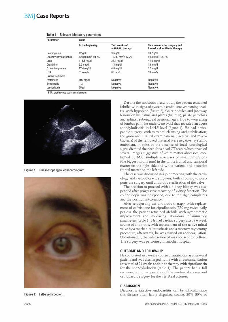
The transoesophageal echocardiogram showed one globular vegetation (12 mm in diameter) on the anterior mitral leafl et ( fi gure 1 ); severe mitral regurgitation (III/IV); aortic valve with normal morphology and mild regurgita-tion. The left ventricular outfl ow tract gradient was in the range of 30–35 mm Hg by trans thoracic echocardiogram. The patient was started on vancomycin (1g qd intrave-nous), due to alleged penicillin allergy, and gentamycin (120 mg qd intravenous, according to his creatinine clear-ance). On the 14th day as inpatient, we were notifi ed about the growth of a gram-negative Haemophilus species, Actinomycetemcomitans , Cardiobacterium hominis , Eikenella cor-rodens and Kingella species (HACEK) group organism, with the isolation of A actinomycetemcomitans on the 17th day, with a subsequent substitution of vancomycin for ceftriax-one (2g qd intravenously).
Unusual association of diseases/symptoms
Actinobacillus endocarditis associated with hypertrophic cardiomyopathy
Vanda Cristina Jorge, Ana Carolina Araújo, Ana Grilo, Carla Noronha, António Panarra, Nuno Riso,
Manuel Vaz Riscado
Department 2, Curry Cabral‘s Hospital, Lisbon, Portugal
Correspondence to Dr Vanda Cristina Jorge, [email protected]
Summary Infective endocarditis can be associated with complex clinical presentations, sometimes with a diffi cult multi-disciplinary management.
Actinobacillus actinomycetemcomitans belongs to the Haemophilus species, Actinomycetemcomitans , Cardiobacterium hominis , Eikenella
corrodens and Kingella species group, responsible for 5% to 10% of infective endocarditis in native heart valves. These organisms have slow
fastidious growth pattern, often associated with negative cultures, and cause systemic embolism with abscess formation. The authors present
the case of a 59-year-old man, admitted due to fever of unknown origin, with a personal history of obstructive hypertrophic cardiomyopathy
and recent dental manipulation. The diagnosis of mitral valve’s endocarditis was established after a transoesophageal ecocardiography, with
a late isolation of A actinomycetemcomitans in blood culture. Despite the institution of antibiotic therapy, the patient suffered from multiple
episodes of septic embolism: skin, mucosae, cerebral abscesses, spondylodiscitis and uveitis. He was submitted to heart surgery with
miectomy and replacement of the native mitral valve by a mechanical prosthesis, while on antibiotics.
BMJ Case Reports 2012; doi:10.1136/bcr.04.2011.41402 of 5
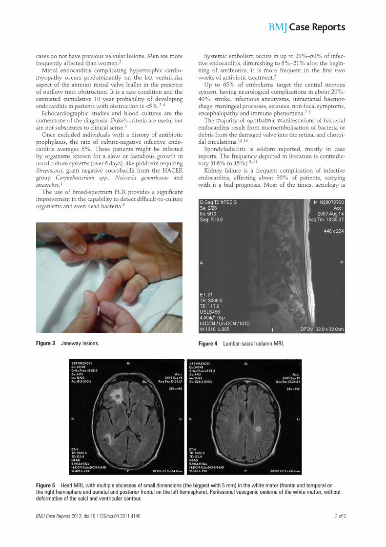
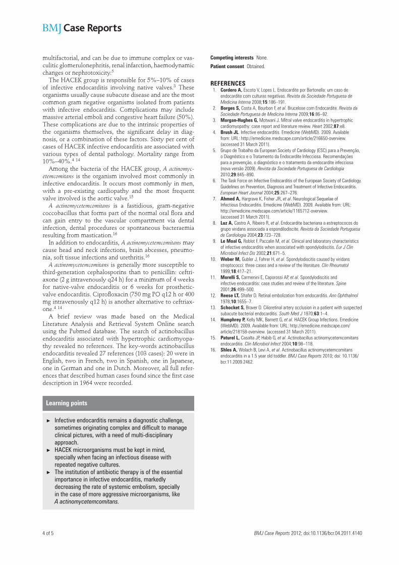
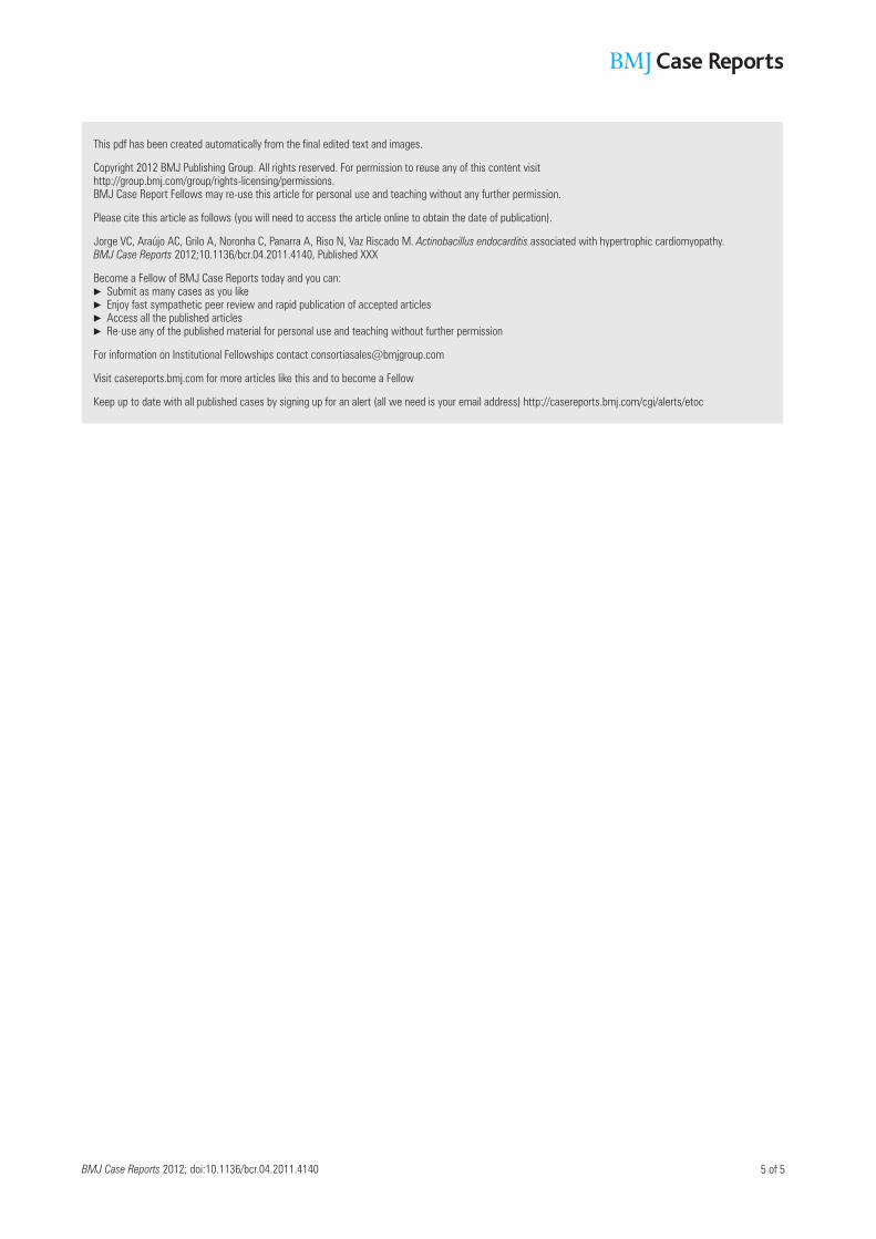
Despite the antibiotic prescription, the patient remained febrile, with signs of systemic embolism: worsening uvei-tis, with hypopion ( fi gure 2 ), Osler nodules and Janeway lesions on his palms and plants ( fi gure 3 ), palate petechias and splinter subungueal haemorrhages. Due to worsening of lumbar pain, he underwent MRI that revealed an acute spondylodiscitis in L4/L5 level ( fi gure 4 ). He had ortho-paedic surgery, with vertebral cleansing and stabilisation; the gram and cultural examinations (bacterial and myco-bacteria) of the removed material were negative. Systemic embolism, in spite of the absence of focal neurological signs, dictated the need for a head CT scan, which revealed several images suggestive of white matter abscesses, con-fi rmed by MRI: multiple abscesses of small dimensions (the biggest with 5 mm) in the white frontal and temporal matter on the right side and white parietal and posterior frontal matter on the left side.
The case was discussed in a joint meeting with the cardi-ology and cardiothoracic surgeons, both choosing to post-pone the surgery until antibiotic sterilisation of the valve.
The decision to proceed with a kidney biopsy was sus-pended after progressive recovery of kidney function. The colonoscopy was postponed, due to the algic complaints and the position intolerance.
After re-adjusting the antibiotic therapy, with replace-ment of ceftriaxone for ciprofl oxacin (750 mg twice daily per os), the patient remained afebrile with symptomatic improvement and improving laboratory infl ammatory parameters ( table 1 ). He had cardiac surgery after a 6 week course of antibiotic, with replacement of the native mitral valve by a mechanical prosthesis and a morrow myectomy procedure; afterwards, he was started on anticoagulation. Unfortunately, the valve removed was not sent for culture. The surgery was performed in another hospital.
OUTCOME AND FOLLOW-UP He completed an 8 weeks course of antibiotics as an inward patient and was discharged home with a recommendation for a total of 24 weeks antibiotic therapy with ciprofl oxacin for the spondylodiscitis ( table 1 ). The patient had a full recovery, with disappearance of the cerebral abscesses and orthopaedic surgery for the vertebral column.
DISCUSSION Diagnosing infective endocarditis can be diffi cult, since this disease often has a disguised course. 20%–30% of
Figure 1 Transoesophageal echocardiogram.
Figure 2 Left-eye hypopion.
Table 1 Relevant laboratory parameters Parameter Value
In the beginning Two weeks of antibiotic therapy
Two weeks after surgery and 6 weeks of antibiotic therapy
Haemoglobin 12 g/dl 9.6 g/dl 10.2 g/dlLeucocytes/neutrophils 12100 mm 3 ; 90.7% 12300 mm 3 ; 87.2% 5900 mm 3 ; 85.7%Urea 116.6 mg/dl 27.4 mg/dl 44.6 mg/dlCreatinine 3.2 mg/dl 1.3 mg/dl 1.6 mg/dlC reactive protein 27.4 mg/dl 4.6 mg/dl 1.2 mg/dlESR 31 mm/h 66 mm/h 58 mm/hUrinary sedimentProteinuria 100 mg/dl Negative NegativeEritrocituria +2 Negative NegativeLeucocituria 25 µl Negative Negative
ESR, erythrocyte sedimentation rate.
BMJ Case Reports 2012; doi:10.1136/bcr.04.2011.4140 3 of 5
cases do not have previous valvular lesions. Men are more frequently affected than women. 2
Mitral endocarditis complicating hypertrophic cardio-myopathy occurs predominantly on the left ventricular aspect of the anterior mitral valve leafl et in the presence of outfl ow tract obstruction. It is a rare condition and the estimated cumulative 10 year probability of developing endocarditis in patients with obstruction is <5%. 3 4
Echocardiographic studies and blood cultures are the cornerstone of the diagnosis. Duke’s criteria are useful but are not substitutes to clinical sense. 5
Once excluded individuals with a history of antibiotic prophylaxis, the rate of culture-negative infective endo-carditis averages 5%. These patients might be infected by organisms known for a slow or fastidious growth in usual culture systems (over 6 days), like piridoxin requiring Streptococci , gram negative coccobacilli from the HACEK group Corynebacterium spp., Neisseria gonorrhoeae and anaerobes . 1
The use of broad-spectrum PCR provides a signifi cant improvement in the capability to detect diffi cult-to-culture organisms and even dead bacteria. 6
Systemic embolism occurs in up to 20%–50% of infec-tive endocarditis, diminishing to 6%–21% after the begin-ning of antibiotics; it is more frequent in the fi rst two weeks of antibiotic treatment. 5
Up to 65% of embolisms target the central nervous system, having neurological complications in about 20%-40%: stroke, infectious aneurysms, intracranial haemor-rhage, meningeal processes, seizures, non-focal symptoms, encephalopathy and immune phenomena. 7 8
The majority of ophthalmic manifestations of bacterial endocarditis result from microembolisation of bacteria or debris from the damaged valve into the retinal and choroi-dal circulations. 12 13
Spondylodiscitis is seldom reported, mostly in case reports. The frequency depicted in literature is contradic-tory (0.6% to 15%). 8 – 11
Kidney failure is a frequent complication of infective endocarditis, affecting about 30% of patients, carrying with it a bad prognosis. Most of the times, aetiology is
Figure 3 Janeway lesions. Figure 4 Lumbar-sacral column MRI.
Figure 5 Head MRI, with multiple abcesses of small dimensions (the biggest with 5 mm) in the white mater (frontal and temporal on the right hemisphere and parietal and posterior frontal on the left hemisphere). Perilesional vasogenic oedema of the white matter, without deformation of the sulci and ventricular contour.
BMJ Case Reports 2012; doi:10.1136/bcr.04.2011.41404 of 5
multifactorial, and can be due to immune complex or vas-culitic glomerulonephritis, renal infarction, haemodynamic changes or nephrotoxicity. 5
The HACEK group is responsible for 5%–10% of cases of infective endocarditis involving native valves. 3 These organisms usually cause subacute disease and are the most common gram negative organisms isolated from patients with infective endocarditis. Complications may include massive arterial emboli and congestive heart failure (50%). These complications are due to the intrinsic properties of the organisms themselves, the signifi cant delay in diag-nosis, or a combination of these factors. Sixty per cent of cases of HACEK infective endocarditis are associated with various types of dental pathology. Mortality range from 10%–40%. 4 14
Among the bacteria of the HACEK group, A actinomyc-etemcomitans is the organism involved most commonly in infective endocarditis. It occurs most commonly in men, with a pre-existing cardiopathy and the most frequent valve involved is the aortic valve. 15
A actinomycetemcomitans is a fastidious, gram-negative coccobacillus that forms part of the normal oral fl ora and can gain entry to the vascular compartment via dental infection, dental procedures or spontaneous bacteraemia resulting from mastication. 16
In addition to endocarditis, A actinomycetemcomitans may cause head and neck infections, brain abcesses, pneumo-nia, soft tissue infections and urethritis. 16
A actinomycetemcomitans is generally more susceptible to third-generation cephalosporins than to penicillin: ceftri-axone (2 g intravenously q24 h) for a minimum of 4 weeks for native-valve endocarditis or 6 weeks for prosthetic-valve endocarditis. Ciprofl oxacin (750 mg PO q12 h or 400 mg intravenously q12 h) is another alternative to ceftriax-one. 4 14
A brief review was made based on the Medical Literature Analysis and Retrieval System Online search using the Pubmed database. The search of actinobacillus endocarditis associated with hypertrophic cardiomyopa-thy revealed no references. The key-words actinobacillus endocarditis revealed 27 references (103 cases): 20 were in English, two in French, two in Spanish, one in Japanese, one in German and one in Dutch. Moreover, all full refer-ences that described human cases found since the fi rst case description in 1964 were recorded.
Learning points
▶ Infective endocarditis remains a diagnostic challenge, sometimes originating complex and diffi cult to manage clinical pictures, with a need of multi-disciplinary approach. HACEK microorganisms must be kept in mind, ▶
specially when facing an infectious disease with repeated negative cultures. The institution of antibiotic therapy is of the essential ▶
importance in infective endocarditis, markedly decreasing the rate of systemic embolism, specially in the case of more aggressive microorganisms, like A actinomycetemcomitans.
Competing interests None.
Patient consent Obtained.
REFERENCES 1. Cordero A, Escoto V, Lopes L . Endocardite por Bartonella: um caso de
endocardite com culturas negativas . Revista da Sociedade Portuguesa de
Medicina Interna 2008 ; 15 : 186 – 191 .
2. Borges S, Costa A, Bourbon F, et al . Brucelose com Endocardite . Revista da
Sociedade Portuguesa de Medicina Interna 2009 ; 16 : 86 – 92 .
3. Morgan-Hughes G, Motwani J . Mitral valve endocarditis in hypertrophic
cardiomyopathy: case report and literature review. Heart 2002 ; 87 : e8 .
4. Brush JL . Infective endocarditis. Emedicine (WebMD) . 2009 . Available
from: URL: http://emedicine.medscape.com/article/216650-overview .
(accessed 31 March 2011).
5. Grupo de Trabalho da European Society of Cardiology (ESC) para a Prevenção,
o Diagnóstico e o Tratamento da Endocardite Infecciosa. Recomendações
para a prevenção, o diagnóstico e o tratamento da endocardite infecciosa
(nova versão 2009) . Revista da Sociedade Portuguesa de Cardiologia
2010 ; 29 : 845 – 890 .
6. The Task Force on Infective Endocarditis of the European Society of Cardiology .
Guidelines on Prevention, Diagnosis and Treatment of Infective Endocarditis .
European Heart Journal 2004 ; 25 : 267 – 276 .
7. Ahmed A, Hargrave K, Fisher JR, et al . Neurological Sequelae of
Infectious Endocarditis. Emedicine (WebMD) . 2009 . Available from: URL:
http://emedicine.medscape.com/article/1165712-overview .
(accessed 31 March 2011).
8. Luz A, Castro A, Ribeiro R, et al . Endocardite bacteriana a estreptococos do
grupo viridans associada a espondilodiscite . Revista da Sociedade Portuguesa
de Cardiologia 2004 ; 23 : 723 – 728 .
9. Le Moal G, Roblot F, Paccalin M, et al . Clinical and laboratory characteristics
of infective endocarditis when associated with spondylodiscitis. Eur J Clin
Microbiol Infect Dis 2002 ; 21 : 671 – 5 .
10. Weber M, Gubler J, Fahrer H, et al . Spondylodiscitis caused by viridans
streptococci: three cases and a review of the literature. Clin Rheumatol
1999 ; 18 : 417 – 21 .
11. Morelli S, Carmenini E, Caporossi AP, et al . Spondylodiscitis and
infective endocarditis: case studies and review of the literature. Spine
2001 ; 26 : 499 – 500 .
12. Reese LT, Shafer D . Retinal embolization from endocarditis. Ann Ophthalmol
1978 ; 10 : 1655 – 7 .
13. Schocket S, Braver D . Cilioretinal artery occlusion in a patient with suspected
subacute bacterial endocarditis. South Med J 1970 ; 63 : 1 – 4 .
14. Humphrey P, Kelly MK, Barnett G, et al . HACEK Group Infections. Emedicine
(WebMD) . 2009 . Available from: URL: http://emedicine.medscape.com/
article/218158-overview . (accessed 31 March 2011).
15. Paturel L, Casalta JP, Habib G, et al . Actinobacillus actinomycetemcomitans
endocarditis. Clin Microbiol Infect 2004 ; 10 : 98 – 118 .
16. Shles A, Wolach B, Levi A, et al . Actinobacillus actinomycetemcomitans
endocarditis in a 1.5 year old toddler . BMJ Case Reports 2010 ; doi: 10.1136/
bcr.11.2009.2462.
BMJ Case Reports 2012; doi:10.1136/bcr.04.2011.4140 5 of 5
This pdf has been created automatically from the fi nal edited text and images.
Copyright 2012 BMJ Publishing Group. All rights reserved. For permission to reuse any of this content visit http://group.bmj.com/group/rights-licensing/permissions. BMJ Case Report Fellows may re-use this article for personal use and teaching without any further permission.
Please cite this article as follows (you will need to access the article online to obtain the date of publication).
Jorge VC, Araújo AC, Grilo A, Noronha C, Panarra A, Riso N, Vaz Riscado M. Actinobacillus endocarditis associated with hypertrophic cardiomyopathy. BMJ Case Reports 2012;10.1136/bcr.04.2011.4140, Published XXX
Become a Fellow of BMJ Case Reports today and you can:Submit as many cases as you like ▶Enjoy fast sympathetic peer review and rapid publication of accepted articles ▶Access all the published articles ▶Re-use any of the published material for personal use and teaching without further permission ▶
For information on Institutional Fellowships contact [email protected]
Visit casereports.bmj.com for more articles like this and to become a Fellow
Keep up to date with all published cases by signing up for an alert (all we need is your email address) http://casereports.bmj.com/cgi/alerts/etoc