Embed Size (px)
Citation preview
Rev Esp Cardiol. 2016;69(2):178–187
Document downloaded from http://www.revespcardiol.org, day 23/02/2017. This copy is for personal use. Any transmission of this document by any media or format is strictly prohibited.
Review article
Update on Myocarditis and Inflammatory Cardiomyopathy:Reemergence of Endomyocardial Biopsy
Fernando Dominguez,a,b,* Uwe Kuhl,b Burkert Pieske,b,c Pablo Garcia-Pavia,a and Carsten Tschopeb,d,e
a Unidad de Insuficiencia Cardiaca y Cardiopatıas Familiares, Servicio de Cardiologıa, Hospital Universitario Puerta de Hierro, Mahadahonda, Madrid, Spainb Department of Cardiology, Charite Campus Virchow Klinikum (CVK), Berlin, Germanyc Department of Cardiology, Deutsches Herzzentrum Berlin, Berlin, Germanyd Berliner Zentrum fur Regenerative Therapien (BCRT), Campus Virchow Klinikum (CVK), Berlin, Germanye Deutsches Zentrum fur Herz Kreislaufforschung (DZHK), Berlin/Charite, Berlin, Germany
Article history:
Available online 12 January 2016
Keywords:
Myocarditis
Inflammatory cardiomyopathy
Endomyocardial biopsy
A B S T R A C T
Myocarditis is defined as an inflammatory disease of the heart muscle and is an important cause of acute
heart failure, sudden death, and dilated cardiomyopathy. Viruses account for most cases of myocarditis
or inflammatory cardiomyopathy, which could induce an immune response causing inflammation even
when the pathogen has been cleared. Other etiologic agents responsible for myocarditis include drugs,
toxic substances, or autoimmune conditions. In the last few years, advances in noninvasive techniques
such as cardiac magnetic resonance have been very useful in supporting diagnosis of myocarditis, but
toxic, infectious-inflammatory, infiltrative, or autoimmune processes occur at a cellular level and only
endomyocardial biopsy can establish the nature of the etiological agent. Furthermore, after the
generalization of immunohistochemical and viral genome detection techniques, endomyocardial biopsy
provides a definitive etiological diagnosis that can lead to specific treatments such as antiviral or
immunosuppressive therapy. Endomyocardial biopsy is not commonly performed for the diagnosis of
myocarditis due to safety reasons, but both right- and left endomyocardial biopsies have very low
complication rates when performed by experienced operators. This document provides a state-of-the-art
review of myocarditis and inflammatory cardiomyopathy, with special focus on the role of endomyocardial
biopsy to establish specific treatments.
� 2015 Sociedad Espanola de Cardiologıa. Published by Elsevier Espana, S.L.U. All rights reserved.
Actualizacion sobre miocarditis y miocardiopatıa inflamatoria: el resurgirde la biopsia endomiocardica
Palabras clave:
Miocarditis
Miocardiopatıa inflamatoria
Biopsia endomiocardica
R E S U M E N
La miocarditis se define como una enfermedad inflamatoria del musculo cardiaco y es una causa
importante de insuficiencia cardiaca aguda, muerte subita y miocardiopatıa dilatada. Los virus son la causa
de la mayorıa de los casos de miocarditis o miocardiopatıa inflamatoria y pueden inducir una respuesta
inmunitaria causante de inflamacion pese a haberse eliminado el patogeno. Otros agentes etiologicos
causantes de miocarditis son los farmacos, las sustancias toxicas o los trastornos autoinmunitarios. En los
ultimos anos, los avances de tecnicas no invasivas como la resonancia magnetica cardiaca han sido de gran
utilidad para respaldar el diagnostico de miocarditis, pero los procesos toxicos, infecciosos e inflamatorios,
infiltrantes o autoinmunitarios se producen en las celulas, y solamente la biopsia endomiocardica permite
establecer la naturaleza del agente etiologico. Ademas, despues de la generalizacion de las tecnicas
inmunohistoquımicas y de deteccion del genoma viral, la biopsia endomiocardica proporciona un
diagnostico etiologico definitivo que puede conducir a tratamientos especıficos como los antivirales o los
inmunosupresores. No se realiza con frecuencia para el diagnostico de miocarditis por razones de
seguridad, pero la biopsia endomiocardica, tanto derecha como izquierda, tiene una tasa de complicaciones
muy baja cuando la realiza un operador experto. En este documento se presenta una revision actualizada de
la miocarditis y la miocardiopatıa inflamatoria haciendo especial referencia al papel de la biopsia
endomiocardica para establecer un tratamiento especıfico.
� 2015 Sociedad Espanola de Cardiologıa. Publicado por Elsevier Espana, S.L.U. Todos los derechos reservados.
* Corresponding author: Unidad de Insuficiencia Cardiaca y Cardiopatıas Familiares, Servicio de Cardiologıa, Hospital Universitario Puerta de Hierro, Manuel de Falla 1,
28222 Majadahonda, Madrid, Spain.
E-mail address: [email protected] (F. Dominguez).
http://dx.doi.org/10.1016/j.rec.2015.10.015
1885-5857/� 2015 Sociedad Espanola de Cardiologıa. Published by Elsevier Espana, S.L.U. All rights reserved.
F. Dominguez et al. / Rev Esp Cardiol. 2016;69(2):178–187 179
Document downloaded from http://www.revespcardiol.org, day 23/02/2017. This copy is for personal use. Any transmission of this document by any media or format is strictly prohibited.
INTRODUCTION
The term myocarditis refers to an inflammation of the heartmuscle, which can be caused by infections, toxic substances, orautoimmune processes. During the acute phase, a specific triggerinduces an immune response, which can range from transient andmild to fulminant. In the case of viral myocarditis, if the host doesnot success in eliminating the infectious pathogen, chronicinfection develops, with or without ongoing inflammation.Furthermore, inflammation can persist even if the pathogen hasbeen cleared. Thus, inflammatory dilated cardiomyopathy (DCM)is an independent entity with its own pathogenic mechanisms anda potential cause of heart failure. As understanding of this diseaseincreases, it is now evident that the pathological injury occurs atthe cellular level, and therefore an accurate diagnosis requirestissue analysis with endomyocardial biopsy (EMB).1 Histologicalfindings have been proved to have prognostic implications,2 and inseveral cases specific treatments can be added to the basicsymptomatic heart failure treatment.3 T this review aims to serveas a practical document for the diagnosis and treatment ofmyocarditis and inflammatory cardiomyopathy, with special focuson EMB as a diagnostic tool, as well as on subsequent tailoredtreatment based on its results.
DEFINITIONS AND ETIOLOGY OF MYOCARDITIS
Myocarditis is defined by an inflammation of the myocardiumdiagnosed by established histological, immunological, andimmunohistochemical criteria. As stated in the consensus paperof the European Society of Cardiology Working Group onMyocardial and Pericardial Diseases,1 it is histologically definedby the presence of inflammatory infiltrates in the myocardiumassociated with myocyte degeneration and necrosis of nonis-chemic cause, following the Dallas criteria.4 Regarding immu-
Abbreviations
CMR: cardiac magnetic resonance
DCM: dilated cardiomyopathy
EMB: endomyocardial biopsy
HHV-6: human herpes virus type 6
LVEF: left ventricular ejection fraction
PVB19: parvovirus B19
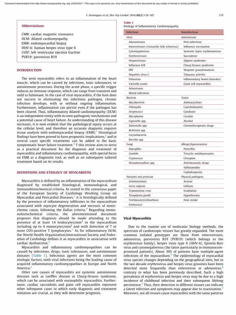
Table 1Etiology of Inflammatory Cardiomyopathy
Infectious Noninfectious
Virus Autoimmune
Adenoviruses Post-infectious
Enteroviruses (Coxsackie A/B, echovirus) Influenza vaccination
Cytomegalovirus Systemic lupus erythematosus
Erythroviruses Sarcoidosis
Herpesviruses Sjogren syndrome
Influenza A/B Churg-Strauss syndrome
HIV Wegener granulomatosis
Hepatitis virus C Takayasu arteritis
Poliovirus Inflammatory bowel disorders
Varicella zoster Giant cell myocarditis
Arboviruses
Mixed infections
Bacteria Toxins
Mycobacteria Anthracyclines
Chlamydia Catecholamines
Streptococci Cytokines
Mycoplasma Cocaine
Legionella spp. Alcohol
Salmonella spp. Chemotherapeutic drugs
Rickettsia spp.
Corynebacteria
Borrelia spp.
Fungi Allergic/hypersensitive
Aspergillus Penicillin
Candida Tricyclic antidepressants
Cryptococus Clozapine
Histoplasmodium spp. Antirheumatic drugs
Sulfonamides
Cephalosporins
Parasites and protozoa Physical pathogens
Schistosomiasis Arsenic
Larva migrans Lithium
Trypanosoma cruzi Irradiation
Toxoplasma gondii Hypothermia
Trichinosis/trichinellosis Heat stroke
Echinococci
nohistochemical criteria, the aforementioned documentproposes that diagnosis should be made attending to thepresence of at least 14 leukocytes/mm2 in the myocardiumincluding up to 4 monocytes/mm2 and with detection of 7 ormore CD3-positive T lymphocytes.1 As for inflammatory DCM,the World Health Organization/International Society and Feder-ation of Cardiology defines it as myocarditis in association withcardiac dysfunction.1
Myocarditis and inflammatory cardiomyopathies can becaused by infections, drugs, toxic substances, and autoimmunediseases (Table 1). Infectious agents are the most commonetiologic factors, with viral infections being the leading cause ofacquired inflammatory cardiomyopathies in Europe and NorthAmerica.2
Other rare causes of myocarditis are systemic autoimmunediseases such as Loeffler disease or Churg-Strauss syndrome,which can be associated with eosinophilic myocarditis. Further-more, cardiac sarcoidosis and giant cell myocarditis representother infrequent cases in which early diagnosis and treatmentinitiation are crucial, as they will determine prognosis.
Viral Myocarditis
Due to the routine use of molecular biology methods, thespectrum of cardiotropic viruses has greatly expanded. The mostcommon isolated genotypes are those from enteroviruses,adenovirus, parvovirus B19 (PVB19) (which belongs to theerythrovirus family), herpes virus type 6 (HHV-6), Epstein-Barrvirus and cytomegalovirus (the latter particularly in immunocom-promised patients). About 30% of patients have multiple-agentinfections of the myocardium.5 The epidemiology of myocardialvirus species changes depending on the geographical sites, but inthe last decade erythrovirus and herpes virus genomes have beendetected more frequently than enterovirus or adenovirus,6
contrary to what has been previously described. Such a highprevalence of erythrovirus and herpes virus may be due to a highincidence of childhood infection and their subsequent lifelongpersistence.7 Thus, their detection in different tissues can indicatea latent infection and symptoms may appear due to reactivation.3
Moreover, not all viruses cause myocarditis with the same patterns
F. Dominguez et al. / Rev Esp Cardiol. 2016;69(2):178–187180
Document downloaded from http://www.revespcardiol.org, day 23/02/2017. This copy is for personal use. Any transmission of this document by any media or format is strictly prohibited.
of infection. For instance, enteroviruses and adenoviruses directlyinfect cardiomyocytes in animals and humans and in the last fewyears, 10% to 15% of viral myocarditis have been caused by theseagents.8
The situation of erythroviruses such as PVB19 is quite different.This virus primarily infects erythroid progenitor cells in the bonemarrow9 and endothelial cells, leading to asymptomatic and latentinfections. Then, when the virus becomes reactivated, angina-likesymptoms have been related to endothelial dysfunction.10
CLINICAL SYMPTOMS OF MYOCARDITIS AND NONINVASIVEDIAGNOSIS
The clinical presentation of myocarditis varies widely, rangingfrom ischemic-like chest pain to syncope or acute heart failure.Although most patients present with mild symptoms or transientelectrocardiographic changes, myocarditis can also cause acuteheart failure and life-threatening cardiogenic shock.1
It frequently starts 1 to 4 weeks after an infection, normallyrespiratory or gastrointestinal. However, due to its variedsymptoms, myocarditis can be difficult to diagnose, and coronaryartery disease must always be excluded, given its high prevalenceand similar clinical presentation. Moreover, EMB is undoubtedlythe gold standard diagnostic tool in myocarditis and inflammatorycardiomyopathy. No other test can provide a definite diagnosis,and noninvasive techniques are used to help clinicians rule outother diagnoses and indirectly recognize myocarditis.
Electrocardiogram
All patients with suspected myocarditis should receive a12-lead electrocardiogram.1 Electrocardiographic findings inmyocarditis patients include T-wave and ST-segment changes,ST-segment elevation mimicking acute myocardial infarction orconduction abnormalities (as seen in Lyme disease, cardiacsarcoidosis, or giant cell myocarditis).11 These changes arenonspecific and can be found in other clinical settings, but theelectrocardiogram is still an easily available screening tool.Regarding prognosis, prolonged QRS duration of > 120 ms is theonly independent factor for heart transplantation or cardiacdeath.12
Imaging Techniques
Echocardiography remains the key method for analyzingventricular function in suspected myocarditis and helps to ruleout other entities such as valve disease. Thus, all patients withsuspected myocarditis should undergo echocardiographic studiesat presentation and during follow-up.1 However, findings arenonspecific, and include global ventricular dysfunction, regionalwall motion abnormalities, or diastolic dysfunction. Both in acuteand fulminant myocarditis, wall thickness may be mildlyincreased, but left ventricular (LV) diastolic dimensions aretypically larger in acute myocarditis. As for systolic function,better recovery is normally seen in patients that survive after theacute phase of fulminant myocarditis when compared with acutemyocarditis.13 In fact, it has been observed that fulminantmyocarditis may have a good outcome in severe clinical settingssuch as Dengue when specific treatment is applied.14 Regardingpatients with preserved left ventricular ejection fraction (LVEF),speckle tracking is a promising tool. In patients with biopsy-provenmyocardial inflammation, global longitudinal strain rate andglobal longitudinal strain are significantly impaired comparedwith patients without inflammation, regardless of conventional
echocardiographic parameters15 Therefore, this technique has ahigher sensitivity in the detection of mild myocardial damage inpatients with preserved LVEF and plays a role in predictingoutcome, as patients with impaired baseline strain show worsefollow-up echocardiographic results.
Cardiac magnetic resonance (CMR) can help confirm thediagnosis of myocarditis, especially in the acute phase of thedisease. The combined use of 3 different CMR techniques issuggested, and findings are compatible with myocardial inflam-mation if at least 2 Lake Louise criteria are met.
These include: a) Regional or global myocardial signal intensityincrease in T2-weighted edema images; b) an increased globalmyocardial early gadolinium enhancement ratio between myo-cardium and skeletal muscle in gadolinium-enhanced T1-weight-ed images, and c) at least 1 focal lesion with nonischemic regionaldistribution in inversion recovery-prepared gadolinium-enhancedT1-weighted images.16 When at least 2 criteria are met, asensitivity of 76% and specificity of 96% have been reported inpatients with clinically suspected acute myocarditis and pseudo-infarction presentation.17
Moreover, recent studies have shown good correlationsbetween CMR results and EMB in acute myocarditis, with up to79% accuracy when new CMR techniques are used.18 However,obtaining the biopsy from the region of late gadolinium enhance-ment of the CMR has not proven to increase the yield of diagnosis19
and, in chronic myocarditis, the diagnostic performance of CMRwas found to be worse (sensitivity, 63%; specificity, 40%).16
Therefore, CMR might not be appropriate to guide clinicalmanagement in chronic myocarditis.
Biomarkers
Cardiac troponins are highly suggestive of acute myocarditis,when other potential causes of myocardial necrosis, such as acutecoronary syndromes, have been excluded.20 Elevation of cardiactroponin I or T levels is more common than creatine kinase MB andpersistent high levels indicate ongoing necrosis. NT-pro-BNP orBNP levels should be measured when heart failure is suspected, butnormal values do not exclude myocarditis.21 Newer cardiacbiomarkers, such as copeptin or midregional pro-adrenomedullin,do not provide additional diagnostic or prognostic information.20
The usefulness of viral serologies is limited, especially inchronic myocarditis or inflammatory cardiomyopathy, as IgGantibodies for cardiotropic virus can be found in the bloodstream of the general population without accompanying cardiacinvolvement.22 A positive virus polymerase chain reaction (PCR) inperipheral blood does not prove viral myocarditis either. However,when viral genome is present in EMB, blood viral PCR can excludeor confirm systemic infection.2 It may also allow the discriminationof an acute viral infection from endogenous viral reactivation, inwhich there is higher virus replication.
Regarding serum cardiac autoantibodies (anti fibrillary, organ-specific and partially organ-specific antiheart, anti-intercalateddisks, anti-interfibrillary, etc.), these can be useful when highlevels are present in the absence of viral genome in EMB,suggesting an immune mediated myocarditis or inflammatorycardiomyopathy.
ENDOMYOCARDIAL BIOPSY
Endomyocardial biopsy is the gold standard technique for thediagnosis of myocarditis and inflammatory cardiomyopathy. Thetoxic, infectious-inflammatory, infiltrative or autoimmune pro-cesses that cause myocarditis occur at a cellular level, and no otherdiagnostic techniques can establish the nature of the etiological
F. Dominguez et al. / Rev Esp Cardiol. 2016;69(2):178–187 181
Document downloaded from http://www.revespcardiol.org, day 23/02/2017. This copy is for personal use. Any transmission of this document by any media or format is strictly prohibited.
agent. As well as detection of inflammation or viral genomes in theacute phase of myocarditis, EMB adds important prognosticinformation during the follow-up of patients that can influencetherapeutic decisions. The 2007 American Heart Association/American College of Cardiology Foundation/European Society ofCardiology scientific statement on EMB limited its class Irecommendations to unexplained new-onset heart failure of lessthan 2 weeks’ duration associated with hemodynamic compromiseor unexplained new-onset heart failure of 2 weeks to 3 months’duration associated with a dilated LV and new ventriculararrhythmias or conduction disturbances.23 However, in a recentposition statement from the European Society of Cardiology,1 therecommendation for EMB was extended, including patients with apseudo-infarct presentation after exclusion of coronary arterydisease. This change reflects the generalization of immunohisto-chemical and viral genome detection techniques, which haveenabled progress in the etiological diagnosis of myocarditis. Hence,an increasing number of patients can benefit from specifictreatments.
The main reason for the restriction of EMB procedures in somecenters is safety. Nonetheless, when performed by experiencedoperators, both right and left EMB have very low complicationrates. In a single-center study that analyzed 3048 EMB in anontransplant setting, the risk of major complications includingcardiac tamponade and atrioventricular block requiring perma-nent pacemaker implantation was 0.12%. No deaths wereregistered.24 Previous studies also reported a major complicationsrate of less than 0.5%.25 Left ventricular biopsy has also beenproven to be a safe procedure.26 Chimenti et al26 documented thatover a 28-year period and over 4000 EMB, complications appearedin only 0.33% of patients who underwent left EMB.
How to Perform an Endomyocardial Biopsy
Endomyocardial biopsy is performed with the patient in asupine position under local anesthesia with 2% lidocaine. Thepatient must be monitored with 3-lead electrocardiogram,noninvasive blood pressure monitoring, and oxygen saturation.An international normalized ratio of < 1.5 is required before theEMB, and anticoagulation therapy should be discontinued 16 hoursbefore and 12 hours after the procedure. Vascular access for rightventricular (RV) EMB is usually through the femoral or rightinternal jugular vein. Left ventricular or biventricular EMB ispreferred through the right femoral vein and femoral artery foraccess to the RV and LV.27 The bioptomes used are warranted to beflexible in order to ensure safety. We recommend the modifiedCordis bioptomes. This bioptome (B-18110; MedizintechnikMeiners, Monheim, Germany) has been used in clinical practicesince 1985. It has a 6 Fr diameter and a length of 1100 mm.Compared with the conventional Cordis bioptome, it has a moreflexible polytetrafluoroethylene (Teflon) tube.
Endomyocardial biopsy should be guided by fluoroscopy tolocate the intraventricular septum in case of RV EMB, as thethinness of the free wall lead to a high perforation risk. When itcomes to LV EMB, one of the main concerns is potential severemitral regurgitation due to biopsied chordae, so fluoroscopy canalso help to prevent this situation. Moreover, an echocardiogram isrecommended before and after the procedure to exclude pericar-dial effusion.
A recent study has evaluated the feasibility and safety of LVEMB via transradial access with promising results,28 offering a lessinvasive alternative to the classic femoral approach, and couldreduce hospital stays.
With reference to the number of samples taken by theprocedure, we recommend at least 5 and up to 10 to guarantee
reliable results. Focal tissue involvement is frequent in myocarditisand so different parts of the RV septum or the LV should bebiopsied. Samples for histology and immunohistochemical analy-sis should be at least 1-2 mm and promptly fixed in 10% formalin orsnap frozen in liquid nitrogen depending on the antibody that isgoing to be used. Samples for virus genome analysis should be snapfrozen in liquid nitrogen, stored at �80 8C, or stored in RNAlaterW
tubes at room temperature.1
Left Ventricular vs Right Ventricular Endomyocardial Biopsy
The diagnostic value of LV vs RV EMB has been analyzed invarious studies, and the results are not homogeneous. Whereassome observe that the diagnostic yield of LV EMB is superior to thatof RV EMB when routine immunohistochemistry and viral genomeamplification are used in suspected LV myocarditis,26 more recentdata indicate that both procedures are similar when inflammationor viral genome are being assessed in the myocardium. However, inthis latter study, morphological changes such as interstitial fibrosisand cardiac collagen type I expression were more reliably found inLV EMB.29
Interpretation of Endomyocardial Biopsy Results andPrognostic Implications
In all suspected cases of myocarditis, tissue samples from LV orRV should be analyzed using histology, immunohistochemistry,and viral genomes (viral PCR in EMB and blood).1 All of thesefeatures help us to diagnose inflammation and the presence of viralgenome, which have prognostic implications and require specifictreatments.
Inflammation
The histology of inflammation in the myocardium wasoriginally defined by the qualitative Dallas criteria (presence ofinflammatory infiltrates in the myocardium associated withmyocyte degeneration and necrosis of nonischemic cause).
Later, the addition of immunohistochemical criteria withdifferent monoclonal antibodies increased the EMB sensitivity inthe diagnosis of myocarditis30 and inflammation was quantitativelyestablished at � 14 leucocytes /mm2. During EMB analysis, specificinflammatory cells can be distinguished by cluster differentiation(CD). B-cells are CD20 positive and all T cells are CD3 positive.T cell subpopulations include CD4 (helper), CD8 (suppressor)and CD45R0 (memory or activated T-cells) or perforin-positivecytotoxic lymphocytes. CD68 and CD11 stand for macrophages.Attending these subpopulations, inflammation can be morespecifically diagnosed by > 7.0 CD3+ lymphocytes/mm2
and/or > 35.0 CD11b+/Mac-1+ macrophages/mm2.31
During an acute inflammatory disease course, the histology orimmunohistology samples normally contain focal or diffuse cellinfiltration by lymphocytes and/or macrophages. Other cells suchas eosinophils or giant cells are rare.
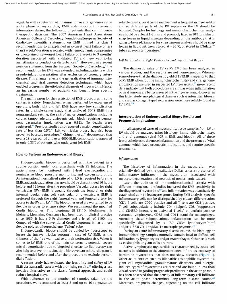
Active lymphocytic myocarditis is characterized by acute cellnecrosis in addition to the aforementioned infiltrates, contrary toborderline myocarditis that does not show necrosis (Figure 1).Other acute entities such as idiopathic eosinophilic myocarditis,giant cell myocarditis, granulomatous disorders, and allergic-induced types of myocarditis are rare and are found in less than20% of cases.8 Regarding prognostic predictors in the acute phase, ithas been observed that the density of inflammatory cell infiltratein the acute phase determines long-term disease course.32
Moreover, prognosis changes, depending on the cell infiltrate
F. Dominguez et al. / Rev Esp Cardiol. 2016;69(2):178–187182
Document downloaded from http://www.revespcardiol.org, day 23/02/2017. This copy is for personal use. Any transmission of this document by any media or format is strictly prohibited.
characteristics. Thus, borderline focal myocarditis has an excellentprognosis, whereas the early mortality of fulminant lymphocyticmyocarditis is 40% in the first month.32 Outcome is even worse inuntreated eosinophilic or giant cell myocarditis, in which survivalis less than 20% after 4 years.33,34
Regarding patients with DCM and chronic heart failure,inflammation is seen in up to 30% of biopsies.8 However, cellstend to distribute in a more diffuse manner than in the acute phaseand other features are present on histological examination, such ashypertrophy of the cardiomyocytes and interstitial fibrosis(Figure 2).
Lately, immunohistological signs of inflammation have beenalso related to poor outcome in suspected myocarditis. Actually,positive immunohistology for invading immune cells andexpression of HLA-DR-a, but not the Dallas criteria alone, wasassociated with a higher risk of cardiac death or heart transplanta-tion in patients with acute and chronic myocarditis.35 In amore recent study that excluded patients with acute myocardi-tis, perforin was a predictor of LVEF course in patients withchronic inflammatory cardiomyopathy and negative genomes forcardiotropic virus (enteroviruses, adenoviruses, Epstein-Barrvirus, HHV-6). Erythrovirus was present in 54% of patients,but without evidence of transcriptional activity.31 Even thoughall patients received recommended heart failure treatmentduring follow-up, a perforin value of more than 2.95 wasassociated with LVEF deterioration (94.2% sensitivity and 80.4%specificity).31
Figure 1. Different types of acute myocarditis. A: Acute lymphocitic myocarditis wiB: Cardiac sarcoidosis, with evidence of granuloma (black arrow). C: Giant celD: Eosinophilic myocarditis.
Presence of Viral Genome
In western countries, most of the infectious agents causingmyocarditis are viruses, and the viral spectrum differs withgeographical location.8 The PCR identifies viral DNA or RNA in themyocardium with very high sensitivity.1 Firstly, nested PCRidentifies the virus qualitatively, and if positive, viral load ismeasured by real-time PCR.
All samples should be compared with negative controls andcontrolled by amplifying adequate positive samples1 and latentinfections can be differentiated from acute cases with parallelanalyses of the blood stream.
Among the viral agents causing myocarditis, it is important todistinguish 2 groups: newly acquired infections and endogenousvirus infections with subsequent reactivations.
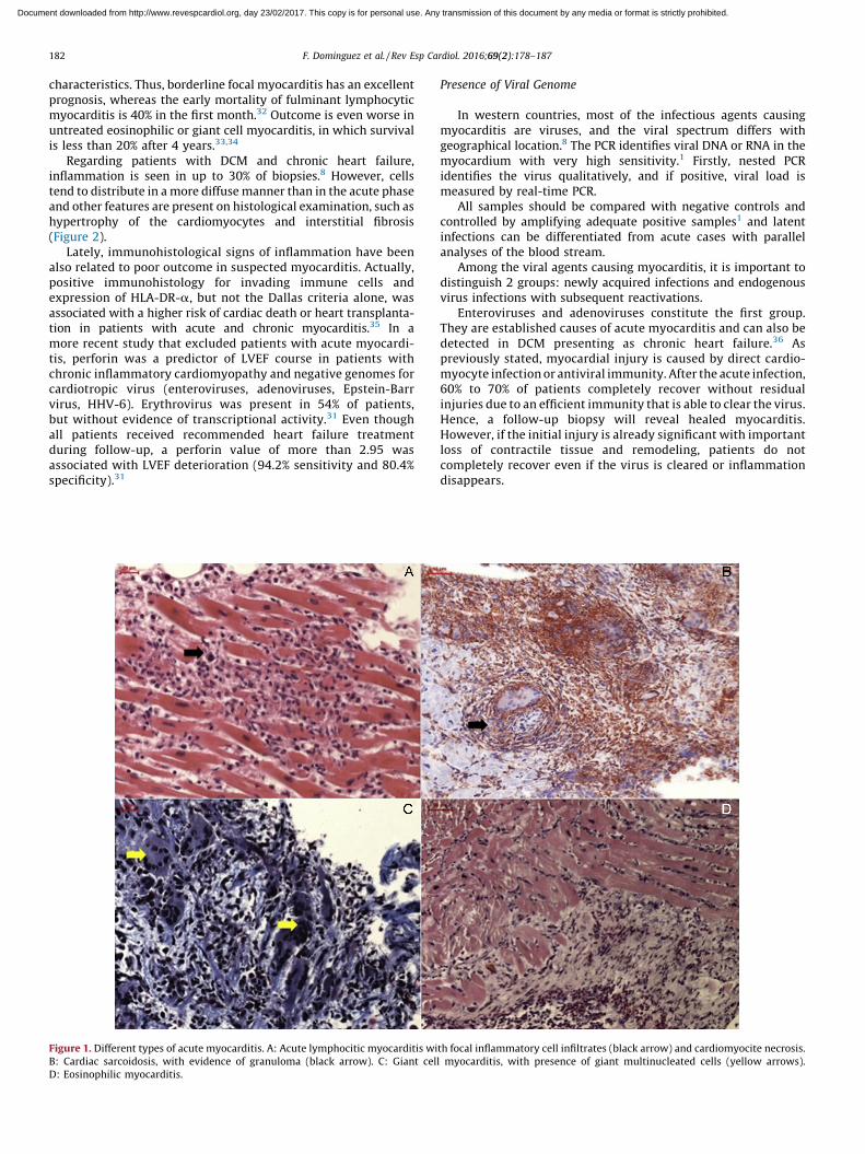
Enteroviruses and adenoviruses constitute the first group.They are established causes of acute myocarditis and can also bedetected in DCM presenting as chronic heart failure.36 Aspreviously stated, myocardial injury is caused by direct cardio-myocyte infection or antiviral immunity. After the acute infection,60% to 70% of patients completely recover without residualinjuries due to an efficient immunity that is able to clear the virus.Hence, a follow-up biopsy will reveal healed myocarditis.However, if the initial injury is already significant with importantloss of contractile tissue and remodeling, patients do notcompletely recover even if the virus is cleared or inflammationdisappears.
th focal inflammatory cell infiltrates (black arrow) and cardiomyocite necrosis.l myocarditis, with presence of giant multinucleated cells (yellow arrows).
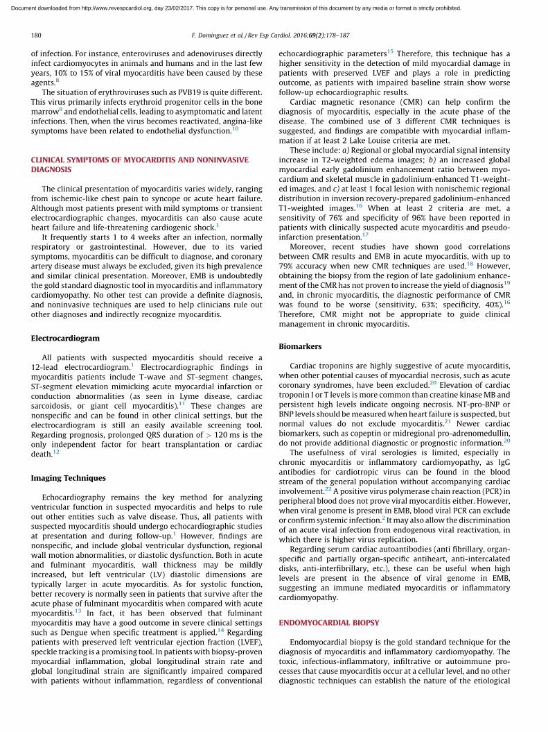
Viral infection
Active myocarditis
Chronic viralcardiomyopathy
Innate/adaptiveimmune response
Dilatedcardiomyopathy
Severe myocardial injury
Inflammatorycardiomyopathy
Severe myocardial injury
Healed myocarditisMinor myocardial injury
Chronic myocarditisMinor myocardial injury
Virus clearance?
Persisting inflammation?
No
± inflammation
No
Yes
Yes Yes
Figure 2. Pathogenesis of viral and inflammatory cardiomyopathy.
F. Dominguez et al. / Rev Esp Cardiol. 2016;69(2):178–187 183
Document downloaded from http://www.revespcardiol.org, day 23/02/2017. This copy is for personal use. Any transmission of this document by any media or format is strictly prohibited.
Various studies have evaluated the effect of enteroviral genomepersistence.
While it is true that the effect of viral persistence on outcome isunclear in other virus species (PVB19 or HHV-6), mortality ishigher in patients with noncleared enterovirus. Why et al37
observed 25% mortality at 25 months in myocarditis/DCM patientswith persistent enteroviral infection, as opposed to 4% mortality inenterovirus-negative patients. Similar data have been morerecently published, with a mortality as high as 41% in patientswith enteroviral genome persistence after a 5-year follow-up.38
Regarding PVB19 and HHV-6 infections, the most commonclinical entities are persistent latent virus infections withreactivation episodes.22 PVB19 is a common acute disease duringchildhood, rarely seen in adults. Basically, infected cells are limitedto erythroid progenitors in the bone marrow, but the primaryerythrovirus receptor is also present in endothelial cells, includingthe heart. Although it has been exceptionally localized in venuolesor arterioles during fulminant myocarditis in children,39 in mostcases the infection is latent and asymptomatic. Recently, we havereported that about 30% of PVB19-positive EMB had messengerRNA, which may indicate reactivation of the virus.10 In this context,it has been observed that cardiac gene expression is altered. Forinstance, genes involved in inflammatory response (tissue necrosisfactor alpha, related orphan receptor C) or mitochondrial energymetabolism (cyclooxygenase-1) are deregulated in messengerRNA-positive patients compared with those with only DNA.40
However, the effect of PVB19 DNA persistence on outcome is stillnot clear, as case series in which this virus was the most prevalentdid not demonstrate a higher risk of death or high transplantationrates.35 Moreover, systolic dysfunction has not been clearly related
to the presence of PVB19, but in a group of 37 patients withunexplained diastolic dysfunction, 84% were PVB19-positive inEMB, suggesting a relationship with the endothelial dysfunctioncaused by the virus.41
The HHV-6A and HHV-6B also usually cause acute infectionsduring childhood, and like PVB19, remains latent in > 70% ofadults. Although HHV-6 is mainly a lymphotropic virus, it can alsoinfect both endothelial cells and cardiomyocytes. In addition, itsgenome can be integrated into human chromosomes andtransmitted through the germ line.42 Similar to PVB19, HHV-6can become reactivated causing heart failure symptoms, and arecent study suggests that HHV-6 persistence could lead to aworsening in LVEF and clearance to an improvement.43
Irrespective of the initial viral etiology, if a biopsy is performedwhen the patient is already in the chronic phase without evidenceof a previous viral infection or persistent inflammation, thediagnosis will be idiopathic DCM.22 Other clinical scenarios arepersistent lytic virus infection without inflammation (chronic viralheart disease), or continual autoimmune mechanisms even whenthe virus has been cleared (inflammatory cardiomyopathy). Whenboth inflammation and viral infection persist, then the diagnosis ischronic viral cardiomyopathy.8 All these clinical entities aresummarized in Figure 2.
TREATMENT OF MYOCARDITIS AND INFLAMMATORYCARDIOMYOPATHY
Regardless of its etiology, the basic treatment of myocarditis isthe optimal care of heart failure and arrhythmias in accordance
F. Dominguez et al. / Rev Esp Cardiol. 2016;69(2):178–187184
Document downloaded from http://www.revespcardiol.org, day 23/02/2017. This copy is for personal use. Any transmission of this document by any media or format is strictly prohibited.
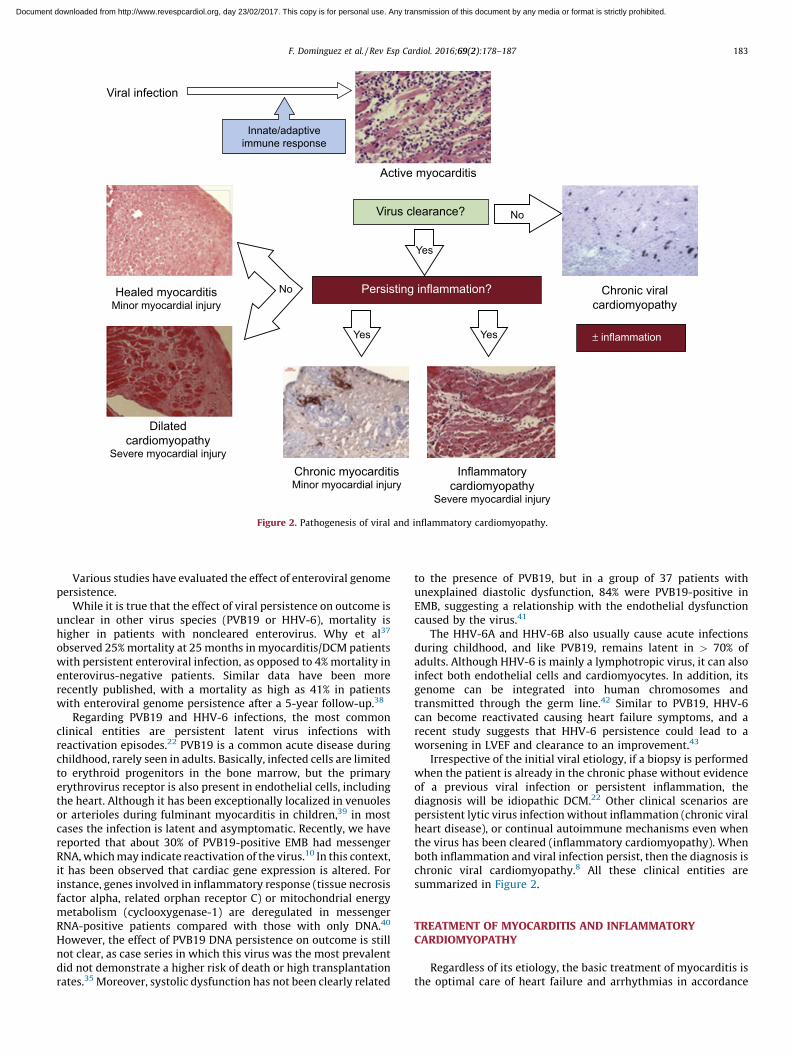
with evidence-based guidelines.44 Nonconventional and specifictreatments depend on the result of the EMB, taking into accountthe patients’ symptoms and the disease course33,34,45 (Figure 3).
Conventional Treatment of Myocarditis
Hemodynamically unstable patients should be managed inintensive care units with invasive monitoring and a skilled team ofprofessionals for cardiac catheterization and the performanceof EMB. In patients who develop progressive deterioration ofcardiac pump function despite conventional treatment, EMB isessential to diagnose potentially treatable causes such as giant cellor eosinophilic myocarditis. However, as myocardial injuriesprogress rapidly and can quickly become irreversible, a mechanicalcardio-pulmonary assist device or extracorporeal membraneoxygenation may sometimes be needed as a bridge to hearttransplantation or recovery.1
Stable patients with systolic ventricular dysfunction should betreated with diuretics, renin angiotensin aldosterone systeminhibitors, and beta-adrenergic blockade. The specific momentwhen these drugs should be withdrawn after LVEF recovery is notwell defined.46 Regarding nonsteroidal anti-inflammatory drugs,their use is not recommended due to a mortality increase in animalexperimental models of myocarditis, even though they are widelyimplemented in the treatment of pericarditis. Implantablecardioverter defibrillator implantation is only recommended ifsymptoms and systolic cardiac dysfunction persist after the acutephase. In the meantime, when a patient is discharged after acutemyocarditis with low LVEF, wearable cardioverter defibrillators(LifeVestW) can provide protection as a bridge to implantablecardioverter defibrillator decision.
Clinically suspected
Clinical presen
EMB
RepeatEMB in
3-6 months
Inflammation +(virus negative)
Treatment of specific causesGCM: ATG + CyA + PredSarcoidosis/eosinoph:Pred + HF treatment
EV�
PociH�
�
+
Virus +(± inflammation) (
3-6 mon
Symptomatic HF treatment
Symptomatic HF treatment
New onset HF(2 weeks-3 monwithout significa
CAD
Acute coronarysyndrome-like
Figure 3. Myocarditis treatment according to clinical setting and endomyocardial bdisease; ciHHV-6, chromosomally integrated human herpes virus type 6; CyA, cyclobyopsy; GCM, giant cell myocarditis; HF, heart failure; IFNb, interferon beta; mRNpatients, consider interferon beta or other potential options under study such as
treatment, consider ganciclovir or valganciclovir (see text).
Specific Treatments During the Acute Phase
Biopsy-proven acute viral myocarditis often improves sponta-neously in more than 60% of patients with conventional heartfailure treatment and therefore close follow-up is usually sufficientin these patients.8 In fact, the initial cardiac inflammation helps toeliminate the virus as soon as possible to prevent irreversiblemyocardial injuries, and anti-inflammatory or immunosuppres-sive therapy can favor viral persistence and therefore worsen thepatient’s outcome.47 However, it is still not well studied whetherthe presence of specific markers such as perforin during acutemyocarditis affect prognosis, and if these patients could benefitfrom early treatment.
On the other hand, other clinical entities benefit stronglyfrom specific treatments during the acute phase. Combinedtreatment of giant cell myocarditis with antithymoglobulin,cyclosporine (through level 100-120 mg/mL) and cortisonehas proved to improve survival in previous studies34 (Table 2,treatment regimens).
Hypereosinophilic syndrome, or Loeffler disease, usuallydevelops in 3 stages. In the acute phase, mature eosinophilsinfiltrate and damage the myocardium and hypereosinophilia isevidenced in peripheral blood. Then, valve involvement and apicalobliteration are observed and the final stage is endomyocardialfibrosis.48 During the acute phase, in which extensive irreversiblefibrosis is not present, antihelminthic or antiprotozoal drugs can beused in the tropical form of the disease. In all other clinicalscenarios, immunosuppression is recommended. The most com-mon treatment regimen is cortisone and azathioprine, withcortisone being decreased every 2 weeks by 10 mg from an initialdose of 1 mg/kg until a maintenance dose of 10 mg for 6 months.Other therapeutic options that have proved some benefit in this
myocarditis
tation
/ADV IFN βsitive PVB19 mRNAa/HV-6b:
Individualizea,b
No specific therapy HF treatment
Inflammation +(virus negative)
Azathioprine + Pred+ HF treatment
Consider geneticorigin
Inflammation –Virus –
Virus +± inflammation)
EMB
± DCM
ths
ths)nt
Chronic HF(>3 months) without
significant CAD
iopsy results. ADV, adenovirus; ATG, anti thymoglobulin; CAD, coronary arterysporin; DCM, dilated cardiomyopathy; EV, enterovirus; EMB, endomyocardial
A, messenger RNA; Pred, prednisone; PVB19, parvovirus B19. aIn symptomatictelbivudine (see text). bIn symptomatic patients despite optimal heart failure
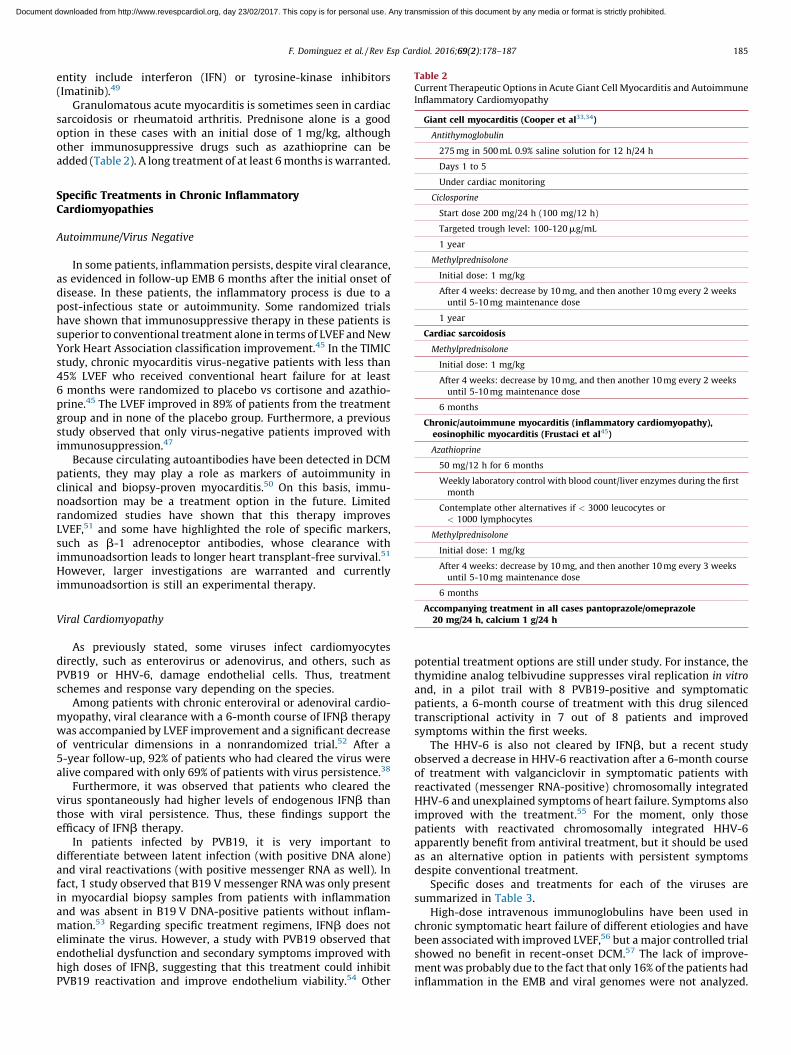
Table 2Current Therapeutic Options in Acute Giant Cell Myocarditis and AutoimmuneInflammatory Cardiomyopathy
Giant cell myocarditis (Cooper et al33,34)
Antithymoglobulin
275 mg in 500 mL 0.9% saline solution for 12 h/24 h
Days 1 to 5
Under cardiac monitoring
Ciclosporine
Start dose 200 mg/24 h (100 mg/12 h)
Targeted trough level: 100-120 mg/mL
1 year
Methylprednisolone
Initial dose: 1 mg/kg
After 4 weeks: decrease by 10 mg, and then another 10 mg every 2 weeks
until 5-10 mg maintenance dose
1 year
Cardiac sarcoidosis
Methylprednisolone
Initial dose: 1 mg/kg
After 4 weeks: decrease by 10 mg, and then another 10 mg every 2 weeks
until 5-10 mg maintenance dose
6 months
Chronic/autoimmune myocarditis (inflammatory cardiomyopathy),eosinophilic myocarditis (Frustaci et al45)
Azathioprine
50 mg/12 h for 6 months
Weekly laboratory control with blood count/liver enzymes during the first
month
Contemplate other alternatives if < 3000 leucocytes or
< 1000 lymphocytes
Methylprednisolone
Initial dose: 1 mg/kg
After 4 weeks: decrease by 10 mg, and then another 10 mg every 3 weeks
until 5-10 mg maintenance dose
6 months
Accompanying treatment in all cases pantoprazole/omeprazole20 mg/24 h, calcium 1 g/24 h
F. Dominguez et al. / Rev Esp Cardiol. 2016;69(2):178–187 185
Document downloaded from http://www.revespcardiol.org, day 23/02/2017. This copy is for personal use. Any transmission of this document by any media or format is strictly prohibited.
entity include interferon (IFN) or tyrosine-kinase inhibitors(Imatinib).49
Granulomatous acute myocarditis is sometimes seen in cardiacsarcoidosis or rheumatoid arthritis. Prednisone alone is a goodoption in these cases with an initial dose of 1 mg/kg, althoughother immunosuppressive drugs such as azathioprine can beadded (Table 2). A long treatment of at least 6 months is warranted.
Specific Treatments in Chronic InflammatoryCardiomyopathies
Autoimmune/Virus Negative
In some patients, inflammation persists, despite viral clearance,as evidenced in follow-up EMB 6 months after the initial onset ofdisease. In these patients, the inflammatory process is due to apost-infectious state or autoimmunity. Some randomized trialshave shown that immunosuppressive therapy in these patients issuperior to conventional treatment alone in terms of LVEF and NewYork Heart Association classification improvement.45 In the TIMICstudy, chronic myocarditis virus-negative patients with less than45% LVEF who received conventional heart failure for at least6 months were randomized to placebo vs cortisone and azathio-prine.45 The LVEF improved in 89% of patients from the treatmentgroup and in none of the placebo group. Furthermore, a previousstudy observed that only virus-negative patients improved withimmunosuppression.47
Because circulating autoantibodies have been detected in DCMpatients, they may play a role as markers of autoimmunity inclinical and biopsy-proven myocarditis.50 On this basis, immu-noadsortion may be a treatment option in the future. Limitedrandomized studies have shown that this therapy improvesLVEF,51 and some have highlighted the role of specific markers,such as b-1 adrenoceptor antibodies, whose clearance withimmunoadsortion leads to longer heart transplant-free survival.51
However, larger investigations are warranted and currentlyimmunoadsortion is still an experimental therapy.
Viral Cardiomyopathy
As previously stated, some viruses infect cardiomyocytesdirectly, such as enterovirus or adenovirus, and others, such asPVB19 or HHV-6, damage endothelial cells. Thus, treatmentschemes and response vary depending on the species.
Among patients with chronic enteroviral or adenoviral cardio-myopathy, viral clearance with a 6-month course of IFNb therapywas accompanied by LVEF improvement and a significant decreaseof ventricular dimensions in a nonrandomized trial.52 After a5-year follow-up, 92% of patients who had cleared the virus werealive compared with only 69% of patients with virus persistence.38
Furthermore, it was observed that patients who cleared thevirus spontaneously had higher levels of endogenous IFNb thanthose with viral persistence. Thus, these findings support theefficacy of IFNb therapy.
In patients infected by PVB19, it is very important todifferentiate between latent infection (with positive DNA alone)and viral reactivations (with positive messenger RNA as well). Infact, 1 study observed that B19 V messenger RNA was only presentin myocardial biopsy samples from patients with inflammationand was absent in B19 V DNA-positive patients without inflam-mation.53 Regarding specific treatment regimens, IFNb does noteliminate the virus. However, a study with PVB19 observed thatendothelial dysfunction and secondary symptoms improved withhigh doses of IFNb, suggesting that this treatment could inhibitPVB19 reactivation and improve endothelium viability.54 Other
potential treatment options are still under study. For instance, thethymidine analog telbivudine suppresses viral replication in vitro
and, in a pilot trail with 8 PVB19-positive and symptomaticpatients, a 6-month course of treatment with this drug silencedtranscriptional activity in 7 out of 8 patients and improvedsymptoms within the first weeks.
The HHV-6 is also not cleared by IFNb, but a recent studyobserved a decrease in HHV-6 reactivation after a 6-month courseof treatment with valganciclovir in symptomatic patients withreactivated (messenger RNA-positive) chromosomally integratedHHV-6 and unexplained symptoms of heart failure. Symptoms alsoimproved with the treatment.55 For the moment, only thosepatients with reactivated chromosomally integrated HHV-6apparently benefit from antiviral treatment, but it should be usedas an alternative option in patients with persistent symptomsdespite conventional treatment.
Specific doses and treatments for each of the viruses aresummarized in Table 3.
High-dose intravenous immunoglobulins have been used inchronic symptomatic heart failure of different etiologies and havebeen associated with improved LVEF,56 but a major controlled trialshowed no benefit in recent-onset DCM.57 The lack of improve-ment was probably due to the fact that only 16% of the patients hadinflammation in the EMB and viral genomes were not analyzed.
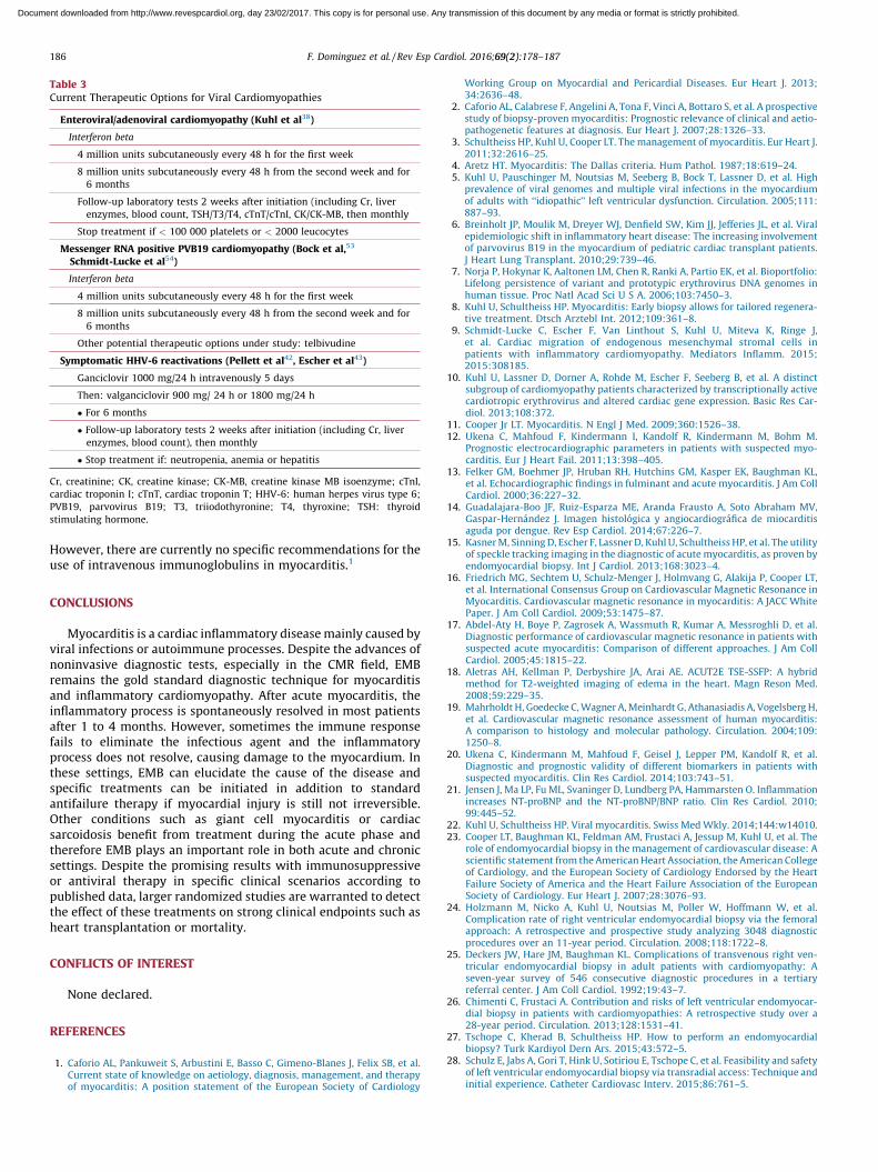
Table 3Current Therapeutic Options for Viral Cardiomyopathies
Enteroviral/adenoviral cardiomyopathy (Kuhl et al38)
Interferon beta
4 million units subcutaneously every 48 h for the first week
8 million units subcutaneously every 48 h from the second week and for
6 months
Follow-up laboratory tests 2 weeks after initiation (including Cr, liver
enzymes, blood count, TSH/T3/T4, cTnT/cTnI, CK/CK-MB, then monthly
Stop treatment if < 100 000 platelets or < 2000 leucocytes
Messenger RNA positive PVB19 cardiomyopathy (Bock et al,53
Schmidt-Lucke et al54)
Interferon beta
4 million units subcutaneously every 48 h for the first week
8 million units subcutaneously every 48 h from the second week and for
6 months
Other potential therapeutic options under study: telbivudine
Symptomatic HHV-6 reactivations (Pellett et al42, Escher et al43)
Ganciclovir 1000 mg/24 h intravenously 5 days
Then: valganciclovir 900 mg/ 24 h or 1800 mg/24 h
� For 6 months
� Follow-up laboratory tests 2 weeks after initiation (including Cr, liver
enzymes, blood count), then monthly
� Stop treatment if: neutropenia, anemia or hepatitis
Cr, creatinine; CK, creatine kinase; CK-MB, creatine kinase MB isoenzyme; cTnI,
cardiac troponin I; cTnT, cardiac troponin T; HHV-6: human herpes virus type 6;
PVB19, parvovirus B19; T3, triiodothyronine; T4, thyroxine; TSH: thyroid
stimulating hormone.
F. Dominguez et al. / Rev Esp Cardiol. 2016;69(2):178–187186
Document downloaded from http://www.revespcardiol.org, day 23/02/2017. This copy is for personal use. Any transmission of this document by any media or format is strictly prohibited.
However, there are currently no specific recommendations for theuse of intravenous immunoglobulins in myocarditis.1
CONCLUSIONS
Myocarditis is a cardiac inflammatory disease mainly caused byviral infections or autoimmune processes. Despite the advances ofnoninvasive diagnostic tests, especially in the CMR field, EMBremains the gold standard diagnostic technique for myocarditisand inflammatory cardiomyopathy. After acute myocarditis, theinflammatory process is spontaneously resolved in most patientsafter 1 to 4 months. However, sometimes the immune responsefails to eliminate the infectious agent and the inflammatoryprocess does not resolve, causing damage to the myocardium. Inthese settings, EMB can elucidate the cause of the disease andspecific treatments can be initiated in addition to standardantifailure therapy if myocardial injury is still not irreversible.Other conditions such as giant cell myocarditis or cardiacsarcoidosis benefit from treatment during the acute phase andtherefore EMB plays an important role in both acute and chronicsettings. Despite the promising results with immunosuppressiveor antiviral therapy in specific clinical scenarios according topublished data, larger randomized studies are warranted to detectthe effect of these treatments on strong clinical endpoints such asheart transplantation or mortality.
CONFLICTS OF INTEREST
None declared.
REFERENCES
1. Caforio AL, Pankuweit S, Arbustini E, Basso C, Gimeno-Blanes J, Felix SB, et al.Current state of knowledge on aetiology, diagnosis, management, and therapyof myocarditis: A position statement of the European Society of Cardiology
Working Group on Myocardial and Pericardial Diseases. Eur Heart J. 2013;34:2636–48.
2. Caforio AL, Calabrese F, Angelini A, Tona F, Vinci A, Bottaro S, et al. A prospectivestudy of biopsy-proven myocarditis: Prognostic relevance of clinical and aetio-pathogenetic features at diagnosis. Eur Heart J. 2007;28:1326–33.
3. Schultheiss HP, Kuhl U, Cooper LT. The management of myocarditis. Eur Heart J.2011;32:2616–25.
4. Aretz HT. Myocarditis: The Dallas criteria. Hum Pathol. 1987;18:619–24.5. Kuhl U, Pauschinger M, Noutsias M, Seeberg B, Bock T, Lassner D, et al. High
prevalence of viral genomes and multiple viral infections in the myocardiumof adults with ‘‘idiopathic’’ left ventricular dysfunction. Circulation. 2005;111:887–93.
6. Breinholt JP, Moulik M, Dreyer WJ, Denfield SW, Kim JJ, Jefferies JL, et al. Viralepidemiologic shift in inflammatory heart disease: The increasing involvementof parvovirus B19 in the myocardium of pediatric cardiac transplant patients.J Heart Lung Transplant. 2010;29:739–46.
7. Norja P, Hokynar K, Aaltonen LM, Chen R, Ranki A, Partio EK, et al. Bioportfolio:Lifelong persistence of variant and prototypic erythrovirus DNA genomes inhuman tissue. Proc Natl Acad Sci U S A. 2006;103:7450–3.
8. Kuhl U, Schultheiss HP. Myocarditis: Early biopsy allows for tailored regenera-tive treatment. Dtsch Arztebl Int. 2012;109:361–8.
9. Schmidt-Lucke C, Escher F, Van Linthout S, Kuhl U, Miteva K, Ringe J,et al. Cardiac migration of endogenous mesenchymal stromal cells inpatients with inflammatory cardiomyopathy. Mediators Inflamm. 2015;2015:308185.
10. Kuhl U, Lassner D, Dorner A, Rohde M, Escher F, Seeberg B, et al. A distinctsubgroup of cardiomyopathy patients characterized by transcriptionally activecardiotropic erythrovirus and altered cardiac gene expression. Basic Res Car-diol. 2013;108:372.
11. Cooper Jr LT. Myocarditis. N Engl J Med. 2009;360:1526–38.12. Ukena C, Mahfoud F, Kindermann I, Kandolf R, Kindermann M, Bohm M.
Prognostic electrocardiographic parameters in patients with suspected myo-carditis. Eur J Heart Fail. 2011;13:398–405.
13. Felker GM, Boehmer JP, Hruban RH, Hutchins GM, Kasper EK, Baughman KL,et al. Echocardiographic findings in fulminant and acute myocarditis. J Am CollCardiol. 2000;36:227–32.
14. Guadalajara-Boo JF, Ruiz-Esparza ME, Aranda Frausto A, Soto Abraham MV,Gaspar-Hernandez J. Imagen histologica y angiocardiografica de miocarditisaguda por dengue. Rev Esp Cardiol. 2014;67:226–7.
15. Kasner M, Sinning D, Escher F, Lassner D, Kuhl U, Schultheiss HP, et al. The utilityof speckle tracking imaging in the diagnostic of acute myocarditis, as proven byendomyocardial biopsy. Int J Cardiol. 2013;168:3023–4.
16. Friedrich MG, Sechtem U, Schulz-Menger J, Holmvang G, Alakija P, Cooper LT,et al. International Consensus Group on Cardiovascular Magnetic Resonance inMyocarditis. Cardiovascular magnetic resonance in myocarditis: A JACC WhitePaper. J Am Coll Cardiol. 2009;53:1475–87.
17. Abdel-Aty H, Boye P, Zagrosek A, Wassmuth R, Kumar A, Messroghli D, et al.Diagnostic performance of cardiovascular magnetic resonance in patients withsuspected acute myocarditis: Comparison of different approaches. J Am CollCardiol. 2005;45:1815–22.
18. Aletras AH, Kellman P, Derbyshire JA, Arai AE. ACUT2E TSE-SSFP: A hybridmethod for T2-weighted imaging of edema in the heart. Magn Reson Med.2008;59:229–35.
19. Mahrholdt H, Goedecke C, Wagner A, Meinhardt G, Athanasiadis A, Vogelsberg H,et al. Cardiovascular magnetic resonance assessment of human myocarditis:A comparison to histology and molecular pathology. Circulation. 2004;109:1250–8.
20. Ukena C, Kindermann M, Mahfoud F, Geisel J, Lepper PM, Kandolf R, et al.Diagnostic and prognostic validity of different biomarkers in patients withsuspected myocarditis. Clin Res Cardiol. 2014;103:743–51.
21. Jensen J, Ma LP, Fu ML, Svaninger D, Lundberg PA, Hammarsten O. Inflammationincreases NT-proBNP and the NT-proBNP/BNP ratio. Clin Res Cardiol. 2010;99:445–52.
22. Kuhl U, Schultheiss HP. Viral myocarditis. Swiss Med Wkly. 2014;144:w14010.23. Cooper LT, Baughman KL, Feldman AM, Frustaci A, Jessup M, Kuhl U, et al. The
role of endomyocardial biopsy in the management of cardiovascular disease: Ascientific statement from the American Heart Association, the American Collegeof Cardiology, and the European Society of Cardiology Endorsed by the HeartFailure Society of America and the Heart Failure Association of the EuropeanSociety of Cardiology. Eur Heart J. 2007;28:3076–93.
24. Holzmann M, Nicko A, Kuhl U, Noutsias M, Poller W, Hoffmann W, et al.Complication rate of right ventricular endomyocardial biopsy via the femoralapproach: A retrospective and prospective study analyzing 3048 diagnosticprocedures over an 11-year period. Circulation. 2008;118:1722–8.
25. Deckers JW, Hare JM, Baughman KL. Complications of transvenous right ven-tricular endomyocardial biopsy in adult patients with cardiomyopathy: Aseven-year survey of 546 consecutive diagnostic procedures in a tertiaryreferral center. J Am Coll Cardiol. 1992;19:43–7.
26. Chimenti C, Frustaci A. Contribution and risks of left ventricular endomyocar-dial biopsy in patients with cardiomyopathies: A retrospective study over a28-year period. Circulation. 2013;128:1531–41.
27. Tschope C, Kherad B, Schultheiss HP. How to perform an endomyocardialbiopsy? Turk Kardiyol Dern Ars. 2015;43:572–5.
28. Schulz E, Jabs A, Gori T, Hink U, Sotiriou E, Tschope C, et al. Feasibility and safetyof left ventricular endomyocardial biopsy via transradial access: Technique andinitial experience. Catheter Cardiovasc Interv. 2015;86:761–5.
F. Dominguez et al. / Rev Esp Cardiol. 2016;69(2):178–187 187
Document downloaded from http://www.revespcardiol.org, day 23/02/2017. This copy is for personal use. Any transmission of this document by any media or format is strictly prohibited.
29. Escher F, Lassner D, Kuhl U, Gross U, Westermann D, Poller W, et al. Analysis ofendomyocardial biopsies in suspected myocarditis—diagnostic value of leftversus right ventricular biopsy. Int J Cardiol. 2014;177:76–8.
30. Richardson P, McKenna W, Bristow M, Maisch B, Mautner B, O’Connell J, et al.Report of the 1995 World Health Organization/International Society and Fed-eration of Cardiology Task Force on the Definition and Classification of Cardio-myopathies. Circulation. 1996;93:841–2.
31. Escher F, Kuhl U, Lassner D, Stroux A, Westermann D, Skurk C, et al. Presence ofperforin in endomyocardial biopsies of patients with inflammatory cardiomy-opathy predicts poor outcome. Eur J Heart Fail. 2014;16:1066–72.
32. McCarthy RE, Boehmer JP, Hruban RH, Hutchins GM, Kasper EK, Hare JM, et al.Long-term outcome of fulminant myocarditis as compared with acute (non-fulminant) myocarditis. N Engl J Med. 2000;342:690–5.
33. Cooper Jr LT, Berry GJ, Shabetai R. Idiopathic giant-cell myocarditis—naturalhistory and treatment. Multicenter Giant Cell Myocarditis Study Group Inves-tigators. N Engl J Med. 1997;336:1860–6.
34. Cooper Jr LT, Hare JM, Tazelaar HD, Edwards WD, Starling RC, Deng MC, et al.Giant Cell Myocarditis Treatment Trial Investigators. Usefulness of immuno-suppression for giant cell myocarditis. Am J Cardiol. 2008;102:1535–9.
35. Kindermann I, Kindermann M, Kandolf R, Klingel K, Bultmann B, Muller T, et al.Predictors of outcome in patients with suspected myocarditis. Circulation.2008;118:639–48.
36. Pauschinger M, Bowles NE, Fuentes-Garcia FJ, Pham V, Kuhl U, SchwimmbeckPL, et al. Detection of adenoviral genome in the myocardium of adult patientswith idiopathic left ventricular dysfunction. Circulation. 1999;99:1348–54.
37. Why HJ, Meany BT, Richardson PJ, Olsen EG, Bowles NE, Cunningham L, et al.Clinical and prognostic significance of detection of enteroviral RNA in themyocardium of patients with myocarditis or dilated cardiomyopathy. Circula-tion. 1994;89:2582–9.
38. Kuhl U, Lassner D, Von Schlippenbach J, Poller W, Schultheiss HP. Interferon-beta improves survival in enterovirus-associated cardiomyopathy. J Am CollCardiol. 2012;60:1295–6.
39. Bultmann BD, Klingel K, Sotlar K, Bock CT, Baba HA, Sauter M, et al. Fatalparvovirus B19-associated myocarditis clinically mimicking ischemic heartdisease: An endothelial cell-mediated disease. Hum Pathol. 2003;34:92–5.
40. Kuhl U, Rohde M, Lassner D, Gross UM, Escher F, Schultheiss HP. miRNA asactivity markers in Parvo B19 associated heart disease. Herz. 2012;37:637–43.
41. Tschope C, Bock CT, Kasner M, Noutsias M, Westermann D, Schwimmbeck PL,et al. High prevalence of cardiac parvovirus B19 infection in patients withisolated left ventricular diastolic dysfunction. Circulation. 2005;111:879–86.
42. Pellett PE, Ablashi DV, Ambros PF, Agut H, Caserta MT, Descamps V, et al.Chromosomally integrated human herpesvirus 6: Questions and answers. RevMed Virol. 2012;22:144–55.
43. Escher F, Kuhl U, Gross U, Westermann D, Poller W, Tschope C, et al. Aggravationof left ventricular dysfunction in patients with biopsy-proven cardiac humanherpesvirus A and B infection. J Clin Virol. 2015;63:1–5.
44. Camm AJ, Lip GY, De Caterina R, Savelieva I, Atar D, Hohnloser SH, et al.2012 focused update of the ESC guidelines for the management of atrial
fibrillation: An update of the 2010 ESC guidelines for the management of atrialfibrillation—developed with the special contribution of the European HeartRhythm Association. Europace. 2012;14:1385–413.
45. Frustaci A, Russo MA, Chimenti C. Randomized study on the efficacy of immu-nosuppressive therapy in patients with virus-negative inflammatory cardio-myopathy: The TIMIC study. Eur Heart J. 2009;30:1995–2002.
46. Anguita-Sanchez M, Castillo-Domınguez JC, Mesa-Rubio D, Ruiz-Ortiz M,Lopez-Granados A, Suarez de Lezo J.
?
Se deben mantener los inhibidores dela enzima de conversion de la angiotensina a largo plazo en pacientes quenormalizan la fraccion de eyeccion tras un episodio de miocarditis aguda? RevEsp Cardiol. 2006;59:1199–201.
47. Frustaci A, Chimenti C, Calabrese F, Pieroni M, Thiene G, Maseri A. Immuno-suppressive therapy for active lymphocytic myocarditis: Virological and im-munologic profile of responders versus nonresponders. Circulation. 2003;107:857–63.
48. Maisch B, Pankuweit S. Current treatment options in (peri)myocarditis andinflammatory cardiomyopathy. Herz. 2012;37:644–56.
49. Metzgeroth G, Walz C, Erben P, Popp H, Schmitt-Graeff A, Haferlach C, et al.Safety and efficacy of imatinib in chronic eosinophilic leukaemia and hyper-eosinophilic syndrome: A phase-II study. Br J Haematol. 2008;143:707–15.
50. Caforio AL, Goldman JH, Haven AJ, Baig KM, Libera LD, McKenna WJ. Circulatingcardiac-specific autoantibodies as markers of autoimmunity in clinical andbiopsy-proven myocarditis. The Myocarditis Treatment Trial Investigators. EurHeart J. 1997;18:270–5.
51. Dandel M, Wallukat G, Englert A, Lehmkuhl HB, Knosalla C, Hetzer R. Long-termbenefits of immunoadsorption in beta(1)-adrenoceptor autoantibody-positivetransplant candidates with dilated cardiomyopathy. Eur J Heart Fail. 2012;14:1374–88.
52. Kuhl U, Pauschinger M, Schwimmbeck PL, Seeberg B, Lober C, Noutsias M, et al.Interferon-beta treatment eliminates cardiotropic viruses and improves leftventricular function in patients with myocardial persistence of viral genomesand left ventricular dysfunction. Circulation. 2003;107:2793–8.
53. Bock CT, Klingel K, Kandolf R. Human parvovirus B19-associated myocarditis. NEngl J Med. 2010;362:1248–9.
54. Schmidt-Lucke C, Spillmann F, Bock T, Kuhl U, Van Linthout S, Schultheiss HP,et al. Interferon beta modulates endothelial damage in patients with cardiacpersistence of human parvovirus B19 infection. J Infect Dis. 2010;201:936–45.
55. Kuhl U, Lassner D, Wallaschek N, Gross UM, Krueger GR, Seeberg B, et al.Chromosomally integrated human herpesvirus 6 in heart failure: Prevalenceand treatment. Eur J Heart Fail. 2015;17:9–19.
56. Orange JS, Hossny EM, Weiler CR, Ballow M, Berger M, Bonilla FA, et al. Use ofintravenous immunoglobulin in human disease: A review of evidence bymembers of the Primary Immunodeficiency Committee of the American Acad-emy of Allergy, Asthma and Immunology. J Allergy Clin Immunol. 2006;117:S525–53.
57. McNamara DM, Holubkov R, Starling RC, Dec GW, Loh E, Torre-Amione G, et al.Controlled trial of intravenous immune globulin in recent-onset dilated car-diomyopathy. Circulation. 2001;103:2254–9.