Embed Size (px)
Citation preview
Orlando, Florida – October 7-9, 2011
Update on Treatment of Ventricular TachycardiaRalph Augostini, MD FACC FHRS
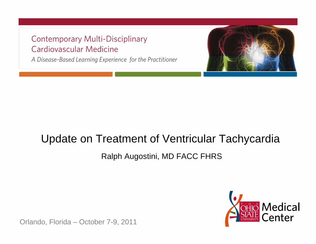
Ventricular Tachycardia Therapy Non-ICD Options
Anti-arrhythmic DrugCatheter AblationDo Nothing
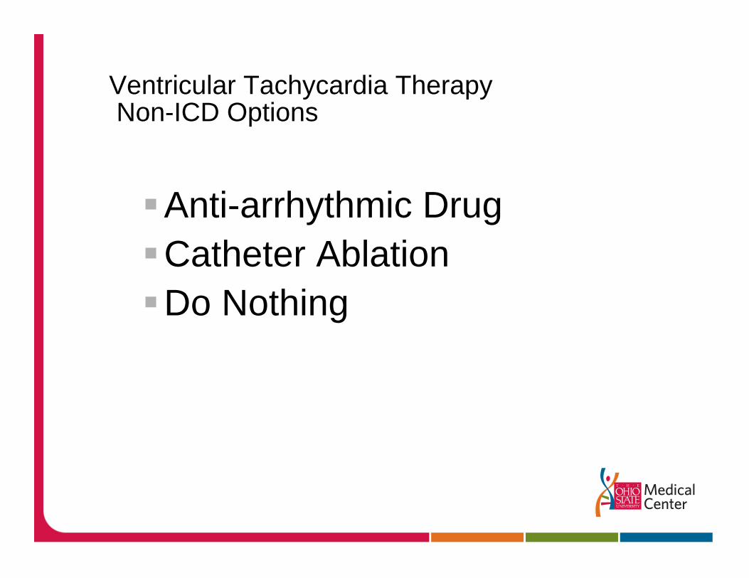
Non-ICD VT TherapySpecific Clinical / Arrhythmia Subgroups
Idiopathic VTLeft bundle inferior axisRight bundle superior axis
“Benign” PVC’sRefractory VT and recurrent ICD shocks
Commonly ischemia mediated
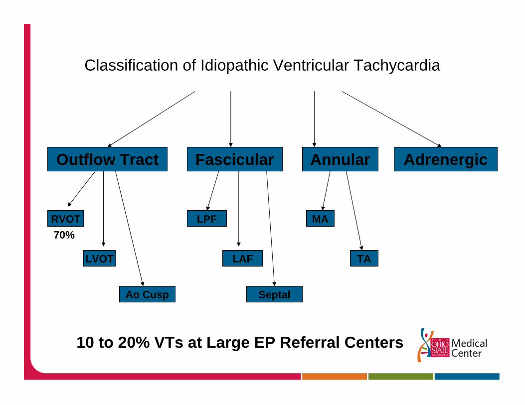
Classification of Idiopathic Ventricular Tachycardia
Outflow Tract Fascicular Annular Adrenergic
RVOT
LVOT
Ao Cusp
LPF
LAF
Septal
TA
MA70%
10 to 20% VTs at Large EP Referral Centers
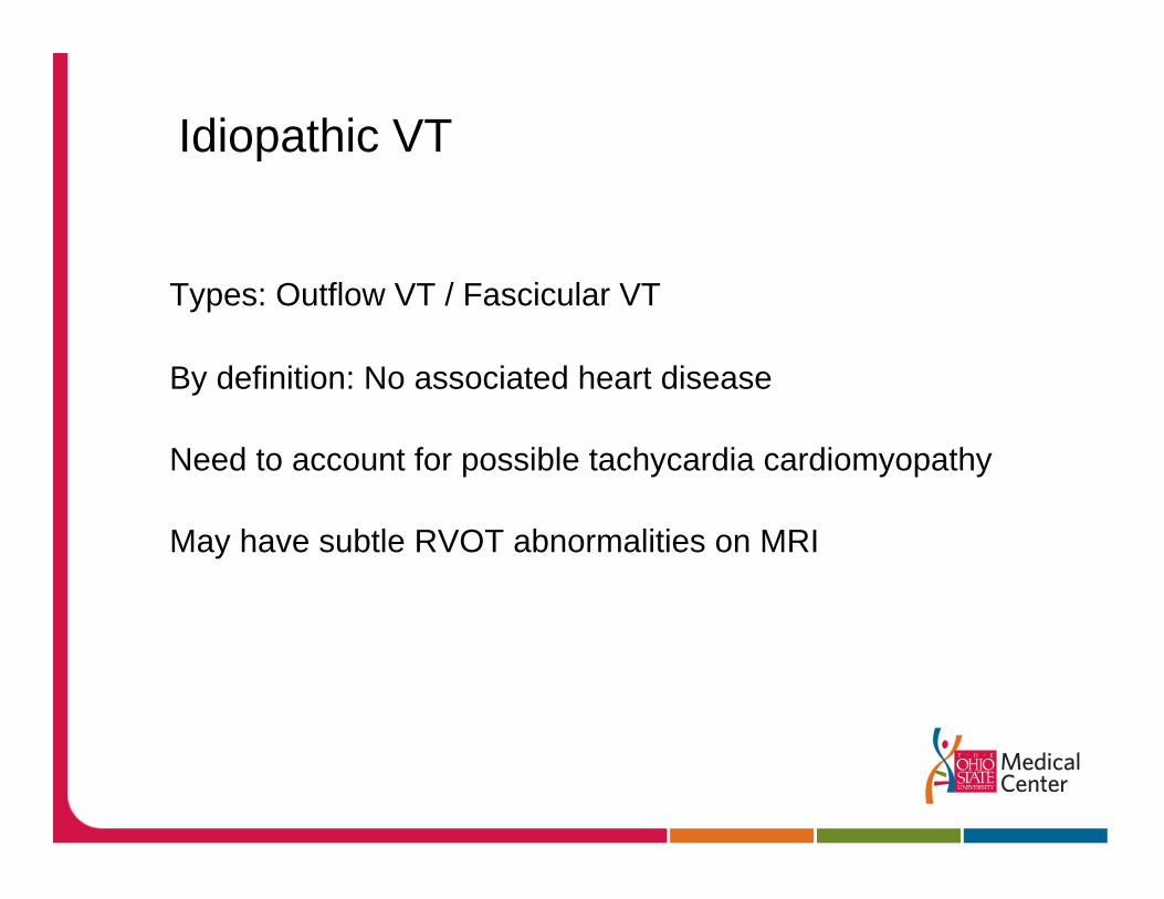
Idiopathic VT
Types: Outflow VT / Fascicular VT
By definition: No associated heart disease
Need to account for possible tachycardia cardiomyopathy
May have subtle RVOT abnormalities on MRI
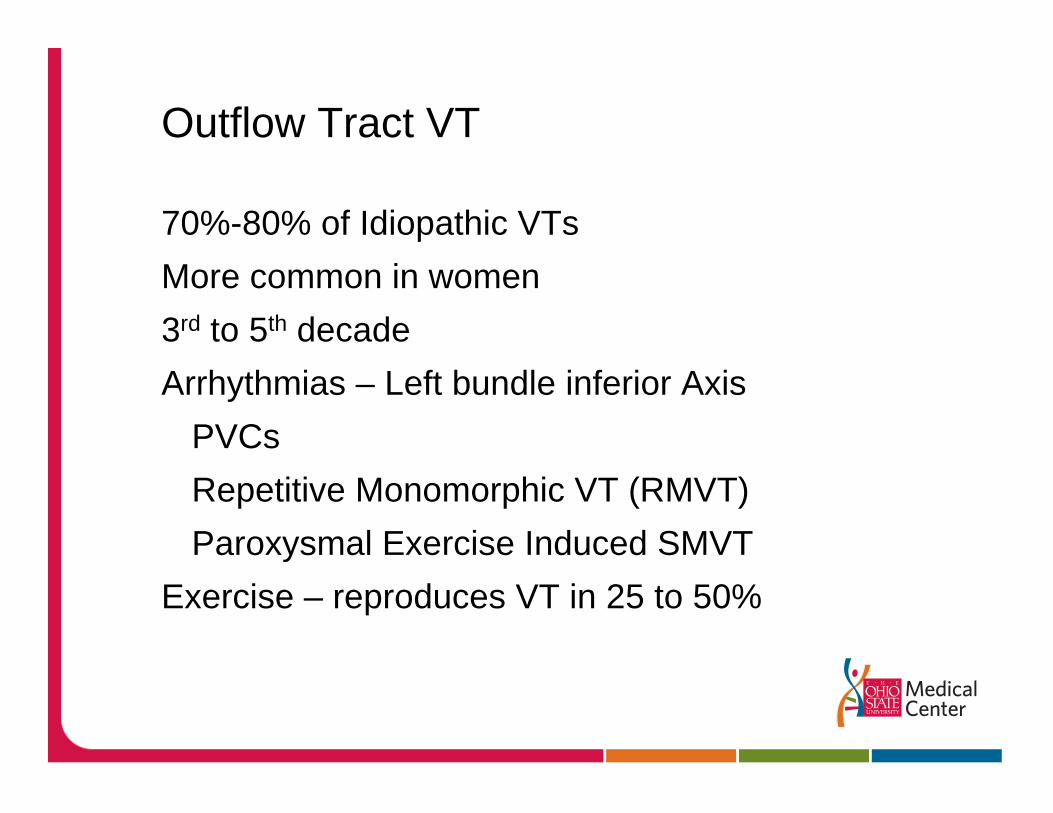
Outflow Tract VT
70%-80% of Idiopathic VTsMore common in women3rd to 5th decadeArrhythmias – Left bundle inferior Axis
PVCsRepetitive Monomorphic VT (RMVT)Paroxysmal Exercise Induced SMVT
Exercise – reproduces VT in 25 to 50%
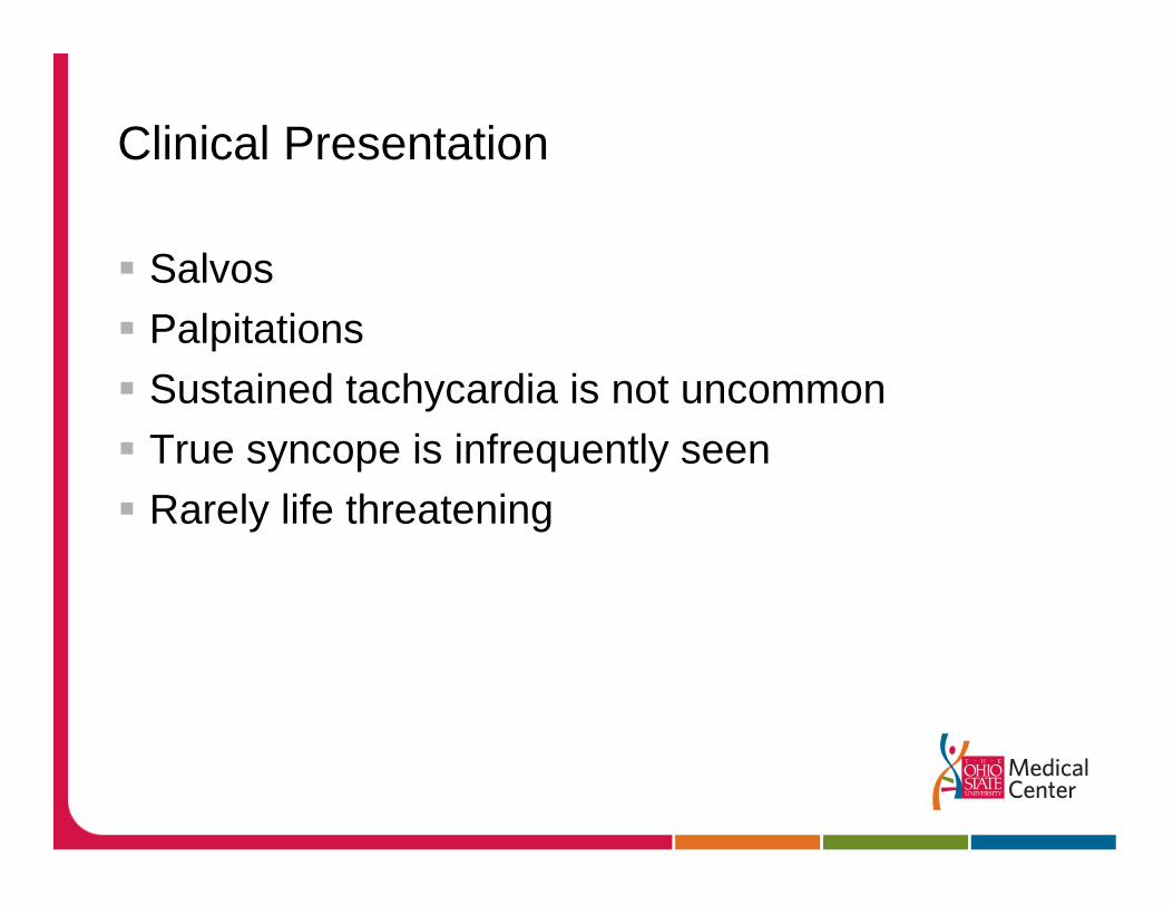
Clinical Presentation
SalvosPalpitationsSustained tachycardia is not uncommonTrue syncope is infrequently seenRarely life threatening
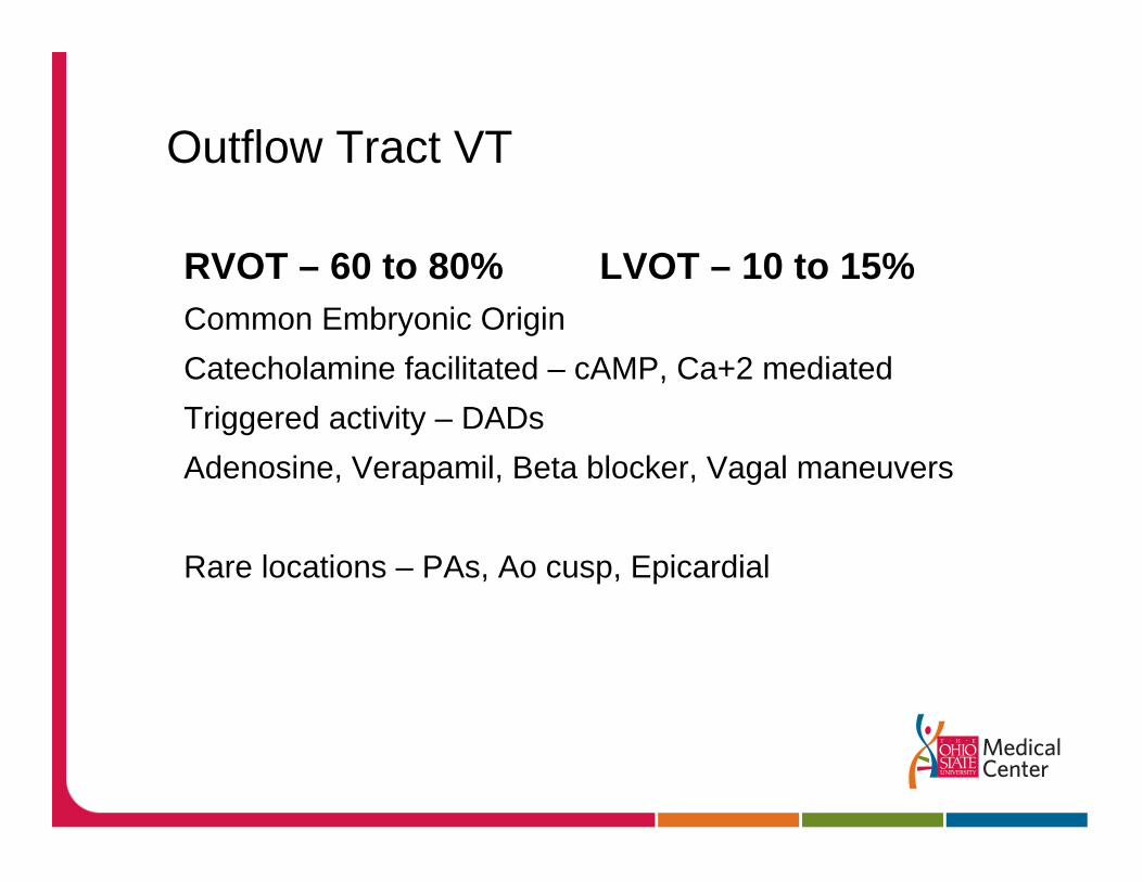
Outflow Tract VT
RVOT – 60 to 80% LVOT – 10 to 15%Common Embryonic OriginCatecholamine facilitated – cAMP, Ca+2 mediatedTriggered activity – DADsAdenosine, Verapamil, Beta blocker, Vagal maneuvers
Rare locations – PAs, Ao cusp, Epicardial
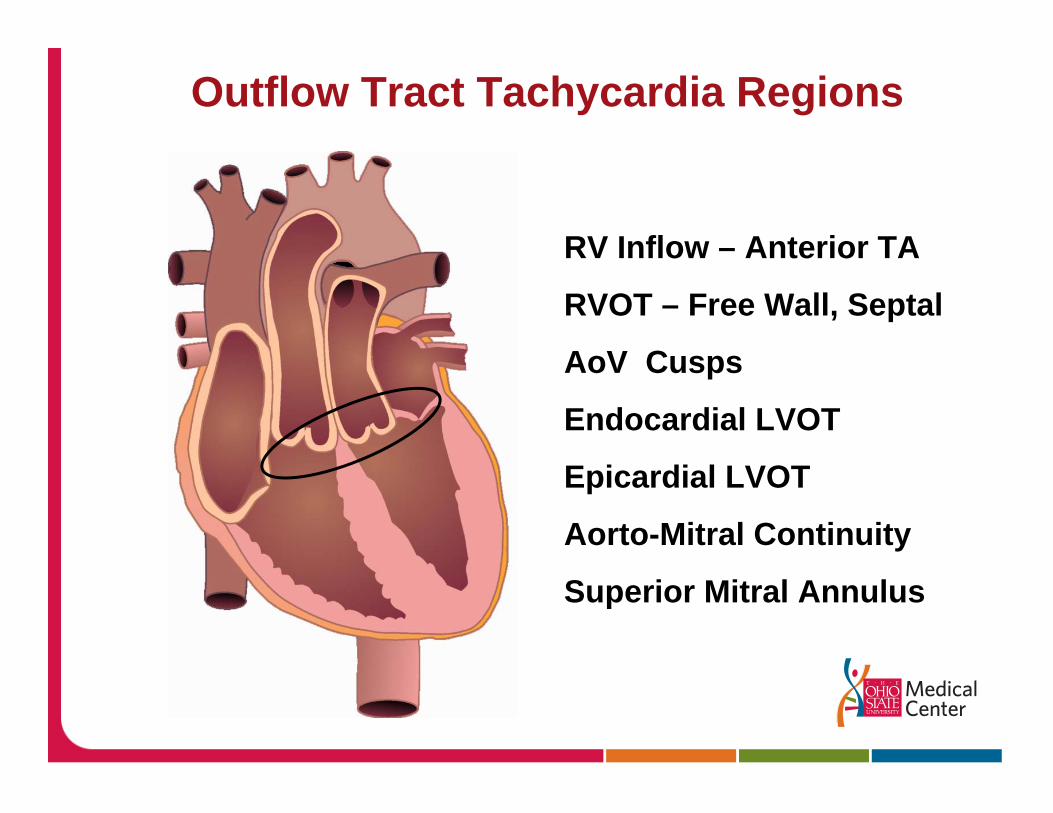
Outflow Tract Tachycardia Regions
RV Inflow – Anterior TA
RVOT – Free Wall, Septal
AoV Cusps
Endocardial LVOT
Epicardial LVOT
Aorto-Mitral Continuity
Superior Mitral Annulus
Outflow Tract VT
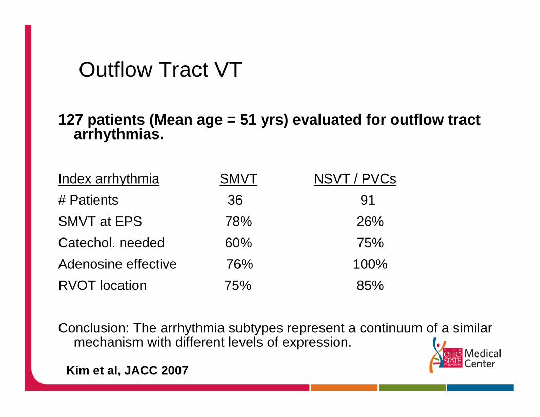
127 patients (Mean age = 51 yrs) evaluated for outflow tract arrhythmias.
Index arrhythmia SMVT NSVT / PVCs# Patients 36 91SMVT at EPS 78% 26%Catechol. needed 60% 75%Adenosine effective 76% 100%RVOT location 75% 85%
Conclusion: The arrhythmia subtypes represent a continuum of a similar mechanism with different levels of expression.
Kim et al, JACC 2007
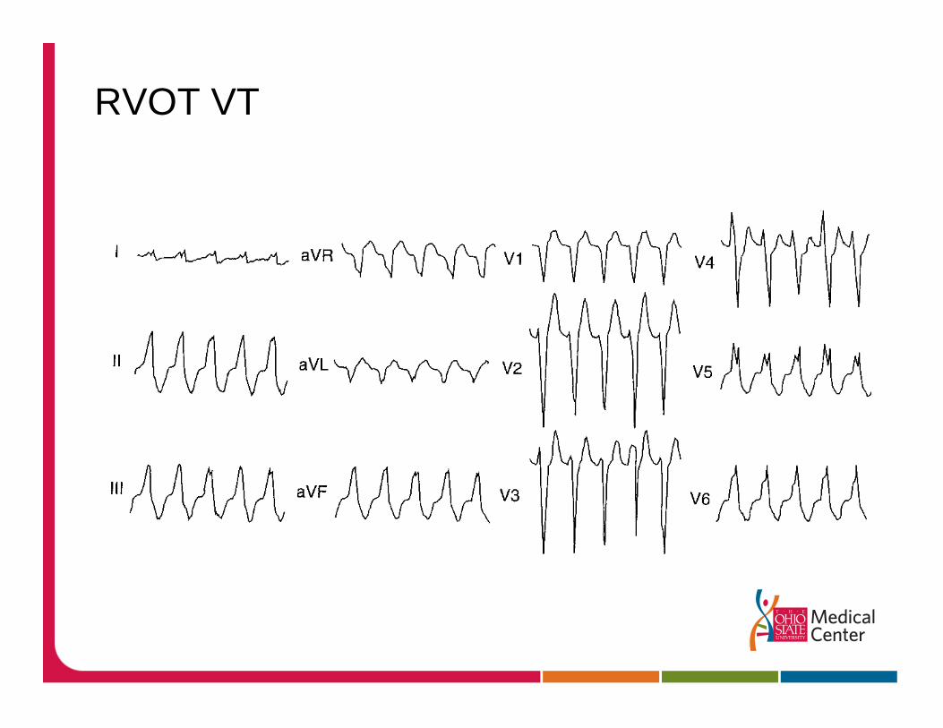
RVOT VT
Outflow Tract VT – Therapy
Medical TherapyAcute Termination
Vagal ManeuversAdenosineIV VerapamilLidocaine
Oral Medical TX – 25 to 50% response1st line: Beta blockers, Calcium blockers2nd line: Sotalol, FlecainideLast: Amio, Class IAs
RVOT VT – Therapy
Indications for AblationMedically RefractorySustained VTVery SymptomaticTachycardia CardiomyopathyYounger Patients
MappingPace Mapping: want > 11/12 match Activation: -10 to 45 ms
90% Efficacy 10% Recurrence rate
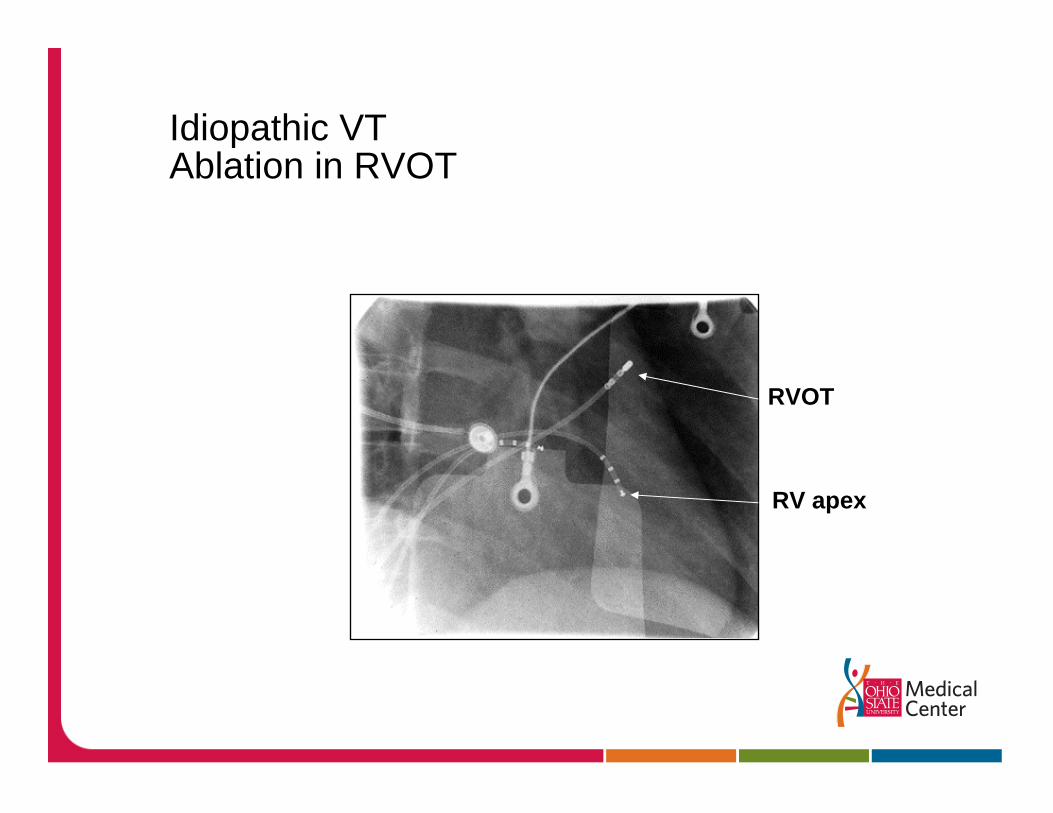
Idiopathic VT Ablation in RVOT
RVOT
RV apex
Differential Diagnosis of LBBB Morphology TachycardiaBenign vs Malignant Arrhythmias
BenignRVOT / LVOT idiopathic VTWPW / Mahaim tachycardia
MalignantARVDVT in Repaired Tetralogy of FallotBundle Branch Reentry – Dilated CM / IVCD
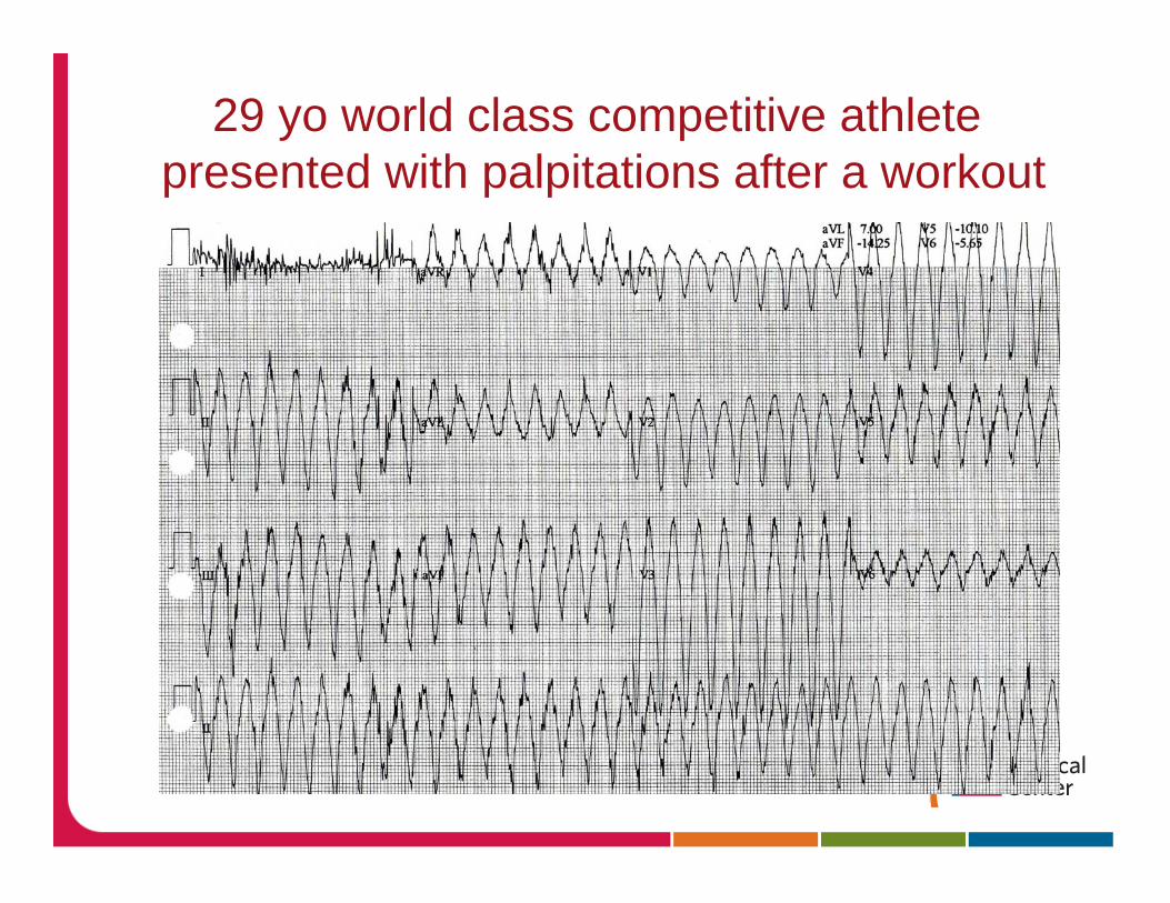
29 yo world class competitive athlete presented with palpitations after a workout
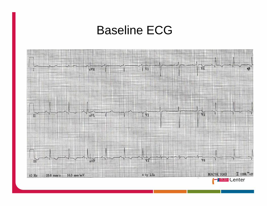
Baseline ECG
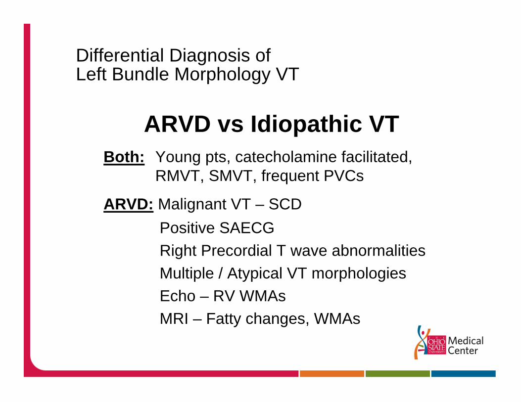
Differential Diagnosis of Left Bundle Morphology VT
ARVD vs Idiopathic VTBoth: Young pts, catecholamine facilitated,
RMVT, SMVT, frequent PVCs
ARVD: Malignant VT – SCDPositive SAECGRight Precordial T wave abnormalitiesMultiple / Atypical VT morphologiesEcho – RV WMAsMRI – Fatty changes, WMAs
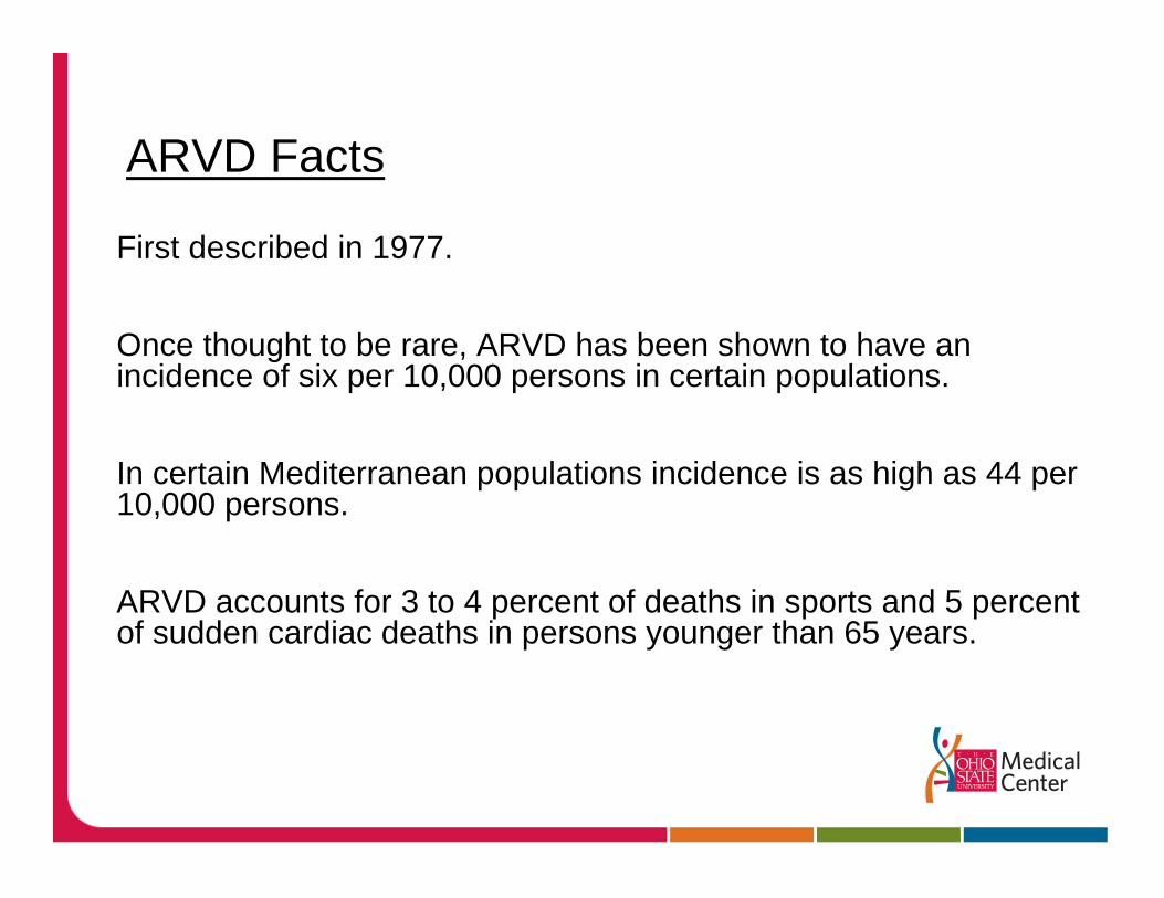
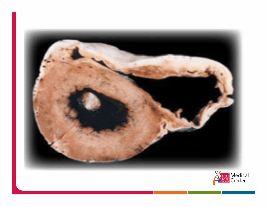
ARVD Facts
First described in 1977.
Once thought to be rare, ARVD has been shown to have an incidence of six per 10,000 persons in certain populations.
In certain Mediterranean populations incidence is as high as 44 per 10,000 persons.
ARVD accounts for 3 to 4 percent of deaths in sports and 5 percent of sudden cardiac deaths in persons younger than 65 years.
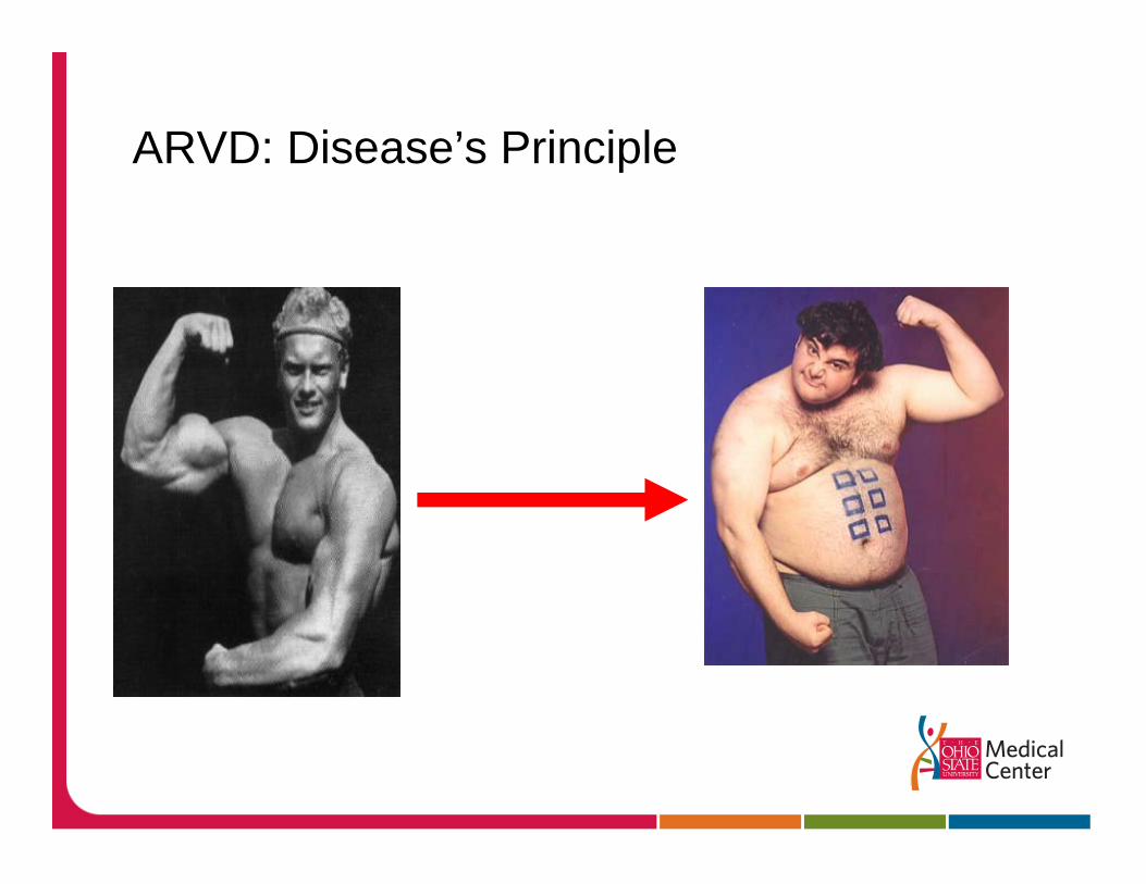
ARVD: Disease’s Principle
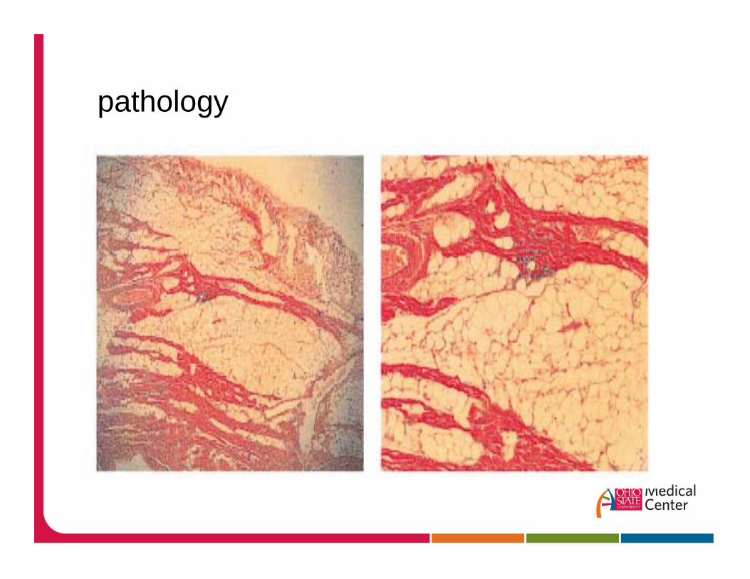
pathology
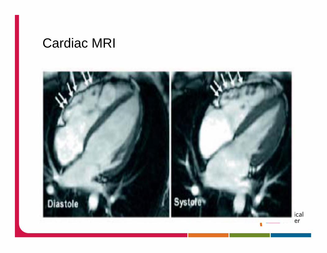
Cardiac MRI
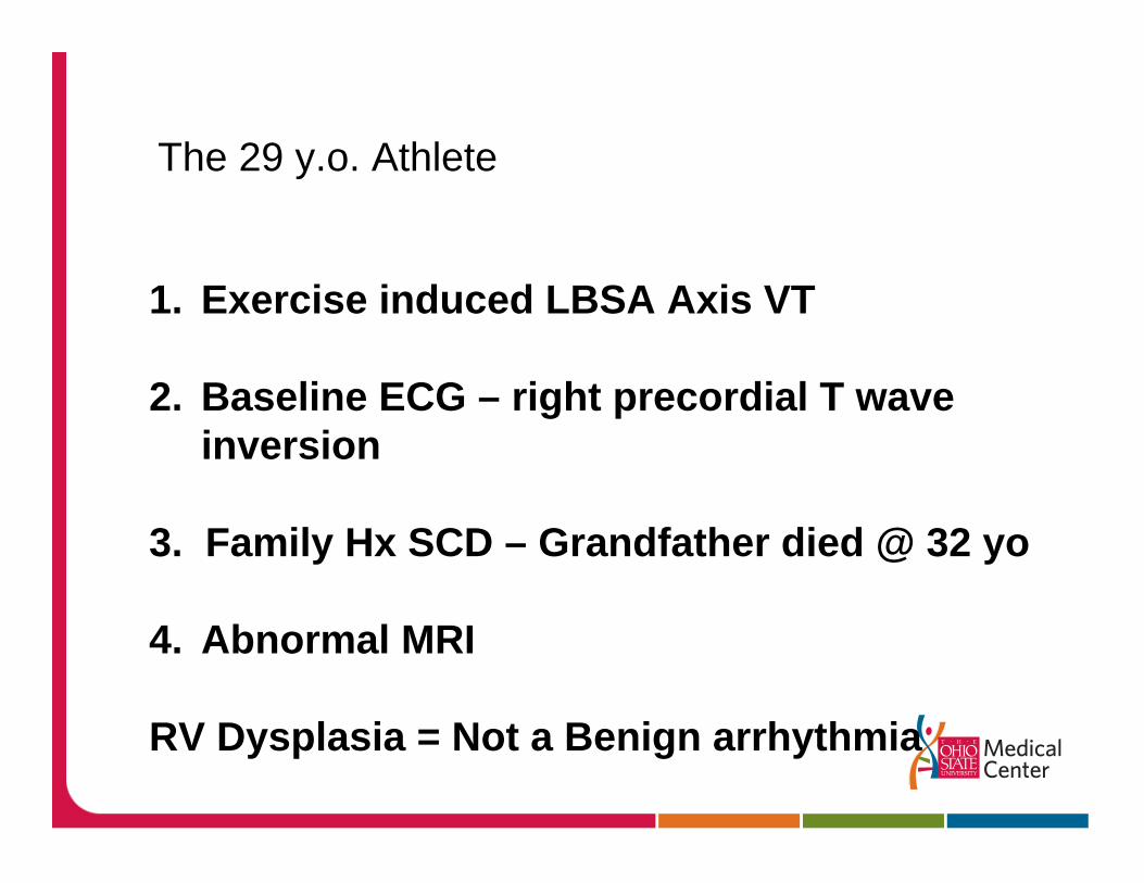
The 29 y.o. Athlete
1. Exercise induced LBSA Axis VT
2. Baseline ECG – right precordial T wave inversion
3. Family Hx SCD – Grandfather died @ 32 yo
4. Abnormal MRI
RV Dysplasia = Not a Benign arrhythmia
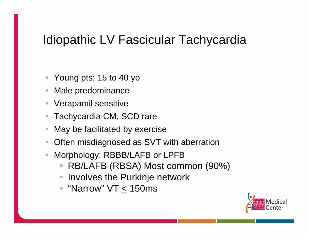
Idiopathic LV Fascicular Tachycardia
Young pts: 15 to 40 yoMale predominanceVerapamil sensitiveTachycardia CM, SCD rareMay be facilitated by exerciseOften misdiagnosed as SVT with aberrationMorphology: RBBB/LAFB or LPFB
RB/LAFB (RBSA) Most common (90%)Involves the Purkinje network“Narrow” VT < 150ms
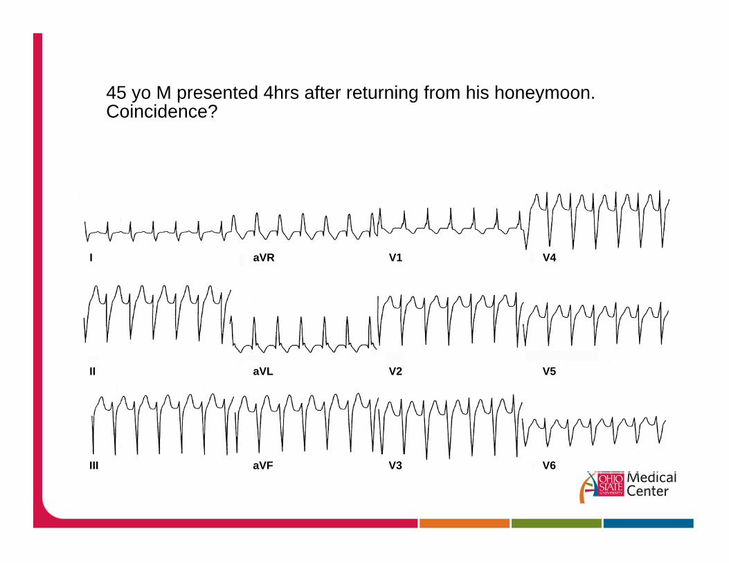
45 yo M presented 4hrs after returning from his honeymoon. Coincidence?
I aVR V1 V4
II aVL V2 V5
III aVF V3 V6
Ventriculartachycardia
PPEarliest PP
PP
QRS
DP
Sinusrhythm
PP
QRS
Retro PP
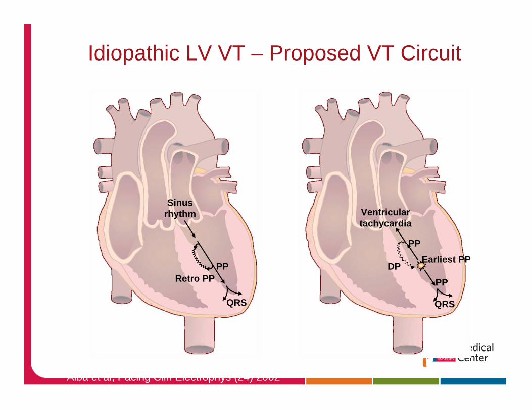
Idiopathic LV VT – Proposed VT Circuit
Aiba et al, Pacing Clin Electrophys (24) 2002
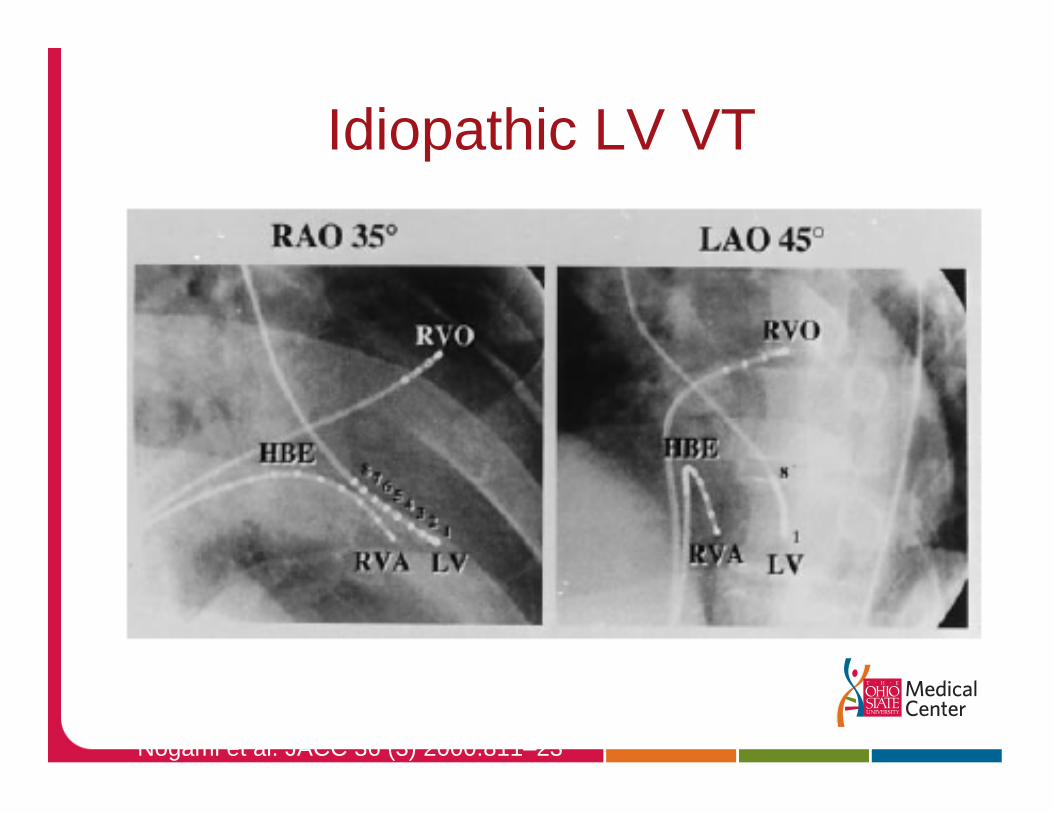
Nogami et al. JACC 36 (3) 2000:811–23
Idiopathic LV VT
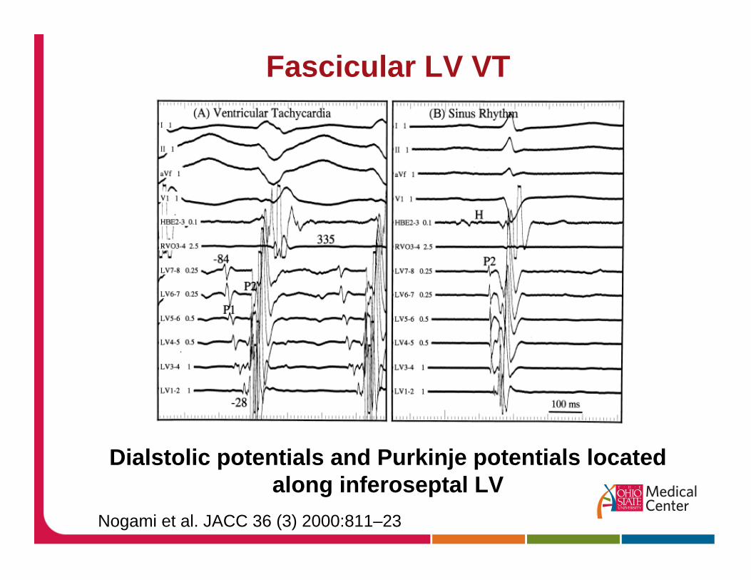
Nogami et al. JACC 36 (3) 2000:811–23
Dialstolic potentials and Purkinje potentials locatedalong inferoseptal LV
Fascicular LV VT
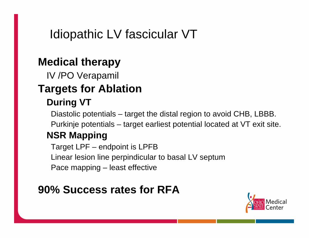
Idiopathic LV fascicular VT
Medical therapyIV /PO Verapamil
Targets for AblationDuring VTDiastolic potentials – target the distal region to avoid CHB, LBBB.Purkinje potentials – target earliest potential located at VT exit site.
NSR MappingTarget LPF – endpoint is LPFBLinear lesion line perpindicular to basal LV septum Pace mapping – least effective
90% Success rates for RFA
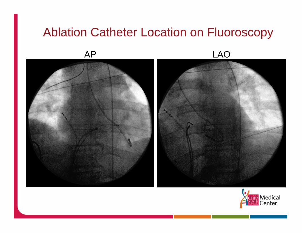
Ablation Catheter Location on Fluoroscopy
LAOAP
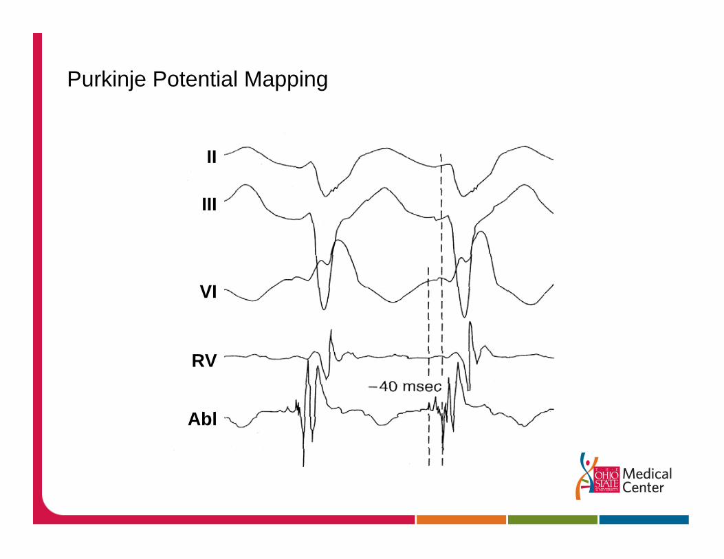
Purkinje Potential Mapping
II
III
VI
RV
Abl
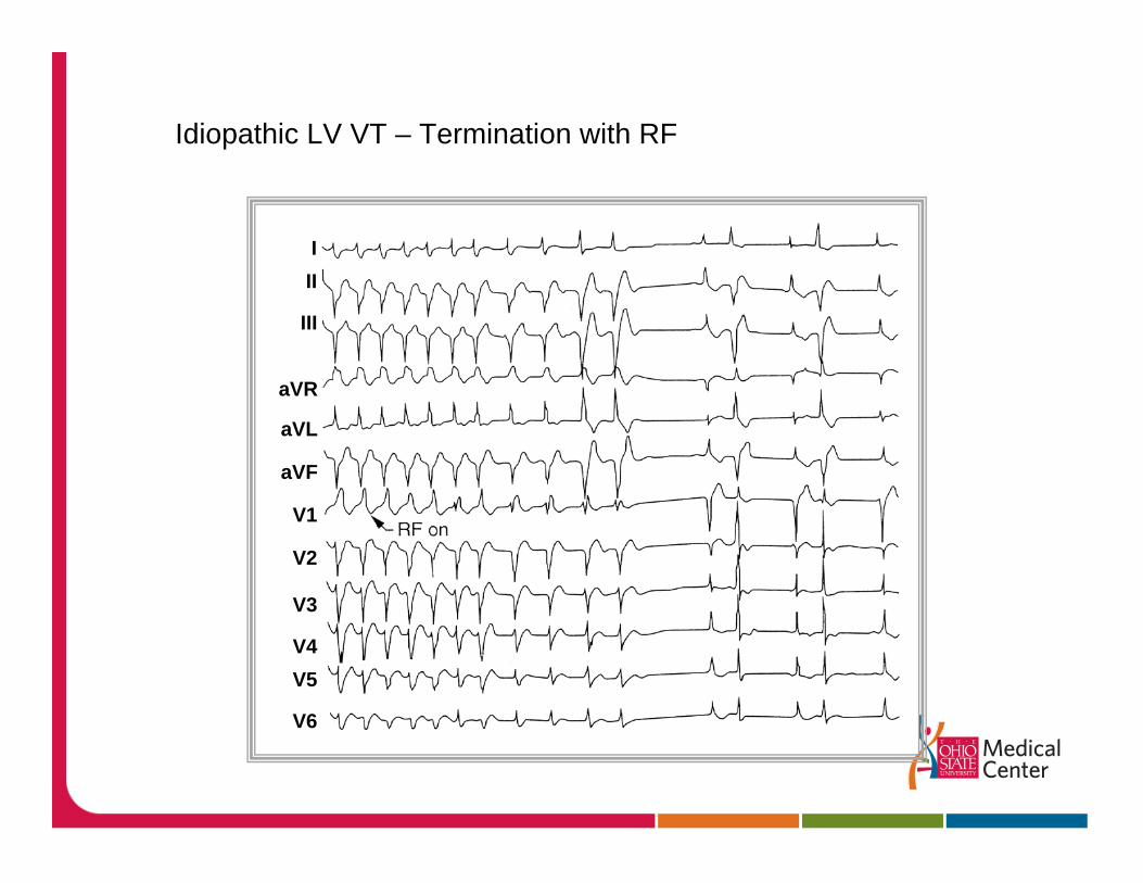
Idiopathic LV VT – Termination with RF
III
III
aVR
aVL
aVF
V1
V2
V3
V4V5
V6
What About Recurrent ICD Shocks?
The device is working but the patient can’t take it
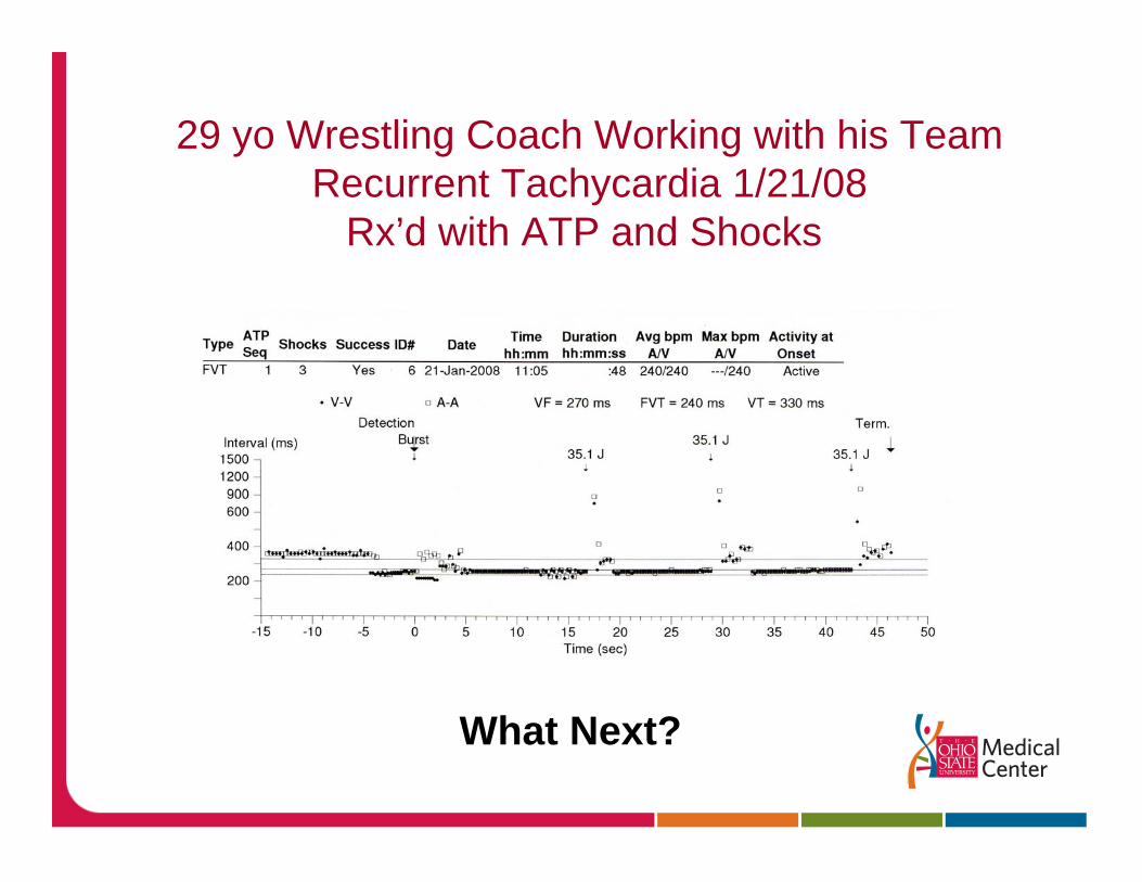
29 yo Wrestling Coach Working with his TeamRecurrent Tachycardia 1/21/08
Rx’d with ATP and Shocks
What Next?
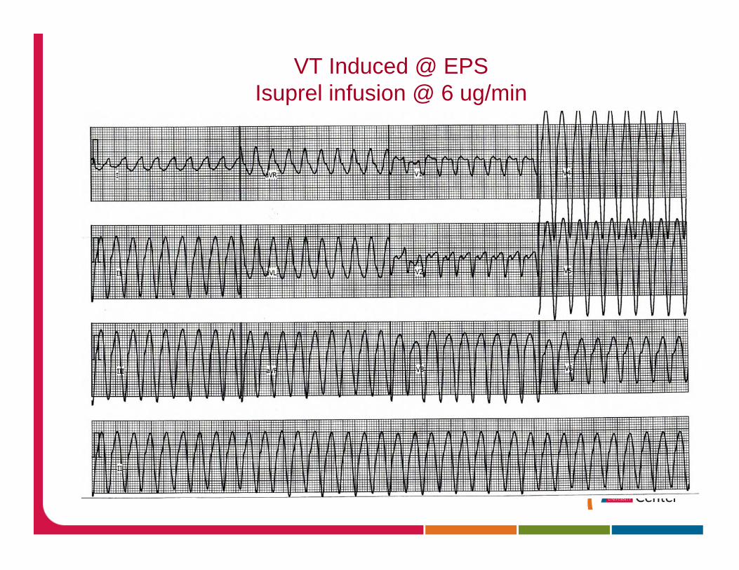
VT Induced @ EPSIsuprel infusion @ 6 ug/min
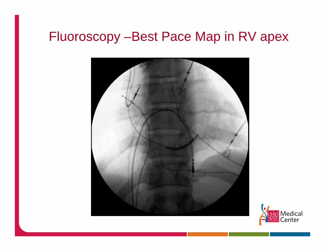
Fluoroscopy –Best Pace Map in RV apex
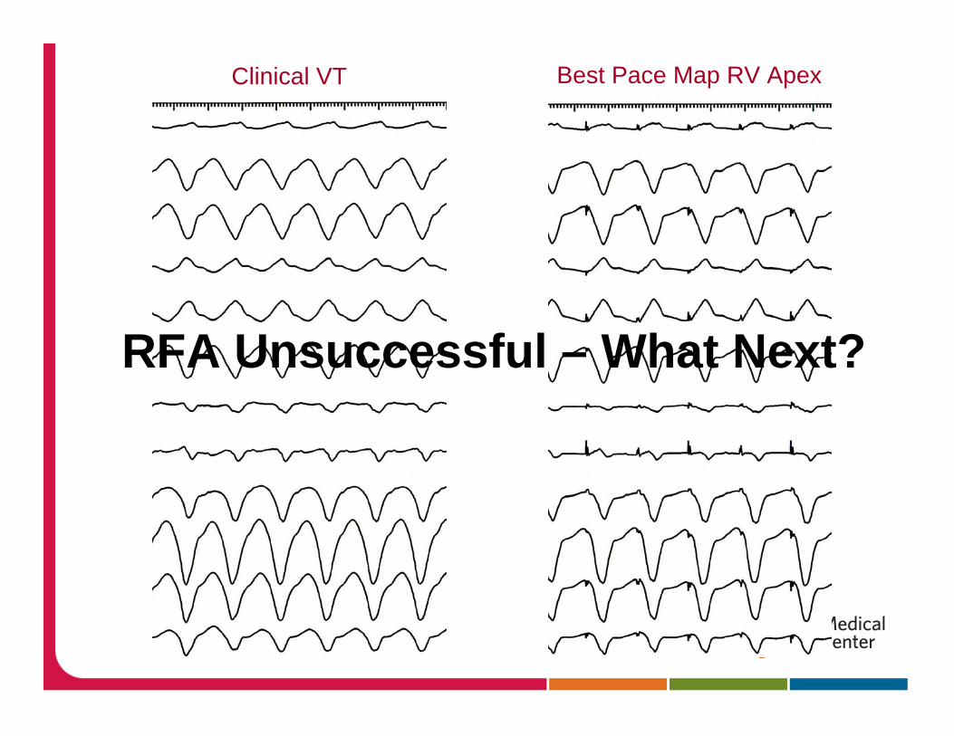
Clinical VT Best Pace Map RV Apex
RFA Unsuccessful – What Next?
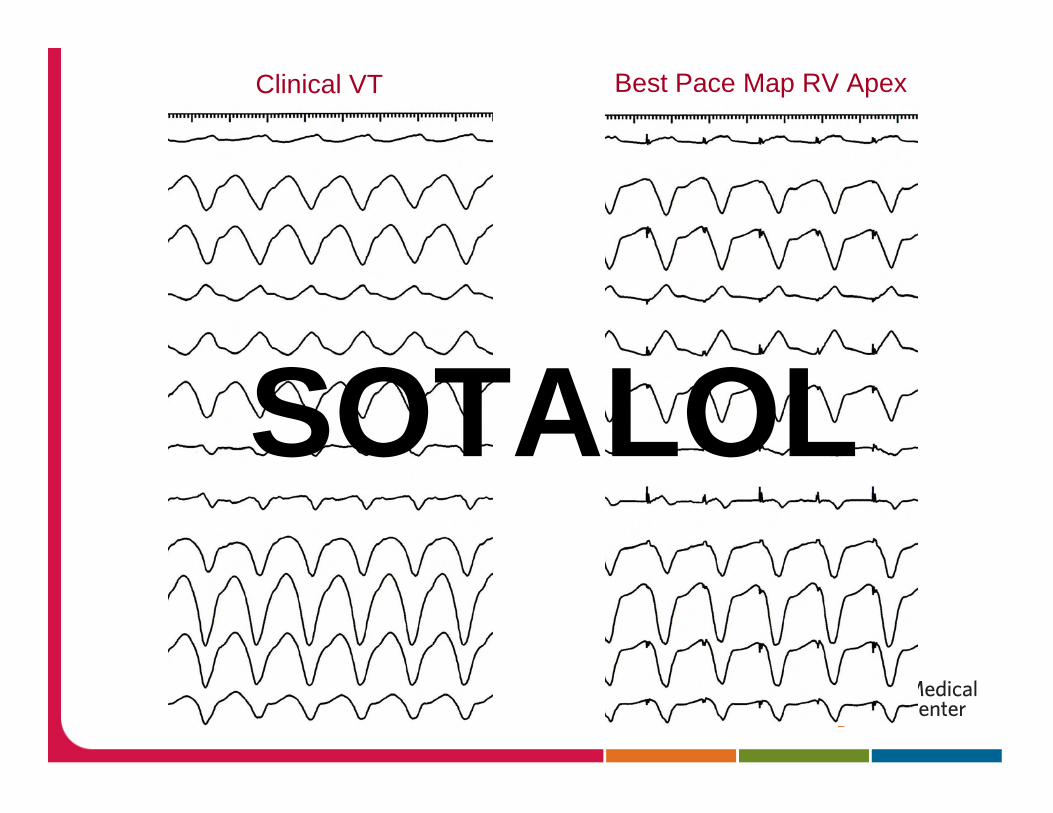
Clinical VT Best Pace Map RV Apex
SOTALOL
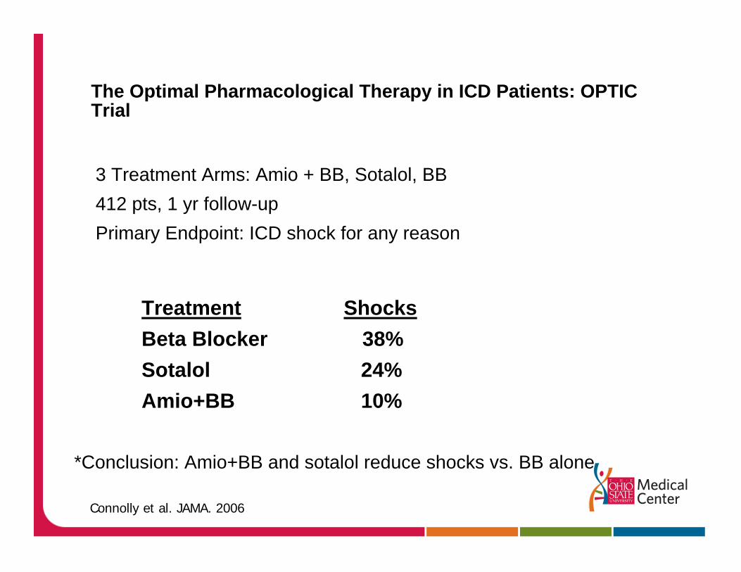
The Optimal Pharmacological Therapy in ICD Patients: OPTIC Trial
3 Treatment Arms: Amio + BB, Sotalol, BB412 pts, 1 yr follow-upPrimary Endpoint: ICD shock for any reason
Treatment ShocksBeta Blocker 38% Sotalol 24%Amio+BB 10%
*Conclusion: Amio+BB and sotalol reduce shocks vs. BB alone
Connolly et al. JAMA. 2006

Major Advances in VT RFA Technology / Approaches
Large Tip (8mm) / Cool Tip technology
Nearly doubled lesion size (14 x 8mm)
3D Catheter Mapping technology
Epicardial Ablation
Remote Navigation: Stereotaxis
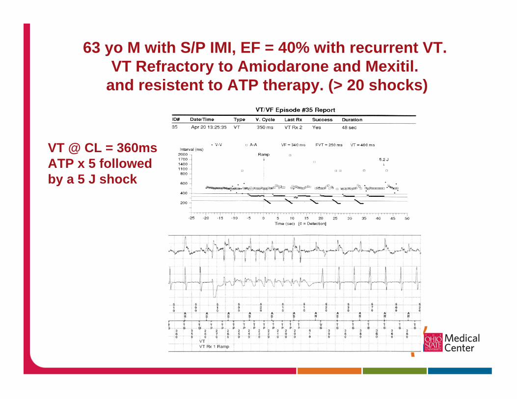
63 yo M with S/P IMI, EF = 40% with recurrent VT. VT Refractory to Amiodarone and Mexitil.
and resistent to ATP therapy. (> 20 shocks)
VT @ CL = 360msATP x 5 followed by a 5 J shock

Initial LV Scar MapRF lesions
along scar border
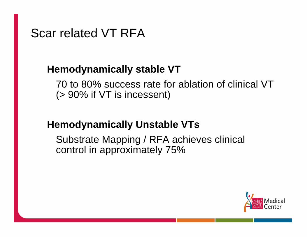
Scar related VT RFA
Hemodynamically stable VT70 to 80% success rate for ablation of clinical VT (> 90% if VT is incessent)
Hemodynamically Unstable VTsSubstrate Mapping / RFA achieves clinical control in approximately 75%
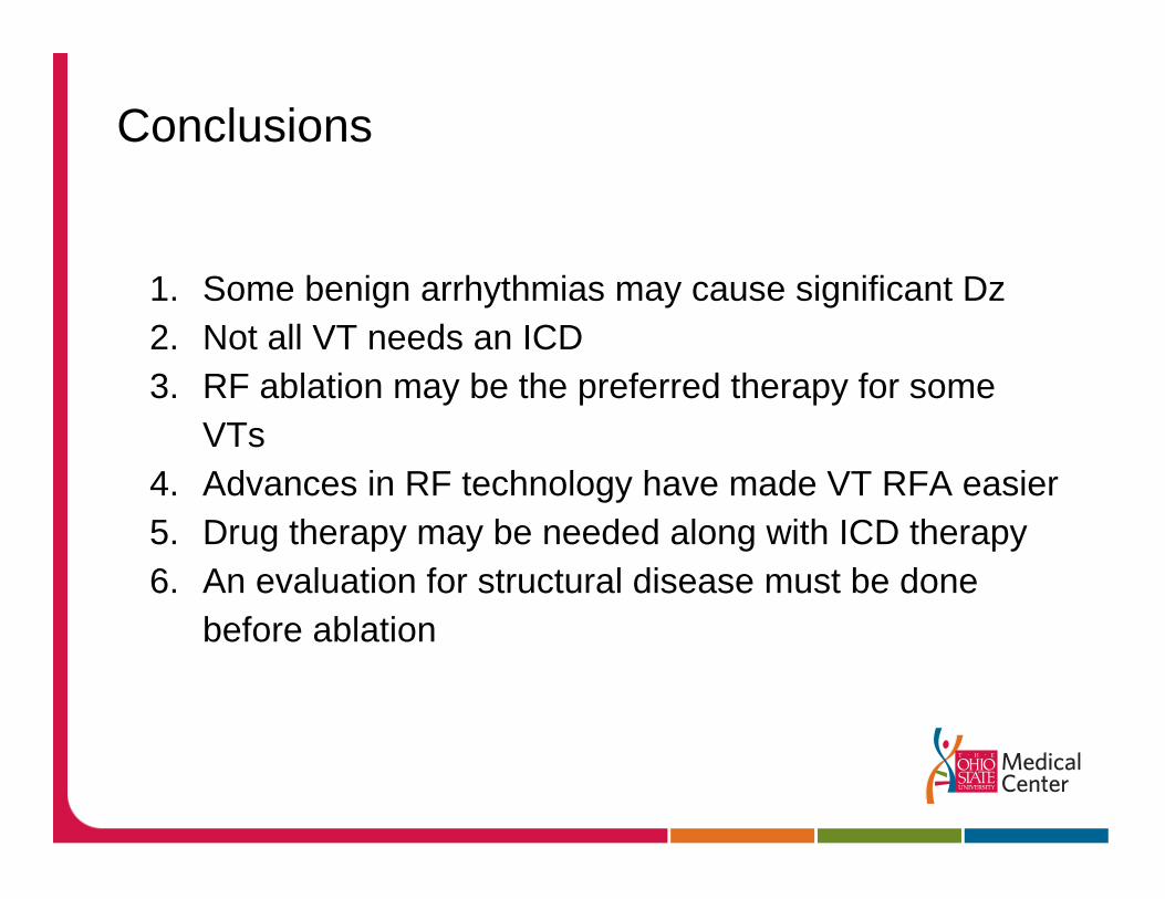
Conclusions
1. Some benign arrhythmias may cause significant Dz2. Not all VT needs an ICD3. RF ablation may be the preferred therapy for some
VTs4. Advances in RF technology have made VT RFA easier5. Drug therapy may be needed along with ICD therapy6. An evaluation for structural disease must be done
before ablation