Embed Size (px)
Citation preview
Upper Airway Dysfunction in Obstructive Sleep Apnea and its Relationship to Laryngopharyngeal Reflux and Postoperative Morbidities in Cancer of the
Oral Cavity and Cancer of the Oropharynx
Dr. Richard 1. Payne
Department of Otolaryngology McGill University, Montreal
September 2004
A thesis submitted to McGill University in partial fulfillment of the requirements of the degree ofMaster of Science in Otolaryngology.
© Richard J. Payne, M.D., 2004

1+1 Library and Archives Canada
Bibliothèque et Archives Canada
Published Heritage Branch
Direction du Patrimoine de l'édition
395 Wellington Street Ottawa ON K1A ON4 Canada
395, rue Wellington Ottawa ON K1A ON4 Canada
NOTICE: The author has granted a nonexclusive license allowing Library and Archives Canada to reproduce, publish, archive, preserve, conserve, communicate to the public by telecommunication or on the Internet, loan, distribute and sell th es es worldwide, for commercial or noncommercial purposes, in microform, paper, electronic and/or any other formats.
The author retains copyright ownership and moral rights in this thesis. Neither the thesis nor substantial extracts from it may be printed or otherwise reproduced without the author's permission.
ln compliance with the Canadian Privacy Act some supporting forms may have been removed from this thesis.
While these forms may be included in the document page count, their removal does not represent any loss of content from the thesis.
• •• Canada
AVIS:
Your file Votre référence ISBN: 0-494-12518-7 Our file Notre référence ISBN: 0-494-12518-7
L'auteur a accordé une licence non exclusive permettant à la Bibliothèque et Archives Canada de reproduire, publier, archiver, sauvegarder, conserver, transmettre au public par télécommunication ou par l'Internet, prêter, distribuer et vendre des thèses partout dans le monde, à des fins commerciales ou autres, sur support microforme, papier, électronique et/ou autres formats.
L'auteur conserve la propriété du droit d'auteur et des droits moraux qui protège cette thèse. Ni la thèse ni des extraits substantiels de celle-ci ne doivent être imprimés ou autrement reproduits sans son autorisation.
Conformément à la loi canadienne sur la protection de la vie privée, quelques formulaires secondaires ont été enlevés de cette thèse.
Bien que ces formulaires aient inclus dans la pagination, il n'y aura aucun contenu manquant.

2
ACKNOWLEDGEMENTS
l extend my deepest gratitude to my supervisor, Dr. R. John Kimoff, for his mentorship throughout this entire process. l appreciate ail of his efforts at directing through the world of basic research. The lessons that l have learned have left a mark on me that is indelible.
l am also indebted to my co-supervisor, Dr. Saul Frenkiel, for his sound advice and guidance throughout this project as weil as throughout the early stages of my career through his teachings on the "art of medicine ".
l would like to acknowledge the contributions made by Dr. Karen Kost, Dr. Michael Hier, and Dr. Martin Black. The hours that they spent supporting me with the clinical research aspects of this project is greatly appreciated and will a!ways be remembered
l would like to thank Dr. Bernard Segal for his assistance and guidance throughout this en tire process.
My thanks to Dr. George Se jean, Dr. Anthony Zeitouni, and Dr. Robert Sweet for their assistance in the recruitment of patients.
l would like to thank Dr. Qutayba Hamid for providing his expertise at the MeakinsChristie Laboratories.
l would like to acknowledge Naftaly Naor for his considerable contributions, work ethic, and resourcefulness. Without his aid this project would not have been possible.
My appreciation goes to the Department of Otolaryngology and the Division of Respirology, McGill University, for their support in arranging and carrying out this project, and to the Canadian Institute of Health and Research for their financial support.
To my wife Karen who's unwavering support acts as thefoundation that ail ofmy dreams are based on. Finally to my daughters Alexandra and Kayla, the greatest accomplishments that anyone can achieve, to whom l dedicate ail that l attain.
3
ABSTRACT:
Obstructive sleep apnea (OSA) is a disease pro cess characterized by collapse of
the upper airway during periods of sleep leading to the cessation airflow despite persistent
respiratory efforts. The aim of this research project is to investigate for associations and
correlations between OSA and other clinical entities using two separate prospective
studies. The initial objective was to evaluate the prevalence of laryngopharyngeal reflux
(LPR) in patients with OSA. LPR was present in 26/28 (93%) of OSA patients.
Moreover, there were significant correlations between LPR and OSA severity (eg. r =
0.57, P = 0.001). The second objective of this research study was to determine the
prevalence of OSA in patients with cancer of the oral cavity and oropharynx, and to
correlate the presence of OSA and the occurrence of postoperative morbidities. OSA was
present in 76% of patients. Overall, postoperative complications were observed in 67% of
OSA and 25% of non-OSA patients, although this difference was not yet significant (p =
0.27, Fisher exact test).
4
SUMMAIRE:
L'apnée obstructive du sommeil (AOS) est un syndrome caractérisé par un
affaissement des voies aériennes supérieures pendant certaines périodes du sommeil,
causant la cessation du flux respiratoire en dépit d'efforts persistants pour respirer. Le but
du présent projet de recherche est d'établir les associations et corrélations existant entre
l'AOS et d'autres entités cliniques à l'aide de deux études prospectives distinctes.
L'objectif initial est d'évaluer chez les patients atteints d'AOS : la prévalence du reflux
gastro-Iaryngé (RGL). Un RGL était présent chez 26/28 des patients atteints d'AOS. Des
corrélations significatives ont été établies entre les sévérités du RGL et de l'AOS (ex. :
r=0,57, p=O,OOl). Le deuxième objectif de la présente étude est de déterminer la
prévalence d'AOS chez les patients avec un cancer de la cavité buccale et de
l'oropharynx, et d'établir des corrélations entre la présence d'AOS et l'occurrence de
morbidités post-opératoires. L'AOS était présente chez 76% des patients. Les
complications post-opératoires ont été observées chez 67% des patients avec l'AOS et
25% des patients sans AOS. L'analyse statistique a démontré que les résultats n'étaient
pas significatifs (p = 0.27, Fisher exact test).

5
ABBREVIA TIONS
AHI Apnea-Hypopnea Index
AR Arytenoid
CVA Cerebrovascular Accident
EST Endoscopic Sensory Testing
GERD Gastroesophageal Reflux Disease
HTN Hypertension
leu Intensive Care Unit
LAR Laryngeal Adductor Reflex
LPR Laryngopharyngeal Reflux
NS Not Significant
OP Oropharynx
OSA Obstructive Sleep Apnea
PSG Polysomnography
REM Rapid eye movement stage of sleep
RFS Reflux Finding Score
Sa02 Oxygen saturation
SE Standard Error
TNF-a Tumour Necrosis Factor Alpha
TNM Tumour, Node, Metastasis
TST Total sleep time
UES Upper Esophageal Sphincter
VP Velopharynx

6
LIST OF TABLES AND FIGURES
FIGURES PAGENUMBER
Figure 1: Patient Grouping 19
Figure 2: Inter-Scorer Agreement 22
Figure 3: Reflux Finding Score 23
Figure 4: Relationship Between LPR and AHI 32
Figure 5: Relationship Between LAR and LPR 33
Figure 6: Relationship Between LAR and AHI 34
Figure 7: Patient Grouping With Respect to AHI Score 37

TABLES
Table 1:
Table 2:
Table 3:
Table 4:
Table 5:
Table 6:
Table 7:
Table 8:
7
Sleep and Respiratory Variables
PAGENUMBER
30
Sensory Thresholds 31
Subject Characteristics 35
Size and Site of the Primary Tumour For Ali Subjects 36
Sleep Data For Ali Subjects 38
Group Characteristics for OSA and Non-OSA Subjects 39
Postoperative Complications 41
Postoperative Complications Separated By Grouping 41
8
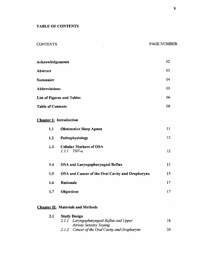
TABLE OF CONTENTS
CONTENTS PAGENUMBER
Acknowledgements 02
Abstract 03
Summaire 04
Abbreviations 05
List of Figures and Tables 06
Table of Contents 08
Chapter 1: Introduction
1.1 Obstructive Sleep Apnea 11
1.2 Pathophysiology 12
1.3 Cellular Markers of OSA 1.3.1 TNF-a 12
1.4 OSA and Laryngopharyngeal Reflux 13
1.5 OSA and Cancer of the Oral Cavity and Oropharynx 15
1.6 Rationale
1.7 Objectives
Chapter fi: Materials and Methods
2.1 Study Design 2.1.1 Laryngopharyngeal Reflux and Upper
Airway Sensory Testing 2.1.2 Cancer of the Oral Cavity and Oropharynx
17
17
18
20

9
2.2 Diagnosis of OSA 20
2.3 Laryngopharyngeal Reflux Testing 2.3.1 Reflux Finding Score 21 2.3.2 Endoscopie Sensory Testing 24
2.4 Cancer of the Oral Cavity and Oropharynx 2.4.1 Determination of Prevalence 25 2.4.2 Measures of Postoperative Morbidity 25
2.5 Statistical Analysis 26
Chapter III: Results
3.1 Laryngopharyngeal Reflux 27 3.1.1 LPRandOSA 32 3.1.2 LPR and Upper Airway Sensation 33 3.1.3 OSA and Upper Airway Sensation 34
3.2 Cancer of the Oral Cavity and Oropharynx 35 3.2.1 Prevalence of OSA 38 3.2.2 Postoperative Morbidities 40
Chapter IV: Discussion
4.1 Discussion of Results 4.1.1 Laryngopharyngeal Reflux 42 4.1.2 Cancer of the Oral Cavity and Oropharynx 47
4.2 Limitations 4.2.1 Laryngopharyngeal Reflux 49 4.2.2 Cancer of the Oral Cavity and Oropharynx 50
4.3 Clinical Implications and Future Directions 4.3.1 Laryngopharyngeal Reflux 51 4.3.2 Cancer of the Oral Cavity and Oropharynx 51
4.4 Conclusions 4.4.1 Laryngopharyngeal Reflux 53 4.4.2 Cancer of the Oral Cavity and Oropharynx 54
Chapter V: References 55

Chapter VI: Appendices
6.1
6.2
Consent Forms (English and French)
Ethics Certificate
71
88
10
11
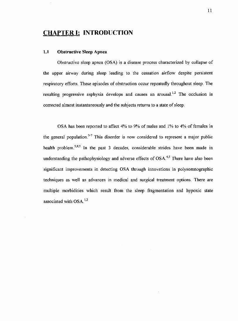
CHAPTERI: INTRODUCTION
1.1 Obstructive Sleep Apnea
Obstructive sleep apnea (OSA) is a disease process characterized by collapse of
the upper airway during sleep leading to the cessation airflow despite persistent
respiratory efforts. These episodes of obstruction occur repeatedly throughout sleep. The
resulting progressive asphyxia develops and causes an arousa1. 1,2 The occlusion is
corrected almost instantaneously and the subjects retums to a state of sleep.
OSA has been reported to affect 4% to 9% of males and 1 % to 4% of females in
the general population.3-? This disorder is now considered to represent a major public
health problem. 5,8,9 In the past 3 decades, considerable strides have been made in
understanding the pathophysiology and adverse effects of OSA. 4,5 There have also been
significant improvements in detecting OSA through innovations in polysomnographic
techniques as weIl as advances in medical and surgical treatment options. There are
multiple morbidities which result from the sleep fragmentation and hypoxic state
associated with OSA. 1,2

12
1.2 Pathophysiology
Despite the prevalence of OSA, many aspects of the pathophysiology remain
poorly understood. Research has been centered around both systemic and local factors
that may predispose individuals to developing sleep apnea. Recent advances have focused
on both anatomic factors and alterations in the integrated neuromuscular function of
upper airway structures which contribute to airway collapse during sleep.lO,ll Anatomical
factors associated with OSA include obesity, abundance of soft tissues in the upper
airway, craniofacial anomalies, hypopharyngeal and laryngeal edema and sensory
denervation secondary to LPR, and airway obstruction secondary to the mass effect of
neoplasms.6,10-18 At the cellular level, an increase in the inflammatory cytokine tumour
necrosis factor alpha (TNF-a.) has been uncovered in the serum of patients with OSA. 19
Whether the local upper airway tissues are responsible for secreting the TNF-a. has not
yet been determined. Nonetheless, it has been postulated that an accumulation of this
inflammatory cytokine in tissues of the upper airway may partly explain the muscle
dysfunction associated with OSA.
1.3 Cellular Markers of OSA
1.3.1 TNF-a
TNF-a. is an inflammatory cytokine. Increases in circulating TNF-a. has been
reported to correlate with measures of disease severity in OSA patients. 19,20 In addition to
promoting edema formation, inflammatory mediator release can also have direct effects
on muscle function. TNF-a. release from macrophages has been implicated as an etiologic
factor in mediating nerve damage in inflammatory neuropathy.21,22
13
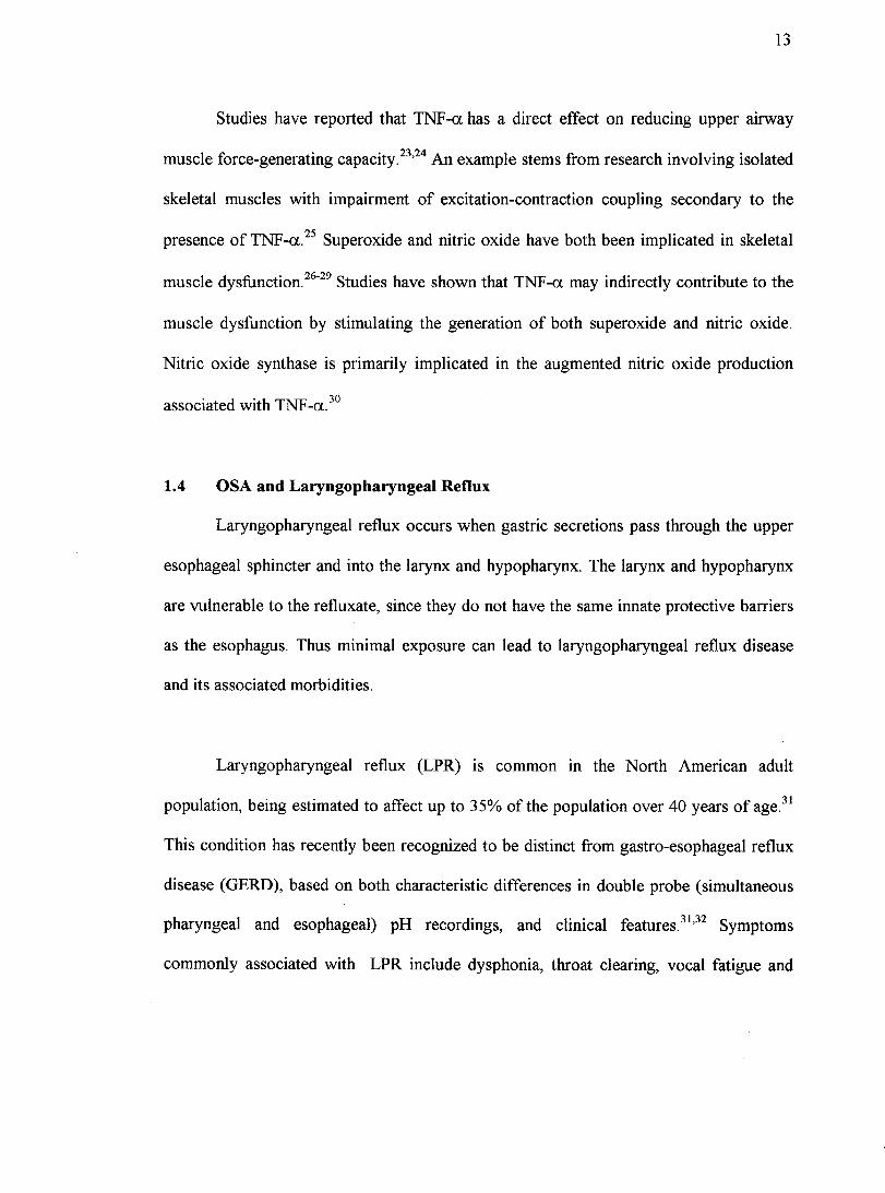
Studies have reported that TNF-a has a direct effect on reducing upper airway
muscle force-generating capacity?3,24 An example stems from research involving isolated
skeletal muscles with impairment of excitation-contraction coupling secondary to the
presence of TNF_a. 25 Superoxide and nitric oxide have both been implicated in skeletal
muscle dysfunction?6-29 Studies have shown that TNF-a may indirectly contribute to the
muscle dysfunction by stimulating the generation of both superoxide and nitric oxide.
Nitric oxide synthase is primarily implicated in the augmented nitric oxide production
associated with TNF -a. 30
1.4 OSA and Laryngopharyngeal Reflux
Laryngopharyngeal reflux occurs when gastric secretions pass through the upper
esophageal sphincter and into the larynx and hypopharynx. The larynx and hypopharynx
are vulnerable to the refluxate, since they do not have the same innate protective barriers
as the esophagus. Thus minimal exposure can lead to laryngopharyngeal reflux disease
and its associated morbidities.
Laryngopharyngeal reflux (LPR) is common in the North American adult
population, being estimated to affect up to 35% of the population over 40 years of age. 31
This condition has recently been recognized to be distinct from gastro-esophageal reflux
disease (GERD), based on both characteristic differences in double probe (simultaneous
pharynge al and esophageal) pH recordings, and clinical features. 31,32 Symptoms
commonly associated with LPR include dysphonia, throat clearing, vocal fatigue and

14
cough.33 LPR has also been linked to more substantial morbidities including
laryngospasm, arytenoid fixation, laryngeal stenosis, and glottic carcinoma 13,34-36
These clinical manifestations of LPR are attributable to inflammation of the
laryngopharyngeal mucosa which result from repeated exposure to acid and pepsin. 13,31-36
While double-probe pH monitoring is considered to be the gold-standard for the
identification of LPR, it has been shown that the diagnosis can reliably be established
using an endoscopic scoring algorithm, the reflux finding score (RFS), which quantifies
the extent and severity of hypopharyngeal and laryngeal mucosal inflammatory changes
which are characteristic ofthis disorder?7
Progress has also been made at identifying risk factors for LPR, which include age
over 60 years, obesity, cigarette smoking and heavy alcohol consumption.5,12,15,38-40 It is
of note that the se risk factors clearly also predispose patients to OSA. Given this, together
with the anatomic effects of LPR on upper airway calibre due to irritation, edema and
inflammation of the soft tissues as weIl as alterations in sensory nerve function, an
increase in the prevalence of sleep apnea among patients with laryngopharyngeal reflux
disease would be anticipated.
Recent work has identified a mucosal sensory impairment in the oropharynx,
velopharynx and larynx of OSA patients using endoscopic sensory testing (EST).14,15
Correlations between the severity of the laryngeal sensory impairment and apnea severity
strongly suggest that this sensory impairment plays a role in the pathophysiology of OSA.

15
Further studies support the concept that upper airway inflammation contributes to this
altered sensory function. 16,17
Aviv and co-workers previously reported that laryngeal sensation is impaired in
patients with LPR, and that treatment of LPR results in improved sensory function. 18
While previous studies have shown that GERD is prevalent among patients with OSA,
there has been no previous evaluation ofLPR in this patient population.41
1.5 OSA and Cancer of the Oral Cavity and Oropharynx
Patients with cancer of the oral cavity often undergo surgery and/or radiation
therapy for cure or as a mode of palliation. The tumour node metastasis (TNM) stage of
the malignancy dictates the treatment regimen. It is not uncommon for these patients to
undergo extensive surgi cal resections of the oral cavity or oropharynx, necessitating
microvascular free flap reconstruction and tracheotomy. In addition to the TNM stage, the
patients health status in terms of comorbidities is evaluated when deciding on a treatment
plan.
It is anticipated that sleep apnea is a comorbidity that is prevalent in patients with
malignancies of the oral cavity and oropharynx. There are many factors that predispose
this group of cancer patients to OSA. Cancer of the oral cavity and oropharynx and OSA
have many common etiologic factors including age over 60 years, cigarette smoking and
h 1 h 1 . 5 12 1538-40 A 1 f h . œ . eavy a co 0 consumpt1on.' , , sa resu t 0 t e anatomlc ellects on upper alrway
calibre resulting from primary malignancies as weB as potential alterations in
neuromuscular functional relationships of upper airway structures due to the presence of

16
neoplasms, an increase in prevalence of OSA among patients with primary head and neck
malignant tumours is anticipated.
Friedman et al determined the prevalence of OSA in patients treated for head and
neck malignancies to be 92%.6 Interestingly, the prevalence of OSA prior to surgical
intervention in patients with cancer of the head and neck has not been evaluated in a
systematic fashion. A link between head and neck cancer and OSA may be of
considerable potential importance in terms of perioperative morbidity at the time of
surgical intervention. There is a rapidly growing body of evidence linking OSA to
cardiopulmonary complications including hypertension, cardiac arrhythmias, myocardial
infarction, pulmonary hypertension, congestive heart failure, and cerebrovascular
events.3,42-47 Thus the detrimental effects of untreated OSA places patients in a sub
optimal preoperative state of health resulting in a potentially greater postoperative risk for
morbidities and mortalities.42,44
Treatment of the OSA may be warranted prior to surgery since the se preoperative
cardiopulmonary conditions often improve as the sleep apnea resolves.48-6o According to
Meoli et al, perioperative control of the airway, postoperative monitoring, and care with
medications is essential at avoiding airway complications following surgery.53 Esclamado
et al determined that 13% (18/135) of patients with OSA undergoing surgery developed
perioperative complications of which 77% (14/18) were airway problems and 5% (1/18)
were cardiac related (arrhythmia).58 Finally, Rennotte et al conclude that nasal CPAP be
administered to patients with OSA in the perioperative period after respiratory
complications (respiratory arrest) developed in patients who failed to receive treatment. 60
17
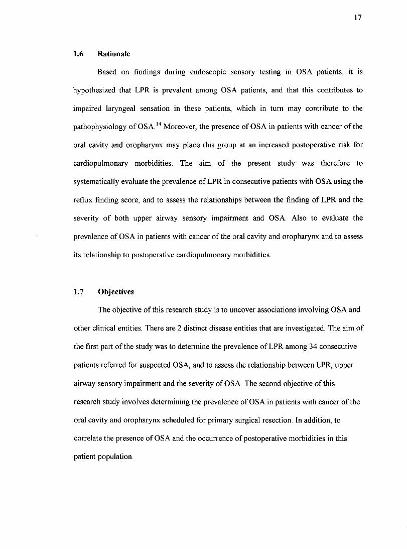
1.6 Rationale
Based on findings during endoscopie sensory testing In OSA patients, it is
hypothesized that LPR is prevalent among OSA patients, and that this contributes to
impaired laryngeal sensation in these patients, which in turn may contribute to the
pathophysiology of OSA. 14 Moreover, the presence of OSA in patients with cancer of the
oral cavity and oropharynx may place this group at an increased postoperative risk for
cardiopulmonary morbidities. The aim of the present study was therefore to
systematically evaluate the prevalence ofLPR in consecutive patients with OSA using the
reflux finding score, and to assess the relationships between the finding of LPR and the
severity of both upper airway sensory impairment and OSA. Aiso to evaluate the
prevalence of OSA in patients with cancer of the oral cavity and oropharynx and to assess
its relationship to postoperative cardiopulmonary morbidities.
1. 7 Objectives
The objective of this research study is to uncover associations involving OSA and
other clinical entities. There are 2 distinct disease entities that are investigated. The aim of
the first part of the study was to determine the prevalence ofLPR among 34 consecutive
patients referred for suspected OSA, and to assess the relationship between LPR, upper
airway sensory impairment and the severity of OSA. The second objective ofthis
research study involves determining the prevalence of OSA in patients with cancer of the
oral cavity and oropharynx scheduled for primary surgical resection. In addition, to
correlate the presence of OSA and the occurrence of postoperative morbidities in this
patient population.

18
CHAPTER II: MATERIALS AND METHODS
2.1 Study Design
Patients were recruited from the Jewish General Hospital, Royal Victoria
Hospital, and St. Mary' s Hospital. Consecutive patients were approached and informed
consent was obtained prior to including an individual in the study. The research study
obtained full scientific and ethical approval from the review boards at each institution
(see chapter VI).
2.1.1 Laryngopharyngeal Reflux and Upper Airway Sensory Testing
Thirty-seven patients suspected of having OSA were recruited for confirmation of
sleep apnea as weIl as for assessment of laryngopharyngeal reflux and upper airway
sensory impairment in a prospective and blinded manner. Recruitment of subjects was
conducted completely independent of any symptoms suggestive of laryngopharyngeal
reflux to avoid any selection bias. Patients were expected to undergo polysomnography
(PSG), upper airway pulse endoscopic sensory testing (EST) of the laryngeal adductor
reflex (LAR) and aryepiglottic fold sensation, and assessment of LPR using the RFS.
Thirty-four patients completed both PSG and LPR testing. Twenty-seven patients
completed PSG, LPR, and endoscopie sensory testing (Figure 1). Patients on eontinuous
positive airway pressure or taking anti-reflux medications were excluded from the study,
as were patients under the age of 18 years oid. A questionnaire was used to determine
patients' smoking and alcohol consumption histories. The study was approved by the
Researeh Ethics Boards of the participating hospitals, and written informed consent was
obtained from each subject.
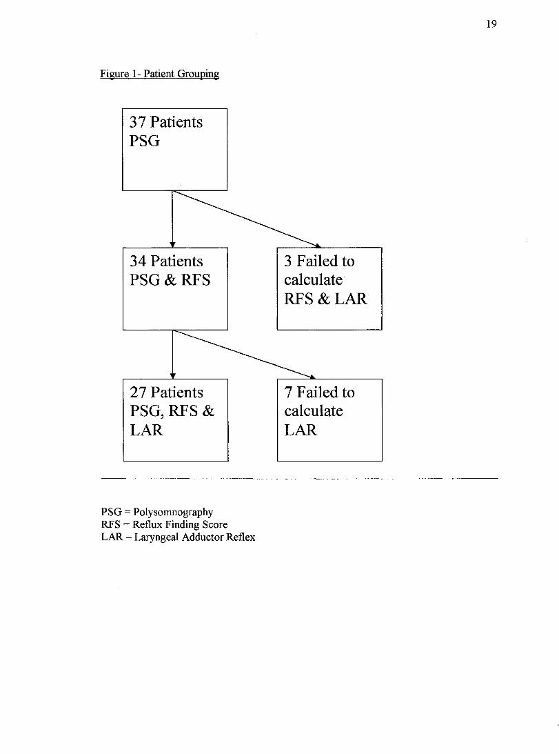
Figure 1- Patient Grouping
37 Patients PSG
34 Patients PSG&RFS
27 Patients PSG, RFS & LAR
PSG = Polysomnography RFS = Reflux Finding Score LAR - Laryngeal Adductor Reflex
3 Failed to calculate RFS&LAR
7 Failed to calculate LAR
19
20
2.1.2 Cancer of the Oral Cavity and Oropharynx
Seventeen patients with primary malignant tumours of the oral cavity and
oropharynx were recruited for this aspect of the study. Consecutive patients were
approached for participation in the study if it was determined that their malignancy was
amenable to primary surgi cal resection. Recruitment of subjects was conducted
completely independent of any symptoms suggestive of obstructive sleep apnea or other
sleep complaint to avoid any selection bias. Patients with a tracheotomy prior to surgery
were excluded from the study, as were patients with malignancies of the head and neck
spreading secondarily to the oral cavity or oropharynx, such as nasopharyngeal carcinoma
and supraglottic carcinoma. A chart review was conducted to determine patients'
smoking, alcohol consumption, and cardiopulmonary histories. The tumours were
measured by radiologists specialized in head and neck oncology using computed
tomographic images enhanced with intravenous contrast in the axial, coronal, and sagittal
planes.
2.2 Diagnosis of OSA
Complete overnight diagnostic polysomnography was performed usmg the
Suzanne (Melville, Tyco, Ottawa) portable recording system. Studies were conducted in
the patient's home, with set-up of the apparatus and verification of signais conducted by a
trained technologist in the evening, followed by unattended recording through the night.
The signaIs recorded included standard electroencephalographic leads (C4-AIIC3-A2),
bilateral electrooculogram, chin and anterior tibialis electromyograms, pulse oximetery,
airflow via nasal pressure cannula and oronasal thermistor, thoracoabdominal movements
21
via piezo bands, body position via mercury position sensor and sound via a microphone
taped to the lateral aspect of the neck.
The polysomnographic data was downloaded to a personal computer and scored
manually by trained, experienced polysomnographic technologists with review by an
expert physician. Sleep-wake state was defined according to standard criteria. 6 l An
obstructive apnea was defined as an episode of cessation of airflow lasting at least 10
seconds with persistent respiratory effort, and an obstructive hypopnea as a discrete
episode of reduction in airflow with inspiratory flow limitation on the nasal cannula
pressure signal lasting> 10 seconds with associated desaturation > 2% or arousal defined
according to American Sleep Disorders Association criteria.62 The apnea-hypopnea index
(ARI) defined as the number of apneas and hypopneas per hour of sleep, was the primary
polysomnographic outcome measure. A diagnosis of OSA was made on the basis on an
AHI value 2: 15 events per hour. A diagnosis of OSA requiring treatment was made on
the basis of an ARI value 2: 20 events per hour.
2.3 Laryngopharyngeal Reflux Testing
2.3.1 Reflux Finding Score37
Laryngopharyngeal reflux was assessed usmg the reflux finding score. Each
patient underwent a standardized videotaped upper airway flexible fiberoptic
nasolaryngoscopy using the Pentax® FNL 10ap flexible laryngoscope with the
Olympus® OPV F2 video attachment. The recordings were viewed and scored on a
JVC® 32 inch television screen. Two investigators blinded to OSA and sensory status
determined the RFS for each patient independently. The results from both scorers were

22
tested for agreement and the mean score was used in the calculations (Figure 2). An RFS
> 7 was eonsidered as signifieant for LPR.
The reflux finding score is a reliable predietor of LPR when compared to double-
probe pH monitoring. Caleulating the reflux finding score is less expensive, less time
eonsuming, and easier for patients to tolerate than pH probe. Scoring of laryngeal
inflammation and irritation may in faet be a more physiologieally relevant measure than
pH measurements (Figure 3).
Figure 2 - Inter-Seorer Agreement
Inter-Scorer Agreement 20
18 • r = 0.85, P < 0.001 •
16 • ("l • M 14 • • Q) M • 0 0 12 • • r./).
r./). • • ~ 10
~ 8
• 6
• 4
4 6 8 10 12 14 16 18 20
RFS: Scorer 1

23
Figure 3 - Reflux Finding Score
fugn Definition Score
Laryngeal ErythemaIHyperemia 2 = arytenoids 4 = diffuse
Vocal Fold Edema 1 = mi Id 2 = moderate 3 = severe 4 = polypoid
Diffuse Laryngeal Edema 1 = mild 2 = moderate 3 = severe 4 = obstructing
Ventricular edema 2 = partial 4 = complete
Subglottic edema 0= absent 2 = present
Posterior commissure hypertrophy 1 = mild 2 = moderate 3 = severe 4 = obstructing
Granuloma/Granulation tissue 0= absent 2 = present
ThickMucus 0= absent 2 = present
24
2.3.2 Endoscopie Sensory Testing
Upper airway sensory testing was performed using the endoscopie air pressure
pulse technique. The Pentax® FNL lOap flexible laryngoscope with the Olympus® OPV
F2 video attachment was used with the AP 4000 air pulse stimulator. IncrementaI
pressure pulses 2 - 10 mm Hg were used until the LAR was stimulated. True and sham
pulses were both employed. Sensory detection threshold was considered positive if the
reflex was positively detected for 4 out of 5 pulses. The degree of sensation measured at
the aryepiglottic folds was performed in a similar manner. The amount of air pressure
needed was considered as the subjects score (2-10). A patient failing to have a positive
reflex or sensation at the maximum setting was scored as Il.
25
2.4 Cancer of the Oral Cavity and Oropharynx
2.4.1 Determination of Prevalence
The apnea-hypopnea index was calculated for aIl 17 patients within 2 to 14 days
of the surgery. Patients with an AHI value 2': 20 events per hour were considered as
positive for having OSA requiring treatment.
2.4.2 Measures of Postoperative Morbidity
The hospital charts of aH subjects were reviewed by an investigator unaware of
the subject's OSA status to determine the postoperative course for the period up to 60
days following surgi cal intervention. Values for the following variables were determined
for each patient: length of stay in the intensive care unit (lCU) or monitored setting, hours
on a ventilator, and number of cardiopulmonary and other OSA related postoperative
complications. Intensive care unit stay greater than 24 hours and the need for mechanical
ventilation were considered to be surgi cal morbidities. Cardiopulmonary complications
were defined as newly diagnosed arrhythmias requiring medical treatment, myocardial
infarction, cerebrovascular events, hypoxemia, pulmonary edema, pleural effusion, and
pneumoma.

26
2.5 Statistical Analysis
AHI, RFS, LAR, and sensation at the level of the aryepiglottic folds were
compared for associations by calculating the correlation coefficient. Outcome variables
for the patients with cancer were compared between the obstructive sleep apnea and non
OSA groups using an unpaired t-test in the case of normally distributed continuous
variables, and the Mann-Whitney rank-sum test for non-normally distributed variables.
Categorical comparisons in a two-by-two format were made using the Fisher exact text.
Statistical calculations were made using SigmaStat software (Jandel, XX). A value of p <
0.05 was used for statistical significance.
27
CHAPTER III: RESULTS
3.1 Laryngopharyngeal Reflux
Subjects:
There were 26 males and 8 females enrolled in the study. For the group overall,
the mean age was 43.9 ± 2.4 (SE) years and the mean body mass index was 26.5 ± 0.8
kg/m2. For the subjects with OSA (n= 29) these values were 45.4 ± 2.3 years and 27.2 ±
0.8 kg/m2.
Polysomnographic findings:
The polysomnographic recordings were of high quality, and were adequate for
diagnosis in an subjects. Sleep and respiratory data for the group overal1 and for subjects
with OSA are shown in Table 1. AHI values ranged from 7.5 to 108.1 events per hour.
Given that the subjects recruited had been referred for polysomnography in the clinical
context of suspected OSA, a majority (29/34) were found to have an AHI ~ 15 events per
hour. This therefore represents a prevalence for OSA in this subject group of 85%. In that
the small size of the non-OSA group (5 subjects) does not allow for meaningful
comparisons between the two groups, the primary focus of the analysis below is on the
OSA group (Figure 4).
Air pulse endoscopie sensory testing findings:
Sensory threshold values were obtained in an subjects for the oropharynx, and
while the video recording was adequate in an subjects for RFS scoring, several were
unable to tolerate the EST procedure sufficiently to allow determination of sensory

28
thresholds Via the trans-nasal approach. Thus sensory detection thresholds were
determined at the VP and AR in 28 subjects, and LAR thresholds were determined in a
total of29. Sensory threshold data and LAR values are shown in Table 2 and in Figures 5
and 6 below. As we described previously, there was strong correlation between AR
sensory and LAR thresholds (r = 0.86, P < 0.0001).14 There was also a significant
correlation between sensory thresholds at the OP and VP (r = 0.46, P < 0.03), while
neither of these correlated with laryngeal sensory measures. The sensory threshold values
for OSA subjects were considerably elevated compared with those in normal non-snoring
controls previously evaluated in our laboratory.14,15
Laryngopharyngeal reflux assessment findings:
The video recordings of the larynx were of high quality, and were adequate for
diagnosis in aIl 34 subjects. There was very close correlation between the independent
values from the two scorers, with a correlation coefficient r = 0.85 (p< 0.001). The mean
difference between RFS for scorer 1 vs. 2 was only 0.8 ± 0.3 units, and for OSA patients
there was 100% concordance regarding the diagnosis ofLPR. Of the 34 subjects, 30 were
found to have a mean RFS > 7, yielding a prevalence of LPR in this subject group of
88%. The prevalence of LPR in subjects with OSA was 93% (26/28). Mean RFS values
for the group overall are shown in Table 2, with the values ranging from 5 to 17.5. RFS
Scores from OSA subjects are also shown in Table 2 and in Figure 4 and 5 below.
Relationships between LPR, apnea severity and upper airway sensory fonction:
For patients with OSA, there was a significant correlation between the severity of
LPR and OSA severity as reflected in the AHI (Figure 4). There was also a slightly
29
weaker but still significant correlation between RFS scores and the nadir Sa02 for the
night (r= -0.38, p < 0.05). These findings therefore point to a strong relationship between
LPR as reflected in the RFS score, and apnea severity.
There were also significant relationships between RFS scores and upper airway
sensory function. RFS correlated strongly with LAR values (Figure 5) as weIl as with AR
sensory threshold values (r = 0.46, P < 0.03). Of note, if the outlier subject (upper left
corner of Figure 5) was removed from the calculations, these correlations become
considerably stronger, with r = 0.70, P < 0.0001 for RFS vs. LAR, and r = 0.62, P < 0.002
for RFS vs. AR sensory threshold. In contrast to the findings at the larynx, there were no
significant correlations between RFS and sensory measures at the OP or VP. Thus LPR
appears to be strongly related to laryngeal, but not oropharyngeal or velopharyngeal
sensory dysfunction.
As recently described for the OSA subject cohort, significant correlations between
laryngeal but not OP or VP sensory function and apnea severity were observed (Figure
6).14

30
Table 1 - Sleep and Respiratory Variables
Ali Subjects OSA Subjects (n=34) (n=29)
Sleep Variables Total Sleep Time (h) 5.9 ± 0.3 6.0 ± 0.3
Sleep Efficiency (%) 76.8 ± 2.4 76.5 ± 2.6
Microarousal Index (#/h) 34.4 ± 3.6 36.4 ± 3.8
Stage 1 (% TST) 6.7±1.1 7.2 ± 1.1
Stage 2 (% TST) 57.1 ± 2.8 57.8 ± 2.9
Stage 3 &4 (%TST) 15.4 ± 1.6 14.3 ± 1.6
REM (% TST) 20.8 ± 1.2 20.7 ± 1.3
Respiratory Variables AHI (events/h) 35.2 ± 4.0 39.2 ± 3.9
Apnea Index (events/h) 12.4 ± 3.8 10.7±3.7
Hypopnea Index 22.0 ± 2.2 23.9 ± 2.2
(events/h)
Mean Event Duration (s) 15.9 ± 0.8 16.7 ± 0.8
Minimum SaÜ2 (%) 86.7 ± 1.5 85.8 ± 1.5 (Values are Mean ± SE)

31
Table 2 - Sensory Thresholds
Sensory Ali Subjects OSA Subjects Thresholds (n=34) (n=29)
Oropharynx (mm Hg) 5.3 ± 0.6 5.4 ± 0.6
Velopharynx (mm Hg) 9.5 ± 0.5 9.6 ± 0.5
Larynx (mm Hg) 7.4 ± 0.6 7.6 ± 0.6
LAR Threshold (mm Hg) 6.2 ± 0.5 6.4 ± 0.5
RFS Values 11.6 ± 0.6 12.0 ± 0.6 (Values are Mean ± SE)
32
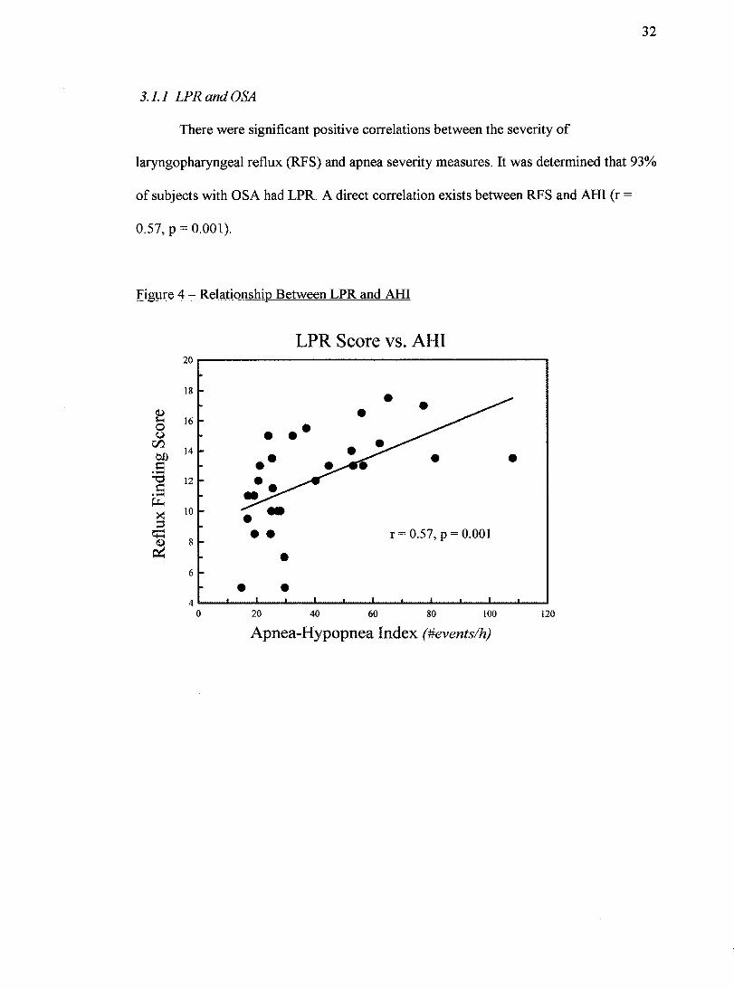
3.1.1 LPR and OSA
There were significant positive correlations between the severity of
laryngopharyngeal reflux (RFS) and apnea severity measures. It was determined that 93%
of subjects with OSA had LPR. A direct correlation exists between RFS and AHI (r =
0.57, p = 0.001).
Figure 4 - Relationship Between LPR and AHI
20
18
~ 16 0 U
r:n. 14 on
~ ...... '"0 12 ~ ...... ~
><! 10
::s ~
8 ~
6
4 0
LPR Score vs. AHI
••• ••
•• •
• • 20 40
• • •
•
r = 0.57, P = 0.001
60 80 100
Apnea-Hypopnea Index (#events/h)
•
120
33
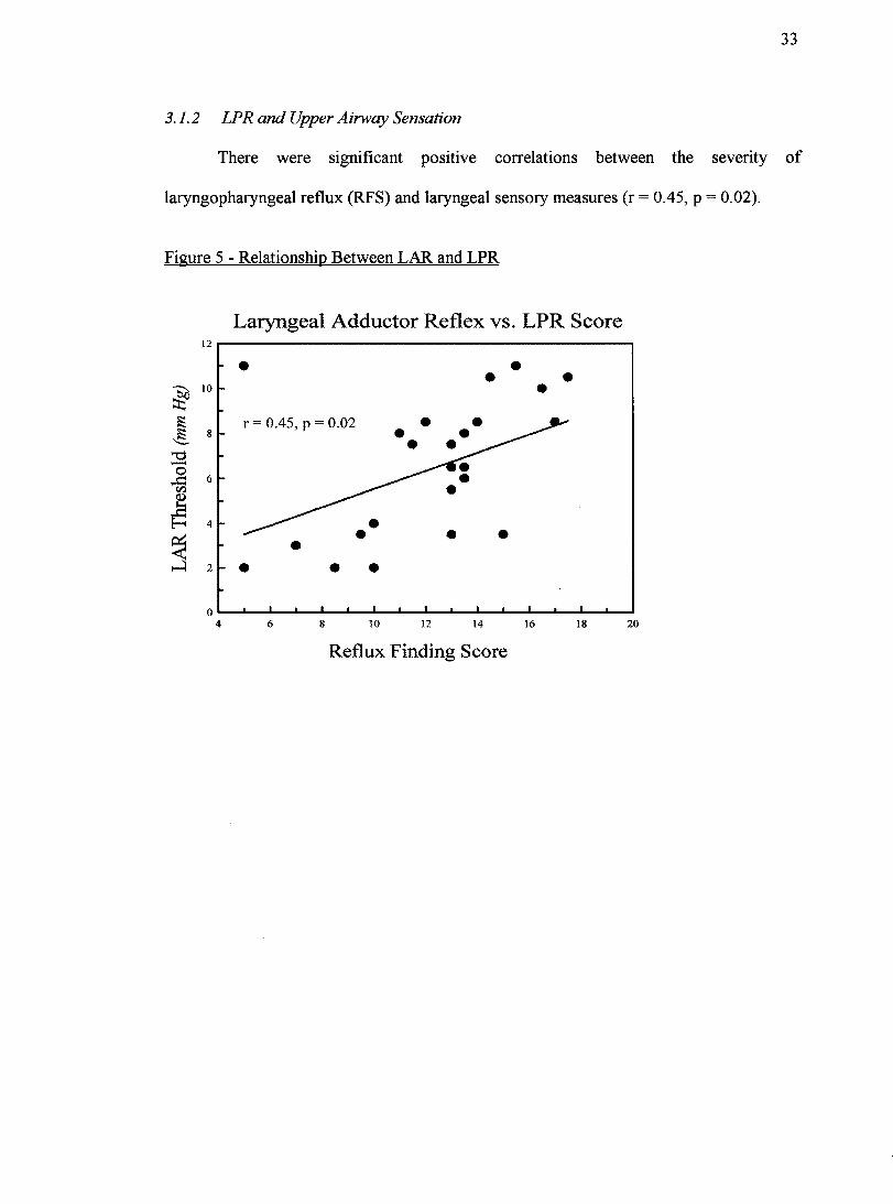
3.1.2 LPR and Upper Airway Sensation
There were significant positive correlations between the severity of
laryngopharyngeal reflux (RFS) and laryngeal sens ory measures (r = 0.45, P = 0.02).
Figure 5 - Relationship Between LAR and LPR
Laryngeal Adductor Reflex vs. LPR Score 12r---------------------------------------~
• • r = 0.45, p = 0.02
• • •
• • •
• • •
•
• • •
•
oL-~-L~~~~~~~~~~~~~ __ ~~~~
4 6 8 10 12 14 16 18 20
Reflux Finding Score

34
3.1.3 OSA and Upper Airway Sensation
There were significant positive correlations between both LAR and subjective
laryngeal sensory thresholds and apnea-hypopnea index (r = 0.50, P = 0.01).
Figure 6 - Relationship Between LAR and AH!
Laryngeal Adductor Reflex vs. AHI
• •
• ... . • •• •
r = 0.50, P = 0.01
o~~-----~-----~~------------~--~~------------~~-----~ o 20 40 60 80 100 120
Apnea-Hypopnea Index (#events/h)
35
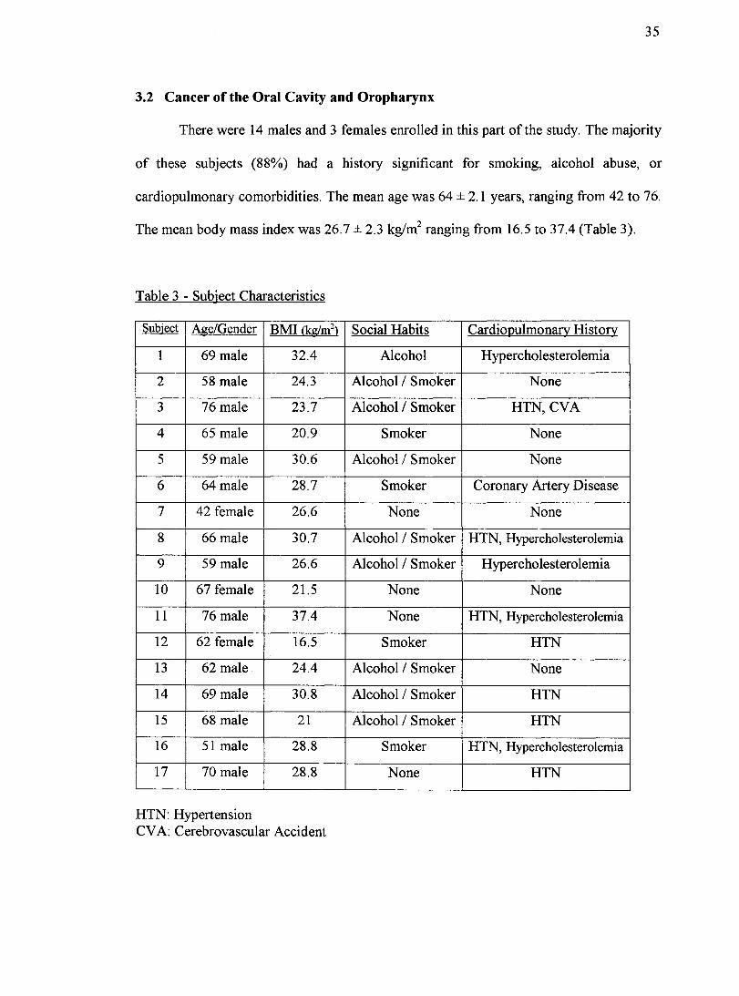
3.2 Cancer of the Oral Cavity and Oropharynx
There were 14 males and 3 females enrolled in this part of the study. The majority
of these subjects (88%) had a history significant for smoking, alcohol abuse, or
cardiopulmonary comorbidities. The mean age was 64 ± 2.1 years, ranging from 42 to 76.
The mean body mass index was 26.7 ± 2.3 kg/m2 ranging from 16.5 to 37.4 (Table 3).
Table 3 - Subject Characteristics
Subject Age/Gender BMI (kglm2) Social Habits CardioQulmonary History
1 69 male 32.4 Alcohol Hypercholesterolemia
2 58 male 24.3 Alcohol / Smoker None
3 76 male 23.7 Alcohol / Smoker HTN, CVA
4 65 male 20.9 Smoker None
5 59 male 30.6 Alcohol / Smoker None
6 64 male 28.7 Smoker Coronary Artery Disease
7 42 female 26.6 None None
8 66 male 30.7 Alcohol / Smoker HTN, Hypercholesterolemia
9 59 male 26.6 Alcohol / Smoker Hypercholesterolemia
10 67 female 21.5 None None
11 76 male 37.4 None HTN, Hypercholesterolemia
12 62 female 16.5 Smoker HTN
13 62 male 24.4 Alcohol / Smoker None
14 69 male 30.8 Alcohol / Smoker HTN
15 68 male 21 Alcohol / Smoker HTN
16 51 male 28.8 Smoker HTN, Hypercholesterolemia
17 70 male 28.8 None HTN
HTN: Hypertension CV A: Cerebrovascular Accident
36
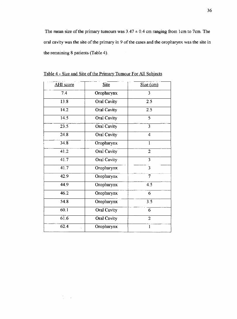
The mean size of the primary tumours was 3.47 ± 0.4 cm ranging from Icm to 7cm. The
oral cavity was the site of the primary in 9 of the cases and the oropharynx was the site in
the remaining 8 patients (Table 4).
Table 4 - Size and Site of the Primary Tumour For AlI Subjects
AHI score Site Size (cm)
7.4 Oropharynx 3
13.8 Oral Cavity 2.5
14.2 Oral Cavity 2.5
14.5 Oral Cavity 5
23.5 Oral Cavity 3
24.8 Oral Cavity 4
34.8 Oropharynx 1
41.2 Oral Cavity 2
41.7 Oral Cavity 3
41.7 Oropharynx 3
42.9 Oropharynx 7
44.9 Oropharynx 4.5
46.2 Oropharynx 6
54.8 Oropharynx 3.5
60.1 Oral Cavity 6
61.6 Oral Cavity 2
62.4 Oropharynx 1

37
Of the 17 patients enrolled in the study, 4 underwent polysomnography but failed
to undergo surgery. Two of the patients developed comorbid conditions that precluded
surgical intervention, one of which was directly re1ated to cardiopulmonary status. One
patient needed a tracheotomy prior to the definitive surgery due to respiratory distress.
One patient died of a myocardial infarction secondary to an arrhythmia 2 days before the
scheduled operation. As a result, the data used for comparing postoperative morbidities
was compiled from the remaining 13 patients (Figure 7).
Figure 7 - Patient Grouping With Respect to AHI Score
1 17 Patients 1 AID>y ~20 1 13 OSA (76%) 1 4 Non-OSA (24%) 1
~ 9 Surgery J 1 4 No Surgery 1 4 ~~rgery 1
~ /~ 6 Patients 3 Patients 1 Patient 3 Patients
(67%) (33%) (25%) (75%) Complications None Complications None
38
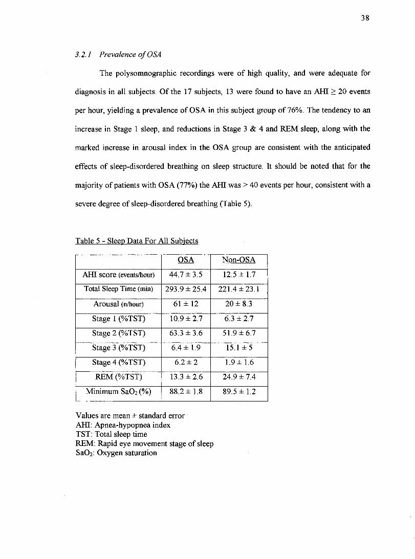
3.2.1 Prevalence of OSA
The polysomnographic recordings were of high quality, and were adequate for
diagnosis in aIl subjects. Of the 17 subjects, 13 were found to have an AH! 2: 20 events
per hour, yielding a prevalence of OSA in this subject group of 76%. The tendency to an
increase in Stage 1 sleep, and reductions in Stage 3 & 4 and REM sleep, along with the
marked increase in arousal index in the OSA group are consistent with the anticipated
effects of sleep-disordered breathing on sleep structure. It should be noted that for the
majority of patients with OSA (77%) the AH! was > 40 events per hour, consistent with a
severe degree of sleep-disordered breathing (Table 5).
Table 5 - Sleep Data For AlI Subjects
OSA
AH! score (events/hour) 44.7 ± 3.5
Total Sleep Time (min) 293.9 ± 25.4
Arousal (nlhour) 61 ± 12
Stage 1 (% TST) 1O.9± 2.7
Stage 2 (% TST) 63.3 ± 3.6
Stage 3 (% TST) 6.4 ± 1.9
Stage 4 (% TST) 6.2±2
REM (%TST) 13.3 ± 2.6
Minimum Sa02 (%) 88.2 ± 1.8
Values are mean ± standard error AH!: Apnea-hypopnea index TST: Total sleep time REM: Rapid eye movement stage of sleep Sa02: Oxygen saturation
Non-OSA
12.5 ± 1.7
221.4 ± 23.1
20 ± 8.3
6.3 ± 2.7
51.9±6.7
15.1 ± 5
1.9 ± 1.6
24.9 ± 7.4
89.5 ± 1.2
39
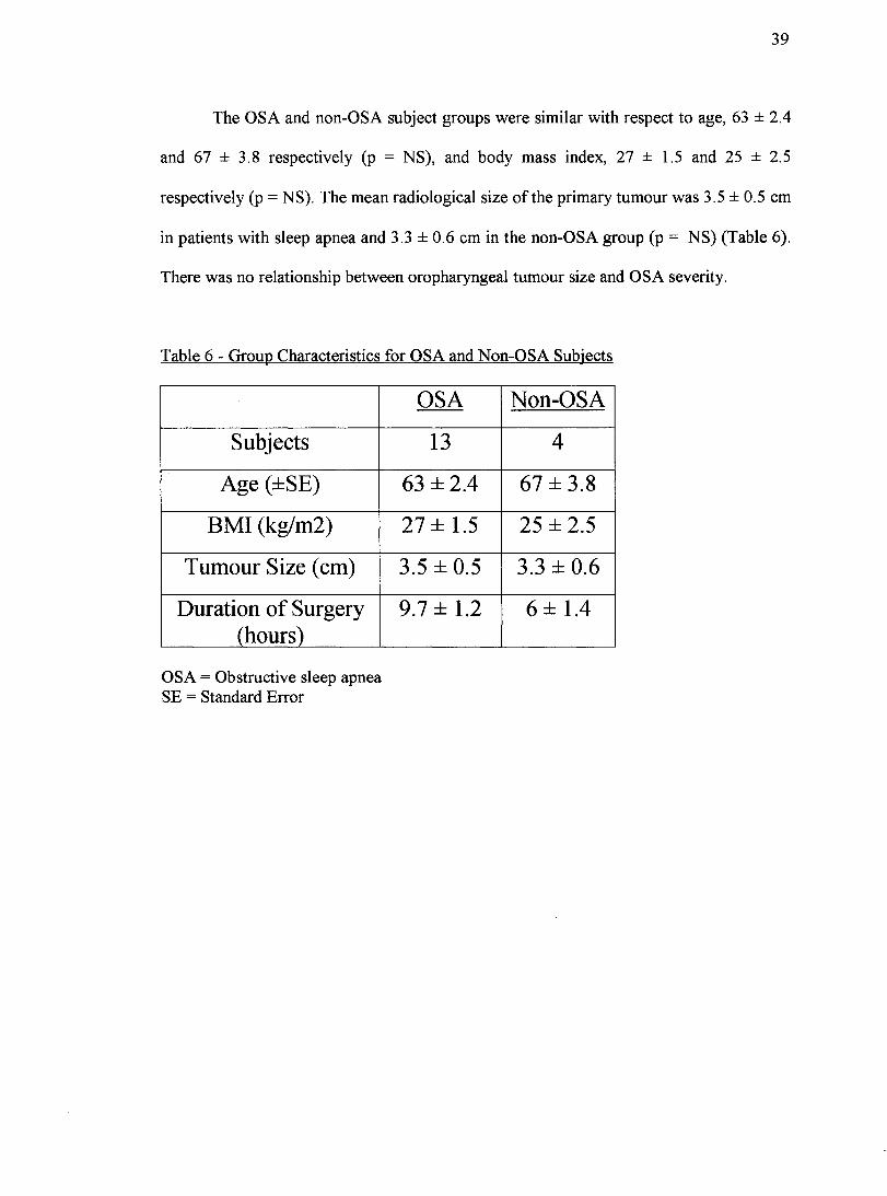
The OSA and non-OSA subject groups were similar with respect to age, 63 ± 2.4
and 67 ± 3.8 respectively (p = NS), and body mass index, 27 ± 1.5 and 25 ± 2.5
respectively (p = NS). The mean radiological size of the primary tumour was 3.5 ± 0.5 cm
in patients with sleep apnea and 3.3 ± 0.6 cm in the non-OSA group (p = NS) (Table 6).
There was no relationship between oropharyngeal tumour size and OSA severity.
Table 6 - Group Characteristics for OSA and Non-OSA Subjects
Subjects
Age (±SE)
BMI (kg/m2)
Tumour Size (cm)
Duration of Surgery (hours)
OSA = Obstructive sleep apnea SE = Standard Error
OSA Non-OSA
13 4
63 ± 2.4 67 ± 3.8
27 ± 1.5 25 ± 2.5
3.5 ± 0.5 3.3 ± 0.6
9.7 ± 1.2 6±1.4

40
3.2.2 Postoperative Morbidities
Preoperative cardiopulmonary complications occurred in 3 subjects. One of the se
patients died of an arrhythmia leading to a myocardial infarction 2 days prior to surgery.
Intraoperative complications such as prolonged operative time, airway management
difficulties, induction of anesthesia, and cardiopulmonary complications as previously
defined did not occur in any of the subjects. The average duration of the surgery was 9.7
± 1.2 hours in OSA and 6 ± lA hours in non-OSA patients. Prolonged ICU stay occurred
in 56% (5/9) of patients with OSA and 25% (114) of patients with an AHI<20. The mean
ICU stay was 3.3 days for OSA patients and 1 dayfor non-OSA patients. Mechanical
ventilation was necessary for 3 patients with OSA and not required for patients without
sleep apnea. Cardiopulmonary complications occurred in 33% (3/9) of patients with an
AHl2:20 and failed to occur in the other patient group. The cardiopulmonary morbidities
which were observed in patients with OSA included arrhythmias, pulmonary edema,
pleural effusion, and hypoxemia. In total, 67% of patients with an AHI2:20 and 25% of
patients with an AHI<20 (p = 0.27, Fisher exact test) had one of the above-described
postoperative complications (Tables 7 and 8). If the preoperative cardiopulmonary
complications are considered together with postoperative complications, a total of9 of 13
(69%) of OSA patients and 1 of 4 (25%) of non-OSA experienced perioperative
morbidity (p = 0.11, Fisher exact test). Thus while these findings do not achieve statistical
significance in this relatively small sample size, and it is not possible to establish a direct
link between OSA and perioperative complications, there appears to be a clear tendency
for a higher rate of perioperative complications among patients with OSA than those
without OSA.

41
Table 7 - Postoperative Complications
AHl leU Ventilated eardio~ulmonary
7.4 1 0 None
13.8 1 0 None
14.2 0 0 None
14.5 2 0 None
23.5 10 144 Pulmonary Edema, H -.YQoxemia
24.8 3 41 None
41.7 9 0 Pleural Effusion, Arrhythmia
41.7 1 0 Arrhythmia
42.9 1 0 None
46.2 2 26 None
54.8 1 0 None
60.1 2 0 None
61.6 1 0 None
Table 8 - Postoperative Complications Separated By Grouping
AHI ICU Stay Prolonged Mechanical Cardio12ulmonary Total Score (mean days) ICU Stav Ventilation Comnlications Complications <20 1 ± 0.4 n=l n=O n=O n=l (n=4)
~20 3.3 ± 1.2 n=5 n=3 n=3 n=6 (n=9)

42
CHAPTER IV: DISCUSSION
4.1 Discussion of Results
4.1.1 Laryngopharyngeal Reflux
In this study, we used a validated endoscopic scoring algorithm to assess LPR
among patients with obstructive sleep apnea and found a dramatically higher prevalence
(93%) for LPR than that reported for the general population. 13,31,33,63,64 Furthermore, the
severity of LPR as reflected by RFS values correlated significantly with several key
measures of apnea severity, pointing to an important interaction between these two
disorders. The severity of LPR correlated with laryngeal sensory dysfunction (as
previously reported by Aviv and co-workersI8), but not oropharyngeal or velopharyngeal
sensory impairment (not previously evaluated). Moreover, previous observations that
laryngeal, but not oropharyngeal or velopharyngeal sensory dysfunction correlates with
. l'd d 14 apnea seventy was va 1 ate .
To our knowledge, the relationship between OSA and LPR has not previously
been evaluated. A number of studies have focussed on the relationship between OSA and
gastro-esophageal reflux disease, reporting a prevalence of GERD ranging from 55 - 75%
in OSA patients.65-67 The severity of GERD based on questionnaire correlates with apnea
severitl5, although nocturnal physiologic recording studies have not shown a direct
temporallink between apneic and GERD events.68,69 Mechanisms postulated to account
for the interaction of GERD and OSA include the large negative intrathoracic pressure
swings generated during obstructive apneas, and respiratory-related microarousals which
appear to be associated with LES relaxation. 67,68

43
As noted above, LPR has come to be recognized as not simply the
hypopharyngeal manifestation of GERD, but rather as a distinct entity within the
spectrum of gastric material reflux syndromes, with characteristic double pH probe
profiles and clinical symptoms. 13,31-33,64,7o-n While the clinical syndrome of GERD is
primarily related to overflow of gastric contents into the esophagus due to LES
dysfunction, with relative protection of the upper airway by the UES, LPR is believed to
occur typically in the context of minimal esophageal reflux but UES malfunction,
resulting in primarily pharyngolaryngeal symptoms.32,7o-n
Given the pathophysiologic differences between these conditions, a high
prevalence of GERD among OSA patients cannot be assumed to be equivalent to a high
prevalence of LPR. However in the present study, we specifically evaluated consecutive
OSA patients for LPR and found a very high prevalence of RFS values consistent with
LPR in the se subjects. This study did not address the mechanisms which may lead to LPR
in OSA, and while these may not be dissimilar to those factors which affect LES function,
the interaction between OSA, UES function and LPR will have to be specifically
evaluated using manometric and pH recording studies during sleep.
The gold standard for the diagnosis of LPR is ambulatory 24-hour double-probe
pH monitoring. 31,37,73 However the endoscopic reflux finding score has gained increasing
acceptance as an effective predictor of LPR when compared to pH monitoring.37,74 One
potential criticism of this study, however, is that the RFS, which is based on visual
scoring of inflammation and irritation, has not been validated in the context of known

44
OSA. There is considerable previous clinical, radiologic and histopathologic evidence for
inflammation of the oropharyngeal mucosa in OSA patients. 16,17,75 This inflammation has
previously been postulated to result from mechanical trauma during snoring and apneas,
d 'bl ~ ", 'd' h k' d 1 h 1 5 12 1538-40 S h an pOSSl y 1rom lrntatIOn assoclate Wlt smo mg an a co o.' , , uc
mechanisms could therefore have accounted for sorne or all of the findings noted on
laryngeal endoscopic examination in our OSA and without pH monitoring, the RFS
findings cannot necessarily be assumed to be related to LPR in this context.
However the changes we observed during endoscopy in our OSA patients were
entirely typical of those found in patients presenting with classical clinical findings of
LPR. Furthermore, the strong relationship between LPR and OSA in this study suggests
that there may well have been patients with undiagnosed OSA included in previous
studies which compared pH probe and RFS results?3,37,72,74 In addition, there was a strong
correlation between RFS values and laryngeal sensory and LAR thresholds, which has
previously been reported in the context of pH - documented LPR. 18 It is therefore
believed that the inflammatory changes of the hypopharyngeal and laryngeal mucosa
observed in our OSA patients are indeed reflective ofLPR.
Recent studies from our centre provide evidence that the neural changes
underlying the sensory impairment of the upper airway mucosal in OSA are mediated by
inflammatory mechanisms. 14-17 Thus the correlation between RFS values and laryngeal
sensory measures suggests that LPR-related inflammation leads to mucosal neural
dysfunction at the laryngeal level in OSA. In contrast, the oropharyngeal and
velopharyngeal sensory thresholds in our patients correlated with each other, but not with

45
laryngeal thresholds, and not with the severity of LPR based on RFS values. The sensory
impairment at these higher levels may be more related to inflammation resulting from
mechanical trauma, particularly in view of the fact that this is typically the site of upper
. 'b' d c. c. 1 . Il d' . d 14-1676 Th alrway VI ratIon an lorcelu suctlOn co apse unng snonng an apneas. ' e
laryngeal mucosa on the other hand is below the site of obstruction and may therefore be
subjected to less mechanical trauma or deformation, with in jury and inflammation being
more closely related to reflux of gastric material.
A major question raised by the correlations between LPR and OSA in this study is
whether this interaction is due to a worsening of LPR by OSA, or whether LPR leads to
worsened OSA. In fact, both may be true; there may be a reciprocal interaction between
the two conditions. Thus, neural and/or mechanical factors associated with OSA likely
lead to UES dysfunction and LPR. 67,68 However the resulting irritation and edema of the
laryngeal mucosa then undoubtedly contribute to worsened upper airway function.
Mucosal inflammation in OSA has been shown to reduce upper airway calibre due to
edema and may alter tissue biomechanics which could compromise upper airway
function. 75,77 As discussed above, mucosal inflammation likely also produces the sensory
dysfunction we have described here,14,15 and there is considerable evidence that inhibition
of afferent upper airway neural function can increase upper airway resistance,78,79 prolong
apneic events80 and inhibit dilator muscle reflexes (which have important laryngeal
inputs) that act to defend airway patency in the context ofthreatened collapse.79,81,82 Thus,
laryngeal mucosal injury from LPR would be expected contribute to worsen upper airway
obstruction during sleep, and this worsening of OSA would in turn exacerbate LPR.

46
Further studies will be required to evaluate whether this proposed interaction
indeed occurs, the precise mechanisms involved, and the extent to which this contributes
to the overall pathophysiology of OSA. The contribution of LPR to the development
and/or progression of OSA could be evaluated by means of a randomized controlled trial
of the effects of LPR therapy with proton pump inhibitor medication on apnea severity in
OSA patients with LPR. Su ch a study is warranted based on the findings presented here,
and that this may potentially represent an important innovative therapeutic adjunct in
OSA.
It should be noted that while the present study evaluated patients referred for
primary complaints referable to OSA without any screening for symptoms of LPR, the
close relationship between OSA and LPR raises the intriguing possibility that there may
be many cases of unrecognized OSA among patients presenting for evaluation of LPR.
Further study is warranted to specifically assess the prevalence of OSA among patients
presenting to otolaryngology clinics with primary complaints related to LPR.

47
4.1.2 Cancer of the Oral Cavity and Oropharynx
In the present study, we found a very high prevalence (76%) of OSA among
patients awaiting surgi cal intervention for malignancies of the oral cavity and oropharynx.
This represents the first report assessing OSA prevalence in consecutive patients with
head and neck malignancies. The patients in this study were not selected with respect to
symptomatology of OSA, but rather if they had malignancies of the oral cavity or
oropharynx amenable to primary surgical resection. The high prevalence of OSA in this
patient population is likely representative of the larger population, but should be
corroborated with larger trials. Complete polysomnography was utilized to document the
presence of sleep-disordered breathing in this population. However this approach is cost
and labour-intensive and not readily accessible in aU centers. Nonetheless there have been
considerable advances in screening methodology for OSA which may improve access to
appropriate testing. 53,83
It is generaUy accepted that OSA develops in association with upper airway
anatomical abnormalities, upper airway neuromuscular dysfunction, and truncal
b . 34158485 A . 1 b 1'" 1 d' d c: • . f h 1 d o eSlty. " " natomlca a norma It1es mc u mg elormlt1es 0 t e septum, en arge
tongue base, redundant soft palate mucosa, and skeletal irregularities have aU been
implicated. An increase in OSA in patients with malignancy could result from mass effect
of the tumour leading to either anatomic obstruction or alterations in functional
neuromuscular relationships due to distortion of upper airway structures. Altematively,
common etiologic factors, such as airway effects of cigarette smoke and alcohol use
could play a role. Furthermore, recent studies provide evidence of increases in both upper
airway and systemic inflammation in OSA patients without malignancies, which may

48
contribute to upper airway neuromuscular dysfunction. Thus local and/or systemic
mediator release from upper airway malignancies cou Id potentially predispose to OSA
through similar mechanisms. 11,12,22,38,39,49,86,87 In the present study the presence or absence
of sleep apnea tended to be independent of size and location of the primary tumour,
suggesting that the mechanism of interaction is other than simply anatomie. However,
further studies will be required to determine the nature of the interaction between sleep
disordered breathing and head and neck malignancies.
There are many cardiopulmonary morbidities associated with obstructive sleep
apnea. These conditions include hypertension, arrhythmias, pulmonary hypertension,
myocardial infarction, congestive heart failure, cerebrovascular events, and
hypoxemia.3,42-48,88,89 It would therefore be anticipated that increased cardiopulmonary
morbidity would be observed in patients with untreated OSA subjected to major surgery.
In the present study, a tendency for increased postoperative complication rates among
OSA versus non-OSA (67% versus 25%) defined as prolonged leU stay, need for
mechanical ventilation, and cardiopulmonary morbidities was observed. These results are
independent of variables such as age, body mass index, and cardiopulmonary history in
this patient population. More than half (4/7) of the patients developing postoperative
complications had no prior history of cardiopulmonary disease. On the other hand, 86%
of patients suffering from postoperative complications had an AHI~20 events per hour
(Tables 4 and 5). This finding is significant in that it identifies sleep apnea as a potential
contributing factor to postoperative morbidity in patients with cancer of the oral cavity
and oropharynx. This finding is consistent with that of Gupta et al who identified OSA as

49
a risk factor for adverse perioperative events in individuals undergoing outpatient
surgery.90
A difference in me an operating time exists between the 2 groups. The average
duration of surgery was 9.7 ± 1.2 hours in OSA patients and 6 ± 1.4 hours in non-OSA
patients. This disparity may have contributed to the increase in postoperative morbidities
in the OSA group. Nonetheless, the increased complication rate can also be attributed to
the presence of OSA, a combination of the effects of the longer operative time and OSA,
as well as the multitude of other potential factors contributing to postoperative
morbidities. While patients with OSA appear to be at higher postoperative risk of adverse
cardiopulmonary and other complications, carefully conducted large-scale trials will be
necessary to evaluate the independent contribution of OSA to perioperative morbidity and
the mechanisms by which this may occur?2,42-48,60,86-100
4.2 Limitations
4.2.1 Laryngopharyngeal Reflux
Flexible fiberoptic laryngoscopy to calculate the RFS was utilized to document
the presence of LPR. While ambulatory 24 . hour double-probe pH monitoring is
considered as the go Id standard in the diagnosis of LPR, RFS has been shown to be of
similar diagnostic value.37 Nevertheless, the findings ofthis study should be corroborated
by a trial involving both RFS scoring as weIl as ambulatory 24 hour double-probe pH
monitoring.
50
The patients in this study were selected based on the suspicion of sleep apnea. As
a result of the study design, the prevalence of OSA in patients with LPR is subject to bias.
Moreover, in order to account for confounders, a randomized control trial is necessary to
truly assess the prevalence ofLPR in patients with OSA.
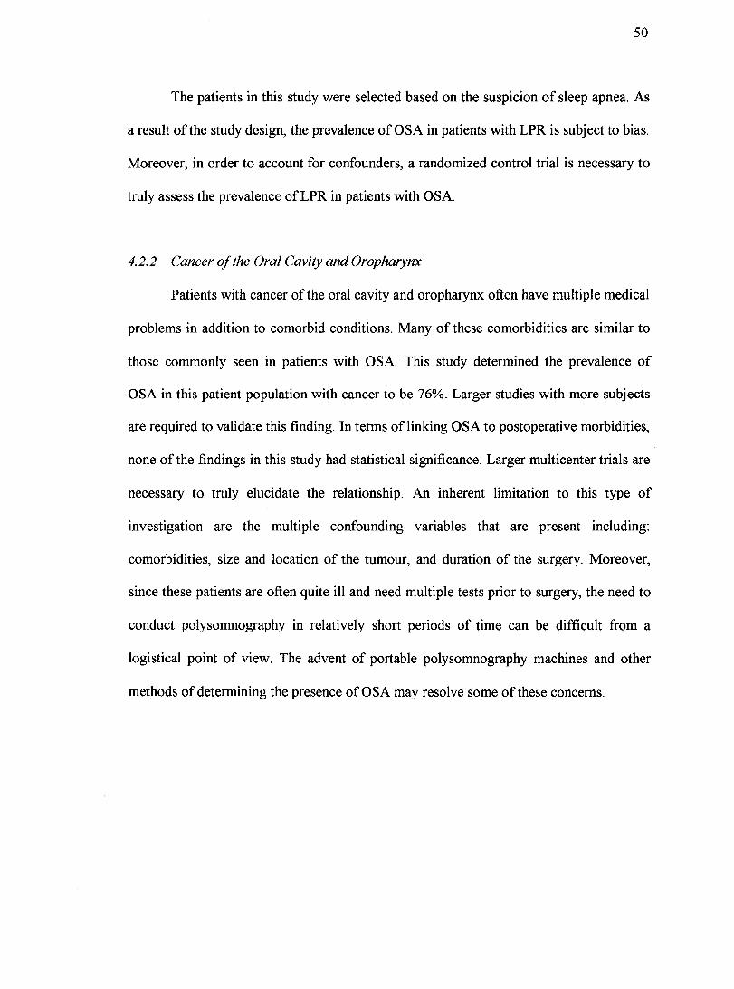
4.2.2 Cancer a/the Oral Cavity and Oropharynx
Patients with cancer of the oral cavity and oropharynx often have multiple medical
problems in addition to comorbid conditions. Many of the se comorbidities are similar to
those commonly seen in patients with OSA. This study determined the prevalence of
OSA in this patient population with cancer to be 76%. Larger studies with more subjects
are required to validate this finding. In terms of linking OSA to postoperative morbidities,
none of the findings in this study had statistical significance. Larger multicenter trials are
necessary to truly elucidate the relationship. An inherent limitation to this type of
investigation are the multiple confounding variables that are present including:
comorbidities, size and location of the tumour, and duration of the surgery. Moreover,
sin ce these patients are often quite ill and need multiple tests prior to surgery, the need to
conduct polysomnography in relatively short periods of time can be difficult from a
logistical point of view. The advent of portable polysomnography machines and other
methods of determining the presence of OSA may resolve sorne of the se concerns.

51
4.3 Clinical Implications and Future Directions
4.3.1 Laryngopharyngeal Reflux
This study demonstrates a direct association between laryngopharyngeal reflux
and obstructive sleep apnea by comparing AHI, RFS, LAR, and aryepiglottic fold
sensation. The mechanism leading to this association is unknown, TNF-a. may be one of a
multitude of contributors, however it is evident that the progression of one of the diseases
results in a concurrent worsening of the other. A potential role in the upper airway of
OSA may exist. This could lead to a cyclical interaction of LPR, leading to worsening
upper airway sensory dysfunction, contributing to perpetuation and/or progression of
apnea severity. Consequently treating one of the conditions may in fact lead to an
improvement in both. Randomized, controlled trials are needed to corroborate the
findings ofthis study and to further elucidate the pathophysiological relationship between
OSA and LPR.
4.3.2 Cancer of the Oral Cavity and Oropharynx
As a result of the very high prevalence of sleep apnea in this patient population
and it' s links to cardiovascular disease and increased perioperative complications, there is
a strong rationale to prioritize evaluation for OSA and initiation of definitive treatment
• 3515358-60909195 1 fu h f h' dfi" fOSA prIor to surgery.' " ", n rt er support 0 t IS, e Inttlve treatment 0
with nasal CP AP and tracheotomy have been shown to improve or reverse many of the
adverse cardiopulmonary sequelae of sleep apnea. 42-51
,91,92 For example, in OSA patients
with hypertension nasal CP AP reduces not only the AHI but also the mean blood
42 43 46 91 92 K k 1 h d d h 1 ft . 1 . . fi . pressure. ' , " ane 0 et a ave emonstrate t at e ventncu ar ejectlon ractlOn
improves in OSA patients with heart failure after 1 month of CP AP. 48 Kaye reported on
52
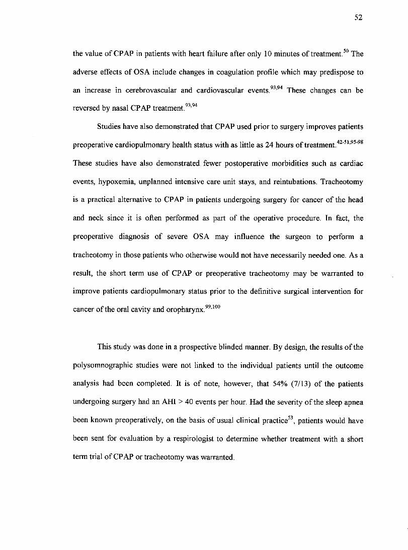
the value of CP AP in patients with heart failure after only 10 minutes of treatment. 50 The
adverse effects of OSA include changes in coagulation profile which may predispose to
an increase in cerebrovascular and cardiovascular events. 93,94 These changes can be
reversed by nasal CP AP treatment. 93,94
Studies have also demonstrated that CP AP used prior to surgery improves patients
. d' 1 h 1 h . h l' 1 24 h f 42-5195-98 preoperatlve car 10pU monary ea t status Wlt as ltt e as ours 0 treatment. '
These studies have also demonstrated. fewer postoperative morbidities such as cardiac
events, hypoxemia, unplanned intensive care unit stays, and reintubations. Tracheotomy
is a practical alternative to CP AP in patients undergoing surgery for cancer of the head
and neck since it is often performed as part of the operative procedure. In fact, the
preoperative diagnosis of severe OSA may influence the surgeon to perform a
tracheotomy in those patients who otherwise would not have necessarily needed one. As a
result, the short term use of CP AP or preoperative tracheotomy may be warranted to
improve patients cardiopulmonary status prior to the definitive surgi cal intervention for
cancer of the oral cavity and oropharynx. 99, 100
This study was done in a prospective blinded manner. By design, the results of the
polysomnographic studies were not linked to the individual patients until the outcome
analysis had been completed. It is of note, however, that 54% (7/13) of the patients
undergoing surgery had an AHI > 40 events per hour. Had the severity of the sleep apnea
been known preoperatively, on the basis ofusual clinical practice53, patients would have
been sent for evaluation by a respirologist to determine whether treatment with a short
term trial of CP AP or tracheotomy was warranted.

53
It is evident that more studies are needed with larger sample sizes to corroborate
the findings of an association between OSA and postoperative morbidities in patients with
malignancies of the oral cavity and oropharynx. Moreover, investigations determining
whether the above association exists for other regions of the head and neck, specifically
the nasopharynx, larynx and hypopharynx, should be undertaken. The effectiveness of
preoperative treatment of OSA using short term CP AP and tracheotomy must be
evaluated in patients with malignancies of the head and neck. Finally the potential role of
TNF-a in producing neuromuscular dysfunction in the upper airway in OSA must be
further investigated.
4.4 Conclusions (claims to originality are boldfaced)
4.4.1 Laryngopharyngeal Reflux
Laryngopharyngeal reflux is corn mon among patients with OSA, occurring at a
frequency much above that in the general population (93% in this study). Laryngeal
sensation, as reflected in both the sensory detection and laryngeal adductor reflex
thresholds correlates with apnea severity. The severity of laryngopharyngeal reflux,
measured by the reflux finding score, correlat es with laryngeal but not pharyngeal
sensory measures, as well as with OSA severity. These findings are consistent with the
hypothesis that LPR contributes to laryngeal sensory dysfunction in OSA, which in turn
contributes to the perpetuation or progression of apnea severity.
Further studies will be required to test the validity of this proposed interaction and to
investigate the extent to which this contributes to the overall pathophysiology of OSA. A

54
randomized controlled trial of the effects of LPR therapy with proton pump inhibitor
medication on apnea severity in OSA patients with LPR may be warranted, since it may
represent an important innovative therapeutic adjunct in OSA.
4.4.2 Cancer of the Oral Cavity and Oropharynx
The prevalence of OSA in patients with malignancies of the oral cavity and
oropharynx amenable to primary surgical resection is significantly higher than in
the general population (76% in this subject group). Further investigations are required
to evaluate the prevalence of OSA in a large sample of subjects awaiting surgical
intervention for head and neck malignancies, to determine the mechanisms underlying
this association, and to validate methods for preoperative screening of sleep apnea. There
was a tendency for postoperative complications, as measured by prolonged ICU stay, the
need for mechanical ventilation, and cardiopulmonary morbidities, to be more common
among patients with OSA, occurring in 67% of OSA patients and 25% of non-OSA
patients. Clinical trials are warranted to evaluate the role of preoperative treatment of
OSA with CP AP or tracheotomy in decreasing postoperative morbidity and mortality.

CHAPTER V: REFERENCES
1. Cistulli PA, Sullivan CE. Pathophysiology of sleep apnea. In: Sleep and
Breathing, edited by Saunders NA and Sullivan CE. New York: Marcel Dekker,
1994, p. 405-448.
2. Weil JV, Cherniack NS, Dempsey JA, et al. NIll.,BI workshop summary.
Respiratory disorders of sleep. Pathophysiology, clinical implications, and
therapeutic approaches. Am Rev Respir Dis 1987; 136:755-761.
3. Loadsman J A, Hillman DR. Anaesthesia and sleep apnoea. Br J Anaesth 2001;
86:254-266.
55
4. Zonato AI, Bittencourt LR, Martinho FL, et al. Association of systematic head and
neck physical examination with severity of obstructive sleep apnea-hypopnea
syndrome. Laryngoscope 2003; 113:973-980.
5. Young T, Palta M, Dempsey J, et al. The occurrence of sleep-disordered breathing
among middle-aged adults. NEnglJMed 1993; 328:1230-1235.
6. Friedman M, Landsberg R, Pryor S, et al. The occurrence of sleep-disordered
breathing among patients with head and neck cancer. Laryngoscope 2001;
111:1917-1919.

56
7. Lavie P. Incidence of sleep apnea in a presumably healthy working population: a
significant relationship with excessive daytime sleepiness. Sleep 1983; 6:312-318.
8. OIson LG, King MT, Hensley Ml, et al. A community study ofsnoring and sleep
disordered breathing. Am J Respir Crit Care Med 1995; 152:711-716.
9. Phillipson EA. Sleep apnea - A major public health problem. New Engl J Med
1993; 328: 1271-1273.
10. Contencin P, Guilleminault C, Manach Y. Long-term follow-up and mechanisms
of Obstructive Sleep Apnea (OSA) and related syndromes through infancy and
childhood. Int J Pediatr Otorhinolaryngol2003; 67:S119-S123.
Il. Ciscar MA, Juan G, Martinez V, et al. Magnetic resonance imaging of the
pharynx in OSA patients and healthy subjects. Eur Respir J 2001; 17:79-86.
12. Bloom JW, Kaltenborn WT, Quan SF, et al. Risk factors in a general population
for snoring. Importance of cigarette smoking and obesity. Chest 1988; 93 :678-
683.
13. Bain WM, Harrington JW, Thomas LE, et al. Head and neck manifestations of
gastroesophageal reflux. Laryngoscope 1983; 93: 175-9.

14. Nguyen ATD, Jobin V, Payne RJ, et al. Laryngeal and velopharyngeal sensory
impairment in obstructive sleep apnea. Manuscript in Review.
57
15. KimoffRJ, Sforza E, Champagne V, et al. Upper airway sensation in snoring and
obstructive sleep apnea. Am J Respir Crit Care Med 2001; 164:250-255.
16. Boyd JH, PetrofBJ, Hamid Q, Fraser R, KimoffRJ. Upper Airway Muscle
Inflammation and Denervation Changes in Obstructive Sleep Apnea: Am J Respir
Crit Care Med: published ahead ofprint May 19,2004 as
doi: 10.1164/rccm.200308-11000C.
17. Hemandes L, Payne RJ, Naor N, et al. Relationship between nerve tissue and
inflammatory cell infiltration in the upper airway of obstructive sleep apnoea
patients (abstract). Eur Resp J: In Press.
18. Aviv JE, Liu H, Parides M, et al. Laryngopharyngeal sensory deficits in patients
with laryngopharyngeal reflux and dysphagia. Ann Otol Rhinol Laryngol 2000;
109: 1000-6.
19. Alberti A, Sarchielli P, Gallinella E, et al. Plasma cytokine levels in patients with
obstructive sleep apnea syndrome: a preliminary study. J Sleep Res 2003; 12:305-
11.

58
20. Vgontzas AN, Papanicolaou DA, Bixler EO, et al. Elevation of plasma cytokines
in disorders of excessive daytime sleepiness: Role of sleep disturbance and
obesity. J Clin Endocrinol Metab 1997; 82: 1313-1316.
21. Hartung H-P, Gold R, Jung S. Local immune responses in the peripheral nervous
system. In: Clinical Neuroimmunology, edited by Antel J, Birnbaum G, and
Hartung H-P. London: Blackwell Science, 1998, p. 40-54.
22. Oka N, Akiguchi l, Kawasaki T, et al. Tumour necrosis factor-alpha in peripheral
nerve lesions. Acta Neuropathol1998; 95:57-62.
23. Wilcox P, Milliken C, Bressler B. High-dose tumour necrosis factor cr produces
an impairment of hamster diaphragm contractility. Am J Respir Crit Care Med
1996; 153: 1611-1615.
24. Wilcox PG, Wakai Y, Walley KR, et al. Tumour necrosis factor cr decreases in
vivo diaphragm contractility in dogs. Am J Respir Crit Care Med 1994;
150: 1368-1373.
25. Tracey, K.J., S.F. Lowry, B. Beutler, et al. Cachectinltumor necrosis factor
mediates changes of skeletal muscle plasma membrane potential. J Exp Med
1986; 164:1368-1373.

26. Nathan C. Nitric oxide as a secretory product of mammalian cells. F ASEB 1.
1992; 6: 3051-3064.
27. Luss H, Watkins SC, Freeswick PD, et al. Characterization ofinducible nitric
oxide synthase expression in endotoxemic rat cardiac myocytes in vivo and
following cytokine exposure in vitro. J Mol Cell Cardiol1995; 27:2015-2029.
28. Geller DA, Nussler AK, Di Silvio M, et al. Cytokines, endotoxin, and
glucocorticoids regulate the expression of inducible nitric oxide synthase in
hepatocytes. Proc Natl Acad Sci USA 1993; 90:522-526.
59
29. Hennet T, Richter C, Peterhans E. Tumour necrosis factor-a. induces superoxide
anion generation in mitochondria ofL929 cells. Biochem J 1993; 289:587-592.
30. Reid MB. Role ofnitric oxide in skeletal muscle: synthesis, distribution and
functional importance. Acta Physiol Scand 1998; 162:401-409.
31. Koufman JA. Laryngopharyngeal reflux 2002: A new paradigm of airway disease.
ENT - Ear Nose Throat Journal 2002;81(Suppl 2):2-6.
32. Koufman JA. Laryngopharyngeal reflux is different from classic gastroesophageal
reflux disease. ENT - Ear Nose Throat Journal 2002;81(Suppl 2):7-9.

33. Koufman JA. The otolaryngologic manifestations ofgastroesophageal reflux
disease (GERD): A clinical investigation of225 patients using ambulatory 24-
hour pH monitoring and an experimental investigation of the role of acid and
pepsin in the development of laryngeal in jury. Laryngoscope 1991; 1 0 1 (Suppl
53): 1-78.
34. Halstead LA. Gastroesophageal reflux: A critical factor in pediatric subglottic
stenosis. Otolaryngol Head Neck Surg 1999;120:683-8.
35. Ward PH, Hanson DG. Reflux as an etiological factor of carcinoma of the
laryngopharynx. Laryngoscope 1988;98: 1195-9.
36. Freije JE, Beatty TW, Campbell BH, et al. Carcinoma of the larynx in patients
with gastroesophageal reflux. Am J OtolaryngoI1996;17:386-90.
37. BeIafsky PC, Postma GN, Koufman JA. Validity and reliability of the reflux
finding score (RFS). Laryngoscope 2001; 111: 1313-7.
60
38. Vitiello MY, Prinz PN, Personius JP, et al. Relationship of alcohol abuse history
to nighttime hypoxemia in abstaining chronic alcoholic men. J Stud Alcohol 1990;
51:29-33.
39. Robinson RW, Zwillich CW. The effects of drugs on breathing during sleep. Clin
Chest Med 1985; 6:603-614.

61
40. Sahota PK, Jain SS, Dhand R. Sleep disorders in pregnancy. Curr Opin Pulrn Med
2003; 9:477-483.
41. Senior BA, Khan M, Schwirnrner C, et al. Gastroesophageal reflux and
obstructive sleep apnea. Laryngoscope 2001;111:2144-6.
42. Lattirnore ID, Celerrnajer DS, Wilcox 1. Obstructive sleep apnea and
cardiovascular disease. J Am Coll Cardiol 2003; 41: 1429-1437.
43. Richert A, Ansarin K, Baran AS. Sleep apnea and hypertension: pathophysiologic
rnechanisrns. Sernin Nephro12002; 22:71-77.
44. Wolk R, Sorners VK. Cardiovascular consequences of obstructive sleep apnea.
Clin Chest Med 2003; 24:195-205.
45. Burke AJ, Duke SG, Clyne S, et al. Incidence ofpulrnonary ederna after
tracheotorny for obstructive sleep apnea. Otolaryngol Head Neck Surg 2001;
125:319-323.
46. Shepard JW. Hypertension, cardiac arrhythrnias, rnyocardial infarction, and stroke
in relation to obstructive sleep apnea. Clin Chest Med 1992; 13:437-458.
62
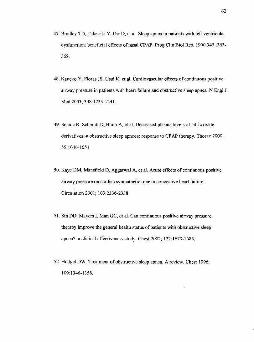
47. Bradley TD, Takasaki Y, Orr D, et al. Sleep apnea in patients with left ventricular
dysfunction: beneficial effects ofnasal CPAP. Prog Clin Biol Res. 1990;345 :363-
368.
48. Kaneko Y, Floras JS, Usui K, et al. Cardiovascular effects of continuous positive
airway pressure in patients with heart failure and obstructive sleep apnea. N Engl J
Med 2003; 348:1233-124l.
49. Schulz R, Schmidt D, Blum A, et al. Decreased plasma levels ofnitric oxide
derivatives in obstructive sleep apnoea: response to CP AP therapy. Thorax 2000;
55:1046-105l.
50. Kaye DM, Mansfield D, Aggarwal A, et al. Acute effects of continuous positive
airway pressure on cardiac sympathetic tone in congestive heart failure.
Circulation 2001; 103:2336-2338.
51. Sin DD, Mayers l, Man GC, et al. Can continuous positive airway pressure
therapy improve the general health status of patients with obstructive sleep
apnea?: a clinical effectiveness study. Chest 2002; 122:1679-1685.
52. Hudgel DW. Treatment of obstructive sleep apnea. A review. Chest 1996;
109: 1346-1358.

53. Meoli AL, Rosen CL, Kristo D, et al. Upper airway management of the adult
patient with obstructive sleep apnea in the perioperative period - avoiding
complications. Sleep 2003; 26: 1060-1065.
63
54. Jarrell L. Preoperative diagnosis and postoperative management of adult patients
with obstructive sleep apnea syndrome: a review of the literature. J Perianesth
Nurs 1999; 14: 193-200.
55. Mehta Y, Manikappa S, Juneja R, et al. Obstructive sleep apnea syndrome:
anesthetic implications in the cardiac surgi cal patient. J Cardiothorac Vasc Anesth
2000; 14:449-453.
56. Rosenberg J, Rosenberg-Adamsen S, Kehlet H. Post-operative sleep disturbances:
causes, factors and effects on outcome. Eur J Anaesthesiol1995; 10:28-30.
57. Montserrat JM, Montserrat F, Hemandez L, et al. Effectiveness ofCPAP
treatment in daytime function in sleep apnea syndrome: A randomized controlled
study with an optimized placebo. Am. 1. Respir. Crit. Care Med 2001; 164:608-
613.
58. Esc1amado RM, Glenn MG, McCulloch TM, et al. Perioperative complications
and risk factors in the surgical treatment of obstructive sleep apnea syndrome.
Latyngoscope 1989; 99:1125-1129.
64
59. Powell NB, Riley RW, Guilleminault C, et al. Obstructive sleep apnea, continuous
positive airway pressure, and surgery. Otolaryngol Head Neck Surg 1988; 99:362-
369.
60. Rennotte MT, Baele P, Aubert G, et al. Nasal continuous positive airway pressure
in the perioperative management of patients with obstructive sleep apnea
submitted to surgery. Chest 1995; 107:367-374.
61. Rechtschaffen A, Kales A. A manual of standardized terminology, technique and
scoring system for sleep stages ofhuman sleep. Los Angeles Brain Information
Service. Brain Information Service, DCLA, 1968
62. EEG arousals: scoring rules and examples: a preliminary report from the Sleep
Disorders Atlas Task Force of the American Sleep Disorders Association. SLEEP
1992; 15:173-84.
63. Wiener GJ, Koufman JA, Wu WC, et al. Chronic hoarseness secondary to
gastroesophageal reflux disease: Documentation with 24-h pH monitoring. Am J
GastroenteroI1989;84:1503-8.
64. Reulbach TR, Belafsky PC, Blalock PD, et al. Occult laryngeal pathology in a
community-based cohort. Otolaryngol Head Neck Surg 2001;124:448-50.

65
65. Green BT, Bourghton W A, O'Connor JB. Marked improvement in nocturnal
gastroesophageal reflux in a large cohort of patients with obstructive sleep apnea
treated with continuous positive airway pressure. Arch Int Med 2003; 163:41-5.
66. Bernard F, Dupont C, Viala O. Gastroesophageal reflux and upper airway disease.
Clin Rev Allergy 1990;8:403-425.
67. Ing AJ, Ngu MC, Breslin AB. Obstructive sleep apnea and gastroesophageal
reflux. Am J Med. 2000; 108(suppl 4A): 120S-125S.
68. Kerr P, Shoenut JP, Millar T, et al. Nasal CP AP reduces gastroesophageal reflux
in obstructive sleep apnea syndrome. Chest 1992;101:1539-1544.
69. Graf KI, Karaus M, Heinemann, et al. Gastroesophageal reflux in patients with
sleep apnea syndrome. Z Gastroenterol 1995;33:689-693.
70. Heading R. Epidemiology of oesophageal reflux disease. Scand J Gastroenterol
1989; 24 (suppl. 168): 33-7.
71. Postma GN, Tomek MS, Belafsky PC, Koufman JA. Esophageal motor function
in laryngopharyngeal reflux is superior to that in classic gastroesophageal reflux
disease. Ann Otol Rhinol Laryngol2001: 110: 1114-6.

66
72. Koufman JA, Belafsky PC, Daniel E, et al. Prevalence of esophagitis in patients
with pH-documented laryngopharyngeal reflux. Laryngoscope 2002;112:1606-9.
73. Richter JE, ed. Ambulatory Esophageal pH Monitoring : Practical Approach and
Clinical Applications. 2nd ed. Baltimore: Williams & Wilkins, 1997.
74. Oelschlager BK, Eubanks TR, Maronian N, et al. Laryngoscopy and pharyngeal
pH are complementary in the diagnosis of gastroesophageal-Iaryngeal reflux. J
Gastrointest Surg 2002;6: 189-94.
75. Ryan CF, Lowe AA, Li D, et al. Magnetic resonance imaging of the upper airway
in obstructive sleep apnea before and after chronic nasal continuous positive
airway pressuretherapy. Am Rev Respir Dis 1991;144:939-44.
76. Schwab RJ. Sleep apnea is an anatomic disorder. Am J Resp Crit Care Med 2003;
168;270-1.
77. Sériès F, Cote C, St Pierre S. Dysfunctional mechanical coupling ofupper airway
tissues in sleep apnea syndrome. Am J Respir Crit Care Med 1999; 159: 1551-9.
78. Chadwick G, Crowley AP, Fitzgerald MX, et al. Obstructive sleep apnea
following topical oropharyngeal anesthesia in loud snorers. Am Rev Respir Dis
1991; 143:810-3.

67
79. Fogel RB, Malhotra A, Shea SA, et al. Reduced genioglossal activity with upper
airway anesthesia in awake patients with OSA. J Appl Physiol2000; 88: 1346-54.
80. Cala SJ, Sliwinski P, Cosio MG, et al. Effect oftopical upper airway anesthesia on
apnea duration through the night in obstructive sleep apnea. J Appl Physioi 1996;
81:2618-26.
81. Homer RL, Innes JA, Hoiden lIB, et al. Afferent pathway(s) for pharynge al
dilator reflex to negative pressure in man: A study using upper airway anaesthesia.
JPhysiol1991; 436: 31-44.
82. Sant'Ambrogio G, Tsubone H, Sant'Ambrogio FB. Sensory information from the
upper airway: role in the control ofbreathing. Respir Physiol 1995; 102: 1-16.
83. Chesson AL Jr, Berry RB, Pack A. Practice parameters for the use of portable
monitoring devices in the investigation of suspected obstructive sleep apnea in
aduits. Sleep 2003; 26:907-913.
84. KimoffRJ, Olha AB, Charbonneau M, et al. Mechanisms ofapnea termination in
obstructive sleep apnea: role of mechanoreceptor and chemoreceptor stimuli. Am
Rev Respir Dis 1994; 149:707-714.
85. Garni AS, Caples SM, Somers VK. Obesity and obstructive sleep apnea.
Endocrinol Metab Clin North Am 2003; 32:869-894.

86. Woodson BT, Garancis JC, Toohill RJ. Histopathologic changes in snoring and
obstructive sleep apnea syndrome. Laryngoscope 1991; 101: 1318-1322.
68
87. Friberg D, Gazelius B, Hokfelt T, et al. Abnormal afferent nerve endings in the
solft palatal mucosa of sleep apneics and habituaI snorers. Regul Pept 1997; 71:
29-36.
88. Koskenvuo M, Partinen M, Sama S, et al. Snoring as a risk factor for hypertension
and angina pectoris. Lancet 1985; 1:893-896.
89. Koskenvuo M, Kapiro J, Telakivi T, et al. Snoring as a risk factor for ischaemic
heart disease and stroke in men. BMJ 1987; 294:16-19.
90. Gupta RM, Parvizi J, Hanssen AD, et al. Postoperative complications in patients
with obstructive sleep apnea syndrome undergoing hip or knee replacement: a
case-control study. Mayo Clin Proc 2001; 76:897-905.
9l. Reeder MK, Goldman MD, Loh L, et al. Postoperative obstructive sleep apnoea:
haemodynamic effects oftreatment with nasal CPAP. Anaesthesia 1991; 46:849-
853.

69
92. Tun Y, Hida W, Okabe S, et al. Can nasal continuous positive airway pressure
decrease clinic blood pressure in patients with obstructive sleep apnea? J Exp Med
2003; 201: 181-190.
93. Von Kanel R, Dimsdale JE. Hemostatic alterations in patients with obstructive
sleep apnea and the implications for cardiovascular disease. Chest 2003;
124:1956-1967.
94. Feng H, Feng H, Zhang C, et al. Significance of the changes ofplatelet activation
and fibrinolytic activity in patients with obstructive sleep apnea-hypopnea
syndrome. Zhonghua Jie He He Hu Xi Za Zhi 2002; 25:531-534.
95. Jiang H, Ni D, Li W. Preoperative nasal continuous positive airway pressure
treatment as substitute for protective tracheostomy in severe obstructive sleep
apnea syndrome. Zhonghua Er Bi Yan Hou Ke Za Zhi 1998; 33: 117 -119.
96. Harman EM, Wynne JW, Block Al The effect ofweight loss on sleep-disordered
breathing and oxygen desaturation in morbidly obese men. Chest 1982; 82:291-
294.
97. Miller FR, Watson D, Boseley M. The role of the genial bone advancement
trephine system in conjunction with uvulopalatopharyngoplasty in the multilevel
management of obstructive sleep apnea. Otolaryngol Head Neck Surg 2004;
130:73-79.

70
98. Goh YH, Lim KA. Modified maxillomandibular advancement for the treatment of
obstructive sleep apnea: a preliminary report. Laryngoscope 2003; 1l3: 1577-
1582.
99. Garrigue S, Bordier P, Jaïs P, et al. Benefit of atrial pacing in sleep apnea
syndrome. N Engl J Med 2002; 346:404-412.
100. Hedner J, Kraiczi H, Peker Y, et al. Reduction ofsleep-disordered
breathing after physostigmine. Am J Respir Crit Care Med 2003; 168: 1246-1251.
CHAPTER VI: APPENDICES
6.1 Consent Form (English)
Consent Form
Upper Airway Afferent Stimulation in Snorers (SN)and Obstructive Sleep Apnea (OSA) patients.
McGiII University Health Center Sleep Laboratory Principal Investigator: Dr John Kimoff
Introduction:
'
Centre unive15itaire de santé McGill McGi1l University Health Centre Hôpital Royal VIctoria Hospital 687, avenue des Pill5 Ouest, Montréal, Québec H3A lAl
71
Obstructive sleep apnea (OSA) is a common and important medical condition, of which the causes and the ways in which it progresses over time are not weIl understood. Previous research suggests that changes in the function of the sensory nerves in the upper airway (mouth and throat) contribute to this disease. These studies you are being asked to participate in are being conducted by Dr. John Kimoff and his colleagues in the Sleep Laboratory of the Royal Victoria Hospital pavilion of the McGill University Health Center, to see if stimulation of the nerves in the throat will have an influence on upper airway function.
Study Procedures:
Module 1: Overnight screening study: (normal controls only): You will undergo a home monitoring study to be sure you do not have any significant snoring or unexpected irregularities of breathing during sleep. You will be given a device to take home which records snoring, breathing movements and oxygen level. You will be instructed at the hospital on how to use the machine, take the machine home for one night and record yourself and then retum the equipment to the Hospital the next day. If this study is found to be ab normal, you will be asked to attend a more complete sleep study in the hospital (module 2). After the study is done, if it still shows significant abnormalities, you will be offered to consult a physician specialized in sleep diseases to explain the results to you and possible implications for your health. If it shows your sleep to be normal, you will be asked to continue in the study.
72
Risks and discom{orts: The recording is not painful or dangerous in any way. There may be sorne minor inconvenience associated with sleeping with wires attached on the skin, as weIl as the minor inconvenience oftransporting the small device to and from the hospital.
Module 2: Overnight sleep study (Polysomnography) (OSA patients and Snorers) This study permits a complete evaluation of your sleep and is done regularly in the Royal Victoria Hospital-MURC Sleep Laboratory. It measures your heart rate, breathing movements, electroencephalogram (your brain activity) and movements of your jaw and legs. These measurements are made with wires that are put on your skin with glue, with a pressure sens or that measures airflow under your nose and movement sensors around your waist and chest. You wiU be asked to sleep in the laboratory for the entire night.
Risks and discomforts: The recording is not painful or dangerous in any way. There may be sorne minor inconvenience associated with sleeping with wires attached. A technician wiU be available aU night long if any problem or question occurs.
Module 3: Upper airway sensory testing (Ali Subjects): This will involve two sessions (on two different days) ofapproximately 20-30 minutes of testing during the daytime. Just before beginning the measurements, a dilute solution that contains capsaicin, menthol or the liquid in which these substances are dissolved (which is water with a minimal concentration of alcohol (0.1%)) will be sprayed at the back of your throat using an aerosol device. Also, a small tube (approximately 4 mm. external diameter) will be inserted down the back of the no se and throat and will permit the application the same solution throughout the experiment. Capsaicin is the active ingredient in chili peppers. Menthol is a mint-like substance present in food and candy. One of the two sessions will be with capsaicin or menthol infused, the other session (on a different day) will be with liquid in which these two substances are dissolved. Neither you nor the technician performing the test will be aware of which of the two solutions you are receiving. After the substance is sprayed at the back of your throat and as it is infused , your sensation of touch will be tested in several places including your hand, lower lip and the inside of your mouth at the back. The testing will involve touching various objects to the skin at the se points, and asking you to report the presence or absence of a gentle buzzing sensation, or to indicate whether you are being touched in one or two places.No needles or medications are involved. The objects used to test you will be sterilized prior to use.
Risks and discomforts: There is no major risk associated with these tests. There is no discomfort with the sensory testing on the hand or lip. Testing in the throat may in sorne instances cause gagging. If this is a problem, testing will be stopped. You might feel a slightly burning or cold sensation and you might have a little bit of coughing when the substance is sprayed at the back of your throat. Before the tube is inserted, a small amount of numbing or freezing medication will be applied to the nose and back of the throat, in order to minimize any discomfort associated with the placement of the tube. If you have any history of allergy or adverse reaction to freezing or local anesthetic medications, you should not participate in this part of the study. There is a very small risk of provoking bleeding in the nose during placement of the catheter. Ifbleeding occurs, the catheter will be withdrawn. There could be minor discomfort with the catheter in the nose and throat
73
during the testing procedures. AIso, because the study substance will be infused throughout the experiment with the catheter, you may feel a little discomfort by having liquid at the back of your throat that will make you swallow more than usuai. If this is excessive, the tube will be removed. The capsaicin solution may cause a little burning sensation as if eating spicy food. Menthol solution may cause a co Id sensation.
Module 4: Daytime testing afupper airway dilatar rejlexes: This will involve 1 to 2 hours of testing during daytime sessions ( It will be done after testing described in module 3 on the same day.) The testing will be done on separate days with and without one of the study substance sprayed and infused on to the back of the throat as described in module 3. The aim of the testing is to measure the responses of two upper airway muscles to brief (one-half second) pulses of suction pressure applied to the mouth and throat. Two sets of electrodes will be used to measure the activity of each of the 2 upper airway muscles. These will consist of 4 fine wires inserted through needles into a fold of skin on the si de of the mouth at the back and under the tongue. A soft tube will be placed through the nose and advanced to the back of the throat for measurement of pressure. You will then breathe on a mouthpiece through sorne tubing with a noseclip in place. Intermittently you will feel the brief pulses of suction of varying strength applied to the mouthpiece, and we will record the muscle responses to those suction pulses.
Risks and discamforts: There is a minor risk of bleeding associated with placement of the pressure catheter through the no se, and with placement of needle electrodes. If bleeding oceurs, it will be treated with withdrawal of the catheter or pressure on the electrodes, respectively. There may be minor discomfort associated with the catheter in the nose and throat, which will be minimized during insertion by application of sorne freezing medication. If you have any history of allergy or adverse reaction to freezing or local anesthetic medications, you should not participate in this part of the study. The back of the mouth will also be frozen or anesthetized for insertion of the needle electrodes. The needle is inserted gently into the area which has been frozen and then is withdrawn leaving the very fine (hair-like) wire electrode in place. The application of negative pressure pulses will be extremely brief and associated with minimal discomfort. The pressure pulses will not. interfere with breathing. There could be mi Id discomfort associated with the administration of capsaicin or menthol as noted in module 3.
Potential Benefits:
Participation in this study willlikely not be of immediate benefit to you. However, the results will provide important insights into the nature and causes of a serious medical disorder, and thus could help others in the longer term.
Subject Rights:
Your participation in this study is voluntary. You may decline participation or withdraw from this study at any time and for any reason, and your decision to withdraw will not affect your treatment or medical management in any way. Ifyou have any questions about your rights as a research subject, you may contact the Patient Representative at 842-1231, local 5655.
Confidentiality:
AlI information derived from the se studies will be kept strictly confidential and will not be released to anyone other than the study investigators and research personnel. You will remain anonymous in any scientific presentations or publications reporting the results of this study
74
Statement of Consent
Upper Airway Afferent Stimulation in Snorers (SN)and Obstructive Sleep Apnea (OSA) patients
Dr. John R. Kimoff
1, , have read the information in this consent form. 1 understand that this is a research study and the procedures to be followed and the possible risks and benefits of the study and am aware of the other treatments available for my illness.
1 have had the opportunity to ask Dr. _________ questions and have received satisfactory answers to ail of them.
1 understand 1 am free to withdraw from this study anytime for any reason and the decision to stop taking part will not affect my future medical care. By signing this document, 1 am not giving up any of my legal rights. 1 will be given a signed copy of this consent form.
Having read ail the pages of this consent form and understood the requirements of the study, my signature below indicates that 1 voluntarily consent to participate in the study.
Subject's Name (please print)
Investigator's Signature (please print)
Subject's Signature Date
Investigator's Signature Date
For Further Information: Dr John Kimoff 514-842-1231-1568
75
Consent Form (Prospective Study: Control Subjects)
"
Centre univetSitaire de ~ McGill McGiU Univnsity Health Centre Hôpital Royal Victoria Hospital 687. avenue des Pins Ouest, Montréal, Québec H3A tA l
"Upper Airway Sensation and Tissue Changes in Snorers and Obstructive Sleep Apnea Patients"
McGill University Health Center Sleep Laboratory Principal Investigator: Dr John Kimoff
Introduction: Obstructive sleep apnea (OSA) is a common and important medical condition, of which the causes and the ways in which it progresses over time are not well understood. Previous research suggests that changes in the function of the sensory nerves in the upper airway (mouth and throat) contribute to this disease. These studies you are being asked to participate in are being conducted by Dr. John Kimoff and his colleagues in the Sleep Laboratory of the McGill University Health Center, to see if snoring and/or sleep apnea are associated with changes in the muscles and nerves in the upper airway tissues. You are being asked to participate in this study because you have decided with your doctor to undergo surgery on the upper airway. The object of this research study is to perform sorne specialized analyses on the tissues which are removed during your routine
surgical procedure.
Study Procedures:
1. Overnight screening study: You will undergo a home monitoring study to be sure you do not have any significant snoring or unexpected irregularities of breathing during sleep. You will be given a device to take home which records snoring, breathing movements and oxygen level. You will be instructed at the hospital on how to use the machine, take the machine home for one night and record yourself and then return the equipment to the Hospital the next day. If this study is found to be ab normal, you will be asked to attend a more complete sleep study in the hospital. After the study is done, if it still shows significant abnormalities, you will be offered to consult a physician specialized in sleep diseases to explain the
76
results to you and possible implications for your health. If it shows your sleep to be normal, you will be asked to continue in the study.

Risks and discomforts: The recording is not painful or dangerous in any way. There may be sorne minor inconvenience associated with sleeping with wires attached on the skin, as weU as the minor inconvenience of transporting the small device to and from the hospital.
2. Overnight sleep study (polysomnography) Ifyou are required to undergo an ovemight sleep study, this will be done in the Royal Victoria Hospital-MURC Sleep Laboratory. This test provides a detaited evaluation of your sleep and will be done in exactly the same way as for routine evaluation of patients from the Sleep Clinic. The test measures your heart rate, breathing movements, electroencephalogram (your brain activity) and movements ofyour jaw and legs. These measurements are made with wires that are attached to your skin with a special cream and tape, as weU as with a pressure sensor that measures airflow under your nose and movement sensors around your waist and chest. You will be asked to sleep in the laboratory for the entire night.
Risks and discomforts: The recording is not painful or dangerous in any way. There may be sorne minor inconvenience associated with sleeping with wires attached. A technician is present aU night long if any problem or question occurs.
3. Upper airway sensory testing: This testing will be done in the research area of the Royal Victoria Hospital-MURC Sleep Laboratory. The testing session lasts approximately 60 minutes in total. The testing will involve touching various objects to the skin on the hand, the lip and in the inside of your mouth and asking you to report the presence or absence of a gentle buzzing sensation, or to indicate whether you are being touched in one or two places. No needles or medications are involved. The objects used to test you will be sterilized prior to use. There will also be testing using gentle puffs of air delivered through a flexible tube called a laryngoscope. The pressure of the air puffs will varied slightly and you will be asked to report the presence or absence of a tapping sensation. T esting will be done on the lip and in the mouth, and then the tube will be passed gently into the nose and testing will be done at severallevels in the throat.
Risks and discomforts: These testing procedures have been conducted on many subjects in our laboratory. There have been no serious complications, and the procedures
77
have been associated with minimal discomfort. Testing in the mouth may in sorne instances cause gagging. If this is a problem, testing will be stopped. If at any time you find the procedure too uncomfortable, the tube will be withdrawn and the testing stopped. There is also a very small risk of the tube causing a nose bleed. In one recent study using the same apparatus, nosebleeding occurred only 3 times in 500 testing sessions. In all 3 cases the bleeding stopped on its own after removal of the tube.
4. Authorization for use of tissue obtained at the time of surgery (ail patients). The object of this study is to perform specialized testing on tissue that is removed during the standard operation that you have planned to undergo in consultation with your surgeon. No modification of the surgi cal technique is required for this study and no additional tissue will be removed other than what is normally removed during the standard operation.

Risks and discomforts: There are no additional risks or discomfort associated with using your tissues for this research study, beyond those which are normally associated with your surgi cal procedure, which have been explained to you by your surgeon.
5. Blood test (ail patients). This test will be performed before sensory testing in the same manner as when one has blood taken at a c1inicallaboratory, using a sterile needle. 14 ml ofblood will be taken, with this procedure taking approximately five minutes. The blood will be processed to obtain its plasma, and this portion will be frozen and subsequently analyzed for stress hormones we believe to be increased by OSA.
Risks and discomforts: There is minor discomfort with the insertion of a needle to draw blood, and a small risk of local bruising. AlI instruments will be sterile, so there is no risk of disease transmission.
Potential Benefits:
Participation in this study willlikely not be of immediate benefit to you. However, the results will provide important new knowledge conceming the causes of an important me di cal condition, and thus could help others in the longer term.
Subject Rights:
Your participation in this study is voluntary. You may decline participation or withdraw from this study at any time and for any reason, and your decision to withdraw will not affect your treatment or medical management in any way. Ifyou have any questions about your rights as a research subject, you may contact the Patient Representative at 842-1231, local 35655.
Confidentiality:
AlI information derived from the se studies will be kept strictly confidential and will not be released to anyone other than the study investigators and research personnel. Your tissues will be identified with a confidential code. Your name will not be linked to any samples or data obtained. Your tissue specimens will be kept in a secure storage area and aH unused samples will be destroyed. If you choose to withdraw from the study your tissue samples will be destroyed. You will remain anonymous in any scientific presentations or publications reporting the results ofthis study.
78
Statement of Consent:
"Neuromuscular Factors in the Pathophysiology of Obstructive Sleep Apnea"
l, , have read the information in this consent form. 1 understand that this is a research study and the procedures to be foUowed and the possible risks and benefits of the study and am aware of the other treatments available for my iUness.
1 have had the opportunity to ask Dr. _________ questions and have received satisfactory answers to aU ofthem.
1 understand 1 am free to withdraw from this study anytime for any reason and the decision to stop taking part will not affect my future medical care. By signing this document, 1 am not giving up any of my legal rights. 1 will be given a signed copy of this consent form.
Having read aIl the pages of this consent form and understood the requirements of the study, my signature below indicates that 1 voluntarily consent to participate in the study.
Subject's Name (please print)
Investigator's Signature (please print)
Subject's Signature
Investigator's Signature
For Further Information: Dr John Kimoff 514-843-1568
Date
Date
79

Consent Form (French)
Formule de consentement:
"
Centre univel8ÎtUre de aanté McGIll McGill UnIversity Hea1th Centft Hôpital Ratai VIctoria Hospital 687, avenue des Pins Ouest, Montréal, Québ«H3A lAI
"Stimulation des afférences sensitives des voies aériennes supérieures chez les patients souffrant d'apnée obstructive du sommeil (AOS) et chez les patients ronfleurs"
Investigateur principal: Dr John Kimoff
Centre universitaire de santé McGiII Laboratoire de sommeil
Introduction l'Apnée obtructive sommeil (AOS) est une condition médicale fréquente et importante dont les causes et la progression dans le temps ne sont pas bien comprises. Des recherches précédentes suggèrent que les changements de la fonction sensitive dans les voies aériennes supérieures (la bouche et la gorge) contribuent à cette maladie. L'étude à laquelle on vous demande de participer est dirigée par Dr. John Kimoff et ses collègues au Laboratoire de sommeil de l'hôpital de Royal Victoria du Centre Universitaire de Santé Mcgill (CUSM) et permettra de vérifier si la stimulation des nerfs dans la gorge aura une influence sur la fonction des voies aériennes supérieures.
Procédures
Module 1: Évaluation nocturne du sommeil: (sujets témoins seulement): On vous fera passer un test à domicile pour s'assurer que vous ne souffrez pas d'un problème de ronflement sérieux ou d'irrégularités respiratoires pendant le sommeil. On vous fournira un appareil permettant d'enregistrer le ronflement, les mouvements de la respiration et le niveau d'oxygène. De plus, on vous expliquera le fonctionnement de l'appareil. Vous l'apporterez à la maison pour une nuit, procéderez vous même à l'enregistrement et le rapporterez à l'hôpital de lendemain. Si cette étude s'avère être anormale, on vous demandera de passer un autre examen de votre sommeil plus complet tel que décrit au module 2. Après que ce nouvel examen, si on note toujours des anomalies significatives, on vous offrira la possibilité de consulter un médecin spécialisé dans les troubles du sommeil pour vous expliquer les résultats ainsi que les implications sur votre santé. Si l'examen est
80
normal, on vous demandera de continuer l'étude.
Risques et Désagréments: L'enregistrement n'est pas douloureux et ne présente aucun danger. Vous pourrez ressentir un léger inconfort à dormir en présence de fils d'enregistrement sur votre peau. Le transport de l'appareil entre l'hôpital et la maison peut également présenter un certain inconvénient même si l'appareil est petit.
Module 2: L'étude nocturne du sommeil (polysomnographie) Cette étude permet une évaluation complète de votre sommeil et se fait de façon régulière dans le laboratoire de sommeil de l'hôpital Royal-Victoria (CUSM). On mesurera la fréquence cardiaque, les mouvements respiratoires, l'électroencéphalogramme (l'activité du cerveau) ainsi que les mouvements de la mâchoire et des jambes. Toutes ces mesures sont faites à l'aide de fils (électrodes) qui sont fixés sur votre peau avec de la colle, avec un capteur de pression qui mesure le flux d'air sous le nez ainsi qu'un capteur de mouvement autour de votre taille et de votre poitrine. Vous serez invité(e) à dormir dans le laboratoire pour la nuit entière.
Risques et désagréments: L'enregistrement n'est pas douloureux. Il peut y avoir un certain dérangement causé par les fils et capteurs sur votre peau. Un technicien sera disponible toute la nuit au besoin.
Module 3: Évaluation Sensorielle des voies aériennes supérieures (tous les sujets) Cette évaluation, d'une durée de 20 à 30 minutes, sera effectuée le jour (deux journées différentes). Avant de débuter les mesures, une solution diluée soit de capsaïcine ou de mentol ou uniquement le liquide contenant la capsaicine ou le menthol (i.e.:le véhicule consistant en de l'eau avec une concentration très faible en alcool (0,1%» sera vaporisée à 1" arrière de votre gorge. De plus, un petit tube (diamètre externe d'approximativement 4 millimètres) sera inséré dans votre narine jusqu'à votre gorge et permettra l'application de la solution pendant l'expérience. La capsaïcine est le constituant actif du piment de cayenne. Le menthol est une substance utilisée dans beaucoup de produits comestibles disponibles sur le marché. Une des deux journées, l'expérimentation se fera avec l'une des deux substance dans la solution et l'autre journée se fera avec la solution (véhicule) seulement sans capsaïcine ou mentol. On déterminera le niveau de sensibilité tactile de diverses parties de votre corps (main, lèvre inférieure, fond de la gorge). À l'aide de différents objets, on touchera votre peau et on vous demandera de signaler la présence ou l'absence d'une légère sensation de vibration et d'indiquer si on vous touche à un ou deux endroits à la fois. Aucune aiguille ne sera utilisée. Les objets servant à faire les tests auront été stérilisés au préalable.
Risques et Désagréments: Ces tests ne comportent aucun risque important. L'évaluation sensorielle de la main et de la lèvre ne cause pas d'inconfort. L'évaluation de la gorge peut provoquer des nausées. Si cela devient trop gênant, on mettra fin à l'évaluation. Vous pourriez ressentir une sensation de brûlure ou de froid ou bien tousser un peu lorsque la solution sera vaporisée au fond de la gorge. Avant que le cathéter soit inséré, un médicament sera appliqué dans le nez et dans la gorge, afin de réduire au maximum tout inconfort lié à la mise en place du cathéter. Si vous avez une histoire
81

d'allergie ou d'intolérance aux anesthésiques locaux, vous ne devriez pas participer à cette partie de l'étude. Il y a un très petit risque de provoquer un saignement dans le nez pendant la mise en place du cathéter. Si un saignement se produit, le cathéter sera retiré. De plus, il pourrait y avoir un léger inconfort du fait d'avoir le cathéter en place dans le nez et la gorge pendant la procédure. De plus, puisque la solution sera infusée tout au long de l'expérience avec il se peut que vous sentiez le besoin d'avaler plus souvent qu'à l'habitude. La solution de menthol pourrait causer une sensation de froid et la capsaïcine pourrait causer une sensation de chaleur.
Module 4: Étude de jour des réflexes dilateurs des voies aériennes supérieures. (tous les patients) Ceci demandera 1 à 2 heures de votre temps pendant le jour (à la suite des tests décrits au module 3). L'étude sera faite pendant des jours différents avec et sans une des substances à l'étude (capsaicine ou menthol) qui sera vaporisée et infusée au fond de la gorge tel que décrit au module 3. Le but de l'étude est de mesurer la réponse de deux muscles dilatateurs des voies aériennes supérieures à de brèves impulsions (une demi-seconde) de pression appliquées à la bouche et à la gorge. Deux ensembles d'électrodes seront utilisés pour mesurer l'activité de chacun des 2 muscles des voies aériennes supérieures. Cela consistera en 4 petits fils insérés à travers une aiguille dans un pli de la peau à l'arrière de la gorge et sous la langue. Un tube mou sera placé dans le nez et inséré au fond de la gorge pour mesurer la pression. Vous respirerez alors par un embout de caoutchouc connecté à des tuyaux. Par intermittence vous sentirez de brèves impulsions de pression d'intensité variable appliquées à l'embout, et nous enregistrerons les réponses des muscles dilatateurs à ces impulsions de pression.
Risques et désagréments: Il y a un risque mineur de saignement lié à la mise en place du cathéter par le nez et à la mise en place des électrodes. Si un saignement se produit, il sera traité respectivement par retrait du cathéter ou par une pression locale sur les électrodes. Il peut y avoir un inconfort mineur lié au cathéter dans le nez et la gorge qui sera réduit au minimum pendant l'insertion par l'application d'une petite quantité d'un médicament anesthétique. Si vous avez une histoire d'allergie ou d'intolérance aux anesthésiques locaux, vous ne devriez pas participer à cette partie de l'étude. Également, l'intérieur de la bouche sera anesthésié pour faciliter l'insertion des électrodes. L'électrode est montée dans une aiguille qui est insérée doucement dans l'endroit gelé et l'aiguille est alors retirée laissant ainsi l'électrode (grosseur d'un cheveux) en place. L'application des impulsions de pression sera extrêmement brève et ne devrait pas provoquer d'inconfort. De plus elles ne gêneront pas la respiration. Il pourrait y un léger inconfort lié à l'administration de la substance à l'étude tel que mentionné dans le module 3.
Avantages possibles:
Vous ne retirez aucun avantage immédiat de votre participation à cette étude. Cependant, les resultats obtenus permettront aux investigateurs de mieux comprendre la nature et les causes de ce trouble médical grave et ainsi d'aider d'autres patients à plus long terme.
82

Droits des sujets:
Votre participation à cette étude est volontaire. Vous pouvez refuser d'y participer ou vous en retirer en tout temps, pour quelques raisons que ce soit, sans préjudice quant à votre traitement ou à la qualité des soins qui vous seront prodigués. Si vous avez des questions au sujet de vos droits en tant que sujet de recherche, vous pouvez communiquer avec le représentant des patients, au 514-842-1231, local 5655.
Confidentialité:
Tous les renseignements recueillis dans le cadre de cette étude seront gardés strictement confidentiels et seuls les investigateurs de l'étude et le personnel de recherche y auront accès. Votre identité ne sera divulguée dans aucune présentation ou publication scientifique portant sur les résultats de l'étude.
Consentement
"Stimulation des afférences sensitives des voies aériennes supérieures chez
83
les patients souffrant d'apnée obstructive du sommeil (ADS) et chez les patients ronfleurs"
Dr. John R. Kimoff
J'ai, , lu l'information de ce formulaire de consentement. Je comprends que c'est une étude de recherche et les procédures à suivre. Je comprends aussi les risques et les avantages possibles de l'étude. Je suis averti des autres traitements disponibles pour ma maladie.
J'ai eu l'occasion de poser des questions au Dr. _______ et j'ai reçu des réponses satisfaisantes à toutes mes questions.
Je comprends que je suis libre de me retirer de cette étude n'importe quand pour n'importe quelle raison. et La décision de cesser de participer n'affectera pas mes soins médicaux futurs. En signant ce document, je ne renie aucun de mes droits légaux. J'ai reçu une copie signée de ce formulaire de consentement.
Après avoir lu toutes les pages de ce formulaire de consentement et ayant compris les conditions de l'étude, ma signature ci-dessous indique que je consens volontairement à participer à l'étude.
nom du sujet signature date
nom de l'investigateur signature date
pour plus d'information: Dr John Kimoff514-842-1231 poste 1568
Formulaire de Consentement:
\?' Centre wùvelSitaire de santé McGill McGill Univetsity Healtlt Centre
Hôpital Royal VICtoria Hospital 687, avenue des Pins Ouest, Montréal, Québec H3A lAl
(Étude prospective: sujets témoins)Sensation des voies aériennes supérieures et changement dans les tissus chez les patients ronfleurs et chez les patients souffrant d'apnée obstructive du sommeil (AOS)
Laboratoire de Sommeil du Centre universitaire de santé McGill Investigateur principal: Dr John Kimoff
Introduction L'apnée obstructive du sommeil (AOS) est une condition médicale
fréquente et importante dont les causes et la façon de progresser ne sont pas bien comprises. Des recherches précédentes suggèrent que les changements des nerfs sensoriels dans les voies aériennes supérieures (la bouche et la gorge) contribuent à cette maladie. L'étude à laquelle on vous demande de participer est dirigée par Dr John Kimoff et ses collègues au Laboratoire de sommeil de l'hôpital Royal Victoria du Centre universitaire de santé Mcgill (CUSM) et permettra de vérifier si le ronflement et/ou l'AOS sont associé à des changements dans les muscles et les nerfs dans les tissus des voies aériennes supérieures. On vous demande de participer à cette étude parce que vous avez décidé avec votre médecin de subir une intervention chirurgicale sur vos voies aériennes supérieures dans le but de traiter votre problème de ronflement ou d'AOS. Le but de cette étude est de réaliser des analyses spécialisées sur les tissus qui seront enlevés durant votre chirurgie.
Procédures
1. Évaluation nocturne du sommeil : On vous fera passer un test à domicile pour s'assurer que vous ne soutITez pas d'un problème de ronflement sérieux ou d'irrégularités respiratoires pendant le sommeil. On vous fournira un appareil permettant d'enregistrer le ronflement, les mouvements de la respiration et le niveau d'oxygène. De plus, on vous expliquera à l'hôpital le fonctionnement de l'appareil. Vous l'apporterez à la maison pendant une nuit, procéderez vous même à l'enregistrement et le rapporterez à l' hôpital de lendemain. Si cette étude s'avère anormale, on vous demandera de passer un autre examen de votre sommeil plus complet. Après ce nouvel examen, si on note toujours des anomalies significatives, on vous offrira la possibilité de consulter un médecin spécialisé dans les troubles du sommeil pour vous expliquer les résultats ainsi que les conséquences sur votre santé. Si l'examen est normal, on vous demandera de continuer l'étude.
84

Risques et Désagréments: L'enregistrement n'est pas douloureux et ne présente aucun danger d'aucune manière. Vous pourrez ressentir un léger inconfort à dormir en présence de fils d'enregistrement sur votre peau. Le transport de l'appareil entre l'hôpital et la maison peut également présenter un certain inconvénient même si l'appareil est petit.
2: L'étude nocturne du sommeil (polysomnographie) On vous demande de faire cette étude de nuit sur le sommeil qui sera réalisée au laboratoire de sommeil de l'hôpital Royal Victoria (CUSM). Cette étude permet une évaluation complète de votre sommeil et se fera exactement de la même façon que nos tests habituels chez les patients de la Clinique de sommeil. On mesurera la fréquence cardiaque, les mouvements respiratoires, l'électroencéphalogramme (l'activité du cerveau) ainsi que les mouvements de la mâchoire et des jambes. Toutes ces mesures sont faites à l'aide de fils (électrodes) qui sont fixés sur votre peau avec une crème spéciale et du ruban et avec un capteur de pression qui mesure le flux d'air sous votre nez ainsi qu'un capteur de mouvement autour de votre taille et de votre poitrine. Vous serez invité(e) à dormir dans le laboratoire pendant une nuit entière.
Risques et désagréments: L'enregistrement n'est pas douloureux ni dangereux d'aucune façon. Il peut y avoir un certain dérangement causé par les fils et capteurs sur votre peau. Un technicien sera disponible toute la nuit en cas de problèmes ou de questions.
3: Évaluation sensorielle des voies aériennes supérieures: Cette évaluation sera réalise dans le centre de recherche du laboratoire de sommeil de l'hôpital Royal Victoria (CU SM). La séance sera d'une durée totale d'environ 60 minutes. À l'aide de différents objets, on touchera votre peau sur la main, la lèvre et l'intérieur de la bouche et on vous demandera de signaler la présence ou l'absence d'une légère sensation de vibration et d'indiquer si on vous touche à un ou deux endroits à la fois. Aucune aiguille ne sera utilisée. Les objets servant à faire les tests auront été stérilisés au préalable. Il y aura aussi une évaluation de la sensibilité avec un stimulateur produisant des bouffées d'air sous pression délivrées par un tube flexible appelé laryngoscope. Ces bouffées d'air produisent une sensation semblable à un toucher très léger. La pression des bouffées d'air sera variée et on vous demandera d'indiquer si vous sentez la présence ou non de ces stimulus. La sensation sera testée sur la lèvre et à l'intérieur de la bouche, et ensuite le laryngoscope sera passé doucement par le nez dans le but d'examiner différents niveaux dans la gorge.
Risques et désagréments: Ces tests ont été réalisés chez nombreux sujets dans notre laboratoire. Il n'y a pas eu de complication grave, et les tests ont été associés à un inconfort minime. L'évaluation sensorielle de la gorge peut provoquer des nausées. Si cela devient trop gênant, on mettra fin à l'évaluation. Vous pourriez ressentir un léger inconfort ou des nausées lors du passage du tube dans le nez et la gorge pendant la procédure. L'inconfort est minimisé par le petit calibre du laryngoscope (diamètre de 7 mm), et par l'utilisation d'un lubrifiant sur le tube. Si à tout moment vous trouvez le test trop inconfortable, le tube sera retiré et l'évaluation sera terminée. Il y a un très petit risque de provoquer un saignement dans le nez pendant le passage du tube. Dans une étude récente utilisant le même équipement, un saignement du nez ne s'est produit que 3 fois sur 500 procédures. Dans les 3 cas, le saignement s'est arrêté tout seul après que le tube soit retiré.
4. Autorisation à utiliser des tissus enlevés lors de votre chirurgie:
85
Le but de cette étude est de réaliser des analyses spécialisées sur le tissu qui sera enlevé pendant votre chirurgie, ce que vous avez décidé d'entreprendre avec votre chirurgien. TI n'y aura aucune modification de la technique chirurgicale dans le but de cette étude et aucun tissu additionnel ne sera enlevé autre que celui qui est normalement enlevé durant l'opération courante.
Risques et désagréments: Il n'y aura aucun risque ou inconfort associé à l'utilisation de vos tissus pour ce projet de recherche, autre que ceux qui sont normalement associés à l'intervention habituelle, ce qui vous a été expliqué par votre chirurgien.
Avantages possibles :Vous ne retirez aucun avantage immédiat de votre participation dans cette étude. Cependant, les résultats obtenus permettront aux investigateurs de mieux comprendre la nature et les causes de cette condition médicale grave et ainsi à aider d'autres patients à plus long terme.
Droits des sujets:
Votre participation dans cette étude est volontaire. Vous pouvez refuser d'y participer ou vous en retirer en tout temps, pour quelque raison que ce soit, sans influencer négativement votre traitement actuel ou la qualité des soins qui vous seront prodigués. Si vous avez des questions au sujet de vos droits en tant que sujet de recherche, vous pouvez communiquer avec l'ombudsman, au 514-842-1231, local 35655.
Confidentialité:
Tous les renseignements recueillis dans le cadre de cette étude seront gardés strictement confidentiels et seuls les investigateurs de l'étude et le personnel de recherche y auront accès. Vos tissus seront identifiés avec un code confidentiel. Votre nom ne sera pas associé à des tissus ou des données obtenus. Les spécimens de vos tissus seront gardés dans un endroit sécurisé, et les tissus qui ne sont pas utilisés pour l'étude seront détruits. Si vous optez de vous retirer de cette étude, les échantillons de vos tissus seront détruits. Votre identité ne sera pas divulguée dans aucune présentation ou publication scientifique portant sur les résultats de l'étude.
86

Consentement
Facteurs neuromusculaires dans la pathophysiologie de l'apnée obstructive du sommeil
J'ai,lu l'information dans ce formulaire de consentement. Je comprends que c'est une étude de recherche et qu'il y a des procédures à suivre. Je comprends aussi les risques et les avantages possibles de l'étude. Je suis au courant des autres traitements disponibles contre ma maladie.
J'ai eu l'occasion de poser des questions au Dr _______ et j'ai reçu des réponses satisfaisantes à toutes mes questions.
Je comprends que je suis libre de me retirer de cette étude n'importe quand pour n'importe quelle raison. La décision de cesser de participer n'affectera pas mes soins médicaux futurs. En signant ce document, je ne renie aucun de mes droits. Je recevrai une copie signée de ce formulaire de consentement.
Après avoir lu toutes les pages de ce formulaire de consentement et ayant compris les conditions de l'étude, ma signature ci-dessous indique que je consens volontairement à participer à l'étude.
nom du sujet signature date
nom de l'investigateur signature date
pour plus d'information: Dr John Kimoff 514-843-1568
87

6.2 Ethics Certificate
Centre hospitalier de St. Mary St. Mary's Hospital Center 3830, avenue Lacombe, Montréal (Québec) H3T lMS
RESEARCH REVIEW OFFICE DEPARTMENT OF CLINICAL EPIDEMIOLOGY AND COMMUNITY STUDIES ROOM 2506 .:. TEL.: (514) 345-3511 EXT. 3698 .:. FAX: (514) 734-2652
Date: January 20,2004
To: Dr. J. Kimoff Royal Victoria Hospital Respiratory Div. Rm. L408 687 Pins Ave. W. Montreal, QC H3A lAI
Re: Status of Proto col #03-29 entitled "Upper airway sensation and tissue changes in snorers (SN) and. Obstructive sleep apnea (OSA) patients"
We are pleased to inform you that the above-mentioned protocol has received full institutional approval at St. Mary's Hospital Center. This approval is valid for a period of one year.from January 20, 2004 ta January 19, 2005.
The following documents were approved:
Protocol dated: December 8. 2003 English Consent forms dated January 19. 2004
Consent Form: The English consent forms have been stamped to indicate the Chair of the Research Ethics Committee approval. Please make copies of this stamped consent form for use in your study. Please forward the French Consent form for approval as soon as possible.
A copy of the consent form is to be filed in the patient's chart with a coyer sheet. Enclosed is acover sheet that must be completed for each patient emolled on the protocol. Please complete and return a caver master sheet of al! standard information for approval (this should include al! required information except for the date of study entry).
For each patient emolled in the study, the following should be given to the ward clerk for filing in the outpatient section of the patient chart: 1) The cover sheet cOIl}pleted with the date of study emolment and the signature of the investigator or
delegated research assistant . 2) A copy of the signed consent form. The investigator (or delegated research assistant) should write on the
copy of the consent form "copie conforme à l'original" and sign this.
Research Subject Log: Enclosed you will also find a log shyet requesting the chart number and date for each patient emolled on the study. Copies of the form can be made as required. This information should be forwarded to the Research Review Office of the Department of Clinical Epidemiology and Community Studies on a monthly basis.
Affilié à l'Université McGill ;~ Affiliated with McGili University
88

Monitoring: The REC has recommended patient consent form monitoring for your protocol. Please be advised that our Research Monitor, Ms. Tina Emond, will contact you shortly to schedule a meeting to discuss the monitoring in greater detai1.
<\.dditional Information: In addition, you will find enc10sed an information sheet for fully approved protocois ietailing specifie guidelines to follow to maintain this full institutional approval status. Vou are responsible for nforming this office of any changes to the proto col or of serious adverse eventS. It is strongly advised that you ceep this information sheet throughout the course of your research for reference purposes.
'lease forward aIl future correspondence to Ms. Claudette Garrison, Research Administrative Secretary, )epartrnent ofClinicai Epidemiology and Community Studies, St. Mary's Hospital, (514) 345-3511 ext. 3698
)r. Martin Cole - l \,.. :hair, Research Ethics Co it1ee
~C:
Ir. Richard Payne 'r. J. R. Sutton . 'r. George Sejean [s. Tina Emond
ncl. pproved consent formes) onsent form cover sheet o:search subject log formation sheet
Dr. Jane McCusker Chair, Scientific Research Committee
2