Embed Size (px)
Citation preview

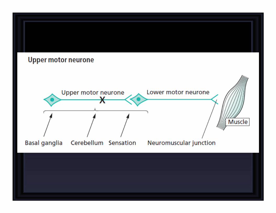
UPPER AND LOWER MOTOR
NEURON LESIONS
Prof. Syed Shahid HabibMBBS DSDM FCPS
Professor & Consultant Clinical Neurophysiology
Dept. of PhysiologyKing Saud University

Describe the functional anatomy of upper and lower motor neurons
Describe and differentiate the features of upper and lower motor neuron lesions
Explain features of Brown Sequard Syndrome
Correlate the site of lesion with pattern of loss of sensations
Describe facial, bulbar and pseudobulbar palsy
OBJECTIVES
At the end of this lecture you should be able to

31
segments
Embryological
development�
growth of cord
lags behind �
mature spinal
cord ends at
L1

Upper cervical cord
lesions produce
quadriplegia and
weakness of the
diaphragm
Lesions at C4-C5
produce quadriplegia


COMAPRISON BETWEEN UPPER & LOWER MOTOR
NEURON LESIONS
UMN LESION• Paralysis affect
movements
• Wasting not pronounced.
• Spasticity Muscles hypertonic (Clasp Knife).
• Tendon reflexes
increased.
• Superficial reflexes
diminished
• Babinski’s sign +ve,
� NCV- normal
� No denervation potentials
in EMG
LMN LESION• Individual muscle or group
of muscles are affected.
• Wasting pronounced.
• Flaccidity. Muscles hypotonic.
• Tendon reflexes diminished
or absent.
� NCV- abnormal
� Denervation potentials in
EMG (fibrillations)
• Muscle contractures
• Trophic changes in skin
and nails

Characteristic of upper
motor neurone lesions:• no wasting;
• Loss of skilled
finger/toe movements
• increased tone of clasp-
knife type;
• weakness most evident
in anti-gravity muscles;
• increased reflexes and
clonus;
• extensor plantar
responses.
Characteristics of lower
motor neurone lesions:• wasting;
• fasciculation (tapping produce it)
• decreased tone (i.e.
flaccidity);
• weakness;
• decreased or absent
reflexes;
• flexor or absent plantar
responses.
COMAPRISON BETWEEN UPPER & LOWER MOTOR NEURON LESIONS


Brown Sequard syndrome
Ipsilateral Loss:• Fine touch, Vibration, Proprioception (Dorsal Column)
• Leg Ataxia (Dorsal Spinocerebellar)
• Spastic Paresis below lesion (Lat Corticospinal)• Flaccid Paralysis (Vent horn destruction)
• Dermatomal Anesthesia (Dorsal Horn destruction)
Contralateral Loss:• Loss of pain and temp (lat Spinothalamic)• Loss of crude touch and Pressure (Vent Spinothalamic)
• Minor Contralat Muscle Weakness (Vent Corticospinal)• Leg Ataxia (Vent Spinocerebellar)
HEMISECTION OF SPINAL CORD

1. Ipsilateral lower motor neuron paralysis in the segment of
the lesion and muscular atrophy. These signs are caused by
damage to the neurons on the anterior gray column and
possibly by damage to the nerve roots of the same segment.
2. Ipsilateral spastic paralysis below the level of the lesion.
An ipsilateral Babinski sign is present, and depending on the
segment of the cord damaged, an ipsilateral loss of the
superficial abdominal reflexes and cremasteric reflex occurs.
All these signs are due to loss of the corticospinal tracts on
the side of the lesion. Spastic paralysis is produced by
interruption of the descending tracts other than the
corticospinal tracts.
3. Ipsilateral band of cutaneous anesthesia in the segment of
the lesion. This results from the destruction of the posterior
root and its entrance into the spinal cord at the level of the
lesion.

4. Ipsilateral loss of tactile discrimination and of vibratory and
proprioceptive sensations below the level of the lesion. These
signs are caused by destruction of the ascending tracts in the
posterior white column on the same side of the lesion.
5. Contralateral loss of pain and temperature sensations below
the level of the lesion. This is due to destruction of the crossed
lateral spinothalamic tracts on the same side of the lesion.
Because the tracts cross obliquely, the sensory loss occurs two
or three segments below the lesion distally.
6. Contralateral but not complete loss of tactile sensation below the level of
the lesion. This condition is brought about by destruction of the crossed
anterior spinothalamic tracts on the side of the lesion. Here, again, because
the tracts cross obliquely, the sensory impairment occurs two or three
segments below the level of the lesion distally. The contralateral loss of
tactile sense is incomplete because discriminative touch traveling in the
ascending tracts in the contralateral posterior white column remains intact.

Contralateral monoparesis
A lesion situated peripherally in
the cerebral hemisphere, i.e.
involving part of the motor
homunculus only, produces
weakness of part of the
contralateral side of the body, e.g.
the contralateral leg. If the lesion
also involves the adjacent sensory
homunculus in the postcentral
gyrus, there may be some sensoryloss in the same part of the body.
Contralateral monoparesis

Contralateral hemiparesis
Lesions situated deep in the
cerebral hemisphere, in the region
of the internal capsule, are much
more likely to produce weakness
of the whole of the contralateral
side of the body, face, arm and leg.
Because of the funnelling of fibre
pathways in the region of the
internal capsule, such lesions
commonly produce significant
contralateral sensory loss
(hemianaesthesia) and visual loss
(homonymous hemianopia), in
addition to the hemiparesis.
Contralateral hemiparesis

Ipsilateral hemiparesis
A unilateral high cervical cord
lesion will produce a hemiparesis
similar to that which is caused
by a contralateral cerebral
hemisphere lesion, except that the
face cannot be involved in the
hemiparesis, vision will be
normal, and the same dissociation
of sensory loss (referred to above)
may be found below the level of
the lesion.
Ipsilateral hemiparesis

A spinal cord lesion more usually causes upper motor
neurone signs in both legs, often asymmetrically since the
pathology rarely affects both sides of the spinal cord equally.
Paraparesis, if the lesion is at orbelow the cervical portion of the
spinal cord.
Paraparesis,

Ipsilateral monoparesis
A unilateral lesion in the spinal
cord below the level of the neck
produces upper motor neurone
weakness in one leg. There may
be posterior column (position
sense) sensory loss in the same
leg, and spinothalamic (pain and
temperature) sensory loss in the
contralateral leg. This is known as
dissociated sensory loss, and the
whole picture is sometimes
referred to as the Brown-Séquard
syndrome.
Ipsilateral monoparesis

Tetraparesis or quadriparesis, if
the lesion is in the upper cervical
cord or brainstem.
A spinal cord lesion more usually causes upper motor
neurone signs in both legs, often asymmetrically since the
pathology rarely affects both sides of the spinal cord equally.
Tetraparesis or quadriparesis


Generalized LMN weakness may
also result from widespread
damage to the axons of the LMNs.
This is the nature of peripheral
neuropathy (also called
polyneuropathy). The axons of
the dorsal root sensory neurones
are usually simultaneously
involved. The LMN weakness
and sensory loss tend to be most
marked distally in the limbs.
Generalized LMN weakness

Generalized LMN weakness may
result from pathology affecting
the LMNs throughout the spinal
cord and brainstem, as in motor
neurone disease or poliomyelitis.
Generalized limb weakness
(proximal and distal), trunk and
bulbar weakness characterize this
sort of LMN disorder.
Generalized LMN weakness

LMN weakness may be confined
to the distribution of one spinal
root (above) or one individual
peripheral nerve (below). In such
circumstances, the LMN signs
are found only in the muscles
supplied by the particular nerve
root or peripheral nerve in
question. Almost always there is
sensory impairment in the area
supplied by the nerve or nerve
root. Examples of such lesions are
an S1 nerve root syndrome caused
by a prolapsed intervertebral disc,
or a common peroneal nerve
palsy caused by pressure in the
region of the neck of the fibula.
LMN weakness of one spinal root

Motor neuron disease
• Selectively affect motor neurons, that
control voluntary muscle activity
• Types-� Amyotrophic lateral sclerosis- UMN+LMN
� Primary lateral sclerosis- UMN
� Progressive muscular atrophy- LMN
� Bulbar palsy- bulbar LMN
� Pseudobulbar palsy- bulbar UMN

Spinal cord
•Transverse myelitis
�Upper sensory level for all sensations,
LMN signs at the level of lesion, flaccid paralysis (spinal
shock)�UMN signs distally, Bladder/Bowel involved
•Anterior spinal artery syndrome
�Upper sensory level for pain/temperature,
sparing of posterior columns, UMN signs distally
•Brown-Sequard syndrome
�I/L spastic paralysis & loss of joint/position sense,
C/L loss of pain/temperature sensation

Bulbar palsy
� B/L LMN defect of IX-XII cranial nerves
� Dysphagia (liquid>solid), nasal regurgitation, slurred speech
� Nasal speech, wasted tongue with fasciculation, absent gag reflex
Pseudobulbar palsy-� B/L UMN defect of IX-XII cranial nerves
� Dysphagia, dysarthria, emotional lability
� Slow indistinct speech, spastic tongue, brisk jaw jerk
� Frontal release signs
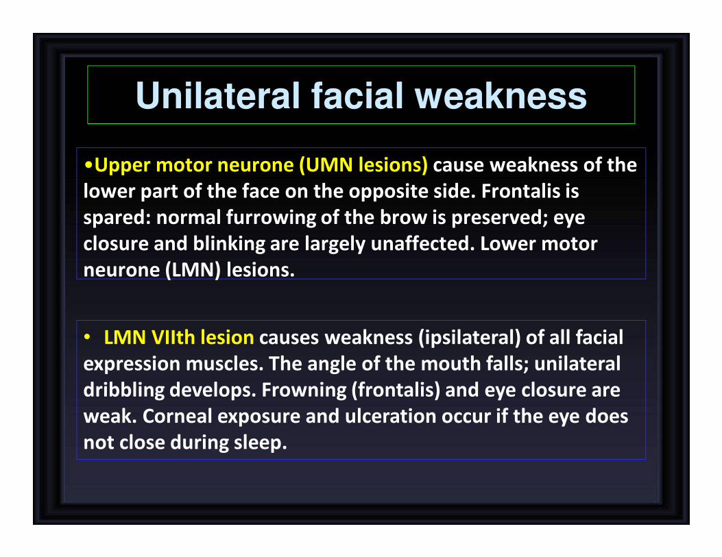
Unilateral facial weakness
•Upper motor neurone (UMN lesions) cause weakness of the
lower part of the face on the opposite side. Frontalis is
spared: normal furrowing of the brow is preserved; eye
closure and blinking are largely unaffected. Lower motor
neurone (LMN) lesions.
• LMN VIIth lesion causes weakness (ipsilateral) of all facial
expression muscles. The angle of the mouth falls; unilateral
dribbling develops. Frowning (frontalis) and eye closure are
weak. Corneal exposure and ulceration occur if the eye does
not close during sleep.

Intramedullary and Extramedullary
Syndromes
Extramedullary lesions,
radicular pain is often
prominent, and there is
early sacral sensory loss
(lateral spinothalamic
tract) and spastic weakness in the legs
(corticospinal tract) due to
the superficial location of leg fibers in the
corticospinal tract
Early UMN signs
Intramedullary lesions tend
to produce poorly localized
burning pain rather than
radicular pain and spare
sensation in the perineal
and sacral areas ("sacral sparing"), reflecting the
laminated configuration of
the spinothalamic tract with sacral fibers
outermost; corticospinal
tract signs appear later.Late UMN signs

Cauda equina and conus medullaris lesions
CONUS MEDULLARIS CAUDA EQUINA
B/L saddle anaesthesia asymmetric leg weakness and
sensory loss
Prominent bowel, bladder
symptoms, impotence
Relative sparing of bowel-
bladder function
Bulbocavernous ( S2-s4) and
anal reflexes (s4-s5) are absent
Variable areflexia in lower
extremities
Muscle strength largely
preserved
Low back and radicular pain


![UPPER AND LOWER BOUNDS SOLUTIONS...UPPER AND LOWER BOUNDS SOLUTIONS [ESTIMATED TIME: 70 minutes] GCSE (+ IGCSE) EXAM QUESTION PRACTICE 1. [Edexcel, 2011] Upper and Lower Bounds [2](https://img.pdfslide.net/doc/110x75/611c5a9b4ee60a3993262b59/upper-and-lower-bounds-solutions-upper-and-lower-bounds-solutions-estimated.jpg)