Embed Size (px)
Citation preview
Yadav et al. World Journal of Pharmaceutical and Medical Research
www.wjpmr.com
71
URINALYSIS IN CHEMICAL PATHOLOGY: AN INNOVATIVE REVIEW
2Jha R. K. (MBBS, MD), *
1Dr.
Yadav D. P.
(PhD) and
3Sharma S.
1Associate Professor, Department of Biochemistry, B&C Medical College, Teaching Hospital and Research.
3Professor & HOD Department of Pathology, B&C Medical College, Teaching Hospital and Research. 3Department of Microbiology, B&C Medical College, Teaching Hospital and Research.
Article Received on 29/11/2017 Article Revised on 20/12/2017 Article Accepted on 10/01/2018
INTRODUCTION
Urinalysis is one of the invaluable tool in the diagnosis
of renal states such as calculi, urinary tract infection
(UTI), and malignancy. The oldest of Laboratory manual
used in traditional medicine is the inspection of urine for
diagnostic parameter. The ancients paid concern to the
character of urine in renal and metabolic disease.
According to the Disease prognosis, a careful examination of all excreta was used as a basis for
estimating the course of disease. In advance medical
science interpretation of urine examination is possible.
Routine urine analysis is mainly performed for the
diagnostic and prognostic purpose to detect various
intrinsic conditions that may adversely affect the urinary
tract or the kidneys, urethra and to reveal metabolic or
endocrine abnormalities of the body. This innovative
review of analysis is a part of the education of medical
students, medical technologists, and other healthcare
workers because the analysis of urine chemical constituents, coupled with a careful review of the
microscopic elements in urine sediment,can provide
physicians with valuable diagnostic information.
MATERIAL AND METHODS OF SAMPLE
COLLECTION
A midstream clean-catch technique usually is adequate in
men and women after cleansing the external urethral
meatus and should be collected with minimum
contamination.
If not possible, bladder catheterization is appropriate
for adults-risk of contracting a urinary tract infection
is negligible for a single catheterization.
Suprapubic aspiration is used in infants.
Some points to be noted
High urine osmolality and low pH favor cellular
preservation; hence first voided morning urine is
preferred.
Chemical composition of urine changes with
standing and formed elements degrade over time.
Hence, urine is best examined when fresh but a brief
period of refrigeration is acceptable.
Bacteria in urine multiply at room temperature;
hence bacterial counts from unrefrigerated urine are
unreliable.
The specimen must be properly labeled. For urine routine examination.
The sample should be examined immediately
because on keeping at room temperature, the
reaction might change (from acidic to alkaline),
casts might disintegrate, crystalline precipitate not
originally present may appear and bacterial growth
may make the sample turbid.[1]
wjpmr, 2018,4(2), 71-84
SJIF Impact Factor: 4.103
Review Article
ISSN 2455-3301
WJPMR
WORLD JOURNAL OF PHARMACEUTICAL
AND MEDICAL RESEARCH
www.wjpmr.com
*Corresponding Author: Dr. Yadav D. P.
Associate Professor, Department of Biochemistry, B&C Medical College, Teaching Hospital and Research.
ABSTRACT A complete urinalysis includes physical, chemical, and microscopic examinations. Midstream clean and sterile
collection is required in most of the time, and should be examined within two hours of aseptic collection. Both
Kidneys excrete the unwanted substance i.e. metabolic waste including those substances which are present in
excessive quantities in the body, through urine. Mostly the concentration of metabolic substances found in the
urine is affected by various factors like as dietary intake, body metabolism, endocrine function, physical activity,
body position. Complete urinalysis can reveal diseases that have gone unidentified because they do not produce
any striking signs and symptoms. The urinalysis can be used as a screening and diagnostic as well as prognostic
tool noticed various metabolic and kidney state and disorders. It is evaluated widely and routinely to diagnose any
abnormalities that require follow up. In many acute or chronic conditions, such as kidney disease, the urinalysis
may be rapidly provides important information about acute renal disorder and metabolic diseases. This review
article corrects interpretation and understanding of concepts for performing urinalysis.
KEYWORDS: Urine, Renal disease, metabolic disorder, Urinalysis, Urinary tract etc.
Yadav et al. World Journal of Pharmaceutical and Medical Research
www.wjpmr.com
72
PROPER URINALYSIS AND INTERPRETAION
Urinalysis is a basic investigation trend in condition
characterized by disturb of metabolism. Cultural
examination is done for diagnosis of urinary carriers of
these infections. In general, the quantity, quality and the
content of urine reflects the state of metabolic products of circulatory dynamics, water and electrolyte balance,
the structure and functions of the Kidneys and urinary
tract as the mechanism of urine formation indulge
various processes. Other substances found in urine
include pigment, enzymes hormones. RBCs, WBCs,
epithelial cells, crystals, mucus and bacteria also may
found in urine.[2]
Routine urinalysis includes physical, chemical, and
microscopic examinations.
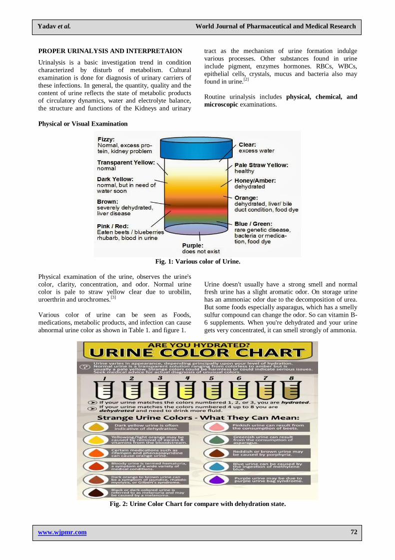
Physical or Visual Examination
Fig. 1: Various color of Urine.
Physical examination of the urine, observes the urine's
color, clarity, concentration, and odor. Normal urine color is pale to straw yellow clear due to urobilin,
uroerthrin and urochromes.[3]
Various color of urine can be seen as Foods,
medications, metabolic products, and infection can cause
abnormal urine color as shown in Table 1. and figure 1.
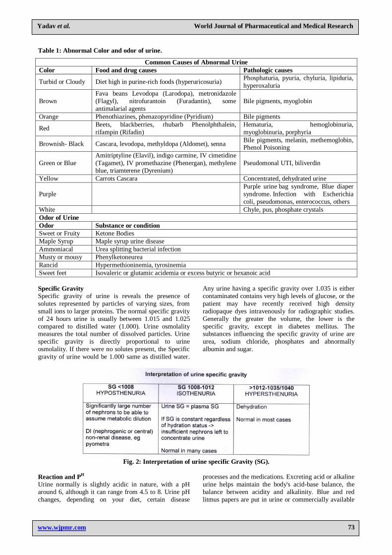
Urine doesn't usually have a strong smell and normal fresh urine has a slight aromatic odor. On storage urine
has an ammoniac odor due to the decomposition of urea.
But some foods especially asparagus, which has a smelly
sulfur compound can change the odor. So can vitamin B-
6 supplements. When you're dehydrated and your urine
gets very concentrated, it can smell strongly of ammonia.
Fig. 2: Urine Color Chart for compare with dehydration state.
Yadav et al. World Journal of Pharmaceutical and Medical Research
www.wjpmr.com
73
Table 1: Abnormal Color and odor of urine.
Common Causes of Abnormal Urine
Color Food and drug causes Pathologic causes
Turbid or Cloudy Diet high in purine-rich foods (hyperuricosuria) Phosphaturia, pyuria, chyluria, lipiduria,
hyperoxaluria
Brown
Fava beans Levodopa (Larodopa), metronidazole
(Flagyl), nitrofurantoin (Furadantin), some
antimalarial agents
Bile pigments, myoglobin
Orange Phenothiazines, phenazopyridine (Pyridium) Bile pigments
Red Beets, blackberries, rhubarb Phenolphthalein,
rifampin (Rifadin)
Hematuria, hemoglobinuria,
myoglobinuria, porphyria
Brownish- Black Cascara, levodopa, methyldopa (Aldomet), senna Bile pigments, melanin, methemoglobin, Phenol Poisoning
Green or Blue
Amitriptyline (Elavil), indigo carmine, IV cimetidine
(Tagamet), IV promethazine (Phenergan), methylene
blue, triamterene (Dyrenium)
Pseudomonal UTI, biliverdin
Yellow Carrots Cascara Concentrated, dehydrated urine
Purple
Purple urine bag syndrome, Blue diaper
syndrome. Infection with Escherichia
coli, pseudomonas, enterococcus, others
White Chyle, pus, phosphate crystals
Odor of Urine
Odor Substance or condition
Sweet or Fruity Ketone Bodies
Maple Syrup Maple syrup urine disease
Ammoniacal Urea splitting bacterial infection
Musty or mousy Phenylketoneurea
Rancid Hypermethioninemia, tyrosinemia
Sweet feet Isovaleric or glutamic acidemia or excess butyric or hexanoic acid
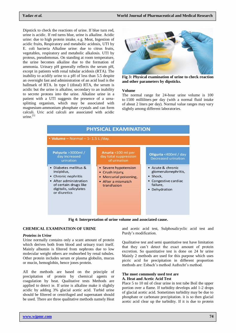
Specific Gravity
Specific gravity of urine is reveals the presence of
solutes represented by particles of varying sizes, from
small ions to larger proteins. The normal specific gravity
of 24 hours urine is usually between 1.015 and 1.025
compared to distilled water (1.000). Urine osmolality measures the total number of dissolved particles. Urine
specific gravity is directly proportional to urine
osmolality. If there were no solutes present, the Specific
gravity of urine would be 1.000 same as distilled water.
Any urine having a specific gravity over 1.035 is either
contaminated contains very high levels of glucose, or the
patient may have recently received high density
radiopaque dyes intravenously for radiographic studies.
Generally the greater the volume, the lower is the
specific gravity, except in diabetes mellitus. The substances influencing the specific gravity of urine are
urea, sodium chloride, phosphates and abnormally
albumin and sugar.
Fig. 2: Interpretation of urine specific Gravity (SG).
Reaction and PH
Urine normally is slightly acidic in nature, with a pH
around 6, although it can range from 4.5 to 8. Urine pH
changes, depending on your diet, certain disease
processes and the medications. Excreting acid or alkaline
urine helps maintain the body's acid-base balance, the
balance between acidity and alkalinity. Blue and red
litmus papers are put in urine or commercially available
Yadav et al. World Journal of Pharmaceutical and Medical Research
www.wjpmr.com
74
Dipstick to check the reactions of urine. If blue turn red,
urine is acidic. If red turns blue, urine is alkaline. Acidic
urine: due to high protein intake, e.g. Meat, Ingestion of
acidic fruits, Respiratory and metabolic acidosis, UTI by
E. coli bacteria Alkaline urine: due to citrus fruits,
vegetables, respiratory and metabolic alkalosis. UTI by proteus, pseudomonas. On standing at room temperature,
the urine becomes alkaline due to the formation of
ammonia. Urinary pH generally reflects the serum pH,
except in patients with renal tubular acidosis (RTA). The
inability to acidify urine to a pH of less than 5.5 despite
an overnight fast and administration of an acid load is the
hallmark of RTA. In type I (distal) RTA, the serum is
acidic but the urine is alkaline, secondary to an inability
to secrete protons into the urine. Alkaline urine in a
patient with a UTI suggests the presence of a urea-
splitting organism, which may be associated with
magnesium-ammonium phosphate crystals and can form calculi. Uric acid calculi are associated with acidic
urine.[5]
Fig 3: Physical examination of urine to check reaction
and other parameters by dipsticks.
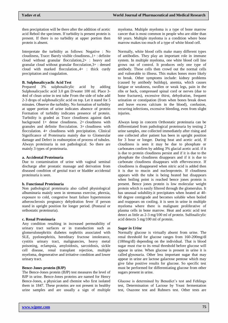
Volume
The normal range for 24-hour urine volume is 100
to 1500 milliliters per day (with a normal fluid intake of about 2 liters per day). Normal value ranges may vary
slightly among different laboratories.
Fig 4: Interpretation of urine volume and associated cause.
CHEMICAL EXAMINATION OF URINE
Proteins in Urine
Urine normally contains only a scant amount of protein
which derives both from blood and urinary tract itself.
Mainly albumin is filtered from nephrons due to low molecular weight others are reabsorbed by renal tubules.
Other protein includes serum or plasma globulin, mucus
or mucin, hemoglobin, bence jones protein.
All the methods are based on the principle of
precipitation of protein by chemical agents or
coagulation by heat. Qualitative tests Methods are
applied to detect ie. If urine is alkaline make it slightly
acidic by adding 3% glacial acetic acid. Turbid urine
should be filtered or centrifuged and supernatant should
be used. There are three qualitative methods namely Heat
and acetic acid test, Sulphosalicyclic acid test and
Purdy’s modification.
Qualitative test and semi quantitative test have limitation
that they can’t detect the exact amount of protein excretion. So quantitative test is done on 24 hr urine
Mainly 2 methods are used for this purpose which uses
picric acid for precipitation in different proportion
methods are: Esbach’s method Aufrecht’s method.
The most commonly used test are
A. Heat and Acetic Acid Test Place 5 to 10 ml of clear urine in test tube Boil the upper
portion over a flame. If turbidity develops add 1-2 drops
of glacial acetic acid. Sometimes turbidity may be due to
phosphate or carbonate precipitation. it is so then glacial
acetic acid clear up the turbidity. If it is due to protein
Yadav et al. World Journal of Pharmaceutical and Medical Research
www.wjpmr.com
75
then precipitation will be there after the addition of acetic
acid Reboil the specimen. If turbidity is present protein is
present. If there is no turbidity at upper portion then
protein is absent.
Interpretate the turbidity as follows: Negative : No cloudiness, Trace: Barely visible cloudiness.,1+ : definite
cloud without granular flocculation,2+ : heavy and
granular cloud without granular flocculation,3+ : densed
cloud with marked flocculation.,4+ : thick curdy
precipitation and coagulation.
B. Sulphosalicyaclic Acid Test Prepared 3% sulphosalicylic acid by adding
Sulphosalicyaclic acid 3.0 gm D/water 100 ml. Place 3-
4ml of clean urine in test tube From the side of tube add
2-3 drops of sulphosalicylic acid on top. Let it stand for 5
minutes. Observe the turbidity. No formation of turbidity at upper portion of urine indicates absence of protein
Formation of turbidity indicates presence of protein.
Turbidity is graded as Trace cloudiness against dark
background 1+ dense cloudiness. 2+ cloudiness with
granules and definite flocculation. 3+ cloudiness with
flocculation. 4+ cloudiness with precipitation. Clinical
Significance of Proteinuria mainly due to Glomerular
damage and Defect in reabsorption of process of tubules.
Always proteinuria in not pathological. So there are
mainly 3 types of proteinuria.
a. Accidental Proteinuria
Due to contamination of urine with vaginal seminal
discharge after prostatic massage and derivation from
diseased condition of genital tract or bladder accidental
proteinuria is seen.
b. Functional Proteinuria
Non pathological proteinuria also called physiological
albuminuria mainly seen in strenuous exercise, phrexia,
exposure to cold, congestive heart failure hypertension
atherosclerosis pregnancy dehydration fever if person
stand in upright position for longer period. (Postural or orthostatic proteinuria).
c. Renal Proteinuria
Any condition resulting in increased permeability of
urinary tract surfaces or in transduction such as
glomerulonephritis diabetes nephritis associated with
SLE, pyelonephritis, hereditary fructose intolerance,
cystitis urinary tract, malignancies, heavy metal
poisoning, eclampsia, amyloidosis, sarcoidosis, sickle
cell disease, renal transplant rejection, multiple
myeloma, degenerative and irritative condition and lower urinary tract.
Bence-Jones protein (BJP)
The Bence-Jones protein (BJP) test measures the level of
BJP in urine. Bence-Jones proteins are named for Henry
Bence-Jones, a physician and chemist who first isolated
them in 1847. These proteins are not present in healthy
urine samples and are usually a sign of multiple
myeloma. Multiple myeloma is a type of bone marrow
cancer that is most common in people who are older than
60 years. Multiple myeloma is a condition where bone
marrow makes too much of a type of white blood cell.
Normally, white blood cells make many different types of antibodies. They play an important role in immune
system. In multiple myeloma, one white blood cell line
grows out of control. It produces only one type of
antibody. These cells then crowd out the normal cells
and vulnerable to illness. This makes bones more likely
to break. Other symptoms include: kidney problems
(caused by antibody buildup), anemia, which causes
fatigue or weakness, swollen or weak legs, pain in the
ribs or back, compressed spinal cord or nerves (due to
bone fractures), excessive thirst, dehydration, frequent
urination or constipation (from when bones break down
and leave excess calcium in the blood), confusion, recurring infections, excessive bleeding, even from slight
injuries.
Always keep in concern Orthostatic proteinuria can be
differentiated from pathological proteinuria by testing 2
urine samples, one collected immediately after rising and
one collected after patient has been in upright position
for 3 hour or longer. During heat and acetic acid, If
cloudiness is seen it may be due to phosphate or
carbonates confirm by adding 3% glacial acetic acid. if it
is due to protein cloudiness persist and if it is due to the phosphate the cloudiness disappears and if it is due to
carbonate cloudiness disappears with effervescence. If
cloudiness is disappeared when nitric acid is added then
it is due to mucin and nucleoprotein. If cloudiness
appears with the tube is being heated but disappears
when boiling point is reached bence jones protein is
present. Bence jones protein is low molecular weight
protein which is easily filtered through the glomerulus. It
has unusual solubility.it precipitates when heated at 40-
60 degree centigrade and becomes soluble when boiled
and reappears on cooling. it is seen in urine in multiple
myeloma where there is malignant proliferative of plasma cells in bone marrow. Heat and acetic acid test
detect as little as 2-3 mg/100 ml of protein. Sulfosalicylic
acid detects 5 mg/100 ml of protein.
Sugar in Urine
Normally glucose is virtually absent from urine. The
renal threshold for glucose ranges from 160-200mg/dl
(180mg/dl) depending on the individual. That is blood
sugar must rise to its renal threshold before glucose will
appear in urine. When glucose is present in urine it is
called glysosuria. Other less important sugar that may appear in urine are lactose galactose pentose which may
give false positive results for glucose. So specific test
must be performed for differentiating glucose from other
sugars present in urine.
Glucose is determined by Benedict’s test and Fehlings
test, Determination of Lactose by Yeast fermentation
test, Osazone test and Rubners test. Other tests are
Yadav et al. World Journal of Pharmaceutical and Medical Research
www.wjpmr.com
76
Seliwanoffs Test for Fructose and Bial’s Test For
Pentoses.
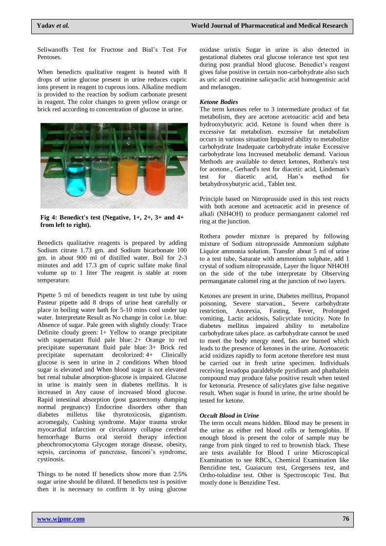
When benedicts qualitative reagent is heated with 8
drops of urine glucose present in urine reduces cupric
ions present in reagent to cuprous ions. Alkaline medium is provided to the reaction by sodium carbonate present
in reagent. The color changes to green yellow orange or
brick red according to concentration of glucose in urine.
Fig 4: Benedict's test (Negative, 1+, 2+, 3+ and 4+
from left to right).
Benedicts qualitative reagents is prepared by adding
Sodium citrate 1.73 gm. and Sodium bicarbonate 100
gm. in about 900 ml of distilled water. Boil for 2-3
minutes and add 17.3 gm of cupric sulfate make final
volume up to 1 liter The reagent is stable at room
temperature.
Pipette 5 ml of benedicts reagent in test tube by using
Pasteur pipette add 8 drops of urine heat carefully or
place in boiling water bath for 5-10 mins cool under tap
water. Interpretate Result as No change in color i.e. blue:
Absence of sugar. Pale green with slightly cloudy: Trace
Definite cloudy green: 1+ Yellow to orange precipitate
with supernatant fluid pale blue: 2+ Orange to red
precipitate supernatant fluid pale blue: 3+ Brick red
precipitate supernatant decolorized: 4+ Clinically
glucose is seen in urine in 2 conditions When blood sugar is elevated and When blood sugar is not elevated
but renal tubular absorption-glucose is impaired. Glucose
in urine is mainly seen in diabetes mellitus. It is
increased in Any cause of increased blood glucose.
Rapid intestinal absorption (post gastrectomy dumping
normal pregnancy) Endocrine disorders other than
diabetes milletus like thyrotoxicosis, gigantism.
acromegaly, Cushing syndrome. Major trauma stroke
myocardial infarction or circulatory collapse cerebral
hemorrhage Burns oral steroid therapy infection
pheochromocytoma Glycogen storage disease, obesity,
sepsis, carcinoma of pancrease, fanconi’s syndrome, cystinosis.
Things to be noted If benedicts show more than 2.5%
sugar urine should be diluted. If benedicts test is positive
then it is necessary to confirm it by using glucose
oxidase uristix Sugar in urine is also detected in
gestational diabetes oral glucose tolerance test spot test
during post prandial blood glucose. Benedict’s reagent
gives false positive in certain non-carbohydrate also such
as uric acid creatinine salicyaclic acid homogentisic acid
and melanogen.
Ketone Bodies The term ketones refer to 3 intermediate product of fat
metabolism, they are acetone acetoacitic acid and beta
hydrooxybutyric acid. Ketone is found when there is
excessive fat metabolism. excessive fat metabolism
occurs in various situation Impaired ability to metabolize
carbohydrate Inadequate carbohydrate intake Excessive
carbohydrate loss Increased metabolic demand. Various
Methods are available to detect ketones, Rothera's test
for acetone., Gerhard's test for diacetic acid, Lindeman's
test for diacetic acid, Han’s method for betahydroxybutyric acid., Tablet test.
Principle based on Nitroprusside used in this test reacts
with both acetone and acetoacetic acid in presence of
alkali (NH4OH) to produce permanganent calomel red
ring at the junction.
Rothera powder mixture is prepared by following
mixture of Sodium nitroprusside Ammonium sulphate
Liquior ammonia solution. Transfer about 5 ml of urine
to a test tube, Saturate with ammonium sulphate, add 1 crystal of sodium nitroprusside, Layer the liquor NH4OH
on the side of the tube interpretate by Observing
permanganate calomel ring at the junction of two layers.
Ketones are present in urine, Diabetes mellitus, Propanol
poisoning, Severe starvation., Severe carbohydrate
restriction, Anorexia, Fasting, Fever, Prolonged
vomiting, Lactic acidosis, Salicyclate toxicity. Note In
diabetes mellitus impaired ability to metabolize
carbohydrate takes place. as carbohydrate cannot be used
to meet the body energy need, fats are burned which
leads to the presence of ketones in the urine. Acetoacetic acid oxidizes rapidly to form acetone therefore test must
be carried out in fresh urine specimen. Individuals
receiving levadopa paraldehyde pyridium and phathalein
compound may produce false positive result when tested
for ketonuria. Presence of salicylates give false negative
result. When sugar is found in urine, the urine should be
tested for ketone.
Occult Blood in Urine The term occult means hidden. Blood may be present in
the urine as either red blood cells or hemoglobin. If enough blood is present the color of sample may be
range from pink tinged to red to brownish black. These
are tests available for Blood I urine Microscopical
Examination to see RBCs, Chemical Examination like
Benzidine test, Guaiacum test, Gregersens test, and
Ortho-toluidine test. Other is Spectroscopic Test. But
mostly done is Benzidine Test.
Yadav et al. World Journal of Pharmaceutical and Medical Research
www.wjpmr.com
77
The test is based on the principle that peroxidase activity
of hemoglobin present in urine decomposes hydrogen
peroxide and the liberated oxygen oxidized benzidine to
form a green- blue colored complex.
Place a pinch of benzidine in a test tube, Add 2- 3 drops of 5% glacial acetic acid. Mix well Add 2 ml of
hydrogen peroxide solution. Transfer supernatant to a
test tube label as T Add few drops of urine and observe
the color.
Clinical significance of Blood in urine is Hematuria,
hemoglobinuria and Myoglobinuria Presence of more
number of red blood cells in urine is called hematuria
which is associated with disease of or damage to the
genitourinary tract. other disorder commonly used
associated with hematuria includes acute infection
chronic glomerulonephritis tuberculosis of kidney nephritic syndrome toxic damage to glomerulus
malignant hypertension infarction renal calculi trauma to
kidney, acute cystitis, calculi, tumors in the ureter or
bladder and kidney stones. In other clinical conditions
such as bleeding disorder (leukemia, thrombocytopenia,
coagulation factor deficiency, sickle disease or traits,
scurvy), use of anticoagulant drugs.
Hemoglobinuria: It is the presence of free hemoglobin
in urine as a result of intravascular hemolysis. Causes of
hemoglobinuria Acute and Chronic Acute, Incompatible blood transfusion, Hemolytic anemia due to drugs and
chemicals., Favism., Paroxysmal cold hemoglobunuria.,
March (exertional) hemoglobunuria., Hemolytic anemia
associated with eclampsia, Hemolytic uraemic
syndrome., Hemolytic anemia due to burns, Snake and
spider bites. Chronic, PNH, Cardiac hemolytic anemia,
Cold haemagglutination disease.
Myoglobinuria: Myoglobin is the haem protein of
striated muscle. Myoglobin is very toxic to the renal
tubules and in large amounts it is associated with acute
renal failure.
Clinical Conditions are, Myocardial infarction Infarction
of large skeletal muscle Destruction of muscle with crush
injury heat stroke electric shock, Trauma.
Things to be keep in mind while interpretation False
positive result is seen in women during menstruation due
to contamination of urine with menstrual blood. So this
test should be avoided during menstruation cycle. Free
Hb is not normally found in the urine. Instead any Hb
that could be presented to the glomerulus combines with heptoglobin. The resultant Hb heptoglobin complex is
too large to pass through the glomerular membrane. If
the amount of free Hb exceeds the amount of
heptoglobulin, however the Hb will pass through the
glomerulus and ultimately be excreted into the urine.
Any disorder associated with hemolysis of red blood
cells and resultant release of Hb may lead to the
appearance of Hb in urine Hematuria can be
differentiated from hemoglobunuria by doing
microscopical examination. In hematuria RBC seen in
microscopy. In hemoglobunuria, RBC cannot see even
though the test for occult blood is positive. This test can
be done for stool as occult blood for stool.
Bile in Urine Bilirubin, bile salt, bile pigment, urobilin, urobilinogen
are the constituents of bile.
Determination of Bile Salt by Hay's test Bile salts when
present lower the surface tension of urine. When sulphur
powder is added to the urine, sulphur particles sink to the
bottom of the tube. In the case of normal urine, it will
float on the surface. Place about 10 ml of urine in a test
tube, Sprinkle a little dry sulphur powder on to the
surface of urine. Observe sulphur particles. Other
Methods, Foam test, Gmelin's test, Smiths test, Fouchet's test, Ehrlich's aldehyde test Schlesingers test.
Foam test, Shake some urine in a test tube. If the foam
on the top is yellow, the bile pigments are present.
Fouchet’s test, When barium chloride is added to urine it
combines with sulphate radicals in urine and precipitate
of barium phosphate is formed. If bile pigments are
present in urine, they will adhere to these large
molecules. Ferric chloride present in fouchet reagent
then oxidizes yellow bilirubin in presence of trichloroacetic acid to green bilverdin.
Fouchet’s reagent composed of Trichloroacetic acid 25
gm Distilled water 100 ml 10 % ferric chloride
solution 10 ml 10ml of urine + 2.5 ml of barium chloride,
Filter, Unfold the filter paper and spread it on the dry
filter paper. Allow 1 drop of Fauchet’s reagent on the
precipitate A green or blue color indicates presence of
bilirubin.
Ehrlichs Aldehyde Test for Urobilinogen Take 5 ml of
urine in test tube and add half volume i.e. about 2.5 ml of barium chloride. mix well and filter. Take 2.3 ml of
filtrate and add 0.5 ml of aldehyde reagent. allow to
stand for 3 mins. View the top column of urine against a
white background. A pink color denotes the presence of
urobilinogen. Repeat the test with 1:10, 1:20, 1:50,
1:100, 1:200 dilution and report a terms of highest
dilution giving a positive reaction.
Ehrlich's reagentscomposed of
Paradimethylaminobenzaldehyde 2gms. 20% HCl 100
ml.
Schlesingers Test for Urobilin Take 10 ml of urine and 6
drops of tincture of iodine in a test tube. Take 1 gm of
powdered zinc acetate and 10 ml 95% alcohol in another
test tube. Mix by pouring a into b and vice versa
repeatedly until the solid zinc acetate has mostly gone
into solution. Filter. Examine the filtrate. A green is due
to compound of zinc with urobilin, confirm
Yadav et al. World Journal of Pharmaceutical and Medical Research
www.wjpmr.com
78
spectroscopically absorption band junction of green and
blue.
Clinical significance, Determination of bile salts, bile
pigments, and urobilinogen is useful in the diagnosis of
jaundice. Bilirubin may be found in urine in liver disease and is usually found in clients who have biliary tract
obstructions. Conjugated bilirubin appearing in urine
generally indicates that there is excess conjugated
bilirubin in blood stream. Bilirubinuria is seen when
intracanalicular pressure rises secondary to periportal
inflammation, fibrosis or hepatocyte swelling. Gallstones
in the common bile duct or carcinoma of the head of
pancreas are possible sources of extra hepatic biliary
obstruction leading to bilirubinuria. Congenital
hyperbilirubinemia seen in gilberts disease or crigler
najjar disease. When liver cells are damaged, excreation
of urobilinogen in the bile decreased, where as its urinary excreation is increased. This may be seen in cirrhosis,
hepatitis and congenital heart failure with congestion of
the liver. Excessive urobilinogen also may be found in
the urine of those with liver disease or hemolytic
disorder.
Condition Urine bilirubin Urine urobilinogen
Bile duct obstruction + + + Negative
Liver damage + or - + +
Hemolytic disease negative + + +
The bilirubin in urine is made confirmed by doing
confirmatory bilirubin test called as diazo test. The
Fouchet’s test is also called Harrison spot test. Fresh
urine sample should be used for bilirubin determination
because exposure of urine to light and room air may give
false negative result. large amount of ascorbic acid and
nitrates also give false negative result. Acidic urine will
result in decreased urinary level of urobilinogen. High
levels of nitrates in the urine also may cause false negative results in test for urobilinogen.
Leukocyte Esterase
Leukocyte esterase is an enzyme present in most white
blood cells (WBCs). Normally, a few white blood cells
(see microscopic examination) are present in urine and
this test is negative. WBCs in urine increases
significantly, it means that there is inflammation in the
urinary tract or kidneys. There is common cause for
WBCs in urine (leukocyturia) is a bacterial urinary tract
infection (UTI), such as a bladder or kidney infection.
Organisms such as Chlamydia and Ureaplasma urealyticum should be considered in patients with pyuria
and negative cultures. Other causes of sterile pyuria
include balanitis, urethritis, tuberculosis, bladder tumors,
viral infections, nephrolithiasis, foreign bodies, exercise,
glomerulonephritis, and corticosteroid and
cyclophosphamide (Cytoxan) use.
Nitrite
Normally the urinary tract and urine are free of bacteria.
When bacteria find, they can cause a urinary tract
infection (UTI). A positive nitrite test result can indicate a UTI. Gram negative rods such as E. coli are more
likely to give a positive test.
The Microscopic Urinalysis
Microscopic examination is an indispensable part of
urinalysis; the identification of casts, cells, crystals, and
bacteria aids in the diagnosis of a variety of conditions. It
will typically be done when there are abnormal findings
on the physical or chemical examination. It is performed
on urine sediment. To prepare a urine specimen for
microscopic analysis; afresh sample of 5 to 10 mL of
urine should be centrifuged at 1,500 to 3,000 rpm for
five minutes. The supernatant then is discarded and the
sediment remixed in the residual liquid. A single drop is
transferred to a clean glass slide, and a cover glass is
applied smoothly to prevent air bubble. In addition, some
entities, if present, are estimated as "few," "moderate," or
"many," such as Pus cells (WBCs) epithelial cells, bacteria, and crystals. The numbers of casts seen are
usually reported 5-10 hyaline casts/L casts/LPF. Next,
examination is carried out at high power to identify
crystals, cells, and bacteria. The various types of cells are
usually described as 2-4 Pus cells/HPF. Always reported
in the standard format i.e. in even number example 0-2,
2-4, 4-6 cells/HPF. Report in difference of 2 up to 10
cells more than that (10 to 20) keep difference of 5 and
up to 30 keep difference of 10 i.e. 4-6cell/HPF, 10-
15cells/HPF and 20-30cells/HPF. More than 30 cells are
reported as Plenty cells/HPF.[5,6,7] Details are tabulated in
Table 2.

Yadav et al. World Journal of Pharmaceutical and Medical Research
www.wjpmr.com
79
Table 2: Microscopic findings in urine and their description and clinical interpretation.
Variables Description and Clinical Interpretation Microscopic Image
Pus
Cells(WBCs)
Leukocytes may be seen under low- and high-power
magnification. Men normally have fewer than two white
blood cells per HPF; women normally have fewer than five
WBCs per HPF. The pus cells can enter in urine anywhere
from the glomerulus to the urethra. These are mostly
neutrophils. Pyuria refers to the presence of abnormal
numbers of leukocytes that may appear with infection in
urinary tract or with acute glomerulonephritis. When the
number is high, it indicates an infection or inflammation
somewhere in the urinary tract. WBCs can also be a
contaminant, such as those from vaginal and cervical secretions.
Epithelial
Cells
These cells can originate from any site in the urinary tract
from the proximal convoluted tubule to the urethra or from
vagina. “Three main types of epithelial cells -
Tubular epithelial cells Slightly larger than leukocytes and
large round nucleus. Transitional epithelial cells from the
renal pelvis, ureter, or bladder have more regular cell
borders, larger nuclei, and smaller overall size than squamous
epithelium They have pear shaped or round with two nuclei. Squamous epithelial cells from the skin surface or from the
outer urethra can appear in urine. They are large, flat and
irregularly shaped with small central nuclei.
`
Erythrocytes
(Red Blood
Cell)
These seem refrectile particles. Red cells in urine due to:
glomerular damage, tumors which erode the urinary tract
anywhere along its length, kidney trauma, urinary tract
stones, renal infarcts, acute tubular necrosis, nephrotoxins,
Inflammation, injury in the bladder or urethra, can cause
RBCs to leak out of the blood vessels into the urine, and
physical stress. In addition, red cell ghosts may simulate yeast. RBCs can also be a contaminant due to an improper
sample collection and blood from hemorrhoids. Normal in
menstruation.
Casts
Urinary casts are formed in the lumen of the tubules of the
kidney. Casts can form as the result of precipitation of gelatin
of Tammslors fall mucoprotein. Clumping of cells on other
material with in protein matrix. Coagulation of material
within the lumen.
a. Granular
Casts
When cellular casts remain in the nephron for some time
before they are flushed into the bladder urine, the cells may degenerate to become a coarsely granular cast Significant
renal disease, present due to the degeneration of cellular
casts.
Yadav et al. World Journal of Pharmaceutical and Medical Research
www.wjpmr.com
80
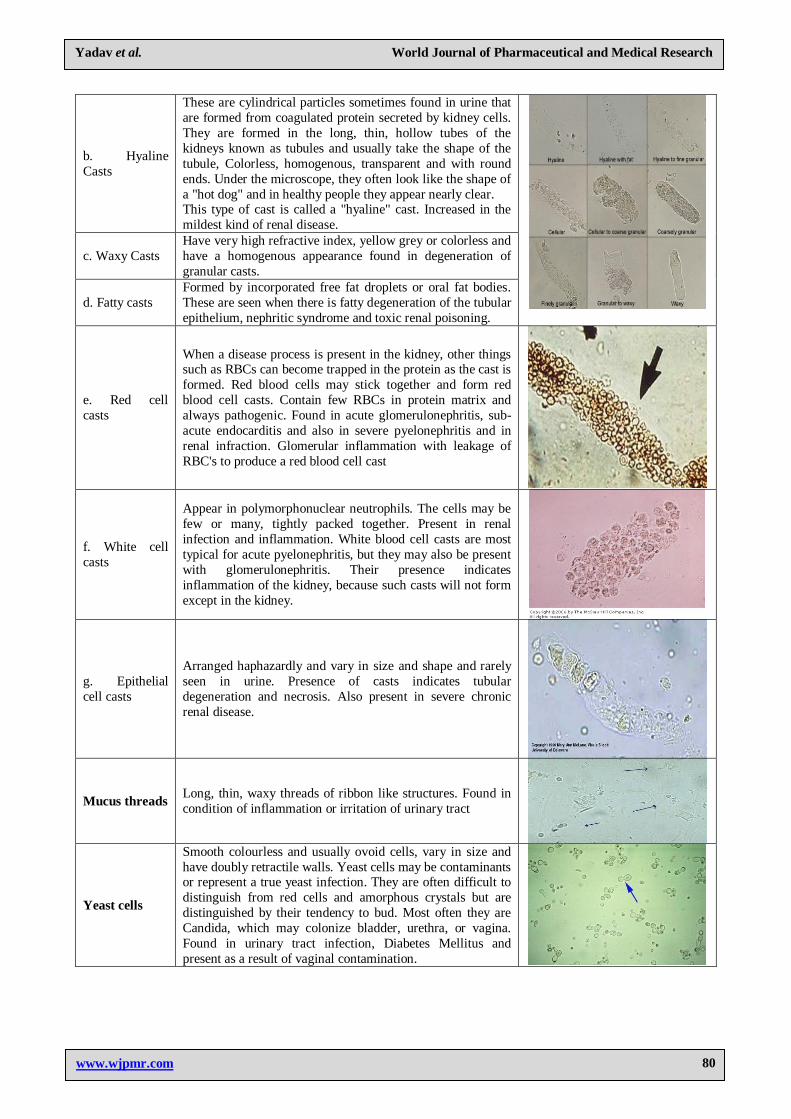
b. Hyaline
Casts
These are cylindrical particles sometimes found in urine that
are formed from coagulated protein secreted by kidney cells.
They are formed in the long, thin, hollow tubes of the
kidneys known as tubules and usually take the shape of the
tubule, Colorless, homogenous, transparent and with round
ends. Under the microscope, they often look like the shape of
a "hot dog" and in healthy people they appear nearly clear. This type of cast is called a "hyaline" cast. Increased in the
mildest kind of renal disease.
c. Waxy Casts
Have very high refractive index, yellow grey or colorless and
have a homogenous appearance found in degeneration of
granular casts.
d. Fatty casts
Formed by incorporated free fat droplets or oral fat bodies.
These are seen when there is fatty degeneration of the tubular
epithelium, nephritic syndrome and toxic renal poisoning.
e. Red cell
casts
When a disease process is present in the kidney, other things such as RBCs can become trapped in the protein as the cast is
formed. Red blood cells may stick together and form red
blood cell casts. Contain few RBCs in protein matrix and
always pathogenic. Found in acute glomerulonephritis, sub-
acute endocarditis and also in severe pyelonephritis and in
renal infraction. Glomerular inflammation with leakage of
RBC's to produce a red blood cell cast
f. White cell
casts
Appear in polymorphonuclear neutrophils. The cells may be
few or many, tightly packed together. Present in renal
infection and inflammation. White blood cell casts are most
typical for acute pyelonephritis, but they may also be present
with glomerulonephritis. Their presence indicates
inflammation of the kidney, because such casts will not form
except in the kidney.
g. Epithelial
cell casts
Arranged haphazardly and vary in size and shape and rarely
seen in urine. Presence of casts indicates tubular
degeneration and necrosis. Also present in severe chronic
renal disease.
Mucus threads Long, thin, waxy threads of ribbon like structures. Found in
condition of inflammation or irritation of urinary tract
Yeast cells
Smooth colourless and usually ovoid cells, vary in size and
have doubly retractile walls. Yeast cells may be contaminants or represent a true yeast infection. They are often difficult to
distinguish from red cells and amorphous crystals but are
distinguished by their tendency to bud. Most often they are
Candida, which may colonize bladder, urethra, or vagina.
Found in urinary tract infection, Diabetes Mellitus and
present as a result of vaginal contamination.
Yadav et al. World Journal of Pharmaceutical and Medical Research
www.wjpmr.com
81
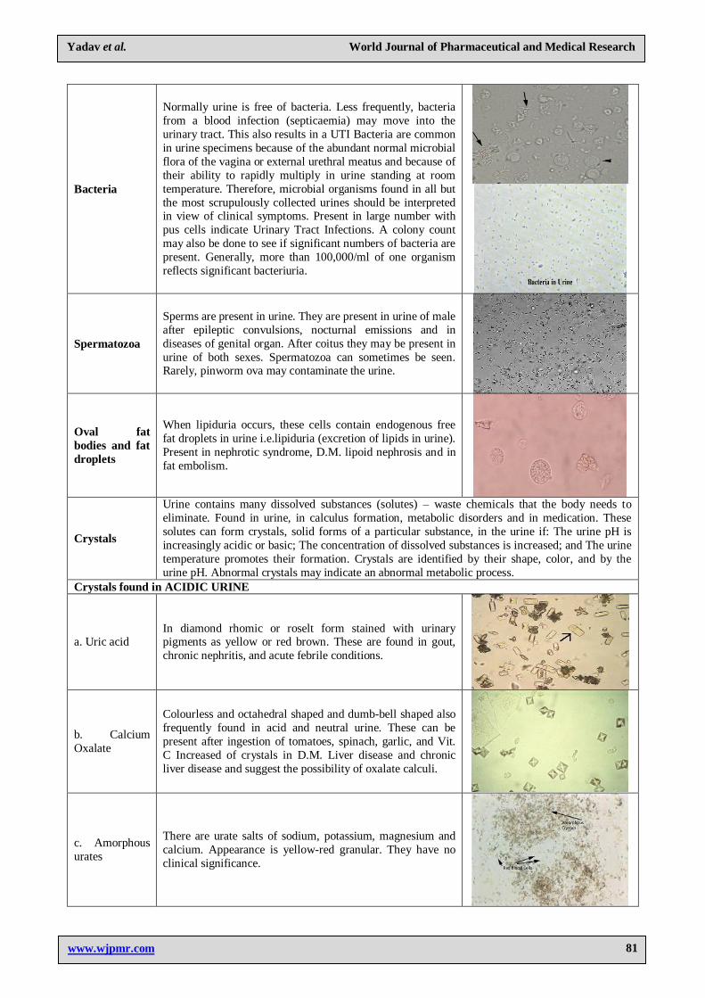
Bacteria
Normally urine is free of bacteria. Less frequently, bacteria
from a blood infection (septicaemia) may move into the
urinary tract. This also results in a UTI Bacteria are common
in urine specimens because of the abundant normal microbial
flora of the vagina or external urethral meatus and because of
their ability to rapidly multiply in urine standing at room
temperature. Therefore, microbial organisms found in all but
the most scrupulously collected urines should be interpreted in view of clinical symptoms. Present in large number with
pus cells indicate Urinary Tract Infections. A colony count
may also be done to see if significant numbers of bacteria are
present. Generally, more than 100,000/ml of one organism
reflects significant bacteriuria.
Spermatozoa
Sperms are present in urine. They are present in urine of male
after epileptic convulsions, nocturnal emissions and in
diseases of genital organ. After coitus they may be present in
urine of both sexes. Spermatozoa can sometimes be seen. Rarely, pinworm ova may contaminate the urine.
Oval fat
bodies and fat
droplets
When lipiduria occurs, these cells contain endogenous free
fat droplets in urine i.e.lipiduria (excretion of lipids in urine).
Present in nephrotic syndrome, D.M. lipoid nephrosis and in
fat embolism.
Crystals
Urine contains many dissolved substances (solutes) – waste chemicals that the body needs to
eliminate. Found in urine, in calculus formation, metabolic disorders and in medication. These
solutes can form crystals, solid forms of a particular substance, in the urine if: The urine pH is
increasingly acidic or basic; The concentration of dissolved substances is increased; and The urine
temperature promotes their formation. Crystals are identified by their shape, color, and by the
urine pH. Abnormal crystals may indicate an abnormal metabolic process.
Crystals found in ACIDIC URINE
a. Uric acid In diamond rhomic or roselt form stained with urinary pigments as yellow or red brown. These are found in gout,
chronic nephritis, and acute febrile conditions.
b. Calcium
Oxalate
Colourless and octahedral shaped and dumb-bell shaped also
frequently found in acid and neutral urine. These can be
present after ingestion of tomatoes, spinach, garlic, and Vit.
C Increased of crystals in D.M. Liver disease and chronic
liver disease and suggest the possibility of oxalate calculi.
c. Amorphous
urates
There are urate salts of sodium, potassium, magnesium and
calcium. Appearance is yellow-red granular. They have no
clinical significance.
Yadav et al. World Journal of Pharmaceutical and Medical Research
www.wjpmr.com
82
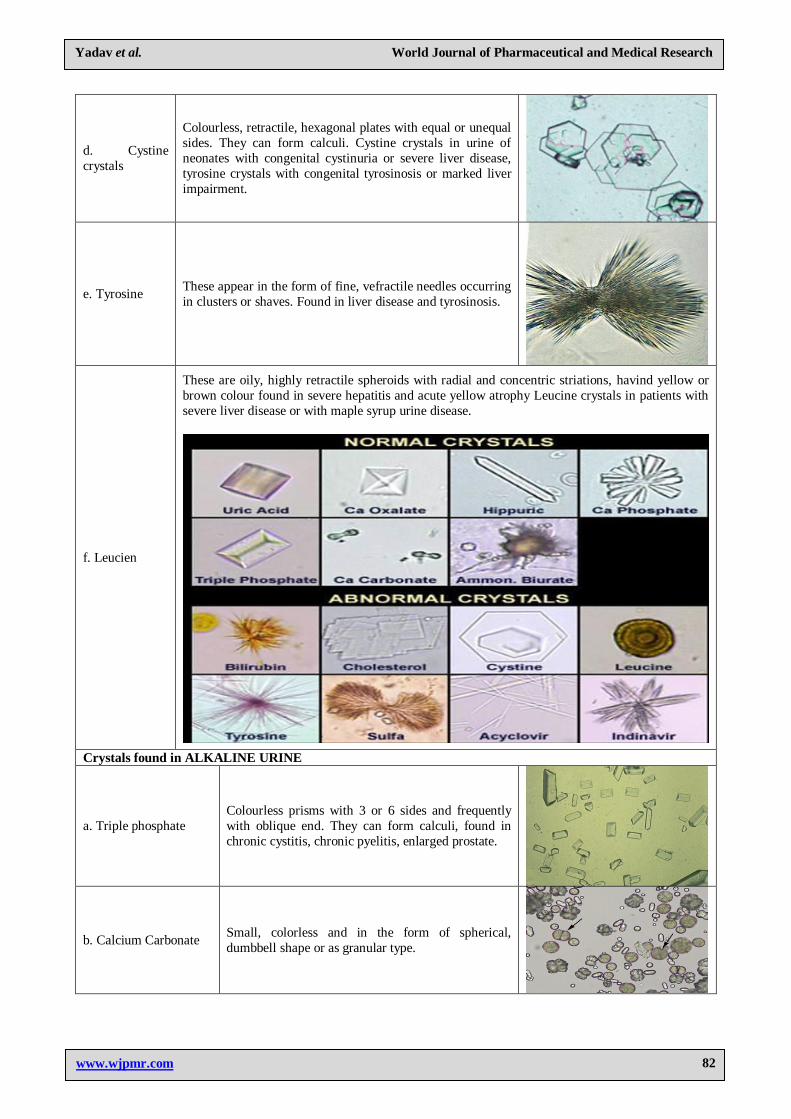
d. Cystine
crystals
Colourless, retractile, hexagonal plates with equal or unequal
sides. They can form calculi. Cystine crystals in urine of
neonates with congenital cystinuria or severe liver disease,
tyrosine crystals with congenital tyrosinosis or marked liver
impairment.
e. Tyrosine These appear in the form of fine, vefractile needles occurring
in clusters or shaves. Found in liver disease and tyrosinosis.
f. Leucien
These are oily, highly retractile spheroids with radial and concentric striations, havind yellow or
brown colour found in severe hepatitis and acute yellow atrophy Leucine crystals in patients with
severe liver disease or with maple syrup urine disease.
Crystals found in ALKALINE URINE
a. Triple phosphate
Colourless prisms with 3 or 6 sides and frequently
with oblique end. They can form calculi, found in
chronic cystitis, chronic pyelitis, enlarged prostate.
b. Calcium Carbonate Small, colorless and in the form of spherical,
dumbbell shape or as granular type.
Yadav et al. World Journal of Pharmaceutical and Medical Research
www.wjpmr.com
83
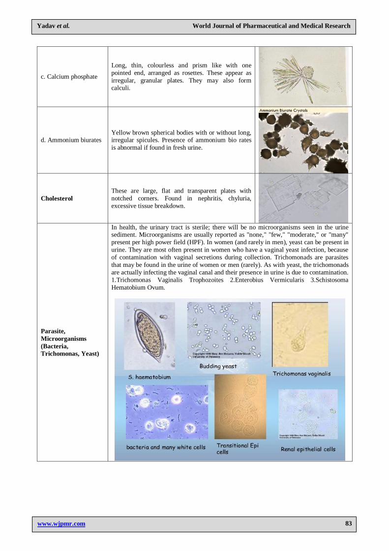
c. Calcium phosphate
Long, thin, colourless and prism like with one
pointed end, arranged as rosettes. These appear as
irregular, granular plates. They may also form
calculi.
d. Ammonium biurates Yellow brown spherical bodies with or without long, irregular spicules. Presence of ammonium bio rates
is abnormal if found in fresh urine.
Cholesterol
These are large, flat and transparent plates with
notched corners. Found in nephritis, chyluria,
excessive tissue breakdown.
Parasite,
Microorganisms
(Bacteria,
Trichomonas, Yeast)
In health, the urinary tract is sterile; there will be no microorganisms seen in the urine sediment. Microorganisms are usually reported as "none," "few," "moderate," or "many"
present per high power field (HPF). In women (and rarely in men), yeast can be present in
urine. They are most often present in women who have a vaginal yeast infection, because
of contamination with vaginal secretions during collection. Trichomonads are parasites
that may be found in the urine of women or men (rarely). As with yeast, the trichomonads
are actually infecting the vaginal canal and their presence in urine is due to contamination.
1.Trichomonas Vaginalis Trophozoites 2.Enterobius Vermicularis 3.Schistosoma
Hematobium Ovum.
Yadav et al. World Journal of Pharmaceutical and Medical Research
www.wjpmr.com
84
CONCLUSION
In summary, a properly collected clean-catch, midstream
fresh voided urine after cleansing of the urethral meatus
is adequate for complete urinalysis. A period of
dehydration may precede urine collection if testing of
renal concentration is desired, but any specific gravity > 1.022 measured in a randomly collected specimen
denotes adequate renal concentration so long as there are
no abnormal solutes in the urine.
The sample should transported to Laboratory within a
hour and should analyzed within two hours of collection.
Changes which occur with time after collection include:
1) decreased clarity due to crystallization of solutes, 2)
rising pH, 3) loss of ketone bodies, 4) loss of bilirubin, 5)
dissolution of cells and casts, and 6) overgrowth of
contaminating microorganisms. Urinalysis also called as routine and microscopy examination of urine, and one of
the most common tools of clinical diagnosis. The target
parameters that can be measured or quantified in
urinalysis include naked eye (gross) examination for
color and smell plus analysis for many substances and
cells, as well as other properties, such as specific gravity.
Urinalysis can reveal diseases that have gone unnoticed
because they do not produce striking signs or symptoms.
Examples include diabetes mellitus, various forms of
glomerulonephritis, and chronic urinary tract
infections.[8]
REFERENCES
1. Rabinovitch A. Urinalysis and collection,
transportation, and preservation of urine specimens:
approved guideline. 2d ed. Wayne, Pa.: National
Committee for Clinical Laboratory Standards, 2001.
NCCLS document GP16-A2.
2. Hand Book of Medical Laboratory Technology,
Chapter 7,Reprint 1993, Durai at the vesleypress,
Mysore, India, By editors-Dr. Chitra Bharucha,
Miss. Harmina Meyer, Dr. Hoshang Bharucha,
Mr.Anthony Moody, Dr, Robert h. Carman. Page no.142-159
3. file:///H:/URINALYSIS/p1153urine.pdf dated on 02
jan.2016.
4. file:///H:/URINALYSIS/urin.pdf dated on 02
jan.2016.
5. file:///H:/URINALYSIS/Urinalysis.html dated on 02
jan.2016.
6. Text book on Medical Laboratory Technology II –
edition-2003 By Dr. Praful Godkar and Dr. Darshan
P. Godkar. Bhalani Publishing house, Mumbai, Page
no.901,902,903. 7. Hand Book of Medical Laboratory Technology,
Chapter 7,Reprint 1993, Durai at the vesleypress,
Mysore, India, By editors-Dr. Chitra Bharucha,
Miss. Harmina Meyer, Dr. Hoshang Bharucha,
Mr.Anthony Moody, Dr, Robert h. Carman. Page
no.142-159.
8. A Text Book of Practical Pathology(including
Ayurved Concept), Sectyion C, Chapter 01,By Dr.
Bhojraj A. Chaudhari, First edition 2013,Wizcraft
Publications & Distribution Pvt. Ltd, Solapur,
Maharashtra.