Embed Size (px)
Citation preview
Today’s Veterinary Practice May/June 201486
Today’s Technician Peer reviewed
tvpjournal.com
Urinalysis (UA) provides information about the urinary system as well as other body systems. It should be performed to:
•Evaluate any animal with clinical signs related to the uri-nary tract
•Assess an animal with systemic illness•Monitor response to treatment.
The first article in this 2-part series discussed collec-tion, sample handling, and initial evaluation of urine in small animals (March/April 2014, available at tvpjournal .com). This article will describe more detailed evaluation, including chemical analysis and microscopic examination of sediment.
CHEMICAL ANALYSIS Urine chemistry test strips have multiple pads impregnat-ed with reagents that change color when the substance of interest is present. The degree of color change corre-sponds to the approximate amount of the substance pres-ent. Because color changes can be subtle, results may be con-siderably varied between indi-viduals reading the test.
Several chemistry multiple-test reagent strips are available, including: •Chemstrip (poc.roche.com)• Diastix (healthcare.bayer.com)•Multistix (healthcare.siemens
.com)• Petstix (idexx.com).
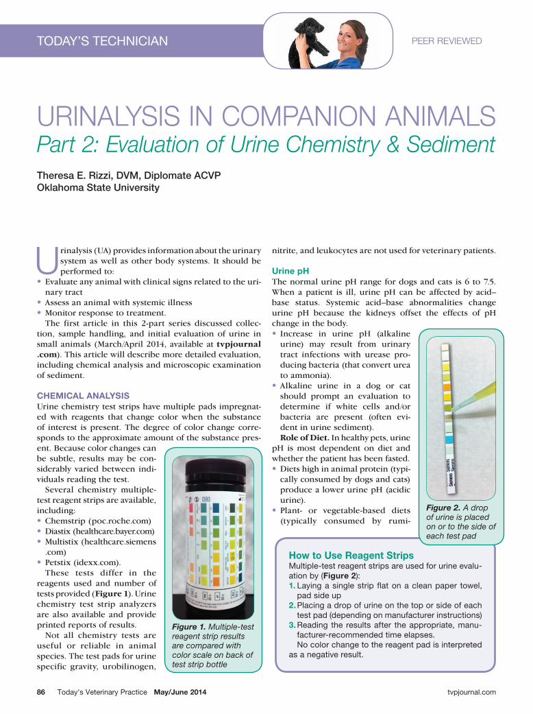
These tests differ in the reagents used and number of tests provided (Figure 1). Urine chemistry test strip analyzers are also available and provide printed reports of results.
Not all chemistry tests are useful or reliable in animal species. The test pads for urine specific gravity, urobilinogen,
nitrite, and leukocytes are not used for veterinary patients.
Urine pHThe normal urine pH range for dogs and cats is 6 to 7.5. When a patient is ill, urine pH can be affected by acid–base status. Systemic acid–base abnormalities change urine pH because the kidneys offset the effects of pH change in the body. • Increase in urine pH (alkaline
urine) may result from urinary tract infections with urease pro-ducing bacteria (that convert urea to ammonia).
•Alkaline urine in a dog or cat should prompt an evaluation to determine if white cells and/or bacteria are present (often evi-dent in urine sediment).Role of Diet. In healthy pets, urine
pH is most dependent on diet and whether the patient has been fasted. •Diets high in animal protein (typi-
cally consumed by dogs and cats) produce a lower urine pH (acidic urine).
• Plant- or vegetable-based diets (typically consumed by rumi-
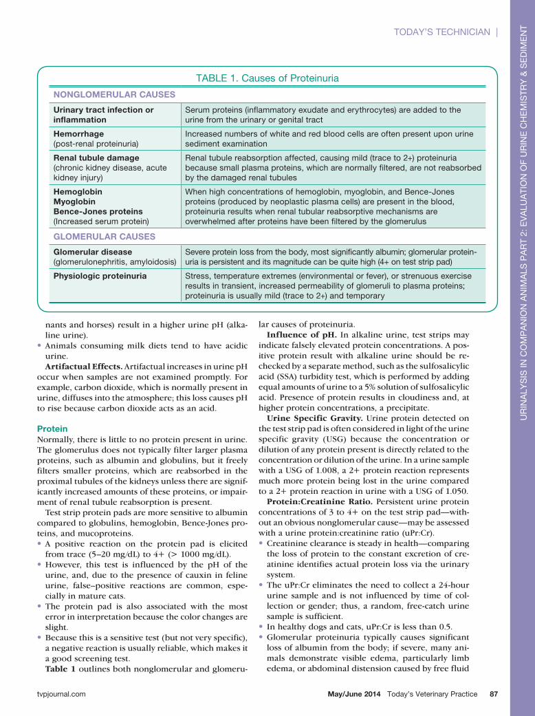
How to Use Reagent StripsMultiple-test reagent strips are used for urine evalu-ation by (Figure 2): 1. Laying a single strip flat on a clean paper towel,
pad side up2. Placing a drop of urine on the top or side of each
test pad (depending on manufacturer instructions)3. Reading the results after the appropriate, manu-
facturer-recommended time elapses. no color change to the reagent pad is interpreted
as a negative result.
Urinalysis in ComPanion animals Part 2: Evaluation of Urine Chemistry & Sediment
Figure 1. Multiple-test reagent strip results are compared with color scale on back of test strip bottle
Theresa E. Rizzi, DVM, Diplomate ACVPOklahoma State University
Figure 2. A drop of urine is placed on or to the side of each test pad
May/June 2014 Today’s Veterinary Practice 87
UR
ina
Lys
is in
co
MP
an
ion
an
iMa
Ls P
aR
T 2:
eV
aLU
aTi
on
of
UR
ine
ch
eM
isTR
y &
se
diM
en
TToday’s Technician |
tvpjournal.com
nants and horses) result in a higher urine pH (alka-line urine).
•Animals consuming milk diets tend to have acidic urine. Artifactual Effects. Artifactual increases in urine pH
occur when samples are not examined promptly. For example, carbon dioxide, which is normally present in urine, diffuses into the atmosphere; this loss causes pH to rise because carbon dioxide acts as an acid.
ProteinNormally, there is little to no protein present in urine. The glomerulus does not typically filter larger plasma proteins, such as albumin and globulins, but it freely filters smaller proteins, which are reabsorbed in the proximal tubules of the kidneys unless there are signif-icantly increased amounts of these proteins, or impair-ment of renal tubule reabsorption is present.
Test strip protein pads are more sensitive to albumin compared to globulins, hemoglobin, Bence-Jones pro-teins, and mucoproteins.•A positive reaction on the protein pad is elicited
from trace (5–20 mg/dL) to 4+ (> 1000 mg/dL). •However, this test is influenced by the pH of the
urine, and, due to the presence of cauxin in feline urine, false–positive reactions are common, espe-cially in mature cats.
•The protein pad is also associated with the most error in interpretation because the color changes are slight.
•Because this is a sensitive test (but not very specific), a negative reaction is usually reliable, which makes it a good screening test.Table 1 outlines both nonglomerular and glomeru-
lar causes of proteinuria. Influence of pH. In alkaline urine, test strips may
indicate falsely elevated protein concentrations. A pos-itive protein result with alkaline urine should be re-checked by a separate method, such as the sulfosalicylic acid (SSA) turbidity test, which is performed by adding equal amounts of urine to a 5% solution of sulfosalicylic acid. Presence of protein results in cloudiness and, at higher protein concentrations, a precipitate.
Urine Specific Gravity. Urine protein detected on the test strip pad is often considered in light of the urine specific gravity (USG) because the concentration or dilution of any protein present is directly related to the concentration or dilution of the urine. In a urine sample with a USG of 1.008, a 2+ protein reaction represents much more protein being lost in the urine compared to a 2+ protein reaction in urine with a USG of 1.050.
Protein:Creatinine Ratio. Persistent urine protein concentrations of 3 to 4+ on the test strip pad—with-out an obvious nonglomerular cause—may be assessed with a urine protein:creatinine ratio (uPr:Cr). •Creatinine clearance is steady in health—comparing
the loss of protein to the constant excretion of cre-atinine identifies actual protein loss via the urinary system.
•The uPr:Cr eliminates the need to collect a 24-hour urine sample and is not influenced by time of col-lection or gender; thus, a random, free-catch urine sample is sufficient.
• In healthy dogs and cats, uPr:Cr is less than 0.5. •Glomerular proteinuria typically causes significant
loss of albumin from the body; if severe, many ani-mals demonstrate visible edema, particularly limb edema, or abdominal distension caused by free fluid
TabLe 1. causes of Proteinuria
NONGLOMERULAR CAUSES
Urinary tract infection or inflammation
serum proteins (inflammatory exudate and erythrocytes) are added to the urine from the urinary or genital tract
Hemorrhage (post-renal proteinuria)
increased numbers of white and red blood cells are often present upon urine sediment examination
Renal tubule damage (chronic kidney disease, acute kidney injury)
Renal tubule reabsorption affected, causing mild (trace to 2+) proteinuria because small plasma proteins, which are normally filtered, are not reabsorbed by the damaged renal tubules
HemoglobinMyoglobinBence-Jones proteins (increased serum protein)
When high concentrations of hemoglobin, myoglobin, and bence-Jones proteins (produced by neoplastic plasma cells) are present in the blood, proteinuria results when renal tubular reabsorptive mechanisms are overwhelmed after proteins have been filtered by the glomerulus
GLOMERULAR CAUSES
Glomerular disease(glomerulonephritis, amyloidosis)
severe protein loss from the body, most significantly albumin; glomerular protein-uria is persistent and its magnitude can be quite high (4+ on test strip pad)
Physiologic proteinuria stress, temperature extremes (environmental or fever), or strenuous exercise results in transient, increased permeability of glomeruli to plasma proteins; proteinuria is usually mild (trace to 2+) and temporary
Today’s Veterinary Practice May/June 201488
| Today’s Technician
tvpjournal.com
accumulation. Microalbuminuria. Species-specific
microalbuminuria assays can detect urine albumin concentrations as low as 1 mg/dL. Opinions vary on the use of these assays and their clinical implication, but some consider even small amounts of albumin in the urine as abnormal, possibly indicat-ing early or subclinical glomerular disease.
GlucoseGlucose is not normally present in the urine in quantities detectable on dipsticks. The test strip pad detects glucose by an enzymatic chemical reaction that results in a color change proportional to the amount of glucose present.
While this reaction is specific for glucose, it is important to realize enzyme activity is limited, and outdated strips may give false–negative results. Temperature can also affect enzyme activity; refrigerated samples need to be at room temperature before testing.
Glucose filtered through the glomeru-lus is normally reabsorbed in the proximal tubules. Glucosuria occurs with any con-dition that causes blood glucose levels to exceed the renal threshold for reabsorp-tion (renal threshold: dogs, 180–220 mg/dL; cats, approximately 290 mg/dL).1 •Diabetes mellitus is a common cause of
glucosuria due to excessive blood glu-cose concentrations.
• Stress in some animals (particularly cats) can cause marked transient hyper-glycemia; if hyperglycemia has sufficient magnitude, glucosuria results.
•Renal tubular dysfunction is present when glucosuria is associated with nor-mal blood glucose concentrations; this dysfunction may be inherited (primary renal glucosuria, Fanconi syndrome) or associated with acquired renal tubular diseases.
KetonesKetones are normally produced at low lev-els that are undetectable in urine. They are formed during fat metabolism and include acetone, acetoacetic acid, and beta-hydroxybutyric acid. The glomerulus free-ly filters ketones, which are then excreted in the urine.
The test strip pad detects excessive ketones in the urine by nitroprusside reac-tion. •This test is most sensitive to acetoacetic
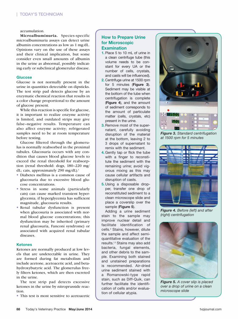
How to Prepare Urine for Microscopic Examination1. Place 5 to 10 mL of urine in
a clean centrifuge tube (this volume needs to be con-stant for every Ua or the number of cells, crystals, and casts will be influenced).
2. centrifuge urine at 1500 rpm for 5 minutes (Figure 3). sediment may be visible at the bottom of the tube when centrifugation is complete (Figure 4), and the amount of sediment corresponds to the amount of particulate matter (cells, crystals, etc) present in the urine.
3. Remove most of the super-natant, carefully avoiding disruption of the material at the bottom, leaving 2 to 3 drops of supernatant to remix with the sediment.
4. Gently tap or flick the tube with a finger to reconsti-tute the sediment with the remaining urine; avoid vig-orous mixing as this may cause cellular artifacts and disruption of casts.
5. Using a disposable drop-per, transfer one drop of reconstituted sediment to a clean microscope slide and place a coverslip over the sample (Figure 5).adding a urine sediment
stain to the sample may improve nuclear detail and facilitate identification of cells.2 stains, however, dilute the sample and affect semi-quantitative evaluation of the results.1,3 stains may also add bacteria, fungal elements, and other debris to the sam-ple. examining both stained and unstained preparations is recommended. air-dried urine sediment stained with a Romanowski-type rapid stain, such as diff-Quik, can further facilitate the identifi-cation of cells and/or evalua-tion of cellular atypia.
Figure 3. Standard centrifugation at 1500 rpm for 5 minutes
Figure 4. Before (left) and after (right) centrifugation
Figure 5. A cover slip is placed over a drop of urine on a clean microscope slide
May/June 2014 Today’s Veterinary Practice 89
UR
ina
Lys
is in
co
MP
an
ion
an
iMa
Ls P
aR
T 2:
eV
aLU
aTi
on
of
UR
ine
ch
eM
isTR
y &
se
diM
en
TToday’s Technician |
tvpjournal.com
samples should not be directly exposed to light. D i s co lo r a t ion of urine (due to hemoglobinuria and myoglobin-uria) causes non-specif ic color change in the bilirubin reagent pad, which inter-feres with read-ing the test strip.
MICROSCOPIC EXAMINATION OF URINE SEDIMENTAfter preparation of the urine sediment slide (see How to Prepare Urine for Microscopic Examination), micro-scopic examination of the sediment is performed with the sub-stage condenser of the microscope lowered.
The initial scanning of the slide is performed on low power (10×), which enables the examiner to evaluate the quantity of material present and quality of the sample prep-aration. Using the fine focus while scanning, the examiner can assess particles suspended in different planes of the fluid.
Examination at high power (40×) enables the examiner to evaluate cell number and morphology, and identify casts and crystals. Each of these elements may be counted by aver-aging the number of elements in 10 fields. The cells, casts, and crystals are reported as the average number per high-power field (HPF) or low-power field (LPF).
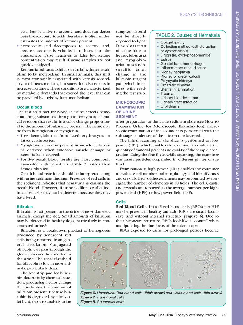
CellsRed Blood Cells. Up to 5 red blood cells (RBCs) per HPF may be present in healthy animals. RBCs are small, bicon-cave, and without internal structure (Figure 6). Due to their biconcave structure, RBCs look like a “donuts” when manipulating the fine focus of the microscope.
RBCs exposed to urine for prolonged periods become
acid, less sensitive to acetone, and does not detect beta-hydroxybutyric acid; therefore, it often under-estimates the amount of ketones present.
•Acetoacetic acid decomposes to acetone and, because acetone is volatile, it diffuses into the atmosphere. False negatives or false low ketone concentration may result if urine samples are not quickly analyzed. Ketonuria indicates a shift from carbohydrate metab-
olism to fat metabolism. In small animals, this shift is most commonly associated with ketosis second-ary to diabetes mellitus, but starvation also results in increased ketones. These conditions are characterized by metabolic demands that exceed the level that can be provided by carbohydrate metabolism.
Occult BloodThe test strip pad for blood in urine detects heme-containing substances through an enzymatic chemi-cal reaction that results in a color change proportion-al to the amount of substance present. The heme may be from hemoglobin or myoglobin.• Free hemoglobin is from lysed erythrocytes or
intact erythrocytes. •Myoglobin, a protein present in muscle cells, can
be detected when extensive muscle damage or necrosis has occurred.
• Positive occult blood results are most commonly associated with hematuria (Table 2) rather than hemoglobinuria. Occult blood reactions should be interpreted along
with urine sediment findings. Presence of red cells in the sediment indicates that hematuria is causing the occult blood. However, if urine is dilute or alkaline, intact red cells may not be detected because they may have lysed.
BilirubinBilirubin is not present in the urine of most domestic animals, except the dog. Small amounts of bilirubin may be detected in healthy dogs, particularly in con-centrated urine.1,3
Bilirubin is a breakdown product of hemoglobin produced by senescent red cells being removed from gen-eral circulation. Conjugated bilirubin can pass through the glomerulus and be excreted in the urine. The renal threshold for bilirubin is low in most ani-mals, particularly dogs.
The test strip pad for biliru-bin detects it by chemical reac-tion, producing a color change that indicates the amount of bilirubin present. Because bili-rubin is degraded by ultravio-let light, prior to analysis urine
TabLe 2. causes of hematuria
• coagulopathy • collection method (catheterization
or cystocentesis)• drugs (ie, cyclophosphamide)• estrus• Genital tract hemorrhage• inflammatory renal disease• Kidney neoplasia• Kidney or ureter calculi• Polycystic kidneys• Prostatic disease• sterile inflammation• Trauma • Urinary bladder tumor• Urinary tract infection• Urolithiasis
Figure 6. Hematuria: Red blood cells (thick arrow) and white blood cells (thin arrow)Figure 7. Transitional cellsFigure 8. Squamous cells
Today’s Veterinary Practice May/June 201490
| Today’s Technician
tvpjournal.com
spiculated and small due to dehyd r a t ion , and lyse in urine that is not ana-lyzed quickly. Lysis is acceler-ated in either di lute urine (USG < 1.006)
or alkaline urine, which results in a positive occult blood reading on the dipstick, but no visible erythrocytes on sed-iment examination. This result may be misdiagnosed as hemoglobinuria associated with intravascular hemolysis.
White Blood Cells. Up to 5 white blood cells (WBCs) per HPF may be present in healthy animals. WBCs are round, approximately 1½ to 2 times the size of RBCs, and have refractive internal granularity that is more pronounced with fine focusing manipulation.
WBCs in urine are typically neutrophils. Pyuria describes the presence of 6 to 10 or more neutrophils per HPF (Table 3). Unstained wet mount preparations present 2 challeng-es: (1) leukocytes may be present but cannot be readily differentiated and (2) it may be difficult to differentiate WBCs from small epithelial cells.
WBCs deteriorate in urine and may decline up to 50% within an hour of collection if the sample is kept at room temperature.3
Epithelial Cells. Low numbers of epithelial cells are found in the urine of healthy animals; particularly those samples obtained by catheterization, as cells slough and are replaced by new cells. In unstained wet mounts, it is difficult to differentiate epithelial cells based on size. •Renal tubule cells are typically small, but distinguish-
ing them from WBCs or small transitional cells may not be possible. Increased numbers of small epithelial cells should prompt evaluation of an air-dried, stained cytology preparation to distinguish WBCs and/or tran-sitional cells from renal tubule cells. Sloughing of renal tubule cells indicates renal tubule damage.
• Transitional cells (Figure 7, page 89) line the renal pel-vis, ureters, urinary bladder, and most of the urethra. They vary greatly in size, but are typically 2 to 4 times larger than WBCs, with a round nucleus and granular cytoplasm. Increased numbers of transitional cells may be seen with inflammation of the urinary bladder. This
finding should prompt cytologic examination of an air-dried, stained sample to evaluate the cells for evidence of malignancy.
• Squamous cells (Figure 8, page 89) are located at the distal urethra and genital tract of females. They are large, polygonal cells considered contaminants.
CastsUrinary casts are cylindrical molds formed in the lumens of the renal tubules. They are primarily composed of a mucoprotein secreted by renal tubule cells. Concentrat-ed urine, decreased urine flow, and acidic urine favor the formation of casts.
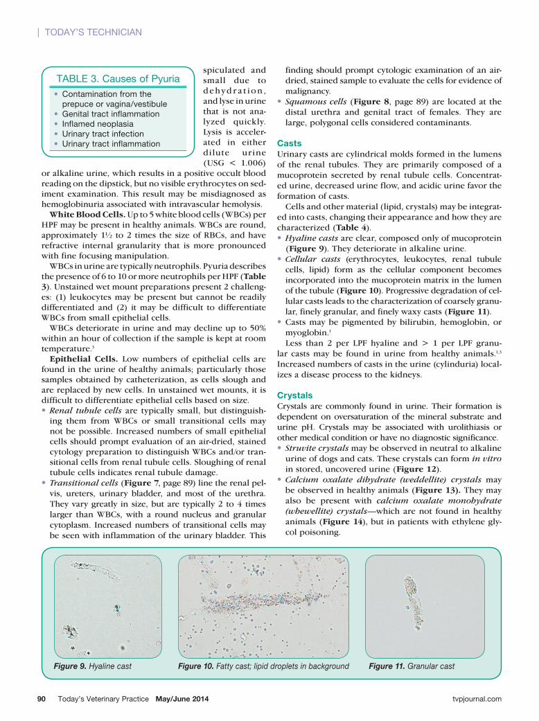
Cells and other material (lipid, crystals) may be integrat-ed into casts, changing their appearance and how they are characterized (Table 4). •Hyaline casts are clear, composed only of mucoprotein
(Figure 9). They deteriorate in alkaline urine. •Cellular casts (erythrocytes, leukocytes, renal tubule
cells, lipid) form as the cellular component becomes incorporated into the mucoprotein matrix in the lumen of the tubule (Figure 10). Progressive degradation of cel-lular casts leads to the characterization of coarsely granu-lar, finely granular, and finely waxy casts (Figure 11).
•Casts may be pigmented by bilirubin, hemoglobin, or myoglobin.1 Less than 2 per LPF hyaline and > 1 per LPF granu-
lar casts may be found in urine from healthy animals.1,3 Increased numbers of casts in the urine (cylinduria) local-izes a disease process to the kidneys.
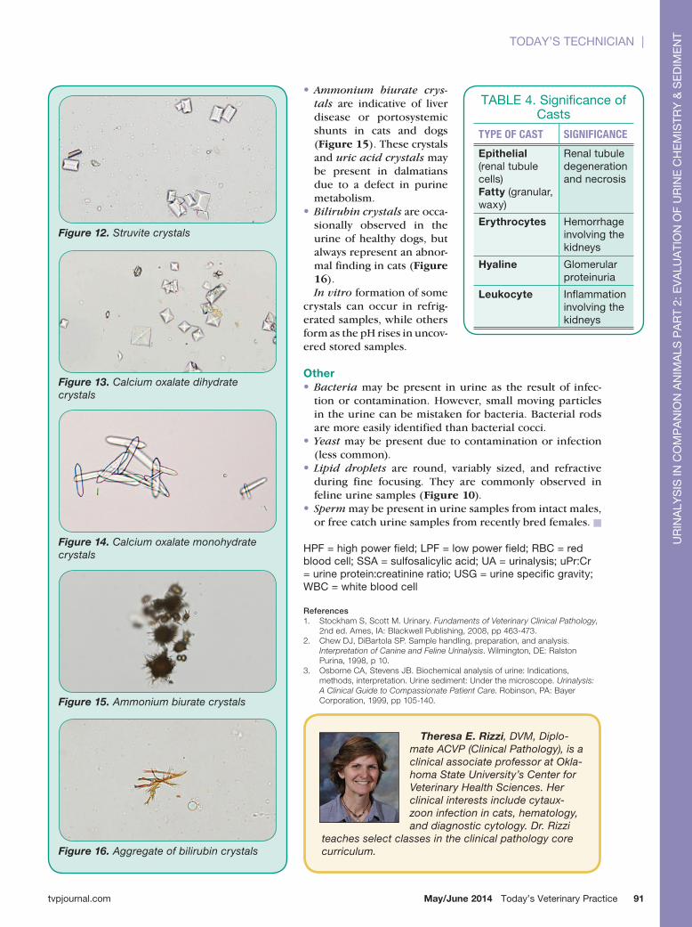
CrystalsCrystals are commonly found in urine. Their formation is dependent on oversaturation of the mineral substrate and urine pH. Crystals may be associated with urolithiasis or other medical condition or have no diagnostic significance. • Struvite crystals may be observed in neutral to alkaline
urine of dogs and cats. These crystals can form in vitro in stored, uncovered urine (Figure 12).
•Calcium oxalate dihydrate (weddellite) crystals may be observed in healthy animals (Figure 13). They may also be present with calcium oxalate monohydrate (whewellite) crystals—which are not found in healthy animals (Figure 14), but in patients with ethylene gly-col poisoning.
Figure 9. Hyaline cast Figure 10. Fatty cast; lipid droplets in background Figure 11. Granular cast
TabLe 3. causes of Pyuria• contamination from the
prepuce or vagina/vestibule • Genital tract inflammation• inflamed neoplasia• Urinary tract infection• Urinary tract inflammation
May/June 2014 Today’s Veterinary Practice 91
UR
ina
Lys
is in
co
MP
an
ion
an
iMa
Ls P
aR
T 2:
eV
aLU
aTi
on
of
UR
ine
ch
eM
isTR
y &
se
diM
en
TToday’s Technician |
tvpjournal.com
•Ammonium biurate crys-tals are indicative of liver disease or portosystemic shunts in cats and dogs (Figure 15). These crystals and uric acid crystals may be present in dalmatians due to a defect in purine metabolism.
•Bilirubin crystals are occa-sionally observed in the urine of healthy dogs, but always represent an abnor-mal finding in cats (Figure 16).In vitro formation of some
crystals can occur in refrig-erated samples, while others form as the pH rises in uncov-ered stored samples.
Other•Bacteria may be present in urine as the result of infec-
tion or contamination. However, small moving particles in the urine can be mistaken for bacteria. Bacterial rods are more easily identified than bacterial cocci.
• Yeast may be present due to contamination or infection (less common).
• Lipid droplets are round, variably sized, and refractive during fine focusing. They are commonly observed in feline urine samples (Figure 10).
• Sperm may be present in urine samples from intact males, or free catch urine samples from recently bred females. n
hPf = high power field; LPf = low power field; Rbc = red blood cell; ssa = sulfosalicylic acid; Ua = urinalysis; uPr:cr = urine protein:creatinine ratio; UsG = urine specific gravity; Wbc = white blood cell
References 1. stockham s, scott m. Urinary. Fundaments of Veterinary Clinical Pathology,
2nd ed. ames, ia: Blackwell Publishing, 2008, pp 463-473.2. Chew dJ, diBartola sP. sample handling, preparation, and analysis.
Interpretation of Canine and Feline Urinalysis. wilmington, de: ralston Purina, 1998, p 10.
3. osborne Ca, stevens JB. Biochemical analysis of urine: indications, methods, interpretation. Urine sediment: Under the microscope. Urinalysis: A Clinical Guide to Compassionate Patient Care. robinson, Pa: Bayer Corporation, 1999, pp 105-140.
Theresa E. Rizzi, DVM, Diplo-mate ACVP (Clinical Pathology), is a clinical associate professor at Okla-homa State University’s Center for Veterinary Health Sciences. Her clinical interests include cytaux-zoon infection in cats, hematology, and diagnostic cytology. Dr. Rizzi
teaches select classes in the clinical pathology core curriculum.
Figure 12. Struvite crystals
Figure 13. Calcium oxalate dihydrate crystals
Figure 14. Calcium oxalate monohydrate crystals
Figure 16. Aggregate of bilirubin crystals
Figure 15. Ammonium biurate crystals
TabLe 4. significance of casts
TYPE OF CAST SIGNIFICANCE
Epithelial (renal tubule cells)Fatty (granular, waxy)
Renal tubule degeneration and necrosis
Erythrocytes hemorrhage involving the kidneys
Hyaline Glomerular proteinuria
Leukocyte inflammation involving the kidneys