Embed Size (px)
Citation preview
U.S. ARMY MEDICAL DEPARTMENT CENTER AND SCHOOL FORT SAM HOUSTON, TEXAS 78234-6100
URINALYSIS SUBCOURSE MD0852 EDITION 200
DEVELOPMENT
This subcourse is approved for resident and correspondence course instruction. It reflects the current thought of the Academy of Health Sciences and conforms to printed Department of the Army doctrine as closely as currently possible. Development and progress render such doctrine continuously subject to change.
ADMINISTRATION
For comments or questions regarding enrollment, student records, or shipments, contact the Nonresident Instruction Section at DSN 471-5877, commercial (210) 221-5877, toll-free 1-800-344-2380; fax: 210-221-4012 or DSN 471-4012, e-mail [email protected], or write to: COMMANDER AMEDDC&S ATTN MCCS HSN 2105 11TH STREET SUITE 4192 FORT SAM HOUSTON TX 78234-5064 Approved students whose enrollments remain in good standing may apply to the Nonresident Instruction Section for subsequent courses by telephone, letter, or e-mail. Be sure your social security number is on all correspondence sent to the Academy of Health Sciences.
CLARIFICATION OF TRAINING LITERATURE TERMINOLOGY When used in this publication, words such as "he," "him," "his," and "men" are intended to include both the masculine and feminine genders, unless specifically stated otherwise or when obvious in context. .
USE OF PROPRIETARY NAMES
The initial letters of the names of some products are capitalized in this subcourse. Such names are proprietary names, that is, brandnames or trademarks. Proprietary names have been used in this subcourse only to make it a more effective learning aid. The use of any name, proprietary or otherwise, should not be interpreted as an endorsement, deprecation, or criticism of a product. Nor should such use be considered to interpret the validity of proprietary rights in a name, whether it is registered or not. .
MD0852 i
TABLE OF CONTENTS Lesson Paragraphs
INTRODUCTION 1 THE COLLECTION AND PRESERVATION OF SPECIMENS; MACROSCOPIC AND PHYSICAL EXAMINATION OF URINE Section I. Collection and Preservation of Specimens 1-1--1-7 Section II. Macroscopic and Physical Examination of Urine 1-8--1-15 Exercises 2 CHEMICAL TESTS FOR SUBSTANCES IN URINE Section I. Protein in Urine 2-1--2-3 Section II. Glucose and other Reducing Substances in Urine 2-4--2-5 Section III. Ketone Bodies in Urine 2-6--2-7 Section IV. Blood in Urine 2-8--2-9 Section V. Bilirubin and Urobilinogen in Urine 2-10--2-13 Section VI. Calcium in Urine 2-14--2-15 Section VII. Porphyrins in Urine (Porphyrinuria) 2-16--2-17 Section VIII. Miscellaneous Tests 2-18--2-20 Section IX. Urinary Calculi 2-21--2-22 Exercises 3 THE MICROSCOPIC EXAMINATION OF URINARY SEDIMENT Section I. Preparation and Illumination 3-1--3-4 Section II. Microscopic Examination of Organized Sediment 3-5--3-12 Section III. Microscopic Examination of Unorganized Sediment 3-13--3-17 Section IV. The Microscopic Examination of Stained Urinary Sediment Using the Sternheimer-Malbin Stain. 3-18--3-25 Exercises
MD0852 ii
CORRESPONDENCE COURSE OF THE U.S. ARMY MEDICAL DEPARTMENT CENTER AND SCHOOL
SUBCOURSE MD0852
URINALYSIS
INTRODUCTION
Due in part to the development of multiple reagent strips (dipstix) for urinalysis, more laboratory tests are now performed each year on urine than on any other body fluid. A typical urinalysis includes tests for glucose, protein, pH, ketone bodies, bilirubin, occult (unseen) blood, urobilinogen, and specific gravity and microscopic examination of urinary sediment. Many common abnormalities can be recognized by urine studies. Urine tests are the method of choice to monitor the treatment of diabetes. Urine is an excretion product, but it is usually clean and sterile. Its chief components are urea, sodium chloride, and water. The stench of stale urine is largely due to the decomposition of urea to ammonia by bacteria. The odor of fresh urine is not unpleasant to most persons. Urine is not a significant source of infection. The disagreeable characteristics arising from decomposition can usually be avoided. This subcourse will focus on the analysis of urine. The contents of the text will present and discuss the topics outlined above. However, you should remember that the subcourse is not intended to provide you with all that is known about urinalysis. For this reason, you should read other texts and journals, discuss the subcourse contents with your fellow workers and supervisors, and search other sources of knowledge to expand your knowledge of this important topic. Subcourse Components: This subcourse consists of three lessons. The lessons are as follows: Lesson 1. The Collection and Preservation of Specimens; Macroscopic and Physical Examination of Urine. Lesson 2. Chemical Tests for Substances in Urine. Lesson 3. The Microscopic Examination of Urinary Sediment. Credit Awarded: To receive credit hours, you must be officially enrolled and complete an examination furnished by the Nonresident Instruction Section at Fort Sam Houston, Texas. Upon successful completion of the examination for this subcourse, you will be awarded 7 credit hours. You can enroll by going to the web site http://atrrs.army.mil and enrolling under "Self Development" (School Code 555).
MD0852 iii
A listing of correspondence courses and subcourses available through the Nonresident Instruction Section is found in Chapter 4 of DA Pamphlet 350-59, Army Correspondence Course Program Catalog. The DA PAM is available at the following website: http://www.usapa.army.mil/pdffiles/p350-59.pdf.
LESSON ASSIGNMENT LESSON 1 The Collection and Preservation of Specimens; Macroscopic and Physical Examination of Urine. TEXT ASSIGNMENT Paragraphs 1-1 through 1-15. LESSON OBJECTIVES After completing this lesson, you should be able to: 1-1. Select the statement that best describes the
clinical importance of urinalysis. 1-2. Select the statement that best describes a type
of specimen. 1-3. Select the statement that best contrasts the two
special methods of urine collection. 1-4. Select the statement that best describes the
means to preserve a urine sample. 1-5. Select the volume that is the median amount of
urine produced by an average adult during a 24 hour period.
1-6. Select the definition that correctly describes the
following terms: polyuria, oliguria, or anuria. 1-7. Select the substance(s)/conditions(s) which
might cause urine to be a certain color. 1-8. Select the statement that best describes the
macroscopic means of evaluating a urine sample.
SUGGESTION After studying the assignment, complete the exercises
of this lesson. These exercises will help you to achieve the lesson objectives.
MD0852 1-1
LESSON 1
THE COLLECTION AND PRESERVATION OF SPECIES; MACROSCOPIC AND PHYSICAL EXAMINATION OF URINE
Section I. COLLECTION AND PRESERVATION OF SPECIMENS
1-1. IMPORTANCE OF URINALYSIS Purification of the circulating blood is crucial for the continuation of life. The urinary system is a group of organs that serves to remove from the blood nonvolatile waste products that cannot be removed through the respiratory system. The organ that has the major role in both filtration and reabsorption of necessary substances is the kidney. Consequently, urinalysis is an extremely valuable tool for demonstrating pathological conditions in the excretory system and as an index for the general metabolic condition of an individual. There are several different kinds of samples used in urinalysis. 1-2. RANDOM SPECIMEN A random urine specimen is satisfactory for most qualitative tests and may be collected at any time. However, factors such as the type and amount of food consumed, and the performance of exercise must be considered when interpreting results. For example, an elevated urine sugar may be obtained after an exceedingly high carbohydrate meal. All random specimens should be freshly voided and delivered to the laboratory as quickly as possible. Urine is an excellent culture medium for many types of microorganisms. As bacterial growth and metabolism increase, decomposition of the urine proceeds rapidly. If a delay of several hours is unavoidable, the urine specimen should be kept in the refrigerator. 1-3. FIRST MORNING VOID A first morning sample is collected when the patient rises in the morning. Only a clean container is necessary. Early morning specimens are used most frequently for analysis due to their day-to-day consistency. It is the most concentrated of the urine samples and is used for qualitative analysis. It is also essential for preventing false-negative pregnancy tests and for evaluating orthostatic proteinuria. 1-4. TWO-HOUR POSTPRANDIAL This specimen is collected two hours after the patient has eaten a meal and requires only a clean container. The specimen is tested for glucose, and the results are used to monitor insulin therapy in patients with diabetes mellitus.
MD0852 1-2
1-5. TWENTY-FOUR HOUR SPECIMEN a. A 24-hour specimen is required in order to obtain significant results in the quantitative analyses. It is essential that a clean container and the proper preservative be used. The 24-hour specimen is made up of the total urinary output for a specific 24-hour period and to obtain an accurate timed specimen. It is necessary to begin the collection period with an empty bladder and end the collection period with an empty bladder. The procedure is as follows: (1) Day one. First thing in the morning, the patient should void and discard that specimen, after which the remainder of urine is collected for the next 24-hours. The patient should be instructed to urinate into a separate urine collection cup and pour the contents into the 24-hour collection container (caused by the possibility of splashing preservative onto exposed skin). (2) Day two. At the same time as the beginning of the first collection, the patient voids and adds this urine to previously collected urine. (3) Storage. The patient should be advised to store partial collections at 4-6oC and deliver the completed 24-hour urine collection to the laboratory as soon as possible after completion. b. Upon arrival in the laboratory, the 24-hour specimen must be thoroughly mixed and the volume accurately measured and recorded. Only an aliquot is needed for testing, but the amount saved must be adequate to permit repeat or additional testing. 1-6. SPECIAL METHODS OF URINE COLLECTION When bacteriological studies are to be done, special collection techniques may be necessary to avoid contamination of the specimen. a. Catheterization. Catheterization is used for some bacteriological tests performed on urine. However, even the most careful sterile technique cannot entirely prevent contamination of the bladder and the upper urinary tract during the passage of the catheter. This method is not used very often as it causes the patient much discomfort. b. Midstream (Clean Catch) Specimen. A midstream specimen is used more often than a specimen from catheterization. Although this method does not eliminate contamination as much as catheterization, it is satisfactory if it is carefully collected. (1) With men, the glans penis should be adequately exposed and cleaned with soap or a mild antiseptic solution. The initial flow of urine should be allowed to escape, but the midstream urine should be collected in a sterile container.
MD0852 1-3
(2) With women, the urethral opening should be plainly exposed and well cleaned with soapy cotton balls. The area should be thoroughly rinsed with sterile, water-saturated cotton balls. The female patient should void the first portion of urine forcibly and then allow the midstream portion of about 20 to 100 ml to be caught in a sterile container. c. Suprapubic Aspiration. Urine may be collected by external introduction of a needle into the bladder. The bladder is sterile under normal conditions. This collection method provides a sample for bacterial culture free of extraneous contamination and may be used for cytological studies. 1-7. PRESERVATION There is no substitute for a fresh urine specimen, and in all cases the analysis should be performed as soon as possible. A delay in analysis leads to a degeneration of the formed elements and decomposition of chemical constituents. Occasionally, however, the analysis has to be delayed, or a specimen must be shipped. When such situations occur, deterioration of the specimen may be inhibited by the use of some form of preservation. The methods most commonly employed for preservation are the following: a. Refrigeration. The best general method of preservation up to 8 hours is refrigeration at 4-6ºC. Refrigerated specimens are warmed to room temperature before performing an analysis. b. Toluene (Toluol). If only the chemical contents of the urine are of interest, as with most 24-hour specimens, toluene may be used. Toluene merely lies on the surface of the urine, forming a thin layer and acting as a physical barrier to air and bacteria. However, anaerobic bacteria, if present, are not inhibited. To measure portions of the specimen, it is necessary either to remove the toluene or to pipet from below the surface. c. Formalin (10 percent). Ten percent formalin is an excellent preservative for the formed (microscopic) elements in urine. About 4 drops of formalin may be used for each 100 ml of urine. However, it interferes with some qualitative chemical tests, and it should not be used when the glucose concentration is to be determined. d. Boric Acid (0.8 percent). Boric acid is a satisfactory preservative for general purposes. It will not interfere with examinations for protein, sugar, or ketone bodies. e. Thymol (10 percent in Isopropanol). Thymol is another general purpose preservative. Approximately 10 ml of the prepared solution is used for each 24-hour collection. f. Chloroform. Chloroform may be used as a preservative, but it interferes with some chemical tests and may cause cellular changes.
MD0852 1-4
g. Sodium Fluoride. Sodium fluoride may be used as a preservative for urine samples when one is concerned with glucose. It inhibits tests for glucose on the reagent strip. h. Sodium Carbonate. To preserve urobilinogen in urine requires special precautions. To assure alkalinity, a half-teaspoonful of sodium carbonate is placed in the specimen bottle before the urine is voided into the bottle. i. Strong Mineral Acids. Analysis for amino acids, delta-aminolevulinic acid, and total nitrogen requires acidification with a strong mineral acid (for example, hydrochloric acid to pH 3.0).
Section II. MACROSCOPIC AND PHYSICAL EXAMINATION OF URINE 1-8. INTRODUCTION Macroscopic analysis deals with those procedures or examinations, which are accomplished without the aid of a microscope. Included in this category are measurement of volume, color, appearance, pH, and specific gravity. Before performing microscopic or chemical tests on a urine specimen, a macroscopic examination is accomplished. As the metabolic waste products filtering into the kidneys are constantly changing in relation to body intake, so the urine is constantly changing with respect to volume, color, appearance, specific gravity, and pH. Therefore, an accurate description of these physical properties furnishes the physician and/or physician extender with valuable information regarding kidney function. 1-9. VOLUME The total 24-hour volume of urine voided by the normal adult is influenced by food and fluid intake, temperature, exercise, seasonal change, and the use of diuretics such as caffeine. Nonetheless, a consistent normal range has been established. The average adult produces between 750 and 2,000 ml of urine during a 24-hour period, with a median of about 1,400 ml. Volume determination is a quantitative analysis and therefore, the 24-hour specimen is used. When the total volume of a urine specimen is to be measured, the smallest graduated cylinder that will hold the entire quantity should be used. The amount of liquid preservative that has been added is not included in the total volume measurement. It should be noted that the amount of urine excreted might fall above or below the normal range without the existence of a pathological condition. However, abnormalities can cause marked deviations in total urinary output, resulting in one of the three following conditions: a. Polyuria. This term refers to an abnormal increase in the total volume of urine excreted (more than 2,000 ml/24-hours). Polyuria is associated with such pathological conditions as diabetes mellitus, diabetes insipidus, certain tumors of brain and spinal cord acromegaly, myxedema, and certain kidney diseases. The nonpathologic cause is usually increased fluid intake.
MD0852 1-5
b. Oliguria. A reduction in the total volume of urine excreted is called oliguria (less than 200 ml/24-hours). This condition is associated with febrile states, excessive vomiting, severe diarrhea, or extreme dehydration. Nonpathological causes are decreased fluid intake and excessive sweating. c. Anuria. This term literally means "no urine" and refers to a complete lack of urine excretion. It results from blockage of the kidneys or urinary tract, certain bacterial infections of the kidneys, and prolonged states of dehydration. There are not any tnonpathological causes. 1-10. COLOR Urine color is another physical property that is evaluated in the routine urinalysis. The color of normal urine is caused by the presence of various pigments, which are collectively referred to as urochrome. The various shades of yellow in urine specimens vary with the intensity of the urochrome present; the intensity of the color also varies with the specific gravity. Urine can show a typical coloration because of pathological conditions and as a result of the ingestion of certain substances, including food pigments, dyes, drugs, and so forth. It is important that one note the exact color observed and indicate on the laboratory slip any changes that occur on standing. The physician determines the diagnostic significance of the observed color. a. Yellow. Normal urine has a color of straw, yellow, or amber. Urines that are concentrated are usually amber; very dilute specimens may be almost colorless. (1) In addition, a yellow color may be produced by the following substances: (a) Cascara--a laxative. (b) Phenacetin--to ease fever or pain. (c) Food colors. (d) Atabrine® (brand name)--an anti-malarial. (e) Azulfidine® (brand name) (2) A specimen that is a very pale yellow, greenish-yellow, or nearly colorless can be the result of several pathological conditions, specifically: (a) Severe iron deficiency. (b) Chronic kidney disease. (c) Diabetes mellitus. (d) Diabetes insipidus.
MD0852 1-6
b. Green and Blue-Green. The blue-green color is frequently due to the mixture of the color blue with the yellow of the urine. The following can impart a green or blue-green color to the urine: (1) Oral contraceptives. (2) Bile pigment. (3) Diagnex Blue® (brand name). (4) Elavil® (brand name). (5) Indican in large amounts. (6) Vitamin B complex. (7) Blue diaper syndrome. (8) Evans blue. (9) Methylene blue in kidney medication. (10) Yeast concentrate. (11) Pseudomonas toxemia. (12) Increased serum copper concentrations. c. Brown and Black. Brown-colored or black-colored urine can be produced by the following: (1) Porphyrins. (2) Bilirubin. (3) Injectable iron compounds. (4) Melanin pigment. (5) Phenol poisoning. (6) Alkapton bodies. (7) Methemoglobin. (8) Tertian malaria.
MD0852 1-7
d. Red, Pink, or Reddish-Orange. Quite a few substances can give the urine a pink or reddish coloring. These substances include the following materials: (1) Beets. (2) Dilantin® (brand name). (3) Food colors. (4) Blood. (5) Azo Gantrisin® (brand name). (6) Senna in alkaline urines. (7) Pyridium® (brand name). (8) Porphyrin. (9) Hemoglobin. (10) Povan® (brand name). (11) Rhubarb in alkaline urines. (12) Phenolsulfophthalein. (13) Myoglobin. (14) Bromsulphalein. (15) Chromogenic bacteria. e. Orange. The following list of substances can give the urine an orange color: (1) Senna. (2) Rhubarb. (3) Azo Gantrisin® (brand name). (4) Carotene. (5) Furoxone® (brand name). (6) Riboflavin.
MD0852 1-8
(7) Food colors. (8) Santonin in acid urines. (9) Pyridium. NOTE: A highly concentrated urine resulting from fever, inadequate water intake, or excessive water loss may also appear orange in color. 1-11. GENERAL APPEARANCE OF THE URINE SAMPLE The general appearance of a urine specimen should also be evaluated routinely. Normally, fresh urine is clear, but the specimen can also be hazy or cloudy. Freshly voided urine should be used, since, if allowed to stand, all samples become turbid due to bacterial contamination. This uniform turbidity does not disappear upon heating or acidification. a. Clear. Normal, freshly voided urine is usually clear as it has no visible particles. b. Hazy. When the sample contains a small amount of particles, it is designated as hazy. Normal urine specimens may have a hazy appearance. Haziness may be due to mucus, epithelial cells, or amorphous urates or phosphates. Amorphous urates can be removed from the urine specimen by gently heating the specimen in warm water or by gently heating the prepared microscope slide. These techniques cause the crystals to redissolve. c. Cloudy. Moderate to large amounts of visible particles produce a cloudy urine. Cloudiness may be caused by crystallized mineral salts that have precipitated due to long standing, or to the increase of bacteria when urine is left standing at room temperature. Cloudiness may also result from pathological conditions that produce blood or pus. The bacteria resulting from acute infections may also produce a cloudy urine. 1-12. SPECIFIC GRAVITY A good test of total kidney function is the determination of specific gravity. Such a determination will measure the kidney's ability to concentrate urine. Specific gravity is a comparison of the density of urine to the density of distilled water, which is regarded as 1.000. Generally, the greater the volume of urine excreted, the lower the specific gravity. There is considerable variation in the specific gravity range of 1.003 to 1.030. Pathological conditions often result in an elevated or decreased specific gravity. In pathological conditions, the range of urine specific gravity may be 1.001 to 1.060. The determination of specific gravity involves the use of the following two instruments:
MD0852 1-9
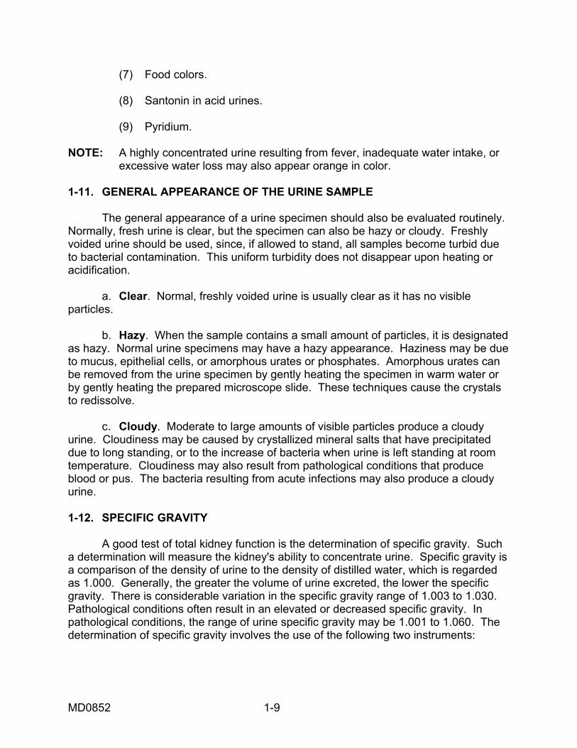
a. Standard Urinometer. The equipment required for the determination of specific gravity includes the urinometer and glass cylinder. A new urinometer should always be checked prior to use. When calibrated using distilled water, this instrument should read 1.000 at the temperature specified by the manufacturer. If a large discrepancy is noted, the urinometer should be discarded. If the discrepancy is small, a correction factor may be used. In addition, if the temperature at which readings are taken differs from the manufacturer's specified temperature, a temperature correction of .001 should be added or subtracted for every three degrees above or below manufacturer's calibration temperature. (See figure 1-1 for an illustration of an urinometer.)
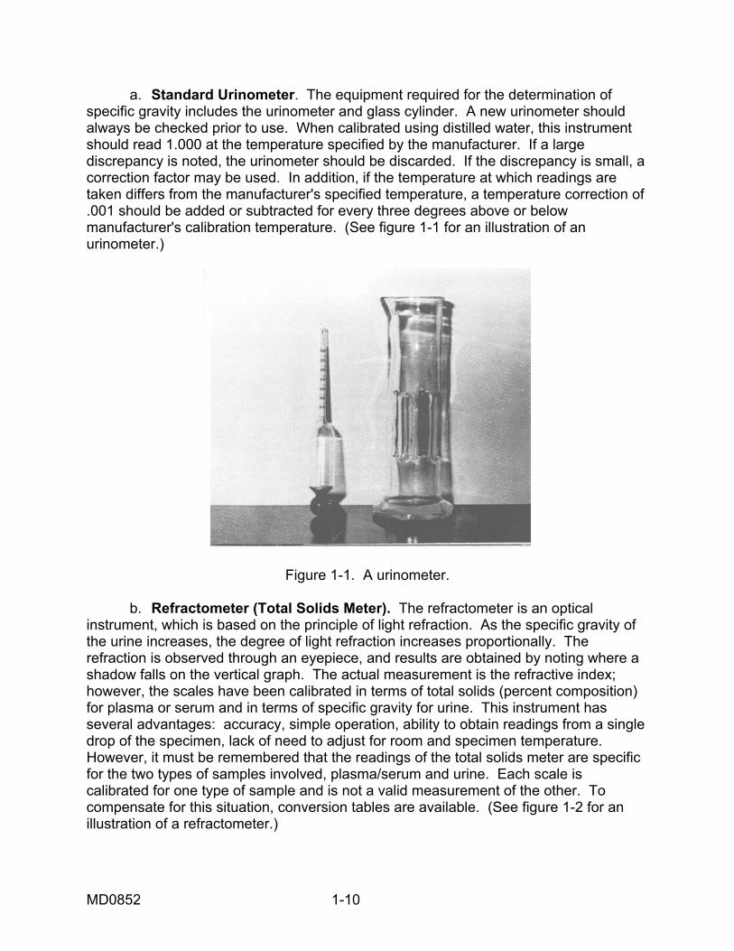
Figure 1-1. A urinometer. b. Refractometer (Total Solids Meter). The refractometer is an optical instrument, which is based on the principle of light refraction. As the specific gravity of the urine increases, the degree of light refraction increases proportionally. The refraction is observed through an eyepiece, and results are obtained by noting where a shadow falls on the vertical graph. The actual measurement is the refractive index; however, the scales have been calibrated in terms of total solids (percent composition) for plasma or serum and in terms of specific gravity for urine. This instrument has several advantages: accuracy, simple operation, ability to obtain readings from a single drop of the specimen, lack of need to adjust for room and specimen temperature. However, it must be remembered that the readings of the total solids meter are specific for the two types of samples involved, plasma/serum and urine. Each scale is calibrated for one type of sample and is not a valid measurement of the other. To compensate for this situation, conversion tables are available. (See figure 1-2 for an illustration of a refractometer.)
MD0852 1-10
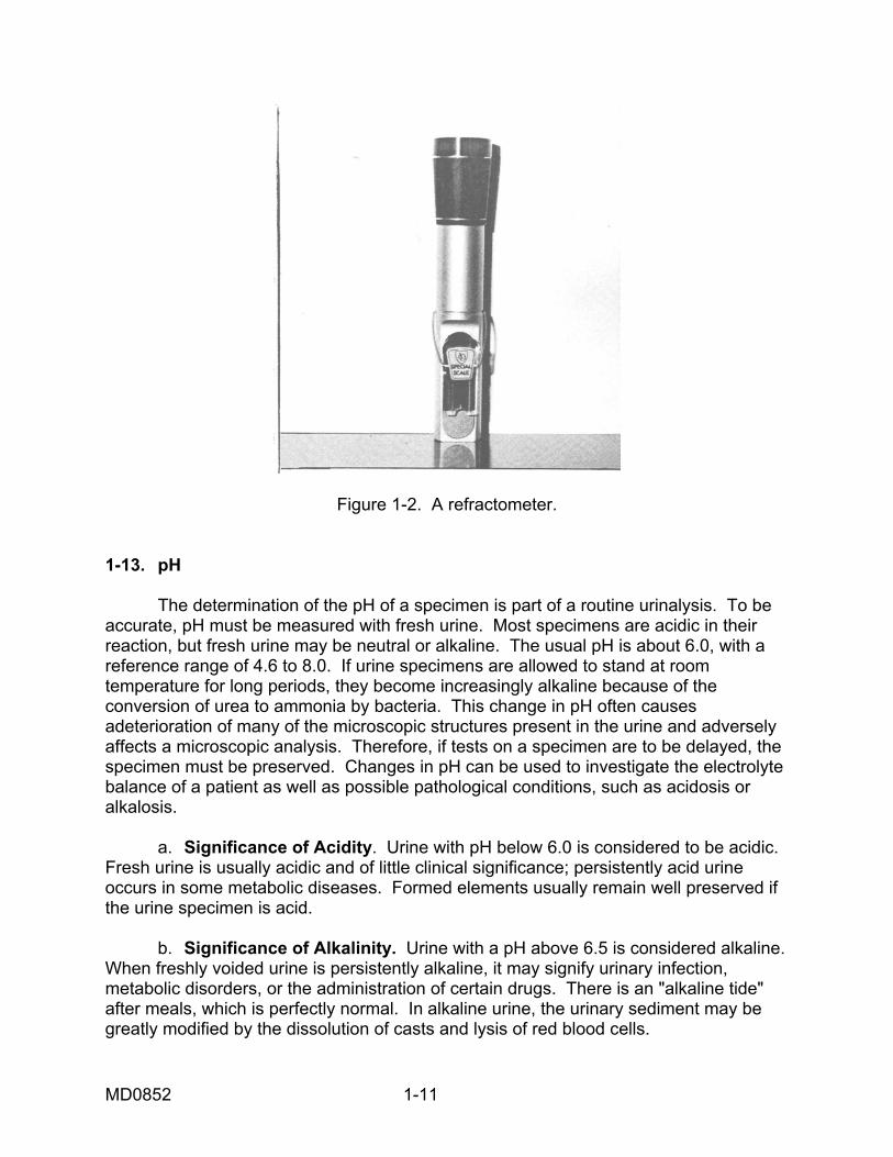
Figure 1-2. A refractometer.
1-13. pH The determination of the pH of a specimen is part of a routine urinalysis. To be accurate, pH must be measured with fresh urine. Most specimens are acidic in their reaction, but fresh urine may be neutral or alkaline. The usual pH is about 6.0, with a reference range of 4.6 to 8.0. If urine specimens are allowed to stand at room temperature for long periods, they become increasingly alkaline because of the conversion of urea to ammonia by bacteria. This change in pH often causes adeterioration of many of the microscopic structures present in the urine and adversely affects a microscopic analysis. Therefore, if tests on a specimen are to be delayed, the specimen must be preserved. Changes in pH can be used to investigate the electrolyte balance of a patient as well as possible pathological conditions, such as acidosis or alkalosis. a. Significance of Acidity. Urine with pH below 6.0 is considered to be acidic. Fresh urine is usually acidic and of little clinical significance; persistently acid urine occurs in some metabolic diseases. Formed elements usually remain well preserved if the urine specimen is acid. b. Significance of Alkalinity. Urine with a pH above 6.5 is considered alkaline. When freshly voided urine is persistently alkaline, it may signify urinary infection, metabolic disorders, or the administration of certain drugs. There is an "alkaline tide" after meals, which is perfectly normal. In alkaline urine, the urinary sediment may be greatly modified by the dissolution of casts and lysis of red blood cells.
MD0852 1-11
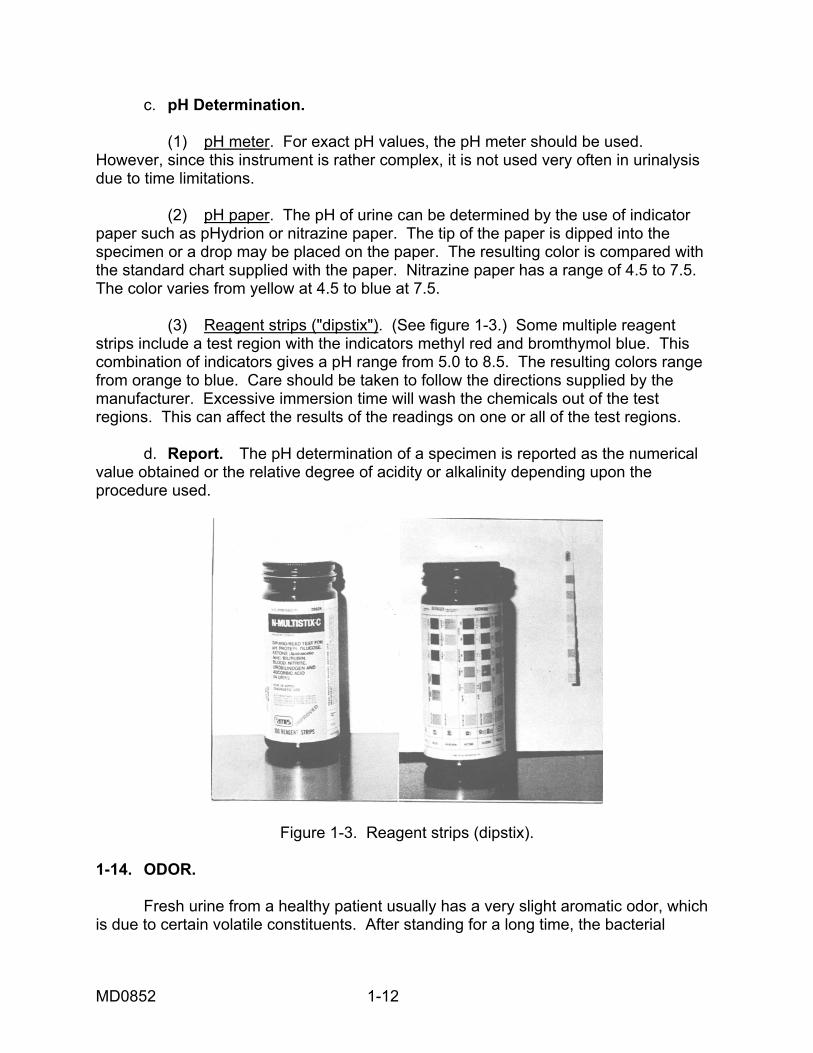
c. pH Determination. (1) pH meter. For exact pH values, the pH meter should be used. However, since this instrument is rather complex, it is not used very often in urinalysis due to time limitations. (2) pH paper. The pH of urine can be determined by the use of indicator paper such as pHydrion or nitrazine paper. The tip of the paper is dipped into the specimen or a drop may be placed on the paper. The resulting color is compared with the standard chart supplied with the paper. Nitrazine paper has a range of 4.5 to 7.5. The color varies from yellow at 4.5 to blue at 7.5. (3) Reagent strips ("dipstix"). (See figure 1-3.) Some multiple reagent strips include a test region with the indicators methyl red and bromthymol blue. This combination of indicators gives a pH range from 5.0 to 8.5. The resulting colors range from orange to blue. Care should be taken to follow the directions supplied by the manufacturer. Excessive immersion time will wash the chemicals out of the test regions. This can affect the results of the readings on one or all of the test regions. d. Report. The pH determination of a specimen is reported as the numerical value obtained or the relative degree of acidity or alkalinity depending upon the procedure used.
Figure 1-3. Reagent strips (dipstix). 1-14. ODOR. Fresh urine from a healthy patient usually has a very slight aromatic odor, which is due to certain volatile constituents. After standing for a long time, the bacterial
MD0852 1-12
decomposition of urea produces a characteristic odor of ammonia. The ingestion of certain foods (for example, asparagus) produces a characteristic odor. 1-15. FOAM A slight amount of foam is formed when normal urine is shaken. This foam is white. The presence of bile pigments in the urine usually produces a yellow foam, but the presence of certain chemicals or drugs (for example, phenylazodiaminopyridine) will also produce a yellow foam. Excess urine protein (proteinuria) causes a marked increase in the foaming quality of urine.
Continue with Exercises
MD0852 1-13
EXERCISES, LESSON 1 INSTRUCTIONS: Answer the following exercises by marking the lettered response that best answers the exercise, by completing the incomplete statement, or by writing the answer in the space provided at the end of the exercise. After you have completed all of the exercises, turn to "Solutions to Exercises " at the end of the lesson and check your answers. For each exercise answered incorrectly, reread the material referenced with the solution. 1. What is the clinical significance of urinalysis? a. Urinalysis can provide useful information on the patient's ability to produce volatile wastes. b. Urinalysis provides a good indication of the overall metabolic condition of the patient. c. Urinalysis serves as a means of evaluating the patient's state of health in every major system in his body. d. Urinalysis can provide the physician with specific information about the patient's state of health. 2. Select the statement that best describes a two-hour postprandial urine sample. a. This type of sample tends to reveal abnormalities in the patient's metabolism. b. This type of sample is collected two hours after an initial urine sample has been collected from the patient. c. This type of sample must be collected in a sterile container. d. This type of sample must be mixed with an appropriate preservative.
MD0852 1-14
3. Which statement best contrasts urine collection by the catheterization method and the midstream (clean catch) method? a. Catheterization is used more often than the midstream method to obtain urine specimens. b. The midstream method usually obtains specimens, which are sterile, while samples gathered by catheterization are usually contaminated. c. The urine collected by catheterization should be placed in a sterile container, while the urine collected by the midstream method should be collected in only a clean container. d. The midstream method is used more frequently than the catheterization method to collect urine. 4. Select the statement that best describes the preservation of urine by formalin (10 percent). a. This preservative is required when there is a need to preserve the urobilinogen in the sample. b. This preservative should not be used when the glucose concentration in the urine is to be determined. c. This preservative forms a thin layer on the top of the sample and acts as a physical barrier to air and bacteria. d. This preservative is required when the sample is to be analyzed for amino acids on total nitrogen. 5. Which of the following is the median amount of urine produced by an average adult during a 24-hour period? a. 1000 milliliters. b. 1250 milliliters. c. 1400 milliliters. d. 2000 milliliters.
MD0852 1-15
6. Select the meaning of the term "oliguria." a. An abnormal increase in the urine output during a 24 hour period. b. A reduction in the volume of urine excreted. c. A complete lack of urine production. d. A reduction in the total volume of urine caused by diabetes mellitus and/or diabetes insipidus. 7. Anuria means: a. A complete lack of urine excretion. b An abnormal reduction in the volume of urine excreted. c. The production of urine which contains excessive numbers of negative ions. d. The production of excessively concentrated urine. 8. A patient's urine sample is orange. Which of the following substance(s) could produce such orange-colored urine? [Note: More than one response may be correct.] a. Bile pigment. b. Carotene. c. Pyridium. d. All the above.
MD0852 1-16
9. A patient is very concerned because her urine is red. What substance could be the cause of such red-colored urine? [Note: More than one response may be correct.] a. Porphyrins. b. Pyridium. c. Melanin. d. All the above. 10. Which statement best describes the principle of the refractometer in the evaluation of urine specific gravity? a. Specific gravity compares the density of urine to the density of distilled water. b. This method is of little value in determining whether or not the patient has a pathological condition. c. Early morning urine samples should have a smaller specific gravity than samples taken in the afternoon. d. Little variation is seen in the specific gravity of random samples taken during the course of 24 hours. 11. Select the statement which best describes the evaluation of foam produced in urine. a. White foam is usually present in samples, which contain high levels of bile pigments. b. Proteinuria will produce a marked increase in the foaming quality of urine. c. Normal urine, even when shaken vigorously, should produce no foam. d. Yellow foam in urine is always a sign of a pathological condition in a patient.
Check Your Answers on Next Page
MD0852 1-17

MD0852 1-18
SOLUTIONS TO EXERCISES, LESSON 1 1. b (para 1-1) 2. a (para 1-4) 3. d (para 1-6) 4. b (para 1-7c) 5. c (para 1-9) 6. b (para 1-9b) 7. a (para 1-9c) 8. b c (para 1-10e) 9. a b (para 1-10 d, e) 10. a (para 1-12) 11. b (para 1-15)
End of Lesson 1
LESSON ASSIGNMENT LESSON 2 Chemical Tests for Substances in Urine. TEXT ASSSIGNMENT Paragraphs 2-1 through 2-22. LESSON OBJECTIVES After completing this lesson, you should be able to: 2-1. Select the statement that best describes the clinical significance of a particular type of chemical substance (that is protein, glucose, and
so forth, found in urine. 2-2. Select the average amount of protein detectable in a 24-hour sample of urine. 2-3. Select the statement that best differentiates between albumin and globulin. 2-4. Select the statement that best describes a type of proteinuria. 2-5. Select the statement that best describes a particular test or type of test for a chemical substance in urine. 2-6. Select the statement that best describes either diabetic or nondiabetic ketonuria. 2-7. Select the statement that best defines the following terms: hematuria, hemoglobinuria, and myoglobinuria. 2-8. Select the meaning of the term porphyrins. 2-9. Select the best description of the chemical analysis of calculi. 2-10. Select the chemical substance(s) that are often the components of calculi. SUGGESTION After studying the assignment, complete the exercises at the end of this lesson. These exercises will help you achieve the lesson objectives.
MD0852 2-1
LESSON 2
CHEMICAL TESTS FOR SUBSTANCES IN URINE
Section I. PROTEIN IN URINE 2-1. GENERAL COMMENTS In recent years, a number of advances have been made in the development of qualitative and semi-quantitative urinalysis tests. Many commercial reagents have been devised specifically for the rapid detection of certain chemical substances in urine (glucose, protein, acetone, bilirubin, blood, hemoglobin, and so forth). These procedures include tablets, papers, and reagent strips ("stix") which have been designed for particular analyses. Most of these tests require only a simple visual interpretation. The tests are based on chemical principles similar to the more lengthy classical tests performed in the laboratory. Since these preparations save both time and space, they are useful in routine screening procedures. These rapid tests are not without limitations. False-negative reactions can occur as the commercial preparation ages or deteriorates. Contamination from spilled specimens and carelessness are potential sources of error. It is recommended that a confirmatory test and control be used, particularly when a new product becomes available, or a new medical laboratory specialist is being trained. Finally, the tendency to use "sloppy" technique must be avoided. It is essential that you follow the manufacturers' instructions explicitly when performing any tests. Positive and negative controls should be set up to ensure that the reagents in use are satisfactory and that the techniques being used are correct. Doubtful results should be confirmed by additional testing. Neat, legible, and complete entries of examination results should be made on appropriate laboratory forms. Negative findings should be reported by recording the entire word, not just a symbol. 2-2. PROTEIN IN URINE (PROTEINURIA) a. Introduction. The occurrence of urinary proteins (proteinuria) is perhaps the best single indicator of a renal abnormality. For this reason, the qualitative test for protein is a useful screening procedure for the detection of renal abnormalities. Proteinuria does not ordinarily occur as the result of abnormalities or infections of the lower urinary tract. Consequently, when pus is present (pyuria) without proteinuria, it is reasonably certain that the pus originates in the lower urinary tract, and that the kidney is not involved. Although urinary protein usually indicates the presence of a renal lesion, it does not necessarily indicate a lesion of clinical importance. Benign or functional proteinuria may be caused by several factors, particularly temporary stress placed on the renal system. Functional proteinuria is often observed after strenuous exercise and can be caused by certain drugs such as epinephrine. On the other hand, pathological proteinuria resulting from disease or damage to the renal system is of great clinical significance and must be detected accurately.
MD0852 2-2
b. Normal Amount of Protein in the Glomerular Filtrate. Blood enters the kidney by means of the renal artery. As the blood reaches the glomerulus, it is filtered by the glomerulus, and the glomerular filtrate is formed. The glomerular filtrate normally has a small amount of protein, which is usually less than 30 mg per deciliter. When the glomerular filtrate passes through the tubules, most of the protein is reabsorbed and leaves the urine relatively free of protein. This small amount cannot be detected by the routine qualitative procedures. c. Average Amount Detectable in a 24-Hour Specimen. The average amount of protein detectable in a 24-hour specimen is 50 to 150 mg. The normal amount can range up to about 10 mg/dL in a random specimen. d. Protein Components. The two proteins of primary interest are albumin and globulin. (1) Albumin. Albumin has a molecular weight of approximately 69,000. Due to the size of the albumin particles, it is more readily filtered by the glomerulus into the filtrate. Therefore, albumin is the most common type of protein found in urine. The physician can determine the extent of damage to the kidney by knowing the amount of protein present in the urine and by knowing the type of protein present. Excessive albumin in urine is a condition called albuminuria. (2) Globulin. Globulin has a molecular weight of approximately 150,000. It is therefore a much larger molecule than albumin. When globulin is present consistently in excessive amounts, the condition is called globulinuria. e. Types of Proteinuria. As mentioned previously, persistent proteinuria is probably the most important and most frequent pathological change in urine. Proteinuria may be either accidental or renal, so all cases of proteinuria should not be regarded as indicative of renal disease. (1) Accidental proteinuria. This condition is also known as false proteinuria since it is not caused by kidney disease but to an admixture (a mixture) with the urine of albuminous types of fluids, such as pus, blood, or vaginal discharge. This type of proteinuria occurs most often in cases of pyelitis, cystitis, and chronic vaginitis. The quantity of albumin in such cases is usually very small. Severe bleeding, particularly in the lower urinary tract, causes protein to be found in urine. (2) Renal proteinuria. This condition exists when protein has passed from the blood into the urine through the walls of the kidney tubules or the glomeruli. Renal proteinuria may be due to one or more causes and is nearly always accompanied by tube casts.
MD0852 2-3
(a) Circulatory changes in the kidney. Congestion or anemia, such as occurs in chronic or severe heart disease, or any type of pressure on the renal veins may cause proteinuria. The amount of protein is usually small, and the presence is either constant or temporary, depending on the cause. Eclampsia is a condition of convulsive disorders found in pregnant women with accompanying high protenuria. (b) Renal disease. Persistent proteinuria is usually the result of renal disease that can cause degenerative organic changes in the kidney. Examples of such diseases are nephrosis, glomerulonephritis resulting from a streptococcal infection, and pyelonephritis produced by a bacterial infection. Renal tumors can also result in proteinuria. The amount of protein produced by these conditions varies from minute traces to 20 grams or more in a 24-hour period. 2-3. TESTS FOR PROTEINURIA a. Reagent Strips. Perhaps the most common method to detect protein in urine is the reagent strip system, which simultaneously tests a urine specimen for protein and other chemical constituents. This test depends on the fact that at a fixed pH certain indicators have one color in the presence of protein and another color in the absence of protein. The protein square on the reagent strip is impregnated with citrate buffers that maintain the pH on the square at 3.0. The indicator, tetrabromphenol blue, has a yellow color, but it becomes green to blue with the presence of increasing amounts of protein. The sensitivity of the test is about 20-30 mg of protein per deciliter of urine. Advantages of this test are that it does not give false-positive results with tolbutamide, x-ray contrast media, or other drugs. Some potential disadvantages include improper technique, a false-positive reaction from alkaline, highly buffered urine, and the fact that the test is not as sensitive to globulins as to albumin. b. Sulfosalicylic Acid Test. This is a semi-qualitative test, which is based on protein precipitation. The urine becomes cloudy with the addition of one part of three percent sulfosalicylic acid to one-part urine and results in the precipitation of urinary protein. The amount of protein is determined by the degree of turbidity and is semiquantified as "Trace, 1+, 2+, 3+, or 4+." The test is sensitive enough to disclose a protein concentration of 10 mg/dL of urine. False-positive tests may occur in patients who have recently taken the drug tolbutamide or organic iodine compounds used as x-ray contrast media. False-positive results may occur with highly buffered alkaline urine. c. Heat and Acetic Acid Test. The heat and acetic acid test is based on the fact that proteins are coagulated by heat. The acetic acid is added to dissolve precipitated phosphates and carbonates and to enhance the coagulation of protein. If the urine is not acid initially, the later addition of acetic acid may be insufficient to obtain heat coagulation of protein. If too much acid is added, traces of protein may be dissolved. In addition, mucin may give a false-positive test. Two milliliters of saturated sodium chloride solution should be added to the urine prior to boiling in order to avoid the mucin effect. Alternatively, perform a mucin test, filter, and test the filtrate.
MD0852 2-4
d. Quantitative Tests for 24-Hour Specimens. (1) Trichloroacetic acid test. The addition of trichloroacetic acid (TCA) to a urine specimen precipitates the protein in a fine suspension that is quantified photometrically at 420 nm (nanometers) by comparison with a similarly treated standard. (2) Kingsbury-Clark Test. Acetic acid is added to the specimen in order to clear the urine of phosphates. Sulfosalicylic acid is used to precipitate the protein. The resulting turbidity is read photometrically at 600 nm against a standard concentration. e. Testing for Bence-Jones Protein. The toluene sulfonic acid test followed by a heat precipitation test can be used to detect Bence-Jones protein. This abnormal protein is found in the urine of patients suffering from multiple myeloma, a disease characterized by neoplastic proliferation of plasma cells in the bone marrow and subsequently in the peripheral blood. (1) Toluene Sulfonic Acid Screening Test. The toluene sulfonic acid (TSA) reagent is comprised of 12 g of p-toluene sulfonic acid in 100 mL of acetic acid. This reagent precipitates Bence-Jones protein even in such small amounts as 0.3 mg per deciliter of urine. It does not precipitate albumin in much higher concentrations than 25 g per deciliter; it precipitates globulins in concentrations higher than 5 mg per deciliter of urine. To perform this test, one adds 1 ml of the reagent to 2 mL of urine. The reagent should be added slowly by allowing it to run down the side of the test tube. The tube is then "finger-flicked." If a precipitate occurs within 5 minutes, the test is positive for Bence-Jones protein. (2) Heat Precipitation Test. As the TSA method can occasionally produce both false-negative and false-positive tests, a heat precipitation test should also be performed. This test is based on the unique solubility pattern of Bence-Jones protein. The protein precipitates between 40ºC and 60ºC and redissolves when the temperature reaches 85º to 100ºC. It appears again when cooled to 60º to 85ºC. The test is performed by centrifuging fresh urine and then placing 10 mL in a fresh test tube. The specimen is adjusted to pH 5 by mixing with 25 percent acetic acid and then slowly heated in a water bath for 15 minutes. Temperature is monitored by placing a thermometer in the test tube. The formation of a precipitate at 60ºC indicates the presence of Bence-Jones protein. If a precipitate occurs over 60ºC, it is due to albumins and globulins. In order to separate Bence-Jones protein from albumin, the specimen is filtered at boiling temperature, thereby allowing albumin to be removed. Then the heat precipitation test for Bence-Jones protein is performed as described above.
MD0852 2-5
Section II. GLUCOSE AND OTHER REDUCING SUBSTANCES IN URINE 2-4. GLUCOSE AND OTHER REDUCING SUBSTANCES IN URINE a. Significance. The presence of excessive glucose in urine (glycosuria) caused by diseases such as diabetes and renal tuberculosis. While sugar in the urine is usually associated with diabetes, its presence may be indicative of other disorders. For example, lactose in urine may normally accompany pregnancy and lactation. Even glucose in urine may only indicate the ingestion of a high carbohydrate meal or the administration of an intravenous glucose solution. This does not alter the reporting of results but means that the diagnosis is determined by the physician. b. Renal Threshold for Glucose. The normal blood glucose level is 70 to 110 mg per deciliter. When the blood glucose level rises above the normal limits, the glucose concentration in the glomerular filtrate also rises. When the nephron tubules can no longer reabsorb the glucose, the renal threshold has been reached, and then the glucose "spills" over into the urine, indicating incomplete glucose metabolism. The maximum reabsorptive capacity of the tubules for glucose is approximately 160 to 170 mg per deciliter (mg/dl) of filtrate. 2-5. TESTS FOR URINARY SUGAR a. Glucose Oxidase Reagent Strip Test. There are two methods for testing the presence of urinary sugars. One of these, the reagent test strip, is specific for glucose; the other two methods are nonspecific. They test for reducing substances and not merely for glucose. The glucose test square on the strip contains the enzyme glucose oxidase, which reacts only with glucose. This enzyme forms gluconic acid and hydrogen peroxide in the presence of glucose. The paper strip must be dipped into a portion of the urine sample. After a minimum of 30 seconds, the reaction can be read. Various companies have these strips on the market and, for this reason, the colors produced may vary, depending on the product. b. Tests for Reducing Substances (Nonspecific). Benedict's test and the related tablet test indicate the presence of reducing substances and thus are not specific for glucose. Therefore, it is advisable to confirm the presence of glucose with a glucose oxidase reagent strip test. However, tests for reducing sugars may be useful in detecting metabolic disorders, such as galactosemia, especially when performed routinely on children. (1) Benedict's Test (Qualitative). One of the oldest methods for the detection of reducing substances is Benedict's test. In this method cupric sulfate in an alkaline solution is reduced to cuprous oxide by heating with glucose and other reducing agents. The diagnostic degree of reduction is indicated by the presence of a yellow to red precipitate.
MD0852 2-6
(2) Clinitest™ (trademark). Like Benedict's test, Clinitest™ is a copper reduction test and has largely superseded Benedict's test. It makes use of the same essential ingredients as Benedict's test, but these ingredients are combined into a tablet. Clinitest™ tablets have an ingredient (sodium hydroxide) that produces heat when mixed with the proper quantities of urine and water. The reaction and varying colors produced are the same as with Benedict's test. Clinitest™ is one of the most commonly used methods for detecting reducing substances.
Section III. KETONE BODIES IN URINE 2-6. KETONE BODIES IN URINE (KETONURIA) The three ketone bodies found in urine are acetoacetic acid (20 percent), beta-hydroxybutyric acid (78 percent), and acetone (2 percent). These ketone bodies are the product of incomplete fat metabolism, and their presence in urine indicates the possibility of acidosis. The increase of ketone bodies in the urine is called ketosis. a. Nondiabetic Ketonuria. Nondiabetic ketonuria is often due to the increased catabolism of adipose tissue when there is limited intake of food. Ketonuria is frequently seen in infants or children with acute febrile diseases or toxic states which produce vomiting or diarrhea. Ketonuria is also found when there is vomiting due to general ill health, pregnancy, or anesthesia. Other causes of ketonuria include the administration of a ketogenic diet to treat seizures in children, glycogen storage disease (Gierke's) and, occasionally, exposure to cold or severe exercise. b. Diabetic Ketonuria. Ketonuria in diabetics indicates ketosis (diabetic acidosis), the possibility of an impending coma, and other problems in the management of diabetes. Tests for ketonuria are often used to monitor diabetic patients using oral hypoglycemic drugs and patients undergoing a change in prescribed diabetic therapy. 2-7. TESTS FOR KETONE BODIES a. Reagent Strips. The reagent strip test is the simplest to perform as it takes only 15 seconds to react completely. The reagent strips are impregnated with the optimum concentration of nitroprusside, which reacts positively when dipped into urine or serum containing ketone bodies. The colors produced range from lavender to deep purple in the presence of ketone bodies. b. Acetest® Tablets (Brand Name). Acetest® tablets also provide a simple means of testing for ketone bodies in urine. A drop of the specimen is placed directly on the test tablet. If ketone bodies are present, the reaction of the urine or serum with the Acetest® tablet produces colors ranging from lavender to deep purple. The reaction should occur within 30 seconds. Acetest® tablets react positively to acetone and acetoacetic acid in urine or serum. As with the reagent strips, this reaction is based on the nitroprusside method for detecting ketone bodies.
MD0852 2-7
c. Lange's Method. Lange's test uses liquid reagents. Sodium nitroprusside reacts with acetone and acetoacetic acid in a buffered alkaline medium to produce a red to purple color. The colorimetric reaction takes place within two minutes. d. Sources of Error. All three of these urine ketone tests are based on a nitroprusside reaction. Consequently, if a patient has been taking large amounts of salicylates (such as aspirin) a false-positive reaction may result. The reagent strip is less sensitive to this false-positive reaction. e. Hart Test for Beta-Hydroxybutyric Acid. A separate test should be employed to detect beta-hydroxybutyric acid since it does not react with sodium nitroprusside. Basically the test involves the conversion of beta- hydroxybutyric acid to acetone, which can then be detected by the nitro- prusside method. First, 20 mL of urine is acidified with diluted acetic acid and then boiled until it becomes half of the original volume. This amount is cooled and then raised to the original volume with water. The purpose of this process is to remove the acetoacetic acid and acetone. The specimen is then divided into two portions and put into two test tubes. Next, 1 mL of hydrogen peroxide is added to the first portion, which is warmed gently and cooled. In this process, beta-hydroxybutyric acid is changed to acetone. Then ten drops of nitroprusside solution are added to both tubes and overlaid with ammonia. The presence of beta-hydroxybutyric acid is indicated by a purple-red color reaction that occurs in the sample treated with hydrogen peroxide.
Section IV. BLOOD IN URINE 2-8. BLOOD IN URINE a. The presence of blood in the urine can often be of great significance. For the detection of urinary blood, it is necessary to distinguish among hematuria, hemoglobinuria, and myoglobinuria. Hematuria is the presence of red blood cells in the urine and may indicate urinary tract bleeding or glomerular damage. Hemoglobinuria is the presence of dissolved hemoglobin in urine and can indicate the destruction of circulating red blood cells, as in malaria or transfusion reactions. Myoglobinuria, the presence of myoglobin in urine, colors the urine red or brown and results from rapid destruction of skeletal muscle. b. Hematuria is detected by microscopic examination. Hemoglobinuria and myoglobinuria are usually detected and differentiated by chemical means. 2-9. TESTS FOR BLOOD IN URINE a. Reagent Strips. Hemastix (brand name), a screening test for hemoglobin in urine, is often useful in addition to the microscopic examination for intact red blood cells. A paper strip is available for hemoglobin detection. The strip is impregnated with hydrogen peroxide, orthotolidine, and buffers. Hemoglobin and myoglobin catalyze the
MD0852 2-8
interaction of peroxide and orthotolidine, resulting in the oxidation of orthotolidine to produce a blue color. The reaction takes 30 seconds to go to completion. b. Blood Test Tablets. Occultest (brand name) reagent tablets, which are able to detect hemoglobin and myoglobin in any body fluid, are often used to test urine. In this test, as with the reagent strip test, the hemoglobin or myoglobin catalytically decomposes hydrogen peroxide, freeing oxygen to oxidize orthotolidine to a blue color. A tablet is placed on a piece of filter paper to which a drop of urine has been added. After two drops of water have been placed on the tablet, the filter paper is observed for the appearance of a blue color within 2 minutes. c. Benzidine and Hydrogen Peroxide Test. The benzidine and hydrogen peroxide test uses liquid reagents for the detection of hemoglobin and myoglobin in urine. Hemoglobin liberates oxygen from hydrogen peroxide. The released oxygen oxidizes benzidine to produce a green to blue color. This test is rarely used, however, since benzidine has been shown to have a carcinogenic effect. d. Sources of Error. In using both the reagent strip tests and the tablet tests, accurate timing is important. A false-positive reaction may occur after the specified reaction time has passed. This is due to auto- oxidation of the test reagent and may occur regardless of the presence of any additional chemicals in the urine. Large amounts of ascorbic acid in urine may inhibit the reaction of hemoglobin with the peroxide-orthotolidine system. Therefore, the reagent strip tests and the tablet tests should not be the only procedures performed, particularly if the patient is taking high doses of ascorbic acid. If a large amount of ascorbic acid is suspected, microscopy should be used. e. Differentiation Between Hemoglobinuria and Myoglobinuria. (1) Observe a fresh morning urine specimen or one voided after exercise. Urine with myoglobinuria is characteristically red when fresh and turns black upon standing. (2) Mix 1 mL of urine with 3 mL of 3 percent sulfosalicylic acid. Filter. If the pigment is precipitated, it is a protein. If the filtrate is a normal color, no abnormal nonprotein pigments, such as porphyrins, dyes, and drugs are present. (3) Dissolve 2.8 grams of ammonium sulfate in 5 mL of urine by mixing. Filter or centrifuge. A normally colored supernatant (overlying liquid) indicates that the precipitated pigment is hemoglobin. If the supernatant is colored, it is evidence of myoglobin. Diagnosis of myoglobinuria is usually based on the patient's history, and serum tests for enzymes elevated by muscle destruction.
MD0852 2-9
Section V. BILIRUBIN AND UROBILINOGEN IN URINE 2-10. BILIRUBIN IN URINE (BILIRUBINURIA) Bilirubin (bile pigment) is formed from the breakdown of hemoglobin by the reticuloendothelial cells of the spleen and bone marrow. Bilirubin passes from the blood to the liver, where it becomes water soluble, and into the bile ducts. It then enters the intestine with the bile. Normally, there is no bilirubin in the urine. However, bilirubin may appear in the urine in cases of hepatitis, in cirrhosis, and in other conditions where there is damage to liver cells. The bilirubin test can be used to differentiate between hemolytic jaundice and obstructive jaundice. In cases of obstructive jaundice urinary bilirubin is present; in cases of hemolytic jaundice bilirubin is characteristically absent from urine. Hence, urinary bilirubin is a useful indicator of the early phases of chemical or viral injury to the liver. 2-11. TESTS FOR BILIRUBIN IN URINE Urine should be tested for bilirubin within one hour of collection, since bilirubin is not stable and oxidizes, especially in light, to biliverdin. a. Foam Test for Bilirubin. The simplest test for bilirubin is the foam test. When urine is shaken, the foam is normally white. In the presence of bilirubin the foam is yellow or green-yellow. The results, however, should be confirmed by chemical tests. b. Reagent Strip Test (Diazotization Test). Ictostix (brand name), a convenient reagent strip test, has an area impregnated with stabilized diazotized 2-2, 4-dichloroaniline. With this test, a positive brown color results from 0.2 mg of bilirubin per deciliter of urine. c. Ictotest (Diazotization Test). Ictotest (brand name) is a reagent tablet containing stabilized p-nitrobenzene diazonium p-toluene sulfonate. In this method the bilirubin is coupled to the p-nitrobenzene diazonium p-toluene to produce a blue or purple color. The test is performed by placing ten drops of urine onto the center of an asbestos-cellulose mat, positioning a test tablet on the mat over the urine. Then, place one drop of water onto the tablet and a second drop after 5 seconds, so that water will run off the tablet onto the mat. The positive bluish-purple color develops on the mat within thirty seconds. A pink or red color is negative. The Ictotest is more sensitive than the reagent strip test and detects 0.05 to 0.1 mg of bilirubin per deciliter of urine. This test reacts positively to bilirubin; urobilin, other pigments, and urine constituents do not form a purple color. However, the diazotization tests must be performed on fresh urine as the tests do not react with bilirubin, which has been oxidized or hydrolyzed from exposure to light. In addition, ascorbic acid, chlorpromazine and phenazopyridine may interfere with the tests.
MD0852 2-10
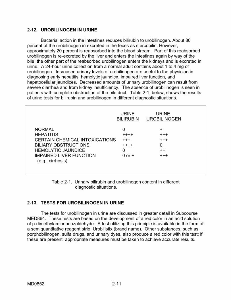
2-12. UROBILINOGEN IN URINE Bacterial action in the intestines reduces bilirubin to urobilinogen. About 80 percent of the urobilinogen in excreted in the feces as stercobilin. However, approximately 20 percent is reabsorbed into the blood stream. Part of this reabsorbed urobilinogen is re-excreted by the liver and enters the intestines again by way of the bile; the other part of the reabsorbed urobilinogen enters the kidneys and is excreted in urine. A 24-hour urine collection from a normal adult contains about 1 to 4 mg of urobilinogen. Increased urinary levels of urobilinogen are useful to the physician in diagnosing early hepatitis, hemolytic jaundice, impaired liver function, and hepatocellular jaundices. Decreased amounts of urinary urobilinogen can result from severe diarrhea and from kidney insufficiency. The absence of urobilinogen is seen in patients with complete obstruction of the bile duct. Table 2-1, below, shows the results of urine tests for bilirubin and urobilinogen in different diagnostic situations. URINE URINE BILIRUBIN UROBILINOGEN NORMAL 0 + HEPATITIS ++++ +++ CERTAIN CHEMICAL INTOXICATIONS +++ +++ BILIARY OBSTRUCTIONS ++++ 0 HEMOLYTIC JAUNDICE 0 ++ IMPAIRED LIVER FUNCTION 0 or + +++ (e.g., cirrhosis)
Table 2-1. Urinary bilirubin and urobilinogen content in different diagnostic situations. 2-13. TESTS FOR UROBILINOGEN IN URINE The tests for urobilinogen in urine are discussed in greater detail in Subcourse MED864. These tests are based on the development of a red color in an acid solution of p-dimethylaminobenzaldehyde. A test utilizing this principle is available in the form of a semiquantitative reagent strip, Urobilistix (brand name). Other substances, such as porphobilinogen, sulfa drugs, and urinary dyes, also produce a red color with this test; if these are present, appropriate measures must be taken to achieve accurate results.
MD0852 2-11
Section VI. CALCIUM IN URINE 2-14. CALCIUM IN URINE Calcium is one of the principal minerals of the bone. It also has an important role in blood coagulation, in maintaining a proper heartbeat rhythm, in adequate milk absorption, and in muscle contraction. Skeletal weight, the amount of dietary calcium, and endocrine factors influence the urinary output of calcium. The urinary output in adults on a normal diet is 50 to 400 mg of calcium per day. a. Increased Calcium in Urine. High concentrations of urinary calcium can occur in hyperparathyroidism, in osteolytic bone diseases (bone dissolution due to calcium loss), in osteoporosis (bone dimineralization), and bone tumors. Renal tubular disease and vitamin D intoxication can also produce an elevated calcium output. b. Decreased Calcium in Urine. Urinary calcium concentrations are usually low when serum calcium concentratiions are low. Low output occurs in hypoparathyroidism, in reduced calcium absorption, in steatorrhea (high concentration of fecal fats), and in vitamin D deficiency. 2-15. TESTING FOR CALCIUM IN URINE The ease of testing urine for calcium content has encouraged the administration of a rapid screening test for the detection of bone defects and hypoparathyroidism. a. Qualitative Test (Sulkowitch Test). The most common test for urine calcium is the Sulkowitch test, which measures the relative concentration of urinary calcium. This test is based on the precipitation of insoluble calcium oxalate at a pH where the calcium and magnesium phosphates are soluble. A mixture of one part urine and one part Sulkowitch reagent results in the precipitation of the calcium oxalate. The resulting turbidity indicates the rate of urinary calcium excretion. A hazy reaction with a moderate amount of precipitation is considered to be normal. A person whose urine shows little or no precipitation has decreased or negative calcium excretion; and opaque or milky turbidity indicates excessive calcium excretion. A patient undergoing this test should be on a low calcium diet for 72 hours prior to specimen collection in order to prevent undue dietary influence on the examination. A 24-hour specimen gives a better analysis than a random specimen. The Sulkowitch reagent used in this test is composed of the following ingredients: (1) 2.5 g oxalic acid. (2) 2.5 g ammonium oxalate. (3) 5.0 mL glacial acetic acid. (4) 150.0 mL distilled water.
MD0852 2-12
b. Quantitative Testing (Titration Determination). For quantitative determination, calcium can be titrated by ethylenediamine tetra-acetic acid (EDTA). However, in urinary calcium analysis, direct titration is not as valid as it is with serum calcium analysis, probably because of the high phosphate concentration in urine. For this reason, the oxalate of calcium must be precipitated first. The test involves the following procedures: (1) The urine is thoroughly mixed, and an aliquot of 10 mL is removed. This is acidified to a pH of 1.0 by a few drops of concentrated hydrochloric acid (use wide-range pH paper). (2) With occasional mixing, the portion that has been acidified is heated for 15 minutes to 60ºC. Then 1 mL of the heated mixture is placed in a conical centrifuge tube by pipet. Next, 0.2 mL of ten percent ammonium oxalate is added and mixed, followed by the addition of one-drop methyl red indicator (0.1 percent in alcohol). Next, five percent ammonium hydroxide is slowly added to produce an orange color. (3) The tube is placed in a boiling water bath for 20 minutes and then cooled to room temperature and centrifuged. (4) The supernatant fluid is decanted, and the centrifuge tube is inverted and drained. The resulting precipitate is dissolved in 0.5 mL 1N hydrochloric acid and 0.5 mL citrate sodium (0.05 mol/L) and then, using 10 mL water, is transferred quantitatively to a 50-mL Erlenmeyer flask. (5) Five drops of potassium hydroxide (8 mol/L) and the indicator Calver II (brand name) are added. The urine specimen can now be titrated with the EDTA solution, using a 5-mL buret with a calibration in 0.02 mL. The EDTA solution is prepared by dissolving 9.25 g EDTA in water and diluting to one liter. The standard can be titrated without the precipitation. The end-point of the urine-specimen titration is reached when the red color disappears, and a blue color remains. (6) The following calculations are used for determining quantitative calcium levels. Titration volume of sample X 10 = mg/dL calcium Titration of volume standard This formula is applied to 10 mg/dL (a standard). mg/dl calcium X 24-hour urine volume in mL = mg calcium/24 hr 100mL/dL
MD0852 2-13
Section VII. PORPHYRINS IN URINE (PORPHYRINURIA) 2-16. PORPHYRINS IN URINE (PORPHYRINURIA) a. Porphyrin Formation and Metabolism. Porphyrins are complex, cyclic compounds formed by the linkage of four pyrrole rings with methylene bridges. They are intermediaries in the synthesis of heme, which is part of hemoglobin, myoglobin, and several respiratory enzymes. This synthesis occurs in the long bones and in the liver. In this process glycine and succinyl CoA condense to form delta-aminolevulinic acid (ALA). ALA then condenses to form porphobilinogen (PBG). When two units of porphobilinogen join, porphyrin is formed. Porphyrin then condenses to form heme. SuCoA + Glycine-->+ ALA--> Porphobilinogen--> porphyrin--> Heme In a healthy individual, porphyrins are excreted in urine and feces mainly as coproporphyrin. Disorders involving disturbed porphyrin metabolism are called porphyrias. Such disorders are usually accompanied by porphyrinuria, the presence of excessive porphyrins in urine. In some cases, porphyrin precursors, such as porphobilinogen, may also be excreted in urine. b. Causes of Porphyrin Increases. Excessive urinary porphyrins can be the result of genetic disorders or can be caused by alcoholic cirrhosis of the liver, by anemias, and by intoxication, primarily from lead. (1) Congenital porphyria. Congenital porphyrias are due to one or more congenital metabolic defects. (2) Toxic conditions. Porphyrins may be found in increased amounts in various toxic conditions such as heavy metal poisoning. Porphyrin disturbances can also result from liver disease, from alcoholism, and from the use of barbiturates. c. Characteristics of Porphyrinuria. Urine which is a reddish color may be excreted in porphyrinuria. When urine samples turn black upon standing, it is also an indication that porphyrins may be present. 2-17. TESTS FOR PORPHYRINS AND PORPHYRIN PRECURSORS The basic tests applied to urine in detecting porphyrin disturbances involve: (1) a test for porphobilinogen, and (2) a test for delta-aminolevulinic acid, and (3) an ultraviolet light screening test in which porphyrins (coproporphyrin and uroporphyrin) emit a characteristic red fluorescence.
MD0852 2-14
a. Test for the Presence of Porphobilinogen. (1) Reagents. The following reagents are used in the performance of the test for the presence of porphobilinogen (PBG). (a) Ehrlich's reagent (Fisher's modification). (b) p-dimethylaminobenzaldehyde 0.7 g. (c) Distilled water (100 mL). (d) HCl, concentrated (150 mL). (e) Saturated, aqueous sodium acetate. (f) Chloroform. (g) n-Butanol. (2) Procedures. The test is based on the reaction of porphobilinogen with modified Ehrlich reagent to produce a red or pink color. A random urine specimen can be collected if testing for PBG alone; if testing for coproporphyrin and uroporphyrin, a 24-hour specimen is collected and placed in a brown bottle with 5 g of sodium carbonate. The specimen is refrigerated, and the pH is maintained in the range of 6.5 to 9.5. The first step in the procedure is to combine 2.5 mL of fresh urine with 2.5 mL of modified Ehrlich reagent and to shake the mixture for 30 seconds. Then 5 mL of a saturated solution of sodium acetate is added and mixed thoroughly. This solution is tested with pH paper and adjusted to pH 5.5. A pinkish color development indicates the presence of PBG occurs right after the addition of the modified Ehrlich reagent. If no pink color appears, PBG is not present, and the test should be discontinued. If a pink color does appear, the extraction procedure should be undertaken. For extraction, the test solution is combined with 5 mL of chloroform, shaken, and left standing for several moments. Two layers then separate out; the upper layer is water, and the lower layer is chloroform. PBG remains in the upper, aqueous layer. If the upper layer is red or pink and the lower layer is a light yellow-brown or colorless, the test is positive for porphobilinogen. The pink or red upper layer is then decanted and shaken with one-half volume of n-butanol. This solution then separates into an upper butanol layer and a lower aqueous layer. A pink or red color in the water layer is the result of PBG alone. A normal urine specimen has either no PBG present or only small traces. An increased amount is found in some congenital porphyrias and in the acquired porphyrias. The difficulty with the test is the interference of colors produced by other substances (aminosalicylic acid, phenothiazine, and phenazopyridine).
MD0852 2-15
b. Test for Presence of Delta-Aminolevulinic Acid. As previously mentioned, Delta-Aminolevulinic Acid (ALA) is a precursor of PBG and the porphyrins. If the conversion of ALA into PBG is inhibited by heavy metal intoxication, then ALA accumulates in the body fluids and is excreted in urine. Thus, an increased presence of urinary ALA is regarded as an index of lead poisoning. The test involves separating ALA from interfering substances by using ion- exchange resin columns. The specimen is first moved through a column to remove all PGB. The ALA is then isolated on a second column, through which impurities and other interfering materials can pass. After elution (separation by washing) from the column with sodium acetate solution, ALA is heated with acetylacetone to form a pyrrole analogous to porphobilinogen. This product gives a red color in reaction with the modified Ehrlich reagent. (1) Reagents. (a) Dowex 2-X8 resin (200-400 mesh). The resin is placed in water and washed until a clear supernatant is obtained. It is then suspended in several volumes of 3 mol/L sodium acetate and stirred for 30 minutes. When the resin settles, the supernatant is removed, and the same process is repeated. The resin is then washed a few times with five volumes of distilled water, and then stored as a slurry (thin mixture of water and fine, insoluble material) in water. (b) Dowex 50-X4 resin (200-400 mesh). The resin is placed in water and washed until the supernatant becomes clear. It is then left standing overnight in three volumes of 2mol/L sodium hydroxide. The supernatant is removed, and the resin is washed until an almost neutral supernatant appears. It is then washed twice with four volumes of 2 mol/L HCl and, using distilled water, is washed again several times. (c) Sodium hydroxide, 2 mol/L. Sodium hydroxide (80 g) is dissolved in water and diluted to one liter. (d) Sodium acetate, 3mol/L. Sodium acetate trihydrate (408.9 g) is dissolved in distilled water to form one liter. (e) Acetate buffer of pH 4.6. Sodium acetate trihydrate (136 g) is dissolved in 26 mL of concentrated HCl and, using glacial acetic acid, is diluted to 100 mL. (f) Hydrochloric acid (HCl), 4mol/L. Concentrated HCl (336 g) is diluted with water to form one liter. (g) Ehrlich reagent (modified). P-dimethylaminobenzaldehyde (6 g) is dissolved in 26 mL of concentrated HCl and, using glacial acetic acid, is diluted to 100 mL.
MD0852 2-16
(h) Acetylacetone. (i) Delta-aminolevulinic acid standards. The stock standard (0.001mol/L) is made by dissolving 13.1 mg of the amino acid in the acetate buffer to make 100 mL. The working standard is made by diluting 1.0 mL of the stock standard to a volume of 10.0 mL with the acetate buffer. (2) Procedure. A 24-hour sample is collected. It should be collected with enough HCl or acetic acid to have a pH below 7.0. It is refrigerated until needed. The urine pH is regulated to pH 6.0 - 7.0 immediately before use. The two columns are arranged so that the elute (separated material) from the Dowex-two column goes into the top of the Dowex-50 column, or so that the first column elute passes in small amounts to the second column, where it is collected. The columns are washed with five mL portions of water; the washing water is then thrown away. Next, 1 mL of urine is placed in the Dowex-two column to which three 4 mL portions of water are added. This elute drops into the Dowex-50 column or is changed to the Dowex-50 column as it is collected. The Dowex- two column is then either thrown away or kept for later analysis. The Dowex-50 column is washed with two 4 mL portions of water, which are subsequently thrown away. Next, the ALA is eluted (extracted) by the addition of 8 mL of 1 mol/L sodium acetate, prepared by a dilution of the 3 mol/L solution. This elute is collected in either a 10 mL volumetric flask or a graduated test tube of 10 mL. Seven milliliters of 1 mol/L sodium acetate and 1 mL of working standard are placed in a separate flask. Then, 0.2 mL of acetyl-acetone is added to each tube and diluted to the mark by means of the acetate buffer. The flasks or tubes are covered and heated in a boiling water bath for 10 minutes. Two-milliliter aliquots of the sample that have been treated and the standard are then transferred to test tubes where 2 mL of modified Ehrlich reagent are added and mixed. The tubes are left standing for 15 minutes to permit color development. Then the standard and the unknown (sample) are read against the blank at 553 nm. The blank contains equal portions of acetate buffer and Ehrlich reagent. (3) Calculations. The following calculations are performed: Absorbance of sample X 1 = umo1/m1 ALA. Absorbance of standard umo1/mL X 131 - ug/mL. umo1/mL (or ug) X 24-hr urine vol in mL = umo1 (or ug/24 hr). (a) Absorbance refers to the capacity of a substance to absorb radiant energy. Absorption spectroscopy is concerned with the fact that molecules placed in a light path absorb particular wavelengths of light. Therefore, a substance has a high absorbance reading if it absorbs more of the light at a given wavelength. As the color
MD0852 2-17
formed in a sample may depend on factors other than the substance alone, a standard of known concentration is often run at the same time as the unknown sample. In addition, a blank, composed of the reagents without the substance under investigation, is prepared with the absorbance reading adjusted to zero. The reagents are used in the blank so that their absorbance is not confused with that of the substance being determined. For mathematical details, refer to MD0862, Clinical Chemistry II. (b) A mole (mol) is the amount of a substance whose weight in grams is such that the number of grams is equal to its molecular weight. The symbol for "micro" (one-millionth) is µ. Therefore, µmol signifies micromole or one-millionth of a mole, and µg signifies microgram or one-millionth of a gram. c. Screening Test for Porphyrins (Coproporphyrins and Uroporphyrins). The reagents used in the test are ethyl ether and ethyl acetate. Ethyl ether extracts coproporphyrins from urine, and ethyl acetate extracts uroporphyrins. The procedure involves acidifying 100 mL of urine with 10 mL of glacial acetic acid, mixing, and allowing to stand overnight. (1) With coproporphyrins, the extraction of the acetic acid-urine mixture is performed three times with two or three times the volume of ethyl ether by means of a separatory funnel. The ether extracts are then combined and, using 50 mL of distilled water, are washed once. The water is returned to the original urine sample, and the ether is then extracted three times with 2 mL of 25 percent aqueous HCl. Next, the acid extracts are combined and examined under long wavelength ultraviolet light. The appearance of a red fluorescence indicates the presence of coproporphyrins. (2) For uroporphyrins, the acidity of the acetic acid-urine mixture is adjusted to pH 3.0 by adding one percent aqueous HCl. The urine is extracted three times, using one to two times the volume of ethyl acetate; the extracts are then combined and washed with 50 mL of distilled water. Next, the ethyl acetate extracts are extracted three times with 2 mL of HCl. The acid extracts are combined, and the long wavelength ultraviolet light examination is applied. If a red fluorescence appears, uroporphyrins are present.
Section VIII. MISCELLANEOUS TESTS 2-18. PHENYLKETONURIA AND SCREENING TEST Phenylketonuria (PKU) is a hereditary, metabolic disorder characterized by the presence of phenylpyruvic acid in the urine. It results from a defect in converting the amino acid phenylalanine into the amino acid tyrosine. As a result of this defect, phenylalanine accumulates in the blood. If this condition is not detected in time and treated by diet, it results in serious mental retardation. Since a portion of the
MD0852 2-18
phenylalanine is metabolized to phenylpyruvic acid and excreted in the urine, PKU can be detected through a urine screening test or a serum screening test. A reagent strip, Phenistix® (brand name), is available for urine testing. This strip contains ferric ions that react with phenylpyruvic acid to give a gray to gray-green color for a positive reaction. By the age of 4 weeks, often earlier, intermediate metabolites of phenylalanine, particularly phenylpyruvic acid, begin appearing in infant's urine. 2-19. CYSTINE IN URINE (CYSTINURIA) AND SCREENING TEST Cystinuria, the presence of increased amounts of cystine in the urine, is a congenital defect in which cystine and other diamine amino acids are not reabsorbed by the renal tubules. This causes an increase in cystine concentration in the urine. As cystine is involved in the formation of renal calculi (stones), this condition can cause severe kidney damage. Screening tests can be used to detect urinary cystines, but the results should be confirmed by amino acid chromatography. The test is based on the reaction of nitroprusside with sulfhydryl groups after the cyanide reduction of cystine. A deep red color is positive. Normal urine may show a pale pink if small amounts of cystine are present. 2-20. TESTS OF RENAL FUNCTION a. Concentration Test. The concentration test measures the ability of the kidney to vary the concentration and volume of urine according to the food and fluid intake. The concentration test is very simple to perform since it is necessary only to determine the specific gravity and measure the volume. Strict adherence to the dietary instructions and time intervals of specimen collections are required. Normal results will be indicated by a night urine of lesser volume and higher specific gravity than the total of the day specimens. The total night volume should not exceed 500 mL and the specific gravity should be at least 1.018. The difference between the lowest and highest specific gravities of the day should approach 0.009. After food intake, an increased volume and lower specific gravity should be observed. Impaired renal function will be indicated by the inability of the kidneys to concentrate urine at appropriate times. Marked kidney impairment is characterized by the excretion of urine with a specific gravity that is consistently about 1.010. b. Phenolsulfonphthalein Test. Phenosulfonphthalein (PSP) (phenol red) is a dye that is readily removed from the blood and excreted by healthy kidneys. Normally about 70 percent is excreted by the kidneys. As this dye is not naturally present in the body and must be infused into the blood stream, the PSP test is an exogenous (external) clearance test. It differs from the endogenous (internal) method that determines renal excretion of substances that occur naturally in the body. The PSP test is primarily a test of tubular secretion. In addition, the rate of PSP excretion depends on the state of renal blood flow. Thus, this test gives an indication of the excretory state of the kidneys. A diminished dye excretion usually indicates impaired renal circulation. Normal kidneys excrete at least 25 percent of the dye in 15 minutes, and a total of at least 65 percent of the dye in 2 hours. The rate of excretion during the first 15 minutes
MD0852 2-19
is the most sensitive indicator of renal blood flow. The test is especially useful in detecting early renal disease. Other factors may also reduce PSP excretion. For example, the use of certain drugs, such as penicillin, and congestive heart failure will result in decreased PSP excretion. Normally, a small amount of PSP is also removed by the liver and then excreted in the bile. However, if the liver is damaged, more dye is excreted by the kidneys. Therefore, an exceptionally high PSP concentration may occur in the urine of patients with liver disorders. Because of health risks involved using phenosulfonphthalein dye, the PSP test is very rarely used clinically. c. Creatinine Clearance Test. (1) Creatinine formation and value in testing. Creatinine is formed in the muscles from creatine, a nitrogenous compound synthesized from glycine and parts of two other amino acids. It is produced from either non-stable creatine phosphate through the loss of H3PO4 (phosphoric acid) or from stable creatine through the loss of water. Creatinine is removed by glomerular filtration and, in cases of raised serum concentrations, by the tubules. It is then excreted in the urine without being reabsorbed by the tubules to any significant degree. In addition, creatinine production does not reflect a high protein diet or urine volume, instead it relates more closely to muscle mass or bodily creatine content. These two facts make the creatinine clearance test a valuable aid in revealing renal disease. The creatinine clearance test measures the amount of plasma which could be theoretically cleared of creatinine per minute by both kidneys. Unlike the PSP test, the creatinine clearance test is an endogenous test as it determines renal clearance of a substance occurring naturally in the body. (2) Basic test procedure. Serum and urine creatinine concentratons are compared in order to determine the glomerular filtration rate. Basically, the technique involves hydrating the patient with 600 mL of water and then collecting timed urine specimens of 4, 12 or 24 hours. The 24-hour collection is preferred. A blood specimen should be collected midway through the urine collection period. The serum and urine creatinine concentrations are then quantified by a method based upon Jaffe's reaction. This reaction occurs between creatinine and an alkaline picrate solution to form a red tautomer of creatinine picrate. The intensity of the red color is proportional to the creatinine concentration and is measured spectrophotometrically at 520nm. (3) Calculations. The following calculations are used: U X V X 1.73 = mL of plasma cleared by kidneys per minute. XP A where: U is the urine creatinine concentration (as mg/dL). V is the volume of urine (as mL/min). P is the serum creatinine concentration (as mg/dL). A is the patient's body surface area (as square meters). 1.73 is the generally accepted average body surface area.
MD0852 2-20
The normal urine clearance ranges from 85 to 125 mL per minute. Incorrect timing or improper collection causes most errors in the creatinine clearance determination. Body surface area may be determined by a formula or, most often, it can be determined from a nomogram found in standard texts. The formula for body surface area is: logA = (0.425 logW) + (0.725 logH) - 2.144. where: A is the body surface area in square meters. W is the patient's weight in kilograms. H is the patient's height in centimeters. log signifies logarithm.
Section IX. URINARY CALCULI 2-21. URINARY CALCULI a. Formation of Calculi. A calculus, commonly called a "stone" is a solid chemical deposit formed in the body in such places as the urinary tract, gallbladder, salivary glands, pancreas, prostate, and tonsils. Calculi may be composed of both organic and inorganic compounds. A careful laboratory analysis of calculi can aid the physician in prescribing certain drugs or dietary foods to avoid formation of more calculi. Some small stones may be passed by urination. Large stones are removed by surgery. Pain and tenderness are common symptoms of calculi. The cause of calculi formation is not precisely known, although it may be related to the insolubility of various crystalloids found in excretions and secretions. Internal factors contributing to calculi formation are urinary obstructions, infections, mucosal metaplasia (vitamin A deficiency), metabolic disorders (gout, cystinuria), and endocrinopathies, such as hyperparathyroidism. Calculi formation is also linked to various external conditions, such as dehydration, dietary excess of certain substances, drug excess, and chemotherapy. Isohydruria, a condition in which the urine pH remains fixed, also promotes the formation of calculi. Isohydruria encourages calculi deposition because certain chemical compounds require a specific pH for dissolution. Since they are not able to dissolve in a urine with an invariable pH, they tend to precipitate out as compact deposits. b. Calculi Composition. Calculi "grow" from core nuclei, which may be formed from bacteria, epithelial cells, blood clots, or precipitated salts from a supersaturated urine. Calculi growth depends on the pH of the urine, the solubility of each substance, and the availability of the different salts. The composition also relates to the location of the calculus. Bladder calculi are usually composed of uric acid whereas kidney calculi contain calcium oxalate, triple phosphate, or apatite, a complex calcium phosphate that may contain carbonate. Biliary calculi (gallstones) may be composed of cholesterol, bile pigments, calcium, phosphate carbonate, and trace amounts of iron, copper,
MD0852 2-21
magnesium, manganese, and organic matter. About 30 to 40 percent of calculi are pure; they are formed from a single compound. These calculi are sometimes called "pseudo" stones. Calculi may also be made up of a mixture of compounds. Such calculi are termed "true" stones. Generally, prolonged isohydruria encourages the formation of pure calculi, and a varying pH is associated with calculi of mixed composition. Calculi may also be classified according to the manner in which they develop. There are two basic patterns of formation: (1) Concretionary. This type of calculus grows outward from a discrete nucleus with a structure of concentric laminations. It may also have radial striations. (2) Sedimentary. The sedimentary calculus grows or crystallizes from multiple nuclei. If this type of calculus incorporates sufficient organic material, it may stop growing while still small. 2-22. METHODS OF CALCULI ANALYSIS Calculi are analyzed by three basic methods: chemical analysis, x-ray diffraction, and infrared spectrophotometry. All of these methods, more or less, show agreement with respect to results. Chemical analysis was formerly the method of choice; however, more and more laboratories are currently employing infrared spectrophotometry as the preferred procedure for calculi analysis. A brief account of these three methods is given below. a. Chemical Analysis. A preliminary examination of physical appearance is performed before the chemical analysis. After the calculus is washed clean of any blood clots, fibrin, or preservatives with deionized water, it is allowed to dry at room temperature for 24 hours. It is then described as to weight in milligrams, size in millimeters, shape, color, consistency, and surface appearance. The color, surface appearance, and consistency are quite useful since they are closely related to the types of compounds in the calculus. In addition, accurate reporting of physical characteristics is helpful in confirming the results of the chemical tests. Table 2-2 lists some of the physical characteristics of calculi. After noting physical characteristics, the calculus is crushed, and a small portion is used for flame analysis. By observing the response of the specimen to the heat of a flame, a general idea of the constituents in the calculus can be formed. Generally, inorganic salts are present if the specimen does not burn. If the specimen burns almost completely, organic materials are present. Various qualitative chemical tests are then performed to determine the composition of the calculi. Kits with procedures for the chemical analysis of calculi are available. Since this method requires no special or expensive equipment and uses easily obtainable reagents, it is relatively easy to utilize. However, as a thorough description of the chemical method is rather lengthy and as infrared spectrophotometry has largely replaced chemical analysis, an account of the various chemical tests is not given here. b. X-ray Diffraction. Analysis by x-ray diffraction has the advantage of giving quantitative results. However, as the instruments involved are expensive, this
MD0852 2-22
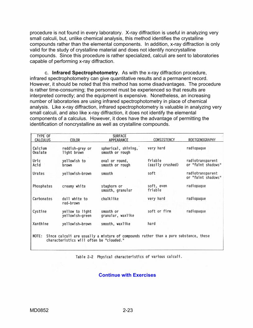
procedure is not found in every laboratory. X-ray diffraction is useful in analyzing very small calculi, but, unlike chemical analysis, this method identifies the crystalline compounds rather than the elemental components. In addition, x-ray diffraction is only valid for the study of crystalline material and does not identify noncrystalline compounds. Since this procedure is rather specialized, calculi are sent to laboratories capable of performing x-ray diffraction. c. Infrared Spectrophotometry. As with the x-ray diffraction procedure, infrared spectrophotometry can give quantitative results and a permanent record. However, it should be noted that this method has some disadvantages. The procedure is rather time-consuming; the personnel must be experienced so that results are interpreted correctly; and the equipment is expensive. Nonetheless, an increasing number of laboratories are using infrared spectrophotometry in place of chemical analysis. Like x-ray diffraction, infrared spectrophotometry is valuable in analyzing very small calculi, and also like x-ray diffraction, it does not identify the elemental components of a calculus. However, it does have the advantage of permitting the identification of noncrystalline as well as crystalline compounds.
Continue with Exercises
MD0852 2-23
EXERCISES, LESSON 2 INSTRUCTIONS. Answer the following exercises by marking the lettered response that best answers the exercise, by completing the incomplete statement, or by writing the answer in the space provided at the end of the exercise. After you have completed all of these exercises, turn to "Solutions to Exercises" at the end of the lesson and check your answers. For each exercise answered incorrectly, reread the material referenced with the solution. 1. Which statement best describes the clinical significance of protein in the urine? a. Any presence of protein in the urine is a clear sign of severe kidney damage. b. Persistent proteinuria is usually the result of such diseases as nephrosis or pyelonephritis. c. A large amount of protein in the urine is always found in congestion and anemia. d. A large amount of protein in the urine is indicative of kidney irritation. 2. What is the clinical significance of increased calcium concentrations in the urine? a. High concentrations of calcium in the urine are indicative of steatorrhea. b. High concentrations of calcium in the urine are indicative of hyperparathyroidism. c. High concentrations of calcium in the urine indicate hypoparathyroidism. d. High concentrations of calcium in the urine are found in severe vitamin D deficiencies.
MD0852 2-24
3. Which of the statements below best describe the clinical significance of excessive porphyrins in the urine? a. This condition can result from alcoholic cirrhosis of the liver. b. This condition can result from lead intoxication. c. This condition can result from use of barbiturates. d. All the above. 4. The average amount of protein detectable in a 24-hour sample of urine is a 5-15 milligrams. b. 25-75 milligrams. c 50-150 milligrams. d. 150-250 milligrams. 5. Accidental proteinuria is best described as a. A condition which exists when the blood accidentally passes into the urine through the walls of the kidney tubules. b. A condition caused by an admixture with the urine of albuminous types of fluids. c. A condition caused by congestion or anemia which is often seen in chronic heart disease. d. A condition found in cases of degenerative organic changes in the kidney.
MD0852 2-25
6. Which statement best describes diabetic ketonuria? a. Ketonuria in diabetics indicates the possibility of impending coma. b. Ketonuria in diabetics is often due to increased catabolism of adipose tissue when there is limited intake of food. c. Ketonuria in diabetics is usually associated with pregnancy and general ill health. d. Diabetic ketonuria is found in patients who are following a ketogenic diet. 7. Select the statement, which best describes the Reagent Strip test for bilirubin. a. A bluish-purple color reflects a 0.1 to 0.05 mg of bilirubin per deciliter of urine. b. With this test, a pink or red color is negative. c. A brown color results from 0.2 mg of bilirubin per deciliter of urine. d. This test is based on the development of a red color in an acid solution of p-dimethylaminobenzaldehyde. 8. The Phenistix test is best described by which of the following statements? a. This test shows positive results when a brown color develops on the test strip. b. Gray to gray-green color development on the test strip means the child has phenylketonuria. c. A positive result (red to pink color on the strip) means the child has excessive concentrations of phenolsulfonphthalein. d. This test strip contains calcium ions which react with the phenylalanine in the patient's urine.
MD0852 2-26
9. Select the statement which best describes the trichloroacetic acid test for proteinuria. a. This is a qualitative test for protein in the urine. b. This test precipitates the protein and requires you to quantify the sample photometrically. c. This test is used specifically to determine Bence-Jones protein in urine samples. d. This semi-qualitative test is based on the precipitation of protein in the urine. 10. Myoglobinuria is defined as a. A condition that indicates urinary tract bleeding or glomerular damage. b. A condition that indicates the destruction of circulating red blood cells. c. A condition that results from rapid destruction of skeletal muscle. d. The presence of red blood cells in the urine. 11. Porphyrins are best defined as a. Substances that are excreted in the form of porphobilinogen in the feces and urine of healthy persons. b. Substances that are synthesized in the long bones and the liver to form hemoglobin. c. Complex compounds which are derived from the breakdown of red blood cells, which indicate liver damage if they are present in the urine. d. Complex cyclic compounds which are intermediaries in the synthesis of heme.
MD0852 2-27
12. Which of the following are often found as components of calculi? a. Uric acid. b. Carbonates. c. Xanthine. d. All the above.
Check Your Answers on Next Page
MD0852 2-28

MD0852 2-29
SOLUTIONS TO EXERCISES, LESSON 2 1. b (para 2-2e(2)(b)) 2. b (para 2-14a) 3. d (para 2-16b) 4. c (para 2-2c) 5. b (para 2-2e(1)) 6. a (para 2-6b) 7. c (para 2-11b) 8. b (para 2-18) 9. b (para 2-3d(1) ) (Obj 2-5) 10. c (para 2-8a) 11. d (para 2-16a) 12. d (Table 2-2)
End of Lesson 2
LESSON ASSIGNMENT LESSON 3 The Microscopic Examination of Urinary Sediment. TEXT ASSIGNMENT Paragraphs 3-1 through 3-25. LESSON OBJECTIVES After completing this lesson, you should be able to: 3-1. Select the statement that best describes the
clinical importance of microscopic examination of urinary sediment.
3-2. Select the statement that best describes the
basic technique that should be used to prepare the urine sample for microscopic examination.
3-3. Select the statement that best describes the use
of the microscope in the microscopic examination of urine sediment.
3-4. Select the principal organized structure(s) found
in urine sediment.
3-5. Select the statement that best describes the type of sediment or its clinical significance.
3-6. Select the type of sediment that best describes a
description of sediment observed in the microscopic examination of a urine sample.
3-7. Select the name of the sediment, commonly
found in urine, as shown in an illustration.
3-8. Select the statement that best describes the significance of the three-bottle urine sample in terms of sample collection.
3-9. Select the best definition of the term cast. SUGGESTION After studying the assignment, complete them
exercises at the end of this lesson. These exercises will help you to achieve the lesson objectives.
MD0852 3-1
LESSON 3
THE MICROSCOPIC EXAMINATION OF URINARY SEDIMENT
Section I. REPARATION AND ILLUMINATION 3-1. REPARATION OF URINE SEDIMENT FOR EXAMINATION (UNSTAINED) A properly performed microscopic examination of urinary sediment can provide valuable information that enables the physician to diagnose renal as well as other abnormalities. The importance of this procedure, which has been compared to a biopsy with respect to its significance, cannot be overemphasized. Thus it is crucial that the many structures occurring in urinary sediment be identified correctly and that the diagnostically significant elements be distinguished from extraneous substances. 3-2. BASIC TECHNIQUE a. The urine sample should be examined macroscopically and checked for cloudiness, color, possible blood, and a syrupy consistency that may indicate mucus and casts. Such an examination can provide clues regarding the nature of the sediment. b. The specimen should be thoroughly mixed to ensure proper dispersal of constituents. Twelve to 15 mL of the specimen is placed in a conical centrifuge tube and centrifuged at 1500 rpm (revolutions per minute) for 5 minutes. c. The centrifuged tube is then inverted into a clean test tube. The supernatant urine is saved for chemical testing; the sediment that remains in the bottom of the tube is examined microscopically. d. Next, the sediment is resuspended by "finger-flicking" the tube. Then, one drop of sediment is transferred to a clean, dry glass slide by a pipet or a dropper, and a cover slip is applied. e. Next, the sediment is scanned by using the low power (10X) objective and is examined in detail by using the high power (43X) objective. f. It should be noted that the appearance of the urine does not necessarily correlate with the results of a microscopic analysis. Clear, normal-appearing urine may often reveal abnormal elements of diagnostic importance upon microscopic examination. Urine that gives only slight sediment after centrifugation may contain important structures; on the other hand, cloudy urine that contains heavy sediment may not disclose any clinically significant elements.
MD0852 3-2
3-3. ILLUMINATION OF MICROSCOPIC FIELD a. Light Intensity. The technique for use of the microscope must be changed somewhat from ordinary practice due to the transparent nature of urine sediment. Since many of the structures to be examined are hyaline (semi-transparent) in nature, the light should be subdued for ordinary work. Before the structures can be seen and identified, the intensity of light must be reduced to a minimum. The light intensity is reduced by practically closing the iris diaphragm of the microscope. b. Scanning and Reporting. The drop should be scanned under the low power objective and with moderate light. Low power is used because the low power objective covers a larger field and thus allows a more rapid scanning of the total preparation. Certain large structures such as parasite ova can be spotted using low power. Due to their large size, casts are also reported as the average number per low power fields. Smaller elements are reported in terms of average number per high field. When switching the objectives from low to high power, the condenser should be raised slightly to bring the light to its former intensity. Red blood cells, white blood cells, and epithelial cells are reported as the average number per high power field, counting 10 fields (for example, 15-19 rbc/HPF). If very few or very many cells are seen, descriptive adjectives are used, for example, "few," "occasional," "too numerous to count." Crystals should be reported when they are observed. However, an actual count is unnecessary; a report of the type of crystal and an indication of relative occurrence, such as "few," "many," "and so forth," is sufficient. If bacteria are seen in a fresh specimen, they are reported as present. Bacteria and epithelial cells are less significant in urine from females than in urine from males, but awareness and reporting of their presence in all specimens is a safe procedure. 3-4. COMMON SOURCES OF ERROR a. Delayed Analysis. The urine must be examined while still fresh. As the nature of the sediment changes with the passage of time, the analysis should be performed within 2 hours after voiding. If immediate examination is not possible, the specimen should be refrigerated or preserved with formalin. b. Improper Illumination. This is the most common error in the microscopic analysis of urine. As mentioned previously, subdued light is necessary so that hyaline semi-transparent structures are not obscured by intense illumination. c. Improper Placement of Sediment on Slide. Placing drops of sediment from too many patients on the same slide is another frequent source of error. As a result, the drops tend to run together. In order to avoid this problem, a slide should not contain sediment from more than two patients.
MD0852 3-3
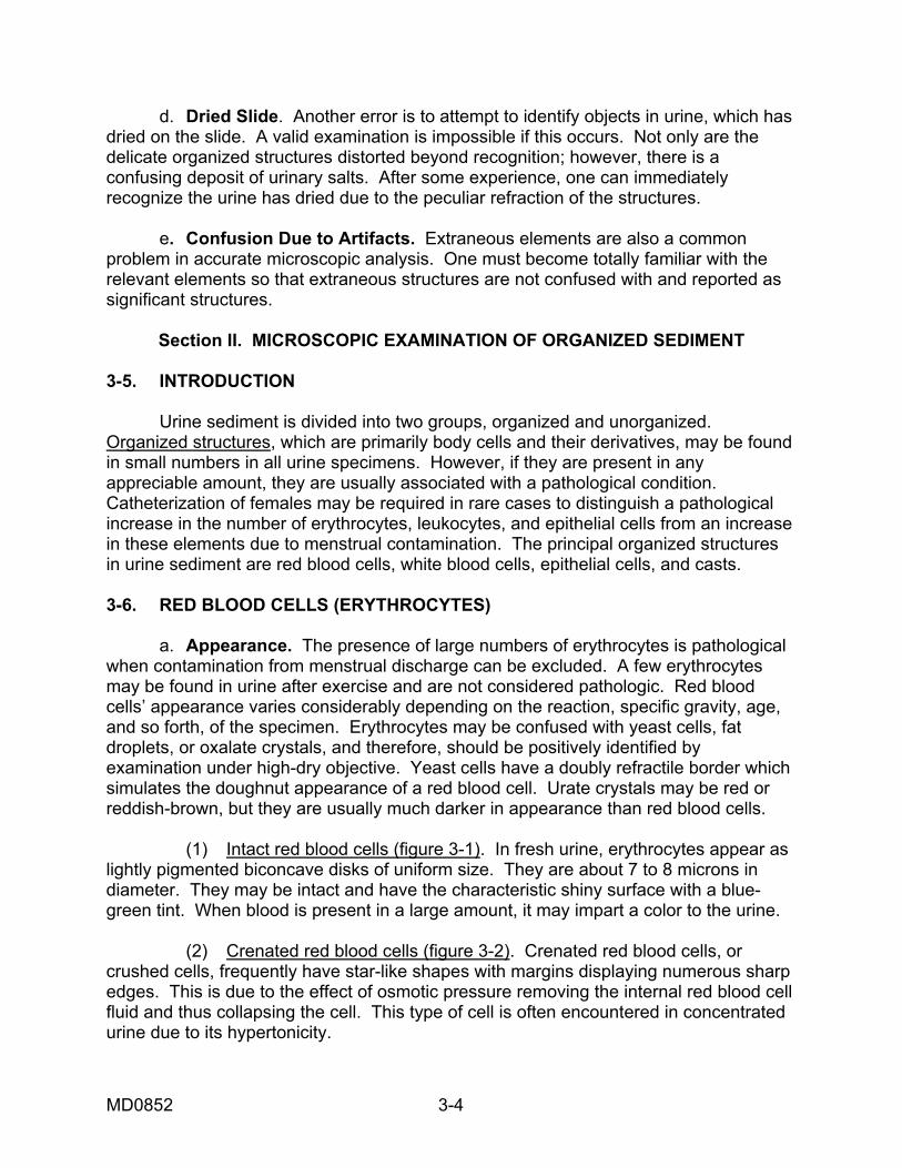
d. Dried Slide. Another error is to attempt to identify objects in urine, which has dried on the slide. A valid examination is impossible if this occurs. Not only are the delicate organized structures distorted beyond recognition; however, there is a confusing deposit of urinary salts. After some experience, one can immediately recognize the urine has dried due to the peculiar refraction of the structures. e. Confusion Due to Artifacts. Extraneous elements are also a common problem in accurate microscopic analysis. One must become totally familiar with the relevant elements so that extraneous structures are not confused with and reported as significant structures.
Section II. MICROSCOPIC EXAMINATION OF ORGANIZED SEDIMENT 3-5. INTRODUCTION Urine sediment is divided into two groups, organized and unorganized. Organized structures, which are primarily body cells and their derivatives, may be found in small numbers in all urine specimens. However, if they are present in any appreciable amount, they are usually associated with a pathological condition. Catheterization of females may be required in rare cases to distinguish a pathological increase in the number of erythrocytes, leukocytes, and epithelial cells from an increase in these elements due to menstrual contamination. The principal organized structures in urine sediment are red blood cells, white blood cells, epithelial cells, and casts. 3-6. RED BLOOD CELLS (ERYTHROCYTES) a. Appearance. The presence of large numbers of erythrocytes is pathological when contamination from menstrual discharge can be excluded. A few erythrocytes may be found in urine after exercise and are not considered pathologic. Red blood cells’ appearance varies considerably depending on the reaction, specific gravity, age, and so forth, of the specimen. Erythrocytes may be confused with yeast cells, fat droplets, or oxalate crystals, and therefore, should be positively identified by examination under high-dry objective. Yeast cells have a doubly refractile border which simulates the doughnut appearance of a red blood cell. Urate crystals may be red or reddish-brown, but they are usually much darker in appearance than red blood cells. (1) Intact red blood cells (figure 3-1). In fresh urine, erythrocytes appear as lightly pigmented biconcave disks of uniform size. They are about 7 to 8 microns in diameter. They may be intact and have the characteristic shiny surface with a blue-green tint. When blood is present in a large amount, it may impart a color to the urine. (2) Crenated red blood cells (figure 3-2). Crenated red blood cells, or crushed cells, frequently have star-like shapes with margins displaying numerous sharp edges. This is due to the effect of osmotic pressure removing the internal red blood cell fluid and thus collapsing the cell. This type of cell is often encountered in concentrated urine due to its hypertonicity.
MD0852 3-4
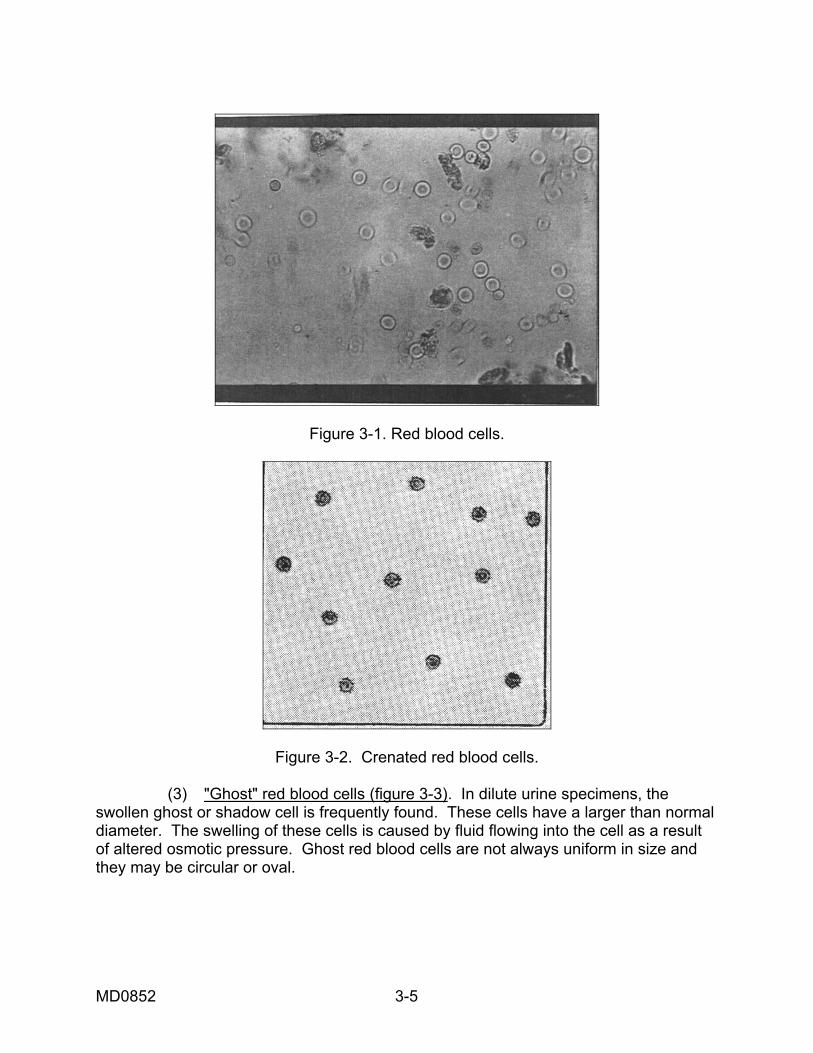
Figure 3-1. Red blood cells.
Figure 3-2. Crenated red blood cells. (3) "Ghost" red blood cells (figure 3-3). In dilute urine specimens, the swollen ghost or shadow cell is frequently found. These cells have a larger than normal diameter. The swelling of these cells is caused by fluid flowing into the cell as a result of altered osmotic pressure. Ghost red blood cells are not always uniform in size and they may be circular or oval.
MD0852 3-5
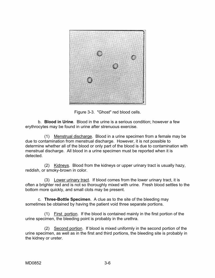
Figure 3-3. "Ghost" red blood cells. b. Blood in Urine. Blood in the urine is a serious condition; however a few erythrocytes may be found in urine after strenuous exercise. (1) Menstrual discharge. Blood in a urine specimen from a female may be due to contamination from menstrual discharge. However, it is not possible to determine whether all of the blood or only part of the blood is due to contamination with menstrual discharge. All blood in a urine specimen must be reported when it is detected. (2) Kidneys. Blood from the kidneys or upper urinary tract is usually hazy, reddish, or smoky-brown in color. (3) Lower urinary tract. If blood comes from the lower urinary tract, it is often a brighter red and is not so thoroughly mixed with urine. Fresh blood settles to the bottom more quickly, and small clots may be present. c. Three-Bottle Specimen. A clue as to the site of the bleeding may sometimes be obtained by having the patient void three separate portions. (1) First portion. If the blood is contained mainly in the first portion of the urine specimen, the bleeding point is probably in the urethra. (2) Second portion. If blood is mixed uniformly in the second portion of the urine specimen, as well as in the first and third portions, the bleeding site is probably in the kidney or ureter.
MD0852 3-6
(3) Third portion. If most of the blood is mainly in the last portion, the bleeding site is probably in the bladder. d. Alkaline Urine. In alkaline urine, red blood cells are small in size or may be entirely disintegrated. To differentiate between erythrocytes and leukocytes, yeast cells, or contaminants, a drop of 10 percent acetic acid is added to the sediment. Red cells, if present, will dissolve while other structures remain unaffected. e. Associated Protein. Urine that contains blood is always proteinaceous. A very small amount of blood may not be observed macroscopically. If large numbers of red blood cells are present, a positive protein will be obtained from the supernatant fluid of a centrifuged specimen. 3-7. WHITE BLOOD CELLS (LEUKOCYTES) A few leukocytes are present in normal urine, particularly when much mucus is found. They are numerous only as a result of a pathological process. Catheterization or a "two-bottle test" may be required to distinguish urethral infection from infection of other parts of the genitourinary system. The two-bottle test is conducted in the manner of the three-bottle test described previously. However, only two portions of urine are obtained instead of three. If the greater portion of leukocytes is found in the first portion of the urine specimen, a urethral infection is indicated. If the greater portion of the leukocytes is found in the second portion, an infection involving some other part of the genitourinary system may be suspected. The presence of increased numbers of white blood cells or pus constitutes a condition called pyuria. a. Macroscopic Appearance. When abundant, white blood cells form a white sediment resembling amorphous phosphates. b. Microscopic Appearance (figure 3-4). Leukocytes are true cells with well developed nuclei. Most white blood cells are neutrophils and are stainable with neutral dyes. Under the microscope they appear as colorless granular spheres, about 10 to 15 microns in diameter, and larger than red blood cells most of the time. The granules are composed of normal neutrophilic granules and granular products of degeneration. Diluted acetic acid can dissolve the granules and thus allow the nuclear characteristics to be seen. In freshly voided urine, many white cells exhibit ameboid motion and assume irregular outlines. c. Alkaline Urine. In alkaline urine, white blood cells are often swollen, very granular, and tend to adhere in clumps. The addition of a drop of 10 percent acetic acid not only allows differentiation from erythrocytes but brings the nuclei more clearly into view. d. Acid Urine. In moderately acid urine, white blood cells are well preserved. In strongly acid urine, they may be shrunken and irregularly shaped, suggesting ameboid forms.
MD0852 3-7
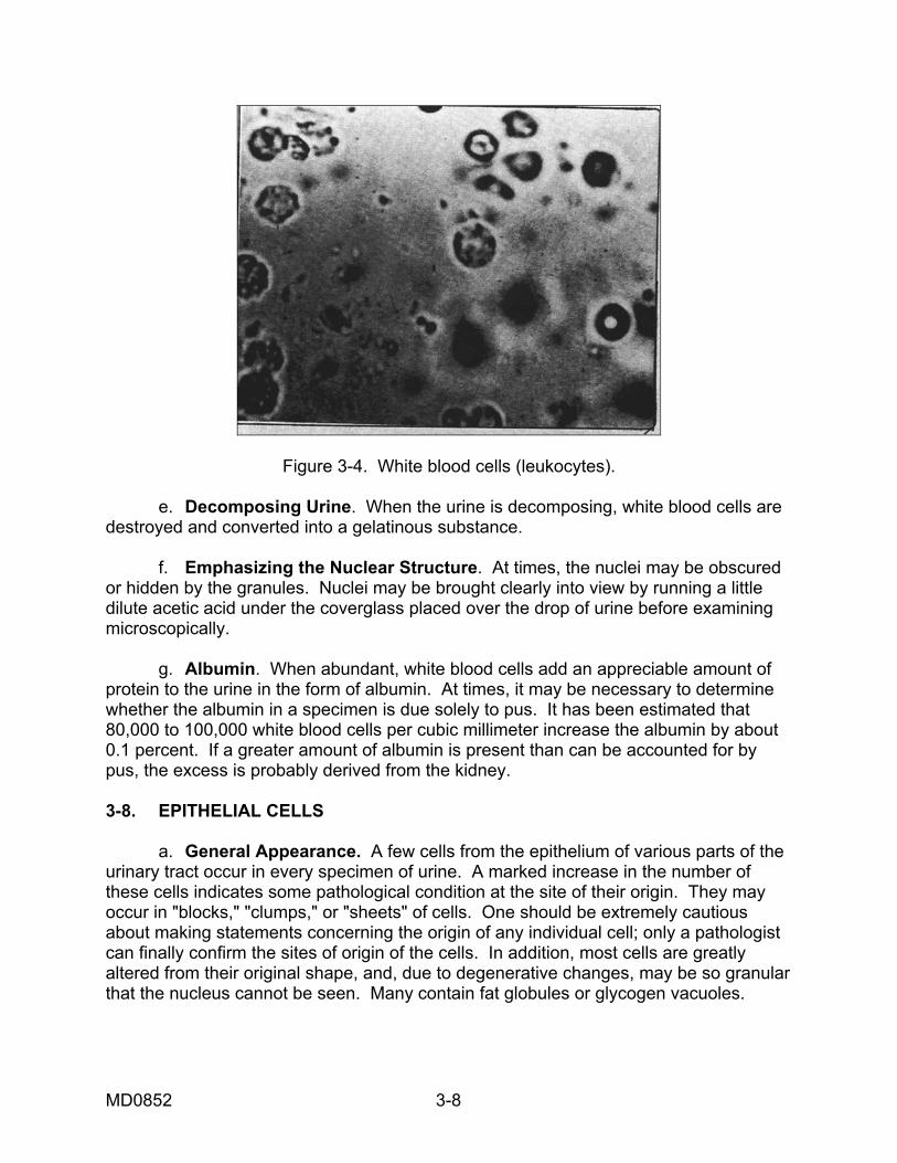
Figure 3-4. White blood cells (leukocytes). e. Decomposing Urine. When the urine is decomposing, white blood cells are destroyed and converted into a gelatinous substance. f. Emphasizing the Nuclear Structure. At times, the nuclei may be obscured or hidden by the granules. Nuclei may be brought clearly into view by running a little dilute acetic acid under the coverglass placed over the drop of urine before examining microscopically. g. Albumin. When abundant, white blood cells add an appreciable amount of protein to the urine in the form of albumin. At times, it may be necessary to determine whether the albumin in a specimen is due solely to pus. It has been estimated that 80,000 to 100,000 white blood cells per cubic millimeter increase the albumin by about 0.1 percent. If a greater amount of albumin is present than can be accounted for by pus, the excess is probably derived from the kidney. 3-8. EPITHELIAL CELLS a. General Appearance. A few cells from the epithelium of various parts of the urinary tract occur in every specimen of urine. A marked increase in the number of these cells indicates some pathological condition at the site of their origin. They may occur in "blocks," "clumps," or "sheets" of cells. One should be extremely cautious about making statements concerning the origin of any individual cell; only a pathologist can finally confirm the sites of origin of the cells. In addition, most cells are greatly altered from their original shape, and, due to degenerative changes, may be so granular that the nucleus cannot be seen. Many contain fat globules or glycogen vacuoles.
MD0852 3-8
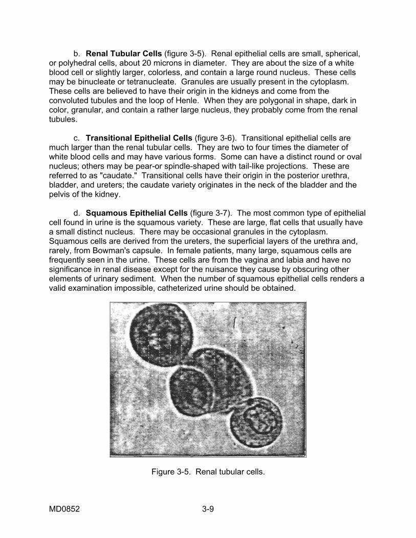
b. Renal Tubular Cells (figure 3-5). Renal epithelial cells are small, spherical, or polyhedral cells, about 20 microns in diameter. They are about the size of a white blood cell or slightly larger, colorless, and contain a large round nucleus. These cells may be binucleate or tetranucleate. Granules are usually present in the cytoplasm. These cells are believed to have their origin in the kidneys and come from the convoluted tubules and the loop of Henle. When they are polygonal in shape, dark in color, granular, and contain a rather large nucleus, they probably come from the renal tubules. c. Transitional Epithelial Cells (figure 3-6). Transitional epithelial cells are much larger than the renal tubular cells. They are two to four times the diameter of white blood cells and may have various forms. Some can have a distinct round or oval nucleus; others may be pear-or spindle-shaped with tail-like projections. These are referred to as "caudate." Transitional cells have their origin in the posterior urethra, bladder, and ureters; the caudate variety originates in the neck of the bladder and the pelvis of the kidney. d. Squamous Epithelial Cells (figure 3-7). The most common type of epithelial cell found in urine is the squamous variety. These are large, flat cells that usually have a small distinct nucleus. There may be occasional granules in the cytoplasm. Squamous cells are derived from the ureters, the superficial layers of the urethra and, rarely, from Bowman's capsule. In female patients, many large, squamous cells are frequently seen in the urine. These cells are from the vagina and labia and have no significance in renal disease except for the nuisance they cause by obscuring other elements of urinary sediment. When the number of squamous epithelial cells renders a valid examination impossible, catheterized urine should be obtained.
Figure 3-5. Renal tubular cells.
MD0852 3-9
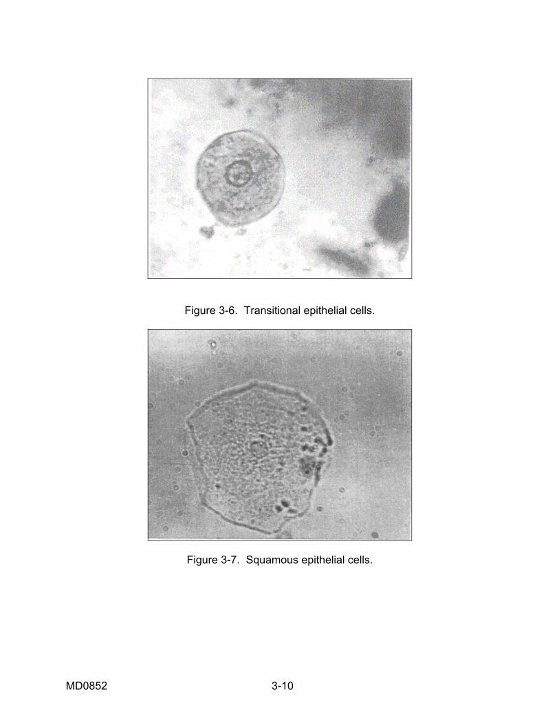
Figure 3-6. Transitional epithelial cells.
Figure 3-7. Squamous epithelial cells.
MD0852 3-10
3-9. CASTS--GENERAL COMMENTS a. Significance. Casts are proteinaceous products of the renal tubules, which act as molds for the casts. Casts, therefore, are tubular in shape and are a gelatinous impression of the kidney tubules. Their presence in the urine usually indicates some pathological change in the kidney, although the change may be slight or transitory. They are rarely found in the urinary sediment of normal individuals. Since casts are formed in and forced out of the renal tubules, they vary in shape and size according to the site of their origin. They may also differ in length, thickness, and consistency. A positive protein is often found when many casts are present. b. Formation. Normal and occasionally abnormal plasma proteins constitute the source of the protein involved in cast formation. These proteins are not reabsorbed in the proximal convoluted tubules. In the distal convoluted tubules and the collecting ducts, acidification of the urine, and the relative concentration of solutes due to water reabsorption favor coagulation of protein. In addition, a marked decrease in urine flow and the presence of abnormal ionic or protein constituents encourage cast development. After formation, most casts are washed out of the tubules into the urine by increased hydrostatic pressure from behind, which causes the tubules to dilate around them. A simple way to visualize the formation and variety of these structures is to regard the process as a gel formation. This gelling process is similar to events that occur in the preparation of a gelatin dessert. When the proper temperature and concentration of gelatin are obtained in the solution, there is a sudden increase in viscosity. If sliced fruit has been added to the fluid mixture, the fruit fragments are included within the gelatinized mass. In much the same manner red blood cells, white blood cells, and epithelial cells become trapped in the gelatinized casts and thereby preserve a record of the tubular contents for examination in the urinary sediment. c. Identification. If the urine is very dilute or alkaline, these casts dissolve. Therefore, it is imperative that the specimens be analyzed as soon as possible. Under the microscope, casts generally appear as clear, slightly refractive cylinders and are best recognized by using low power with dim light. However, all casts should be verified by using high power. Higher magnification is important in classifying casts as to type. 3-10. NONCELLULAR CASTS a. Hyaline Casts (figure 3-8). The simple hyaline cast is composed primarily of protein and has no inclusions. It is actually the basic material for all types of casts and is often referred to as a "hyaline matrix." Hyaline casts are colorless, homogenous, and semitransparent structures with cylindrical bodies that have parallel sides and rounded ends. The length of a hyaline cast varies. Generally it is straight, but occasionally may be slightly rounded or convoluted.
MD0852 3-11
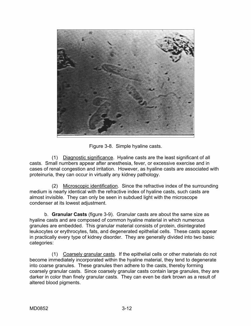
Figure 3-8. Simple hyaline casts. (1) Diagnostic significance. Hyaline casts are the least significant of all casts. Small numbers appear after anesthesia, fever, or excessive exercise and in cases of renal congestion and irritation. However, as hyaline casts are associated with proteinuria, they can occur in virtually any kidney pathology. (2) Microscopic identification. Since the refractive index of the surrounding medium is nearly identical with the refractive index of hyaline casts, such casts are almost invisible. They can only be seen in subdued light with the microscope condenser at its lowest adjustment. b. Granular Casts (figure 3-9). Granular casts are about the same size as hyaline casts and are composed of common hyaline material in which numerous granules are embedded. This granular material consists of protein, disintegrated leukocytes or erythrocytes, fats, and degenerated epithelial cells. These casts appear in practically every type of kidney disorder. They are generally divided into two basic categories: (1) Coarsely granular casts. If the epithelial cells or other materials do not become immediately incorporated within the hyaline material, they tend to degenerate into coarse granules. These granules then adhere to the casts, thereby forming coarsely granular casts. Since coarsely granular casts contain large granules, they are darker in color than finely granular casts. They can even be dark brown as a result of altered blood pigments.
MD0852 3-12
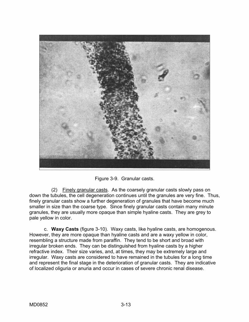
Figure 3-9. Granular casts. (2) Finely granular casts. As the coarsely granular casts slowly pass on down the tubules, the cell degeneration continues until the granules are very fine. Thus, finely granular casts show a further degeneration of granules that have become much smaller in size than the coarse type. Since finely granular casts contain many minute granules, they are usually more opaque than simple hyaline casts. They are grey to pale yellow in color. c. Waxy Casts (figure 3-10). Waxy casts, like hyaline casts, are homogenous. However, they are more opaque than hyaline casts and are a waxy yellow in color, resembling a structure made from paraffin. They tend to be short and broad with irregular broken ends. They can be distinguished from hyaline casts by a higher refractive index. Their size varies, and, at times, they may be extremely large and irregular. Waxy casts are considered to have remained in the tubules for a long time and represent the final stage in the deterioration of granular casts. They are indicative of localized oliguria or anuria and occur in cases of severe chronic renal disease.
MD0852 3-13
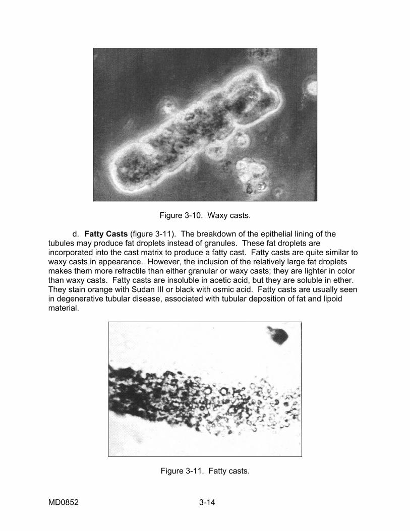
Figure 3-10. Waxy casts. d. Fatty Casts (figure 3-11). The breakdown of the epithelial lining of the tubules may produce fat droplets instead of granules. These fat droplets are incorporated into the cast matrix to produce a fatty cast. Fatty casts are quite similar to waxy casts in appearance. However, the inclusion of the relatively large fat droplets makes them more refractile than either granular or waxy casts; they are lighter in color than waxy casts. Fatty casts are insoluble in acetic acid, but they are soluble in ether. They stain orange with Sudan III or black with osmic acid. Fatty casts are usually seen in degenerative tubular disease, associated with tubular deposition of fat and lipoid material.
Figure 3-11. Fatty casts.
MD0852 3-14
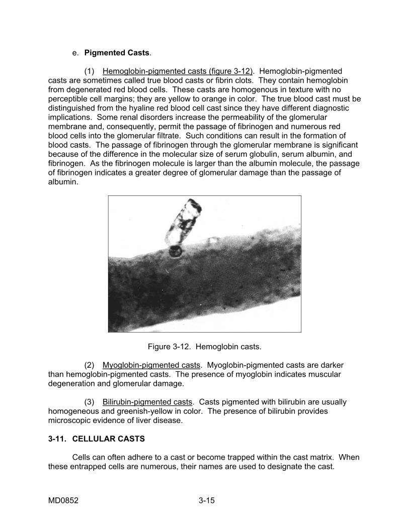
e. Pigmented Casts. (1) Hemoglobin-pigmented casts (figure 3-12). Hemoglobin-pigmented casts are sometimes called true blood casts or fibrin clots. They contain hemoglobin from degenerated red blood cells. These casts are homogenous in texture with no perceptible cell margins; they are yellow to orange in color. The true blood cast must be distinguished from the hyaline red blood cell cast since they have different diagnostic implications. Some renal disorders increase the permeability of the glomerular membrane and, consequently, permit the passage of fibrinogen and numerous red blood cells into the glomerular filtrate. Such conditions can result in the formation of blood casts. The passage of fibrinogen through the glomerular membrane is significant because of the difference in the molecular size of serum globulin, serum albumin, and fibrinogen. As the fibrinogen molecule is larger than the albumin molecule, the passage of fibrinogen indicates a greater degree of glomerular damage than the passage of albumin.
Figure 3-12. Hemoglobin casts. (2) Myoglobin-pigmented casts. Myoglobin-pigmented casts are darker than hemoglobin-pigmented casts. The presence of myoglobin indicates muscular degeneration and glomerular damage. (3) Bilirubin-pigmented casts. Casts pigmented with bilirubin are usually homogeneous and greenish-yellow in color. The presence of bilirubin provides microscopic evidence of liver disease. 3-11. CELLULAR CASTS Cells can often adhere to a cast or become trapped within the cast matrix. When these entrapped cells are numerous, their names are used to designate the cast.
MD0852 3-15
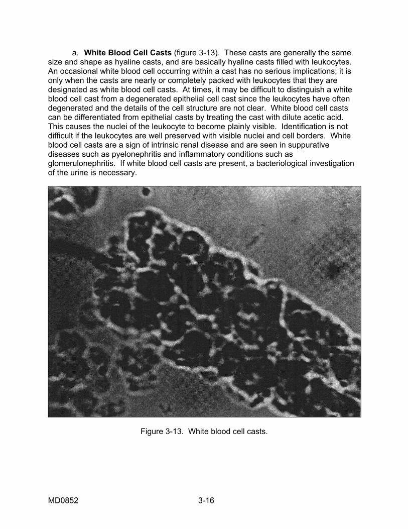
a. White Blood Cell Casts (figure 3-13). These casts are generally the same size and shape as hyaline casts, and are basically hyaline casts filled with leukocytes. An occasional white blood cell occurring within a cast has no serious implications; it is only when the casts are nearly or completely packed with leukocytes that they are designated as white blood cell casts. At times, it may be difficult to distinguish a white blood cell cast from a degenerated epithelial cell cast since the leukocytes have often degenerated and the details of the cell structure are not clear. White blood cell casts can be differentiated from epithelial casts by treating the cast with dilute acetic acid. This causes the nuclei of the leukocyte to become plainly visible. Identification is not difficult if the leukocytes are well preserved with visible nuclei and cell borders. White blood cell casts are a sign of intrinsic renal disease and are seen in suppurative diseases such as pyelonephritis and inflammatory conditions such as glomerulonephritis. If white blood cell casts are present, a bacteriological investigation of the urine is necessary.
Figure 3-13. White blood cell casts.
MD0852 3-16
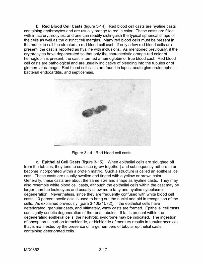
b. Red Blood Cell Casts (figure 3-14). Red blood cell casts are hyaline casts containing erythrocytes and are usually orange to red in color. These casts are filled with intact erythrocytes, and one can readily distinguish the typical spherical shape of the cells as well as the distinct cell margins. Many red blood cells must be present in the matrix to call the structure a red blood cell cast. If only a few red blood cells are present, the cast is reported as hyaline with inclusions. As mentioned previously, if the erythrocytes have degenerated so that only the characteristic orange-red color of hemoglobin is present, the cast is termed a hemoglobin or true blood cast. Red blood cell casts are pathological and are usually indicative of bleeding into the tubules or of glomerular damage. Red blood cell casts are found in lupus, acute glomerulonephritis, bacterial endocarditis, and septicemias.
Figure 3-14. Red blood cell casts. c. Epithelial Cell Casts (figure 3-15). When epithelial cells are sloughed off from the tubules, they tend to coalesce (grow together) and subsequently adhere to or become incorporated within a protein matrix. Such a structure is called an epithelial cell cast. These casts are usually swollen and tinged with a yellow or brown color. Generally, these casts are about the same size and shape as hyaline casts. They may also resemble white blood cell casts, although the epithelial cells within the cast may be larger than the leukocytes and usually show more fatty and hyaline cytoplasmic degeneration. Nevertheless, since they are frequently confused with white blood cell casts, 10 percent acetic acid is used to bring out the nuclei and aid in recognition of the cells. As explained previously, [para 3-10b(1), (2)], if the epithelial cells have deteriorated, granular casts, and ultimately, waxy casts are formed. Epithelial cell casts can signify aseptic degeneration of the renal tubules. If fat is present within the degenerating epithelial cells, the nephrotic syndrome may be indicated. The ingestion of phosphorus, carbon tetrachloride, or bichloride of mercury results in tubular necrosis that is manifested by the presence of large numbers of tubular epithelial casts containing deteriorated cells.
MD0852 3-17
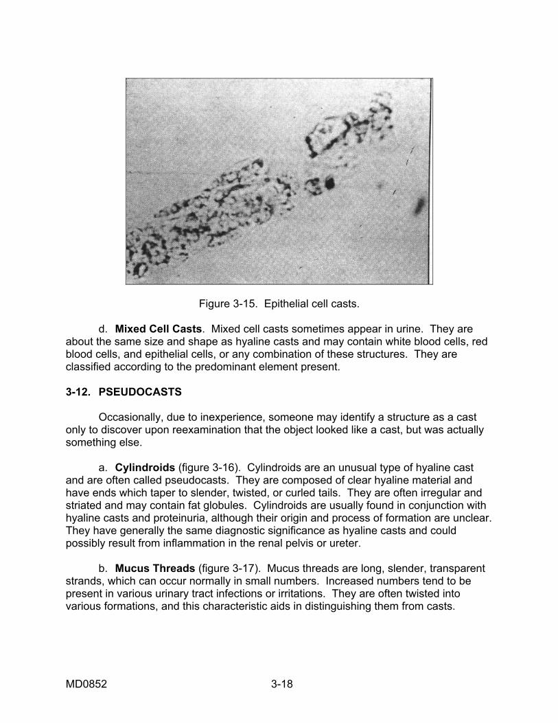
Figure 3-15. Epithelial cell casts. d. Mixed Cell Casts. Mixed cell casts sometimes appear in urine. They are about the same size and shape as hyaline casts and may contain white blood cells, red blood cells, and epithelial cells, or any combination of these structures. They are classified according to the predominant element present. 3-12. PSEUDOCASTS Occasionally, due to inexperience, someone may identify a structure as a cast only to discover upon reexamination that the object looked like a cast, but was actually something else. a. Cylindroids (figure 3-16). Cylindroids are an unusual type of hyaline cast and are often called pseudocasts. They are composed of clear hyaline material and have ends which taper to slender, twisted, or curled tails. They are often irregular and striated and may contain fat globules. Cylindroids are usually found in conjunction with hyaline casts and proteinuria, although their origin and process of formation are unclear. They have generally the same diagnostic significance as hyaline casts and could possibly result from inflammation in the renal pelvis or ureter. b. Mucus Threads (figure 3-17). Mucus threads are long, slender, transparent strands, which can occur normally in small numbers. Increased numbers tend to be present in various urinary tract infections or irritations. They are often twisted into various formations, and this characteristic aids in distinguishing them from casts.
MD0852 3-18
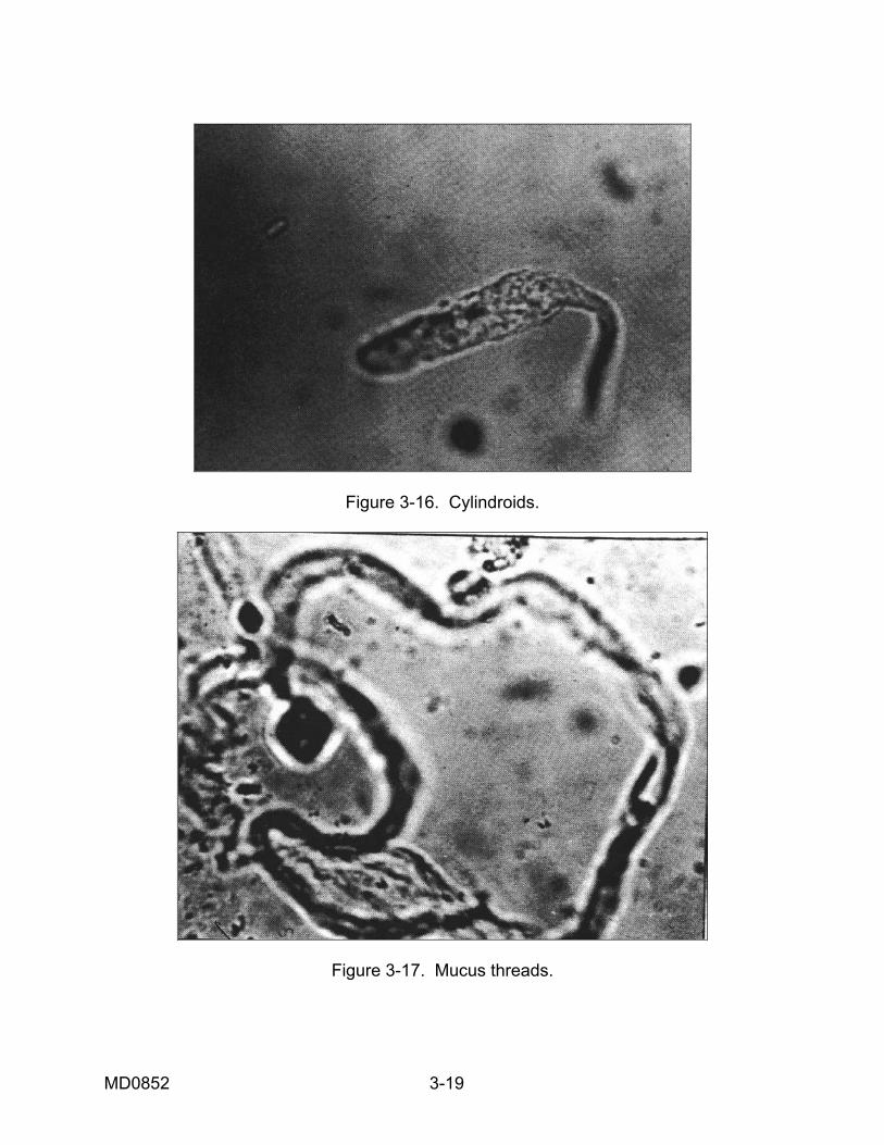
Figure 3-16. Cylindroids.
Figure 3-17. Mucus threads.
MD0852 3-19
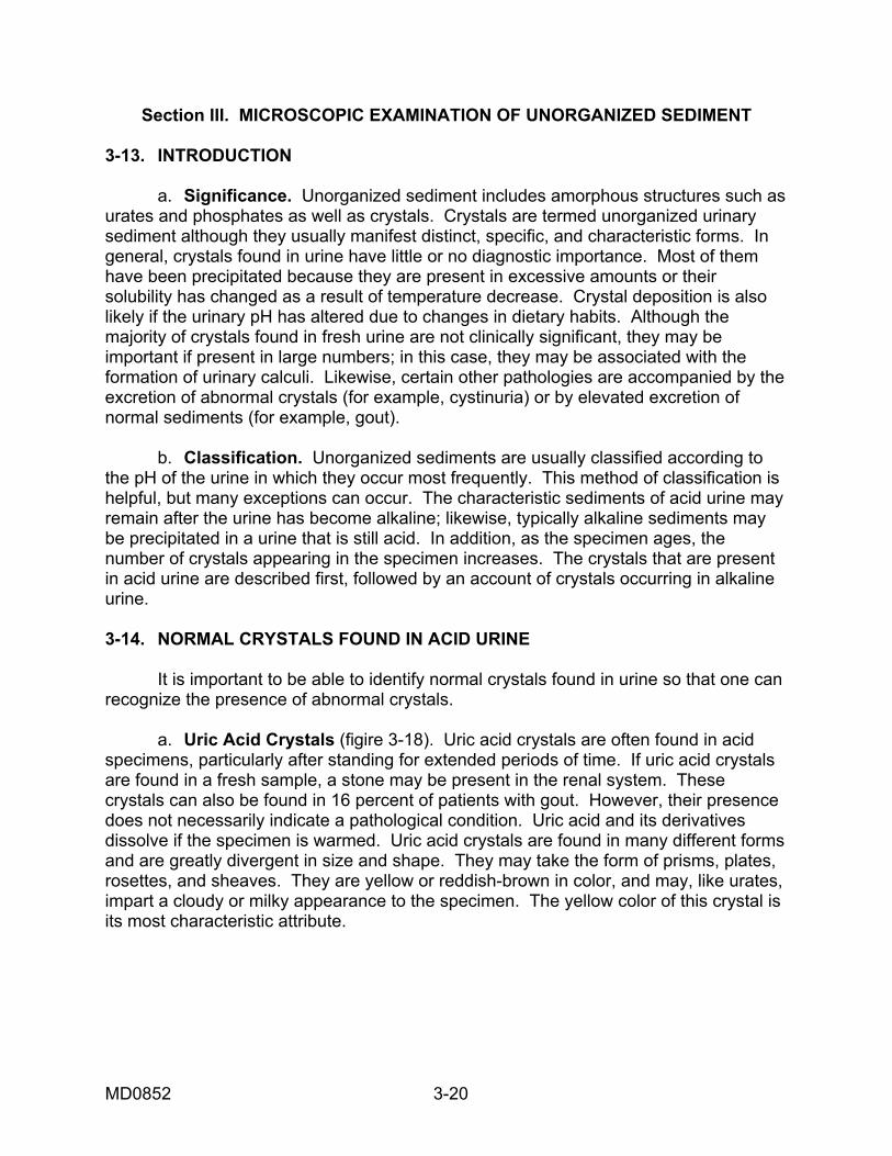
Section III. MICROSCOPIC EXAMINATION OF UNORGANIZED SEDIMENT 3-13. INTRODUCTION a. Significance. Unorganized sediment includes amorphous structures such as urates and phosphates as well as crystals. Crystals are termed unorganized urinary sediment although they usually manifest distinct, specific, and characteristic forms. In general, crystals found in urine have little or no diagnostic importance. Most of them have been precipitated because they are present in excessive amounts or their solubility has changed as a result of temperature decrease. Crystal deposition is also likely if the urinary pH has altered due to changes in dietary habits. Although the majority of crystals found in fresh urine are not clinically significant, they may be important if present in large numbers; in this case, they may be associated with the formation of urinary calculi. Likewise, certain other pathologies are accompanied by the excretion of abnormal crystals (for example, cystinuria) or by elevated excretion of normal sediments (for example, gout). b. Classification. Unorganized sediments are usually classified according to the pH of the urine in which they occur most frequently. This method of classification is helpful, but many exceptions can occur. The characteristic sediments of acid urine may remain after the urine has become alkaline; likewise, typically alkaline sediments may be precipitated in a urine that is still acid. In addition, as the specimen ages, the number of crystals appearing in the specimen increases. The crystals that are present in acid urine are described first, followed by an account of crystals occurring in alkaline urine. 3-14. NORMAL CRYSTALS FOUND IN ACID URINE It is important to be able to identify normal crystals found in urine so that one can recognize the presence of abnormal crystals. a. Uric Acid Crystals (figire 3-18). Uric acid crystals are often found in acid specimens, particularly after standing for extended periods of time. If uric acid crystals are found in a fresh sample, a stone may be present in the renal system. These crystals can also be found in 16 percent of patients with gout. However, their presence does not necessarily indicate a pathological condition. Uric acid and its derivatives dissolve if the specimen is warmed. Uric acid crystals are found in many different forms and are greatly divergent in size and shape. They may take the form of prisms, plates, rosettes, and sheaves. They are yellow or reddish-brown in color, and may, like urates, impart a cloudy or milky appearance to the specimen. The yellow color of this crystal is its most characteristic attribute.
MD0852 3-20
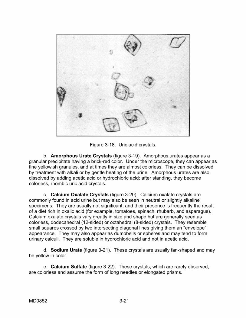
Figure 3-18. Uric acid crystals. b. Amorphous Urate Crystals (figure 3-19). Amorphous urates appear as a granular precipitate having a brick-red color. Under the microscope, they can appear as fine yellowish granules, and at times they are almost colorless. They can be dissolved by treatment with alkali or by gentle heating of the urine. Amorphous urates are also dissolved by adding acetic acid or hydrochloric acid; after standing, they become colorless, rhombic uric acid crystals. c. Calcium Oxalate Crystals (figure 3-20). Calcium oxalate crystals are commonly found in acid urine but may also be seen in neutral or slightly alkaline specimens. They are usually not significant, and their presence is frequently the result of a diet rich in oxalic acid (for example, tomatoes, spinach, rhubarb, and asparagus). Calcium oxalate crystals vary greatly in size and shape but are generally seen as colorless, dodecahedral (12-sided) or octahedral (8-sided) crystals. They resemble small squares crossed by two intersecting diagonal lines giving them an "envelope" appearance. They may also appear as dumbbells or spheres and may tend to form urinary calculi. They are soluble in hydrochloric acid and not in acetic acid. d. Sodium Urate (figure 3-21). These crystals are usually fan-shaped and may be yellow in color. e. Calcium Sulfate (figure 3-22). These crystals, which are rarely observed, are colorless and assume the form of long needles or elongated prisms.
MD0852 3-21
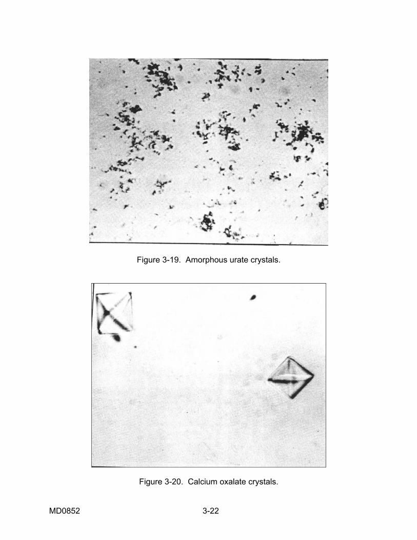
Figure 3-19. Amorphous urate crystals.
Figure 3-20. Calcium oxalate crystals.
MD0852 3-22
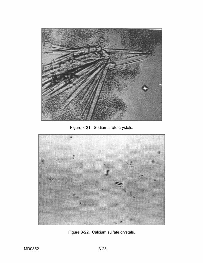
Figure 3-21. Sodium urate crystals.
Figure 3-22. Calcium sulfate crystals.
MD0852 3-23
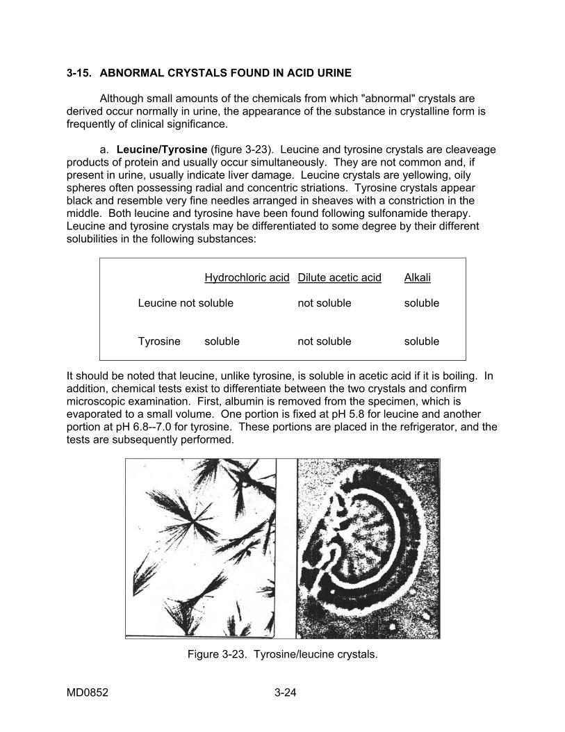
3-15. ABNORMAL CRYSTALS FOUND IN ACID URINE Although small amounts of the chemicals from which "abnormal" crystals are derived occur normally in urine, the appearance of the substance in crystalline form is frequently of clinical significance. a. Leucine/Tyrosine (figure 3-23). Leucine and tyrosine crystals are cleaveage products of protein and usually occur simultaneously. They are not common and, if present in urine, usually indicate liver damage. Leucine crystals are yellowing, oily spheres often possessing radial and concentric striations. Tyrosine crystals appear black and resemble very fine needles arranged in sheaves with a constriction in the middle. Both leucine and tyrosine have been found following sulfonamide therapy. Leucine and tyrosine crystals may be differentiated to some degree by their different solubilities in the following substances:
Hydrochloric acid Dilute acetic acid Alkali Leucine not soluble not soluble soluble Tyrosine soluble not soluble soluble
It should be noted that leucine, unlike tyrosine, is soluble in acetic acid if it is boiling. In addition, chemical tests exist to differentiate between the two crystals and confirm microscopic examination. First, albumin is removed from the specimen, which is evaporated to a small volume. One portion is fixed at pH 5.8 for leucine and another portion at pH 6.8--7.0 for tyrosine. These portions are placed in the refrigerator, and the tests are subsequently performed.
Figure 3-23. Tyrosine/leucine crystals.
MD0852 3-24
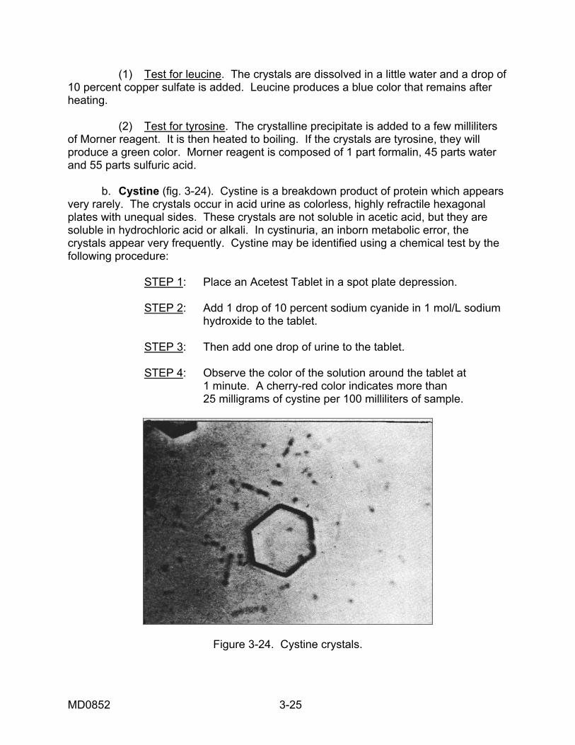
(1) Test for leucine. The crystals are dissolved in a little water and a drop of 10 percent copper sulfate is added. Leucine produces a blue color that remains after heating. (2) Test for tyrosine. The crystalline precipitate is added to a few milliliters of Morner reagent. It is then heated to boiling. If the crystals are tyrosine, they will produce a green color. Morner reagent is composed of 1 part formalin, 45 parts water and 55 parts sulfuric acid. b. Cystine (fig. 3-24). Cystine is a breakdown product of protein which appears very rarely. The crystals occur in acid urine as colorless, highly refractile hexagonal plates with unequal sides. These crystals are not soluble in acetic acid, but they are soluble in hydrochloric acid or alkali. In cystinuria, an inborn metabolic error, the crystals appear very frequently. Cystine may be identified using a chemical test by the following procedure:
STEP 1: Place an Acetest Tablet in a spot plate depression.
STEP 2: Add 1 drop of 10 percent sodium cyanide in 1 mol/L sodium hydroxide to the tablet. STEP 3: Then add one drop of urine to the tablet. STEP 4: Observe the color of the solution around the tablet at 1 minute. A cherry-red color indicates more than 25 milligrams of cystine per 100 milliliters of sample.
Figure 3-24. Cystine crystals.
MD0852 3-25
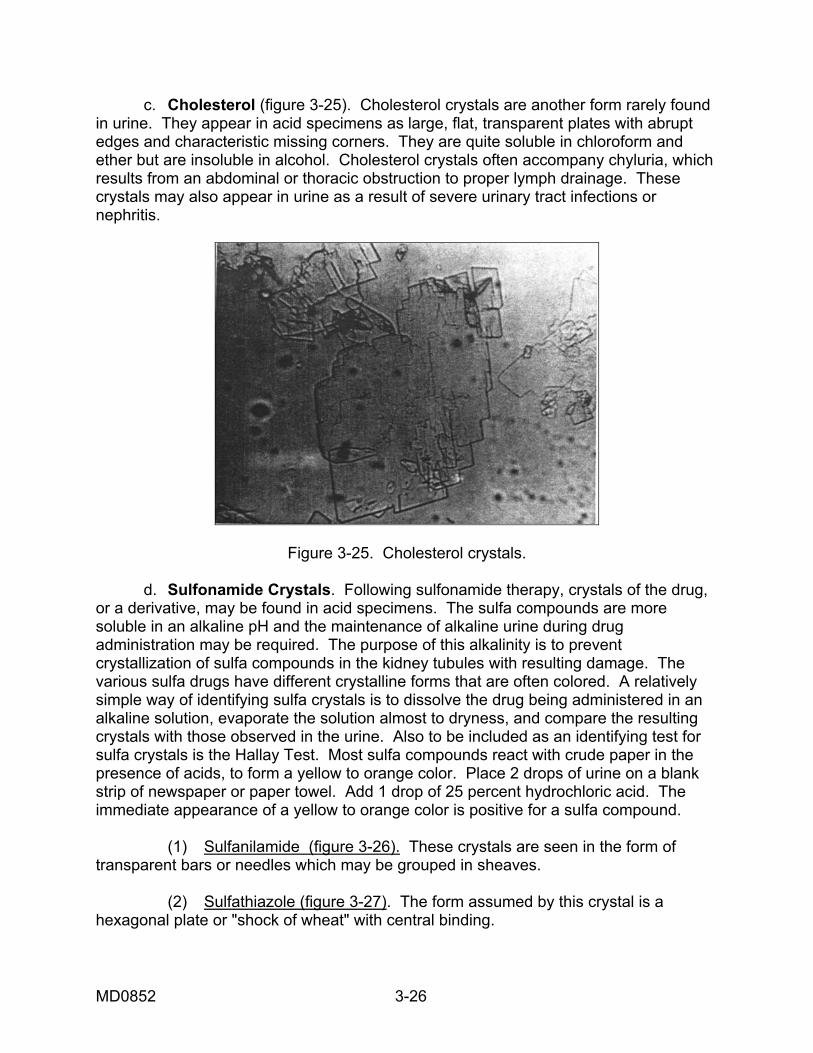
c. Cholesterol (figure 3-25). Cholesterol crystals are another form rarely found in urine. They appear in acid specimens as large, flat, transparent plates with abrupt edges and characteristic missing corners. They are quite soluble in chloroform and ether but are insoluble in alcohol. Cholesterol crystals often accompany chyluria, which results from an abdominal or thoracic obstruction to proper lymph drainage. These crystals may also appear in urine as a result of severe urinary tract infections or nephritis.
Figure 3-25. Cholesterol crystals. d. Sulfonamide Crystals. Following sulfonamide therapy, crystals of the drug, or a derivative, may be found in acid specimens. The sulfa compounds are more soluble in an alkaline pH and the maintenance of alkaline urine during drug administration may be required. The purpose of this alkalinity is to prevent crystallization of sulfa compounds in the kidney tubules with resulting damage. The various sulfa drugs have different crystalline forms that are often colored. A relatively simple way of identifying sulfa crystals is to dissolve the drug being administered in an alkaline solution, evaporate the solution almost to dryness, and compare the resulting crystals with those observed in the urine. Also to be included as an identifying test for sulfa crystals is the Hallay Test. Most sulfa compounds react with crude paper in the presence of acids, to form a yellow to orange color. Place 2 drops of urine on a blank strip of newspaper or paper towel. Add 1 drop of 25 percent hydrochloric acid. The immediate appearance of a yellow to orange color is positive for a sulfa compound. (1) Sulfanilamide (figure 3-26). These crystals are seen in the form of transparent bars or needles which may be grouped in sheaves. (2) Sulfathiazole (figure 3-27). The form assumed by this crystal is a hexagonal plate or "shock of wheat" with central binding.
MD0852 3-26
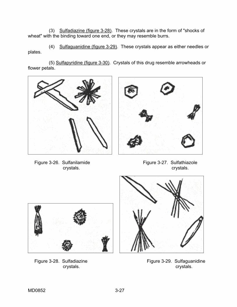
(3) Sulfadiazine (figure 3-28). These crystals are in the form of "shocks of wheat" with the binding toward one end, or they may resemble burrs. (4) Sulfaguanidine (figure 3-29). These crystals appear as either needles or plates. (5) Sulfapyridine (figure 3-30). Crystals of this drug resemble arrowheads or flower petals.
Figure 3-26. Sulfanilamide Figure 3-27. Sulfathiazole crystals. crystals.
Figure 3-28. Sulfadiazine Figure 3-29. Sulfaguanidine crystals. crystals.
MD0852 3-27
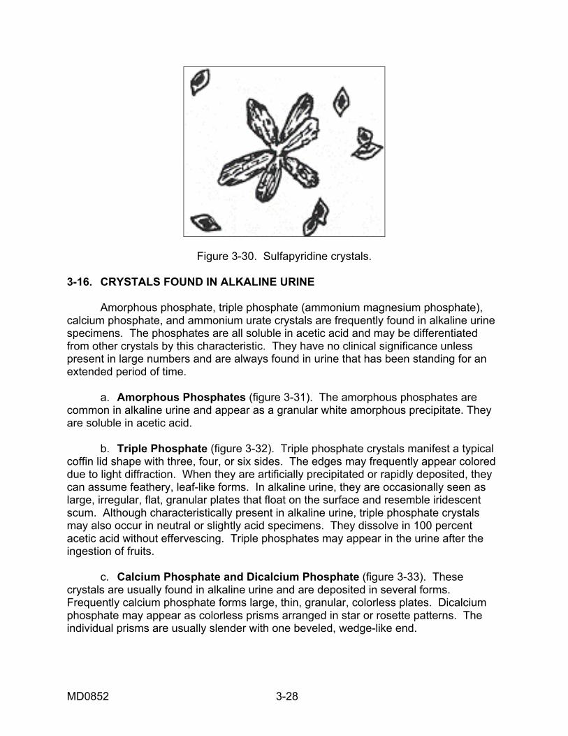
Figure 3-30. Sulfapyridine crystals.
3-16. CRYSTALS FOUND IN ALKALINE URINE Amorphous phosphate, triple phosphate (ammonium magnesium phosphate), calcium phosphate, and ammonium urate crystals are frequently found in alkaline urine specimens. The phosphates are all soluble in acetic acid and may be differentiated from other crystals by this characteristic. They have no clinical significance unless present in large numbers and are always found in urine that has been standing for an extended period of time. a. Amorphous Phosphates (figure 3-31). The amorphous phosphates are common in alkaline urine and appear as a granular white amorphous precipitate. They are soluble in acetic acid. b. Triple Phosphate (figure 3-32). Triple phosphate crystals manifest a typical coffin lid shape with three, four, or six sides. The edges may frequently appear colored due to light diffraction. When they are artificially precipitated or rapidly deposited, they can assume feathery, leaf-like forms. In alkaline urine, they are occasionally seen as large, irregular, flat, granular plates that float on the surface and resemble iridescent scum. Although characteristically present in alkaline urine, triple phosphate crystals may also occur in neutral or slightly acid specimens. They dissolve in 100 percent acetic acid without effervescing. Triple phosphates may appear in the urine after the ingestion of fruits. c. Calcium Phosphate and Dicalcium Phosphate (figure 3-33). These crystals are usually found in alkaline urine and are deposited in several forms. Frequently calcium phosphate forms large, thin, granular, colorless plates. Dicalcium phosphate may appear as colorless prisms arranged in star or rosette patterns. The individual prisms are usually slender with one beveled, wedge-like end.
MD0852 3-28
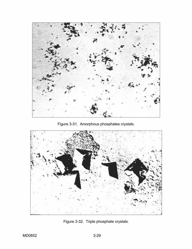
Figure 3-31. Amorphous phosphates crystals.
Figure 3-32. Triple phosphate crystals.
MD0852 3-29
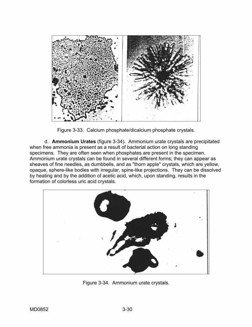
Figure 3-33. Calcium phosphate/dicalcium phosphate crystals. d. Ammonium Urates (figure 3-34). Ammonium urate crystals are precipitated when free ammonia is present as a result of bacterial action on long standing specimens. They are often seen when phosphates are present in the specimen. Ammonium urate crystals can be found in several different forms; they can appear as sheaves of fine needles, as dumbbells, and as "thorn apple" crystals, which are yellow, opaque, sphere-like bodies with irregular, spine-like projections. They can be dissolved by heating and by the addition of acetic acid, which, upon standing, results in the formation of colorless uric acid crystals.
Figure 3-34. Ammonium urate crystals.
MD0852 3-30
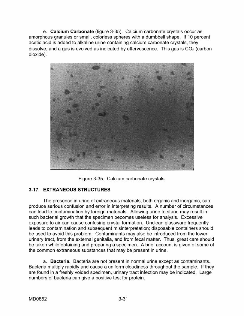
e. Calcium Carbonate (figure 3-35). Calcium carbonate crystals occur as amorphous granules or small, colorless spheres with a dumbbell shape. If 10 percent acetic acid is added to alkaline urine containing calcium carbonate crystals, they dissolve, and a gas is evolved as indicated by effervescence. This gas is CO2 (carbon dioxide).
Figure 3-35. Calcium carbonate crystals. 3-17. EXTRANEOUS STRUCTURES The presence in urine of extraneous materials, both organic and inorganic, can produce serious confusion and error in interpreting results. A number of circumstances can lead to contamination by foreign materials. Allowing urine to stand may result in such bacterial growth that the specimen becomes useless for analysis. Excessive exposure to air can cause confusing crystal formation. Unclean glassware frequently leads to contamination and subsequent misinterpretation; disposable containers should be used to avoid this problem. Contaminants may also be introduced from the lower urinary tract, from the external genitalia, and from fecal matter. Thus, great care should be taken while obtaining and preparing a specimen. A brief account is given of some of the common extraneous substances that may be present in urine. a. Bacteria. Bacteria are not present in normal urine except as contaminants. Bacteria multiply rapidly and cause a uniform cloudiness throughout the sample. If they are found in a freshly voided specimen, urinary tract infection may be indicated. Large numbers of bacteria can give a positive test for protein.
MD0852 3-31
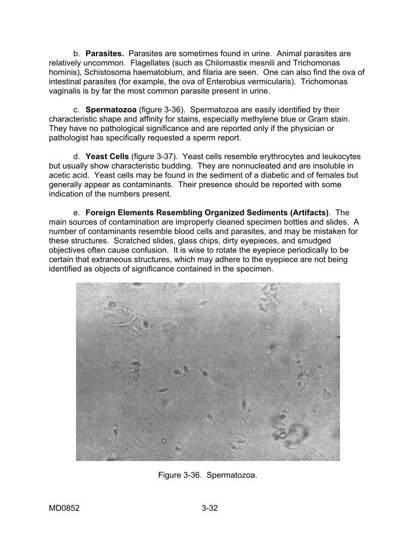
b. Parasites. Parasites are sometimes found in urine. Animal parasites are relatively uncommon. Flagellates (such as Chilomastix mesnili and Trichomonas hominis), Schistosoma haematobium, and filaria are seen. One can also find the ova of intestinal parasites (for example, the ova of Enterobius vermicularis). Trichomonas vaginalis is by far the most common parasite present in urine. c. Spermatozoa (figure 3-36). Spermatozoa are easily identified by their characteristic shape and affinity for stains, especially methylene blue or Gram stain. They have no pathological significance and are reported only if the physician or pathologist has specifically requested a sperm report. d. Yeast Cells (figure 3-37). Yeast cells resemble erythrocytes and leukocytes but usually show characteristic budding. They are nonnucleated and are insoluble in acetic acid. Yeast cells may be found in the sediment of a diabetic and of females but generally appear as contaminants. Their presence should be reported with some indication of the numbers present. e. Foreign Elements Resembling Organized Sediments (Artifacts). The main sources of contamination are improperly cleaned specimen bottles and slides. A number of contaminants resemble blood cells and parasites, and may be mistaken for these structures. Scratched slides, glass chips, dirty eyepieces, and smudged objectives often cause confusion. It is wise to rotate the eyepiece periodically to be certain that extraneous structures, which may adhere to the eyepiece are not being identified as objects of significance contained in the specimen.
Figure 3-36. Spermatozoa.
MD0852 3-32
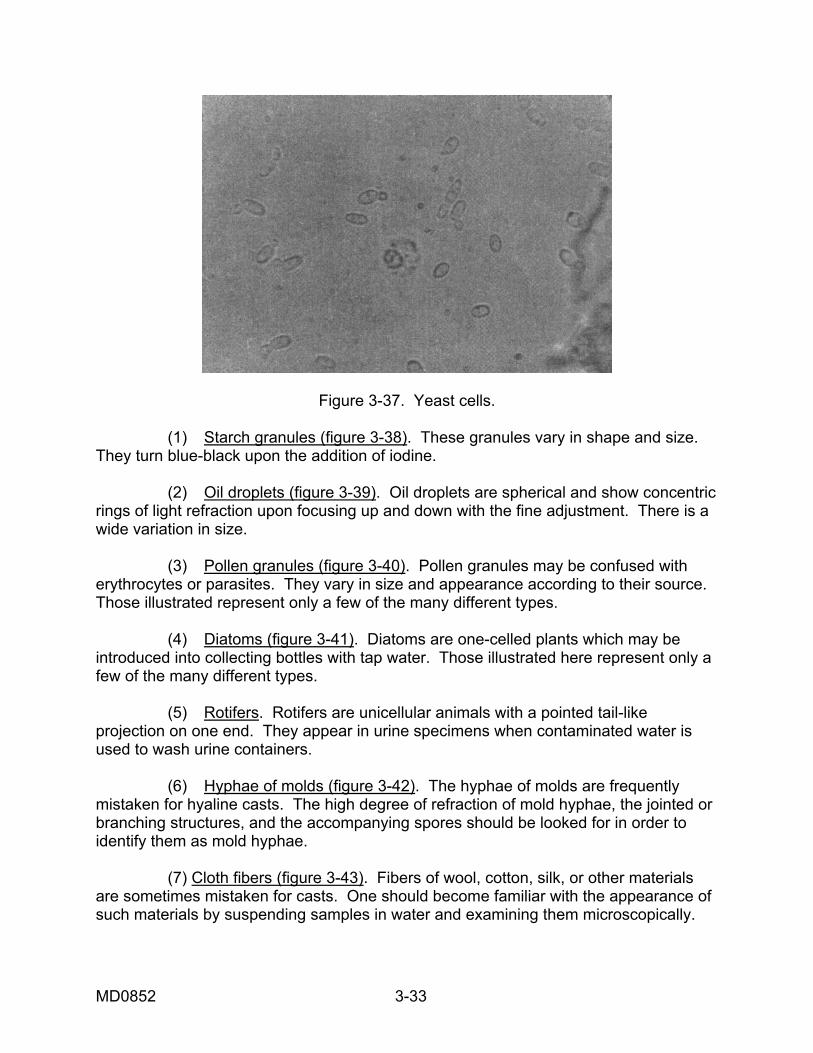
Figure 3-37. Yeast cells. (1) Starch granules (figure 3-38). These granules vary in shape and size. They turn blue-black upon the addition of iodine. (2) Oil droplets (figure 3-39). Oil droplets are spherical and show concentric rings of light refraction upon focusing up and down with the fine adjustment. There is a wide variation in size. (3) Pollen granules (figure 3-40). Pollen granules may be confused with erythrocytes or parasites. They vary in size and appearance according to their source. Those illustrated represent only a few of the many different types. (4) Diatoms (figure 3-41). Diatoms are one-celled plants which may be introduced into collecting bottles with tap water. Those illustrated here represent only a few of the many different types. (5) Rotifers. Rotifers are unicellular animals with a pointed tail-like projection on one end. They appear in urine specimens when contaminated water is used to wash urine containers. (6) Hyphae of molds (figure 3-42). The hyphae of molds are frequently mistaken for hyaline casts. The high degree of refraction of mold hyphae, the jointed or branching structures, and the accompanying spores should be looked for in order to identify them as mold hyphae. (7) Cloth fibers (figure 3-43). Fibers of wool, cotton, silk, or other materials are sometimes mistaken for casts. One should become familiar with the appearance of such materials by suspending samples in water and examining them microscopically.
MD0852 3-33
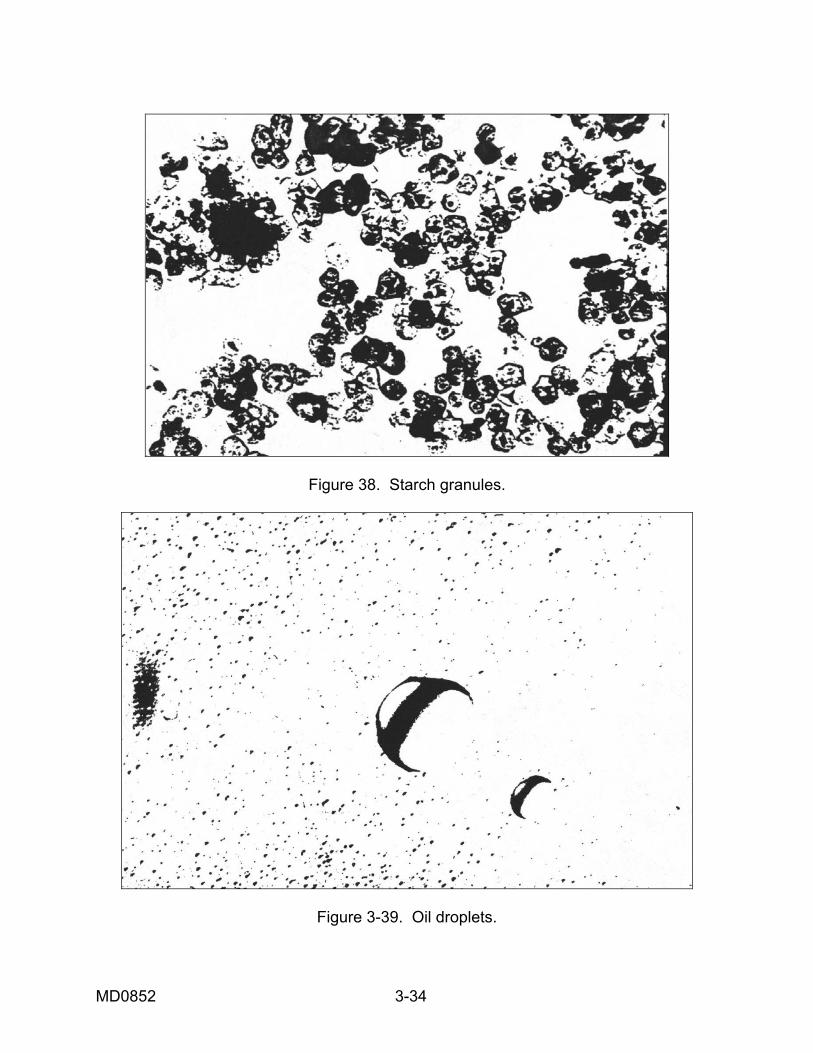
Figure 38. Starch granules.
Figure 3-39. Oil droplets.
MD0852 3-34
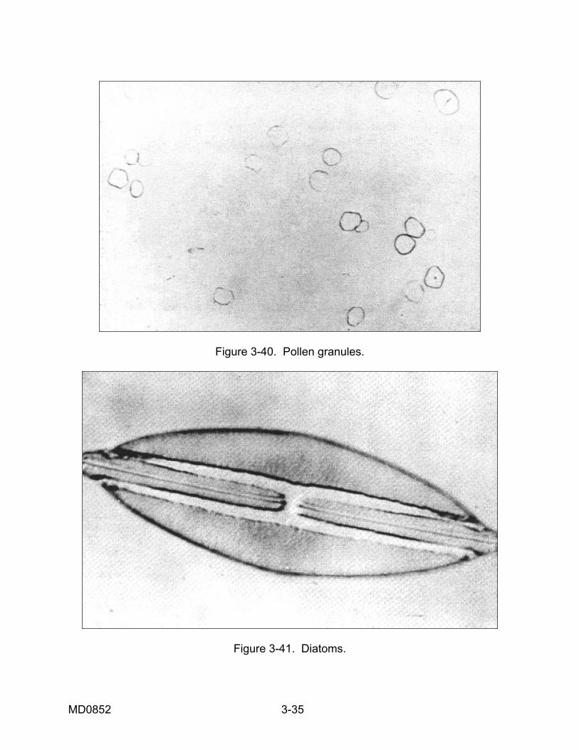
Figure 3-40. Pollen granules.
Figure 3-41. Diatoms.
MD0852 3-35
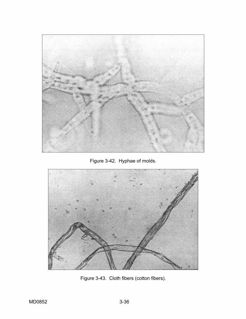
Figure 3-42. Hyphae of molds.
Figure 3-43. Cloth fibers (cotton fibers).
MD0852 3-36
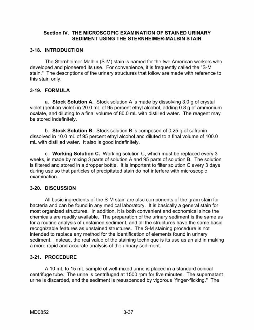
Section IV. THE MICROSCOPIC EXAMINATION OF STAINED URINARY SEDIMENT USING THE STERNHEIMER-MALBIN STAIN 3-18. INTRODUCTION The Sternheimer-Malbin (S-M) stain is named for the two American workers who developed and pioneered its use. For convenience, it is frequently called the "S-M stain." The descriptions of the urinary structures that follow are made with reference to this stain only. 3-19. FORMULA a. Stock Solution A. Stock solution A is made by dissolving 3.0 g of crystal violet (gentian violet) in 20.0 mL of 95 percent ethyl alcohol, adding 0.8 g of ammonium oxalate, and diluting to a final volume of 80.0 mL with distilled water. The reagent may be stored indefinitely. b. Stock Solution B. Stock solution B is composed of 0.25 g of safranin dissolved in 10.0 mL of 95 percent ethyl alcohol and diluted to a final volume of 100.0 mL with distilled water. It also is good indefinitely. c. Working Solution C. Working solution C, which must be replaced every 3 weeks, is made by mixing 3 parts of solution A and 95 parts of solution B. The solution is filtered and stored in a dropper bottle. It is important to filter solution C every 3 days during use so that particles of precipitated stain do not interfere with microscopic examination. 3-20. DISCUSSION All basic ingredients of the S-M stain are also components of the gram stain for bacteria and can be found in any medical laboratory. It is basically a general stain for most organized structures. In addition, it is both convenient and economical since the chemicals are readily available. The preparation of the urinary sediment is the same as for a routine analysis of unstained sediment, and all the structures have the same basic recognizable features as unstained structures. The S-M staining procedure is not intended to replace any method for the identification of elements found in urinary sediment. Instead, the real value of the staining technique is its use as an aid in making a more rapid and accurate analysis of the urinary sediment. 3-21. PROCEDURE A 10 mL to 15 mL sample of well-mixed urine is placed in a standard conical centrifuge tube. The urine is centrifuged at 1500 rpm for five minutes. The supernatant urine is discarded, and the sediment is resuspended by vigorous "finger-flicking." The
MD0852 3-37
staining procedure is quite simple. A drop of stain is added for each estimated drop of sediment. The sediment and stain are mixed by "finger-flicking" and then examined under the microscope, using a cover slip. 3-22. WHITE BLOOD CELLS White blood cells are characterized by lobulated nuclei and relatively scant cytoplasm. The nuclei of granulocytes (granular white blood cells; neutrophils, eosinophils, and basophils) stain either pale blue or dark red to purple. a. Pale-Staining Variety. The pale-staining variety is often larger than the dark-staining variety. Occasionally these granulocytes may be small with a glassy appearance and with an indistinct nucleus. However, they usually appear swollen and variable in shape. The nucleus is typically multilobulated or divided into four separated nuclei; it stains a very light blue. The cytoplasm of such cells contains granules, which show brownian movement if the specific gravity of the urine is not too low. As this brownian movement causes a constant variation of reflected light, the pale-staining white blood cells are often termed "glitter cells." Previously these cells were regarded as specific for pyelonephritis. However, it has been shown that glitter cells can be seen in almost any active urinary tract infection. Apparently pale-staining white blood cells are still living and are unable to bind dye molecules. b. Dark-Staining Variety. Dark-staining white blood cells represent inert forms that have undergone autolysis and thus have binding sites available to take up the dye. These cells have dense, purple nuclei. Granules in the cytoplasm are either not evident or are characterized by a purple granularity. They are generally uniform in size and occur commonly in lower urinary tract infections with renal involvement. 3-23. EPITHELIAL CELLS a. Renal Epithelial Cells. Renal epithelial cells are only slightly larger than white blood cells. They have a very thin rim of cytoplasm and a round nucleus with a dark band of chromatin at the periphery. The cytoplasm stains an orange-purple color. b. Bladder Epithelial Cells (Caudate Cells). Bladder epithelial cells are frequently boat-shaped (navicular cells), and some appear to have tails. The cells with tails are often called caudate cells. These cells also have a round nucleus. However, they have more cytoplasm than the renal cell, and the cytoplasm is distinctively pale blue with occasional inclusions. c. Squamous Epithelial Cells. Squamous epithelial cells have small, dark purple, pyknotic (thickened, shrunken) nuclei, and extensive pale purple cytoplasm. They frequently occur in sheets. It is not possible to differentiate squamous cells by their site of origin.
MD0852 3-38
3-24. CASTS a. Waxy Casts. Waxy casts represent the ultimate stage in cellular degeneration. Their typical features are a homogeneous "ground glass" appearance, indentations, and angulation. The ends are sharp as if they were broken off. They stain pale pink or may not stain at all. b. Hyaline Casts. Hyaline casts stain pale pink to light purple and have a homogeneous matrix. These casts are much more readily observed with the S-M stain than without any stain. However, at times these casts may not stain at all. c. Granular Casts. The individual cells, which originally composed the coarsely granular cast have lost their integrity and demonstrate indistinguishable cell margins. The granules stain deep purple. The finely granular casts have fine granules, which stain a lighter purple and the hyaline matrix is light pink. d. Red Blood Cell Casts. Red blood cell casts appear as hyaline casts with unstained or pale lavender red blood cells in a pale pink hyaline matrix. 3-25. MISCELLANEOUS STRUCTURES a. Crystals. Crystals have the same general appearance when they are stained as when they are unstained. However, it should be noted that improper filtration can result in a precipitate of the stain which can be confused with various types of crystals. Therefore, it is crucial that the staining solution be filtered properly before using it. b. Spermatozoa. Spermatozoa appear as usual except for the heads, which stain purple or blue. For this reason, they can be confused with other structures when the tail is not attached. c. Trichomonas. They stain a pale blue with a purple nucleus. d. Bacteria. Bacteria vary in color when stained due to their great diversity. e. Identification of Double Refractile Fat Bodies. Refractile fat bodies often occur together with fatty casts. Therefore, one must be quite careful in identifying fat bodies. Staining and the use of polarized light facilitate the examination for fat bodies, particularly those containing cholesterol. Under polarized light, the double refractile bodies stand out against a dark background. They also manifest the distinctive Maltese cross pattern whereby the body appears to be divided into four quadrants. This technique also highlights hair and clothing particles and crystals; however, these structures do not exhibit the Maltese cross pattern. Likewise, neutral fat (triglyceride) does not exhibit the Maltese cross form.
Continue with Exercises
MD0852 3-39
EXERCISES, LESSON 3 INSTRUCTIONS: Answer the following exercises by marking the lettered response that best answers the exercise, by completing the incomplete statement, or by writing the answer in the space provided at the end of the exercise. After you have completed all these exercises, turn to "Solutions to Exercises" at the end of the lesson and check your answers. For each exercise answered incorrectly, reread the material referenced with the solution. 1. Microscopic examination of urinary sediment is clinically important because a. Such examination can quickly identify the constituents of calculi present in the sample. b. It can provide valuable information that enables the physician to diagnose renal and other abnormalities. c. Bacteria contaminating the sample can be identified in order that proper antibiotic therapy can be instituted by the physician. d. Cloudy urine always reveals clinically significant elements under microscopic examination. 2. Which of the following best describes the basic technique, which should be used to prepare a urine sample for microscopic examination? a. The urine sample is shaken well and small volumes of it are examined using the 10X objective. b. The urine sample is well mixed and then centrifuged prior to its microscopic examination. c. The urine sample is well mixed and 15 milliliters of the sample are centrifuged. Only the bottom contents of the tube are examined microscopically. d. Approximately 15 milliliters of the sample are centrifuged and carefully examined under the microscope.
MD0852 3-40
3. Which of the following are organized structures found in urine sediment? a. Red blood cells. b. White blood cells. c. Epithelial cells. d. All of the above. 4. Casts in the urine are clinically significant because. a. Their presence in the urine usually indicates some pathological change in the kidney. b. The casts tend to be formed when the urine is very dilute or alkaline. c. The presence of these casts usually indicates the patient has proteinuria. d. Casts are gelatinous products that can cause serious damage by occluding the kidney tubules. 5. What is the clinical significance of epithelial cell casts? a. These casts indicate the patient has a severe infection of the kidney. b. These casts are found in acute glomerulonephritis and septicemias. c. These casts can signify tubular degeneration. d. These cells usually signify the end stage of severe renal disease and approaching renal failure.
MD0852 3-41
6. Which of the following is the best description of calcium oxalate crystals? a. Yellow crystals which are divergent in shape and size and can be found in 16 percent of patients who have gout. b. Granular crystals, which are brick-red and can be dissolved by gentle heating of the urine. c. Crystals, which are colorless and have the form of long needles or elongated prisms. d. Crystals commonly found in acid urine, which are usually the result of a diet rich in oxalic acid. 7. Which of the following is the best description of sulfadiazine crystals? a. These crystals exist in the form of transparent bars or needles. b. These crystals exist in the form of "shocks of wheat" with the binding toward one end. c. These crystals exist in the form of "shocks of wheat" with central binding. d. These crystals appear as needles or plates. 8. A cast is best defined as a. A proteinaceous product of the renal tubules, which is often shaped in the form of the tubules. b. A gelatinous secretion produced by the renal tubules which is often found in the urine of healthy persons. c. A gelatinous substance, which causes a marked decrease in urine flow. d. A proteinaceous mold of the renal tubules which is formed when the urine is very acidic.
MD0852 3-42
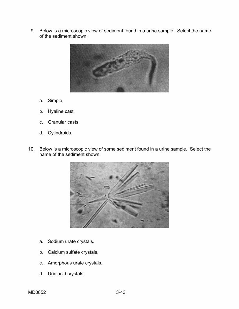
9. Below is a microscopic view of sediment found in a urine sample. Select the name of the sediment shown.
a. Simple. b. Hyaline cast. c. Granular casts. d. Cylindroids. 10. Below is a microscopic view of some sediment found in a urine sample. Select the name of the sediment shown.
a. Sodium urate crystals. b. Calcium sulfate crystals. c. Amorphous urate crystals. d. Uric acid crystals.
MD0852 3-43
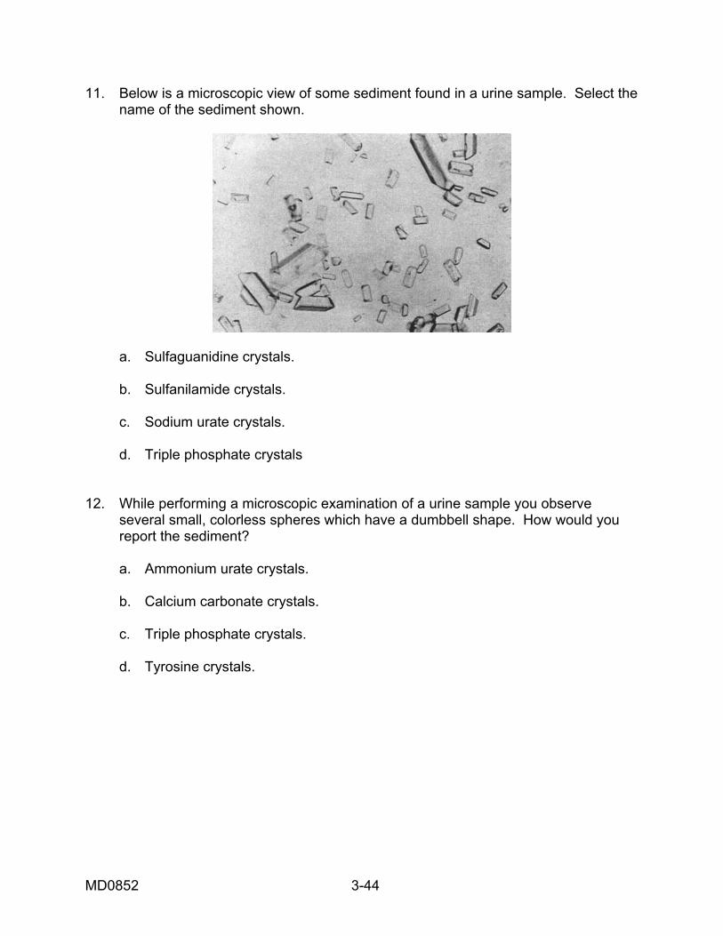
11. Below is a microscopic view of some sediment found in a urine sample. Select the name of the sediment shown.
a. Sulfaguanidine crystals. b. Sulfanilamide crystals. c. Sodium urate crystals. d. Triple phosphate crystals 12. While performing a microscopic examination of a urine sample you observe several small, colorless spheres which have a dumbbell shape. How would you report the sediment? a. Ammonium urate crystals. b. Calcium carbonate crystals. c. Triple phosphate crystals. d. Tyrosine crystals.
MD0852 3-44
13. While performing a microscopic examination of a urine sample you observe several colorless and semitransparent structures with cylindrical bodies that have parallel sides and rounded ends. How would you report these? a. Waxy casts. b. Red blood cell casts. c. Epithelial cell casts. d. Hyaline casts.
Check Your Answers on Next Page
MD0852 3-45
MD0852 3-46
SOLUTIONS TO EXERCISES 1. b (para 3-1) 2. c (para 3-2) 3. d (para 3-5) 4. a (para 3-9a) 5. c (para 3-11c)) 6. d (para 3-14c) 7. b (para 3-15d(3)) 8. a (para 3-9a) 9. d (para 3-12a, figure 3-16) 10. a (para 3-14d, figure 3-21) 11. d (para 3-16b, figure 3-32)) 12. b (para 3-16e, figure 3-35) 13. d (para 3-10, figure 3-8)
End of Lesson 3