Embed Size (px)
Citation preview

URINARY TRACT IMAGING -
BASIC PRINCIPLES
Clinical Radiology
Every physician needs a basic
understanding of diagnostic imaging to
understand how to order the
appropriate studies and to understand
the resulting radiologist's report.

• Urologists and nephrologists both treat kidney problems.
Urologists are surgical specialists who focus on anatomical or
structural disorders of the kidneys and urinary tract. They treat
problems such as kidney stones, kidney blockages, and kidney
cancer. Urologists are qualified to perform surgery and outpatient
medical procedures to correct such conditions.
• Nephrologists are medical specialists who focus on disorders
that affect the way the kidneys work such as diabetes and chronic
kidney disease. Nephrologists prescribe nonsurgical medical
treatments for these disorders.
What is the difference between a urologist
and a nephrologist?

Urologic Radiology
Science has given urologists a bevy of tools to probe the most
private parts of the body in diagnosing urinary and renal disease.
Every modern imaging technology, from conventional X-rays to
radionuclide imaging, has found its way into urologic radiology's
arsenal. The good news for physicians is that they have many
options to explore the kidneys, ureters, bladder and surrounding
structures. The better news for patients is that today's tests are
thorough, relatively pain-free and often quick.

Imaging can help the health care provider find the cause of
1.urinary retention—the inability to empty the bladder completely
2.urinary frequency—urination eight or more times a day
3.urinary urgency—the inability to delay urination
4.urinary incontinence—the accidental loss of urine
5.blockage of urine
6.abdominal mass
7.pain in the groin or lower back
8.blood in the urine
9.high blood pressure
10.kidney failure
What problems could require imaging of the
urinary tract?

METHODS OF INVESTIGATION
ULTRASONOGRAPHY
RADIOLOGY
Simple abdominal X-ray
Intravenous urography
Retrograde pieloureterography
Cystography
Uretrography
NUCLEAR MEDICINE
Static studies: static renal scintigraphy
Dynamic studies: renogram
CT (wi thout , wi th contras t , Angio)
MRI (wi thout , wi th contras t , Angio)

In imaging the urinary tract, the modality of choice for the
initial examination will almost universally be ultrasound (US).
US is inexpensive, immediate, painless, requires no sedation or
anesthetic, is widely available, and is radiation free. US can be
used to scan in any plane at the discretion of the operator, and
whereas the technique is entirely operator dependent, most
centers have staff with a high level of skill.
Imaging the urinary tract – which modality to use for first-line
examination?

Doppler USG
High frequency- high resolution but low penetration depth
Renal- parenchyma
evaluate hematuria, solid mass, cysts
congenital abnormalities, stones
Adrenal- CT/MRI better
Nodules, cysts, hemorrhage, location, tumors
Bladder- examine wall , lesions
Transvaginal, transabdominal, transrectal
Normal wall >= 6 mm
bladder volume
Prostate- transrectal , access for biopsy
ULTRASOUND

Scrotal-
Evaluate- mass, pain, torsion, orchitis, epididymitis,
hydrocele, hernia, varicoceles
Testicle- 4 x 3 cm
Veins- >2mm= varicocele- evaluate in erect position
ULTRASOUND


Doppler Ultrasound

PLAIN FILM OF THE ABDOMEN
The kidneys-ureters-bladder is often the first imaging study
performed to visualize the abdomen and urinary tract
The film is taken with the patient supine and
should include the entire abdomen from the base
of the sternum to the pubic symphisis
Can show bony abnormalities, calcification and
large soft tissue masses


Rapidly concentrated by kidneys and
opacifies urinary tract
Low osmolar iodine nonionic contrast
material less osmolar load- fewer
complications than high osmolar
Reactions:
Allergic, renal toxicity, shock
CONTRAST FILMS
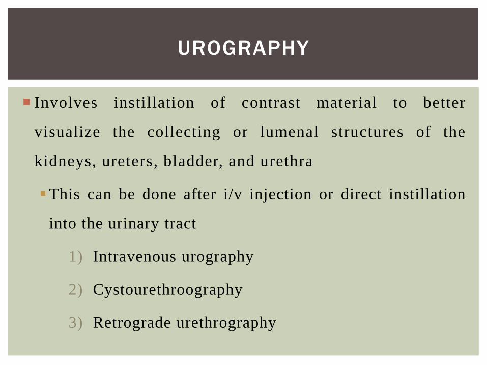
UROGRAPHY
Involves instillation of contrast material to better
visualize the collecting or lumenal structures of the
kidneys, ureters, bladder, and urethra
This can be done after i/v injection or direct instillation
into the urinary tract
1) Intravenous urography
2) Cystourethroography
3) Retrograde urethrography

INTRAVENOUS UROGRAPHY
IVU/ intravenous pyelogram is the classic modality of
imaging the entire urethelial tract from pyelocalyceal
system trhough the ureters and bladder
Excellent for indentifying small urethelial lesions as
well as the severity of obstruction from calculi
Provides anatomical and qualitative functional
information about the kidneys


CYSTOGRAPHY
Permits imaging of an opacified urinary bladder
after retrograde instillation of contrast media
through a urethral or suprapubic catheter
Imaging is performed to demonstrate a suspected
urine leak, either from traumatic bladder rupture
or after bladder surgery
Can also demonstrate a presence of a fistula
between the bladder and vagina or to characterize
bladder diverticuli

NORMAL MALE CYSTOGRAM

RETROGRADE URETHROGRAPHY (RUG)
Complete evaluation of the urethra includes both
antegrade and retrograde urethrography
Allows visualization of the anterior male urethra
Used for evaluating a suspected traumatic urethral injury
or urethral stricture
Can also be useful for diagnosis of a urethral diverticulum
in females
Evaluate anterior and posterior urethra- strictures, trauma
8-16 F foley in fossa navicularis, fill balloon with 1-2 mL
and inject 30-50% contrast while filming obliquely

Multi-Detector Computed Tomography (MDCT)

CT + ANGIO CT SCAN
often used examine structures in
the abdomen and pelvis (liver,
pancreas, gallbladder, spleen and
intestines).
CT Scans are a diagnostic tool that
urologists use to detect and
diagnose: recurrent urinary tract
infections, sources of blood in the
urine (hematuria), kidney stones,
renal cysts and masses. It can help
urologists rule out prostate,
bladder and renal cancers

Contrast- parenchyma, adrenals
3-D to evaluate vascular abnormality
100-150 mL i/v bolus injection
Renal- stages:
Precontrast- stones, parenchyma, vascular calcifications,
renal contour
30 sec- cortex vs medulla
Nephrographic- 100 sec- uniform enhancement of
parencyma (masses)
Pyelographic- excretory- collecting system
CT + ANGIO CT

CT
NO CONTRAST

CONTRAST
CT

No iodinated contrast
Soft tissue resolution better than CT
Contraindications- pacemaker, aneurysm
clips
T1- fluid dark, fat bright
T2- fluid bright, fat dark
MRI

MRI

Renal- will not evaluate stones, determine tumor
Adrenal- contain more fat than cancers, bright on T2, isodense
with liver
Bladder- to determin invasion of wall by cell cancer or other
pelvic neoplasms (on T2)
Prostate- evaluate prostate cancer for capsular invasion. T1-
distinct from surrounding fat/seminal vesicles (intermediate
intensity), T2- peripheral zone (high intensity), central
(intermediate), neurovascular bundles bright, seminal vesicles
(high)
MRI

CTMRI
Uses ionizing radiation,
high-dose procedure
Uses magnetic resonance, no ionizing
radiation
Excellent spatial
resolutionExcellent contrast resolution
Actual scanning time
measured in seconds
(typically <10 s)
Actual scanning time measured in minutes
(typically 45 min)
Rarely requires general
anesthetic in children
Frequently requires general anesthetic in
children, depending on age
Table 1Comparison of advantages and disadvantages between
computed tomography (CT)
and magnetic resonance (MR) imaging modalities

Excellent at showing
calcificationPoor at showing calcification (signal void)
Poor at showing edema or
pathological changes in specific
tissue types
Excellent at showing edema and pathological changes in
specific tissue types
Usually requires intravenous
contrast (unless looking for
calcification when not required)
Usually requires intravenous administration of contrast
(but certain sequences can be tailored if this is
contraindicated)
No known risk of nephrogenic
systemic fibrosis (NSF)
Risk of NSF (rare, but renal patients believed to be at
increased risk)
Less expensive Expensive
Usually available as an
emergency imaging techniqueNot routinely available as an emergency technique
No significant contraindications
Contraindicated in patients with any internal ferrous
objects (pacemakers, defibrillators, recent orthopedic
metalware, other implanted metallic devices, metallic
foreign bodies)
Open-style scannersGenerally quite enclosed scanners – risk of
claustrophobia

NUCLEAR MEDICINE
uses the radiation released by radionuclides (called nuclear
decay) to produce images
A radionuclide, usually technetium-99m, is combined with
different stable, metabolically active compounds to form a
radiopharmaceutical that localizes to a particular anatomic or
diseased structure (target tissue).
tracer goes to the target organ and can then be imaged with a
gamma camera, which takes pictures of the radiation photons
emitted by the radioactive tracer
Physiologic and anatomic info


ANGIOGRAPHY

AORTOGRAPHY: LEFT RENAL ARTERY THROMBOSIS

MR ANGIOGRAPHY
Left renal artery stenosis

Imagistic Criteria for HematuriaRADIOLOGIC EXAMINATION
PROCEDURE
APPROPRIATENESS
RATING COMMENTS
Multidetector CT urography 8 This is becoming the method of choice for hematuria, supplanting
intravenous pyelography
Radiography, intravenous
urography (intravenous pyelogram,
excretory urography)
8 —
Ultrasonography, kidney and
bladder, transabdominal
6 May miss ureteral and urothelial lesions; abdominal radiography,
retrograde pyelography, and cystoscopy are useful adjuncts
Radiography, retrograde urography 5 —
MRI urography 4 —
CT, abdomen and pelvis 4 CT may follow intravenous pyelogram or ultrasonography if initial
findings are ambiguous
Kidney, angiography 4 Rarely, vascular malformations may cause hematuria and require
angiography for diagnosis
Radiography, abdomen, KUB 2 It is assumed that a plain film of the abdomen will be part of the
indicated intravenous pyelogram; if an intravenous pyelogram is
not performed, KUB may be performed with ultrasonography
MRI, abdomen, and pelvis 2 —
Urinary tract scintigraphy 2 —
Virtual cystoscopy 2 —

Testicular torsion
US testes was performed which demonstrate the left testicle assuming an abnormal
orientation and lack normal color and power Doppler flow with maintained testicular normal
echogenicity, consistent with acute testicular torsion. The right testicle is within normal.

Ultrasound - stones

Nephrocalcinosis
Calcification which
appears medullary over the
left renal shadow.

PLAIN FILM-
LEFT DISTAL URETERAL CALCULUS

Intravenous urography


Retrograde urethrogram
20mm stricture in the bulbous urethra.

Urethral stricture
Anatomy of the normal
ureter on ascending
urethrogram.


Renal hypoplasia

Cazul 2
Renal hypoplasia

Ectopic kidney

Ectopic kidney
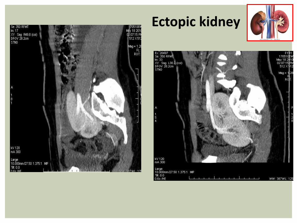
Ectopic kidney

Ectopic kidney

Ectopic kidney

Tumors

Tumors

Tumor

TUMOR
CT

TUMOR
CT

Hypervascular
process left kidney

Hypervascular
process left kidney

Hypervascular
process left kidney

Hypervascular
process left kidney

MRI
renal carcinoma

PARAPELVICAL CYST

Bosniak renal cyst classification
The Bosniak classification system for CT evaluation of renal cysts is helpful in
determining both malignant risk and required follow-up and/or treatment.
Bosniak 1
simple cyst, imperceptible wall, rounded
work up : nil
% malignant : ~ 0%
Bosniak 2
minimally complex, a few thin (< 1mm) septa, thin Ca++; non-enhancing high-attenuation
(due to to proteinaceous or haemorrhagic fluid) renal lesions of less than 3 cm are also
included in this category; these lesions are generally well marginated.
work up : nil
% malignant : ~ 0%
Bosniak 2F
minimally complex but requiring follow up.
increased number of septa, minimally thickened or enhancing septa or wall
thick Ca++,
hyperdense cyst that is:
> 3 cm diameter, mostly intrarenal (less than 25% of wall visible); no enhancement
work up : needs ultrasound / CT follow up
% malignant : ~ 25 %6

Bosniak 3
indeterminate, thick or multiple septations, mural
nodule, hyperdense on CT (see 2F)
treatment / work up : partial nephrectomy or RF
ablation in elderly / poor surgical risk
% malignant : ~ 54%6
Bosniak 4
clearly malignant, solid mass with large cystic or
necrotic component
treatment: partial / total nephrectomy
% malignant : ~100%

Extrarenal
renal cyst
expansion

Extrarenal
renal cyst
expansion

Extrarenal
renal cyst
expansion

Polycystic kidney disease: CT vs MRI

Polytraumaextensive skin emphysema kidney
contusion.

TRAUMA
CT

Normal prostate gland
Axial T2


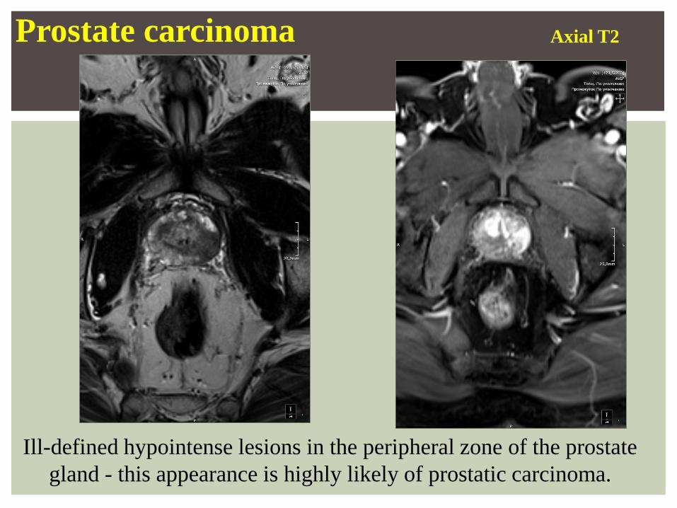
Prostate carcinoma Axial T2
Ill-defined hypointense lesions in the peripheral zone of the prostate
gland - this appearance is highly likely of prostatic carcinoma.