Embed Size (px)
Citation preview

URINARY SYSTEM
Human Anatomy Unit 3
Components
• Kidneys • Ureters • Urinary bladder • Urethra
Func:ons
• Storage of urine – Bladder stores up to 1 L of urine
• Excre:on of urine – Transport of urine out of body
• Regula:on: – Plasma pH – Blood volume/pressure – Plasma ion concentra:ons (Ca2+, Na+, K+, CL-‐) – Assist liver in detoxifica:on, amino acid metabolism
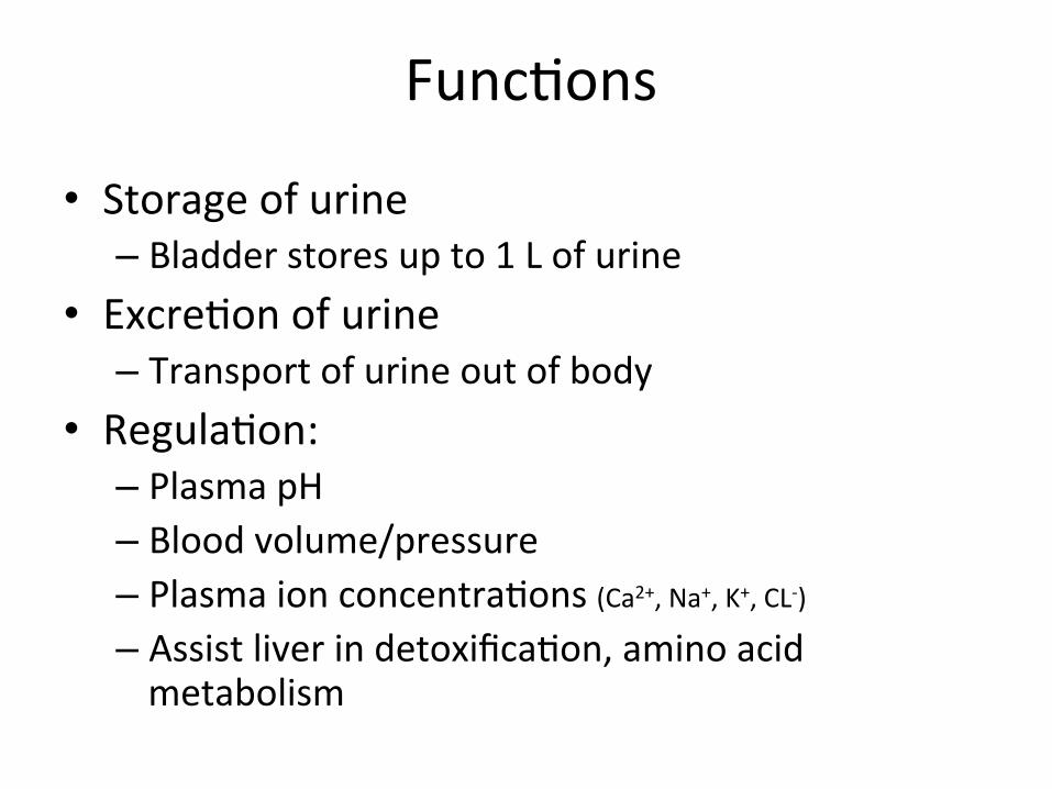
Kidney Gross Anatomy • Retroperitoneal
– Anterior surface covered with peritoneum
– Posterior surface directly against posterior abdominal wall
• Superior surface at about T12
• Inferior surface at about L3 • Ureters enter urinary
bladder posteriorly • LeT kidney 2cm superior to
right – Size of liver

Structure of the Kidney
• Hilum = the depression along the medial border through which several structures pass – renal artery – renal vein – ureter – renal nerves

Surrounding Tissue • Fibrous capsule
– Innermost layer of dense irregular CT
– Maintains shape, protec:on • Adipose capsule
– Adipose ct of varying thickness – Cushioning and insula:on
• Renal fascia – Dense irregular CT – Anchors kidney to peritoneum
& abdominal wall • Paranephric fat
– Outermost, adipose CT between renal fascia and peritoneum

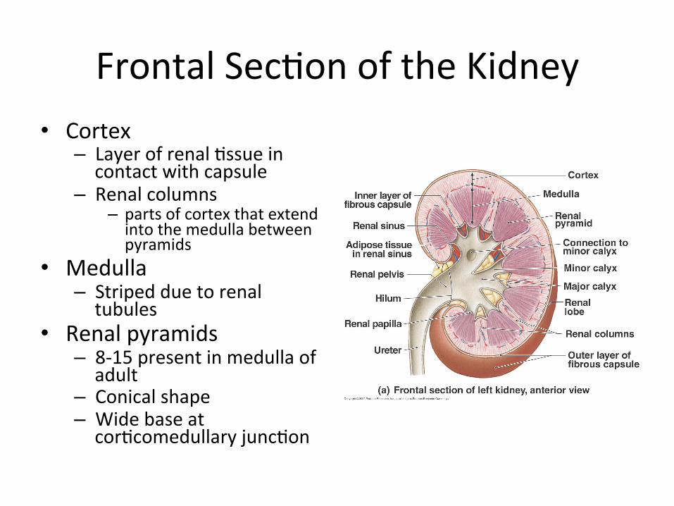
Frontal Sec:on of the Kidney • Cortex
– Layer of renal :ssue in contact with capsule
– Renal columns – parts of cortex that extend into the medulla between pyramids
• Medulla – Striped due to renal tubules
• Renal pyramids – 8-‐15 present in medulla of adult
– Conical shape – Wide base at cor:comedullary junc:on

Flow of Filtrate/Urine • Collec:ng ducts
– Collect from mul:ple nephrons • Minor calyx
– Collect from each pyramid
• Major calyx – Collect from minor calyx
• Renal pelvis – Collects from calyces, passes
onto • Ureter
– Collects from pelvis
• Urinary Bladder – Collects from ureters
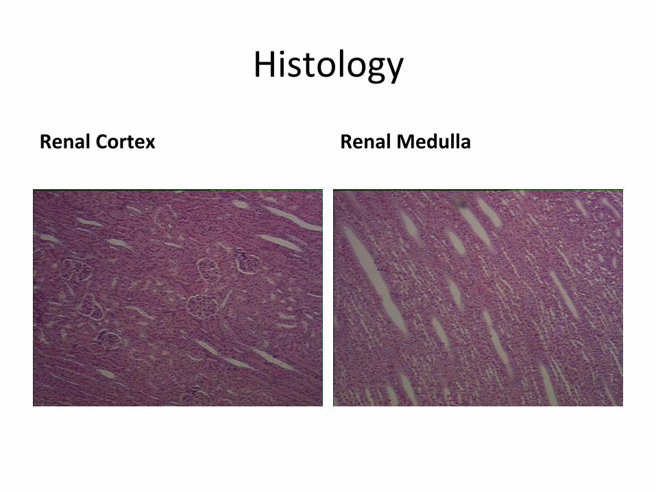
Histology
Renal Cortex Renal Medulla

Renal Tubules
• Nephron – func:onal unit of the kidney.
• Each kidney contains approximately 1 million nephrons
• Form urine by filtering and adjus:ng composi:on of blood carried by renal vasculature
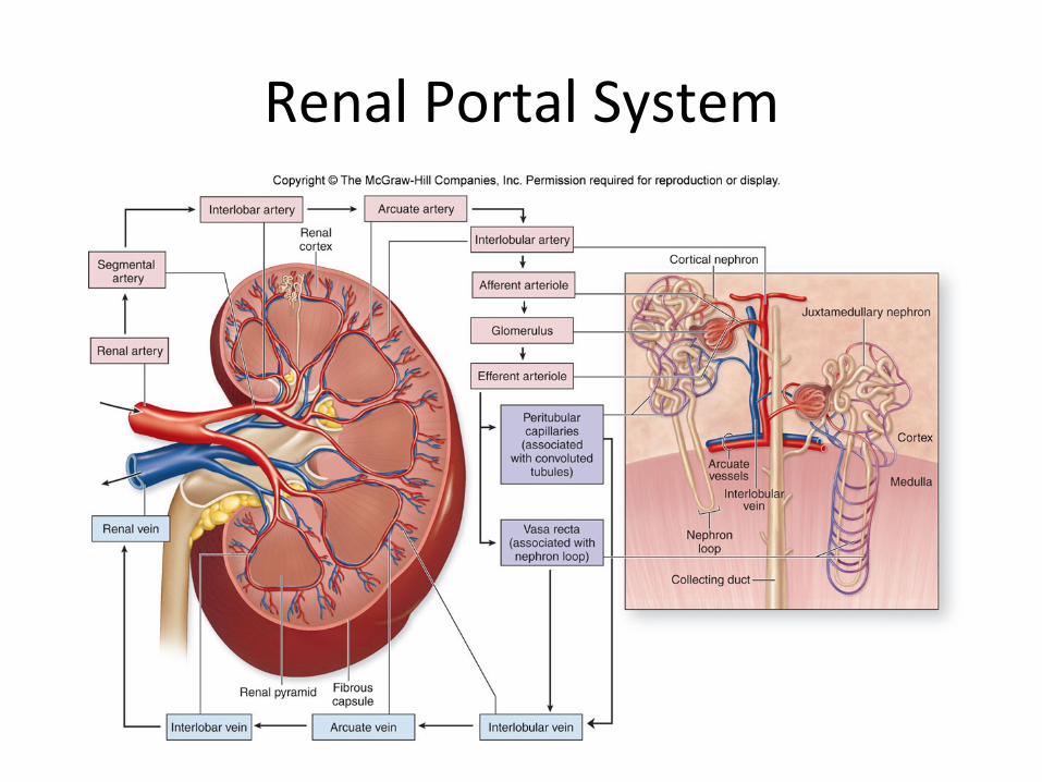
Renal Portal System

Renal Portal System

Histological Structure of a Nephron • Renal corpuscle
– Glomerulus – Bowman’s capsule
• Renal tubules – Proximal convoluted tubule
– Descending limb of LOH – Loop of Henle – Ascending limb of LOH – Distal convoluted tubule – Collec:ng duct
• Associated blood vessels - Peritubular capillaries - Vasa recta

Types of Nephrons

The Glomerulus
• Bowman’s capsule • Glomerulus • Afferent arteriole • Efferent arteriole • Podocytes
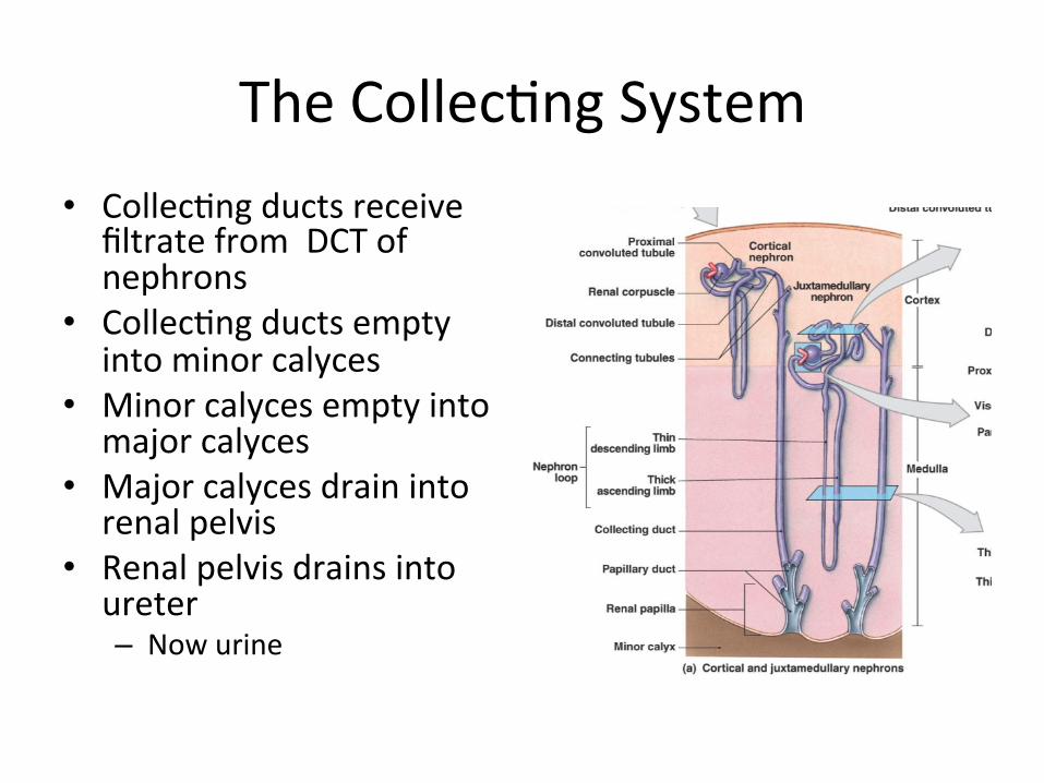
The Collec:ng System • Collec:ng ducts receive
filtrate from DCT of nephrons
• Collec:ng ducts empty into minor calyces
• Minor calyces empty into major calyces
• Major calyces drain into renal pelvis
• Renal pelvis drains into ureter – Now urine

The Ureters • Expandable tubes that exit the renal pelvis
• 3 walls – Mucosa
• Transi:onal epithelium – Muscularis
• smooth muscle layer – Adven::a
• protec:ve fibrous CT – Ureters drain into the posterior por:on of the urinary bladder

The Urinary Bladder
• Func:ons to store urine • Structure – Rugae
• macroscopic folds as in the stomach
• flaeen when the urinary bladder is distended
– Trigone • triangular region of the bladder
• no rugae • loca:on of openings to the ureters and urethra

Histology of the Urinary Bladder
• Mucosal lining – transi:onal epithelium
• Submucosa – fibrous CT
• Muscularis – detrusor muscle
• 3 layers of smooth muscle
• Serosa – loose CT – visceral peritoneum

The Female Urethra
• Drains urine from urinary bladder to exterior
• 1-‐2 inches • higher risk for bladder infec:ons

The Male Urethra
• 3 regions: – prosta:c – membranous – penile

Histology of the Urethra
• Mucosa – varies from bladder to exterior especially in males
• Muscularis layer • Adven::a • Sphincters – internal = smooth muscle (involuntary)
– external = skeletal muscle (voluntary)


![[PPT]Atypical Bacteria - Mt. SAC Faculty Directoryfaculty.mtsac.edu/trevell/micro22/m22u1cs.ppt · Web viewTitle Atypical Bacteria Author Tim Revell Last modified by Tim Revell Created](https://img.pdfslide.net/doc/110x75/5ac91f257f8b9a51678cf177/pptatypical-bacteria-mt-sac-faculty-viewtitle-atypical-bacteria-author-tim.jpg)