Embed Size (px)
Citation preview
U R O B I L I N PHYSIOLOGY AND PATHOLOGY.
V. THE I~LATION BETWEEN UROBILIN AND CONDITIONS INVOLVING INCREASED RED CELL DESTRUCTION.
Bx ROBERT ELM.AN, M.D., Am) PHILIP D. McMASTER, M.D.
(From the Laboratories of The Rockefeller Institute for Medical Research.)
(Received for publication, June 25, 1925.)
Much of the interest in urobilin has come from the fact tha t i t is found in the urine of patients suffering from diseases characterized by an excessive destruction of red blood cells. The occurrence of urobillnuria has been reported in hemolytic icterus, pernicious anemia, malaria, and under various circumstances involving hemor- rhage, as, for example, those of hemophilia, apoplexy, and hemo- thorax. 1-3 There have been m a n y clinical studies in such connec- tion, bu t little experimental work has been reported, and despite a general belief in the importance of the phenomenon, no uniform con- ception has developed of its pathological or diagnostic significance in the conditions mentioned.
Many workers have assumed that urobilin may be manufactured directly from the hemoglobin or bilirubin accumulation in the tissues as result of blood extra- vasation and destruction. The pigment thus produced is supposed to be carried by the blood to the kidneys and excreted by the urine. And yet Kunkel 4 was una~ole to show this experimentally; and Quincke 5 repeat.edly attempted to demonstrate urobilin in old blood clots without success.
The presence of large amounts of urobilin in the stools of patients suffering from hemolysis has long been recognized. Indeed, the estimation of the pigment in the feces has, in recent years, been taken as an approximate measure of the amount of red cell destruction. 8 Yet, many extraneous factors have been shown
1 Hoppe-Seyler, G., Virckows Arch. path. Anat. 1891, cxxiv, 30. ~ Meyer-Betz, F., Ergebn. inn. Med. u. Kinderheilk., 1913, xii, 733. 8 Eppinger, H., Die hepato-lienalen Erkrankungen, p. 76, in Enzyldopmdie
der klinischen Medizin, Berlin, 1920. 4 Kunkel, A., Virchows Arch. path. Anat,, 1880, lxxix, 455. 5 Quincke, H., Virchows Arch. path. Anat., 1884, xcv, 125.
Robertson, O. H., Arch. Int. Med., 1915, xv, 1072. 619
620 UROBILIN PHYSIOLOGY AND PATI~IOLOGY. V
to influence in practice the output of fecal urobilin. Even in healthy animals these cause wide fluctuation in the values. 7
In our earlier papers the normal physiology of urobilin has been studied. ~-9 The communication 1~ immediately preceding the pres- ent one dealt with conditions of liver damage and biliary obstruc- tion. We are here concerned with the effects of increased blood destruction and allied conditions. For the purposes of the investi- gation the quantitative measurement of urobilin was made by a method which has already been described. 8 The errors encountered in the study of bile obtained from the open fistula of Schwann have been avoided by a sterile method of intubation7 ,u which renders possible precise determinations of the entire liver output.
Methods.
The general methods adopted for the intubation of the bile ducts of dogs, care of the animals, and the daily collection of and pigment determinations on urine, stool, and bile have already been described. 7,8 All the animals were permanently intubated, some for the collection of the total bile, others for that of but a sample of the liver output, while still others were so intubated as to permit the bile to flow either into the duodenum or to the collecting bag at the will of the observer. Every day or two portions of the collected bile were incubated with agar to deter- mine whether it had become infected, and furthermore, centrifugalized specimens were frequently examined to the same end. When bacteria were found the animal was discarded for the purposes of this work. All the experimental procedures were carried out under aseptic conditions.
In some instances the effects of an excessive destruction of red blood cells, consequent on intercurrent disturbances, were studied, while in others destruction was induced by the giving of a hemolytic substance or by the intravenous injection of distilled water. Several instances of localized infection, especially of the respira- tory tract, accompanied by increased blood destruction, came under observation.
Toluylenediamine in aqueous solution causes a marked red cell destruction. 12 I t was given, through a stomach tube, in doses averaging 0.05 to 0.1 gin. per kilo of animal. Icterus developed, yet the dogs remained active and in what appeared
7 McMaster, P. D., and Elman, R., J. Exp. Med., 1925, xli, 513. 8 Elman, R., and McMaster, P. D., J. Exp. Med., 1925, xli, 503. 9 McMaster, P. D., and Elman, R., J. Exp. Med., 1925, xli, 719.
1o Elman, R., and McMaster, P. D., J. Exp. Med., 1925, xlii, 99. 11 Rous, P., and McMaster, P. D., J. Exp. Med., 1923, xxxvii, 11. 13 Eppinger, H., Die hepato-lienalen Erkrankungen, p. 124, Berlin, 1920.
ROBERT ELMAN AND PHILIP D. McMASTER 621
to be good condition. Toluylenediamine not only damages the red cells, however, but causes more or less severe liver injury. 1° Sodium oleate, 50 to 75 cc. of a 1 per cent aqueous solution, was given to some animals intravenously on successive days.
To produce an immediate hemolysis distilled water was employed. It was either given directly into a vein,--in amounts of 250 to 300 cc. of it to animals of about 10 kilos during a period of 3 to 5 minutes,---or 100 to 150 cc. of blood was withdrawn, shaken with 200 to 250 cc. of sterile water to hemolyze it, and the mixture reinjected after straining it through a cotton mesh.
The effects of the extravasation of blood into the tissues could be studied as an incidental result of the intubation, since the operative procedures inevitably in- volved more or less hemorrhage into the tissues about the incision. In certain cases artificial hematomas were produced by the subcutaneous injection of sterile blood, usually obtained from the animal's own vein.
For special purposes, large amounts of urobilin-free bile (two to three times the ordinary 24 hour output of the animal) were given by garage. To imitate the increased passage of bilirubin into the intestine which follows upon increased blood destruction this pigment was given by garage on one occasion. It had been prepared from fresh dog bile by a method already described. 9
In all of the cases the percentage of the circulating hemoglobin was determined daffy by the Newcomer method, following a procedure already described. ~ This percentage varies somewhat from day to day and in the absence of any complica- tion the output of bilirubin in the bile of intuhated dogs tends to vary in parallel with it. ~a The relation that exists between the two pigments is not strictly quantitative, yet it is sufficiently close to give ground for the supposition that excessive blood destruction is going on whenever any marked increase occurs in the bilirubin output34
Absence of Urobilinuria during Increased Blood Destruction under Conditions Involving Total Bile Loss.
I t may be recalled from our previous papers tha t when the common
duct is so in tubated tha t none of the bile reaches the intestine bu t
the whole is lost to the organism, urobilin disappears from this secre-
tion as also from the stools and urine; 7 and even serious damage to
the liver does not cause it to reappear30 The same holds true when
the damage has been to the blood, or to both blood and liver, as in the case of toluylenediamine.
la Broun, G. O., McMaster, P. D., and Rous, P., J. Exp. Med., 1923, xxxvii, 733. t4 Rous, P., Physiol. Rev., 1923, iii, 75.
622 UROBILIN PHYSIOLOGY AND PATHOLOGY. V
Specimen Protocols.
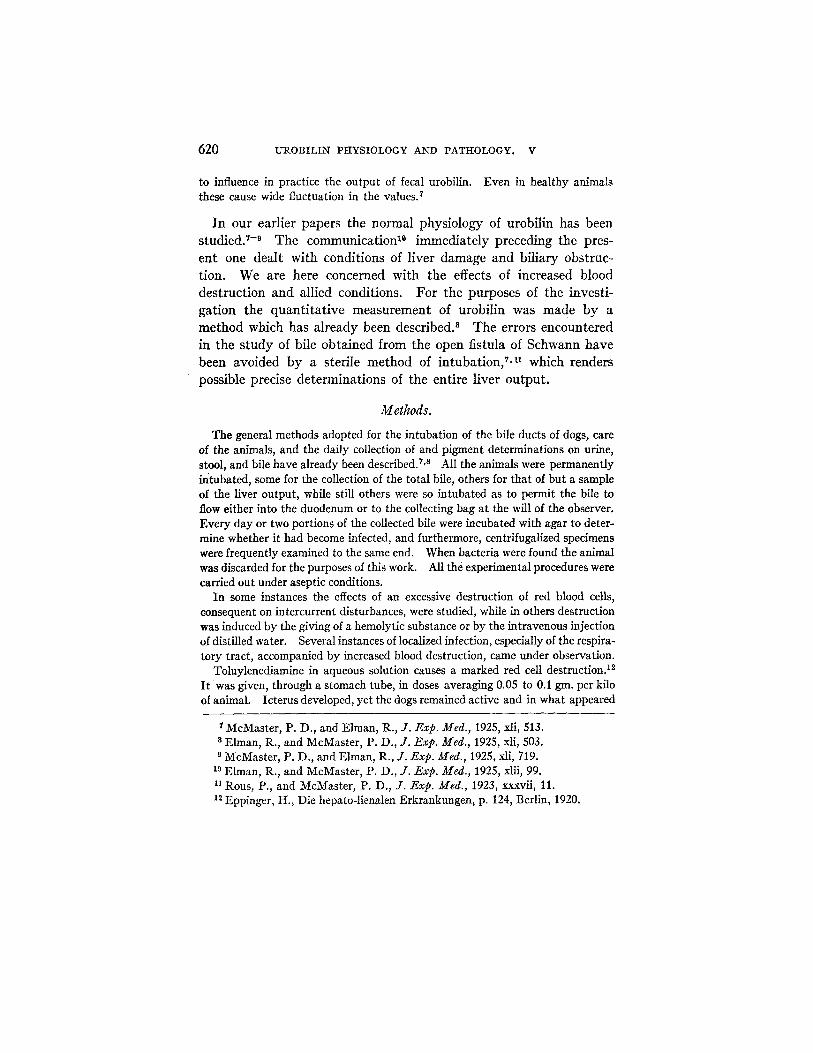
I. Female dog, weight 93 kilos (see Text-fig. I). Under ether, intubation of the common duct was performed. By the 3rd day
the stool and urine had become urobilin-free, and they remained so thereafter.
tfi
~ too ~to5 ~ '),, ? 9 0 u 100 ,1~ ' "=
" '! / "5 A
/' 40 / , 1o u ~oDfl in
X~ 50 C
z0 /
#
o < .,"I ~o ~Po~,i1{n, ~r~. 6.. - . .
Dcxys 30 3% 3Z 33 34 35 36 37
Aftet~ opepation
T~xT-FrG. 1. Absence of urobilinuria during pathological blood destruction in a dog losing the total bile.
Toluylenediamine in aqueous solution was given by mouth to an animal with intubated common duct. There resulted a progressive fall in the percentage of circulating hemoglobin, and the bilirubin output nearly doubled. There was a marked bilirubinuria and tissue icterus, yet no urobilin was to be found in the bile or urine, and the stools remained consistently acholic and practically free from urobilin (see Protocol I).
1 month after operation the animal was healthy and active; and the bilirubin output of the bile was constant at about 50 rag. in 24 hours. A small dose (0.475 tim.) of toluylenediamine was now given. There resulted, as the chart shows, a progressive fall in hemoglobin; and a tissue icterus developed which persisted several days. The bilirubin output in the bile reached a maximum of 92 nag. in the fourth 24 hour specimen after administration of the hemolytic agent. Bill-
ROBERT ELMAN AND PHILIP D. M¢~6ASTER 623
rubinuria was marked. At no time however was urobilin found in the bile or urine. The stools contained about 2 or 3 mg. in each 24 hours. The presence of this amount is to be attributed to the escape of bile pigment from the jaundiced wall of the gut into the lumen, with subsequent bacterial action. ¢
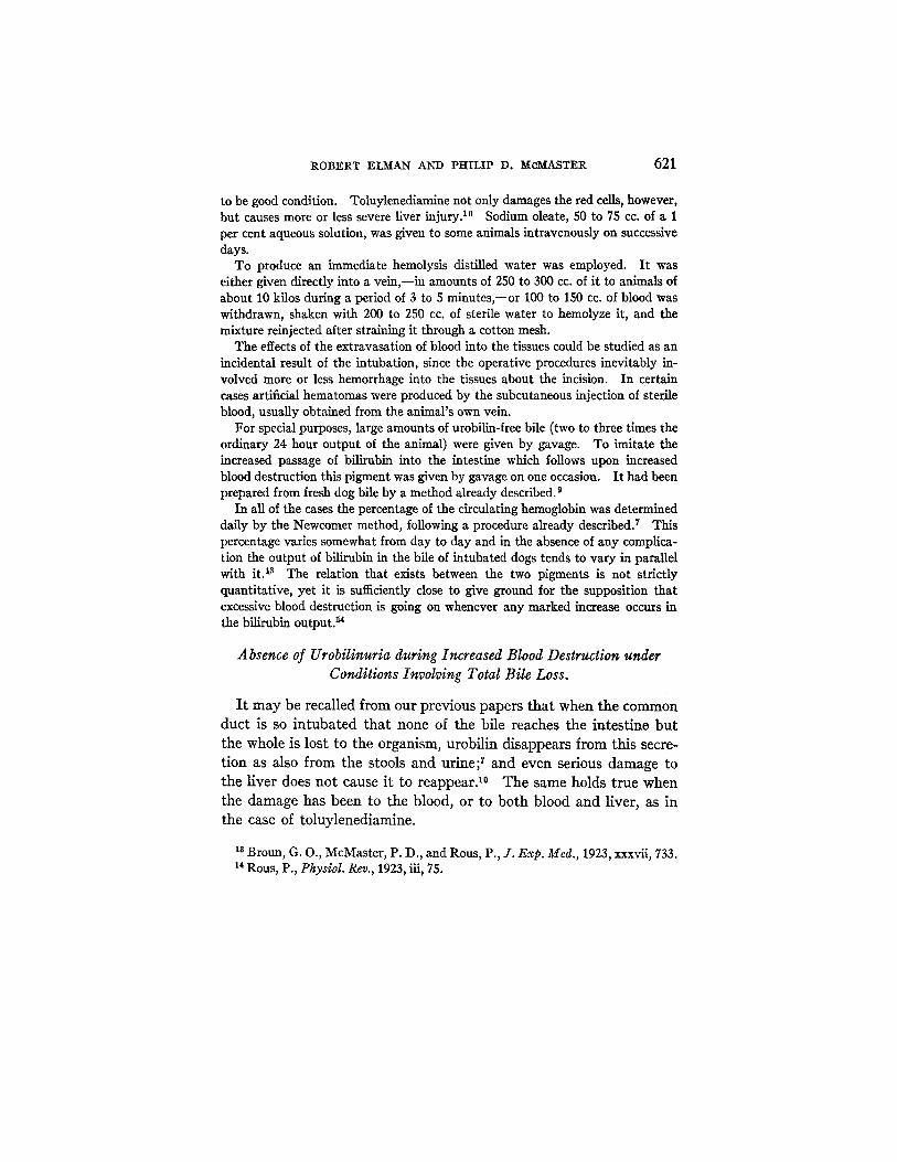
I I . Male dog, weight 10½ kilos (see Text-fig. 2). The common duct had been cannulated under ether anesthesia, and daily col-
lections of the bile made. I t remained sterile, and, after the 3rd day, contained no urobilin.
190
i20
110 o~
too p-
80 ~'
g 6o ,~ so
:~ 50 ~ 20
40 ~ tO
i ~ too
P~
/ D~ 4Z
/
43
J
/ • ! N o 1~ob fin k bite i ~k_,
%
\ N ut~ )bilic aPia 44 45 4S 47 48
A~te~ opePation
TExT-Fro. 2. Absence of urobilinuria in a dog losing the total bile and in- jected with hemolyzed blood.
A portion of the animal's own blood laked outside of the body with distilled water was injected intravenously. The drop in hemoglobin was 18 per cent, and there was hemoglobinuria and bilirubinuria, yet no urobilin was found at any time in the urine, bile, or feces (see Protocol II) .
After 6 weeks collection of bile specimens, 100 cc. of blood was withdrawn from the jugular vein of the animal, shaken with 200 cc. of sterile, distilled water, and the whole reinjected intravenously after straining. The injection required 3 minutes. The bilirubin output rose in the next 24 hours from the "normal" of 45 rag. to 125 rag. The urine contained large amounts of hemoglobin, and, on the day following, much bilirubin. The hemoglobin percentage fell 18 points. In 3 days the bile and urine had returned to normal. At no time was urobilin found in the bile, the urine, or the stools.
624 UROBILIN PHYSIOLOGY AND PATHOLOGY. V
Urobilinuria after Increased Blood Destruction under Conditions of Partial Bile Loss.
In the course of our previous work it was shown r tha t when only a fraction of the bile is collected each day, with the rest flowing into the intestine as usual, urobilin continues to be a const i tuent of bo th bile and stools, while furthermore, upon liver injury, 1~ urobilinuria develops. Sodium oleate exerts a direct hemolyt ic action on the red cells, even in vitro, ~ but there seems to be no evidence tha t it injures the liver. Following the giving of it urobilinuria is usually not pronounced. Toluylenediamine injures the liver as well as the blood, TM and it might be expected from what has just been said, tha t a marked urobilinuria would follow the giving of it, as is the actual case. The amount of urobilin in the stools increases greatly, in corollary to the increase in the amount of bilirubin reaching the bowel as the result of blood destruction.
The difference in the degree of urobilinuria in the two instances selected for i l lustration m ay lie in the fact tha t toluylenediamine, unlike sodium oleate, causes liver damage, but the prot rac ted action of the substance would in itself lead to a sustained and perhaps a cumulation effect. The effect of the intravenous injections of sodium oleate is practically immediate and m ay be completed within a few minutes. The fall in the hemoglobin percentage was about the same in each of the instances now to be given.
Specimen Protocol.
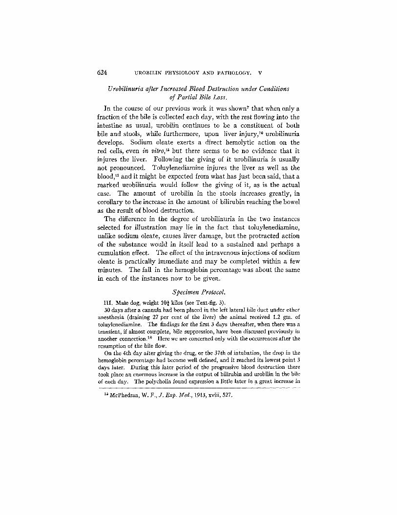
III. Male dog, weight 10~ kilos (see Text-fig. 3). 30 days after a cannula had been placed in the left lateral bile ctuct under ether
anesthesia (draining 27 per cent of the liver) the animal received 1.2 gin. of toluylenediamine. The findings for the first 3 days thereafter, when there was a transient, if almost complete, bile suppression, have been discussed previously in another connection. 1° Here we are concerned only with the occurrences after the resumption of the bile flow.
On the 4th day after giving the drug, or the 37th of intubation, the drop in the hemoglobin percentage had become well defined, and it reached its lowest point 3 days later. During this later period of the progressive blood destruction there took place an enormous increase in the output of bilirubin and urobilin in the bile of each day. The polycholia found expression a little later in a great increase in
Is McPhedran, W. F., J. Exp. Meal., 1913, xviii, 527.
ROBERT ELIIIAN AND PHILIP D. McMASTER 625
the urobilin of the stools. The disturbances subsided gradually, and in 10 days after the giving of the toluylenediamine its effect was almost spent.
The urobilinuria attendant upon the blood destruction was intense, and in general followed the curve representing the increase in bilirubin elimination in the
P..O0 36O ~30
.~ 3oo
r~ . 150 ~ Z~O
8o ~ ~ z4o ,~ ~oo
I BO~-IOO ,~ 180 I~,00 150 e 250 so~ ~ z
40 V~ t o F~ ~oo go 150
~o ~ ~ ~o ~ ~o
o
goo ~ 80 ""
gO0
200 c
650
~SgO I r lll I H I I I .~_, 450
I
D ~ 3 3 34 35 36 3~ 38 39 40 4t 42 43 44 45 46 4~ 48 Al~teP operation
T~xT-FIo. 3. Urobilinuria during excessive blood destruction in a dog losing but a fraction of the total bile.
Toluylenediamine in aqueous solution was given by stomach tube. The findings for the first 3 days, when there was almost complete bile suppression, have been discussed before. I° With the resumption of bile flow, and the excretion of bile pigment in great quantity, the bilirubinuria consequent upon the suppres- sion lessened and disappeared, whereas urobilin appeared in stool and urine, the quantity soon becoming great and only lessening again as the output of bilirubin in the bile lessened. Concomitantly there was a parallel increase and then a de- crease in the urobilin of the bile. These changes were attended by a fall in hemoglobin of 30 per cent--another gauge of the degree of blood destroyed (see Protocol III) .
626 UROBILIN PIIYSIOLOGY AND PATHOLOGY. V
bile. Bilirubinuria was somewhat less pronounced and of shorter duration, as would follow from the circumstance that the bile,flow was not long impeded. Thus bilirubinurla disappeared the day after the peak of the blood destruction was reached, while urobilinurla continued for 3 more days. Such a relationship, which will be further illustrated in experiments to be described, is characteristic of the two pigments in conditions characterized by excessive hemolysis.
5~ SO0
zGo
40 ~ 240 100
eo 28 180 ~ ~0
C 24 ~Z i60 - -
20 .~ i40 . . . . . .
]~:~i6 SO ~-i20 ,el
~ o 0 N 0 i " ~.:,.'.
D~ys 25 26 27 25 Z9 30 St A~te~ ope~t ion
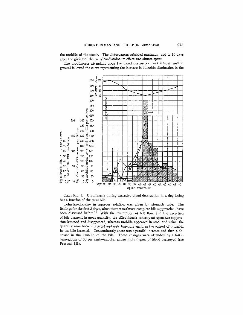
TExT-FIG. 4. Urohilinuria during excessive blood destruction in a dog losing but a fraction of the total bile.
Intravenous injections of sodium oleate were given on successive days. The blood destruction was reflected in a tremendously increased output of bilirubin in the bile. There was a slight urobilinuria when the disturbance was at its height, but no billrubin ever was found in the urine (see Protocol IV).
Specimen Protocol.
IV. Male dog, weight 12 kilos (see Text-fig. 4). The intubation under ether anesthesia made it possible to collect each day
the bile from the left lateral lobe of the liver, which was found later a t necropsy
ROBERT ELMAN AND P1KILIP D. McMASTER 627
to comprise 32 per cent of the organ. The bUe remained sterile and showed, like the stool, an approximately uniform content of urobilin.
4 weeks after the operation, 50 cc. of a 1 per cent solution of sodium oleate in water was injected intravenously on 2 successive days. There resulted a drop of 20 points in the hemoglobin percentage. The bile of the 24 hours after the first injection contained four times the usual amounts of bilirubin and urobilin. The stool also contained increased amounts of urobitin. There was a further increase in the pigments after the second injection, but 48 hours later the conditions had returned to the previous "normal." There was a transient urobilinuria but no bilirubinuria.
Urobilinuria during the Extravasation of Blood after Operation.
Immediately after the operation to intubate a common duct the output of bile pigment is greater than at later times, and it is especially prone to be so during the second or third 24 hours of bile collection. TM ~* I t has been pointed out that this is, in part, to be accounted for by the pigment derived from blood extravasated during the operative manipulations. The possibility that the anesthetic causes hepatic injury must also be borne in mind in connection with the urobilin findings now to be described.
When the common duct has been intubated, with result that all of the biliary secretion is lost to the organism, the animal rapidly becomes urobilin-free, there being none of the pigment in bile, urine, or stools7 despite the blood extravasation and potential liver injury just referred to. But when, on the other hand, the bile from one portion of the liver only is collected while the rest flows as usual to the gut, there is almost regularly urobilinuria following the intuba- tion. In only one instance out of eighteen has it been absent. I t develops at a t i m e when animals with intubated common duct are becoming urobilin-free.
The amount of the pigment often does not exceed 3 to 4 mg. per day during the 2 or 3 days that it is present. But in one instance it was abundant (Dog 4), an instance in which the abdominal incision had become infected and purulent. During 4 days, when the infec- tion was at its height, 175 rag. of urobilin was excreted in the urine and the 24 hour bilirubin content of the bile rose from 70 rag. to 103,
~s McMaster, P. D., Broun, G. O., and Rous, P., J. Exp. Med., 1923, xxxvil, 395.
628 UROBILIN PHYSIOLOGY AND PATIZIOLOGY. V
107, and 100 rag. on 3 successive days. Even then there was no bflirubinuria. Thereafter the purulence lessened rapidly and the incision healed.
Specimen Protocols.
V. Male dog, weight 12¼ kilos (see Text-fig. 5). The urine for the 5 days preceding operation was urobilin-free. Intubatiun
under ether anesthesia was performed in such wise that two-thirds of the biliary
eJ
80 O-
f
ff
3O
¢ 4
~ o ~" Q. mI
° ~
c c
ffo ' Days 0 i 2 3 4
A~tep opep~t ion
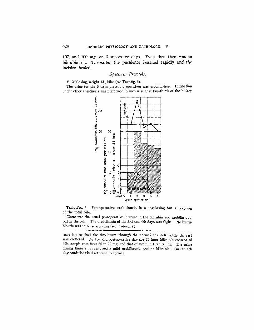
TEXT-FIG. S. Postoperative urobilinuria in a dog losing but a fraction of the total bile.
There was the usual postoperative increase in the bilirubin and urobilin out- put in the bile. The urobilinurla of the 3rd and 4th days was slight. No biliru- hinurla was noted at any time (see Protocol V).
secretion reached the duodenum through the normal channels, while the rest was collected. On the 2nd postoperative day the 24 hour bilirubin content of bile sample rose from 66 to 90 rag. and that of urobilin 10 to 30 nag. The urine during these 2 days showed a mild urobilinuria, and no bilirubin. On the 4th day condltions*had returned to normal.
ROBERT ELMAN AND PHILIP D. McMASTER 629
VI. Male dog, weight 12½ kilos (see Text-fig. 6). Under ether the gal l bladder was removed and an "altercursive" intubation was
carried out, ~ two cannulas being inserted into the common duct which was cut between. The upper cannula and tube collected all the bile from the liver, while the lower one connected with the duodenum through the ampulla of Vater. The
'700
650
600
550
~ 500
&50
~ 400 o -
30 _ 3o0 , ~ o °
~5 ~ ~ o
.~ 2.0 ~ ~00
t5 ,50
~: tO X: ioo / \ D~y~ 0 t Z 3 4 5
A~teV operation 6 ~ 8
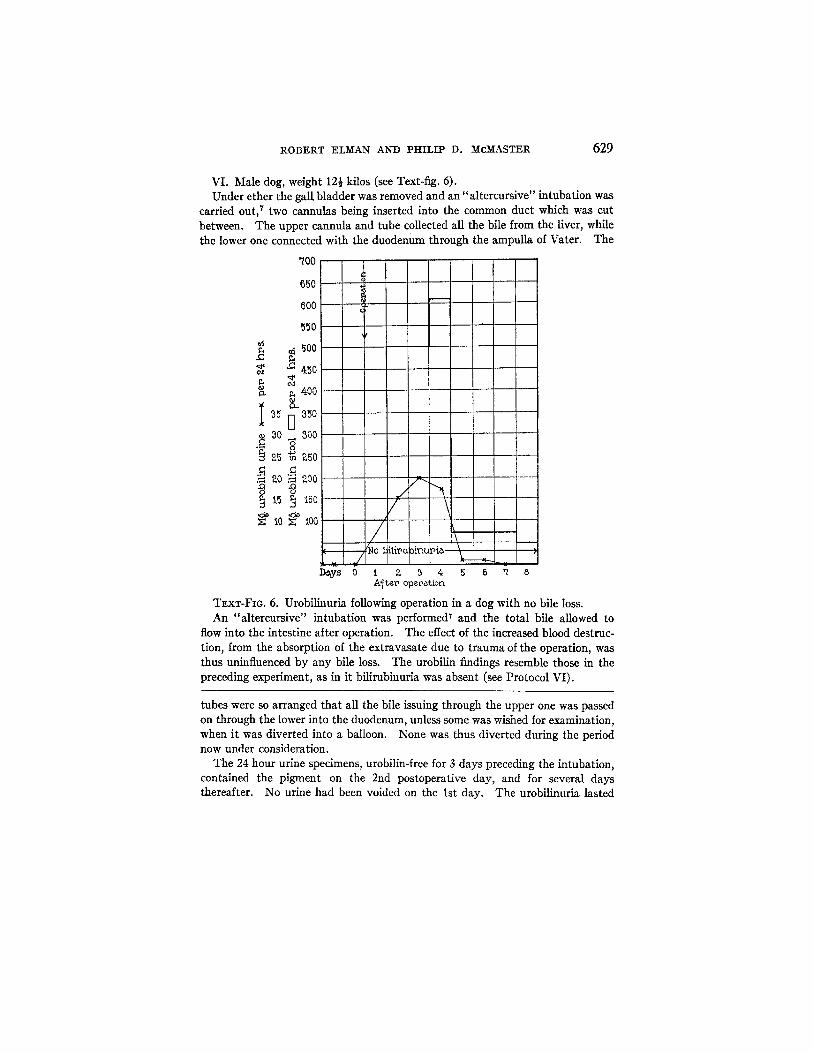
TExT-Fla. 6. Urobilinuria following operation in a dog with no bile loss. An "altercursive" intubation was performed ~ and the total bile allowed to
flow into the intestine after operation. The effect of the increased blood destruc- tion, from the absorption of the extravasate due to trauma of the operation, was thus uninfluenced by any bile loss. The urobllin findings resemble those ill the preceding experiment, as in it bilirubinuria was absent (see Protocol VI).
tubes were so arranged that all the bile issuing through the upper one was passed on through the lower into the duodenum, unless some was wished for examination, when it was diverted into a balloon. None was thus diverted during the period now under consideration.
The 24 hour urine specimens, urobilin-free for 3 days preceding the intubation, contained the pigment on the 2nd postoperative day, and for several days thereafter. No urine had been voided on the 1st day. The urobilinuria lasted

630 U-ROBILIN PHYSIOLOGY AND PATHOLOGY. V
longer than usual. No bilirubinuria was found at any time, indicating a complete ability of the liver to handle this pigment. Since no bile was collected, our only measure of the postoperative polycholia was the stool urobilin which was, for a brief period, notably abundant.
Absence of Urobilinuria during the Absorption of Extravasated Blood from ttematomas.
The effects of extravasation of blood into the tissues many days after operation, when there was no suspicion of hepatic injury, were also studied. In seven animals, three draining the total bile, and four, partial bile, artificial hematomas were produced, in the cervical region and in the abdominal wails, by subcutaneous injections of sterile blood, 30 to 60 cc., obtained from the animal's own vein. In all the instances there followed an increase in the bilirubin output through the bile but no urobilinuria.
Urobilinuria Associated with Excessive Blood Destruction during Infections.
Many animals showed evidence of increased blood destruction during infections, as revealed by an increase in the bilirubin output in the bile. These cases are therefore included in this report. One such instance, of infection of a healing incision, has already been referred to, and other similar ones could be cited. In such cases the relative parts played in the causation of the urobilinuria by the opera- tion, the blood extravasation incident thereto, and the infection could not be discriminated.
The most common infection was distemper with which many dogs suffered, particularly during the early spring months. In such of the distemper animals as were losing the total bile urobilin was never found, whereas in those losing but a fraction of it, or none at all ("altercursive" intubafion) the affection frequently brought on urobilinuria. The finding has special interest in view of the fact that urobilinuria is encountered during lobar pneumonia in human beings with intact bile ducts.
Specimen Protocol.
VII. Female dog, weight 24 kilos (see Text-fig. 7). 6 days after the operation for intubation of one of the branches of the hepatic
duct, under ether anesthesia, the animal developed a respiratory infection, be-
ROBERT ELM_AN AND PHILIP D. ~cM_ASTER 631
ginning with a purulent nasal discharge, and becoming gradually worse until there was frank dyspnea. I t was somewhat prostrated, ate very little, and lost its former activity. The severe symptoms lasted only 3 days, and recovery was complete within a week.
On the 2rid day of the animal's illness the amount of bile elaborated by the in- tubated portion of the liver fell from 97 to 87 cc. but the secretion was darker and
.c
~xt
too
I ~0
I tBo
too
8o
o
60
40
20
D~y~ 6 7 8 9 10 11 A~ter opepation
12 13
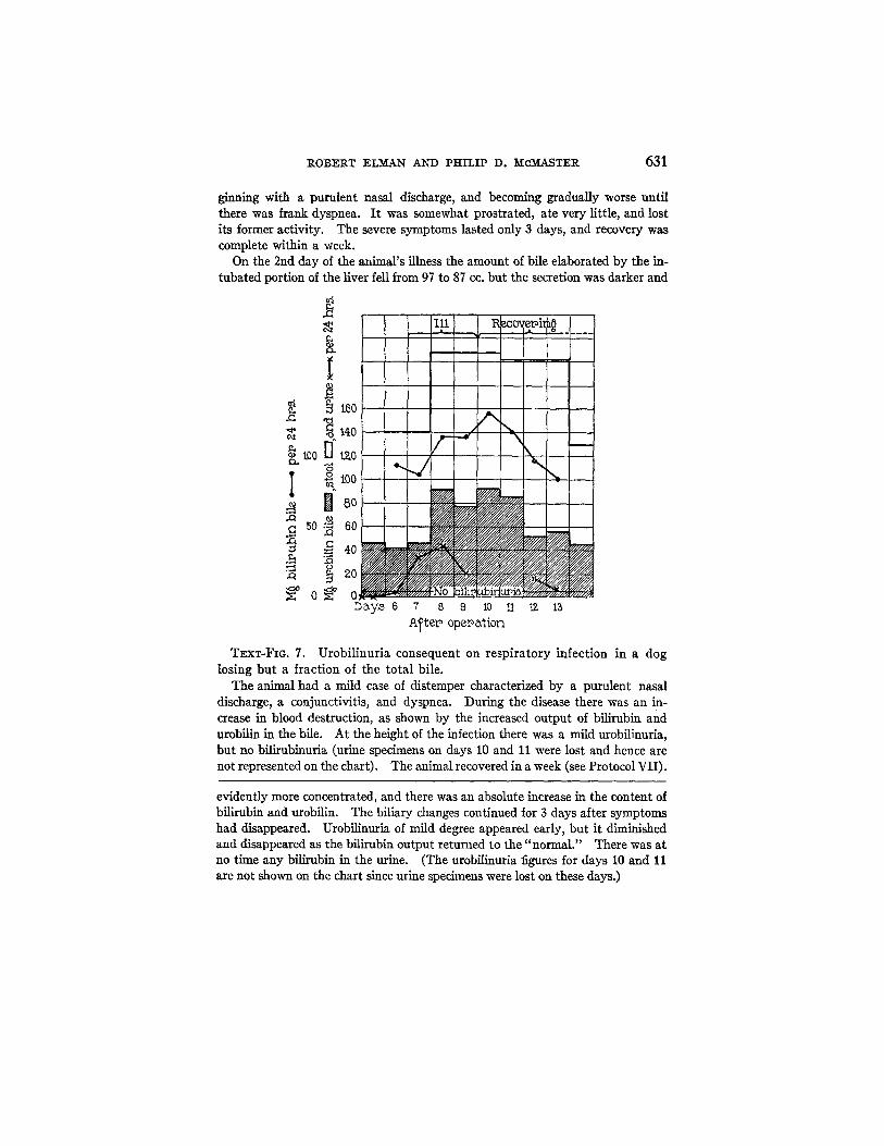
TxxT-FIQ. 7. Urobil inuria consequent on respira tory infection in a dog losing but a fraction of the total bile.
The animal had a mild case of distemper characterized by a purulent nasal discharge, a conjunctivitis, and dyspnea. During the disease there was an in- crease in blood destruction, as shown by the increased output of bilirubin and urobilin in the bile. At the height of the infection there was a mild urobilinuria, but no bilirubinuria (urine specimens on days 10 and 11 were lost and hence are not represented on the chart). The animal recovered in a week (see Protocol vii).
evidently more concentrated, and there was an absolute increase in the content of bilirubin and urobilin. The biliary changes continued for 3 days after symptoms had disappeared. Urobilinuria of mild degree appeared early, but i t diminished and disappeared as the bilirubin output returned to the "normal." There was at no time any bilirubin in the urine. (The urobilinuria figures for days 10 and 11 are not shown on the chart since urine specimens were lost on these days.)
632 UROBILIN PHYSIOLOGY AND PATHOLOGY. V
Urobilinuria after the Giving of Large A mounts of Bile by Garage.
In a preceding paper of the present series 9 the fact has been re- corded that when each day there is returned, through a stomach tube, to an animal losing all of the bile after intubation the amount of bile lost, urobilin reappears in the bile and stool, only to disappear again soon after the feedings are stopped. No urobilin appears in the urine as long as the amount of bilirubin given does not exceed the normal pigment output of the liver.
Through inadvertence a dog was one day given a specimen of uro- bilin-free bile containing two and one-half times the usual amount of bflirubin. To our surprise, the urine next day contained large amounts of urobilin, though none had been found before. The ex- periment was many times repeated on the same animal, and on others, always with the same result. The urobilinuria was less when the excess of pigment fed was lessened.
Example.--In Dog 25 the giving of 75 rag. of bflirubin per day,--approxi- mately the amount it lost through an intubated common duct,--caused the appearance of urobilin in the bile and stool, but none in the urine. When 100 rag. was administered a slight urobilinuria followed,--5 nag. of the pigment being excreted in 24 hours. When the dose of bile fed was increased so that the dog was receiving 250 rag. of bilirubin per day the 24 hour output of urobilin in the urine was over 100 nag.
The same experiment was performed on dogs losing only a fraction of their liver output. With them, too, the giving of large amounts of bile by gavage was attended by the development of urobflinuria. In an instance selected for illustration (Protocol IX) the result was attained by feeding a solution of pure crystalline bilirubin prepared from dog bile2 We have not sought to learn precisely how con- siderable an excess of bile must be fed in order to produce urobilin- uria. But certainly the excess need not be great. Under the circum- stances of the feeding a great deal of bile passes along the intestine at one t ime--a very different state of affairs from the normal--and doubtless urobilin is formed and absorbed, en masse, so to speak, with result that some gets by the liver. In extreme instances, when very great amounts of bile are fed, there may be bilirubinuria as well, an occurrence which Schiff 17 long ago noted.
l~ Schiff, M., Arch. ges. Physiol., 1870, iii, 598.
ROBERT ELMAIW AND PHILIP D. MC~ASTER 633
Specimen Protocols.
VIII. Male dog, weight 161 kilos (see Text-fig. 8). Under ether anesthesia, the common duct was intubated 20 days prior to the
experiment. The bile remained sterile, and it contained no urobilin after the 3rd
e e c
.c s0
i ~ 50 ~ 40
-q 30
0
0 D~xyS Z0 ZI 2Z Z3 24 Z5 26 27 28 Z9 30 31
A~tep opepat[on
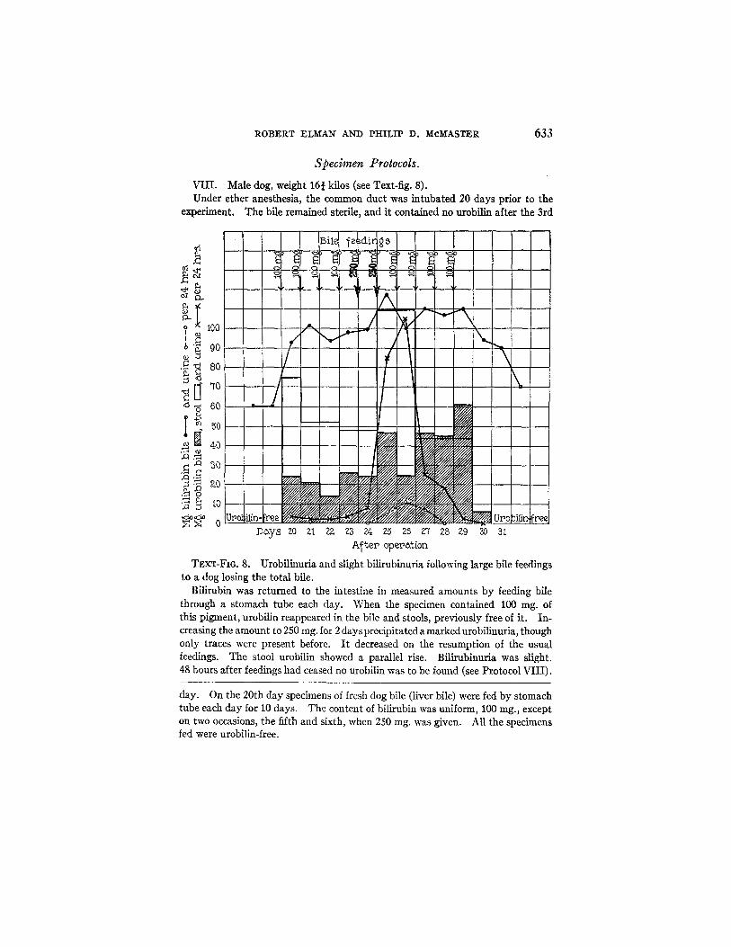
TExT-FIG. 8. Urobilinuria and slight bilirubinuria foilowing large bile feedings to a dog losing the total bile.
Bilirubin was returned to the intestine in measured amounts by feeding bile through a stomach tube each day. When the specimen contained 100 mg. of this pigment, urobilin reappeared in the bile and stools, previously free of it. In- creasing the amount to 250 mg. for 2 days precipitated a marked urobilinuria, though only traces were present before. I t decreased on the resumption of the usual feedings. The stool urobilin showed a parallel rise. Bilirubinuria was slight. 48 hours after feedings had ceased no urobilin was to be found (see Protocol VIII).
day. On the 20th day specimens of fresh dog bile (liver bile) were fed by stomach tube each day for 10 days. The content of bilirubin was uniform, 100 mg., except on two occasions, the fifth and sixth, when 250 mg. was given. All the specimens fed were urobilin-free.
634 UROBILIN PH'YSIOLOGY AND PATHOLOGY. V
The first four feedings (each containing 100 rag. of bilirubin--considerably more than the 60 mg. lost) were attended by an increased output of this pigment by the liver, and the appearance of appreciable amounts of urobilin in bile and stool, and of a small quantity (3 to 4 rag. for 24 hours) in the urine. Similar findings in many experiments of this kind have been described in a previous paper. 9 Following the giving of larger amounts of bile, containing over double the amount of bilirubin (250 rag.), the urobilinuria became a striking phenomenon, 205 rag. of the substance appearing in the urine during 72 hours, while there was a slight
o . . . . ~y~ ~ 3~ 33 34 ~
A~ter~ opeeation
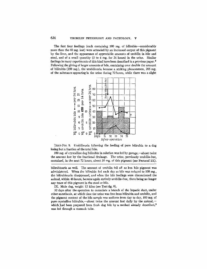
TExT-FI6. 9. Urobilinuria following the feeding of pure bilirubin to a dog losing but a fraction of the total bile.
100 rag. of crystalline dog bilirubin in solution was fed by gavage,--about twice the amount lost by the fractional drainage. The urine, previously urobilin-free, contained, in the next 72 hours, about 30 mg. of this pigment (see Protocol IX).
bilirubinuria as well. The amount of urobilin fell off as less bile pigment was administered. When the bilirubin fed each day as bile was reduced to 100 rag., the bilirubinuria disappeared, and when the bile feedings were discontinued the animal, within 48 hours, became again entirely urobilin-free, there being no longer any trace of this pigment in the stool or bile.
IX. Male dog, weight 12 kilos (see Text-fig. 9). 32 days after the operation to cannulate a branch of the hepatic duct, under
ether anesthesia, at which time the urine was free from billrubin and urobilin, and the pigment content of the bile sample was uniform from day to day, 100 mg. of pure crystalline bilirubin,--about twice the amount lost daily by the animal,-- which .~ad been prepared from fresh dog bile by a method already described, 9 was fed through a stomach tube.
ROBERT ELMAN AND PHILIP D. ~[CMakSTER 635
The urine collected 18 hours later contained urobilin and continued to carry it for 48 hours more. Curiously enough, the bile showed no change in its content of this pigment, though it contained more bilirubin than previously. The disturb- ance was over in 3 days. 5 days after the feeding B. subtilis was found in the bile for the first time, doubtless as an air contaminant that had entered the tube system during the manipulations to empty the collecting balloon. The animal was chloroformed at once. No pathological changes were found at autopsy. The cannula was draining 32 per cent of the liver tissue.
DISCUSSION.
The experiments should lead, it would seem, to a better under- standing of the relation between urobilin and conditions characterized by excessive blood destruction.
In previous papers 7,1°,18 we have proved that normally the forma- tion of urobilin takes place only in the gastrointestinal tract. Even when the liver is seriously injured, or there is billary obstruction with jaundice, no evidence is to be found of the participation of the tissue cells in general, or of the hepatic cells in particular, in the change of bilirubin to urobilin--so long at least as the liver and bile passages remain sterile. Under such conditions the intestinal source of uro- bilin remains the only one.
The same fact holds true when there is blood destruction. Ex- tensive hemolysis, sufficient in many instances to lead to tissue icterus, and in some cases associated with fiver injury, is never at- tended by the appearance of urobilin in the bile, urine, or stools, so long as bile is prevented from reaching the duodenum. The extra- vasafion of blood into the tissues does not lead to the excretion of the pigment under such circumstances, nor do infectious processes.
When bile flow to the intestine is going on, as in dogs intubated for the collection of bu t a fraction of the bite, and blood destruction is produced in one of the ways just mentioned, urobilinuria develops and there is an increase in the urobilin content of bile and stool (see charts). An especially pronounced urobilinuria occurs after toluyl- enediamine has been given (see Protocol III) . During the period of increased blood destruction the curve of urobilin elimination in the urine closely follows both in time and intensity the curves of the increase of bilirubin and urobilin in the bile. K~hl is records similar
18 Kilhl, G., Arch. exp. Path. u. Pharmakol., 1924, ciii, 247.
636 UROBILIN PHYSIOLOGY AND PATHOLOGY. V
findings after the use of phenylhydrazine. A similar, though less definite relationship was found to exist after intravenous hemoly- sis (see Protocol IV). In all the cases in which blood destruction occurred or was induced, bile was reaching the intestine through the common duct; in all there was an increase in pigment output by the liver; and in nearly all there was a marked fall in the percentage of the circulating hemoglobin.
What brings about the appearance of urobilin in the urine under such circumstances? Liver damage might be invoked to explain it in some. Though this may, indeed, have been partly responsible in certain instances, it is significant that in every case there was an increase in the urobilin and bilirubin output of the liver, indicating that there can have been no profound impairment in the ability of the organ to excrete the two pigments. During the height of the blood destruction caused by toluylenediamine the liver was, in one instance, excreting eight times the usual amount of bilirubin.
Unexpected light was thrown upon the problem thus posed by the observation that the giving of large amounts of bile by mouth to healthy animals will cause urobilinuria. This happens in animals losing the total bile after intubation, as also in those losing but a fraction of it ,--though, the amount of bile which must be fed to pro- duce urobilinuria is larger under the circumstances first mentioned (see Protocols VIII and IX). In such instances, and especially when pure bilirubin has been fed, there can be no question of liver injury. Evidently the determining factor is the presence in the intestine of an unusually large amount of bile pigment. Taken together the facts lead to the conclusion that urobilinuria during increased blood de- struction is a secondary manifestation of the polychol/a consequent on the liberation of hemoglobin. The large output of bilirubin derived from the liberated blood pigment leads to a large formation of urobilin in the intestines, which is absorbed therefrom in amounts too considerable for all to be dealt with by the liver, with the conse- quence that part escapes into the general circulation to be removed by the kidneys and urine.
The well known increase in the urobilin content of the stools during the course of diseases involving blood destruction finds a simple exem- plification in our observations (see Text-figs. 3, 4, and 6). But it
ROBERT ELMAN AND PHILIP D. McMASTER 637
will be noted that the increase in the stools usually occurs a day or two after the appearance of urobilinuria. Often it is not found at all. As already remarked, the amount of urobilin in the stools is normally subject to great fluctuations. I t increases during diar- rhea, in the absence of hemolysis, 7 and lessens with constipation. Its value as a measure of increased blood destruction is thus greatly diminished. In our experiments changes in the urobilin quantity in the stool have been far less dependable and significant than the occurrence of urobilin in the urine.
The constant occurrence of urobilinuria during the more acute stages of pernicious anemia has repeatedly excited the attention of clinicians. In such connections the question has often been raised whether there may not be a biliary disturbance traceable to some special involvement of the liver in the disease process. But the ability of the liver of pernicious anemia to excrete large amounts of bilirubin is shown by the finding of tremendous amounts of fecal urobilin in these cases. The responsible factor would seem, from this evidence, to be an augmentation in the rate of red cell destruction, though it is true that van den Bergh, 19 and Broun and others ~° have frequently found hemoglobin, hematin, and bilirubin in the serum of patients with pernicious anemia, and in fatal cases, Peabody and Broun 2~ have seen tremendously increased phagocytosis of red cells in the bone marrow removed at autopsy. Warthin, ~ before them, had described similar findings in the spleen, lymph, and hemolymph glands and concluded that "the poison of pernicious anemia stimu- lates the phagocytes . . . . to increased hemolysis (cellular hemolysis)." From the evidence of the experiments just discussed blood destruction alone would seem to be sufficient to account for the intense urobilinuria. The passage of increased amounts of bilirubin into the intestine leads to the formation and absorption of more urobilin than the liver, often siderosed and fatty, can handle; the pigment escapes complete removal from the portal stream; reaches the general circulation; and is excreted by the kidneys.
19 van den Bergh, A. A. H., Der Gallenfarbstoff im Blute, Leiden, 1918. 2o Broun, G. O., Ames, O., Warren, S., and Peabody, F. W., J. Clin. Inv., 1924-
25, i, 295. 21 Peabody, F. W., and Broun, G. O., Am. ] . Path., 1925, i, 169.
Warthin, A. S., Am. J. Med. Sc., 1902, cxxiv, 674.
638 UROBILIlq PHYSIOLOGY AND PATHOLOGY. V
Urobilinuria has long been known to occur in lobar pneumonia. This, with the occasional icterus, points to a disturbance in pigment metabolism. Since the liver in fatal cases with jaundice and uro- bilinuria usually shows cloudy swelling and even necrosis, the uro- bilinuria has been taken by many to be a reflection of the biliary disturbance. But not only is there liver injury to account for the finding, but also some evidence exists of excessive blood destruction. Peabody and Broun ~'1 have described increased phagocytosis in the bone marrow from patients dying of lobar pneumonia. I t may be recalled in this connection that we have found decided increases in the amount of bilirubin put out by the liver of dogs suffering from distemper (see Text-fig. 7). We would suggest that increased blood destruction may well be a factor in the causation of the urobilinuria of infectious diseases though, of course, liver damage must often play a large part.
Thus far the results of frank blood destruction only have been considered. But urobilinuria has also been found in another group of conditions, those characterized by the escape of blood into the tissues with a subsequent slow destruction of the red cells and ab- sorption of the resulting pigments. ~ As already stated we produced hematomas in a few intubated dogs, but the subsequent increases in bflirubin output were very slight, and urobilinuria was not found. On the other hand, this latter phenomenon occurred regularly after operation, in animals from which only a part or none of the bile was being drained. In such of these cases as were intubated there was always to be noted a postoperative increase in the bilirubin of the bile, referable without doubt to the absorption of pigment from the blood extravasate consequent on the trauma of operation. The as- sumption that the anesthetic caused some slight liver derangement in such cases, a factor lacking when artificial hematomas alone were pro- duced without it, will explain the difference in the urobilin findings.
How may one compare the urobilinuria of excessive blood destruc- tion with that following liver disease? In each instance it is evident that some or all of the pigment has escaped removal from the portal blood by the liver, in the one case because there is too much of it to handle; in the other because the cells are diseased and cannot handle it all. In liver disease the functional reserve is decreased or lost; in excessive blood destruction it is overstepped.
ROBERT ELM.AN AND PI-IILIP D. McM.ASTER 639
From the practical point of view analysis of the findings reported in this and the preceding paper permits of at least two inferences which may prove to have diagnostic worth. Liver damage as such is never responsible for a high urobilin content of the stool; the amount of this pigment tends, on the contrary, to be low, as would follow from the tendency to bile suppression. In such connection the duration and intensity of the urobilinuria may have considerable value. In acute and severe liver disease urobilin may be abundant in the urine, but only transiently; for it disappears practically as soon as bile ceases to flow into the intestine. 1° The urine thereafter con- rains only the bilirubin of the developing jaundice.
By contrast with the foregoing when there is sustained blood de- struction without serious liver injury urobflinuria is likewise sus- tained. Bilirubin may also appear in the renal output, if there be secondarily some obstruction to bile outflow, as not infrequently happens on sudden hemolysis. During persistent, sustained blood destruction the urobilinuria varies in degree with the amount of blood destroyed (see Text-fig. 3, which exemplifies this strikingly).
S u M M A r y .
Further evidence is presented, in addition to that of our previous papers, that the intestinal tract is, under ordinary circumstances, the sole place of origin of urobflin. So long as the biliary tract re- mains sterile the presence of the pigment in bile and urine is entirely dependent upon the passage of bile to the intestine.
Animals rendered urobflin-free by the collection of all the bile from the intubated, uninfected common duct, remain urobilin-free during and after extensive blood destruction caused by intravenous injec- tions of distilled water, as also after reinjections of the animal's own blood, hemolyzed in vitro. No urobilin appears in the bile, urine, or feces of animals so intubated when blood destruction has been caused by sodium oleate, or by an agent, toluylenediamine, which damages the liver as well as the blood.
On the other hand, when bile flow into the intestine is uninter- rupted, urobilinuria occurs during blood destruction caused in any of the ways mentioned and it parallels, both in severity and duration, the destructive process.
640 UROBILII~ PHYSIOLOGY AND PATHOLOGY. V
Merely increasing the amount of bilirubin within the intestines of healthy dogs by feeding urobilin-free bile, will lead to marked uro- bilinuria. The extravasation of blood into the tissues, resulting from the trauma of an operation for intubation of a bile duct, does not lead to urobilinuria in animals losing all of the bile after this opera- tion, but may do so when only a small fraction of the bile is drained, while the remainder reaches the intestine as usual. The production of artificial hematomas, without operation, is not followed by uro- bilinuria, under the circumstances last mentioned, but merely by an increase in the bilirubin of the bile. The effect on the liver of the anesthetic employed during the intubation may be responsible for the difference in the two cases.
During the course of certain intercurrent infections affecting some of the intubated animals, notably distemper, there was a drop in the hemoglobin percentage of the circulating blood, accompanied by an increased output of bile pigment or further by urobilinuria, when the conditions were such that bile still reached the intestine. The findings pointed to increased blood destruction as a factor in the uro- bilinuria.
The evidence presented, taken with that of our previous papers, su~ces to demonstrate, that urobilinuria, occurring during blood destruction, is primarily the result of an increased excretion of bili- rubin from which, in turn, an unusually large quantity of urobilin is formed within the intestine. The liver fails to remove from the portal blood all of the latter pigment which is resorbed and consequently some of it reaches the kidneys and urine.
Our work has been carried out on animals with uninfected biliary tracts and livers, save for one case which has special mention. The influence of infection of the biliary tract on the place of formation of urobilin and the development of urobilinuria will be discussed in a succeeding communication.