Embed Size (px)
Citation preview
1
2-5-03
Urogenital DevelopmentGreg DresslerAssoc. ProfessorDept. of Pathology
x46490
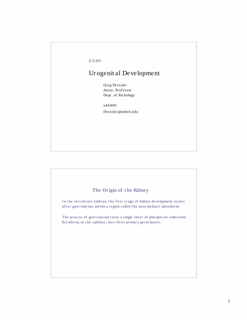
The Origin of the Kidney
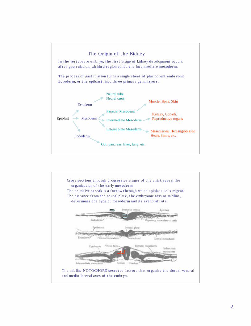
In the vertebrate embryo, the first stage of kidney development occurs after gastrulation, within a region called the intermediate mesoderm.
The process of gastrulation turns a single sheet of pluripotent embryonicEctoderm, or the epiblast, into three primary germ layers.
2
The Origin of the KidneyIn the vertebrate embryo, the first stage of kidney development occursafter gastrulation, within a region called the intermediate mesoderm.
The process of gastrulation turns a single sheet of pluripotent embryonicEctoderm, or the epiblast, into three primary germ layers.
Epiblast
Ectoderm
Mesoderm
Endoderm
Neural tubeNeural crest
Paraxial Mesoderm
Intermediate Mesoderm
Lateral plate Mesoderm
Gut, pancreas, liver, lung, etc.
Muscle, Bone, Skin
Mesenteries, HemangioblasticHeart, limbs, etc.
Kidney, Gonads, Reproductive organs
Cross sections through progressive stages of the chick reveal the organization of the early mesodermThe primitive streak is a furrow through which epiblast cells migrateThe distance from the neural plate, the embryonic axis or midline, determines the type of mesoderm and its eventual fate
The midline NOTOCHORD secretes factors that organize the dorsal-ventraland medio-lateral axes of the embryo.

3
The first epithelial component of the urogenital system is the NEPHRIC DUCT.It arises within the intermediate mesoderm adjacent to the 10-12th somitesand extends posteriorly.
DiI lineage tracing in the chick embryo reveals that the Nephric Duct formsby extension rather than by recruitment of mesoderm to the epithelial duct.
Obara-Ishihara et al., Develop. 126, 1103 (1999)
As the Nephric duct forms and grows caudally, Lim1 and Pax2 expression mark the epithelium of the duct.
Lim1 Pax2
Lim1 is essential for nephric duct formationPax2 and its related gene Pax8 are also essential for nephric duct formation
4
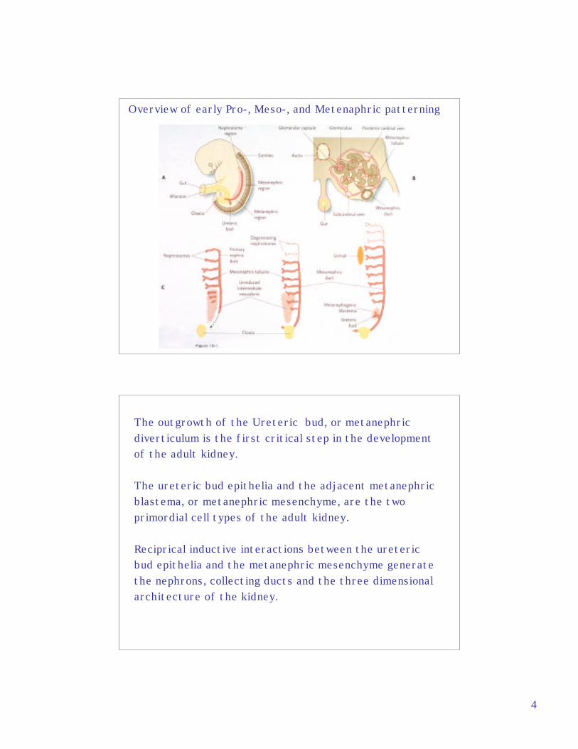
Overview of early Pro-, Meso-, and Metenaphric patterning
The outgrowth of the Ureteric bud, or metanephricdiverticulum is the first critical step in the developmentof the adult kidney.
The ureteric bud epithelia and the adjacent metanephricblastema, or metanephric mesenchyme, are the twoprimordial cell types of the adult kidney.
Reciprical inductive interactions between the uretericbud epithelia and the metanephric mesenchyme generate the nephrons, collecting ducts and the three dimensional architecture of the kidney.
5
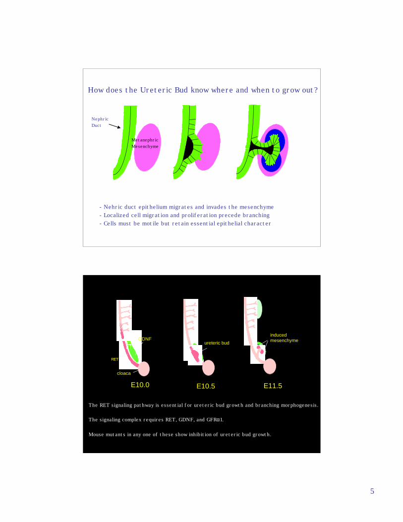
- Nehric duct epithelium migrates and invades the mesenchyme- Localized cell migration and proliferation precede branching- Cells must be motile but retain essential epithelial character
NephricDuct
Metanephric Mesenchyme
How does the Ureteric Bud know where and when to grow out?
E10.0 E10.5 E11.5
cloaca
GDNFureteric bud
inducedmesenchyme
RET
The RET signaling pathway is essential for ureteric bud growth and branching morphogenesis.
The signaling complex requires RET, GDNF, and GFRα1.
Mouse mutants in any one of these show inhibition of ureteric bud growth.
6
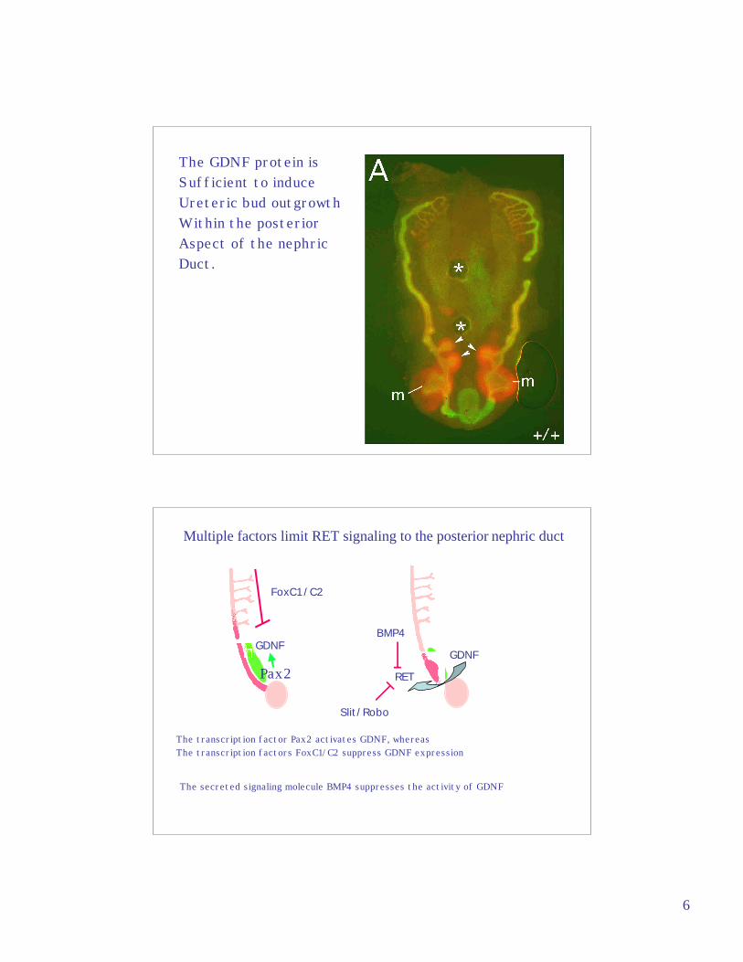
The GDNF protein isSufficient to induceUreteric bud outgrowth Within the posteriorAspect of the nephric Duct.
GDNF
Multiple factors limit RET signaling to the posterior nephric duct
FoxC1/C2
RET
BMP4
GDNF
Slit/Robo
Pax2
The transcription factor Pax2 activates GDNF, whereasThe transcription factors FoxC1/C2 suppress GDNF expression
The secreted signaling molecule BMP4 suppresses the activity of GDNF
7
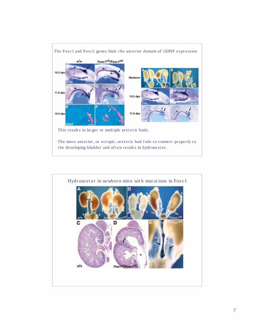
The Foxc1 and Foxc2 genes limit the anterior domain of GDNF expression
This results in larger or multiple ureteric buds.
The more anterior, or ectopic, ureteric bud fails to connect properly to the developing bladder and often results in hydroureter.
Hydroureter in newborn mice with mutations in Foxc1
8
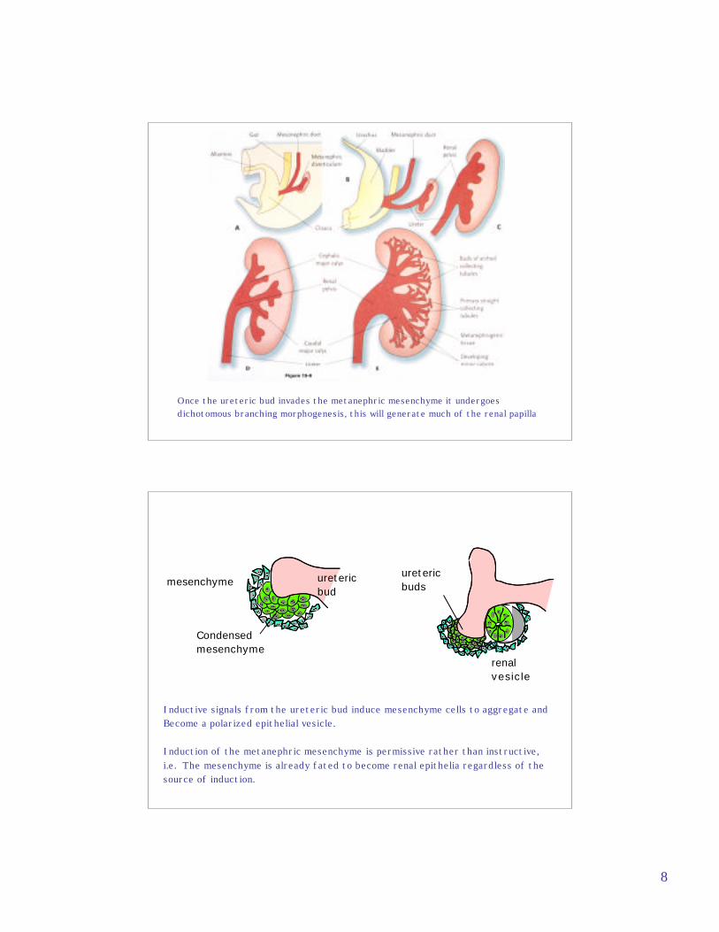
Once the ureteric bud invades the metanephric mesenchyme it undergoesdichotomous branching morphogenesis, this will generate much of the renal papilla
renalvesicle
Condensedmesenchyme
uretericbuds
uretericbud
mesenchyme
Inductive signals from the ureteric bud induce mesenchyme cells to aggregate and Become a polarized epithelial vesicle.
Induction of the metanephric mesenchyme is permissive rather than instructive,i.e. The mesenchyme is already fated to become renal epithelia regardless of thesource of induction.
9
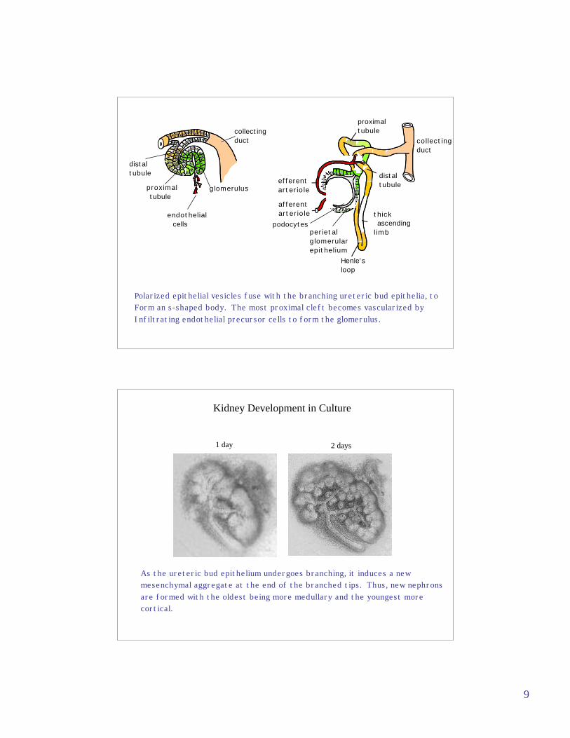
endothelial cells
glomerulus
distaltubule
proximal tubule
afferentarteriole
efferentarteriole
podocytesperietalglomerularepithelium
thick ascendinglimb
distaltubule
collectingduct
proximaltubule
Henle'sloop
collectingduct
Polarized epithelial vesicles fuse with the branching ureteric bud epithelia, toForm an s-shaped body. The most proximal cleft becomes vascularized byInfiltrating endothelial precursor cells to form the glomerulus.
Kidney Development in Culture
1 day 2 days
As the ureteric bud epithelium undergoes branching, it induces a new mesenchymal aggregate at the end of the branched tips. Thus, new nephronsare formed with the oldest being more medullary and the youngest morecortical.
10
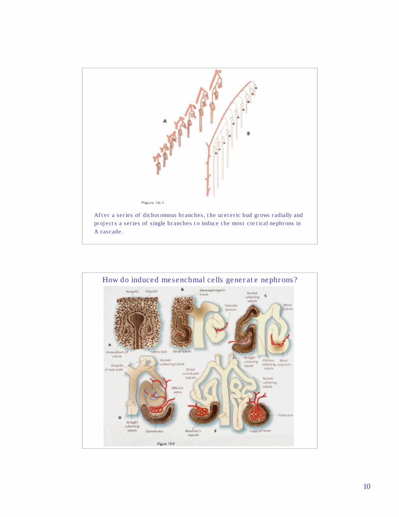
After a series of dichotomous branches, the ureteric bud grows radially andprojects a series of single branches to induce the most cortical nephrons inA cascade.
How do induced mesenchmal cells generate nephrons?
11
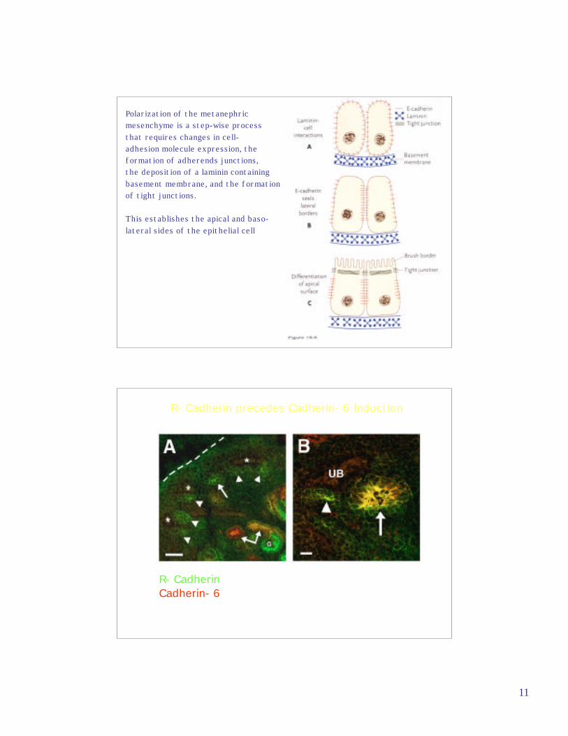
Polarization of the metanephricmesenchyme is a step-wise processthat requires changes in cell-adhesion molecule expression, theformation of adherends junctions,the deposition of a laminin containingbasement membrane, and the formationof tight junctions.
This establishes the apical and baso-lateral sides of the epithelial cell
R- CadherinCadherin- 6
R- Cadherin precedes Cadherin- 6 Induction
12
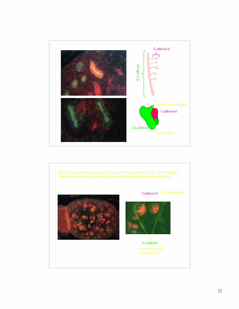
Cadherin-6
E-C
adhe
rin
Cadherin-6
E-Cadherin
Ureteric bud
Induced mesenchyme
Cadherin-6
E-cadherin
By E14, compartmentalization of the newly formed epithelia is clear. The S-shapedbody is demarcated into glomerular, proximal tubule and distal tubule epithelia.
Distal tubule and Collecting duct
Proximal tubule
13
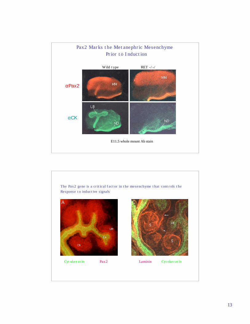
αPax2
αCK
Pax2 Marks the Metanephric Mesenchyme Prior to Induction
E11.5 whole mount Ab stain
Wild type RET -/-/
Cytokeratin Pax2 Laminin Cytokeratin
The Pax2 gene is a critical factor in the mesenchyme that controls the Response to inductive signals
14
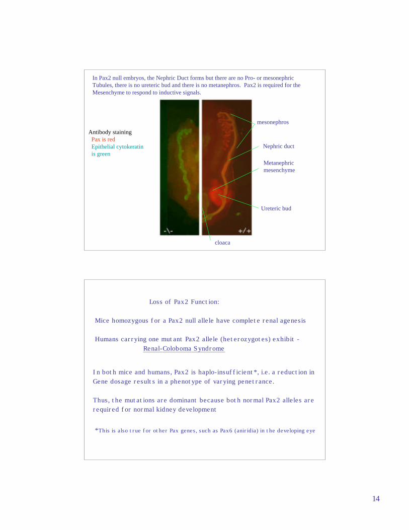
In Pax2 null embryos, the Nephric Duct forms but there are no Pro- or mesonephric Tubules, there is no ureteric bud and there is no metanephros. Pax2 is required for theMesenchyme to respond to inductive signals.
mesonephros
Metanephric mesenchyme
Ureteric bud
cloaca
Antibody staining Pax is red Epithelial cytokeratin is green
Nephric duct
Loss of Pax2 Function:
Mice homozygous for a Pax2 null allele have complete renal agenesis
Humans carrying one mutant Pax2 allele (heterozygotes) exhibit -Renal-Coloboma Syndrome
In both mice and humans, Pax2 is haplo-insufficient*, i.e. a reduction in Gene dosage results in a phenotype of varying penetrance.
Thus, the mutations are dominant because both normal Pax2 alleles are required for normal kidney development
*This is also true for other Pax genes, such as Pax6 (aniridia) in the developing eye
15
Renal-Coloboma Syndrome
Patients typically exhibit at least these 3 symptoms:
1- Renal hypoplasia - due to reduced proliferation of the mesenchyme derived epithelia during development.
2- Vesicouretral Reflux - most likely due to improper connection of the ureter to the bladder or possibly due to inherent defects in epithelial cells of the mature ureter. 3- Optic Nerve Colobomas - due to failure of the optic fissure to fuse. Expression of Pax2 is observed in part of the optic cup and optic stalk.
Other critical regulators of the Mesenchymal-to-epitheial transitionInclude:
WT1- Wilms tumor suppressor gene
Wnt-4 - secreted signaling molecule
BMP7 - secreted signal
BF-2 - stromal transcription factor
16
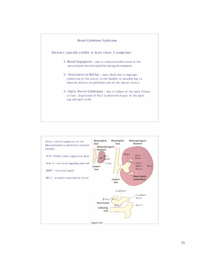
The WT1 gene is associated with Wilms’ TumorAn embryonic kidney tumor that exhibits a triphasic histologyConsisting of blastemal, stromal, and epithelial cell types
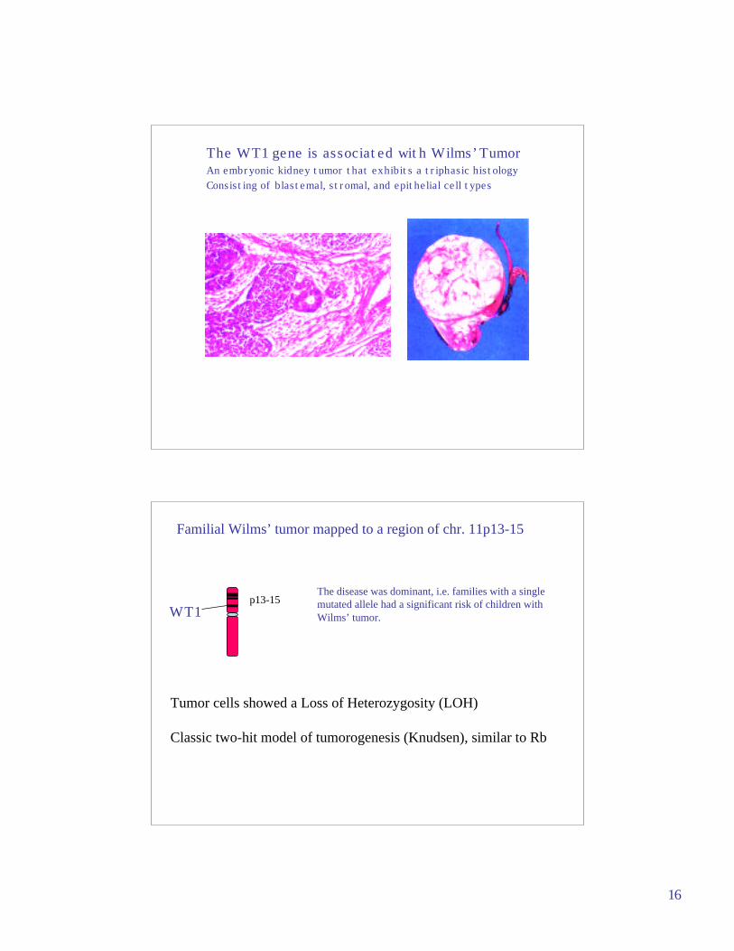
Familial Wilms’ tumor mapped to a region of chr. 11p13-15
p13-15The disease was dominant, i.e. families with a singlemutated allele had a significant risk of children withWilms’ tumor.
Tumor cells showed a Loss of Heterozygosity (LOH)
Classic two-hit model of tumorogenesis (Knudsen), similar to Rb
WT1
17
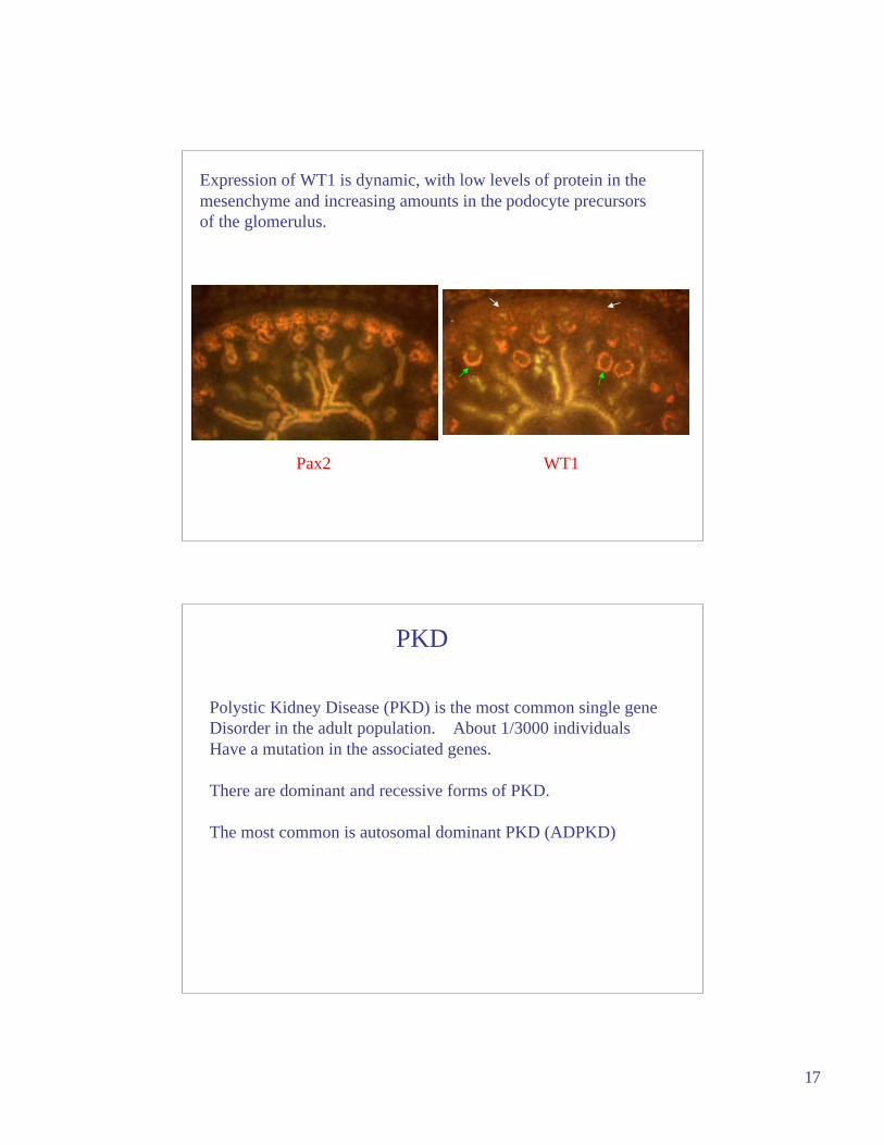
Pax2 WT1
Expression of WT1 is dynamic, with low levels of protein in the mesenchyme and increasing amounts in the podocyte precursors of the glomerulus.
Polystic Kidney Disease (PKD) is the most common single gene Disorder in the adult population. About 1/3000 individuals Have a mutation in the associated genes.
There are dominant and recessive forms of PKD.
The most common is autosomal dominant PKD (ADPKD)
PKD
18
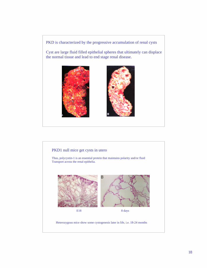
PKD is characterized by the progressive accumulation of renal cysts
Cyst are large fluid filled epithelial spheres that ultimately can displacethe normal tissue and lead to end stage renal disease.
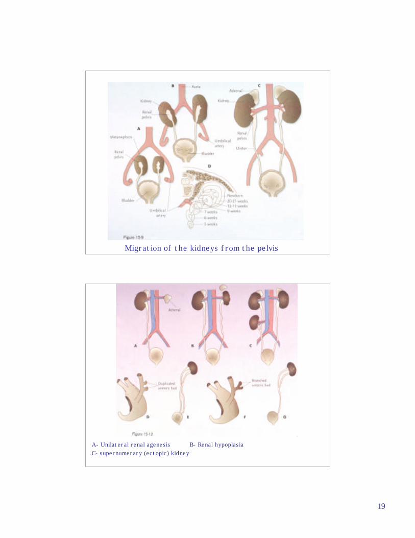
PKD1 null mice get cysts in utero
Thus, polycystin-1 is an essential protein that maintains polarity and/or fluidTransport across the renal epithelia.
E18 8 days
Heterozygous mice show some cystogenesis later in life, i.e. 18-24 months
19
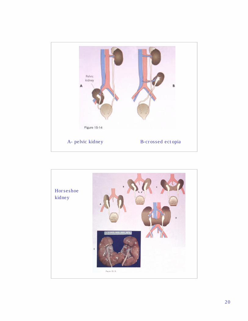
Migration of the kidneys from the pelvis
A- Unilateral renal agenesis B- Renal hypoplasiaC- supernumerary (ectopic) kidney
20
A- pelvic kidney B-crossed ectopia
Horseshoekidney