Embed Size (px)
Citation preview

Urologic Emergencies
Denise Watt
Feb 7, 2002

Outline
• Cases• Renal calculi
– Epidemiology / pathophysiology– Clinical presentation– DI– Management
• Hematuria• Urinary retention

Case 1
62 yo male, sudden onset Rt flank/back pain.
Constant, can’t get comfortable, N&V.
PMHx: HTN
O/E: pale, bp 100/75, HR 105, T37
Urine dip: + hematuria
DDx: ?

Case 2
02:00
45 yo male, sudden onset lt flank pain to
groin, hematuria, prior renal calculi x 2
To image or not to image?

Case 3
30 yo female, RLQ/pelvic pain x 12 hr.
Fever, chills, N&V.
O/E: 140/90, 110, T 38.5
DDx: ?
Imaging modality?
Rx?

Case 4

Renal CalculiEpidemiology
• Present since antiquity• 3-5% of population, 12% white males• 50% recurrence in 10 yrs• whites, rare in Africans & Natives• peak incidence b/w 20-50• M:F = 3:1• familial• ‘stone belt’: SE US• peak months July-September: heat + sunlight?

Epidemiology
• types: Ca oxalate, struvite, uric acid, cystine, misc
• 80% Calcium– most Ca oxalate, some Ca phosphate
– most hypercalciuric (absorptive, resorptive, ideopathic)
• hypercalcemic: hyperparathyroid, hyperthyroid, sarcoidosis
• hereditary causes: PCKD, RTA, PTH, cystinuria

Pathophysiology: Ca oxalate
• urine supersaturated Ca & oxalate crystals• lack of inhibitors: pyrophosphate, citrate• crystals usually washed away into bladder• crystals stick in tubules/ducts, grow, obstruct
– medullary sponge kidney, intramed stasis, abn tubular epithelium
• diet: protein? Ca? oxalate? Na?• low urine volume: water intake, bowel disease

Pathophysiology: Struvite
• 10-20%• staghorn calculi: Mg, NH4, PO4• requires pH>7.2 & NH4 in urine • caused by urease-producing UTIs
– Proteus, Klebsiella, Staph, Providencia, Corynebacterium
• atypical presentation in subset: malaise, weakness

Pathophysiology: Others
• Uric acid– 6-10%, most common radiolucent stones secretion uric acid, acidic urine, urine vol– 1%/yr after 1st gouty attack
• Cystine– 1%, insoluable in low pH– cystinuria: autosomal recessive
• Drugs– triamterene, indinavir, sulfonamides, CA inh.

History
• Renal colic: worse than labour???– Severe, sudden, paroxysmal pain, flankgroin,
referred to testicle, writhe– urinary sx (UVJ/bladder)– N/V (celiac plexus)
• Risk factors: prior episode, FH• Complications: UTIs, solitary kidney, renal
transplant, anat abn, immunocompromised• r/o DDx

Physical Exam
• VS: adrenergic, no fever– Hypotension rare (vasovagal): r/o AAA, sepsis
• Flank pain (r/o pyelo), no peritoneal signs
• CV exam: r/o embolic ds
• r/o bladder retention
• pv exam (PID, preg)

Differential Diagnosis
• HUGE!• AAA
– commonly misdiagnosed as renal colic
– suspect > 50, hypotension
– can have hydronephrosis, hematuria
• renal artery thrombosis/dissection– diff dx, contrast CT
• appendicitis: can have hematuria, CT• pyelo/cystitis: mimic or mask; infected obstruction is
emergency

Lab
• U/A– 90% hematuria– sens with acute flank pain 89%; spec 29%– pyuria common w/o infection– pH: high struvite, RTA; low uric acid– crystals
• Lytes AG met acidosis + Ca oxalate = ?– NonAG met acidosis + hypokalemia = ?
Lab
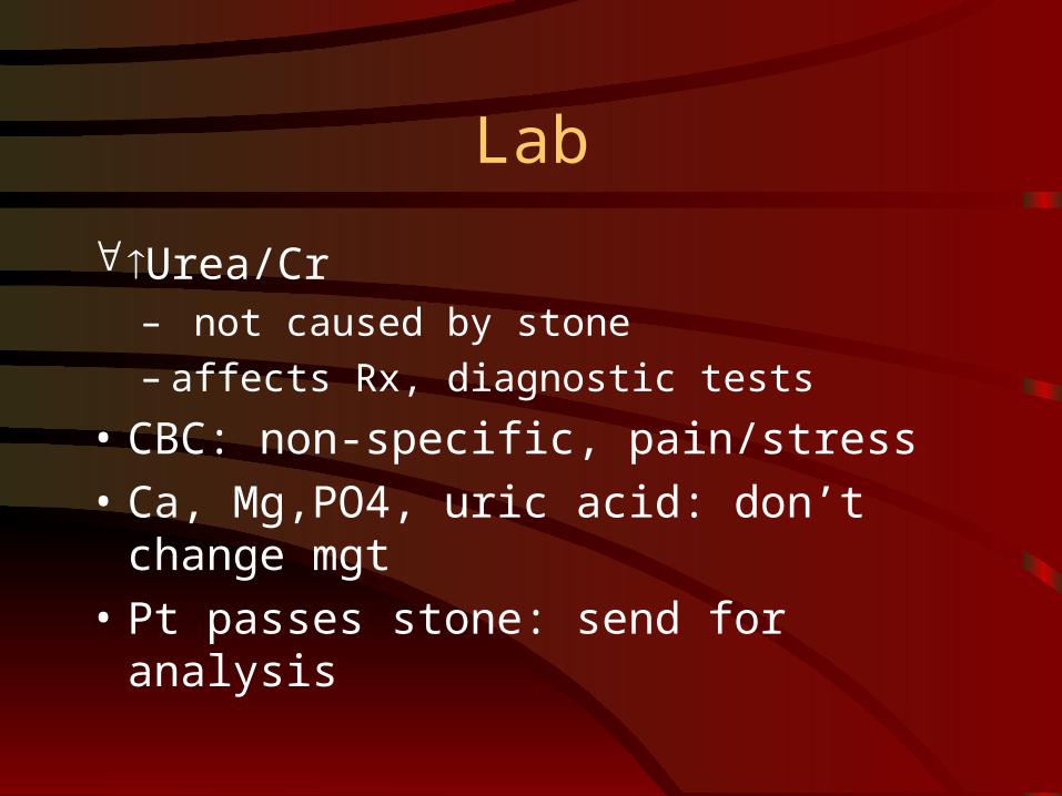
Urea/Cr– not caused by stone– affects Rx, diagnostic tests
• CBC: non-specific, pain/stress
• Ca, Mg,PO4, uric acid: don’t change mgt
• Pt passes stone: send for analysis

Diagnostic Imaging
• Role– Confirm dx– R/o other serious dx– Detect complications of stone– Define site/size of stone
• Who needs emergent imaging?– No consensus: 1st stone, suspect other dx

Diagnostic Imaging
• How good are we clinically?– High PTP: 70% had stones on IVP (Twinem,
Wrenn)– Are we missing serious other disease if we
don’t image?
• Bottom line: we must decide how confident we are with our dx, r/o serious DDx (esp. in elderly), close F/U to confirm dx

Diagnostic Imaging Modalities
Study Sens. Spec. Advantages Limitations
Plain film
45-59% 71-77% Cheap, accessible Only radiolucent calculi, no info on other causes, function
U/S 19%
97% Accessible, no radiation, good for renal calculi/ hydronephrosis/ gyne dx
poor for ureteral stones, no function, operator dependent
IVP 64-87% 92-94% Assess function & anatomy both kidneys, stone size & location, degree of obst
Contrast, bowel prep, time consuming, no non-urinary info
Non-contrast CT
95-100%
94-96% Most sens & spec, indirect signs of degree of obst (kidney size, inflam, stranding, distention), non-urinary info
More $ (slight), no functional info

Ultrasound

IVP

CT

Diagnostic Imaging
• IVP– (Relative) contraindications:
• Cr >130, allergy, DM, multiple myeloma, dehydration, pregnancy
– obstruction: delayed nephrogram or hydro– delayed films until comlunization in 2 – extravasation: renal calyx or ureteral rupture– no infection extravasation not treated– no kidney uptake: think renal infarction

Diagnostic Imaging
• Non-contrast CT– Chen, J Emerg Med 1999;17:299-303– 100 pt: sens 98%, spec 100%– alternate dx in 50% pt w/o stone– cost $600 vs. IVP $400– study of choice? – same as IVP for hydronephrosis/ hydroureter;
better at ID stone

Management:r/o Emergency
• Urosepsis + obstructing stone– Need drainage (nephrostomy, stent) + IV Abx
• Acute renal failure/ anuria– bilat obst/ solitary kidney

Management:Criteria for Admission
• Absolute– intractable
vomiting/pain
– solitary kidney or transplant with obstruction
– UTI with obstruction
– hypercalcemic crisis
• Relative– stone > 6 mm
– high grade obstruction
– solitary kidney/ transplant w/o obstruction
– intrinsic renal disease
– extravasation
– social issues
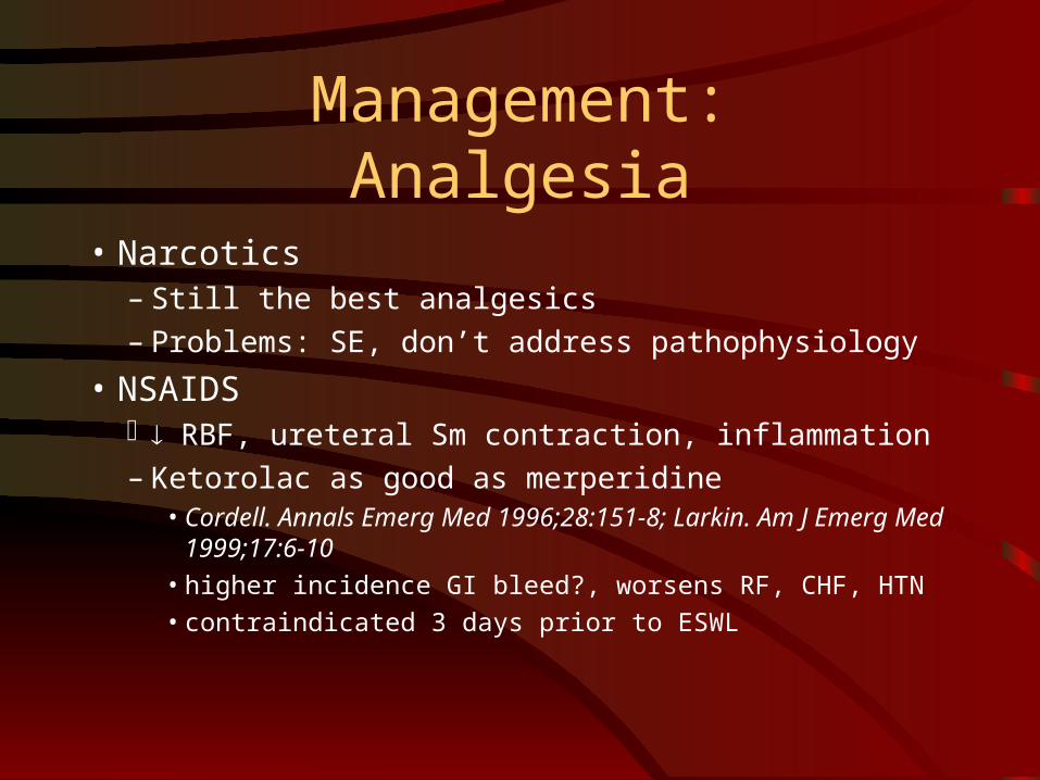
Management:Analgesia
• Narcotics– Still the best analgesics– Problems: SE, don’t address pathophysiology
• NSAIDS RBF, ureteral Sm contraction, inflammation– Ketorolac as good as merperidine
• Cordell. Annals Emerg Med 1996;28:151-8; Larkin. Am J Emerg Med 1999;17:6-10
• higher incidence GI bleed?, worsens RF, CHF, HTN• contraindicated 3 days prior to ESWL

Probability of Stone Passage
Stone location and size
Probability of passage (%)
Proximal ureter >5 mm 0 5 mm 57 <5 mm 53 Middle section of ureter
>5 mm 0 5 mm 20 <5 mm 38 Distal ureter >5 mm 25 5 mm 45 <5 mm 74

Disposition: Stones < 5mm
• Analgesia
• strain urine until stone passes
• send for analysis
• return to ED if worsens, fever

Disposition:Urology Referral
• Emergencies
• stones >= 5 mm
• renal stones, incl. staghorn
• not passed after 2-4 weeks observation– complication rate triples (29%)

Complications
• renal failure – rare if no infection, other kidney works– need obstruction x 4 weeks? (Campbell’s)
• ureteral stricture
• infection, sepsis
• urine extravasation
• perinephric abscess

Special concerns
• Pregnancy– U/S to minimize radiation
• Children– Rare: look for metabolic cause
• Elderly– Rare first-time stones; look for other causes– CT (consider contrast)– beware NSAIDS/ narcotics

Surgical Management
• ESWL– Ureteral stones < 1cm
– Renal stones < 2 cm
• Uretoscopy– Ureteral stones
• Ureterorenoscopy– Renal stones < 2 cm
• Percutaneous nephrolithotomy– Renal stones > 2 cm
– Proximal ureteral stones > 1cm

Doc - I never want to go through this again!
• increase fluid intake
• diet– low Calcium: stones secondary to oxalate
• Borghi et al, NEJM 2002;346:77-84
– low protein: acid-induced Ca excr. & urate– low Sodium: urinary Na Ca No clinical excr.– Low oxalate: nuts, chocolate, rhubarb, beets,
dark green veggies

Prevention
• thiazides, amiloride (hypercalciuria)
• allopurinal, alkalinize urine if pH low (uric acid)
• potassium citrate (hypocitraturia)

Summary
• stones are common
• clinical dx + hematuria isn’t reliable
• CT probably the best DI
• analgesia: NSAID + opiod
• watch out for emergencies
• when to consult
• prevention

Hematuria: the quick and dirty
• DDx huge: look it up– infection 25%– stones 20%– NYD 10%– others: trauma, glomerular, renal, extrarenal,
coagulopathy, factitious, pigmenturia– most common worldwide: schistosomiasis

Hematuria
• Gross– r/o bladder/kidney CA
• Micro– >3 RBC/hpf

History
• Quantity
• Timing– initial hematuria: anterior urethral lesion
• urethritis, stricture, meatal stenosis
– terminal:post. urethra, bladder, neck, trigone• post urethritis, polyps, tumours
– total: source above bladder• stone, tumour, infection

Hematuria is an Emergency! (sometimes)
• trauma
• gross hematuria causing hypovolemic shock
• urosepsis
• obstructing stones + infection or RF
• glomerulonephropathies + CHF, HTN emergencies, RF, infection
• coagulopathy, bleeding from multiple sites

Diagnosis
• Dipstick– 90% sensitive– FP: Mb, menses
• U/A– RBC casts + proteinuria = GMN
• DI– stones– trauma
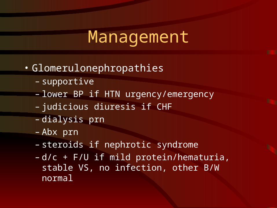
Management
• Glomerulonephropathies– supportive– lower BP if HTN urgency/emergency– judicious diuresis if CHF– dialysis prn– Abx prn– steroids if nephrotic syndrome– d/c + F/U if mild protein/hematuria, stable VS, no
infection, other B/W normal

Management:Microscopic hematuria
• F/U U/A – Fam MD or urologist
• Urology W/U if:– >3 RBC/hpf on at least 2 U/A or one episode
>100 RBC/hpf or gross hematuria
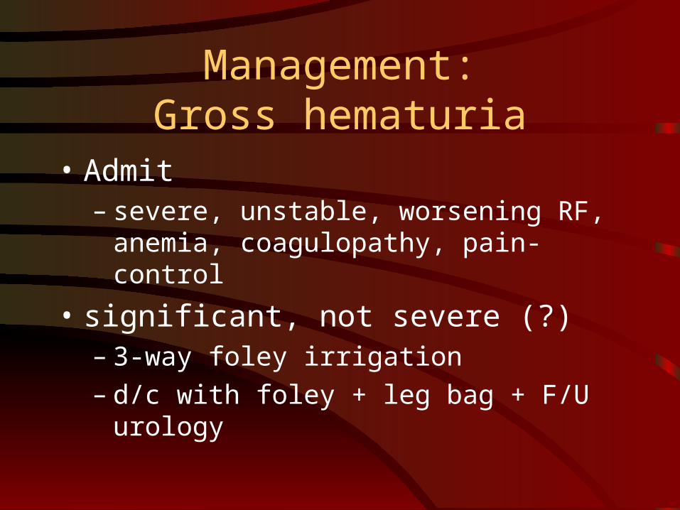
Management:Gross hematuria
• Admit– severe, unstable, worsening RF, anemia,
coagulopathy, pain-control
• significant, not severe (?)– 3-way foley irrigation– d/c with foley + leg bag + F/U urology

Hematuria Trivia
1. Micro hematuria in 6 yr old 2 weeks after URI + sore throat.
> Post-Strep GMN
2. Known narcotic abuser + gross hematuria
> Papillary necrosis
3.Micro hematuria in pt with RA on Indocid
> Interstitial nephritis

Hematuria Trivia
4. Gross hematuria in 5 yr old, FH of RF
> Medullary sponge kidney
5.Recent travel to Africa, gross hematuria
> Schistosomiasis
6. Hemopytsis + chronic micro hematuria
> Goodpasture’s

Hematuria Trivia
7. Micro hematuria in9 yo with FH deafness
> Alport’s
8. African-Canadian 11 yo, abd pain, dehydrated, gross hematuria
> sickle cell crisis
9. URI 2 days ago, micro hematuria + protein
> IgA nephropathy


![Renal calculi/stones a Hematuria (blood in the urine) a Incontinence (can't control) c] Bladder Infections a Difficulty urinating a Kidney disease a Dialysis a Other a None of the](https://img.pdfslide.net/doc/110x75/5e53b1bccba5cf222f04ed65/-renal-calculistones-a-hematuria-blood-in-the-urine-a-incontinence-cant.jpg)