Embed Size (px)
Citation preview
RESEARCH ARTICLE
Use of rhodamine B to mark the body and
seminal fluid of male Aedes aegypti for mark-
release-recapture experiments and
estimating efficacy of sterile male releases
Brian J. Johnson1,2*, Sara N. Mitchell3, Christopher J. Paton1,2, Jessica Stevenson3,
Kyran M. Staunton1,2, Nigel Snoad3, Nigel Beebe4,5, Bradley J. White3, Scott A. Ritchie1,2
1 College of Public Health, Medical and Veterinary Sciences, James Cook University, Cairns, Australia,
2 Australian Institute of Tropical Health and Medicine, James Cook University, Cairns, Australia, 3 Verily Life
Sciences, South San Francisco, CA. United States of America, 4 School of Biological Sciences, University of
Queensland, St Lucia, Brisbane, Queensland, Australia, 5 Commonwealth Scientific and Industrial Research
Organisation, Dutton Park, Queensland, Australia
Abstract
Background
Recent interest in male-based sterile insect technique (SIT) and incompatible insect tech-
nique (IIT) to control Aedes aegypti and Aedes albopictus populations has revealed the
need for an economical, rapid diagnostic tool for determining dispersion and mating success
of sterilized males in the wild. Previous reports from other insects indicated rhodamine B, a
thiol-reactive fluorescent dye, administered via sugar-feeding can be used to stain the body
tissue and seminal fluid of insects. Here, we report on the adaptation of this technique for
male Ae. aegypti to allow for rapid assessment of competitiveness (mating success) during
field releases.
Methodology/Principle findings
Marking was achieved by feeding males on 0.1, 0.2, 0.4 or 0.8% rhodamine B (w/v) in 50%
honey solutions during free flight. All concentrations produced >95% transfer to females and
successful body marking after 4 days of feeding, with 0.4 and 0.8% solutions producing the
longest-lasting body marking. Importantly, rhodamine B marking had no effect on male mat-
ing competitiveness and proof-of-principle field releases demonstrated successful transfer
of marked seminal fluid to females under field conditions and recapture of marked males.
Conclusions/Significance
These results reveal rhodamine B to be a potentially useful evaluation method for male-
based SIT/IIT control strategies as well as a viable body marking technique for male-based
mark-release-recapture experiments without the negative side-effects of traditional marking
methods. As a standalone method for use in mating competitiveness assays, rhodamine B
marking is less expensive than PCR (e.g. paternity analysis) and stable isotope semen
PLOS Neglected Tropical Diseases | https://doi.org/10.1371/journal.pntd.0005902 September 28, 2017 1 / 18
a1111111111
a1111111111
a1111111111
a1111111111
a1111111111
OPENACCESS
Citation: Johnson BJ, Mitchell SN, Paton CJ,
Stevenson J, Staunton KM, Snoad N, et al. (2017)
Use of rhodamine B to mark the body and seminal
fluid of male Aedes aegypti for mark-release-
recapture experiments and estimating efficacy of
sterile male releases. PLoS Negl Trop Dis 11(9):
e0005902. https://doi.org/10.1371/journal.
pntd.0005902
Editor: Jason L. Rasgon, The Pennsylvania State
University, UNITED STATES
Received: March 7, 2017
Accepted: August 23, 2017
Published: September 28, 2017
Copyright: © 2017 Johnson et al. This is an open
access article distributed under the terms of the
Creative Commons Attribution License, which
permits unrestricted use, distribution, and
reproduction in any medium, provided the original
author and source are credited.
Data Availability Statement: All relevant data are
within the paper.
Funding: This study was funded by the
Commonwealth Scientific and Industrial Research
Organisation and National Health and Medical
Research Council of Australia Project Grant
1082127. The funders had no role in study design,
data collection and analysis, decision to publish, or
preparation of the manuscript.
labelling methods and less time-consuming than female fertility assays used to assess com-
petitiveness of sterilised males.
Author summary
The recent resurgence in male-based “rear and release” strategies (release of radiation
sterilised males or those harbouring Wolbachia) to control diseases transmitted by urban
Aedes mosquitoes, including dengue, chikungunya, and Zika, has revealed the need for an
economical and rapid diagnostic tool to assess the mating competitiveness of released
males against wild counterparts for receptive females. Our report details the optimisation
of rhodamine B labelling of male seminal fluid and visible body staining via sugar-feeding
with no significant impact on survival or mating competitiveness, as well as proof-of-prin-
ciple field releases. These results reveal rhodamine B labelling to be an economical and
rapid evaluation method for male-based control strategies as well as a viable body marking
technique for male focused mark-release-recapture experiments without the negative
side-effects of traditional marking methods. The use of this method has the potential to
enable operators to determine the efficacy of their control method quickly and easily,
allowing them to optimise releases to achieve optimal population suppression.
Introduction
Knowledge of key entomological parameters that contribute to the transmission of mosquito-
borne diseases such as dispersal range, blood-feeding rates, population size, survival, and
length of gonotrophic cycles is invaluable for understanding disease transmission dynamics
and for determining the extent of control measures necessary to interrupt transmission [1–5].
Traditionally, much of this information has been obtained by conducting mark-release-recap-
ture (MRR) field experiments, during which the target insect is collected either from labora-
tory colonies or from the field, marked, released into the field, and recaptured at given time
and distance intervals after their release [6, 7]. The recaptured insects are then checked for the
presence of the marker to distinguish them from unmarked individuals. Although a variety of
methods have been used to mark mosquitoes, including dyes, paints, trace elements, and
radioactive isotopes, the current “gold-standard” marking technique for mosquito vectors is
the use of fluorescent dusts (also commonly referred to as powders) to externally mark the
body of released individuals (for review, see [6–8]). These dusts are available in a wide range of
colours and are easily applied and detected with or without inexpensive UV lights. This
method in one form or another has been used to mark insects for > 70 years [9], including
mosquitoes belonging to the genera Aedes, Anopheles, Culex, as well as many others [7].
Despite its simplicity, the use of these dusts can increase mortality, decrease mobility and affect
the sensory organs, in addition to poor persistence and potential transference to unmarked
individuals, giving biased results in MRR studies [8, 10]. The use of dyes and paints can have
similar impacts [11–13], whereas the use of protein and isotope labelling can be time consum-
ing, expensive and require advanced analysis [14–16]. Thus, the development of alternative
methods that are cheap, easy to detect, and have minimal fitness costs is needed.
Although the usefulness of MRR experiments is well known, for mosquitoes they have over-
whelmingly focused on assessing female dispersal and survival due to their importance in dis-
ease transmission. A recent review of MRR experiments involving Aedes, Anopheles, and Culex
A novel diagnostic tool for mosquito-based sterile insect technique releases
PLOS Neglected Tropical Diseases | https://doi.org/10.1371/journal.pntd.0005902 September 28, 2017 2 / 18
Competing interests: The authors have declared
that no competing interests exist.
mosquitoes revealed 774 out of 800 reports focused on adult females [7]. Consequently, much
less is known of male survival, dispersal, and population dynamics in general. However,
renewed interest in male-based sterile insect technique (SIT) control programs, including
those reliant on radiation [17], genetic modification [18, 19], and the use of the Wolbachia bac-
teria to induce male sterility (incompatible insect technique (IIT) [20])), has revived interest in
understanding male ecology. This is knowledge best attained through MRR experiments, and
because the goal of SIT/IIT programs is to reduce mosquito populations and disease transmis-
sion, they would benefit from a marking technique that allows them to quickly and easily
assess the efficacy of their method. Specifically, SIT/IIT programs would benefit from a mark-
ing technique that allows for marking of the male body and, more importantly, one that marks
the seminal fluid or sperm to enable researchers to assess the mating success of their released
males with wild females quickly and affordably. This would allow operators to obtain better
estimates of intervention efficacy and enable them to alter operations accordingly in real-time.
Such a technique may be achieved by simply feeding males on sugar solutions containing a
fluorescent dye, rhodamine B, to stain the seminal fluid and body tissue. Rhodamine B is a
thiol-reactive fluorescent dye prized for its photostability and solubility that forms covalent
bonds to proteins. It produces a red-violet staining of tissue and emits a distinct bright red col-
our when fluoresced (excitation maximum 540 nm, emission maximum 625 nm). It has
proven to be particularly useful for marking insects, for example, when administered via
sugar-feeding it has been used to assess mating events in tobacco budworm moths [21], fire-
flies [22, 23], and Drosophila [24], as well as the successful staining of the body of female Culexmosquitoes [25]. Although rhodamine B marking appears to have no effect on mating behav-
iour, a shortened lifespan of Photinus fireflies after feeding has been reported [22]; however,
the same was not observed for Culex mosquitoes [25] or moths [21]. These observations, com-
bined with a minimal chance of damaging specimens or of cross-contamination by body con-
tact, suggest rhodamine B labelling may be advantageous to fluorescent powders and an
extremely useful marking technique to assess male mating success.
Thus, the main objective of this study was to assess the potential of rhodamine B as a dual
marking technique for male Ae. aegypti, including a Wolbachia-infected (wMel) strain due to
recent releases of Wolbachia-infected Ae. aegypti and Ae. albopictus, for either its virus-block-
ing capabilities [26], sterilization by cytoplasmic incompatibility [20], or both [27]. Specifically,
we report on a series of experiments with the goals of determining optimal feeding regimes for
ideal body and seminal fluid marking, as well as any potential impacts of rhodamine B feeding
on male survival and male mating competitiveness. We conclude by presenting results from
proof-of-principle field releases.
Materials and methods
Mosquitoes
All mosquito colonies were maintained using standard laboratory rearing protocols [28, 29].
Wild type Ae. aegypti were sourced from routine ovitrap collections from multiple locations in
Innisfail, QLD, Australia in 2016. Field collected ovistrips were hatched, larvae sorted to spe-
cies, and reared to adults. Adult females were then blood-fed and allowed to oviposit, after
which the eggs (F1 generation) were stored at 28˚C and 70% RH until needed. A colony of
wMel (Wolbachia) infected Ae. aegypti was established from ovistrip collections from multiple
locations in Cairns, QLD in 2016. This colony was continually supplemented from additional
ovitrap collections leading up to the experiments, but has been maintained at a moderate pop-
ulation size of a couple hundred individuals since establishment. Laboratory and semi-field
cage experiments were conducted at the James Cook University Mosquito Research Facility in
A novel diagnostic tool for mosquito-based sterile insect technique releases
PLOS Neglected Tropical Diseases | https://doi.org/10.1371/journal.pntd.0005902 September 28, 2017 3 / 18
Cairns, QLD. Laboratory experiments were conducted at 28˚ C and 70% RH, whereas temper-
atures in the semi-field cage ranged from *25˚ C in the colder months (May-September) to
*30 C during the hotter months (October-February). Field studies were conducted at a single
Queenslander style residence located in the Edge Hill suburb of Cairns, QLD from August-
September 2016.
Rhodamine B feeding
Wild type and wMel Ae. aegypti males were fed on 0.1, 0.2, 0.4, or 0.8% (w/v) rhodamine B
(Sigma Aldrich, 95% dye content, HPLC) dissolved in a 50% honey solution (diluted in dis-
tilled water). Males had free access to the solutions via a sponge (3 x 3 x 0.5 cm) holding ~5 ml
of solution placed on top of a 70 ml collection jar lid that was placed inside a small cage (30 x
30 x 30 cm; BugDorm, MegaView Science Co., Ltd.) and rewetted every two days. Mosquitoes
were held at 28˚ C, 70% RH, and on a 12/12 h L: D cycle during feeding. To ensure virginity
prior to being placed into the cages, males and females were separated during the pupal stage
individually according to size and kept in cups (720 ml) containing 10–20 pupae until emer-
gence without sugar. The cups were then inspected to ensure that no females were present
before males were released into the cage. All males were 12–24 h old at the start of each
experiment.
Experiment 1: Effect of concentration and feeding time on rhodamine B
body and seminal fluid marking
To obtain information on the minimum number of days necessary to adequately mark the
body (conspicuous visual staining) and seminal fluid of males (successful transfer to females)
via rhodamine B feeding, the following experiment was conducted. Twenty wild type males
were placed in a cage containing either a 0.1, 0.2, 0.4 or 0.8% rhodamine B solution for a period
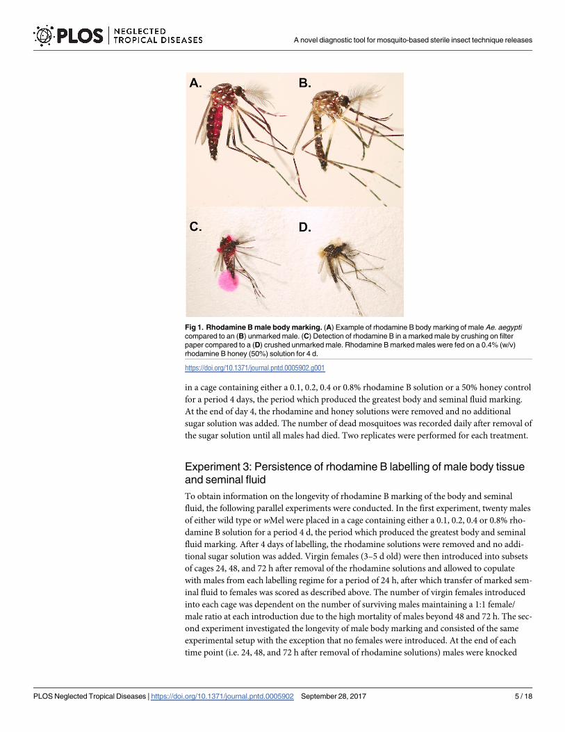
of 1, 2, 3, or 4 days. Male body staining was determined by the visual presence of a dark to
bright red colour in the body, primarily in the abdomen and thorax (Fig 1). Staining of seminal
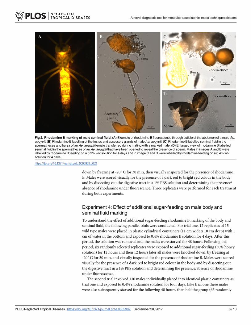
fluid was determined by successful transfer to females during mating as determined by the
presence of rhodamine B in the bursa, spermathecae or both under fluorescence (Fig 2). To do
so, at the end of each time point, twenty 3–5 d old virgin females were introduced into each
cage and the rhodamine solution removed and no additional sugar solution added. The mos-
quitoes were left to copulate for 24 h, after which the mosquitoes were frozen at -20˚ C for 1 h,
then dissected to remove the bursa and spermathecae. The bursa and spermathecae were dis-
sected into a 1% PBS solution and then placed on a microscope slide containing a drop of fluo-
rescent preservative containing DAPI (Fluoroshield with DAPI, Sigma Aldrich) to preserve
the fluorescence of rhodamine B and help visualise the presence of sperm via DAPI staining.
Once placed on the slide, the sample was gently crushed with a cover slip. The presence of rho-
damine B was examined using a fluorescent microscope (Nikon Eclipse Ci) provisioned with
an illuminator (Nikon Intensilight C-HGFI) and a G-2A long pass filter (absorption 510 to
600 nm/emission 575 nm). This filter optimised visual differentiation of rhodamine-treated
compared to untreated testes/MAGs (Fig 2). Images were electronically captured using a
Nikon DigiRetina 16 camera and related software. A Nikon DAPI filter cube (excitation 340
nm/emission 488 nm) was used to help determine the presence/absence of sperm. Three repli-
cates were performed for each treatment.
Experiment 2: Effect of rhodamine B feeding on male survival
To obtain information on the effect of rhodamine B feeding on male survival, the following
experiment was conducted. Twenty males of either wild type or wMel Ae. aegypti were placed
A novel diagnostic tool for mosquito-based sterile insect technique releases
PLOS Neglected Tropical Diseases | https://doi.org/10.1371/journal.pntd.0005902 September 28, 2017 4 / 18
in a cage containing either a 0.1, 0.2, 0.4 or 0.8% rhodamine B solution or a 50% honey control
for a period 4 days, the period which produced the greatest body and seminal fluid marking.
At the end of day 4, the rhodamine and honey solutions were removed and no additional
sugar solution was added. The number of dead mosquitoes was recorded daily after removal of
the sugar solution until all males had died. Two replicates were performed for each treatment.
Experiment 3: Persistence of rhodamine B labelling of male body tissue
and seminal fluid
To obtain information on the longevity of rhodamine B marking of the body and seminal
fluid, the following parallel experiments were conducted. In the first experiment, twenty males
of either wild type or wMel were placed in a cage containing either a 0.1, 0.2, 0.4 or 0.8% rho-
damine B solution for a period 4 d, the period which produced the greatest body and seminal
fluid marking. After 4 days of labelling, the rhodamine solutions were removed and no addi-
tional sugar solution was added. Virgin females (3–5 d old) were then introduced into subsets
of cages 24, 48, and 72 h after removal of the rhodamine solutions and allowed to copulate
with males from each labelling regime for a period of 24 h, after which transfer of marked sem-
inal fluid to females was scored as described above. The number of virgin females introduced
into each cage was dependent on the number of surviving males maintaining a 1:1 female/
male ratio at each introduction due to the high mortality of males beyond 48 and 72 h. The sec-
ond experiment investigated the longevity of male body marking and consisted of the same
experimental setup with the exception that no females were introduced. At the end of each
time point (i.e. 24, 48, and 72 h after removal of rhodamine solutions) males were knocked
Fig 1. Rhodamine B male body marking. (A) Example of rhodamine B body marking of male Ae. aegypti
compared to an (B) unmarked male. (C) Detection of rhodamine B in a marked male by crushing on filter
paper compared to a (D) crushed unmarked male. Rhodamine B marked males were fed on a 0.4% (w/v)
rhodamine B honey (50%) solution for 4 d.
https://doi.org/10.1371/journal.pntd.0005902.g001
A novel diagnostic tool for mosquito-based sterile insect technique releases
PLOS Neglected Tropical Diseases | https://doi.org/10.1371/journal.pntd.0005902 September 28, 2017 5 / 18
down by freezing at -20˚ C for 30 min, then visually inspected for the presence of rhodamine
B. Males were scored visually for the presence of a dark red to bright red colour in the body
and by dissecting out the digestive tract in a 1% PBS solution and determining the presence/
absence of rhodamine under fluorescence. Three replicates were performed for each treatment
during both experiments.
Experiment 4: Effect of additional sugar-feeding on male body and
seminal fluid marking
To understand the effect of additional sugar-feeding rhodamine B marking of the body and
seminal fluid, the following parallel trials were conducted. For trial one, 12 replicates of 15
wild type males were placed in plastic cylindrical containers (11 cm wide x 10 cm deep) with 1
cm of water in the bottom and exposed to 0.4% rhodamine B solution for 4 days. After this
period, the solution was removed and the males were starved for 48 hours. Following this
period, six randomly selected replicates were exposed to additional sugar-feeding (50% honey
solution) for 12 hours and then 12 hours later all males were knocked down, by freezing at
-20˚ C for 30 min, and visually inspected for the presence of rhodamine B. Males were scored
visually for the presence of a dark red to bright red colour in the body and by dissecting out
the digestive tract in a 1% PBS solution and determining the presence/absence of rhodamine
under fluorescence.
The second trial involved 130 males individually placed into identical plastic containers as
trial one and exposed to 0.4% rhodamine solution for four days. Like trial one these males
were also subsequently starved for the following 48 hours, then half the group (65 randomly
Fig 2. Rhodamine B marking of male seminal fluid. (A) Example of rhodamine B fluorescence through cuticle of the abdomen of a male Ae.
aegypti. (B) Rhodamine B labelling of the testes and accessory glands of male Ae. aegypti. (C) Rhodamine B labelled seminal fluid in the
spermathecae and bursa of an Ae. aegypti female transferred during mating with a marked male. (D) Enlarged view of rhodamine B labelled
seminal fluid in the spermathecae of an Ae. aegypti that have been opened to reveal the presence of sperm. Males in images A and B were
labelled by rhodamine B feeding on a 0.2% w/v solution for 4 days and in image C and D were labelled by rhodamine feeding on a 0.4% w/v
solution for 4 days.
https://doi.org/10.1371/journal.pntd.0005902.g002
A novel diagnostic tool for mosquito-based sterile insect technique releases
PLOS Neglected Tropical Diseases | https://doi.org/10.1371/journal.pntd.0005902 September 28, 2017 6 / 18
selected males) was allowed to feed on 50% honey solution for 12 hours to simulate a nectar
feeding in the wild. After an additional 12 hours, a virgin female (of identical age to the males
and only fed on 50% honey solution) was introduced into each of the male’s containers for the
next 24 hours to facilitate mating. After these events (96 h after removal of the rhodamine B
solution), mosquitoes were knocked down and males inspected for the presence of rhodamine
B as described in trial one. The females were inspected for rhodamine B in the bursa and sper-
mathecae, as well as the presence of sperm as described above.
Experiment 5: Mating competitiveness of rhodamine B marked males
under semi-field conditions
We performed a series (n = 6) of free-flight mating competitiveness assays within large semi-
field cages (17.5 m x 8.7 m x 4.1 m and featuring a simulated house and yard [30]) between
0.4% rhodamine B marked (fed 4 d) and unmarked males to assess the effect of rhodamine B
feeding on male mating competitiveness. To ensure that males cannot transfer rhodamine B to
previously mated females, rhodamine B marked males (fed 0.4% solution for 4 d; n = 40) were
introduced to females (n = 40) exposed to unmarked males (n = 40) for 72 h. After 48 h in the
presence of rhodamine B marked males, no transfer of rhodamine was observed and all
females were positive for sperm, revealing no transference of rhodamine to previously mated
females. For each competitiveness assay replication, we placed 80 virgin females (3–5 d old),
40 virgin marked males, and 40 virgin unmarked males (4–5 d old) in a semi-field cage. 72 h
later, females were collected using a Prokopack aspirator [31] and dissected to determine the
presence/absence of rhodamine B as described above. These assays were performed for both
wild type and wMel Ae. aegypti. A female was considered mated by a rhodamine B marked
male by the presence of rhodamine B in the bursa, spermathecae, or both, as well as the pres-
ence of sperm. A female was considered mated by an unmarked male by the presence of sperm
without rhodamine in the bursa and spermathecae.
Experiment 6: Rhodamine B transfer to females during a field release
A series (n = 3) of proof-of-principle field releases were conducted to determine if rhodamine
B transfer from marked males to receptive females occurs under field conditions. For each
experimental replicate, 150 marked wMel Ae. aegypti males (0.4% rhodamine B, fed 4 d) were
released at 08:00 in the front of a single residence located in the Edge Hill suburb of Cairns,
QLD where wMel Ae. aegypti have been established since 2012 [32]. Because the releases
occurred late in the dry season, a period during low Ae. aegypti abundance [33], we released a
total of 30 virgin wMel Ae. aegypti females (3–5 d old) inside the house 24 h prior to the release
of males. Indoor releases were conducted to determine if marked males released outside would
enter houses, the preferred resting site of female Ae. aegypti [34]. Mosquitoes were sampled
indoors by 10 min sweep net (sprayed with a pyrethroid-based surface spray; Mortein Outdoor
Barrier Surface Spray; Imiprothrin 0.3 g/Kg, 0.6 g/Kg Deltamethrin) collections performed
daily by a single individual between 16:00–17:00 for a period of 2 d starting 24 h after the
release of males. Sweep net collections were supplemented by Biogents Sentinel (operated con-
tinuously) [35] and Gravid Aedes Trap (GAT) [36] collections, with both traps being placed on
the back patio. Captured mosquitoes were stored at -20 C until processed to determine the
presence of rhodamine B as described above. Each subsequent release occurred 1 week after
the prior release. During releases, temperatures ranged from 21.7–28.7 C and relative humidity
ranged from 45–85% with no significant rain events. The owners of the residence gave permis-
sion for the study to be conducted on their property.
A novel diagnostic tool for mosquito-based sterile insect technique releases
PLOS Neglected Tropical Diseases | https://doi.org/10.1371/journal.pntd.0005902 September 28, 2017 7 / 18
Statistical analysis
Differences in male body staining, both visually and under fluorescence, seminal fluid mark-
ing, and persistence of male seminal fluid and body marking were analysed by two-way
ANOVA with Tukey HSD post-hoc analysis. The data, represented as percentages, were arc-
sine transformed prior to analysis [37]. Differences in daily survivorship, determined by
Kaplan-Meier survivorship curves, were analysed by the log-rank test [38]. Differences in mat-
ing competitiveness (i.e. proportion of females mated) was analysed using chi-square tests
with the null hypothesis of 1:1 mating ratio. Mean comparisons of the proportions of males
stained (arcsin transformed) between replicates exposed to and not exposed to additional
sugar-feeding occurred using a t-test. Differences in male body staining (i.e. proportion of
males stained) and seminal fluid marking (i.e. proportion of marked females that had mated
with stained males) for the two treatment groups was analysed by the Fisher’s Exact Test with
null hypotheses of 1:1 marking ratios. All statistical analyses were performed using the R
(http://www.r-project.org/) and Prism (ver. 7.03; GraphPad) statistical software.
Results
Experiment 1: Effect of concentration and feeding time on male body and
seminal fluid marking
There were significant effects of length of feeding (F3,20 = 3.34, P = 0.045) and concentration
(F3,20 = 4.73, P = 0.02) on body and seminal fluid marking (Fig 3A; Table 1), with increases in
both yielding the best marking results. Overall, 89.9±1.6% of males fed on 0.4% and 0.8% solu-
tions were visually marked after 1 d of feeding, and 96.7±0.9% were marked after 4 d of feed-
ing. When fed on 0.1% and 0.2% solutions, 60.3±3.6% were marked after 1 d of feeding and
88.3±1.6% were marked after 4 d of feeding. Similar effects of time (F3,20 = 43.9, P<0.001) and
concentration (F3,20 = 5.95, P = 0.01) were observed for successful seminal fluid marking and
transfer to females (Fig 3B; Table 1). Again, males fed on 0.4% and 0.8% produced the greatest
seminal fluid marking, with 56.3±3.0% of females being positive for rhodamine in the bursa or
spermathecae after exposure to males when those males were fed for 2 d, compared to 33.8
±2.7% at 0.1% and 0.2%, respectively. After 4 d of male rhodamine feeding, there was no signif-
icant difference in seminal fluid marking across all treatments with 96.5±2.7% of females being
positive for rhodamine B following male exposure.
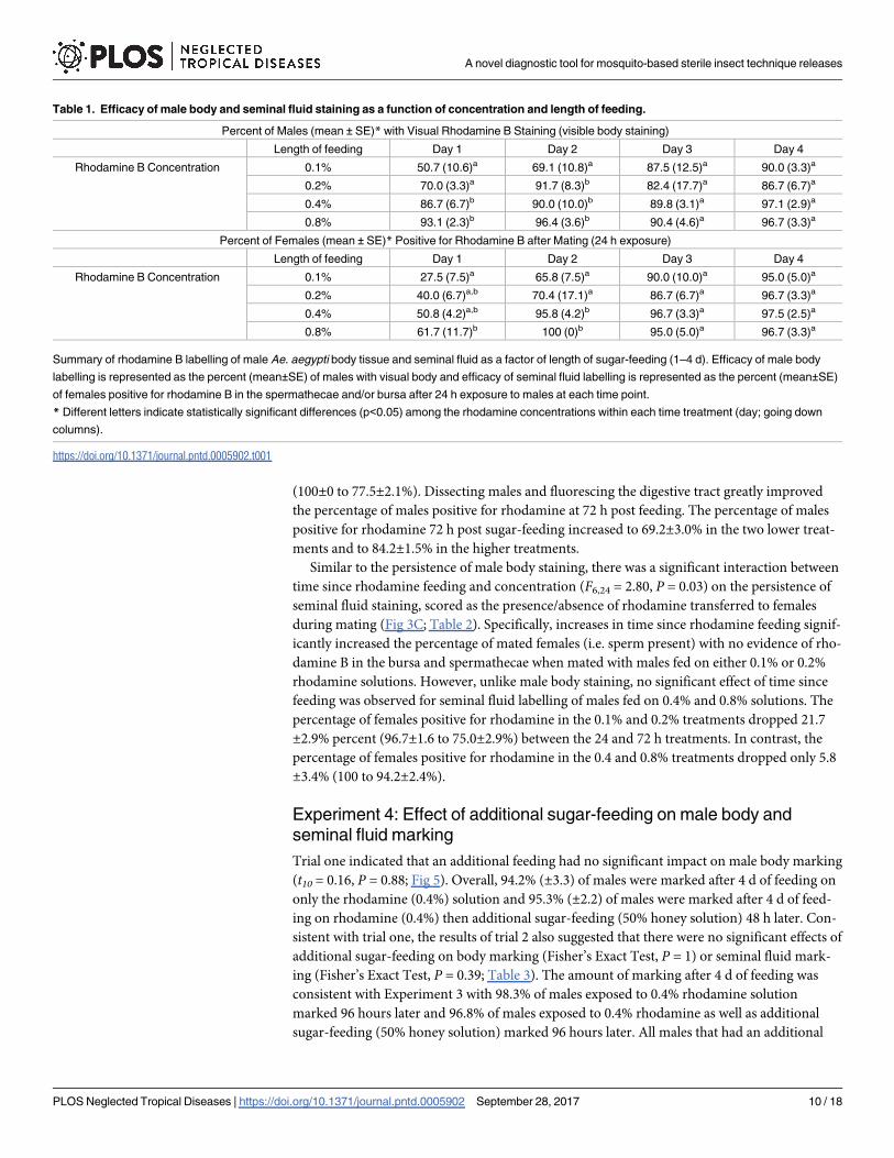
Experiment 2: Effect of rhodamine feeding on male survival
No significant effect of rhodamine B feeding on survival was observed for either wild type (χ1
= 0.27, P = 0.60) or wMel (χ1 = 0.28, P = 0.61) Ae. aegypti relative to honey-fed controls across
all concentrations (Fig 4). No mortality was observed during honey-feeding and survival at the
cessation of honey feeding among all concentrations and between the two strains was similar.
Across all concentrations, the percent of wild type males surviving 1, 2, 3, and 4 d after cessa-
tion of honey-feeding as 86.9±5.0, 52.5±6.3, 21.3±8.1, and 1.8±0.9%. For wMel Ae. aegyptimales, survivorship 1, 2, 3, and 4 d after cessation of honey-feeding was 85.6±4.5, 43.7±3.4,
15.0±2.7, and 0%.
Experiment 3: Persistence of rhodamine labelling of male body tissue
and seminal fluid
There was a significant interaction between time since rhodamine feeding and concentration
on the persistence of male body staining when scored visually (conspicuous body marking)
(F6,24 = 17.13, P<0.001) and with fluorescence (F6,24 = 3.2, P = 0.02; Table 2). Specifically,
A novel diagnostic tool for mosquito-based sterile insect technique releases
PLOS Neglected Tropical Diseases | https://doi.org/10.1371/journal.pntd.0005902 September 28, 2017 8 / 18
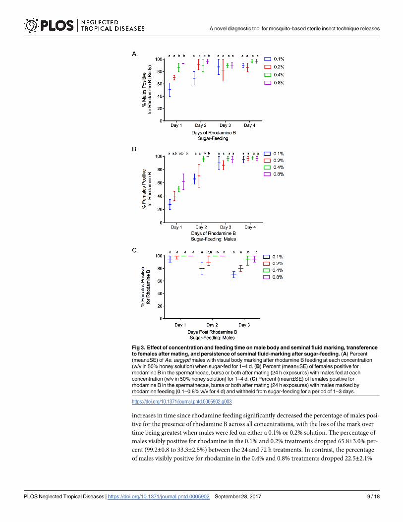
increases in time since rhodamine feeding significantly decreased the percentage of males posi-
tive for the presence of rhodamine B across all concentrations, with the loss of the mark over
time being greatest when males were fed on either a 0.1% or 0.2% solution. The percentage of
males visibly positive for rhodamine in the 0.1% and 0.2% treatments dropped 65.8±3.0% per-
cent (99.2±0.8 to 33.3±2.5%) between the 24 and 72 h treatments. In contrast, the percentage
of males visibly positive for rhodamine in the 0.4% and 0.8% treatments dropped 22.5±2.1%
Fig 3. Effect of concentration and feeding time on male body and seminal fluid marking, transference
to females after mating, and persistence of seminal fluid-marking after sugar-feeding. (A) Percent
(mean±SE) of Ae. aegypti males with visual body marking after rhodamine B feeding at each concentration
(w/v in 50% honey solution) when sugar-fed for 1–4 d. (B) Percent (mean±SE) of females positive for
rhodamine B in the spermathecae, bursa or both after mating (24 h exposures) with males fed at each
concentration (w/v in 50% honey solution) for 1–4 d. (C) Percent (mean±SE) of females positive for
rhodamine B in the spermathecae, bursa or both after mating (24 h exposures) with males marked by
rhodamine feeding (0.1–0.8% w/v for 4 d) and withheld from sugar-feeding for a period of 1–3 days.
https://doi.org/10.1371/journal.pntd.0005902.g003
A novel diagnostic tool for mosquito-based sterile insect technique releases
PLOS Neglected Tropical Diseases | https://doi.org/10.1371/journal.pntd.0005902 September 28, 2017 9 / 18
(100±0 to 77.5±2.1%). Dissecting males and fluorescing the digestive tract greatly improved
the percentage of males positive for rhodamine at 72 h post feeding. The percentage of males
positive for rhodamine 72 h post sugar-feeding increased to 69.2±3.0% in the two lower treat-
ments and to 84.2±1.5% in the higher treatments.
Similar to the persistence of male body staining, there was a significant interaction between
time since rhodamine feeding and concentration (F6,24 = 2.80, P = 0.03) on the persistence of
seminal fluid staining, scored as the presence/absence of rhodamine transferred to females
during mating (Fig 3C; Table 2). Specifically, increases in time since rhodamine feeding signif-
icantly increased the percentage of mated females (i.e. sperm present) with no evidence of rho-
damine B in the bursa and spermathecae when mated with males fed on either 0.1% or 0.2%
rhodamine solutions. However, unlike male body staining, no significant effect of time since
feeding was observed for seminal fluid labelling of males fed on 0.4% and 0.8% solutions. The
percentage of females positive for rhodamine in the 0.1% and 0.2% treatments dropped 21.7
±2.9% percent (96.7±1.6 to 75.0±2.9%) between the 24 and 72 h treatments. In contrast, the
percentage of females positive for rhodamine in the 0.4 and 0.8% treatments dropped only 5.8
±3.4% (100 to 94.2±2.4%).
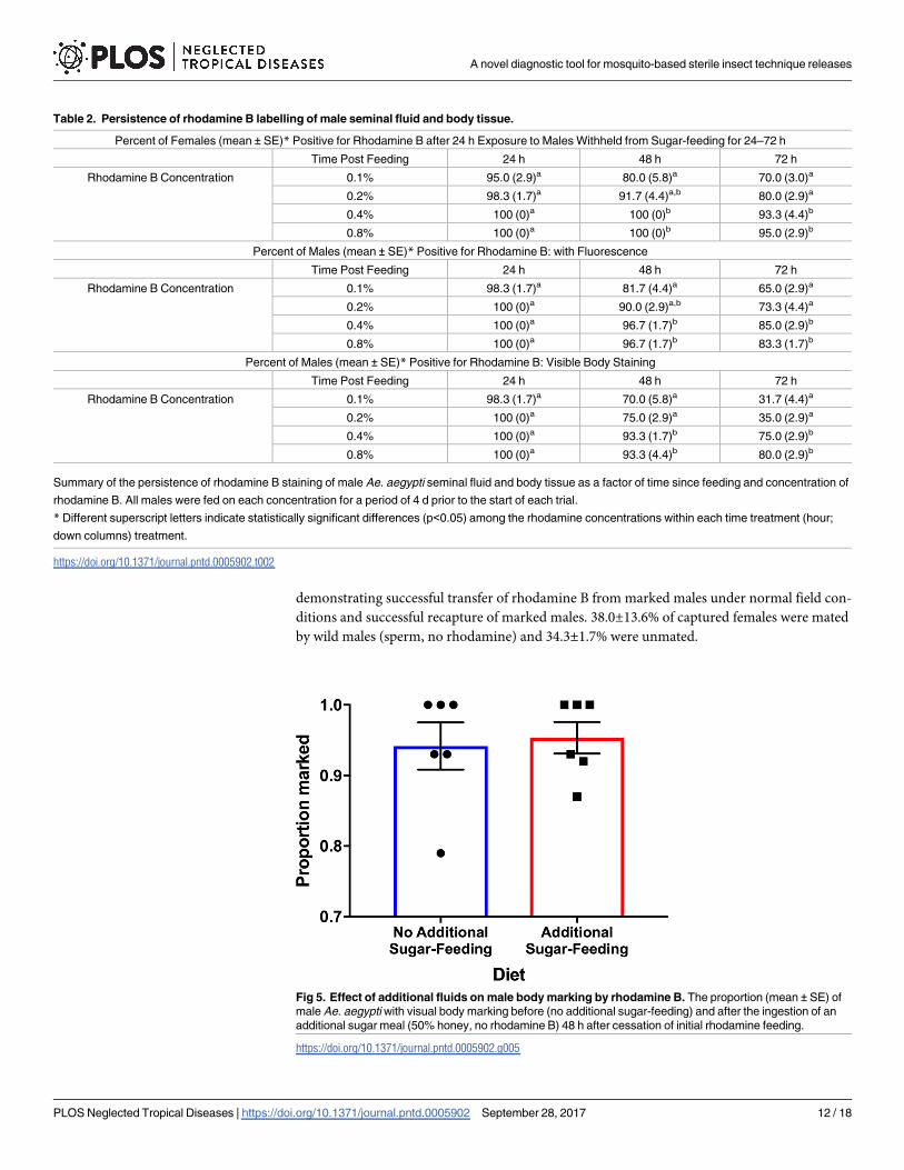
Experiment 4: Effect of additional sugar-feeding on male body and
seminal fluid marking
Trial one indicated that an additional feeding had no significant impact on male body marking
(t10 = 0.16, P = 0.88; Fig 5). Overall, 94.2% (±3.3) of males were marked after 4 d of feeding on
only the rhodamine (0.4%) solution and 95.3% (±2.2) of males were marked after 4 d of feed-
ing on rhodamine (0.4%) then additional sugar-feeding (50% honey solution) 48 h later. Con-
sistent with trial one, the results of trial 2 also suggested that there were no significant effects of
additional sugar-feeding on body marking (Fisher’s Exact Test, P = 1) or seminal fluid mark-
ing (Fisher’s Exact Test, P = 0.39; Table 3). The amount of marking after 4 d of feeding was
consistent with Experiment 3 with 98.3% of males exposed to 0.4% rhodamine solution
marked 96 hours later and 96.8% of males exposed to 0.4% rhodamine as well as additional
sugar-feeding (50% honey solution) marked 96 hours later. All males that had an additional
Table 1. Efficacy of male body and seminal fluid staining as a function of concentration and length of feeding.
Percent of Males (mean ± SE)* with Visual Rhodamine B Staining (visible body staining)
Length of feeding Day 1 Day 2 Day 3 Day 4
Rhodamine B Concentration 0.1% 50.7 (10.6)a 69.1 (10.8)a 87.5 (12.5)a 90.0 (3.3)a
0.2% 70.0 (3.3)a 91.7 (8.3)b 82.4 (17.7)a 86.7 (6.7)a
0.4% 86.7 (6.7)b 90.0 (10.0)b 89.8 (3.1)a 97.1 (2.9)a
0.8% 93.1 (2.3)b 96.4 (3.6)b 90.4 (4.6)a 96.7 (3.3)a
Percent of Females (mean ± SE)* Positive for Rhodamine B after Mating (24 h exposure)
Length of feeding Day 1 Day 2 Day 3 Day 4
Rhodamine B Concentration 0.1% 27.5 (7.5)a 65.8 (7.5)a 90.0 (10.0)a 95.0 (5.0)a
0.2% 40.0 (6.7)a,b 70.4 (17.1)a 86.7 (6.7)a 96.7 (3.3)a
0.4% 50.8 (4.2)a,b 95.8 (4.2)b 96.7 (3.3)a 97.5 (2.5)a
0.8% 61.7 (11.7)b 100 (0)b 95.0 (5.0)a 96.7 (3.3)a
Summary of rhodamine B labelling of male Ae. aegypti body tissue and seminal fluid as a factor of length of sugar-feeding (1–4 d). Efficacy of male body
labelling is represented as the percent (mean±SE) of males with visual body and efficacy of seminal fluid labelling is represented as the percent (mean±SE)
of females positive for rhodamine B in the spermathecae and/or bursa after 24 h exposure to males at each time point.
* Different letters indicate statistically significant differences (p<0.05) among the rhodamine concentrations within each time treatment (day; going down
columns).
https://doi.org/10.1371/journal.pntd.0005902.t001
A novel diagnostic tool for mosquito-based sterile insect technique releases
PLOS Neglected Tropical Diseases | https://doi.org/10.1371/journal.pntd.0005902 September 28, 2017 10 / 18
honey feeding were noted to be engorged when knocked down. Of the females that had mated
with stained males not exposed to additional sugar-feeding 83% displayed seminal fluid mark-
ing while 90% of females that mated with stained males exposed to additional sugar-feeding
displayed seminal fluid marking under fluorescence (Table 3).
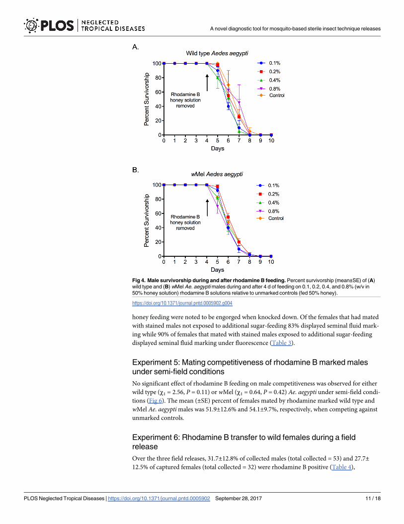
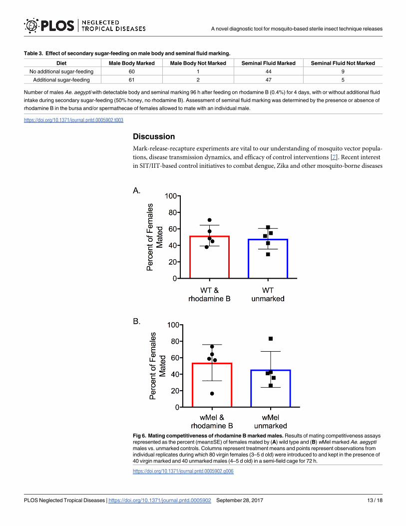
Experiment 5: Mating competitiveness of rhodamine B marked males
under semi-field conditions
No significant effect of rhodamine B feeding on male competitiveness was observed for either
wild type (χ1 = 2.56, P = 0.11) or wMel (χ1 = 0.64, P = 0.42) Ae. aegypti under semi-field condi-
tions (Fig 6). The mean (±SE) percent of females mated by rhodamine marked wild type and
wMel Ae. aegypti males was 51.9±12.6% and 54.1±9.7%, respectively, when competing against
unmarked controls.
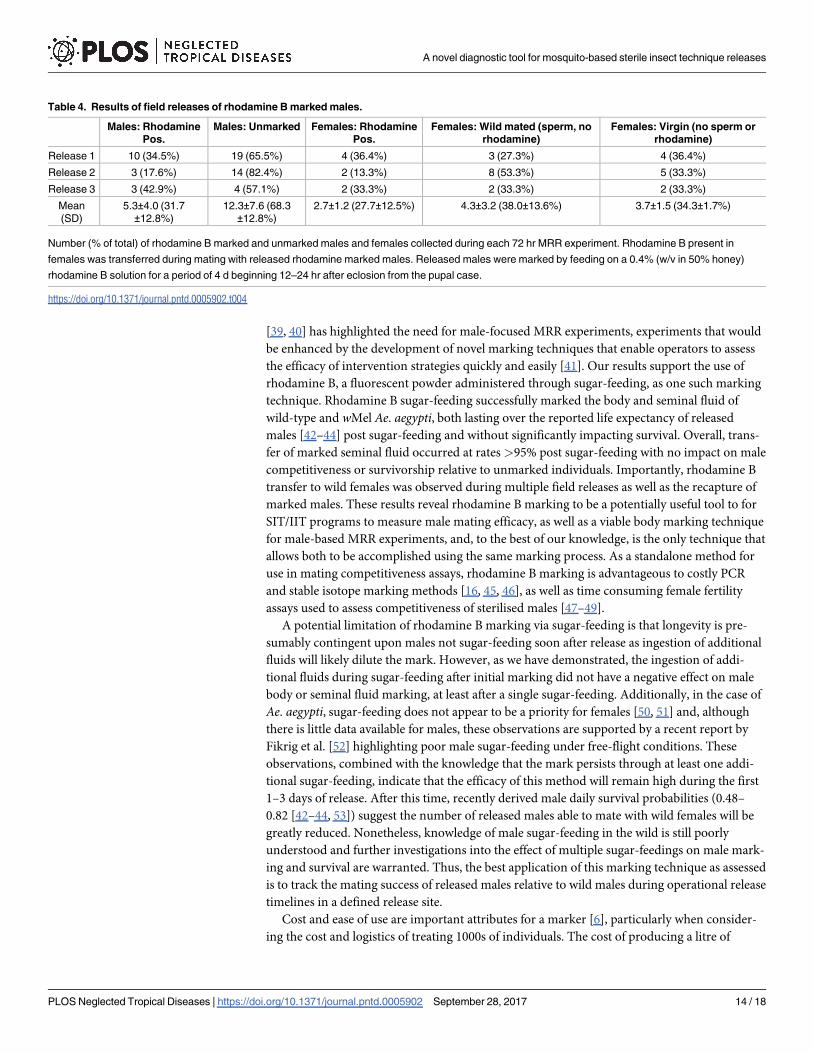
Experiment 6: Rhodamine B transfer to wild females during a field
release
Over the three field releases, 31.7±12.8% of collected males (total collected = 53) and 27.7±12.5% of captured females (total collected = 32) were rhodamine B positive (Table 4),
Fig 4. Male survivorship during and after rhodamine B feeding. Percent survivorship (mean±SE) of (A)
wild type and (B) wMel Ae. aegypti males during and after 4 d of feeding on 0.1, 0.2, 0.4, and 0.8% (w/v in
50% honey solution) rhodamine B solutions relative to unmarked controls (fed 50% honey).
https://doi.org/10.1371/journal.pntd.0005902.g004
A novel diagnostic tool for mosquito-based sterile insect technique releases
PLOS Neglected Tropical Diseases | https://doi.org/10.1371/journal.pntd.0005902 September 28, 2017 11 / 18
demonstrating successful transfer of rhodamine B from marked males under normal field con-
ditions and successful recapture of marked males. 38.0±13.6% of captured females were mated
by wild males (sperm, no rhodamine) and 34.3±1.7% were unmated.
Table 2. Persistence of rhodamine B labelling of male seminal fluid and body tissue.
Percent of Females (mean ± SE)* Positive for Rhodamine B after 24 h Exposure to Males Withheld from Sugar-feeding for 24–72 h
Time Post Feeding 24 h 48 h 72 h
Rhodamine B Concentration 0.1% 95.0 (2.9)a 80.0 (5.8)a 70.0 (3.0)a
0.2% 98.3 (1.7)a 91.7 (4.4)a,b 80.0 (2.9)a
0.4% 100 (0)a 100 (0)b 93.3 (4.4)b
0.8% 100 (0)a 100 (0)b 95.0 (2.9)b
Percent of Males (mean ± SE)* Positive for Rhodamine B: with Fluorescence
Time Post Feeding 24 h 48 h 72 h
Rhodamine B Concentration 0.1% 98.3 (1.7)a 81.7 (4.4)a 65.0 (2.9)a
0.2% 100 (0)a 90.0 (2.9)a,b 73.3 (4.4)a
0.4% 100 (0)a 96.7 (1.7)b 85.0 (2.9)b
0.8% 100 (0)a 96.7 (1.7)b 83.3 (1.7)b
Percent of Males (mean ± SE)* Positive for Rhodamine B: Visible Body Staining
Time Post Feeding 24 h 48 h 72 h
Rhodamine B Concentration 0.1% 98.3 (1.7)a 70.0 (5.8)a 31.7 (4.4)a
0.2% 100 (0)a 75.0 (2.9)a 35.0 (2.9)a
0.4% 100 (0)a 93.3 (1.7)b 75.0 (2.9)b
0.8% 100 (0)a 93.3 (4.4)b 80.0 (2.9)b
Summary of the persistence of rhodamine B staining of male Ae. aegypti seminal fluid and body tissue as a factor of time since feeding and concentration of
rhodamine B. All males were fed on each concentration for a period of 4 d prior to the start of each trial.
* Different superscript letters indicate statistically significant differences (p<0.05) among the rhodamine concentrations within each time treatment (hour;
down columns) treatment.
https://doi.org/10.1371/journal.pntd.0005902.t002
Fig 5. Effect of additional fluids on male body marking by rhodamine B. The proportion (mean ± SE) of
male Ae. aegypti with visual body marking before (no additional sugar-feeding) and after the ingestion of an
additional sugar meal (50% honey, no rhodamine B) 48 h after cessation of initial rhodamine feeding.
https://doi.org/10.1371/journal.pntd.0005902.g005
A novel diagnostic tool for mosquito-based sterile insect technique releases
PLOS Neglected Tropical Diseases | https://doi.org/10.1371/journal.pntd.0005902 September 28, 2017 12 / 18
Discussion
Mark-release-recapture experiments are vital to our understanding of mosquito vector popula-
tions, disease transmission dynamics, and efficacy of control interventions [7]. Recent interest
in SIT/IIT-based control initiatives to combat dengue, Zika and other mosquito-borne diseases
Table 3. Effect of secondary sugar-feeding on male body and seminal fluid marking.
Diet Male Body Marked Male Body Not Marked Seminal Fluid Marked Seminal Fluid Not Marked
No additional sugar-feeding 60 1 44 9
Additional sugar-feeding 61 2 47 5
Number of males Ae. aegypti with detectable body and seminal marking 96 h after feeding on rhodamine B (0.4%) for 4 days, with or without additional fluid
intake during secondary sugar-feeding (50% honey, no rhodamine B). Assessment of seminal fluid marking was determined by the presence or absence of
rhodamine B in the bursa and/or spermathecae of females allowed to mate with an individual male.
https://doi.org/10.1371/journal.pntd.0005902.t003
Fig 6. Mating competitiveness of rhodamine B marked males. Results of mating competitiveness assays
represented as the percent (mean±SE) of females mated by (A) wild type and (B) wMel marked Ae. aegypti
males vs. unmarked controls. Columns represent treatment means and points represent observations from
individual replicates during which 80 virgin females (3–5 d old) were introduced to and kept in the presence of
40 virgin marked and 40 unmarked males (4–5 d old) in a semi-field cage for 72 h.
https://doi.org/10.1371/journal.pntd.0005902.g006
A novel diagnostic tool for mosquito-based sterile insect technique releases
PLOS Neglected Tropical Diseases | https://doi.org/10.1371/journal.pntd.0005902 September 28, 2017 13 / 18
[39, 40] has highlighted the need for male-focused MRR experiments, experiments that would
be enhanced by the development of novel marking techniques that enable operators to assess
the efficacy of intervention strategies quickly and easily [41]. Our results support the use of
rhodamine B, a fluorescent powder administered through sugar-feeding, as one such marking
technique. Rhodamine B sugar-feeding successfully marked the body and seminal fluid of
wild-type and wMel Ae. aegypti, both lasting over the reported life expectancy of released
males [42–44] post sugar-feeding and without significantly impacting survival. Overall, trans-
fer of marked seminal fluid occurred at rates >95% post sugar-feeding with no impact on male
competitiveness or survivorship relative to unmarked individuals. Importantly, rhodamine B
transfer to wild females was observed during multiple field releases as well as the recapture of
marked males. These results reveal rhodamine B marking to be a potentially useful tool to for
SIT/IIT programs to measure male mating efficacy, as well as a viable body marking technique
for male-based MRR experiments, and, to the best of our knowledge, is the only technique that
allows both to be accomplished using the same marking process. As a standalone method for
use in mating competitiveness assays, rhodamine B marking is advantageous to costly PCR
and stable isotope marking methods [16, 45, 46], as well as time consuming female fertility
assays used to assess competitiveness of sterilised males [47–49].
A potential limitation of rhodamine B marking via sugar-feeding is that longevity is pre-
sumably contingent upon males not sugar-feeding soon after release as ingestion of additional
fluids will likely dilute the mark. However, as we have demonstrated, the ingestion of addi-
tional fluids during sugar-feeding after initial marking did not have a negative effect on male
body or seminal fluid marking, at least after a single sugar-feeding. Additionally, in the case of
Ae. aegypti, sugar-feeding does not appear to be a priority for females [50, 51] and, although
there is little data available for males, these observations are supported by a recent report by
Fikrig et al. [52] highlighting poor male sugar-feeding under free-flight conditions. These
observations, combined with the knowledge that the mark persists through at least one addi-
tional sugar-feeding, indicate that the efficacy of this method will remain high during the first
1–3 days of release. After this time, recently derived male daily survival probabilities (0.48–
0.82 [42–44, 53]) suggest the number of released males able to mate with wild females will be
greatly reduced. Nonetheless, knowledge of male sugar-feeding in the wild is still poorly
understood and further investigations into the effect of multiple sugar-feedings on male mark-
ing and survival are warranted. Thus, the best application of this marking technique as assessed
is to track the mating success of released males relative to wild males during operational release
timelines in a defined release site.
Cost and ease of use are important attributes for a marker [6], particularly when consider-
ing the cost and logistics of treating 1000s of individuals. The cost of producing a litre of
Table 4. Results of field releases of rhodamine B marked males.
Males: Rhodamine
Pos.
Males: Unmarked Females: Rhodamine
Pos.
Females: Wild mated (sperm, no
rhodamine)
Females: Virgin (no sperm or
rhodamine)
Release 1 10 (34.5%) 19 (65.5%) 4 (36.4%) 3 (27.3%) 4 (36.4%)
Release 2 3 (17.6%) 14 (82.4%) 2 (13.3%) 8 (53.3%) 5 (33.3%)
Release 3 3 (42.9%) 4 (57.1%) 2 (33.3%) 2 (33.3%) 2 (33.3%)
Mean
(SD)
5.3±4.0 (31.7
±12.8%)
12.3±7.6 (68.3
±12.8%)
2.7±1.2 (27.7±12.5%) 4.3±3.2 (38.0±13.6%) 3.7±1.5 (34.3±1.7%)
Number (% of total) of rhodamine B marked and unmarked males and females collected during each 72 hr MRR experiment. Rhodamine B present in
females was transferred during mating with released rhodamine marked males. Released males were marked by feeding on a 0.4% (w/v in 50% honey)
rhodamine B solution for a period of 4 d beginning 12–24 hr after eclosion from the pupal case.
https://doi.org/10.1371/journal.pntd.0005902.t004
A novel diagnostic tool for mosquito-based sterile insect technique releases
PLOS Neglected Tropical Diseases | https://doi.org/10.1371/journal.pntd.0005902 September 28, 2017 14 / 18
rhodamine B-treated honey was $1.40 (0.4% w/v), which is sufficient to treat�2,500 males.
Additionally, as others have noted [54], this method is easy to implement for feeding hundreds
of insects at once as it requires no special skills or equipment to administer. Although treat-
ment costs are minimal, this method requires fluorescent microscopy equipment (e.g. micro-
scope, illuminator, filters), which in this study cost ca. $11,000 USD (Scope Scientific Pty Ltd,
Queensland, Australia). Despite these significant upfront costs, per sample analysis and treat-
ment costs are marginal relative to alternative methods allowing researchers to recoup upfront
costs. For example, Hamer et al. [15] detail treatment costs of $1.50 and $3.69 per 100 larvae
for 15N and 13C enrichment, respectively, and $7 per sample (1–5 individuals) analysis costs
(dual15N and 13C mass spectrometry analysis). Further, the time required for determining the
presence/absence of rhodamine B is<30 min once staff have acquired the appropriate dissec-
tion and microscopy skills, whereas stable isotope analysis may range anywhere from 1 to 20
weeks depending on the facility and expense of rapid analysis. Of note, it is important to
acknowledge that rhodamine B is a notifiable chemical with limited evidence of carcinogenic-
ity in animals and no adequate data for humans [55]. It is equally important to acknowledge
that it has long been used as a biomarker in wildlife studies [56–58] and lethal dose calculations
for a variety of invertebrates and vertebrates are relatively high [57], well above what an indi-
vidual mosquito could ingest (<2 mg) during a single sugar meal (max volume 0.4 μl [59]). At
these amounts rhodamine B poses little health risk to humans or species that may consume
released mosquitoes; however, best practice dictates that a full assessment of the risk of rhoda-
mine B in this manner be performed by appropriate institutional research review boards and
ethics committees prior to initiation of field and laboratory studies.
In summary, this study reveals rhodamine B labelling to be an economical and rapid evalua-
tion method for male-based control strategies as well as a potential body marking technique
for male focused MRR experiments. The use of this method has the potential to enable opera-
tors to determine the efficacy of their control method quickly and easily, allowing them to opti-
mise releases to achieve optimal population suppression. These observations are bound by the
limitations in extending laboratory observations as a prediction of what to expect in the field
and we strongly recommend that small-scale field MRR experiments be performed to obtain
more accurate estimates of male survival and mark persistence prior to adoption for opera-
tional assessments. These studies would benefit from comparisons to classical marking tech-
niques such as fluorescent powders to better understand the utility of rhodamine B body
marking in male-focused survival and dispersal studies.
Acknowledgments
The authors would like to thank Sandra Taylor, Ana Ramirez, Lili Usher-Chandler, Catherine
Liddington, and Jason Anderson for their assistance in data acquisition.
Author Contributions
Conceptualization: Brian J. Johnson, Sara N. Mitchell, Nigel Snoad, Nigel Beebe, Bradley J.
White, Scott A. Ritchie.
Data curation: Brian J. Johnson, Sara N. Mitchell, Jessica Stevenson.
Formal analysis: Brian J. Johnson.
Investigation: Brian J. Johnson, Sara N. Mitchell, Christopher J. Paton, Jessica Stevenson,
Kyran M. Staunton, Bradley J. White.
Methodology: Brian J. Johnson, Sara N. Mitchell, Nigel Snoad, Scott A. Ritchie.
A novel diagnostic tool for mosquito-based sterile insect technique releases
PLOS Neglected Tropical Diseases | https://doi.org/10.1371/journal.pntd.0005902 September 28, 2017 15 / 18
Supervision: Nigel Snoad, Nigel Beebe, Bradley J. White, Scott A. Ritchie.
Visualization: Brian J. Johnson.
Writing – original draft: Brian J. Johnson, Scott A. Ritchie.
Writing – review & editing: Brian J. Johnson, Sara N. Mitchell, Kyran M. Staunton, Nigel
Beebe, Bradley J. White, Scott A. Ritchie.
References1. Reiter P. Oviposition, dispersal, and survival in Aedes aegypti: implications for the efficacy of control
strategies. Vector Borne Zoonotic Dis. 2007; 7(2):261–73. https://doi.org/10.1089/vbz.2006.0630
PMID: 17627447
2. Harrington LC, Poulson RL. Considerations for accurate identification of adult Culex restuans (Diptera:
Culicidae) in field studies. J Med Entomol. 2008; 45(1):1–8. PMID: 18283935
3. Maciel-de-Freitas R, Eiras AE, Lourenco-de-Oliveira R. Calculating the survival rate and estimated pop-
ulation density of gravid Aedes aegypti (Diptera, Culicidae) in Rio de Janeiro, Brazil. Cadernos de
Saude Publica. 2008; 24(12):2747–54. PMID: 19082265
4. Araujo Md-S, Gil LHS. Larval food quantity affects development time, survival and adult biological traits
that influence the vectorial capacity of Anopheles darlingi under laboratory conditions. Malaria J. 2012;
11(1):1.
5. Reiner RC, Perkins TA, Barker CM, Niu T, Chaves LF, Ellis AM, et al. A systematic review of mathemat-
ical models of mosquito-borne pathogen transmission: 1970–2010. J R Soc Interface. 2013; 10
(81):20120921. https://doi.org/10.1098/rsif.2012.0921 PMID: 23407571
6. Hagler JR, Jackson CG. Methods for marking insects: current techniques and future prospects. Annu
Rev Entomol. 2001; 46(1):511–43.
7. Guerra CA, Reiner RC Jr., Perkins TA, Lindsay SW, Midega JT, Brady OJ, et al. A global assembly of adult
female mosquito mark-release-recapture data to inform the control of mosquito-borne pathogens. Parasit
Vectors. 2014; 7:276. Epub 2014/06/21. https://doi.org/10.1186/1756-3305-7-276 PMID: 24946878
8. Verhulst NO, Loonen JA, Takken W. Advances in methods for colour marking of mosquitoes. Parasit
Vectors. 2013; 6(1):1.
9. Zukel JW. Marking Anopheles mosquitoes with fluorescent compounds. Science. 1945.
10. Dickens BL, Brant HL. Effects of marking methods and fluorescent dusts on Aedes aegypti survival.
Parasit Vectors. 2014; 7(1):1.
11. Naranjo SE. Influence of two mass-marking techniques on survival and flight behavior of Diabrotica vir-
gifera virgifera (Coleoptera: Chrysomelidae). J Econ Entomol. 1990; 83(4):1360–4.
12. Bellini R, Albieri A, Balestrino F, Carrieri M, Porretta D, Urbanelli S, et al. Dispersal and survival of
Aedes albopictus (Diptera: Culicidae) males in Italian urban areas and significance for sterile insect
technique application. J Med Entomol. 2010; 47(6):1082–91. PMID: 21175057
13. De Souza AR, Ribeiro B, Jose N, Prezoto F. Paint marking social wasps: an evaluation of behavioral
effects and toxicity. Entomol Exp Appl. 2012; 144(2):244–7.
14. Hagler JR, Miller E. An alternative to conventional insect marking procedures: detection of a protein
mark on pink bollworm by ELISA. Entomol Exp Appl. 2002; 103(1):1–9.
15. Hamer GL, Donovan DJ, Hood-Nowotny R, Kaufman MG, Goldberg TL, Walker ED. Evaluation of a sta-
ble isotope method to mark naturally-breeding larval mosquitoes for adult dispersal studies. J Med Ento-
mol. 2012; 49(1):61–70. PMID: 22308772
16. Medeiros MCI, Boothe EC, Roark EB, Hamer GL. Dispersal of male and female Culex quinquefasciatus
and Aedes albopictus mosquitoes using stable isotope enrichment. PLoS Neg Trop Dis. 2017; 11(1):
e0005347. https://doi.org/10.1371/journal.pntd.0005347 PMID: 28135281
17. Bellini R, Calvitti M, Medici A, Carrieri M, Celli G, Maini S. Use of the sterile insect technique against
Aedes albopictus in Italy: First results of a pilot trial. Area-Wide Control of Insect Pests: Springer; 2007.
p. 505–15.
18. Harris AF, Nimmo D, McKemey AR, Kelly N, Scaife S, Donnelly CA, et al. Field performance of engi-
neered male mosquitoes. Nat Biotechnol. 2011; 29(11):1034–7. https://doi.org/10.1038/nbt.2019
PMID: 22037376
19. Harris AF, McKemey AR, Nimmo D, Curtis Z, Black I, Morgan SA, et al. Successful suppression of a
field mosquito population by sustained release of engineered male mosquitoes. Nat Biotechnol. 2012;
30(9):828–30. https://doi.org/10.1038/nbt.2350 PMID: 22965050
A novel diagnostic tool for mosquito-based sterile insect technique releases
PLOS Neglected Tropical Diseases | https://doi.org/10.1371/journal.pntd.0005902 September 28, 2017 16 / 18
20. Mains JW, Brelsfoard CL, Rose RI, Dobson SL. Female adult Aedes albopictus suppression by Wolba-
chia-infected male mosquitoes. Sci Reports. 2016; 6.
21. Blanco CA, Perera O, Ray JD, Taliercio E, Williams L III. Incorporation of rhodamine B into male
tobacco budworm moths Heliothis virescens to use as a marker for mating studies. J Insect Sci. 2006; 6
(5):1–10.
22. Evd Reijden, Monchamp JD, Lewis SM. The formation, transfer, and fate of spermatophores in Photi-
nus fireflies (Coleoptera: Lampyridae). Can J Zool. 1997; 75(8):1202–7.
23. South A, Sota T, Abe N, Yuma M, Lewis SM. The production and transfer of spermatophores in three
Asian species of Luciola fireflies. J Insect Physiol. 2008; 54(5):861–6. https://doi.org/10.1016/j.jinsphys.
2008.03.008 PMID: 18479701
24. Kamimura Y. Twin intromittent organs of Drosophila for traumatic insemination. Biol Lett. 2007; 3
(4):401–4. https://doi.org/10.1098/rsbl.2007.0192 PMID: 17519186
25. Bailey SF, Eliason D, Iltis W. Some marking and recovery techniques in Culex tarsalis Coq. flight stud-
ies. Mosquito News. 1962; 22(1):1–10.
26. Walker T, Johnson P, Moreira L, Iturbe-Ormaetxe I, Frentiu F, McMeniman C, et al. The wMel Wolba-
chia strain blocks dengue and invades caged Aedes aegypti populations. Nature. 2011; 476
(7361):450–3. https://doi.org/10.1038/nature10355 PMID: 21866159
27. Zhang D, Lees RS, Xi Z, Bourtzis K, Gilles JR. Combining the Sterile Insect Technique with the Incom-
patible Insect Technique: III-robust mating competitiveness of irradiated triple Wolbachia-infected
Aedes albopictus males under semi-field conditions. PloS One. 2016; 11(3):e0151864. https://doi.org/
10.1371/journal.pone.0151864 PMID: 26990981
28. Hoffmann A, Montgomery B, Popovici J, Iturbe-Ormaetxe I, Johnson P, Muzzi F, et al. Successful
establishment of Wolbachia in Aedes populations to suppress dengue transmission. Nature. 2011; 476
(7361):454–7. https://doi.org/10.1038/nature10356 PMID: 21866160
29. Ritchie SA, Townsend M, Paton CJ, Callahan AG, Hoffmann AA. Application of wMelPop Wolbachia
strain to crash local populations of Aedes aegypti. PLoS Negl Trop Dis. 2015; 9(7):e0003930. https://
doi.org/10.1371/journal.pntd.0003930 PMID: 26204449
30. Ritchie SA, Johnson PH, Freeman AJ, Odell RG, Graham N, DeJong PA, et al. A secure semi-field sys-
tem for the study of Aedes aegypti. PLoS Negl Trop Dis. 2011; 5(3):e988–e. https://doi.org/10.1371/
journal.pntd.0000988 PMID: 21445333
31. Vazquez-Prokopec GM, Galvin WA, Kelly R, Kitron U. A new, cost-effective, battery-powered aspirator
for adult mosquito collections. J Med Entomol. 2009; 46(6):1256–9. PMID: 19960668
32. Hoffmann AA, Iturbe-Ormaetxe I, Callahan AG, Phillips BL, Billington K, Axford JK, et al. Stability of the
wMel Wolbachia infection following invasion into Aedes aegypti populations. PLoS Negl Trop Dis. 2014;
8(9):e3115. https://doi.org/10.1371/journal.pntd.0003115 PMID: 25211492
33. Azil AH, Long SA, Ritchie SA, Williams CR. The development of predictive tools for pre-emptive dengue
vector control: a study of Aedes aegypti abundance and meteorological variables in North Queensland,
Australia. Trop Med Int Health. 2010; 15(10):1190–7. Epub 2010/07/20. https://doi.org/10.1111/j.1365-
3156.2010.02592.x PMID: 20636303.
34. Ritchie SA. Dengue Vector Bionomics: Why Aedes aegypti is Such a Good Vector. In: Gubler DJ, Ooi
E-E, Vasudevan S, Farrar J, editors. Dengue and Dengue Hemorrhagic Fever. 2nd ed. Oxford, UK:
CAB International; 2014. p. 455–80.
35. Johnson P, Spitzauer V, Ritchie S. Field sampling rate of BG-Sentinel traps for Aedes aegypti (Diptera:
Culicidae) in suburban Cairns, Australia. J Med Entomol. 2012; 49(1):29–34. PMID: 22308768
36. Ritchie SA, Buhagiar TS, Townsend M, Hoffmann A, Van Den Hurk AF, McMahon JL, et al. Field valida-
tion of the Gravid Aedes Trap (GAT) for collection of Aedes aegypti (Diptera: Culicidae). J Med Entomol.
2014; 51(1):210–9. PMID: 24605471
37. Fernandez GC. Residual analysis and data transformations: important tools in statistical analysis. Hort
Science. 1992; 27(4):297–300.
38. Bewick V, Cheek L, Ball J. Statistics review 12: survival analysis. Crit Care. 2004; 8(5):389. https://doi.
org/10.1186/cc2955 PMID: 15469602
39. Alphey L, Benedict M, Bellini R, Clark GG, Dame DA, Service MW, et al. Sterile-insect methods for con-
trol of mosquito-borne diseases: an analysis. Vector-Borne Zoonotic Dis. 2010; 10(3):295–311. https://
doi.org/10.1089/vbz.2009.0014 PMID: 19725763
40. Lees RS, Gilles JRL, Hendrichs J, Vreysen MJB, Bourtzis K. Back to the future: the sterile insect tech-
nique against mosquito disease vectors. Curr Opin Insect Sci. 2015; 10:156–62. https://doi.org/https://
doi.org/10.1016/j.cois.2015.05.011
A novel diagnostic tool for mosquito-based sterile insect technique releases
PLOS Neglected Tropical Diseases | https://doi.org/10.1371/journal.pntd.0005902 September 28, 2017 17 / 18
41. Ritchie SA, Johnson BJ. Advances in vector control science: Rear and release strategies show promi-
se. . .but don’t forget the basics. J Infect Dis. 2017. https://doi.org/10.1093/infdis/jiw575 PMID:
28403439
42. Muir LE, Kay BH. Aedes aegypti survival and dispersal estimated by mark-release-recapture in northern
Australia. Am J Trop Med Hyg. 1998; 58(3):277–82. PMID: 9546403
43. Lacroix R, McKemey AR, Raduan N, Kwee Wee L, Hong Ming W, Guat Ney T, et al. Open field release
of genetically engineered sterile male Aedes aegypti in Malaysia. PLoS ONE. 2012; 7(8):e42771.
https://doi.org/10.1371/journal.pone.0042771 PMID: 22970102
44. Winskill P, Carvalho DO, Capurro ML, Alphey L, Donnelly CA, McKemey AR. Dispersal of engineered
male Aedes aegypti mosquitoes. PLoS Neg Trop Dis. 2015; 9(11):e0004156. https://doi.org/10.1371/
journal.pntd.0004156 PMID: 26554922
45. Helinski ME, Valerio L, Facchinelli L, Scott TW, Ramsey J, Harrington LC. Evidence of polyandry for
Aedes aegypti in semifield enclosures. Am J Trop Med Hyg. 2012; 86(4):635–41. https://doi.org/10.
4269/ajtmh.2012.11-0225 PMID: 22492148
46. Carrasquilla MC, Lounibos LP. Detection of insemination status in live Aedes aegypti females. J Insect
Physiol. 2015; 75:1–4. https://doi.org/10.1016/j.jinsphys.2015.01.015 PMID: 25721054
47. Chambers EW, Peel BA, Bossin H, Dobson SL. Male mating competitiveness of a Wolbachia-intro-
gressed Aedes polynesiensis strain under semi-field conditions. PLoS Negl Trop Dis. 2011; 5(8):e1271.
https://doi.org/10.1371/journal.pntd.0001271 PMID: 21829750
48. Bellini R, Balestrino F, Medici A, Gentile G, Veronesi R, Carrieri M. Mating competitiveness of Aedes
albopictus radio-sterilized males in large enclosures exposed to natural conditions. J Med Entomol.
2013; 50(1):94–102. PMID: 23427657
49. Segoli M, Hoffmann AA, Lloyd J, Omodei GJ, Ritchie SA. The effect of virus-blocking Wolbachia on
male competitiveness of the dengue vector mosquito, Aedes aegypti. PLoS Negl Trop Dis. 2014; 8(12):
e3294. https://doi.org/10.1371/journal.pntd.0003294 PMID: 25502564
50. Edman JD, Strickman D, Kittayapong P, Scott TW. Female Aedes aegypti (Diptera: Culicidae) in Thai-
land rarely feed on sugar. J Med Entomol. 1992; 29(6):1035–8. PMID: 1460619
51. Harrington LC, Edman JD, Scott TW. Why do female Aedes aegypti (Diptera: Culicidae) feed preferen-
tially and frequently on human blood? J Med Entomol. 2001; 38(3):411–22. PMID: 11372967
52. Fikrig K, Johnson BJ, Fish D, Ritchie SA. Assessment of synthetic floral-based attractants and sugar
baits to capture male and female Aedes aegypti (Diptera: Culicidae). Parasit Vectors. 2017; 10(1):32.
https://doi.org/10.1186/s13071-016-1946-y PMID: 28095875
53. Valerio L, Facchinelli L, Ramsey JM, Scott TW. Dispersal of male Aedes aegypti in a coastal village in
southern Mexico. Am J Trop Med Hyg. 2012; 86(4):665–76. https://doi.org/10.4269/ajtmh.2012.11-
0513 PMID: 22492152
54. Blanco CA, Perera O, Ray JD, Taliercio E, Williams L. Incorporation of rhodamine B into male tobacco
budworm moths Heliothis virescens to use as a marker for mating studies. J Insect Sci. 2006; 6(1):5.
55. IARC. Monographs on the evaluation of the carcinogenic risk of chemicals to humans. Geneva: World
Health Organization, International Agency for Research on Cancer, 1972-present. p. S7 71 (1987).
56. Southey A, Sleeman D, Gormley E. Sulfadimethoxine and rhodamine B as oral biomarkers for Euro-
pean badgers (Meles meles). J Wildl Dis. 2002; 38(2):378–84. https://doi.org/10.7589/0090-3558-38.2.
378 PMID: 12038137
57. Fisher P. Review of using Rhodamine B as a marker for wildlife studies. Wildl Soc Bull (1973–2006).
1999; 27(2):318–29.
58. Mascari T, Foil L. Evaluation of rhodamine B as an orally delivered biomarker for rodents and a feed-
through transtadial biomarker for phlebotomine sand flies (Diptera: Psychodidae). J Med Entomol.
2009; 46(5):1131–7. PMID: 19769045
59. Kessler S, Vlimant M, Guerin PM. The sugar meal of the African malaria mosquito Anopheles gambiae
and how deterrent compounds interfere with it: a behavioural and neurophysiological study. J Exp Biol.
2013; 216(Pt 7):1292–306. Epub 2012/12/25. https://doi.org/10.1242/jeb.076588 PMID: 23264482.
A novel diagnostic tool for mosquito-based sterile insect technique releases
PLOS Neglected Tropical Diseases | https://doi.org/10.1371/journal.pntd.0005902 September 28, 2017 18 / 18