Embed Size (px)
Citation preview
122 Correspondence and communications
selection bias existedwith brachycephalic patients excludedfrom the analysis. It would have been of interest to havethese included as a comparison.
Whilst this study shows an interesting improvementasymmetrical deformational plagiocephaly, without a ran-domized study in which there is a randomized group whomdid not receive treatment to compare to a randomizedgroup whom have received treatment, it is difficult toascertain and confirm advantages of helmet improvementover natural improvement.
It is however, the methodology and application thatopens the door for 3D volumetric analysis in place of moretraditional methods. Previous measurements limited totransverse plains are becoming obsolete. 3D photogram-metry enables previous limitations to be overcome andoffers more objective results and hence a better evaluationof the treatment.
The authors are to be congratulated, both for the use ofthe methodology and an interesting and thought provokingpaper.
Conflict of interest/Funding
None.
Reference
1. Moghaddam M, Brown T, Clausen A, et al. Outcome analysisafter helmet therapy using 3D photogrammetry in patients withdeformational plagiocephaly: the role of root mean square. JPlastic Reconstr Aesthetic Surg February 2014;67(2):159e65.
Thomas I. LemonUniversity Hospital of Wales, Cardiff CF14 4XW, UK
E-mail addresses: [email protected],[email protected]
ª 2014 British Association of Plastic, Reconstructive and AestheticSurgeons. Published by Elsevier Ltd. All rights reserved.
http://dx.doi.org/10.1016/j.bjps.2014.02.023
Using electronic tablet as ateaching tool for markingcleft lip repairs
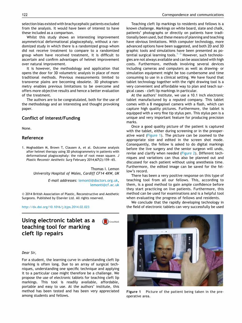
Figure 1 Picture of the patient being taken in the pre-operative area.
Dear Sir,
For a student, the learning curve in understanding cleft lipmarking is often long. Due to an array of surgical tech-niques, understanding one specific technique and applyingit to a particular case might therefore be a challenge. Wepropose the use of electronic tablets for teaching cleft lipmarkings. This tool is readily available, affordable,portable and easy to use. At the authors’ institute, thismethod has been tested and has been very appreciatedamong students and fellows.
Teaching cleft lip markings to residents and fellows is aknown challenge. Markings on white board, slate and chalk,patients’ photographs or directly on patients have tradi-tionally been used, but thesemeans of planning and teachinghave obvious limitations. With computer technology, moreadvanced options have been suggested, and both 2D and 3Dgraphic tools and simulations have been presented as po-tential surgical learning tools.1e3 However, such technolo-gies are not always available and can be associated with highcosts. Furthermore, methods involving several devicesincluding cameras and computers as well as drawing- orsimulation equipment might be too cumbersome and timeconsuming to use in a clinical setting. We have found thattablet technology together with the right drawing tool is avery convenient and affordable way to plan and teach sur-gical cases - cleft lip markings in particular.
At the authors’ Institute, we use a 10.1 inch electronictablet manufactured by a reputed company. This tabletcomes with a 8 megapixel camera with a flash, which cancapture high quality pictures. Furthermore, the tablet isequipped with a very fine tip stylus pen. This stylus pen is aunique and very important feature for producing precisionmarks.
Once a good quality picture of the patient is capturedwith the tablet, either during screening or in the preoper-ative ward (Figure 1). The picture can be zoomed to theappropriate size and edited in the screen shot mode.Consequently, the fellow is asked to do digital markingsbefore the live surgery and the senior surgeon will undo,revise and clarify when needed (Figure 2). Different tech-niques and variations can thus also be planned out anddiscussed for each patient without using anesthesia time.Furthermore, the edited image can be saved for the fel-low’s record.
There has been a very positive response on this type ofteaching tool from all our fellows. This, according tothem, is a good method to gain ample confidence beforethey start practicing on live patients. Furthermore, thismethod can be used for examinations and is a helpful toolwhen evaluating the progress of fellows and residents.
We conclude that the rapidly developing technology inthe field of electronic tablets can very successfully be used
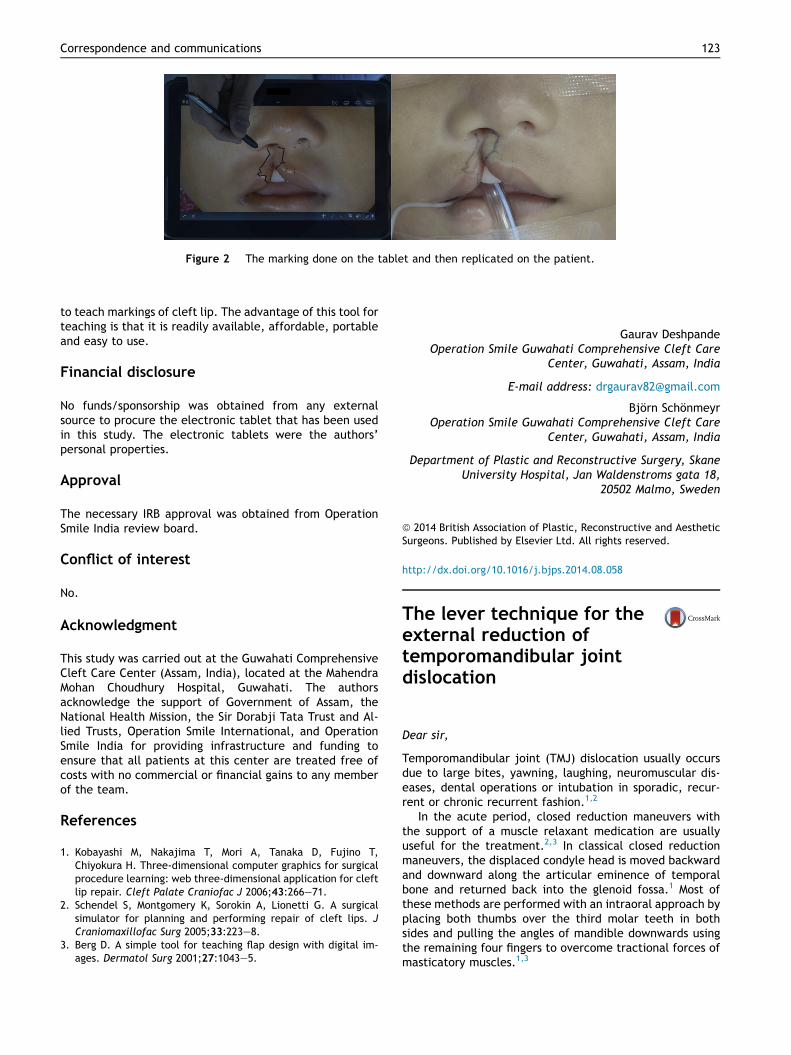
Figure 2 The marking done on the tablet and then replicated on the patient.
Correspondence and communications 123
to teach markings of cleft lip. The advantage of this tool forteaching is that it is readily available, affordable, portableand easy to use.
Financial disclosure
No funds/sponsorship was obtained from any externalsource to procure the electronic tablet that has been usedin this study. The electronic tablets were the authors’personal properties.
Approval
The necessary IRB approval was obtained from OperationSmile India review board.
Conflict of interest
No.
Acknowledgment
This study was carried out at the Guwahati ComprehensiveCleft Care Center (Assam, India), located at the MahendraMohan Choudhury Hospital, Guwahati. The authorsacknowledge the support of Government of Assam, theNational Health Mission, the Sir Dorabji Tata Trust and Al-lied Trusts, Operation Smile International, and OperationSmile India for providing infrastructure and funding toensure that all patients at this center are treated free ofcosts with no commercial or financial gains to any memberof the team.
References
1. Kobayashi M, Nakajima T, Mori A, Tanaka D, Fujino T,Chiyokura H. Three-dimensional computer graphics for surgicalprocedure learning: web three-dimensional application for cleftlip repair. Cleft Palate Craniofac J 2006;43:266e71.
2. Schendel S, Montgomery K, Sorokin A, Lionetti G. A surgicalsimulator for planning and performing repair of cleft lips. JCraniomaxillofac Surg 2005;33:223e8.
3. Berg D. A simple tool for teaching flap design with digital im-ages. Dermatol Surg 2001;27:1043e5.
Gaurav DeshpandeOperation Smile Guwahati Comprehensive Cleft Care
Center, Guwahati, Assam, India
E-mail address: [email protected]
Bjorn SchonmeyrOperation Smile Guwahati Comprehensive Cleft Care
Center, Guwahati, Assam, India
Department of Plastic and Reconstructive Surgery, SkaneUniversity Hospital, Jan Waldenstroms gata 18,
20502 Malmo, Sweden
ª 2014 British Association of Plastic, Reconstructive and AestheticSurgeons. Published by Elsevier Ltd. All rights reserved.
http://dx.doi.org/10.1016/j.bjps.2014.08.058
The lever technique for theexternal reduction oftemporomandibular jointdislocation
Dear sir,
Temporomandibular joint (TMJ) dislocation usually occursdue to large bites, yawning, laughing, neuromuscular dis-eases, dental operations or intubation in sporadic, recur-rent or chronic recurrent fashion.1,2
In the acute period, closed reduction maneuvers withthe support of a muscle relaxant medication are usuallyuseful for the treatment.2,3 In classical closed reductionmaneuvers, the displaced condyle head is moved backwardand downward along the articular eminence of temporalbone and returned back into the glenoid fossa.1 Most ofthese methods are performed with an intraoral approach byplacing both thumbs over the third molar teeth in bothsides and pulling the angles of mandible downwards usingthe remaining four fingers to overcome tractional forces ofmasticatory muscles.1,3


![Advances of Plastic & Reconstructive Surgeryopenaccessebooks.com/...surgery/cleft-lip-nasal... · Cleft nasal reconstruction can be divided into primary and secondary repairs [1]](https://img.pdfslide.net/doc/110x75/6053986506853e25ba06de46/advances-of-plastic-reconstructive-sur-cleft-nasal-reconstruction-can-be-divided.jpg)