Embed Size (px)
Citation preview
RESEARCH Open Access
Utility of frozen section analysis for fungalorganisms in soft tissue wounddebridement margin determinationNives Zimmermann1,4*, Matthew C Hagen1, Jason J Schrager2, Renee S Hebbeler-Clark3 and Sreeharsha Masineni1
Abstract
Background: Zygomycetes cause different patterns of infection in immunosuppressed individuals, includingsino-orbito-cerebral, pulmonary, skin/soft tissue infection and disseminated disease. Infections with Zygomycetes havea high mortality rate, even with prompt treatment, which includes anti-fungal agents and surgical debridement. Insome centers, clear margins are monitored by serial frozen sections, but there are no specific guidelines for the use offrozen sections during surgical debridement. Studies in fungal rhinosinusitis found 62.5–85 % sensitivity of frozensection analysis in margin assessment. However, the utility of frozen section analysis for margin evaluation indebridement of skin/soft tissue infection has not been published.
Methods: We present a case of zygomycosis of decubitus ulcers in which we assessed statistical measures ofperformance of frozen section analysis for presence of fungal organisms on the margin, compared withformalin-fixed paraffin embedded (FFPE) sections as gold standard. A total of 33 specimens (94 blocks) weresectioned, stained with H&E and evaluated by both frozen and FFPE analysis. Negative interpretations wereconfirmed by Gomori methenamine silver stain on FFPE sections.
Results: H&E staining of frozen sections had 68.4 % sensitivity and 100 % specificity for assessing margins clear offungal organisms. The negative and positive predictive values were 70.0 % and 100 %, respectively. Using presence ofacute inflammation and necrosis as markers of fungal infection improved sensitivity (100 %) at the expense ofspecificity (42.9 %).
Conclusion: Use of intraoperative assessment of skin and soft tissue margins for fungal infection is a valuable tool inthe management of skin and soft tissue fungal infection treatment.
Keywords: Fungi, Frozen section, Margin, Wound
BackgroundInvasive fungal infections are an important cause ofmorbidity and mortality in immunocompromised hosts.While invasive candidiasis and invasive aspergillosis stillaccount for the majority of these infections, zygomycetesalso cause significant proportion of invasive fungal infec-tions [1]. Zygomycosis refers to infections caused byfungi in the Zygomycota phylum, which includes patho-gens such as Mucor, Apophysomyces, Rhizomucor, Rhizo-pus and Absidia. These infections occur with greater
frequency in immunosuppressed patients with under-lying diseases, such as diabetes and malignancy, but havealso been described in previously healthy patients(reviewed in [2]). The infection most commonly involvessinuses (39 %), lung (24 %) and skin (19 %). The locationof infection and likelihood of dissemination are alsoinfluenced by underlying clinical conditions. In a largemeta analysis of zygomycosis cases (47 % Rhizopus spe-cies, 18 % Mucor species), the majority of patients withmalignancy were found to have pulmonary infection(60 %), while those with diabetes had rhino-cerebral dis-ease (43 %) [2]. Importantly, these infections have highmortality rates, which can be significantly influenced bythe site of infection: 96 % in patients with disseminated
* Correspondence: [email protected] of Pathology and Laboratory Medicine, University of CincinnatiCollege of Medicine, Cincinnati, Ohio4Cincinnati Children’s Hospital Medical Center, Cincinnati, OhioFull list of author information is available at the end of the article
© 2015 Zimmermann et al. Open Access This article is distributed under the terms of the Creative Commons Attribution 4.0International License (http://creativecommons.org/licenses/by/4.0/), which permits unrestricted use, distribution, andreproduction in any medium, provided you give appropriate credit to the original author(s) and the source, provide a link tothe Creative Commons license, and indicate if changes were made. The Creative Commons Public Domain Dedication waiver(http://creativecommons.org/publicdomain/zero/1.0/) applies to the data made available in this article, unless otherwise stated.
Zimmermann et al. Diagnostic Pathology (2015) 10:188 DOI 10.1186/s13000-015-0423-9
disease, 76 % with pulmonary infections, and 31 % withcutaneous infections [2]. Patient outcomes are also sig-nificantly affected by treatment [2, 3]. Survival rates are:3 % in untreated cases, 61 % and 57 % for patientstreated with antifungals or surgery alone, respectively,and 70 % for patients treated with both antifungal agentsand surgery [2]. Thus, optimal treatment for invasivezygomycosis is multi-modal, and includes antifungalagents, surgical debridement, and correction of under-lying condition predisposing to the disease [4, 5].There are reports in the literature of cases where
surgical margins were evaluated by frozen sections intra-operatively [6–8]. Mathews et al. and Weinberg et al.describe cases of Apophysomyces elegans infection inpreviously healthy patients following C-section and brownrecluse spider bite, respectively [6, 7]. In both cases,patients survived following a prolonged course of ampho-tericin B and multiple surgical debridements using frozensection analysis for margin assessment.Intraoperative margin assessment is especially useful
during rhino-sino-orbital fungal infections when delicate/vital structures, such as the orbit, could be spared if unin-volved. Case series on the role of frozen section in acutefungal sinusitis by Taxy et al. [9] and Ghadiali et al. [10]have shown 62.5 and 84 % sensitivity, respectively. Thestudy by Ghadiali et al. [10] involved 20 patients with fun-gal rhinosunisitis over a 12 year period, 11 of which wereinfected with Mucor species, and 9 with Aspergillus. In asubgroup (6 patients; 1 with Aspergillus and 5 withMucor;30 slides total), frozen sections were used to assess marginstatus during surgical debridement. Using permanent sec-tion as the gold standard, the sensitivity (on a “per slide”basis) was 84 % and specificity was 100 %. The outcomeof patients was not reported in this study.The study by Taxy et al. [9] involved 8 patients with
acute fungal sinusitis (including Mucor, Aspergillus fla-vus, niger and/or fumigatus, Fusarium and Alternaria)with both frozen and permanent sections, and in 5 ofthose cases fungal organisms were seen on frozen sec-tion. In two of the cases, fungal organisms were not seenon H&E-stained permanent sections either, and requiredspecial staining (methenamine). One case was negativein both frozen and permanent staining of frozen blocks,but positive on non-frozen tissue. Thus, the sensitivity(on a “per case” basis) of frozen sections for determin-ation of margin status by frozen sections in this studywas 62.5 %. Despite aggressive management, none of thepatients survived.However, no studies assessed the utility of intraopera-
tive margin assessment for fungal infections of skin andsoft tissue, where more liberal margins can be taken.Furthermore, in contrast to head and neck specimens,skin/soft tissue is technically more challenging due toincreased adipose tissues and the surface area needing
assessment could be much larger. These factors cansignificantly affect the performance of frozen sectionanalysis for margin assessment.
MethodsA 51-year-old female with past medical history ofStevens-Johnson syndrome (eye, s/p corneal transplantsrequiring immunosuppression with high-dose steroidsand mycophenolate) and hypertension was admitted formanagement of pulmonary embolus and Legionellapneumonia. Following a complicated inpatient course,which included acute respiratory failure with acuterespiratory distress syndrome (ARDS) requiring extra-corporeal membrane oxygenation (ECMO), Clostridiumdifficile colitis, and steroid-induced hyperglycemia, shedeveloped decubitus ulcers on her back and neck.Wound culture identified Rhizopus microsporus var. rhi-zopodiformis. The patient was started on IV liposomalamphotericin B and surgical wound debridement wasperformed. Margins were assessed by frozen section ana-lysis and debridement continued until margins wereconfirmed clear. This took three separate surgicaldebridements over 4 days (days 1, 2 and 4 post woundculture organism identification). After 16 days of sys-temic liposomal amphotericin B, she was bridged toposaconazole for a total of 6 weeks of antifungal agents.The patient has been free of fungal infection since(>9 months of follow up), and the wounds have healed.Soft tissue specimens were received fresh and the sur-
gical margin was inked, shaved and submitted en face inthe majority of specimens. Tissue was frozen in OCT,sectioned at 5–7 μm, and stained with H&E. Slides weremicroscopically assessed for the presence of branchingnon-septate hyphae. Presence of fungal elements any-where on the en face slides was considered a positivemargin, the surgeons were notified and the margin wasreexcised. In a few initial specimens, the tissue marginwas submitted perpendicular, and presence of fungal ele-ments on ink was considered positive margin. Followingpathological determination of frozen section, the tissuewas fixed in formalin, processed by routine processing,embedded in paraffin, cut and stained with H&E forFFPE section analysis. In select cases, additional slideswere stained with Gomori methenamine silver stain tohighlight fungal elements. All slides were rereviewed forthe research study and data presented are from this rere-view. There was only one case of interpretation error,where margin was called negative for clinical purposes (onboth frozen and FFPE), but fungal elements were foundon rereview of the same slides (both frozen and FFPE).Both slides (frozen and FFPE) were scored for presence
of fungal elements and acute inflammation/necrosis. Datawere entered into a truth table, with frozen section ana-lysis as test and FFPE section as gold standard. Sensitivity,
Zimmermann et al. Diagnostic Pathology (2015) 10:188 Page 2 of 6
specificity, negative and positive predictive value werecalculated. We calculated the performance on a “perspecimen” basis because once a positive margin wasidentified on a slide, the margin was reported as positiveand the remainder of the slides from the specimen werenot analyzed.This study has been reviewed by the University of
Cincinnati Institutional Review Board and deemed “nothuman subjects research”.
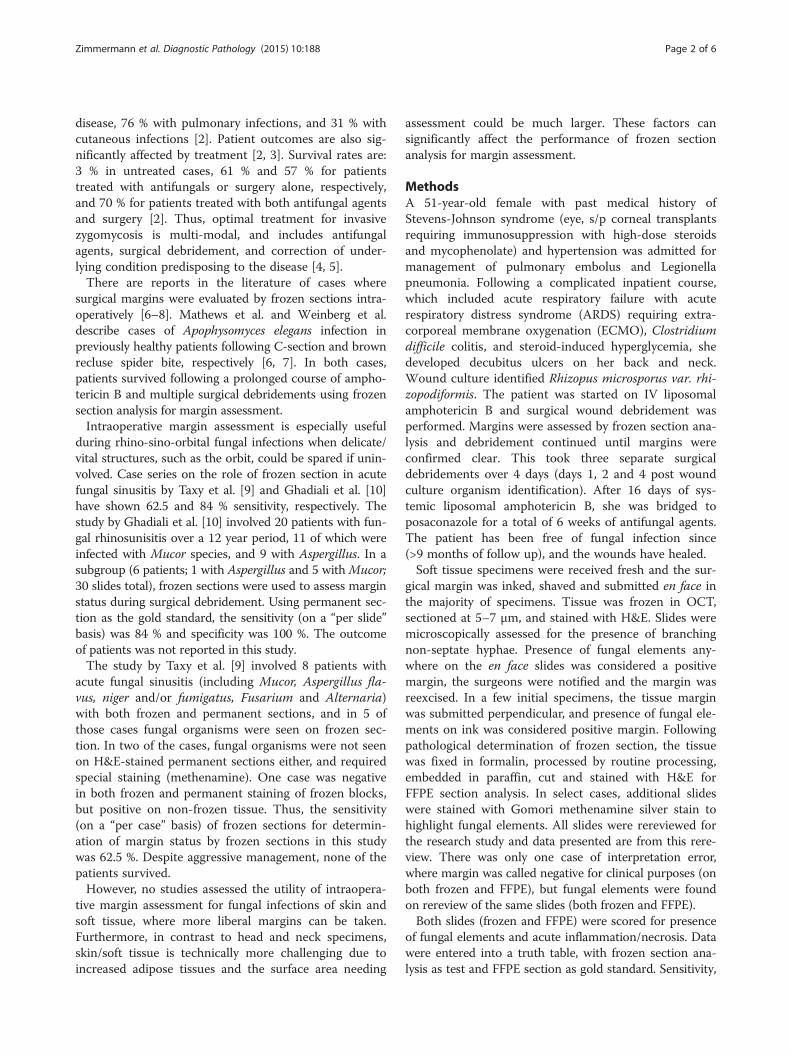
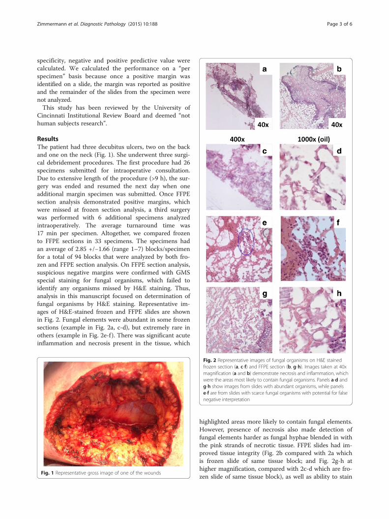
ResultsThe patient had three decubitus ulcers, two on the backand one on the neck (Fig. 1). She underwent three surgi-cal debridement procedures. The first procedure had 26specimens submitted for intraoperative consultation.Due to extensive length of the procedure (>9 h), the sur-gery was ended and resumed the next day when oneadditional margin specimen was submitted. Once FFPEsection analysis demonstrated positive margins, whichwere missed at frozen section analysis, a third surgerywas performed with 6 additional specimens analyzedintraoperatively. The average turnaround time was17 min per specimen. Altogether, we compared frozento FFPE sections in 33 specimens. The specimens hadan average of 2.85 +/−1.66 (range 1–7) blocks/specimenfor a total of 94 blocks that were analyzed by both fro-zen and FFPE section analysis. On FFPE section analysis,suspicious negative margins were confirmed with GMSspecial staining for fungal organisms, which failed toidentify any organisms missed by H&E staining. Thus,analysis in this manuscript focused on determination offungal organisms by H&E staining. Representative im-ages of H&E-stained frozen and FFPE slides are shownin Fig. 2. Fungal elements were abundant in some frozensections (example in Fig. 2a, c-d), but extremely rare inothers (example in Fig. 2e-f ). There was significant acuteinflammation and necrosis present in the tissue, which
highlighted areas more likely to contain fungal elements.However, presence of necrosis also made detection offungal elements harder as fungal hyphae blended in withthe pink strands of necrotic tissue. FFPE slides had im-proved tissue integrity (Fig. 2b compared with 2a whichis frozen slide of same tissue block; and Fig. 2g-h athigher magnification, compared with 2c-d which are fro-zen slide of same tissue block), as well as ability to stain
Fig. 1 Representative gross image of one of the wounds
Fig. 2 Representative images of fungal organisms on H&E stainedfrozen section (a, c-f) and FFPE section (b, g-h). Images taken at 40xmagnification (a and b) demonstrate necrosis and inflammation, whichwere the areas most likely to contain fungal organisms. Panels a-d andg-h show images from slides with abundant organisms, while panelse-f are from slides with scarce fungal organisms with potential for falsenegative interpretation
Zimmermann et al. Diagnostic Pathology (2015) 10:188 Page 3 of 6
with Gomori methenamine silver stain to highlightfungal elements (data not shown).Of the 33 specimens analyzed, 13 were positive for
fungal elements on frozen section analysis, and all ofthose were confirmed on FFPE sections. In contrast, ofthe 20 specimens, which were called negative on frozensection analysis, 6 revealed fungal elements on FFPE.Thus, while the specificity is 100 %, the sensitivity of fro-zen section analysis is 68.4 %. The negative predictivevalue (NPV) and positive predictive value (PPV) is70.0 % and 100 %, respectively (Table 1A).Due to presence of abundant acute inflammation and
necrosis as harbinger of fungal elements, we hypothesizedthat calling a margin positive based on presence of inflam-mation/necrosis alone would increase the sensitivity.Indeed, sensitivity and negative predictive value increasedto 100 %; however, this lead to decreased specificity of42.9 % and positive predictive value of 70.4 % (Table 1B).
DiscussionWe report a case of invasive fungal infection of decubitusulcers in an immunosuppressed patient, treated with com-bination of surgical debridement and antifungal therapy.Margins were assessed for fungal elements by intraopera-tive analysis of frozen section slides, and later confirmedby FFPE sections. This allowed us to assess statistical mea-sures of performance of the frozen section analysis.The sensitivity in our case (68.4 %) was comparable to
that published previously for head and neck fungal infec-tion [9, 10]. A significant false negative rate is likelyattributable to sampling bias, technical challenges of sec-tioning fatty tissue, frozen artifacts, and inconspicuousnessof fungal organisms among other structures in necrotictissue on H&E stained slides. Sampling bias could be due toFFPE sections being deeper, and thus further away from thetrue margin (in case of en face margins); however, this islikely still clinically significant as a positive margin. To overcome the technical challenges of sectioning of fatty tissueand frozen artifact, one may consider using touch imprints
or tissue surface scraping instead of frozen sectioning. Fu-ture studies should compare if this approach improves sen-sitivity. Detection of fungal elements may be improved withrapid fungal stains of frozen sections (such as rapid Roma-nowski stain, [9] or Gram staining [11] of tissue). However,when we attempted to retrospectively stain the FFPE tissueswith above stains along with H&E, neither rapid Roma-nowski stain nor Gram stain improved detection of fungalorganisms compared to H&E (data not shown).Alternatively, instead of immediate intraoperative mar-
gin assessment, team members should consider improvingaccuracy of margin assessment with formalin fixation(leading to better tissue integrity), as well as special stainsfor fungal organisms, such as GMS (leading to improveddetection of fungal elements). As such, team membersneed to discuss whether immediate results with potentialfalse negatives are beneficial compared with more reliablebut delayed results. Factors such as ability to repeat mar-gin excision in a few days and proximity to vital structuresshould be considered. Another alternative to consider israpid processing which may provide turnaround time in-between intraoperative consultation and routine FFPEprocessing. In addition to improving accuracy, consideringalternatives to intraoperative consultation is importantfrom the standpoint of time to results. Standard max-imum turnaround time at our institution for frozensection analysis is 20 min per specimen. For instance, theinitial surgery in our case consisted of 29 specimens (85blocks total) submitted for margin assessment. Pathologyassessment became the rate-limiting step causing the sur-gery to last more than 9 h. Prolonging surgery can havesignificant detrimental outcomes for the patient as it pro-longs the time under anesthesia and associated risks. Thisis especially true in patients with invasive fungal infectionswho can be quite unstable at the start of the operation.Thus, anticipated time for intraoperative results should becalculated based on the wound size, and logistical ap-proaches to increase speed without compromising qualityshould be considered. For instance, the number of
Table 1 Statistical measures of performance of frozen section analysis for margin assessment during wound debridement surgery
A. Fungal organisms on FFPE
yes no
Fungal organisms on frozen section positive 13 0 13
negative 6 14 20
19 14 33
B. Fungal organisms on FFPE
yes no
Inflammation/ necrosis on frozen section positive 19 8 27
negative 0 6 6
19 14 33
Criteria used to call a margin positive were presence of fungalorganisms (in A) or presence of inflammation/necrosis (in B). Presence of fungal organisms ofpermanent sections was used as gold standard in both cases
Zimmermann et al. Diagnostic Pathology (2015) 10:188 Page 4 of 6
personnel, available microtomes and other hardware couldbe optimized. In summary, improving time to results andaccuracy of results would drive decisions to assess marginsintra- or post-operatively, and if the former, a number ofsteps could be taken to improve patient’s outcome byimproving speed and accuracy.Furthermore, team members should discuss whether
decreasing false negative calls, at the expense of increas-ing false positives, may be desirable and can be achievedby using acute inflammation and necrosis as surrogatemarkers of fungal elements. For example, if there isample distance to vital structures and repeat surgery isundesirable based on the patient’s clinical comorbidities,this may be a viable approach. However, false positivesand a specificity of 42.9 % come with their own set ofchallenges. Creating a larger than necessary wound bur-den carries with it prolonged wound care needs, in-creased chance of secondary infection, increased debility(with decreased mobility both short- and long-term) andpossibly increased intensive care unit days (with in-creased risk of nosocomial infections, delirium, and evenmortality). As such, the balance of false positives andfalse negatives needs to be carefully considered and theapproach for calling positive margins should be agreedupon by pathologist and surgeon.Our study has its limitations. First, our findings are
limited to a single patient. However, this is a rare sce-nario and thus there is currently limited experience. Bysharing our experience and analyzing the multiple sam-ples (total of 33 specimens), we hope to provide newknowledge, increasing the awareness about the utility ofthis approach and its limitations, thus aiding in futuredecision making. Also, an increase in reported cases willfacilitate study of a broader spectrum of cases with dif-ferent clinicopathological and microbiological character-istics. The second limitation is that our study does notaddress whether clear margins during debridement areindeed necessary for optimal clinical outcome, andwhether debridement with margin assessment is superiorto debridement guided by other measures (e.g. assessmentof margin viability and lack of infection by gross inspec-tion). Evidence for use of frozen sections in margin assess-ment of infected soft tissue debridement is lacking in theliterature, and the approach of debridement for clear mar-gins is chosen based on clinical assessment (for instance,trying to balance wound management with the fact thatan immunocompromised patient will be unable to cleareven a low load of infection) and gravity of the situation(known high mortality of the disease). We are aware ofonly one case series that described cases with and withoutdebridement guided by frozen sections [10]; however,patient outcome was not reported in that study. The ques-tions our study does address are the analytic specificityand sensitivity of frozen section use for margin assessment
during infected soft tissue debridement, and how they areaffected by using identification of fungal organisms versusinflammation/necrosis. This provides useful informationto team members for informed decision-making.In summary, our study provides statistical measures of
performance for frozen section analysis of margins duringdebridement surgery for invasive fungal infections of softtissue and also provides comparison of these measures fordifferent criteria (fungal organisms versus inflammation/necrosis), which should be considered when making deci-sions on which approach to use in individual clinicalcircumstances.
ConclusionUse of intraoperative assessment of skin and soft tissuemargins for fungal infection is a valuable tool in themanagement of skin and soft tissue fungal infectiontreatment. Whether presence of fungal elements ornecrosis/acute inflammation is used as cutoff to callpositive margins needs to be decided based on clinicalscenario using sensitivity and specificity presented here.
AbbreviationsARDS: Acute respiratory distress syndrome; FFPE: Formalin-fixed, paraffin-embedded; NPV: Negative predictive value; PPV: Positive predictive value.
Competing interestsThe authors declare that they have no competing interests.
Authors’ contributionsNZ conceived of the study, participated in its design, acquired and analyzeddata, and drafted the manuscript. MH, JS and RH-C participated in the designof the study and editing of the manuscript. SM participated in the design ofthe study, acquisition of data, and editing of the manuscript. All authors readand approved the final manuscript.
AcknowledgmentThe authors thank Drs. Jiang Wang, Shagufta Khan and Meggan Peak forcritical review of the manuscript and helpful suggestions.
Author details1Department of Pathology and Laboratory Medicine, University of CincinnatiCollege of Medicine, Cincinnati, Ohio. 2Department of Surgery, University ofCincinnati College of Medicine, Cincinnati, Ohio. 3Department of Medicine,University of Cincinnati College of Medicine, Cincinnati, Ohio. 4CincinnatiChildren’s Hospital Medical Center, Cincinnati, Ohio.
Received: 20 August 2015 Accepted: 9 October 2015
References1. Neofytos D, Horn D, Anaissie E, Steinbach W, Olyaei A, Fishman J, et al.
Epidemiology and outcome of invasive fungal infection in adulthematopoietic stem cell transplant recipients: analysis of MulticenterProspective Antifungal Therapy (PATH) Alliance registry. Clin Infect Dis.2009;48(3):265–73. doi:10.1086/595846.
2. Roden MM, Zaoutis TE, Buchanan WL, Knudsen TA, Sarkisova TA, SchaufeleRL, et al. Epidemiology and outcome of zygomycosis: a review of 929reported cases. Clin Infect Dis. 2005;41(5):634–53. doi:10.1086/432579.
3. Almyroudis NG, Sutton DA, Linden P, Rinaldi MG, Fung J, Kusne S.Zygomycosis in solid organ transplant recipients in a tertiary transplantcenter and review of the literature. Am J Transplant. 2006;6(10):2365–74.doi:10.1111/j.1600-6143.2006.01496.x.
4. Skiada A, Lanternier F, Groll AH, Pagano L, Zimmerli S, Herbrecht R, et al.Diagnosis and treatment of mucormycosis in patients with hematological
Zimmermann et al. Diagnostic Pathology (2015) 10:188 Page 5 of 6
malignancies: guidelines from the 3rd European Conference on Infectionsin Leukemia (ECIL 3). Haematologica. 2013;98(4):492–504. doi:10.3324/haematol.2012.065110.
5. Cornely OA, Arikan-Akdagli S, Dannaoui E, Groll AH, Lagrou K, Chakrabarti A,et al. ESCMID and ECMM joint clinical guidelines for the diagnosis andmanagement of mucormycosis 2013. Clin Microbiol Infect. 2014;20 Suppl3:5–26. doi:10.1111/1469-0691.12371.
6. Mathews MS, Raman A, Nair A. Nosocomial zygomycotic post-surgicalnecrotizing fasciitis in a healthy adult caused by Apophysomyces elegans insouth India. J Med Vet Mycol. 1997;35(1):61–3.
7. Weinberg WG, Wade BH, Cierny 3rd G, Stacy D, Rinaldi MG. Invasiveinfection due to Apophysomyces elegans in immunocompetent hosts.Clin Infect Dis. 1993;17(5):881–4.
8. Reed C, Bryant R, Ibrahim AS, Edwards Jr J, Filler SG, Goldberg R, et al.Combination polyene-caspofungin treatment of rhino-orbital-cerebralmucormycosis. Clin Infect Dis. 2008;47(3):364–71. doi:10.1086/589857.
9. Taxy JB, El-Zayaty S, Langerman A. Acute fungal sinusitis: natural history andthe role of frozen section. Am J Clin Pathol. 2009;132(1):86–93. doi:10.1309/AJCP9HTH9NRPMYCT.
10. Ghadiali MT, Deckard NA, Farooq U, Astor F, Robinson P, Casiano RR.Frozen-section biopsy analysis for acute invasive fungal rhinosinusitis.Otolaryngol Head Neck Surg. 2007;136(5):714–9. doi:10.1016/j.otohns.2007.01.002.
11. Musto L, Flanigan M, Elbadawi A. Ten-minute silver stain for Pneumocystiscarinii and fungi in tissue sections. Arch Pathol Lab Med. 1982;106(6):292–4.
Submit your next manuscript to BioMed Centraland take full advantage of:
• Convenient online submission
• Thorough peer review
• No space constraints or color figure charges
• Immediate publication on acceptance
• Inclusion in PubMed, CAS, Scopus and Google Scholar
• Research which is freely available for redistribution
Submit your manuscript at www.biomedcentral.com/submit
Zimmermann et al. Diagnostic Pathology (2015) 10:188 Page 6 of 6