Embed Size (px)
Citation preview
UvA-DARE is a service provided by the library of the University of Amsterdam (http://dare.uva.nl)
UvA-DARE (Digital Academic Repository)
Natural adaptive immune responses in humans against Toxoplasma gondii and Herpessimplex virus type IMeek, B.
Link to publication
Citation for published version (APA):Meek, B. (2002). Natural adaptive immune responses in humans against Toxoplasma gondii and Herpessimplex virus type I.
General rightsIt is not permitted to download or to forward/distribute the text or part of it without the consent of the author(s) and/or copyright holder(s),other than for strictly personal, individual use, unless the work is under an open content license (like Creative Commons).
Disclaimer/Complaints regulationsIf you believe that digital publication of certain material infringes any of your rights or (privacy) interests, please let the Library know, statingyour reasons. In case of a legitimate complaint, the Library will make the material inaccessible and/or remove it from the website. Please Askthe Library: http://uba.uva.nl/en/contact, or a letter to: Library of the University of Amsterdam, Secretariat, Singel 425, 1012 WP Amsterdam,The Netherlands. You will be contacted as soon as possible.
Download date: 05 May 2019
Chapterr 6
Dissectingg the IgM antibody response during the acute and latent
phasee of toxoplasmosis
Publishedd in: Diagnostic Microbiology and Infectious Diseases, 2001
Bobb Meek, Tom van Gooi, Henk Gili s and Ron Peek.
Abstract t
AA major problem in anti-toxoplasma IgM serology is the occurrence of clinically non-relevant (CNR) IgM responses.
Thee susceptibility for CNR IgM of the Toxo ISAGA IgM, Platelia Toxo IgM and Vidas Toxo IgM assays was
determinedd using sera with a CNR IgM titer in the Abbott IMx Toxo IgM assay. The specificity of CNR-IgM
antibodiess was determined by immunoblotting, and compared with IgM antibodies in sera of acutely infected (AI)
individuals. .
Onlyy 6/19 samples were found positive in the ISAGA IgM, compared with 16/19 with the VIDAS IgM and 17/19
withh the Platelia Toxo IgM. Staining intensity of non-reduced SAG1 by IgM antibodies allowed a clear distinction
betweenn CNR IgM and AI IgM antibodies.
Anyy IgM assay is susceptible to CNR IgM antibodies. In cases of doubt, immunoblotting using non-reduced T.gondii
antigenss is of value as confirmation method. Because CNR IgM antibodies are specific for toxoplasma, it will be difficultt to improve IgM ELISAs.
ChapterChapter 6
Introductio n n
ToxoplasmaToxoplasma gondii (T.gondii) is a protozoan parasite capable of infecting many vertebrate species, including
humans.. The symptoms of disease in immuno-competent humans are usually benign and self limiting. In contrast, in
immuno-compromisedd individuals toxoplasmosis is a major opportunistic infection that may lead to toxoplasmic
encephalitis.. Primary infection during pregnancy may cause serious complications or even death in congenitally
infectedd children [1].
Anti-toxoplasmaa IgM antibodies are considered a reliable marker for acute infection. A major problem of
anti-toxoplasmaa IgM serology is the occurrence of clinically non-relevant (CNR) IgM responses [2-4]. A positive
resultt in an IgM test due to CNR IgM antibodies suggests acute toxoplasmosis while the patient actually is in the
latentt stage of toxoplasma infection. Detection of these CNR anti-toxoplasma IgM antibodies during pregnancy has
ledd to unnecessary distress and even abortion [5],
Surprisingly,, very littl e attention has been paid to the characterization of these CNR IgM responses. Anti-
toxoplasmaa IgM kits frequently used for routine diagnostic purposes and confirmatory testing, such as the Abbott IMx
Toxoo IgM, Toxo ISAGA IgM, Platelia Toxo IgM and Vidas Toxo IgM, have never been evaluated with sera
containingg CNR IgM antibodies. In this study sera were used from latently infected individuals with an established
CNRR IgM titer in the Abbott IMx Toxo IgM test to determine and compare the susceptibility of the Toxo ISAGA
IgM,, Platelia Toxo IgM and Vidas Toxo IgM kits for these antibodies. Immunoblotting was performed to
characterizee CNR-IgM antibodies and to compare their specificity with those of IgM antibodies in sera of acutely
infectedd individuals.
Materia ll & Methods
Sera:: Serum samples of 54 cases were submitted to the Academic Medical Center for routine examination
forr toxoplasmosis. Sera were stored at -20°C. Sera of immuno-compromised patients, including AIDS patients, were
excluded. .
Serologicc tests: All sera were examined for anti-toxoplasma specific antibodies by the Sabin-Feldman dye
testt (SF) [6]. Sera were examined for specific IgM by immunoblotting and with the following assays: Abbott IMx
Toxoo IgM (IM-x Toxo IgM, Abbott Laboratories, Abbott Park, 111.), Toxo ISAGA IgM (ISAGA IgM, bioMérieux,
Lyon,, France), Platelia Toxo IgM (Platelia Toxo IgM Sanofi Diagnostics Pasteur, Marnes la Coquette, France),
bioMérieuxx VIDA S Toxo IgM (VIDA S Toxo IgM, bioMérieux, Lyon, France). All commercially available kits were
performedd according to the instructions given by the manufacturers. Sera with indexes or ODs between the cut-off
valuesvalues were considered equivocal/borderline. For the SF dye test the cut-off value was 1 IU/ml. For the IgM assays
thesee values were an OD of 0.600 for the IMx Toxo IgM kit, an index of 9 for the ISAGA IgM kit (borderline index
6-8),, an OD of > 100% of the cut-off serum for the Platelia Toxo IgM kit (~ 0.350), and an OD of 0.500 for the
VIDA SS IgM kit. Sera with values greater than or equal to the cut off value were considered positive. If anti-
toxoplasmaa IgM antibodies were detected in the first sample, then sequential samples were obtained within 3-4 weeks
andd analyzed with the SF dye test.
Seraa were divided into 4 groups on the basis of the results of the dye test and anti-toxoplasma IgM assays.
AII group: serum samples taken from 21 cases with acute toxoplasmosis (AI) , characterized by an IgG titer >
2500 IU/ml in the SF dye test and/or a three fold increase of the SF dye test titer in the follow up sample taken 3-4
weekss later, and positive results in the IMx IgM and ISAGA IgM assays.
CNRR group: serum samples from 19 persons who were latently infected with toxoplasma as characterized by
90 0
DissectingDissecting the CNR IgM response
aa low and stable SF titer between 6 and 63 IU/ml in two serum samples taken three weeks apart and a clinically non-
relevantt (CNR) IgM titer in die IMx IgM assay.
LII group: serum samples of 7 latently infected (LI) individuals with negative results in the IMx IgM and
ISAGAA IgM assays and a low SF titer between 6 and 63 IU/ml.
SNN group: serum samples from 7 subjects witfi negative results in die IMx IgM and ISAGA IgM assays, and
thee SF dye test.
Antigenn preparation: Antigen preparation was performed as described [7,8]. T.gondii tachyzoites of the
RHH strain were propagated in Swiss mice (Harlan laboratories, Horst, The Netherlands). The animals were sacrificed
488 hours post-injection and tachyzoites were collected and purified. Following freeze/thawing, die tachyzoite
suspensionn was sonicated on ice (8 x 15 sec 30 kHz rnicroprobe, Soniprep 150, MSE, Loughborough, GB), and
insolublee material was removed by centrifugation. The sonicate was frozen in small aliquots and kept at -70°C until
use. .
SDS-PAGEE and Western blotting:
SDS-PAGEE was performed according to [8,9]. Briefly, 200 ug of the sonicate was loaded onto 10% SDS-PAGE gels
andd transferred to polyvinyldifluoride (PVDF) membranes (Millipor e Corporation, Bedford, MA) after
electrophoresis.. To reduce toxoplasma lysate antigens, |$-mercaptoethanol was added to a final concentration of 5%
priorr to electrophoresis. Membranes weree blocked witii Tris-buffered saline (TBS: 50 mM TrisHCl, 150 mM NaCl,
pHH 10) containing 0.5% Tween20 and 2% non-fat milk powder.
Immunostainingg of peroxidase conjugated antibodies: Except when stated otherwise, all samples and
conjugatess were diluted in TBS containing 0.5% Tween20 and 0.03% non-fat milk powder (TBS-T). All incubations
weree done using a multiscreen apparatus (Mini-Protean n, Bio-Rad Laboratories, Hercules, CA). Isotype specific
antibodiess conjugated to peroxydase were obtained from DAKO (Glostrup, Denmark). The conjugates were diluted
1:5000,, and incubated witii die blots for 75 min. All incubations were performed at room temperature.
Serumm samples were diluted 50x, incubated widi the blots for 75 min, and tested for the presence of saü-T. gondii
IgM.. As a positive control, a serum containing anti-glycosylphosphatidyl-inositol (GPI) IgM antibodies was aliquoted
andd included on each immunoblot in a 50x dilution. A mouse monoclonal antibody against SAG1 (HyTest, Turku,
Finland)) was also included, diluted 250x/1000x on immunoblots containing R or NR antigens, respectively.
Chemiluminescentt substrate was prepared according to the manufacturers' descriptions (ECL, Amersham Pharmacia
Biotech,, Essex, UK). The membranes were incubated with the chemiluminescent substrate for 1 min, wrapped in
plasticc and used to expose X-ray film (Fuji Super RX, Fuji Photo Film Co., Tokyo, Japan) for 5 min, 3 min, 1 min,
andd 30 sec.
Evaluationn of the IgM staining patterns: Serum samples of die categories AI and CNR were always tested
togetiier,, allowing direct comparison of the staining patterns. The films exposed for 1 minute allowed a good
distinctionn between staining patterns of AI and CNR, and subsequently was selected for the evaluation The intensity
off each band was scored in relation to die positive control serum included on each blot.
AA '++' score was assigned to intensely stained bands on the films exposed for 1 minute, a '+' score was assigned to
clearlyy stained bands, and a ' score was assigned to bands mat were well identifiable, but weakly stained on the
filmsfilms exposed for 1 minute.
91 1
ChapterChapter 6
Results s
Evaluationn of the IgM kits: Serum samples of 19 latently infected individuals proven to have clinically
non-relevantt (CNR) anti-toxoplasma IgM titers in the Abbott Toxo IMx Toxo IgM test were analyzed using the
ISAGAA IgM, Platelia Toxo IgM and VIDAS Toxo IgM kits (table 1).
Inn the CNR group, 7 out of 19,17/19 and 16/19 serum samples had a positive result in the ISAGA IgM, Platelia Toxo
IgM,, and VIDA S Toxo IgM assays, respectively. Importantly, none of the samples containing CNR IgM antibodies
wass negative in all three assays (table 1), and only 2 CNR samples had a negative result in 2 assays. In the AI group,
alll samples were positive by the commercially available IgM tests. All samples in the LI and SN groups had a
negativee result in every IgM assay (table 1).
IgMM antibody profiles analyzed by immunoblotting: To determine which antigens were recognized by the
CNRR IgM antibodies and to assess the diagnostic value of IgM immunoblotting in discrimination between CNR and
AII IgM, the panels of sera were tested on blots containing size-fractionated reduced or non-reduced 7*. gondii
tachyzoitee antigens.
Uponn initial analysis, it appeared that CNR IgM antibodies recognized the same antigens as the AI IgM
antibodies.. Therefore the comparison of antigens stained by CNR IgM versus AI IgM antibodies was focused on the
antigenss most often recognized by AI IgM antibodies. As shown in figure 1, IgM antibodies in AI samples recognized
antigenss with a molecular weight of 4-5 kDa, 23-25 kDa, 28-30 kDa, 32 kDa, 35 kDa and 55 kDa on blots with
reducedd antigens, and 4-5 kDa, 20 kDa, 30 kDa, 35 kDa, 38 kDa and 55 kDa on blots with non-reduced antigens. The
antigenn running at 4-5 kDa was identified as the glycosylphosphatidyl-inositol (GPI) anchor, while the antigens at 32
kDaa (reduced) and 30 kDa (non-reduced) were identified as SAG1 (not shown).
CNRR IgM antibodies most often recognized the GPI anchor of T.gondii: 68%, followed by SAG1: 58%
reducedd / 42% non-reduced. AI IgM antibodies also had a preference for the GPI anchor and the SAG1 antigen: at
leastt 95% of the samples contained IgM antibodies that stained the GPI anchor and SAG1 (reduced/non-reduced).
IgMM antibodies in our panel of control sera occasionally did bind antigens associated with acute infection
(tablee 1), and antigens with a molecular weight of more than 80 kDa.
Importantly,, there was a marked difference in the intensity of staining of the GPI anchor between LI and AI
individuals,, despite the overlap in titers in to the commercially available IgM assays. If the [+] and [++] scores on
filmss exposed for 1 minute for the GPI anchor were considered, only 11% of the CNR samples (2/19) clearly stained
thiss antigen versus 81% of the AI individuals. A similar, even more striking result was obtained with the SAG1
antigen;; none of the CNR samples displayed intense staining of this antigen, versus 76% (reduced) and 100% (non-
reduced)) of the AI individuals. This latter difference in intensity of staining by IgM of reduced SAG1 versus non-
reducedd SAG1 also demonstrated that IgM antibodies in AI sera preferably stained non-reduced SAG1 (figure 1: AI
122 and 17), while this preference was never seen in the CNR group.
Inn combination with clear staining of the GPI anchor and the SAG 1 antigens staining of a 55 kDa antigen by AI IgM
wass also frequently found (50%), irrespective of antigen reduction. The same accounted for the 23-25 kDa antigen(s)
onn reduced blots; 41% of the AI showed this particular combination. None of these combinatory stainings were
observedd in the CNR group (table 1).
92 2
DissectingDissecting the CNR IgM response
Tablee 1: Overview serological data and analysis of the IgM patterns
Group p
AI I AI I AI I
AI I
AI I AI I AI I
AI I AI I
AI I AI I
AI I
AI I AI I
AI I AI I AI I
AI I
AI I
AI I AI I
Nr r
1 1 2 2 3 3
4 4
S S 6 6 7 7
8 8 9 9
10 0
n n 12 2
13 3 14 4
IS S 16 6
17 7
18 8 19 9
20 0 21 1
CNR R CNR R
CNR R
CNR R CNR R CNR R
CNR R
CNR R
CNR R CNR R
CNR R CNR R
CNR R CNR R
CNR R
CNR R CNR R CNR R
2 2 3 3 4 4
5 5 6 6 7 7
8 8
9 9
10 10 11 11 12 12 13 13
14 14 15 15
16 16
17 17 18 18 19 19
LI I
LI I LI I
LI I
LI I U U
2 2 3 3 4 4
5 5
6 6 7 7
SN N
SN N
SN N
SN N SN N
SN N
2 2 3 3 4 4 5 5
J J 77 1
Serologicc tests
SF F (lU/ml) )
1000 0 1000 0 1000 0
1000 0
250 0 500 0
4000 0
500 0 250 0
250 0 500 0
250 0
250 0 250 0
1000 0 4000 0
4000 0 250 0
500 0
250 0
2000 0
I M - K M M
23.50 0
8.68 8 7.76 6
6.55 5
6.50 0 6.48 8
6.33 3 5.94 4
5.63 3
5.61 1 5.13 3
5.09 9
4.63 3 4.52 2 4.49 9
4.31 1
4.31 1 4.12 2 2.64 4
2.40 0
1.29 9
ISAGA--
M M
12 2
12 2 12 2
12 2
12 2 12 2
12 2 12 2
12 2 12 2
12 2
12 2
12 2 12 2
12 2 12 2
12 2 12 2
12 2 12 2
12 2
Platelia a
M M
1.74 4 1.57 7
ND D
1.17 7
1.64 4 1.62 2
1.63 3 ND D
1.66 6 1.57 7
ND D
1.20 0
1.70 0 ND D
1.55 5 1.72 2
1.70 0 ND D ND D
i.21 1 1.64 4
Vidas-M M
7.34 4
8.68 8 ND D
1.15 5 5.6 6
7.38 8
5.8 8 ND D
3.87 7 5.27 7
ND D
6.78 8 3.89 9
ND D 4.05 5
3.43 3 4.56 6
ND D ND D
6.62 2
1.2 2
64 4 64 4
32 2
32 2 32 2 32 2
32 2
16 6
32 2 16 6
8 8 4 4
64 4 32 2
32 2
32 2 16 6 16 6
2.22 2 1.98 8
1.86 6
1.80 0 1.42 2 1.20 0
1.17 7
1.13 3
1.12 2 0.99 9
0.99 9
0.92 2
0.84 4 0.83 3
0.75 5
0.74 4 0.72 2 0.70 0
12 2
12 2
12 2 12 2 12 2
6 6
;::.,:: 3 6 6 7 7 9 9
8 8
: :
== .,:i,.;:.'.-... :\3 ::
8 8
--
6 6 7 7
1.04 4
1.54 4 0.86 6
1.76 6 0.90 0
0.40 0 0.34 4
0.36 6
/ïJÉ2»::ï ï 1.00 0
.&Ms.&Ms i 0.50 0
0.53 3
0.61 1 1.13 3
0.37 7 0.42 2
0.48 8
1.44 4 2.54 4
0.98 8
2.72 2
1.35 5 0.55 5 0.97 7
1.03 3
0.78 8 0.73 3
]] 'i-QMr\ 'i-QMr\ ; .*ysM--
1.03 3
0.68 8 0.74 4
0.91 1
QM QM 0.85 5
2 2
4 4 8 8
8 8
16 6 8 8
0 32 2
0.25 5 6.24 4
0.1» »
OM OM ,fcl* " "
0 0
2 2
m*&r 1 1
mtwrr mtwrr 0 0
ND D
ND D ND D
ND D
ND D ND D
ND D
ND D ND D
ND D
ND D ND D
00 -0 0 0 0
: $
e e © ©
Qt.13 Qt.13
6.Ï3 3 0.11 1
0.11 1
0.10 0 6.10 0
J * » » * *
.::,,V"1W W
,:,.:,;^:7.:, , -- ® :j;:,-. :
'\;,:,©j..:..;,:::: :
- : * : r # - * : :
ND D
ND D ND D
ND D
ND D ND D
ND D
ND D ND D
ND D
ND D ND D
Bandss (in Reducedd (R)
4.5 5 1 1
GPI I
23--8 8
25 5
^ RR V- «/-
ym, ym,
ü~ ~ pB|| -
BBB -
s~ ~
wmk wmk _^^^ _^^^
__^-__^-
--
--
pip" --; 1 #. .
--
28-- 32 355 55
300 SAGl
H^^^m m -- imp;*fe: -
kDa) )
4.5 5
GPI I
-- :i0m: - - 1 -
^ P ^ A '' ^ -- Sfcfe - f ü l T - ^ ^^ 7 s'SI^MI -- ammMKi - la»*
" " -- WÊÊÊÊ - SM -- ^BHf */» «/,'
*Si55 - - | p ^
™ ™ ^^ ^ —m^*-^-^^i i -- +y- - -
3ÜEE -
MmMm -m*m* -ss -
------
-- uW&li iP i -
-- ewflAJ --- i W B ï i p f i --- - - ~ ^ H
Non-Reducedd (NR)
30 0 200 35 38 55
SAGl l
HH - : ^ Mfc'̂ 11 -S p ^^ - - :.#.B
H H ^"~I"TBM BB . f H y i ^ ^ B H T ^ Ê f ^ ^ H H
- p p p f ll ii ii -- M H - - +/-
^ ^^ - - !
" ^ " T I B ^ ï K ^ — ^ H H -- iHÉ«ip:i - a #i -- i . l
.. ^ S _̂ ^^^^m^^^^m +JJ _ ^ M
-- ^ ' PHH -- - +/-
«ïriif r r
!<Ü* : :
Ü l l Sfe e ffJSs s
-- piÉSf --- l ü l ^ M i . -- siK:SlBi* --- *» i ; , ï - ^ M
!Ü JJ - -- -- 3ÊÜ - fmtimm
-- 'hite:; - - iMm ::;:ifc:!ii -
i : i * aa -----
-- ::NJi|p -
------
; :§p** . -- Zéê>ê Zéê>ê
; lp.. - - - - - ---
»*-- - --
-- ;;1ÉH -
l i ïpï : :
rïïfc c . .
-- HÉS*' ---- - - - --
--isi*4 4
3 # »' ' ------ - - - i:' :4& :> ' :
w- «.. --LL - - - - - - 1 - - . ——r— 1
-- -- +/-- - ^ H V
::M*M* - -- . .
---- -- . .
. .
-- .:i,*fe-- . . ----
-- -- . .
-- -,Mh', -
MP--
--. . --
. .
. . --_ _ --
--_ _
AII = Acutely Infected individuals, CNR = Individuals with a clinically non-relevant IgM response, LI = Latently
Infectedd individuals, SN=Seronegative individuals.
IgMM titers of the CNR group are corrected for IgM rheumatoid factor.
93 3
ChapterChapter 6
IgMM patterns, reduced antigens (R) IgMM patterns, non-reduced antigens (NR)
Exposure e
MW W (kD) )
33 min A
555 >
355 > SAG1> > 2S-30> > 23-25> >
GPII >
11 min B
555 >
355 > SAG1> > 28-30> > 23-25> >
A l l
tt i
1 6 6
1
<
« «
* ! ! ii f 1 1
ii i '' 12 1
f f
11
! ! !
11 .
tt77 4
** f
II .
i i
2 0 0
LI I IgMM [+]
I I
5 5
m m
t t
mm 4
I
1919 1
9 9 f
ii
44 3
--
LI I IgMM [-]
I I
3 3 4 4
S N N
I I
3 3
' '
MW W (kDa) )
C C
555 >
388 > 355 >
SAG1> >
200 >
GPII >
D D
555 >
388 > 355 >
SAG1> > 200 >
A i i
ff " §i i
i 6 6
# #
<
* *
11
rr 12
>>
II «
1 1 1 7 7
* *
1 1
t t
LII LI S N
IgMM M IQM H
11 f
It t * *
i i
»» *
44 20 5
00 . ## * »
i i
* *
19 19
* *
1414 3
* *
| |
33 4 3
I I
Figuree l: IgM staining patterns of acutely and latently infected patients and seronegative individuals.
AI ,, CNR, LI, and SN individuals were incubated on blots containing either R or NR antigens of T.gondii, and
IgMM reactivity was demonstrated using chemiluminescence as described in Material and Methods. The staining
patternss after exposure-periods of 3 (A, C) and l (B, D) minutes are shown. The numbers of the individuals,
correspondingg with those in table 1, are depicted in between. The antigens most frequently recognized by IgM
aree indicated in the columns labeled A - D (>).R and NR SAG1 run at 32 kDa and 30 kDa, respectively. The
GPII anchor runs at 4-5 kDa.
Reductionn of the antigens did not only affect the mobility of several antigens but frequently also the immunogenicity
(forr example serum AI 12; SAG1, and serum AI 17; SAG1 and the 20 kDa antigen, figure 1). As expected, the
immunogenicityy of the GPI anchor, having no disulfide bonds, was not affected by antigen reduction.
Itt was not possible to establish a relation between the IgM staining profiles of AI and CNR samples and the
('false-positive')) results in the commercially available IgM tests.
Discussion n
Ourr study shows that next to the IMx Toxo IgM, also the ISAGA IgM, Platelia Toxo IgM and Vidas Toxo
IgMM assays are susceptible for clinically non-relevant (CNR) anti-toxoplasma IgM antibodies.
Al ll commercially available IgM kits used in this study are well evaluated, though with variable results [4,5,10-13]. Of
interestt is that only in one study a group was included known to contain CNR IgM antibodies [4]. The ISAGA IgM
assayy did show the best performance with the panel of 19 CNR samples: only 6/19 (32%) samples were found
positive,, compared with 16/19 (84%) with the VIDAS IgM and 17/19 (89%) with the Platelia Toxo IgM. Seven
sampless in the CNR-IgM group did have an ISAGA IgM index between 6-8. We followed the recommendations of
thee manufacturer by regarding these sera as unequivocal, with a need to repeat the examination.
94 4
DissectingDissecting the CNR IgM response
Al ll samples used in this study were negative for rheumatoid factor, and immunocapture assays like the ISAGA IgM,
VIDA SS Toxo IgM and Platelia IgM are not sensitive for anti-nuclear antibodies. Therefore, the differences in
performancee of commercially available assays with samples containing CNR IgM antibodies are not related to factors
knownn to cause "false-positive" reactions. More likely, these differences are due to differences in antigenic
compositionn and preparation of the antigens.
Too determine which of the specific toxoplasma antigen(s) are involved in these false-positive reactions,
immunoblottingg was performed. IgM staining patterns of CNR IgM were compared with those of IgM antibodies in
seraa of acutely infected (AI) individuals. CNR IgM antibodies clearly recognized the same antigens as those most
oftenn stained by AI IgM antibodies, i.e. GPI anchor and SAG1, followed by the 35 and 55 kDa antigens. Many early
studiess have identified the GPI anchor and SAG1 as being the main targets of the IgM antibodies during acute
infectionn [14-18]. Importantly, the CNR IgM staining patterns did not constitute as many different antigens and the
stainingg of these antigens was not as intense as those from AI individuals. This was especially apparent when the
antigenss were not reduced. In addition, there were antigens exclusively stained by AI sera, namely the 23-25 kDa
antigenss (reduced) and the 38 kDa antigen (non-reduced). Due to these quantitative differences a clear distinction
couldd be made between CNR and AI IgM staining patterns, which clearly demonstrates the diagnostic value of IgM
immunoblottingg as a confirmatory test. Although it has been reported that CNR or "long-lived" IgM antibodies
recognizee the same antigens as AI IgM antibodies [ 19], that study missed the very clear quantitative differences IgM
stainingg patterns between the two groups.
Itt was not possible to determine a direct relation between the CNR and AI IgM staining patterns on
immunoblott and anti-toxoplasma titer in any of the commercially available assays, even though there was overlap in
CNRR and AI IgM titers. For instance, both the Platelia Toxo IgM and the VIDAS Toxo IgM assays are antibody
capturee ELISAs specifically designed to detect antibodies specific for SAG1, but there was no relation between the
titerr in these assays and the intensity of staining of SAG1 on immunoblots. A possible explanation for these
discrepanciess could be that in some cases most of the antibodies are able to recognize SAG1 in its native
conformationn only, while antigens are denaturated on immunoblot. It is remarkable that the ISAGA IgM assay is
capablee of ruling out most of the CNR samples proven to contain toxoplasma specific antibodies, while on the other
handd it is regarded as being very sensitive [20,21].
Occasionallyy the antigens recognized by AI and CNR IgM were bound by IgM antibodies in our panel of
controll serum samples as well (though weakly), and more frequently recognized antigens with a molecular weight of
moree than 80 kDa. These IgM antibodies probably originate from the naturally occurring antibody repertoire [10,22].
Inn general, identification of T.gondii proteins on the basis of their molecular weight on immunoblot is not
veryy accurate. Recognition of a 35 kDa antigen by antibodies has been reported before [23] and this antigen could be
SRS33 (P35), while the 38 kDa antigen recognized by several AI sera could be SAG3 (P43) [24]. Reduction of the
latterr antigen virtually abolished recognition by IgM which would be consistent with the properties of the structurally
relatedd major membrane protein SAG 1 [25,26]. The 23-25 kDa antigens recognized only by AI IgM could be SAG2
(P22)) and/or P23 [24]. The identity of the 28-30 kDa and 55 kDa bands is not clear, but these proteins have parasite
surfacee exposed epitopes, because IgM specific for these antigens were efficiently depleted by pre-incubation
experimentss with intact parasites (unpublished observation).
Inn recent studies the diagnostic value of IgM immunoblotting using other preparations of T.gondii antigens
hass been investigated [27,28]. However, none studied the IgM antibody reactivity against non-reduced lysate antigens
orr sera of latently infected individuals with a CNR IgM titer [27]. In view of the conformation dependent
95 5
ChapterChapter 6
immunogenicityy of several important T. gondii antigens the use of only reduced antigens by these studies is
remarkable. .
Thee results presented suggest that the CNR IgM antibodies in sera of latently infected individuals could be
duee to persistence of specific anti-toxoplasma IgM antibodies, many years after the acute phase. Most anti-
toxoplasmaa IgM antibody studies have demonstrated that IgM can persist for 3 months to one year after acute
infectionn [3]. The fact that IgM antibodies remain detectable for many years after infection has not been reported
before,, and seems to be a unique phenomenon for toxoplasmosis. The course of the anti-toxoplasma IgM levels in
CNRR sera probably follows the levels of the matured IgG antibodies. This is substantiated by the stability of the anti-
T.T. gondii IgM levels parallel to the mti-T. gondii IgG levels and suggests that the plasma cells producing these IgM
antibodiess have missed the antibody class-switching process, but may have gone through the affinity maturation
event.. Although matured antibodies are generally of the IgG class, recent evidence suggests that matured IgM
antibodiess may exist as well [29].
Thiss study demonstrates that any IgM assay is susceptible to CNR IgM antibodies. In case of doubt, the
immunoblott method using non-reduced T.gondii antigens can be of value as a confirmation method. Especially
stainingg intensity of non-reduced SAG1 by IgM antibodies allowed the distinction between CNR IgM and AI IgM
antibodies.. In order to have an objective cut-off value with the immunoblot, a small panel of IgM positive samples of
latentlyy infected individuals should be tested in parallel with samples of interest. This prerequisite clearly limits the
usee of IgM immunoblotting in routine screening of sera for the diagnosis of acute acquired toxoplasma infection. Any
assayy should take care not to focus on the reactivity against the highly immunogenic GPI anchor. In contrast to SAG1,
thiss antigen can be stained intensely by both CNR IgM and AI IgM antibodies, while not every AI serum contains
IgMM antibodies specific for the GPI anchor. Because CNR IgM antibodies are specific for toxoplasma, it wil l be
difficul tt to improve IgM ELISAs with defined, recombinant toxoplasma antigens.
96 6
ChapterChapter 6, addendum
Reference: :
[17] ] [15] ] [18] ]
[30] ]
[14] ]
[31], , Reduced d
%% sera conta reactivee to a
ban n
Number r off sera
19 9 4 4 2 2
1 1
17 7
22 2
iningg IgM particular r d d
Major r bands s
6. . 6,22,32. .
35. .
6,25,35. .
4,, 35.
AIIg M M Minorr 1 bandss |
19. . 45,66. .
50. . 14,, 30, 40,, 50. 27-30." "
Other r bands s
4.5,, 32.
>80% %
23-25,35, , 55. .
30-80% %
28-30 0
<30% %
Number r off sera
------
--
--
19 9
Major r bands s
------
--
--
CNRR IgM 11 Minor 11 bands
------
--
--
Other r bands s
------
--
--
--
>80% %
4.5,, 32
30-80% %
35,55 5
<30% %
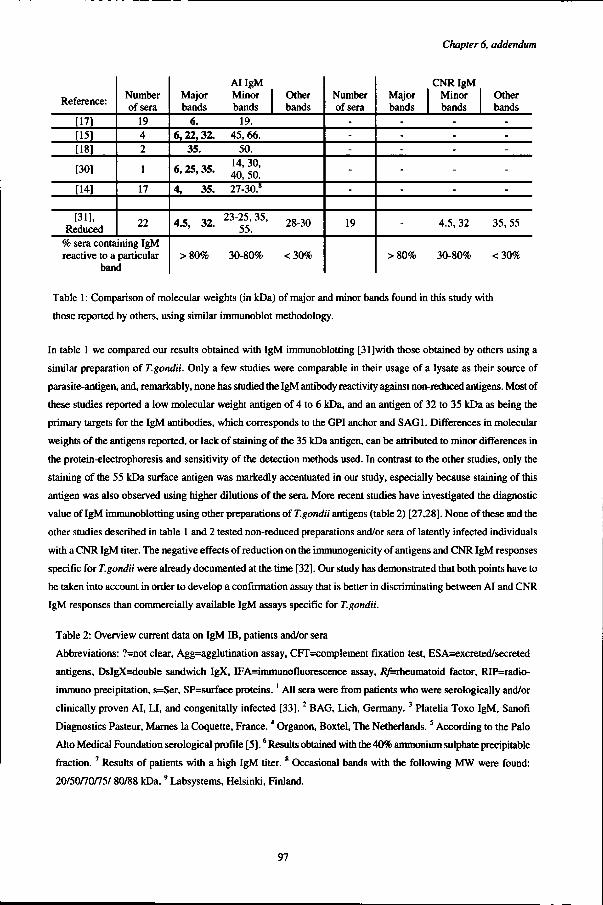
Tablee 1: Comparison of molecular weights (in kDa) of major and minor bands found in this study with
thosee reported by others, using similar immunoblot methodology.
Inn table 1 we compared our results obtained with IgM immunoblotting [31]with those obtained by others using a
similarr preparation of T.gondii. Only a few studies were comparable in their usage of a lysate as their source of
parasite-antigen,, and, remarkably, none has studied the IgM antibody reactivity against non-reduced antigens. Most of
thesee studies reported a low molecular weight antigen of 4 to 6 kDa, and an antigen of 32 to 35 kDa as being the
primaryy targets for the IgM antibodies, which corresponds to the GPI anchor and SAG1. Differences in molecular
weightss of the antigens reported, or lack of staining of the 35 kDa antigen, can be attributed to minor differences in
thee protein-electrophoresis and sensitivity of the detection methods used. In contrast to the other studies, only the
stainingg of the 55 kDa surface antigen was markedly accentuated in our study, especially because staining of this
antigenn was also observed using higher dilutions of the sera. More recent studies have investigated the diagnostic
valuee of IgM immunoblotting using other preparations of T.gondii antigens (table 2) [27,28]. None of these and the
otherr studies described in table 1 and 2 tested non-reduced preparations and/or sera of latently infected individuals
withh a CNR IgM titer. The negative effects of reduction on the immunogenicity of antigens and CNR IgM responses
specificc for T.gondii were already documented at the time [32]. Our study has demonstrated that both points have to
bee taken into account in order to develop a confirmation assay that is better in discriminating between AI and CNR
IgMM responses than commercially available IgM assays specific for T.gondii.
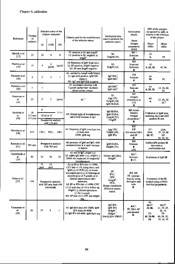
Tablee 2: Overview current data on IgM IB, patients and/or sera
Abbreviations:: ?=not clear, Agg=agglutination assay, CFT=complement fixation test, ESA=excreted/secreted
antigens,, DsIgX=double sandwich IgX, IFA=immunofluorescence assay, /?/=rheumatoid factor, RIP=radio-
immunoo precipitation, s=Ser, SP=surface proteins.' All sera were from patients who were serologically and/or
clinicallyy proven AI, LI, and congenitally infected [33]. 2 BAG, Lich, Germany.3 Platelia Toxo IgM, Sanofi
Diagnosticss Pasteur, Marnes la Coquette, France. 4 Organon, Boxtel, The Netherlands.5 According to the Palo
Altoo Medical Foundation serological profile [5].6 Results obtained with the 40% ammonium sulphate precipitable
fraction.. 7 Results of patients with a high IgM titer.8 Occasional bands with the following MW were found:
20/50/70/75// 80/88 kDa. 9 Labsystems, Helsinki, Finland.
97 7
ChapterChapter 6, addendum
Reference: :
Ehrlic hh et al. [17] ]
Sharmaa et at. [15] ]
Partanenn et al. [18] ]
Partanenn et al. [30] ]
Potasmann et at. at. [14] ]
Herbrin kk et al. al. [36] ]
Decosterr et al. [38] ]
Rotmanss et al. [41] ]
Verhofstedee et al. al. [19] ]
Grosss et al. [27] ]
Yamamotoo et al. al. [28] ]
Number r of f
subjects: :
37 7
10 0
5 5
I I
22 2
18 8 1111 sera 1155 sera
610 0
7633 sera
80 0
144 4
26 6
Infectionn status of the subjectss analyzed:
AII CNR SN
199 9 9
44 [pli ] [p0011
22 2 1
1 1
177 5 [pool]
8 8
lOAIor U U 1111 s
ProspectiveProspective analysis withh 1! 5 sera
1399 s 432 s 140 s
ProspectiveProspective analysis withh 763 sera
200 30 30
100 10 pooll pool
ProspectiveProspective analysis, withh 195 sera from 144
individual s s
19 9 6 6 1 1
Criteri aa used for the establishment off the infection status:
Al :: positive in SF and dstgM1
LI :: positive in SF, negative in dsIgM' '
Al :: Detection of IgM (high titer ) LI :: SF positive, dsIgM negative
SN:: SF and dsIgM negative
Al :: verified by lymph node biopsy LI :: IgG EIA positive, IgM EIA
negative e SN:: IgG and IgM EIA negative
Al :: accidental infection with T.gondiiT.gondii tachyzoites via direct
blood-ascitess contact.
Al: ' '
Al :: clinical signs of toxoplasmosis andd 4-fold increase of IgG
Al :: Detection of IgM in at least two off three tests
CNR:: IgM neg
AI :: presence of IgM and IgG, with seroconversionseroconversion or a rapid increase
inn titer(s)
Al :: rise in IgG, dsIgM [+] LI :: stable IgG titers (ly), dsIgM [-] Child:: not suspected of congenital
toxoplasmosis s Al :: a) SF or IFA titer >1:1000,
CFTT titer >1:10, rising titers, and IgMM [+] , b) PCR [+] , c) mouse
inoculationn test [+] , d) histological identificationn of T.gondii, or e)
clinicall improvement after treatment. .
LI :: SFor IFA titer <1:1000, CFT << 1:10 with titer <, \: 10 in follow up,
dsIgMM [-], clinical specimens [-]] for T.gondii.
SN:: SF titer <1:2, CFT and dsIgM neg. .
AI :: IgG IFA titers 64-32000, IgM IFAA titers 64-4096
U:: IgG FA 64-1096, IgM+Ig A neg
Serologicall tests usedd to establish the
infectionn status:
SF, , DsIgMM [34]
SF, , DsIgMM [34]
IgGG EIA' , IgMM EIA 9.
IgGG EIA 9, IgMM EIA' .
SF, , Aggg [35],
DsIgMM [34], IgMM ISAGA.
IgGG EUSA, IgMM ELISA, DsIgMM [37],
DsIgM" . .
Aggg £39], DsIgMM [40],
IgMM IFA.
IgMM EUSA, DsIgMM [37].
Indirectt IgG Elisa, DsIgM' . .
SF, , FA 2. . CFT, ,
DsIgM3, , PCR, ,
Mousee inoculation. Differentt strains
tested. .
IgGG FA IgMM FA DsIgM.' ' DsIgA.' '
Dot-EUSAA (PBST)
lmmunoblot t details: :
Strain n Antigen n
preparation. . R/NR R RH? ?
Sonicate. . R R
RH H Sonicate/ / Sediment. .
R R
RH H Sonicate e
R R
RH H Sonicate e
R R RH H
Antigen n preparationn as
[15] ] R R
RHH (strain Deelen) ) Antigen n
preparationn ? R R
RH H RIP P
RA-labeledd ESA andd SP
R R RH H
Sonicate. . R R
T36] ]
RH? ? Sonicate. .
R(?). .
RH,, BK TP,, sonicate, lysiss by using detergentss and
salts s R. .
RH ? ? TP,, ESA-AP precipitated. .
R. .
MWW of the antigens recognizedd by IgM, in relationn to the infection
off the subject:
Majorr Minor (kDa)) (kDa)
Al : : 6.. 19.
Al : : 6,, 22,32. 45, 66.
Al : : 35.. 50.
AT ' '
6,25,35.. ' « - g ^
AA^^ 27, 29, 30, 4-- 35.8
Evaluationn of IgM tests, includingg the 6 kD (GPI
AI 7:: ESA: ESA:: 37, 34, 86
69,97.. SP: 30,, 35, 43.
66 kDa (GPI anchor) IB wass used as
confirmationn test
Evaluationn of IgM IB
Evaluationn of the IB methodd using a SAG1 enrichedd preparation.
Al : : ESA6: : 32-30. .
TP: : 35,32-30, ,
14. .
ESA6: : 41,39,35, ,
26,, 14. TP: :
60,, 56, 42, 39,24,, 19
98 8