Embed Size (px)
Citation preview

UvA-DARE is a service provided by the library of the University of Amsterdam (http://dare.uva.nl)
UvA-DARE (Digital Academic Repository)
Regulation and cross-talk between environmental triggers of local immune responsesin airway epithelial cellsGobski, K.
Link to publication
Citation for published version (APA):Gobski, K. (2017). Regulation and cross-talk between environmental triggers of local immune responses inairway epithelial cells
General rightsIt is not permitted to download or to forward/distribute the text or part of it without the consent of the author(s) and/or copyright holder(s),other than for strictly personal, individual use, unless the work is under an open content license (like Creative Commons).
Disclaimer/Complaints regulationsIf you believe that digital publication of certain material infringes any of your rights or (privacy) interests, please let the Library know, statingyour reasons. In case of a legitimate complaint, the Library will make the material inaccessible and/or remove it from the website. Please Askthe Library: http://uba.uva.nl/en/contact, or a letter to: Library of the University of Amsterdam, Secretariat, Singel 425, 1012 WP Amsterdam,The Netherlands. You will be contacted as soon as possible.
Download date: 26 Aug 2018

Chapter
7


The transcription factor GATA3 is essential for the function of human type 2 innate lymphoid
cells Jenny Mjösberg1, Jochem Bernink3, Korneliusz Golebski2, Julien J. Karrich3, Charlotte P. Peters1, Bianca Blom3, Anje A. te Velde1, Wytske J. Fokken23, Cornelis M. van Drunen2, and Hergen Spits1
1 Tytgat Institute for Liver and Intestinal Research, University of Amsterdam 2 Department of Otorhinolaryngology, Academic Medical Center, University of Amsterdam 3 Department of Cell Biology, University of Amsterdam, Academic Medical Center, University of Amsterdam
Immunity, Volume 37, Issue 4, 19 October 2012, Pages 649–659

119
Chapter 7
SUMMARY
Type 2 innate lymphoid cells (ILC2s) are part of a large family of ILCs that are important effectors in innate immunity, lymphoid organogenesis, and tissue remodeling. ILC2s mediate parasite expulsion but also contribute to airway inflammation, emphasizing the functional similarity between these cells and Th2 cells. Consistent with this, we report that the transcription factor GATA3 was highly expressed by human ILC2s. CRTH2+ ILC2s were enriched in nasal polyps of patients with chronic rhinosinusitis, a typical type 2-mediated disease. Nasal polyp epithelial cells expressed TSLP, which enhanced STAT5 activation, GATA3 expression, and type 2 cytokine production in ILC2s. Ectopic expression of GATA3 in Lin−CD127+CRTH2− cells resulted in induction of CRTH2 and the capacity to produce high amounts of type 2 cytokines in response to TSLP plus IL-33. Hence, we identify GATA3, potently regulated by TSLP, as an essential transcription factor for the function of human ILC2s. INTRODUCTION Innate lymphoid cells (ILCs) are emerging as important effectors in innate immunity, lymphoid tissue formation, and tissue remodeling. ILCs are characterized by a lymphoid morphology, the absence of markers for T, B, and myeloid cells, and high expression of the IL-7 receptor (IL-7αR; CD127). ILCs are related to natural killer (NK) cells; they share with NK cells a dependency on the transcriptional repressor Id2, but whereas NK cells depend on IL-15, ILCs require IL-7 for their development and survival (reviewed in Spits and Cupedo, 2012). IL-7-dependent ILCs can be divided into several subpopulations on the basis of transcription factor dependency, cytokine secretion profile, and functions. The prototypic ILC population, lymphoid tissue inducer (LTi) cells, is instrumental for the formation of secondary lymphoid organs during embryonic development and depends on the transcription factor RORγt (Eberl et al., 2004). After birth, IL-22-producing RORγt-dependent ILCs are dominating in mucosal tissues, playing essential roles in innate immunity and wound healing (Cella et al., 2009; Crellin et al., 2010; Satoh-Takayama et al., 2008; Zenewicz et al., 2008).
Type 2 cytokine-producing ILCs were described by Rennick (Fort et al., 2001) and Coffman (Hurst et al., 2002). These cells lacked T and B cell markers, were dependent on the gamma common (γc) chain of the IL-2 receptor, and produced high amounts of IL-5 and IL-13 in response to the Th2 cell-stimulating cytokine IL-25. More recently, IL-5- and IL-13-producing ILCs, which were called natural helper (NH) cells, were found in fat-associated lymphoid tissue where they were essential for the innate immune response against the nematode Nippostrongulus brasiliensis (Moro et al., 2010). These cells responded not only to IL-25 but also IL-33. Two other research groups used IL-13 reporter mice to trace IL-25- and IL-33-responsive ILCs in a variety of tissues including lung and gut (Neill et al., 2010; Price et al., 2010). These IL-13-producing ILCs have been named nuocytes (Neill et al., 2010) or innate helper type 2 (ih2) cells (Price et al., 2010). Although some differences were noted in phenotypes and tissue distribution of NH cells, nuocytes, and ih2 cells, they may represent the same cell type, here referred to as type 2 ILCs (ILC2s). Also, in humans IL-25- and IL-33-responsive ILC2s have been defined (Monticelli et al., 2011; Mjösberg et al., 2011). These cells express CD127, the prostaglandin D2 receptor, chemoattractant receptor expressed on Th2 cells (CRTH2), and the

120
The transcription factor GATA3 is essential for the function of ILC2s
common ILC and NK cell marker CD161 (Mjösberg et al., 2011). Besides a critical role in the early innate immune response against helminths, ILC2s are also involved in both airway inflammation (Chang et al., 2011) and tissue repair (Monticelli et al., 2011) after influenza virus infection. Moreover, these ILCs can mediate type 2 disease as shown in several mouse asthma models (Barlow et al., 2012; Bartemes et al., 2012; Halim et al., 2012; Kim et al., 2012; Wolterink et al., 2012). The increased presence of ILC2s in inflamed nasal polyps of patients suffering from chronic rhinosinusitis (CRS) (Mjösberg et al., 2011) suggests an involvement of these cells in human type 2 inflammatory diseases as well. Thus, ILC2s are emerging as essential partners in the so-called type 2 franchise, which include basophils, eosinophils, mast cells, Th2 cells, Th9 cells, and IgE-producing B cells, which together form the immune response against parasites, but also mediate type 2 inflammatory diseases including asthma and allergic diseases (reviewed in Oliphant et al., 2011; Paul and Zhu, 2010). The various type 2 leukocyte populations are governed by factors produced by hematopoietic and nonhematopoietic accessory cells. Besides IL-25 and IL-33, these factors include thymic stromal lymphopoetin (TSLP). TSLP is produced predominantly by epithelial cells and is believed to prime dendritic cells (DCs) to facilitate development of Th2 cells (Ito et al., 2005), which is associated with the pathogenesis of allergic inflammatory diseases (reviewed in Ziegler and Artis, 2010).
The developmental requirements and the transcription factors that are responsible for differentiation and function of ILCs are being unraveled. RORγt is required for development and function of LTi cells and ILC22 but is dispensable for development of ILC2s (Moro et al., 2010; Neill et al., 2010). Instead, the related transcription factor RAR-related orphan receptor alpha (RORα, also known as TR1F1) is needed for ILC2s as shown by the fact that RORα-deficient mice fail to develop ILC2s (Wong et al., 2012). Other transcription factors that affect the function of ILC2s have yet to be identified. The transcription GATA3 might be a candidate. GATA3 is needed for development of Th2 cells and acts by binding directly to several genes of the Th2 cell cytokine locus including IL4, IL5, and IL13, and concomitantly inhibiting development of Th1 cells (Zheng and Flavell, 1997; Ouyang et al., 1998).
Previously we noted striking similarities between ILCs and Th cell subsets with respect to their cytokine production profiles and proposed that ILCs and Th cells share transcriptional programs that regulate their development and function (Spits and Di Santo, 2011). Consistent with this hypothesis, we show here a critical role of GATA3 in the function of human ILC2s. Although GATA3 is broadly expressed among the different ILC populations, including NK cells, ILC2s were characterized by high GATA3 expression. Activation of resting ILC2s by TSLP increased the expression of GATA3. TSLP strongly enhanced IL-4, IL-5, and IL-13 production, activated STAT5, and acted with IL-33 to synergistically induce IL-4, IL-5, and IL-13 production. GATA3 was essential for the function of ILC2s, as shown by the fact that ectopic expression of GATA3 by retrovirus-mediated gene transfer in Lin−CD117+CD127+CRTH2− cells resulted in induction of CRTH2 and in acquisition of the capacity to produce high amounts of IL-5 and IL-13 as well as IL-4 in response to TSLP plus IL-33. Hence, we identify GATA3 as an essential transcription factor for the function of human ILC2s, emphasizing the similarity of ILC2s and Th2 cells as we proposed earlier (Spits and Di Santo, 2011).

121
Chapter 7
RESULTS Nasal Polyp Epithelium Express IL33 and TSLP We previously showed that ILC2s, defined as Lin−CD127+CRTH2+, are strikingly enriched in inflamed nasal polyps of CRS patients as compared to noninflamed nasal mucosa of healthy control subjects (Mjösberg et al., 2011). Further characterization revealed that these ILC2s respond to IL-25 and IL-33 with increased production of IL-13 (Figure S1A available online), a behavior characteristic of the ILC2s that we previously identified in peripheral blood and fetal gut, defined as Lin−CD127+CRTH2+CD161+CD25+ (Mjösberg et al., 2011). Like ILC2s in peripheral blood, short-term primary cell lines of nasal polyp ILC2s (henceforth referred to as “ILC2 cell lines”) could be established that kept their surface expression of CD127 and CRTH2 (Figure S1B). Although only a minority of these cells produced IL-22 and IFN-γ, they all produced IL-13 but no IL-17 upon short-term culture with PMA plus ionomycin (Figure S1C), demonstrating their type 2 cytokine production profile. ILC2 cell lines expressed receptors for IL-25 and IL-33 (Figures S1D and S1E) and also responded to IL-25 and IL-33 in combination with IL-2 in vitro (Figures S1F and S1G). These data demonstrated that both freshly isolated and ILC2 cell lines from nasal polyps were similar to the ILC2s that we previously described in peripheral blood and fetal gut (Mjösberg et al., 2011). In addition, the ILC2s were characterized by expression of CD25 (Mjösberg et al., 2011) and ST2 mRNA (Figure S1E), indicating that they are similar to those previously described by Monticelli et al. (2011)).
Figure 1. TSLP and IL-33 Are Expressed by Nasal Epithelial Cells (A–C) Nasal epithelial cells from CRS polyps (filled dots), healthy mucosa (open squares), or H292 airway epithelial cells lines (open triangles) were cultured with poly(I:C) and analyzed for expression of IL33 (A), long-form TSLP (B), and total TSLP (C) mRNA by RT-PCR. (D–F) Nasal epithelial cells from healthy mucosa was analyzed for IL33 (D), long-form TSLP (E), and total TSLP (F) by RT-PCR. Expression was normalized to expression of the house-keeping gene GAPDH and to the expression of unstimulated cells to generate the relative expression ratio at each time point. All data are shown as mean ± SD and are representative of at least two independent experiments performed in triplicate.

122
The transcription factor GATA3 is essential for the function of ILC2s
To broaden our understanding of the physiological stimuli that contribute to the accumulation and maintenance of ILC2s in the nasal polyps, we investigated nasal epithelial cells, a major source of immune regulatory cytokines, for expression of potential ILC2-regulating cytokines. Nasal epithelial cells from healthy individuals and CRS nasal polyps constitutively, and at comparable amounts, expressed factors that were previously shown to regulate ILC2s including IL33 (Figure 1A) and also TSLP mRNA (Figures 1B and 1C). In this study, we analyzed both the total TSLP transcript expression (including both the short and long splice variants of the TSLP mRNA) and the long splice variant of the TSLP mRNA separately, because the latter was shown to be associated with actual TSLP protein production (Harada et al., 2009).
It was previously demonstrated that polyinosinic:polycytidylic acid [poly(I:C)], a double-stranded RNA analog and Toll-like receptor (TLR) 3 ligand, induces TSLP expression in human bronchial epithelial cells (Harada et al., 2009). We extended this finding to also include nasal epithelial cells, because poly(I:C) upregulated the expression of long-form TSLP, as well as IL33, in nasal polyp epithelial cells, nasal epithelial cells from healthy individuals, and the respiratory airway epithelial cell line H292 (Figures 1A and 1B). IL33 and long-form TSLP were not selectively induced by poly(I:C); stimulation with flagellin, R848, and PGN also resulted in an increase in IL33 and long-form TSLP expression (Figures 1D and 1E).
In summary, these data show that nasal epithelial cells from both healthy nasal mucosa and CRS polyps constitutively, and at similar amounts, express both total and long-form TSLP, which is induced by a wide range of TLR ligands. Collectively, this means that nasal epithelial cells are able to respond to microbial challenge by producing factors, including IL-33 and TSLP, known to be involved in the type 2-mediated inflammation seen in these nasal polyps (van Drunen et al., 2012). ILC2s Respond to TSLP Expressed by Nasal Polyp Epithelium Having identified TSLP as an immune regulatory cytokine expressed by nasal epithelial cells, we hypothesized that TSLP could have the capacity to induce cytokine production from ILC2s, similar to what was previously described for IL-33. TSLP signals through a receptor heterodimer consisting of the TSLP receptor and the IL-7 receptor α chain (CD127), the latter being constitutively expressed by all subsets of human and mouse ILCs. Analyzing the expression of the TSLP receptor (TSLPR) subunit, we found a high expression in both freshly isolated and ILC2 cell lines from tonsils, blood, and nasal polyps as compared to other ILC subsets, including NK cells (Figures 2A and 2B).
Consequently, stimulating freshly isolated ILC2s from blood and nasal polyps with TSLP in combination with IL-2 resulted in significantly increased secretion of IL-13, one of the signature cytokines for the human ILC2s (Figures 2C and 2D). In addition, ILC2s also produced a number of additional cytokines associated with type 2 immune responses, including IL-4, IL-5, IL-9, and GM-CSF and the production was clearly enhanced by IL-33, and to a lesser extent also TSLP (Figures 2C and 2D). We also analyzed a range of other cytokines, of which the ILC2s produced IL-6, IL-8, and IP10 at concentrations above 100 pg/mL. Many other cytokines were produced at concentrations below 100 pg/mL and were not affected by IL-33 or TSLP stimulation (see Table S1).
The fact that TSLP triggered resting ILC2s freshly isolated from peripheral blood (Figure 2C) suggests that these cells do not require preactivation to respond to TSLP, in contrast to what was previously reported for Th2 cells (Kitajima et al., 2011).
123
Chapter 7
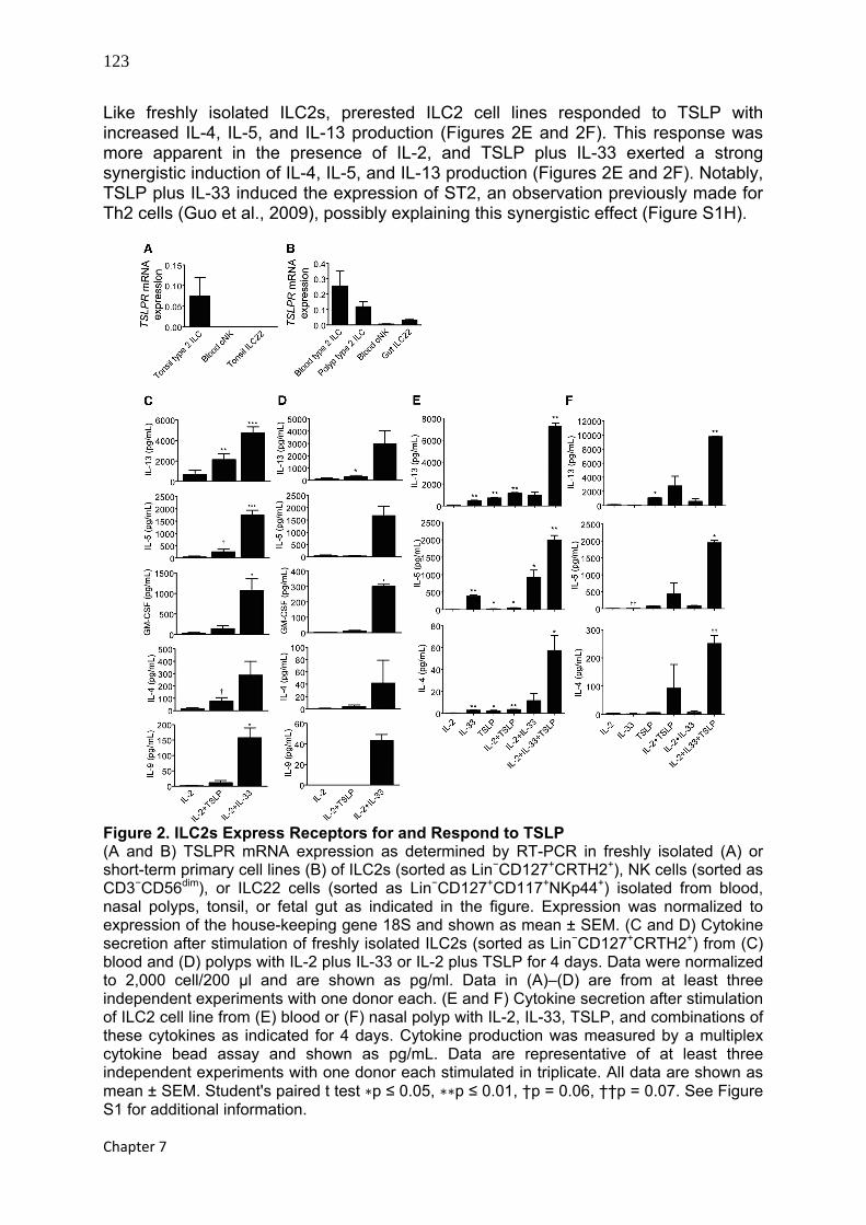
Like freshly isolated ILC2s, prerested ILC2 cell lines responded to TSLP with increased IL-4, IL-5, and IL-13 production (Figures 2E and 2F). This response was more apparent in the presence of IL-2, and TSLP plus IL-33 exerted a strong synergistic induction of IL-4, IL-5, and IL-13 production (Figures 2E and 2F). Notably, TSLP plus IL-33 induced the expression of ST2, an observation previously made for Th2 cells (Guo et al., 2009), possibly explaining this synergistic effect (Figure S1H).
Figure 2. ILC2s Express Receptors for and Respond to TSLP (A and B) TSLPR mRNA expression as determined by RT-PCR in freshly isolated (A) or short-term primary cell lines (B) of ILC2s (sorted as Lin−CD127+CRTH2+), NK cells (sorted as CD3−CD56dim), or ILC22 cells (sorted as Lin−CD127+CD117+NKp44+) isolated from blood, nasal polyps, tonsil, or fetal gut as indicated in the figure. Expression was normalized to expression of the house-keeping gene 18S and shown as mean ± SEM. (C and D) Cytokine secretion after stimulation of freshly isolated ILC2s (sorted as Lin−CD127+CRTH2+) from (C) blood and (D) polyps with IL-2 plus IL-33 or IL-2 plus TSLP for 4 days. Data were normalized to 2,000 cell/200 μl and are shown as pg/ml. Data in (A)–(D) are from at least three independent experiments with one donor each. (E and F) Cytokine secretion after stimulation of ILC2 cell line from (E) blood or (F) nasal polyp with IL-2, IL-33, TSLP, and combinations of these cytokines as indicated for 4 days. Cytokine production was measured by a multiplex cytokine bead assay and shown as pg/mL. Data are representative of at least three independent experiments with one donor each stimulated in triplicate. All data are shown as mean ± SEM. Student's paired t test ∗p ≤ 0.05, ∗∗p ≤ 0.01, †p = 0.06, ††p = 0.07. See Figure S1 for additional information.

124
The transcription factor GATA3 is essential for the function of ILC2s
Figure 3. TSLP Stimulates Phosphorylation of STAT5 in ILC2s (A and B) Expression of phosphorylated STAT3 and STAT5 as measured by flow cytometry. ILC2s (sorted as Lin−CD127+CRTH2+) or NK (sorted as CD3−CD56dim) short-term cultured cells were rested overnight in medium without cytokines and subsequently pulsed with the indicated cytokines for 20 min. Data are representative of three independent experiments with at least one donor each. (C) Tamoxifen (4HT) treatment of prerested, presorted (GFP+ or GFP−) peripheral blood ILC2 cell lines transduced (GFP+) with retroviral constructs encoding STAT5b or STAT3 linked to the estrogen receptor (ER) or empty control vector (EV). Data are from two experiments with one donor each and shown as mean ± SEM. 4HT, 4-hydroxytamoxifen. When following IL-33 plus TSLP response over time, we observed that IL-13 was induced at day 2, before significant cell proliferation was induced by TSLP (data not shown). Hence, the increased IL-13 concentration seen in the supernatants after IL-33 plus TSLP stimulation was most probably the result of increased IL-13 production on a per cell basis rather than proliferation of a subpopulation of IL-13+ cells. IL-25, especially in combination with IL-33, triggered significant IL-13 production from ILC2s (Figures S1F and S1G). IL-25 also acted in synergy with TSLP to induce IL-13 production but to a much lesser extent than what we observe for the combination of IL-33 and TSLP (data not shown). Hence, we focused our studies on TSLP and IL-33, because these cytokines had the strongest effects on the ILC2s.
Collectively, our data identify TSLP, expressed by nasal epithelial cells, as a potent stimulator that acts in synergy with IL-33 to induce cytokine secretion from ILC2s in nasal polyps. TSLP Triggers STAT5 Activation that Drives IL-13 Production in ILC2s We next asked the question which signaling pathway was involved in TSLP-induced IL-13 production. In Th2 cells, TSLP triggers phosphorylation of STAT5 (Kitajima et al., 2011) but STAT3 has also been reported to be activated through TSLP (Reche et al., 2001). Stimulating ILC2s with TSLP, IL-2, or IL-7 (data not shown) led to phosphorylation of STAT5, but not STAT3, whereas IL-15, a known trigger of conventional NK cells, stimulated phosphorylation of both STAT5 and STAT3 in NK cell lines (Figures 3A and 3B ). NK cells, however, did not respond to TSLP with STAT5 phosphorylation (Figure 3A), consistent with the low to undetectable TSLPR expression in these cells (Figures 2A and 2B).
To examine the hypothesis that STAT5 activation alone is sufficient to drive TSLP-induced IL-13 production in ILC2s, we made use of a tamoxifen (4HT)-inducible retroviral construct encoding STAT5b linked to the estrogen receptor (ER) (Scheeren et al., 2005). With this approach, activation of STAT5, but not STAT3, in prerested ILC2s led to a clear induction of IL-13 production (Figure 3C), showing that
125
Chapter 7
the isolated activation of the STAT5 pathway is sufficient to induce IL-13 production in ILC2s.
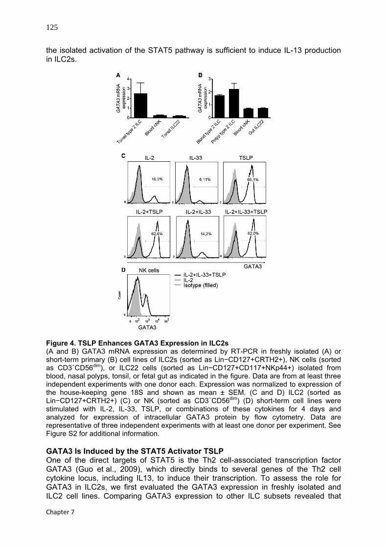
Figure 4. TSLP Enhances GATA3 Expression in ILC2s (A and B) GATA3 mRNA expression as determined by RT-PCR in freshly isolated (A) or short-term primary (B) cell lines of ILC2s (sorted as Lin−CD127+CRTH2+), NK cells (sorted as CD3−CD56dim), or ILC22 cells (sorted as Lin−CD127+CD117+NKp44+) isolated from blood, nasal polyps, tonsil, or fetal gut as indicated in the figure. Data are from at least three independent experiments with one donor each. Expression was normalized to expression of the house-keeping gene 18S and shown as mean ± SEM. (C and D) ILC2 (sorted as Lin−CD127+CRTH2+) (C) or NK (sorted as CD3−CD56dim) (D) short-term cell lines were stimulated with IL-2, IL-33, TSLP, or combinations of these cytokines for 4 days and analyzed for expression of intracellular GATA3 protein by flow cytometry. Data are representative of three independent experiments with at least one donor per experiment. See Figure S2 for additional information. GATA3 Is Induced by the STAT5 Activator TSLP One of the direct targets of STAT5 is the Th2 cell-associated transcription factor GATA3 (Guo et al., 2009), which directly binds to several genes of the Th2 cell cytokine locus, including IL13, to induce their transcription. To assess the role for GATA3 in ILC2s, we first evaluated the GATA3 expression in freshly isolated and ILC2 cell lines. Comparing GATA3 expression to other ILC subsets revealed that

126
The transcription factor GATA3 is essential for the function of ILC2s
GATA3 was highly expressed by both freshly isolated and ILC2 cell lines (Figures 4A and 4B ). GATA3 mRNA expression was not exclusively restricted to the ILC2 subset; ILC22 and NK cells also expressed GATA3 (Figures 4A and 4B), the latter finding being consistent with the previously described role of GATA3 as a regulator of NK cells (Samson et al., 2003).
Having shown that TSLP triggers STAT5 activation, which alone is sufficient to drive expression of IL-13, we investigated the capacity of TSLP to induce GATA3 expression in ILC2s. TSLP increased the frequency of cells expressing GATA3 protein (Figure 4C). Furthermore, TSLP stimulation led to increased GATA3 mRNA expression (Figure S2). NK cells also expressed GATA3 protein but at a lower level than did ILC2s (Figure 4D). Importantly, GATA3 expression was not triggered by TSLP in NK cells (Figure 4D), in line with the low expression of TSLPR on these cells (Figures 2A and 2B). Analyzing GATA3 protein expression over time revealed that GATA3 was induced by TSLP in nondividing cells by day 2, demonstrating that TSLP induces expression of GATA3 protein and not preferential outgrowth of GATA3+ cells (Figure S2). Ectopic Expression of GATA3 Is Sufficient to Drive the ILC2 Program In ILC2s, TSLP induced IL-13 production in a STAT5-dependent fashion, which also correlated with induction of GATA3. In Th2 cells, GATA3 is the master transcription factor that governs a wide spectrum of features unique to Th2 cells, including expression of ST2, thereby making the cells responsive to IL-33 as shown by high IL-13 production (Guo et al., 2009). To formally test the role of GATA3 in ILC2 function, we asked whether GATA3 overexpression is sufficient to drive the program of ILC2 phenotype and function. Overexpression of GATA3, as confirmed at mRNA (Figure 5A) and protein (Figure S3A) level, in a population of Lin−CD127+CD117+NKp44−CRTH2− ILCs led to expression of the cardinal surface marker of ILC2s and Th2 cells, CRTH2 (Figure 5B), suggesting that forced GATA3 expression is sufficient to induce differentiation toward the ILC2 lineage. In strong support of this, GATA3-overexpressing ILCs upregulated the expression of ST2 and TSLPR (Figures 5C and 5D) and also resulted in acquisition of the capacity to respond to IL-33 plus TSLP by secreting high amounts of IL-13, IL-4, IL-5, and GM-CSF (Figures 5E–5H). Notably, GATA3 overexpression by itself was not sufficient to drive significant type 2 cytokine production in these cells, as indicated by the fact that GATA3-overexpressing cells rested in low amounts of IL-2 produced only modest amounts of IL-4, IL-5, IL-13, and GM-CSF (Figures 5E–5H). This notion is in agreement with the observation that GATA3 alone is not sufficient to induce IL-4 production in M12 B cell lymphoma cells, which requires additional activating signals provided by PMA plus ionomycin (Zheng and Flavell, 1997). Hence, GATA3 most probably acts as a transactivator of IL-4, IL-5, and IL-13 production, requiring additional signals provided by TSLP and IL-33. ILC2s were recently shown to depend on the transcription factor RORα, because RORα-deficient mice lack ILC2s and fail to expel helminth parasites in the intestine, a process normally mediated by ILC2s (Wong et al., 2012). In agreement with these data, we find that human ILC2s also express higher amounts of RORA as compared to ILC22s and NK cells (Figure S3B). As seen for GATA3, RORA expression was not exclusively restricted to the ILC2 subset; ILC22 and NK cells also expressed RORA. GATA3 overexpression in CRTH2− ILCs did not lead to significant upregulation of RORA expression (Figure S3C). Furthermore, TSLP stimulation, which enhances GATA3 expression in ILC2s, did not upregulate RORA (Figure S3D). Hence, we

127
Chapter 7
conclude that RORA is not potently regulated by GATA3 and that GATA3 can induce TSLP plus IL-33 responsiveness, resulting in IL-13 production independent of enhanced RORA expression.
Figure 5. GATA3 Overexpression Is Sufficient to Drive the ILC2 Program (A) Lin−CD127+CD117+NKp44−CRTH2− cells were transduced with a retroviral vector encoding GATA3 wild-type (WT) or empty vector (EV), sorted as transduced (GFP+) or untransduced (GFP−) cells on day 3 and analyzed for GATA3 mRNA expression by RT-PCR. Expression was normalized to expression of the house-keeping gene 18S. Data are from two independent experiments with at least one donor each. (B) CRTH2 surface expression in empty vector control transduced (left) or GATA3 overexpressing (right) cells (GFP+) as determined by flow cytometry. The percentages within the dot plots refer to the percentage of CRTH2+ ILCs within the GFP+ gate representing transduced cells. Data are from three independent experiments with at least one donor each. (C and D) Lin−CD127+CD117+NKp44−CRTH2− cells were transduced as described in (A) and analyzed for ST2 (C) or TSLPR (D) mRNA expression by RT-PCR. Expression was normalized to expression of the house-keeping gene 18S. Data are from two independent experiments with at least one donor each. (E–H) IL-13, IL-5, IL-4, and GM-CSF production from empty vector control (black bars) or GATA3 overexpressing (unfilled bars) cells upon IL-2 or IL-2, IL-33 plus TSLP stimulation for 4 days. Cells were transduced and sorted as described in (A). Cytokine production was normalized to that of the EV-transduced cells and shown as percentage of EV control. Data are from three independent experiments with at total of six donors. All data are shown as mean ± SEM. Wilcoxon's matched pairs test ∗p ≤ 0.05, †p = 0.06. See Figure S3 for additional information.
128
The transcription factor GATA3 is essential for the function of ILC2s
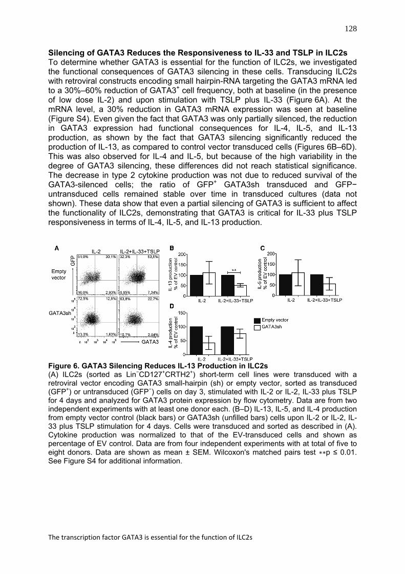
Silencing of GATA3 Reduces the Responsiveness to IL-33 and TSLP in ILC2s To determine whether GATA3 is essential for the function of ILC2s, we investigated the functional consequences of GATA3 silencing in these cells. Transducing ILC2s with retroviral constructs encoding small hairpin-RNA targeting the GATA3 mRNA led to a 30%–60% reduction of GATA3+ cell frequency, both at baseline (in the presence of low dose IL-2) and upon stimulation with TSLP plus IL-33 (Figure 6A). At the mRNA level, a 30% reduction in GATA3 mRNA expression was seen at baseline (Figure S4). Even given the fact that GATA3 was only partially silenced, the reduction in GATA3 expression had functional consequences for IL-4, IL-5, and IL-13 production, as shown by the fact that GATA3 silencing significantly reduced the production of IL-13, as compared to control vector transduced cells (Figures 6B–6D). This was also observed for IL-4 and IL-5, but because of the high variability in the degree of GATA3 silencing, these differences did not reach statistical significance. The decrease in type 2 cytokine production was not due to reduced survival of the GATA3-silenced cells; the ratio of GFP+ GATA3sh transduced and GFP− untransduced cells remained stable over time in transduced cultures (data not shown). These data show that even a partial silencing of GATA3 is sufficient to affect the functionality of ILC2s, demonstrating that GATA3 is critical for IL-33 plus TSLP responsiveness in terms of IL-4, IL-5, and IL-13 production.
Figure 6. GATA3 Silencing Reduces IL-13 Production in ILC2s (A) ILC2s (sorted as Lin−CD127+CRTH2+) short-term cell lines were transduced with a retroviral vector encoding GATA3 small-hairpin (sh) or empty vector, sorted as transduced (GFP+) or untransduced (GFP−) cells on day 3, stimulated with IL-2 or IL-2, IL-33 plus TSLP for 4 days and analyzed for GATA3 protein expression by flow cytometry. Data are from two independent experiments with at least one donor each. (B–D) IL-13, IL-5, and IL-4 production from empty vector control (black bars) or GATA3sh (unfilled bars) cells upon IL-2 or IL-2, IL-33 plus TSLP stimulation for 4 days. Cells were transduced and sorted as described in (A). Cytokine production was normalized to that of the EV-transduced cells and shown as percentage of EV control. Data are from four independent experiments with at total of five to eight donors. Data are shown as mean ± SEM. Wilcoxon's matched pairs test ∗∗p ≤ 0.01. See Figure S4 for additional information.

129
Chapter 7
DISCUSSION We report here that the transcription factor GATA3 is important for the function of human ILC2s, emphasizing the functional similarity between ILC2s and Th2 cells. Although GATA3 is broadly expressed among the different ILC populations, including NK cells, ILC2s are characterized by high GATA3 expression. We previously showed that ILC2s are enriched in nasal polyps of patients with CRS, a typical type 2-mediated inflammatory disease. We observed that TSLP, expression of which is enhanced in nasal epithelial cells upon microbial challenge, is a potent regulator of ILC2s, as shown by the fact that TSLP enhances GATA3 expression and production of a wide range of type 2 cytokines in these cells. In contrast to Th2 cells, this occurs without prior activation of the cells, showing that ILC2s are able to directly respond to epithelial-derived TSLP to initiate the type 2 response. In ILC2s, TSLP signals through STAT5 and acts with IL-33 to synergistically induce IL-13 production. GATA3 is crucial for the function of ILC2 because ectopic expression of GATA3 by retrovirus-mediated gene transfer in Lin−CD127+CD117+NKp44−CRTH2− cells results in induction of surface CRTH2, in expression of ST2 and TSLPR, and in acquisition of the capacity to produce high amounts of type 2 cytokines in response to TSLP plus IL-33.
Asthma, allergic rhinitis, and atopic dermatitis are atopic diseases where elevated levels of TSLP have been reported. Together with the notion that TSLP polymorphisms are associated with atopy (reviewed in Ziegler and Artis, 2010) and that TSLP receptor-deficient mice are resistant to ovalbumin-induced asthma (Zhou et al., 2005), this suggests a role for TSLP in the pathogenesis of these diseases. TSLP induces polarization of dendritic cells, with increased expression of OX40L, promoting differentiation of naive T cells to Th2 cells (Ito et al., 2005). Th2 cells produce IL-4, IL-5, and IL-13, key cytokines in allergy and asthma, indicating that TSLP is involved in exacerbation of allergic inflammation driven by Th2 cells. However, experimental allergen-induced asthma occurs also in RAG-deficient mice, which lack T cells, suggesting that cells of the innate immune system are critical effectors in type 2-mediated inflammation (Bartemes et al., 2012; Halim et al., 2012; Wolterink et al., 2012). Indeed, several studies have demonstrated a role for ILC2s in various models of allergic airway inflammation (Barlow et al., 2012; Kim et al., 2012; Wilhelm et al., 2011; Bartemes et al., 2012; Halim et al., 2012; Wolterink et al., 2012). Furthermore, ILC2s are involved in influenza virus-mediated airway hyperreactivity by promoting inflammation (Chang et al., 2011). In contrast, ILC2s are also critical for tissue repair and remodeling inflicted by influenza virus infection (Monticelli et al., 2011). We observed that TSLP induces the production of high amounts of IL-13 and IL-5, but also IL-4, IL-9, and GM-CSF by ILC2s. In the mouse, it was reported that Th2 cells express TSLP receptors and respond to TSLP by production of the Th2 cell type cytokines IL-4, IL-5, and IL-13 (Kitajima et al., 2011), demonstrating the functional similarity between ILC2s and Th2 cells. However, TSLP responsiveness in Th2 cells was significant only after activation via the T cell receptor (Kitajima et al., 2011). Our data showed that freshly isolated ILC2s from nasal polyps, but also from blood, most probably in a resting state, respond to TSLP without prior activation. Hence, ILC2s are capable of directly responding to TSLP, initiating the type 2-mediated response.
In search for the cellular source of TSLP in the nasal polyps, we found that in epithelial cells, TSLP was expressed at baseline and that expression was promoted upon stimulation with several TLR agonists including poly(I:C), flagellin, R848, and

130
The transcription factor GATA3 is essential for the function of ILC2s
PGN, indicating that microbes might be the indirect trigger of high type 2 cytokine production by ILC2s. While completing our work, a study of papain-induced asthma demonstrated that also in mice, stromal-derived TSLP and IL-33 induce IL-13 production from ILC2s, driving the allergic inflammation (Halim et al., 2012). Thus, both in mice and humans, airway pathology may be mediated by aberrant stimulation of epithelial cells that initiate a cascade of reactions leading to overstimulation of ILC2s.
TSLP has been reported to signal through STAT5 (Kitajima et al., 2011) and STAT3 (Reche et al., 2001). We found that TSLP stimulation of ILC2s led to activation of STAT5 but not STAT3. Consistent with our findings on ILC2s, in Th2 cells, TSLP enhances phosphorylation of STAT5 and production of type 2 cytokines such as IL-4, IL-5, and IL-13 (Kitajima et al., 2011). ChIP analysis revealed that STAT5 binds directly to the GATA3 gene (Kitajima et al., 2011), suggesting that STAT5 directly upregulates the expression of GATA3. Indeed, our studies support this idea: the isolated activation of STAT5 alone enhanced IL-13 production in ILC2s. Hence, we identify the TSLP signaling pathway in ILC2s as being dependent on STAT5 and GATA3, with subsequent production of IL-13, IL-5, and IL-4 shown to be direct targets for GATA3 (Yagi et al., 2011).
Recently, it was demonstrated that bone marrow-derived Lin−CD127+Flt3− common lymphoid progenitors in the presence of Notch1, IL-7, and IL-33 signaling gave rise to ILC2s. These cells failed to develop in mice with a spontaneous deletion in the gene for the transcription factor RORα. Consequently, because ILC2s are crucial for parasite expulsion in the intestine, RORα-deficient mice failed to mount an appropriate immune response to the helminth parasite N. brasiliensis (Wong et al., 2012). Our data identified GATA3 as crucial for the function of ILC2s. A recent study showed that transgenic mice where GATA3 was deleted only in IL-13-producing cells, of which the majority was ILC2s during N. brasiliensis infection, were phenocopies of IL-13-deficient mice, exhibiting reduced worm clearance (Liang et al., 2012). This shows that GATA3 is critical for IL-13 production in ILC2s. These data parallel our findings, which identify GATA3 as crucial for the function of ILC2s. In ILC2s, GATA3, potently induced by TSLP, seems to regulate not only the production of type 2 cytokines in conjunction with IL-33, possibly through NF-κB signaling, but also the expression of ST2 and TSLPR, thereby controlling the IL-33 plus TSLP-induced activation of these cells. Hence, GATA3 is a central player in the function of ILC2s, emphasizing the similarity of these cells and Th2 cells, consistent with the idea that ILC2s and Th2 cells share not only cytokine secretion profiles, but also transcriptional programs (Spits and Di Santo, 2011). A critical difference between these type 2 effector cells is, however, that RORα is required for ILC2 but not for Th cell development (Wong et al., 2012). In our studies of human ILC2s, we find that RORA is highly expressed, but not restricted to, ILC2s; ILC22 and NK cells also express intermediate levels of RORA. Notably, GATA3 does not seem to control the expression of RORA, because neither ectopic nor enhancement of endogenous GATA3 expression by TSLP induced RORA expression. In an accompanying paper (Hoyler et al., 2012), it is demonstrated that GATA3 is essential for the development of ILC2s and that during development of ILC2s from its progenitor, high levels of GATA3 is expressed before RORα. Our studies suggest that RORα is not regulated by GATA3. Hence, it is possible that RORα acts in parallel with GATA3 in regulating ILC2 development. Future studies should further unravel the mechanisms underlying the role of RORα and GATA3 in the development of these cells.

131
Chapter 7
In conclusion, we show here that ILC2s are directly induced by nasal stromal-derived factors such as TSLP, and we identify the TSLP-GATA3 axis as crucial in controlling the function of these cells. ILC2s are emerging as critical players in type 2-mediated immune pathology in the airways. Hence, they constitute a member of the “type 2 franchise,” including mast cells, basophils, eosinophils, and Th2 cells that collectively mediate immunity against helminth parasites but are also involved in allergy and asthma. ILC2s may constitute an early source of type 2 cytokines, capable of initiating and amplifying Th2 cell-mediated responses. In addition, ILC2s are important for tissue repair but may also mediate pathology. As such, ILC2s and the cytokines they produce may be attractive targets for therapy of type 2-mediated immune pathologies.
MATERIALS AND METHODS Collection of Human Tissues Inflamed nasal polyps were obtained from chronic rhinosinusitis patients during surgery and healthy nasal tissues were from nasal correction surgeries. Tonsils were from routine tonsillectomies. All nasal and tonsil tissue collection was done at the Academic Medical Center (AMC) (Amsterdam, The Netherlands) and approved by the Medical Ethical Commission of the AMC. Human fetal tissues were obtained from elective abortions at the Stichting Bloemenhove clinic in Heemstede (The Netherlands) upon on the receipt of informed consents. The use of human abortion tissues was approved by the Medical Ethical Commission of the AMC. Gestational age was determined by ultrasonic measurement of the diameter of the skull or femur and ranged from 14 to 17 weeks. Buffy coats were provided by the blood bank at Sanquin (Amsterdam) after written informed consent. Isolation and Stimulation of Human Nasal Epithelial Cells NCI-H292 human airway epithelial cells (American Type Culture Collection, USA) were cultured at 37°C in RPMI 1640 culture medium (Invitrogen, The Netherlands) supplemented with 10% (v/v) fetal calf serum (HyClone, USA), 1.25 mM of glutamine, 100 U/ml of penicillin, and 100 μg/mL of streptomycin. Primary epithelial cells were obtained by digestion of nasal biopsies or polyps with 0.5 mg/mL collagenase 4 (Worthington Biochemical Corp., Lakewood, USA) with a subsequent incubation with anti-EpCAM MicroBeads (Miltenyi Biotec, Germany) and a positive selection on a magnetic column. Primary cells were cultured in BEGM (Lonza Clonetics, The Netherlands) in fully humidified air containing 5% of CO2 at 37°C. Primary epithelial cells and NCI-H292 cell line were cultured to 80% confluence and rested for 24 hr in RPMI 1640 medium containing 100 U/ml of penicillin and 100 μg/ml of streptomycin. Cells were then stimulated with 20 μg/mL of polyinosinic:polycytidylic acid [poly(I:C)], 0.5 μM CpG-ODN, 20 μg/mL peptidoglycan (PGN+), or 1 μg/mL Flagellin diluted in RPMI. Flagellin, R848, and CpG were purchased from InvivoGen (USA) and poly(I:C) and PGN from Sigma Aldrich (Germany). 1, 2, 4, 6 hr and/or 8, 16, and 24 hr after stimulation, cells were collected for RNA extraction. All stimulation experiments were performed in triplicate. Isolation of ILCs from Peripheral Blood and Nasal Polyps Polyp tissues were cut into fine pieces and digested for 30–45 min at 37°C with Liberase TM (125 μg/mL) and DNase I (200 μg/mL) (both from Roche). Alternatively,

132
The transcription factor GATA3 is essential for the function of ILC2s
cells were isolated by mechanically disrupting the tissue with the Stomacher 80 Biomaster (Seward). The cell suspensions were filtered through a 70 μm nylon mesh. Peripheral mononuclear cells (PBMCs) were isolated on Lymphoprep (Nycomed) or Ficoll-Paque Plus (GE Healthcare). PBMCs were further depleted of T, B, and NK cells and monocytes by labeling with FITC-conjugated CD3, CD14, CD16, and CD19 antibodies (clones described below) and anti-FITC microbeads (Miltenyi). Depletion of lineage-positive cells was performed with a LD column (Miltenyi). For flow cytometric sorting, lin-depleted cells from buffy coats or total single cell suspensions from nasal polyps were stained with the following antibody mix (clone name within brackets): Fluorescein isothiocyanate (FITC)-conjugated anti-CD1a (HI149), CD3 (OKT3), CD11c (3.9), CD14 (HCD14), CD16 (3G8), CD19 (HIB19), CD34 (581), CD94 (DX22), CD123 (6H6), FcER1α (AER-37); phycoerythrin (PE)-anti-NKp44 (P44-8), peridinin chlorophyll protein-cyanine 5.5-conjugated anti-CD117 (104D2) (all from Biolegend), FITC-conjugated anti-CD4 (RPA-T4), CD56 (NCAM16.2), TCRαβ (IP26), TCRγδ (B1), Alexa Fluor-647 (AF647)-conjugated anti-CRTH2 (CD294; BM16), allophycocyanin (APC)-cyanine 7 (Cy7) anti-CD45 (2D1) and isotypes PE, APC, PECy7 (X40) (all from Beckton Dickinson), PE-Cy7 anti-human CD127 (R34.34) (Beckman Coulter), FITC-conjugated anti-human BDCA2 (CD303; AC144; Milenyi). Lineage−CD127+CRTH2+ ILCs from nasal polyps and Lineage−CD127+CD117−CRTH2+ ILCs or NK cells (CD3−CD56dim) from peripheral blood were sorted on a FACSAria (BD) to ≥98% purity. For phenotyping, cells were also stained with PE-conjugated anti-GATA3 (TWAJ) (eBioscience) according to the manufacturer's instructions. For FACS phenotype analysis, data were acquired on an LSRFortessa (BD) and analyzed with FlowJo software (TreeStar, Inc.). Establishment of Primary ILC Cell Lines Lin−CD127+CRTH2+ ILC2s and Lin−CD127+CRTH2−NKp44− ILCs were cultured in Yssel's Medium (AMC, in-house made) supplemented with 1% Human AB Serum (Invitrogen). Cells were expanded with a feeder mixture, composed by irradiated allogeneic PBMCs (25 Gy), irradiated JY EBV-transformed B cells (50 Gy), 1 μg/mL PHA (Oxoid), and 100 U/mL IL-2 (Novartis). IL-2 was replenished every 2−3 days. NK cells were expanded with the same protocol. Expanded Lin−CD127+CRTH2+ cells are referred to as ILC2 cell lines and were used for experiments within 3 weeks of primary isolation. Retroviral Constructs and Transductions For overexpression of GATA3, the retroviral construct pLZRS-GATA3-IRES-GFP was used according to previous descriptions (Dontje et al., 2006). For expression of inducible STAT5 and STAT3, cells were transduced with LZRS-STAT5-ER-IRES-GFP or LZRS-STAT3-ER-IRES-GFP, respectively, as described before (Scheeren et al., 2005). Control virus was LZRS-IRES-GFP. For GATA3 silencing, we used pSINSUPER-pol3-GATA3i (position 308) and pSINSUPER-pol3-GATA3i (position 1274) retroviral constructs. Control virus was pSINSUPER-pol3. For virus production, constructs were transfected into Phoenix-GalV packaging cells as described before (Scheeren et al., 2005). For transduction, ILCs were transferred to plates coated with retronectin (50 μg/mL, Takara, Kyoto, Japan) and incubated with virus supernatants for 6 hr. To induce nuclear translocation of ER-tagged STAT3 and STAT5, cells were treated with 1 μM 4-hydroxytamoxifen (4HT; Sigma-Aldrich, St Louis, MO, USA) for 2 days.
133
Chapter 7
Cytokine Stimulation of Fresh ILC2s and Cell Lines To test cytokine production from fresh cells, Lin−CD127+CRTH2+ ILCs were plated at a density of 1.5–2 × 10⁴ cells/mL in 96-well plates and stimulated for 4 days with IL-2 (1–10 U/mL, Novartis), IL-2 (1–10 U/mL) plus IL-25 (50 ng/mL; R&D Systems), IL-33 (50 ng/mL; R&D Systems), or TSLP (50 ng/mL; R&D Systems). ILC2 cell lines were seeded at 1.3 × 10⁵ cells/mL, rested without cytokines for 24 hr, and stimulated with IL-2, IL-25, IL-33, TSLP (R&D), or combinations of these cytokines for 4 days. Transduced cells were sorted as GFP+ (untransduced cells as GFP−) on day 3 after transduction, plated at a density of 2–5 × 10⁴ cells/mL in 96-well plates, and stimulated for 4 days with IL-2 (1 U/mL) or IL-2 (1 U/mL) plus IL-33 and TSLP (50 ng/mL; R&D Systems). Multiple cytokine detection (see Table S1) in the supernatants was performed with the MILLIPLEX MAP Human Cytokine/Chemokine Panel (Merck Millipore) and the Biorad Bioplex-200 analysis instrument, all according to the manufacturers' instructions. The Bioplex Manager 4.1 software was used for data analysis. In some experiments, IL-13 in supernatants was analyzed with ELISA (Sanquin). Intracellular Cytokine and Transcription Factor Staining Intracellular cytokine staining was performed on ex vivo expanded cell lines (0.5–2 × 10⁵ cells/mL) stimulated for 6 hr with 10 ng/ml PMA (Sigma) and 500 nM ionomycin (Merck) in the presence of Golgiplug (BD) for the final 4 hr of culture. Cell permeabilization, staining, and subsequent washings were performed with the Cytofix/cytoperm kit (BD). The following antibodies were used: APC-conjugated anti-IL-13 (JES10-5A2, BioLegend), APC-conjugated IL-17 (BL168, BioLegend), PE-conjugated anti-IL-22 (142928, R&D), and anti-IFN-γ (B27, BD Bioscience). Intracellular GATA3 was analyzed in ex vivo expanded cell lines or transduced cells stimulated for 4 days with IL-2 (1 U/mL), IL-7, IL-33, TSLP (all 50 ng/mL, R&D), or combinations of these cytokines. GATA3 staining was performed with a GATA3 (TWAJ) (eBioscience) antibody according to the manufacturer's instructions. For cell proliferation analysis, cells were incubated for 30 min at 37°C with 1 μM of CellTrace Violet stain (Invitrogen) prior to cytokine stimulation and GATA3 staining as described above. Intracellular staining for pSTAT5 was done on ex vivo expanded cell lines after 15–20 min of incubation at 37°C with IL-2 (1–10 U/mL), IL-7, IL-15, IL-33, TSLP (all 50 ng/mL, R&D), or combinations of these cytokines. This was followed by addition of fixation/permeabilization reagent (BD) and subsequent fixation in ice-cold methanol. Staining was done with an Alexa 647-conjugated monoclonal antibody specific for phosphorylated STAT5 (pY694, clone 47) or PE-conjugated anti-STAT3 (pY705, clone 4/P-STAT3) (both from BD). Cells were incubated for 60 min on ice and subsequently washed with the permeabilization buffer of the fixation/permeabilization kit (BD). Data were acquired on an LSRFortessa or LSRII (BD) and analyzed with FlowJo software (Tree Star, Inc.). Quantitative Real-Time PCR For nasal epithelial cells and H292 cell lines, total RNA was extracted by TRIzol (Life Technologies, USA) and chloroform (Merck, Germany) phase separation method and additionally purified with nucleospin RNA II kit (Machery-Nagel) according to the manufacturer's protocol. The MBI Fermentas first strand cDNA synthesis kit (Fermentas GmbH, Germany) was used to synthesize cDNA according to the

134
The transcription factor GATA3 is essential for the function of ILC2s
manufacturer's instructions. PCRs were performed in Bio-Rad iCycler (Bio-Rad, France) with a mRNA-specific TaqMan gene expression assay (Applied Biosystems, USA) for TSLP-long form (Hs01572933_m1) or with IQ SYBR Green Supermix (Bio-Rad, France) using the primers described in Table S1. Expression levels of evaluated genes were calculated with the comparative ∆Ct method. Each value was corrected for the expression of the housekeeping gene and compared to the control (unstimulated) condition. For all other samples, RNA isolation, cDNA synthesis, and PCR was performed as previously described (Mjösberg et al., 2011). Additional primers used for PCR are described in Table S2. Statistical Analysis Student's t test or Wilcoxon's matched pairs test was used to determine the statistical differences between the data sets. ∗p ≤ 0.05, ∗∗p ≤ 0.01, †p = 0.06, ††0.07, ns, not significant. REFERENCE LIST Barlow, J.L., Bellosi, A., Hardman, C.S., Drynan, L.F., Wong, S.H., Cruickshank, J.P., and McKenzie, A.N. Innate IL-13-producing nuocytes arise during allergic lung inflammation and contribute to airways hyperreactivity. J. Allergy Clin. Immunol. 2012; 129: 191–198 Bartemes, K.R., Iijima, K., Kobayashi, T., Kephart, G.M., McKenzie, A.N., and Kita, H. IL-33-responsive lineage- CD25+ CD44(hi) lymphoid cells mediate innate type 2 immunity and allergic inflammation in the lungs. J. Immunol. 2012; 188: 1503–1513 Cella, M., Fuchs, A., Vermi, W., Facchetti, F., Otero, K., Lennerz, J.K., Doherty, J.M., Mills, J.C., and Colonna, M. A human natural killer cell subset provides an innate source of IL-22 for mucosal immunity. Nature. 2009; 457: 722–725 Chang, Y.J., Kim, H.Y., Albacker, L.A., Baumgarth, N., McKenzie, A.N., Smith, D.E., Dekruyff, R.H., and Umetsu, D.T. Innate lymphoid cells mediate influenza-induced airway hyper-reactivity independently of adaptive
immunity. Nat. Immunol. 2011; 12: 631–638 Crellin, N.K., Trifari, S., Kaplan, C.D., Cupedo, T., and Spits, H. Human NKp44+IL-22+ cells and LTi-like cells constitute a stable RORC+ lineage distinct from conventional natural killer cells. J. Exp. Med. 2010; 207: 281–290 Dontje, W., Schotte, R., Cupedo, T., Nagasawa, M., Scheeren, F., Gimeno, R., Spits, H., and Blom, B. Delta-like1-induced Notch1 signaling regulates the human plasmacytoid dendritic cell versus T-cell lineage decision through control of GATA-3 and Spi-B. Blood. 2006; 107: 2446–2452 Eberl, G., Marmon, S., Sunshine, M.J., Rennert, P.D., Choi, Y., and Littman, D.R. An essential function for the nuclear receptor RORgamma(t) in the generation of fetal lymphoid tissue inducer cells. Nat. Immunol. 2004; 5: 64–73 Fort, M.M., Cheung, J., Yen, D., Li, J., Zurawski, S.M., Lo, S., Menon, S., Clifford, T., Hunte, B., Lesley, R. et al. IL-25 induces IL-4, IL-5, and IL-13 and

135
Chapter 7
Th2-associated pathologies in vivo. Immunity. 2001; 15: 985–995 Guo, L., Wei, G., Zhu, J., Liao, W., Leonard, W.J., Zhao, K., and Paul, W. IL-1 family members and STAT activators induce cytokine production by Th2, Th17, and Th1 cells. Proc. Natl. Acad. Sci. USA. 2009; 106: 13463–13468 Halim, T.Y., Krauss, R.H., Sun, A.C., and Takei, F. Lung natural helper cells are a critical source of Th2 cell-type cytokines in protease allergen-induced airway inflammation. Immunity. 2012; 36: 451–463 Harada, M., Hirota, T.,Jodo, A.I., Doi, S., Kameda, M., Fujita, K., Miyatake, A., Enomoto, T., Noguchi, E., Yoshihara, S. et al. Functional analysis of the thymic stromal lymphopoietin variants in human bronchial epithelial cells. Am. J. Respir. Cell Mol. Biol. 2009; 40: 368–374 Hoyler, T., Klose, C.S.N., Souabni, A., Turqueti-Neves, A., Pfeifer, D., Rawlins, E.L., Voehringer, D., Busslinger, M., and Diefenbach, A. The transcription factor GATA3 controls cell fate and maintenance of type 2 innate lymphoid cells. Immunity. 2012; 37: 634–648 Hurst, S.D., Muchamuel, T., Gorman, D.M., Gilbert, J.M., Clifford, T., Kwan, S., Menon, S., Seymour, B., Jackson, C., Kung, T.T. et al. New IL-17 family members promote Th1 or Th2 responses in the lung: in vivo function of the novel cytokine IL-25. J. Immunol. 2002; 169: 443–453 Ito, T., Wang, Y.H., Duramad, O., Hori, T., Delespesse, G.J., Watanabe, N., Qin, F.X., Yao, Z., Cao, W., and Liu, Y.J. TSLP-activated dendritic cells induce an inflammatory T helper type 2
cell response through OX40 ligand. J. Exp. Med. 2005; 202: 1213–1223 Kim, H.Y., Chang, Y.J., Subramanian, S., Lee, H.H., Albacker, L.A., Matan- gksombut, P., Savage, P.B., McKenzie, A.N., Smith, D.E., Rottman, J.B. et al. Innate lymphoid cells responding to IL-33 mediate airway hyperreactivity independently of adaptive immunity. J. Allergy Clin. Immunol. 2012; 129: 216–227 Kitajima, M., Lee, H.C., Nakayama, T., and Ziegler, S.F. TSLP enhances the function of helper type 2 cells. Eur. J. Immunol. 2011; 41: 1862–1871 Liang, H.E., Reinhardt, R.L., Bando, J.K., Sullivan, B.M., Ho, I.C., and Locksley, R.M. Divergent expression patterns of IL-4 and IL-13 define unique functions in allergic immunity. Nat. Immunol. 2012; 13: 58–66 Mjösberg, J.M., Trifari, S., Crellin, N.K., Peters, C.P., van Drunen, C.M., Piet, B., Fokkens, W.J., Cupedo, T., and Spits, H. Human IL-25- and IL-33-responsive type 2 innate lymphoid cells are defined by expression of CRTH2 and CD161. Nat. Immunol. 2011; 12: 1055–1062 Monticelli, L.A., Sonnenberg, G.F., Abt, M.C., Alenghat, T., Ziegler, C.G., Doering, T.A., Angelosanto, J.M., Laidlaw, B.J., Yang, C.Y., Sathaliyawala, T. et al. Innate lymphoid cells promote lung-tissue homeostasis after infection with influenza virus. Nat. Immunol. 2011; 12: 1045–1054 Moro, K., Yamada, T., Tanabe, M., Takeuchi, T., Ikawa, T., Kawamoto, H., Furusawa, J., Ohtani, M., Fujii, H., and Koyasu, S. Innate production of T(H)2 cytokines by adipose tissue-associated c-Kit(+)Sca-1(+) lymphoid cells. Nature. 2010; 463: 540–544

136
The transcription factor GATA3 is essential for the function of ILC2s
Neill, D.R., Wong, S.H., Bellosi, A., Flynn, R.J., Daly, M., Langford, T.K., Bucks, C., Kane, C.M., Fallon, P.G., Pannell, R. et al. Nuocytes represent a new innate effector leukocyte that mediates type-2 immunity. Nature. 2010; 464: 1367–1370 Oliphant, C.J., Barlow, J.L., and McKenzie, A.N. Insights into the initiation of type 2 immune responses. Immunology. 2011; 134: 378–385 Ouyang, W., Ranganath, S.H., Weindel, K., Bhattacharya, D., Murphy, T.L., Sha, W.C., and Murphy, K.M. Inhibition of Th1 development mediated by GATA-3 through an IL-4-independent mechanism. Immunity. 1998; 9: 745–755 Paul, W.E. and Zhu, J. How are T(H)2-type immune responses initiated and amplified?. Nat. Rev. Immunol. 2010; 10: 225–235 Price, A.E., Liang, H.E., Sullivan, B.M., Reinhardt, R.L., Eisley, C.J., Erle, D.J., and Locksley, R.M. Systemically dispersed innate IL-13-expressing cells in type 2 immunity. Proc. Natl. Acad. Sci. USA. 2010; 107: 11489–11494 Reche, P.A., Soumelis, V., Gorman, D.M., Clifford, T., Liu, M.R., Travis, M., Zurawski, S.M., Johnston, J., Liu, Y.J., Spits, H. et al. Human thymic stromal lymphopoietin preferentially stimulates myeloid cells. J. Immunol. 2001; 167: 336–343 Samson, S.I., Richard, O., Tavian, M., Ranson, T., Vosshenrich, C.A., Colucci, F., Buer, J., Grosveld, F., Godin, I., and Di Santo, J.P. GATA-3 promotes maturation, IFN-gamma production, and liver-specific homing of NK cells. Immunity. 2003; 19: 701–711
Satoh-Takayama, N., Vosshenrich, C.A., Lesjean-Pottier, S., Sawa, S., Lochner, M., Rattis, F., Mention, J.J., Thiam, K., Cerf-Bensussan, N., Mandelboim, O. et al. Microbial flora drives interleukin 22 production in intestinal NKp46+ cells that provide innate mucosal immune defense. Immunity. 2008; 29: 958–970 Scheeren, F.A., Naspetti, M., Diehl, S., Schotte, R., Nagasawa, M., Wijnands, E., Gimeno, R., Vyth-Dreese, F.A., Blom, B., and Spits, H. STAT5 regulates the self-renewal capacity and differentiation of human memory B cells and controls Bcl-6 expression. Nat. Immunol. 2005; 6: 303–313 Spits, H. and Cupedo, T. Innate lymphoid cells: emerging insights in development, lineage relationships, and function. Annu. Rev. Immunol. 2012; 30: 647–675 Spits, H. and Di Santo, J.P. The expanding family of innate lymphoid cells: regulators and effectors of immunity and tissue remodeling. Nat. Immunol. 2011; 12: 21–27 van Drunen, C.M., Mjösberg, J.M., Segboer, C.L., Cornet, M.E., and Fokkens, W.J. Role of innate immunity in the pathogenesis of chronic rhinosinusitis: progress and new avenues. Curr. Allergy Asthma Rep. 2012; 12: 120–126 Wilhelm, C., Hirota, K., Stieglitz, B., Van Snick, J., Tolaini, M., Lahl, K., Sparwasser, T., Helmby, H., and Stockinger, B. An IL-9 fate reporter demonstrates the induction of an innate IL-9 response in lung inflammation. Nat. Immunol. 2011; 12: 1071–1077 Wolterink, R.G., Kleinjan, A., van Nimwegen, M., Bergen, I., de Bruijn,
137
Chapter 7
M., Levani, Y., and Hendriks, R.W. Pulmonary innate lymphoid cells are major producers of IL-5 and IL-13 in murine models of allergic asthma. Eur. J. Immunol. 2012; 42: 1106–1116 Wong, S.H., Walker, J.A., Jolin, H.E., Drynan, L.F., Hams, E., Camelo, A., Barlow, J.L., Neill, D.R., Panova, V., Koch, U. et al. Transcription factor RORα is critical for nuocyte development. Nat. Immunol. 2012; 13: 229–236 Yagi, R., Zhu, J., and Paul, W.E. An updated view on transcription factor GATA3-mediated regulation of Th1 and Th2 cell differentiation. Int. Immunol. 2011; 23: 415–420 Zenewicz, L.A., Yancopoulos, G.D., Valenzuela, D.M., Murphy, A.J., Stevens, S., and Flavell, R.A. Innate
and adaptive interleukin-22 protects mice from inflammatory bowel disease. Immunity. 2008; 29: 947–957 Zheng, W. and Flavell, R.A. The trans- cription factor GATA-3 is necessary and sufficient for Th2 cytokine gene expression in CD4 T cells. Cell. 1997; 89: 587–596 Zhou, B., Comeau, M.R., De Smedt, T., Liggitt, H.D., Dahl, M.E., Lewis, D.B., Gyarmati, D., Aye, T., Campbell, D.J., and Ziegler, S.F. Thymic stromal lymphopoietin as a key initiator of allergic airway inflammation in mice. Nat. Immunol. 2005; 6: 1047–1053 Ziegler, S.F. and Artis, D. Sensing the outside world: TSLP regulates barrier immunity. Nat. Immunol. 2010; 11: 289–293

138
The transcription factor GATA3 is essential for the function of ILC2s
SUPLEMENTARY FIGURES
Figure S1, related to figure 2. ILC2s in nasal polyps respond to IL-25 and IL-33 and stable cell lines can be established. Blood ILC2 cell lines upregulate ST2 expression upon IL-33 plus TSLP stimulation. (A) Freshly isolated ILC2s (sorted as Lin-CD127+CRTH2+) were cultured with IL-2 plus IL-25 or IL-2 plus IL-33 for 4 days and subsequently analyzed for IL-13 production by ELISA. Data were normalized to 2000 cells/200 μL. Data are from two independent experiments with one donor each and shown as mean±SEM. (B) ILC2s (black lines) from nasal polyps were expanded with feeder cells and analyzed for expression of CD127 and CRTH2 using flow cytometry after two weeks of expansion. NK cells were used as controls (grey lines) and isotype controls are shown as filled grey histograms. Data are representative of at least 5 independent experiments with one donor each. (C) ILC2 cell lines from nasal polyps were cultured with medium (filled grey) or PMA plus ionomycin (black lines) and subsequently analyzed for intracellular cytokines using flow cytometry. Data are representative of two independent experiments with one donor each and shown as mean ± SEM. (D-E) ILC2s from nasal polyps were isolated and expanded with feeder cells for two weeks before they were analyzed for expression of receptors for IL-25 (IL17RB) and IL-33 (ST2). As comparison, the expression in blood NK cells and fetal gut ILC22 cells was used. Data are from at least 3 donors per group and shown as mean±SEM. (F-G) ILC2 cell lines (F: polyp; G: blood) were stimulated with IL-2, IL-25, IL-33 and combinations of these cytokines for 4 days before analyzing the supernatants for secreted IL-13. Data are representative of at least two experiments with one donor each stimulated in triplicates and shown as mean ± SEM. (H-I) ILC2s from blood were isolated and expanded with feeder cells for two weeks, rested in low dose IL-2 (1U/mL) before they were stimulated with the indicated cytokines for 2-4 days. The samples were thereafter analyzed for expression of receptors for IL-33 (ST2) (H) and TSLP (TSLPR) (I). Data are from two independent experiments with one donor in each and shown as mean±SEM.

139
Chapter 7
Figure S2, related to figure 4. TSLP upregulates GATA3 expression in ILC2s. (A) Blood ILC2 cell lines were stimulated with IL-2 or IL-2 plus TSLP for 4 days before analyzing the expression of GATA3 mRNA using PCR. Data are from two experiments with at least one donor each and shown as mean±SEM. (B) Blood ILC2 cell lines were stained with a cell tracking dye, stimulated with IL-2, IL-25, IL-33, TSLP and combinations of these cytokines for 4 days and subsequently analyzed for expression of intracellular GATA3 protein expression. Data are representative of two independent experiments with at least one donor per experiment.
140
The transcription factor GATA3 is essential for the function of ILC2s
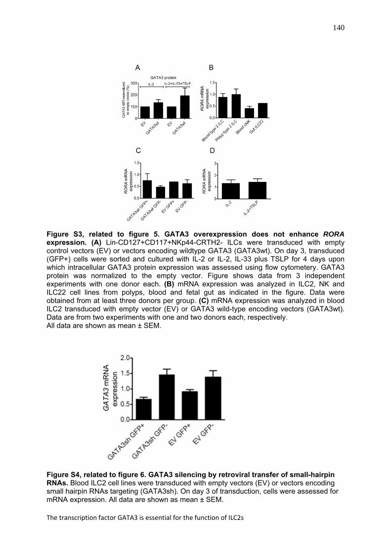
Figure S3, related to figure 5. GATA3 overexpression does not enhance RORA expression. (A) Lin-CD127+CD117+NKp44-CRTH2- ILCs were transduced with empty control vectors (EV) or vectors encoding wildtype GATA3 (GATA3wt). On day 3, transduced (GFP+) cells were sorted and cultured with IL-2 or IL-2, IL-33 plus TSLP for 4 days upon which intracellular GATA3 protein expression was assessed using flow cytometery. GATA3 protein was normalized to the empty vector. Figure shows data from 3 independent experiments with one donor each. (B) mRNA expression was analyzed in ILC2, NK and ILC22 cell lines from polyps, blood and fetal gut as indicated in the figure. Data were obtained from at least three donors per group. (C) mRNA expression was analyzed in blood ILC2 transduced with empty vector (EV) or GATA3 wild-type encoding vectors (GATA3wt). Data are from two experiments with one and two donors each, respectively. All data are shown as mean ± SEM.
Figure S4, related to figure 6. GATA3 silencing by retroviral transfer of small-hairpin RNAs. Blood ILC2 cell lines were transduced with empty vectors (EV) or vectors encoding small hairpin RNAs targeting (GATA3sh). On day 3 of transduction, cells were assessed for mRNA expression. All data are shown as mean ± SEM.

141
Chapter 7
Cytokines > 100 pg/mL
Cytokines < 100 pg/mL
GM-CSF sCD40L IL-4 Eotaxin IL-5 GRO IL-6 IFN-γ IL-8 IL-1β IL-9 TNFα
IL-13 IL-10 IP10 IL-12p40
IL-12p70 IL-15 IL-17a MCP1 MCP3 MIP1a MIP1b RANTES VEGF TGFα
Table S1. Cytokines analyzed in supernatants of freshly isolated blood or nasal polyp ILC2s. Fresh ILC2s (2-3000 cells/culture) were cultured for 4 days with IL-2, IL-2 plus IL-33 or IL-2 plus TSLP. The table shows the cytokines produced at concentrations above 100 pg/mL (left column) or less than 100 pg/mL (right column) with any stimuli. Table S2. Sequences of real-time PCR primers designed in-house.
mRNA primer sequence
GAPHD F GAA GGT GAA GGT CGG AGT C
R GAA GAT GGT GAT GGG ATT TC
IL33 F GCC TGT CAA CAG CAG TCT ACT G
R TGT CTT AGA GAA GCA AGA TAC
TSLP F TAT CTG GTG CCC AGG CTA TTC G
R TGA AGC GAC GCC ACA ATC CTT G
TSLPR F GAG TGG CAG TCC AAA CAG GAA
R ACA TCC TCC ATA GCC TTC ACC
GATA3 F ACC ACA ACC ACA CTC TGG AGG A
R TCG GTT TCT GGT CTG GAT GCC T
RORA F ACA AGC AGC GGG AGG TGA TGT
R TGA GAG TCA AAG GCA CGG C
Table S2. Sequences of real-time PCR primers designed in-house.


![GATA3 GATA Binding Protein 3 [Homo Sapiens (Human)] - Gene - NCBI](https://img.pdfslide.net/doc/110x75/55cf8ea8550346703b94582f/gata3-gata-binding-protein-3-homo-sapiens-human-gene-ncbi.jpg)


