Embed Size (px)
Citation preview

RibesLunaRos.indb V 18.03.2008 12:48:44

Ribes · Luna · Ros
Learning Diagnostic Imaging
RibesLunaRos.indb I 18.03.2008 12:48:40
R. Ribes · A. Luna · P. R. Ros (Eds.)
LearningDiagnostic Imaging100 Essential Cases
With Contributions by
L. Alcala Mata · M. Alvarez Benito · F. Bravo-Rodriguez · J. Camps Herrero P. Daltro · R. Diaz-Aguilera · S. Espejo · E. Feliu · L. C. Hygino Cruz Jr.J. Lopez Mora · A. Luna Alcala · S. Mejia · R. do A. Nogueira · M. T. C. PortoM. Potolicchio · A. C. Rebollo Aguire · R. Ribes · S. E. Rossi · P. SeguiJ. A. Vallejo Casas · J. C. Vilanova
With 347 Figures in 397 Separate Illustrations, 50 in Color
123
RibesLunaRos.indb III 18.03.2008 12:48:44
Ramon Ribes, MD, PhDInterventional Radiology and MR UnitsReina Sofi a University Hospital14005 CordobaSpain
Antonio Luna, MDChief, MR UnitClinica Las NievesSercosa23007 JaenSpain
Pablo R. Ros, MD, MPHChairman, Department of RadiologySant Pau HospitalAutonomous University08025 BarcelonaSpainandProfessor, Department of RadiologyHarvard Medical SchoolBrigham & Women’s Hospital75 Francis St.Boston MA 02115USA
ISBN 978-3-540-71206-0 e-ISBN 978-3-540-71207-7
DOI 10.1007/978-3-540-71207-7
Library of Congress Control Number: 2007943071
© 2008, Springer-Verlag Berlin Heidelberg
This work is subject to copyright. All rights are reserved, whether the whole or part of the material is concerned, specifi cally the rights of translation, reprinting, reuse of illustrations, recitations, broadcasting, reproduction on microfi lm or in any other way, and storage in data banks. Duplication of this publication or parts thereof is per mitted only under the provisions of the German Copyright Law of September 9, 1965, in its current version, and permis-sion for use must always be obtained from Springer-Verlag. Violations are liable for prosecution under the German Copyright Law.
The use of general descriptive names, trademarks, etc. in this publication does not imply, even in the absence of a specifi c statement, that such names are exempt from the relevant protective laws and regulations and therefore free for general use.
Product liability: The publishers cannot guarantee the accuracy of any information about dosage and application contained in this book. In every individual case the user must check such information by consulting the relevant literature.
Cover design: Frido Steinen-Broo, eStudio Calamar, SpainLayout: Verlagsservice Teichmann, 69256 Mauer, Germany
Printed on acid-free paper
9 8 7 6 5 4 3 2 1
springer.com
RibesLunaRos.indb IV 18.03.2008 12:48:44
To my daughters
Rosario and Reyes Ramon Ribes
To all
my teachersespecially to the most motivating ones
Antonio Luna
To all
my teachers and students,
for enriching me with energy and
enthusiasm. Among my teachers,
a special mention to
my parents
(for my father, in memoriam),
my brother, my children and my wife Pablo R. Ros
RibesLunaRos.indb V 18.03.2008 12:48:44

RibesLunaRos.indb V 18.03.2008 12:48:44
Preface
In my fi rst days as a Radiology resident I remember myself in dire need of a book in
which I could begin to study the specialty. In those days I was thirsty for radiologi-
cal knowledge and was recommended a classic manual on conventional chest X-ray
which was the very same the oldest radiologist of the staff had studied when he was
a beginner. Unfortunately the book I was recommended covered less than 5% of the
specialty as it is conceived nowadays.
“Learning Diagnostic Imaging. A Teaching File” is intended to provide medical
students, residents of Radiology, and anybody else beginning to be involved in the
radiological world, with a useful tool that gives them a quick and comprehensive
overview of Radiology. With this book, written in a user-friendly format, Radiology
residents, nurses, technicians, and medical students would see their fi rst radiologi-
cal images in a sort of introduction of what will become their professional activities
in the rest of their lives.
One of the main problems that Radiology residents face throughout their resi-
dency is that in rounds and clinical sessions they receive information they cannot
apprehend because they lack the essential background needed to integrate what
they are taught. For example, when, in a radiological session, a resident is looking
at an angiogram and has no prior interventional radiology education, he/she in-
evitably misses the opportunity of learning by being exposed to a great deal of in-
formation without the necessary tools to assimilate it. Whenever a mammogram
is shown at the radiological session, a fi rst year resident, who has no prior training
on breast imaging, knowing beforehand that he is not supposed to be asked about
it, loses interest in the subject and it is likely that his mind drifts away until the
next case is shown. In clinical sessions, the level of concentration of residents is
optimal only in the cases they have prior education on.
With this book, residents independently of their year of residency, would count
on the foundations of the specialty as a whole and optimize their time in rounds and
clinical sessions. Although initially aimed at Radiology residents, the book would
be useful, on the one hand, for medical students, Radiology nurses and technicians
and, on the other hand, for senior radiologists and residents and referring physi-
cians of other specialties. As a way of example, the breast imaging senior radiologist
would have a basic overview of cardiac imaging at his disposal in just a few pages
that otherwise would be impossible for him because no mammographer is supposed
to be interested in classic cardiac imaging books as no cardiovascular radiologist
has any interest in breast imaging manuals.
RibesLunaRos.indb VII 18.03.2008 12:48:44

VIII Preface
The scarcity of books of this sort is easily understandable taking into account
that Radiology is divided up in many subspecialties that constitute a universe of
their own. To convey an overall view of the specialty is extremely diffi cult nowadays
and will be even more diffi cult in the future because, due to the fragmentation of the
specialty, the old-days only Radiological session, which was attended by the whole
Department, has been replaced by a myriad of subspecialty sessions that, by defi ni-
tion, cannot be attended by the entire Radiology Department.
Cordoba, November 22, 2007 Ramon Ribes
RibesLunaRos.indb VIII 18.03.2008 12:48:44
Contents IX
Contents
1 Breast Imaging Marina Alvarez Benito and Julia Camps Herrero
Melcior Sentis Criville and Maria Martinez Galvez (Contributors) . . . . . . . . . . . . . . . . 1
Case 1 Ductal Carcinoma in Situ . . . . . . . . . . . . . . . . . . . . . . . . . . . . . . . . . . . . . 2 Case 2 Invasive Ductal Carcinoma with MRI Staging . . . . . . . . . . . . . . . . . . . 4 Case 3 Breast Implant Rupture . . . . . . . . . . . . . . . . . . . . . . . . . . . . . . . . . . . . . . 6 Case 4 Papillary Lesions . . . . . . . . . . . . . . . . . . . . . . . . . . . . . . . . . . . . . . . . . . . . 8 Case 5 Microcalcifi cations . . . . . . . . . . . . . . . . . . . . . . . . . . . . . . . . . . . . . . . . . . 10 Case 6 Architectural Distortion . . . . . . . . . . . . . . . . . . . . . . . . . . . . . . . . . . . . . 12 Case 7 Breast Cancer in Men . . . . . . . . . . . . . . . . . . . . . . . . . . . . . . . . . . . . . . . . 14 Case 8 Breast Cancer and Simple Cyst . . . . . . . . . . . . . . . . . . . . . . . . . . . . . . . . 16 Case 9 Breast Metastases . . . . . . . . . . . . . . . . . . . . . . . . . . . . . . . . . . . . . . . . . . . 18 Case 10 Locorregional Estadifi cation . . . . . . . . . . . . . . . . . . . . . . . . . . . . . . . . . . 20
Further Readings . . . . . . . . . . . . . . . . . . . . . . . . . . . . . . . . . . . . . . . . . . . . . . . . . . . . 22 2 Cardiovascular Imaging Eliosa Feliu, Ramon Ribes, and Sergio Mejia . . . . . . . . . . . . . . . . . . . . . . . . . . . . . 27
Case 1 Acute Myocardial Infarction . . . . . . . . . . . . . . . . . . . . . . . . . . . . . . . . . . 28 Case 2 Hypertrophic Cardiomyopathy . . . . . . . . . . . . . . . . . . . . . . . . . . . . . . . 30 Case 3 Arrhythmogenic Right Ventricular Dysplasia . . . . . . . . . . . . . . . . . . . 32 Case 4 Myocarditis. . . . . . . . . . . . . . . . . . . . . . . . . . . . . . . . . . . . . . . . . . . . . . . . . 34 Case 5 Aortic Coarctation . . . . . . . . . . . . . . . . . . . . . . . . . . . . . . . . . . . . . . . . . . 36 Case 6 Constrictive Pericarditis . . . . . . . . . . . . . . . . . . . . . . . . . . . . . . . . . . . . . 38 Case 7 Restrictive Cardiomyopathy . . . . . . . . . . . . . . . . . . . . . . . . . . . . . . . . . . 40 Case 8 Congenital Anomalies of Coronary Arteries . . . . . . . . . . . . . . . . . . . . 42 Case 9 Coronary Artery S Stenoses . . . . . . . . . . . . . . . . . . . . . . . . . . . . . . . . . . . 44 Case 10 Aortocoronary Bypass . . . . . . . . . . . . . . . . . . . . . . . . . . . . . . . . . . . . . . . 46
Further Readings . . . . . . . . . . . . . . . . . . . . . . . . . . . . . . . . . . . . . . . . . . . . . . . . . . . . 48
3 Thoracic Radiology Santiago E. Rossi and Joaquina Lopez Mora . . . . . . . . . . . . . . . . . . . . . . . . . . . . . . 53
Case 1 Usual Interstitial Pneumonia (UIP) . . . . . . . . . . . . . . . . . . . . . . . . . . . . 54 Case 2 Lymphangitic Carcinomatosis . . . . . . . . . . . . . . . . . . . . . . . . . . . . . . . . 56 Case 3 Thrombotic Pulmonary Embolism . . . . . . . . . . . . . . . . . . . . . . . . . . . . 58 Case 4 Metastases . . . . . . . . . . . . . . . . . . . . . . . . . . . . . . . . . . . . . . . . . . . . . . . . . . 60 Case 5 Thoracic Neurofi broma . . . . . . . . . . . . . . . . . . . . . . . . . . . . . . . . . . . . . . 62 Case 6 Hodgkin Lymphoma . . . . . . . . . . . . . . . . . . . . . . . . . . . . . . . . . . . . . . . . . 64 Case 7 Lung Cancer . . . . . . . . . . . . . . . . . . . . . . . . . . . . . . . . . . . . . . . . . . . . . . . . 66 Case 8 Bronchiectasis . . . . . . . . . . . . . . . . . . . . . . . . . . . . . . . . . . . . . . . . . . . . . . 68 Case 8 Malignant Mesothelioma . . . . . . . . . . . . . . . . . . . . . . . . . . . . . . . . . . . . . 70 Case 10 Tuberculosis . . . . . . . . . . . . . . . . . . . . . . . . . . . . . . . . . . . . . . . . . . . . . . . . 72
Further Readings . . . . . . . . . . . . . . . . . . . . . . . . . . . . . . . . . . . . . . . . . . . . . . . . . . . . 74
RibesLunaRos.indb IX 18.03.2008 12:48:44
X Contents
4 Gastrointestinal System and Disorders of the Liver, Pancreas, Spleen, and Biliary System Antonio Luna Alcala, Lidia Alcala Mata, and Ramon Ribes . . . . . . . . . . . . . 77
Case 1 Acute Cholecystitis and Choledocholithiasis with Secondary Pancreatitisand and Hepatic Abscess . . . . . . . . . . . . . . . . 78 Case 2 Cirrhosis with Portal Hypertension . . . . . . . . . . . . . . . . . . . . . . . . . . . . 80 Case 3 Hepatic Metastases . . . . . . . . . . . . . . . . . . . . . . . . . . . . . . . . . . . . . . . . . . 82 Case 4 Pancreatic Insulinoma . . . . . . . . . . . . . . . . . . . . . . . . . . . . . . . . . . . . . . . 84 Case 5 Splenic Lymphoma . . . . . . . . . . . . . . . . . . . . . . . . . . . . . . . . . . . . . . . . . . 86 Case 6 Crohn’s Disease . . . . . . . . . . . . . . . . . . . . . . . . . . . . . . . . . . . . . . . . . . . . . 88 Case 7 Complicated Diverticulitis . . . . . . . . . . . . . . . . . . . . . . . . . . . . . . . . . . . 90 Case 8 Hiatal Hernia . . . . . . . . . . . . . . . . . . . . . . . . . . . . . . . . . . . . . . . . . . . . . . . 92 Case 9 Primary Small Bowel Lymphoma . . . . . . . . . . . . . . . . . . . . . . . . . . . . . . 94 Case 10 Duodenal Gallstone Ileus . . . . . . . . . . . . . . . . . . . . . . . . . . . . . . . . . . . . . 96
Further Readings . . . . . . . . . . . . . . . . . . . . . . . . . . . . . . . . . . . . . . . . . . . . . . . . . . . . 98
5 Genitourinary, Gynecological, and Obstetrical Imaging Antonio Luna Alcala, Marcelo Potolicchio, and Lidia Alcala Mata . . . . 103
Case 1 Adrenal Myelolipoma . . . . . . . . . . . . . . . . . . . . . . . . . . . . . . . . . . . . . . . . 104 Case 2 Complicated Cortical Renal Cyst . . . . . . . . . . . . . . . . . . . . . . . . . . . . . . 106 Case 3 Prostate Cancer . . . . . . . . . . . . . . . . . . . . . . . . . . . . . . . . . . . . . . . . . . . . . 108 Case 4 Retroperitoneal Liposarcoma . . . . . . . . . . . . . . . . . . . . . . . . . . . . . . . . . 110 Case 5 Acute Obstruction by Ureteral Lithiasis . . . . . . . . . . . . . . . . . . . . . . . . 112 Case 6 Adenomyosis . . . . . . . . . . . . . . . . . . . . . . . . . . . . . . . . . . . . . . . . . . . . . . . 114 Case 7 Cervical Cancer . . . . . . . . . . . . . . . . . . . . . . . . . . . . . . . . . . . . . . . . . . . . . 116 Case 8 Endometrial Polyp . . . . . . . . . . . . . . . . . . . . . . . . . . . . . . . . . . . . . . . . . . 118 Case 9 Fetal Lissencephaly . . . . . . . . . . . . . . . . . . . . . . . . . . . . . . . . . . . . . . . . . . 120 Case 10 Ovarian Serous Cystoadenocarcinoma . . . . . . . . . . . . . . . . . . . . . . . . . 122
Further Readings . . . . . . . . . . . . . . . . . . . . . . . . . . . . . . . . . . . . . . . . . . . . . . . . . . . . 124
6 Musculoskeletal Imaging Joan C. Vilanova and Ramon Ribes
Sandra Baleato (Contributor) . . . . . . . . . . . . . . . . . . . . . . . . . . . . . . . . . . . . . . . . . . . . . . . . . 127
Case 1 Osteomyelitis . . . . . . . . . . . . . . . . . . . . . . . . . . . . . . . . . . . . . . . . . . . . . . . 128 Case 2 Acute Meniscal and Ligament Tears of the Knee . . . . . . . . . . . . . . . . . 130 Case 3 Radius Fracture . . . . . . . . . . . . . . . . . . . . . . . . . . . . . . . . . . . . . . . . . . . . . 132 Case 4 Ewing Sarcoma . . . . . . . . . . . . . . . . . . . . . . . . . . . . . . . . . . . . . . . . . . . . . 134 Case 5 Schwannoma . . . . . . . . . . . . . . . . . . . . . . . . . . . . . . . . . . . . . . . . . . . . . . . 136 Case 6 Soft-Tissue Liposarcoma . . . . . . . . . . . . . . . . . . . . . . . . . . . . . . . . . . . . . 138 Case 7 Chordoma . . . . . . . . . . . . . . . . . . . . . . . . . . . . . . . . . . . . . . . . . . . . . . . . . . 140 Case 8 Osteonecrosis . . . . . . . . . . . . . . . . . . . . . . . . . . . . . . . . . . . . . . . . . . . . . . . 142 Case 9 Bone Lymphoma . . . . . . . . . . . . . . . . . . . . . . . . . . . . . . . . . . . . . . . . . . . . 144 Case 10 Enchondroma . . . . . . . . . . . . . . . . . . . . . . . . . . . . . . . . . . . . . . . . . . . . . . . 146
Further Readings . . . . . . . . . . . . . . . . . . . . . . . . . . . . . . . . . . . . . . . . . . . . . . . . . . . . 148
7 Neuroimaging and Head and Neck Imaging F. Bravo-Rodriguez and Rocio Diaz-Aguilera . . . . . . . . . . . . . . . . . . . . . . . . . . . . 153
Case 1 Meningioma . . . . . . . . . . . . . . . . . . . . . . . . . . . . . . . . . . . . . . . . . . . . . . . . 154 Case 2 Multiple Sclerosis . . . . . . . . . . . . . . . . . . . . . . . . . . . . . . . . . . . . . . . . . . . 156 Case 3 Cerebral Abscess . . . . . . . . . . . . . . . . . . . . . . . . . . . . . . . . . . . . . . . . . . . . 158 Case 4 Acute Head Trauma . . . . . . . . . . . . . . . . . . . . . . . . . . . . . . . . . . . . . . . . . 160 Case 5 Stroke . . . . . . . . . . . . . . . . . . . . . . . . . . . . . . . . . . . . . . . . . . . . . . . . . . . . . . 162
RibesLunaRos.indb X 18.03.2008 12:48:44
Contents XI
Case 6 Subarachnoid Hemorrhage . . . . . . . . . . . . . . . . . . . . . . . . . . . . . . . . . . . 164 Case 7 Cerebral Venous Thrombosis . . . . . . . . . . . . . . . . . . . . . . . . . . . . . . . . . 166 Case 8 Cavernous Angioma . . . . . . . . . . . . . . . . . . . . . . . . . . . . . . . . . . . . . . . . . 168 Case 9 Epidermoid Cyst . . . . . . . . . . . . . . . . . . . . . . . . . . . . . . . . . . . . . . . . . . . . 170 Case 10 Juvenile Nasopharyngeal Angiofi broma . . . . . . . . . . . . . . . . . . . . . . . . 172
Further Readings . . . . . . . . . . . . . . . . . . . . . . . . . . . . . . . . . . . . . . . . . . . . . . . . . . . . 174
8 Nuclear Medicine Juan Antonio Vallejo Casas and Angel C. Rebollo Aguirre Luisa Maria Mena Bares (Contributor) . . . . . . . . . . . . . . . . . . . . . . . . . . . . . . . . . . . . . . . . . 179
Case 1 Brain Death . . . . . . . . . . . . . . . . . . . . . . . . . . . . . . . . . . . . . . . . . . . . . . . . 180 Case 2 Gastrointestinal Bleeding . . . . . . . . . . . . . . . . . . . . . . . . . . . . . . . . . . . . 182 Case 3 Infl ammatory Bowel Disease . . . . . . . . . . . . . . . . . . . . . . . . . . . . . . . . . . 184 Case 4 Movement Disorders . . . . . . . . . . . . . . . . . . . . . . . . . . . . . . . . . . . . . . . . . 186 Case 5 Obstructive Uropathy . . . . . . . . . . . . . . . . . . . . . . . . . . . . . . . . . . . . . . . . 188 Case 6 Thromboembolic Pulmonary Disease . . . . . . . . . . . . . . . . . . . . . . . . . . 190 Case 7 Coronary Disease . . . . . . . . . . . . . . . . . . . . . . . . . . . . . . . . . . . . . . . . . . . 192 Case 8 Parathyroid Adenoma . . . . . . . . . . . . . . . . . . . . . . . . . . . . . . . . . . . . . . . 194 Case 9 Pulmonary Solitary Node. PET-CT Evaluation . . . . . . . . . . . . . . . . . . 196 Case 10 Lymphoma. PET-CT Evaluation . . . . . . . . . . . . . . . . . . . . . . . . . . . . . . . 198
Further Readings . . . . . . . . . . . . . . . . . . . . . . . . . . . . . . . . . . . . . . . . . . . . . . . . . . . . 200
9 Pediatric Radiology Pedro Daltro, L. Celso Hygino Cruz Jr., Renata do A. Nogueira, and Miriam T. C. Porto . . . . . . . . . . . . . . . . . . . . . . . . . . . . . . . . . . . . . . . . . . . . . . . . . . . 205
Case 1 Hip Dysplasia . . . . . . . . . . . . . . . . . . . . . . . . . . . . . . . . . . . . . . . . . . . . . . . 206 Case 2 Hypertrophic Pyloric Stenosis . . . . . . . . . . . . . . . . . . . . . . . . . . . . . . . . 208 Case 3 Intussusception . . . . . . . . . . . . . . . . . . . . . . . . . . . . . . . . . . . . . . . . . . . . . 210 Case 4 Round Pneumonia . . . . . . . . . . . . . . . . . . . . . . . . . . . . . . . . . . . . . . . . . . . 212 Case 5 Congenital Cystic Adenomatoid Malformation . . . . . . . . . . . . . . . . . . 214 Case 6 Neuroblastoma . . . . . . . . . . . . . . . . . . . . . . . . . . . . . . . . . . . . . . . . . . . . . . 216 Case 7 Wilms Tumor . . . . . . . . . . . . . . . . . . . . . . . . . . . . . . . . . . . . . . . . . . . . . . . 218 Case 8 Medulloblastoma . . . . . . . . . . . . . . . . . . . . . . . . . . . . . . . . . . . . . . . . . . . . 220 Case 9 Corpus Callosum Agenesis . . . . . . . . . . . . . . . . . . . . . . . . . . . . . . . . . . . 222 Case 10 Tethered Cord . . . . . . . . . . . . . . . . . . . . . . . . . . . . . . . . . . . . . . . . . . . . . . 224
Further Readings . . . . . . . . . . . . . . . . . . . . . . . . . . . . . . . . . . . . . . . . . . . . . . . . . . . . 226
10 Ultrasound Imaging Pedro Segui and Simona Espejo . . . . . . . . . . . . . . . . . . . . . . . . . . . . . . . . . . . . . . . . . . . 231
Case 1 Acute Cholecystitis . . . . . . . . . . . . . . . . . . . . . . . . . . . . . . . . . . . . . . . . . . 232 Case 2 Appendicitis . . . . . . . . . . . . . . . . . . . . . . . . . . . . . . . . . . . . . . . . . . . . . . . . 234 Case 3 Acute Colonic Diverticulitis . . . . . . . . . . . . . . . . . . . . . . . . . . . . . . . . . . 236 Case 4 Epididymoorchitis . . . . . . . . . . . . . . . . . . . . . . . . . . . . . . . . . . . . . . . . . . 238 Case 5 Deep Venous Thrombosis . . . . . . . . . . . . . . . . . . . . . . . . . . . . . . . . . . . . 240 Case 6 Hodgkin’s Lymphoma . . . . . . . . . . . . . . . . . . . . . . . . . . . . . . . . . . . . . . . 242 Case 7 Abdominal Wall Endometriosis . . . . . . . . . . . . . . . . . . . . . . . . . . . . . . . 244 Case 8 Carotid Artery Stenosis . . . . . . . . . . . . . . . . . . . . . . . . . . . . . . . . . . . . . . 246 Case 9 Carotid Artery Dissection . . . . . . . . . . . . . . . . . . . . . . . . . . . . . . . . . . . . 248 Case 10 Choroidal Melanoma . . . . . . . . . . . . . . . . . . . . . . . . . . . . . . . . . . . . . . . . 250
Further Readings . . . . . . . . . . . . . . . . . . . . . . . . . . . . . . . . . . . . . . . . . . . . . . . . . . . . 252
RibesLunaRos.indb XI 18.03.2008 12:48:45

RibesLunaRos.indb V 18.03.2008 12:48:44
Contributors XIII
Contributing Authors
Lidia Alcala MataRadiology ResidentDepartment of RadiologyCiudad de Jaen HospitalJaenSpain
Marina Alvarez BenitoChief, Breast UnitReina Sofi a University HospitalCordobaSpain
F. Bravo-RodriguezCT and MR UnitsReina Sofi a University HospitalCordobaSpain
Julia Camps HerreroChief, Radiology DepartmentHospital de la RiberaAlzira, ValenciaSpain
Pedro DaltroCDPIPediatric RadiologyRio de JaneiroBrazil
Rocio Diaz-AguileraRadiology DepartmentHospital Alto GuadalquivirJaenSpain
Simona EspejoCT and MR UnitRadiology DepartmentReina Sofi a University HospitalCordobaSpain
Eloisa FeliuMR (Magnetic Resonance)InscannerAlicanteSpain
L. Celso Hygino Cruz Jr.NeuroradiologyCDPIRio de JaneiroBrazil
Antonio Luna AlcalaChief, MR (Magnetic Resonance)Clinica Las NievesSercosaJaenSpain
Sergio MejiaChief, Cardiology DepartmentXanit International HospitalBenalmadenaMalagaSpain
Joaquina Lopez MoraCentro de Diagnostico Dr. E. RossiAffi liated to Buenos Aires UniversityBuenos AiresArgentina
RibesLunaRos.indb XIII 18.03.2008 12:48:45
XIV Contributors
Renata do A. NogueiraPediatric Radiology CDPIRio de JaneiroBrazil
Miriam T. C. PortoPediatric Radiology CDPIRio de JaneiroBrazil
Marcelo PotolicchioUltrasound SectionDADISACadizSpain
Angel C. Rebollo AguirreNuclear Medicine DepartmentVirgen de las Nieves HospitalGranadaSpain
Ramon RibesInterventional Radiology and MR UnitsReina Sofi a University HospitalCordobaSpain
Santiago E. RossiCentro de Diagnostico Dr. E. RossiAffi liated to Buenos Aires UniversityBuenos AiresArgentina
Pedro SeguiUltrasound UnitRadiology DepartmentReina Sofi a University HospitalCordobaSpain
Juan Antonio Vallejo CasasDepartment of Nuclear MedicineReina Sofi a HospitalCordobaSpain
Joan C. VilanovaDepartment of Magnetic ResonanceClinica GironaGironaSpain
Contributors
Sandra BaleatoDepartment of Magnetic ResonanceClinica GironaGironaSpain
Melcior Sentis CrivilleBreast and Gynecology UnitsFundacio Parc Tauli, SabadellBarcelonaSpain
Maria Martinez GalvezChief, Radiology DepartmentHospital de TorreviejaAlicanteSpain
Luisa Maria Mena BaresResident, Nuclear Medicine DepartmentReina Sofi a HospitalCordobaSpain
RibesLunaRos.indb XIV 18.03.2008 12:48:45

RibesLunaRos.indb V 18.03.2008 12:48:44
Nuclear Medicine
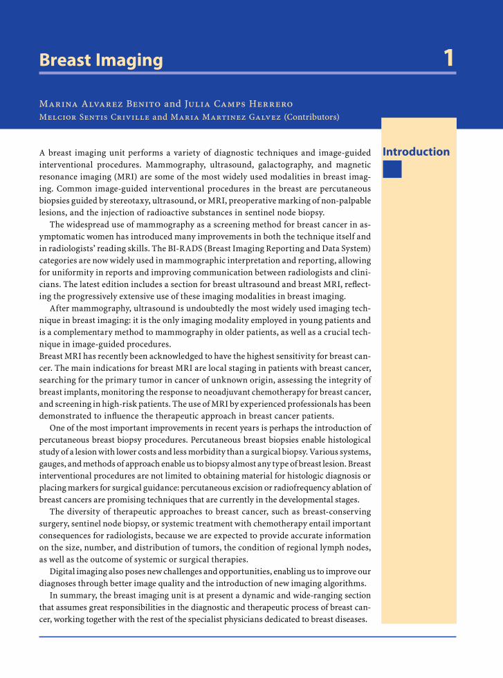
Breast Imaging 1
Marina Alvarez Benito and Julia Camps HerreroMelcior Sentis Criville and Maria Martinez Galvez (Contributors)
IntroductionA breast imaging unit performs a variety of diagnostic techniques and image-guided interventional procedures. Mammography, ultrasound, galactography, and magnetic resonance imaging (MRI) are some of the most widely used modalities in breast imag-ing. Common image-guided interventional procedures in the breast are percutaneous biopsies guided by stereotaxy, ultrasound, or MRI, preoperative marking of non-palpable lesions, and the injection of radioactive substances in sentinel node biopsy.
The widespread use of mammography as a screening method for breast cancer in as-ymptomatic women has introduced many improvements in both the technique itself and in radiologists’ reading skills. The BI-RADS (Breast Imaging Reporting and Data System) categories are now widely used in mammographic interpretation and reporting, allowing for uniformity in reports and improving communication between radiologists and clini-cians. The latest edition includes a section for breast ultrasound and breast MRI, refl ect-ing the progressively extensive use of these imaging modalities in breast imaging.
After mammography, ultrasound is undoubtedly the most widely used imaging tech-nique in breast imaging: it is the only imaging modality employed in young patients and is a complementary method to mammography in older patients, as well as a crucial tech-nique in image-guided procedures.Breast MRI has recently been acknowledged to have the highest sensitivity for breast can-cer. The main indications for breast MRI are local staging in patients with breast cancer, searching for the primary tumor in cancer of unknown origin, assessing the integrity of breast implants, monitoring the response to neoadjuvant chemotherapy for breast cancer, and screening in high-risk patients. The use of MRI by experienced professionals has been demonstrated to infl uence the therapeutic approach in breast cancer patients.
One of the most important improvements in recent years is perhaps the introduction of percutaneous breast biopsy procedures. Percutaneous breast biopsies enable histological study of a lesion with lower costs and less morbidity than a surgical biopsy. Various systems, gauges, and methods of approach enable us to biopsy almost any type of breast lesion. Breast interventional procedures are not limited to obtaining material for histologic diagnosis or placing markers for surgical guidance: percutaneous excision or radiofrequency ablation of breast cancers are promising techniques that are currently in the developmental stages.
The diversity of therapeutic approaches to breast cancer, such as breast-conserving surgery, sentinel node biopsy, or systemic treatment with chemotherapy entail important consequences for radiologists, because we are expected to provide accurate information on the size, number, and distribution of tumors, the condition of regional lymph nodes, as well as the outcome of systemic or surgical therapies.
Digital imaging also poses new challenges and opportunities, enabling us to improve our diagnoses through better image quality and the introduction of new imaging algorithms.
In summary, the breast imaging unit is at present a dynamic and wide-ranging section that assumes great responsibilities in the diagnostic and therapeutic process of breast can-cer, working together with the rest of the specialist physicians dedicated to breast diseases.
RibesLunaRos.indb 1 18.03.2008 12:48:45
2 M. Alvarez Benito and J. Camps Herrero
Case 1
Ductal Carcinoma in Situ
Fig. 1.1.1
Fig. 1.1.2
Fig. 1.1.3
Fig. 1.1.4
Fig. 1.1.5
RibesLunaRos.indb 2 18.03.2008 12:48:45
Breast Imaging 3
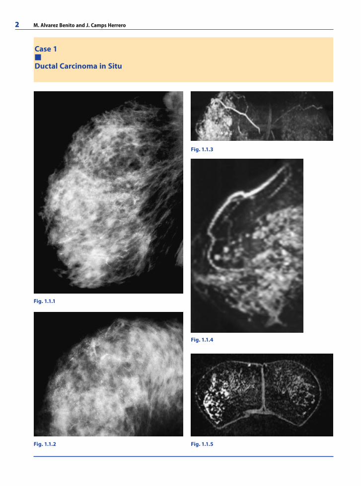
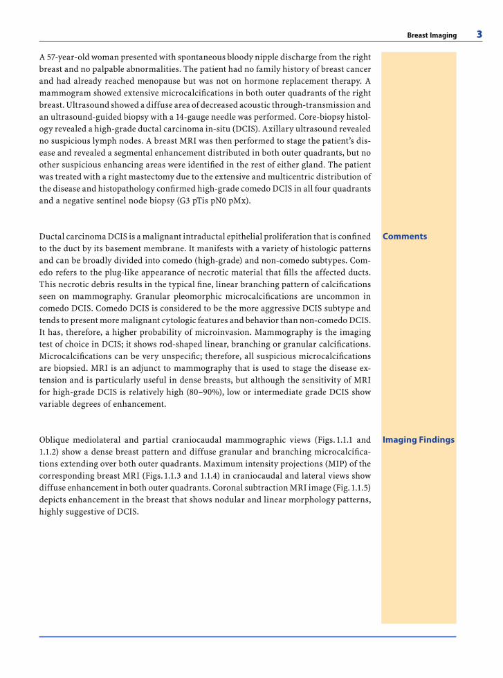
A 57-year-old woman presented with spontaneous bloody nipple discharge from the right breast and no palpable abnormalities. The patient had no family history of breast cancer and had already reached menopause but was not on hormone replacement therapy. A mammogram showed extensive microcalcifi cations in both outer quadrants of the right breast. Ultrasound showed a diffuse area of decreased acoustic through-transmission and an ultrasound-guided biopsy with a 14-gauge needle was performed. Core-biopsy histol-ogy revealed a high-grade ductal carcinoma in-situ (DCIS). Axillary ultrasound revealed no suspicious lymph nodes. A breast MRI was then performed to stage the patient’s dis-ease and revealed a segmental enhancement distributed in both outer quadrants, but no other suspicious enhancing areas were identifi ed in the rest of either gland. The patient was treated with a right mastectomy due to the extensive and multicentric distribution of the disease and histopathology confi rmed high-grade comedo DCIS in all four quadrants and a negative sentinel node biopsy (G3 pTis pN0 pMx).
CommentsDuctal carcinoma DCIS is a malignant intraductal epithelial proliferation that is confi ned to the duct by its basement membrane. It manifests with a variety of histologic patterns and can be broadly divided into comedo (high-grade) and non-comedo subtypes. Com-edo refers to the plug-like appearance of necrotic material that fi lls the affected ducts. This necrotic debris results in the typical fi ne, linear branching pattern of calcifi cations seen on mammography. Granular pleomorphic microcalcifi cations are uncommon in comedo DCIS. Comedo DCIS is considered to be the more aggressive DCIS subtype and tends to present more malignant cytologic features and behavior than non-comedo DCIS. It has, therefore, a higher probability of microinvasion. Mammography is the imaging test of choice in DCIS; it shows rod-shaped linear, branching or granular calcifi cations. Microcalcifi cations can be very unspecifi c; therefore, all suspicious microcalcifi cations are biopsied. MRI is an adjunct to mammography that is used to stage the disease ex-tension and is particularly useful in dense breasts, but although the sensitivity of MRI for high-grade DCIS is relatively high (80–90%), low or intermediate grade DCIS show variable degrees of enhancement.
Imaging FindingsOblique mediolateral and partial craniocaudal mammographic views (Figs. 1.1.1 and 1.1.2) show a dense breast pattern and diffuse granular and branching microcalcifi ca-tions extending over both outer quadrants. Maximum intensity projections (MIP) of the corresponding breast MRI (Figs. 1.1.3 and 1.1.4) in craniocaudal and lateral views show diffuse enhancement in both outer quadrants. Coronal subtraction MRI image (Fig. 1.1.5) depicts enhancement in the breast that shows nodular and linear morphology patterns, highly suggestive of DCIS.
RibesLunaRos.indb 3 18.03.2008 12:48:47
4 M. Alvarez Benito and J. Camps Herrero
Case 2
Invasive Ductal Carcinoma with MRI Staging
Fig. 1.2.1 Fig. 1.2.2
Fig. 1.2.3 Fig. 1.2.4
RibesLunaRos.indb 4 18.03.2008 12:48:47
Breast Imaging 5
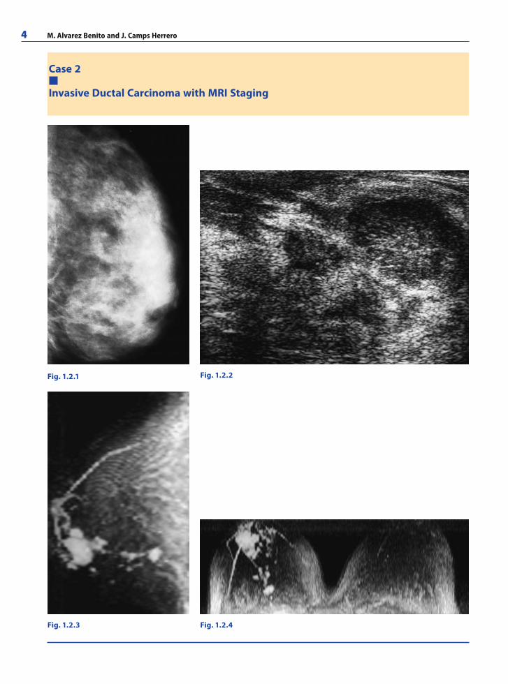
A 32-year-old woman without a family history of breast cancer presented with a pal-pable lump in the lower-outer quadrant of the left breast. Four months earlier, breast ultrasound diagnosed a fi broadenoma in the same location but she had noticed that the palpable lump had become harder and had increased in size. Ultrasound was performed fi rst and showed two ill-defi ned microlobulated solid masses that were biopsied with a 14-gauge needle because of the morphology and the rapid growth of the palpable lump. Axillary ultrasound showed no suspicious lymph nodes in the left axilla. Mammography showed dense breasts but no other remarkable fi ndings. Core-biopsy histopathology showed an invasive ductal carcinoma (IDC). Breast MRI was performed to stage the disease and a multifocal distribution in the union of both lower quadrants with sparing of the nipple-areola complex was found. The patient underwent breast conserving surgery. Histological study of the surgical specimen confi rmed IDC, not otherwise specifi ed, grade II (pT2, pN0, pMx), and all the margins were free of tumor within 10 mm.
CommentsInvasive (or infi ltrating) breast cancer can be broadly divided into ductal and lobular subtypes. Invasive ductal carcinoma accounts for the majority (90%) of invasive breast cancers and can be further divided into those “not otherwise specifi ed” (NOS) and “special type” tumors. NOS breast carcinomas account for 50–75% of all invasive breast cancers. Their most distinctive appearance in mammography is that of a spiculated mass, although in women with dense breasts the margins of the tumor may be diffi cult to identify. Mammography has a 85–90% sensitivity for breast cancer; however, in dense breasts this fi gure drops to 40–60%. In these cases, palpation and ultrasound play a major role. At ultrasound, IDC shows decreased through-transmission and irregular margins. Breast MRI is increasingly being used to stage invasive breast cancer because its capability to depict tumor angiogenesis is independent of breast density. In expert hands, breast MRI improves breast cancer staging and changes the initial therapeutic approach planned taking only mammography and ultrasound into account in 18–30% of patients.
Imaging FindingsOblique mediolateral view of the left breast (Fig. 1.2.1) shows a dense breast pattern where no distinct breast masses can be identifi ed. Ultrasound image (Fig. 1.2.2) depicts two solid masses with microlobulated margins. The presence of two adjacent masses raises the possibility of multifocal disease but only MRI (Figs. 1.2.3 and 1.2.4) is able to show the distribution of the multifocal tumor along the union of both lower quadrants, providing more accurate information to guide surgical excision.
RibesLunaRos.indb 5 18.03.2008 12:48:48
6 M. Alvarez Benito and J. Camps Herrero
Case 3
Breast Implant Rupture
Fig. 1.3.1
Fig. 1.3.2
Fig. 1.3.3
RibesLunaRos.indb 6 18.03.2008 12:48:48
Breast Imaging 7
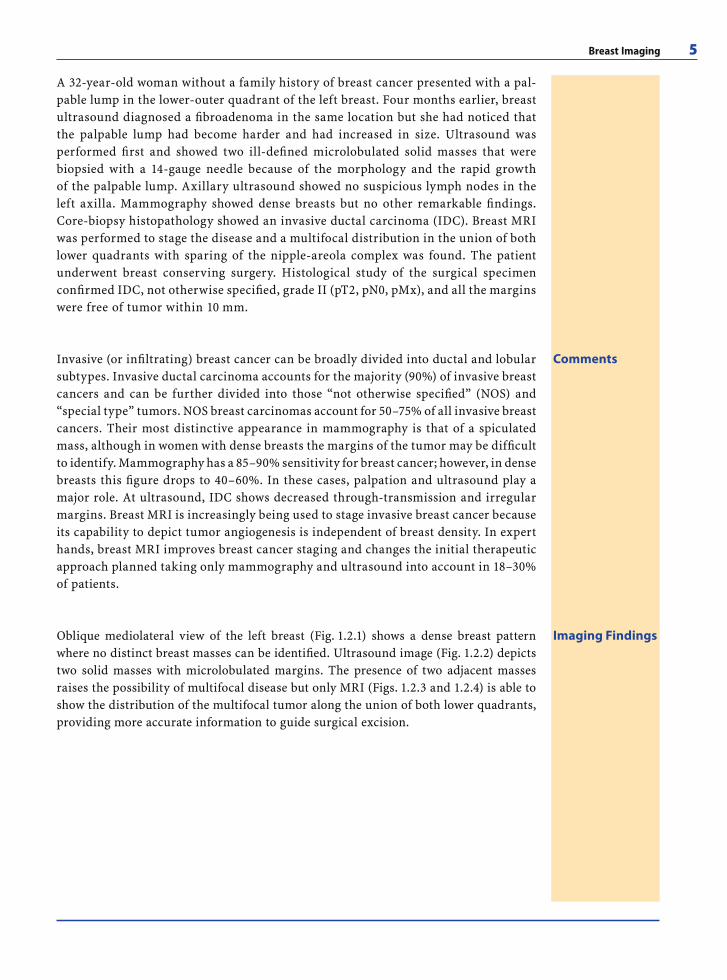
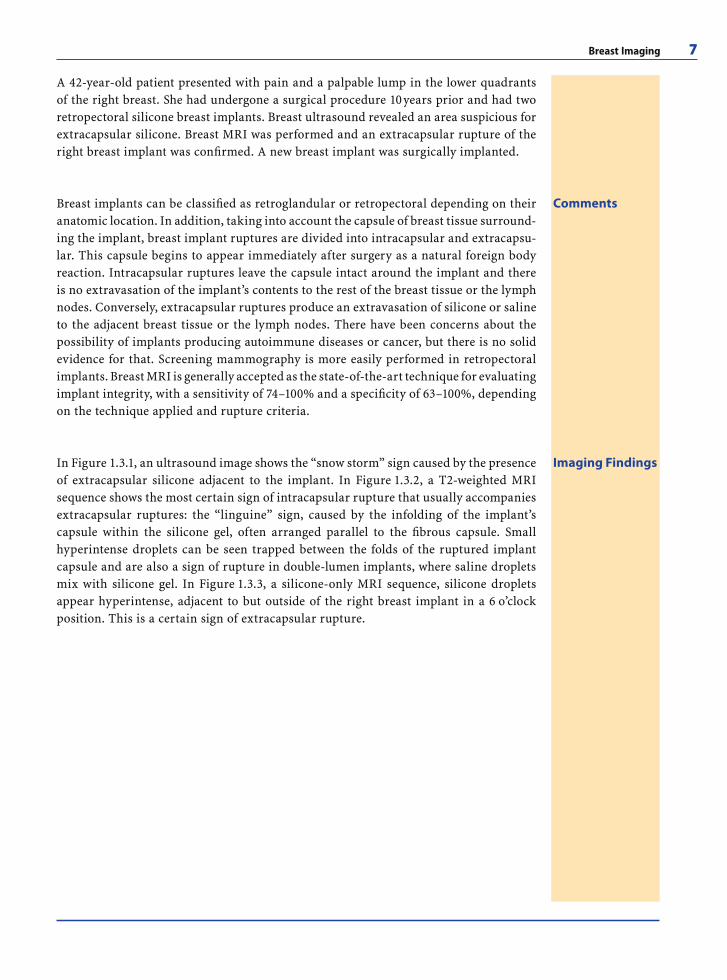
A 42-year-old patient presented with pain and a palpable lump in the lower quadrants of the right breast. She had undergone a surgical procedure 10 years prior and had two retropectoral silicone breast implants. Breast ultrasound revealed an area suspicious for extracapsular silicone. Breast MRI was performed and an extracapsular rupture of the right breast implant was confi rmed. A new breast implant was surgically implanted.
CommentsBreast implants can be classifi ed as retroglandular or retropectoral depending on their anatomic location. In addition, taking into account the capsule of breast tissue surround-ing the implant, breast implant ruptures are divided into intracapsular and extracapsu-lar. This capsule begins to appear immediately after surgery as a natural foreign body reaction. Intracapsular ruptures leave the capsule intact around the implant and there is no extravasation of the implant’s contents to the rest of the breast tissue or the lymph nodes. Conversely, extracapsular ruptures produce an extravasation of silicone or saline to the adjacent breast tissue or the lymph nodes. There have been concerns about the possibility of implants producing autoimmune diseases or cancer, but there is no solid evidence for that. Screening mammography is more easily performed in retropectoral implants. Breast MRI is generally accepted as the state-of-the-art technique for evaluating implant integrity, with a sensitivity of 74–100% and a specifi city of 63–100%, depending on the technique applied and rupture criteria.
Imaging FindingsIn Figure 1.3.1, an ultrasound image shows the “snow storm” sign caused by the presence of extracapsular silicone adjacent to the implant. In Figure 1.3.2, a T2-weighted MRI sequence shows the most certain sign of intracapsular rupture that usually accompanies extracapsular ruptures: the “linguine” sign, caused by the infolding of the implant’s capsule within the silicone gel, often arranged parallel to the fi brous capsule. Small hyperintense droplets can be seen trapped between the folds of the ruptured implant capsule and are also a sign of rupture in double-lumen implants, where saline droplets mix with silicone gel. In Figure 1.3.3, a silicone-only MRI sequence, silicone droplets appear hyperintense, adjacent to but outside of the right breast implant in a 6 o’clock position. This is a certain sign of extracapsular rupture.
RibesLunaRos.indb 7 18.03.2008 12:48:49
8 M. Alvarez Benito and J. Camps Herrero
Case 4
Papillary Lesions
Fig. 1.4.2
Fig. 1.4.4Fig. 1.4.3
Fig. 1.4.1
RibesLunaRos.indb 8 18.03.2008 12:48:49
Breast Imaging 9
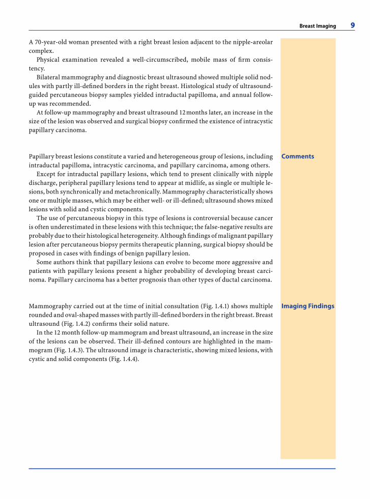
A 70-year-old woman presented with a right breast lesion adjacent to the nipple-areolar complex.
Physical examination revealed a well-circumscribed, mobile mass of fi rm consis-tency.
Bilateral mammography and diagnostic breast ultrasound showed multiple solid nod-ules with partly ill-defi ned borders in the right breast. Histological study of ultrasound-guided percutaneous biopsy samples yielded intraductal papilloma, and annual follow-up was recommended.
At follow-up mammography and breast ultrasound 12 months later, an increase in the size of the lesion was observed and surgical biopsy confi rmed the existence of intracystic papillary carcinoma.
CommentsPapillary breast lesions constitute a varied and heterogeneous group of lesions, including intraductal papilloma, intracystic carcinoma, and papillary carcinoma, among others.
Except for intraductal papillary lesions, which tend to present clinically with nipple discharge, peripheral papillary lesions tend to appear at midlife, as single or multiple le-sions, both synchronically and metachronically. Mammography characteristically shows one or multiple masses, which may be either well- or ill-defi ned; ultrasound shows mixed lesions with solid and cystic components.
The use of percutaneous biopsy in this type of lesions is controversial because cancer is often underestimated in these lesions with this technique; the false-negative results are probably due to their histological heterogeneity. Although fi ndings of malignant papillary lesion after percutaneous biopsy permits therapeutic planning, surgical biopsy should be proposed in cases with fi ndings of benign papillary lesion.
Some authors think that papillary lesions can evolve to become more aggressive and patients with papillary lesions present a higher probability of developing breast carci-noma. Papillary carcinoma has a better prognosis than other types of ductal carcinoma.
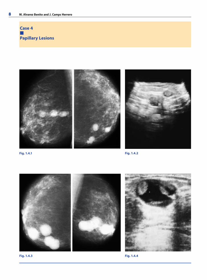
Imaging FindingsMammography carried out at the time of initial consultation (Fig. 1.4.1) shows multiple rounded and oval-shaped masses with partly ill-defi ned borders in the right breast. Breast ultrasound (Fig. 1.4.2) confi rms their solid nature.
In the 12 month follow-up mammogram and breast ultrasound, an increase in the size of the lesions can be observed. Their ill-defi ned contours are highlighted in the mam-mogram (Fig. 1.4.3). The ultrasound image is characteristic, showing mixed lesions, with cystic and solid components (Fig. 1.4.4).
RibesLunaRos.indb 9 18.03.2008 12:48:51
10 M. Alvarez Benito and J. Camps Herrero
Case 5
Microcalcifi cations
Fig. 1.5.1
Fig. 1.5.4
Fig. 1.5.3
Fig. 1.5.2a,ba b
RibesLunaRos.indb 10 18.03.2008 12:48:51
Breast Imaging 11
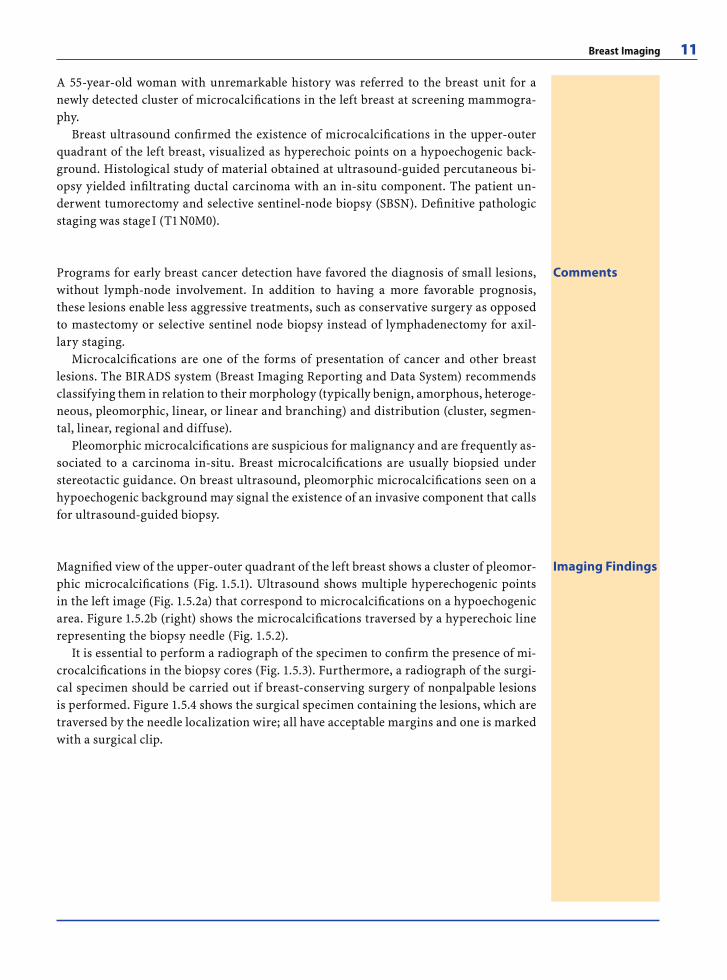
A 55-year-old woman with unremarkable history was referred to the breast unit for a newly detected cluster of microcalcifi cations in the left breast at screening mammogra-phy.
Breast ultrasound confi rmed the existence of microcalcifi cations in the upper-outer quadrant of the left breast, visualized as hyperechoic points on a hypoechogenic back-ground. Histological study of material obtained at ultrasound-guided percutaneous bi-opsy yielded infi ltrating ductal carcinoma with an in-situ component. The patient un-derwent tumorectomy and selective sentinel-node biopsy (SBSN). Defi nitive pathologic staging was stage I (T1 N0M0).
CommentsPrograms for early breast cancer detection have favored the diagnosis of small lesions, without lymph-node involvement. In addition to having a more favorable prognosis, these lesions enable less aggressive treatments, such as conservative surgery as opposed to mastectomy or selective sentinel node biopsy instead of lymphadenectomy for axil-lary staging.
Microcalcifi cations are one of the forms of presentation of cancer and other breast lesions. The BIRADS system (Breast Imaging Reporting and Data System) recommends classifying them in relation to their morphology (typically benign, amorphous, heteroge-neous, pleomorphic, linear, or linear and branching) and distribution (cluster, segmen-tal, linear, regional and diffuse).
Pleomorphic microcalcifi cations are suspicious for malignancy and are frequently as-sociated to a carcinoma in-situ. Breast microcalcifi cations are usually biopsied under stereotactic guidance. On breast ultrasound, pleomorphic microcalcifi cations seen on a hypoechogenic background may signal the existence of an invasive component that calls for ultrasound-guided biopsy.
Imaging FindingsMagnifi ed view of the upper-outer quadrant of the left breast shows a cluster of pleomor-phic microcalcifi cations (Fig. 1.5.1). Ultrasound shows multiple hyperechogenic points in the left image (Fig. 1.5.2a) that correspond to microcalcifi cations on a hypoechogenic area. Figure 1.5.2b (right) shows the microcalcifi cations traversed by a hyperechoic line representing the biopsy needle (Fig. 1.5.2).
It is essential to perform a radiograph of the specimen to confi rm the presence of mi-crocalcifi cations in the biopsy cores (Fig. 1.5.3). Furthermore, a radiograph of the surgi-cal specimen should be carried out if breast-conserving surgery of nonpalpable lesions is performed. Figure 1.5.4 shows the surgical specimen containing the lesions, which are traversed by the needle localization wire; all have acceptable margins and one is marked with a surgical clip.
RibesLunaRos.indb 11 18.03.2008 12:48:52
12 M. Alvarez Benito and J. Camps Herrero
Case 6
Architectural Distortion
Fig. 1.6.1 Fig. 1.6.2
Fig. 1.6.3 Fig. 1.6.4
RibesLunaRos.indb 12 18.03.2008 12:48:52
Breast Imaging 13
A 25-year-old woman presented with a retroareolar mass. Breast ultrasound was initially performed and showed multiple bilateral circumscribed solid masses and a hypoechoic area in the upper-outer quadrant of the right breast.
The diagnostic study was completed with mammography, in which an image of ar-chitectural distortion in the upper-outer quadrant of the right breast could be observed. Histological study of material obtained by stereotactic-guided vacuum-assisted percuta-neous biopsy found a radial scar, and this fi nding was confi rmed in the surgical biopsy specimen some weeks later.
CommentsIn patients younger than 35, or younger than 30 with a family history of breast cancer, breast ultrasound should be the fi rst diagnostic test when there is a palpable abnormality. This test should be complemented with a mammogram when ultrasound does not show any abnormality or shows a suspicious lesion.
Architectural distortion is one type of mammographic presentation of breast patholo-gies. It is often a subtle fi nding, described as the reorganization of the mammary tissue toward an eccentric point from the nipple. There are strands of tissue that converge to-ward a point forming a typical “star” or “whirlwind” shape.
Approximately 50% of cases are malignant, and it is impossible to predict its benign or malignant nature by imaging tests alone. Biopsy should be performed in cases of archi-tectural distortion without previous surgery, injury, or biopsy, although the performance of a percutaneous biopsy in these lesions is controversial, since underestimation of the lesion or false-negative results can be obtained.
Larger gauge needles, such as those used in vacuum-assisted biopsy, improve the ac-curacy of the technique in comparison to core needle biopsy. Some authors claim that when a minimum of 12 cylinders are obtained with these systems, surgical biopsy can be avoided in the absence of atypia.
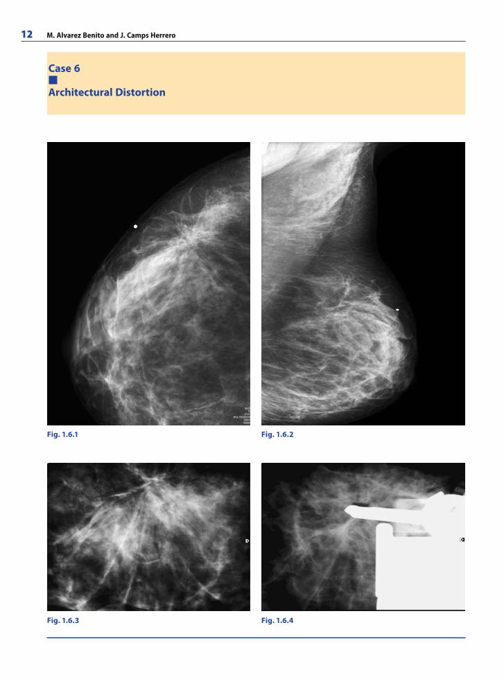
Imaging FindingsCraniocaudal and oblique mammographic views of the right breast show architec-tural distortion or alteration in the distribution of the mammary tissue in the upper-outer quadrant; linear tracts converge toward a point forming the typical “star” shape (Figs. 1.6.1 and 1.6.2).
Localized view (Fig. 1.6.3) of the percutaneous stereotactic biopsy depicts the linear tracts converging toward a central point in greater detail. Figure 1.6.4 confi rms the loca-tion of the biopsy needle in relation to the lesion.
RibesLunaRos.indb 13 18.03.2008 12:48:53
14 M. Alvarez Benito and J. Camps Herrero
Case 7
Breast Cancer in Men
Fig. 1.7.3
Fig. 1.7.1 Fig. 1.7.2
RibesLunaRos.indb 14 18.03.2008 12:48:53
Breast Imaging 15
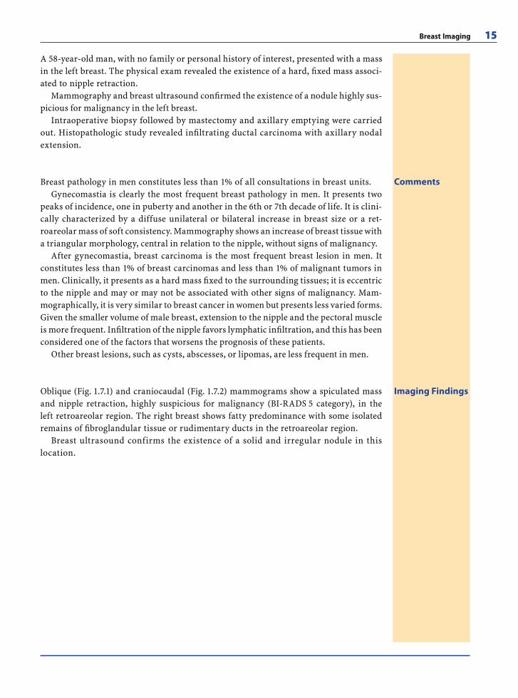
A 58-year-old man, with no family or personal history of interest, presented with a mass in the left breast. The physical exam revealed the existence of a hard, fi xed mass associ-ated to nipple retraction.
Mammography and breast ultrasound confi rmed the existence of a nodule highly sus-picious for malignancy in the left breast.
Intraoperative biopsy followed by mastectomy and axillary emptying were carried out. Histopathologic study revealed infi ltrating ductal carcinoma with axillary nodal extension.
CommentsBreast pathology in men constitutes less than 1% of all consultations in breast units.Gynecomastia is clearly the most frequent breast pathology in men. It presents two
peaks of incidence, one in puberty and another in the 6th or 7th decade of life. It is clini-cally characterized by a diffuse unilateral or bilateral increase in breast size or a ret-roareolar mass of soft consistency. Mammography shows an increase of breast tissue with a triangular morphology, central in relation to the nipple, without signs of malignancy.
After gynecomastia, breast carcinoma is the most frequent breast lesion in men. It constitutes less than 1% of breast carcinomas and less than 1% of malignant tumors in men. Clinically, it presents as a hard mass fi xed to the surrounding tissues; it is eccentric to the nipple and may or may not be associated with other signs of malignancy. Mam-mographically, it is very similar to breast cancer in women but presents less varied forms. Given the smaller volume of male breast, extension to the nipple and the pectoral muscle is more frequent. Infi ltration of the nipple favors lymphatic infi ltration, and this has been considered one of the factors that worsens the prognosis of these patients.
Other breast lesions, such as cysts, abscesses, or lipomas, are less frequent in men.
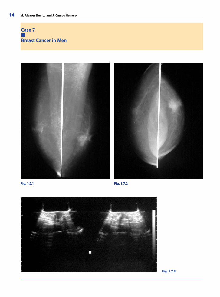
Imaging FindingsOblique (Fig. 1.7.1) and craniocaudal (Fig. 1.7.2) mammograms show a spiculated mass and nipple retraction, highly suspicious for malignancy (BI-RADS 5 category), in the left retroareolar region. The right breast shows fatty predominance with some isolated remains of fi broglandular tissue or rudimentary ducts in the retroareolar region.
Breast ultrasound confirms the existence of a solid and irregular nodule in this location.
RibesLunaRos.indb 15 18.03.2008 12:48:54
16 M. Alvarez Benito and J. Camps Herrero
Case 8
Breast Cancer and Simple Cyst
Fig. 1.8.1
Fig. 1.8.3
Fig. 1.8.2
Fig. 1.8.4
RibesLunaRos.indb 16 18.03.2008 12:48:54
Breast Imaging 17
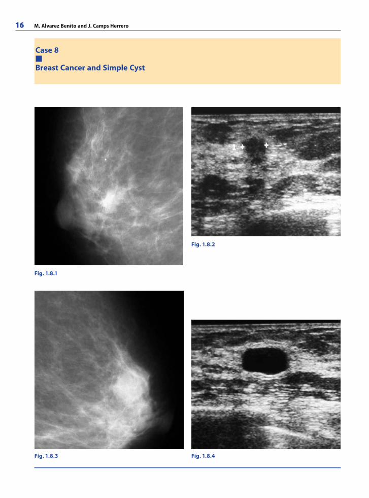
A 64-year-old woman was referred from single-reader screening for a circumscribed retroareolar mass in the left breast.
Localized projections of both retroareolar regions and bilateral breast ultrasound showed an irregular solid mass in the right retroareolar region and a simple cyst in the left retroareolar region.
US-guided percutaneous breast biopsy confi rmed the presence of an infi ltrating ductal carcinoma in the right breast. The patient underwent mastectomy and axillary emptying, and the defi nitive classifi cation was stage I (T1 N0 M0).
CommentsMammography is the only accepted method for breast cancer screening and in recent years its use among healthy women has become more common through population-based screening programs. Population-based screening programs aim to reduce breast cancer mortality. These programs strive to obtain the maximum sensitivity (e.g., the highest detection rate) while keeping morbidity for participating women as low as possible. Some programs use double reading to improve their sensitivity.
The European guidelines recommend that a screening program should obtain a rate of participation of at least 70% of the target population and establish that the rate of de-tection should be higher than 3 in 1000 in women who participate for the fi rst time and higher than 1.5 in 1000 in women who participate in successive rounds. The recall rate (i.e, the percentage of women sent for complementary tests) should also be kept within ac-ceptable limits (lower than 7% in initial rounds and lower than 5% in successive rounds), since these practices cause anxiety and could affect the future involvement of women in the program.
The present case was referred to the breast unit by a single reader. The lesion that prompted the referral turned out to be a simple cyst. A carcinoma was detected in the other breast in the reference unit.
Imaging FindingsThe localized mammographic views show an irregular-shaped mass with spiculated margins in the right retroareolar region (BI-RADS 5) (Fig. 1.8.1) and a well-circumscribed mass in left retroareolar region (BI-RADS 3) (Fig. 1.8.3).
Breast ultrasound confi rmed the existence of a suspicious mass in right retroareolar region (Fig. 1.8.2).
The well-circumscribed mass in the left breast is anechoic with posterior acoustic en-hancement; these fi ndings are typical of a simple cyst, so the lesion that prompted referral is benign (BI-RADS 2) (Fig. 1.8.4).
RibesLunaRos.indb 17 18.03.2008 12:48:56
18 M. Alvarez Benito and J. Camps Herrero
Case 9
Breast Metastases
Fig. 1.9.3
Fig. 1.9.1 Fig. 1.9.2
Fig. 1.9.4
RibesLunaRos.indb 18 18.03.2008 12:48:56
Breast Imaging 19
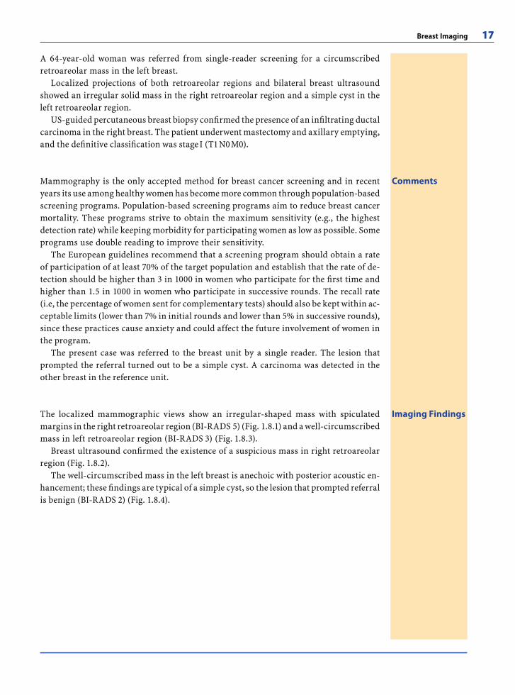
A 61-year-old woman, with no family history of interest, presented with an ipsilateral axillary node six years after the excision of a cutaneous melanoma. She underwent axil-lary emptying and nodal infi ltration by melanoma was confi rmed.
One year after axillary emptying, she presented with a right mammary nodule and an axillary node. Bilateral mammography confi rmed the existence of a nodule in her right breast. Ultrasound demonstrated the solid nature of the nodule and revealed a right axil-lary node.
US-guided percutaneous biopsy of both the mammary nodule and the axillary node yielded melanoma metastases in both at histological examination. The patient underwent right tumorectomy and removal of the axillary node and was transferred to the oncology department to complete treatment.
CommentsThe mammary gland is an infrequent site for metastasis. In 1903 the fi rst case of mam-mary metastasis was reported and until 1991, only 300 cases of different metastatic tumors in the mammary gland had been published, the most frequent being leukemias, lymphomas, ovary neoplasms, and soft-tissue sarcomas.
The differential diagnosis between metastasis and primary neoplasms of the breast should be carried out due to the prognostic and therapeutic implications involved.
Mammographically, metastatic lesions tend to appear as single nodules, although they can also be multiple. They tend to be superfi cial and can be well- or ill-defi ned. Diagnostic ultrasound is used to confi rm the solid nature of the lesions, to improve their character-ization, and to guide biopsy.
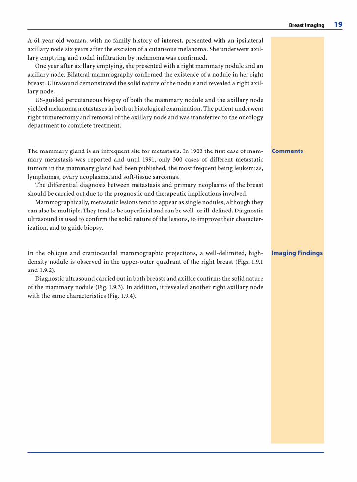
Imaging FindingsIn the oblique and craniocaudal mammographic projections, a well-delimited, high-density nodule is observed in the upper-outer quadrant of the right breast (Figs. 1.9.1 and 1.9.2).
Diagnostic ultrasound carried out in both breasts and axillae confi rms the solid nature of the mammary nodule (Fig. 1.9.3). In addition, it revealed another right axillary node with the same characteristics (Fig. 1.9.4).
RibesLunaRos.indb 19 18.03.2008 12:48:57
20 M. Alvarez Benito and J. Camps Herrero
Case 10
Locorregional Staging
Fig. 1.10.2
Fig. 1.10.1
Fig. 1.10.4
Fig. 1.10.3
RibesLunaRos.indb 20 18.03.2008 12:48:57
Breast Imaging 21
56-year-old woman without prior history of interest presented with a nodule in her left breast after some months’ evolution.
Bilateral mammography found a spiculated, hyperdense nodule in the upper-outer quadrant of the left breast, highly suspicious for malignancy (BI-RADS 5).
Diagnostic ultrasound confi rmed its solid nature and revealed a suspicious node in the left axilla.
In the same diagnostic act, US-guided percutaneous mammary and axillary node core biopsy was carried out; infi ltrating ductal carcinoma and axillary node infi ltration were diagnosed at histology.
A dynamic breast MRI was performed to rule out other tumor foci in either breast. The patient underwent tumorectomy and axillary emptying, and the defi nitive classifi cation was stage II (T1 N1 M0).
CommentsNodules are one of the most frequent presentations of breast cancer. BIRADS recom-mends classifying them according to shape (round, oval-shaped, lobulated, or irregular), contour (circumscribed, ill-defi ned, microlobulated, darkened, or spiculated), and den-sity (similar, equal or superior to the mammary parenchyma, or with fatty content).
Spiculated nodules have the highest probability of malignancy. Percutaneous biopsy enables histological study of lesions with less morbidity and lower costs than surgical bi-opsy and also allows women to participate in decisions about the therapeutic approach.
With the advent of selective sentinel node biopsy in the treatment of early stage breast cancer as an alternative to axillary dissection, sonographic assessment of the axilla with biopsy of suspicious adenopathies has gained great importance since it allows patients to be selected for the technique and helps to avoid false negatives. Loss of the oval-shaped morphology, loss of the fatty hilum, focal cortical enlargement, diffuse enlargement of the node cortex, and increased size are considered sonographic signs suspicious for neo-plastic infi ltration of a node.
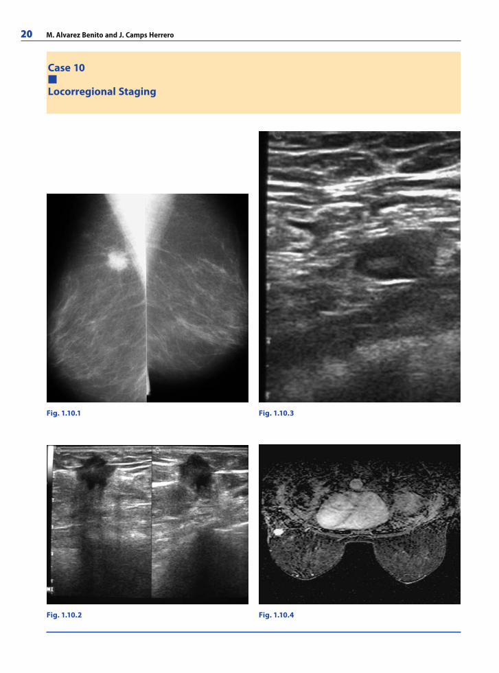
Imaging FindingsOblique mammographic projections (Fig. 1.10.1) show a round hyperdense nodule with spiculated contours in the upper-outer quadrant of the left breast. Diagnostic ultrasound confi rms its solid nature and angular margins (Fig. 1.10.2).
A suspicious lymph node was identifi ed on ultrasound. Although the node main-tains its oval shape and fatty center, there is a focal enlargement of the lower pole cortex (Fig. 1.10.3).Breast MRI (Fig. 1.10.4) confi rms the existence of a contrast-enhanced nodule in the left breast. No other suspicious nodules were found.
RibesLunaRos.indb 21 18.03.2008 12:48:59
22 M. A. Benito and J. C. Herrero
Further Readings
Books
Americam College of Radiology. Breast Imaging Reporting and Data System. Reston VA; ACR, 2003
Breast Cancer – The Art and Science of Early Detection with Mammography. Tabar L, Tot T, Dean PB (2004). Thieme Medical Publishers. ISBN-13: 9781588902597
Breast Imaging. 3rd ed. Kopans DB (2006) Lippincott Wil-liams & Wilkins. ISBN-13: 9780781747684
Breast Imaging Companion. Cardeñosa G (1997) Lippincott-Raven. ISBN-13: 9780397517787
Diagnostic Breast Imaging: Mammography, Sonography, Magnetic Resonance Imaging, and Interventional Pro-cedures. 2nd ed. Heywang-Koebrunner S (2001) Thieme Medical Publishers. ISBN-13: 9781588900333
European guidelines for quality assurance in mammog-raphy screening. de Wolf CJ, Perry N (1996). European Communities / Union (EUR-OP/OOPEC/OPOCE). ISBN-13: 9789282774304
Practical Breast Pathology. Tot T, Tabar L, Dean PB (2002) Thieme Medical Publishers. ISBN-13: 9781588900913
Radiology: Diagnosis, Imaging, Intervention. Taveras JM, Ferrucci JT (2003) Lippincott Williams & Wilkins. ISBN-13: 9780015342449
The Practice of Breast Ultrasound: Techniques, Findings, Differential Diagnosis. Madjar H (2000) Thieme Medical Publishers. ISBN-13: 9781588904485
Diagnosis of Diseases of the Breast. 2nd ed. Bassett LW, Jack-son V, Fu K, Fu Y (1997) Saunders Company. ISBN-13 9780721695631
Americam College of Radiology. Breast Imaging Reporting And Data System. Reston VA; ACR, 2003
Breast Imaging. Kopans DB 2001. Lippincott-Raven
Web-Links
http://acr.org/s_acr/sec.asp?CID=549&DID=14210http://emedicine.com/radio/BREAST.htmhttp://guideline.gov/Compare/comparison.aspx?fi le
=BRSCREEN13.inchttp://imaginis.com/pro/teaching_fi les/http://radiographics.rsnajnls.org/cgi/collection/breast_
imaginghttp://rad.washington.edu/breast/http://radquiz.com/Breast%20Imaging.htmhttp://rsna.org/education/archive/breast.cfmhttp://sbi-online.org/sbi_home/education/fellowship_
programs/breast_imaging__3http://sprojects.mmi.mcgill.ca/mammography/cases.htm
Articles
Adepoju LJ, Chun J, El-Tamer M, Ditkoff BA, Schnabel F, Joseph KA. The value of clinical characteristics and breast-imaging studies in predicting a histopathologic diagnosis of cancer or high-risk lesion in patients
with spontaneous nipple discharge. Am J Surg 2005; 190(4):644–646
Adrales G, Turk P, Wallace T, Bird R, Norton HJ, Greene F. Is surgical excision necessary for atypical ductal hyperpla-sia of the breast diagnosed by Mammotome? Am J Surg 2000; 180(4):313–315
Agoff SN, Lawton TJ. Papillary lesions of the breast with and without atypical ductal hyperplasia: can we accurately predict benign behavior from core needle biopsy? Am J Clin Pathol 2004; 122(3):440–443
Al Sarakbi W, Worku D, Escobar PF, Mokbel K. Breast papil-lomas: current management with a focus on a new diag-nostic and therapeutic modality. Int Semin Surg Oncol 2006; 3:1
Alleva DQ, Smetherman DH, Farr GH, Cederbom GJ. Radial scar of the breast: Radiologic-pathologic correlation in 22 cases. Radiographics 1999; 19:S27–S35
Alonso-Bartolomé P, Vega-Bolívar A, Torres-Tabanera M, Ortega E, Acebal-Blanco M, Garijo-Ayensa F, Rodrigo I, Muñoz-Cacho P. Sonographically guided 11-G direc-tional vacuum-assisted breast biopsy as an alternative to surgical excision: Utility and cost study in probably benign lesions. Acta Radiológica 2004; 4:390–396
Baker KS, Davey DD, Stelling CB. Ductal abnormalities detected with galactography: frequency of adequate ex-cisional biopsy. AJR Am J Roentgenol 1994; 162(4):821–824
Baum F, Fischer U, Vosshenrich R, Grabbe E. Classifi cation of hypervascularized lesions in CE MR imaging of the breast. Eur Radiol 2002; 12(5):1087–1092
Bedrosian I, Mick R, Orel SG et al. Changes in the surgical management of patients with breast carcinoma based on preoperative magnetic resonance imaging. Cancer 2003; 98:468–473
Berg WA. When is core breast biopsy or fi ne-needle aspira-tion not enough? Radiology 1996; 198:313–315
Berg WA, Arnoldus Ch L, Teferra E, Bhargavan M. Biopsy of amorphous breast calcifi cations: Pathologic outcome and yield at stereotactic biopsy. Radiology 2001; 221:495–503
Berube M, Curpen B, Ugolini P, Lalonde L, Ouimet-Olivia D. Level of suspicion of a mammographic lesion: use of features defi ned by BI-RADS lexicon and correlation with large-core breast biopsy. Can Assoc Radiol J 1998; 49(4):223–228
Brenner RJ. Strategies in the evaluation of breast asymme-tries. Appl Radiol 1998; 27:15–20
Brenner RJ, Sickles EA. Surveillance mammography and ste-reotactic core breast biopsy for probably benign lesions: a cost comparison study. Acad Radiol 1997; 4:419–425
Ciatto S, Houssami N, Ambrogetti D, Bonardi R, Collini G, Del Turco MR. Minority report – false negative breast assessment in women recalled for suspicious screening mammography: imaging and pathological features, and associated delay in diagnosis. Breast Cancer Res Treat 2007; 105(1):37–43
Ciatto S, Morrone D, Catarzi S, Del Turco MR, Bianchi S, Am-brogetti D, Cariddi A. Radial scars of the breast: Review of 38 consecutive mammographic diagnoses. Radiology 1993; 187:757–760
RibesLunaRos.indb 22 18.03.2008 12:48:59
Breast Imaging 23
Cohen MA, Sferlazza SJ. Role of sonography in evaluation of scars of the breast. AJR Am J Roentgenol 2000; 174:1075–1078
Costantini M, Belli P, Lombardi R, Franceschini G, Mule A, Bonomo L. Characterization of solid breast masses: use of the sonographic breast imaging reporting and data sys-tem lexicon. J Ultrasound Med 2006; 25(5):649–659
Dershaw DD, Morris EA, Liberman L, Abramson AF. Non-diagnostic stereotaxic core breast biopsy: Results of rebi-opsy. Radiology 1996; 198:323–325
Diebold T, Hahn T, Solbach C, Rody A, Balzer JO, Hansmann ML, Marx A, Viana F, Peters J, Jacobi V, Kaufmann M, Vogl TJ. Evaluation of the stereotactic 8G vacuum-as-sisted breast biopsy in the histologic evaluation of sus-picious mammography fi ndings (BI-RADS IV). Invest Radiol 2005; 40(7):465–471
Diebold T, Jacobi V, Krapfl E, von Minckwitz G, Solbach C, Ballenberger S, Hochmuth K, Balzer JO, Fellbaum M, Kaufmann M, Vogl TJ. The role of stereotactic 11G vacuum biopsy for clarifi cation of BI-RADS IV fi ndings in mammography. Rofo 2003; 175(4):489–494
Feig SA. Adverse effects of screening mammography. Radiol Clin N Am 2004; 42:807–819
Fine RE, Israel PZ, Walker LC, Corgan KR, Greenwald LV, Berenson JE, Boyd BA, Oliver MK, McClure T, Elberfeld J. A prospective study of the removal rate of imaged breast lesions by an 11-gauge vacuum-assisted biopsy probe sys-tem. Am J Surg 2001; 182(4):335–340
Frouge C, Tristant H, Guinebretiere JM, Meunier M, Conteso G, Paola R, Blery M. Mammographic lesions suggestive of radial scars: Microscopic fi ndings in 40 cases. Radiology 1995; 195:623–625
Gill HK, Ioffe OB, Berg WA. When is a diagnosis of scleros-ing adenosis acceptable at core biopsy? Radiology 2003; 228:50–57
Govindarajulu S, Narreddy SR, Shere MH, Ibrahim NB, Sahu AK, Cawthorn SJ. Sonographically guided mammotome excision of ducts in the diagnosis and management of single duct nipple discharge. Eur J Surg Oncol 2006; 32(7):725–728
Graf O, Helbich TH, Fuchsjaeger MH, Hopf G, Morgun M, Graf C, Mallek R, Sickles EA. Follow-up of palpable cir-cumscribed noncalcifi ed solid breast masses at mam-mography and US: Can biopsy be averted? Radiology 2004; 233:850–856
Guenin MA. Benign intraductal papilloma: Diagnosis and re-moval at stereotactic vacuum-assisted directional biopsy guided by galactography. Radiology 2001; 218:576–579
Gupta RK, Gaskell D, Dowle CS, Simpson JS, King BR, Naran S, Lallu S, Fauck R. The role of nipple discharge cytol-ogy in the diagnosis of breast disease: a study of 1948 nipple discharge smears from 1530 patients. Cytopathol-ogy 2004; 15(6):326–330
Harms SE. Breast magnetic resonance imaging. Semin Ul-trasound CT MR 1998; 19:104–120
Harvey JA. Sonography of palpable breast masses. Semin Ultrasound CT MR 2006; 27(4):284–297
Helvie MA, Pennes DR, Rebner M, Adler DD. Mammographic follow-up of low suspicion lesions: Compliance rate and diagnostic yield. Radiology 1991; 178:155–158
Hendrick RE, smith RA, Rutlege JH, Smart CR. Benefi t of screening mammography in women aged 40–49: a new meta-analysis of randomized controlled trials. Monogr natl Cancer Inst 1997; 22:87–92
Hou MF, Huang TJ, Liu GC. The diagnostic value of galac-tography in patients with nipple discharge. Clin Imaging. 2001; 25(2):75–81
Huber S, Wagner M, Medl M, Czembirek H. Benign breast lesions: minimally invasive vacuum-assisted biopsy with 11-gauge needles-Patient acceptance and effect on follow-up imaging fi ndings. Radiology 2003; 226:783–790
Hunerbein M, Raubach M, Gebauer B, Schneider W, Schlag PM. Ductoscopy and intraductal vacuum assisted biopsy in women with pathologic nipple discharge. Breast Can-cer Res Treat 2006; 99:301–307
Huynh PT, Jarolimek AM, Daye S. The false-negative mam-mogram. Radiographics 1998; 18:1137–1154
Jackman RJ, Nowelss KW, Rodriguez-Soto J, Marzoni FA, Finkelstein SL, Shepard MJ. Stereotactic, automated, Large-core needle biopsy of nonpalpable breast lesions: False-negative and histologic understimation rates after long-term follow-up. Radiology 1999; 210:799–805
Johnson AT, Henry-Tillman RS, Smith LF, et al. Percutane-ous excisional breast biopsy. Am J Surg 2002; 184:550–554
Kerlikowske K, Grady D, Rubin SM, Sandrock C, Ernster VL. Effi cacy of screening mammography: a meta-analysis. JAMA 1995; 273:149–154
Kerlikowske K, Smith-Bindman R, Abraham LA, Lehman CD, Yankaskas BC, Ballard-Barbash R, Barlow WE, Voeks JH, Geller BM, Carney PA, Sickles EA. Breast cancer yield for screening mammographic examinations with recom-mendation for short-interval follow-up. Radiology 2005; 234:684–692
Kettritz U, Morack G, Decker T. Stereotactic vacuum-as-sisted breast biopsies in 500 women with microcalcifi ca-tions: radiological and pathological correlations. Eur J Radiol 2005; 5(2):270–276
King TA, Scharfenberg JC, Smetherman DH, Farkas EA, Bolton JS, Fuhrman GM. A better understanding of the term radial scar. Am J Surg 2000; 180:428–433
Kirwan SE, Denton ERE, Nash RM, Humphreys S, Michell MJ. Multiple 14G Stereotactic core biopsies in the diagno-sis of mammographically detected stellate lesions of the breast. Clin Radiol 2000; 55:763–766
Kuhl CK. MRI of breast tumor. Eur Radiol 2000; 10:46–58Kuzmiak CM, Dancel R, Pisano E, Zeng D, Cole E, Koomen
MA, McLelland R. Consensus review: A method of as-sessment of calcifi cations that appropriately undergo a six-month follow-up. Acad Radiol 2006; 13(5):621–629
Lacquement MA, Mitchell D, Hollingswotrth AB. Positive predictive value of the breast imaging reporting and data system. J Am Coll Surg 1999; 189(1):34–40
Lee Ch. Screening mammography: proven benefi t, contin-ued controversy. Radiol Clin North Am 2002; 40(3):395–407
Lee CH, Philpotts LE, Horvath LJ, Tocino I. Follow-up of breast lesions diagnosed as benign with stereotactic core-needle biopsy: Frequency of mammographic change and false-negative rate. Radiology 1999; 212:189–194
RibesLunaRos.indb 23 18.03.2008 12:48:59
24 M. A. Benito and J. C. Herrero
Lee JM, Orel SG, Czerniecki BJ, Solin LJ, Schnall MD. MRI before reexcision surgery in patients with breast cancer. AJR Am J Roentgenol 2004; 182:473–480
Liberman L. Clinical management issues in percutaneous core breast biopsy. Radiol Clin North Am 2000; 38(4):791–806
Liberman L, Dershaw DD, Morris EA, Abramson AF, Thorn-ton CM, Rosen PP. Clip placement after sterotactic vac-uum-assisted breast biopsy. Radiology 1997; 205:417–422
Liberman L, Abramson AF, Squires FB, Glassman JR, Mor-ris EA, Dershaw DD. The Breast Imaging and Reporting Data System: Positive predictive value of mammographic features and fi nal assessment categories. AJR Am J Roent-genol 1998; 171:35–40
Liberman L, Feng TL, Dershaw DD, Morris EA, Abramson AF. US-guided core breast biopsy: use and cost-effective-ness. Radiology 1998; 208(3):717–723
Liberman L, Bracero N, Vuolo MA, et al. Percutaneous large-core biopsy of papillary breast lesions. AJR Am J Roent-genol 1999; 172:331–337
Liberman L, Kaplan JB, Morris EA, Abramson AF, Menell JH, Dershaw DD. To excise or to sample the mammo-graphic target: What is the goal of stereotactic 11-gauge vacuum-assisted breast biopsy? AJR Am J Roentgenol 2002; 179:679–683
Lindfors KK, O’Connor J, Acredolo CR, Liston SE. Short-in-terval follow-up mammography versus immediate core biopsy of benign breast lesions: Assessment of patient stress. AJR Am J Roentgenol 1998; 171:55–58
Litherland JC. Should fi ne needle aspiration cytology in breast assessment be abandoned? Clin Radiol 2002; 57:81–84
Lomoschitz FM, Helbich TH, Rudas M, Pfarl G, Linnau KF, Stadler A, Jackman RJ. Stereotactic 11-gauge vacuum-as-sisted breast biopsy: Infl uence of number of specimens on diagnostic accuracy. Radiology 2004; 232(3):897–903
Lorenzen J, Wedel AK, Lisboa BW, Loning T, Adam G. Diag-nostic mammography and sonography: concordance of the breast imaging reporting assessments and fi nal clini-cal outcome. Rofo 2005; 177(11):1545–1551
Mandelblatt JS, Wheat ME, Monane M, Moshief RD, Hollen-berg JP, Tang J. Breast cancer screening for elderly women with and without comorbid conditions: a decision analy-sis model. Ann Intern Med 1992; 116:722–730
Mendelson EB, harris KM, Doshi N, Tobon H. Infi ltrating lobular carcinoma: mammographic patterns with patho-logic correlation. AJR Am J Roentgenol 1989; 153:265–271
Mendez A, Cabanillas F, Echenique M, Malekshamran K, Perez I, Ramos E. Mammographic features and corre-lation with biopsy fi ndings using 11-gauge stereotactic vacuum-assisted breast biopsy (SVABB). Ann Oncol 2003; 14:450–454
Mercado CL, Hamele-Bena D, Singer C, Koenigsberg T, Pile-Spellman E, Higgins H, Smith SJ. Papillary Lesions of the Breast: Evaluation with stereotactic directional vacuum-assisted biopsy. Radiology 2001; 221:650–655
Morris EA. Breast cancer imaging with MRI. Radiol Clin North Am 2002; 40:443–466
Moy L, Slanetz PJ, Moore R, Satija S, Yeh FD, McCarthy KA, Hall D, Staffa M, Rafferty EA, Halpern E, Kopans DB.
Specifi city of mammography and US in the evaluation of a palpable abnormality: Retrospective review. Radiology 2002; 225:176–181
Orel SG. MR imaging of the breast. Radiol Clin North Am 2000; 38:899–913
Orel SG, Schnall MD. MR Imaging of the breast for the detec-tion, diagnosis, and staging of breast cancer. Radiology 2001; 220:13–30
Parker SH, Burbank F, Jackman RJ, et al. Percutaneous large-core breast biopsy:A multi-institutional study. Radiology 1994; 193:359–364
Parker SH, Jobe WE, Dennis MA, et al. US-guided automated large-core breast biopsy. Radiology 1993; 187(2):507–511
Paterok EM, Rosenthal H, Sabel M. Nipple discharge and abnormal galactogram. Results of a long-term study (1964–1990). Eur J Obstet Gynecol Reprod Biol 1993; 50:227–234
Pearson LK, Sickles EA, Frankel SD, Leung J. Effi cacy of step-oblique mammography for çconfi rmation and local-ization of densities seen on only one standard mammo-graphic view. AJR Am J Roentgenol 2000; 174:745–752
Philpotts LE, Shaheen NA, Jain KS, Carter D, Lee CCH. Uncommon high-risk lesions of the breast diagnosed at stereotactic core-needle biopsy: Clinical importance. Ra-diology 2000; 216:831–837
Pijnappel M, Peeters HM, Hendriks HC and Mali Th M. Re-producibility of mammographic classifi cations for non-palpable suspect lesions with microcalcifi cations. Br J Radiol 2004; 77:312–314
Puglisi F, Zuiani C, Bazzocchi M, Valent F, Aprile G, Pertoldi B, Minisini AM, Cedolini C, Londero V, Piga A, Di Loreto C. Role of mammography, ultrasound and large core bi-opsy in the diagnostic evaluation of papillary breast le-sions. Oncology 2003; 65(4):311–315
Rahbar G, Sie AC, Hansen GC, et al. Benign versus malignant solid breast masses: US differentiation. Radiology 1999; 213:889–894
Rosen EL, Bentley RC, Baker JA, Soo MS. Imaging-guided core needle biopsy of papillary lesions of the breast. AJR Am J Roentgenol 2002; 179:1185–1192
Rosen EL, Blackwell KL, Baker JA, et al. Accuracy of MRI in the detection of residual breast cancer after neoadju-vant chemotherapy. AJR Am J Roentgenol 2003; 181:1275–1282
Samardar P, de Paredes SE, Grimes MM, Wilson JD. Focal asymmetric densities seen at mammography: US and pathologic correlation. Radiographics 2002; 22:19–33
Sardanelli F, Giusepetti G, Panizza P, et al. Sensitivity of MRI versus mammography for detecting foci of multifo-cal, multicentric breast cancer in fatty and dense breasts using the whole-breast pathologic examination as a gold standard. AJR Am J Roentgenol 2004; 183(4):1149–1157
Sauter ER, Schlatter L, Lininger J, Hewett JE. The association of bloody nipple discharge with breast pathology. Surgery 2004; 136(4):780–785
Shapiro S, Venet W, Strax P, Venet L, Rosser R. 10 to 14-year effect of screening on breast cancer mortality. J Natl can-cer Inst 1982; 69:349–355
Shulman SG, March DE. Ultrasound-Guided Breast Inter-ventions: Accuracy of biopsy techniques and applications
RibesLunaRos.indb 24 18.03.2008 12:48:59
Breast Imaging 25
in patient management. Sem Ultrasound CT MR 2006; 27(4):298–307
Sickles EA. Periodic mammographic follow-up of probably benign breast lesions: results in 3184 consecutive cases. Radiology 1991; 179:463–468
Sickles EA. Non-palpable circumscribed, non-calcifi ed solid masses: Likelihood of malignancy based on lesion size and age of patient. Radiology 1994; 192:439–442
Sickles EA. Management of probably benign breast lesions. Radiol Clin North Am 1995; 33:1123–1130
Sickles EA. Probably benign breast lesions: When should fol-low-up be recommended and what is the optimal follow-up protocol? Radiology 1999; 213:11–14
Sickles EA, Parker SH. Appropriate role of core breast biopsy in the management of probably benign lesions. Radiology 1993; 188:315
Simon JR, Kalbhen CL, Cooper RA, Flisak ME. Accuracy and complication guided vacuum-assisted core breast biopsy: Initial results. Radiology 2000; 215: 694–697
Simsir A, Waisman J, Thorner K, Cangiarella J. Mammary lesions diagnosed as “papillary” by aspiration biopsy: 70 cases with follow-up. Cancer 2003; 99(3):156-165
Smith DN, Rosenfi eld Darling ML, Meyer JE, et al. The utility of ultrasonographically guided large-core needle biopsy: results from 500 consecutive breast biopsies. J Ultrasound Med 2001; 20(1):43–49
Smith RA, Duffy SW, MPhil RG, Tabar L, Yen A, Chen T. The randomized trials of breast cancer screening: What have we learned? Radiol Clin North Am 2004; 42:793–806
Smith-bindman R, Kerlikowske K, Gebretsadik T. Is screen-ing mammography effective in elderly women? Am J Med 2000: 108:112–119
Spencer NJB, Evans AJ, Galea M, et al. Pathological-radio-logical correlations in benign lesions excised during a breast screening programme. Clin Radiol 1994; 49:853–856
Thurfjell MG, Lindgren A, Thurfjell E. Nonpalpable breast cancer: Mammographic appearance as predictor of his-tologic type. Radiology 2002; 222:165–170
Varas X, Leborgne F, Leborgne JH. Non-palpable, probably benign lesions: role of follow-up mammography. Radiol-ogy 1992, 184:409–414
Varas X, Leborgne JH, Leborgne F, Mezzera J, Jaumandreu S, Leborgne F. Revisiting the mammographic follow-up of BI-RADS category 3 lesions. AJR Am J Roentgenol 2002; 179(3):691–695
Vargas HI, Vargas MP, Eldrageely K, González KD, Burla MI, Venegas R, Khalkhali I. Outcomes of surgical and sono-graphic assessment of breast masses in women younger than 30. Am Surg 2005; 71(9):716–719
Vizcaíno I, Gadea L, Andreo L, Salas D, Ruiz-perales F, Cue-vas D, Herranz C, Bueno F. Short-term Follow-up Results in 795 Nonpalpable probably benign lesions detected at screening mammography. Radiology 2001; 219:475–483
Yasmeen S, Romano PS, Pettinger M, Chlebowski RT, Rob-bins JA, Lane DS, Hendrix SL. Frequency and predictive value of a mammographic recommendation for short-in-terval follow-up. J Natl Cancer Inst 2003; 95:429–436
RibesLunaRos.indb 25 18.03.2008 12:48:59

RibesLunaRos.indb V 18.03.2008 12:48:44
Nuclear Medicine
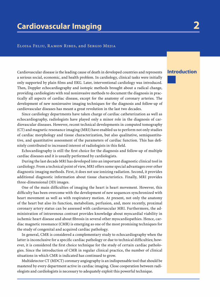
Cardiovascular Imaging 2
Eloisa Feliu, Ramon Ribes, and Sergio Mejia
IntroductionCardiovascular disease is the leading cause of death in developed countries and represents a serious social, economic, and health problem. In cardiology, clinical tasks were initially only supported by plain fi lms and EKG. Later, interventional cardiology was introduced. Then, Doppler echocardiography and isotopic methods brought about a radical change, providing cardiologists with real noninvasive methods to document the diagnosis in prac-tically all aspects of cardiac disease, except for the anatomy of coronary arteries. The development of new noninvasive imaging techniques for the diagnosis and follow-up of cardiovascular diseases has meant a great revolution in the last two decades.
Since cardiology departments have taken charge of cardiac catheterization as well as echocardiography, radiologists have played only a minor role in the diagnosis of car-diovascular diseases. However, recent technical developments in computed tomography (CT) and magnetic resonance imaging (MRI) have enabled us to perform not only studies of cardiac morphology and tissue characterization, but also qualitative, semiquantita-tive, and quantitative assessment of the parameters of cardiac function. This has defi -nitely contributed to increased interest of radiologists in this fi eld.
Echocardiography is still the fi rst choice for the diagnosis and follow-up of multiple cardiac diseases and it is usually performed by cardiologists.
During the last decade MRI has developed into an important diagnostic clinical tool in cardiology. From a technical point of view, MRI offers some special advantages over other diagnostic imaging methods. First, it does not use ionizing radiation. Second, it provides additional diagnostic information about tissue characteristics. Finally, MRI provides three-dimensional (3D) images.
One of the main diffi culties of imaging the heart is heart movement. However, this diffi culty has been overcome with the development of new sequences synchronized with heart movement as well as with respiratory motion. At present, not only the anatomy of the heart but also its function, metabolism, perfusion, and, more recently, proximal coronary artery status can be assessed with cardiovascular MRI. Furthermore, the ad-ministration of intravenous contrast provides knowledge about myocardial viability in ischemic heart disease and about fi brosis in several other myocardiopathies. Hence, car-diac magnetic resonance (CMR) is emerging as one of the most promising techniques for the study of congenital and acquired cardiac pathology.
In general, CMR is considered a complementary study to echocardiography when the latter is inconclusive for a specifi c cardiac pathology or due to technical diffi culties; how-ever, it is considered the fi rst choice technique for the study of certain cardiac patholo-gies. Since the introduction of CMR in regular clinical practice, the number of clinical situations in which CMR is indicated has continued to grow.
Multidetector CT (MDCT) coronary angiography is an indispensable tool that should be mastered by every department active in cardiac imaging. Close cooperation between radi-ologists and cardiologists is necessary to adequately exploit this powerful technique.
RibesLunaRos.indb 27 18.03.2008 12:49:00
28 E. Feliu, R. Ribes, and S. Mejia
Case 1
Acute Myocardial Infarction
Fig. 2.1.1
Fig. 2.1.3a–d
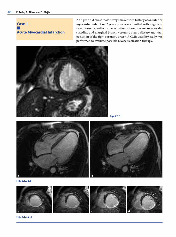
A 57-year-old obese male heavy smoker with history of an inferior myocardial infarction 2 years prior was admitted with angina of recent onset. Cardiac catheterization showed severe anterior de-scending and marginal branch coronary artery disease and total occlusion of the right coronary artery. A CMR viability study was performed to evaluate possible revascularization therapy.
Fig. 2.1.2a,b
a b c d
a b
RibesLunaRos.indb 28 18.03.2008 12:49:00
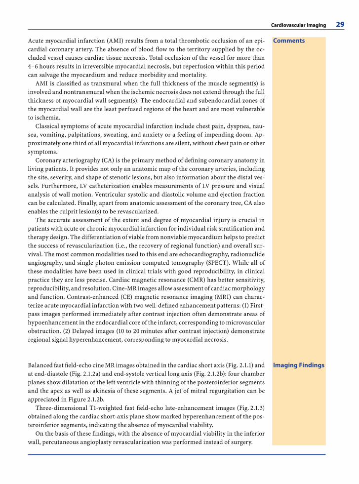
Cardiovascular Imaging 29
CommentsAcute myocardial infarction (AMI) results from a total thrombotic occlusion of an epi-cardial coronary artery. The absence of blood fl ow to the territory supplied by the oc-cluded vessel causes cardiac tissue necrosis. Total occlusion of the vessel for more than 4–6 hours results in irreversible myocardial necrosis, but reperfusion within this period can salvage the myocardium and reduce morbidity and mortality.
AMI is classifi ed as transmural when the full thickness of the muscle segment(s) is involved and nontransmural when the ischemic necrosis does not extend through the full thickness of myocardial wall segment(s). The endocardial and subendocardial zones of the myocardial wall are the least perfused regions of the heart and are most vulnerable to ischemia.
Classical symptoms of acute myocardial infarction include chest pain, dyspnea, nau-sea, vomiting, palpitations, sweating, and anxiety or a feeling of impending doom. Ap-proximately one third of all myocardial infarctions are silent, without chest pain or other symptoms.
Coronary arteriography (CA) is the primary method of defi ning coronary anatomy in living patients. It provides not only an anatomic map of the coronary arteries, including the site, severity, and shape of stenotic lesions, but also information about the distal ves-sels. Furthermore, LV catheterization enables measurements of LV pressure and visual analysis of wall motion. Ventricular systolic and diastolic volume and ejection fraction can be calculated. Finally, apart from anatomic assessment of the coronary tree, CA also enables the culprit lesion(s) to be revascularized.
The accurate assessment of the extent and degree of myocardial injury is crucial in patients with acute or chronic myocardial infarction for individual risk stratifi cation and therapy design. The differentiation of viable from nonviable myocardium helps to predict the success of revascularization (i.e., the recovery of regional function) and overall sur-vival. The most common modalities used to this end are echocardiography, radionuclide angiography, and single photon emission computed tomography (SPECT). While all of these modalities have been used in clinical trials with good reproducibility, in clinical practice they are less precise. Cardiac magnetic resonance (CMR) has better sensitivity, reproducibility, and resolution. Cine-MR images allow assessment of cardiac morphology and function. Contrast-enhanced (CE) magnetic resonance imaging (MRI) can charac-terize acute myocardial infarction with two well-defi ned enhancement patterns: (1) First-pass images performed immediately after contrast injection often demonstrate areas of hypoenhancement in the endocardial core of the infarct, corresponding to microvascular obstruction. (2) Delayed images (10 to 20 minutes after contrast injection) demonstrate regional signal hyperenhancement, corresponding to myocardial necrosis.
Imaging FindingsBalanced fast fi eld-echo cine MR images obtained in the cardiac short axis (Fig. 2.1.1) and at end-diastole (Fig. 2.1.2a) and end-systole vertical long axis (Fig. 2.1.2b): four chamber planes show dilatation of the left ventricle with thinning of the posteroinferior segments and the apex as well as akinesia of these segments. A jet of mitral regurgitation can be appreciated in Figure 2.1.2b.
Three-dimensional T1-weighted fast fi eld-echo late-enhancement images (Fig. 2.1.3) obtained along the cardiac short-axis plane show marked hyperenhancement of the pos-teroinferior segments, indicating the absence of myocardial viability.
On the basis of these fi ndings, with the absence of myocardial viability in the inferior wall, percutaneous angioplasty revascularization was performed instead of surgery.
RibesLunaRos.indb 29 18.03.2008 12:49:02
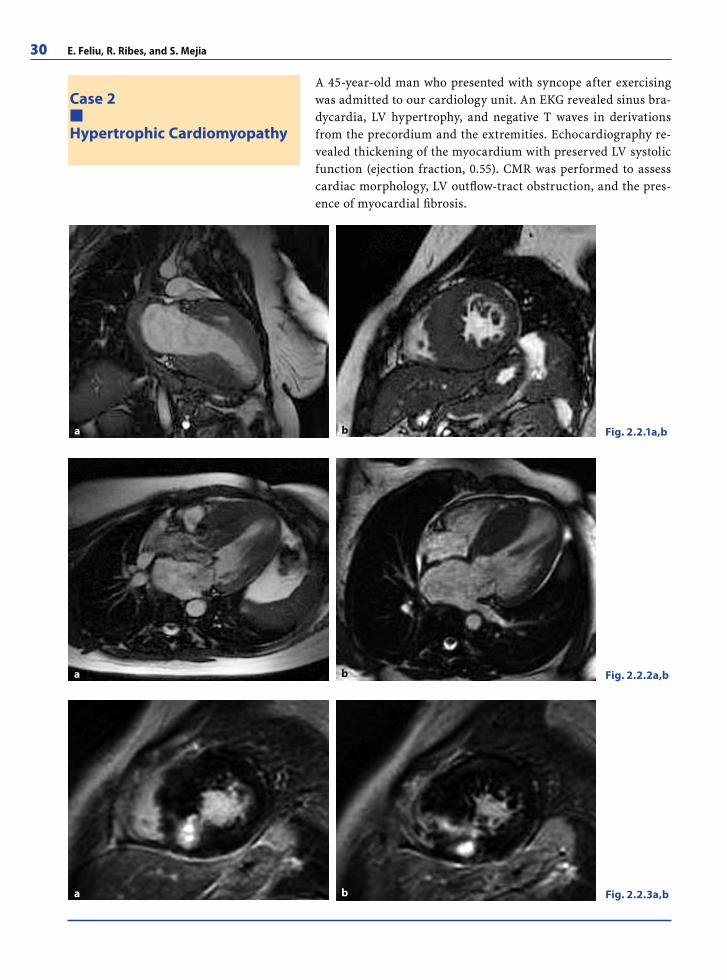
30 E. Feliu, R. Ribes, and S. Mejia
Case 2
Hypertrophic Cardiomyopathy
A 45-year-old man who presented with syncope after exercising was admitted to our cardiology unit. An EKG revealed sinus bra-dycardia, LV hypertrophy, and negative T waves in derivations from the precordium and the extremities. Echocardiography re-vealed thickening of the myocardium with preserved LV systolic function (ejection fraction, 0.55). CMR was performed to assess cardiac morphology, LV outfl ow-tract obstruction, and the pres-ence of myocardial fi brosis.
Fig. 2.2.1a,b
Fig. 2.2.2a,b
Fig. 2.2.3a,b
a b
a b
a b
RibesLunaRos.indb 30 18.03.2008 12:49:02
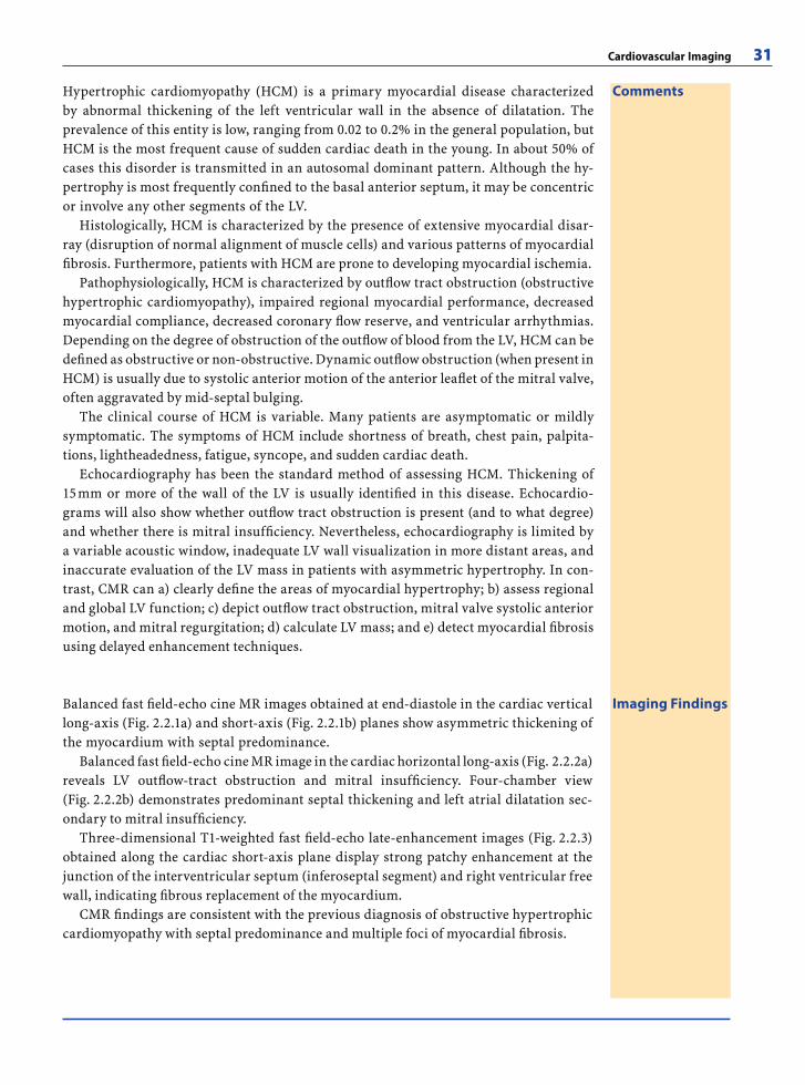
Cardiovascular Imaging 31
CommentsHypertrophic cardiomyopathy (HCM) is a primary myocardial disease characterized by abnormal thickening of the left ventricular wall in the absence of dilatation. The prevalence of this entity is low, ranging from 0.02 to 0.2% in the general population, but HCM is the most frequent cause of sudden cardiac death in the young. In about 50% of cases this disorder is transmitted in an autosomal dominant pattern. Although the hy-pertrophy is most frequently confi ned to the basal anterior septum, it may be concentric or involve any other segments of the LV.
Histologically, HCM is characterized by the presence of extensive myocardial disar-ray (disruption of normal alignment of muscle cells) and various patterns of myocardial fi brosis. Furthermore, patients with HCM are prone to developing myocardial ischemia.
Pathophysiologically, HCM is characterized by outfl ow tract obstruction (obstructive hypertrophic cardiomyopathy), impaired regional myocardial performance, decreased myocardial compliance, decreased coronary fl ow reserve, and ventricular arrhythmias. Depending on the degree of obstruction of the outfl ow of blood from the LV, HCM can be defi ned as obstructive or non-obstructive. Dynamic outfl ow obstruction (when present in HCM) is usually due to systolic anterior motion of the anterior leafl et of the mitral valve, often aggravated by mid-septal bulging.
The clinical course of HCM is variable. Many patients are asymptomatic or mildly symptomatic. The symptoms of HCM include shortness of breath, chest pain, palpita-tions, lightheadedness, fatigue, syncope, and sudden cardiac death.
Echocardiography has been the standard method of assessing HCM. Thickening of 15 mm or more of the wall of the LV is usually identifi ed in this disease. Echocardio-grams will also show whether outfl ow tract obstruction is present (and to what degree) and whether there is mitral insuffi ciency. Nevertheless, echocardiography is limited by a variable acoustic window, inadequate LV wall visualization in more distant areas, and inaccurate evaluation of the LV mass in patients with asymmetric hypertrophy. In con-trast, CMR can a) clearly defi ne the areas of myocardial hypertrophy; b) assess regional and global LV function; c) depict outfl ow tract obstruction, mitral valve systolic anterior motion, and mitral regurgitation; d) calculate LV mass; and e) detect myocardial fi brosis using delayed enhancement techniques.
Imaging FindingsBalanced fast fi eld-echo cine MR images obtained at end-diastole in the cardiac vertical long-axis (Fig. 2.2.1a) and short-axis (Fig. 2.2.1b) planes show asymmetric thickening of the myocardium with septal predominance.
Balanced fast fi eld-echo cine MR image in the cardiac horizontal long-axis (Fig. 2.2.2a) reveals LV outfl ow-tract obstruction and mitral insuffi ciency. Four-chamber view (Fig. 2.2.2b) demonstrates predominant septal thickening and left atrial dilatation sec-ondary to mitral insuffi ciency.
Three-dimensional T1-weighted fast fi eld-echo late-enhancement images (Fig. 2.2.3) obtained along the cardiac short-axis plane display strong patchy enhancement at the junction of the interventricular septum (inferoseptal segment) and right ventricular free wall, indicating fi brous replacement of the myocardium.
CMR fi ndings are consistent with the previous diagnosis of obstructive hypertrophic cardiomyopathy with septal predominance and multiple foci of myocardial fi brosis.
RibesLunaRos.indb 31 18.03.2008 12:49:05
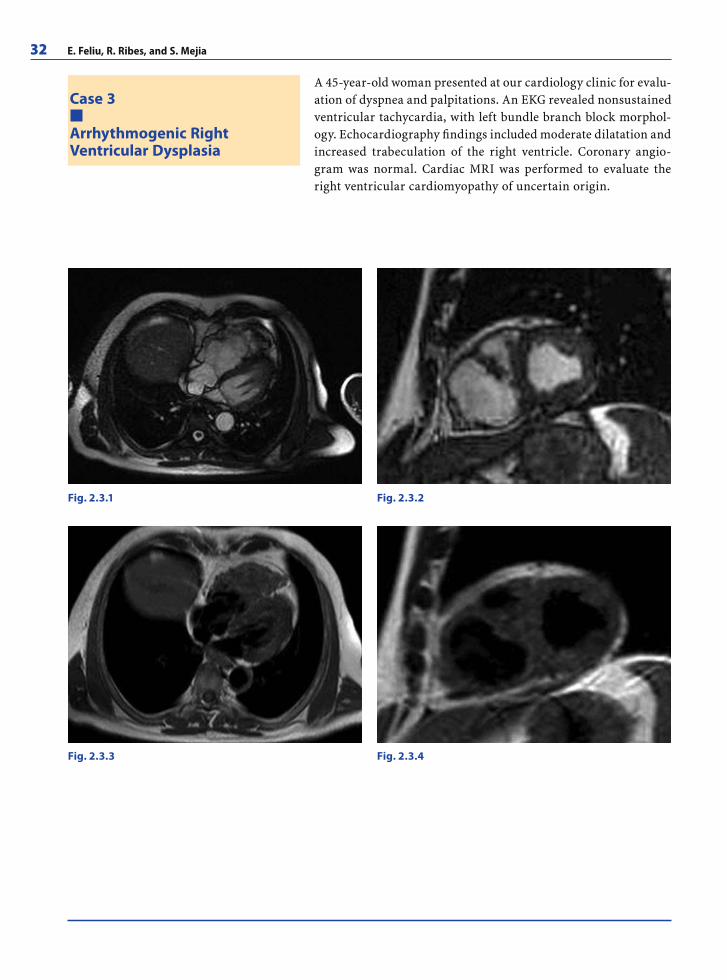
32 E. Feliu, R. Ribes, and S. Mejia
Case 3
Arrhythmogenic RightVentricular Dysplasia
A 45-year-old woman presented at our cardiology clinic for evalu-ation of dyspnea and palpitations. An EKG revealed nonsustained ventricular tachycardia, with left bundle branch block morphol-ogy. Echocardiography fi ndings included moderate dilatation and increased trabeculation of the right ventricle. Coronary angio-gram was normal. Cardiac MRI was performed to evaluate the right ventricular cardiomyopathy of uncertain origin.
Fig. 2.3.1 Fig. 2.3.2
Fig. 2.3.3 Fig. 2.3.4
RibesLunaRos.indb 32 18.03.2008 12:49:05
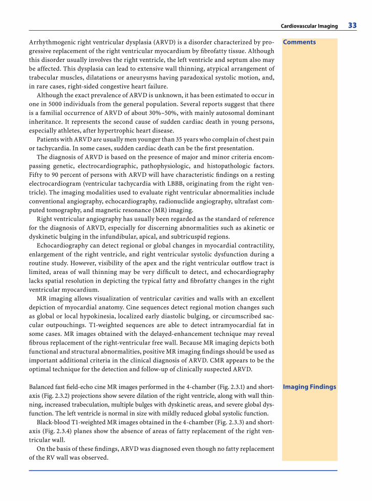
Cardiovascular Imaging 33
CommentsArrhythmogenic right ventricular dysplasia (ARVD) is a disorder characterized by pro-gressive replacement of the right ventricular myocardium by fi brofatty tissue. Although this disorder usually involves the right ventricle, the left ventricle and septum also may be affected. This dysplasia can lead to extensive wall thinning, atypical arrangement of trabecular muscles, dilatations or aneurysms having paradoxical systolic motion, and, in rare cases, right-sided congestive heart failure.
Although the exact prevalence of ARVD is unknown, it has been estimated to occur in one in 5000 individuals from the general population. Several reports suggest that there is a familial occurrence of ARVD of about 30%–50%, with mainly autosomal dominant inheritance. It represents the second cause of sudden cardiac death in young persons, especially athletes, after hypertrophic heart disease.
Patients with ARVD are usually men younger than 35 years who complain of chest pain or tachycardia. In some cases, sudden cardiac death can be the fi rst presentation.
The diagnosis of ARVD is based on the presence of major and minor criteria encom-passing genetic, electrocardiographic, pathophysiologic, and histopathologic factors. Fifty to 90 percent of persons with ARVD will have characteristic fi ndings on a resting electrocardiogram (ventricular tachycardia with LBBB, originating from the right ven-tricle). The imaging modalities used to evaluate right ventricular abnormalities include conventional angiography, echocardiography, radionuclide angiography, ultrafast com-puted tomography, and magnetic resonance (MR) imaging.
Right ventricular angiography has usually been regarded as the standard of reference for the diagnosis of ARVD, especially for discerning abnormalities such as akinetic or dyskinetic bulging in the infundibular, apical, and subtricuspid regions.
Echocardiography can detect regional or global changes in myocardial contractility, enlargement of the right ventricle, and right ventricular systolic dysfunction during a routine study. However, visibility of the apex and the right ventricular outfl ow tract is limited, areas of wall thinning may be very diffi cult to detect, and echocardiography lacks spatial resolution in depicting the typical fatty and fi brofatty changes in the right ventricular myocardium.
MR imaging allows visualization of ventricular cavities and walls with an excellent depiction of myocardial anatomy. Cine sequences detect regional motion changes such as global or local hypokinesia, localized early diastolic bulging, or circumscribed sac-cular outpouchings. T1-weighted sequences are able to detect intramyocardial fat in some cases. MR images obtained with the delayed-enhancement technique may reveal fi brous replacement of the right-ventricular free wall. Because MR imaging depicts both functional and structural abnormalities, positive MR imaging fi ndings should be used as important additional criteria in the clinical diagnosis of ARVD. CMR appears to be the optimal technique for the detection and follow-up of clinically suspected ARVD.
Imaging FindingsBalanced fast fi eld-echo cine MR images performed in the 4-chamber (Fig. 2.3.1) and short-axis (Fig. 2.3.2) projections show severe dilation of the right ventricle, along with wall thin-ning, increased trabeculation, multiple bulges with dyskinetic areas, and severe global dys-function. The left ventricle is normal in size with mildly reduced global systolic function.
Black-blood T1-weighted MR images obtained in the 4-chamber (Fig. 2.3.3) and short-axis (Fig. 2.3.4) planes show the absence of areas of fatty replacement of the right ven-tricular wall.
On the basis of these fi ndings, ARVD was diagnosed even though no fatty replacement of the RV wall was observed.
RibesLunaRos.indb 33 18.03.2008 12:49:06
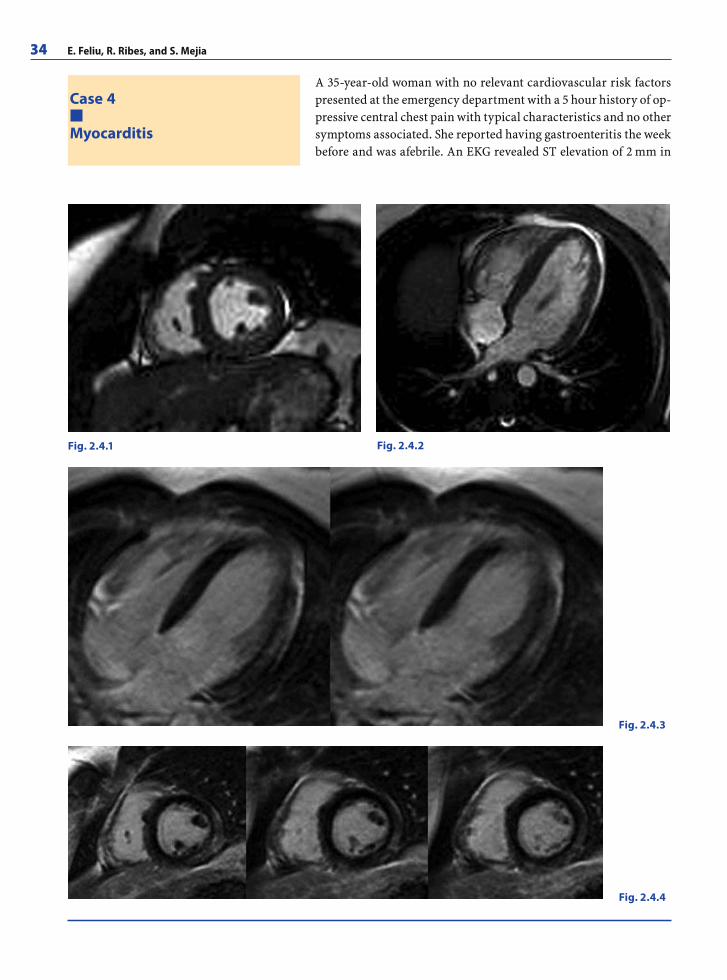
34 E. Feliu, R. Ribes, and S. Mejia
Case 4
Myocarditis
A 35-year-old woman with no relevant cardiovascular risk factors presented at the emergency department with a 5 hour history of op-pressive central chest pain with typical characteristics and no other symptoms associated. She reported having gastroenteritis the week before and was afebrile. An EKG revealed ST elevation of 2 mm in
Fig. 2.4.1 Fig. 2.4.2
Fig. 2.4.3
Fig. 2.4.4
RibesLunaRos.indb 34 18.03.2008 12:49:07
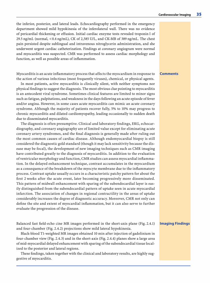
Cardiovascular Imaging 35
CommentsMyocarditis is an acute infl ammatory process that affects the myocardium in response to the action of various infectious (most frequently viruses), chemical, or physical agents.
In most patients, active myocarditis is clinically silent, with neither symptoms nor physical fi ndings to suggest the diagnosis. The most obvious clue pointing to myocarditis is an antecedent viral syndrome. Sometimes clinical features are limited to minor signs such as fatigue, palpitations, and weakness in the days following an acute episode of fever and/or angina. However, in some cases acute myocarditis can mimic an acute coronary syndrome. Although the majority of patients recover fully, 5% to 10% may progress to chronic myocarditis and dilated cardiomyopathy, leading occasionally to sudden death due to disseminated myocarditis.
The diagnosis is often presumptive. Clinical and laboratory fi ndings, EKG, echocar-diography, and coronary angiography are of limited value except for eliminating acute coronary artery syndromes, and the fi nal diagnosis is generally made after ruling out the most common causes of cardiac disease. Although endomyocardial biopsy is still considered the diagnostic gold standard (though it may lack sensitivity because the dis-ease may be focal), the development of new imaging techniques such as CMR imaging have contributed greatly to the diagnosis of myocarditis. In addition to the evaluation of ventricular morphology and function, CMR studies can assess myocardial infl amma-tion. In the delayed enhancement technique, contrast accumulates in the myocardium as a consequence of the breakdown of the myocyte membrane due to the infl ammatory process. Contrast uptake usually occurs in a characteristic patchy pattern for about the fi rst 2 weeks after the acute event, later becoming progressively more disseminated. This pattern of midwall enhancement with sparing of the subendocardial layer is eas-ily distinguished from the subendocardial pattern of uptake seen in acute myocardial infarction. The association of changes in regional contractility in the areas of uptake considerably increases the degree of diagnostic accuracy. Moreover, CMR not only can defi ne the site and extent of myocardial infl ammation, but it can also serve to further evaluate the progression of the disease.
Imaging FindingsBalanced fast fi eld-echo cine MR images performed in the short-axis plane (Fig. 2.4.1) and four-chamber (Fig. 2.4.2) projections show mild lateral hypokinesia.
Black-blood T1-weighted MR images obtained 10 min after injection of gadolinium in four-chamber view (Fig. 2.4.3) and in the short-axis (Fig. 2.4.4) planes show a large area of mid-myocardial delayed enhancement with sparing of the subendocardial tissue local-ized to the posterior and lateral regions.
These fi ndings, taken together with the clinical and laboratory results, are highly sug-gestive of myocarditis.
the inferior, posterior, and lateral leads. Echocardiography performed in the emergency department showed mild hypokinesia of the inferolateral wall. There was no evidence of pericardial thickening or effusion. Initial cardiac enzyme tests revealed troponin I of 29.5 ng/mL (normal, < 0.4 ng/mL), CK of 2,585 U/L, and CK-MB of 389 ng/mL. The chest pain persisted despite sublingual and intravenous nitroglycerin administration, and she underwent urgent cardiac catheterization. Findings at coronary angiogram were normal and myocarditis was suspected. CMR was performed to assess cardiac morphology and function, as well as possible areas of infl ammation.
RibesLunaRos.indb 35 18.03.2008 12:49:09
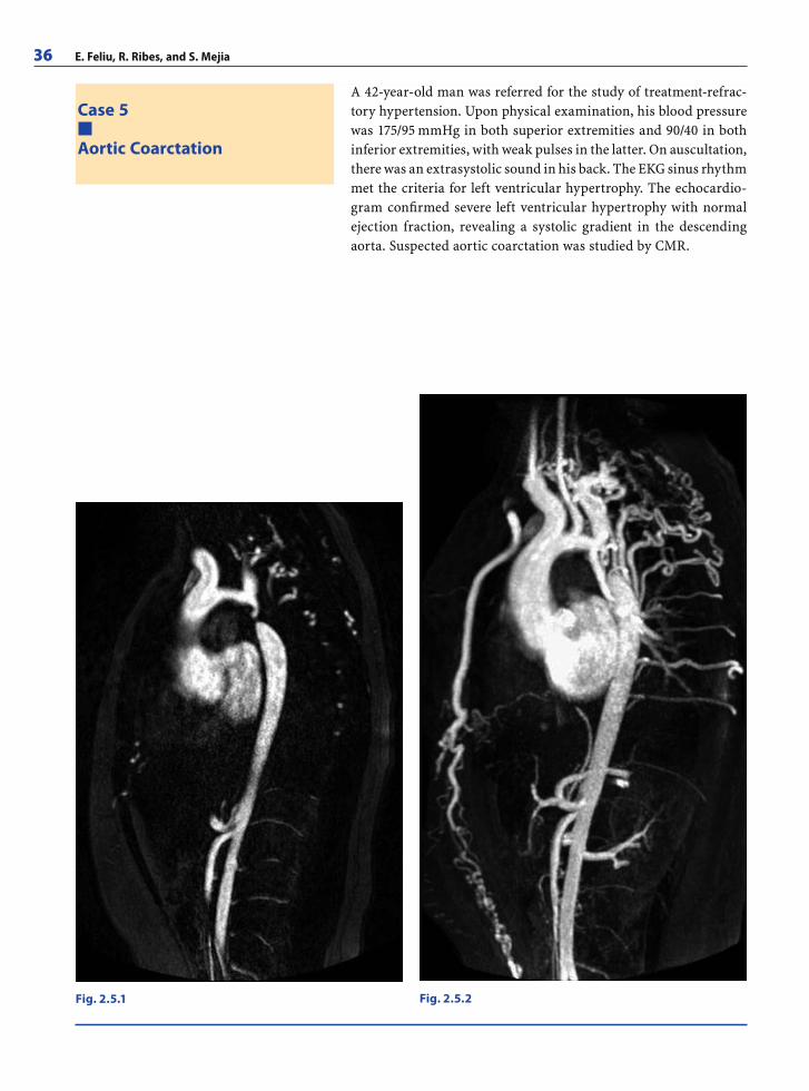
36 E. Feliu, R. Ribes, and S. Mejia
Case 5
Aortic Coarctation
A 42-year-old man was referred for the study of treatment-refrac-tory hypertension. Upon physical examination, his blood pressure was 175/95 mmHg in both superior extremities and 90/40 in both inferior extremities, with weak pulses in the latter. On auscultation, there was an extrasystolic sound in his back. The EKG sinus rhythm met the criteria for left ventricular hypertrophy. The echocardio-gram confi rmed severe left ventricular hypertrophy with normal ejection fraction, revealing a systolic gradient in the descending aorta. Suspected aortic coarctation was studied by CMR.
Fig. 2.5.1 Fig. 2.5.2
RibesLunaRos.indb 36 18.03.2008 12:49:09
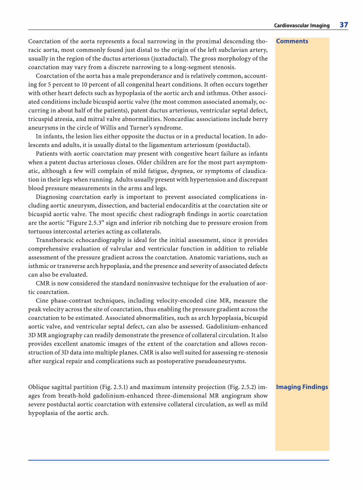
Cardiovascular Imaging 37
CommentsCoarctation of the aorta represents a focal narrowing in the proximal descending tho-racic aorta, most commonly found just distal to the origin of the left subclavian artery, usually in the region of the ductus arteriosus (juxtaductal). The gross morphology of the coarctation may vary from a discrete narrowing to a long-segment stenosis.
Coarctation of the aorta has a male preponderance and is relatively common, account-ing for 5 percent to 10 percent of all congenital heart conditions. It often occurs together with other heart defects such as hypoplasia of the aortic arch and isthmus. Other associ-ated conditions include bicuspid aortic valve (the most common associated anomaly, oc-curring in about half of the patients), patent ductus arteriosus, ventricular septal defect, tricuspid atresia, and mitral valve abnormalities. Noncardiac associations include berry aneurysms in the circle of Willis and Turner’s syndrome.
In infants, the lesion lies either opposite the ductus or in a preductal location. In ado-lescents and adults, it is usually distal to the ligamentum arteriosum (postductal).
Patients with aortic coarctation may present with congestive heart failure as infants when a patent ductus arteriosus closes. Older children are for the most part asymptom-atic, although a few will complain of mild fatigue, dyspnea, or symptoms of claudica-tion in their legs when running. Adults usually present with hypertension and discrepant blood pressure measurements in the arms and legs.
Diagnosing coarctation early is important to prevent associated complications in-cluding aortic aneurysm, dissection, and bacterial endocarditis at the coarctation site or bicuspid aortic valve. The most specifi c chest radiograph fi ndings in aortic coarctation are the aortic “Figure 2.5.3” sign and inferior rib notching due to pressure erosion from tortuous intercostal arteries acting as collaterals.
Transthoracic echocardiography is ideal for the initial assessment, since it provides comprehensive evaluation of valvular and ventricular function in addition to reliable assessment of the pressure gradient across the coarctation. Anatomic variations, such as isthmic or transverse arch hypoplasia, and the presence and severity of associated defects can also be evaluated.
CMR is now considered the standard noninvasive technique for the evaluation of aor-tic coarctation.
Cine phase-contrast techniques, including velocity-encoded cine MR, measure the peak velocity across the site of coarctation, thus enabling the pressure gradient across the coarctation to be estimated. Associated abnormalities, such as arch hypoplasia, bicuspid aortic valve, and ventricular septal defect, can also be assessed. Gadolinium-enhanced 3D MR angiography can readily demonstrate the presence of collateral circulation. It also provides excellent anatomic images of the extent of the coarctation and allows recon-struction of 3D data into multiple planes. CMR is also well suited for assessing re-stenosis after surgical repair and complications such as postoperative pseudoaneurysms.
Imaging FindingsOblique sagittal partition (Fig. 2.5.1) and maximum intensity projection (Fig. 2.5.2) im-ages from breath-hold gadolinium-enhanced three-dimensional MR angiogram show severe postductal aortic coarctation with extensive collateral circulation, as well as mild hypoplasia of the aortic arch.
RibesLunaRos.indb 37 18.03.2008 12:49:10
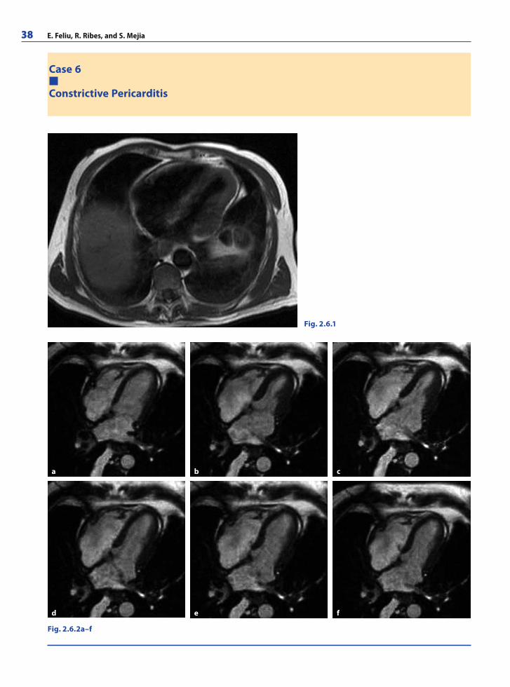
38 E. Feliu, R. Ribes, and S. Mejia
Case 6
Constrictive Pericarditis
Fig. 2.6.1
Fig. 2.6.2a–f
a cb
d e f
RibesLunaRos.indb 38 18.03.2008 12:49:10
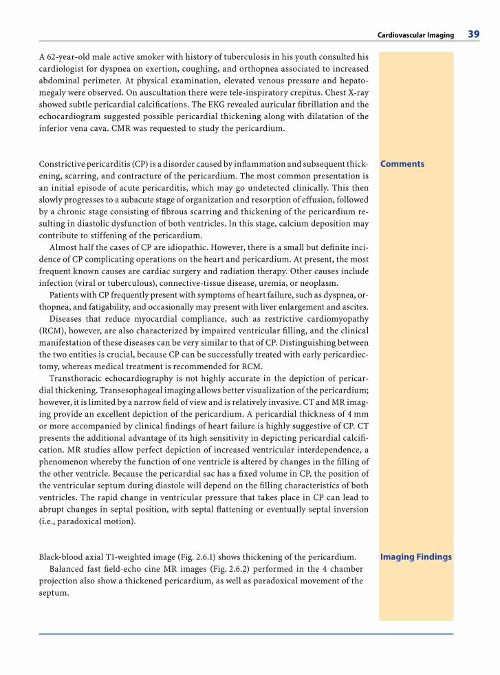
Cardiovascular Imaging 39
A 62-year-old male active smoker with history of tuberculosis in his youth consulted his cardiologist for dyspnea on exertion, coughing, and orthopnea associated to increased abdominal perimeter. At physical examination, elevated venous pressure and hepato-megaly were observed. On auscultation there were tele-inspiratory crepitus. Chest X-ray showed subtle pericardial calcifi cations. The EKG revealed auricular fi brillation and the echocardiogram suggested possible pericardial thickening along with dilatation of the inferior vena cava. CMR was requested to study the pericardium.
CommentsConstrictive pericarditis (CP) is a disorder caused by infl ammation and subsequent thick-ening, scarring, and contracture of the pericardium. The most common presentation is an initial episode of acute pericarditis, which may go undetected clinically. This then slowly progresses to a subacute stage of organization and resorption of effusion, followed by a chronic stage consisting of fi brous scarring and thickening of the pericardium re-sulting in diastolic dysfunction of both ventricles. In this stage, calcium deposition may contribute to stiffening of the pericardium.
Almost half the cases of CP are idiopathic. However, there is a small but defi nite inci-dence of CP complicating operations on the heart and pericardium. At present, the most frequent known causes are cardiac surgery and radiation therapy. Other causes include infection (viral or tuberculous), connective-tissue disease, uremia, or neoplasm.
Patients with CP frequently present with symptoms of heart failure, such as dyspnea, or-thopnea, and fatigability, and occasionally may present with liver enlargement and ascites.
Diseases that reduce myocardial compliance, such as restrictive cardiomyopathy (RCM), however, are also characterized by impaired ventricular fi lling, and the clinical manifestation of these diseases can be very similar to that of CP. Distinguishing between the two entities is crucial, because CP can be successfully treated with early pericardiec-tomy, whereas medical treatment is recommended for RCM.
Transthoracic echocardiography is not highly accurate in the depiction of pericar-dial thickening. Transesophageal imaging allows better visualization of the pericardium; however, it is limited by a narrow fi eld of view and is relatively invasive. CT and MR imag-ing provide an excellent depiction of the pericardium. A pericardial thickness of 4 mm or more accompanied by clinical fi ndings of heart failure is highly suggestive of CP. CT presents the additional advantage of its high sensitivity in depicting pericardial calcifi -cation. MR studies allow perfect depiction of increased ventricular interdependence, a phenomenon whereby the function of one ventricle is altered by changes in the fi lling of the other ventricle. Because the pericardial sac has a fi xed volume in CP, the position of the ventricular septum during diastole will depend on the fi lling characteristics of both ventricles. The rapid change in ventricular pressure that takes place in CP can lead to abrupt changes in septal position, with septal fl attening or eventually septal inversion (i.e., paradoxical motion).
Imaging FindingsBlack-blood axial T1-weighted image (Fig. 2.6.1) shows thickening of the pericardium. Balanced fast fi eld-echo cine MR images (Fig. 2.6.2) performed in the 4 chamber
projection also show a thickened pericardium, as well as paradoxical movement of the septum.
RibesLunaRos.indb 39 18.03.2008 12:49:12
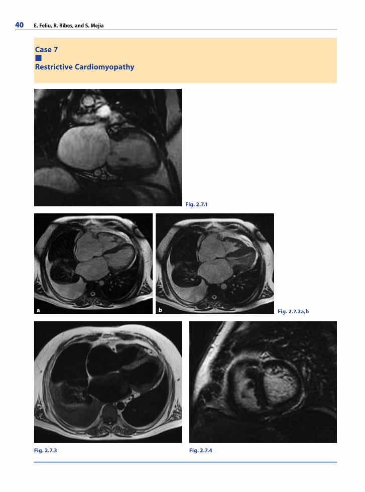
40 E. Feliu, R. Ribes, and S. Mejia
Case 7
Restrictive Cardiomyopathy
Fig. 2.7.3 Fig. 2.7.4
Fig. 2.7.1
Fig. 2.7.2a,ba b
RibesLunaRos.indb 40 18.03.2008 12:49:12
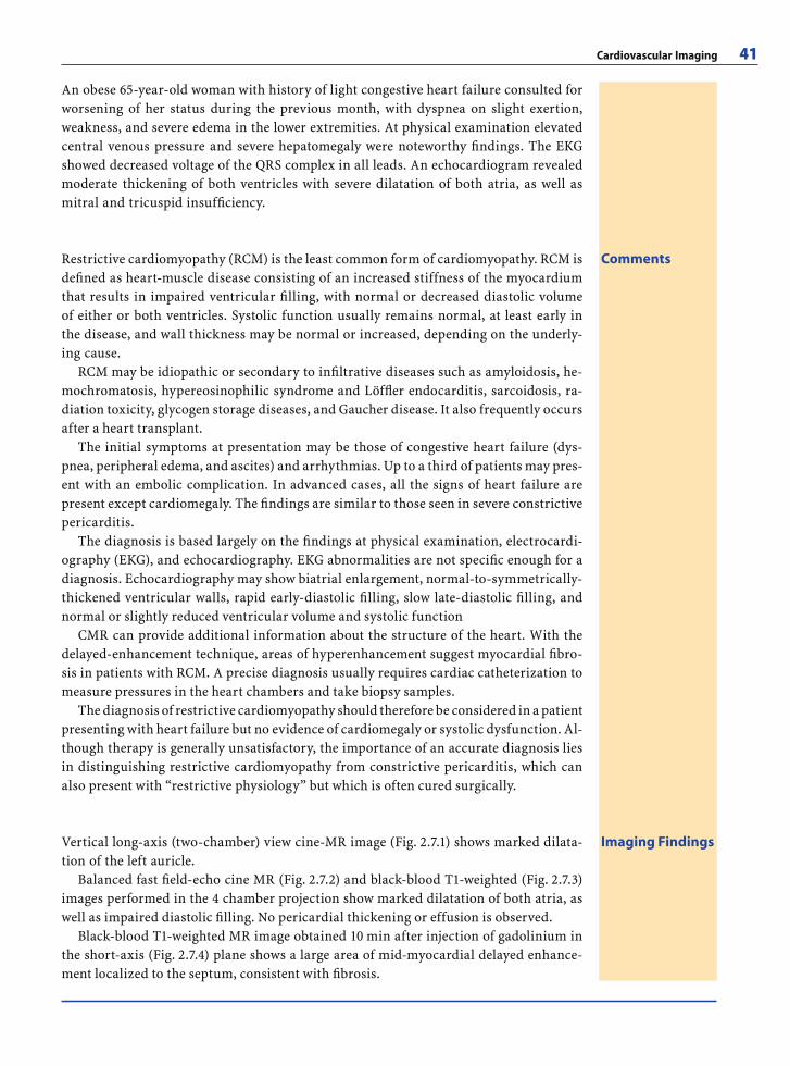
Cardiovascular Imaging 41
An obese 65-year-old woman with history of light congestive heart failure consulted for worsening of her status during the previous month, with dyspnea on slight exertion, weakness, and severe edema in the lower extremities. At physical examination elevated central venous pressure and severe hepatomegaly were noteworthy fi ndings. The EKG showed decreased voltage of the QRS complex in all leads. An echocardiogram revealed moderate thickening of both ventricles with severe dilatation of both atria, as well as mitral and tricuspid insuffi ciency.
CommentsRestrictive cardiomyopathy (RCM) is the least common form of cardiomyopathy. RCM is defi ned as heart-muscle disease consisting of an increased stiffness of the myocardium that results in impaired ventricular fi lling, with normal or decreased diastolic volume of either or both ventricles. Systolic function usually remains normal, at least early in the disease, and wall thickness may be normal or increased, depending on the underly-ing cause.
RCM may be idiopathic or secondary to infi ltrative diseases such as amyloidosis, he-mochromatosis, hypereosinophilic syndrome and Löffl er endocarditis, sarcoidosis, ra-diation toxicity, glycogen storage diseases, and Gaucher disease. It also frequently occurs after a heart transplant.
The initial symptoms at presentation may be those of congestive heart failure (dys-pnea, peripheral edema, and ascites) and arrhythmias. Up to a third of patients may pres-ent with an embolic complication. In advanced cases, all the signs of heart failure are present except cardiomegaly. The fi ndings are similar to those seen in severe constrictive pericarditis.
The diagnosis is based largely on the fi ndings at physical examination, electrocardi-ography (EKG), and echocardiography. EKG abnormalities are not specifi c enough for a diagnosis. Echocardiography may show biatrial enlargement, normal-to-symmetrically-thickened ventricular walls, rapid early-diastolic fi lling, slow late-diastolic fi lling, and normal or slightly reduced ventricular volume and systolic function
CMR can provide additional information about the structure of the heart. With the delayed-enhancement technique, areas of hyperenhancement suggest myocardial fi bro-sis in patients with RCM. A precise diagnosis usually requires cardiac catheterization to measure pressures in the heart chambers and take biopsy samples.
The diagnosis of restrictive cardiomyopathy should therefore be considered in a patient presenting with heart failure but no evidence of cardiomegaly or systolic dysfunction. Al-though therapy is generally unsatisfactory, the importance of an accurate diagnosis lies in distinguishing restrictive cardiomyopathy from constrictive pericarditis, which can also present with “restrictive physiology” but which is often cured surgically.
Imaging FindingsVertical long-axis (two-chamber) view cine-MR image (Fig. 2.7.1) shows marked dilata-tion of the left auricle.
Balanced fast fi eld-echo cine MR (Fig. 2.7.2) and black-blood T1-weighted (Fig. 2.7.3) images performed in the 4 chamber projection show marked dilatation of both atria, as well as impaired diastolic fi lling. No pericardial thickening or effusion is observed.
Black-blood T1-weighted MR image obtained 10 min after injection of gadolinium in the short-axis (Fig. 2.7.4) plane shows a large area of mid-myocardial delayed enhance-ment localized to the septum, consistent with fi brosis.
RibesLunaRos.indb 41 18.03.2008 12:49:14
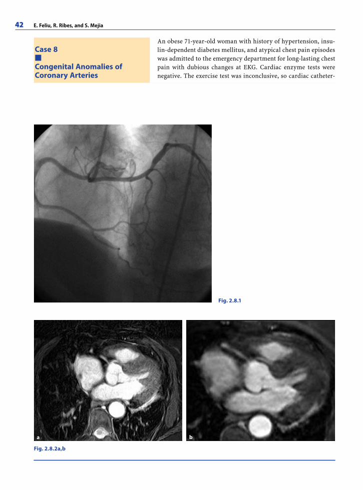
42 E. Feliu, R. Ribes, and S. Mejia
Case 8
Congenital Anomalies ofCoronary Arteries
An obese 71-year-old woman with history of hypertension, insu-lin-dependent diabetes mellitus, and atypical chest pain episodes was admitted to the emergency department for long-lasting chest pain with dubious changes at EKG. Cardiac enzyme tests were negative. The exercise test was inconclusive, so cardiac catheter-
Fig. 2.8.2a,b
Fig. 2.8.1
a b
RibesLunaRos.indb 42 18.03.2008 12:49:14
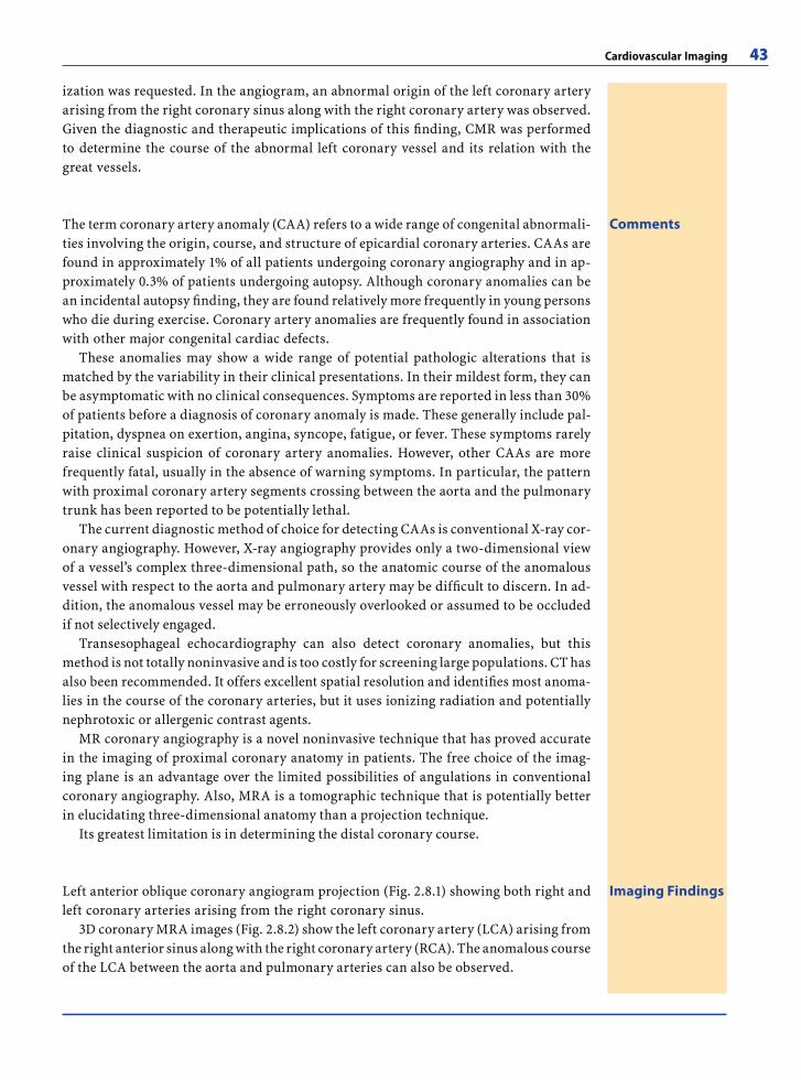
Cardiovascular Imaging 43
CommentsThe term coronary artery anomaly (CAA) refers to a wide range of congenital abnormali-ties involving the origin, course, and structure of epicardial coronary arteries. CAAs are found in approximately 1% of all patients undergoing coronary angiography and in ap-proximately 0.3% of patients undergoing autopsy. Although coronary anomalies can be an incidental autopsy fi nding, they are found relatively more frequently in young persons who die during exercise. Coronary artery anomalies are frequently found in association with other major congenital cardiac defects.
These anomalies may show a wide range of potential pathologic alterations that is matched by the variability in their clinical presentations. In their mildest form, they can be asymptomatic with no clinical consequences. Symptoms are reported in less than 30% of patients before a diagnosis of coronary anomaly is made. These generally include pal-pitation, dyspnea on exertion, angina, syncope, fatigue, or fever. These symptoms rarely raise clinical suspicion of coronary artery anomalies. However, other CAAs are more frequently fatal, usually in the absence of warning symptoms. In particular, the pattern with proximal coronary artery segments crossing between the aorta and the pulmonary trunk has been reported to be potentially lethal.
The current diagnostic method of choice for detecting CAAs is conventional X-ray cor-onary angiography. However, X-ray angiography provides only a two-dimensional view of a vessel’s complex three-dimensional path, so the anatomic course of the anomalous vessel with respect to the aorta and pulmonary artery may be diffi cult to discern. In ad-dition, the anomalous vessel may be erroneously overlooked or assumed to be occluded if not selectively engaged.
Transesophageal echocardiography can also detect coronary anomalies, but this method is not totally noninvasive and is too costly for screening large populations. CT has also been recommended. It offers excellent spatial resolution and identifi es most anoma-lies in the course of the coronary arteries, but it uses ionizing radiation and potentially nephrotoxic or allergenic contrast agents.
MR coronary angiography is a novel noninvasive technique that has proved accurate in the imaging of proximal coronary anatomy in patients. The free choice of the imag-ing plane is an advantage over the limited possibilities of angulations in conventional coronary angiography. Also, MRA is a tomographic technique that is potentially better in elucidating three-dimensional anatomy than a projection technique.
Its greatest limitation is in determining the distal coronary course.
Imaging FindingsLeft anterior oblique coronary angiogram projection (Fig. 2.8.1) showing both right and left coronary arteries arising from the right coronary sinus.
3D coronary MRA images (Fig. 2.8.2) show the left coronary artery (LCA) arising from the right anterior sinus along with the right coronary artery (RCA). The anomalous course of the LCA between the aorta and pulmonary arteries can also be observed.
ization was requested. In the angiogram, an abnormal origin of the left coronary artery arising from the right coronary sinus along with the right coronary artery was observed. Given the diagnostic and therapeutic implications of this fi nding, CMR was performed to determine the course of the abnormal left coronary vessel and its relation with the great vessels.
RibesLunaRos.indb 43 18.03.2008 12:49:16
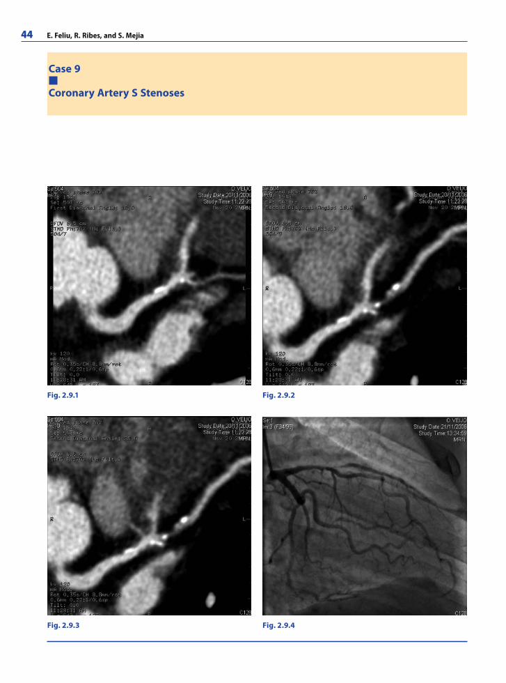
44 E. Feliu, R. Ribes, and S. Mejia
Case 9
Coronary Artery S Stenoses
Fig. 2.9.1 Fig. 2.9.2
Fig. 2.9.3 Fig. 2.9.4
RibesLunaRos.indb 44 18.03.2008 12:49:17
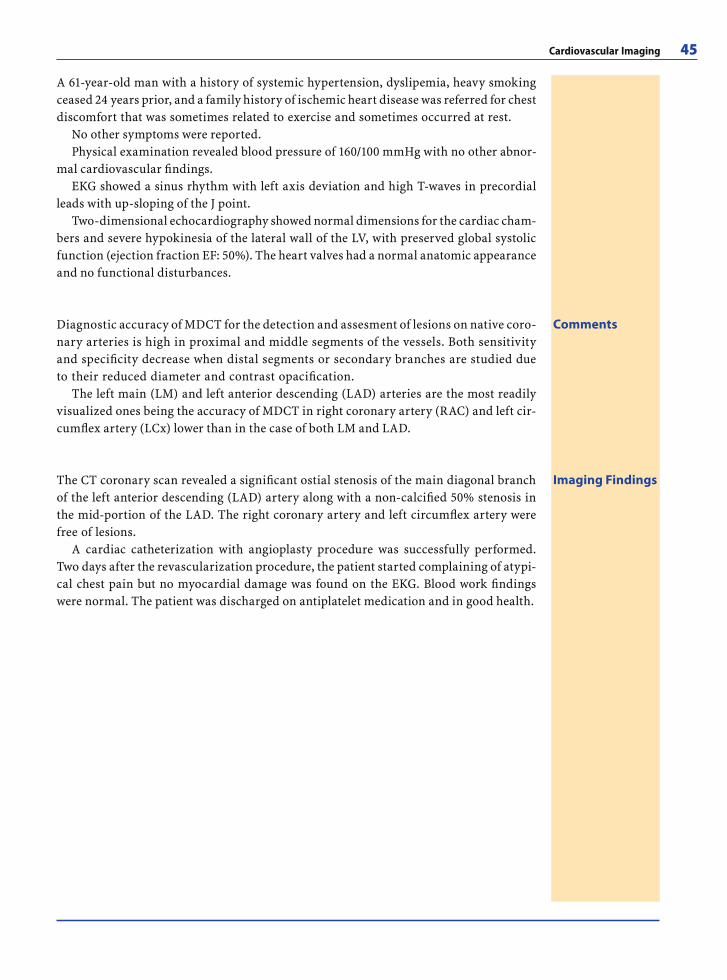
Cardiovascular Imaging 45
A 61-year-old man with a history of systemic hypertension, dyslipemia, heavy smoking ceased 24 years prior, and a family history of ischemic heart disease was referred for chest discomfort that was sometimes related to exercise and sometimes occurred at rest.
No other symptoms were reported.Physical examination revealed blood pressure of 160/100 mmHg with no other abnor-
mal cardiovascular fi ndings.EKG showed a sinus rhythm with left axis deviation and high T-waves in precordial
leads with up-sloping of the J point.Two-dimensional echocardiography showed normal dimensions for the cardiac cham-
bers and severe hypokinesia of the lateral wall of the LV, with preserved global systolic function (ejection fraction EF: 50%). The heart valves had a normal anatomic appearance and no functional disturbances.
CommentsDiagnostic accuracy of MDCT for the detection and assesment of lesions on native coro-nary arteries is high in proximal and middle segments of the vessels. Both sensitivity and specifi city decrease when distal segments or secondary branches are studied due to their reduced diameter and contrast opacifi cation.
The left main (LM) and left anterior descending (LAD) arteries are the most readily visualized ones being the accuracy of MDCT in right coronary artery (RAC) and left cir-cumfl ex artery (LCx) lower than in the case of both LM and LAD.
Imaging FindingsThe CT coronary scan revealed a signifi cant ostial stenosis of the main diagonal branch of the left anterior descending (LAD) artery along with a non-calcifi ed 50% stenosis in the mid-portion of the LAD. The right coronary artery and left circumfl ex artery were free of lesions.
A cardiac catheterization with angioplasty procedure was successfully performed. Two days after the revascularization procedure, the patient started complaining of atypi-cal chest pain but no myocardial damage was found on the EKG. Blood work fi ndings were normal. The patient was discharged on antiplatelet medication and in good health.
RibesLunaRos.indb 45 18.03.2008 12:49:19
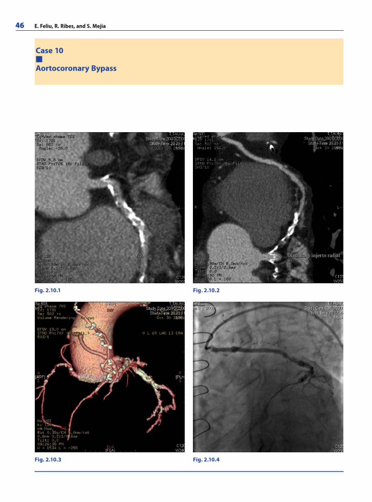
46 E. Feliu, R. Ribes, and S. Mejia
Case 10
Aortocoronary Bypass
Fig. 2.10.1 Fig. 2.10.2
Fig. 2.10.3 Fig. 2.10.4
RibesLunaRos.indb 46 18.03.2008 12:49:19
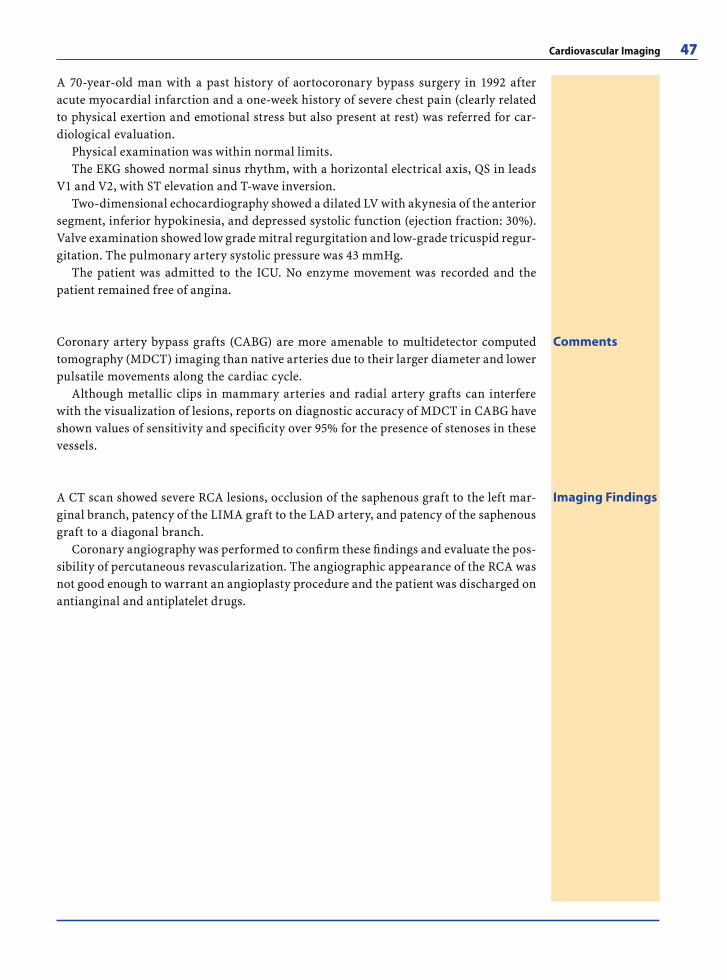
Cardiovascular Imaging 47
A 70-year-old man with a past history of aortocoronary bypass surgery in 1992 after acute myocardial infarction and a one-week history of severe chest pain (clearly related to physical exertion and emotional stress but also present at rest) was referred for car-diological evaluation.
Physical examination was within normal limits.The EKG showed normal sinus rhythm, with a horizontal electrical axis, QS in leads
V1 and V2, with ST elevation and T-wave inversion.Two-dimensional echocardiography showed a dilated LV with akynesia of the anterior
segment, inferior hypokinesia, and depressed systolic function (ejection fraction: 30%). Valve examination showed low grade mitral regurgitation and low-grade tricuspid regur-gitation. The pulmonary artery systolic pressure was 43 mmHg.
The patient was admitted to the ICU. No enzyme movement was recorded and the patient remained free of angina.
CommentsCoronary artery bypass grafts (CABG) are more amenable to multidetector computed tomography (MDCT) imaging than native arteries due to their larger diameter and lower pulsatile movements along the cardiac cycle.
Although metallic clips in mammary arteries and radial artery grafts can interfere with the visualization of lesions, reports on diagnostic accuracy of MDCT in CABG have shown values of sensitivity and specifi city over 95% for the presence of stenoses in these vessels.
Imaging FindingsA CT scan showed severe RCA lesions, occlusion of the saphenous graft to the left mar-ginal branch, patency of the LIMA graft to the LAD artery, and patency of the saphenous graft to a diagonal branch.
Coronary angiography was performed to confi rm these fi ndings and evaluate the pos-sibility of percutaneous revascularization. The angiographic appearance of the RCA was not good enough to warrant an angioplasty procedure and the patient was discharged on antianginal and antiplatelet drugs.
RibesLunaRos.indb 47 18.03.2008 12:49:24
48 E. Feliu, R. Ribes, and S. Mejia
Further Readings
Books
Atlas of Non-Invasive Coronary Angiography by Multidetec-tor Computed Tomography. Pons-Lladó G, Leta-Petracca R (2006) Springer-Verlag, Berlin. ISBN-13: 9780387330440
Atlas of Practical Applications of Cardiovascular Magnetic Resonance (Developments in Cardiovascular Medicine). Pons-Lladó G, Carreras F (2005) Springer-Verlag, Berlin. ISBN-13: 9780387236322
Cardiac CT Imaging: Diagnosis of Cardiovascular Disease. Budoff MJ, Shinbane JS (2006) Springer, London. ISBN-13: 9781846280283
Cardiac CT Made Easy: An Introduction to Cardiovascular Multi-Detector Computed Tomography. Schoenhagen P, Stillman AE, White RD (2005) Informa Healthcare. ISBN-13: 9781841846187
Cardiovascular Magnetic Resonance. Manning WJ, Pennell DJ (2002) Churchill Livingstone, Philadelphia. ISBN-13: 9780443075193
Cardiovascular Magnetic Resonance: Established and Emerg-ing Applications. Lardo AC, Fayad ZA, Chronos NAF, Fus-ter V (2003) Informa Healthcare. ISBN-13: 1841842028
Computed Tomography of the Coronary Arteries. de Feyter PJ, Krestin GP (2005) Informa Healthcare. ISBN-13: 9781841844398
CT of the Heart: Principles and Applications (Contempo-rary Cardiology). Schoepf UJ. Humana Press, New Jersey. ISBN-13: 9781588293039
MRI and CT of the Cardiovascular System. 2nd ed. Higgings CB, de Roos A (2005) Lippincott Williams & Wilkins. ISBN-13: 9780781762717
Pocket Atlas of Cardiac MRI (Radiology Pocket Atlas Series). 2nd ed. Woodard PK, Brown JJ, Higgins CB (2004) Lippin-cott Williams & Wilkins. ISBN-13: 9780781748704
Weblinks
http://diagnosticimaging.com/cardiovascular/http://emedicine.com/radio/CARDIAC.htmhttp://gehealthcare.com/euen/ct/products/products_
technologies/products/lsvct_index.htmlhttp://mayoclinic.com/health/heart-disease/HB99999http://medstudents.com.br/cardiohttp://med.yale.edu/intmed/cardio/imaging/http://radiographics.rsnajnls.org/cgi/collection/cardiac_
radiologyhttp://scmr.org/http://search.medscape.com/all-search?queryText
=Coronary%20CT%20scanhttp://theheart.org/search.do?searchString=Coronary+CT
&x=29&y=5
Articles
CMR
Abdel-Aty H, Boye P, Zagrosek A, Wassmuth R, Kumar A, Messroghli D, Bock P, Dietz R, Friedrich MG, Schulz-
Menger J. Diagnostic performance of cardiovascular magnetic resonance in patients with suspected acute myocarditis: comparison of different approaches. J Am Coll Cardiol 2005; 45(11):1815–1822
Bogaert J, Kuzo R, Dymarkowski S, Janssen L, Celis I, Budts W, Gewillig M. Follow-up of patients with previous treat-ment for coarctation of the thoracic aorta: comparison be-tween contrast-enhanced MR angiography and fast spin-echo MR imaging. Eur Radiol 2000; 10(12):1847–1854
Bogaert J, Goldstein M, Tannouri F, Golzarian J, Dymar-kowski S. Late myocardial enhancement in hypertrophic cardiomyopathy with contrast-enhanced MR imaging. AJR Am J Roentgenol 2003; 180(4):981–985
Bucciarelli-Ducci C, Locca D, Barbeau G, Prasad SK. Value of cardiovascular magnetic resonance for determining cardiac involvement in systemic amyloidosis. Eur Heart J 2006; 28:1186. Epub 2006 Nov 22
Bunce NH, Pennell DJ. Magnetic resonance of coronary ar-teries. Eur Radiol 2001; 11(5):721–731
Cheng AS, Banning AP, Mitchell AR, Neubauer S, Selvanaya-gam JB. Cardiac changes in systemic amyloidosis: visu-alisation by magnetic resonance imaging. Int J Cardiol 2006; 113(1):E21–23
Cheong B, Huber S, Muthupillai R, Flamm SD. Evaluation of myocardial iron overload by T2* cardiovascular magnetic resonance imaging. Tex Heart Inst J 2005; 32(3):448–449
Chinnaiyan KM, Leff CB, Marsalese DL. Constrictive peri-carditis versus restrictive cardiomyopathy: challenges in diagnosis and management. Cardiol Rev 2004; 12(6):314–320
Constantine G, Shan K, Flamm SD, Sivananthan MU. Role of MRI in clinical cardiology. Lancet 2004; 363(9427):2162–2171
Dall’armellina E, Hamilton CA, Hundley WG. Assessment of blood fl ow and valvular heart disease using phase-con-trast cardiovascular magnetic resonance. Echocardiog-raphy 2007; 24(2):207–216
Debl K, Djavidani B, Buchner S, Lipke C, Nitz W, Feuerbach S, Riegger G, Luchner A. Delayed hyperenhancement in magnetic resonance imaging of left ventricular hyper-trophy caused by aortic stenosis and hypertrophic car-diomyopathy: visualisation of focal fi brosis. Heart 2006; 92(10):1447–1451. Epub 2006 Apr 10
Desai MY, Lima JA, Bluemke DA. Cardiovascular magnetic resonance imaging: current applications and future di-rections. Methods Enzymol 2004; 386:122–148
Didier D, Saint-Martin C, Lapierre C, Trindade PT, Lahlaidi N, Vallee JP, Kalangos A, Friedli B, Beghetti M. Coarcta-tion of the aorta: pre and postoperative evaluation with MRI and MR angiography; correlation with echocar-diography and surgery. Int J Cardiovasc Imaging 2006; 22(3–4):457–475
Dodd JD, Ferencik M, Liberthson RR, Cury RC, Hoffmann U, Brady TJ, Abbara S. Congenital anomalies of coronary artery origin in adults: 64-MDCT appearance. AJR Am J Roentgenol 2007; 188(2):W138–W146
Dumont CA, Monserrat L, Soler R, Rodriguez E, Fernan-dez X, Peteiro J, Bouzas B, Pinon P, Castro-Beiras A. Clinical signifi cance of late gadolinium enhancement on cardiovascular magnetic resonance in patients with
RibesLunaRos.indb 48 18.03.2008 12:49:24
Cardiovascular Imaging 49
hypertrophic cardiomyopathy. Rev Esp Cardiol 2007; 60(1):15–23
Finsterer J, Stollberger C. Cardiac involvement in primary myopathies. Cardiology 2000; 94(1):1–11
Francone M, Dymarkowski S, Kalantzi M, Bogaert J. Mag-netic resonance imaging in the evaluation of the peri-cardium. A pictorial essay. Radiol Med (Torino) 2005; 109(1–2):64–74; quiz 75–76
Giorgi B, Mollet NR, Dymarkowski S, Rademakers FE, Bo-gaert J. Clinically suspected constrictive pericarditis: MR imaging assessment of ventricular septal motion and confi guration in patients and healthy subjects. Radiol-ogy 2003; 228(2):417–424
Hancock EW. Differential diagnosis of restrictive cardio-myopathy and constrictive pericarditis. Heart 2001; 86(3):343–349
Hunold P, Schlosser T, Vogt FM, Eggebrecht H, Schmer-mund A, Bruder O, Schuler WO, Barkhausen J. Myo-cardial late enhancement in contrast-enhanced cardiac MRI: distinction between infarction scar and non-in-farction-related disease. AJR Am J Roentgenol 2005; 184(5):1420–1426
Ichikawa Y, Sakuma H, Suzawa N, Kitagawa K, Makino K, Hirano T, Takeda K. Late gadolinium-enhanced magnetic resonance imaging in acute and chronic myocardial in-farction. Improved prediction of regional myocardial contraction in the chronic state by measuring thickness of nonenhanced myocardium. J Am Coll Cardiol 2005; 45(6):901–909
Ingkanisorn WP, Rhoads KL, Aletras AH, Kellman P, Arai AE. Gadolinium delayed enhancement cardiovascu-lar magnetic resonance correlates with clinical mea-sures of myocardial infarction. J Am Coll Cardiol 2004; 43(12):2253–2259
Isbell DC, Kramer CM. The evolving role of cardiovascular magnetic resonance imaging in nonischemic cardiomy-opathy. Semin Ultrasound CT MR 2006; 27(1):20–31
Keenan NG, Pennell DJ. CMR of Ventricular Function. Echo-cardiography 2007; 24(2):185–193
Kim SY, Seo JB, Do KH, Heo JN, Lee JS, Song JW, Choe YH, Kim TH, Yong HS, Choi SI, Song KS, Lim TH. Coronary artery anomalies: classifi cation and ECG-gated multi-detector row CT fi ndings with angiographic correlation. Radiographics 2006; 26(2):317–333
Klessen C, Post F, Meyer J, Thelen M, Kreitner KF. Depiction of anomalous coronary vessels and their relation to the great arteries by magnetic resonance angiography. Eur Radiol 2000; 10(12):1855–1857
Krinsky GA, Rofsky NM, DeCorato DR, Weinreb JC, Earls JP, Flyer MA, Galloway AC, Colvin SB. Thoracic aorta: comparison of gadolinium-enhanced three-dimensional MR angiography with conventional MR imaging. Radiol-ogy 1997; 202(1):183–193
Kuijpers D. Diagnosis of coronary artery disease with dobu-tamine-stress MRI. Eur Radiol 2005; 15 Suppl 2:B48–51
Kwong RY, Falk RH. Cardiovascular magnetic resonance in cardiac amyloidosis. Circulation 2005; 111(2):122–124
Laissy JP, Hyafi l F, Feldman LJ, Juliard JM, Schouman-Claeys E, Steg PG, Faraggi M. Differentiating acute myocardial infarction from myocarditis: diagnostic value of early-
and delayed-perfusion cardiac MR imaging. Radiology 2005; 237(1):75–82
Liu PP, Yan AT. Cardiovascular magnetic resonance for the diagnosis of acute myocarditis: prospects for detect-ing myocardial infl ammation. J Am Coll Cardiol 2005; 45(11):1823–1825
Lund GK, Stork A, Saeed M, Bansmann MP, Gerken JH, Muller V, Mester J, Higgins CB, Adam G, Meinertz T. Acute myocardial infarction: evaluation with fi rst-pass enhancement and delayed enhancement MR imaging compared with 201Tl SPECT imaging. Radiology 2004; 232(1):49–57
Luo AK, Wu KC. Imaging microvascular obstruction and its clinical signifi cance following acute myocardial infarc-tion. Heart Fail Rev 2006; 11(4):305–312
Mahrholdt H, Wagner A, Judd RM, Sechtem U. Assessment of myocardial viability by cardiovascular magnetic reso-nance imaging. Eur Heart J 2002; 23(8):602–619
Mahrholdt H, Wagner A, Deluigi CC, Kispert E, Hager S, Meinhardt G, Vogelsberg H, Fritz P, Dippon J, Bock CT, Klingel K, Kandolf R, Sechtem U. Presentation, patterns of myocardial damage, and clinical course of viral myo-carditis. Circulation 2006; 114(15):1581–1590
Marcu CB, Nijveldt R, Beek AM, Van Rossum AC. Delayed contrast enhancement magnetic resonance imaging for the assessment of cardiac disease. Heart Lung Circ 2007; 16(2):70–78. Epub 2007 Feb 21
Masui T, Finck S, Higgins CB. Constrictive pericarditis and restrictive cardiomyopathy: evaluation with MR imag-ing. Radiology 1992; 182(2):369–373
Matsunaka T, Hamada M, Matsumoto Y, Higaki J. First-pass myocardial perfusion defect and delayed contrast enhancement in hypertrophic cardiomyopathy assessed with MRI. Magn Reson Med Sci 2003; 2(2):61–69
McConnell MV, Ganz P, Selwyn AP, Li W, Edelman RR, Manning WJ. Identifi cation of anomalous coronary ar-teries and their anatomic course by magnetic resonance coronary angiography. Circulation 1995; 92(11):3158–3162
Mollet NR, Dymarkowski S, Volders W, Wathiong J, Herbots L, Rademakers FE, Bogaert J. Visualization of ventricular thrombi with contrast-enhanced magnetic resonance im-aging in patients with ischemic heart disease. Circulation 2002; 106(23):2873–2876
Moon JC, Prasad SK. Cardiovascular magnetic resonance and the evaluation of heart failure. Curr Cardiol Rep. 2005; 7(1):39–44
Moon JC, McKenna WJ, McCrohon JA, Elliott PM, Smith GC, Pennell DJ. Toward clinical risk assessment in hypertro-phic cardiomyopathy with gadolinium cardiovascular magnetic resonance. J Am Coll Cardiol 2003; 41(9):1561–1567
Moon JC, Mogensen J, Elliott PM, Smith GC, Elkington AG, Prasad SK, Pennell DJ, McKenna WJ. Myocardial late gadolinium enhancement cardiovascular magnetic reso-nance in hypertrophic cardiomyopathy caused by muta-tions in troponin I. Heart 2005; 91(8):1036–1040
Mulder BJ, van der Wall EE. Optimal imaging protocol for evaluation of aortic coarctation; time for a reappraisal. Int J Cardiovasc Imaging 2006; 22(5):695–697
RibesLunaRos.indb 49 18.03.2008 12:49:24
50 E. Feliu, R. Ribes, and S. Mejia
Nishimura RA. Constrictive pericarditis in the modern era: a diagnostic dilemma. Heart 2001; 86(6):619–623
Oyama N, Oyama N, Komuro K, Nambu T, Manning WJ, Miyasaka K. Computed tomography and magnetic reso-nance imaging of the pericardium: anatomy and pathol-ogy. Magn Reson Med Sci 2004; 3(3):145–152
Paelinck BP, Lamb HJ, Bax JJ, Van der Wall EE, de Roos A. Assessment of diastolic function by cardiovascular mag-netic resonance. Am Heart J 2002; 144(2):198–205
Pons-Llado G. Assessment of cardiac function by CMR. Eur Radiol 2005; 15 Suppl 2:B23–32
Post JC, van Rossum AC, Bronzwaer JG, de Cock CC, Hofman MB, Valk J, Visser CA. Magnetic resonance angiography of anomalous coronary arteries. A new gold standard for delineating the proximal course? Circulation 1995; 92(11):3163–3171
Rerkpattanapipat P, Gregory Hundley W. Dobutamine stress magnetic resonance imaging. Echocardiography 2007; 24(3):309–315
Rochitte CE, Tassi EM, Shiozaki AA The emerging role of MRI in the diagnosis and management of cardiomyopa-thies. Curr Cardiol Rep 2006; 8(1):44–52
Saeed M, Weber O, Lee R, Do L, Martin A, Saloner D, Ursell P, Robert P, Corot C, Higgins CB. Discrimination of myo-cardial acute and chronic (scar) infarctions on delayed contrast enhanced magnetic resonance imaging with intravascular magnetic resonance contrast media. J Am Coll Cardiol 2006; 48(10):1961–1968
Sipola P, Lauerma K, Jaaskelainen P, Laakso M, Peuhkurinen K, Manninen H, Aronen HJ, Kuusisto J. Cine MR imaging of myocardial contractile impairment in patients with hypertrophic cardiomyopathy attributable to Asp175Asn mutation in the alpha-tropomyosin gene. Radiology 2005; 236(3):815–824
Soler R, Rodriguez E, Remuinan C, Bello MJ, Diaz A. Mag-netic resonance imaging of primary cardiomyopathies. J Comput Assist Tomogr 2003; 27(5):724–734
Soler R, Rodriguez E, Monserrat L, Mendez C, Martinez C. Magnetic resonance imaging of delayed enhancement in hypertrophic cardiomyopathy: relationship with left ventricular perfusion and contractile function. J Comput Assist Tomogr 2006; 30(3):412–420
Stern HC, Locher D, Wallnofer K, Weber F, Scheid KF, Emmrich P, Buhlmeyer K. Noninvasive assessment of coarctation of the aorta: comparative measurements by two-dimensional echocardiography, magnetic resonance, and angiography. Pediatr Cardiol 1991; 12(1):1–5
Syed MA, Carlson K, Murphy M, Ingkanisorn WP, Rhoads KL, Arai AE. Long-term safety of cardiac magnetic res-onance imaging performed in the fi rst few days after bare-metal stent implantation. J Magn Reson Imag 2006; 24(5):1056–1061
Tarantini G, Razzolini R, Cacciavillani L, Bilato C, Sarais C, Corbetti F, Marra MP, Napodano M, Ramondo A, Ili-ceto S. Infl uence of transmurality, infarct size, and severe microvascular obstruction on left ventricular remodeling and function after primary coronary angioplasty. Am J Cardiol 2006; 98(8):1033–1040
Taylor AJ, Al-Saadi N, Abdel-Aty H, Schulz-Menger J, Mess-roghli DR, Friedrich MG. Detection of acutely impaired
microvascular reperfusion after infarct angioplasty with magnetic resonance imaging. Circulation 2004; 109(17):2080–2085. Epub 2004 Apr 26
Vliegen HW, Doornbos J, de Roos A, Jukema JW, Bekedam MA, van der Wall EE. Value of fast gradient echo mag-netic resonance angiography as an adjunct to coronary arteriography in detecting and confi rming the course of clinically signifi cant coronary artery anomalies. Am J Cardiol 1997; 79(6):773–776
Vogel-Claussen J, Rochitte CE, Wu KC, Kamel IR, Foo TK, Lima JA, Bluemke DA. Delayed enhancement MR im-aging: utility in myocardial assessment. Radiographics 2006; 26(3):795–810
Wang ZJ, Reddy GP, Gotway MB, Yeh BM, Hetts SW, Higgins CB. CT and MR imaging of pericardial disease. Radio-graphics 2003; 23 Spec No:S167–S180
Waxman S, Eustace S, Hartnell GG. Myocardial involvement in primary hemochromatosis demonstrated by magnetic resonance imaging. Am Heart J 1994; 128(5):1047–1049
Wilson JM, Villareal RP, Hariharan R, Massumi A, Muth-upillai R, Flamm SD. Magnetic resonance imaging of myocardial fi brosis in hypertrophic cardiomyopathy. Tex Heart Inst J 2002; 29(3):176–180
Yan AT, Gibson CM, Larose E, Anavekar NS, Tsang S, Solo-mon SD, Reynolds G, Kwong RY. Characterization of microvascular dysfunction after acute myocardial in-farction by cardiovascular magnetic resonance fi rst-pass perfusion and late gadolinium enhancement imaging. J Cardiovasc Magn Reson 2006; 8(6):831–837
MDCT
Beck T, Burgstahler C, Reimann A, Kuettner A, Heuschmid M, Kopp AF, Schroeder S. Technology insight: possible applications of multislice computed tomography in clinical cardiology. Nat Clin Pract Cardiovasc Med 2005; 2(7):361–368
Cademartiri F, Aldrovandi A, Palumbo A, Maffei E, Fusaro M, Tresoldi S, Messalli G, Rossi A, Rengo M, Pugliese F, Salamousas BV, Reverberi C, Meijboom WB, Mollet NR, Ardissino D, De Feyter PJ. Multislice computed tomog-raphy coronary angiography: clinical applications. Mi-nerva Cardioangiol 2007; 55(5):647–658
Cademartiri F, Palumbo A, Maffei E, Casolo G, Mollet NR, Meijboom BW, Ligthart JM. Follow-up of internal mam-mary artery stent with 64-slice CT. Int J Cardiovasc Imag-ing 2007; 23(4):537–539
Cademartiri F, Schuijf JD, Pugliese F, Mollet NR, Jukema JW, Maffei E, Kroft LJ, Palumbo A, Ardissino D, Serruys PW, Krestin GP, Van der Wall EE, de Feyter PJ, Bax JJ. Usefulness of 64-slice multislice computed tomography coronary angiography to assess in-stent restenosis. J Am Coll Cardiol 2007; 49(22):2204–2210
de Feyter PJ, Mollet NR, Cadermartiri F, Nieman K, Patty-nama P. MS-CT coronary imaging. J Interv Cardiol 2003; 16(6):465–468
de Feyter PJ, Meijboom WB, Weustink A, Van Mieghem C, Mollet NR, Vourvouri E, Nieman K, Cademartiri F. Spiral multislice computed tomography coronary angiography: a current status report. Clin Cardiol 2007; 30(9):437–442
RibesLunaRos.indb 50 18.03.2008 12:49:24
Cardiovascular Imaging 51
Dewey M, Rutsch W, Schnapauff D, Teige F, Hamm B. Coro-nary artery stenosis quantifi cation using multislice com-puted tomography. Invest Radiol 2007; 42(2):78–84
Ehara M, Surmely JF, Kawai M, Katoh O, Matsubara T, Ter-ashima M, Tsuchikane E, Kinoshita Y, Suzuki T, Ito T, Takeda Y, Nasu K, Tanaka N, Murata A, Suzuki Y, Sato K, Suzuki T. Diagnostic accuracy of 64-slice computed tomography for detecting angiographically signifi cant coronary artery stenosis in an unselected consecutive pa-tient population: comparison with conventional invasive angiography. Circ J 2006; 70(5):564–571
Gilard M, Cornily JC, Pennec PY, Joret C, Le Gal G, Mansou-rati J, Blanc JJ, Boschat J. Accuracy of multislice computed tomography in the preoperative assessment of coronary disease in patients with aortic valve stenosis. J Am Coll Cardiol 2006; 47(10):2020–2024
Goldstein JA, Gallagher MJ, O’Neill WW, Ross MA, O’Neil BJ, Raff GL. A randomized controlled trial of multi-slice coronary computed tomography for evaluation of acute chest pain. J Am Coll Cardiol 2007; 49(8):863–871
Haberl R, Tittus J, Bohme E, Czernik A, Richartz BM, Buck J, Steinbigler P. Multislice spiral computed tomographic angiography of coronary arteries in patients with sus-pected coronary artery disease: an effective fi lter before catheter angiography? Am Heart J 2005; 149(6):1112–1119
Hoffmann MH, Shi H, Schmitz BL, Schmid FT, Lieberknecht M, Schulze R, Ludwig B, Kroschel U, Jahnke N, Hae-rer W, Brambs HJ, Aschoff AJ. Noninvasive coronary angiography with multislice computed tomography. JAMA 2005; 293(20):2471–2478. Erratum in: JAMA 2005; 294(10):1208
Mather R. Multislice CT: 64 slices and beyond. Radiol Man-age 2005; 27(3):46–48, 50–52
Meijboom WB, van Mieghem CA, Mollet NR, Pugliese F, Weustink AC, van Pelt N, Cademartiri F, Nieman K, Boersma E, de Jaegere P, Krestin GP, de Feyter PJ. 64-slice computed tomography coronary angiography in patients with high, intermediate, or low pretest probability of sig-nifi cant coronary artery disease. J Am Coll Cardiol 2007; 50(15):1469–1475
Meijboom WB, van Pelt N, de Feyter P. Use of High-resolution Spiral CT for the Diagnosis of Coronary Artery Disease. Curr Treat Options Cardiovasc Med 2007; 9(1):29–36
Mollet NR, Cademartiri F, van Mieghem CA, Runza G, Mc-Fadden EP, Baks T, Serruys PW, Krestin GP, de Feyter PJ. High-resolution spiral computed tomography coro-nary angiography in patients referred for diagnostic conventional coronary angiography. Circulation 2005; 112(15):2318–2323
Motoyama S, Kondo T, Sarai M, Sugiura A, Harigaya H, Sato T, Inoue K, Okumura M, Ishii J, Anno H, Virmani R, Ozaki Y, Hishida H, Narula J. Multislice computed tomographic characteristics of coronary lesions in acute coronary syn-dromes. J Am Coll Cardiol 2007; 50(4):319–326
Nicol ED, Underwood SR. X-ray computed tomography coro-nary angiography-defi ning the role of a new technique. Int J Cardiovasc Imaging 2007; 23(5):615–616
Nikolaou K, Knez A, Rist C, Wintersperger BJ, Leber A, John-son T, Reiser MF, Becker CR. Accuracy of 64-MDCT in the diagnosis of ischemic heart disease. AJR Am J Roentgenol 2006; 187(1):111–117
Pontone G, Andreini D, Quaglia C, Ballerini G, Nobili E, Pepi M. Accuracy of multidetector spiral computed tomogra-phy in detecting signifi cant coronary stenosis in patient populations with differing pre-test probabilities of dis-ease. Clin Radiol 2007; 62(10):978–985
Pundziute G, Schuijf JD, Jukema JW, de Roos A, van der Wall EE, Bax JJ. Advances in the noninvasive evaluation of coronary artery disease with multislice computed to-mography. Expert Rev Med Devices 2006; 3(4):441–451
Raff GL, Gallagher MJ, O’Neill WW, Goldstein JA. Diagnos-tic accuracy of noninvasive coronary angiography using 64-slice spiral computed tomography. J Am Coll Cardiol 2005; 46(3):552–557
Ropers D. Multislice computer tomography for detection of coronary artery disease. J Interv Cardiol 2006; 19(6):574–582
Schuijf JD, Pundziute G, Jukema JW, Lamb HJ, van der Ho-even BL, de Roos A, van der Wall EE, Bax JJ. Diagnostic accuracy of 64-slice multislice computed tomography in the noninvasive evaluation of signifi cant coronary artery disease. Am J Cardiol 2006; 98(2):145–148
Schussler JM, Grayburn PA. Non-invasive coronary angi-ography using multislice computed tomography. Heart 2007; 93(3):290–297
Shrivastava V, Vundavalli S, Mitchell L, Dunning J. Is car-diac computed tomography a reliable alternative to per-cutaneous coronary angiography for patients awaiting valve surgery? Interact Cardiovasc Thorac Surg 2007; 6(1):105–109
Soon KH, Kelly AM, Cox N, Chaitowitz I, Bell KW, Lim YL. Non-invasive multislice computed tomography coronary angiography for imaging coronary arteries, stents and bypass grafts. Intern Med J 2006; 36(1):43–50
Sun Z, Jiang W. Diagnostic value of multislice computed tomography angiography in coronary artery disease: a meta-analysis. Eur J Radiol 2006; 60(2):279–286
Van Mieghem CA, Cademartiri F, Mollet NR, Malagutti P, Valgimigli M, Meijboom WB, Pugliese F, McFadden EP, Ligthart J, Runza G, Bruining N, Smits PC, Regar E, van der Giessen WJ, Sianos G, van Domburg R, de Jaegere P, Krestin GP, Serruys PW, de Feyter PJ. Multislice spiral computed tomography for the evaluation of stent patency after left main coronary artery stenting: a comparison with conventional coronary angiography and intravascu-lar ultrasound. Circulation 2006; 114(7):645–653
Vembar M, Walker MJ, Johnson PC. Cardiac imaging us-ing multislice computed tomography scanners: technical considerations. Coron Artery Dis 2006; 17(2):115–123
RibesLunaRos.indb 51 18.03.2008 12:49:25

RibesLunaRos.indb V 18.03.2008 12:48:44
Nuclear Medicine
Thoracic Radiology 3
Santiago E. Rossi and Joaquina Lopez Mora
IntroductionChest radiology plays an essential role in the detection and characterization of conditions involving the thorax. Knowledge of a patient’s clinical history is particularly important in thoracic radiology, since the radiological fi ndings may suggest a myriad of possible interpretations in any given case. A wide range of imaging techniques is useful in the study of the thorax. The different imaging procedures in thoracic radiology can be sum-marized as:
Chest X-rays are usually the fi rst-line imaging modality in thoracic pathology and give a detailed overview of the lungs, heart, and chest wall. Computed Tomography (CT) is generally performed to characterize a chest X-ray fi nd-ing. High resolution CT (HRCT) has a signifi cant contribution in the evaluation of diffuse lung diseases.Magnetic Resonance (MR) has a limited use in thoracic imaging; it is especially useful in the evaluation of mediastinal and pleural masses. It has the advantage of not using ionizing radiation. Ventilation-perfusion scan (V/Q) is a type of diagnostic scintigraphy used to study the distributions of pulmonary blood fl ow and alveolar ventilation.PET-CT has all the advantages of the two methods that make up this dual imaging modality, computed tomography and positron emission tomography, and combines both anatomical and metabolic fi ndings. It is especially used in oncology, including the evaluation of solitary pulmonary nodules, lung cancer staging and re-staging, and the response to therapy.
With the increasing number of diagnostic modalities, thoracic radiologists must mas-ter a wide spectrum of different technologies and determine the most appropriate diag-nostic test for each clinical situation. Chest radiology includes the evaluation of different systems: the lung and airways, heart and great vessels, chest wall, diaphragm, and me-diastinum. The evaluation of the heart and great vessels is a growing area of interest for thoracic radiologists, and the development of cardiovascular applications of both MR and multislice CT has expanded the role of the thoracic radiologist in the study diseases af-fecting the heart and great vessels. The recent introduction of multislice CT and PET-CT has also had a great impact on patient management.
Although, this chapter focuses on pulmonary imaging cases, interventional radiology plays a vital role in daily clinical practice. Among other invasive diagnostic and thera-peutic techniques, thoracic radiologists biopsy pulmonary nodules and treat lung tumors using radiofrequency ablation.
In conclusion, thoracic radiology is in constant evolution and includes much more than reading chest X-rays. In our opinion, its versatility makes thoracic radiology one of the most attractive radiology subspecialties.
�
�
�
�
�
RibesLunaRos.indb 53 18.03.2008 12:49:25
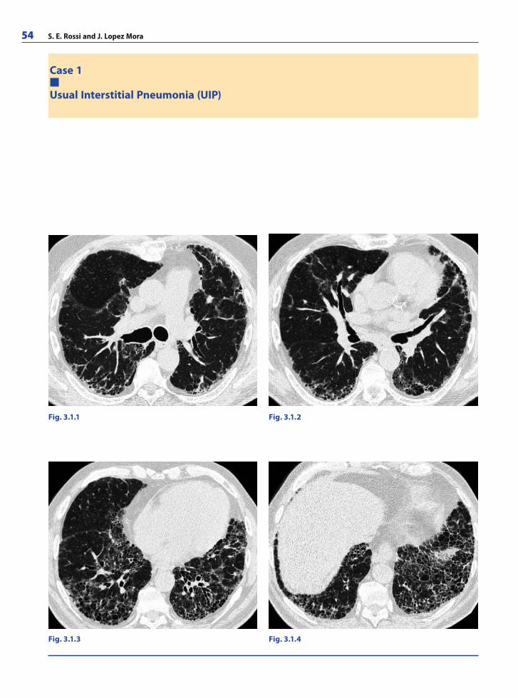
54 S. E. Rossi and J. Lopez Mora
Case 1
Usual Interstitial Pneumonia (UIP)
Fig. 3.1.1 Fig. 3.1.2
Fig. 3.1.4Fig. 3.1.3
RibesLunaRos.indb 54 18.03.2008 12:49:25
Thoracic Radiology 55
A 54-year-old man presented with progressive dyspnea and non-productive cough. Drug toxicity, environmental exposure, and collagen vascular disease were ruled out. Bibasilar velcro-type end-inspiratory crackles were detected on auscultation. Lung function tests revealed a moderate restrictive defect, decreased PAO2, and decreased diffusing capacity of the lung for carbon monoxide (DLCO).
CommentsIdiopathic pulmonary fi brosis (IPF) is the clinical syndrome associated with an intersti-tial pneumonia of unknown cause. Histopathological fi ndings show a pattern of usual interstitial pneumonia.
Surgical lung biopsy is the most defi nitive method for establishing the diagnosis. Patho-logic changes have a predilection for the subpleural and paraseptal parenchyma. Fibrotic zones with associated honeycombing alternate with areas of relatively unaffected lung tis-sue. Areas of chronic lung injury contrast with regions of acute injury with foci of actively proliferating fi broblasts and myofi broblasts. Interstitial infl ammation is mild and gener-ally associated with fi brosis. Spatial and temporal heterogeneity are characteristic.
Patients with IPF have an increased incidence of lung carcinoma. The disease follows a progressive course with a mean survival time of 3 years. Treatment options include corticosteroids, azathioprine, and lung transplantation.
Differential diagnoses:Nonspecifi c interstitial pneumonia (ground-glass pattern predominance, usually no honeycombing) Asbestosis (subpleural lines and parenchymal bands)Drug toxicity (ground glass and consolidations)
�
��
Imaging FindingsChest radiograph demonstrates:Bilateral reticular opacities Lower lobes, peripheral predominanceCystic dilatation of the distal air spaces “honeycombing”Traction bronchiectasis
High resolution computed tomography (HRCT) increases spatial resolution, facilitat-ing visualization of parenchymal detail to the level of the secondary pulmonary lobule.
HRCT (UIP) demonstrates:Interlobular septal thickeningGround-glass attenuation patternTraction bronchiectasis and bronchiolectasisArchitectural distortionHoneycomb patternBasal and peripheral predominance
����
������
RibesLunaRos.indb 55 18.03.2008 12:49:28
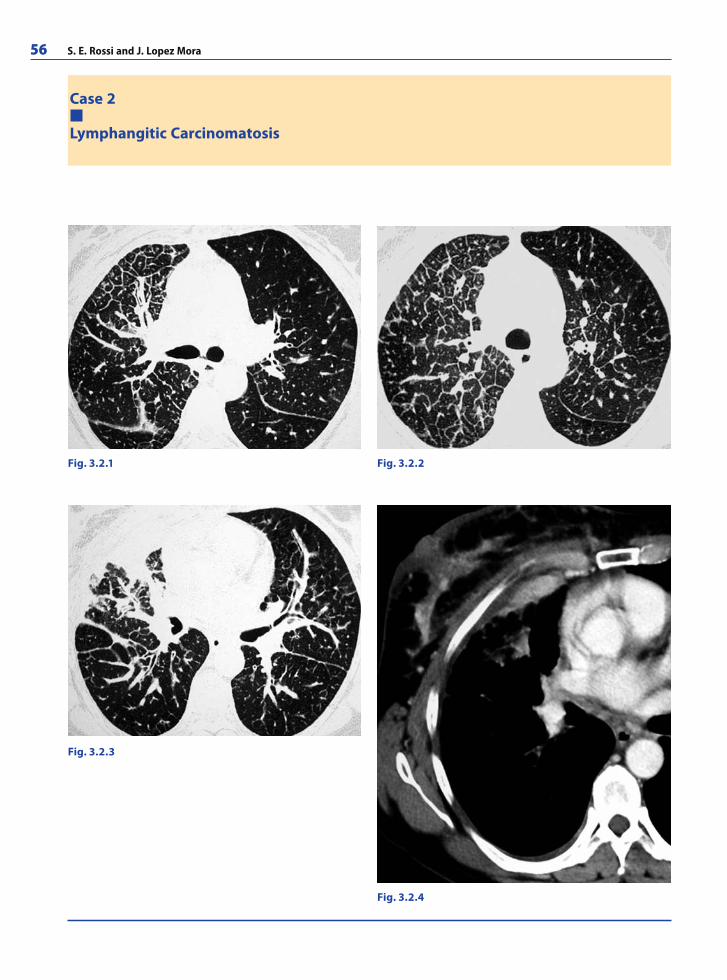
56 S. E. Rossi and J. Lopez Mora
Case 2
Lymphangitic Carcinomatosis
Fig. 3.2.1 Fig. 3.2.2
Fig. 3.2.3
Fig. 3.2.4
RibesLunaRos.indb 56 18.03.2008 12:49:28
Thoracic Radiology 57
A 59-year-old woman with a 30 pack/year history of smoking presented with shortness of breath, nonproductive cough, fatigue, and weakness. Physical examination revealed tachypnea; tachycardia, and cyanosis. Auscultation revealed diffuse fi ne rales in the basal regions and a restrictive ventilatory defect. Past medical included a history of breast carcinoma 10 years prior. Bronchoalveolar lavage detected neoplastic cells and an increased number of lymphocytes.
Imaging FindingsChest radiograph shows:Normal fi ndings, usually (25% sensitivity)Reticulonodular pattern, more frequent in the lower lobesKerley B lines (Kerley B lines in the absence of congestive heart failure is very sugges-tive of lymphangitic carcinomatosis)Pleural effusionHilar and mediastinal adenopathy
High resolution computed tomography (HRCT) shows:Nodular, irregular or smooth reticular pattern (beaded appearance)Peribronchovascular, centrilobular, septal, and subpleural distributionUnilateral, bilateral, or patchy distributionPulmonary architecture is preservedNormal lung volumePleural effusionHilar or mediastinal adenopathy
���
��
�������
CommentsLymphangitic carcinomatosis represents intrapulmonary spread of metastatic neoplasm via lymphatics and adjacent connective tissue. It is a frequent pattern of neoplastic spread to the lungs (35–55%). The most common primary malignancies producing lymphangitic spread include adenocarcinomas of the breast, lung, stomach, pancreas, cervix, and colon. Most cases of lymphangitic spread occur as a result of tumor microembolization. Other potential mechanisms include retrograde spread from involved lymph nodes, local extension from a primary pulmonary tumor, and transdiaphragmatic spread. Pulmonary architecture is preserved, although the lymphatic vessels appear distended by neoplastic cells and a perilymphatic fi brotic reaction is usual. In the presence of an unknown pri-mary tumor, the diagnosis is based on histopathologic specimens.
Differential Diagnosis:Pulmonary edema (interlobular septal thickening, nodules are absent, ground-glass, and consolidation)Sarcoidosis (bilateral, peribronchovascular, upper lobes predominance, distortion of pulmonary architecture)Silicosis (upper lobes, associated with masses and parenchymal distortion)Pneumoconiosis Hypersensitivity pneumonitisLymphoma and Kaposi’s Sarcoma
�
�
����
RibesLunaRos.indb 57 18.03.2008 12:49:31
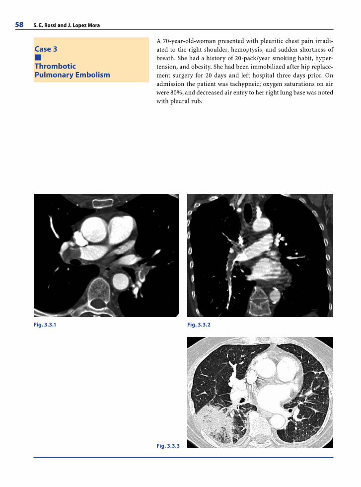
58 S. E. Rossi and J. Lopez Mora
A 70-year-old-woman presented with pleuritic chest pain irradi-ated to the right shoulder, hemoptysis, and sudden shortness of breath. She had a history of 20-pack/year smoking habit, hyper-tension, and obesity. She had been immobilized after hip replace-ment surgery for 20 days and left hospital three days prior. On admission the patient was tachypneic; oxygen saturations on air were 80%, and decreased air entry to her right lung base was noted with pleural rub.
Case 3
ThromboticPulmonary Embolism
Fig. 3.3.1 Fig. 3.3.2
Fig. 3.3.3
RibesLunaRos.indb 58 18.03.2008 12:49:31
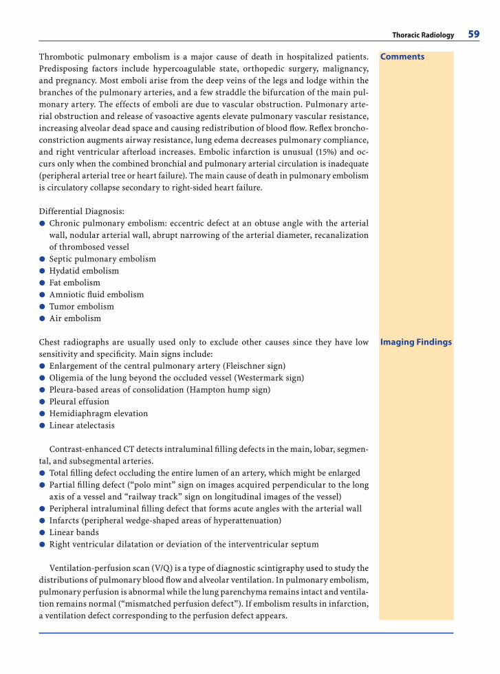
Thoracic Radiology 59
Imaging FindingsChest radiographs are usually used only to exclude other causes since they have low sensitivity and specifi city. Main signs include:
Enlargement of the central pulmonary artery (Fleischner sign)Oligemia of the lung beyond the occluded vessel (Westermark sign)Pleura-based areas of consolidation (Hampton hump sign)Pleural effusionHemidiaphragm elevation Linear atelectasis
Contrast-enhanced CT detects intraluminal fi lling defects in the main, lobar, segmen-tal, and subsegmental arteries.
Total fi lling defect occluding the entire lumen of an artery, which might be enlargedPartial fi lling defect (“polo mint” sign on images acquired perpendicular to the long axis of a vessel and “railway track” sign on longitudinal images of the vessel) Peripheral intraluminal fi lling defect that forms acute angles with the arterial wall Infarcts (peripheral wedge-shaped areas of hyperattenuation) Linear bandsRight ventricular dilatation or deviation of the interventricular septum
Ventilation-perfusion scan (V/Q) is a type of diagnostic scintigraphy used to study the distributions of pulmonary blood fl ow and alveolar ventilation. In pulmonary embolism, pulmonary perfusion is abnormal while the lung parenchyma remains intact and ventila-tion remains normal (“mismatched perfusion defect”). If embolism results in infarction, a ventilation defect corresponding to the perfusion defect appears.
������
��
����
CommentsThrombotic pulmonary embolism is a major cause of death in hospitalized patients. Predisposing factors include hypercoagulable state, orthopedic surgery, malignancy, and pregnancy. Most emboli arise from the deep veins of the legs and lodge within the branches of the pulmonary arteries, and a few straddle the bifurcation of the main pul-monary artery. The effects of emboli are due to vascular obstruction. Pulmonary arte-rial obstruction and release of vasoactive agents elevate pulmonary vascular resistance, increasing alveolar dead space and causing redistribution of blood fl ow. Refl ex broncho-constriction augments airway resistance, lung edema decreases pulmonary compliance, and right ventricular afterload increases. Embolic infarction is unusual (15%) and oc-curs only when the combined bronchial and pulmonary arterial circulation is inadequate (peripheral arterial tree or heart failure). The main cause of death in pulmonary embolism is circulatory collapse secondary to right-sided heart failure.
Differential Diagnosis:Chronic pulmonary embolism: eccentric defect at an obtuse angle with the arterial wall, nodular arterial wall, abrupt narrowing of the arterial diameter, recanalization of thrombosed vesselSeptic pulmonary embolismHydatid embolismFat embolismAmniotic fl uid embolismTumor embolismAir embolism
�
������
RibesLunaRos.indb 59 18.03.2008 12:49:33
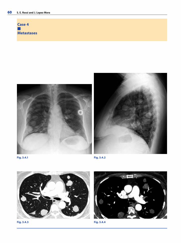
60 S. E. Rossi and J. Lopez Mora
Case 4
Metastases
Fig. 3.4.4Fig. 3.4.3
Fig. 3.4.1 Fig. 3.4.2
RibesLunaRos.indb 60 18.03.2008 12:49:33
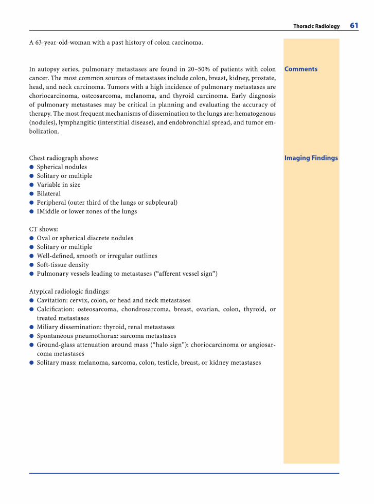
Thoracic Radiology 61
A 63-year-old-woman with a past history of colon carcinoma.
CommentsIn autopsy series, pulmonary metastases are found in 20–50% of patients with colon cancer. The most common sources of metastases include colon, breast, kidney, prostate, head, and neck carcinoma. Tumors with a high incidence of pulmonary metastases are choriocarcinoma, osteosarcoma, melanoma, and thyroid carcinoma. Early diagnosis of pulmonary metastases may be critical in planning and evaluating the accuracy of therapy. The most frequent mechanisms of dissemination to the lungs are: hematogenous (nodules), lymphangitic (interstitial disease), and endobronchial spread, and tumor em-bolization.
Imaging FindingsChest radiograph shows:Spherical nodulesSolitary or multipleVariable in sizeBilateralPeripheral (outer third of the lungs or subpleural)IMiddle or lower zones of the lungs
CT shows:Oval or spherical discrete nodulesSolitary or multipleWell-defi ned, smooth or irregular outlinesSoft-tissue density Pulmonary vessels leading to metastases (“afferent vessel sign”)
Atypical radiologic fi ndings:Cavitation: cervix, colon, or head and neck metastasesCalcifi cation: osteosarcoma, chondrosarcoma, breast, ovarian, colon, thyroid, or treated metastasesMiliary dissemination: thyroid, renal metastasesSpontaneous pneumothorax: sarcoma metastasesGround-glass attenuation around mass (“halo sign”): choriocarcinoma or angiosar-coma metastasesSolitary mass: melanoma, sarcoma, colon, testicle, breast, or kidney metastases
������
�����
��
���
�
RibesLunaRos.indb 61 18.03.2008 12:49:36
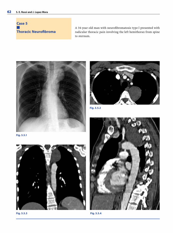
62 S. E. Rossi and J. Lopez Mora
A 34-year-old man with neurofi bromatosis type I presented with radicular thoracic pain involving the left hemithorax from spine to sternum.
Case 5
Thoracic Neurofi broma
Fig. 3.5.1
Fig. 3.5.2
Fig. 3.5.4Fig. 3.5.3
RibesLunaRos.indb 62 18.03.2008 12:49:36
Thoracic Radiology 63
CommentsNeurogenic tumors account for about 9% of primary mediastinal masses in adults and 30% of mediastinal tumors in children. Neurofi bromas are slow-growing neoplasms that originate from the nerve sheath and are derived from Schwann cells. They are seen most commonly in patients between 20 and 30 years of age. Most are benign, although malignant degeneration occasionally occurs (up to 25%). Almost all arise from intercostal or sym-pathetic nerves, adjacent to the spine. They are usually not encapsulated. Neurofi bromas demonstrate proliferation of nerve sheath cells interspersed with thick collagen bundles and may show variable degrees of myxoid degeneration and calcifi cations.
Differential Diagnosis: Other neurogenic tumors including schwannoma, neuroblastoma, ganglioneuromaAdenopathies (lymphoma, tuberculosis, sarcoid, or metastases)Extramedullary hematopoiesisNeurenteric cystsPancoast tumor
�����
Imaging FindingsRadiographs of the spine may show:Widening of neural foraminaFocal scoliosisRib abnormalities
Chest radiographs may show:Well-defi ned massSmooth or lobulated contoursRound or ovalVariable sizeSituated in the posterior mediastinum or grow along intercostal nerves
CT may show:Homogeneous massSoft-tissue density and may have low attenuation zonesCalcifi cation (not usual): fi nely stippledHeterogeneous enhancement after intravenous administration of contrast materialBone erosions of ribs and vertebrae: the cortex is preserved and thickenedMalignant degeneration: heterogeneous density, local invasion, bone destruction, and metastasis to the pleura or lungs
MR may show:Intermediate signal intensity on T1-weighted imagesHeterogeneous signal intensity on T2-weighted images: areas of collagen and fi brous tissue (low-signal) and areas of myxoid tissue or cystic degeneration (high-signal)“Target pattern”: central portion shows higher intensity on T1, and the periphery shows higher intensity on T2 (due to highly cellular central zone and abundant stro-mal material in the peripheral zone)MR shows intraspinal extension best Plexiform neurofi broma: extensive fusiform or infi ltrating mass that tends to sur-round mediastinal vessels with loss of fat planes
���
�����
������
��
�
��
RibesLunaRos.indb 63 18.03.2008 12:49:39
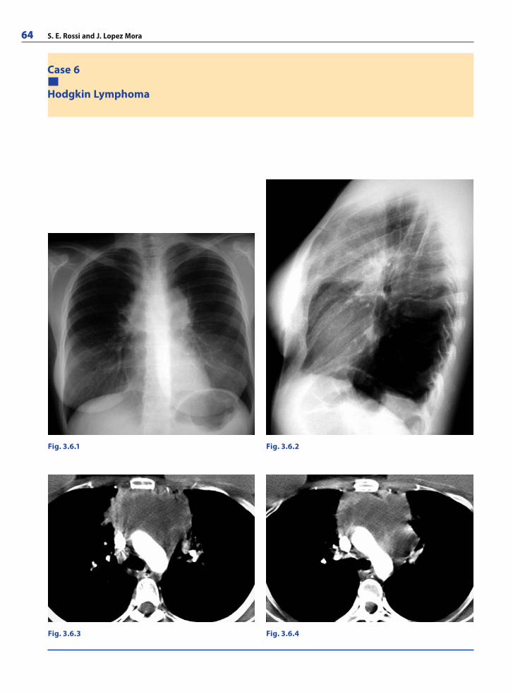
64 S. E. Rossi and J. Lopez Mora
Case 6
Hodgkin Lymphoma
Fig. 3.6.1 Fig. 3.6.2
Fig. 3.6.4Fig. 3.6.3
RibesLunaRos.indb 64 18.03.2008 12:49:39
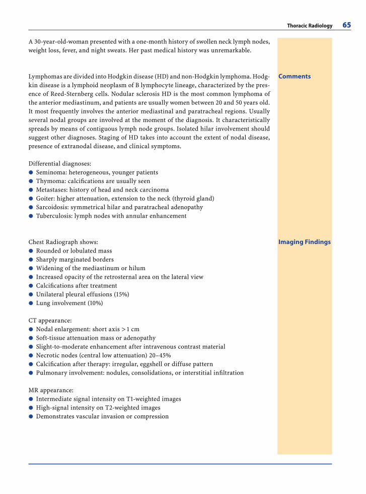
Thoracic Radiology 65
A 30-year-old-woman presented with a one-month history of swollen neck lymph nodes, weight loss, fever, and night sweats. Her past medical history was unremarkable.
Imaging FindingsChest Radiograph shows:Rounded or lobulated massSharply marginated bordersWidening of the mediastinum or hilumIncreased opacity of the retrosternal area on the lateral viewCalcifi cations after treatmentUnilateral pleural effusions (15%)Lung involvement (10%)
CT appearance:Nodal enlargement: short axis > 1 cmSoft-tissue attenuation mass or adenopathySlight-to-moderate enhancement after intravenous contrast materialNecrotic nodes (central low attenuation) 20–45%Calcifi cation after therapy: irregular, eggshell or diffuse patternPulmonary involvement: nodules, consolidations, or interstitial infi ltration
MR appearance:Intermediate signal intensity on T1-weighted imagesHigh-signal intensity on T2-weighted imagesDemonstrates vascular invasion or compression
�������
������
���
CommentsLymphomas are divided into Hodgkin disease (HD) and non-Hodgkin lymphoma. Hodg-kin disease is a lymphoid neoplasm of B lymphocyte lineage, characterized by the pres-ence of Reed-Sternberg cells. Nodular sclerosis HD is the most common lymphoma of the anterior mediastinum, and patients are usually women between 20 and 50 years old. It most frequently involves the anterior mediastinal and paratracheal regions. Usually several nodal groups are involved at the moment of the diagnosis. It characteristically spreads by means of contiguous lymph node groups. Isolated hilar involvement should suggest other diagnoses. Staging of HD takes into account the extent of nodal disease, presence of extranodal disease, and clinical symptoms.
Differential diagnoses:Seminoma: heterogeneous, younger patientsThymoma: calcifi cations are usually seenMetastases: history of head and neck carcinomaGoiter: higher attenuation, extension to the neck (thyroid gland)Sarcoidosis: symmetrical hilar and paratracheal adenopathyTuberculosis: lymph nodes with annular enhancement
������
RibesLunaRos.indb 65 18.03.2008 12:49:42
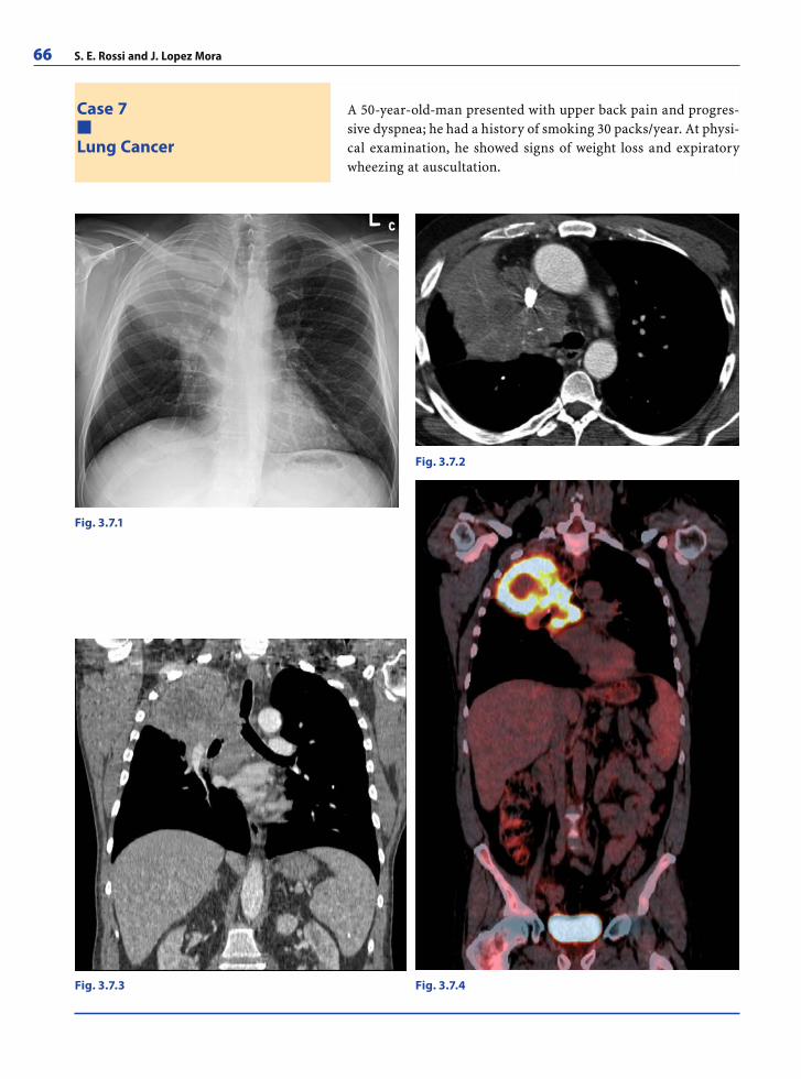
66 S. E. Rossi and J. Lopez Mora
Case 7
Lung Cancer
A 50-year-old-man presented with upper back pain and progres-sive dyspnea; he had a history of smoking 30 packs/year. At physi-cal examination, he showed signs of weight loss and expiratory wheezing at auscultation.
Fig. 3.7.1
Fig. 3.7.2
Fig. 3.7.4Fig. 3.7.3
RibesLunaRos.indb 66 18.03.2008 12:49:42
Thoracic Radiology 67
CommentsBronchogenic carcinoma is the most common cause of cancer-related death in both men and women. It is divided into four main histologic subtypes: adenocarcinoma (the most common), squamous cell carcinoma, small cell carcinoma, and large cell carcinoma.
Adenocarcinoma:Symptoms include dyspnea or cough, pleuritic pain depending on extensionUpper lobe, peripheral, or subpleural predominanceAdrenal, brain, bone, or liver metastasesRadiographic fi nding of a solitary pulmonary node or mass with well-marginated, lobulated, irregular, or poorly defi ned borders
Squamous cell carcinoma:Symptoms include cough and hemoptysisCentral location with endobronchial componentDirect extension to the local lymph nodesSlowest growth rate Radiographic fi nding of a hilar mass with or without obstructive pneumonitis or atel-ectasisCentral necrosis is common in large tumors and central cavitation in 15%Can manifest as a Pancoast tumor (superior sulcus tumor): with invasion of the pleura and rib producing shoulder pain and involvement of the brachial plexus causing arm pain and paresthesia.
Differential Diagnosis:Bronchial carcinoid tumor: calcifi cation, enhances with IV contrast, widening of the approaching bronchusSolitary metastasesPrimary pulmonary sarcomaSPN benign causes including hamartoma (fat/calcium), granuloma, and arteriovenous malformation.
����
�����
��
�
���
Imaging FindingsChest radiograph shows:Nodule or massHilar or mediastinal adenopathy or massPleural effusionObstructive fi ndings: atelectasis, pneumonia, mucoid impaction
CT shows:Nodule or massMorphology: spherical or oval, lobulated or spiculatedDensity: soft-tissue density, amorphous calcifi cation, cavitation with thick wallsContrast enhancement related to angiogenesisEvaluate growth ratePresence of associated collapse and/or consolidationHilar and mediastinal adenopathiesIMediastinal and chest wall invasion
PET-CT Noninvasive diagnostic metabolic and anatomic technique. PET-CT can aid in diagnosis, staging, assessing recurrence, and monitoring response to therapy.
����
��������
RibesLunaRos.indb 67 18.03.2008 12:49:47
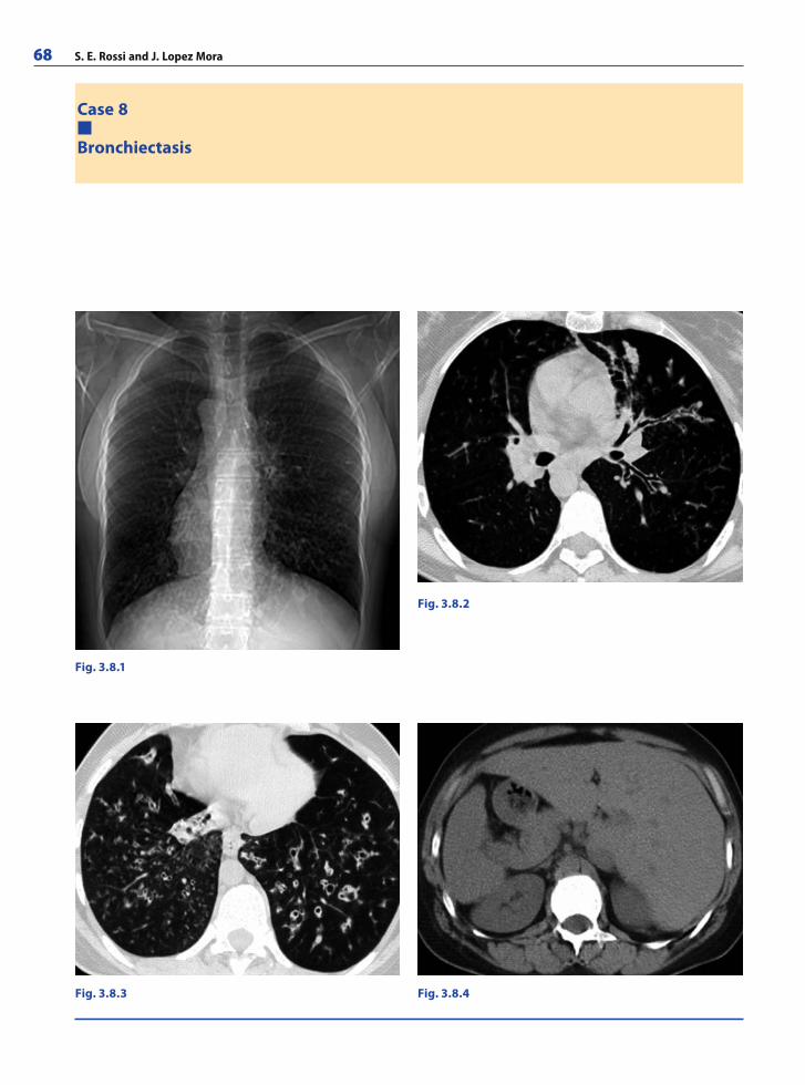
68 S. E. Rossi and J. Lopez Mora
Case 8
Bronchiectasis
Fig. 3.8.1
Fig. 3.8.2
Fig. 3.8.4Fig. 3.8.3
RibesLunaRos.indb 68 18.03.2008 12:49:48
Thoracic Radiology 69
CommentsBronchiectasis is characterized by the permanent dilation of bronchi with destruction of the elastic and muscular components of bronchial walls. It is usually caused by acute or chronic infection. The main symptoms of bronchiectasis include chronic cough and sputum production. Recurrent bacterial colonization and infection lead to progressive airway injury.
Infl ammatory mediators produce infl ammation and destruction of the elastic and muscular components of bronchial walls, while the contractile force of the surrounding lung tissue exerts traction, expanding the diameter of involved airways.
Bronchiectasis is also associated with increased bronchial arterial proliferation and arteriovenous malformations, predisposing to recurrent hemoptysis in some patients. Bronchiectasis may have a focal or diffuse distribution.
Kartagener’s syndrome refers to the triad of situs inversus, bronchiectasis, and recur-rent sinusitis, occurring in half of patients with primary ciliary dyskinesia. This is an autosomal-recessive disorder associated with defective ciliary structure and function.
Differential Diagnosis:Cystic fi brosisAllergic bronchopulmonary aspergillosis (ABPA)Mycobacterium avium complex infectionPulmonary fi brosisBronchial obstruction
�����
Imaging FindingsChest radiograph shows:Dilated and thickened airways (ring-like shadows of airways that are seen on end, or tram lines of airways that are perpendicular to the X-ray beam)Focal pneumonitisScattered irregular opacitiesPlate-like atelectasis
High resolution CT (HRCT) shows:Bronchial wall thickeningBronchial dilatation Bronchus is larger than the adjacent pulmonary artery (signet ring sign) Bronchi are visible within 1 cm of pleuraCylindrical bronchiectasis (smooth bronchial dilatation)Varicose bronchiectasis (beaded bronchial dilatation)Cystic bronchiectasis (air-fl uid levels in dilated airways)Mucoid impaction (tree-in-bud pattern)Atelectasis distal to mucoid plugFocal air-trapping distal to mucoid plug
�
���
����������
A 15-year-old girl with chronic sinusitis and bronchitis presented with a productive cough without fever. Diffuse rhonchi were detected at physical examination.
RibesLunaRos.indb 69 18.03.2008 12:49:51
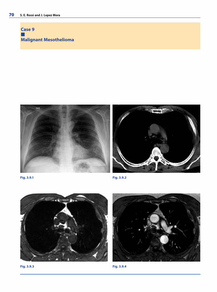
70 S. E. Rossi and J. Lopez Mora
Case 9
Malignant Mesothelioma
Fig. 3.9.3 Fig. 3.9.4
Fig. 3.9.1 Fig. 3.9.2
RibesLunaRos.indb 70 18.03.2008 12:49:51
Thoracic Radiology 71
A 64-year-old man presented with shortness of breath, pleuritic pain, and weight loss. The patient referred pleural effusions drained two years prior and had worked in an electric wiring factory for 20 years. He had no history of smoking.
CommentsMalignant mesothelioma is the most common primary neoplasm of the pleura. It has a strong association with asbestos exposure, with a latency of 35–40 years. Median survival time after diagnosis is between 12–15 months. The main histologic subtypes are epithe-lial, sarcomatous, and mixed. It is locally aggressive, with frequent invasion of the chest wall, mediastinum, and diaphragm. Metastases to the hilar and mediastinal lymph nodes are usual at the moment of diagnosis. The most frequent hematogenous metastases are to the lungs, liver, kidneys, and adrenal glands.
Differential diagnoses:Adenocarcinoma LymphomaThymoma (drop metastases)Asbestos-related benign pleural diseaseInfections
�����
Imaging FindingsChest radiograph shows:Pleural effusion Circumferential pleural thickening Lung nodules or masses
CT shows:Unilateral pleural effusionSheetlike or lobulated pleural thickeningInterlobar fi ssure thickeningTumor encasement of the lung with a rindlike appearanceContraction of the affected hemithorax with associated ipsilateral mediastinal shift, narrowed intercostal spaces, and elevation of the ipsilateral hemidiaphragmChest wall invasion: obliteration of fat planes, invasion of intercostal muscles, infi ltra-tion or separation of ribs, bone destructionInvasion into the mediastinum: obliteration of fat planes or direct tumor invasion to the heart, great vessels, esophagus, and trachea
MR imaging provides excellent soft-tissue contrast, showing: Isointense to muscle or slightly hyperintense on T1-weighted imagesHyperintense on T2-weighted imagesEnhances after contrast administration
PET-CT shows:More extensive disease involvementMost appropriate biopsy site for obtaining positive resultsOccult distant metastases
���
�����
�
�
���
���
RibesLunaRos.indb 71 18.03.2008 12:49:52
72 S. E. Rossi and J. Lopez Mora
Case 10
Tuberculosis
Fig. 3.10.1
Fig. 3.10.2
Fig. 3.10.3
Fig. 3.10.4
RibesLunaRos.indb 72 18.03.2008 12:49:52
Thoracic Radiology 73
A 35-year-old woman presented with a three-month history of non-productive cough, low-grade fever, anorexia, night sweats, and weight loss. Laboratory tests revealed raised peripheral blood leukocyte count and anemia and the absence of HIV antibodies.
CommentsPulmonary tuberculosis is caused by inhalation of droplets laden with Mycobacterium tuberculosis bacilli. The most common site of implantation is the middle and lower lung lobes. The course of the disease depends on the interaction between the host response and organism’s virulence. Primary tuberculosis typically appears as air-space consolida-tion in the lower lobes, hilar and mediastinal lymphadenopathy, and pleural effusion. Postprimary tuberculosis occurs in persons with a degree of acquired immunity from prior infection and usually appears as nodular and linear areas of increased opacity or attenuation in the lung apex. Active and inactive disease can only be reliably differenti-ated radiographically on the basis of temporal evolution (no change over a 4–6 month interval).
Imaging FindingsPrimary disease:Lymphadenopathy: right paratracheal and hilar stations most frequently involved. Central areas of low attenuation with peripheral rim enhancement (most frequently HIV)Parenchymal consolidation: homogeneous, patchy, linear or nodular; segmental or lobar distributionObstructive atelectasis, usually right-sidedUnilateral pleural effusionRadiographs are normal (15%)
Post primary disease:Parenchymal disease with cavitation: heterogeneous opacities in the apical and poste-rior segments of the upper lobes and the superior segment of the lower lobesCavitation: usually multiple, with thin and smooth or thick and irregular walls. May have air-fl uid levelsTuberculoma: solitary round sharply marginated mass with calcifi cationBronchogenic spread: 4 mm centrilobular nodules and sharply marginated linear branching opacities (“tree-in-bud” appearance)Pleural effusionFibroproliferative disease: upper lobe volume loss with cicatricial atelectasis, archi-tectural distortion, and traction bronchiectasis
Complications:Mycetomas: mobile intracavitary masses, air crescent signTraction bronchiectasisResidual cavitiesRasmussen aneurysm: pseudoaneurysm of a pulmonary arteryBroncholithiasis: calcifi ed peribronchial nodes
��
�
���
�
�
��
��
�����
RibesLunaRos.indb 73 18.03.2008 12:49:54
74 S. E. Rossi and J. Lopez Mora
Further Readings
Books
Chest Radiology: Plain Film Patterns and Differential Di-agnosis, 5th ed. Reed JC (2003) Philadelphia, Pa: Elsevier Science. ISBN-13: 9780323026178
CT and MR Imaging of the Whole Body, 4th ed, 2 vols. Haaga JR, Lanzieri CF (2003) St Louis, Mo: Mosby-Elsevier Sci-ence. ISBN-13: 9780323011334
Computed Tomography and Magnetic Resonance of the Tho-rax. 4th ed. Naidich D, Webb WR, Muller NR, Vlahos I, Krinsky GA (2007) Lippincott Williams & Wilkins. ISBN-13: 9780781757652
Diagnosis and Diseases of the Chest. 4th ed. Fraser RS, Muller NL, Colman N, Paré PD (1999) Philadelphia, Pa: Saun-ders. ISBN-13: 9780721661940
Diffuse Lung Diseases. Maffessanti M, Dalpiaz G (2006) Springer, Berlin. ISBN-13: 9788847004290
Felson’s Principles of Chest Roentgenology. 2nd ed. Good-man LR (1999) Philadelphia, Pa: Saunders. ISBN-13: 9780721676852
High-resolution CT of the Lung. 2nd ed. Webb WR, Müller NL, Naidich DP (1996) Philadelphia, Pa: Lippincott-Ra-ven. ISBN-13: 9780781702171
Imaging of Diseases of the Chest. 4th ed. Hansell D, Arm-strong P, Lynch D, McAdams HP (2005) Elsevier Mosby. ISBN-13: 9780723433231
Radiology of the Chest and Related Conditions (2002) Wright FW. London, England: Taylor & Francis. ISBN13: 9789058232298
Thoracic Imaging: Pulmonary and Cardiovascular Radiol-ogy. Higgins CB, Webb WR (2004) Philadelphia, Pa: Lip-pincott Williams & Wilkins. ISBN-13: 9780781741194
Web Links
http://ajronline.orghttp://chestjournal.orghttp://content.nejm.orghttp://erj.ersjournals.comhttp://jcat.orghttp://radiographics.rsnajnls.orghttp://radiology.rsnajnls.orghttp://springerlink.comhttp://thoracicrad.orghttp://thorax.bmj.com
Articles
Althoff Souza C, Müller N, Flint J, Wright J, Churg A. Idio-pathic pulmonary fi brosis: Spectrum of high-resolution CT fi ndings. AJR Am J Roentgenol 2005; 185:1531–1539
Blachere H, Latrabe V, Montaudon M, Valli N, Couffi nhal T, Raherisson C, Leccia F, Laurent F. Pulmonary embolism revealed on helical CT angiography: comparison with ventilation-perfusion radionuclide lung scanning. AJR Am J Roentgenol 2000; 174:1041–1047
Boiselle PM, Patz EF, Vining DJ, Weissleder R, Shepard JA, McLoud TC. Imaging of mediastinal lymph nodes: CT, MR, and FDG PET. Radiographics 1998; 18:1061–1069
Brown K, Mund DF, Aberle DR, Batra P, Young DA. Intratho-racic calcifi cations: radiographic features and differen-tial diagnoses. Radiographics 1994; 14:1247–1261
Burk DL, Brunberg JA, Kanal E, Latchaw RE, Wolf GL. Spinal and paraspinal neurofi bromatosis: surface coil MR imag-ing at 1.5 T1. Radiology 1987; 162:797–801
Carpenter BL, Merten DF. Radiographic manifestations of congenital anomalies affecting the airway. Radiol Clin North Am 1991; 29:219–240
Cartier Y, Kavanagh PV, Johkoh T, Mason AC, Muller NL. Bronchiectasis: accuracy of high-resolution CT in the differentiation of specifi c diseases. AJR Am J Roentgenol 1999; 173:47–52
Chong S, Lee KS, Chung MJ, Han J, Kwon OJ, Kim TS. Pneu-moconiosis: comparison of imaging and pathologic fi nd-ings. Radiographics 2006; 26:59–77
Chooi WK, Matthews S, Bull MJ, Morcos SK. Multislice heli-cal CT: the value of multiplanar image reconstruction in assessment of the bronchi and small airways disease. Br J Radiol 2003; 76:536–540
Coche E, Verschuren F, Keyeux A, Goffette P, Goncette L, Hainaut P, Hammer F, Lavenne E, Zech F, Meert P, Reyn-aert MS. Diagnosis of acute pulmonary embolism in out-patients: Comparison of thin-collimation multi-detector row spiral CT and planar ventilation-perfusion scintig-raphy. Radiology 2003; 229:757–765
Corcoran HL, Renner WR, Milstein MJ. Review of high-reso-lution CT of the lung. Radiographics 1992; 12:917–939
Crow J, Slavin G, Kreel L. Pulmonary metastases: a patho-logic and radiologic study. Cancer 1981; 47:2595–2602
Cymbalista M, Waysberg A, Zacharias C, Ajavon Y, Riquet M, Rebibo G, Grenier P. CT demonstration of the 1996 AJCC-UICC regional lymph node classifi cation for lung cancer staging. Radiographics 1999; 19:899–900
Davis SD. CT evaluation for pulmonary metastases in patients with extrathoracic malignancy. Radiology 1991; 180:1–12
Erasmus JJ, Connolly JE, McAdams HP, Roggli VL. Solitary pulmonary nodules: Part I. Morphologic evaluation for differentiation of benign and malignant lesions. Radio-graphics 2000; 20:43–58
Erasmus JJ, McAdams HP, Patz EF, Goodman PC, Coleman RE. Thoracic FDG PET: state of the art. Radiographics 1998; 18:5–20
Fishman EK, Kuhlman JE, Jones RJ. CT of lymphoma: spec-trum of disease. Radiographics 1991; 11:647–669
Fedullo PF, Tapson VF. Clinical practice: the evaluation of suspected pulmonary embolism. N Engl J Med 2003; 349(13):1247–1256
Fortman BJ, Kuszyk BS, Urban BA, Fishman EK. Neurofi -bromatosis type 1: A diagnostic mimicker at CT. Radio-graphics 2001; 21:601–612
Foster WL, Gimenez EI, Roubidoux MA, Sherrier RH, Shan-non RH, Roggli VL, Pratt PC. The emphysemas: radiologic-pathologic correlations. Radiographics 1993; 13:311–328
Franquet T, Giménez A, Rosón N, Torrubia S, Sabaté JM, Pérez C. Aspiration diseases: fi ndings, pitfalls, and dif-ferential diagnosis. Radiographics 2000; 20:673–685
RibesLunaRos.indb 74 18.03.2008 12:49:54
Thoracic Radiology 75
Fraser RS, Muller NL, Colman N, Paré PD. Diagnosis and diseases of the chest. 4th ed. Saunders, 1999
Frazier AA, Galvin JR, Franks TJ, Rosado-de-Christenson ML. From the archives of the AFIP: Pulmonary vascula-ture: hypertension and infarction. Radiographics 2000; 20:491–524
Ghaye B, Ghuysen A, Bruyere PJ, D’Orio V, Dondelinger RF. Can CT pulmonary angiography allow assessment of severity and prognosis in patients presenting with pul-monary embolism? What the radiologist needs to know. Radiographics 2006; 26:23–39
Giménez A, Franquet T, Erasmus JJ, Martínez S, Estrada P. Thoracic complications of esophageal disorders. Radio-graphics 2002; 22:247–258
Grippi MA. Clinical aspects of lung cancer. Semin Roent-genol 1990; 25:5–11
Goodman LR. Felson’s Principles of Chest Roentgenology. Philadelphia, Pa: Saunders, 1999
Gross BH, Glazer GM, Bookstein FL. Multiple pulmonary nodules detected by computed tomography: diagnostic implications. J Comput Assist Tomogr 1985; 9:880–885
Gross TJ, Hunninghake GW. Medical progress: Idiopatic pulmonary fi brosis. N Engl J Med 2001; 345:517–525
Haaga JR, Lanzieri CF, Gilkeson RC. CT and MR imaging of the whole body. 4th ed, 2 vols. St Louis, Mo: Mosby–Else-vier Science, 2003
Han D, Lee KS, Franquet T, Muller NL, Kim TS, Kim H, Kwon OJ, Byun HS. Thrombotic and nonthrombotic pulmonary arterial embolism: Spectrum of imaging fi ndings. Radio-graphics 2003; 23(6):1521–1539
Hansell D, Armstrong P, Lynch D, McAdams HP. Imaging of diseases of the chest. Elsevier Mosby 2005
Harisinghani MG, McLoud TC, Shepard JA, Ko JP, Shroff MM, Mueller PR. Tuberculosis from head to toe. Radio-graphics 2000; 20:449–470
Hartman TE, Primack SL, Lee KS, Swensen SJ, Muller NL. CT of bronchial and bronchiolar diseases. Radiographics 1994; 14:991–1003
Hayashi K, Aziz A, Ashizawa K, Hayashi H, Nagaoki K, Ot-suji H. Radiographic and CT appearances of the major fi ssures. Radiographics 2001; 21:861–874
Halvorsen RA, Fedyshin PJ, Korobkin M, Foster WL, Thomp-son WM. Ascites or pleural effusion? CT differentiation: four useful criteria. Radiographics 1986; 6:135–149
Helbich TH, Heinz-Peer G, Eichler I, Wunderbaldinger P, Götz M, Wojnarowski C, Brasch RC, Herold CJ. Cystic fi brosis: CT assessment of lung involvement in children and adults. Radiology 1999; 213:537–544
Hunninghake GW, Lynch DA, Galvin JR, et al. Radiologic fi nd-ings are strongly associated with a pathologic diagnosis of usual interstitial pneumonia. Chest 2003; 124:1215–1223
Isabela C, Silva S, Colby TV, Müller NL. Asthma and associ-ated conditions: High-resolution CT and pathologic fi nd-ings. AJR Am J Roentgenol 2004; 183:817–824
Jeung MY, Gasser B, Gangi A, Bogorin A, Charneau D, Wihlm JM, Dietemann JL, Roy C. Imaging of cystic masses of the mediastinum. Radiographics 2002; 22:79–93
Jong PA, Muller NL, Pare PD, Coxson HO. Computed tomo-graphic imaging of the airways: relationship to structure and function. Eur Respir J 2005; 26(1):140–152
Jung JI, Kim HH, Park SH, Song SW, Chung MH, Kim HS, Kim KJ, Ahn MI, Seo SB, Hahn ST. Thoracic manifes-tations of breast cancer and its therapy. Radiographics 2004; 24:1269–1285
Kawashima A, Fishman EK, Kuhlman JE, Nixon MS. CT of posterior mediastinal masses. Radiographics 1991; 11:1045–1067
Kazama T, Faria SC, Varavithya V, Phongkitkarun S, Ito H, Macapinlac HA. FDG PET in the evaluation of treatment for lymphoma: Clinical usefulness and pitfalls. Radio-graphics 2005; 25:191–207
Kazerooni EA. High-resolution CT of the lungs. AJR Am J Roentgenol 2001; 177:501–519
Kim EA, Lee KS, Primack SL, Yoon HK, Byun HS, Kim TS, Suh GY, Kwon OJ, Han J. Viral pneumonias in adults: Radiologic and pathologic fi ndings. Radiographics 2002; 22:137–149
Kim HY, Song KS, Goo JM, Lee JS, Lee KS, Lim TH. Thoracic sequelae and complications of tuberculosis. Radiograph-ics 2001; 21:839–858
Kuhlman JE, Deutsch JH, Fishman EK, Siegelman SS. CT features of thoracic mycobacterial disease. Radiograph-ics 1990; 10:413–431
Kuhlman JE, Singha NK. Complex disease of the pleural space: radiographic and CT evaluation. Radiographics 1997; 17:63–79
Koh DM, Burke S, Davies N, Padley SPG. Transthoracic US of the chest: Clinical uses and applications. Radiograph-ics. 2002; 22:e1
Koyama T, Ueda H, Togashi K, Umeoka S, Kataoka M, Nagai S. Radiologic manifestations of sarcoidosis in various or-gans. Radiographics 2004; 24:87–104
Leung AN, Muller NL, Miller RR. CT in differential diagno-sis of diffuse pleural disease. AJR Am J Roentgenol 1990; 154:487–492
Lucidarme O, Coche E, Cluzel P, Mourey-Gerosa I, Howarth N, Grenier P. Expiratory CT scans for chronic airway dis-ease: correlation with pulmonary function test results. AJR Am J Roentgenol 1998; 170:301–307
Lynch D, Travis WD, Müller NL, Galvin JR, Hansell DM, Gre-nier PA, King TE. Idiopathic interstitial pneumonias: CT Features. Radiology 2005; 236:10–21
Maffessanti M, Dalpiaz G. Diffuse lung diseases. Springer 2006
McAdams HP, Erasmus J, Winter JA. Radiologic manifesta-tions of pulmonary tuberculosis. Radiol Clin North Am 1995; 33:655–678
McAdams HP, Samei E, Dobbins J, Tourassi GD, Ravin CE. Recent advances in chest radiography. Radiology 2006 241:663–683
McCarville MB, Lederman HM, Santana VM, Daw NC, Sho-chat SJ, Li CS, Kaufman RA. Distinguishing benign from malignant pulmonary nodules with helical chest CT in children with malignant solid tumors. Radiology 2006; 239:514–520
McGuinness G, Naidich DP. CT of airways disease and bron-chiectasis. Radiol Clin North Am 2002; 40:1–19
Meziane MA, Hruban RH, Zerhouni EA, Wheeler PS, Khouri NF, Fishman EK, Hutchins GM, Siegelman SS. High reso-lution CT of the lung parenchyma with pathologic cor-relation. Radiographics 1988; 8:27–54
RibesLunaRos.indb 75 18.03.2008 12:49:54
76 S. E. Rossi and J. Lopez Mora
Miller BH, Rosado-de-Christenson ML, Mason AC, Fleming MV, White CC, Krasna MJ. From the archives of the AFIP. Malignant pleural mesothelioma: radiologic-pathologic correlation. Radiographics 1996; 16:613–644
Miller WT, Miller WT Jr. Tuberculosis in the normal host: ra-diological fi ndings. Semin Roentgenol 1993; 28:109–118
Milne EN, Pistolesi M, Miniati M, Giuntini C. The radiologic distinction of cardiogenic and noncardiogenic edema. AJR Am J Roentgenol 1985; 144:879–894
Mitlehner W, Friedrich M, Dissman W. Value of computed tomography of the lung in the management of primary spontaneous pneumothorax. Am J Surg 1991; 162:39–42
Morgan-Parkes JH. Metastases: mechanisms, pathways, and cascades. AJR Am J Roentgenol 1995; 164:1075–1082
Mueller-Mang C, Grosse C, Schmid K, Stiebellehner L, Bankier A. What every radiologist should know about idiopathic interstitial pneumonias. Radiographics 2007; 27:595–615
Müller NL, Colby TV. Idiopathic interstitial pneumonias: high resolution CT and histologic fi ndings. Radiograph-ics 1997; 17:1016–1022
Munk PL, Muller NL, Miller RR, Ostrow DN. Pulmonary lymphangitic carcinomatosis: CT and pathologic fi nd-ings. Radiology 1988; 166:705–709
Naidich DP, Webb WR, Müller NL, Vlahos I, Krinsky GA, Kim EE. Computed tomography and magnetic resonance of the thorax. J Nucl Med 2007; 48(12):2088
Nishino M, Ashiku SK, Kocher ON, Thurer RL, Boiselle PM, Hatabu H. The thymus: A comprehensive review. Radio-graphics 2006; 26:335–348
Noh HM, Fishman EK, Forastiere AA, Bliss DF, Calhoun PF. CT of the esophagus: spectrum of disease with emphasis on esophageal carcinoma. Radiographics 1995; 15:1113–1134
Patel S, Kazerooni EA. Helical CT for the evaluation of acute pulmonary embolism. AJR Am J Roentgenol 2005; 185:135–149
Raoof S, Amchentsev A, Vlahos I, Goud A, Naidich DP. Pic-torial essay: Multinodular disease: a high-resolution CT scan diagnostic algorithm. Chest, Mar 2006; 129:805–815
Reed JC. Plain fi lm patterns and differential diagnosis. Chest Radiology: 5th ed. Philadelphia, Pa: Elsevier Science, 2003
Remy-Jardin M, Remy J, Farre I, Marquette CH. Computed to-mographic evaluation of silicosis and coal worker’s pneu-moconiosis. Radiol Clin North Am 1992; 30:1155–1176
Ribet ME, Cardot GR. Neurogenic tumors of the thorax. Ann Thorac Surg 1994; 58:1091–1095
Roach HW, Davies GJ, Attanoos R, Crane M, Adams H, Phil-lips S. Asbestos: when the dust settles an imaging review of asbestos-related disease. Radiographics 2002; 22:167–184
Rosado-de-Christenson ML, Templeton PA, Moran CA. Bronchogenic carcinoma: radiologic-pathologic correla-tion. Radiographics 1994; 14:429–446
Rosado-de-Christenson ML, Abbott GF, Kirejczyk WM, Galvin JR, Travis WD. From the archives of the AFIP: thoracic carcinoids: Radiologic-pathologic correlation. Radiographics 1999; 19:707–736
Rosen MJ. Chronic cough due to bronchiectasis: ACCP evidence-based clinical practice guidelines. Chest 2006; 129:122S–131S
Schoder H, Gonen MJ. Screening for cancer with PET and PET/CT: Potential and limitations. Nucl Med 2007; 48:4S–18S
Schoepf UJ, Bruening RD, Hong C, Eibel R, Aydemir S, Crispin A, Becker C, Reiser MF. Multislice helical CT of focal and diffuse lung disease: Comprehensive diagnosis with reconstruction of contiguous and high-resolution CT sections from a single thin-collimation scan. AJR Am J Roentgenol 2001; 177:179–184
Seo JB, Im JG, Goo JM, Chung MJ, Kim MY. Atypical pulmo-nary metastases: Spectrum of radiologic fi ndings. Radio-graphics 2001; 21:403–417
Sharma A, Fidias P, Hayman LA, Loomis SL, Taber KH, Aquino SL. Patterns of lymphadenopathy in thoracic malignancies. Radiographics 2004; 24:419–434
Sider L, Weiss AJ, Smith MD, VonRoenn JH, Glassroth J. Var-ied appearance of AIDS-related lymphoma in the chest. Radiology 1989; 171:629–632
Sider L, Gabriel H, Curry DR, Pham MS. Pattern recognition of the pulmonary manifestations of AIDS on CT scans. Radiographics 1993; 13:771–784
Sivit CJ, Schwartz AM, Rockoff SD. Kaposi’s sarcoma of the lung in AIDS: radiologic-pathologic analysis. AJR Am J Roentgenol 1987; 148:25–28
Sokhandon F, Sparschu RA, Furlong JW. Best cases from the AFIP: Bronchogenic squamous cell carcinoma. Ra-diographics 2003; 23:1639–1643
Stein MG, Mayo J, Muller N, Aberle DR, Webb WR, Gamsu G. Pulmonary lymphangitic spread of carcinoma: appear-ance on CT scans. Radiology 1987; 162:371–375
Stern EJ, Frank MS. CT of the lung in patients with pulmo-nary emphysema: diagnosis, quantifi cation, and correla-tion with pathologic and physiologic fi ndings. AJR Am J Roentgenol 1994; 162:791–798
Sullivan EJ. Lymphangioleiomyomatosis: a review. Chest 1998; 114:1689–1703
Tateishi U, Gladish GW, Kusumoto M, Hasegawa T, Yo-koyama R, Tsuchiya R, Moriyama N. Chest wall tumors: Radiologic fi ndings and pathologic correlation: Part 1. benign tumors. Radiographics 2003; 23:1477–1490
Tocino IM, Miller MH, Fairfax WR. Distribution of pneu-mothorax in the supine and semirecumbent critically ill adult. AJR Am J Roentgenol 1985; 144:901–905
Van Hise ML, Primack SL, Israel RS, Muller NL. CT in blunt chest trauma: indications and limitations. Radiographics 1998; 18:1071–1084
Wang ZJ, Reddy GP, Gotway MB, et al. Malignant pleural me-sothelioma: Evaluation with CT, MR imaging, and PET. Radiographics 2004; 24:105–119
Wernecke K, Diederich S. Sonographic features of mediasti-nal tumors. AJR Am J Roentgenol 1994; 163:1357–1364
Whitten CR, Khan S, Munneke GJ, Grubnic S. A diagnostic approach to mediastinal abnormalities. Radiographics 2007; 27:657–671
Winer-Muram HT. The solitary pulmonary nodule. Radiol-ogy 2006; 239: 34–49
Wittram C, Mark E, McLoud T. CT-histologic correlation of the ATS/ERS 2002 classifi cation of idiopathic interstitial pneumonias. Radiographics 2003; 23:1057–1071
Wittram C, Maher MM, Yoo AJ, Kalra MK, Shepard JAO, McLoud TC. Angiography of pulmonary embolism: Diag-nostic criteria and causes of misdiagnosis. Radiographics 2004; 24(5):1219–1238
RibesLunaRos.indb 76 18.03.2008 12:49:55
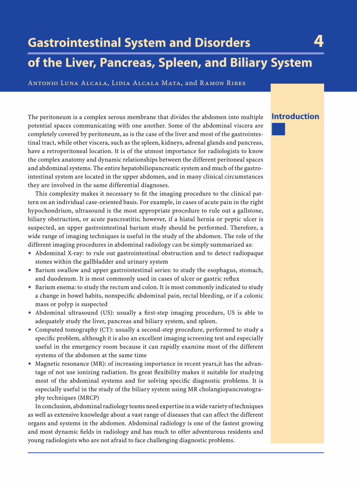
Nuclear Medicine
Gastrointestinal System and Disorders 4of the Liver, Pancreas, Spleen, and Biliary System Antonio Luna Alcala, Lidia Alcala Mata, and Ramon Ribes
IntroductionThe peritoneum is a complex serous membrane that divides the abdomen into multiple potential spaces communicating with one another. Some of the abdominal viscera are completely covered by peritoneum, as is the case of the liver and most of the gastrointes-tinal tract, while other viscera, such as the spleen, kidneys, adrenal glands and pancreas, have a retroperitoneal location. It is of the utmost importance for radiologists to know the complex anatomy and dynamic relationships between the different peritoneal spaces and abdominal systems. The entire hepatobiliopancreatic system and much of the gastro-intestinal system are located in the upper abdomen, and in many clinical circumstances they are involved in the same differential diagnoses.
This complexity makes it necessary to fi t the imaging procedure to the clinical pat-tern on an individual case-oriented basis. For example, in cases of acute pain in the right hypochondrium, ultrasound is the most appropriate procedure to rule out a gallstone, biliary obstruction, or acute pancreatitis; however, if a hiatal hernia or peptic ulcer is suspected, an upper gastrointestinal barium study should be performed. Therefore, a wide range of imaging techniques is useful in the study of the abdomen. The role of the different imaging procedures in abdominal radiology can be simply summarized as:
Abdominal X-ray: to rule out gastrointestinal obstruction and to detect radiopaque stones within the gallbladder and urinary systemBarium swallow and upper gastrointestinal series: to study the esophagus, stomach, and duodenum. It is most commonly used in cases of ulcer or gastric refl uxBarium enema: to study the rectum and colon. It is most commonly indicated to study a change in bowel habits, nonspecifi c abdominal pain, rectal bleeding, or if a colonic mass or polyp is suspectedAbdominal ultrasound (US): usually a fi rst-step imaging procedure, US is able to adequately study the liver, pancreas and biliary system, and spleen.Computed tomography (CT): usually a second-step procedure, performed to study a specifi c problem, although it is also an excellent imaging screening test and especially useful in the emergency room because it can rapidly examine most of the different systems of the abdomen at the same time Magnetic resonance (MR): of increasing importance in recent years,it has the advan-tage of not use ionizing radiation. Its great fl exibility makes it suitable for studying most of the abdominal systems and for solving specifi c diagnostic problems. It is especially useful in the study of the biliary system using MR cholangiopancreatogra-phy techniques (MRCP)In conclusion, abdominal radiology teams need expertise in a wide variety of techniques
as well as extensive knowledge about a vast range of diseases that can affect the different organs and systems in the abdomen. Abdominal radiology is one of the fastest growing and most dynamic fi elds in radiology and has much to offer adventurous residents and young radiologists who are not afraid to face challenging diagnostic problems.
●
●
●
●
●
●
RibesLunaRos.indb 77 18.03.2008 12:49:55
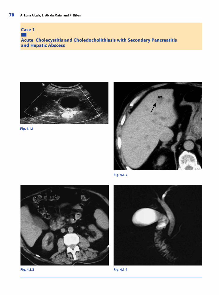
78 A. Luna Alcala, L. Alcala Mata, and R. Ribes
Case 1
Fig. 4.1.1
Fig. 4.1.2
Fig. 4.1.3 Fig. 4.1.4
Acute Cholecystitis and Choledocholithiasis with Secondary Pancreatitisand Hepatic Abscess
RibesLunaRos.indb 78 18.03.2008 12:49:55
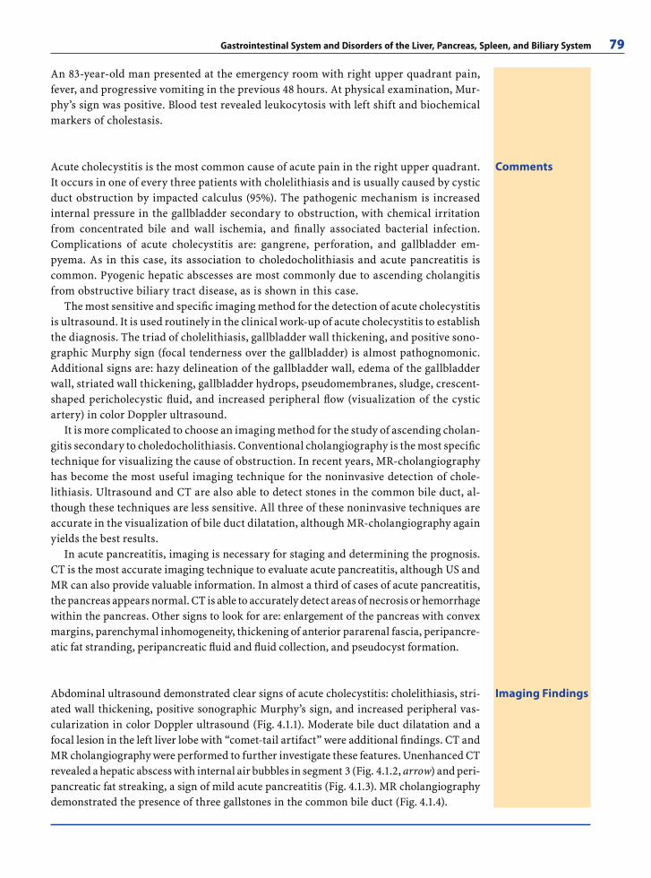
Gastrointestinal System and Disorders of the Liver, Pancreas, Spleen, and Biliary System 79
An 83-year-old man presented at the emergency room with right upper quadrant pain, fever, and progressive vomiting in the previous 48 hours. At physical examination, Mur-phy’s sign was positive. Blood test revealed leukocytosis with left shift and biochemical markers of cholestasis.
Imaging FindingsAbdominal ultrasound demonstrated clear signs of acute cholecystitis: cholelithiasis, stri-ated wall thickening, positive sonographic Murphy’s sign, and increased peripheral vas-cularization in color Doppler ultrasound (Fig. 4.1.1). Moderate bile duct dilatation and a focal lesion in the left liver lobe with “comet-tail artifact” were additional fi ndings. CT and MR cholangiography were performed to further investigate these features. Unenhanced CT revealed a hepatic abscess with internal air bubbles in segment 3 (Fig. 4.1.2, arrow) and peri-pancreatic fat streaking, a sign of mild acute pancreatitis (Fig. 4.1.3). MR cholangiography demonstrated the presence of three gallstones in the common bile duct (Fig. 4.1.4).
CommentsAcute cholecystitis is the most common cause of acute pain in the right upper quadrant. It occurs in one of every three patients with cholelithiasis and is usually caused by cystic duct obstruction by impacted calculus (95%). The pathogenic mechanism is increased internal pressure in the gallbladder secondary to obstruction, with chemical irritation from concentrated bile and wall ischemia, and fi nally associated bacterial infection. Complications of acute cholecystitis are: gangrene, perforation, and gallbladder em-pyema. As in this case, its association to choledocholithiasis and acute pancreatitis is common. Pyogenic hepatic abscesses are most commonly due to ascending cholangitis from obstructive biliary tract disease, as is shown in this case.
The most sensitive and specifi c imaging method for the detection of acute cholecystitis is ultrasound. It is used routinely in the clinical work-up of acute cholecystitis to establish the diagnosis. The triad of cholelithiasis, gallbladder wall thickening, and positive sono-graphic Murphy sign (focal tenderness over the gallbladder) is almost pathognomonic. Additional signs are: hazy delineation of the gallbladder wall, edema of the gallbladder wall, striated wall thickening, gallbladder hydrops, pseudomembranes, sludge, crescent-shaped pericholecystic fl uid, and increased peripheral fl ow (visualization of the cystic artery) in color Doppler ultrasound.
It is more complicated to choose an imaging method for the study of ascending cholan-gitis secondary to choledocholithiasis. Conventional cholangiography is the most specifi c technique for visualizing the cause of obstruction. In recent years, MR-cholangiography has become the most useful imaging technique for the noninvasive detection of chole-lithiasis. Ultrasound and CT are also able to detect stones in the common bile duct, al-though these techniques are less sensitive. All three of these noninvasive techniques are accurate in the visualization of bile duct dilatation, although MR-cholangiography again yields the best results.
In acute pancreatitis, imaging is necessary for staging and determining the prognosis. CT is the most accurate imaging technique to evaluate acute pancreatitis, although US and MR can also provide valuable information. In almost a third of cases of acute pancreatitis, the pancreas appears normal. CT is able to accurately detect areas of necrosis or hemorrhage within the pancreas. Other signs to look for are: enlargement of the pancreas with convex margins, parenchymal inhomogeneity, thickening of anterior pararenal fascia, peripancre-atic fat stranding, peripancreatic fl uid and fl uid collection, and pseudocyst formation.
RibesLunaRos.indb 79 18.03.2008 12:49:57
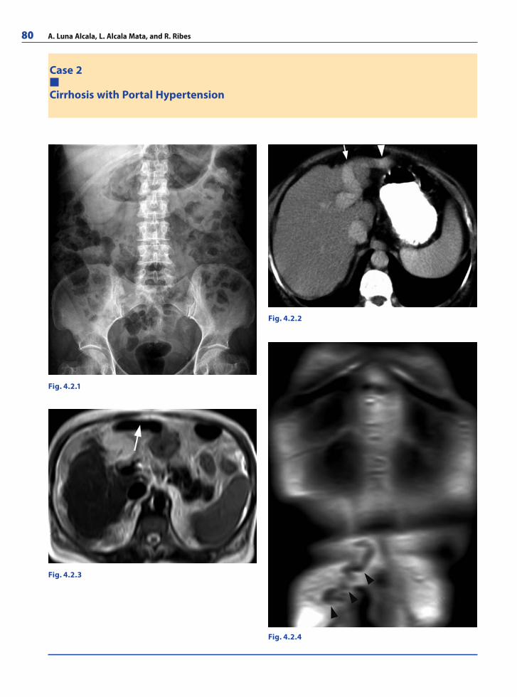
80 A. Luna Alcala, L. Alcala Mata, and R. Ribes
Case 2
Cirrhosis with Portal Hypertension
Fig. 4.2.1
Fig. 4.2.2
Fig. 4.2.3
Fig. 4.2.4
RibesLunaRos.indb 80 18.03.2008 12:49:57
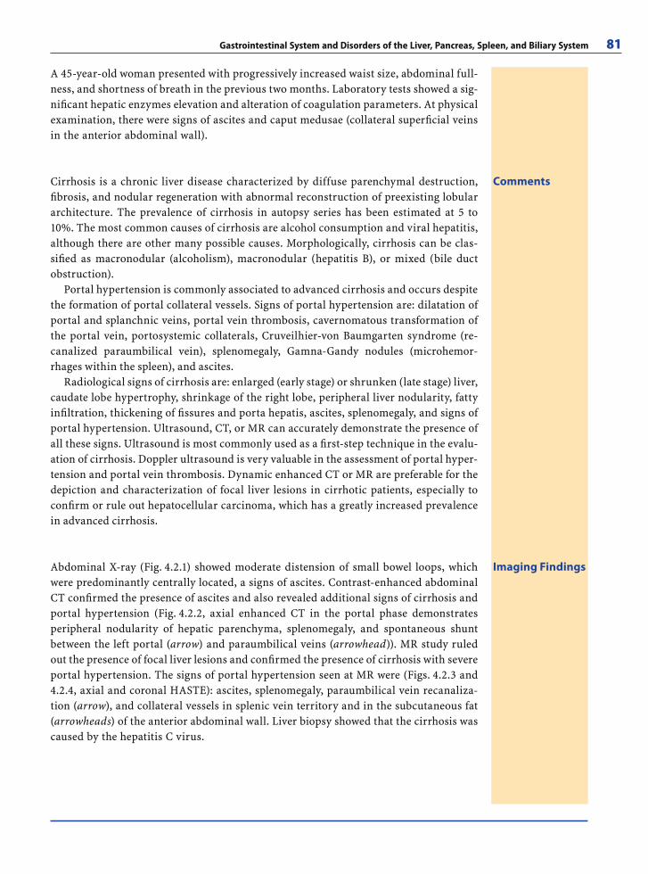
Gastrointestinal System and Disorders of the Liver, Pancreas, Spleen, and Biliary System 81
A 45-year-old woman presented with progressively increased waist size, abdominal full-ness, and shortness of breath in the previous two months. Laboratory tests showed a sig-nifi cant hepatic enzymes elevation and alteration of coagulation parameters. At physical examination, there were signs of ascites and caput medusae (collateral superfi cial veins in the anterior abdominal wall).
CommentsCirrhosis is a chronic liver disease characterized by diffuse parenchymal destruction, fi brosis, and nodular regeneration with abnormal reconstruction of preexisting lobular architecture. The prevalence of cirrhosis in autopsy series has been estimated at 5 to 10%. The most common causes of cirrhosis are alcohol consumption and viral hepatitis, although there are other many possible causes. Morphologically, cirrhosis can be clas-sifi ed as macronodular (alcoholism), macronodular (hepatitis B), or mixed (bile duct obstruction).
Portal hypertension is commonly associated to advanced cirrhosis and occurs despite the formation of portal collateral vessels. Signs of portal hypertension are: dilatation of portal and splanchnic veins, portal vein thrombosis, cavernomatous transformation of the portal vein, portosystemic collaterals, Cruveilhier-von Baumgarten syndrome (re-canalized paraumbilical vein), splenomegaly, Gamna-Gandy nodules (microhemor-rhages within the spleen), and ascites.
Radiological signs of cirrhosis are: enlarged (early stage) or shrunken (late stage) liver, caudate lobe hypertrophy, shrinkage of the right lobe, peripheral liver nodularity, fatty infi ltration, thickening of fi ssures and porta hepatis, ascites, splenomegaly, and signs of portal hypertension. Ultrasound, CT, or MR can accurately demonstrate the presence of all these signs. Ultrasound is most commonly used as a fi rst-step technique in the evalu-ation of cirrhosis. Doppler ultrasound is very valuable in the assessment of portal hyper-tension and portal vein thrombosis. Dynamic enhanced CT or MR are preferable for the depiction and characterization of focal liver lesions in cirrhotic patients, especially to confi rm or rule out hepatocellular carcinoma, which has a greatly increased prevalence in advanced cirrhosis.
Imaging FindingsAbdominal X-ray (Fig. 4.2.1) showed moderate distension of small bowel loops, which were predominantly centrally located, a signs of ascites. Contrast-enhanced abdominal CT confi rmed the presence of ascites and also revealed additional signs of cirrhosis and portal hypertension (Fig. 4.2.2, axial enhanced CT in the portal phase demonstrates peripheral nodularity of hepatic parenchyma, splenomegaly, and spontaneous shunt between the left portal (arrow) and paraumbilical veins (arrowhead)). MR study ruled out the presence of focal liver lesions and confi rmed the presence of cirrhosis with severe portal hypertension. The signs of portal hypertension seen at MR were (Figs. 4.2.3 and 4.2.4, axial and coronal HASTE): ascites, splenomegaly, paraumbilical vein recanaliza-tion (arrow), and collateral vessels in splenic vein territory and in the subcutaneous fat (arrowheads) of the anterior abdominal wall. Liver biopsy showed that the cirrhosis was caused by the hepatitis C virus.
RibesLunaRos.indb 81 18.03.2008 12:50:00
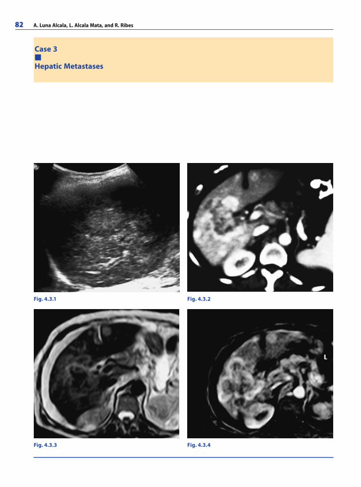
82 A. Luna Alcala, L. Alcala Mata, and R. Ribes
Case 3
Hepatic Metastases
Fig. 4.3.1 Fig. 4.3.2
Fig. 4.3.4Fig. 4.3.3
RibesLunaRos.indb 82 18.03.2008 12:50:00
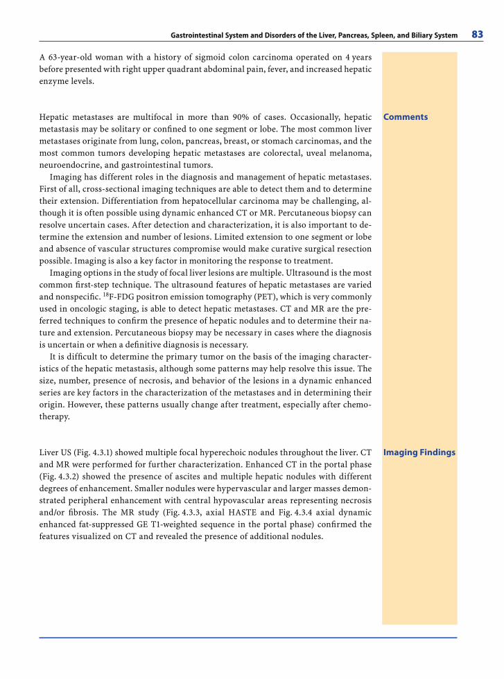
Gastrointestinal System and Disorders of the Liver, Pancreas, Spleen, and Biliary System 83
A 63-year-old woman with a history of sigmoid colon carcinoma operated on 4 years before presented with right upper quadrant abdominal pain, fever, and increased hepatic enzyme levels.
CommentsHepatic metastases are multifocal in more than 90% of cases. Occasionally, hepatic metastasis may be solitary or confi ned to one segment or lobe. The most common liver metastases originate from lung, colon, pancreas, breast, or stomach carcinomas, and the most common tumors developing hepatic metastases are colorectal, uveal melanoma, neuroendocrine, and gastrointestinal tumors.
Imaging has different roles in the diagnosis and management of hepatic metastases. First of all, cross-sectional imaging techniques are able to detect them and to determine their extension. Differentiation from hepatocellular carcinoma may be challenging, al-though it is often possible using dynamic enhanced CT or MR. Percutaneous biopsy can resolve uncertain cases. After detection and characterization, it is also important to de-termine the extension and number of lesions. Limited extension to one segment or lobe and absence of vascular structures compromise would make curative surgical resection possible. Imaging is also a key factor in monitoring the response to treatment.
Imaging options in the study of focal liver lesions are multiple. Ultrasound is the most common fi rst-step technique. The ultrasound features of hepatic metastases are varied and nonspecifi c. 18F-FDG positron emission tomography (PET), which is very commonly used in oncologic staging, is able to detect hepatic metastases. CT and MR are the pre-ferred techniques to confi rm the presence of hepatic nodules and to determine their na-ture and extension. Percutaneous biopsy may be necessary in cases where the diagnosis is uncertain or when a defi nitive diagnosis is necessary.
It is diffi cult to determine the primary tumor on the basis of the imaging character-istics of the hepatic metastasis, although some patterns may help resolve this issue. The size, number, presence of necrosis, and behavior of the lesions in a dynamic enhanced series are key factors in the characterization of the metastases and in determining their origin. However, these patterns usually change after treatment, especially after chemo-therapy.
Imaging FindingsLiver US (Fig. 4.3.1) showed multiple focal hyperechoic nodules throughout the liver. CT and MR were performed for further characterization. Enhanced CT in the portal phase (Fig. 4.3.2) showed the presence of ascites and multiple hepatic nodules with different degrees of enhancement. Smaller nodules were hypervascular and larger masses demon-strated peripheral enhancement with central hypovascular areas representing necrosis and/or fi brosis. The MR study (Fig. 4.3.3, axial HASTE and Fig. 4.3.4 axial dynamic enhanced fat-suppressed GE T1-weighted sequence in the portal phase) confi rmed the features visualized on CT and revealed the presence of additional nodules.
RibesLunaRos.indb 83 18.03.2008 12:50:03
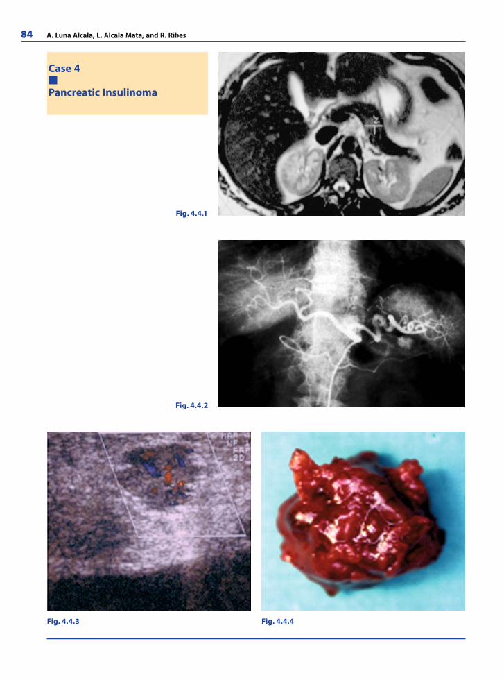
84 A. Luna Alcala, L. Alcala Mata, and R. Ribes
Case 4
Pancreatic Insulinoma
Fig. 4.4.1
Fig. 4.4.2
Fig. 4.4.4Fig. 4.4.3
RibesLunaRos.indb 84 18.03.2008 12:50:03
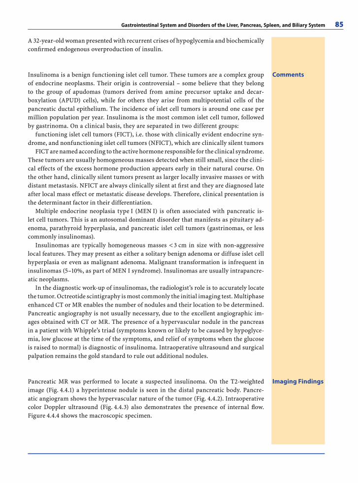
Gastrointestinal System and Disorders of the Liver, Pancreas, Spleen, and Biliary System 85
A 32-year-old woman presented with recurrent crises of hypoglycemia and biochemically confi rmed endogenous overproduction of insulin.
CommentsInsulinoma is a benign functioning islet cell tumor. These tumors are a complex group of endocrine neoplasms. Their origin is controversial – some believe that they belong to the group of apudomas (tumors derived from amine precursor uptake and decar-boxylation (APUD) cells), while for others they arise from multipotential cells of the pancreatic ductal epithelium. The incidence of islet cell tumors is around one case per million population per year. Insulinoma is the most common islet cell tumor, followed by gastrinoma. On a clinical basis, they are separated in two different groups:
functioning islet cell tumors (FICT), i.e. those with clinically evident endocrine syn-drome, and nonfunctioning islet cell tumors (NFICT), which are clinically silent tumors
FICT are named according to the active hormone responsible for the clinical syndrome. These tumors are usually homogeneous masses detected when still small, since the clini-cal effects of the excess hormone production appears early in their natural course. On the other hand, clinically silent tumors present as larger locally invasive masses or with distant metastasis. NFICT are always clinically silent at fi rst and they are diagnosed late after local mass effect or metastatic disease develops. Therefore, clinical presentation is the determinant factor in their differentiation.
Multiple endocrine neoplasia type I (MEN I) is often associated with pancreatic is-let cell tumors. This is an autosomal dominant disorder that manifests as pituitary ad-enoma, parathyroid hyperplasia, and pancreatic islet cell tumors (gastrinomas, or less commonly insulinomas).
Insulinomas are typically homogeneous masses < 3 cm in size with non-aggressive local features. They may present as either a solitary benign adenoma or diffuse islet cell hyperplasia or even as malignant adenoma. Malignant transformation is infrequent in insulinomas (5–10%, as part of MEN I syndrome). Insulinomas are usually intrapancre-atic neoplasms.
In the diagnostic work-up of insulinomas, the radiologist’s role is to accurately locate the tumor. Octreotide scintigraphy is most commonly the initial imaging test. Multiphase enhanced CT or MR enables the number of nodules and their location to be determined. Pancreatic angiography is not usually necessary, due to the excellent angiographic im-ages obtained with CT or MR. The presence of a hypervascular nodule in the pancreas in a patient with Whipple’s triad (symptoms known or likely to be caused by hypoglyce-mia, low glucose at the time of the symptoms, and relief of symptoms when the glucose is raised to normal) is diagnostic of insulinoma. Intraoperative ultrasound and surgical palpation remains the gold standard to rule out additional nodules.
Imaging FindingsPancreatic MR was performed to locate a suspected insulinoma. On the T2-weighted image (Fig. 4.4.1) a hyperintense nodule is seen in the distal pancreatic body. Pancre-atic angiogram shows the hypervascular nature of the tumor (Fig. 4.4.2). Intraoperative color Doppler ultrasound (Fig. 4.4.3) also demonstrates the presence of internal fl ow. Figure 4.4.4 shows the macroscopic specimen.
RibesLunaRos.indb 85 18.03.2008 12:50:09
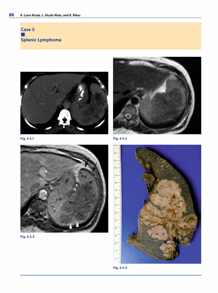
86 A. Luna Alcala, L. Alcala Mata, and R. Ribes
Case 5
Splenic Lymphoma
Fig. 4.5.1 Fig. 4.5.2
Fig. 4.5.3
Fig. 4.5.4
RibesLunaRos.indb 86 18.03.2008 12:50:09
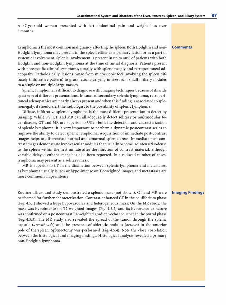
Gastrointestinal System and Disorders of the Liver, Pancreas, Spleen, and Biliary System 87
A 47-year-old woman presented with left abdominal pain and weight loss over 3 months.
CommentsLymphoma is the most common malignancy affecting the spleen. Both Hodgkin and non-Hodgkin lymphoma may present in the spleen either as a primary lesion or as a part of systemic involvement. Splenic involvement is present in up to 40% of patients with both Hodgkin and non-Hodgkin lymphoma at the time of initial diagnosis. Patients present with nonspecifi c clinical symptoms, usually with splenomegaly and retroperitoneal ad-enopathy. Pathologically, lesions range from microscopic foci involving the spleen dif-fusely (infi ltrative pattern) to gross lesions varying in size from small miliary nodules to a single or multiple large masses.
Splenic lymphoma is diffi cult to diagnose with imaging techniques because of its wide spectrum of different presentations. In cases of secondary splenic lymphoma, retroperi-toneal adenopathies are nearly always present and when this fi nding is associated to sple-nomegaly, it should alert the radiologist to the possibility of splenic lymphoma.
Diffuse, infi ltrative splenic lymphoma is the most diffi cult presentation to detect by imaging. While US, CT, and MR can all adequately detect solitary or multinodular fo-cal disease, CT and MR are superior to US in both the detection and characterization of splenic lymphoma. It is very important to perform a dynamic postcontrast series to improve the ability to detect splenic lymphoma. Acquisition of immediate post-contrast images helps to differentiate normal and abnormal splenic areas. Immediate post-con-trast images demonstrate hypovascular nodules that usually become isointense/isodense to the spleen within the fi rst minute after the injection of contrast material, although variable delayed enhancement has also been reported. In a reduced number of cases, lymphoma may present as a solitary mass.
MR is superior to CT in the distinction between splenic lymphoma and metastases, as lymphoma usually is iso- or hypo-intense on T2-weighted images and metastases are more commonly hyperintense.
Imaging FindingsRoutine ultrasound study demonstrated a splenic mass (not shown). CT and MR were performed for further characterization. Contrast-enhanced CT in the equilibrium phase (Fig. 4.5.1) showed a huge hypovascular and heterogeneous mass. On the MR study, the mass was hypointense on T2-weighted images (Fig. 4.5.2) and its hypovascular nature was confi rmed on a postcontrast T1-weighted gradient-echo sequence in the portal phase (Fig. 4.5.3). The MR study also revealed the spread of the tumor through the splenic capsule (arrowheads) and the presence of siderotic nodules (arrows) in the anterior pole of the spleen. Splenectomy was performed (Fig. 4.5.4). Note the close correlation between the histological and imaging fi ndings. Histological analysis revealed a primary non-Hodgkin lymphoma.
RibesLunaRos.indb 87 18.03.2008 12:50:15
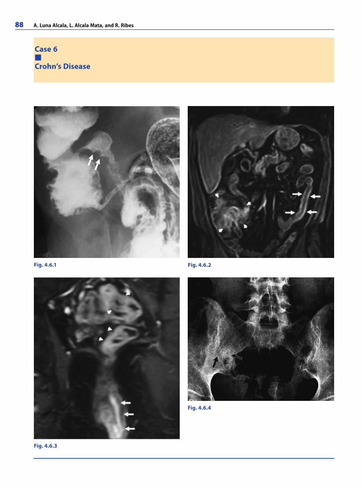
88 A. Luna Alcala, L. Alcala Mata, and R. Ribes
Case 6
Crohn’s Disease
Fig. 4.6.1 Fig. 4.6.2
Fig. 4.6.3
Fig. 4.6.4
RibesLunaRos.indb 88 18.03.2008 12:50:15
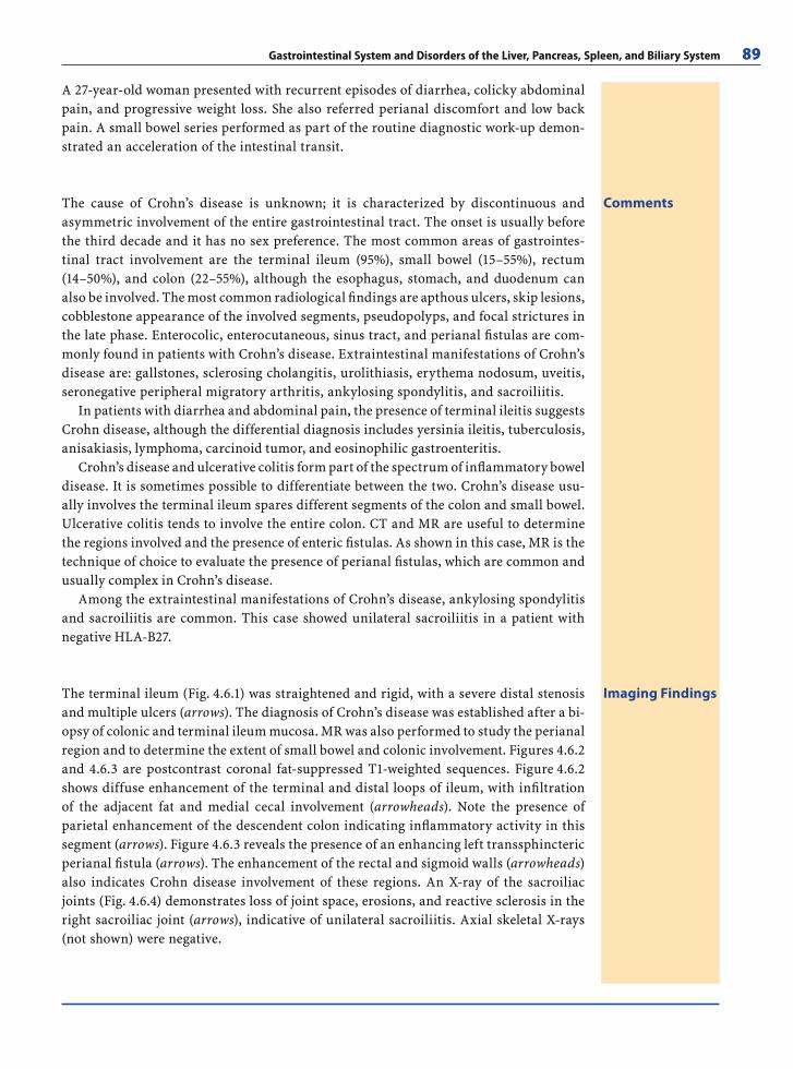
Gastrointestinal System and Disorders of the Liver, Pancreas, Spleen, and Biliary System 89
A 27-year-old woman presented with recurrent episodes of diarrhea, colicky abdominal pain, and progressive weight loss. She also referred perianal discomfort and low back pain. A small bowel series performed as part of the routine diagnostic work-up demon-strated an acceleration of the intestinal transit.
CommentsThe cause of Crohn’s disease is unknown; it is characterized by discontinuous and asymmetric involvement of the entire gastrointestinal tract. The onset is usually before the third decade and it has no sex preference. The most common areas of gastrointes-tinal tract involvement are the terminal ileum (95%), small bowel (15–55%), rectum (14–50%), and colon (22–55%), although the esophagus, stomach, and duodenum can also be involved. The most common radiological fi ndings are apthous ulcers, skip lesions, cobblestone appearance of the involved segments, pseudopolyps, and focal strictures in the late phase. Enterocolic, enterocutaneous, sinus tract, and perianal fi stulas are com-monly found in patients with Crohn’s disease. Extraintestinal manifestations of Crohn’s disease are: gallstones, sclerosing cholangitis, urolithiasis, erythema nodosum, uveitis, seronegative peripheral migratory arthritis, ankylosing spondylitis, and sacroiliitis.
In patients with diarrhea and abdominal pain, the presence of terminal ileitis suggests Crohn disease, although the differential diagnosis includes yersinia ileitis, tuberculosis, anisakiasis, lymphoma, carcinoid tumor, and eosinophilic gastroenteritis.
Crohn’s disease and ulcerative colitis form part of the spectrum of infl ammatory bowel disease. It is sometimes possible to differentiate between the two. Crohn’s disease usu-ally involves the terminal ileum spares different segments of the colon and small bowel. Ulcerative colitis tends to involve the entire colon. CT and MR are useful to determine the regions involved and the presence of enteric fi stulas. As shown in this case, MR is the technique of choice to evaluate the presence of perianal fi stulas, which are common and usually complex in Crohn’s disease.
Among the extraintestinal manifestations of Crohn’s disease, ankylosing spondylitis and sacroiliitis are common. This case showed unilateral sacroiliitis in a patient with negative HLA-B27.
Imaging FindingsThe terminal ileum (Fig. 4.6.1) was straightened and rigid, with a severe distal stenosis and multiple ulcers (arrows). The diagnosis of Crohn’s disease was established after a bi-opsy of colonic and terminal ileum mucosa. MR was also performed to study the perianal region and to determine the extent of small bowel and colonic involvement. Figures 4.6.2 and 4.6.3 are postcontrast coronal fat-suppressed T1-weighted sequences. Figure 4.6.2 shows diffuse enhancement of the terminal and distal loops of ileum, with infi ltration of the adjacent fat and medial cecal involvement (arrowheads). Note the presence of parietal enhancement of the descendent colon indicating infl ammatory activity in this segment (arrows). Figure 4.6.3 reveals the presence of an enhancing left transsphincteric perianal fi stula (arrows). The enhancement of the rectal and sigmoid walls (arrowheads) also indicates Crohn disease involvement of these regions. An X-ray of the sacroiliac joints (Fig. 4.6.4) demonstrates loss of joint space, erosions, and reactive sclerosis in the right sacroiliac joint (arrows), indicative of unilateral sacroiliitis. Axial skeletal X-rays (not shown) were negative.
RibesLunaRos.indb 89 18.03.2008 12:50:16
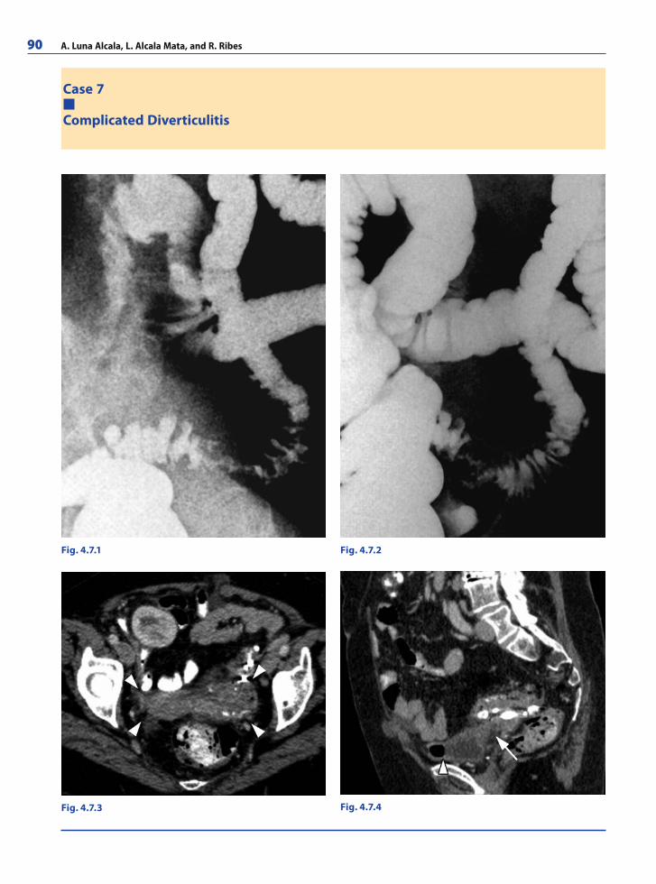
90 A. Luna Alcala, L. Alcala Mata, and R. Ribes
Case 7
Complicated Diverticulitis
Fig. 4.7.1 Fig. 4.7.2
Fig. 4.7.4Fig. 4.7.3
RibesLunaRos.indb 90 18.03.2008 12:50:16
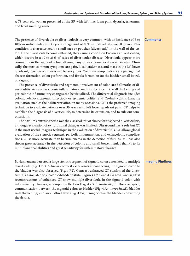
Gastrointestinal System and Disorders of the Liver, Pancreas, Spleen, and Biliary System 91
A 78-year-old woman presented at the ER with left iliac fossa pain, dysuria, tenesmus, and fecal smelling urine.
CommentsThe presence of diverticula or diverticulosis is very common, with an incidence of 5 to 10% in individuals over 45 years of age and of 80% in individuals over 85 years. This condition is characterized by small sacs or pouches (diverticula) in the wall of the co-lon. If the diverticula become infl amed, they cause a condition known as diverticulitis, which occurs in a 10 to 25% of cases of diverticular disease. Diverticula appear more commonly in the sigmoid colon, although any other colonic location is possible. Clini-cally, the most common symptoms are pain, local tenderness, and mass in the left lower quadrant, together with fever and leukocytosis. Common complications are perisigmoid abscess formation, colon perforation, and fi stula formation (to the bladder, small bowel, or vagina).
The presence of diverticula and segmental involvement of colon are hallmarks of di-verticulitis. As in other colonic infl ammatory conditions, concentric wall thickening and pericolonic infl ammatory changes can be visualized. The differential diagnosis includes colonic adenocarcinoma, infectious or ischemic colitis, and Crohn’s colitis. Imaging evaluation enables their differentiation on many occasions. CT is the preferred imaging technique to evaluate patients over 50 years with left lower quadrant pain. CT helps to establish the diagnosis of diverticulitis, to determine its extension, and to rule out com-plications.
The barium contrast-enema was the classical test of choice for suspected diverticulitis, although evaluation of extraluminal changes was limited. Ultrasound has a role but CT is the most useful imaging technique in the evaluation of diverticulitis. CT allows global evaluation of the stenotic segment, pericolic infl ammation, and extracolonic complica-tions. CT is more accurate than barium enema in the detection of fi stulas. MR has also shown great accuracy in the detection of colonic and small bowel fi stulas thanks to its multiplanar capabilities and great sensitivity for infl ammatory changes.
Imaging FindingsBarium enema detected a large stenotic segment of sigmoid colon associated to multiple diverticula (Fig. 4.7.1). A linear contrast extravasation connecting the sigmoid colon to the bladder was also observed (Fig. 4.7.2). Contrast-enhanced CT confi rmed the diver-ticulitis associated to a colonic-bladder fi stula. Figures 4.7.3 and 4.7.4 Axial and sagittal reconstructions of enhanced CT show multiple diverticula in the sigmoid colon with infl ammatory changes, a complex collection (Fig. 4.7.3, arrowheads) in Douglas space, communication between the sigmoid colon to bladder (Fig. 4.7.4, arrowhead), bladder wall thickening, and an air-fl uid level (Fig. 4.7.4, arrow) within the bladder confi rming the fi stula.
RibesLunaRos.indb 91 18.03.2008 12:50:19
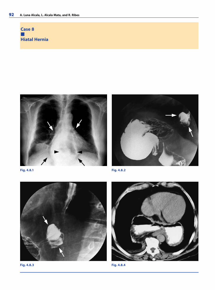
92 A. Luna Alcala, L. Alcala Mata, and R. Ribes
Case 8
Hiatal Hernia
Fig. 4.8.1 Fig. 4.8.2
Fig. 4.8.3 Fig. 4.8.4
RibesLunaRos.indb 92 18.03.2008 12:50:19
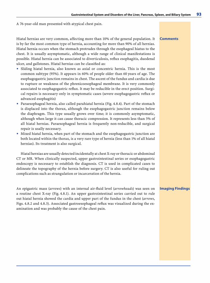
Gastrointestinal System and Disorders of the Liver, Pancreas, Spleen, and Biliary System 93
A 76-year-old man presented with atypical chest pain.
CommentsHiatal hernias are very common, affecting more than 10% of the general population. It is by far the most common type of hernia, accounting for more than 90% of all hernias. Hiatal hernia occurs when the stomach protrudes through the esophageal hiatus to the chest. It is usually asymptomatic, although a wide range of clinical manifestations is possible. Hiatal hernia can be associated to diverticulosis, refl ux esophagitis, duodenal ulcer, and gallstones. Hiatal hernias can be classifi ed as:
Sliding hiatal hernia, also known as axial or concentric hernia. This is the most common subtype (95%). It appears in 60% of people older than 60 years of age. The esophagogastric junction remains in chest. The ascent of the fundus and cardia is due to rupture or weakness of the phrenicoesophageal membrane. It is very commonly associated to esophagogastric refl ux. It may be reducible in the erect position. Surgi-cal repairs is necessary only in symptomatic cases (severe esophagogastric refl ux or advanced esophagitis)Paraesophageal hernia, also called parahiatal hernia (Fig. 4.8.4). Part of the stomach is displaced into the thorax, although the esophagogastric junction remains below the diaphragm. This type usually grows over time; it is commonly asymptomatic, although when large it can cause thoracic compression. It represents less than 5% of all hiatal hernias. Paraesophageal hernia is frequently non-reducible, and surgical repair is usally necessary.Mixed hiatal hernia, when part of the stomach and the esophagogastric junction are both located within the thorax, is a very rare type of hernia (less than 1% of all hiatal hernias). Its treatment is also surgical.
Hiatal hernias are usually detected incidentally at chest X-ray or thoracic or abdominal CT or MR. When clinically suspected, upper gastrointestinal series or esophagogastric endoscopy is necessary to establish the diagnosis. CT is used in complicated cases to delineate the topography of the hernia before surgery. CT is also useful for ruling out complications such as strangulation or incarceration of the hernia.
●
●
●
Imaging FindingsAn epigastric mass (arrows) with an internal air-fl uid level (arrowheads) was seen on a routine chest X-ray (Fig. 4.8.1). An upper gastrointestinal series carried out to rule out hiatal hernia showed the cardia and upper part of the fundus in the chest (arrows, Figs. 4.8.2 and 4.8.3). Associated gastroesophageal refl ux was visualized during the ex-amination and was probably the cause of the chest pain.
RibesLunaRos.indb 93 18.03.2008 12:50:22
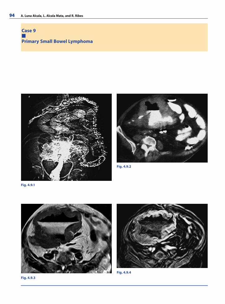
94 A. Luna Alcala, L. Alcala Mata, and R. Ribes
Case 9
Primary Small Bowel Lymphoma
Fig. 4.9.1
Fig. 4.9.2
Fig. 4.9.3Fig. 4.9.4
RibesLunaRos.indb 94 18.03.2008 12:50:22
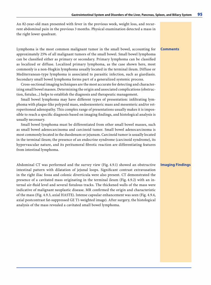
Gastrointestinal System and Disorders of the Liver, Pancreas, Spleen, and Biliary System 95
An 82-year-old man presented with fever in the previous week, weight loss, and recur-rent abdominal pain in the previous 3 months. Physical examination detected a mass in the right lower quadrant.
CommentsLymphoma is the most common malignant tumor in the small bowel, accounting for approximately 25% of all malignant tumors of the small bowel. Small bowel lymphoma can be classifi ed either as primary or secondary. Primary lymphoma can be classifi ed as localized or diffuse. Localized primary lymphoma, as the case shown here, most commonly is a non-Hogkin lymphoma usually located in the terminal ileum. Diffuse or Mediterranean-type lymphoma is associated to parasitic infection, such as giardiasis. Secondary small bowel lymphoma forms part of a generalized systemic process.
Cross-sectional imaging techniques are the most accurate for detecting and character-izing small bowel masses. Determining the origin and associated complications (obstruc-tion, fi stulas...) helps to establish the diagnosis and therapeutic management.
Small bowel lymphoma may have different types of presentation: infi ltrating lym-phoma with plaque-like polypoid mass, endoexoenteric mass and mesenteric and/or ret-roperitoneal adenopathy. This complex range of presentations usually makes it is impos-sible to reach a specifi c diagnosis based on imaging fi ndings, and histological analysis is usually necessary.
Small bowel lymphoma must be differentiated from other small bowel masses, such as small bowel adenocarcinoma and carcinoid tumor. Small bowel adenocarcinoma is most commonly located in the duodenum or jejunum. Carcinoid tumor is usually located in the terminal ileum; the presence of an endocrine syndrome (carcinoid syndrome), its hypervascular nature, and its peritumoral fi brotic reaction are differentiating features from intestinal lymphoma.
Imaging FindingsAbdominal CT was performed and the survey view (Fig. 4.9.1) showed an obstructive intestinal pattern with dilatation of jejunal loops. Signifi cant contrast extravasation in the right iliac fossa and colonic diverticula were also present. CT demonstrated the presence of a cavitated mass originating in the terminal ileum (Fig. 4.9.2) with an in-ternal air-fl uid level and several fi stulous tracks. The thickened walls of the mass were indicative of malignant neoplastic disease. MR confi rmed the origin and characteristic of the mass (Fig. 4.9.3, axial HASTE). Intense capsular enhancement was seen (Fig. 4.9.4, axial postcontrast fat-suppressed GE T1-weighted image). After surgery, the histological analysis of the mass revealed a cavitated small bowel lymphoma.
RibesLunaRos.indb 95 18.03.2008 12:50:25
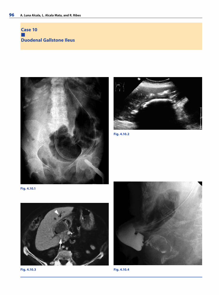
96 A. Luna Alcala, L. Alcala Mata, and R. Ribes
Case 10
Duodenal Gallstone Ileus
Fig. 4.10.1
Fig. 4.10.2
Fig. 4.10.4Fig. 4.10.3
RibesLunaRos.indb 96 18.03.2008 12:50:25
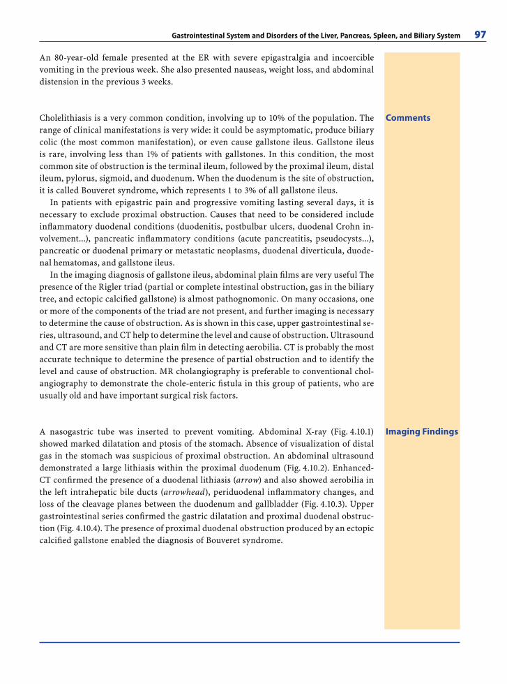
Gastrointestinal System and Disorders of the Liver, Pancreas, Spleen, and Biliary System 97
An 80-year-old female presented at the ER with severe epigastralgia and incoercible vomiting in the previous week. She also presented nauseas, weight loss, and abdominal distension in the previous 3 weeks.
CommentsCholelithiasis is a very common condition, involving up to 10% of the population. The range of clinical manifestations is very wide: it could be asymptomatic, produce biliary colic (the most common manifestation), or even cause gallstone ileus. Gallstone ileus is rare, involving less than 1% of patients with gallstones. In this condition, the most common site of obstruction is the terminal ileum, followed by the proximal ileum, distal ileum, pylorus, sigmoid, and duodenum. When the duodenum is the site of obstruction, it is called Bouveret syndrome, which represents 1 to 3% of all gallstone ileus.
In patients with epigastric pain and progressive vomiting lasting several days, it is necessary to exclude proximal obstruction. Causes that need to be considered include infl ammatory duodenal conditions (duodenitis, postbulbar ulcers, duodenal Crohn in-volvement...), pancreatic infl ammatory conditions (acute pancreatitis, pseudocysts...), pancreatic or duodenal primary or metastatic neoplasms, duodenal diverticula, duode-nal hematomas, and gallstone ileus.
In the imaging diagnosis of gallstone ileus, abdominal plain fi lms are very useful The presence of the Rigler triad (partial or complete intestinal obstruction, gas in the biliary tree, and ectopic calcifi ed gallstone) is almost pathognomonic. On many occasions, one or more of the components of the triad are not present, and further imaging is necessary to determine the cause of obstruction. As is shown in this case, upper gastrointestinal se-ries, ultrasound, and CT help to determine the level and cause of obstruction. Ultrasound and CT are more sensitive than plain fi lm in detecting aerobilia. CT is probably the most accurate technique to determine the presence of partial obstruction and to identify the level and cause of obstruction. MR cholangiography is preferable to conventional chol-angiography to demonstrate the chole-enteric fi stula in this group of patients, who are usually old and have important surgical risk factors.
Imaging FindingsA nasogastric tube was inserted to prevent vomiting. Abdominal X-ray (Fig. 4.10.1) showed marked dilatation and ptosis of the stomach. Absence of visualization of distal gas in the stomach was suspicious of proximal obstruction. An abdominal ultrasound demonstrated a large lithiasis within the proximal duodenum (Fig. 4.10.2). Enhanced-CT confi rmed the presence of a duodenal lithiasis (arrow) and also showed aerobilia in the left intrahepatic bile ducts (arrowhead), periduodenal infl ammatory changes, and loss of the cleavage planes between the duodenum and gallbladder (Fig. 4.10.3). Upper gastrointestinal series confi rmed the gastric dilatation and proximal duodenal obstruc-tion (Fig. 4.10.4). The presence of proximal duodenal obstruction produced by an ectopic calcifi ed gallstone enabled the diagnosis of Bouveret syndrome.
RibesLunaRos.indb 97 18.03.2008 12:50:28
98 A. Luna Alcala, L. Alcala Mata, and R. Ribes
Further Readings
Books
Abdominal-Pelvic MRI. 2nd ed. Semelka RC (2006) Wiley-Liss, New-York. ISBN-13: 9780471692737
Advanced Imaging of the Abdomen. Skucas J (2006) Springer-Verlag, Berlin 2006. ISBN-13: 9781852339920
Clinical Imaging of the Small Intestine. 2nd ed. Herlinger H, Maglinte DDT, Birnbaum BA (2001) Springer-Verlag, Ber-lin. ISBN-13: 9780387953885
Computed Body Tomography with MRI Correlation. Lee JKT, Stanley RJ, Heiken JP (2005) Lippincott Williams & Wilkins ISBN-13: 9780781745260
Diagnostic Ultrasound. Rumack CM, Wilson SR, Char-boneau JW (2005) Mosby. ISBN-13: 9780323020237
Double Contrast Gastrointestinal Radiology. 3rd edition. Levine MS, Rubesin SE, Laufer I (1999) W.B. Saunders Company. ISBN-13: 9780721682112
Liver MRI: Correlation with other Imaging Modalities and Histopathology. Hussain SM (2007) Springer-Verlag, Ber-lin. ISBN-13: 9783540255529
Medical Imaging of the Spleen. De Schepper AM; Van-hoenacker F (2000) Springer-Verlag, Berlin. ISBN-13: 9783540655350
MR Cholangiopancreatography. Atlas with Cross-Sectional Imaging Correlation. 2nd ed. Van Hoe L, Vanbeckevoort D, Mermuys K, van Steenbergen W (2006) Springer-Ver-lag, Berlin. ISBN-13: 9783540222699
Radiology and Imaging of the Colon. Chapman AH (2004) Springer-Verlag, Berlin. ISBN-13: 9783540435976
Radiology of the Pancreas. 2nd rev ed., Baert AL, Delorme G, Van Hoe L (1999) Springer-Verlag, Berlin. ISBN-13: 9783540634799
Web-Links
http://www.esgar.org/http://www.sgr.org/sgr.htmhttp://www.radcentral.com/http://radiographics.rsnajnls.org/cgi/collection/gi_
radiology?page=9http://www.emedicine.com/radio/gastrointestinal.htmhttp://icarus.med.utoronto.ca/imaging/residents/gi%
5Fimaging/index.htmhttp://www.rsna.org/Education/archive/afi p.cfm#
gastrointestinalhttp://chorus.rad.mcw.edu/index/4.htmlhttp://www.ctisus.com/http://www.med-ed.virginia.edu/courses/rad/gi/index.html
Articles
Esophagus
Baker ME, Einstein DM, Herts BR, Remer EM, Motta-Ramirez GA, Ehrenwald E, Rice TW, Richter JE. Gas-troesophageal Refl ux Disease: Integrating the Barium Esophagram before and after Antirefl ux Surgery. Radiol-ogy 2007 243:329–339
Canon CL, Morgan DE, Einstein DM, Herts BR, Hawn MT, Johnson LF. Surgical Approach to Gastroesophageal Re-fl ux Disease: What the Radiologist Needs to Know. Ra-diographics 2005; 25:1485–1499
Giménez A, Franquet T, Erasmus JJ, Martínez S, Estrada P. Thoracic Complications of Esophageal Disorders. Radio-graphics 2002; 22:247–258
Kim TJ, Lee KH, Kim YH, Sung SW, Jheon S, Cho SK, Lee KW. Postoperative Imaging of Esophageal Cancer: What Chest Radiologists Need to Know. Radiographics 2007; 27:409–429
Levine MS, Rubesin SE. Diseases of the Esophagus: Diagno-sis with Esophagography. Radiology 2005 237:414–427
Luedtke P, Levine MS, Rubesin SE, Weinstein DS, Laufer I. Radiologic Diagnosis of Benign Esophageal Strictures: A Pattern Approach. Radiographics 2003; 23:897–909
Noh HM, Fishman EK, Forastiere AA, Bliss DF, CalhounPS. CT of the esophagus: spectrum of disease with emphasis on esophageal carcinoma. Radiographics 1995; 15:1113–1134
Sam JW, Levine MS, Rubesin SE, Laufer I. The “Foamy” Esophagus: A Radiographic Sign of Candida Esophagitis. AJR Am J Roentgenol 2000; 174:999–1002
Schmalfuss IM, Mancuso AA, Tart RP. Postcricoid Region and Cervical Esophagus: Normal Appearance at CT and MR Imaging. Radiology 2000; 214:237–246
Zimmerman SL, Levine MS, Rubesin SE, Mitre MC, Furth EE, Laufer I, Katzka DA. Idiopathic Eosinophilic Esopha-gitis in Adults: The Ringed Esophagus. Radiology 2005; 236:159–165
Stomach
An SK, Han JK, Kim YH, Kim AY, Choi BI, Kim YA, Kim CW. Gastric Mucosa-associated Lymphoid Tissue Lym-phoma: Spectrum of Findings at Double-Contrast Gas-trointestinal Examination with Pathologic Correlation. Radiographics 2001; 21:1491–1502
Chen CY, Hsu JS, Wu DC, Kang WY, Hsieh JS, Jaw TS, Wu MT, Liu GC. Gastric Cancer: Preoperative Local Stag-ing with 3D Multi–Detector Row CT–Correlation with Surgical and Histopathologic Results. Radiology 2007; 242:472–482
Fishman EK, Urban BA, Hruban RH. CT of the stomach: spectrum of disease. Radiographics 1996; 16:1035–1054
Gelfand DW, Ott DJ, Chen MY. Radiologic evaluation of gastritis and duodenitis. AJR Am J Roentgenol 1999; 173:357–361
Hernanz-Schulman M, Zhu Y, Stein SM, Heller RM, Bethel LA. Hypertrophic Pyloric Stenosis in Infants: US Evalua-tion of Vascularity of the Pyloric Canal. Radiology 2003; 229:389–393
Kim HJ, Kim AY, Oh ST, Kim JS, Kim KW, Kim PN, Lee MG, Ha HK. Gastric Cancer Staging at Multi–Detector Row CT Gastrography: Comparison of Transverse and Volumetric CT Scanning. Radiology 2005; 236:879–885
Kim JH, Eun HW, Goo DE, Shim CS, Auh YH. Imaging of Various Gastric Lesions with 2D MPR and CT Gastrog-raphy Performed with Multidetector CT. Radiographics 2006; 26:1101–1116
RibesLunaRos.indb 98 18.03.2008 12:50:28
Gastrointestinal System and Disorders of the Liver, Pancreas, Spleen, and Biliary System 99
Kunz P, Crelier GR, Schwizer W, Borovicka J, Kreiss C, Fried M, Boesiger P. Gastric emptying and motility: assessment with MR imaging–preliminary observations. Radiology 1998; 207:33–40
Pickhardt PJ, Asher DB. Wall Thickening of the Gastric Antrum as a Normal Finding: Multidetector CT with Cadaveric Comparison. AJR Am J Roentgenol 2003; 181:973–979
Rossi M, Broglia L, Graziano P, Maccioni F, Bezzi M, Mas-ciangelo R, Rossi P. Local invasion of gastric cancer: CT fi ndings and pathologic correlation using 5-mm incre-mental scanning, hypotonia, and water fi lling. AJR Am J Roentgenol 1999; 172:383–388
Small Bowel
Applegate KE, Anderson JM, Klatte EC. Intestinal Mal-rotation in Children: A Problem-solving Approach to the Upper Gastrointestinal Series. Radiographics 2006; 26:1485–1500
Balthazar EJ, Noordhoorn M, Megibow AJ, Gordon RB. CT of small-bowel lymphoma in immunocompetent patients and patients with AIDS: comparison of fi ndings. AJR Am J Roentgenol 1997; 168:675–680
Furukawa A, Yamasaki M, Furuichi K, Yokoyama K, Nagata T, Takahashi M, Murata K, Sakamoto T. Helical CT in the Diagnosis of Small Bowel Obstruction. Radiographics 2001; 21:341–355
Hara AK, Leighton JA, Sharma VK, Heigh RI, Fleischer DE. Imaging of Small Bowel Disease: Comparison of Capsule Endoscopy, Standard Endoscopy, Barium Examination, and CT. Radiographics 2005; 25:697–711
Hara AK, Leighton JA, Heigh RI, Sharma VK, Silva AC, De Petris G, Hentz JG, Fleischer DE. Crohn Disease of the Small Bowel: Preliminary Comparison among CT En-terography, Capsule Endoscopy, Small-Bowel Follow-through, and Ileoscopy. Radiology 2006; 238:128–134
Kessler N, Cyteval C, Gallix B, Lesnik A, Blayac PM, Pujol J, Bruel JM, Taourel P. Appendicitis: Evaluation of Sensitiv-ity, Specifi city, and Predictive Values of US, Doppler US, and Laboratory Findings. Radiology 2004 230:472–478
Ledermann HP, Börner N, Strunk H, Bongartz G, Zollikofer C, Stuckmann G. Bowel Wall Thickening on Trans-abdominal Sonography. AJR Am J Roentgenol 2000; 174:107–115
Low RN, Sebrechts CP, Politoske DA, Bennett MT, Flores S, Snyder RJ, Pressman JH. Crohn Disease with Endoscopic Correlation: Single-Shot Fast Spin-Echo and Gadolin-ium-enhanced Fat-suppressed Spoiled Gradient-Echo MR Imaging. Radiology 2002 222:652–660
Macari M, Hines J, Balthazar E, Megibow A. Mesenteric Ade-nitis: CT Diagnosis of Primary Versus Secondary Causes, Incidence, and Clinical Signifi cance in Pediatric and Adult Patients. AJR Am J Roentgenol 2002; 178:853–858
Maccioni F, Bruni A, Viscido A, Colaiacomo MC, Cocco A, Montesani C, Caprilli R, Marini M. MR Imaging in Patients with Crohn Disease: Value of T2- versus T1-weighted Gadolinium-enhanced MR Sequences with Use of an Oral Superparamagnetic Contrast Agent. Radiology 2005 238:517–530
Paulsen SR, Huprich JE, Fletcher JG, Booya F, Young BM, Fidler JL, Johnson CD, Barlow JM, Earnest F 4th. CT Enterography as a Diagnostic Tool in Evaluating Small Bowel Disorders: Review of Clinical Experience with over 700 Cases. Radiographics 2006; 26:641–657
Rubesin SE, Rubin RA, Herlinger H. Small bowel malabsorp-tion: clinical and radiologic perspectives. How we see it. Radiology 1992; 184:297–305
Colon
Frank AJ, Goffner LB, Fruauff AA, Losada RA. Cecal volvulus: The CT whirl sign. Abdom Imaging 1993; 18:288–289
Geenen RW, Hussain SM, Cademartiri F, Poley JW, Siersema PD, Krestin GP. CT and MR Colonography: Scanning Techniques, Postprocessing, and Emphasis on Polyp De-tection. Radiographics 2004; 24:e18
Goh V, Halligan S, Taylor SA, Burling D, Bassett P, Bartram CI. Differentiation between Diverticulitis and Colorectal Cancer: Quantitative CT Perfusion Measurements versus Morphologic Criteria – Initial Experience. Radiology 2007; 242:456–462
Hoeffel C, Crema MD, Belkacem A, Azizi L, Lewin M, Ar-rivé L, Tubiana JM. Multi–Detector Row CT: Spectrum of Diseases Involving the Ileocecal Area. Radiographics 2006; 26:1373-1390
Horton KM, Corl FM , Fishman EK. CT Evaluation of the Colon: Infl ammatory Disease. Radiographics 2000; 20:399–418
Johnson CD, Dachman AH. CT Colonography: The Next Colon Screening Examination? Radiology 2000; 216:331–341
Oldfi eld AL, Wilbur AC. Retrogastric colon: CT demonstra-tion of anatomic variations. Radiology 1993; 186:557–561
Rha SE, Ha HK, Lee SH, Kim JH, Kim JK, Kim JH, Kim PN, Lee MG, Auh YH. CT and MR Imaging Findings of Bowel Ischemia from Various Primary Causes. Radiographics 2000; 20:29–42
Silva AC, Wellnitz CV, Hara AK. Three-dimensional Virtual Dissection at CT Colonography: Unraveling the Colon to Search for Lesions. Radiographics 2006; 26:1669–1686
Tack D, Bohy P, Perlot I, De Maertelaer V, Alkeilani O, Sourtzis S, Gevenois PA. Suspected Acute Colon Diver-ticulitis: Imaging with Low-Dose Unenhanced Multi–De-tector Row CT. Radiology 2005; 237:189–196
Urban BA, Fishman EK. Tailored Helical CT Evaluation of Acute Abdomen: (CME available in print version and on RSNA Link). Radiographics 2000; 20:725–749
Rectum and Anus
Beets-Tan RG, Beets GL. Rectal Cancer: Review with Empha-sis on MR Imaging. Radiology 2004; 232:335–346
Beets-Tan RG, Beets GL, van der Hoop AG, Kessels AG, Vlie-gen RF, Baeten CG, van Engelshoven JM. Preoperative MR Imaging of Anal Fistulas: Does It Really Help the Sur-geon? Radiology 2001; 218:75–84
Blomqvist L, Fransson P, Hindmarsh T. The pelvis after sur-gery and radio-chemotherapy for rectal cancer studied
RibesLunaRos.indb 99 18.03.2008 12:50:28
100 A. Luna Alcala, L. Alcala Mata, and R. Ribes
with Gd-DTPA-enhanced fast dynamic MR imaging. Eur Radiol 1998; 8:781–787
Buchanan GN, Halligan S, Bartram CI, Williams AB, Tar-roni D, Cohen RG. Clinical Examination, Endosonogra-phy, and MR Imaging in Preoperative Assessment of Fis-tula in Ano: Comparison with Outcome-based Reference Standard. Radiology 2004; 233:674–681
Halligan S, Stoker J. Imaging of Fistula in Ano. Radiology 2006; 239:18–33
Iafrate F, Laghi A, Paolantonio P, Rengo M, Mercantini P, Ferri M, Ziparo V, Passariello R. Preoperative Staging of Rectal Cancer with MR Imaging: Correlation with Surgi-cal and Histopathologic Findings. Radiographics 2006; 26:701–714
Lee JH, Lee KH, Chung WS, Hur J, Won JY, Lee DY. Trans-catheter Embolization of the Middle Sacral Artery: Col-lateral Feeder in Recurrent Rectal Bleeding. AJR Am J Roentgenol 2004; 182:1055–1057
Pomerri F, Zuliani M, Mazza C, Villarejo F, Scopece A. De-fecographic Measurements of Rectal Intussusception and Prolapse in Patients and in Asymptomatic Subjects. AJR Am J Roentgenol 2001; 176:641–645
Silva AC, Vens EA, Hara AK, Fletcher JG, Fidler JL, Johnson CD. Evaluation of Benign and Malignant Rectal Lesions with CT Colonography and Endoscopic Correlation. Ra-diographics 2006; 26:1085–1099
Stoker J, Rociu E, Zwamborn AW, Schouten WR, Laméris JS. Endoluminal MR Imaging of the Rectum and Anus: Technique, Applications, and Pitfalls. Radiographics 1999; 19:383–398
Liver
Annet L, Materne R, Danse E, Jamart J, Horsmans Y, Van Beers BE. Hepatic Flow Parameters Measured with MR Imaging and Doppler US: Correlations with Degree of Cirrhosis and Portal Hypertension. Radiology 2003 229:409–414
Bargalló X, Gilabert R, Nicolau C, García-Pagán JC, Bosch J, Brú C. Sonography of the Caudate Vein: Value in Di-agnosing Budd-Chiari Syndrome. AJR Am J Roentgenol 2003; 181:1641–1645
Brancatelli G, Federle MP, Grazioli L, Golfi eri R, Lencioni R. Large Regenerative Nodules in Budd-Chiari Syndrome and Other Vascular Disorders of the Liver: CT and MR Imaging Findings with Clinicopathologic Correlation. AJR Am J Roentgenol 2002; 178:877–883
Brancatelli G, Federle MP, Vilgrain V, Vullierme MP, Marin D, Lagalla R. Fibropolycystic Liver Disease: CT and MR Imaging Findings. Radiographics 2005; 25:659–670
Brancatelli G, Vilgrain V, Federle MP, Hakime A, Lagalla R, Iannaccone R, Valla D. Budd-Chiari Syndrome: Spec-trum of Imaging Findings. AJR Am J Roentgenol 2007; 188:W168–W176
Catalano O, Nunziata A, Lobianco R, Siani A. Real-Time Harmonic Contrast Material–specifi c US of Focal Liver Lesions. Radiographics 2005; 25:333–349
Clark HP, Carson WF, Kavanagh PV, Ho CP, Shen P, Zagoria RJ. Staging and Current Treatment of Hepatocellular Car-cinoma. Radiographics 2005; 25 (Suppl 1):S3-S23
Danet IM, Semelka RC, Leonardou P, Braga L, Vaidean G, Woosley JT, Kanematsu M. Spectrum of MRI Appear-ances of Untreated Metastases of the Liver. AJR Am J Roentgenol 2003; 181:809–817
Elsayes KM, Narra VR, Yin Y, Mukundan G, Lammle M, Brown JJ. Focal Hepatic Lesions: Diagnostic Value of En-hancement Pattern Approach with Contrast-enhanced 3D Gradient-Echo MR Imaging. Radiographics 2005; 25:1299–1320
Gandhi SN, Brown MA, Wong JG, Aguirre DA, Sirlin CB. MR Contrast Agents for Liver Imaging: What, When, How. Radiographics 2006; 26:1621–1636
Hamer OW, Aguirre DA, Casola G, Lavine JE, Woenckhaus M, Sirlin CB. Fatty Liver: Imaging Patterns and Pitfalls. Radiographics 2006; 26:1637–1653
Hussain SM, Terkivatan T, Zondervan PE, Lanjouw E, de Rave S, Ijzermans JN, de Man RA. Focal Nodular Hyper-plasia: Findings at State-of-the-Art MR Imaging, US, CT, and Pathologic Analysis. Radiographics 2004; 24:3–17
Leslie DF, Johnson CD, Johnson CM, Ilstrup DM, Harm-sen WS. Distinction between cavernous hemangiomas of the liver and hepatic metastases on CT: value of con-trast enhancement patterns. AJR Am J Roentgenol 1995; 164:625–629
Mortelé KJ, Ros PR. Cystic Focal Liver Lesions in the Adult: Differential CT and MR Imaging Features. Radiographics 2001; 21:895–910
Powers C, Ros PR, Stoupis C, Johnson WK, Segel KH. Pri-mary liver neoplasms: MR imaging with pathologic cor-relation. Radiographics 1994; 14:459–482
Stevens WR, Johnson CD, Stephens DH, Batts KP. CT fi nd-ings in hepatocellular carcinoma: correlation of tumor characteristics with causative factors, tumor size, and histologic tumor grade. Radiology 1994; 191:531–537
Vilgrain V, Boulos L, Vullierme MP, Denys A, Terris B, Menu Y. Imaging of Atypical Hemangiomas of the Liver with Pathologic Correlation. Radiographics 2000; 20:379–397
Pancreas
Balthazar EJ. Acute Pancreatitis: Assessment of Severity with Clinical and CT Evaluation. Radiology 2002; 223:603–613
Bret PM, Reinhold C, Taourel P, Guibaud L, Atri M, Barkun AN. Pancreas divisum: evaluation with MR cholangio-pancreatography. Radiology 1996; 199:99–103
Gangi S, Fletcher JG, Nathan MA, Christensen JA, Harm-sen WS, Crownhart BS, Chari ST. Time Interval Between Abnormalities Seen on CT and the Clinical Diagnosis of Pancreatic Cancer: Retrospective Review of CT Scans Obtained Before Diagnosis. AJR Am J Roentgenol 2004; 182:897–903
Kawamoto S, Horton KM, Lawler LP, Hruban RH, Fishman EK. Intraductal Papillary Mucinous Neoplasm of the Pan-creas: Can Benign Lesions Be Differentiated from Malig-nant Lesions with Multidetector CT? Radiographics 2005; 25:1451–1468
Kim YH, Saini S, Sahani D, Hahn PF, Mueller PR, Auh YH. Imaging Diagnosis of Cystic Pancreatic Lesions: Pseudo-cyst versus Nonpseudocyst. Radiographics 2005; 25:671–685
RibesLunaRos.indb 100 18.03.2008 12:50:28
Gastrointestinal System and Disorders of the Liver, Pancreas, Spleen, and Biliary System 101
Lim JH, Lee G, Oh YL. Radiologic Spectrum of Intraductal Papillary Mucinous Tumor of the Pancreas. Radiograph-ics 2001; 21:323–337
Matos C, Cappeliez O, Winant C, Coppens E, Devière J, Met-ens T. MR Imaging of the Pancreas: A Pictorial Tour. Ra-diographics 2002; 22:e2
Ros PR, Hamrick-Turner JE, Chiechi MV, Ros LH, Gallego P, Burton SS. Cystic masses of the pancreas. Radiographics 1992; 12:673–686
Semelka RC, Ascher SM. MR imaging of the pancreas. Radi-ology 1993; 188:593–602
Soto JA, Lucey BC, Stuhlfaut JW. Pancreas Divisum: De-piction with Multi–Detector Row CT. Radiology 2005; 235:503–508
Vaughn DD, Jabra AA, Fishman EK. Pancreatic disease in children and young adults: evaluation with CT. Radio-graphics 1998; 18:1171–1187
Gallblader and Biliary System
Altun E, Semelka RC, Elias J Jr, Braga L, Voultsinos V, Patel J, Balci NC, Woosley JT. Acute Cholecystitis: MR Findings and Differentiation from Chronic Cholecystitis. Radiol-ogy 2007; 244:174–183
Bloom CM, Langer B, Wilson SR. Role of US in the Detection, Characterization, and Staging of Cholangiocarcinoma. Radiographics 1999; 19:1199–1218
Fulcher AS, Turner MA, Capps GW. MR Cholangiography: Technical Advances and Clinical Applications. Radio-graphics 1999; 19:25–41
Lee WJ, Lim HK, Jang KM, Kim SH, Lee SJ, Lim JH, Choo IW. Radiologic Spectrum of Cholangiocarcinoma: Emphasis on Unusual Manifestations and Differential Diagnoses. Radiographics 2001; 21:97–116
Levy AD, Murakata LA, Rohrmann CA Jr. Gallbladder Carci-noma: Radiologic-Pathologic Correlation. Radiographics 2001; 21:295–314
Levy AD, Murakata LA, Abbott RM, Rohrmann CA Jr. From the Archives of the AFIP: Benign Tumors and Tumorlike Lesions of the Gallbladder and Extrahepatic Bile Ducts: Radiologic-Pathologic Correlation. Radiographics 2002; 22:387–413
Levy AD, Rohrmann CA Jr, Murakata LA, Lonergan GJ. Car-oli’s Disease: Radiologic Spectrum with Pathologic Cor-relation. AJR Am J Roentgenol 2002; 179:1053–1057
Mortelé KJ, Rocha TC, Streeter JL, Taylor AJ. Multimodality Imaging of Pancreatic and Biliary Congenital Anomalies. Radiographics 2006; 26:715–731
Park HS, Lee JM, Kim SH, Jeong JY, Kim YJ, Lee KH, Choi SH, Han JK, Choi BI. CT Differentiation of Cholangiocar-cinoma from Periductal Fibrosis in Patients with Hepato-lithiasis. AJR Am J Roentgenol 2006; 187:445–453
Park MS, Yu JS, Kim YH, Kim MJ, Kim JH, Lee S, Cho N, Kim DG, Kim KW. Acute cholecystitis: comparison of MR cholangiography and US. Radiology 1998; 209:781–785
Park MS, Yu JS, Kim KW, Kim MJ, Chung JP, Yoon SW, Chung JJ, Lee JT, Yoo HS. Recurrent Pyogenic Cholangitis: Com-parison between MR Cholangiography and Direct Chol-angiography. Radiology 2001; 220:677–682
Rizzo RJ, Szucs RA, MA Turner MA. Congenital abnormali-ties of the pancreas and biliary tree in adults. Radio-graphics 1995; 15:49–86
Rosenthal SJ, Cox GG, Wetzel LH, Batnitzky S. Pitfalls and differential diagnosis in biliary sonography. Radiograph-ics 1990; 10:285–311
Soto JA, Alvarez O, Lopera JE, Múnera F, Restrepo JC, Correa G. Biliary Obstruction: Findings at MR Cholangiography and Cross-sectional MR Imaging. Radiographics 2000; 20:353–366
Vitellas KM, Keogan MT, Freed KS, Enns RA, Spritzer CE, Baillie JM, Nelson RC. Radiologic Manifestations of Scle-rosing Cholangitis with Emphasis on MR Cholangiopan-creatography. Radiographics 2000; 20:959–975
Spleen
Abbott RM, Levy AD, Aguilera NS, Gorospe L, Thompson WM. From the Archives of the AFIP: Primary Vascular Neoplasms of the Spleen: Radiologic-Pathologic Correla-tion. Radiographics 2004; 24:1137–1163
Elsayes KM, Narra VR, Mukundan G, Lewis JS Jr, Menias CO, Heiken JP. MR Imaging of the Spleen: Spectrum of Abnormalities. Radiographics 2005; 25:967–982
Freeman JL, Jafri SZ, Roberts JL, Mezwa DG, Shirkhoda A. CT of congenital and acquired abnormalities of the spleenRadiographics 1993; 13:597–610
Levy AD, Abbott RM, Abbondanzo SL. Littoral Cell Angi-oma of the Spleen: CT Features with Clinicopathologic Comparison. Radiology 2004; 230:485–490
Luna A, Ribes R, Caro P, Luna L, Aumente E, Ros PR. MRI of Focal Splenic Lesions Without and With Dynamic Gadolinium Enhancement. AJR Am J Roentgenol 2006; 186:1533–1547
Megremis SD, Vlachonikolis IG, Tsilimigaki AM. Spleen Length in Childhood with US: Normal Values Based on Age, Sex, and Somatometric Parameters. Radiology 2004; 231:129–134
Minami M, Itai Y, Ohtomo K, Ohnishi S, Niki T, Kokubo T, Yoshikawa K, Iio M. Siderotic nodules in the spleen: MR imaging of portal hypertension. Radiology 1989; 172:681–684
Mortelé KJ, Mortelé B, Silverman SG. CT Features of the Accessory Spleen. AJR Am J Roentgenol 2004; 183:1653–1657
Thompson WM, Levy AD, Aguilera NS, Gorospe L, Abbott RM. Angiosarcoma of the Spleen: Imaging Characteris-tics in 12 Patients. Radiology 2005 235:106–115
Urrutia M, Mergo PJ, Ros LH, Torres GM, Ros PR. Cystic masses of the spleen: radiologic-pathologic correlation. Radiographics 1996; 16:107–129
RibesLunaRos.indb 101 18.03.2008 12:50:28

RibesLunaRos.indb V 18.03.2008 12:48:44
Nuclear Medicine
Genitourinary, Gynecological, and 5
Obstetrical Imaging Antonio Luna Alcala, Marcelo Potolicchio, and Lidia Alcala Mata
IntroductionThe role of the radiologist in both genitourinary and gynecological-obstetrical imaging has dramatically evolved in recent years. In most centers, urologists, gynecologists, and obstetricians are responsible for imaging procedures such as ultrasound, urography, imaging-guided biopsy, and hysterosalpingography. However, radiologists should col-laborate in these techniques as their skills and specifi c training in imaging can add signifi cant value to these procedures. Additionally, the advent of cross-sectional imaging techniques has widened the fi eld of application of imaging in these areas.
In gynecology, ultrasound is the most common imaging technique for evaluating the cervix, uterus, and adnexa. Thanks to its increased spatial resolution, transvaginal ultra-sound has increased the applications of ultrasound even more. Magnetic resonance (MR) is widely used for local staging of gynecological malignancies, to characterize adnexal masses, and to exclude recurrence of treated gynecological cancers. Computed tomogra-phy and PET-CT are used for distant staging of gynecologic tumors.
Imaging in obstetrics is also based on ultrasound. As MR does not use ionizing radia-tion, it has a role in the evaluation of gynecological or abdominal masses discovered in the mother during pregnancy. MR also complements ultrasound in the detection and management of fetal anomalies.
Genitourinary and retroperitoneal imaging examines different organs such as the kid-neys, excretory system, prostate, bladder, testicles, and penis. The great diversity of pa-thologies in these organs means that all the available imaging techniques have their uses. Ultrasound is very useful in the evaluation of the kidneys, prostate, bladder, testicles, and penis. The excretory system is studied by conventional urography or the newer CT or MR urography. Accurate evaluation of the retroperitoneum needs cross-sectional imaging techniques, such as CT or MR. These techniques are also used to stage malignancies of all the organs of the genitourinary system.
RibesLunaRos.indb 103 18.03.2008 12:50:28
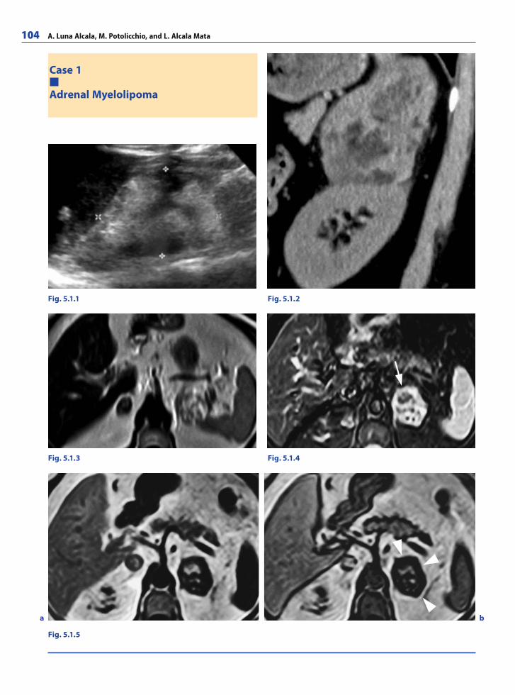
104 A. Luna Alcala, M. Potolicchio, and L. Alcala Mata
Case 1
Adrenal Myelolipoma
Fig. 5.1.1
Fig. 5.1.3
Fig. 5.1.2
Fig. 5.1.4
Fig. 5.1.5
a b
RibesLunaRos.indb 104 18.03.2008 12:50:29
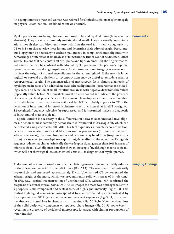
Genitourinary, Gynecological, and Obstetrical Imaging 105
An asymptomatic 54-year-old woman was referred for clinical suspicion of splenomegaly on physical examination. Her blood count was normal.
Imaging FindingsAbdominal ultrasound showed a well-defi ned heterogeneous mass immediately inferior to the spleen and superior to the left kidney (Fig. 5.1.1). The mass was predominantly hyperechoic and measured approximately 11 cm. Unenhanced CT demonstrated the adrenal origin of the mass, which was predominantly solid with areas of intralesional fat (Fig. 5.1.2, sagittal reconstruction of unenhanced CT). Adrenal MR confi rmed the diagnosis of adrenal myelolipoma. On HASTE images the mass was heterogeneous with a peripheral solid component and central areas of high signal intensity (Fig. 5.1.3). This central high signal component corresponded to macroscopic fat, as demonstrated by its suppression on STIR (short-tau inversion recovery) sequences (Fig. 5.1.4, arrow) and the absence of signal loss in chemical-shift imaging (Fig. 5.1.5a,b). Note the signal loss of the solid peripheral component on opposed-phase images (Fig. 5.1.5b, arrowheads), revealing the presence of peripheral microscopic fat (areas with similar proportions of water and fat).
CommentsMyelolipomas are rare benign tumors, composed of fat and myeloid tissue (bone marrow elements). They are most commonly unilateral and small. They are usually asymptom-atic, although they can bleed and cause pain. Intralesional fat is nearly diagnostic, so CT or MT can characterize these lesions and determine their adrenal origin. Percutane-ous biopsy may be necessary to exclude malignancy in complicated myelolipomas with hemorrhage or infarction if small areas of fat within the tumor cannot be detected. Other adrenal lesions that can contain fat are lipoma and liposarcoma; neighboring extraadre-nal lesions that can be confused with adrenal myelolipoma are retroperitoneal lipoma, liposarcoma, and renal angiomyolipoma. First, cross-sectional imaging is necessary to confi rm the origin of adrenal myelolipoma in the adrenal gland. If the mass is large, sagittal or coronal acquisitions or reconstructions may be useful to exclude a renal or retroperitoneal origin. The demonstration of macroscopic fat is almost diagnostic of myelolipoma in cases of an adrenal mass, as adrenal lipomas or liposarcomas are exceed-ingly rare. The detection of small intratumoral areas with negative densitometric values (especially values below -20 Hounsfi eld units) on unenhanced CT indicates the presence of macroscopic fat deposits. Because of intermixed hematopoietic tissue, the attenuation is usually higher than that of retroperitoneal fat. MR is probably superior to CT in the detection of intratumoral fat. Areas isointense to retroperitoneal fat in all T1-weighted, T2-weighted, frequency-selective fat-suppressed, and fat-saturated images is diagnostic of intratumoral macroscopic fat.
Special caution is necessary in the differentiation between adenomas and myelolipo-mas. Adenomas most commonly demonstrate intratumoral microscopic fat, which can be detected using chemical-shift MR. This technique uses a double-echo acquisition, because in areas where water and fat are in similar proportions (ex: microscopic fat in adrenal adenomas), the signal from water and fat signal may be additive (in-phase acqui-sition) or cancelled (opposed-phase acquisition), depending on the echo time. Using this sequence, adenomas characteristically show a drop in signal greater than 20% in areas of microscopic fat. Myelolipomas can also show microscopic fat, although macroscopic fat, which will not show signal loss on chemical-shift MR, is diagnostic of myelolipomas.
RibesLunaRos.indb 105 18.03.2008 12:50:32
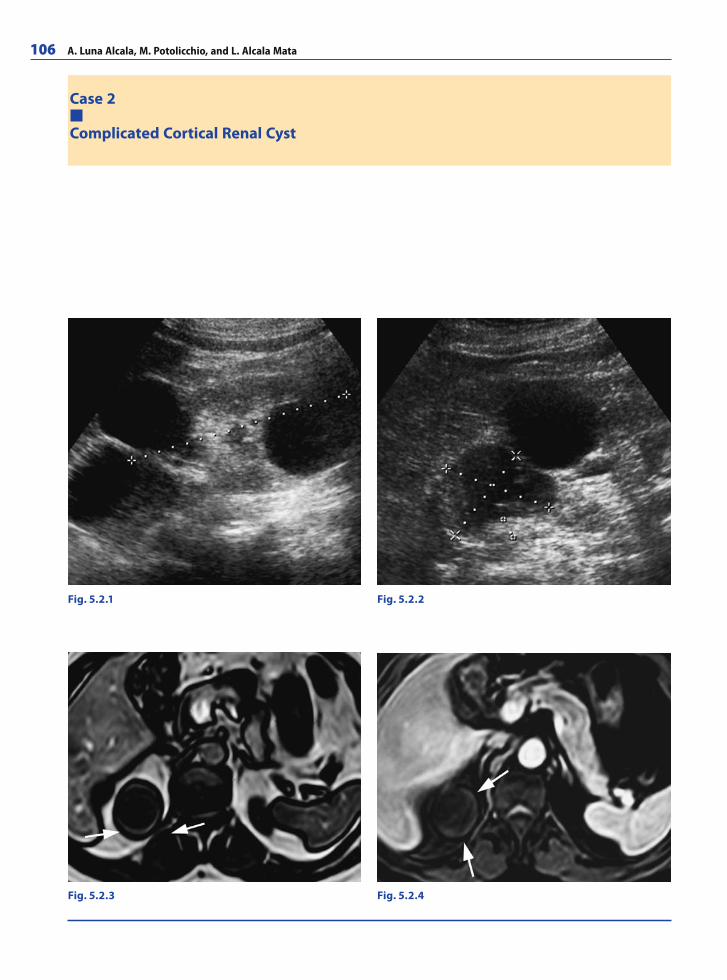
106 A. Luna Alcala, M. Potolicchio, and L. Alcala Mata
Case 2
Complicated Cortical Renal Cyst
Fig. 5.2.1 Fig. 5.2.2
Fig. 5.2.3 Fig. 5.2.4
RibesLunaRos.indb 106 18.03.2008 12:50:32
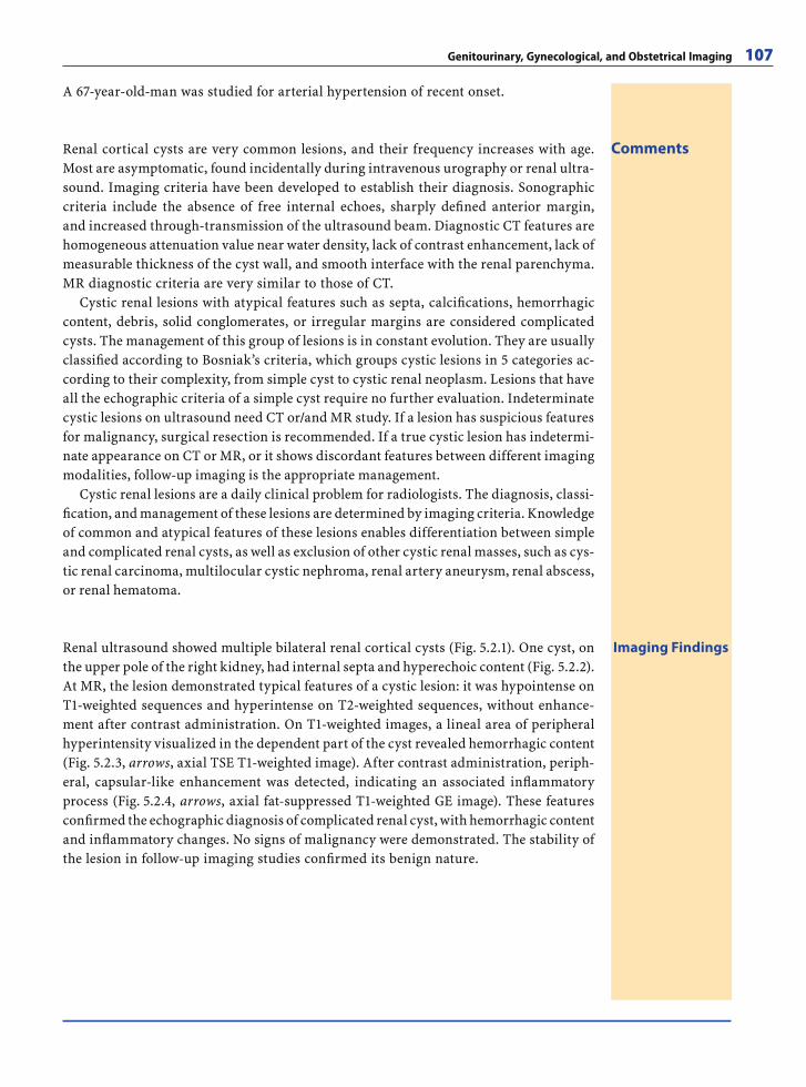
Genitourinary, Gynecological, and Obstetrical Imaging 107
Comments
A 67-year-old-man was studied for arterial hypertension of recent onset.
Renal cortical cysts are very common lesions, and their frequency increases with age. Most are asymptomatic, found incidentally during intravenous urography or renal ultra-sound. Imaging criteria have been developed to establish their diagnosis. Sonographic criteria include the absence of free internal echoes, sharply defi ned anterior margin, and increased through-transmission of the ultrasound beam. Diagnostic CT features are homogeneous attenuation value near water density, lack of contrast enhancement, lack of measurable thickness of the cyst wall, and smooth interface with the renal parenchyma. MR diagnostic criteria are very similar to those of CT.
Cystic renal lesions with atypical features such as septa, calcifi cations, hemorrhagic content, debris, solid conglomerates, or irregular margins are considered complicated cysts. The management of this group of lesions is in constant evolution. They are usually classifi ed according to Bosniak’s criteria, which groups cystic lesions in 5 categories ac-cording to their complexity, from simple cyst to cystic renal neoplasm. Lesions that have all the echographic criteria of a simple cyst require no further evaluation. Indeterminate cystic lesions on ultrasound need CT or/and MR study. If a lesion has suspicious features for malignancy, surgical resection is recommended. If a true cystic lesion has indetermi-nate appearance on CT or MR, or it shows discordant features between different imaging modalities, follow-up imaging is the appropriate management.
Cystic renal lesions are a daily clinical problem for radiologists. The diagnosis, classi-fi cation, and management of these lesions are determined by imaging criteria. Knowledge of common and atypical features of these lesions enables differentiation between simple and complicated renal cysts, as well as exclusion of other cystic renal masses, such as cys-tic renal carcinoma, multilocular cystic nephroma, renal artery aneurysm, renal abscess, or renal hematoma.
Imaging FindingsRenal ultrasound showed multiple bilateral renal cortical cysts (Fig. 5.2.1). One cyst, on the upper pole of the right kidney, had internal septa and hyperechoic content (Fig. 5.2.2). At MR, the lesion demonstrated typical features of a cystic lesion: it was hypointense on T1-weighted sequences and hyperintense on T2-weighted sequences, without enhance-ment after contrast administration. On T1-weighted images, a lineal area of peripheral hyperintensity visualized in the dependent part of the cyst revealed hemorrhagic content (Fig. 5.2.3, arrows, axial TSE T1-weighted image). After contrast administration, periph-eral, capsular-like enhancement was detected, indicating an associated infl ammatory process (Fig. 5.2.4, arrows, axial fat-suppressed T1-weighted GE image). These features confi rmed the echographic diagnosis of complicated renal cyst, with hemorrhagic content and infl ammatory changes. No signs of malignancy were demonstrated. The stability of the lesion in follow-up imaging studies confi rmed its benign nature.
RibesLunaRos.indb 107 18.03.2008 12:50:35
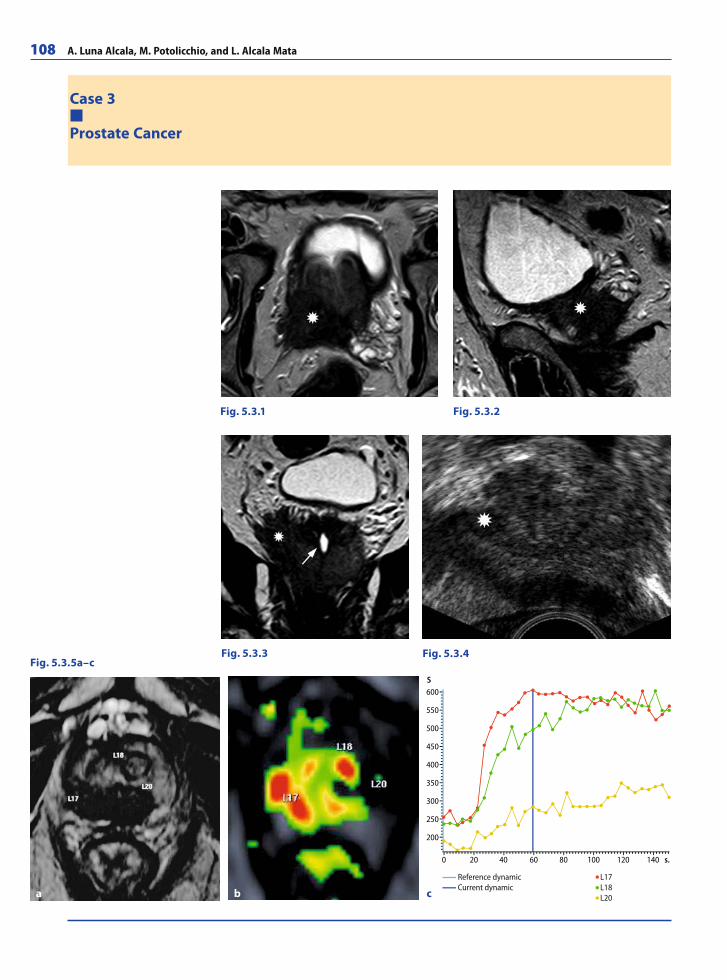
108 A. Luna Alcala, M. Potolicchio, and L. Alcala Mata
Case 3
Prostate Cancer
Fig. 5.3.1 Fig. 5.3.2
Fig. 5.3.3 Fig. 5.3.4Fig. 5.3.5a–c
a b
600
550
500
450
400
350
300
250
200
0 20 40 60 80 100 120 140 s.
S
Reference dynamic L17 Current dynamic L18 L20 c
RibesLunaRos.indb 108 18.03.2008 12:50:35
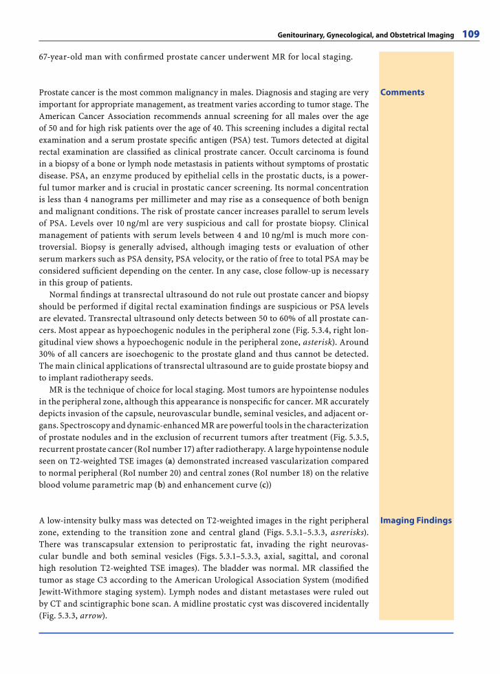
Genitourinary, Gynecological, and Obstetrical Imaging 109
67-year-old man with confi rmed prostate cancer underwent MR for local staging.
Imaging FindingsA low-intensity bulky mass was detected on T2-weighted images in the right peripheral zone, extending to the transition zone and central gland (Figs. 5.3.1–5.3.3, asrerisks). There was transcapsular extension to periprostatic fat, invading the right neurovas-cular bundle and both seminal vesicles (Figs. 5.3.1–5.3.3, axial, sagittal, and coronal high resolution T2-weighted TSE images). The bladder was normal. MR classifi ed the tumor as stage C3 according to the American Urological Association System (modifi ed Jewitt-Withmore staging system). Lymph nodes and distant metastases were ruled out by CT and scintigraphic bone scan. A midline prostatic cyst was discovered incidentally (Fig. 5.3.3, arrow).
CommentsProstate cancer is the most common malignancy in males. Diagnosis and staging are very important for appropriate management, as treatment varies according to tumor stage. The American Cancer Association recommends annual screening for all males over the age of 50 and for high risk patients over the age of 40. This screening includes a digital rectal examination and a serum prostate specifi c antigen (PSA) test. Tumors detected at digital rectal examination are classifi ed as clinical prostrate cancer. Occult carcinoma is found in a biopsy of a bone or lymph node metastasis in patients without symptoms of prostatic disease. PSA, an enzyme produced by epithelial cells in the prostatic ducts, is a power-ful tumor marker and is crucial in prostatic cancer screening. Its normal concentration is less than 4 nanograms per millimeter and may rise as a consequence of both benign and malignant conditions. The risk of prostate cancer increases parallel to serum levels of PSA. Levels over 10 ng/ml are very suspicious and call for prostate biopsy. Clinical management of patients with serum levels between 4 and 10 ng/ml is much more con-troversial. Biopsy is generally advised, although imaging tests or evaluation of other serum markers such as PSA density, PSA velocity, or the ratio of free to total PSA may be considered suffi cient depending on the center. In any case, close follow-up is necessary in this group of patients.
Normal fi ndings at transrectal ultrasound do not rule out prostate cancer and biopsy should be performed if digital rectal examination fi ndings are suspicious or PSA levels are elevated. Transrectal ultrasound only detects between 50 to 60% of all prostate can-cers. Most appear as hypoechogenic nodules in the peripheral zone (Fig. 5.3.4, right lon-gitudinal view shows a hypoechogenic nodule in the peripheral zone, asterisk). Around 30% of all cancers are isoechogenic to the prostate gland and thus cannot be detected. The main clinical applications of transrectal ultrasound are to guide prostate biopsy and to implant radiotherapy seeds.
MR is the technique of choice for local staging. Most tumors are hypointense nodules in the peripheral zone, although this appearance is nonspecifi c for cancer. MR accurately depicts invasion of the capsule, neurovascular bundle, seminal vesicles, and adjacent or-gans. Spectroscopy and dynamic-enhanced MR are powerful tools in the characterization of prostate nodules and in the exclusion of recurrent tumors after treatment (Fig. 5.3.5, recurrent prostate cancer (RoI number 17) after radiotherapy. A large hypointense nodule seen on T2-weighted TSE images (a) demonstrated increased vascularization compared to normal peripheral (RoI number 20) and central zones (RoI number 18) on the relative blood volume parametric map (b) and enhancement curve (c))
RibesLunaRos.indb 109 18.03.2008 12:50:41
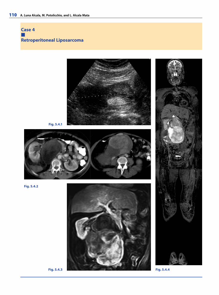
110 A. Luna Alcala, M. Potolicchio, and L. Alcala Mata
Case 4
Retroperitoneal Liposarcoma
Fig. 5.4.4
Fig. 5.4.2
Fig. 5.4.3
Fig. 5.4.1
RibesLunaRos.indb 110 18.03.2008 12:50:41
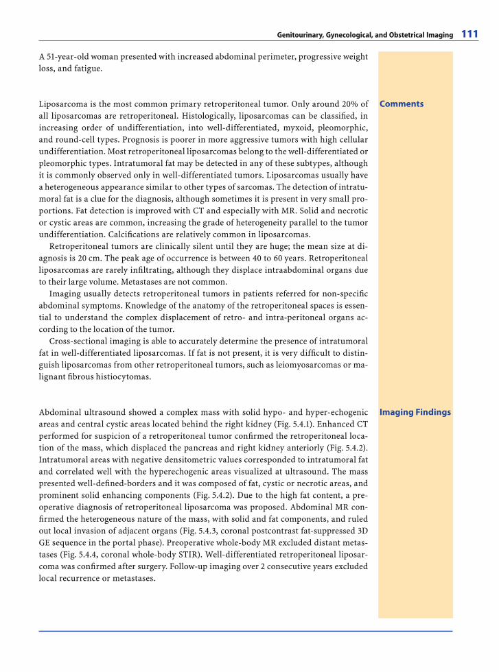
Genitourinary, Gynecological, and Obstetrical Imaging 111
A 51-year-old woman presented with increased abdominal perimeter, progressive weight loss, and fatigue.
CommentsLiposarcoma is the most common primary retroperitoneal tumor. Only around 20% of all liposarcomas are retroperitoneal. Histologically, liposarcomas can be classifi ed, in increasing order of undifferentiation, into well-differentiated, myxoid, pleomorphic, and round-cell types. Prognosis is poorer in more aggressive tumors with high cellular undifferentiation. Most retroperitoneal liposarcomas belong to the well-differentiated or pleomorphic types. Intratumoral fat may be detected in any of these subtypes, although it is commonly observed only in well-differentiated tumors. Liposarcomas usually have a heterogeneous appearance similar to other types of sarcomas. The detection of intratu-moral fat is a clue for the diagnosis, although sometimes it is present in very small pro-portions. Fat detection is improved with CT and especially with MR. Solid and necrotic or cystic areas are common, increasing the grade of heterogeneity parallel to the tumor undifferentiation. Calcifi cations are relatively common in liposarcomas.
Retroperitoneal tumors are clinically silent until they are huge; the mean size at di-agnosis is 20 cm. The peak age of occurrence is between 40 to 60 years. Retroperitoneal liposarcomas are rarely infi ltrating, although they displace intraabdominal organs due to their large volume. Metastases are not common.
Imaging usually detects retroperitoneal tumors in patients referred for non-specifi c abdominal symptoms. Knowledge of the anatomy of the retroperitoneal spaces is essen-tial to understand the complex displacement of retro- and intra-peritoneal organs ac-cording to the location of the tumor.
Cross-sectional imaging is able to accurately determine the presence of intratumoral fat in well-differentiated liposarcomas. If fat is not present, it is very diffi cult to distin-guish liposarcomas from other retroperitoneal tumors, such as leiomyosarcomas or ma-lignant fi brous histiocytomas.
Imaging FindingsAbdominal ultrasound showed a complex mass with solid hypo- and hyper-echogenic areas and central cystic areas located behind the right kidney (Fig. 5.4.1). Enhanced CT performed for suspicion of a retroperitoneal tumor confi rmed the retroperitoneal loca-tion of the mass, which displaced the pancreas and right kidney anteriorly (Fig. 5.4.2). Intratumoral areas with negative densitometric values corresponded to intratumoral fat and correlated well with the hyperechogenic areas visualized at ultrasound. The mass presented well-defi ned-borders and it was composed of fat, cystic or necrotic areas, and prominent solid enhancing components (Fig. 5.4.2). Due to the high fat content, a pre-operative diagnosis of retroperitoneal liposarcoma was proposed. Abdominal MR con-fi rmed the heterogeneous nature of the mass, with solid and fat components, and ruled out local invasion of adjacent organs (Fig. 5.4.3, coronal postcontrast fat-suppressed 3D GE sequence in the portal phase). Preoperative whole-body MR excluded distant metas-tases (Fig. 5.4.4, coronal whole-body STIR). Well-differentiated retroperitoneal liposar-coma was confi rmed after surgery. Follow-up imaging over 2 consecutive years excluded local recurrence or metastases.
RibesLunaRos.indb 111 18.03.2008 12:50:44
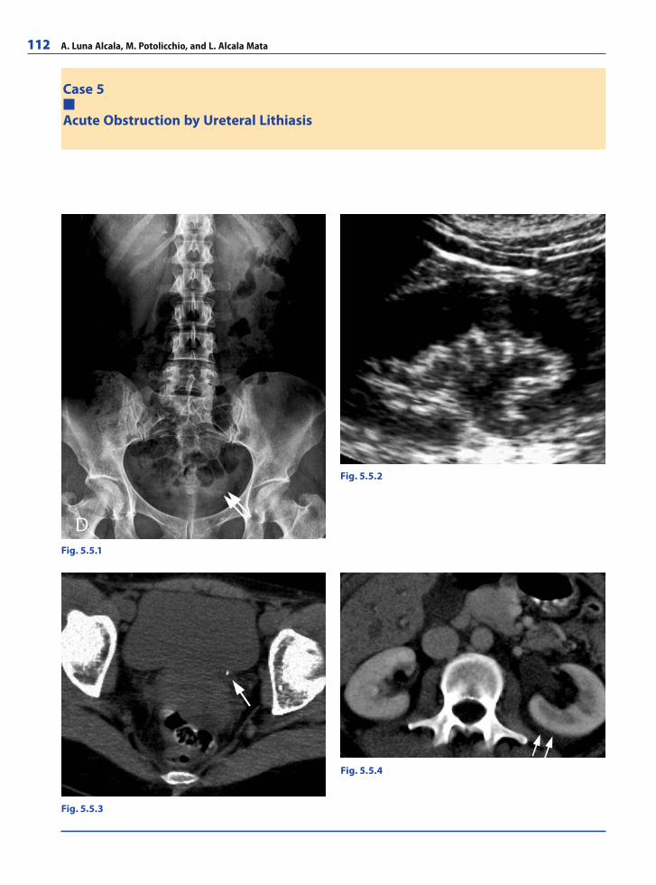
112 A. Luna Alcala, M. Potolicchio, and L. Alcala Mata
Case 5
Acute Obstruction by Ureteral Lithiasis
Fig. 5.5.1
Fig. 5.5.2
Fig. 5.5.4
Fig. 5.5.3
RibesLunaRos.indb 112 18.03.2008 12:50:44
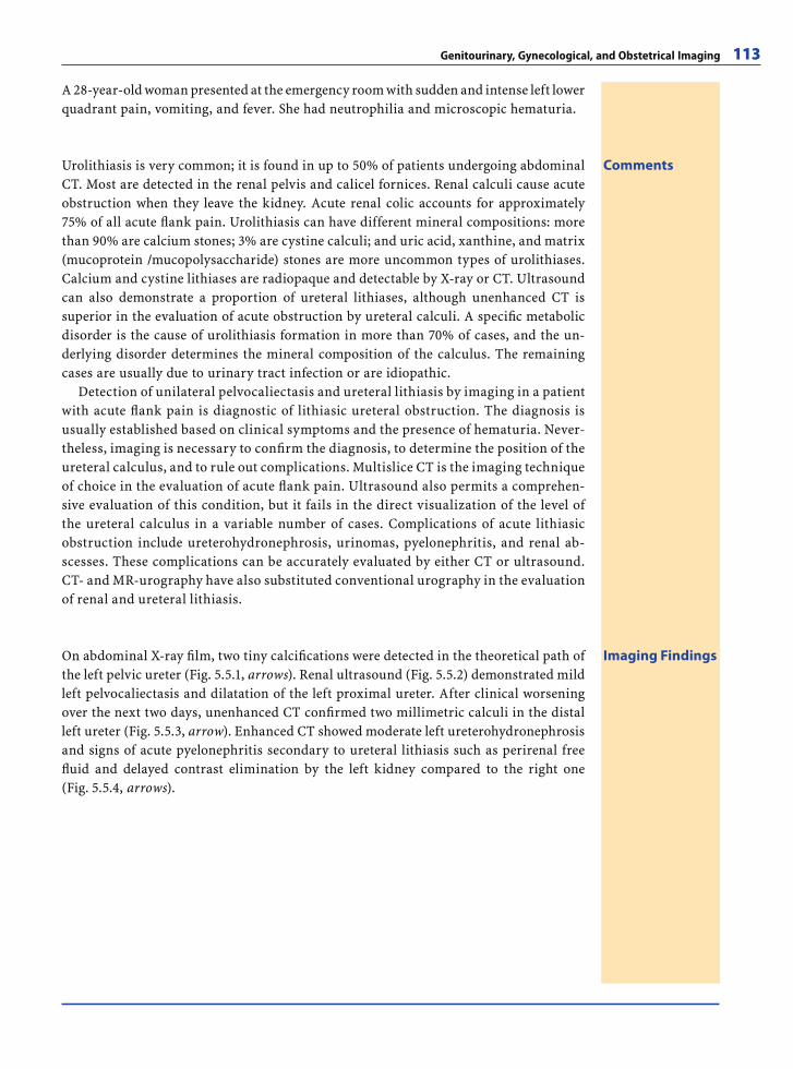
Genitourinary, Gynecological, and Obstetrical Imaging 113
A 28-year-old woman presented at the emergency room with sudden and intense left lower quadrant pain, vomiting, and fever. She had neutrophilia and microscopic hematuria.
CommentsUrolithiasis is very common; it is found in up to 50% of patients undergoing abdominal CT. Most are detected in the renal pelvis and calicel fornices. Renal calculi cause acute obstruction when they leave the kidney. Acute renal colic accounts for approximately 75% of all acute fl ank pain. Urolithiasis can have different mineral compositions: more than 90% are calcium stones; 3% are cystine calculi; and uric acid, xanthine, and matrix (mucoprotein /mucopolysaccharide) stones are more uncommon types of urolithiases. Calcium and cystine lithiases are radiopaque and detectable by X-ray or CT. Ultrasound can also demonstrate a proportion of ureteral lithiases, although unenhanced CT is superior in the evaluation of acute obstruction by ureteral calculi. A specifi c metabolic disorder is the cause of urolithiasis formation in more than 70% of cases, and the un-derlying disorder determines the mineral composition of the calculus. The remaining cases are usually due to urinary tract infection or are idiopathic.
Detection of unilateral pelvocaliectasis and ureteral lithiasis by imaging in a patient with acute fl ank pain is diagnostic of lithiasic ureteral obstruction. The diagnosis is usually established based on clinical symptoms and the presence of hematuria. Never-theless, imaging is necessary to confi rm the diagnosis, to determine the position of the ureteral calculus, and to rule out complications. Multislice CT is the imaging technique of choice in the evaluation of acute fl ank pain. Ultrasound also permits a comprehen-sive evaluation of this condition, but it fails in the direct visualization of the level of the ureteral calculus in a variable number of cases. Complications of acute lithiasic obstruction include ureterohydronephrosis, urinomas, pyelonephritis, and renal ab-scesses. These complications can be accurately evaluated by either CT or ultrasound. CT- and MR-urography have also substituted conventional urography in the evaluation of renal and ureteral lithiasis.
Imaging FindingsOn abdominal X-ray fi lm, two tiny calcifi cations were detected in the theoretical path of the left pelvic ureter (Fig. 5.5.1, arrows). Renal ultrasound (Fig. 5.5.2) demonstrated mild left pelvocaliectasis and dilatation of the left proximal ureter. After clinical worsening over the next two days, unenhanced CT confi rmed two millimetric calculi in the distal left ureter (Fig. 5.5.3, arrow). Enhanced CT showed moderate left ureterohydronephrosis and signs of acute pyelonephritis secondary to ureteral lithiasis such as perirenal free fl uid and delayed contrast elimination by the left kidney compared to the right one (Fig. 5.5.4, arrows).
RibesLunaRos.indb 113 18.03.2008 12:50:48
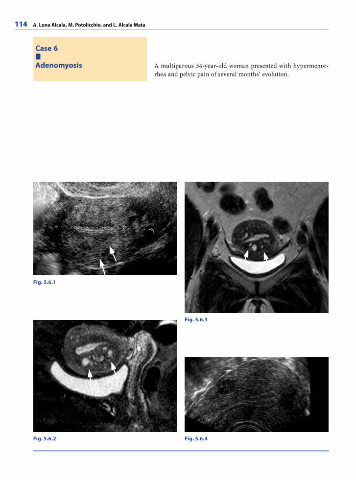
114 A. Luna Alcala, M. Potolicchio, and L. Alcala Mata
A multiparous 34-year-old woman presented with hypermenor-rhea and pelvic pain of several months’ evolution.
Case 6
Adenomyosis
Fig. 5.6.1
Fig. 5.6.3
Fig. 5.6.4Fig. 5.6.2
RibesLunaRos.indb 114 18.03.2008 12:50:48
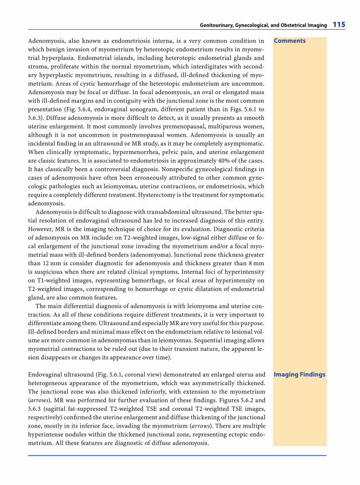
Genitourinary, Gynecological, and Obstetrical Imaging 115
CommentsAdenomyosis, also known as endometriosis interna, is a very common condition in which benign invasion of myometrium by heterotopic endometrium results in myome-trial hyperplasia. Endometrial islands, including heterotopic endometrial glands and stroma, proliferate within the normal myometrium, which interdigitates with second-ary hyperplastic myometrium, resulting in a diffused, ill-defi ned thickening of myo-metrium. Areas of cystic hemorrhage of the heterotopic endometrium are uncommon. Adenomyosis may be focal or diffuse. In focal adenomyosis, an oval or elongated mass with ill-defi ned margins and in contiguity with the junctional zone is the most common presentation (Fig. 5.6.4, endovaginal sonogram, different patient than in Figs. 5.6.1 to 5.6.3). Diffuse adenomyosis is more diffi cult to detect, as it usually presents as smooth uterine enlargement. It most commonly involves premenopausal, multiparous women, although it is not uncommon in postmenopausal women. Adenomyosis is usually an incidental fi nding in an ultrasound or MR study, as it may be completely asymptomatic. When clinically symptomatic, hypermenorrhea, pelvic pain, and uterine enlargement are classic features. It is associated to endometriosis in approximately 40% of the cases. It has classically been a controversial diagnosis. Nonspecifi c gynecological fi ndings in cases of adenomyosis have often been erroneously attributed to other common gyne-cologic pathologies such as leiomyomas, uterine contractions, or endometriosis, which require a completely different treatment. Hysterectomy is the treatment for symptomatic adenomyosis.
Adenomyosis is diffi cult to diagnose with transabdominal ultrasound. The better spa-tial resolution of endovaginal ultrasound has led to increased diagnosis of this entity. However, MR is the imaging technique of choice for its evaluation. Diagnostic criteria of adenomyosis on MR include: on T2-weighted images, low-signal either diffuse or fo-cal enlargement of the junctional zone invading the myometrium and/or a focal myo-metrial mass with ill-defi ned borders (adenomyoma). Junctional zone thickness greater than 12 mm is consider diagnostic for adenomyosis and thickness greater than 8 mm is suspicious when there are related clinical symptoms. Internal foci of hyperintensity on T1-weighted images, representing hemorrhage, or focal areas of hyperintensity on T2-weighted images, corresponding to hemorrhage or cystic dilatation of endometrial gland, are also common features.
The main differential diagnosis of adenomyosis is with leiomyoma and uterine con-traction. As all of these conditions require different treatments, it is very important to differentiate among them. Ultrasound and especially MR are very useful for this purpose. Ill-defi ned borders and minimal mass effect on the endometrium relative to lesional vol-ume are more common in adenomyomas than in leiomyomas. Sequential imaging allows myometrial contractions to be ruled out (due to their transient nature, the apparent le-sion disappears or changes its appearance over time).
Imaging FindingsEndovaginal ultrasound (Fig. 5.6.1, coronal view) demonstrated an enlarged uterus and heterogeneous appearance of the myometrium, which was asymmetrically thickened. The junctional zone was also thickened inferiorly, with extension to the myometrium (arrows). MR was performed for further evaluation of these fi ndings. Figures 5.6.2 and 5.6.3 (sagittal fat-suppressed T2-weighted TSE and coronal T2-weighted TSE images, respectively) confi rmed the uterine enlargement and diffuse thickening of the junctional zone, mostly in its inferior face, invading the myometrium (arrows). There are multiple hyperintense nodules within the thickened junctional zone, representing ectopic endo-metrium. All these features are diagnostic of diffuse adenomyosis.
RibesLunaRos.indb 115 18.03.2008 12:50:51
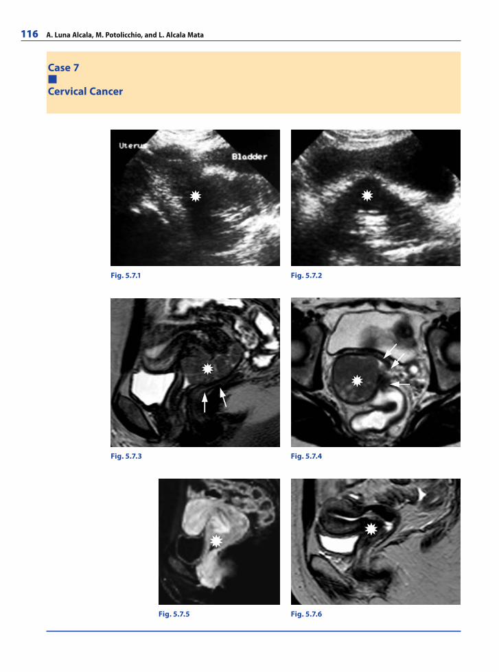
116 A. Luna Alcala, M. Potolicchio, and L. Alcala Mata
Case 7
Cervical Cancer
Fig. 5.7.1
Fig. 5.7.3
Fig. 5.7.5
Fig. 5.7.2
Fig. 5.7.4
Fig. 5.7.6
RibesLunaRos.indb 116 18.03.2008 12:50:51
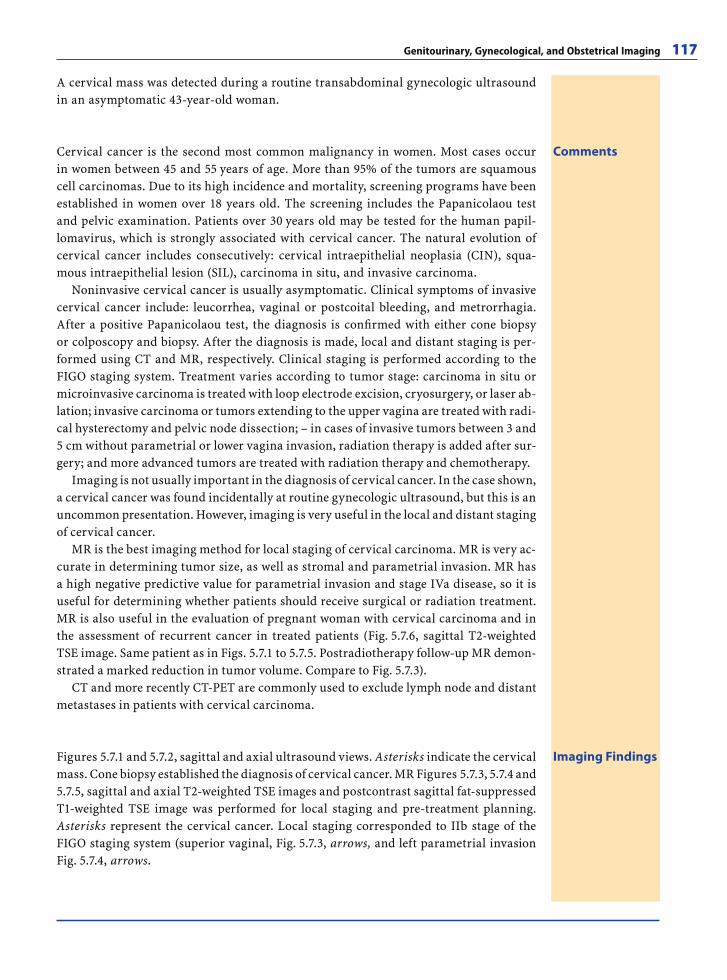
Genitourinary, Gynecological, and Obstetrical Imaging 117
A cervical mass was detected during a routine transabdominal gynecologic ultrasound in an asymptomatic 43-year-old woman.
CommentsCervical cancer is the second most common malignancy in women. Most cases occur in women between 45 and 55 years of age. More than 95% of the tumors are squamous cell carcinomas. Due to its high incidence and mortality, screening programs have been established in women over 18 years old. The screening includes the Papanicolaou test and pelvic examination. Patients over 30 years old may be tested for the human papil-lomavirus, which is strongly associated with cervical cancer. The natural evolution of cervical cancer includes consecutively: cervical intraepithelial neoplasia (CIN), squa-mous intraepithelial lesion (SIL), carcinoma in situ, and invasive carcinoma.
Noninvasive cervical cancer is usually asymptomatic. Clinical symptoms of invasive cervical cancer include: leucorrhea, vaginal or postcoital bleeding, and metrorrhagia. After a positive Papanicolaou test, the diagnosis is confi rmed with either cone biopsy or colposcopy and biopsy. After the diagnosis is made, local and distant staging is per-formed using CT and MR, respectively. Clinical staging is performed according to the FIGO staging system. Treatment varies according to tumor stage: carcinoma in situ or microinvasive carcinoma is treated with loop electrode excision, cryosurgery, or laser ab-lation; invasive carcinoma or tumors extending to the upper vagina are treated with radi-cal hysterectomy and pelvic node dissection; – in cases of invasive tumors between 3 and 5 cm without parametrial or lower vagina invasion, radiation therapy is added after sur-gery; and more advanced tumors are treated with radiation therapy and chemotherapy.
Imaging is not usually important in the diagnosis of cervical cancer. In the case shown, a cervical cancer was found incidentally at routine gynecologic ultrasound, but this is an uncommon presentation. However, imaging is very useful in the local and distant staging of cervical cancer.
MR is the best imaging method for local staging of cervical carcinoma. MR is very ac-curate in determining tumor size, as well as stromal and parametrial invasion. MR has a high negative predictive value for parametrial invasion and stage IVa disease, so it is useful for determining whether patients should receive surgical or radiation treatment. MR is also useful in the evaluation of pregnant woman with cervical carcinoma and in the assessment of recurrent cancer in treated patients (Fig. 5.7.6, sagittal T2-weighted TSE image. Same patient as in Figs. 5.7.1 to 5.7.5. Postradiotherapy follow-up MR demon-strated a marked reduction in tumor volume. Compare to Fig. 5.7.3).
CT and more recently CT-PET are commonly used to exclude lymph node and distant metastases in patients with cervical carcinoma.
Imaging FindingsFigures 5.7.1 and 5.7.2, sagittal and axial ultrasound views. Asterisks indicate the cervical mass. Cone biopsy established the diagnosis of cervical cancer. MR Figures 5.7.3, 5.7.4 and 5.7.5, sagittal and axial T2-weighted TSE images and postcontrast sagittal fat-suppressed T1-weighted TSE image was performed for local staging and pre-treatment planning. Asterisks represent the cervical cancer. Local staging corresponded to IIb stage of the FIGO staging system (superior vaginal, Fig. 5.7.3, arrows, and left parametrial invasion Fig. 5.7.4, arrows.
RibesLunaRos.indb 117 18.03.2008 12:50:54
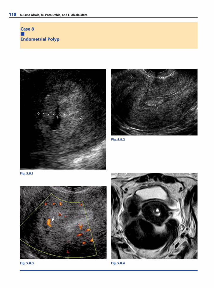
118 A. Luna Alcala, M. Potolicchio, and L. Alcala Mata
Case 8
Endometrial Polyp
Fig. 5.8.1
Fig. 5.8.2
Fig. 5.8.3 Fig. 5.8.4
RibesLunaRos.indb 118 18.03.2008 12:50:54
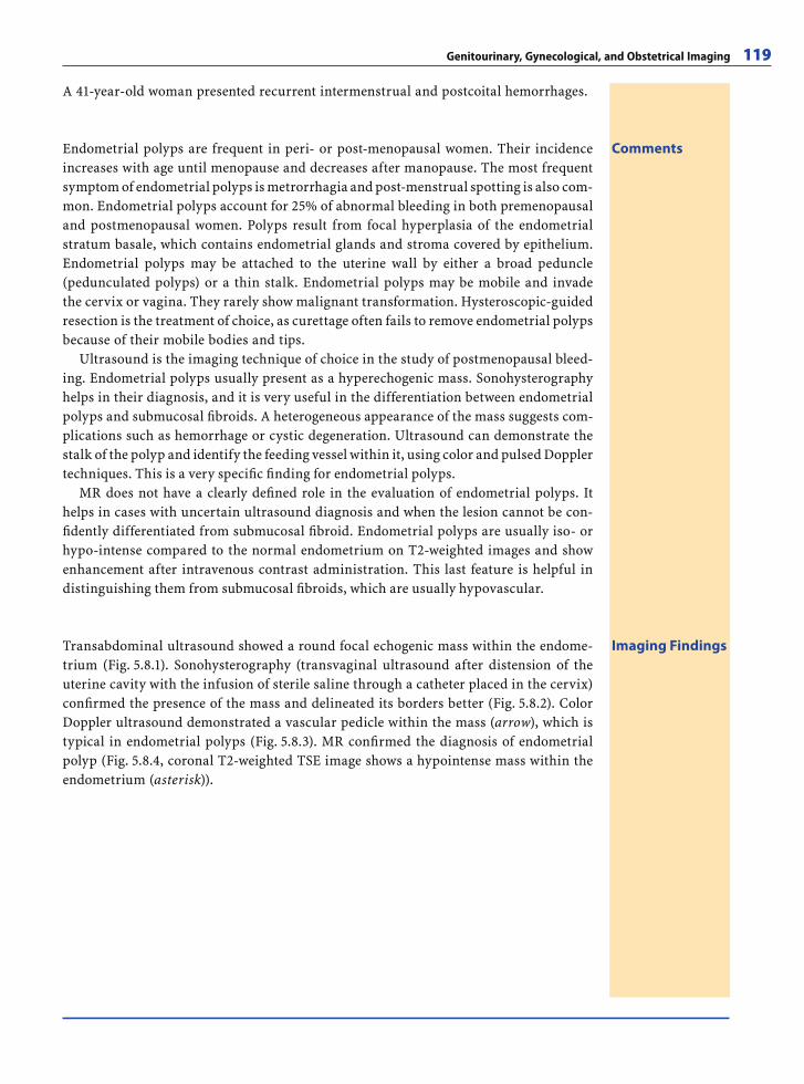
Genitourinary, Gynecological, and Obstetrical Imaging 119
A 41-year-old woman presented recurrent intermenstrual and postcoital hemorrhages.
CommentsEndometrial polyps are frequent in peri- or post-menopausal women. Their incidence increases with age until menopause and decreases after manopause. The most frequent symptom of endometrial polyps is metrorrhagia and post-menstrual spotting is also com-mon. Endometrial polyps account for 25% of abnormal bleeding in both premenopausal and postmenopausal women. Polyps result from focal hyperplasia of the endometrial stratum basale, which contains endometrial glands and stroma covered by epithelium. Endometrial polyps may be attached to the uterine wall by either a broad peduncle (pedunculated polyps) or a thin stalk. Endometrial polyps may be mobile and invade the cervix or vagina. They rarely show malignant transformation. Hysteroscopic-guided resection is the treatment of choice, as curettage often fails to remove endometrial polyps because of their mobile bodies and tips.
Ultrasound is the imaging technique of choice in the study of postmenopausal bleed-ing. Endometrial polyps usually present as a hyperechogenic mass. Sonohysterography helps in their diagnosis, and it is very useful in the differentiation between endometrial polyps and submucosal fi broids. A heterogeneous appearance of the mass suggests com-plications such as hemorrhage or cystic degeneration. Ultrasound can demonstrate the stalk of the polyp and identify the feeding vessel within it, using color and pulsed Doppler techniques. This is a very specifi c fi nding for endometrial polyps.
MR does not have a clearly defi ned role in the evaluation of endometrial polyps. It helps in cases with uncertain ultrasound diagnosis and when the lesion cannot be con-fi dently differentiated from submucosal fi broid. Endometrial polyps are usually iso- or hypo-intense compared to the normal endometrium on T2-weighted images and show enhancement after intravenous contrast administration. This last feature is helpful in distinguishing them from submucosal fi broids, which are usually hypovascular.
Imaging FindingsTransabdominal ultrasound showed a round focal echogenic mass within the endome-trium (Fig. 5.8.1). Sonohysterography (transvaginal ultrasound after distension of the uterine cavity with the infusion of sterile saline through a catheter placed in the cervix) confi rmed the presence of the mass and delineated its borders better (Fig. 5.8.2). Color Doppler ultrasound demonstrated a vascular pedicle within the mass (arrow), which is typical in endometrial polyps (Fig. 5.8.3). MR confi rmed the diagnosis of endometrial polyp (Fig. 5.8.4, coronal T2-weighted TSE image shows a hypointense mass within the endometrium (asterisk)).
RibesLunaRos.indb 119 18.03.2008 12:50:59
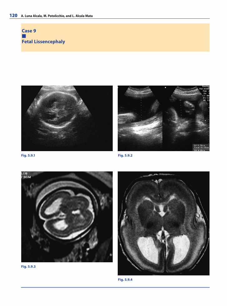
120 A. Luna Alcala, M. Potolicchio, and L. Alcala Mata
Case 9
Fetal Lissencephaly
Fig. 5.9.1 Fig. 5.9.2
Fig. 5.9.3
Fig. 5.9.4
RibesLunaRos.indb 120 18.03.2008 12:50:59
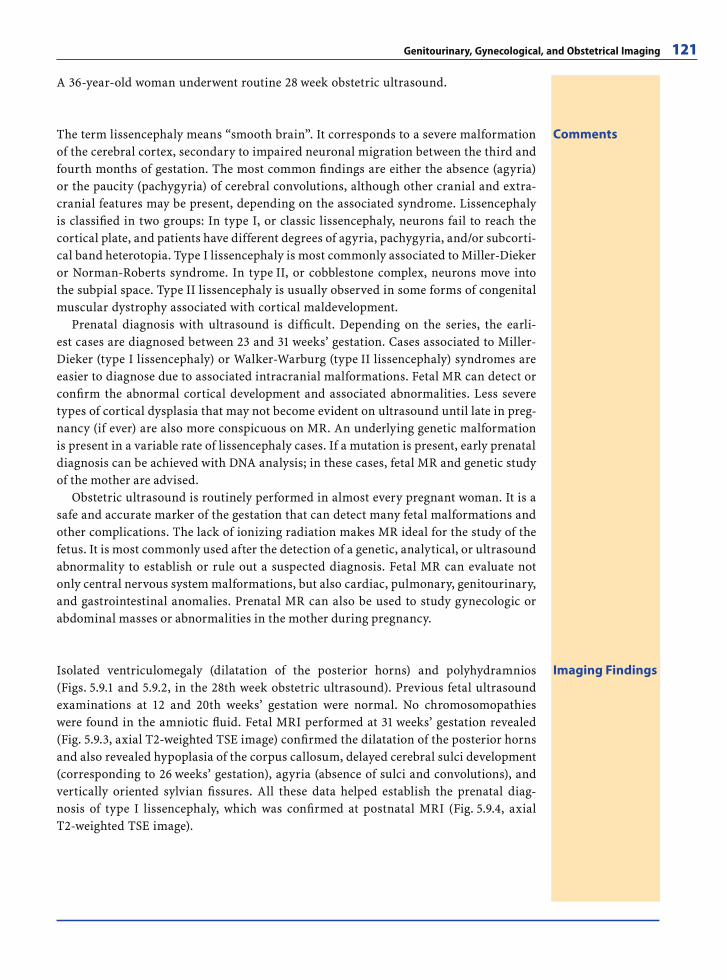
Genitourinary, Gynecological, and Obstetrical Imaging 121
A 36-year-old woman underwent routine 28 week obstetric ultrasound.
CommentsThe term lissencephaly means “smooth brain”. It corresponds to a severe malformation of the cerebral cortex, secondary to impaired neuronal migration between the third and fourth months of gestation. The most common fi ndings are either the absence (agyria) or the paucity (pachygyria) of cerebral convolutions, although other cranial and extra-cranial features may be present, depending on the associated syndrome. Lissencephaly is classifi ed in two groups: In type I, or classic lissencephaly, neurons fail to reach the cortical plate, and patients have different degrees of agyria, pachygyria, and/or subcorti-cal band heterotopia. Type I lissencephaly is most commonly associated to Miller-Dieker or Norman-Roberts syndrome. In type II, or cobblestone complex, neurons move into the subpial space. Type II lissencephaly is usually observed in some forms of congenital muscular dystrophy associated with cortical maldevelopment.
Prenatal diagnosis with ultrasound is diffi cult. Depending on the series, the earli-est cases are diagnosed between 23 and 31 weeks’ gestation. Cases associated to Miller-Dieker (type I lissencephaly) or Walker-Warburg (type II lissencephaly) syndromes are easier to diagnose due to associated intracranial malformations. Fetal MR can detect or confi rm the abnormal cortical development and associated abnormalities. Less severe types of cortical dysplasia that may not become evident on ultrasound until late in preg-nancy (if ever) are also more conspicuous on MR. An underlying genetic malformation is present in a variable rate of lissencephaly cases. If a mutation is present, early prenatal diagnosis can be achieved with DNA analysis; in these cases, fetal MR and genetic study of the mother are advised.
Obstetric ultrasound is routinely performed in almost every pregnant woman. It is a safe and accurate marker of the gestation that can detect many fetal malformations and other complications. The lack of ionizing radiation makes MR ideal for the study of the fetus. It is most commonly used after the detection of a genetic, analytical, or ultrasound abnormality to establish or rule out a suspected diagnosis. Fetal MR can evaluate not only central nervous system malformations, but also cardiac, pulmonary, genitourinary, and gastrointestinal anomalies. Prenatal MR can also be used to study gynecologic or abdominal masses or abnormalities in the mother during pregnancy.
Imaging FindingsIsolated ventriculomegaly (dilatation of the posterior horns) and polyhydramnios (Figs. 5.9.1 and 5.9.2, in the 28th week obstetric ultrasound). Previous fetal ultrasound examinations at 12 and 20th weeks’ gestation were normal. No chromosomopathies were found in the amniotic fl uid. Fetal MRI performed at 31 weeks’ gestation revealed (Fig. 5.9.3, axial T2-weighted TSE image) confi rmed the dilatation of the posterior horns and also revealed hypoplasia of the corpus callosum, delayed cerebral sulci development (corresponding to 26 weeks’ gestation), agyria (absence of sulci and convolutions), and vertically oriented sylvian fi ssures. All these data helped establish the prenatal diag-nosis of type I lissencephaly, which was confi rmed at postnatal MRI (Fig. 5.9.4, axial T2-weighted TSE image).
RibesLunaRos.indb 121 18.03.2008 12:51:03
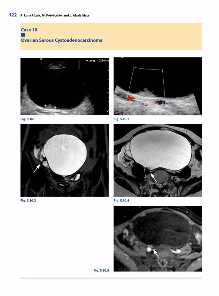
122 A. Luna Alcala, M. Potolicchio, and L. Alcala Mata
Case 10
Ovarian Serous Cystoadenocarcinoma
Fig. 5.10.1 Fig. 5.10.2
Fig. 5.10.3
Fig. 5.10.5
Fig. 5.10.4
RibesLunaRos.indb 122 18.03.2008 12:51:03
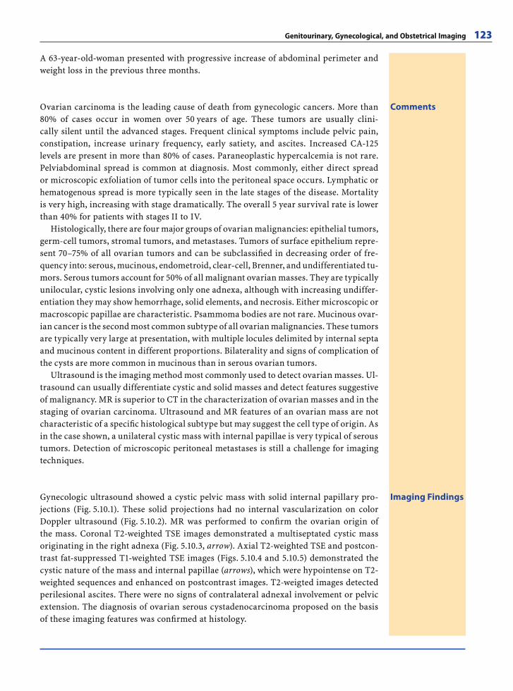
Genitourinary, Gynecological, and Obstetrical Imaging 123
A 63-year-old-woman presented with progressive increase of abdominal perimeter and weight loss in the previous three months.
CommentsOvarian carcinoma is the leading cause of death from gynecologic cancers. More than 80% of cases occur in women over 50 years of age. These tumors are usually clini-cally silent until the advanced stages. Frequent clinical symptoms include pelvic pain, constipation, increase urinary frequency, early satiety, and ascites. Increased CA-125 levels are present in more than 80% of cases. Paraneoplastic hypercalcemia is not rare. Pelviabdominal spread is common at diagnosis. Most commonly, either direct spread or microscopic exfoliation of tumor cells into the peritoneal space occurs. Lymphatic or hematogenous spread is more typically seen in the late stages of the disease. Mortality is very high, increasing with stage dramatically. The overall 5 year survival rate is lower than 40% for patients with stages II to IV.
Histologically, there are four major groups of ovarian malignancies: epithelial tumors, germ-cell tumors, stromal tumors, and metastases. Tumors of surface epithelium repre-sent 70–75% of all ovarian tumors and can be subclassifi ed in decreasing order of fre-quency into: serous, mucinous, endometroid, clear-cell, Brenner, and undifferentiated tu-mors. Serous tumors account for 50% of all malignant ovarian masses. They are typically unilocular, cystic lesions involving only one adnexa, although with increasing undiffer-entiation they may show hemorrhage, solid elements, and necrosis. Either microscopic or macroscopic papillae are characteristic. Psammoma bodies are not rare. Mucinous ovar-ian cancer is the second most common subtype of all ovarian malignancies. These tumors are typically very large at presentation, with multiple locules delimited by internal septa and mucinous content in different proportions. Bilaterality and signs of complication of the cysts are more common in mucinous than in serous ovarian tumors.
Ultrasound is the imaging method most commonly used to detect ovarian masses. Ul-trasound can usually differentiate cystic and solid masses and detect features suggestive of malignancy. MR is superior to CT in the characterization of ovarian masses and in the staging of ovarian carcinoma. Ultrasound and MR features of an ovarian mass are not characteristic of a specifi c histological subtype but may suggest the cell type of origin. As in the case shown, a unilateral cystic mass with internal papillae is very typical of serous tumors. Detection of microscopic peritoneal metastases is still a challenge for imaging techniques.
Imaging FindingsGynecologic ultrasound showed a cystic pelvic mass with solid internal papillary pro-jections (Fig. 5.10.1). These solid projections had no internal vascularization on color Doppler ultrasound (Fig. 5.10.2). MR was performed to confi rm the ovarian origin of the mass. Coronal T2-weighted TSE images demonstrated a multiseptated cystic mass originating in the right adnexa (Fig. 5.10.3, arrow). Axial T2-weighted TSE and postcon-trast fat-suppressed T1-weighted TSE images (Figs. 5.10.4 and 5.10.5) demonstrated the cystic nature of the mass and internal papillae (arrows), which were hypointense on T2-weighted sequences and enhanced on postcontrast images. T2-weigted images detected perilesional ascites. There were no signs of contralateral adnexal involvement or pelvic extension. The diagnosis of ovarian serous cystadenocarcinoma proposed on the basis of these imaging features was confi rmed at histology.
RibesLunaRos.indb 123 18.03.2008 12:51:06
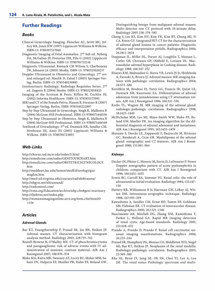
124 A. Luna Alcala, M. Potolicchio, and L. Alcala Mata
Further Readings
Books
Clinical Gynecologic Imaging. Fleischer AC, Javitt MC, Jef-frey RB, Jones HW (1997) Lippincott Williams & Wilkins. ISBN-13: 9780397517060
Diagnostic Imaging of Fetal Anomalies. 2nd Sub ed. Nyberg DA, McGahan JP, Pretorius DH, Pilu G (2002) Lippincott Williams & Wilkins. ISBN-13: 9780781732116
Diagnostic Ultrasound. Rumack CM Wilson S, Charboneau JW, Johnson JA (2005) Mosby. ISBN-13: 9780323020237
Doppler Ultrasound in Obstetrics and Gynecology. 2nd rev. and enlarged ed. Maulik D, Zalud I (2005) Springer-Ver-lag, Berlin. ISBN-13: 9783540230885
Genitourinary Radiology: Radiology Requisites Series. 2nd ed. Zagoria R (2004) Mosby. ISBN-13: 9780323018425
Imaging of the Scrotum & Penis. Rifkin M, Cochlin DL (2002) Informa Healthcare. ISBN-13: 9781853175091
MRI and CT of the Female Pelvis. Hamm B, Forstner R (2007) Springer-Verlag, Berlin. ISBN: 9783540222897
Step by Step Ultrasound in Gynecology. Singh K, Malhotra N (2004) McGraw-Hill Professional. ISBN-13: 9780071446556
Step by Step Ultrasound in Obstetrics. Singh K, Malhotra N (2004) McGraw-Hill Professional. ISBN-13: 9780071446549
Textbook of Uroradiology. 3rd ed. Dunnick NR, Sandler CM, Newhouse JH, Amis ES (2001) Lippincott Williams & Wilkins. ISBN-13: 9780781723893
Web-Links
http://chorus.rad.mcw.edu/index/5.htmlhttp://emedicine.com/radio/GENITOURINARY.htmhttp://emedicine.com/radio/OBSTETRICSGYNECOLOGY.
htmhttp://meddean.luc.edu/lumen/medEd/urology/gui-
maghm.htmhttp://med-ed.virginia.edu/courses/rad/ abdtrauma/http://obgyn.net/ultrasound/http://radcentral.com/http://rsna.org/Education/archive/afi p.cfm#geni-tourinaryhttp://thefetus.net/index.phphttp://womensimagingonline.arrs.org/item.cfm?itemID
=156
Articles
Adrenal Glands
Bae KT, Fuangtharnthip P, Prasad SR, Joe BN, Heiken JP. Adrenal masses: CT characterization with histogram analysis method. Radiology 2003; 228:735–742
Bessell-Browne R, O’Malley ME. CT of pheochromocytoma and paraganglioma: risk of adverse events with IV ad-ministration of nonionic contrast material. AJR Am J Roentgenol 2007; 188:970–974
Blake MA, Kalra MK, Sweeney AT, Lucey BC, Maher MM, Sa-hani DV, Halpern EF, Mueller PR, Hahn PF, Boland GW.
Distinguishing benign from malignant adrenal masses: Multi–detector row CT protocol with 10-minute delay. Radiology 2005 238: 578–585
Chong S, Lee KS, Kim HY, Kim YK, Kim BT, Chung MJ, Yi CA, Kwon GY. Integrated PET-CT for the characterization of adrenal gland lesions in cancer patients: Diagnostic effi cacy and interpretation pitfalls. Radiographics 2006; 26:1811–1824
Doppman JL, Miller DL, Dwyer AJ, Loughlin T, Nieman L, Cutler GB, Chrousos GP, Oldfi eld E, Loriaux DL. Mac-ronodular adrenal hyperplasia in Cushing disease. Radi-ology 1988; 166:347–352
Elsayes KM, Mukundan G, Narra VR, Lewis JS Jr, Shirkhoda A, Farooki A, Brown JJ. Adrenal masses: MR imaging fea-tures with pathologic correlation. Radiographics 2004; 24:S73–S86
Korobkin M, Brodeur FJ, Yutzy GG, Francis IR, Quint LE, Dunnick NR, Kazerooni EA. Differentiation of adrenal adenomas from nonadenomas using CT attenuation val-ues. AJR Am J Roentgenol 1996; 166:531–536
Krebs TL, Wagner BJ. MR imaging of the adrenal gland: radiologic-pathologic correlation. Radiographics 1998; 18:1425–1440
McNicholas MM, Lee MJ, Mayo-Smith WW, Hahn PF, Bo-land GW, Mueller PR. An imaging algorithm for the dif-ferential diagnosis of adrenal adenomas and metastases. AJR Am J Roentgenol 1995; 165:1453–1459
Musante F, Derchi LE, Zappasodi F, Bazzocchi M, Riviezzo GC, Banderali A, Cicio GR. Myelolipoma of the adrenal gland: sonographic and CT features. AJR Am J Roent-genol 1988; 151:961–964
Kidneys
Dacher JN, Pfi ster C, Monroc M, Eurin D, LeDosseur P. Power Doppler sonographic pattern of acute pyelonephritis in children: comparison with CT. AJR Am J Roentgenol 1996; 166:1451–1455
Erwin BC, Carroll BA, Sommer FG. Renal colic: the role of ultrasound in initial evaluation. Radiology 1984; 152:147–150
Hattery RR, Williamson B Jr, Hartman GW, LeRoy AJ, Wit-ten DM. Intravenous urographic technique. Radiology 1988; 167:593–599
Kawashima A, Sandler CM, Ernst RD, Tamm EP, Goldman SM, Fishman EK. CT evaluation of renovascular disease. Radiographics 2000; 20:1321–1340
Nascimento AB, Mitchell DG, Zhang XM, Kamishima T, Parker L, Holland GA. Rapid MR imaging detection of renal cysts: Age-based standards. Radiology 2001; 221:628–632
Prando A, Prando D, Prando P. Renal cell carcinoma: un-usual imaging manifestations. Radiographics 2006; 26:233–244
Prasad SR, Humphrey PA, Menias CO, Middleton WD, Siegel MJ, Bae KT, Heiken JP. Neoplasms of the renal medulla: Radiologic-pathologic correlation. Radiographics 2005; 25:369–380
Rha SE, Byun JY, Jung SE, Oh SN, Choi YJ, Lee A, Lee JM. The renal sinus: Pathologic spectrum and multi-
RibesLunaRos.indb 124 18.03.2008 12:51:07
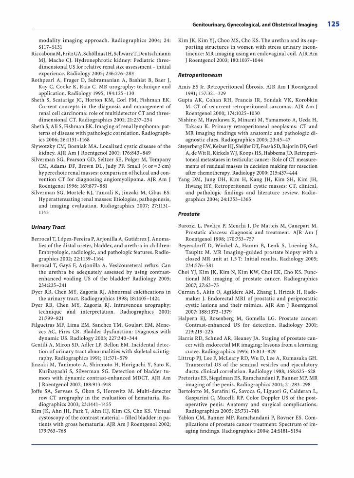
Genitourinary, Gynecological, and Obstetrical Imaging 125
modality imaging approach. Radiographics 2004; 24:S117–S131
Riccabona M, Fritz GA, Schöllnast H, Schwarz T, Deutschmann MJ, Mache CJ. Hydronephrotic kidney: Pediatric three-dimensional US for relative renal size assessment – initial experience. Radiology 2005; 236:276–283
Rothpearl A, Frager D, Subramanian A, Bashist B, Baer J, Kay C, Cooke K, Raia C. MR urography: technique and application. Radiology 1995; 194:125–130
Sheth S, Scatarige JC, Horton KM, Corl FM, Fishman EK. Current concepts in the diagnosis and management of renal cell carcinoma: role of multidetector CT and three-dimensional CT. Radiographics 2001; 21:237–254
Sheth S, Ali S, Fishman EK. Imaging of renal lymphoma: pat-terns of disease with pathologic correlation. Radiograph-ics 2006; 26:1151–1168
Slywotzky CM, Bosniak MA. Localized cystic disease of the kidney. AJR Am J Roentgenol 2001; 176:843–849
Silverman SG, Pearson GD, Seltzer SE, Polger M, Tempany CM, Adams DF, Brown DL, Judy PF. Small (< or = 3 cm) hyperechoic renal masses: comparison of helical and con-vention CT for diagnosing angiomyolipoma. AJR Am J Roentgenol 1996; 167:877–881
Silverman SG, Mortele KJ, Tuncali K, Jinzaki M, Cibas ES. Hyperattenuating renal masses: Etiologies, pathogenesis, and imaging evaluation. Radiographics 2007; 27:1131–1143
Urinary Tract
Berrocal T, López-Pereira P, Arjonilla A, Gutiérrez J. Anoma-lies of the distal ureter, bladder, and urethra in children: Embryologic, radiologic, and pathologic features. Radio-graphics 2002; 22:1139–1164
Berrocal T, Gayá F, Arjonilla A. Vesicoureteral refl ux: Can the urethra be adequately assessed by using contrast-enhanced voiding US of the bladder? Radiology 2005; 234:235–241
Dyer RB, Chen MY, Zagoria RJ. Abnormal calcifi cations in the urinary tract. Radiographics 1998; 18:1405–1424
Dyer RB, Chen MY, Zagoria RJ. Intravenous urography: technique and interpretation. Radiographics 2001; 21:799–821
Filgueiras MF, Lima EM, Sanchez TM, Goulart EM, Mene-zes AC, Pires CR. Bladder dysfunction: Diagnosis with dynamic US. Radiology 2003; 227:340–344
Gentili A, Miron SD, Adler LP, Bellon EM. Incidental detec-tion of urinary tract abnormalities with skeletal scintig-raphy. Radiographics 1991; 11:571–579
Jinzaki M, Tanimoto A, Shinmoto H, Horiguchi Y, Sato K, Kuribayashi S, Silverman SG. Detection of bladder tu-mors with dynamic contrast-enhanced MDCT. AJR Am J Roentgenol 2007; 188:913–918
Joffe SA, Servaes S, Okon S, Horowitz M. Multi-detector row CT urography in the evaluation of hematuria. Ra-diographics 2003; 23:1441–1455
Kim JK, Ahn JH, Park T, Ahn HJ, Kim CS, Cho KS. Virtual cystoscopy of the contrast material – fi lled bladder in pa-tients with gross hematuria. AJR Am J Roentgenol 2002; 179:763–768
Kim JK, Kim YJ, Choo MS, Cho KS. The urethra and its sup-porting structures in women with stress urinary incon-tinence: MR imaging using an endovaginal coil. AJR Am J Roentgenol 2003; 180:1037–1044
Retroperitoneum
Amis ES Jr. Retroperitoneal fi brosis. AJR Am J Roentgenol 1991; 157:321–329
Gupta AK, Cohan RH, Francis IR, Sondak VK, Korobkin M. CT of recurrent retroperitoneal sarcomas. AJR Am J Roentgenol 2000; 174:1025–1030
Nishino M, Hayakawa K, Minami M, Yamamoto A, Ueda H, Takasu K. Primary retroperitoneal neoplasms: CT and MR imaging fi ndings with anatomic and pathologic di-agnostic clues. Radiographics 2003; 23:45–47
Steyerberg EW, Keizer HJ, Sleijfer DT, Fossâ SD, Bajorin DF, Gerl A, de Wit R, Kirkels WJ, Koops HS, Habbema JD. Retroperi-toneal metastases in testicular cancer: Role of CT measure-ments of residual masses in decision making for resection after chemotherapy. Radiology 2000; 215:437–444
Yang DM, Jung DH, Kim H, Kang JH, Kim SH, Kim JH, Hwang HY. Retroperitoneal cystic masses: CT, clinical, and pathologic fi ndings and literature review. Radio-graphics 2004; 24:1353–1365
Prostate
Barozzi L, Pavlica P, Menchi I, De Matteis M, Canepari M. Prostatic abscess: diagnosis and treatment. AJR Am J Roentgenol 1998; 170:753–757
Beyersdorff D, Winkel A, Hamm B, Lenk S, Loening SA, Taupitz M. MR Imaging–guided prostate biopsy with a closed MR unit at 1.5 T: Initial results. Radiology 2005; 234:576–581
Choi YJ, Kim JK, Kim N, Kim KW, Choi EK, Cho KS. Func-tional MR imaging of prostate cancer. Radiographics 2007; 27:63–75
Curran S, Akin O, Agildere AM, Zhang J, Hricak H, Rade-maker J. Endorectal MRI of prostatic and periprostatic cystic lesions and their mimics. AJR Am J Roentgenol 2007; 188:1373–1379
Halpern EJ, Rosenberg M, Gomella LG. Prostate cancer: Contrast-enhanced US for detection. Radiology 2001; 219:219–225
Harris RD, Schned AR, Heaney JA. Staging of prostate can-cer with endorectal MR imaging: lessons from a learning curve. Radiographics 1995; 15:813–829
Littrup PJ, Lee F, McLeary RD, Wu D, Lee A, Kumasaka GH. Transrectal US of the seminal vesicles and ejaculatory ducts: clinical correlation. Radiology 1988; 168:625–628
Pretorius ES, Siegelman ES, Ramchandani P, Banner MP. MR imaging of the penis. Radiographics 2001; 21:283–298
Bertolotto M, Serafi ni G, Savoca G, Liguori G, Calderan L, Gasparini C, Mucelli RP. Color Doppler US of the post-operative penis: Anatomy and surgical complications. Radiographics 2005; 25:731–748
Yablon CM, Banner MP, Ramchandani P, Rovner ES. Com-plications of prostate cancer treatment: Spectrum of im-aging fi ndings. Radiographics 2004; 24:S181–S194
RibesLunaRos.indb 125 18.03.2008 12:51:07
126 A. Luna Alcala, M. Potolicchio, and L. Alcala Mata
Vulva and Vagina
Brown JJ, Gutierrez ED, Lee JK. MR appearance of the nor-mal and abnormal vagina after hysterectomy. AJR Am J Roentgenol 1992; 158:95–99
Dwarkasing S, Hussain SM, Hop WC, Krestin GP. Anovagi-nal fi stulas: Evaluation with endoanal MR imaging. Ra-diology 2004; 231:123–128
Fisseha S, Mueller G. Radiology illustrated: gynecologic im-aging. AJR Am J Roentgenol 2006; 186:594–595
Hahn WY, Israel GM, Lee VS. MRI of female urethral and peri-urethral disorders. AJR Am J Roentgenol 2004; 182:677–682
Kim CR, Eaton BA, Stevens KR Jr. Localization of the apex of the vagina: Implications for radiation therapy planning. Radiology 1999; 212:155–158
Kirks DR, Currarino G. Imperforate vagina with vaginourethral communication. AJR Am J Roentgenol 1977; 129:623–628
Siegelman ES, Outwater EK, Banner MP, Ramchandani P, Anderson TL, Schnall MD. High-resolution MR imaging of the vagina. Radiographics 1997; 17:1183–1203
Sohaib SA, Richards PS, Ind T, Jeyarajah AR, Shepherd JH, Jacobs IJ, Reznek RH. MR imaging of carcinoma of the vulva. AJR Am J Roentgenol 2002; 178:373–377
Togashi K, Nishimura K, Itoh K, Fujisawa I, Nakano Y, Tori-zuka K, Ozasa H, Ohshima M. Vaginal agenesis: classifi -cation by MR imaging. Radiology 1987; 162:675–677
Cervix and Uterus
Atri M, Nazarnia S, Aldis AE, Reinhold C, Bret PM, Kintzen G. Transvaginal US appearance of endometrial abnor-malities. Radiographics 1994; 14:483–492
Baldwin MT, Dudiak KM, Gorman B, Marks CA. Focal in-tracavitary masses recognized with the hyperechoic line sign at endovaginal US and characterized with hystero-sonography. Radiographics 1999; 19:927–935
Davis PC, O‘Neill MJ, Yoder IC, Lee SI, Mueller PR. Sonohys-terographic fi ndings of endometrial and subendometrial conditions. Radiographics 2002; 22:803–816
Fleischer AC. Transvaginal sonography of endometrial dis-orders: an overview. Radiographics 1998; 18:923–930
Jeong YY, Kang HK, Chung TW, Seo JJ, Park JG. Uterine cervical carcinoma after therapy: CT and MR imaging fi ndings. Radiographics 2003; 23:969–981
Mogavero G, Sheth S, Hamper UM. Endovaginal sonography of the nongravid uterus. Radiographics 1993; 13:969–981
Murase E, Siegelman ES, Outwater EK, Perez-Jaffe LA, Tu-reck RW. Uterine leiomyomas: Histopathologic features, MR imaging fi ndings, differential diagnosis, and treat-ment. Radiographics 1999; 19:1179–1197
Nalaboff KM, Pellerito JS, Ben-Levi E. Imaging the endome-trium: Disease and normal variants. Radiographics 2001; 21:1409–1424
Okamoto Y, Tanaka YO, Nishida M, Tsunoda H, Yoshikawa H, Itai Y. MR imaging of the uterine cervix: Imaging-pathologic correlation. Radiographics 2003; 23:425–445
Reinhold C, Tafazoli F, Mehio A, Wang L, Atri M, Siegelman ES, Rohoman L. Uterine adenomyosis: Endovaginal US and MR imaging features with histopathologic correla-tion. Radiographics 1999; 19:147–160
Scanlan KA, Propeck PA, Lee FT Jr. Invasive procedures in the female pelvis: Value of transabdominal, endovaginal, and endorectal US guidance. Radiographics 2001; 21:491–506
Ovary
Kim KA, Park CM, Lee JH, Kim HK, Cho SM, Kim B, Seol HY. Benign ovarian tumors with solid and cystic com-ponents that mimic malignancy. AJR Am J Roentgenol 2004; 182:1259–1265
Reviews
Brammer HM 3rd, Buck JL, Hayes WS, Sheth S, Tavassoli FA. From the archives of the AFIP. Malignant germ cell tumors of the ovary: radiologic-pathologic correlation. Radiographics 1990; 10:715–724
Hertzberg BS, Kliewer MA. Sonography of benign cystic teratoma of the ovary: pitfalls in diagnosis. AJR Am J Roentgenol 1996; 167:1127–1133
Jeong YY, Outwater EK, Kang HK. Imaging evaluation of ovarian masses. Radiographics 2000; 20:1445–1470
Jung SE, Lee JM, Rha SE, Byun JY, Jung JI, Hahn ST. CT and MR imaging of ovarian tumors with emphasis on differ-ential diagnosis. Radiographics 2002; 22:1305–1325
Olson MC, Posniak HV, Tempany CM, Dudiak CM. MR im-aging of the female pelvic region. Radiographics 1992; 12:445–465
Outwater EK, Dunton CJ. Imaging of the ovary and adnexa: clinical issues and applications of MR imaging. Radiol-ogy 1995; 194:1–18
Outwater EK, Mitchell DG. Normal ovaries and functional cysts: MR appearance. Radiology 1996; 198:397–402
Rha SE, Byun JY, Jung SE, Jung JI, Choi BG, Kim BS, Kim H, Lee JM. CT and MR imaging features of adnexal torsion. Radiographics 2002; 22:283–294
Saksouk FA, Johnson SC. Recognition of the ovaries and ovarian origin of pelvic masses with CT. Radiographics 2004; 24:S133–S146
Obstetrics
Braffman BH, Coleman BG, Ramchandani P, Arger PH, No-dine CF, Dinsmore BJ, Louie A, Betsch SE. Emergency de-partment screening for ectopic pregnancy: a prospective US study. Radiology 1994; 190:797–802
Amin RS, Nikolaidis P, Kawashima A, Kramer LA, Ernst RD. Normal anatomy of the fetus at MR imaging. Radiograph-ics 1999; 19:201–214
Fong KW, Toi A, Salem S, Hornberger LK, Chitayat D, Keat-ing SJ, McAuliffe F, Johnson JA. Detection of fetal struc-tural abnormalities with US during early pregnancy. Ra-diographics 2004; 24:157–174
Woodward PJ, Sohaey R, Kennedy A, Koeller KK. From the archives of the AFIP: A comprehensive review of fetal tumors with pathologic correlation. Radiographics 2005; 25:215–242
Taylor KJ, Ramos IM, Feyock AL, Snower DP, Carter D, Sha-piro BS, Meyer WR, De Cherney AH. Ectopic pregnancy: duplex Doppler evaluation. Radiology 1989; 173:93–97
RibesLunaRos.indb 126 18.03.2008 12:51:07
Nuclear Medicine
Musculoskeletal Imaging 6
Joan C. Vilanova and Ramon RibesSandra Baleato (Contributor)
IntroductionMusculoskeletal radiology is a subspecialty which has widely expanded its scope and imaging capabilities with the advent of ultrasound, MRI, multi-detector CT, and PET. Prior to the advent of MRI, the primary tools of musculoskeletal radiologists were con-ventional X-rays and arthrograms. The subspecialty has progressed from primary imag-ing of osseous structures and indirect imaging of articular spaces to direct imaging of soft-tissue structures with direct visualization and precise depiction of musculoskeletal structures.
Specialized musculoskeletal radiology requires a sound knowledge of anatomy, patho-physiology and surgical techniques, coupled with a solid background in the imaging mo-dalities involved in musculoskeletal radiology. Musculoskeletal (MSK) imaging involves all aspects of anatomy, function, disease states and the aspects of interventional radiol-ogy related to the musculoskeletal system. This includes imaging in orthopedics, trauma, rheumatology, metabolic and endocrine diseases as well as aspects of pediatrics, oncol-ogy and sports imaging.
Subspecialty training in MSK radiology should provide the experience necessary to ensure competence in the following techniques: plain radiography, ultrasonography, CT, MRI, nuclear medicine, bone densitometry and fl uoroscopic procedures includ-ing arthrography. It is important for the musculoskeletal radiologist to be aware of the strengths and weaknesses of the different imaging methods in various pathological con-ditions. Furthermore, it is the role of a subspecialty expert to choose the appropriate im-aging technique and/or the appropriate sequence in the investigation of specifi c clinical problems.
An MSK radiologist should have acquired an in-depth understanding of diseases of the MSK system and understand the role of imaging in the diagnosis and treatment of MSK disease. Moreover, due to innovations and new medical imaging modalities, clini-cal specialists requests are increasingly demanding. If radiologists cannot keep up with the increasing demands for musculoskeletal interpretations, clinicians will be forced to compete with radiologists in providing these interpretations.
From the beginning of the subspecialty in the 1970s with the foundation of the Interna-tional Skeletal Society, many multidisciplinary or dedicated skeletal radiology societies have been founded and organized from international or national societies and continue to play an important role in the development of the subspeciality.
RibesLunaRos.indb 127 18.03.2008 12:51:07
128 J. C. Vilanova and R. Ribes
Case 1
Osteomyelitis
Fig. 6.1.1 Fig. 6.1.2
Fig. 6.1.3 Fig. 6.1.4
Fig. 6.1.5
RibesLunaRos.indb 128 18.03.2008 12:51:07
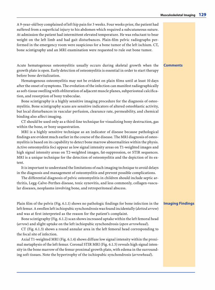
Musculoskeletal Imaging 129
A 9-year-old boy complained of left hip pain for 3 weeks. Four weeks prior, the patient had suffered from a superfi cial injury to his abdomen which required a subcutaneous suture. At admission the patient had intermittent elevated temperature. He was reluctant to bear weight on the left limb and had gait disturbances. Plain-fi lm pelvic radiographs per-formed in the emergency room were suspicious for a bone tumor of the left ischium. CT, bone scintigraphy and an MRI examination were requested to rule out bone tumor.
CommentsAcute hematogenous osteomyelitis usually occurs during skeletal growth when the growth plate is open. Early detection of osteomyelitis is essential in order to start therapy before bone devitalization.
Hematogenous osteomyelitis may not be evident on plain fi lms until at least 10 days after the onset of symptoms. The evolution of the infection can manifest radiographically as soft-tissue swelling with obliteration of adjacent muscle planes, subperiosteal calcifi ca-tion, and resorption of bony trabeculae.
Bone scintigraphy is a highly sensitive imaging procedure for the diagnosis of osteo-myelitis. Bone scintigraphy scans are sensitive indicators of altered osteoblastic activity, but local disturbances in vascular perfusion, clearance rate, permeability, and chemical binding also affect imaging.
CT should be used only as a third-line technique for visualizing bony destruction, gas within the bone, or bony sequestration.
MRI is a highly sensitive technique as an indicator of disease because pathological fi ndings are evident much earlier in the course of the disease. The MRI diagnosis of osteo-myelitis is based on its capability to detect bone marrow abnormalities within the physis. Active osteomyelitis foci appear as low signal intensity areas on T1-weighted images and high signal intensity areas on T2-weighted images, fat-suppression, or STIR sequences. MRI is a unique technique for the detection of osteomyelitis and the depiction of its ex-tent.
It is important to understand the limitations of each imaging technique to avoid delays in the diagnosis and management of osteomyelitis and prevent possible complications.
The differential diagnosis of pelvic osteomyelitis in children should include septic ar-thritis, Legg-Calve-Perthes disease, toxic synovitis, and less commonly, collagen-vascu-lar diseases, neoplasms involving bone, and retroperitoneal abscess.
Imaging FindingsPlain fi lm of the pelvis (Fig. 6.1.1) shows no pathologic fi ndings for bone infection in the left femur. A swollen left ischiopubic synchondrosis was found incidentally (dotted arrow) and was at fi rst interpreted as the reason for the patient’s complaint.
Bone scintigraphy (Fig. 6.1.2) scan shows increased uptake within the left femoral head (arrow) and slight uptake on the left ischiopubic synchondrosis (open arrowhead).
CT (Fig. 6.1.3) shows a round annular area in the left femoral head corresponding to the focal site of infection.
Axial T1-weighted MRI (Fig. 6.1.4) shows diffuse low signal intensity within the proxi-mal metaphysis of the left femur. Coronal STIR MRI (Fig. 6.1.5) reveals high signal inten-sity in the bone marrow of the femur proximal growth plate, with edema in the surround-ing soft tissues. Note the hypertrophy of the ischiopubic synchondrosis (arrowhead).
RibesLunaRos.indb 129 18.03.2008 12:51:11
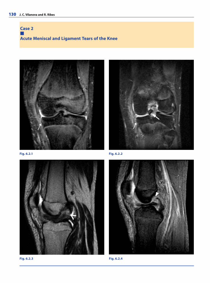
130 J. C. Vilanova and R. Ribes
Case 2
Acute Meniscal and Ligament Tears of the Knee
Fig. 6.2.1 Fig. 6.2.2
Fig. 6.2.3 Fig. 6.2.4
RibesLunaRos.indb 130 18.03.2008 12:51:11
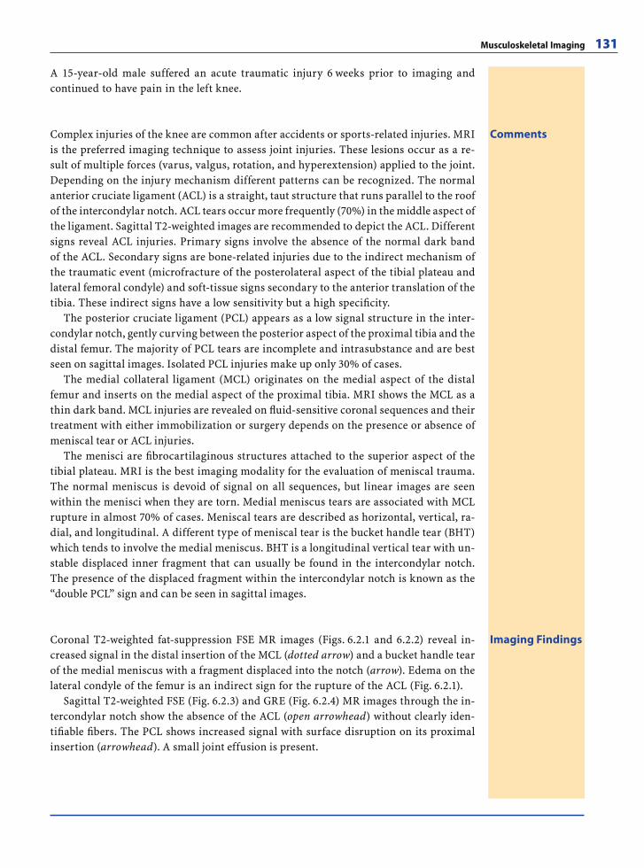
Musculoskeletal Imaging 131
A 15-year-old male suffered an acute traumatic injury 6 weeks prior to imaging and continued to have pain in the left knee.
CommentsComplex injuries of the knee are common after accidents or sports-related injuries. MRI is the preferred imaging technique to assess joint injuries. These lesions occur as a re-sult of multiple forces (varus, valgus, rotation, and hyperextension) applied to the joint. Depending on the injury mechanism different patterns can be recognized. The normal anterior cruciate ligament (ACL) is a straight, taut structure that runs parallel to the roof of the intercondylar notch. ACL tears occur more frequently (70%) in the middle aspect of the ligament. Sagittal T2-weighted images are recommended to depict the ACL. Different signs reveal ACL injuries. Primary signs involve the absence of the normal dark band of the ACL. Secondary signs are bone-related injuries due to the indirect mechanism of the traumatic event (microfracture of the posterolateral aspect of the tibial plateau and lateral femoral condyle) and soft-tissue signs secondary to the anterior translation of the tibia. These indirect signs have a low sensitivity but a high specifi city.
The posterior cruciate ligament (PCL) appears as a low signal structure in the inter-condylar notch, gently curving between the posterior aspect of the proximal tibia and the distal femur. The majority of PCL tears are incomplete and intrasubstance and are best seen on sagittal images. Isolated PCL injuries make up only 30% of cases.
The medial collateral ligament (MCL) originates on the medial aspect of the distal femur and inserts on the medial aspect of the proximal tibia. MRI shows the MCL as a thin dark band. MCL injuries are revealed on fl uid-sensitive coronal sequences and their treatment with either immobilization or surgery depends on the presence or absence of meniscal tear or ACL injuries.
The menisci are fi brocartilaginous structures attached to the superior aspect of the tibial plateau. MRI is the best imaging modality for the evaluation of meniscal trauma. The normal meniscus is devoid of signal on all sequences, but linear images are seen within the menisci when they are torn. Medial meniscus tears are associated with MCL rupture in almost 70% of cases. Meniscal tears are described as horizontal, vertical, ra-dial, and longitudinal. A different type of meniscal tear is the bucket handle tear (BHT) which tends to involve the medial meniscus. BHT is a longitudinal vertical tear with un-stable displaced inner fragment that can usually be found in the intercondylar notch. The presence of the displaced fragment within the intercondylar notch is known as the “double PCL” sign and can be seen in sagittal images.
Imaging FindingsCoronal T2-weighted fat-suppression FSE MR images (Figs. 6.2.1 and 6.2.2) reveal in-creased signal in the distal insertion of the MCL (dotted arrow) and a bucket handle tear of the medial meniscus with a fragment displaced into the notch (arrow). Edema on the lateral condyle of the femur is an indirect sign for the rupture of the ACL (Fig. 6.2.1).
Sagittal T2-weighted FSE (Fig. 6.2.3) and GRE (Fig. 6.2.4) MR images through the in-tercondylar notch show the absence of the ACL (open arrowhead) without clearly iden-tifi able fi bers. The PCL shows increased signal with surface disruption on its proximal insertion (arrowhead). A small joint effusion is present.
RibesLunaRos.indb 131 18.03.2008 12:51:14
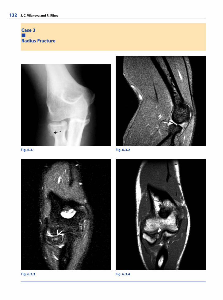
132 J. C. Vilanova and R. Ribes
Case 3
Radius Fracture
Fig. 6.3.1
Fig. 6.3.3 Fig. 6.3.4
Fig. 6.3.2
RibesLunaRos.indb 132 18.03.2008 12:51:14
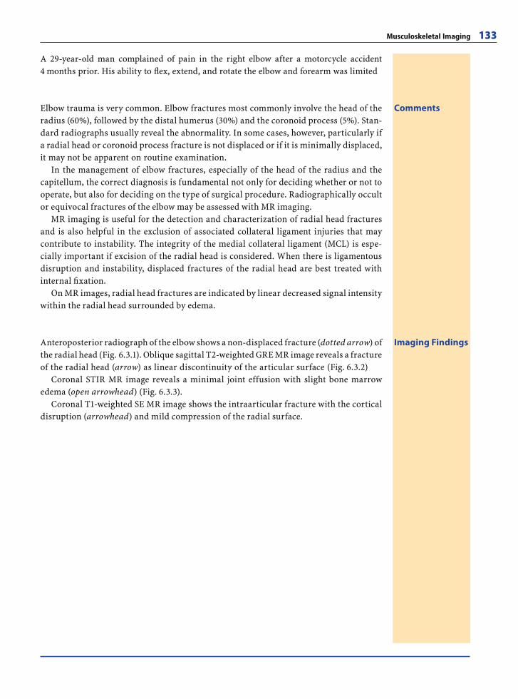
Musculoskeletal Imaging 133
A 29-year-old man complained of pain in the right elbow after a motorcycle accident 4 months prior. His ability to fl ex, extend, and rotate the elbow and forearm was limited
CommentsElbow trauma is very common. Elbow fractures most commonly involve the head of the radius (60%), followed by the distal humerus (30%) and the coronoid process (5%). Stan-dard radiographs usually reveal the abnormality. In some cases, however, particularly if a radial head or coronoid process fracture is not displaced or if it is minimally displaced, it may not be apparent on routine examination.
In the management of elbow fractures, especially of the head of the radius and the capitellum, the correct diagnosis is fundamental not only for deciding whether or not to operate, but also for deciding on the type of surgical procedure. Radiographically occult or equivocal fractures of the elbow may be assessed with MR imaging.
MR imaging is useful for the detection and characterization of radial head fractures and is also helpful in the exclusion of associated collateral ligament injuries that may contribute to instability. The integrity of the medial collateral ligament (MCL) is espe-cially important if excision of the radial head is considered. When there is ligamentous disruption and instability, displaced fractures of the radial head are best treated with internal fi xation.
On MR images, radial head fractures are indicated by linear decreased signal intensity within the radial head surrounded by edema.
Imaging FindingsAnteroposterior radiograph of the elbow shows a non-displaced fracture (dotted arrow) of the radial head (Fig. 6.3.1). Oblique sagittal T2-weighted GRE MR image reveals a fracture of the radial head (arrow) as linear discontinuity of the articular surface (Fig. 6.3.2)
Coronal STIR MR image reveals a minimal joint effusion with slight bone marrow edema (open arrowhead) (Fig. 6.3.3).
Coronal T1-weighted SE MR image shows the intraarticular fracture with the cortical disruption (arrowhead) and mild compression of the radial surface.
RibesLunaRos.indb 133 18.03.2008 12:51:17
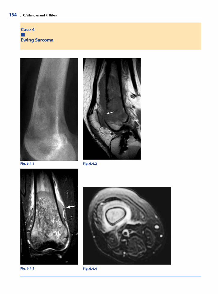
134 J. C. Vilanova and R. Ribes
Case 4
Ewing Sarcoma
Fig. 6.4.1
Fig. 6.4.3
Fig. 6.4.2
Fig. 6.4.4
RibesLunaRos.indb 134 18.03.2008 12:51:17
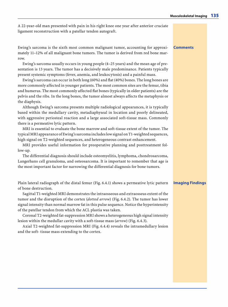
Musculoskeletal Imaging 135
A 22-year-old man presented with pain in his right knee one year after anterior cruciate ligament reconstruction with a patellar tendon autograft.
CommentsEwing’s sarcoma is the sixth most common malignant tumor, accounting for approxi-mately 11–12% of all malignant bone tumors. The tumor is derived from red bone mar-row.
Ewing’s sarcoma usually occurs in young people (4–25 years) and the mean age of pre-sentation is 13 years. The tumor has a decisively male predominance. Patients typically present systemic symptoms (fever, anemia, and leukocytosis) and a painful mass.
Ewing’s sarcoma can occur in both long (60%) and fl at (40%) bones. The long bones are more commonly affected in younger patients. The most common sites are the femur, tibia and humerus. The most commonly affected fl at bones (typically in older patients) are the pelvis and the ribs. In the long bones, the tumor almost always affects the metaphysis or the diaphysis.
Although Ewing’s sarcoma presents multiple radiological appearances, it is typically based within the medullary cavity, metadiaphyseal in location and poorly delineated, with aggressive periosteal reaction and a large associated soft-tissue mass. Commonly there is a permeative lytic pattern.
MRI is essential to evaluate the bone marrow and soft-tissue extent of the tumor. The typical MRI appearance of Ewing’s sarcoma includes low signal on T1-weighted sequences, high signal on T2-weighted sequences, and heterogeneous contrast enhancement.
MRI provides useful information for preoperative planning and posttreatment fol-low-up.
The differential diagnosis should include osteomyelitis, lymphoma, chondrosarcoma, Langerhans cell granuloma, and osteosarcoma. It is important to remember that age is the most important factor for narrowing the differential diagnosis for bone tumors.
Imaging FindingsPlain lateral radiograph of the distal femur (Fig. 6.4.1) shows a permeative lytic pattern of bone destruction.
Sagittal T1-weighted MRI demonstrates the intraosseous and extraosseus extent of the tumor and the disruption of the cortex (dotted arrow) (Fig. 6.4.2). The tumor has lower signal intensity than normal marrow fat in this pulse sequence. Notice the hyperintensity of the patellar tendon from which the ACL plastia was taken.
Coronal T2-weighted fat-suppression MRI shows a heterogeneous high signal intensity lesion within the medullar cavity with a soft-tissue mass (arrow) (Fig. 6.4.3).
Axial T2-weighted fat-suppression MRI (Fig. 6.4.4) reveals the intramedullary lesion and the soft- tissue mass extending to the cortex.
RibesLunaRos.indb 135 18.03.2008 12:51:22
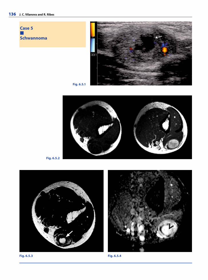
136 J. C. Vilanova and R. Ribes
Case 5
Schwannoma
Fig. 6.5.2
Fig. 6.5.3 Fig. 6.5.4
Fig. 6.5.1
RibesLunaRos.indb 136 18.03.2008 12:51:22
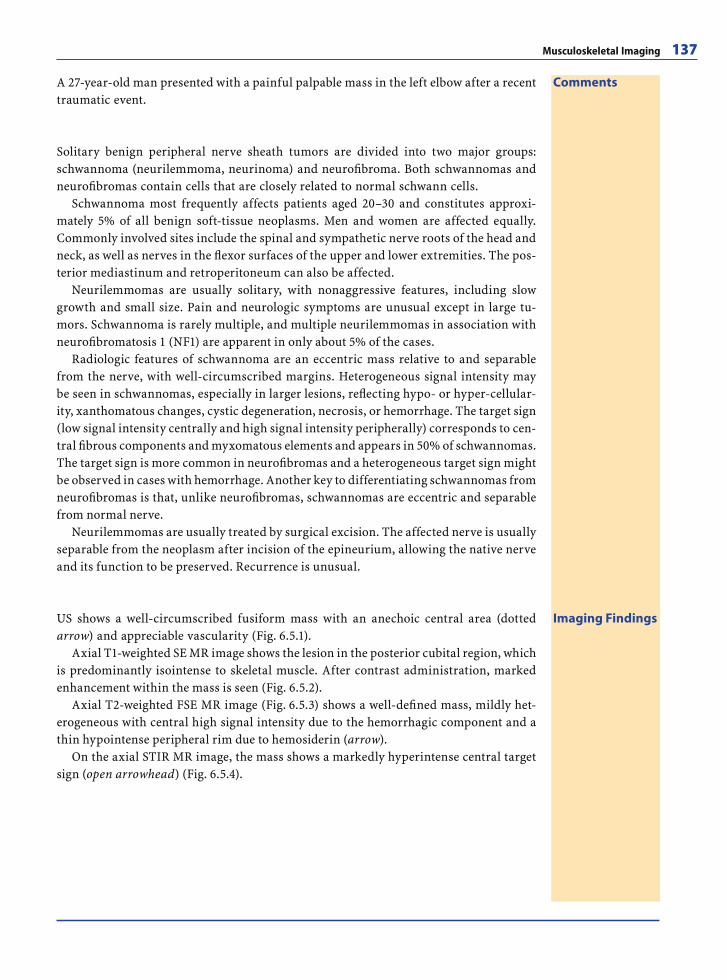
Musculoskeletal Imaging 137
A 27-year-old man presented with a painful palpable mass in the left elbow after a recent traumatic event.
Comments
Solitary benign peripheral nerve sheath tumors are divided into two major groups: schwannoma (neurilemmoma, neurinoma) and neurofi broma. Both schwannomas and neurofi bromas contain cells that are closely related to normal schwann cells.
Schwannoma most frequently affects patients aged 20–30 and constitutes approxi-mately 5% of all benign soft-tissue neoplasms. Men and women are affected equally. Commonly involved sites include the spinal and sympathetic nerve roots of the head and neck, as well as nerves in the fl exor surfaces of the upper and lower extremities. The pos-terior mediastinum and retroperitoneum can also be affected.
Neurilemmomas are usually solitary, with nonaggressive features, including slow growth and small size. Pain and neurologic symptoms are unusual except in large tu-mors. Schwannoma is rarely multiple, and multiple neurilemmomas in association with neurofi bromatosis 1 (NF1) are apparent in only about 5% of the cases.
Radiologic features of schwannoma are an eccentric mass relative to and separable from the nerve, with well-circumscribed margins. Heterogeneous signal intensity may be seen in schwannomas, especially in larger lesions, refl ecting hypo- or hyper-cellular-ity, xanthomatous changes, cystic degeneration, necrosis, or hemorrhage. The target sign (low signal intensity centrally and high signal intensity peripherally) corresponds to cen-tral fi brous components and myxomatous elements and appears in 50% of schwannomas. The target sign is more common in neurofi bromas and a heterogeneous target sign might be observed in cases with hemorrhage. Another key to differentiating schwannomas from neurofi bromas is that, unlike neurofi bromas, schwannomas are eccentric and separable from normal nerve.
Neurilemmomas are usually treated by surgical excision. The affected nerve is usually separable from the neoplasm after incision of the epineurium, allowing the native nerve and its function to be preserved. Recurrence is unusual.
Imaging FindingsUS shows a well-circumscribed fusiform mass with an anechoic central area (dotted arrow) and appreciable vascularity (Fig. 6.5.1).
Axial T1-weighted SE MR image shows the lesion in the posterior cubital region, which is predominantly isointense to skeletal muscle. After contrast administration, marked enhancement within the mass is seen (Fig. 6.5.2).
Axial T2-weighted FSE MR image (Fig. 6.5.3) shows a well-defi ned mass, mildly het-erogeneous with central high signal intensity due to the hemorrhagic component and a thin hypointense peripheral rim due to hemosiderin (arrow).
On the axial STIR MR image, the mass shows a markedly hyperintense central target sign (open arrowhead) (Fig. 6.5.4).
RibesLunaRos.indb 137 18.03.2008 12:51:25
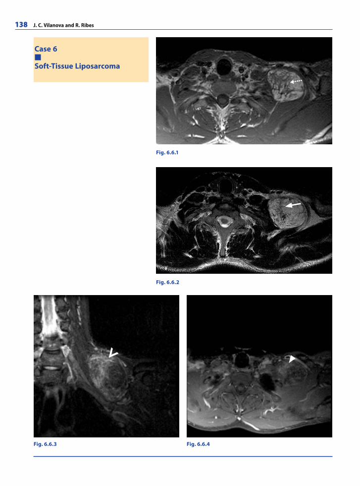
138 J. C. Vilanova and R. Ribes
Case 6
Soft-Tissue Liposarcoma
Fig. 6.6.4Fig. 6.6.3
Fig. 6.6.2
Fig. 6.6.1
RibesLunaRos.indb 138 18.03.2008 12:51:25
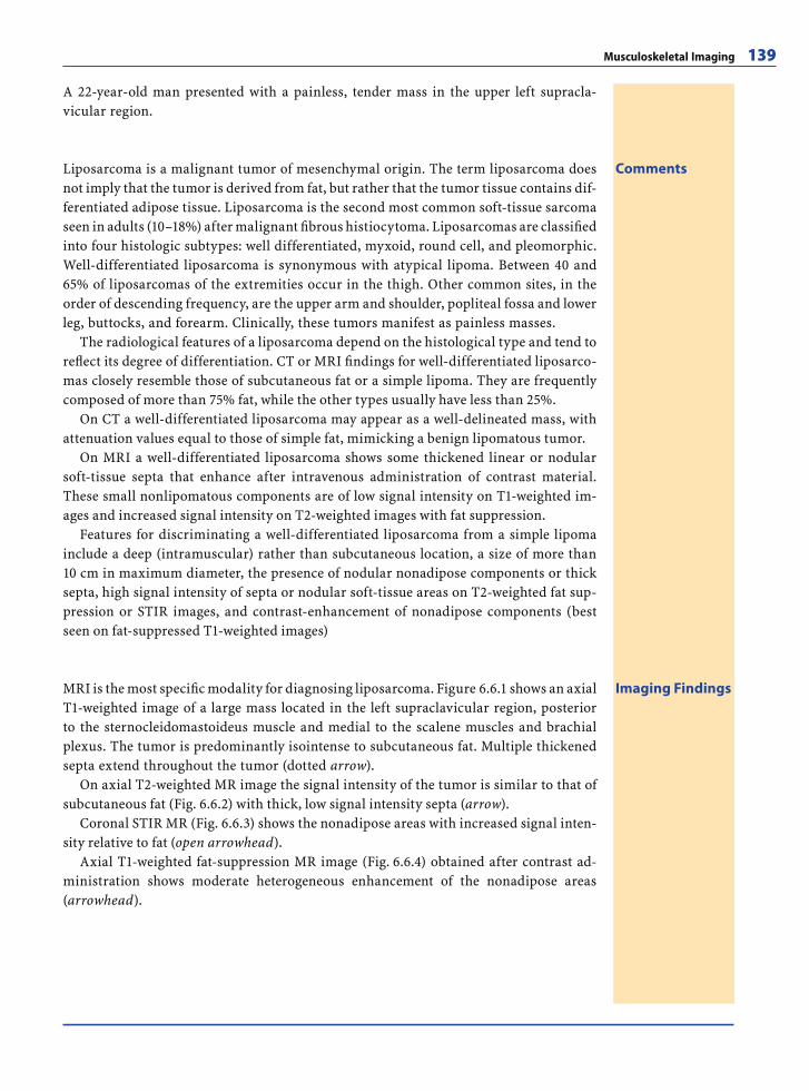
Musculoskeletal Imaging 139
A 22-year-old man presented with a painless, tender mass in the upper left supracla-vicular region.
CommentsLiposarcoma is a malignant tumor of mesenchymal origin. The term liposarcoma does not imply that the tumor is derived from fat, but rather that the tumor tissue contains dif-ferentiated adipose tissue. Liposarcoma is the second most common soft-tissue sarcoma seen in adults (10–18%) after malignant fi brous histiocytoma. Liposarcomas are classifi ed into four histologic subtypes: well differentiated, myxoid, round cell, and pleomorphic. Well-differentiated liposarcoma is synonymous with atypical lipoma. Between 40 and 65% of liposarcomas of the extremities occur in the thigh. Other common sites, in the order of descending frequency, are the upper arm and shoulder, popliteal fossa and lower leg, buttocks, and forearm. Clinically, these tumors manifest as painless masses.
The radiological features of a liposarcoma depend on the histological type and tend to refl ect its degree of differentiation. CT or MRI fi ndings for well-differentiated liposarco-mas closely resemble those of subcutaneous fat or a simple lipoma. They are frequently composed of more than 75% fat, while the other types usually have less than 25%.
On CT a well-differentiated liposarcoma may appear as a well-delineated mass, with attenuation values equal to those of simple fat, mimicking a benign lipomatous tumor.
On MRI a well-differentiated liposarcoma shows some thickened linear or nodular soft-tissue septa that enhance after intravenous administration of contrast material. These small nonlipomatous components are of low signal intensity on T1-weighted im-ages and increased signal intensity on T2-weighted images with fat suppression.
Features for discriminating a well-differentiated liposarcoma from a simple lipoma include a deep (intramuscular) rather than subcutaneous location, a size of more than 10 cm in maximum diameter, the presence of nodular nonadipose components or thick septa, high signal intensity of septa or nodular soft-tissue areas on T2-weighted fat sup-pression or STIR images, and contrast-enhancement of nonadipose components (best seen on fat-suppressed T1-weighted images)
Imaging FindingsMRI is the most specifi c modality for diagnosing liposarcoma. Figure 6.6.1 shows an axial T1-weighted image of a large mass located in the left supraclavicular region, posterior to the sternocleidomastoideus muscle and medial to the scalene muscles and brachial plexus. The tumor is predominantly isointense to subcutaneous fat. Multiple thickened septa extend throughout the tumor (dotted arrow).
On axial T2-weighted MR image the signal intensity of the tumor is similar to that of subcutaneous fat (Fig. 6.6.2) with thick, low signal intensity septa (arrow).
Coronal STIR MR (Fig. 6.6.3) shows the nonadipose areas with increased signal inten-sity relative to fat (open arrowhead).
Axial T1-weighted fat-suppression MR image (Fig. 6.6.4) obtained after contrast ad-ministration shows moderate heterogeneous enhancement of the nonadipose areas(arrowhead).
RibesLunaRos.indb 139 18.03.2008 12:51:28
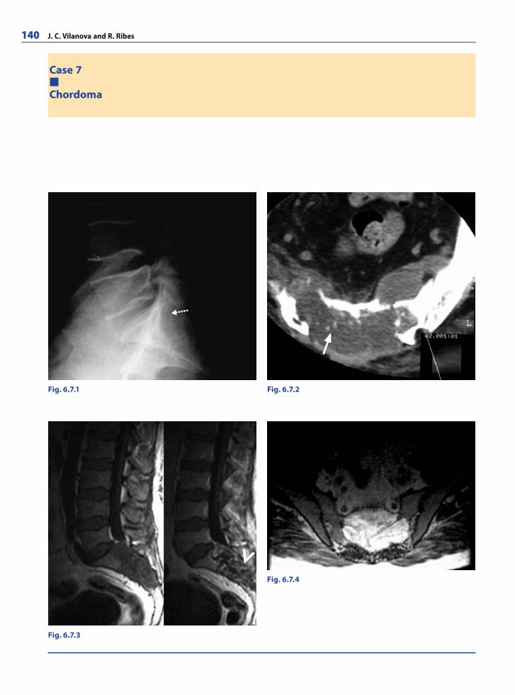
140 J. C. Vilanova and R. Ribes
Case 7
Chordoma
Fig. 6.7.1 Fig. 6.7.2
Fig. 6.7.3
Fig. 6.7.4
RibesLunaRos.indb 140 18.03.2008 12:51:28
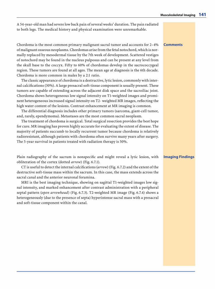
Musculoskeletal Imaging 141
A 54-year-old man had severe low back pain of several weeks’ duration. The pain radiated to both legs. The medical history and physical examination were unremarkable.
CommentsChordoma is the most common primary malignant sacral tumor and accounts for 2–4% of malignant osseous neoplasms. Chordomas arise from the fetal notochord, which is nor-mally replaced by mesodermal tissue by the 7th week of development. Scattered vestiges of notochord may be found in the nucleus pulposus and can be present at any level from the skull base to the coccyx. Fifty to 60% of chordomas develop in the sacrococcygeal region. These tumors are found at all ages. The mean age at diagnosis is the 6th decade. Chordoma is more common in males by a 2:1 ratio.
The classic appearance of chordoma is a destructive, lytic lesion, commonly with inter-nal calcifi cations (30%). A large presacral soft-tissue component is usually present. These tumors are capable of extending across the adjacent disk space and the sacroiliac joint. Chordoma shows heterogeneous low signal intensity on T1-weighted images and promi-nent heterogeneous increased signal intensity on T2- weighted MR images, refl ecting the high water content of the lesions. Contrast enhancement at MR imaging is common.
The differential diagnosis includes other primary tumors (sarcoma, giant-cell tumor, and, rarely, ependymoma). Metastases are the most common sacral neoplasm.
The treatment of chordoma is surgical. Total surgical resection provides the best hope for cure. MR imaging has proven highly accurate for evaluating the extent of disease. The majority of patients succumb to locally recurrent tumor because chordoma is relatively radioresistant, although patients with chordoma often survive many years after surgery. The 5-year survival in patients treated with radiation therapy is 50%.
Imaging FindingsPlain radiography of the sacrum is nonspecifi c and might reveal a lytic lesion, with obliteration of the cortex (dotted arrow) (Fig. 6.7.1).
CT is useful to detect the internal calcifi cations (arrow) (Fig. 6.7.2) and the extent of the destructive soft-tissue mass within the sacrum. In this case, the mass extends across the sacral canal and the anterior neuronal foramina.
MRI is the best imaging technique, showing on sagittal T1-weighted images low sig-nal intensity, and marked enhancement after contrast administration with a peripheral septal pattern (open arrowhead) (Fig. 6.7.3). T2-weighted MR image (Fig. 6.7.4) shows a heterogeneously (due to the presence of septa) hyperintense sacral mass with a presacral and soft-tissue component within the canal.
RibesLunaRos.indb 141 18.03.2008 12:51:30
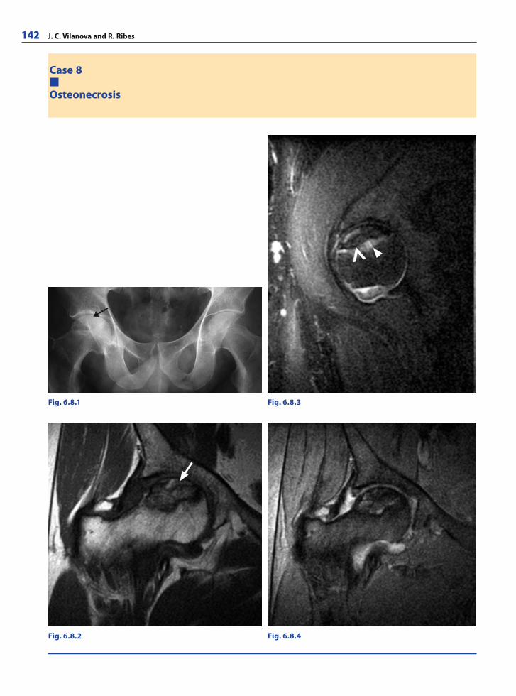
142 J. C. Vilanova and R. Ribes
Case 8
Osteonecrosis
Fig. 6.8.1 Fig. 6.8.3
Fig. 6.8.4Fig. 6.8.2
RibesLunaRos.indb 142 18.03.2008 12:51:30
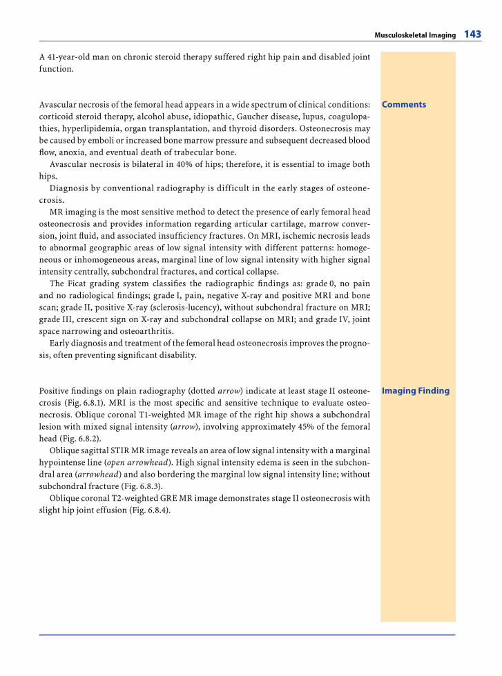
Musculoskeletal Imaging 143
A 41-year-old man on chronic steroid therapy suffered right hip pain and disabled joint function.
CommentsAvascular necrosis of the femoral head appears in a wide spectrum of clinical conditions: corticoid steroid therapy, alcohol abuse, idiopathic, Gaucher disease, lupus, coagulopa-thies, hyperlipidemia, organ transplantation, and thyroid disorders. Osteonecrosis may be caused by emboli or increased bone marrow pressure and subsequent decreased blood fl ow, anoxia, and eventual death of trabecular bone.
Avascular necrosis is bilateral in 40% of hips; therefore, it is essential to image both hips.
Diagnosis by conventional radiography is difficult in the early stages of osteone-crosis.
MR imaging is the most sensitive method to detect the presence of early femoral head osteonecrosis and provides information regarding articular cartilage, marrow conver-sion, joint fl uid, and associated insuffi ciency fractures. On MRI, ischemic necrosis leads to abnormal geographic areas of low signal intensity with different patterns: homoge-neous or inhomogeneous areas, marginal line of low signal intensity with higher signal intensity centrally, subchondral fractures, and cortical collapse.
The Ficat grading system classifi es the radiographic fi ndings as: grade 0, no pain and no radiological fi ndings; grade I, pain, negative X-ray and positive MRI and bone scan; grade II, positive X-ray (sclerosis-lucency), without subchondral fracture on MRI; grade III, crescent sign on X-ray and subchondral collapse on MRI; and grade IV, joint space narrowing and osteoarthritis.
Early diagnosis and treatment of the femoral head osteonecrosis improves the progno-sis, often preventing signifi cant disability.
Imaging FindingPositive fi ndings on plain radiography (dotted arrow) indicate at least stage II osteone-crosis (Fig. 6.8.1). MRI is the most specifi c and sensitive technique to evaluate osteo-necrosis. Oblique coronal T1-weighted MR image of the right hip shows a subchondral lesion with mixed signal intensity (arrow), involving approximately 45% of the femoral head (Fig. 6.8.2).
Oblique sagittal STIR MR image reveals an area of low signal intensity with a marginal hypointense line (open arrowhead). High signal intensity edema is seen in the subchon-dral area (arrowhead) and also bordering the marginal low signal intensity line; without subchondral fracture (Fig. 6.8.3).
Oblique coronal T2-weighted GRE MR image demonstrates stage II osteonecrosis with slight hip joint effusion (Fig. 6.8.4).
RibesLunaRos.indb 143 18.03.2008 12:51:33
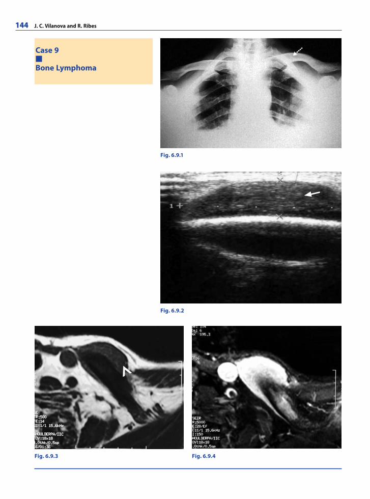
144 J. C. Vilanova and R. Ribes
Case 9
Bone Lymphoma
Fig. 6.9.1
Fig. 6.9.2
Fig. 6.9.3 Fig. 6.9.4
RibesLunaRos.indb 144 18.03.2008 12:51:33
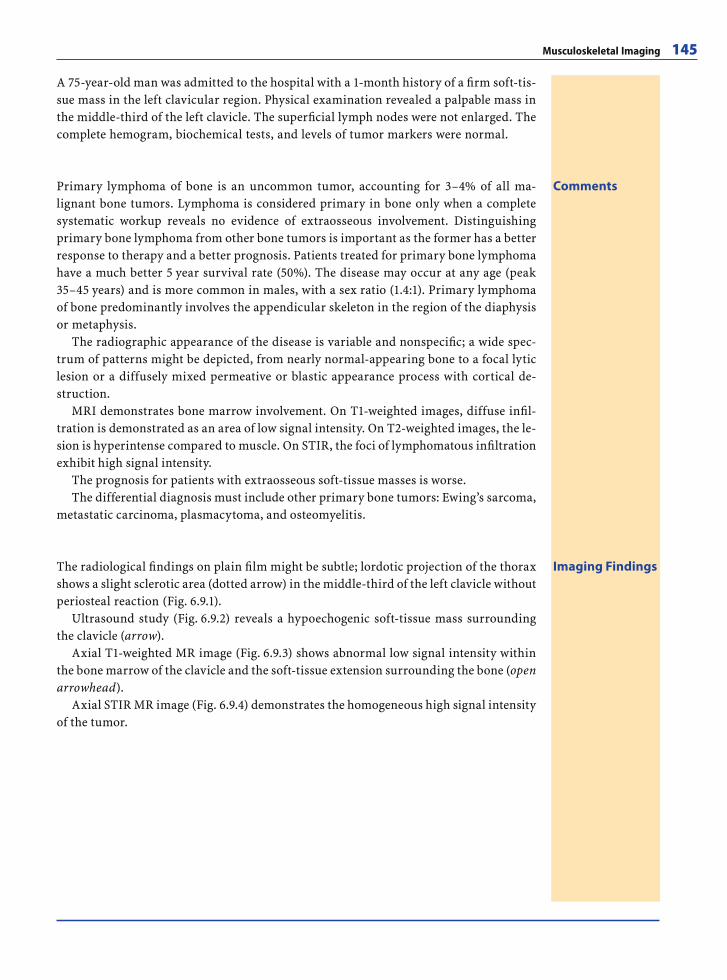
Musculoskeletal Imaging 145
A 75-year-old man was admitted to the hospital with a 1-month history of a fi rm soft-tis-sue mass in the left clavicular region. Physical examination revealed a palpable mass in the middle-third of the left clavicle. The superfi cial lymph nodes were not enlarged. The complete hemogram, biochemical tests, and levels of tumor markers were normal.
CommentsPrimary lymphoma of bone is an uncommon tumor, accounting for 3–4% of all ma-lignant bone tumors. Lymphoma is considered primary in bone only when a complete systematic workup reveals no evidence of extraosseous involvement. Distinguishing primary bone lymphoma from other bone tumors is important as the former has a better response to therapy and a better prognosis. Patients treated for primary bone lymphoma have a much better 5 year survival rate (50%). The disease may occur at any age (peak 35–45 years) and is more common in males, with a sex ratio (1.4:1). Primary lymphoma of bone predominantly involves the appendicular skeleton in the region of the diaphysis or metaphysis.
The radiographic appearance of the disease is variable and nonspecifi c; a wide spec-trum of patterns might be depicted, from nearly normal-appearing bone to a focal lytic lesion or a diffusely mixed permeative or blastic appearance process with cortical de-struction.
MRI demonstrates bone marrow involvement. On T1-weighted images, diffuse infi l-tration is demonstrated as an area of low signal intensity. On T2-weighted images, the le-sion is hyperintense compared to muscle. On STIR, the foci of lymphomatous infi ltration exhibit high signal intensity.
The prognosis for patients with extraosseous soft-tissue masses is worse.The differential diagnosis must include other primary bone tumors: Ewing’s sarcoma,
metastatic carcinoma, plasmacytoma, and osteomyelitis.
Imaging FindingsThe radiological fi ndings on plain fi lm might be subtle; lordotic projection of the thorax shows a slight sclerotic area (dotted arrow) in the middle-third of the left clavicle without periosteal reaction (Fig. 6.9.1).
Ultrasound study (Fig. 6.9.2) reveals a hypoechogenic soft-tissue mass surrounding the clavicle (arrow).
Axial T1-weighted MR image (Fig. 6.9.3) shows abnormal low signal intensity within the bone marrow of the clavicle and the soft-tissue extension surrounding the bone (open arrowhead).
Axial STIR MR image (Fig. 6.9.4) demonstrates the homogeneous high signal intensity of the tumor.
RibesLunaRos.indb 145 18.03.2008 12:51:35
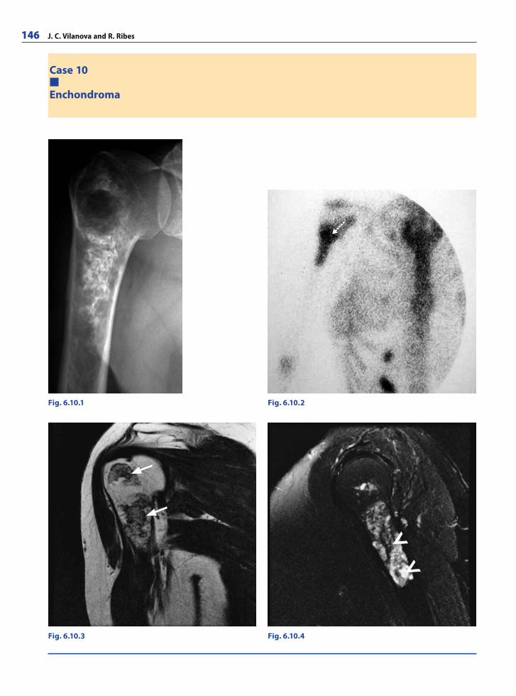
146 J. C. Vilanova and R. Ribes
Case 10
Enchondroma
Fig. 6.10.1 Fig. 6.10.2
Fig. 6.10.4Fig. 6.10.3
RibesLunaRos.indb 146 18.03.2008 12:51:36
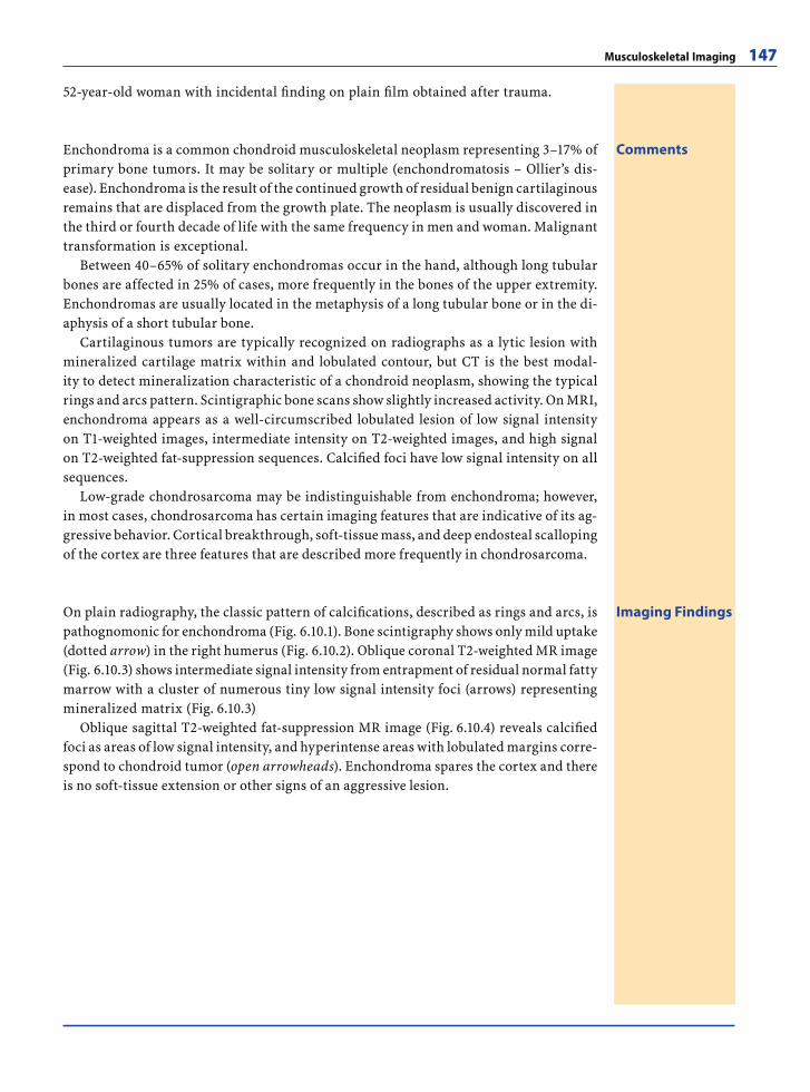
Musculoskeletal Imaging 147
52-year-old woman with incidental fi nding on plain fi lm obtained after trauma.
CommentsEnchondroma is a common chondroid musculoskeletal neoplasm representing 3–17% of primary bone tumors. It may be solitary or multiple (enchondromatosis – Ollier’s dis-ease). Enchondroma is the result of the continued growth of residual benign cartilaginous remains that are displaced from the growth plate. The neoplasm is usually discovered in the third or fourth decade of life with the same frequency in men and woman. Malignant transformation is exceptional.
Between 40–65% of solitary enchondromas occur in the hand, although long tubular bones are affected in 25% of cases, more frequently in the bones of the upper extremity. Enchondromas are usually located in the metaphysis of a long tubular bone or in the di-aphysis of a short tubular bone.
Cartilaginous tumors are typically recognized on radiographs as a lytic lesion with mineralized cartilage matrix within and lobulated contour, but CT is the best modal-ity to detect mineralization characteristic of a chondroid neoplasm, showing the typical rings and arcs pattern. Scintigraphic bone scans show slightly increased activity. On MRI, enchondroma appears as a well-circumscribed lobulated lesion of low signal intensity on T1-weighted images, intermediate intensity on T2-weighted images, and high signal on T2-weighted fat-suppression sequences. Calcifi ed foci have low signal intensity on all sequences.
Low-grade chondrosarcoma may be indistinguishable from enchondroma; however, in most cases, chondrosarcoma has certain imaging features that are indicative of its ag-gressive behavior. Cortical breakthrough, soft-tissue mass, and deep endosteal scalloping of the cortex are three features that are described more frequently in chondrosarcoma.
Imaging FindingsOn plain radiography, the classic pattern of calcifi cations, described as rings and arcs, is pathognomonic for enchondroma (Fig. 6.10.1). Bone scintigraphy shows only mild uptake (dotted arrow) in the right humerus (Fig. 6.10.2). Oblique coronal T2-weighted MR image (Fig. 6.10.3) shows intermediate signal intensity from entrapment of residual normal fatty marrow with a cluster of numerous tiny low signal intensity foci (arrows) representing mineralized matrix (Fig. 6.10.3)
Oblique sagittal T2-weighted fat-suppression MR image (Fig. 6.10.4) reveals calcifi ed foci as areas of low signal intensity, and hyperintense areas with lobulated margins corre-spond to chondroid tumor (open arrowheads). Enchondroma spares the cortex and there is no soft-tissue extension or other signs of an aggressive lesion.
RibesLunaRos.indb 147 18.03.2008 12:51:39
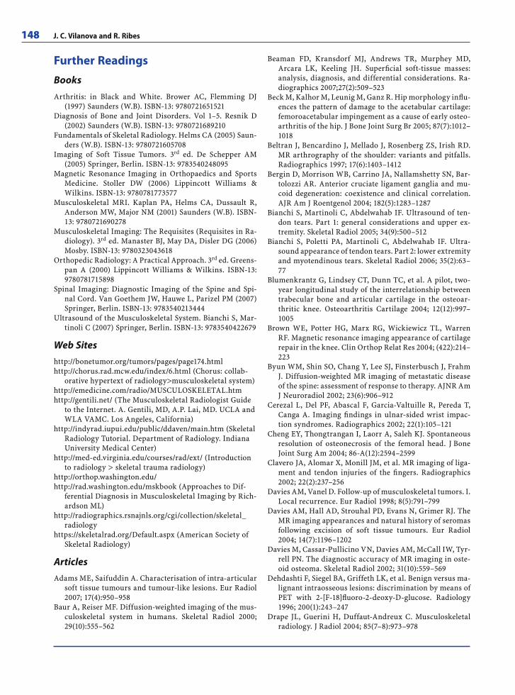
148 J. C. Vilanova and R. Ribes
Further ReadingsBooksArthritis: in Black and White. Brower AC, Flemming DJ
(1997) Saunders (W.B). ISBN-13: 9780721651521Diagnosis of Bone and Joint Disorders. Vol 1–5. Resnik D
(2002) Saunders (W.B). ISBN-13: 9780721689210Fundamentals of Skeletal Radiology. Helms CA (2005) Saun-
ders (W.B). ISBN-13: 9780721605708Imaging of Soft Tissue Tumors. 3rd ed. De Schepper AM
(2005) Springer, Berlin. ISBN-13: 9783540248095Magnetic Resonance Imaging in Orthopaedics and Sports
Medicine. Stoller DW (2006) Lippincott Williams & Wilkins. ISBN-13: 9780781773577
Musculoskeletal MRI. Kaplan PA, Helms CA, Dussault R, Anderson MW, Major NM (2001) Saunders (W.B). ISBN-13: 9780721690278
Musculoskeletal Imaging: The Requisites (Requisites in Ra-diology). 3rd ed. Manaster BJ, May DA, Disler DG (2006) Mosby. ISBN-13: 9780323043618
Orthopedic Radiology: A Practical Approach. 3rd ed. Greens-pan A (2000) Lippincott Williams & Wilkins. ISBN-13: 9780781715898
Spinal Imaging: Diagnostic Imaging of the Spine and Spi-nal Cord. Van Goethem JW, Hauwe L, Parizel PM (2007) Springer, Berlin. ISBN-13: 9783540213444
Ultrasound of the Musculoskeletal System. Bianchi S, Mar-tinoli C (2007) Springer, Berlin. ISBN-13: 9783540422679
Web Siteshttp://bonetumor.org/tumors/pages/page174.htmlhttp://chorus.rad.mcw.edu/index/6.html (Chorus: collab-
orative hypertext of radiology>musculoskeletal system)http://emedicine.com/radio/MUSCULOSKELETAL.htmhttp://gentili.net/ (The Musculoskeletal Radiologist Guide
to the Internet. A. Gentili, MD, A.P. Lai, MD. UCLA and WLA VAMC. Los Angeles, California)
http://indyrad.iupui.edu/public/ddaven/main.htm (Skeletal Radiology Tutorial. Department of Radiology. Indiana University Medical Center)
http://med-ed.virginia.edu/courses/rad/ext/ (Introduction to radiology > skeletal trauma radiology)
http://orthop.washington.edu/http://rad.washington.edu/mskbook (Approaches to Dif-
ferential Diagnosis in Musculoskeletal Imaging by Rich-ardson ML)
http://radiographics.rsnajnls.org/cgi/collection/skeletal_radiology
https://skeletalrad.org/Default.aspx (American Society of Skeletal Radiology)
ArticlesAdams ME, Saifuddin A. Characterisation of intra-articular
soft tissue tumours and tumour-like lesions. Eur Radiol 2007; 17(4):950–958
Baur A, Reiser MF. Diffusion-weighted imaging of the mus-culoskeletal system in humans. Skeletal Radiol 2000; 29(10):555–562
Beaman FD, Kransdorf MJ, Andrews TR, Murphey MD, Arcara LK, Keeling JH. Superfi cial soft-tissue masses: analysis, diagnosis, and differential considerations. Ra-diographics 2007;27(2):509–523
Beck M, Kalhor M, Leunig M, Ganz R. Hip morphology infl u-ences the pattern of damage to the acetabular cartilage: femoroacetabular impingement as a cause of early osteo-arthritis of the hip. J Bone Joint Surg Br 2005; 87(7):1012–1018
Beltran J, Bencardino J, Mellado J, Rosenberg ZS, Irish RD. MR arthrography of the shoulder: variants and pitfalls. Radiographics 1997; 17(6):1403–1412
Bergin D, Morrison WB, Carrino JA, Nallamshetty SN, Bar-tolozzi AR. Anterior cruciate ligament ganglia and mu-coid degeneration: coexistence and clinical correlation. AJR Am J Roentgenol 2004; 182(5):1283–1287
Bianchi S, Martinoli C, Abdelwahab IF. Ultrasound of ten-don tears. Part 1: general considerations and upper ex-tremity. Skeletal Radiol 2005; 34(9):500–512
Bianchi S, Poletti PA, Martinoli C, Abdelwahab IF. Ultra-sound appearance of tendon tears. Part 2: lower extremity and myotendinous tears. Skeletal Radiol 2006; 35(2):63–77
Blumenkrantz G, Lindsey CT, Dunn TC, et al. A pilot, two-year longitudinal study of the interrelationship between trabecular bone and articular cartilage in the osteoar-thritic knee. Osteoarthritis Cartilage 2004; 12(12):997–1005
Brown WE, Potter HG, Marx RG, Wickiewicz TL, Warren RF. Magnetic resonance imaging appearance of cartilage repair in the knee. Clin Orthop Relat Res 2004; (422):214–223
Byun WM, Shin SO, Chang Y, Lee SJ, Finsterbusch J, Frahm J. Diffusion-weighted MR imaging of metastatic disease of the spine: assessment of response to therapy. AJNR Am J Neuroradiol 2002; 23(6):906–912
Cerezal L, Del PF, Abascal F, Garcia-Valtuille R, Pereda T, Canga A. Imaging fi ndings in ulnar-sided wrist impac-tion syndromes. Radiographics 2002; 22(1):105–121
Cheng EY, Thongtrangan I, Laorr A, Saleh KJ. Spontaneous resolution of osteonecrosis of the femoral head. J Bone Joint Surg Am 2004; 86-A(12):2594–2599
Clavero JA, Alomar X, Monill JM, et al. MR imaging of liga-ment and tendon injuries of the fi ngers. Radiographics 2002; 22(2):237–256
Davies AM, Vanel D. Follow-up of musculoskeletal tumors. I. Local recurrence. Eur Radiol 1998; 8(5):791–799
Davies AM, Hall AD, Strouhal PD, Evans N, Grimer RJ. The MR imaging appearances and natural history of seromas following excision of soft tissue tumours. Eur Radiol 2004; 14(7):1196–1202
Davies M, Cassar-Pullicino VN, Davies AM, McCall IW, Tyr-rell PN. The diagnostic accuracy of MR imaging in oste-oid osteoma. Skeletal Radiol 2002; 31(10):559–569
Dehdashti F, Siegel BA, Griffeth LK, et al. Benign versus ma-lignant intraosseous lesions: discrimination by means of PET with 2-[F-18]fl uoro-2-deoxy-D-glucose. Radiology 1996; 200(1):243–247
Drape JL, Guerini H, Duffaut-Andreux C. Musculoskeletal radiology. J Radiol 2004; 85(7–8):973–978
RibesLunaRos.indb 148 18.03.2008 12:51:40
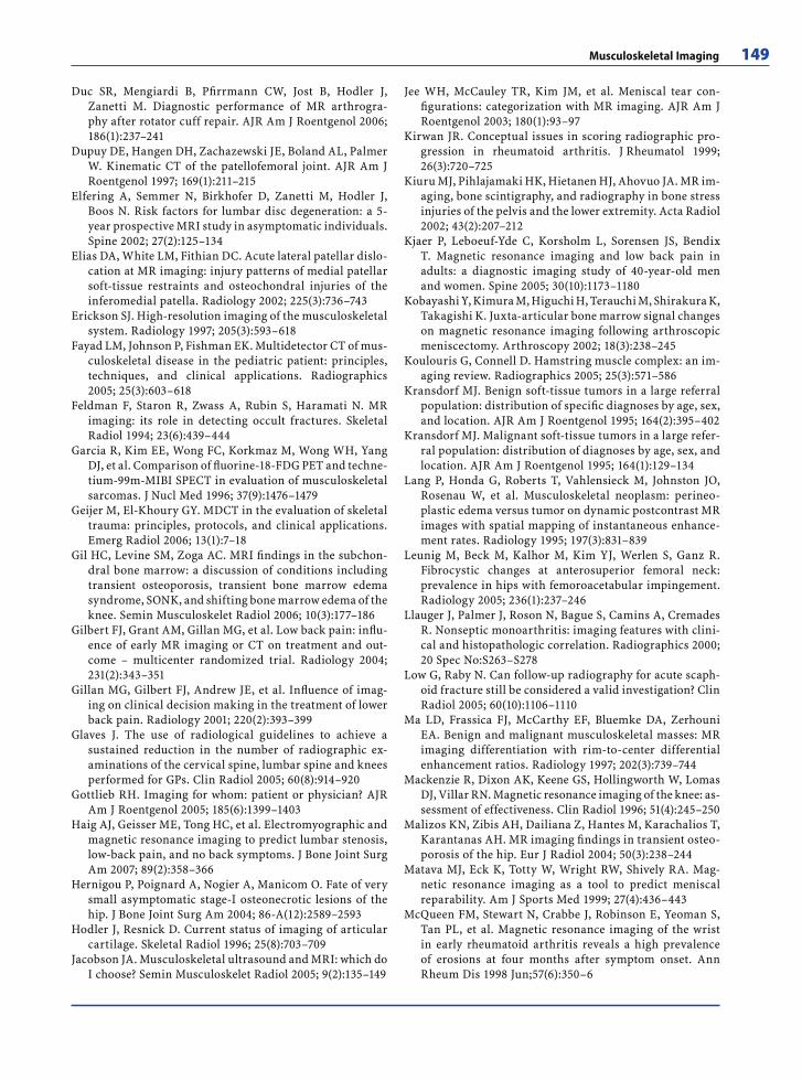
Musculoskeletal Imaging 149
Duc SR, Mengiardi B, Pfi rrmann CW, Jost B, Hodler J, Zanetti M. Diagnostic performance of MR arthrogra-phy after rotator cuff repair. AJR Am J Roentgenol 2006; 186(1):237–241
Dupuy DE, Hangen DH, Zachazewski JE, Boland AL, Palmer W. Kinematic CT of the patellofemoral joint. AJR Am J Roentgenol 1997; 169(1):211–215
Elfering A, Semmer N, Birkhofer D, Zanetti M, Hodler J, Boos N. Risk factors for lumbar disc degeneration: a 5-year prospective MRI study in asymptomatic individuals. Spine 2002; 27(2):125–134
Elias DA, White LM, Fithian DC. Acute lateral patellar dislo-cation at MR imaging: injury patterns of medial patellar soft-tissue restraints and osteochondral injuries of the inferomedial patella. Radiology 2002; 225(3):736–743
Erickson SJ. High-resolution imaging of the musculoskeletal system. Radiology 1997; 205(3):593–618
Fayad LM, Johnson P, Fishman EK. Multidetector CT of mus-culoskeletal disease in the pediatric patient: principles, techniques, and clinical applications. Radiographics 2005; 25(3):603–618
Feldman F, Staron R, Zwass A, Rubin S, Haramati N. MR imaging: its role in detecting occult fractures. Skeletal Radiol 1994; 23(6):439–444
Garcia R, Kim EE, Wong FC, Korkmaz M, Wong WH, Yang DJ, et al. Comparison of fl uorine-18-FDG PET and techne-tium-99m-MIBI SPECT in evaluation of musculoskeletal sarcomas. J Nucl Med 1996; 37(9):1476–1479
Geijer M, El-Khoury GY. MDCT in the evaluation of skeletal trauma: principles, protocols, and clinical applications. Emerg Radiol 2006; 13(1):7–18
Gil HC, Levine SM, Zoga AC. MRI fi ndings in the subchon-dral bone marrow: a discussion of conditions including transient osteoporosis, transient bone marrow edema syndrome, SONK, and shifting bone marrow edema of the knee. Semin Musculoskelet Radiol 2006; 10(3):177–186
Gilbert FJ, Grant AM, Gillan MG, et al. Low back pain: infl u-ence of early MR imaging or CT on treatment and out-come – multicenter randomized trial. Radiology 2004; 231(2):343–351
Gillan MG, Gilbert FJ, Andrew JE, et al. Infl uence of imag-ing on clinical decision making in the treatment of lower back pain. Radiology 2001; 220(2):393–399
Glaves J. The use of radiological guidelines to achieve a sustained reduction in the number of radiographic ex-aminations of the cervical spine, lumbar spine and knees performed for GPs. Clin Radiol 2005; 60(8):914–920
Gottlieb RH. Imaging for whom: patient or physician? AJR Am J Roentgenol 2005; 185(6):1399–1403
Haig AJ, Geisser ME, Tong HC, et al. Electromyographic and magnetic resonance imaging to predict lumbar stenosis, low-back pain, and no back symptoms. J Bone Joint Surg Am 2007; 89(2):358–366
Hernigou P, Poignard A, Nogier A, Manicom O. Fate of very small asymptomatic stage-I osteonecrotic lesions of the hip. J Bone Joint Surg Am 2004; 86-A(12):2589–2593
Hodler J, Resnick D. Current status of imaging of articular cartilage. Skeletal Radiol 1996; 25(8):703–709
Jacobson JA. Musculoskeletal ultrasound and MRI: which do I choose? Semin Musculoskelet Radiol 2005; 9(2):135–149
Jee WH, McCauley TR, Kim JM, et al. Meniscal tear con-fi gurations: categorization with MR imaging. AJR Am J Roentgenol 2003; 180(1):93–97
Kirwan JR. Conceptual issues in scoring radiographic pro-gression in rheumatoid arthritis. J Rheumatol 1999; 26(3):720–725
Kiuru MJ, Pihlajamaki HK, Hietanen HJ, Ahovuo JA. MR im-aging, bone scintigraphy, and radiography in bone stress injuries of the pelvis and the lower extremity. Acta Radiol 2002; 43(2):207–212
Kjaer P, Leboeuf-Yde C, Korsholm L, Sorensen JS, Bendix T. Magnetic resonance imaging and low back pain in adults: a diagnostic imaging study of 40-year-old men and women. Spine 2005; 30(10):1173–1180
Kobayashi Y, Kimura M, Higuchi H, Terauchi M, Shirakura K, Takagishi K. Juxta-articular bone marrow signal changes on magnetic resonance imaging following arthroscopic meniscectomy. Arthroscopy 2002; 18(3):238–245
Koulouris G, Connell D. Hamstring muscle complex: an im-aging review. Radiographics 2005; 25(3):571–586
Kransdorf MJ. Benign soft-tissue tumors in a large referral population: distribution of specifi c diagnoses by age, sex, and location. AJR Am J Roentgenol 1995; 164(2):395–402
Kransdorf MJ. Malignant soft-tissue tumors in a large refer-ral population: distribution of diagnoses by age, sex, and location. AJR Am J Roentgenol 1995; 164(1):129–134
Lang P, Honda G, Roberts T, Vahlensieck M, Johnston JO, Rosenau W, et al. Musculoskeletal neoplasm: perineo-plastic edema versus tumor on dynamic postcontrast MR images with spatial mapping of instantaneous enhance-ment rates. Radiology 1995; 197(3):831–839
Leunig M, Beck M, Kalhor M, Kim YJ, Werlen S, Ganz R. Fibrocystic changes at anterosuperior femoral neck: prevalence in hips with femoroacetabular impingement. Radiology 2005; 236(1):237–246
Llauger J, Palmer J, Roson N, Bague S, Camins A, Cremades R. Nonseptic monoarthritis: imaging features with clini-cal and histopathologic correlation. Radiographics 2000; 20 Spec No:S263–S278
Low G, Raby N. Can follow-up radiography for acute scaph-oid fracture still be considered a valid investigation? Clin Radiol 2005; 60(10):1106–1110
Ma LD, Frassica FJ, McCarthy EF, Bluemke DA, Zerhouni EA. Benign and malignant musculoskeletal masses: MR imaging differentiation with rim-to-center differential enhancement ratios. Radiology 1997; 202(3):739–744
Mackenzie R, Dixon AK, Keene GS, Hollingworth W, Lomas DJ, Villar RN. Magnetic resonance imaging of the knee: as-sessment of effectiveness. Clin Radiol 1996; 51(4):245–250
Malizos KN, Zibis AH, Dailiana Z, Hantes M, Karachalios T, Karantanas AH. MR imaging fi ndings in transient osteo-porosis of the hip. Eur J Radiol 2004; 50(3):238–244
Matava MJ, Eck K, Totty W, Wright RW, Shively RA. Mag-netic resonance imaging as a tool to predict meniscal reparability. Am J Sports Med 1999; 27(4):436–443
McQueen FM, Stewart N, Crabbe J, Robinson E, Yeoman S, Tan PL, et al. Magnetic resonance imaging of the wrist in early rheumatoid arthritis reveals a high prevalence of erosions at four months after symptom onset. Ann Rheum Dis 1998 Jun;57(6):350–6
RibesLunaRos.indb 149 18.03.2008 12:51:40
150 J. C. Vilanova and R. Ribes
Milette PC, Fontaine S, Lepanto L, Dery R, Breton G. Clini-cal impact of contrast-enhanced MR imaging reports in patients with previous lumbar disk surgery. AJR Am J Roentgenol 1996; 167(1):217–223
Milette PC. Reporting lumbar disk abnormalities: at last, consensus! AJNR Am J Neuroradiol 2001; 22(3):428–429
Mitra D, Cassar-Pullicino VN, McCall IW. Longitudinal study of vertebral type-1 end-plate changes on MR of the lumbar spine. Eur Radiol 2004; 14(9):1574–1581
Modic MT. Degenerative disc disease and back pain. Magn Reson Imaging Clin N Am 1999; 7(3):481–491, viii
Modic MT, Obuchowski NA, Ross JS, et al. Acute low back pain and radiculopathy: MR imaging fi ndings and their prognostic role and effect on outcome. Radiology 2005; 237(2):597–604
Mohana-Borges AV, Chung CB, Resnick D. Superior labral anteroposterior tear: classifi cation and diagnosis on MRI and MR arthrography. AJR Am J Roentgenol 2003; 181(6):1449–1462
Moore SL, Teirstein A, Golimbu C. MRI of sarcoidosis pa-tients with musculoskeletal symptoms. AJR Am J Roent-genol 2005; 185(1):154–159
Morag Y, Jacobson JA, Shields G, et al. MR arthrography of rotator interval, long head of the biceps brachii, and biceps pulley of the shoulder. Radiology 2005; 235(1):21–30
Muller R, Hildebrand T, Ruegsegger P. Non-invasive bone biopsy: a new method to analyse and display the three-dimensional structure of trabecular bone. Phys Med Biol 1994; 39(1):145–164
Munter FM, Wasserman BA, Wu HM, Yousem DM. Serial MR Imaging of Annular Tears in Lumbar Intervertebral Disks. AJNR Am J Neuroradiol 2002; 23(7):1105–1109
Murphey MD, Robbin MR, McRae GA, Flemming DJ, Temple HT, Kransdorf MJ. The many faces of osteosarcoma. Ra-diographics 1997; 17(5):1205–1231
Murphey MD, Smith WS, Smith SE, Kransdorf MJ, Temple HT. From the archives of the AFIP. Imaging of muscu-loskeletal neurogenic tumors: radiologic-pathologic cor-relation. Radiographics 1999; 19(5):1253–1280
Murphey MD, Carroll JF, Flemming DJ, Pope TL, Gannon FH, Kransdorf MJ. From the archives of the AFIP: benign musculoskeletal lipomatous lesions. Radiographics 2004; 24(5):1433–1466
Narvaez JA, Narvaez J, Ortega R, Aguilera C, Sanchez A, An-dia E. Painful heel: MR imaging fi ndings. Radiographics 2000; 20(2):333–352
Neyman EG, Corl FS, Fishman EK. 3D-CT evaluation of me-tallic implants: principles, techniques, and applications. Crit Rev Comput Tomogr 2002; 43(6):419–452
Nikken JJ, Oei EH, Ginai AZ, et al. Acute peripheral joint injury: cost and effectiveness of low-fi eld-strength MR imaging – results of randomized controlled trial. Radi-ology 2005; 236(3):958–967
Pfi rrmann CW, Theumann NH, Chung CB, Botte MJ, Trudell DJ, Resnick D. What happens to the triangular fi brocar-tilage complex during pronation and supination of the forearm? Analysis of its morphology and diagnostic as-sessment with MR arthrography. Skeletal Radiol 2001; 30(12):677–685
Pfi rrmann CW, Mengiardi B, Dora C, Kalberer F, Zanetti M, Hodler J. Cam and pincer femoroacetabular impinge-ment: characteristic MR arthrographic fi ndings in 50 pa-tients. Radiology 2006; 240(3):778–785
Potter HG, Weinstein M, Allen AA, Wickiewicz TL, Helfet DL. Magnetic resonance imaging of the multiple-ligament injured knee. J Orthop Trauma 2002; 16(5):330–339
Ramnath RR, Kattapuram SV. MR appearance of SONK-like subchondral abnormalities in the adult knee: SONK rede-fi ned. Skeletal Radiol 2004; 33(10):575–581
Recht MP, Goodwin DW, Winalski CS, White LM. MRI of articular cartilage: revisiting current status and future directions. AJR Am J Roentgenol 2005; 185(4):899–914
Resnick D, Niwayama G, Guerra J Jr, Vint V, Usselman J. Spinal vacuum phenomena: anatomical study and review. Radiology 1981; 139(2):341–348
Robertson DD, Walker PS, Granholm JW, et al. Design of custom hip stem prostheses using three-dimensional CT modeling. J Comput Assist Tomogr 1987; 11(5):804–809
Roposch A, Wright JG. Increased diagnostic information and understanding disease: uncertainty in the diag-nosis of developmental hip dysplasia. Radiology 2007; 242(2):355–359
Rosenberg AE. Malignant fi brous histiocytoma: past, pres-ent, and future. Skeletal Radiol 2003; 32(11):613–618
Rosenthal DI, Hornicek FJ, Wolfe MW, Jennings LC, Geb-hardt MC, Mankin HJ. Percutaneous radiofrequency co-agulation of osteoid osteoma compared with operative treatment. J Bone Joint Surg Am 1998; 80(6):815–821
Rupp S, Seil R, Jochum P, Kohn D. Popliteal cysts in adults. Prevalence, associated intraarticular lesions, and results after arthroscopic treatment. Am J Sports Med 2002; 30(1):112–115
Sabharwal S, Zhao C, McKeon J, Melaghari T, Blacksin M, Wenekor C. Reliability analysis for radiographic mea-surement of limb length discrepancy: full-length stand-ing anteroposterior radiograph versus scanogram. J Pe-diatr Orthop 2007; 27(1):46–50
Sanders TG, Medynski MA, Feller JF, Lawhorn KW. Bone contusion patterns of the knee at MR imaging: footprint of the mechanism of injury. Radiographics 2000; 20 Spec No:S135–S151
Santiago RC, Gimenez CR, McCarthy K. Imaging of osteomy-elitis and musculoskeletal soft tissue infections: current concepts. Rheum Dis Clin North Am 2003; 29(1):89–109
Schweitzer ME, Morrison WB. MR imaging of the diabetic foot. Radiol Clin North Am 2004; 42(1):61–71, vi
Shapeero LG, Vanel D, Verstraete KL, Bloem JL. Fast magnetic resonance imaging with contrast for soft tissue sarcoma viability. Clin Orthop Relat Res 2002; (397):212–227
Shepard MF, Hunter DM, Davies MR, Shapiro MS, Seeger LL. The clinical signifi cance of anterior horn meniscal tears diagnosed on magnetic resonance images. Am J Sports Med 2002; 30(2):189–192
Sims WF, Jacobson KE. The posteromedial corner of the knee: medial-sided injury patterns revisited. Am J Sports Med 2004; 32(2):337–345
Teefey SA, Rubin DA, Middleton WD, Hildebolt CF, Leibold RA, Yamaguchi K. Detection and quantifi cation of rota-tor cuff tears. Comparison of ultrasonographic, magnetic
RibesLunaRos.indb 150 18.03.2008 12:51:40
Musculoskeletal Imaging 151
resonance imaging, and arthroscopic fi ndings in sev-enty-one consecutive cases. J Bone Joint Surg Am 2004; 86-A(4):708–716
Thain LM, Adler RS. Sonography of the rotator cuff and bi-ceps tendon: technique, normal anatomy, and pathology. J Clin Ultrasound 1999; 27(8):446–458
Theumann NH, Pfi rrmann CW, Chung CB, Antonio GE, Trudell DJ, Resnick D. Ligamentous and tendinous anat-omy of the intermetacarpal and common carpometacar-pal joints: evaluation with MR imaging and MR arthrog-raphy. J Comput Assist Tomogr 2002; 26(1):145–152
Theumann NH, Etechami G, Duvoisin B, et al. Association between extrinsic and intrinsic carpal ligament injuries at MR arthrography and carpal instability at radiography: initial observations. Radiology 2006; 238(3):950–957
Vande Berg BC, Malghem J, Lecouvet FE, Maldague B. Mag-netic resonance imaging of normal bone marrow. Eur Radiol 1998; 8(8):1327–1334
Vanel D, Shapeero LG, Tardivon A, Western A, Guinebretiere JM. Dynamic contrast-enhanced MRI with subtraction of aggressive soft tissue tumors after resection. Skeletal Ra-diol 1998; 27(9):505–510
Verstraete KL, Van der Woude HJ, Hogendoorn PC, De-Deene Y, Kunnen M, Bloem JL. Dynamic contrast-en-hanced MR imaging of musculoskeletal tumors: basic principles and clinical applications. J Magn Reson Imag-ing 1996; 6(2):311–321
Vilanova JC, Woertler K, Narvaez JA, et al. Soft-tissue tu-mors update: MR imaging features according to the WHO classifi cation. Eur Radiol 2007; 17(1):125–138
Vincken PW, Ter Braak BP, van Erkell AR, et al. Effectiveness of MR imaging in selection of patients for arthroscopy of the knee. Radiology 2002; 223(3):739–746
Waldschmidt JG, Rilling RJ, Kajdacsy-Balla AA, Boynton MD, Erickson SJ. In vitro and in vivo MR imaging of hyaline cartilage: zonal anatomy, imaging pitfalls, and pathologic conditions. Radiographics 1997; 17(6):1387–1402
Younger AS, Sawatzky B, Dryden P. Radiographic assessment of adult fl atfoot. Foot Ankle Int 2005; 26(10):820–825
Zanetti M, Bruder E, Romero J, Hodler J. Bone marrow edema pattern in osteoarthritic knees: correlation be-tween MR imaging and histologic fi ndings. Radiology 2000; 215(3):835–840
RibesLunaRos.indb 151 18.03.2008 12:51:40

RibesLunaRos.indb V 18.03.2008 12:48:44
Nuclear Medicine
Neuroimaging and Head and Neck Imaging 7
F. Bravo-Rodriguez and Rocio Diaz-Aguilera
IntroductionNeuroradiology is the branch of radiology dealing with both diagnostic and interven-tional aspects of the central nervous system (CNS) and head and neck.
The introduction of computed tomography (CT) in 1972 and magnetic resonance im-aging (MRI) in 1981 have contributed greatly to this highly specialized radiological sub-specialty.
Moreover, radiologists working in the interventional branch of neuroradiology must acquire specialized manual and surgical skills in addition to extensive knowledge of anatomy, physiology, pathology, and diagnostic techniques required of all neuroradio-logists.
Thus, neuroradiology comprises two closely related disciplines which are, at the same time, clearly different from each other. On the one hand, diagnostic neuroradiology based on noninvasive imaging modalities and, on the other hand, interventional neuroradiol-ogy, based on digital subtraction angiography (DSA), which permits the treatment of many neurological conditions by means of endovascular approaches.
Neuroradiology is widely connected to many specialities such as neurology, neuro-surgery, otolaryngology, oral and maxillofacial surgery, among others, and neuroradi-ologists work in close contact with specialists in these fi elds, enabling faster and more accurate diagnoses through imaging modalities such as conventional X-ray, ultrasound, CT, and/or MRI. Moreover, interventional neuroradiologists contribute to the manage-ment of numerous neurological conditions through minimally invasive procedures such as extracranial or intracranial stenting in cases of arterial stenoses, endovascular treat-ment of intracranial aneurysms, embolization of head and neck tumors, etc.
The current scope of this radiological discipline is extensive and its future is promis-ing. The advent of functional MR imaging, diffusion-weighted imaging, and perfusion-weighted imaging, have replaced the mere anatomical view of classic neuroradiology with a wider perspective that includes cerebral pathophysiology. Furthermore, recent research into endovascular stem cell implantation opens a new and fascinating fi eld with regard to the treatment of strokes and degenerative diseases.
RibesLunaRos.indb 153 18.03.2008 12:51:40
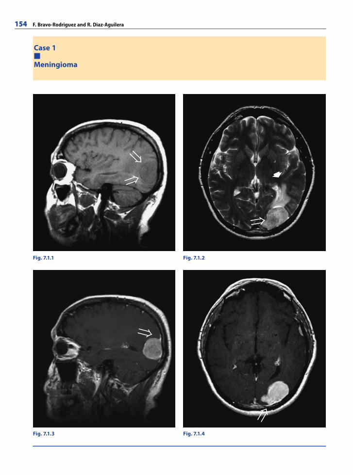
154 F. Bravo-Rodriguez and R. Diaz-Aguilera
Case 1
Meningioma
Fig. 7.1.4Fig. 7.1.3
Fig. 7.1.1 Fig. 7.1.2
�
RibesLunaRos.indb 154 18.03.2008 12:51:40
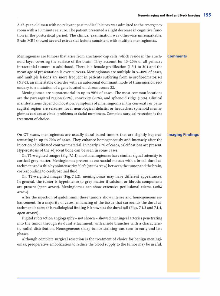
Neuroimaging and Head and Neck Imaging 155
A 43-year-old man with no relevant past medical history was admitted to the emergency room with a 10 minute seizure. The patient presented a slight decrease in cognitive func-tion in the postcritical period. The clinical examination was otherwise unremarkable. Brain MRI showed several extraaxial lesions consistent with multiple meningiomas.
CommentsMeningiomas are tumors that arise from arachnoid cap cells, which reside in the arach-noid layer covering the surface of the brain. They account for 13–20% of all primary intracranial tumors in adulthood. There is a female predilection (1.5:1 to 3:1) and the mean age of presentation is over 50 years. Meningiomas are multiple in 5–40% of cases, and multiple lesions are more frequent in patients suffering from neurofi bromatosis-2 (NF-2), an inheritable disorder with an autosomal dominant mode of transmission sec-ondary to a mutation of a gene located on chromosome 22.
Meningiomas are supratentorial in up to 90% of cases. The most common locations are the parasagittal region (25%), convexity (20%), and sphenoid ridge (15%). Clinical manifestations depend on location. Symptoms of a meningioma in the convexity or para-sagittal region are seizures, focal neurological defi cits, or headaches; sphenoid menin-giomas can cause visual problems or facial numbness. Complete surgical resection is the treatment of choice.
Imaging FindingsOn CT scans, meningiomas are usually dural-based tumors that are slightly hyperat-tenuating in up to 70% of cases. They enhance homogeneously and intensely after the injection of iodinated contrast material. In nearly 25% of cases, calcifi cations are present. Hyperostosis of the adjacent bone can be seen in some cases.
On T1-weighted images (Fig. 7.1.1), most meningiomas have similar signal intensity to cortical gray matter. Meningiomas present as extraaxial masses with a broad dural at-tachment and a thin hypointense rim/cleft (open arrow) between the tumor and the brain, corresponding to cerebrospinal fl uid.
On T2-weighted images (Fig. 7.1.2), meningiomas may have different appearances. In general, the tumor is hypointense to gray matter if calcium or fi brotic components are present (open arrow). Meningiomas can show extensive perilesional edema (solid arrow).
After the injection of gadolinium, these tumors show intense and homogeneous en-hancement. In a majority of cases, enhancing of the tissue that surrounds the dural at-tachment is seen; this radiological fi nding is known as the dural tail (Figs. 7.1.3 and 7.1.4, open arrows).
Digital subtraction angiography – not shown – showed meningeal arteries penetrating into the tumor through its dural attachment, with inside branches with a characteris-tic radial distribution. Homogeneous sharp tumor staining was seen in early and late phases.
Although complete surgical resection is the treatment of choice for benign meningi-omas, preoperative embolization to reduce the blood supply to the tumor may be useful.
RibesLunaRos.indb 155 18.03.2008 12:51:43
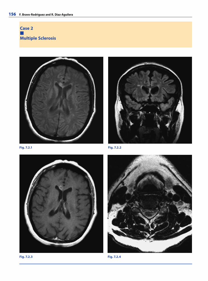
156 F. Bravo-Rodriguez and R. Diaz-Aguilera
Case 2
Multiple Sclerosis
Fig. 7.2.1 Fig. 7.2.2
Fig. 7.2.3 Fig. 7.2.4
RibesLunaRos.indb 156 18.03.2008 12:51:43
Neuroimaging and Head and Neck Imaging 157
A 40-year-old woman with sudden left-limb monoparesis and hyporefl exia. Cerebral MRI showed lesions consistent with multiple sclerosis.
CommentsMultiple sclerosis (MS) is an infl ammatory, demyelinating disease of the central nervous system (CNS). It is considered an autoimmune disease. MS most commonly affl icts young people between the ages of 20 and 30 years, but any age group can be affected. It has a prevalence of nearly 100 cases per 100,000 inhabitants. MS affects females more than males (2:1).
Optic neuritis (ON) or other isolated neurological symptoms are the usual initial pres-entation. The disease can present in different forms, such as primary progressive, re-lapsing remitting, or relapsing progressive. MS diagnosis is mainly based on the clinical symptoms. Visual evoked potential testing and laboratory studies are useful as well; oli-goclonal bands of IgG are demonstrated in cerebrospinal fl uid samples in approximately 85% of patients with MS. With the advent of MRI, the ability to confi rm the diagnosis of MS has improved dramatically. On MRI, MS lesions are detected in up to 95% of patients. Newer MRI techniques promise to yield important information regarding MS heteroge-neity, prognosis, and treatment effects.
Currently, the Barkhof MRI criteria are applied to establish the diagnosis in patients with clinically isolated syndromes suggestive of MS.
Imaging FindingsMRI characteristically shows lesions of high T2, PD and FLAIR signal intensity of vari-able location in the white matter of the brain, brain stem, optic nerves, or spinal cord. In typical cases, the lesions tend to occur in periventricular areas (Fig. 7.2.1) and may affect in the corpus callosum. They have an ovoid shape with their largest axis oriented perpendicular to the ventricular surface (Fig. 7.2.2); they typically involve only the white matter and usually present peripheral enhancement after gadolinium administration (Fig. 7.2.3).
Spinal cord lesions are detected in 75% of cases. They usually appear in the cervical spine and present a vertical orientation (Fig. 7.2.4).
Spatial progress criteria
9 T2-weighted images lesions or 1 enhancing lesion1 or more infratentorial lesions1 or more juxtacortical lesions3 or more periventricular lesions
Time progress criteria
1 new enhancing lesion 3 months after an acute attackNew lesions on T2-weighted images or new enhancing lesions 6 months after a fi rst episode of symp-toms suggestive of MS
No highly effective treatment is currently available to counteract acute MS attacks after their onset. The most widely used therapy is high-dose intravenous steroids. This medi-cation may help expedite the timing of recovery but will not affect the actual degree of recovery. To prevent relapses or disease progression, immunomodulatory drugs (e.g., interferon) are used with different results.
RibesLunaRos.indb 157 18.03.2008 12:51:46
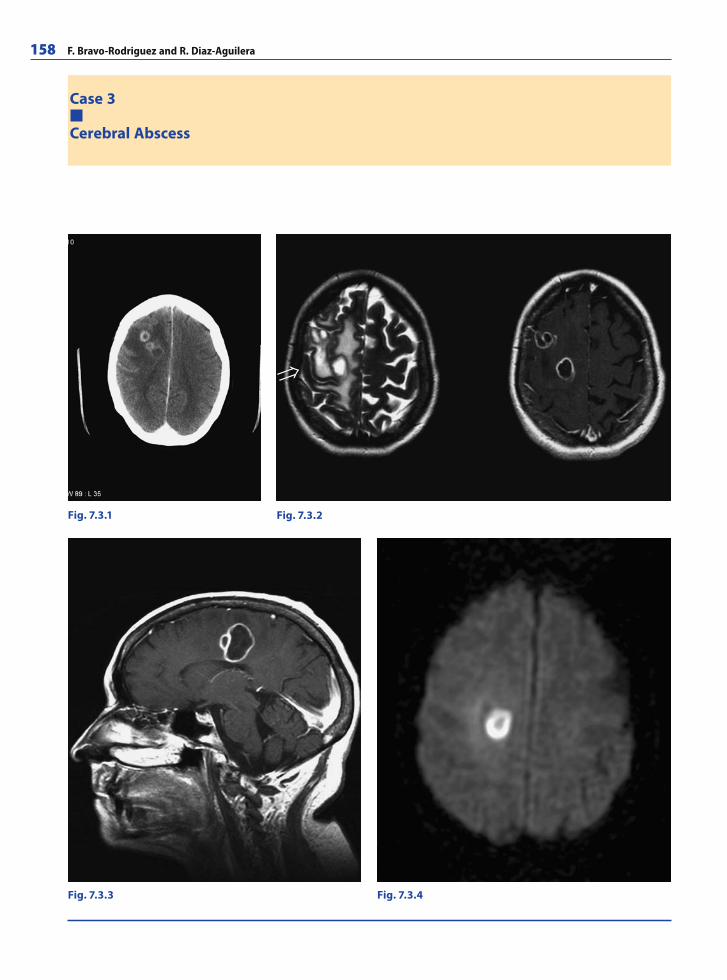
158 F. Bravo-Rodriguez and R. Diaz-Aguilera
Fig. 7.3.1 Fig. 7.3.2
Fig. 7.3.3 Fig. 7.3.4
Case 3
Cerebral Abscess
RibesLunaRos.indb 158 18.03.2008 12:51:46
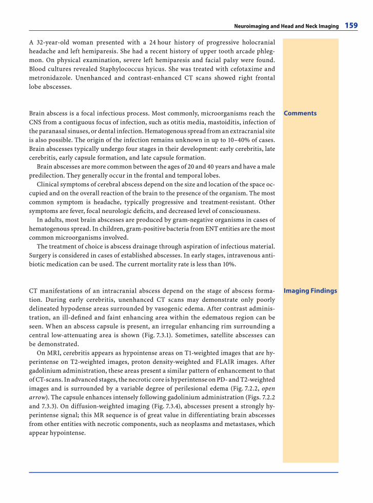
Neuroimaging and Head and Neck Imaging 159
CommentsBrain abscess is a focal infectious process. Most commonly, microorganisms reach the CNS from a contiguous focus of infection, such as otitis media, mastoiditis, infection of the paranasal sinuses, or dental infection. Hematogenous spread from an extracranial site is also possible. The origin of the infection remains unknown in up to 10–40% of cases. Brain abscesses typically undergo four stages in their development: early cerebritis, late cerebritis, early capsule formation, and late capsule formation.
Brain abscesses are more common between the ages of 20 and 40 years and have a male predilection. They generally occur in the frontal and temporal lobes.
Clinical symptoms of cerebral abscess depend on the size and location of the space oc-cupied and on the overall reaction of the brain to the presence of the organism. The most common symptom is headache, typically progressive and treatment-resistant. Other symptoms are fever, focal neurologic defi cits, and decreased level of consciousness.
In adults, most brain abscesses are produced by gram-negative organisms in cases of hematogenous spread. In children, gram-positive bacteria from ENT entities are the most common microorganisms involved.
The treatment of choice is abscess drainage through aspiration of infectious material. Surgery is considered in cases of established abscesses. In early stages, intravenous anti-biotic medication can be used. The current mortality rate is less than 10%.
Imaging FindingsCT manifestations of an intracranial abscess depend on the stage of abscess forma-tion. During early cerebritis, unenhanced CT scans may demonstrate only poorly delineated hypodense areas surrounded by vasogenic edema. After contrast adminis-tration, an ill-defi ned and faint enhancing area within the edematous region can be seen. When an abscess capsule is present, an irregular enhancing rim surrounding a central low-attenuating area is shown (Fig. 7.3.1). Sometimes, satellite abscesses can be demonstrated.
On MRI, cerebritis appears as hypointense areas on T1-weighted images that are hy-perintense on T2-weighted images, proton density-weighted and FLAIR images. After gadolinium administration, these areas present a similar pattern of enhancement to that of CT-scans. In advanced stages, the necrotic core is hyperintense on PD- and T2-weighted images and is surrounded by a variable degree of perilesional edema (Fig. 7.2.2, open arrow). The capsule enhances intensely following gadolinium administration (Figs. 7.2.2 and 7.3.3). On diffusion-weighted imaging (Fig. 7.3.4), abscesses present a strongly hy-perintense signal; this MR sequence is of great value in differentiating brain abscesses from other entities with necrotic components, such as neoplasms and metastases, which appear hypointense.
A 32-year-old woman presented with a 24 hour history of progressive holocranial headache and left hemiparesis. She had a recent history of upper tooth arcade phleg-mon. On physical examination, severe left hemiparesis and facial palsy were found. Blood cultures revealed Staphylococcus hyicus. She was treated with cefotaxime and metronidazole. Unenhanced and contrast-enhanced CT scans showed right frontal lobe abscesses.
RibesLunaRos.indb 159 18.03.2008 12:51:50
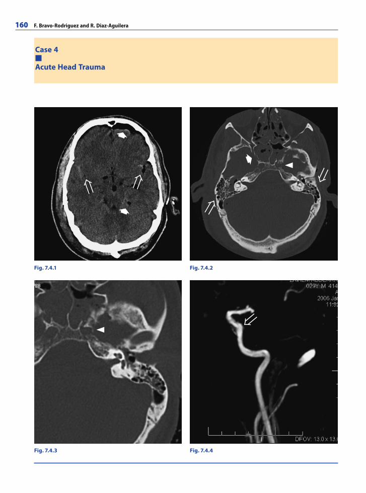
160 F. Bravo-Rodriguez and R. Diaz-Aguilera
Case 4
Acute Head Trauma
Fig. 7.4.1 Fig. 7.4.2
Fig. 7.4.3 Fig. 7.4.4
�
��
RibesLunaRos.indb 160 18.03.2008 12:51:50
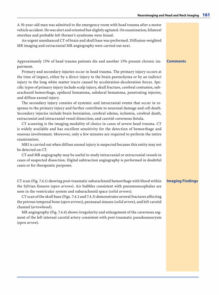
Neuroimaging and Head and Neck Imaging 161
A 30-year-old man was admitted to the emergency room with head trauma after a motor vehicle accident. He was alert and oriented but slightly agitated. On examination, bilateral otorrhea and probable left Horner’s syndrome were found.
An urgent unenhanced CT of brain and skull base was performed. Diffusion-weighted MR imaging and extracranial MR angiography were carried out next.
CommentsApproximately 15% of head trauma patients die and another 15% present chronic im-pairment.
Primary and secondary injuries occur in head trauma. The primary injury occurs at the time of impact, either by a direct injury to the brain parenchyma or by an indirect injury to the long white matter tracts caused by acceleration-deceleration forces. Spe-cifi c types of primary injury include scalp injury, skull fracture, cerebral contusion, sub-arachnoid hemorrhage, epidural hematoma, subdural hematoma, penetrating injuries, and diffuse axonal injury.
The secondary injury consists of systemic and intracranial events that occur in re-sponse to the primary injury and further contribute to neuronal damage and cell death. Secondary injuries include brain herniation, cerebral edema, ischemia, cerebral death, extracranial and intracranial vessel dissection, and carotid-cavernous fi stula.
CT scanning is the imaging modality of choice in cases of severe head trauma. CT is widely available and has excellent sensitivity for the detection of hemorrhage and osseous involvement. Moreover, only a few minutes are required to perform the entire examination.
MRI is carried out when diffuse axonal injury is suspected because this entity may not be detected on CT.
CT and MR angiography may be useful to study intracranial or extracranial vessels in cases of suspected dissection. Digital subtraction angiography is performed in doubtful cases or for therapeutic purposes.
Imaging FindingsCT scan (Fig. 7.4.1) showing post-traumatic subarachnoid hemorrhage with blood within the Sylvian fi ssures (open arrows). Air bubbles consistent with pneumoencephalus are seen in the ventricular system and subarachnoid space (solid arrows).
CT scan of the skull base (Figs. 7.4.2 and 7.4.3) demonstrates several fractures affecting the petrous temporal bone (open arrows), paranasal sinuses (solid arrow), and left carotid channel (arrowhead).
MR angiography (Fig. 7.4.4) shows irregularity and enlargement of the cavernous seg-ment of the left internal carotid artery consistent with post-traumatic pseudoaneurysm (open arrow).
RibesLunaRos.indb 161 18.03.2008 12:51:54
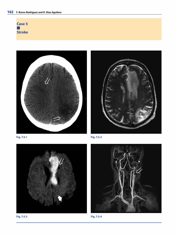
162 F. Bravo-Rodriguez and R. Diaz-Aguilera
Case 5
Stroke
Fig. 7.5.2Fig. 7.5.1
Fig. 7.5.4Fig. 7.5.3
�
RibesLunaRos.indb 162 18.03.2008 12:51:54
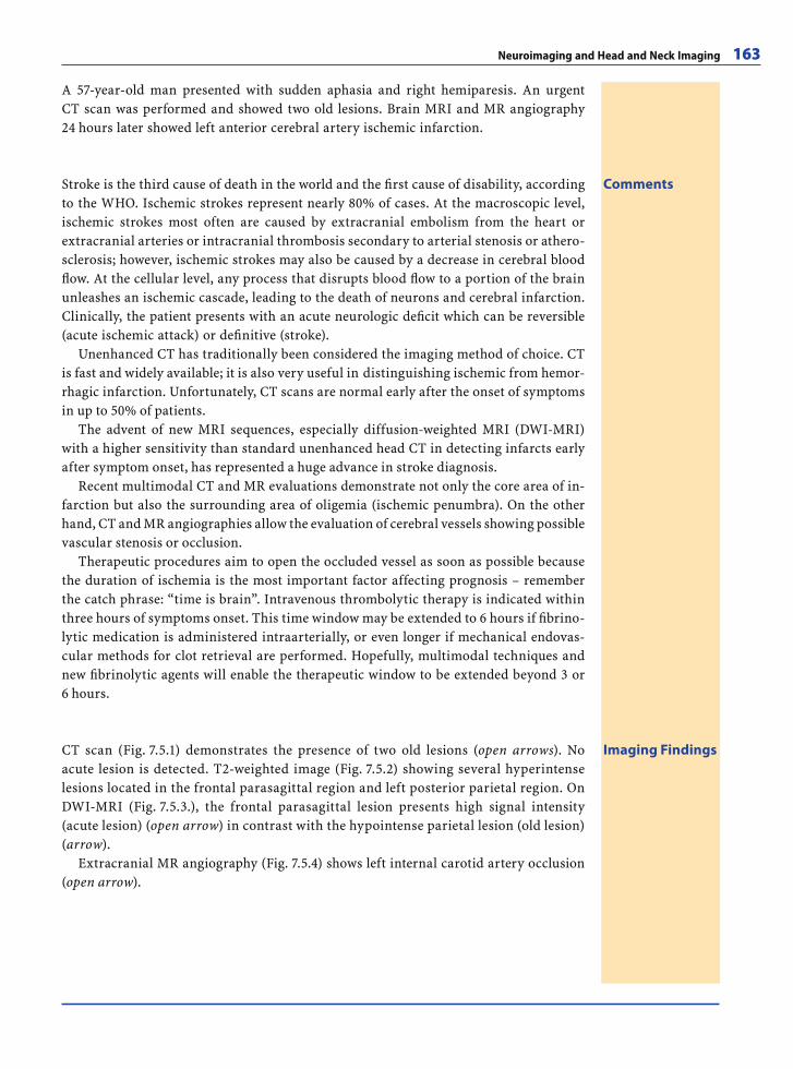
Neuroimaging and Head and Neck Imaging 163
A 57-year-old man presented with sudden aphasia and right hemiparesis. An urgent CT scan was performed and showed two old lesions. Brain MRI and MR angiography 24 hours later showed left anterior cerebral artery ischemic infarction.
CommentsStroke is the third cause of death in the world and the fi rst cause of disability, according to the WHO. Ischemic strokes represent nearly 80% of cases. At the macroscopic level, ischemic strokes most often are caused by extracranial embolism from the heart or extracranial arteries or intracranial thrombosis secondary to arterial stenosis or athero-sclerosis; however, ischemic strokes may also be caused by a decrease in cerebral blood fl ow. At the cellular level, any process that disrupts blood fl ow to a portion of the brain unleashes an ischemic cascade, leading to the death of neurons and cerebral infarction. Clinically, the patient presents with an acute neurologic defi cit which can be reversible (acute ischemic attack) or defi nitive (stroke).
Unenhanced CT has traditionally been considered the imaging method of choice. CT is fast and widely available; it is also very useful in distinguishing ischemic from hemor-rhagic infarction. Unfortunately, CT scans are normal early after the onset of symptoms in up to 50% of patients.
The advent of new MRI sequences, especially diffusion-weighted MRI (DWI-MRI) with a higher sensitivity than standard unenhanced head CT in detecting infarcts early after symptom onset, has represented a huge advance in stroke diagnosis.
Recent multimodal CT and MR evaluations demonstrate not only the core area of in-farction but also the surrounding area of oligemia (ischemic penumbra). On the other hand, CT and MR angiographies allow the evaluation of cerebral vessels showing possible vascular stenosis or occlusion.
Therapeutic procedures aim to open the occluded vessel as soon as possible because the duration of ischemia is the most important factor affecting prognosis – remember the catch phrase: “time is brain”. Intravenous thrombolytic therapy is indicated within three hours of symptoms onset. This time window may be extended to 6 hours if fi brino-lytic medication is administered intraarterially, or even longer if mechanical endovas-cular methods for clot retrieval are performed. Hopefully, multimodal techniques and new fi brinolytic agents will enable the therapeutic window to be extended beyond 3 or 6 hours.
Imaging FindingsCT scan (Fig. 7.5.1) demonstrates the presence of two old lesions (open arrows). No acute lesion is detected. T2-weighted image (Fig. 7.5.2) showing several hyperintense lesions located in the frontal parasagittal region and left posterior parietal region. On DWI-MRI (Fig. 7.5.3.), the frontal parasagittal lesion presents high signal intensity (acute lesion) (open arrow) in contrast with the hypointense parietal lesion (old lesion) (arrow).
Extracranial MR angiography (Fig. 7.5.4) shows left internal carotid artery occlusion (open arrow).
RibesLunaRos.indb 163 18.03.2008 12:51:57
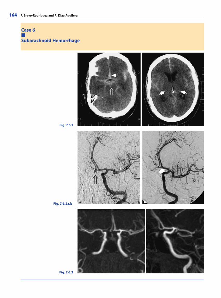
164 F. Bravo-Rodriguez and R. Diaz-Aguilera
Case 6
Subarachnoid Hemorrhage
Fig. 7.6.1
Fig. 7.6.2a,b
Fig. 7.6.3
�
� �a b
RibesLunaRos.indb 164 18.03.2008 12:51:57
Neuroimaging and Head and Neck Imaging 165
A 47-year-old male was admitted to the emergency room with sudden occipital headache, vomiting, and progressive loss of consciousness. CT showed a subarachnoid hemorrhage. The patient was transferred to the intensive care unit and digital subtraction angiography (DSA) carried out 12 hours after the onset of symptoms revealed an anterior communi-cating artery aneurysm. In the same procedure, the aneurysm was completely occluded with GDC coils. A contrast-enhanced MRA performed 6 months after embolization for surveillance showed total occlusion of the aneurysm.
CommentsSubarachnoid hemorrhage (SAH), which implies the presence of blood within the sub-arachnoid space, could be traumatic or nontraumatic. The common medical use of the term SAH refers to the nontraumatic type, usually from rupture of an intracranial berry aneurysm (in 85% of cases), which accounts for 5% of strokes. The incidence is 6–8 events per 100,000 person years, being higher in women than in men.
The most common presentation is an unusually severe headache that starts suddenly. Other possible features are vomiting, focal defi cits, and/or loss of consciousness.
The mortality rate is as high as 25 to 50%.If SAH is suspected, unenhanced CT scan is the fi rst-line imaging modality because
the characteristically hyperdense appearance of extravasated blood in the basal cisterns and sulci is virtually pathognomonic. Additionally, the pattern of hemorrhage often sug-gests the location of any underlying aneurysm (sentinel clot). CT scanning presents a high sensitivity (93–100%) when it is performed within the fi rst 24 hours.
Nevertheless, the method of choice to detect an intracranial aneurysm is DSA, which allows a better assessment of the architecture of the aneurysm in order to decide on the most appropriate therapeutic procedure, endovascular embolization (the current treat-ment of choice ) or surgery.
On the other hand, as DSA is an invasive technique, other less invasive methods, such as CT-angiography or MR-angiography, should be considered during follow-up.
Imaging FindingsUnenhanced CT-scan showing SAH (Fig. 7.6.1) with blood in perimesencephalic cisterns (open arrow), SYLVIAN fi ssures (arrows), and sulci. Notice the sentinel clot in the anterior interhemispheric fi ssure (arrowhead).
DSA (Fig. 7.6.2a) reveals a 2 mm anterior communicating artery aneurysm (open ar-row). DSA after coiling with GDC showing complete occlusion of the aneurysm (arrow) (Fig. 7.6.2b).
Gadolinium-enhanced MRA, MIP reconstruction, obtained 6 months after treatment (Fig. 7.6.3). No recurrence of the aneurysm is seen. Notice the magnetic susceptibility artifact secondary to the coils in the aneurysm. Magnetic susceptibility artifacts can be a limitation in the MRA follow-up of intracranial aneurysms.
RibesLunaRos.indb 165 18.03.2008 12:52:00
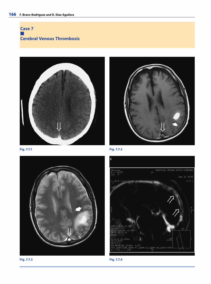
166 F. Bravo-Rodriguez and R. Diaz-Aguilera
Case 7
Cerebral Venous Thrombosis
Fig. 7.7.1 Fig. 7.7.2
Fig. 7.7.3 Fig. 7.7.4
�
�
RibesLunaRos.indb 166 18.03.2008 12:52:00
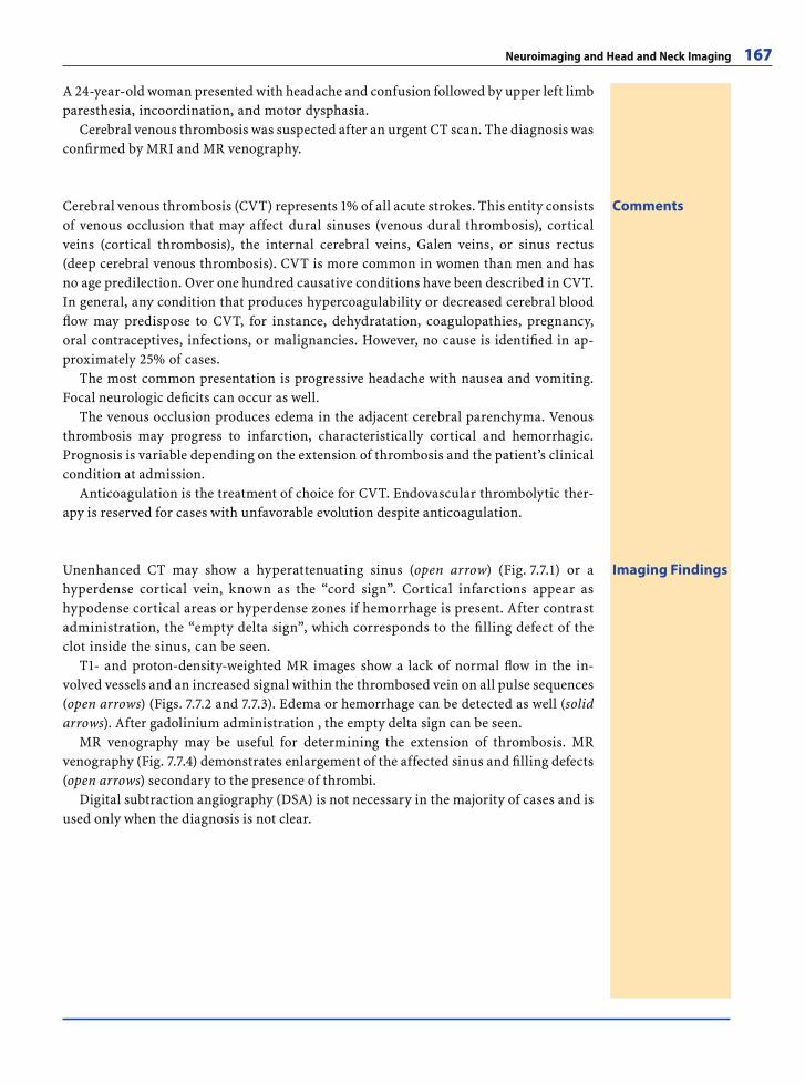
Neuroimaging and Head and Neck Imaging 167
A 24-year-old woman presented with headache and confusion followed by upper left limb paresthesia, incoordination, and motor dysphasia.
Cerebral venous thrombosis was suspected after an urgent CT scan. The diagnosis was confi rmed by MRI and MR venography.
CommentsCerebral venous thrombosis (CVT) represents 1% of all acute strokes. This entity consists of venous occlusion that may affect dural sinuses (venous dural thrombosis), cortical veins (cortical thrombosis), the internal cerebral veins, Galen veins, or sinus rectus (deep cerebral venous thrombosis). CVT is more common in women than men and has no age predilection. Over one hundred causative conditions have been described in CVT. In general, any condition that produces hypercoagulability or decreased cerebral blood fl ow may predispose to CVT, for instance, dehydratation, coagulopathies, pregnancy, oral contraceptives, infections, or malignancies. However, no cause is identifi ed in ap-proximately 25% of cases.
The most common presentation is progressive headache with nausea and vomiting. Focal neurologic defi cits can occur as well.
The venous occlusion produces edema in the adjacent cerebral parenchyma. Venous thrombosis may progress to infarction, characteristically cortical and hemorrhagic. Prognosis is variable depending on the extension of thrombosis and the patient’s clinical condition at admission.
Anticoagulation is the treatment of choice for CVT. Endovascular thrombolytic ther-apy is reserved for cases with unfavorable evolution despite anticoagulation.
Imaging FindingsUnenhanced CT may show a hyperattenuating sinus (open arrow) (Fig. 7.7.1) or a hyperdense cortical vein, known as the “cord sign”. Cortical infarctions appear as hypodense cortical areas or hyperdense zones if hemorrhage is present. After contrast administration, the “empty delta sign”, which corresponds to the fi lling defect of the clot inside the sinus, can be seen.
T1- and proton-density-weighted MR images show a lack of normal fl ow in the in-volved vessels and an increased signal within the thrombosed vein on all pulse sequences (open arrows) (Figs. 7.7.2 and 7.7.3). Edema or hemorrhage can be detected as well (solid arrows). After gadolinium administration , the empty delta sign can be seen.
MR venography may be useful for determining the extension of thrombosis. MR venography (Fig. 7.7.4) demonstrates enlargement of the affected sinus and fi lling defects (open arrows) secondary to the presence of thrombi.
Digital subtraction angiography (DSA) is not necessary in the majority of cases and is used only when the diagnosis is not clear.
RibesLunaRos.indb 167 18.03.2008 12:52:03
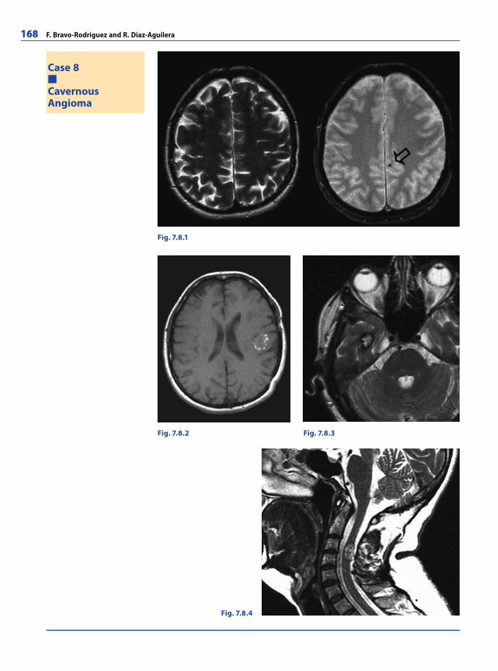
168 F. Bravo-Rodriguez and R. Diaz-Aguilera
Case 8
CavernousAngioma
Fig. 7.8.1
Fig. 7.8.2 Fig. 7.8.3
Fig. 7.8.4
RibesLunaRos.indb 168 18.03.2008 12:52:03
Neuroimaging and Head and Neck Imaging 169
A 35-year-old woman with a prior medical history of seizures and surgical resection of a cavernoma 10 years prior was being treated with carbamazepine and presented with isolated seizures. Brain and spine MRI were performed for surveillance of multiple cavernomatosis.
CommentsCavernous malformations (cavernomas, cavernous angiomas, or hemangiomas,...) con-sist of well-circumscribed sinusoidal vascular channels containing blood in various stages of evolution. They represent approximately 1% of intracranial vascular lesions and 15% of cerebrovascular malformations. With the advent of MRI, cavernomas have become the most commonly identifi ed vascular malformations in the brain. Cavernous angiomas vary from several millimeters to several centimeters in diameter although they are usually < 3 cm in size. Multiple lesions are seen in approximately 10–30% of spontaneous cases. A familial form of the disorder exists and is inherited as an autoso-mal dominant trait with variable expression. Multiple lesions are more common in the familial form, occurring in as many as 80% of patients. About 80–90% of lesions are supratentorial and only 5% of cavernomas are seen in the spinal cord of adult patients. There is no gender predilection.
Cavernous angiomas are considered to be a congenital entity although de novo lesions may occur in patients with previous radiation exposure or in familial form cases. In early studies of major autopsy reports, the calculated prevalence was 0.3–0.9%. Cavernomas can occur at any age, but they are most likely to become clinically apparent in patients aged 20–40 years. Common clinical symptoms are seizures secondary to cortical lesions, focal neurologic defi cits secondary to deep cerebral white matter and pons lesions, and headache as a result of intralesional hemorrhage.
The treatment of choice is surgical removal. Resection is indicated even in cases af-fecting the pons, due to the increased risk of subsequent and progressive neurologic dis-ability in cases of rehemorrhage.
Imaging FindingsThe sensitivity of CT for these lesions is 70–80%. Cavernous angiomas are typically hyperdense and present calcifi cations in 30% of cases. Lesions show a mottled pattern of enhancement after contrast administration. When surrounding edema is present, it is indicative of a recent bleed.
MRI is the imaging modality of choice. Gradient-echo imaging, with its increased sen-sitivity to susceptibility artifacts, is useful in the detection of smaller and concomitant lesions, which may not be detected with SE and FSE sequences (Fig. 7.8.1, open arrow). Cavernous angiomas present as a typical “popcorn-like” lesion with a large amount of he-mosiderin surrounding a core of hemorrhage in different stages of evolution. The central area appears hyperintense on T1-weighted images (Fig. 7.8.2) and the hemosiderin rim is hypointense on gradient-refocused and FSE T2-weighted images (Figs. 7.8.3 and 7.8.4).
In general, cavernomas are considered angiographically occult because of the ex-tremely low fl ow of blood through these lesions.
RibesLunaRos.indb 169 18.03.2008 12:52:06
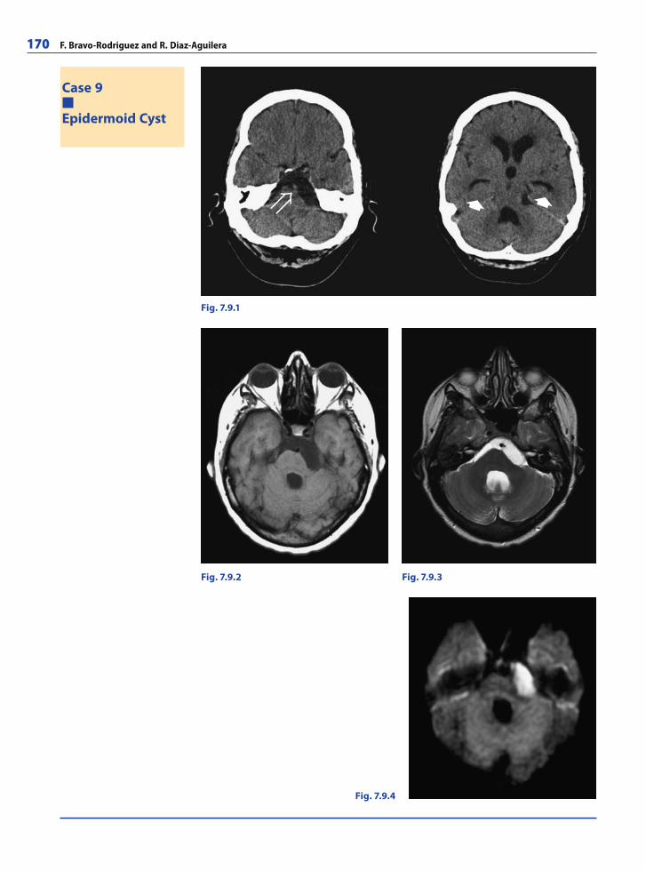
170 F. Bravo-Rodriguez and R. Diaz-Aguilera
Case 9
Epidermoid Cyst
Fig. 7.9.1
Fig. 7.9.2 Fig. 7.9.3
Fig. 7.9.4
� �
RibesLunaRos.indb 170 18.03.2008 12:52:06
Neuroimaging and Head and Neck Imaging 171
A 32-year-old woman was admitted to the emergency room with a 24 hour history of diplopia, left hypoacusia, tinnitus, and vertigo. The patient had a prior medical history of headache and occasional vomiting for 5 years.
On physical examination, bilateral papilledema, diplopia, and hypoalgesia of the left maxillary nerve (V2) and cornea were found.
A CT scan showed hydrocephalus and the presence of an extraaxial lesion located in the posterior fossa, consistent with epidermoid cyst. MRI confi rmed the suspected diag-nosis.
CommentsEpidermoid cysts are benign congenital lesions of ectodermal origin secondary to epithe-lial inclusion during neural tube closure between the third and fi fth weeks of embryonic life. They represent approximately 0.2–1.8% of all primary intracranial tumors. Epider-moid cysts occur most commonly in the cerebellopontine angle (40–50% of cases) and fourth ventricle. They tend to be well-circumscribed, smooth or lobulated, encapsulated lesions with a characteristic glistening, pearl-like sheen. The tumor presents as a slowly enlarging lesion that tends to adapt to the subarachnoid space, which is why patients are usually not symptomatic until age 30–40. Clinical features depend on the location of the lesion. Headache and cranial-nerve neuropathy are the most common symptoms. The differential diagnosis includes arachnoid cysts which appear isointense to CSF in all MRI pulse sequences, including diffusion-weighted images.
The treatment of choice is microsurgery. Total excision of the tumor is not always pos-sible and there is a high rate of recurrences in these cases.
Imaging FindingsCT scan (Fig. 7.9.1) demonstrates an extraaxial hypodense lesion located in the left cerebellopontine angle (CPA) and prepontine cistern. The tumor envelopes the basilar artery (open arrow) and causes a mild mass effect over the pons. A superior slice shows dilatation of the third and fourth ventricles as well as of the frontal and temporal horns (solid arrows).
MRI demonstrates a lesion in the left CPA which extends anteriorly to the prepontine cistern and superiorly to suprasellar and chiasmatic regions and ambiens cistern. Note that the signal intensity of the lesion is slightly different from that of CSF on T1- and T2-weighted images (Figs. 7.9.2 and 7.9.3). As epidermoid cysts have markedly restricted diffusion, they show high signal intensity on diffusion-weighted images (Fig. 7.9.4). This feature is useful in the distinction between arachnoid cysts, which are hypointense on DWI, and epidermoid cysts.
RibesLunaRos.indb 171 18.03.2008 12:52:09
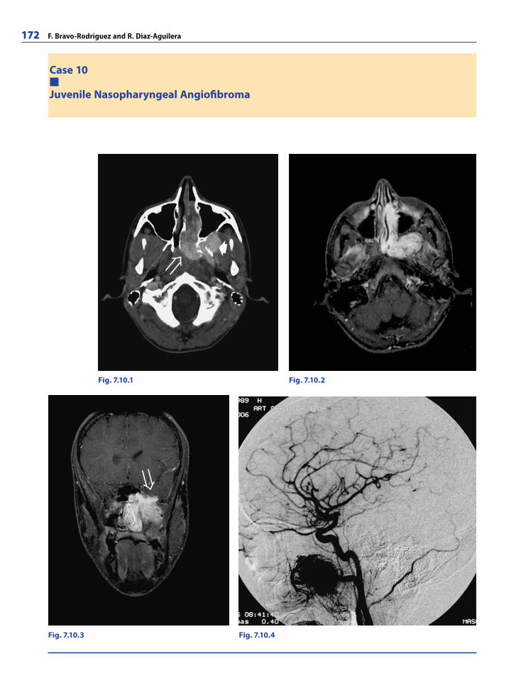
172 F. Bravo-Rodriguez and R. Diaz-Aguilera
Case 10
Juvenile Nasopharyngeal Angiofi broma
Fig. 7.10.1 Fig. 7.10.2
Fig. 7.10.3 Fig. 7.10.4
�
RibesLunaRos.indb 172 18.03.2008 12:52:09
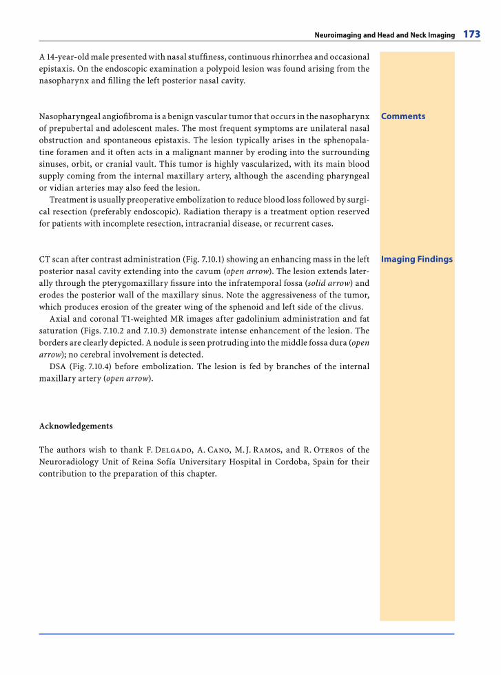
Neuroimaging and Head and Neck Imaging 173
A 14-year-old male presented with nasal stuffi ness, continuous rhinorrhea and occasional epistaxis. On the endoscopic examination a polypoid lesion was found arising from the nasopharynx and fi lling the left posterior nasal cavity.
CommentsNasopharyngeal angiofi broma is a benign vascular tumor that occurs in the nasopharynx of prepubertal and adolescent males. The most frequent symptoms are unilateral nasal obstruction and spontaneous epistaxis. The lesion typically arises in the sphenopala-tine foramen and it often acts in a malignant manner by eroding into the surrounding sinuses, orbit, or cranial vault. This tumor is highly vascularized, with its main blood supply coming from the internal maxillary artery, although the ascending pharyngeal or vidian arteries may also feed the lesion.
Treatment is usually preoperative embolization to reduce blood loss followed by surgi-cal resection (preferably endoscopic). Radiation therapy is a treatment option reserved for patients with incomplete resection, intracranial disease, or recurrent cases.
Imaging FindingsCT scan after contrast administration (Fig. 7.10.1) showing an enhancing mass in the left posterior nasal cavity extending into the cavum (open arrow). The lesion extends later-ally through the pterygomaxillary fi ssure into the infratemporal fossa (solid arrow) and erodes the posterior wall of the maxillary sinus. Note the aggressiveness of the tumor, which produces erosion of the greater wing of the sphenoid and left side of the clivus.
Axial and coronal T1-weighted MR images after gadolinium administration and fat saturation (Figs. 7.10.2 and 7.10.3) demonstrate intense enhancement of the lesion. The borders are clearly depicted. A nodule is seen protruding into the middle fossa dura (open arrow); no cerebral involvement is detected.
DSA (Fig. 7.10.4) before embolization. The lesion is fed by branches of the internal maxillary artery (open arrow).
Acknowledgements
The authors wish to thank F. Delgado, A. Cano, M. J. Ramos, and R. Oteros of the Neuroradiology Unit of Reina Sofía Universitary Hospital in Cordoba, Spain for their contribution to the preparation of this chapter.
RibesLunaRos.indb 173 18.03.2008 12:52:12
174 F. Bravo-Rodriguez and R. Diaz-Aguilera
Further Readings
Books
Clinical Neuroanatomy. 6th ed. Snell RS (2006) Lippincott Williams & Wilkins. ISBN-13: 9780781759939
Diagnostic Cerebral Angiography. 2nd ed. Osborn AG (1999) Lippincott Williams & Wilkins. ISBN-13: 9780397584048
Diagnostic Imaging: Brain. Osborn AG (2004) Elsevier-Amirsys. ISBN-13: 9780721629056
Diagnostic Imaging: Head and Neck. Harnsberger R (2004) Elsevier-Amirsys. ISBN-13: 9780721628905
Diagnostic Imaging: Spine. Ross J, Brant-Zawadzki M, Chen MZ, Moore KR (2005) Elsevier-Amirsys. ISBN-13: 9780721628806
Head and Neck Imaging. Som PM, Curtin HD (2003) Else-vier-Mosby. ISBN-13: 9780323009423
Magnetic Resonance Imaging of the Brain and Spine. 3rd ed. Atlas SW (2001) Lippincott Williams & Wilkins. ISBN-13: 9780781720366
Neurorradiología diagnóstica y terapéutica. Mercader JM, Viñuela F (2004) Elsevier-Masson. ISBN: 9788445813300
Neuroradiology. 2nd ed. Grossman RI, Yousem DM (2003) Elsevier-Mosby. ISBN-13: 9780323005081
Pediatric Neuroimaging 4th ed. Barkovich AJ (2005) Lippin-cott Williams & Wilkins. ISBN-13: 9780781757669
Web Links
http://ashnr.org http://asnr.org/http://esnr.org/00.asphttp://neuroguide.com/http://neurorad.ucsf.edu/index.htmlhttp://radiographics.rsnajnls.org/cgi/collection/neuro-
radiologyhttp://silan.org/Spanish/society.htmlhttp://springerlink.com/content/100446/http://stroke-university.com/http://wfns.org/
Articles
Abe O, Aoki S, Shirouzu I, Kunimatsu A, Hayashi N, Ma-sumoto T, Mori H, Yamada H, Watanabe M, Masutani Y, Ohtomo K. MR imaging of ischemic penumbra. Eur J Radiol 2003; 46(1):67–78
Anslow P. Cranial bacterial infection. Eur Radiol 2004; 14 Suppl 3:E145–154
Barkovich AJ. MR imaging of the neonatal brain. Neuroim-aging Clin N Am 2006; 16(1):117–135
Bode MK, Ruohonen J, Nieminen MT, Pyhtinen J. Potential of diffusion imaging in brain tumors: a review. Acta Ra-diol 2006; 47(6):585–594
Bonneville F, Sourour N, Biondi A. Intracranial aneurysms: an overview. Neuroimaging Clin N Am 2006; 16(3):371–382
Bonneville JF, Bonneville F, Cattin F. Magnetic reso-nance imaging of pituitary adenomas. Eur Radiol 2005; 15(3):543–548. Epub 2004 Dec 31
Brisman JL, Song JK, Newell DW. Cerebral aneurysms. N Engl J Med 2006; 355(9):928–939
Bravo-Rodriguez Fde A, Delgado Acosta F, Cano Sánchez A, Bautista Rodriguez MD. Trombosis venosa cerebral. Tratamiento mediante fi brinolisis local con alteplasa. Ra-diología 2002; 44(1):23–26
Bravo-Rodriguez Fde A, Espejo Herrero JJ, Bautista Rodri-guez MD, Delgado Acosta F. Spontaneous dissection of vertebral arteries. Med Clin (Barc) 2004; 122(8):317–318
Burtscher IM, Holtas S. Proton magnetic resonance spec-troscopy in brain tumours: clinical applications. Neuro-radiology 2001; 43(5):345–352
Castillo M. Imaging of the upper cranial nerves I, III–VIII, and the cavernous sinuses. Neuroimaging Clin N Am 2004; 14(4):579–593
Caruso PA, Rincon S, Weber AL. Lesions of the maxilla: crossroads of the head and neck. Neuroimaging Clin N Am 2003; 13(3):411–426
Cha S. Update on brain tumor imaging: from anatomy to physiology. AJNR Am J Neuroradiol 2006; 27(3):475–487
Charil A, Yousry TA, Rovaris M, Barkhof F, De Stefano N, Fazekas F, Miller DH, Montalban X, Simon JH, Polman C, Filippi M. MRI and the diagnosis of multiple sclerosis: expanding the concept of “no better explanation”. Lancet Neurol 2006; 5(10):841–852
Charles-Edwards EM, deSouza NM. Diffusion-weighted magnetic resonance imaging and its application to can-cer. Cancer Imaging 2006; 6:135–143
Cognard C, Gobin YP, Pierot L, Bailly AL, Houdart E, Casa-sco A, Chiras J, Merland JJ. Cerebral dural arteriovenous fi stulas: clinical and angiographic correlation with a re-vised classifi cation of venous drainage. Radiology 1995; 194(3):671–680
Connor SE, Flis C, Langdon JD. Vascular masses of the head and neck. Clin Radiol 2005; 60(8):856–868
Cruz Junior LC, Sorensen AG. Diffusion tensor magnetic resonance imaging of brain tumors. Neurosurg Clin N Am 2005; 16(1):115–134
DeAngelis LM. Brain tumors. N Engl J Med 2001; 344(2):114–123
Delgado F, Bravo-Rodríguez FA, Bautista MD, Chirosa MA, Molina T, Martos JM, Canis M. Carotid pseudoa-neurysms secondary to dissection: endovascular man-agement with bare stent-graft. Cerebrovasc Dis 2005; 19(2):136–138
Diaz-Aguilera R, Martos-Becerra JM, Bravo-Rodriguez F, Ramos-Gomez MJ, Vinals-Torras M. Transient lesion in the corpus callosum associated with the use of antiepi-leptic drugs. Rev Neurol 2005; 41(4):254–255
Di Costanzo A, Scarabino T, Trojsi F, Giannatempo GM, Popolizio T, Catapano D, Bonavita S, Maggialetti N, To-setti M, Salvolini U, d’Angelo VA, Tedeschi G. Multipa-rametric 3T MR approach to the assessment of cerebral gliomas: tumor extent and malignancy. Neuroradiology 2006; 48(9):622–631. Epub 2006 Jun 3
Drevelegas A. Extra-axial brain tumors. Eur Radiol 2005; 15(3):453–467
Essig M. MultiHance in brain perfusion. Eur Radiol 2004; 14 Suppl 7:O10–15
RibesLunaRos.indb 174 18.03.2008 12:52:12
Neuroimaging and Head and Neck Imaging 175
Esteban JM, Cervera V. Perfusion CT and angio CT in the assessment of acute stroke. Neuroradiology 2004; 46(9):705–715
Fiebach JB, Schellinger PD, Geletneky K, Wilde P, Meyer M, Hacke W, Sartor K. MRI in acute subarachnoid haemor-rhage; fi ndings with a standardised stroke protocol. Neu-roradiology 2004; 46(1):44–48
Flis CM, Connor SE. Imaging of head and neck venous mal-formations. Eur Radiol 2005; 15(10):2185–2193. Epub 2005 Jul 8
Forsting M, Weber J. MR perfusion imaging: a tool for more than stroke. Eur Radiol 2004; 14 Suppl 5:M2–7
Fox AJ. Carotid endarterectomy trials. Neuroimaging Clin N Am 1996; 6(4):931–938
Gao PY, Osborn AG, Smirniotopoulos JG, Harris CP. Radio-logic-pathologic correlation. Epidermoid tumor of the cerebellopontine angle. AJNR Am J Neuroradiol 1992; 13(3):863–872
Ge Y. Multiple sclerosis: the role of MR imaging. AJNR Am J Neuroradiol 2006; 27(6):1165–1176
Gil-Peralta A, Mayol A, Marcos JR, Gonzalez A, Ruano J, Boza F, Duran F. Percutaneous transluminal angio-plasty of the symptomatic atherosclerotic carotid arter-ies. Results, complications, and follow-up. Stroke 1996; 27(12):2271–2273
Glenn OA, Barkovich J. Magnetic resonance imaging of the fetal brain and spine: an increasingly important tool in prenatal diagnosis: part 2. AJNR Am J Neuroradiol 2006; 27(9):1807–1814
Glenn OA, Barkovich AJ. Magnetic resonance imaging of the fetal brain and spine: an increasingly important tool in prenatal diagnosis, part 1. AJNR Am J Neuroradiol 2006; 27(8):1604–1611
Gonzalez A, Mayol A, Martinez E, Gonzalez-Marcos JR, Gil-Peralta A. Mechanical thrombectomy with snare in pa-tients with acute ischemic stroke. Neuroradiology 2007; 49(4):365–372
Guglielmi G, Viñuela F, Dion J, Duckwiler G. Electrothrom-bosis of saccular aneurysms via endovascular approach. Part 2: Preliminary clinical experience.J Neurosurg 1991; 75(1):8–14
Hillis AE, Wityk RJ, Beauchamp NJ, Ulatowski JA, Jacobs MA, Barker PB. Perfusion-weighted MRI as a marker of response to treatment in acute and subacute stroke. Neu-roradiology 2004; 46(1):31–39
Higashida RT, Meyers PM. Intracranial angioplasty and stenting for cerebral atherosclerosis: new treatments for stroke are needed! Neuroradiology 2006; 48(6):367–372
Higashida RT, Furlan AJ, Roberts H, Tomsick T, Connors B, Barr J, Dillon W, Warach S, Broderick J, Tilley B, Sacks D; Technology Assessment Committee of the American Society of Interventional and Therapeutic Neuroradiology; Technology Assessment Committee of the Society of Interventional Radiology. Trial design and reporting standards for intra-arterial cerebral thrombolysis for acute ischemic stroke. Stroke 2003; 34(8):e109–137
Hutter A, Schwetye KE, Bierhals AJ, McKinstry RC. Brain neoplasms: epidemiology, diagnosis, and prospects for
cost-effective imaging. Neuroimaging Clin N Am 2003; 13(2):237–250
Jellison BJ, Field AS, Medow J, Lazar M, Salamat MS, Al-exander AL. Diffusion tensor imaging of cerebral white matter: a pictorial review of physics, fi ber tract anatomy, and tumor imaging patterns. AJNR Am J Neuroradiol 2004; 25(3):356–369
Jensen ME, Dion JE. Percutaneous vertebroplasty in the treatment of osteoporotic compression fractures. Neuro-imaging Clin N Am 2000; 10(3):547–568
Kai Y, Hamada J, Morioka M, Yano S, Todaka T, Ushio Y. Appropriate interval between embolization and surgery in patients with meningioma. AJNR Am J Neuroradiol 2002; 23(1):139–142
Kane I, Sandercock P, Wardlaw J. Magnetic resonance per-fusion diffusion mismatch and thrombolysis in acute ischaemic stroke: a systematic review of the evidence to date. J Neurol Neurosurg Psychiatry 2007; 78(5):485–491. Epub 2006 Oct 20
Kim LJ, Spetzler RF. Classifi cation and surgical management of spinal arteriovenous lesions: arteriovenous fi stulae and arteriovenous malformations. Neurosurgery 2006; 59(5 Suppl 3):S195–201
Korteweg T, Tintore M, Uitdehaag B, Rovira A, Frederiksen J, Miller D, Fernando K, Filippi M, Agosta F, Rocca M, Fazekas F, Enzinger C, Matthews P, Parry A, Polman C, Montalban X, Barkhof F. MRI criteria for dissemination in space in patients with clinically isolated syndromes: a multicentre follow-up study. Lancet Neurol 2006; 5(3):221–227
Lasjaunias PL, Chng SM, Sachet M, Alvarez H, Rodesch G, Garcia-Monaco R. The management of vein of Galen an-eurysmal malformations. Neurosurgery 2006; 59(5 Suppl 3):S184–194
Lai R, Rosenblum MK, DeAngelis LM. Primary CNS lymphoma: a whole-brain disease? Neurology 2002; 59(10):1557–1562
Law M, Yang S, Wang H, Babb JS, Johnson G, Cha S, Knopp EA, Zagzag D. Glioma grading: sensitivity, specifi city, and predictive values of perfusion MR imaging and proton MR spectroscopic imaging compared with con-ventional MR imaging. AJNR Am J Neuroradiol 2003; 24(10):1989–1998
Law M. MR spectroscopy of brain tumors. Top Magn Reson Imaging 2004; 15(5):291–313
Leira R, Davalos A, Silva Y, Gil-Peralta A, Tejada J, Garcia M, Castillo J; Stroke Project, Cerebrovascular Diseases Group of the Spanish Neurological Society. Early neuro-logic deterioration in intracerebral hemorrhage: predic-tors and associated factors. Neurology 2004; 63(3):461–467
Majos C, Alonso J, Aguilera C, Serrallonga M, Coll S, Acebes JJ, Arus C, Gili J. Utility of proton MR spectroscopy in the diagnosis of radiologically atypical intracranial menin-giomas. Neuroradiology 2003; 45(3):129–136. Epub 2003 Feb 19
Mathis JM, Barr JD, Jungreis CA, Yonas H, Sekhar LN, Vin-cent D, Pentheny SL, Horton JA. Temporary balloon test occlusion of the internal carotid artery: experience in 500 cases. AJNR Am J Neuroradiol 1995; 16(4):749–754
RibesLunaRos.indb 175 18.03.2008 12:52:13
176 F. Bravo-Rodriguez and R. Diaz-Aguilera
Mezzapesa DM, Petruzzellis M, Lucivero V, Prontera M, Ti-nelli A, Sancilio M, Carella A, Federico F. Multimodal MR examination in acute ischemic stroke. Neuroradiology 2006; 48(4):238–246
Mitra D, Herwadkar A, Soh C, Gholkar A. Follow-up of intra-cranial aneurysms treated with matrix detachable coils: a single-center experience. AJNR Am J Neuroradiol 2007; 28(2):362–367
Molyneux AJ. Ruptured intracranial aneurysms – clinical aspects of subarachnoid hemorrhage management and the International Subarachnoid Aneurysm Trial. Neuro-imaging Clin N Am 2006; 16(3):391–396
Molyneux A, Kerr R, Stratton I, Sandercock P, Clarke M, Shrimpton J, Holman R; International Subarachnoid An-eurysm Trial (ISAT) Collaborative Group. International Subarachnoid Aneurysm Trial (ISAT) of neurosurgical clipping versus endovascular coiling in 2143 patients with ruptured intracranial aneurysms: a randomised trial. Lancet 2002; 360(9342):1267–1274
Munoz del Castillo F, Jurado Ramos A, Bravo-Rodriguez F, Delgado Acosta F, Lopez Villarejo P. Endoscopic surgery of nasopharyngeal angiofi broma. Acta Otorrinolaringol Esp 2004; 55(8):369–375
Murphy KJ, Deramons H. Percutaneous vertebroplasty in benign and malignant disease. Neuroimaging Clin N Am 2000; 10(3):535–545
Murray TJ. Diagnosis and treatment of multiple sclerosis. BMJ 2006; 332(7540):525–527
Osborn AG, Preece MT. Intracranial cysts: radiologic-patho-logic correlation and imaging approach. Radiology 2006; 239(3):650–664
Parizel PM, Van Goethem JW, Ozsarlak O, Maes M, Phil-lips CD. New developments in the neuroradiological diagnosis of craniocerebral trauma. Eur Radiol 2005; 15(3):569–581
Pinero P, Gonzalez A, Mayol A, Martinez E, Gonzalez-Mar-cos JR, Boza F, Cayuela A, Gil-Peralta A. Silent ischemia after neuroprotected percutaneous carotid stenting: a diffusion-weighted MRI study. AJNR Am J Neuroradiol 2006; 27(6):1338–1345
Poussaint TY, Rodriguez D. Advanced neuroimaging of pediatric brain tumors: MR diffusion, MR perfusion, and MR spectroscopy. Neuroimaging Clin N Am 2006; 16(1):169–192
Provenzale JM, Mukundan S, Barboriak DP. Diffusion-weighted and perfusion MR imaging for brain tumor characterization and assessment of treatment response. Radiology 2006; 239(3):632–649
Reijneveld JC, van der Grond J, Ramos LM, Bromberg JE, Taphoorn MJ. Proton MRS imaging in the follow-up of patients with suspected low-grade gliomas. Neuroradiol-ogy 2005; 47(12):887–891. Epub 2005 Aug 20
Rivera PP, Willinsky RA, Porter PJ. Intracranial cavernous malformations. Neuroimaging Clin N Am 2003; 13(1):27–40
Rovira A, Grive E, Rovira A, Alvarez-Sabin J. Distribution territories and causative mechanisms of ischemic stroke. Eur Radiol 2005; 15(3):416–426
Rovira A, Orellana P, Alvarez-Sabin J, Arenillas JF, Aymerich X, Grive E, Molina C, Rovira-Gols A. Hyperacute isch-
emic stroke: middle cerebral artery susceptibility sign at echo-planar gradient-echo MR imaging. Radiology 2004; 232(2):466–473
Rovira A, Pericot I, Alonso J, Rio J, Grive E, Montalban X. Se-rial diffusion-weighted MR imaging and proton MR spec-troscopy of acute large demyelinating brain lesions: case report. AJNR Am J Neuroradiol 2002; 23(6):989–994
Rovira A, Rovira-Gols A, Pedraza S, Grive E, Molina C, Alva-rez-Sabin J. Diffusion-weighted MR imaging in the acute phase of transient ischemic attacks. AJNR Am J Neuro-radiol 2002; 23(1):77–83
Rovira A, Pedraza S, Molina C, Capellades J, Grive E, Rovira A, Montaner J. Diffusion-weighted magnetic resonance in the diagnosis of acute subcortical infarcts. Rev Neurol 2000; 30(10):914–919
Sanchez-Arjona MB, Sanz-Fernandez G, Franco-Macias E, Gil-Peralta A. Cerebral hemodynamic changes after ca-rotid angioplasty and stenting. AJNR Am J Neuroradiol 2007; 28(4):640–644
Schaefer PW, Copen WA, Lev MH, Gonzalez RG. Diffusion-weighted imaging in acute stroke. Neuroimaging Clin N Am 2005; 15(3):503–530
Schellinger PD, Fiebach JB, Hacke W. Imaging-based deci-sion making in thrombolytic therapy for ischemic stroke: present status. Stroke 2003; 34(2):575–583
Schievink WI. Spontaneous dissection of the carotid and vertebral arteries. N Engl J Med 2001; 344(12):898–906
Shetty SK, Lev MH. CT perfusion in acute stroke. Neuroim-aging Clin N Am 2005; 15(3):481–501
Smith WS. Safety of mechanical thrombectomy and intra-venous tissue plasminogen activator in acute ischemic stroke. Results of the multi Mechanical Embolus Removal in Cerebral Ischemia (MERCI) trial, part I. AJNR Am J Neuroradiol 2006; 27(6): 1177–1182
Srinivasan A, Goyal M, Al Azri F, Lum C. State-of-the-art imaging of acute stroke. Radiographics 2006; 26 Suppl 1:S75–95
Spetzler RF, Martin NA. A proposed grading system for ar-teriovenous malformations. J Neurosurg 1986; 65(4):476–483
Stadnik TW, Demaerel P, Luypaert RR, Chaskis C, Van Rom-paey KL, Michotte A, Osteaux MJ. Imaging tutorial: dif-ferential diagnosis of bright lesions on diffusion-weighted MR images. Radiographics 2003; 23(1):e7
Sundgren PC, Dong Q, Gomez-Hassan D, Mukherji SK, Maly P, Welsh R. Diffusion tensor imaging of the brain: review of clinical applications. Neuroradiology 2004; 46(5):339–350
Tharin S, Golby A. Functional brain mapping and its appli-cations to neurosurgery. Neurosurgery 2007; 60(4 Suppl 2):185–201
Thurnher MM, Castillo M. Imaging in acute stroke. Eur Ra-diol 2005; 15(3): 408–415
Urtasun F, Biondi A, Casaco A, Houdart E, Caputo N, Ay-mard A, Merland JJ. Cerebral dural arteriovenous fi stu-las: percutaneous transvenous embolization. Radiology 1996; 199(1):209–217
Valavanis A. The role of angiography in the evaluation of cerebral vascular malformations. Neuroimaging Clin N Am 1996; 6(3):679–704
RibesLunaRos.indb 176 18.03.2008 12:52:13
Neuroimaging and Head and Neck Imaging 177
van Gijn J, Kerr RS, Rinkel GJ. Subarachnoid haemorrhage. Lancet 2007; 369(9558):306–318
Vates GE, Chang S, Lamborn KR, Prados M, Berger MS. Glio-matosis cerebri: a review of 22 cases. Neurosurgery 2003; 53(2):261–271
Vattipally VR, Bronen RA. MR imaging of epilepsy: strate-gies for successful interpretation. Neuroimaging Clin N Am 2004; 14(3):349–372
Vinuela F, Duckwiler G, Mawad M. Guglielmi detachable coil embolization of acute intracraneial aneurysm: periop-erative anatomical and clinical outcome in 403 patients. J Neurosurg 1997; 86(3):475–482
Weber AL, Caruso P, Sabates NR. The optic nerve: radio-logic, clinical, and pathologic evaluation. Neuroimaging Clin N Am 2005; 15(1):175–201
Wetzel S, Bongartz G. MR angiography: supra-aortic vessels. Eur Radiol 1999; 9(7):1277–1284
White PM, Wardlaw JM, Lindsay KW, Sloss S, Patel DK, Teasdale EM. The non-invasive detection of intracranial aneurysms: are neuroradiologists any better than other observers? Eur Radiol 2003; 13(2):389–396
Wiebers DO. Unruptured intracranial aneurysms: natural history and clinical management. Update on the inter-national study of unruptured intracranial aneurysms. Neuroimaging Clin N Am 2006; 16(3):383–390
Wilms G, Demaerel P, Sunaert S. Intra-axial brain tumours. Eur Radiol 2005; 15(3):468–484
Wintermark M. Brain perfusion-CT in acute stroke patients. Eur Radiol 2005; 15 Suppl 4:D28–31
Witte RJ, Lane JI, Driscoll CL, Lundy LB, Bernstein MA, Kotsenas AL, Kocharian A. Pediatric and adult cochlear implantation. Radiographics 2003; 23(5):1185–1200
Wittenberg KH, Adkins MC. MR imaging of nontraumatic brachial plexopathies: frequency and spectrum of fi nd-ings. Radiographics 2000; 20(4):1023–1032
Yuh WT, Christoforidis GA, Koch RM, Sammet S, Schmal-brock P, Yang M, Knopp MV. Clinical magnetic resonance imaging of brain tumors at ultrahigh fi eld: a state-of-the-art review. Top Magn Reson Imaging 2006; 17(2):53–61
Young RJ, Sills AK, Brem S, Knopp EA. Neuroimaging of metastatic brain disease. Neurosurgery 2005; 57(5 Suppl):S10–23
RibesLunaRos.indb 177 18.03.2008 12:52:13

RibesLunaRos.indb V 18.03.2008 12:48:44
Nuclear Medicine
Nuclear Medicine 8
Juan Antonio Vallejo Casas and Angel C. Rebollo AguirreLuisa M. Mena Bares (Contributor)
IntroductionNuclear medicine is the medical specialty that uses radioactive isotopes, nuclear radia-tion, electromagnetic changes of the nuclear components, and biophysical techniques to prevent, diagnose, and treat medical conditions.
In a nuclear medicine test, small amounts of radiopharmaceuticals are introduced into the body by injection, swallowing, or inhalation. Radiopharmaceuticals are substances that are attracted to specifi c organs and provide information about both their structure and function. The amount of radiopharmaceutical used is carefully selected to ensure an accurate test while providing the least amount of radiation exposure to the patient. A special camera (PET, SPECT or gamma camera) is used to detect the radiopharmaceuti-cal in the target organ or tissue and shows images of the area in question.
The capability of nuclear medicine to determine the presence of disease is based on biophysiological changes rather than anatomical ones.
Widespread clinical use of nuclear medicine began in the early 1950s, with the applica-tion of radioiodine to patients suffering from thyroid disease. In the 1960s and the years that followed, the growth of nuclear medicine was phenomenal. In the 1970s most organs of the body could be visualized. In 1971 the American Medical Association offi cially rec-ognized nuclear medicine as a distinct medical specialty.
As more is learned about the fundamental processes of diseases and new radiophar-maceuticals and analytical tools are developed, PET and CT/PET scanning will prove to be invaluable tools in the diagnosis and management of some of the most critical diseases challenging modern medicine. The development of new therapeutic agents, especially those related to radioimmunotherapy of many oncological conditions, will give nuclear medicine a prominent role in the management and treatment of oncological patients.
RibesLunaRos.indb 179 18.03.2008 12:52:13
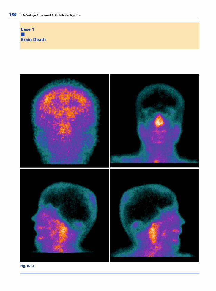
180 J. A. Vallejo Casas and A. C. Rebollo Aguirre
Case 1
Brain Death
Fig. 8.1.1
RibesLunaRos.indb 180 18.03.2008 12:52:13
Nuclear Medicine 181
A 53-year-old woman suffered occipital trauma and presented with loss of conscious-ness. The initial clinical examination showed areactive mydriasis with Glasgow score 3; 48 hours after admission the patient was in deep coma and 99mTc-HMPAO cerebral perfu-sion scintigraphy was performed to confi rm the diagnosis of cerebral death.
CommentsThe term “brain death” is used to signify the irreversible loss of function of the entire brain and, by general consensus, is accepted as a criterion for human death. The deter-mination of brain death has gained importance in modern health care to prevent futile attempts to sustain ventilation and blood circulation artifi cially for prolonged periods and to ensure the potential availability of organs for transplantation.
Although physicians generally agree that a patient can be declared brain dead when the loss of brain function is total and irreversible, different approaches have been taken to defi ne what constitutes brain-death. A thorough clinical examination is essential to the diagnosis. The role of confi rmatory tests differ among countries but they are generally in-dicated when a specifi c part of the clinical examination cannot be performed or is deemed unreliable. Under certain circumstances, confi rmatory tests can be used to shorten the period of clinical observation.
Cerebral scintigraphy is a safe, reliable and widely available alternative that can be performed rapidly and can be interpreted in a straightforward manner. It is not affected by metabolic aberrations or pharmacologic intoxicants. Electrical interference and the presence of skull defects or scalp trauma do not preclude its performance either. The radiopharmaceuticals used in scintigraphy (Tc-99m hexamethylpropyleneamine oxime) have no deleterious effects on potential donor organs.
Tc-99m HMPAO cerebral scintigraphy is highly sensitive and appears to be 100% specifi c.
Imaging FindingsAfter the intravenous administration of Tc-99m HMPAO in an alive patient, the fi rst pass radionuclide cerebral angiogram shows fl ow in the common carotids as well as within the regions supplied by the external carotid arteries and by the middle cerebral arteries. The anterior static view (Fig. 8.1.1) shows the location of the radiopharmaceutical within the cerebral hemispheres.
In brain death, the anterior and middle cerebral artery complexes are not visualized in the fi rst pass. Subsequent planar images (anterior, left and right lateral) show no ra-diopharmaceutical within the cerebral cortices, basal ganglia, or cerebellar hemispheres. The presence of early, increased activity in the nasopharyngeal region at radionuclide blood fl ow scanning, dubbed the “hot nose sign”, is supportive of but not diagnostic of brain death.
RibesLunaRos.indb 181 18.03.2008 12:52:20
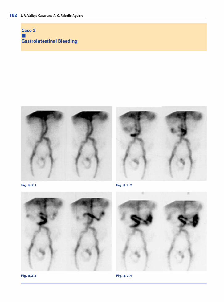
182 J. A. Vallejo Casas and A. C. Rebollo Aguirre
Case 2
Gastrointestinal Bleeding
Fig. 8.2.1 Fig. 8.2.2
Fig. 8.2.4Fig. 8.2.3
RibesLunaRos.indb 182 18.03.2008 12:52:20
Nuclear Medicine 183
A 53-year-old man was referred with suspicion of GI bleeding and negative upper GI endoscopy. At colonoscopy, blood in the bowel precluded the visualization of eventual lesions. Non-colonic diverticulosis or infl ammatory bowel disease were suspected.The patient was hemodynamically unstable, with paleness and severe hypotension, and required blood transfusion. He was referred to the nuclear medicine department to determine the location and extent of GI bleeding. During the isotopic study, the patient had a new episode of hemodynamic instability and a second blood transfusion was performed.
CommentsSuccessful management of patients with acute gastrointestinal (GI) bleeding often depends on accurate location of the bleeding site. In suspected lower GI bleeding, nuclear medicine imaging techniques are the methods of choice because they are highly sensitive.
Isotopic studies, especially 99mTc-labeled red blood cells, may detect bleeding rates as low as 0.05 to 0.1 ml/min, while angiography will only demonstrate bleeding rates greater than 0.5 ml/min. 99mTc-sulphur colloid techniques are also used for this purpose.
The labeled red blood cell technique has the advantage of a longer potential period during which bleeding may be visualized. Moreover, the patient’s clinical needs can be taken care of and blood transfusions can be performed during the study.
The procedure for red blood cell labeling is easy, fast, safe, and inexpensive. The bleeding site will be visualized as a focal area of abnormal radiopharmaceutical
accumulation. Acute GI bleeding is diagnosed when the radiopharmaceutical moves in an antegrade or retrograde direction within the bowel lumen.
A negative isotopic study for GI bleeding indicates no active bleeding and no other imaging modalities are usually needed
Imaging FindingsFigure 8.2.1 shows the major abdominal structures and the liver and spleen blood pools
Figure 8.2.2 shows a focus of activity in the middle abdominal area, deriving from blood extravasated from a small bowel bleeding site. We can see the movement of the activity in the lumen.
Figures 8.2.3 and 8.2.4 (60 and 90 minutes after injection) show a new bleeding episode in the same location and antegrade activity progression within the small bowel lumen.
The diagnosis of active small bowel bleeding at the point shown was confi rmed surgi-cally.
RibesLunaRos.indb 183 18.03.2008 12:52:22
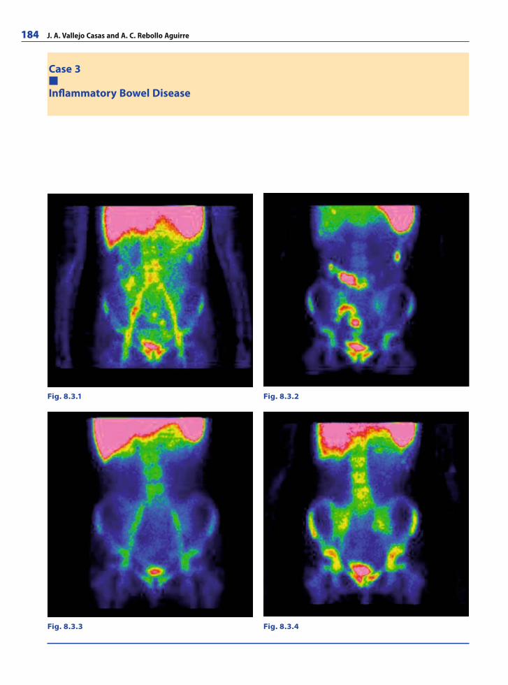
184 J. A. Vallejo Casas and A. C. Rebollo Aguirre
Case 3
Infl ammatory Bowel Disease
Fig. 8.3.1 Fig. 8.3.2
Fig. 8.3.3 Fig. 8.3.4
RibesLunaRos.indb 184 18.03.2008 12:52:22
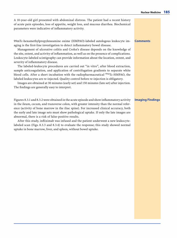
Nuclear Medicine 185
A 10-year-old girl presented with abdominal distress. The patient had a recent history of acute pain episodes, loss of appetite, weight loss, and mucous diarrhea. Biochemical parameters were indicative of infl ammatory activity.
Comments99mTc-hexamethylpropyleneamine oxime (HMPAO)-labeled autologous leukocyte im-aging is the fi rst-line investigation to detect infl ammatory bowel disease.
Management of ulcerative colitis and Crohn’s disease depends on the knowledge of the site, extent, and activity of infl ammation, as well as on the presence of complications. Leukocyte-labeled scintigraphy can provide information about the location, extent, and severity of infl ammatory disease.
The labeled-leukocyte procedures are carried out “in vitro”, after blood extraction, sample anticoagulation, and application of centrifugation gradients to separate white blood cells. After a short incubation with the radiopharmaceutical 99mTc-HMPAO, the labeled leukocytes are re-injected. Quality control before re-injection is obligatory.
Images are obtained at 30 minutes (early set) and 150 minutes (late set) after injection. The fi ndings are generally easy to interpret.
Imaging FindingsFigures 8.3.1 and 8.3.2 were obtained in the acute episode and show infl ammatory activity in the ileum, cecum, and transverse colon, with greater intensity than the normal refer-ence (activity of bone marrow in the iliac spine). For increased clinical accuracy, both the early and late image sets must show pathological uptake. If only the late images are abnormal, there is a risk of false-positive results.
After this study, infl iximab was infused and the patient underwent a new leukocyte-labeled scan (Figs. 8.3.3 and 8.3.4) to evaluate the response; this study showed normal uptake in bone marrow, liver, and spleen, without bowel uptake.
RibesLunaRos.indb 185 18.03.2008 12:52:33
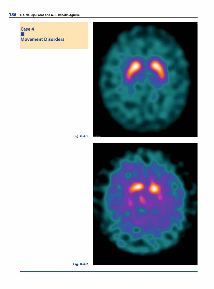
186 J. A. Vallejo Casas and A. C. Rebollo Aguirre
Case 4
Movement Disorders
Fig. 8.4.1
Fig. 8.4.2
RibesLunaRos.indb 186 18.03.2008 12:52:33
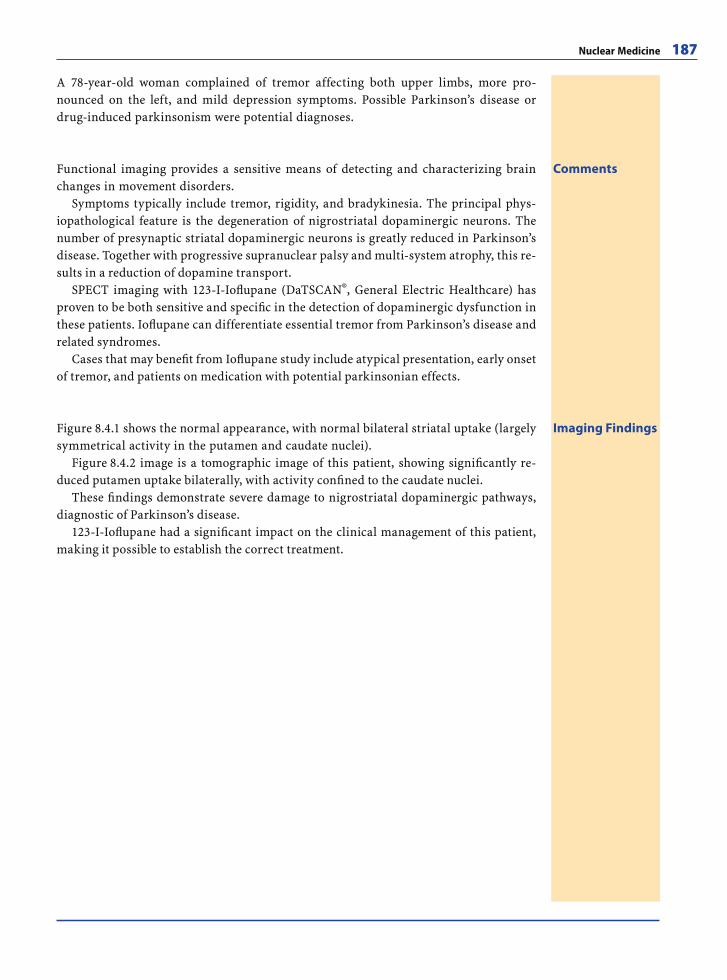
Nuclear Medicine 187
A 78-year-old woman complained of tremor affecting both upper limbs, more pro-nounced on the left, and mild depression symptoms. Possible Parkinson’s disease or drug-induced parkinsonism were potential diagnoses.
CommentsFunctional imaging provides a sensitive means of detecting and characterizing brain changes in movement disorders.
Symptoms typically include tremor, rigidity, and bradykinesia. The principal phys-iopathological feature is the degeneration of nigrostriatal dopaminergic neurons. The number of presynaptic striatal dopaminergic neurons is greatly reduced in Parkinson’s disease. Together with progressive supranuclear palsy and multi-system atrophy, this re-sults in a reduction of dopamine transport.
SPECT imaging with 123-I-Iofl upane (DaTSCAN®, General Electric Healthcare) has proven to be both sensitive and specifi c in the detection of dopaminergic dysfunction in these patients. Iofl upane can differentiate essential tremor from Parkinson’s disease and related syndromes.
Cases that may benefi t from Iofl upane study include atypical presentation, early onset of tremor, and patients on medication with potential parkinsonian effects.
Imaging FindingsFigure 8.4.1 shows the normal appearance, with normal bilateral striatal uptake (largely symmetrical activity in the putamen and caudate nuclei).
Figure 8.4.2 image is a tomographic image of this patient, showing signifi cantly re-duced putamen uptake bilaterally, with activity confi ned to the caudate nuclei.
These fi ndings demonstrate severe damage to nigrostriatal dopaminergic pathways, diagnostic of Parkinson’s disease.
123-I-Iofl upane had a signifi cant impact on the clinical management of this patient, making it possible to establish the correct treatment.
RibesLunaRos.indb 187 18.03.2008 12:52:41
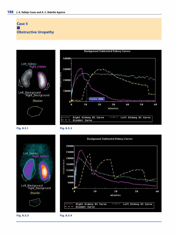
188 J. A. Vallejo Casas and A. C. Rebollo Aguirre
Case 5
Obstructive Uropathy
Fig. 8.5.1 Fig. 8.5.2
Fig. 8.5.3 Fig. 8.5.4
RibesLunaRos.indb 188 18.03.2008 12:52:41
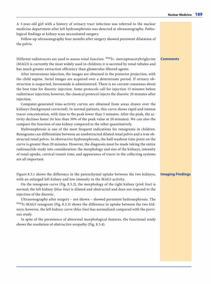
Nuclear Medicine 189
A 3-year-old girl with a history of urinary tract infection was referred to the nuclear medicine department after left hydronephrosis was detected at ultrasonography. Patho-logical fi ndings at kidney scan necessitated surgery.
Follow-up ultrasonography four months after surgery showed persistent dilatation of the pelvis.
CommentsDifferent radiotracers are used to assess renal function. 99mTc- mercaptoacetyltriglycine (MAG3) is currently the most widely used in children; it is secreted by renal tubules and has much greater extraction effi ciency than glomerular-fi ltered agents.
After intravenous injection, the images are obtained in the posterior projection, with the child supine. Serial images are acquired over a determinate period. If urinary ob-struction is suspected, furosemide is administered. There is no current consensus about the best time for diuretic injection. Some protocols call for injection 15 minutes before radiotracer injection; however, the classical protocol injects the diuretic 20 minutes after injection.
Computer-generated time-activity curves are obtained from areas drawn over the kidneys (background corrected). In normal patients, this curve shows rapid and intense tracer concentration, with time to the peak lower than 5 minutes. After the peak, the ac-tivity declines faster (to less than 50% of the peak value at 20 minutes). We can also the compare the function of one kidney compared to the other quantitatively.
Hydronephrosis is one of the most frequent indications for renograms in children. Renograms can differentiate between an unobstructed dilated renal pelvis and a true ob-structed renal pelvis. In obstructive hydronephrosis, the half-washout time point on the curve is greater than 20 minutes. However, the diagnosis must be made taking the entire radionuclide study into consideration: the morphology and size of the kidneys, intensity of renal uptake, cortical transit time, and appearance of tracer in the collecting systems are all important.
Imaging FindingsFigure 8.5.1 shows the difference in the parenchymal uptake between the two kidneys, with an enlarged left kidney and low intensity in the MAG3 activity.
On the renogram curve (Fig. 8.5.2), the morphology of the right kidney (pink line) is normal; the left kidney (blue line) is dilated and obstructed and does not respond to the injection of the diuretic.
Ultrasonography after surgery – not shown – showed persistent hydronephrosis. The 99mTc-MAG3 renogram (Fig. 8.5.3) shows the difference in uptake between the two kid-neys; however, the left kidney curve (blue line) has normalized compared with the previ-ous study.
In spite of the persistence of abnormal morphological features, the functional study shows the resolution of obstructive uropathy (Fig. 8.5.4).
RibesLunaRos.indb 189 18.03.2008 12:52:55
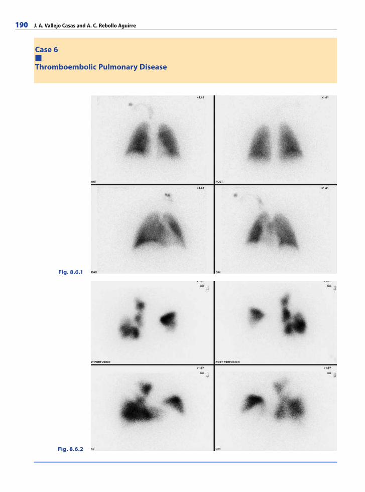
190 J. A. Vallejo Casas and A. C. Rebollo Aguirre
Case 6
Thromboembolic Pulmonary Disease
Fig. 8.6.1
Fig. 8.6.2
RibesLunaRos.indb 190 18.03.2008 12:52:55
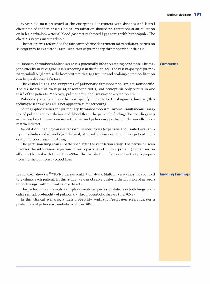
Nuclear Medicine 191
A 65-year-old man presented at the emergency department with dyspnea and lateral chest pain of sudden onset. Clinical examination showed no alterations at auscultation or in leg perfusion. Arterial blood gasometry showed hypoxemia with hypocapnia. The chest X-ray was unremarkable .
The patient was referred to the nuclear medicine department for ventilation-perfusion scintigraphy to evaluate clinical suspicion of pulmonary thromboembolic disease.
CommentsPulmonary thromboembolic disease is a potentially life-threatening condition. The ma-jor diffi culty in its diagnosis is suspecting it in the fi rst place. The vast majority of pulmo-nary emboli originate in the lower extremities. Leg trauma and prolonged immobilization can be predisposing factors.
The clinical signs and symptoms of pulmonary thromboembolism are nonspecifi c. The classic triad of chest paint, thrombophlebitis, and hemoptysis only occurs in one third of the patients. Moreover, pulmonary embolism may be asymptomatic.
Pulmonary angiography is the most specify modality for the diagnosis; however, this technique is invasive and is not appropriate for screening.
Scintigraphic studies for pulmonary thromboembolism involve simultaneous imag-ing of pulmonary ventilation and blood fl ow. The principle fi ndings for the diagnosis are normal ventilation remains with abnormal pulmonary perfusion, the so-called mis-matched defect.
Ventilation imaging can use radioactive inert gases (expensive and limited availabil-ity) or radiolabeled aerosols (widely used). Aerosol administration requires patient coop-eration to coordinate breathing.
The perfusion lung scan is performed after the ventilation study. The perfusion scan involves the intravenous injection of microparticles of human protein (human serum albumin) labeled with technetium-99m. The distribution of lung radioactivity is propor-tional to the pulmonary blood fl ow.
Imaging FindingsFigure 8.6.1 shows a 99mTc-Technegas ventilation study. Multiple views must be acquired to evaluate each patient. In this study, we can observe uniform distribution of aerosols to both lungs, without ventilatory defects.
The perfusion scan reveals multiple mismatched perfusion defects in both lungs, indi-cating a high probability of pulmonary thromboembolic disease (Fig. 8.6.2).
In this clinical scenario, a high probability ventilation/perfusion scan indicates a probability of pulmonary embolism of over 90%.
RibesLunaRos.indb 191 18.03.2008 12:52:58
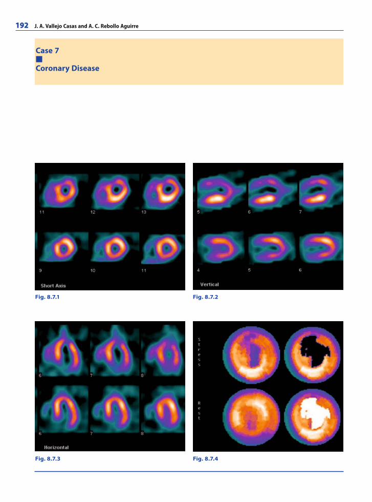
192 J. A. Vallejo Casas and A. C. Rebollo Aguirre
Case 7
Coronary Disease
Fig. 8.7.4Fig. 8.7.3
Fig. 8.7.1 Fig. 8.7.2
RibesLunaRos.indb 192 18.03.2008 12:52:58
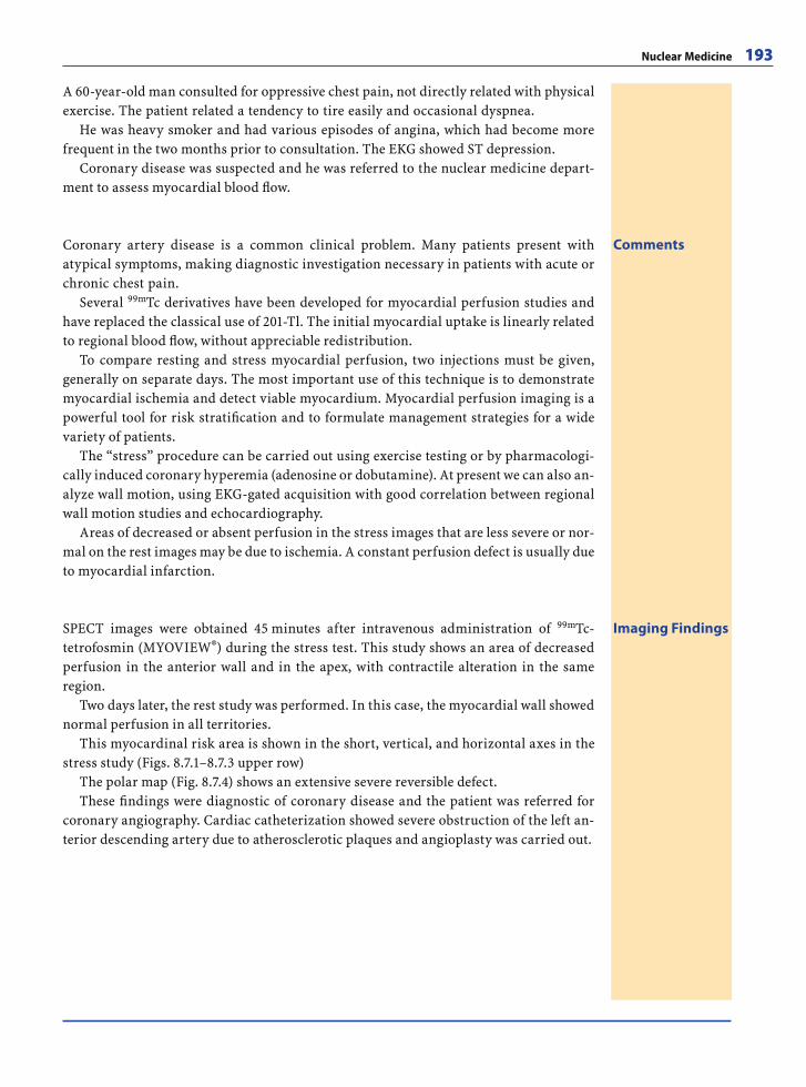
Nuclear Medicine 193
A 60-year-old man consulted for oppressive chest pain, not directly related with physical exercise. The patient related a tendency to tire easily and occasional dyspnea.
He was heavy smoker and had various episodes of angina, which had become more frequent in the two months prior to consultation. The EKG showed ST depression.
Coronary disease was suspected and he was referred to the nuclear medicine depart-ment to assess myocardial blood fl ow.
CommentsCoronary artery disease is a common clinical problem. Many patients present with atypical symptoms, making diagnostic investigation necessary in patients with acute or chronic chest pain.
Several 99mTc derivatives have been developed for myocardial perfusion studies and have replaced the classical use of 201-Tl. The initial myocardial uptake is linearly related to regional blood fl ow, without appreciable redistribution.
To compare resting and stress myocardial perfusion, two injections must be given, generally on separate days. The most important use of this technique is to demonstrate myocardial ischemia and detect viable myocardium. Myocardial perfusion imaging is a powerful tool for risk stratifi cation and to formulate management strategies for a wide variety of patients.
The “stress” procedure can be carried out using exercise testing or by pharmacologi-cally induced coronary hyperemia (adenosine or dobutamine). At present we can also an-alyze wall motion, using EKG-gated acquisition with good correlation between regional wall motion studies and echocardiography.
Areas of decreased or absent perfusion in the stress images that are less severe or nor-mal on the rest images may be due to ischemia. A constant perfusion defect is usually due to myocardial infarction.
Imaging FindingsSPECT images were obtained 45 minutes after intravenous administration of 99mTc- tetrofosmin (MYOVIEW®) during the stress test. This study shows an area of decreased perfusion in the anterior wall and in the apex, with contractile alteration in the same region.
Two days later, the rest study was performed. In this case, the myocardial wall showed normal perfusion in all territories.
This myocardinal risk area is shown in the short, vertical, and horizontal axes in the stress study (Figs. 8.7.1–8.7.3 upper row)
The polar map (Fig. 8.7.4) shows an extensive severe reversible defect.These fi ndings were diagnostic of coronary disease and the patient was referred for
coronary angiography. Cardiac catheterization showed severe obstruction of the left an-terior descending artery due to atherosclerotic plaques and angioplasty was carried out.
RibesLunaRos.indb 193 18.03.2008 12:53:10
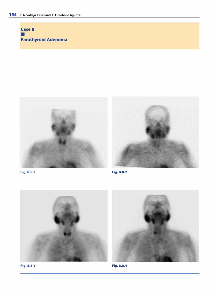
194 J. A. Vallejo Casas and A. C. Rebollo Aguirre
Case 8
Parathyroid Adenoma
Fig. 8.8.1 Fig. 8.8.2
Fig. 8.8.3 Fig. 8.8.4
RibesLunaRos.indb 194 18.03.2008 12:53:10
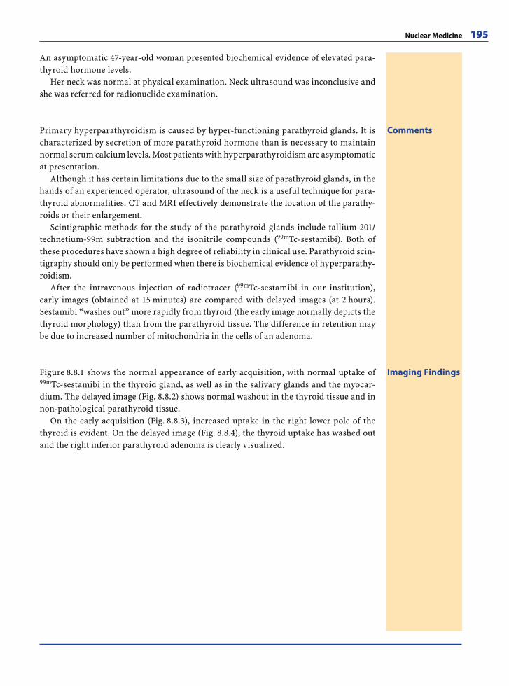
Nuclear Medicine 195
An asymptomatic 47-year-old woman presented biochemical evidence of elevated para-thyroid hormone levels.
Her neck was normal at physical examination. Neck ultrasound was inconclusive and she was referred for radionuclide examination.
CommentsPrimary hyperparathyroidism is caused by hyper-functioning parathyroid glands. It is characterized by secretion of more parathyroid hormone than is necessary to maintain normal serum calcium levels. Most patients with hyperparathyroidism are asymptomatic at presentation.
Although it has certain limitations due to the small size of parathyroid glands, in the hands of an experienced operator, ultrasound of the neck is a useful technique for para-thyroid abnormalities. CT and MRI effectively demonstrate the location of the parathy-roids or their enlargement.
Scintigraphic methods for the study of the parathyroid glands include tallium-201/technetium-99m subtraction and the isonitrile compounds (99mTc-sestamibi). Both of these procedures have shown a high degree of reliability in clinical use. Parathyroid scin-tigraphy should only be performed when there is biochemical evidence of hyperparathy-roidism.
After the intravenous injection of radiotracer (99mTc-sestamibi in our institution), early images (obtained at 15 minutes) are compared with delayed images (at 2 hours). Sestamibi “washes out” more rapidly from thyroid (the early image normally depicts the thyroid morphology) than from the parathyroid tissue. The difference in retention may be due to increased number of mitochondria in the cells of an adenoma.
Imaging FindingsFigure 8.8.1 shows the normal appearance of early acquisition, with normal uptake of 99mTc-sestamibi in the thyroid gland, as well as in the salivary glands and the myocar-dium. The delayed image (Fig. 8.8.2) shows normal washout in the thyroid tissue and in non-pathological parathyroid tissue.
On the early acquisition (Fig. 8.8.3), increased uptake in the right lower pole of the thyroid is evident. On the delayed image (Fig. 8.8.4), the thyroid uptake has washed out and the right inferior parathyroid adenoma is clearly visualized.
RibesLunaRos.indb 195 18.03.2008 12:53:14
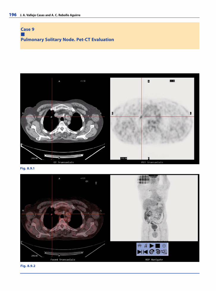
196 J. A. Vallejo Casas and A. C. Rebollo Aguirre
Case 9
Pulmonary Solitary Node. Pet-CT Evaluation
Fig. 8.9.1
Fig. 8.9.2
RibesLunaRos.indb 196 18.03.2008 12:53:14
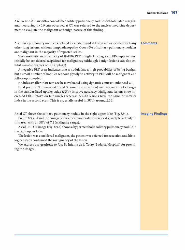
Nuclear Medicine 197
A 68-year-old man with a noncalcifi ed solitary pulmonary nodule with lobulated margins and measuring 1 × 0.9 cms observed at CT was referred to the nuclear medicine depart-ment to evaluate the malignant or benign nature of this fi nding.
CommentsA solitary pulmonary nodule is defi ned as single rounded lesion not associated with any other lung lesions, without lymphadenopathy. Over 40% of solitary pulmonary nodules are malignant in the majority of reported series.
The sensitivity and specifi city of 18-FDG PET is high. Any degree of FDG uptake must initially be considered suspicious for malignancy (although benign lesions can also ex-hibit variable degrees of FDG uptake).
A negative PET scan indicates that a nodule has a high probability of being benign, but a small number of nodules without glycolytic activity in PET will be malignant and follow-up is needed.
Nodules smaller than 1cm are best evaluated using dynamic contrast-enhanced CT.Dual point PET images (at 1 and 3 hours post-injection) and evaluation of changes
in the standardized uptake value (SUV) improve accuracy. Malignant lesions show in-creased FDG uptake on late images whereas benign lesions have the same or inferior index in the second scan. This is especially useful in SUVs around 2.5 I.
Imaging FindingsAxial CT shows the solitary pulmonary nodule in the right upper lobe (Fig. 8.9.1).Figure 8.9.2. Axial PET image shows focal moderately increased glycolytic activity in
this area, with an SUV of 7.2 (malignity range).Axial PET-CT image (Fig. 8.9.3) shows a hypermetabolic solitary pulmonary nodule in
the right upper lobe.The lesion was considered malignant, the patient was referred for resection and histo-
logical study confi rmed the malignancy of the lesion.We express our gratitude to Jose R. Infante de la Torre (Badajoz Hospital) for provid-
ing the images.
RibesLunaRos.indb 197 18.03.2008 12:53:21
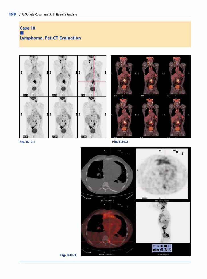
198 J. A. Vallejo Casas and A. C. Rebollo Aguirre
Case 10
Lymphoma. Pet-CT Evaluation
Fig. 8.10.1 Fig. 8.10.2
Fig. 8.10.3
RibesLunaRos.indb 198 18.03.2008 12:53:21
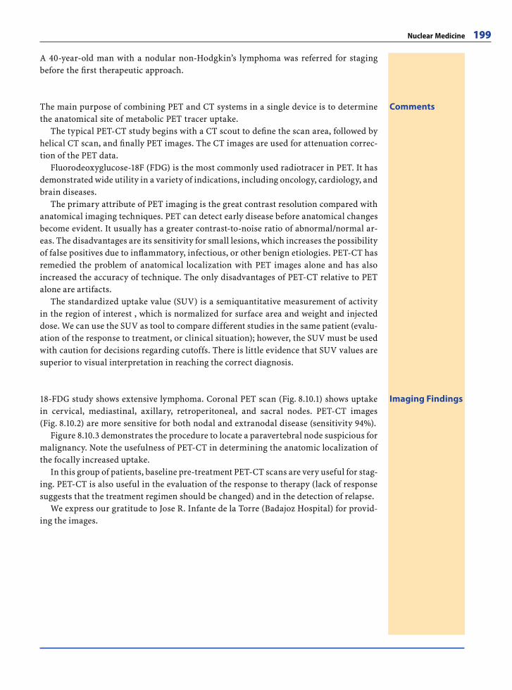
Nuclear Medicine 199
A 40-year-old man with a nodular non-Hodgkin’s lymphoma was referred for staging before the fi rst therapeutic approach.
CommentsThe main purpose of combining PET and CT systems in a single device is to determine the anatomical site of metabolic PET tracer uptake.
The typical PET-CT study begins with a CT scout to defi ne the scan area, followed by helical CT scan, and fi nally PET images. The CT images are used for attenuation correc-tion of the PET data.
Fluorodeoxyglucose-18F (FDG) is the most commonly used radiotracer in PET. It has demonstrated wide utility in a variety of indications, including oncology, cardiology, and brain diseases.
The primary attribute of PET imaging is the great contrast resolution compared with anatomical imaging techniques. PET can detect early disease before anatomical changes become evident. It usually has a greater contrast-to-noise ratio of abnormal/normal ar-eas. The disadvantages are its sensitivity for small lesions, which increases the possibility of false positives due to infl ammatory, infectious, or other benign etiologies. PET-CT has remedied the problem of anatomical localization with PET images alone and has also increased the accuracy of technique. The only disadvantages of PET-CT relative to PET alone are artifacts.
The standardized uptake value (SUV) is a semiquantitative measurement of activity in the region of interest , which is normalized for surface area and weight and injected dose. We can use the SUV as tool to compare different studies in the same patient (evalu-ation of the response to treatment, or clinical situation); however, the SUV must be used with caution for decisions regarding cutoffs. There is little evidence that SUV values are superior to visual interpretation in reaching the correct diagnosis.
Imaging Findings18-FDG study shows extensive lymphoma. Coronal PET scan (Fig. 8.10.1) shows uptake in cervical, mediastinal, axillary, retroperitoneal, and sacral nodes. PET-CT images (Fig. 8.10.2) are more sensitive for both nodal and extranodal disease (sensitivity 94%).
Figure 8.10.3 demonstrates the procedure to locate a paravertebral node suspicious for malignancy. Note the usefulness of PET-CT in determining the anatomic localization of the focally increased uptake.
In this group of patients, baseline pre-treatment PET-CT scans are very useful for stag-ing. PET-CT is also useful in the evaluation of the response to therapy (lack of response suggests that the treatment regimen should be changed) and in the detection of relapse.
We express our gratitude to Jose R. Infante de la Torre (Badajoz Hospital) for provid-ing the images.
RibesLunaRos.indb 199 18.03.2008 12:53:36
200 J. A. Vallejo Casas and A. C. Rebollo Aguirre
Further ReadingsBooksAtlas of PET/CT Imaging in Oncology. Czernin J, Dahlbom
M, Ratib O, Schiepers C (2004) Berlin-Heidelberg: Spring-er-Verlag. ISBN-13: 9783540209522
Cardiac PET and PET/CT Imaging. Di Carli MF, Lipton MJ (2007) Berlin-Heidelberg: Springer-Verlag. ISBN-13: 9780387352756
Diagnostic Nuclear Medicine. Sandler MP, Coleman MP, Paton JA, Wackers FJ, Gottschalk A (2004) Philadelphia: Lippincott Williams & Wilkins. ISBN-13: 9780781732529
Nuclear Cardiology: Practical Applications. Heller GV, Hendel RC (2004) New York: McGraw-Hill. ISBN-13: 9780071386357
Nuclear Medicine: in Clinical Diagnosis and Treatment. Ell PJ, Gambhir SS (2004) Edinburgh: Churchill Livingstone. ISBN-13: 9780443073120
Nuclear Medicine: The Requisites. Ziessman HA, O’Malley JP, Thrall JH (2007) New York: Elsevier. ISBN-13: 9780323029469
Pediatric Nuclear Medicine/PET. Treves ST (2007) New York: Springer-Verlag. ISBN-13: 9780387323213
PET and PET/CT: A clinical guide. Lin EC, Alavi A (2005) New York: Thieme. ISBN-13: 9781588904003
Physics and Radiobiology of Nuclear Medicine. Saha GB (2006) Berlin-Heidelberg: Springer-Verlag. ISBN-13: 9780387307541
The Pathophysiologic Basis of Nuclear Medicine. Elgazzar AH (2006) Berlin-Heidelberg: Springer-Verlag. ISBN-13: 9783540239925
Weblinkshttp://biomedcentral.com/bmcnuc/medhttp://bnms.org.ukhttp://brighamrad.harvard.edu/education/online/introduc-
tion.htmlhttp://eamn.orghttp://gamma.wustl.edu/home.htlmhttp://nucmedinfo.comhttp://semn.eshttp://snmjournals.orghttp://snm.org http://springerlink.com
Articles
Brain
Claus JJ, van Harskamp F, Breteler MM, Krenning EP, de Koning I, van der Cammen TJ, Hofman A, Hasan D. The diagnostic value of SPECT with Tc 99m HMPAO in Al-zheimer’s disease: a population-based study. Neurology 1994; 44:454–461
Conrad GR, Sinha P. Scintigraphy as a confi rmatory test of brain death. Semin Nucl Med 2003; 33(4):312–323
Costa DC, Motteux IM, McCready AC. Diagnosis of brain death with technetium 99m hexamethylpropylene amine oxime. Eur J Nucl Med 1991; 18(7):503–506
de la Riva A, Gonzalez FM, Llamas-Elvira JM, et al. Diagno-sis of brain death: superiority of perfusion studies with 99Tcm-HMPAO over conventional radionuclide cerebral angiography. Br J Radiol 1992; 65(772):289–294
Donohoe KJ, Frey KA, Gerbaudo VH, Mariani G, Nagel JS, Shulkin B. Procedure guideline for brain death scintig-raphy. J Nucl Med 2003; 44(5):846–851
Dougall NJ, Bruggink S, Ebmeier KP. Systematic review of the diagnostic accuracy of 99mTc-HMPAO-SPECT in de-mentia Am J Geriatr Psychiatry 2004; 12(6):554–570
Gordon I. Cerebral blood fl ow imaging in paediatrics: a re-view. Nucl Med Commun 1996; 17(12):1021–1029
Kurtek RW, Lai KK, Tauxe WN, Eidelman BH, Fung JJ. Tc-99m hexamethylpropylene amine oxime scintigraphy in the diagnosis of brain death and its implications for the harvesting of organs used for transplantation. Clin Nucl Med 2000; 25(1):7–10
Uchida Y, Minoshima S, Okada S, Kawata T, Ito H. Diag-nosis of dementia using perfusion SPECT imaging at the patient’s initial visit to a cognitive disorder clinic. Clin Nucl Med 2006; 31(12):764–773
Weckesser M, Schober O. Brain death revisited: utility confi rmed for nuclear medicine. Eur J Nucl Med 1999; 26(11):1387–1391
Movement Disorders
Booij J, Speelman JD, Horstink MW, Wolters EC. The clinical benefi t of imaging striatal dopamine transporters with [123I]FP-CIT SPET in differentiating patients with pre-synaptic parkinsonism from those with other forms of parkinsonism. Eur J Nucl Med 2001; 28(3):266–272
Catafau AM, Tolosa E; DaTSCAN Clinically Uncertain Par-kinsonian Syndromes Study Group. Impact of dopamine transporter SPECT using 123I-Iofl upane on diagnosis and management of patients with clinically uncertain Parkin-sonian syndromes. Mov Disord 2004; 19(10):1175–1182
Filippi L, Manni C, Pierantozzi M, Brusa L, Danieli R, Stanzi-one P, Schillaci O. 123I-FP-CIT in progressive supranucle-ar palsy and in Parkinson’s disease: a SPECT semiquanti-tative study. Nucl Med Commun 2006; 27(4):381–386
Lorberboym M, Djaldetti R, Melamed E, Sadeh M, Lampl Y. 123I-FP-CIT SPECT imaging of dopamine transport-ers in patients with cerebrovascular disease and clinical diagnosis of vascular parkinsonism. J Nucl Med 2004; 45(10):1688–1693
Marshall V, Grosset D. Role of dopamine transporter im-aging in routine clinical practice. Mov Disord 2003; 18(12):1415–1423
Marshall VL, Patterson J, Hadley DM, Grosset KA, Grosset DG. Two-year follow-up in 150 consecutive cases with normal dopamine transporter imaging. Nucl Med Com-mun 2006; 27(12):933–937
McKeith I, O’Brien J, Walker Z, Tatsch K, Booij J, Darcourt J, Padovani A, Giubbini R, Bonuccelli U, Volterrani D, Holmes C, Kemp P, Tabet N, Meyer I, Reininger C; DLB Study Group. Sensitivity and specifi city of dopamine transporter imaging with 123I-FP-CIT SPECT in demen-tia with Lewy bodies: a phase III, multicentre study. Lan-cet Neurol 2007; 6(4):305–313
RibesLunaRos.indb 200 18.03.2008 12:53:36
Nuclear Medicine 201
Schillaci O, Pierantozzi M, Filippi L, Manni C, Brusa L, Dan-ieli R, Bernardi G, Simonetti G, Stanzione P. The effect of levodopa therapy on dopamine transporter SPECT imaging with (123)I-FP-CIT in patients with Parkinson’s disease. Eur J Nucl Med Mol Imaging 2005; 32(12):1452–1456
Spiegel J, Mollers MO, Jost WH, Fuss G, Samnick S, Dill-mann U, Becker G, Kirsch CM. FP-CIT and MIBG scin-tigraphy in early Parkinson’s disease. Mov Disord 2005; 20(5):552–561
Coronary Disease
Cerqueira MD. Nuclear cardiology: fi nally a one-stop shop for diagnosis, risk stratifi cation, and management of coronary artery disease. Clin Cardiol 2006; 29(9 Suppl 1):I26–33
Elhendy A, Bax JJ, Poldermans D. Dobutamine stress myo-cardial perfusion imaging in coronary artery disease. J Nucl Med 2002; 43(12):1634–1646
Hendel RC, Jamil T, Glover DK. Pharmacologic stress test-ing: new methods and new agents. J Nucl Cardiol 2003; 10(2):197–204
Heo J, Iskandrian AS. Technetium-labeled myocardial per-fusion agents. Cardiol Clin 1994; 12(2):187–198
Iskandrian AS. Adenosine myocardial perfusion imaging. J Nucl Med 1994; 35(4):734–736
Kontos MC, Tatum JL. Imaging in the evaluation of the pa-tient with suspected acute coronary syndrome. Semin Nucl Med 2003; 33(4):246–258
Metz LD, Beattie M, Hom R, Redberg RF, Grady D, Fleis-chmann KE. The prognostic value of normal exercise myocardial perfusion imaging and exercise echocar-diography: a meta-analysis. J Am Coll Cardiol 2007; 49(2):227–237
Russell RR 3rd, Zaret BL. Nuclear cardiology: present and future. Curr Probl Cardiol 2006; 31(9):557–629
Tamaki N, Morita K. SPET in cardiology. Diagnosis, prog-nosis, and management of patients with coronary artery disease. Q J Nucl Med Mol Imaging 2005; 49(2):193–203
Udelson JE, Shafer CD, Carrio I. Radionuclide imaging in heart failure: assessing etiology and outcomes and im-plications for management. J Nucl Cardiol 2002; 9(5 Suppl):40S–52S
Gastrointestinal Bleeding
Feingold DL, Caliendo FJ, Chinn BT, et al. Does hemody-namic instability predict positive technetium-labeled red blood cell scintigraphy in patients wit acute lower gas-trointestinal bleeding? A review of 50 patients. Dis Colon Rectum 2005; 48(5):1001–1004
Ford PV, Bartold SP, Fink-Bennett DM, Jolles PR, Lull RJ, Maurer AH, Seabold J. Procedure guideline for gastro-intestinal bleeding and Meckel’s diverticulum scintig-raphy. Society of Nuclear Medicine. J Nucl Med 1999; 40(7):1226–1232
Howarth DM, Tang K, Lees W. The clinical utility of nuclear medicine imaging for the detection of occult gastrointes-
tinal haemorrhage. Nucl Med Commun 2002; 23(6):591–594
Infante JR, Gonzalez FM, Vallejo JA, Torres M, Pacheco C, Latre JM. False-positive results of a gastrointestinal bleeding study caused by an ectopic kidney. Clin Nucl Med 2000; 25(8):645–646
Kan JH, Funaki B, O’Rourke BD, Ward MB, Appelbaum DE. Delayed 99mTc-labeled erythrocyte scintigraphy in pa-tients with lower gastrointestinal tract hemorrhage: ef-fect of positive fi ndings on clinical management. Acad Radiol 2003; 10(5):497–501
Lefkovitz Z, Cappell MS, Kaplan M, Mitty H, Gerard P. Ra-diology in the diagnosis and therapy of gastrointestinal bleeding. Gastroenterol Clin North Am 2000; 29(2):489–512
Olds GD, Cooper GS, Chak A, Sivak M, Chiteale AA, Wong RCK. The yield of bleeding scans in acute lower gas-trointestinal hemorrhage. J Clin Gastroenterol 2005; 39(4):273–277
Ponzo F, Zhuang H, Liu FM, Lacorte LB, Moussavian B, Wang S, Alavi A. Tc-99m sulfur colloid and Tc-99m tagged red blood cell methods are comparable for detecting lower gastrointestinal bleeding in clinical practice. Clin Nucl Med 2002; 27(6):405–409
Zettinig G, Staudenherz A, Leitha T. The importance of de-layed images in gastrointestinal bleeding scintigraphy. Nucl Med Commun 2002; 23(8):803–808
Zuckier LS. Acute gastrointestinal bleeding. Semin Nucl Med 2003; 33(4):297–311
Infl ammatory Bowel Disease
Alberini JL, Badran A, Freneaux E, Hadji S, Kalifa G, Devaux JY, Dupont T. Technetium-99m HMPAO-labeled leuko-cyte imaging compared with endoscopy, ultrasonogra-phy, and contrast radiology in children with infl amma-tory bowel disease. J Pediatr Gastroenterol Nutr 2001; 32(3):278–286
Bennink R, Peeters M, D’Haens G, Rutgeerts P, Mortel-mans L. Tc-99m HMPAO white blood cell scintigraphy in the assessment of the extent and severity of an acute exacerbation of ulcerative colitis. Clin Nucl Med 2001; 26(2):99–104
Charron M. Pediatric infl ammatory bowel disease imaged with Tc-99m white blood cells. Clin Nucl Med 2000; 25(9):708–715
Grahnquist L, Chapman SC, Hvidsten S, Murphy MS. Eval-uation of 99mTc-HMPAO leukocyte scintigraphy in the investigation of pediatric infl ammatory bowel disease. J Pediatr 2003; 143(1):48–53
Gyorke T, Duffek L, Bartfai K, Mako E, Karlinger K, Mes-ter A, Tarjan Z. The role of nuclear medicine in infl am-matory bowel disease. A review with experiences of as-pecifi c bowel activity using immunoscintigraphy with 99mTc anti-granulocyte antibodies. Eur J Radiol 2000; 35(3):183–192
Lachter J, Isseroff HN, Yasin K, Keidar Z, Israel O. Radiola-beled leukocyte imaging in infl ammatory bowel disease: a prospective blinded evaluation. Hepatogastroenterol-ogy 2003; 50(53):1439–1441
RibesLunaRos.indb 201 18.03.2008 12:53:36
202 J. A. Vallejo Casas and A. C. Rebollo Aguirre
Martin-Comin J, Prats E. Clinical applications of radiola-beled blood elements in infl ammatory bowel disease. Q J Nucl Med 1999; 43(1):74–82
Martin-Comin J. Radiolabelled white blood cells in in-fl ammatory bowel diseases. Nucl Med Commun 2002; 23(11):1039–1040
Peters AM. Nuclear medicine imaging in infection and in-fl ammation. J R Coll Physicians Lond 1998; 32(6):512–519
Stokkel MP, Reigman HE, Pauwels EK. Scintigraphic head-to-head comparison between 99mTc-WBCs and 99mTc-LeukoScan in the evaluation of infl ammatory bowel dis-ease: a pilot study. Eur J Nucl Med Mol Imaging 2002; 29(2):251–254
Pulmonary Thromboembolism
Giordano A, Angiolillo DJ. Current role of lung scintigraphy in pulmonary embolism. Q J Nucl Med 2001; 45(4):294–301
Hagen PJ, Hartmann IJ, Hoekstra OS, Stokkel MP, Teule GJ, Prins MH. How to use a gestalt interpretation for ven-tilation-perfusion lung scintigraphy. J Nucl Med 2002; 43(10):1317–1323
Hartmann IJ, Hagen PJ, Stokkel MP, Hoekstra OS, Prins MH. Technegas versus (81m)Kr ventilation-perfusion scintigraphy: a comparative study in patients with sus-pected acute pulmonary embolism. J Nucl Med 2001; 42(3):393–400
Hatabu H, Uematsu H, Nguyen B, Miller WT Jr, Hasegawa I, Gefter WB. CT and MR in pulmonary embolism: A changing role for nuclear medicine in diagnostic strategy. Semin Nucl Med 2002; 32(3):183–192
Itti E, Nguyen S, Robin F, Desarnaud S, Rosso J, Harf A, Meignan. Distribution of ventilation/perfusion ratios in pulmonary embolism: an adjunct to the interpretation of ventilation/perfusion lung scans. J Nucl Med 2002; 43(12):1596–1602
Meignan MA. Lung ventilation/perfusion SPECT: the right technique for hard times. J Nucl Med 2002; 43(5):648–651
Rizzo-Padoin N, Farina A, Le Pen C, Duet M, Mundler O, Leverge R. A comparison of radiopharmaceutical agents used for the diagnosis of pulmonary embolism. Nucl Med Commun 2001; 22(4):375–381
Schumichen C. V/Q-scanning/SPECT for the diagnosis of pulmonary embolism. Respiration 2003; 70(4):329–342
Wilson HT, Meagher TM, Williams S. Combined helical computed tomographic pulmonary angiography and lung perfusion scintigraphy for investigating acute pul-monary embolism. Clin Radiol 2002; 57(1):33–36
Worsley DF, Alavi A. Radionuclide imaging of acute pulmo-nary embolism. Semin Nucl Med 2003; 33(4):259–278
Parathyroid Imaging
Coakley AJ. Parathyroid imaging. Nucl Med Commun 1995; 16(7):522–533
de Jonge FA, Pauwels EK, Hamdy NA. Scintigraphy in the clinical evaluation of disorders of mineral and skel-etal metabolism in renal failure. Eur J Nucl Med 1991; 18(10):839–855
Giordano A, Rubello D, Casara D. New trends in parathyroid scintigraphy. Eur J Nucl Med 2001; 28(9):1409–1420
Gotthardt M, Lohmann B, Behr TM, et al. Clinical value of parathyroid scintigraphy with technetium-99m me-thoxyisobutylisonitrile: discrepancies in clinical data and a systematic metaanalysis of the literature. World J Surg 2004; 28(1):100–107. Epub 2003 Nov 26
Hiromatsu Y, Ishibashi M, Nishida H, Okuda S, Miyake I. Technetium-99m tetrofosmin parathyroid imaging in patients with primary hyperparathyroidism. Intern Med 2000; 39(2):101–106
Kettle AG, O’Doherty MJ. Parathyroid imaging: how good is it and how should it be done? Semin Nucl Med 2006; 36(3):206–211
McBiles M, Lambert AT, Cote MG, Kim SY. Sestamibi para-thyroid imaging. Semin Nucl Med 1995; 25(3):221–234
Mitchell BK, Kinder BK, Cornelius E, Stewart AF. Primary hyperparathyroidism: preoperative localization using technetium-sestamibi scanning. J Clin Endocrinol Metab 1995; 80(1):7–10
Palestro CJ, Tomas MB, Tronco GG. Radionuclide imag-ing of the parathyroid glands. Semin Nucl Med 2005; 35(4):266–276
Smith JR, Oates ME. Radionuclide imaging of the parathy-roid glands: patterns, pearls, and pitfalls. Radiographics. 2004; 24(4):1101–1115
Diuretic Renography
Boubaker A, Prior J, Antonescu C, Meyrat B, Frey P, Delaloye AB. F+0 renography in neonates and infants younger than 6 months: an accurate method to diagnose severe obstructive uropathy. J Nucl Med 2001; 42(12):1780–1788
Dubovsky EV, Russell CD, Bischof-Delaloye A, Bubeck B, Chaiwatanarat T, Hilson AJ, Rutland M, Oei HY, Sfaki-anakis GN, Taylor A Jr. Report of the Radionuclides in Nephrourology Committee for evaluation of transplant-ed kidney (review of techniques). Semin Nucl Med 1999; 29(2):175–188
Eskild-Jensen A, Gordon I, Piepsz A, Frokiaer. Congenital unilateral hydronephrosis: a review of the impact of diuretic renography on clinical treatment. J Urol 2005; 173(5):1471–1476
Gordon I. Assessment of pediatric hydronephrosis using output effi ciency. J Nucl Med 1997; 38(9):1487–1489
Koelliker SL, Cronan JJ. Acute urinary tract obstruction. Im-aging update. Urol Clin North Am 1997; 24(3):571-582
Koff SA, Binkovitz L, Coley B, Jayanthi V. Renal pelvis vol-ume during diuresis in children with hydronephrosis: implications for diagnosing obstruction with diuretic renography. J Urol 2005; 174(1):303–307
O’Reilly PH. Obstructive uropathy. Q J Nucl Med 2002; 46(4):295–303
Piepsz A, Ham HR. Pediatric applications of renal nuclear medicine. Semin Nucl Med 2006; 36:16–35
Taylor A Jr, Nally JV. Clinical applications of renal scintigra-phy. AJR Am J Roentgenol 1995; 164(1):31–41
Wong DC, Rossleigh MA, Farnsworth RH. F+0 diuresis renography in infants and children. J Nucl Med 1999; 40(11):1805–1811
RibesLunaRos.indb 202 18.03.2008 12:53:36
Nuclear Medicine 203
PET in Lymphoma
Baudard M, Comte F, Conge AM, Mariano-Goulart D, Klein B, Rossi JF. Importance of [18F]fl uorodeoxyglucose-posi-tron emission tomography scanning for the monitoring of responses to immunotherapy in follicular lymphoma.Leuk Lymphoma 2007; 48(2):381–388
Brepoels L, Stroobants S, Verhoef G. PET and PET/CT for response evaluation in lymphoma: current practice and developments. Leuk Lymphoma 2007; 48(2):270–282
Burton C, Ell P, Linch D. The role of PET imaging in lym-phoma. Br J Haematol 2004; 126(6):772–784
Collins CD. PET in lymphoma. Cancer Imaging 2006; 6:S63–70
Jerusalem G, Hustinx R, Beguin Y, Fillet G. Evaluation of therapy for lymphoma. Semin Nucl Med 2005; 35(3):186–196
Juweid ME, Stroobants S, Hoekstra OS, et al. Imaging Sub-committee of International Harmonization Project in Lymphoma. Use of positron emission tomography for response assessment of lymphoma: consensus of the Imaging Subcommittee of International Harmonization Project in Lymphoma. J Clin Oncol 2007; 25(5):571–578
Kazama T, Faria SC, Varavithya V, Phongkitkarun S, Ito H, Macapinlac HA. FDG PET in the evaluation of treatment for lymphoma: clinical usefulness and pitfalls. Radio-graphics 2005; 25(1):191–207
Kirby AM, George Mikhaeel N. The role of FDG PET in the management of lymphoma: practical guidelines. Nucl Med Commun 2007; 28(5):355–357
Kumar R, Maillard I, Schuster SJ, Alavi A. Utility of fl uorode-oxyglucose-PET imaging in the management of patients with Hodgkin’s and non-Hodgkin’“s lymphomas. Radiol Clin North Am 2004; 42(6):1083–1100
Mikhaeel NG. Use of FDG-PET to monitor response to che-motherapy and radiotherapy in patients with lymphomas. Eur J Nucl Med Mol Imaging 2006; 33 Suppl 1:22–26
PET in Solitary Pulmonary Nodule
Aquino SL, Kuester LB, Muse VV, Halpern EF, Fischman AJ. Accuracy of transmission CT and FDG-PET in the detec-tion of small pulmonary nodules with integrated PET/CT. Eur J Nucl Med Mol Imaging 2006; 33(6):692–696
Bruzzi JF, Munden RF. PET/CT imaging of lung cancer. J Thorac Imaging 2006; 21(2):123–136
Bunyaviroch T, Coleman R. PET evaluation of lung cancer. J Nucl Med 2006; 47(3):451–469
Coleman RE. PET in lung cancer. J Nucl Med 1999; 40(5):814–820
Goerres GW, von Schulthess GK, Steinert HC. Why most PET of lung and head-and-neck cancer will be PET/CT. J Nucl Med 2004; 45 Suppl 1:66S–71S
Jeong YJ, Yi CA, Lee KS. Solitary pulmonary nodules: detec-tion, characterization, and guidance for further diagnos-tic workup and treatment. AJR Am J Roentgenol 2007; 188(1):57–68
Kim SK, Allen-Auerbach M, Goldin J, Fueger BJ, Dahlbom M, Brown M, et al. Accuracy of PET/CT in character-ization of solitary pulmonary lesions. J Nucl Med 2007; 48(2):214–220
Kuehl H, Veit P, Rosenbaum SJ, Bockisch A, Antoch G. Can PET/CT replace separate diagnostic CT for cancer imaging? Optimizing CT protocols for imaging cancers of the chest and abdomen. J Nucl Med 2007; 48 Suppl 1:45S–57S
Mavi A, Lakhani P, Zhuang H, Gupta NC, Alavi A. Fluoro-deoxyglucose-PET in characterizing solitary pulmonary nodules, assessing pleural diseases, and the initial staging, restaging, therapy planning, and monitoring response of lung cancer. Radiol Clin North Am 2005; 43(1):1–21, ix
Yi CA, Lee KS, Kim BT, Choi JY, Kwon OJ, Kim H, Shim YM, Chung MJ. Tissue characterization of solitary pulmonary nodule: comparative study between helical dynamic CT and integrated PET/CT. J Nucl Med 2006; 47(3):443–450
RibesLunaRos.indb 203 18.03.2008 12:53:36

RibesLunaRos.indb V 18.03.2008 12:48:44
Nuclear Medicine
Pediatric Radiology 9
Pedro Daltro, L. Celso Hygino Cruz Jr, Renata Do A. Nogueira, and Mirriam T. C. Porto
IntroductionThe old medical saying “a kid is not a small grown-up” is perfect to illustrate the impor-tance of pediatric radiology within the fi eld of radiology. As Dr. Lefèbvre (the father of the European Society of Pediatric Radiology (ESPR)) said in 1958: “In pediatrics, losing time entails the risk of losing a life or permanent disability for an entire life… Radiologists who perform children’s examinations must be well informed and competent.”
All important medical centers should have a pediatric radiologist because there are important anatomic differences between children and adults; furthermore, different dis-eases, with their own specifi c characteristics affect children. Lastly, children are espe-cially vulnerable to the effects of ionizing radiation, so the imaging techniques of choice used are often different, too. Like all radiologists, those working in pediatrics must keep abreast of the continual advances in imaging techniques and knowledge about diseases as well as new developments in treatment.
Pediatric radiology can be said to have become fi rmly established in 1945 with the appearance of the fi rst edition of John Caffey’s seminal book Pediatric X-ray Diagnosis (Year Book Publishers, Chicago). This book set guidelines for the imaging of pediatric patients and was and is referred to as “the Book” because it was the fi rst reference work available.
In the second half of the 20th century, pediatric radiology underwent great develop-ments in the U.S., mainly in Boston under the tutelage of Dr. Edward B.D. Neuhauser. Dr. Neuhauser’s department was then known as a “leading light of pediatric radiology”. At the same time, some European physicians also studied and worked to improve pediatric radiology: Dr. Lefèbvre, J Sauvegrain, Cl Fauré, H.Kaufman, U. Rudhe and others. Europe has now outstanding professors in the area of pediatric radiology all over the continent.
Pedro DaltroHonorary Member of |ESPR
Ex- President of SLARP
RibesLunaRos.indb 205 18.03.2008 12:53:36
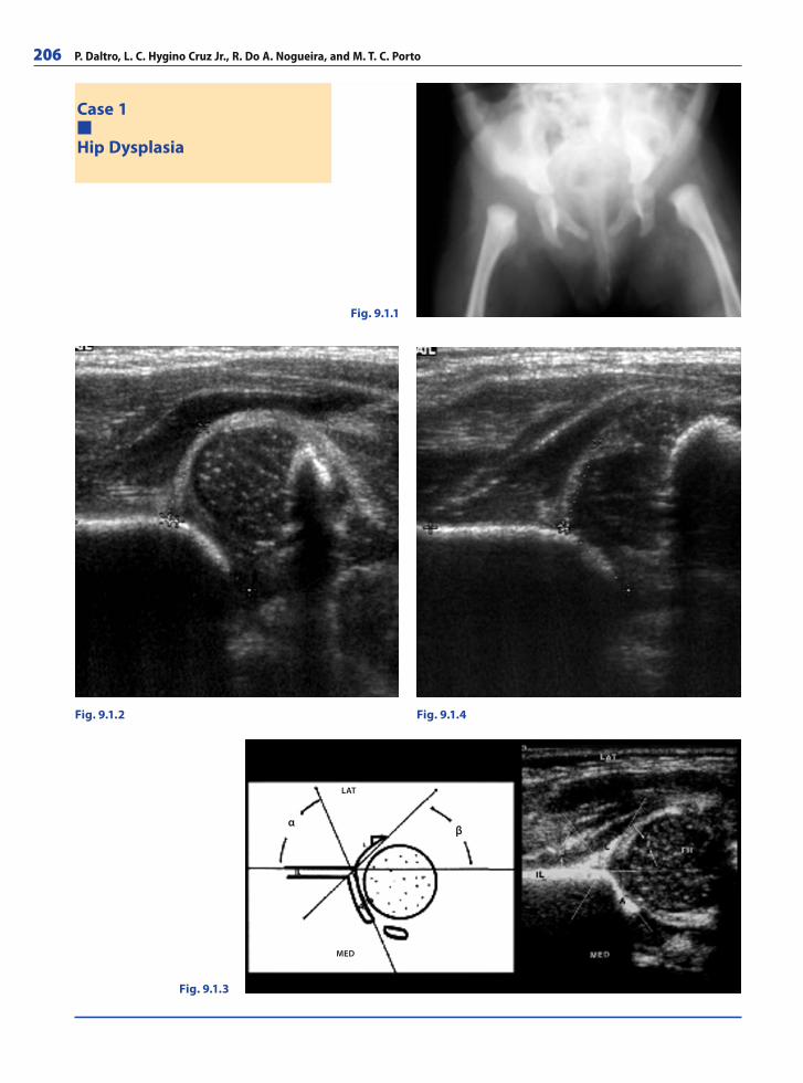
206 P. Daltro, L. C. Hygino Cruz Jr., R. Do A. Nogueira, and M. T. C. Porto
Case 1
Hip Dysplasia
Fig. 9.1.1
Fig. 9.1.2 Fig. 9.1.4
Fig. 9.1.3
αβ
LAT
MED
RibesLunaRos.indb 206 18.03.2008 12:53:37
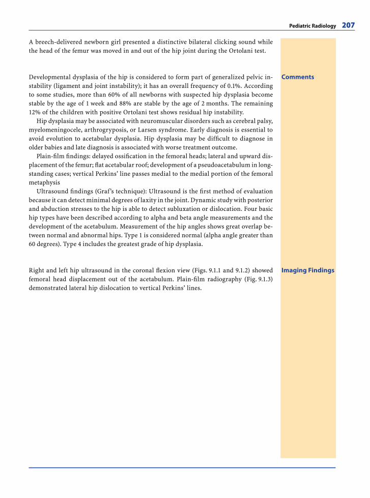
Pediatric Radiology 207
A breech-delivered newborn girl presented a distinctive bilateral clicking sound while the head of the femur was moved in and out of the hip joint during the Ortolani test.
CommentsDevelopmental dysplasia of the hip is considered to form part of generalized pelvic in-stability (ligament and joint instability); it has an overall frequency of 0.1%. According to some studies, more than 60% of all newborns with suspected hip dysplasia become stable by the age of 1 week and 88% are stable by the age of 2 months. The remaining 12% of the children with positive Ortolani test shows residual hip instability.
Hip dysplasia may be associated with neuromuscular disorders such as cerebral palsy, myelomeningocele, arthrogryposis, or Larsen syndrome. Early diagnosis is essential to avoid evolution to acetabular dysplasia. Hip dysplasia may be diffi cult to diagnose in older babies and late diagnosis is associated with worse treatment outcome.
Plain-fi lm fi ndings: delayed ossifi cation in the femoral heads; lateral and upward dis-placement of the femur; fl at acetabular roof; development of a pseudoacetabulum in long-standing cases; vertical Perkins’ line passes medial to the medial portion of the femoral metaphysis
Ultrasound fi ndings (Graf’s technique): Ultrasound is the fi rst method of evaluation because it can detect minimal degrees of laxity in the joint. Dynamic study with posterior and abduction stresses to the hip is able to detect subluxation or dislocation. Four basic hip types have been described according to alpha and beta angle measurements and the development of the acetabulum. Measurement of the hip angles shows great overlap be-tween normal and abnormal hips. Type 1 is considered normal (alpha angle greater than 60 degrees). Type 4 includes the greatest grade of hip dysplasia.
Imaging FindingsRight and left hip ultrasound in the coronal fl exion view (Figs. 9.1.1 and 9.1.2) showed femoral head displacement out of the acetabulum. Plain-fi lm radiography (Fig. 9.1.3) demonstrated lateral hip dislocation to vertical Perkins’ lines.
RibesLunaRos.indb 207 18.03.2008 12:53:39
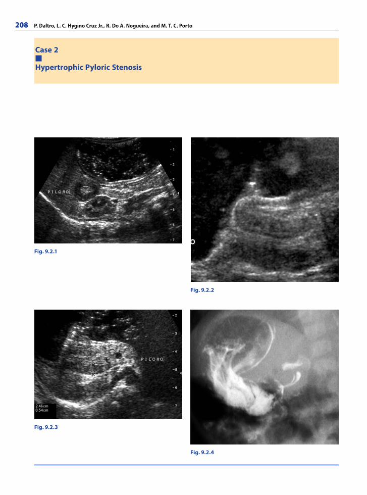
208 P. Daltro, L. C. Hygino Cruz Jr., R. Do A. Nogueira, and M. T. C. Porto
Case 2
Hypertrophic Pyloric Stenosis
Fig. 9.2.1
Fig. 9.2.2
Fig. 9.2.4
Fig. 9.2.3
RibesLunaRos.indb 208 18.03.2008 12:53:39
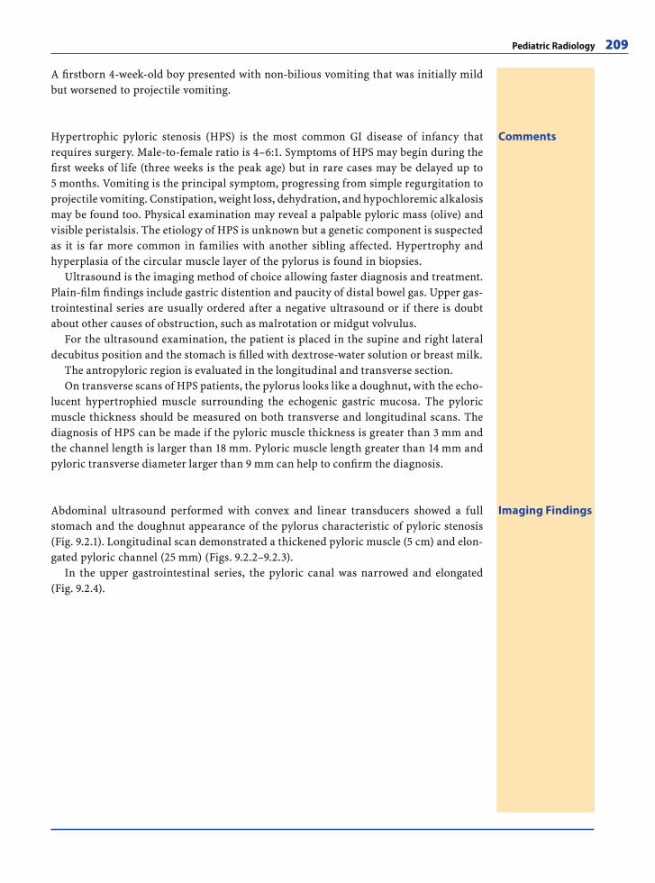
Pediatric Radiology 209
A fi rstborn 4-week-old boy presented with non-bilious vomiting that was initially mild but worsened to projectile vomiting.
CommentsHypertrophic pyloric stenosis (HPS) is the most common GI disease of infancy that requires surgery. Male-to-female ratio is 4–6:1. Symptoms of HPS may begin during the fi rst weeks of life (three weeks is the peak age) but in rare cases may be delayed up to 5 months. Vomiting is the principal symptom, progressing from simple regurgitation to projectile vomiting. Constipation, weight loss, dehydration, and hypochloremic alkalosis may be found too. Physical examination may reveal a palpable pyloric mass (olive) and visible peristalsis. The etiology of HPS is unknown but a genetic component is suspected as it is far more common in families with another sibling affected. Hypertrophy and hyperplasia of the circular muscle layer of the pylorus is found in biopsies.
Ultrasound is the imaging method of choice allowing faster diagnosis and treatment. Plain-fi lm fi ndings include gastric distention and paucity of distal bowel gas. Upper gas-trointestinal series are usually ordered after a negative ultrasound or if there is doubt about other causes of obstruction, such as malrotation or midgut volvulus.
For the ultrasound examination, the patient is placed in the supine and right lateral decubitus position and the stomach is fi lled with dextrose-water solution or breast milk.
The antropyloric region is evaluated in the longitudinal and transverse section.On transverse scans of HPS patients, the pylorus looks like a doughnut, with the echo-
lucent hypertrophied muscle surrounding the echogenic gastric mucosa. The pyloric muscle thickness should be measured on both transverse and longitudinal scans. The diagnosis of HPS can be made if the pyloric muscle thickness is greater than 3 mm and the channel length is larger than 18 mm. Pyloric muscle length greater than 14 mm and pyloric transverse diameter larger than 9 mm can help to confi rm the diagnosis.
Imaging FindingsAbdominal ultrasound performed with convex and linear transducers showed a full stomach and the doughnut appearance of the pylorus characteristic of pyloric stenosis (Fig. 9.2.1). Longitudinal scan demonstrated a thickened pyloric muscle (5 cm) and elon-gated pyloric channel (25 mm) (Figs. 9.2.2–9.2.3).
In the upper gastrointestinal series, the pyloric canal was narrowed and elongated (Fig. 9.2.4).
RibesLunaRos.indb 209 18.03.2008 12:53:42
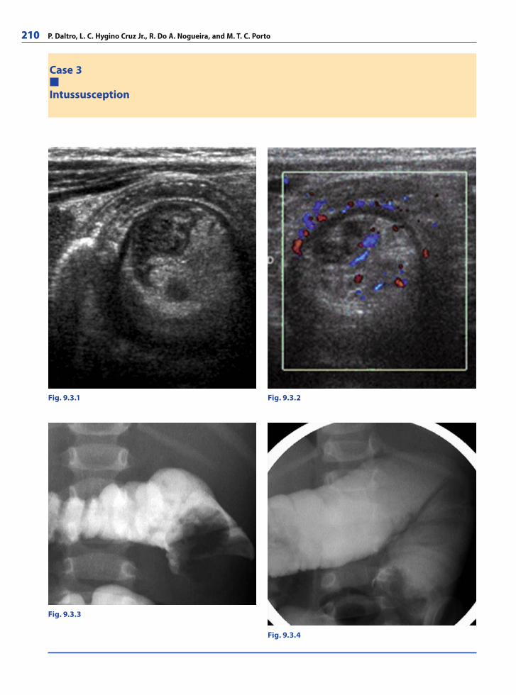
210 P. Daltro, L. C. Hygino Cruz Jr., R. Do A. Nogueira, and M. T. C. Porto
Case 3
Intussusception
Fig. 9.3.1 Fig. 9.3.2
Fig. 9.3.4
Fig. 9.3.3
RibesLunaRos.indb 210 18.03.2008 12:53:42
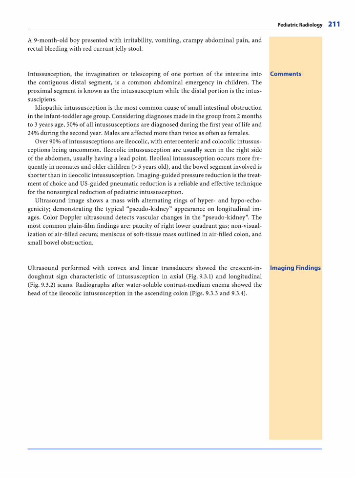
Pediatric Radiology 211
A 9-month-old boy presented with irritability, vomiting, crampy abdominal pain, and rectal bleeding with red currant jelly stool.
CommentsIntussusception, the invagination or telescoping of one portion of the intestine into the contiguous distal segment, is a common abdominal emergency in children. The proximal segment is known as the intussusceptum while the distal portion is the intus-suscipiens.
Idiopathic intussusception is the most common cause of small intestinal obstruction in the infant-toddler age group. Considering diagnoses made in the group from 2 months to 3 years age, 50% of all intussusceptions are diagnosed during the fi rst year of life and 24% during the second year. Males are affected more than twice as often as females.
Over 90% of intussusceptions are ileocolic, with enteroenteric and colocolic intussus-ceptions being uncommon. Ileocolic intussusception are usually seen in the right side of the abdomen, usually having a lead point. Ileoileal intussusception occurs more fre-quently in neonates and older children (> 5 years old), and the bowel segment involved is shorter than in ileocolic intussusception. Imaging-guided pressure reduction is the treat-ment of choice and US-guided pneumatic reduction is a reliable and effective technique for the nonsurgical reduction of pediatric intussusception.
Ultrasound image shows a mass with alternating rings of hyper- and hypo-echo-genicity; demonstrating the typical “pseudo-kidney” appearance on longitudinal im-ages. Color Doppler ultrasound detects vascular changes in the “pseudo-kidney”. The most common plain-fi lm fi ndings are: paucity of right lower quadrant gas; non-visual-ization of air-fi lled cecum; meniscus of soft-tissue mass outlined in air-fi lled colon, and small bowel obstruction.
Imaging FindingsUltrasound performed with convex and linear transducers showed the crescent-in-doughnut sign characteristic of intussusception in axial (Fig. 9.3.1) and longitudinal (Fig. 9.3.2) scans. Radiographs after water-soluble contrast-medium enema showed the head of the ileocolic intussusception in the ascending colon (Figs. 9.3.3 and 9.3.4).
RibesLunaRos.indb 211 18.03.2008 12:53:47
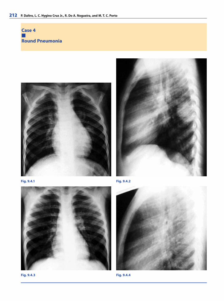
212 P. Daltro, L. C. Hygino Cruz Jr., R. Do A. Nogueira, and M. T. C. Porto
Case 4
Round Pneumonia
Fig. 9.4.1 Fig. 9.4.2
Fig. 9.4.4Fig. 9.4.3
RibesLunaRos.indb 212 18.03.2008 12:53:47
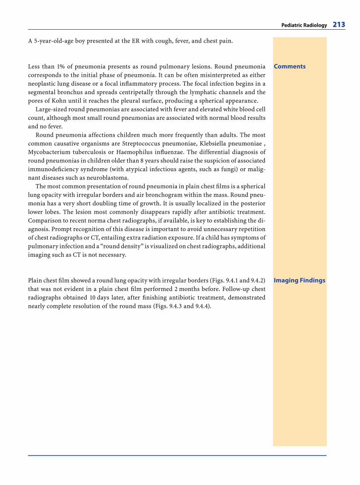
Pediatric Radiology 213
A 5-year-old-age boy presented at the ER with cough, fever, and chest pain.
CommentsLess than 1% of pneumonia presents as round pulmonary lesions. Round pneumonia corresponds to the initial phase of pneumonia. It can be often misinterpreted as either neoplastic lung disease or a focal infl ammatory process. The focal infection begins in a segmental bronchus and spreads centripetally through the lymphatic channels and the pores of Kohn until it reaches the pleural surface, producing a spherical appearance.
Large-sized round pneumonias are associated with fever and elevated white blood cell count, although most small round pneumonias are associated with normal blood results and no fever.
Round pneumonia affections children much more frequently than adults. The most common causative organisms are Streptococcus pneumoniae, Klebsiella pneumoniae , Mycobacterium tuberculosis or Haemophilus infl uenzae. The differential diagnosis of round pneumonias in children older than 8 years should raise the suspicion of associated immunodefi ciency syndrome (with atypical infectious agents, such as fungi) or malig-nant diseases such as neuroblastoma.
The most common presentation of round pneumonia in plain chest fi lms is a spherical lung opacity with irregular borders and air bronchogram within the mass. Round pneu-monia has a very short doubling time of growth. It is usually localized in the posterior lower lobes. The lesion most commonly disappears rapidly after antibiotic treatment. Comparison to recent norma chest radiographs, if available, is key to establishing the di-agnosis. Prompt recognition of this disease is important to avoid unnecessary repetition of chest radiographs or CT, entailing extra radiation exposure. If a child has symptoms of pulmonary infection and a “round density” is visualized on chest radiographs, additional imaging such as CT is not necessary.
Imaging FindingsPlain chest fi lm showed a round lung opacity with irregular borders (Figs. 9.4.1 and 9.4.2) that was not evident in a plain chest fi lm performed 2 months before. Follow-up chest radiographs obtained 10 days later, after fi nishing antibiotic treatment, demonstrated nearly complete resolution of the round mass (Figs. 9.4.3 and 9.4.4).
RibesLunaRos.indb 213 18.03.2008 12:53:50
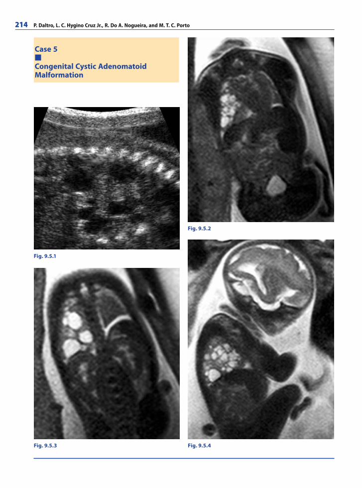
214 P. Daltro, L. C. Hygino Cruz Jr., R. Do A. Nogueira, and M. T. C. Porto
Case 5
Congenital Cystic AdenomatoidMalformation
Fig. 9.5.1
Fig. 9.5.2
Fig. 9.5.4Fig. 9.5.3
RibesLunaRos.indb 214 18.03.2008 12:53:50
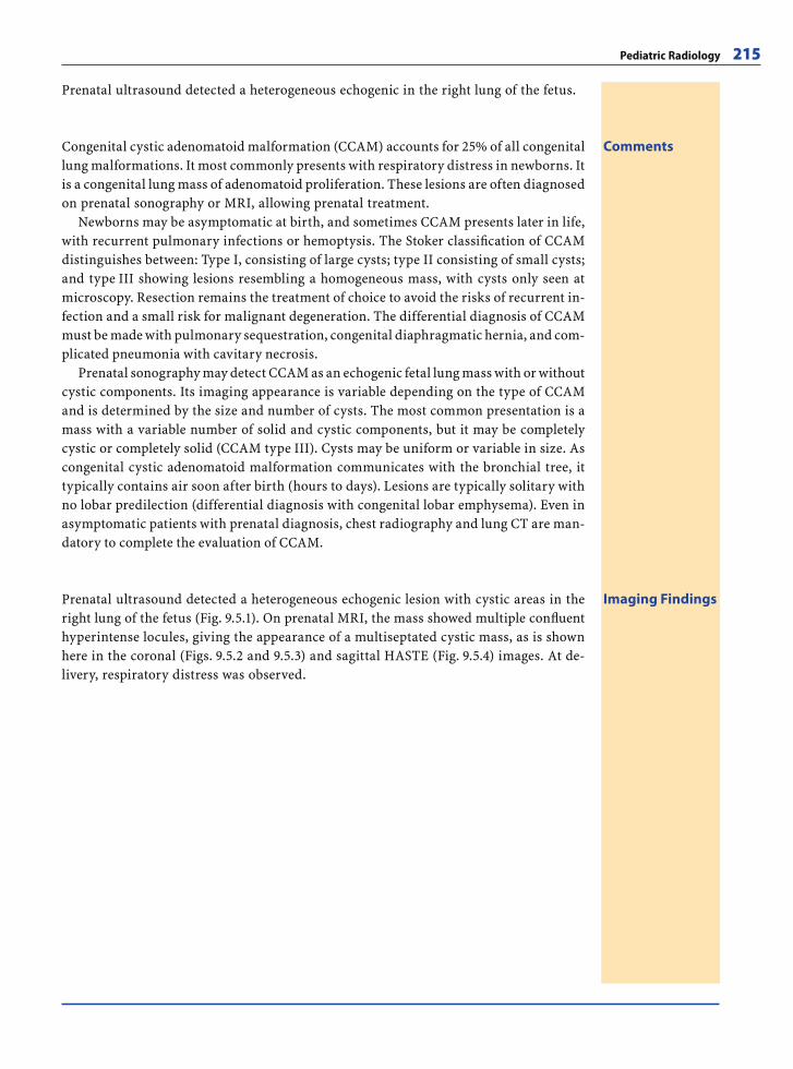
Pediatric Radiology 215
Prenatal ultrasound detected a heterogeneous echogenic in the right lung of the fetus.
CommentsCongenital cystic adenomatoid malformation (CCAM) accounts for 25% of all congenital lung malformations. It most commonly presents with respiratory distress in newborns. It is a congenital lung mass of adenomatoid proliferation. These lesions are often diagnosed on prenatal sonography or MRI, allowing prenatal treatment.
Newborns may be asymptomatic at birth, and sometimes CCAM presents later in life, with recurrent pulmonary infections or hemoptysis. The Stoker classifi cation of CCAM distinguishes between: Type I, consisting of large cysts; type II consisting of small cysts; and type III showing lesions resembling a homogeneous mass, with cysts only seen at microscopy. Resection remains the treatment of choice to avoid the risks of recurrent in-fection and a small risk for malignant degeneration. The differential diagnosis of CCAM must be made with pulmonary sequestration, congenital diaphragmatic hernia, and com-plicated pneumonia with cavitary necrosis.
Prenatal sonography may detect CCAM as an echogenic fetal lung mass with or without cystic components. Its imaging appearance is variable depending on the type of CCAM and is determined by the size and number of cysts. The most common presentation is a mass with a variable number of solid and cystic components, but it may be completely cystic or completely solid (CCAM type III). Cysts may be uniform or variable in size. As congenital cystic adenomatoid malformation communicates with the bronchial tree, it typically contains air soon after birth (hours to days). Lesions are typically solitary with no lobar predilection (differential diagnosis with congenital lobar emphysema). Even in asymptomatic patients with prenatal diagnosis, chest radiography and lung CT are man-datory to complete the evaluation of CCAM.
Imaging FindingsPrenatal ultrasound detected a heterogeneous echogenic lesion with cystic areas in the right lung of the fetus (Fig. 9.5.1). On prenatal MRI, the mass showed multiple confl uent hyperintense locules, giving the appearance of a multiseptated cystic mass, as is shown here in the coronal (Figs. 9.5.2 and 9.5.3) and sagittal HASTE (Fig. 9.5.4) images. At de-livery, respiratory distress was observed.
RibesLunaRos.indb 215 18.03.2008 12:53:54
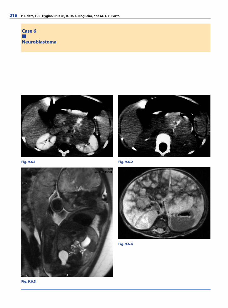
216 P. Daltro, L. C. Hygino Cruz Jr., R. Do A. Nogueira, and M. T. C. Porto
Case 6
Neuroblastoma
Fig. 9.6.1 Fig. 9.6.2
Fig. 9.6.3
Fig. 9.6.4
RibesLunaRos.indb 216 18.03.2008 12:53:54
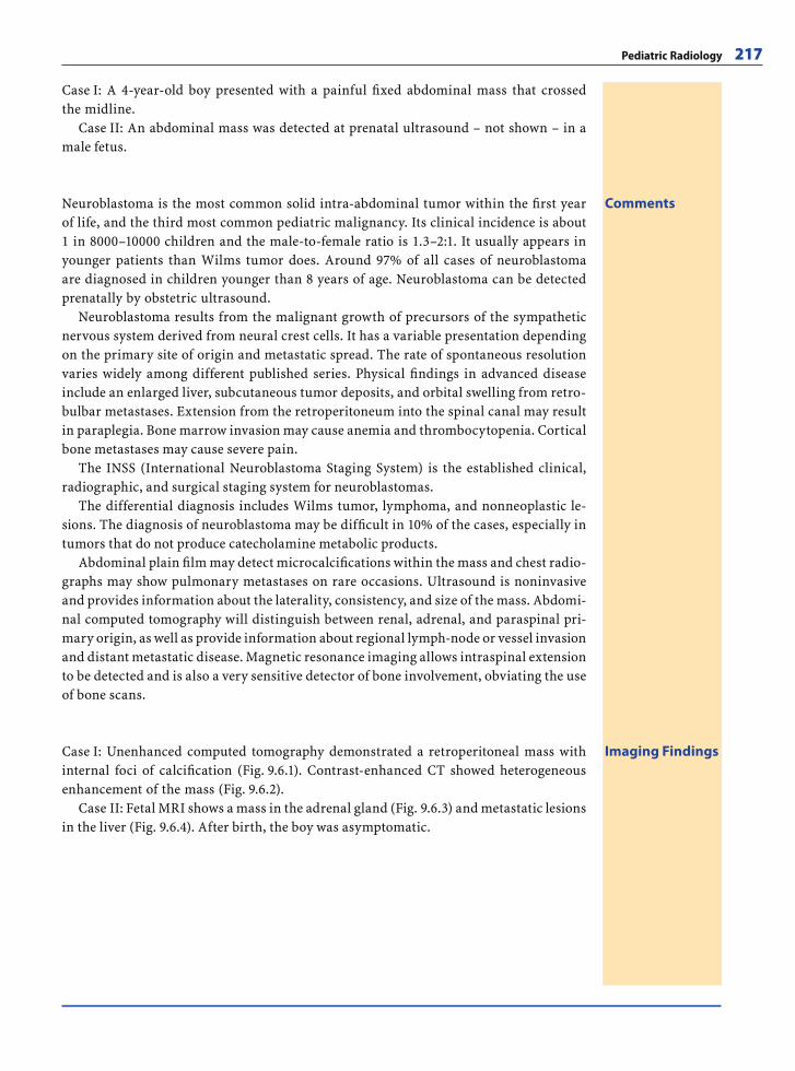
Pediatric Radiology 217
Case I: A 4-year-old boy presented with a painful fi xed abdominal mass that crossed the midline.
Case II: An abdominal mass was detected at prenatal ultrasound – not shown – in a male fetus.
CommentsNeuroblastoma is the most common solid intra-abdominal tumor within the fi rst year of life, and the third most common pediatric malignancy. Its clinical incidence is about 1 in 8000–10000 children and the male-to-female ratio is 1.3–2:1. It usually appears in younger patients than Wilms tumor does. Around 97% of all cases of neuroblastoma are diagnosed in children younger than 8 years of age. Neuroblastoma can be detected prenatally by obstetric ultrasound.
Neuroblastoma results from the malignant growth of precursors of the sympathetic nervous system derived from neural crest cells. It has a variable presentation depending on the primary site of origin and metastatic spread. The rate of spontaneous resolution varies widely among different published series. Physical fi ndings in advanced disease include an enlarged liver, subcutaneous tumor deposits, and orbital swelling from retro-bulbar metastases. Extension from the retroperitoneum into the spinal canal may result in paraplegia. Bone marrow invasion may cause anemia and thrombocytopenia. Cortical bone metastases may cause severe pain.
The INSS (International Neuroblastoma Staging System) is the established clinical, radiographic, and surgical staging system for neuroblastomas.
The differential diagnosis includes Wilms tumor, lymphoma, and nonneoplastic le-sions. The diagnosis of neuroblastoma may be diffi cult in 10% of the cases, especially in tumors that do not produce catecholamine metabolic products.
Abdominal plain fi lm may detect microcalcifi cations within the mass and chest radio-graphs may show pulmonary metastases on rare occasions. Ultrasound is noninvasive and provides information about the laterality, consistency, and size of the mass. Abdomi-nal computed tomography will distinguish between renal, adrenal, and paraspinal pri-mary origin, as well as provide information about regional lymph-node or vessel invasion and distant metastatic disease. Magnetic resonance imaging allows intraspinal extension to be detected and is also a very sensitive detector of bone involvement, obviating the use of bone scans.
Imaging FindingsCase I: Unenhanced computed tomography demonstrated a retroperitoneal mass with internal foci of calcifi cation (Fig. 9.6.1). Contrast-enhanced CT showed heterogeneous enhancement of the mass (Fig. 9.6.2).
Case II: Fetal MRI shows a mass in the adrenal gland (Fig. 9.6.3) and metastatic lesions in the liver (Fig. 9.6.4). After birth, the boy was asymptomatic.
RibesLunaRos.indb 217 18.03.2008 12:53:57
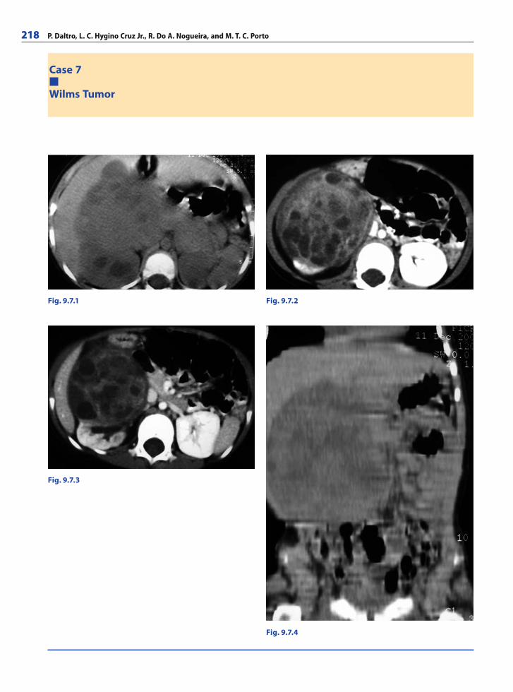
218 P. Daltro, L. C. Hygino Cruz Jr., R. Do A. Nogueira, and M. T. C. Porto
Case 7
Wilms Tumor
Fig. 9.7.1 Fig. 9.7.2
Fig. 9.7.3
Fig. 9.7.4
RibesLunaRos.indb 218 18.03.2008 12:53:58
Pediatric Radiology 219
CommentsWilms tumor (malignant nephroblastoma) is the most frequent abdominal malignancy found in children and the third most common malignant childhood disease. It is typi-cally found in the fi rst years of life, with more than 80% of cases in the 0–14 age range presenting under 5 years of age. Up to 5–10% of cases are bilateral. Most cases are spo-radic, but probably 1–2% of the cases have a familial incidence.
The presence of genitourinary anomalies, such as cryptorchidism or hypospadias, and associated syndromes, including sporadic aniridia, Beckwith-Wiedemann or Denys-Drash syndromes, suggests Wilms tumor.
The diagnostic evaluation of a child with suspected nephroblastoma has three pur-poses:
1-to confi rm the tumor’s location and origin in the kidney and to distinguish it from other abdominal masses such as a neuroblastoma or hepatic tumors;
2-to evaluate the extension of the tumor, specifi cally, to identify local tumor spread and renal vein involvement;
3-to rule out metastatic or bilateral disease. Wilms tumor is often a large heterogeneous tumor mass that bulges the renal cap-
sule, displaces adjacent organs, and may appear quite exophytic. It commonly invades the renal and inferior vena cava veins. It usually grows predominantly by local extension, compressing the surrounding renal parenchyma and giving the appearance of a sharply margined mass, often with a prominent pseudocapsule. Less commonly, small internal areas of fat density or calcifi cation may be noted. Wilms tumors are usually hypovascu-lar, showing poor enhancement in post-contrast series. Lung metastasis occurs in ap-proximately 20% of cases.
Ultrasound is often the fi rst initial imaging examination, but it is often diffi cult to image the entire mass in a single US slice. Doppler-ultrasound may help to detect tumor thrombus extension or vein compression. CT is the most common imaging technique used to establish the diagnosis and for staging. However, the diagnostic accuracy of MRI is similar to that of CT, and MRI has the important advantage of sparing the child ioniz-ing radiation. The multiplanar capabilities of MRI enable the organ of origin to be deter-mined in large abdominal masses. The most common MR presentation of Wilms tumor is a large heterogeneous mass with low signal intensity on T1-weighted images and high signal on T2-weighted images.
Imaging FindingsUnenhanced computed tomography (CT) demonstrated a large heterogeneous tumor displacing the right kidney anteriorly (Fig. 9.7.1) and contrast-enhanced CT showed en-hancement of the tumor and kidney parenchyma (Figs. 9.7.2 and 9.7.3). Figure 9.7.4 shows a coronal reconstruction of the large mass.
A 3-year-old boy presented with a palpable abdominal mass, hematuria, abdominal pain, and hypertension.
RibesLunaRos.indb 219 18.03.2008 12:54:01
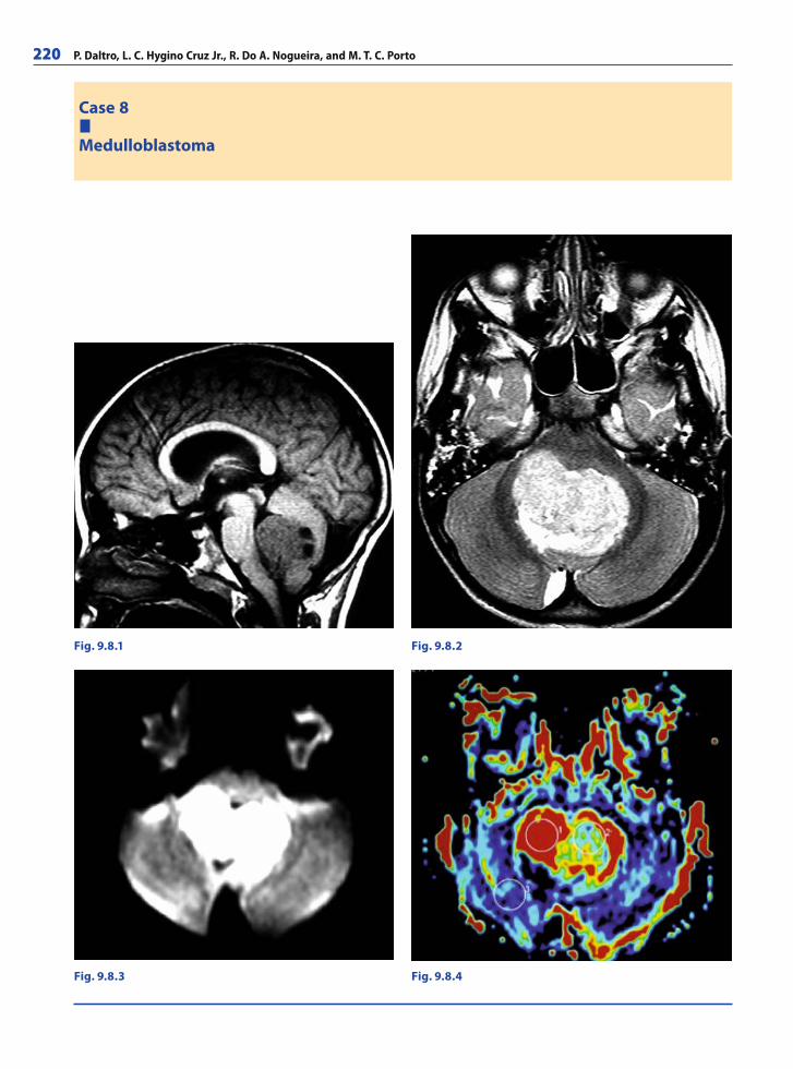
220 P. Daltro, L. C. Hygino Cruz Jr., R. Do A. Nogueira, and M. T. C. Porto
Case 8
Medulloblastoma
Fig. 9.8.1 Fig. 9.8.2
Fig. 9.8.4Fig. 9.8.3
RibesLunaRos.indb 220 18.03.2008 12:54:01
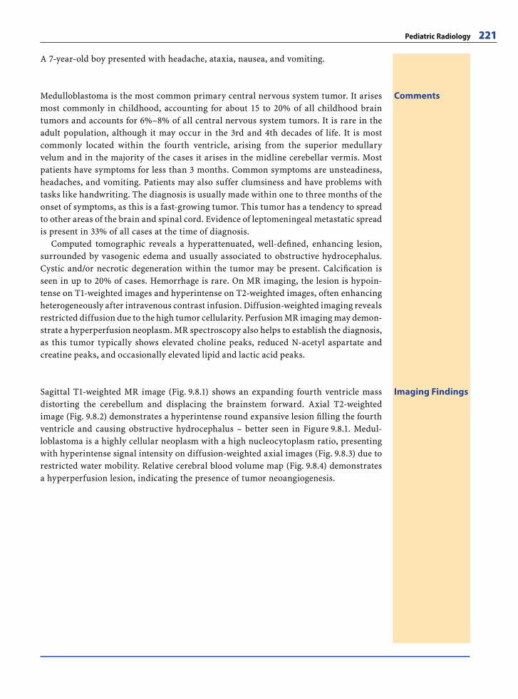
Pediatric Radiology 221
A 7-year-old boy presented with headache, ataxia, nausea, and vomiting.
CommentsMedulloblastoma is the most common primary central nervous system tumor. It arises most commonly in childhood, accounting for about 15 to 20% of all childhood brain tumors and accounts for 6%–8% of all central nervous system tumors. It is rare in the adult population, although it may occur in the 3rd and 4th decades of life. It is most commonly located within the fourth ventricle, arising from the superior medullary velum and in the majority of the cases it arises in the midline cerebellar vermis. Most patients have symptoms for less than 3 months. Common symptoms are unsteadiness, headaches, and vomiting. Patients may also suffer clumsiness and have problems with tasks like handwriting. The diagnosis is usually made within one to three months of the onset of symptoms, as this is a fast-growing tumor. This tumor has a tendency to spread to other areas of the brain and spinal cord. Evidence of leptomeningeal metastatic spread is present in 33% of all cases at the time of diagnosis.
Computed tomographic reveals a hyperattenuated, well-defi ned, enhancing lesion, surrounded by vasogenic edema and usually associated to obstructive hydrocephalus. Cystic and/or necrotic degeneration within the tumor may be present. Calcifi cation is seen in up to 20% of cases. Hemorrhage is rare. On MR imaging, the lesion is hypoin-tense on T1-weighted images and hyperintense on T2-weighted images, often enhancing heterogeneously after intravenous contrast infusion. Diffusion-weighted imaging reveals restricted diffusion due to the high tumor cellularity. Perfusion MR imaging may demon-strate a hyperperfusion neoplasm. MR spectroscopy also helps to establish the diagnosis, as this tumor typically shows elevated choline peaks, reduced N-acetyl aspartate and creatine peaks, and occasionally elevated lipid and lactic acid peaks.
Imaging FindingsSagittal T1-weighted MR image (Fig. 9.8.1) shows an expanding fourth ventricle mass distorting the cerebellum and displacing the brainstem forward. Axial T2-weighted image (Fig. 9.8.2) demonstrates a hyperintense round expansive lesion fi lling the fourth ventricle and causing obstructive hydrocephalus – better seen in Figure 9.8.1. Medul-loblastoma is a highly cellular neoplasm with a high nucleocytoplasm ratio, presenting with hyperintense signal intensity on diffusion-weighted axial images (Fig. 9.8.3) due to restricted water mobility. Relative cerebral blood volume map (Fig. 9.8.4) demonstrates a hyperperfusion lesion, indicating the presence of tumor neoangiogenesis.
RibesLunaRos.indb 221 18.03.2008 12:54:05
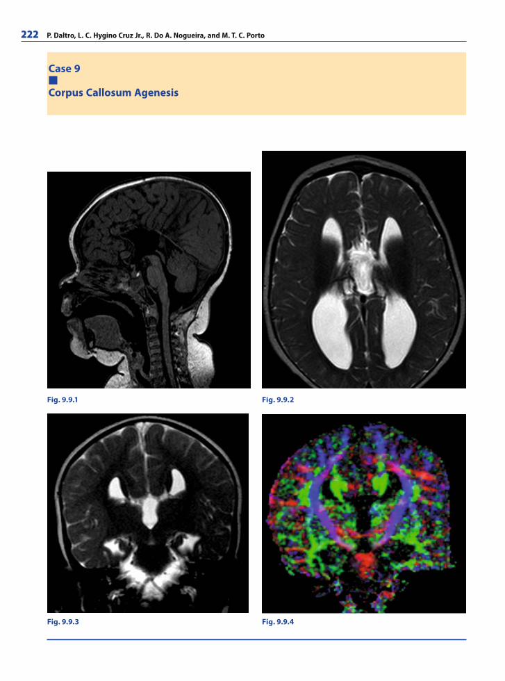
222 P. Daltro, L. C. Hygino Cruz Jr., R. Do A. Nogueira, and M. T. C. Porto
Case 9
Corpus Callosum Agenesis
Fig. 9.9.1 Fig. 9.9.2
Fig. 9.9.4Fig. 9.9.3
RibesLunaRos.indb 222 18.03.2008 12:54:05
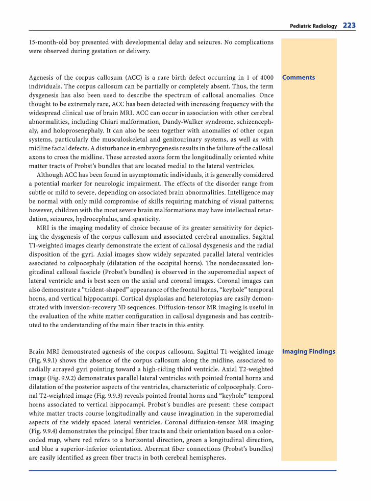
Pediatric Radiology 223
Imaging FindingsBrain MRI demonstrated agenesis of the corpus callosum. Sagittal T1-weighted image (Fig. 9.9.1) shows the absence of the corpus callosum along the midline, associated to radially arrayed gyri pointing toward a high-riding third ventricle. Axial T2-weighted image (Fig. 9.9.2) demonstrates parallel lateral ventricles with pointed frontal horns and dilatation of the posterior aspects of the ventricles, characteristic of colpocephaly. Coro-nal T2-weighted image (Fig. 9.9.3) reveals pointed frontal horns and “keyhole” temporal horns associated to vertical hippocampi. Probst s bundles are present: these compact white matter tracts course longitudinally and cause invagination in the superomedial aspects of the widely spaced lateral ventricles. Coronal diffusion-tensor MR imaging (Fig. 9.9.4) demonstrates the principal fi ber tracts and their orientation based on a color-coded map, where red refers to a horizontal direction, green a longitudinal direction, and blue a superior-inferior orientation. Aberrant fi ber connections (Probst’s bundles) are easily identifi ed as green fi ber tracts in both cerebral hemispheres.
15-month-old boy presented with developmental delay and seizures. No complications were observed during gestation or delivery.
CommentsAgenesis of the corpus callosum (ACC) is a rare birth defect occurring in 1 of 4000 individuals. The corpus callosum can be partially or completely absent. Thus, the term dysgenesis has also been used to describe the spectrum of callosal anomalies. Once thought to be extremely rare, ACC has been detected with increasing frequency with the widespread clinical use of brain MRI. ACC can occur in association with other cerebral abnormalities, including Chiari malformation, Dandy-Walker syndrome, schizenceph-aly, and holoprosenephaly. It can also be seen together with anomalies of other organ systems, particularly the musculoskeletal and genitourinary systems, as well as with midline facial defects. A disturbance in embryogenesis results in the failure of the callosal axons to cross the midline. These arrested axons form the longitudinally oriented white matter tracts of Probst’s bundles that are located medial to the lateral ventricles.
Although ACC has been found in asymptomatic individuals, it is generally considered a potential marker for neurologic impairment. The effects of the disorder range from subtle or mild to severe, depending on associated brain abnormalities. Intelligence may be normal with only mild compromise of skills requiring matching of visual patterns; however, children with the most severe brain malformations may have intellectual retar-dation, seizures, hydrocephalus, and spasticity.
MRI is the imaging modality of choice because of its greater sensitivity for depict-ing the dysgenesis of the corpus callosum and associated cerebral anomalies. Sagittal T1-weighted images clearly demonstrate the extent of callosal dysgenesis and the radial disposition of the gyri. Axial images show widely separated parallel lateral ventricles associated to colpocephaly (dilatation of the occipital horns). The nondecussated lon-gitudinal callosal fascicle (Probst’s bundles) is observed in the superomedial aspect of lateral ventricle and is best seen on the axial and coronal images. Coronal images can also demonstrate a “trident-shaped” appearance of the frontal horns, “keyhole” temporal horns, and vertical hippocampi. Cortical dysplasias and heterotopias are easily demon-strated with inversion-recovery 3D sequences. Diffusion-tensor MR imaging is useful in the evaluation of the white matter confi guration in callosal dysgenesis and has contrib-uted to the understanding of the main fi ber tracts in this entity.
RibesLunaRos.indb 223 18.03.2008 12:54:10
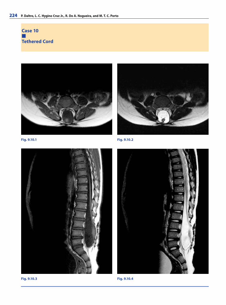
224 P. Daltro, L. C. Hygino Cruz Jr., R. Do A. Nogueira, and M. T. C. Porto
Case 10
Tethered Cord
Fig. 9.10.1 Fig. 9.10.2
Fig. 9.10.4Fig. 9.10.3
RibesLunaRos.indb 224 18.03.2008 12:54:10
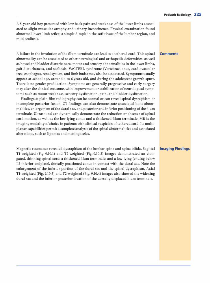
Pediatric Radiology 225
A 5-year-old boy presented with low back pain and weakness of the lower limbs associ-ated to slight muscular atrophy and urinary incontinence. Physical examination found abnormal lower-limb refl ex, a simple dimple in the soft tissue of the lumbar region, and mild scoliosis.
CommentsA failure in the involution of the fi lum terminale can lead to a tethered cord. This spinal abnormality can be associated to other neurological and orthopedic deformities, as well as bowel and bladder disturbances, motor and sensory abnormalities in the lower limbs, gait disturbances, and scoliosis. VACTERL syndrome (Vertebrae, anus, cardiovascular tree, esophagus, renal system, and limb buds) may also be associated. Symptoms usually appear at school age, around 4 to 6 years old, and during the adolescent growth spurt. There is no gender predilection. Symptoms are generally progressive and early surgery may alter the clinical outcome, with improvement or stabilization of neurological symp-toms such as motor weakness, sensory dysfunction, pain, and bladder dysfunction.
Findings at plain-fi lm radiography can be normal or can reveal spinal dysraphism or incomplete posterior fusion. CT fi ndings can also demonstrate associated bone abnor-malities, enlargement of the dural sac, and posterior and inferior positioning of the fi lum terminale. Ultrasound can dynamically demonstrate the reduction or absence of spinal cord motion, as well as the low-lying conus and a thickened fi lum terminale. MR is the imaging modality of choice in patients with clinical suspicion of tethered cord. Its multi-planar capabilities permit a complete analysis of the spinal abnormalities and associated alterations, such as lipomas and meningoceles.
Imaging FindingsMagnetic resonance revealed dysraphism of the lumbar spine and spina bifi da. Sagittal T1-weighted (Fig. 9.10.1) and T2-weighted (Fig. 9.10.2) images demonstrated an elon-gated, thinning spinal cord; a thickened fi lum terminale; and a low-lying (ending below L2 inferior endplate), dorsally positioned conus in contact with the dural sac. Note the enlargement of the inferior portion of the dural sac and the spinal dysraphism. Axial T1-weighted (Fig. 9.10.3) and T2-weighted (Fig. 9.10.4) images also showed the widening dural sac and the inferior-posterior location of the dorsally displaced fi lum terminale.
RibesLunaRos.indb 225 18.03.2008 12:54:13
226 P. Daltro, L. C. da Cruz, R. Do A. Nogueira, and M. T. C. Porto
Further Readings
Books
Caffey s Pediatric Diagnostic Imaging. Kuhn JP, Slovis TL, Haller JO (2003) Philadelphia: Mosby, 2003. ISBN-13: 9780323011099
Diagnostic Imaging: Pediatric. Donnelly LF (2005) Salt Lake City: AMIRSYS. (Philadelphia: Elsevier). ISBN-13: 9781416023333
Fundamentals of Pediatric Radiology. Donnelly LF (2001) Philadelphia: W.B. Saunders. ISBN-13: 9780721690612
Imaging of the newborn infant, and young child. 5th ed. Swischuk LE (2003) Philadelphia: Lippincott Williams & Wilkins. ISBN-13: 9780781734585
Pediatric Body CT. Siegel MJ (1999) Philadelphia: Lippincott Williams & Wilkins. ISBN-13: 9780781712491
Pediatric Chest Imaging. Lucaya J, Strife JL (2002) Springer-Verlag, Berlin-Heidelberg. ISBN-13: 9783540675273
Pediatric Neuroimaging. Barkovich AJ (2005) Philadelphia: Lippincott Williams & Wilkins. ISBN-13: 9780781717403
Pediatric Sonography. 3rd ed. Siegel MJ (2002) Philadelphia: Lippincott Williams & Wilkins. ISBN-13: 9780781727532
Pediatric Uroradiology. Fotter R (2001) Springer-Verlag, Berlin-Heidelberg. ISBN-13: 9783540436850
Pratical Pediatric Imaging: Diagnostic Radiology of Infants and Children. 3rd ed. Kirks DR, Griscom NT (1997) Phila-delphia: Lippincott-Raven. ISBN-13: 9780316494731
Web-Links
http://auntminnie.com/index.asp?sec=defhttp://hawaii.edu/medicine/pediatrics/pemxray/pemxray.
htmlhttp://pediatricradiology.com/http://pediatric-radiology.com/http://pediatricradiologyinfo.com/http://pedrad.info/http://pedsradiology.com/http://radiologyeducation.com/http://rsna.org/Education/archive/bestcases.cfmhttp://.uhrad.com/pedsarc.htm
Articles
Accardo J, Kammann H, Hoon AH Jr. Neuroimaging in cere-bral palsy. J Pediatr 2004; 145(2 Suppl):S19–27
Adamsbaum C, Moutard ML, Andre C, Merzoug V, Ferey S, Quere MP, Lewin F, Fallet-Bianco C. MRI of the fetal posterior fossa. Pediatr Radiol 2005; 35(2):124–140. Epub 2004 Nov 23
Amini A, Liu JK, Kestle JR. Disseminated medulloblastoma. J Neurooncol 2006; 80(2):157–158. Epub 2006 Sep 26
Applegate KE, Anderson JM, Klatte EC. Intestinal malrota-tion in children: a problem-solving approach to the upper gastrointestinal series. Radiographics 2006; 26(5):1485–1500
Argyropoulou MI, Kiortsis DN. MRI of the hypothalamic-pi-tuitary axis in children. Pediatr Radiol 2005; 35(11):1045–1055. Epub 2005 Jun 1
Arthur R. Magnetic resonance imaging in preterm infants. Pediatr Radiol 2006; 36(7):593–607. Epub 2006 May 19
Atkinson DS Jr. Computed tomography of pediatric stroke. Semin Ultrasound CT MR 2006; 27(3):207–218
Avni FE, Garel L, Cassart M, Massez A, Eurin D, Didier F, Hall M, Teele RL. Perinatal assessment of hereditary cys-tic renal diseases: the contribution of sonography. Pediatr Radiol 2006; 36(5):405–414. Epub 2006 Feb 4. Erratum in: Pediatr Radiol 2006; 36(7):731
Bacon S, Clinkard J, Taylor RE. Paediatric medulloblastoma associated with poor prognosis and short volume dou-bling time. Br J Radiol 2005; 78(935):1059–1060
Barkovich AJ, Simon EM, Walsh CA. Callosal agenesis with cyst: a better understanding and new classifi cation. Neu-rology 2001; 56:220–227
Bedeschi MF, Bonaglia MC, Grasso R, Pellegri A, Garghen-tino RR, Battaglia MA, Panarisi AM, Di Rocco M, Balot-tin U, Bresolin N, Bassi MT, Borgatti R. Agenesis of the corpus callosum: clinical and genetic study in 63 young patients. Pediatr Neurol 2006; 34(3):186–193
Boesch RP, Daines C, Willging JP, Kaul A, Cohen AP, Wood RE, Amin RS. Advances in the diagnosis and manage-ment of chronic pulmonary aspiration in children. Eur Respir J 2006; 28(4):847–861
Bousvaros A, Kirks DR, Grossman H. Imaging of neuroblas-toma: an overview. Pediatr Radiol 1986; 16(2):89–106
Brisse H, Ollivier L, Edeline V, Pacquement H, Michon J, Glo-rion C, Neuenschwander S. Imaging of malignant tumours of the long bones in children: monitoring response to neo-adjuvant chemotherapy and preoperative assessment. Pe-diatr Radiol 2004; 34(8):595–605. Epub 2004 Apr 22
Burrows PE, Dubois J, Kassarjian A. Pediatric hepatic vascu-lar anomalies. Pediatr Radiol 2001; 31(8):533–545
Bush A, Davies J. Early detection of lung disease in preschool children with cystic fi brosis. Curr Opin Pulm Med 2005; 11(6):534–538
Byrne AT, Geoghegan T, Govender P, Lyburn ID, Colhoun E, Torreggiani WC. The imaging of intussusception. Clin Radiol 2005; 60(1):39–46. Erratum in: Clin Radiol 2005; 60(3):412
Cass DL, Crombleholme TM, Howell LJ, Stafford PW, Ruchelli ED, Adzick NS. Cystic lung lesions with systemic arterial blood supply: a hybrid of congenital cystic adenomatoid malformation and bronchopulmonary sequestration. J Pediatr Surg 1997; 32(7):986–990
Cleveland RH. A radiologic update on medical diseases of the newborn chest. Pediatr Radiol 1995; 25(8):631–637
Corness JA, McHugh K, Roebuck DJ, Taylor AM. The por-tal vein in children: radiological review of congenital anomalies and acquired abnormalities. Pediatr Radiol 2006; 36(2):87–96, quiz 170-1. Epub 2005 Nov 12
Cross JH. Neurocutaneous syndromes and epilepsy-issues in diagnosis and management. Epilepsia 2005; 46 Suppl 10:17–23
Crowley JJ, Sarnaik S. Imaging of sickle cell disease. Pediatr Radiol 1999; 29(9):646–661
Daltro P, Fricke BL, Kline-Fath BM, Werner H, Rodrigues L, Fazecas T, Domingues R, Donnelly LF. Prenatal MRI of congenital abdominal and chest wall defects. AJR Am J Roentgenol 2005; 184(3):1010–1016
RibesLunaRos.indb 226 18.03.2008 12:54:13
Pediatric Radiology 227
Daltro P, Fricke BL, Kuroki I, Domingues R, Donnelly LF. CT of congenital lung lesions in pediatric patients. AJR Am J Roentgenol 2004; 183(5):1497–1506
Daneman A, Navarro O. Intussusception. Part 1: a review of diagnostic approaches. Pediatr Radiol 2003; 33(2):79–85. Epub 2002 Nov 19
Daneman A, Epelman M, Blaser S, Jarrin JR. Imaging of the brain in full-term neonates: does sonography still play a role? Pediatr Radiol 2006; 36(7):636–646. Epub 2006 May 16
Darge K, Trusen A, Gordjani N, Riedmiller H. Intrarenal refl ux: diagnosis with contrast-enhanced harmonic US. Pediatr Radiol 2003; 33(10):729–731. Epub 2003 Aug 20
de Mello RR, Dutra MV, Ramos JR, Daltro P, Boechat M, de Andrade Lopes JM. Lung mechanics and high-reso-lution computed tomography of the chest in very low birth weight premature infants. Sao Paulo Med J 2003; 121(4):167–172. Epub 2003 Oct 29
de Mello RR, Dutra MV, Ramos JR, Daltro P, Boechat M, Lopes JM. Neonatal risk factors for respiratory morbidity during the fi rst year of life among premature infants. Sao Paulo Med J 2006; 124(2):77–84
Diefenbach KA, Breuer CK. Pediatric infl ammatory bowel disease. World J Gastroenterol 2006; 12(20):3204–3212
Dubois J, Garel L. Imaging and therapeutic approach of hem-angiomas and vascular malformations in the pediatric age group. Pediatr Radiol 1999; 29(12):879–893
Ebel KD. Uroradiology in the fetus and newborn: diagnosis and follow-up of congenital obstruction of the urinary tract. Pediatr Radiol 1998; 28(8):630–635
Epelman M, Daneman A, Blaser SI, Ortiz-Neira C, Konen O, Jarrin J, Navarro OM. Differential diagnosis of intracra-nial cystic lesions at head US: correlation with CT and MR imaging. Radiographics 2006; 26(1):173–196
Erichsen D, Sellstrom H, Andersson H. Small bowel intus-susception after blunt abdominal trauma in a 6-year-old boy: case report and review of 6 cases reported in the literature. J Pediatr Surg 2006; 41(11):1930–1932
Franken EA Jr, Smith JA, Smith WL. Tumors of the chest wall in infants and children. Pediatr Radiol 1977; 6(1):13–18
Frush DP. Pediatric CT: practical approach to diminish the radiation dose. Pediatr Radiol 2002; 32(10):714–717; dis-cussion 751–754. Epub 2002 Aug 29
Garel C. New advances in fetal MR neuroimaging. Pediatr Radiol 2006; 36(7):621–625. Epub 2006 May 3
Garel C. The role of MRI in the evaluation of the fetal brain with an emphasis on biometry, gyration and paren-chyma. Pediatr Radiol 2004; 34(9):694–699. Epub 2004 Jul 28
Garel C, Brisse H, Sebag G, Elmaleh M, Oury JF, Hassan M. Magnetic resonance imaging of the fetus. Pediatr Radiol 1998; 28(4):201–211
Garcia-Pena P, Lucaya J, Hendry GM, McAndrew PT, Duran C. Spontaneous involution of pulmonary sequestration in children: a report of two cases and review of the litera-ture. Pediatr Radiol 1998; 28(4):266–270
Grattan-Smith JD, Jones RA. MR urography in children. Pe-diatr Radiol 2006; 36(11):1119–1132; quiz 1228-1229. Epub 2006 Jun 22
Gressens P, Luton D. Fetal MRI: obstetrical and neurological perspectives. Pediatr Radiol 2004; 34(9):682–684. Epub 2004 Jul 23
Haller JO, Cohen HL. Pediatric HIV infection: an imaging update. Pediatr Radiol 1994; 24(3):224–230
Haller JO, Ginsberg KJ. Tuberculosis in children with ac-quired immunodefi ciency syndrome. Pediatr Radiol 1997; 27(2):186–188
Harris TM, Cohen MD. Abdominal magnetic resonance im-aging. Pediatr Radiol 1989; 20(1–2):10–19
Hawass ND, Badawi MG, al-Muzrakchi AM, al-Sammarai AI, Jawad AJ, Abdullah MA, Bahakim H. Horseshoe lung: dif-ferential diagnosis. Pediatr Radiol 1990; 20(8):580–584
Helton KJ, Fouladi M, Boop FA, Perry A, Dalton J, Kun L, Fuller C. Medullomyoblastoma: a radiographic and clini-copathologic analysis of six cases and review of the litera-ture. Cancer 2004; 101(6):1445–1454
Hedlund G. Congenital frontonasal masses: developmental anatomy, malformations, and MR imaging. Pediatr Radiol 2006; 36(7):647–662; quiz 726-727. Epub 2006 Mar 11
Hetts SW, Sherr EH, Chao S, Gobuty S, Barkovich AJ. Anom-alies of the corpus callosum: an MR analysis of the phe-notypic spectrum of associated malformations. AJR Am J Roentgenol 2006; 187(5):1343–1348
Hsu YR, Lee SY. Prenatal diagnosis of congenital cystic adenomatoid malformation. Chang Gung Med J 2004; 27(1):61–65
Jamieson DH. Imaging intracranial tuberculosis in child-hood. Pediatr Radiol 1995; 25(3):165–170
Keller MS. Musculoskeletal sonography in the neonate and infant. Pediatr Radiol 2005; 35(12):1167–1173; quiz 1293. Epub 2005 Aug 3
Kim MJ, Lee KY. Bronchiolitis obliterans in children with Stevens-Johnson syndrome: follow-up with high resolu-tion CT. Pediatr Radiol 1996; 26(1):22–25
Kirks DR. Air intussusception reduction: “the winds of change”. Pediatr Radiol 1995; 25(2):89–91
Koch BL. Cystic malformations of the neck in children. Pedi-atr Radiol 2005; 35(5):463–477. Epub 2005 Mar 23
Kornreich L, Schwarz M, Karmazyn B, Cohen IJ, Shuper A, Michovitz S, Yaniv I, Fenig E, Horev G. Role of MRI in the management of children with diffuse pontine tumors: a study of 15 patients and review of the literature. Pediatr Radiol 2005; 35(9):872–879. Epub 2005 May 26
Lamego CM, Torloni H. Colorectal adenocarcinoma in child-hood and adolescent. Report of 11 cases and review of the literature. Pediatr Radiol 1989; 19(8):504–508
Laor T. MR imaging of soft tissue tumors and tumor-like lesions. Pediatr Radiol 2004; 34(1):24–37. Epub 2003 Dec 12
Lee SK, Kim DI, Kim J, Kim DJ, Kim HD, Kim DS, Mori S. Diffusion-tensor MR imaging and fi ber tractography: a new method of describing aberrant fi ber connections in developmental CNS anomalies. Radiographics 2005; 25(1):53–65; discussion 66–68
Manson DE, Sikka S, Reid B, Roifman C. Primary immuno-defi ciencies: a pictorial immunology primer for radiolo-gists. Pediatr Radiol 2000; 30(8):501–510
May JA, Krieger MD, Bowen I, Geffner ME. Craniopharyn-gioma in childhood. Adv Pediatr 2006; 53:183–209
RibesLunaRos.indb 227 18.03.2008 12:54:13
228 P. Daltro, L. C. da Cruz, R. Do A. Nogueira, and M. T. C. Porto
McCahon E. Lung tumours in children. Paediatr Respir Rev 2006; 7(3):191–196. Epub 2006 Aug 4
McCarville MB, Hoffer FA, Gingrich JR, Jenkins JJ 3rd. Im-aging fi ndings of hemorrhagic cystitis in pediatric oncol-ogy patients. Pediatr Radiol 2000; 30(3):131–138
McCollough M, Sharieff GQ. Abdominal pain in children. Pediatr Clin North Am 2006; 53(1):107–137, vi
McHugh K. Renal and adrenal tumours in children. Cancer Imaging 2007; 7:41–751
Mendelson KG, Fallat ME. Pediatric injuries: prevention to resolution. Surg Clin North Am 2007; 87(1):207–228, viii
Mercado-Deane MG, Burton EM, Howell CG. Transverse co-lon volvulus in pediatric patients. Pediatr Radiol 1995; 25(2):111–112
Meyer JS, Mackenzie W. Malignant bone tumors and limb-salvage surgery in children. Pediatr Radiol 2004; 34(8):606–13. Epub 2004 Jun 19. Erratum in: Pediatr Ra-diol 2004; 34(12):1030
Moore GJ. Proton magnetic resonance spectroscopy in pe-diatric neuroradiology. Pediatr Radiol 1998; 28(11):805–814
Moskowitz SM, Gibson RL, Effmann EL. Cystic fi brosis lung disease: genetic infl uences, microbial interactions, and radiological assessment. Pediatr Radiol 2005; 35(8):739–757. Epub 2005 May 3
Navarro O, Daneman A. Intussusception. Part 3: Diagnosis and management of those with an identifi able or predis-posing cause and those that reduce spontaneously. Pedi-atr Radiol 2004; 34(4):305–312; quiz 369. Epub 2003 Oct 8
Navarro O, Nunez-Santos E, Daneman A, Faria P, Daltro P. Malignant peripheral nerve-sheath tumor arising in a previously irradiated neuroblastoma: report of 2 cases and a review of the literature. Pediatr Radiol 2000; 30(3):176–180
Nazir Z, Qazi SH, Ahmed N, Atiq M, Billoo AG. Pulmo-nary agenesis--vascular airway compression and gastro-esophageal refl ux infl uence outcome. J Pediatr Surg 2006; 41(6):1165–1169
Neil JJ, Inder TE. Imaging perinatal brain injury in prema-ture infants. Semin Perinatol 2004; 28(6):433–443
Nelson MD Jr, Maher K, Gilles FH. A different approach to cysts of the posterior fossa. Pediatr Radiol 2004; 34(9):720–732. Epub 2004 Jul 30
Neumann G, Benz-Bohm G, Rister M. Wegener’s granulo-matosis in childhood. Review of the literature and case report. Pediatr Radiol 1984; 14(5):267–271
Newman B. Congenital bronchopulmonary foregut mal-formations: concepts and controversies. Pediatr Radiol 2006; 36(8):773–791. Epub 2006 Mar 22
Nijs E, Callahan MJ, Taylor GA. Disorders of the pediat-ric pancreas: imaging features. Pediatr Radiol 2005; 35(4):358–373; quiz 457. Epub 2004 Nov 5
Nofech-Mozes Y, Rachmel A, Schonfeld T, Schwarz M, Stein-berg R, Ashkenazi S. Diffi culties in making the diagnosis of Hirschsprung disease in early infancy. J Paediatr Child Health 2004; 40(12):716–719
Oddone M, Granata C, Vercellino N, Bava E, Toma P. Multi-modality evaluation of the abnormalities of the aortic arches in children: techniques and imaging spectrum
with emphasis on MRI. Pediatr Radiol 2005; 35(10):947–960. Epub 2005 Jun 23
Osorio A, Kessler RM, Guruprasad H, Isaacson G. Isolated intrathoracic presentation of Mycobacterium avium complex in an immunocompetent child. Pediatr Radiol 2001; 31(12):848–851
Owens C. Pearls and pitfalls in HRCT in children. Paediatr Respir Rev 2006; 7 Suppl 1:S44–49. Epub 2006 Jun 5
Pedicelli G, Ciarpaglini LL, De Santis M, Leonetti C. Con-genital bronchial atresia (CBA). A critical review of CBA as a disease entity and presentation of a case series. Ra-diol Med (Torino) 2005; 110(5–6):544–553
Pfl uger T, Czekalla R, Koletzko S, Munsterer O, Willemsen UF, Hahn K. MRI and radiographic fi ndings in Currari-no’s triad. Pediatr Radiol 1996; 26(8):524–527
Prayer D, Brugger PC, Prayer L. Fetal MRI: techniques and protocols. Pediatr Radiol 2004; 34(9):685–693. Epub 2004 Jul 28
Puderbach M, Kauczor HU. Assessment of lung function in children by cross-sectional imaging: techniques and clinical applications. Pediatr Radiol 2006; 36(3):192–204, quiz 280-281. Epub 2005 Nov 15
Reid JR. Complications of pediatric paranasal sinusitis. Pe-diatr Radiol 2004; 34(12):933–942. Epub 2004 Jul 27
Roebuck DJ, Olsen O, Pariente D. Radiological staging in children with hepatoblastoma. Pediatr Radiol 2006; 36(3):176–182. Epub 2005 Dec 10
Roebuck DJ, Perilongo G. Hepatoblastoma: an oncological review. Pediatr Radiol 2006; 36(3):183–186. Epub 2006 Jan 11
Rossi A, Cama A, Consales A, Gandolfo C, Garre ML, Mila-naccio C, Pavanello M, Piatelli G, Ravegnani M, Tortori-Donati P. Neuroimaging of pediatric craniopharyngio-mas: a pictorial essay. J Pediatr Endocrinol Metab 2006; 19 Suppl 1:299–319
Rutherford M, Srinivasan L, Dyet L, Ward P, Allsop J, Coun-sell S, Cowan F. Magnetic resonance imaging in perinatal brain injury: clinical presentation, lesions and outcome. Pediatr Radiol 2006; 36(7):582–592. Epub 2006 May 16
Rutherford M, Ward P, Allsop J, Malamatentiou C, Counsell S. Magnetic resonance imaging in neonatal encephalopa-thy. Early Hum Dev 2005; 81(1):13–25. Epub 2004 Nov 19
Safriel YI, Haller JO, Lefton DR, Obedian R. Imaging of the brain in the HIV-positive child. Pediatr Radiol 2000; 30(11):725–732
Samuel M, Burge DM. Management of antenatally diagnosed pulmonary sequestration associated with congenital cystic adenomatoid malformation. Thorax 1999; 54(8):701–706
Sargent MA. What is the normal prevalence of vesicoureteral refl ux? Pediatr Radiol 2000; 30(9):587–593
Schmahmann S, Haller JO. Neonatal ovarian cysts: patho-genesis, diagnosis and management. Pediatr Radiol 1997; 27(2):101–105
Shemie SD, Pollack MM, Morioka M, Bonner S. Diagnosis of brain death in children. Lancet Neurol 2007; 6(1):87–92
Simon EM. MRI of the fetal spine. Pediatr Radiol 2004; 34(9):712–719. Epub 2004 Jul 28
Sittig SE, Asay GF. Congenital cystic adenomatoid malfor-mation in the newborn: two case studies and review of the literature. Respir Care 2000; 45(10):1188–1195
RibesLunaRos.indb 228 18.03.2008 12:54:13
Pediatric Radiology 229
Sivit CJ. Imaging the child with right lower quadrant pain and suspected appendicitis: current concepts. Pediatr Ra-diol 2004; 34(6):447–453. Epub 2004 Apr 23
Soper JR, De Silva M. Infantile myofi bromatosis: a radiologi-cal review. Pediatr Radiol 1993; 23(3):189–194
Stanton M, Davenport M. Management of congenital lung lesions. Early Hum Dev 2006; 82(5):289–295. Epub 2006 Apr 3
Strouse PJ. Disorders of intestinal rotation and fi xation (“malrotation”). Pediatr Radiol 2004; 34(11):837–851. Epub 2004 Sep 4
Tiddens HA. Chest computed tomography scans should be considered as a routine investigation in cystic fi brosis. Paediatr Respir Rev 2006; 7(3):202–208. Epub 2006 Aug 2
Tokumaru AM, Horiuchi K, Kaji T, Kohyama S, Sakata I, Kusano S. MRI fi ndings of recurrent herpes simplex en-cephalitis in an infant. Pediatr Radiol 2003;33(10):725–728. Epub 2003 Jul 19
Triulzi F, Parazzini C, Righini A. Patterns of damage in the mature neonatal brain. Pediatr Radiol 2006; 36(7):608–620. Epub 2006 May 18
Tsitouridis I, Tsinoglou K, Morichovitou A, Stratilati S, Sioug-garis N, Kontaki T. Scimitar syndrome versus meander-ing pulmonary vein: evaluation with three-dimensional computed tomography. Acta Radiol 2006; 47(9):927–932
Veyrac C, Couture A, Saguintaah M, Baud C. Brain ultraso-nography in the premature infant. Pediatr Radiol 2006; 36(7):626–635. Epub 2006 May 3
Vezina LG. Neuroradiology of childhood brain tumors: new challenges. J Neurooncol 2005; 75(3):243–252
Volle E, Kaufmann HJ. Pulmonary alveolar microlithiasis in pediatric patients –review of the world literature and two new observations. Pediatr Radiol 1987; 17(6):439–442
Wilms G, Demaerel P, Sunaert S. Intra-axial brain tumours. Eur Radiol 2005; 15(3):468–484. Epub 2004 Dec 31
Wihlborg C, Babyn P, Ranson M, Laxer R. Radiologic mim-ics of juvenile rheumatoid arthritis. Pediatr Radiol 2001; 31(5):315–326
White KS, Grossman H. Wilms’ and associated renal tumors of childhood. Pediatr Radiol 1991; 21(2):81–88
Wootton-Gorges SL, Thomas KB, Harned RK, Wu SR, Stein-Wexler R, Strain JD. Giant cystic abdominal masses in children. Pediatr Radiol 2005; 35(12):1277–1288. Epub 2005 Sep 9
Zar H, McIvor B, Furlan G, Jedeikin L, Pitcher R. Congenital lung mass in an asymptomatic patient. S Afr Med J 2006; 96(6):512–513
Zacharia TT, Jaramillo D, Poussaint TY, Korf B. MR imag-ing of abdominopelvic involvement in neurofi bromato-sis type 1: a review of 43 patients. Pediatr Radiol 2005; 35(3):317–322. Epub 2004 Oct 27
Zarewych ZM, Donnelly LF, Frush DP, Bisset GS 3rd. Imag-ing of pediatric mesenteric abnormalities. Pediatr Radiol 1999; 29(9):711–719
Zimmerman RA, Bilaniuk LT. Neuroimaging evaluation of cerebral palsy. Clin Perinatol 2006; 33(2):517–544
RibesLunaRos.indb 229 18.03.2008 12:54:13

RibesLunaRos.indb V 18.03.2008 12:48:44
Nuclear Medicine
Ultrasound Imaging 10
Pedro Segui and Simona Espejo
IntroductionUltrasonography (US) is the most widely used and fastest growing sectional imaging method. The current success of US is based on certain specifi c advantages over other imaging modalities:
There are no known risks or contraindications to its use.The spatial resolution of a high-frequency US image is higher than that of other cross-sectional modalities such as computed tomography (CT) or magnetic resonance (MR). This resolution is particularly high when superfi cial structures are examined.The dynamic qualities of US are unique, enabling real-time observations of move-ments of muscles or tendons, pulsations, and peristalsis. The effects of compression, Valsalva’s maneuver, and gravity can be appreciated.The Doppler effect can be used to make quantitative measurements of blood velocity and to map blood fl ow. Modern US scanners are small and provide high-quality images. Portable and hand-held units can be used in high-care units, operating rooms, and emergency units.US is extensively used to guide punctures and interventions.US examination allows direct communication with the patient and the information the patient provides, e.g., the area of maximum tenderness, is extremely helpful for a specifi c sonographic search.US is relatively inexpensive, especially when compared with modalities such as CT and MR.
US instruments have been used to image the human body for more than 50 years, but primitive real-time sonographic units did not appear until 1975. After improvements in gray-scale US were made, the addition of Doppler techniques greatly increased the capa-bilities of US. US technology is currently evolving at a more rapid pace than ever. Recent developments include tissue harmonic imaging, spatial compound imaging, extended fi eld-of-view imaging, coded pulse excitation, electronic focusing of the image section or plane, three-dimensional and four-dimensional imaging, and many innovations in Dop-pler US techniques. Another innovation is intravascular microbubbled contrast agents, which were initially used for improvement of Doppler studies and have been recently introduced for characterization of focal masses.
Despite these technical innovations, it is important to note that the value of US is ul-timately determined by the training and experience of the individual performing and interpreting the examination. The popularity of US and the availability of low-cost scan-ners have stimulated the rapid proliferation of US into unskilled hands, with the conse-quent results of poorly performed studies and increasing misdiagnosis that have begun to discredit this imaging modality.
●●
●
●
●
●●
●
RibesLunaRos.indb 231 18.03.2008 12:54:14
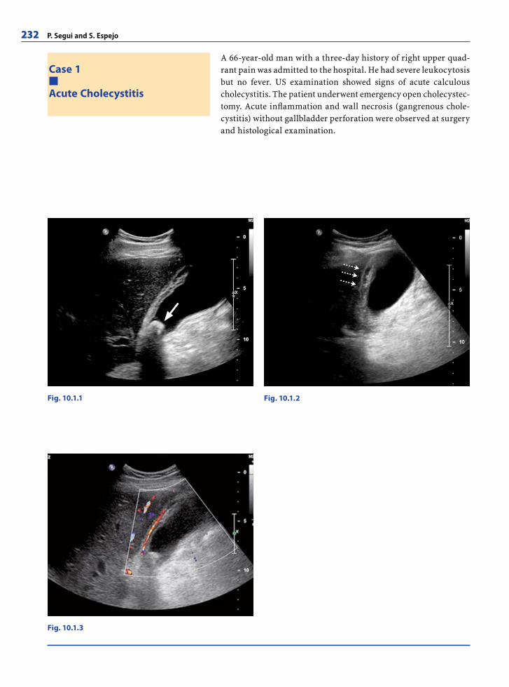
232 P. Segui and S. Espejo
A 66-year-old man with a three-day history of right upper quad-rant pain was admitted to the hospital. He had severe leukocytosis but no fever. US examination showed signs of acute calculous cholecystitis. The patient underwent emergency open cholecystec-tomy. Acute infl ammation and wall necrosis (gangrenous chole-cystitis) without gallbladder perforation were observed at surgery and histological examination.
Case 1
Acute Cholecystitis
Fig. 10.1.3
Fig. 10.1.1 Fig. 10.1.2
RibesLunaRos.indb 232 18.03.2008 12:54:14
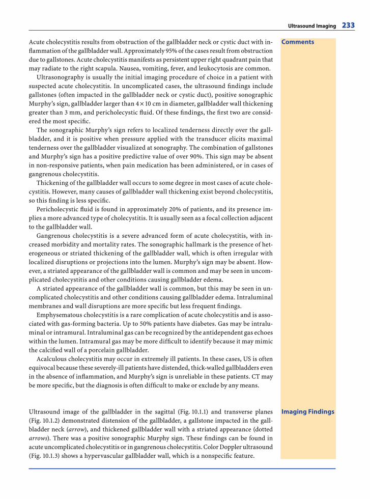
Ultrasound Imaging 233
CommentsAcute cholecystitis results from obstruction of the gallbladder neck or cystic duct with in-fl ammation of the gallbladder wall. Approximately 95% of the cases result from obstruction due to gallstones. Acute cholecystitis manifests as persistent upper right quadrant pain that may radiate to the right scapula. Nausea, vomiting, fever, and leukocytosis are common.
Ultrasonography is usually the initial imaging procedure of choice in a patient with suspected acute cholecystitis. In uncomplicated cases, the ultrasound fi ndings include gallstones (often impacted in the gallbladder neck or cystic duct), positive sonographic Murphy’s sign, gallbladder larger than 4 × 10 cm in diameter, gallbladder wall thickening greater than 3 mm, and pericholecystic fl uid. Of these fi ndings, the fi rst two are consid-ered the most specifi c.
The sonographic Murphy’s sign refers to localized tenderness directly over the gall-bladder, and it is positive when pressure applied with the transducer elicits maximal tenderness over the gallbladder visualized at sonography. The combination of gallstones and Murphy’s sign has a positive predictive value of over 90%. This sign may be absent in non-responsive patients, when pain medication has been administered, or in cases of gangrenous cholecystitis.
Thickening of the gallbladder wall occurs to some degree in most cases of acute chole-cystitis. However, many causes of gallbladder wall thickening exist beyond cholecystitis, so this fi nding is less specifi c.
Pericholecystic fl uid is found in approximately 20% of patients, and its presence im-plies a more advanced type of cholecystitis. It is usually seen as a focal collection adjacent to the gallbladder wall.
Gangrenous cholecystitis is a severe advanced form of acute cholecystitis, with in-creased morbidity and mortality rates. The sonographic hallmark is the presence of het-erogeneous or striated thickening of the gallbladder wall, which is often irregular with localized disruptions or projections into the lumen. Murphy’s sign may be absent. How-ever, a striated appearance of the gallbladder wall is common and may be seen in uncom-plicated cholecystitis and other conditions causing gallbladder edema.
A striated appearance of the gallbladder wall is common, but this may be seen in un-complicated cholecystitis and other conditions causing gallbladder edema. Intraluminal membranes and wall disruptions are more specifi c but less frequent fi ndings.
Emphysematous cholecystitis is a rare complication of acute cholecystitis and is asso-ciated with gas-forming bacteria. Up to 50% patients have diabetes. Gas may be intralu-minal or intramural. Intraluminal gas can be recognized by the antidependent gas echoes within the lumen. Intramural gas may be more diffi cult to identify because it may mimic the calcifi ed wall of a porcelain gallbladder.
Acalculous cholecystitis may occur in extremely ill patients. In these cases, US is often equivocal because these severely-ill patients have distended, thick-walled gallbladders even in the absence of infl ammation, and Murphy’s sign is unreliable in these patients. CT may be more specifi c, but the diagnosis is often diffi cult to make or exclude by any means.
Imaging FindingsUltrasound image of the gallbladder in the sagittal (Fig. 10.1.1) and transverse planes (Fig. 10.1.2) demonstrated distension of the gallbladder, a gallstone impacted in the gall-bladder neck (arrow), and thickened gallbladder wall with a striated appearance (dotted arrows). There was a positive sonographic Murphy sign. These fi ndings can be found in acute uncomplicated cholecystitis or in gangrenous cholecystitis. Color Doppler ultrasound (Fig. 10.1.3) shows a hypervascular gallbladder wall, which is a nonspecifi c feature.
RibesLunaRos.indb 233 18.03.2008 12:54:17
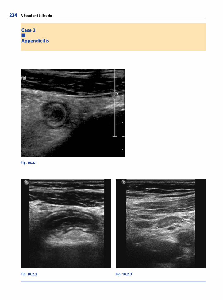
234 P. Segui and S. Espejo
Case 2
Appendicitis
Fig. 10.2.1
Fig. 10.2.2 Fig. 10.2.3
RibesLunaRos.indb 234 18.03.2008 12:54:17
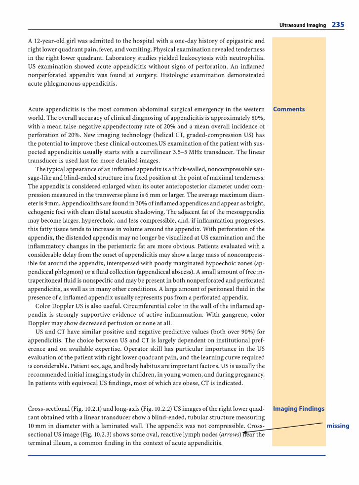
Ultrasound Imaging 235
A 12-year-old girl was admitted to the hospital with a one-day history of epigastric and right lower quadrant pain, fever, and vomiting. Physical examination revealed tenderness in the right lower quadrant. Laboratory studies yielded leukocytosis with neutrophilia. US examination showed acute appendicitis without signs of perforation. An infl amed nonperforated appendix was found at surgery. Histologic examination demonstrated acute phlegmonous appendicitis.
CommentsAcute appendicitis is the most common abdominal surgical emergency in the western world. The overall accuracy of clinical diagnosing of appendicitis is approximately 80%, with a mean false-negative appendectomy rate of 20% and a mean overall incidence of perforation of 20%. New imaging technology (helical CT, graded-compression US) has the potential to improve these clinical outcomes.US examination of the patient with sus-pected appendicitis usually starts with a curvilinear 3.5–5 MHz transducer. The linear transducer is used last for more detailed images.
The typical appearance of an infl amed appendix is a thick-walled, noncompressible sau-sage-like and blind-ended structure in a fi xed position at the point of maximal tenderness. The appendix is considered enlarged when its outer anteroposterior diameter under com-pression measured in the transverse plane is 6 mm or larger. The average maximum diam-eter is 9 mm. Appendicoliths are found in 30% of infl amed appendices and appear as bright, echogenic foci with clean distal acoustic shadowing. The adjacent fat of the mesoappendix may become larger, hyperechoic, and less compressible, and, if infl ammation progresses, this fatty tissue tends to increase in volume around the appendix. With perforation of the appendix, the distended appendix may no longer be visualized at US examination and the infl ammatory changes in the perienteric fat are more obvious. Patients evaluated with a considerable delay from the onset of appendicitis may show a large mass of noncompress-ible fat around the appendix, interspersed with poorly marginated hypoechoic zones (ap-pendiceal phlegmon) or a fl uid collection (appendiceal abscess). A small amount of free in-traperitoneal fl uid is nonspecifi c and may be present in both nonperforated and perforated appendicitis, as well as in many other conditions. A large amount of peritoneal fl uid in the presence of a infl amed appendix usually represents pus from a perforated appendix.
Color Doppler US is also useful. Circumferential color in the wall of the infl amed ap-pendix is strongly supportive evidence of active infl ammation. With gangrene, color Doppler may show decreased perfusion or none at all.
US and CT have similar positive and negative predictive values (both over 90%) for appendicitis. The choice between US and CT is largely dependent on institutional pref-erence and on available expertise. Operator skill has particular importance in the US evaluation of the patient with right lower quadrant pain, and the learning curve required is considerable. Patient sex, age, and body habitus are important factors. US is usually the recommended initial imaging study in children, in young women, and during pregnancy. In patients with equivocal US fi ndings, most of which are obese, CT is indicated.
Imaging FindingsCross-sectional (Fig. 10.2.1) and long-axis (Fig. 10.2.2) US images of the right lower quad-rant obtained with a linear transducer show a blind-ended, tubular structure measuring 10 mm in diameter with a laminated wall. The appendix was not compressible. Cross-sectional US image (Fig. 10.2.3) shows some oval, reactive lymph nodes (arrows) near the terminal illeum, a common fi nding in the context of acute appendicitis.
missing
RibesLunaRos.indb 235 18.03.2008 12:54:18

236 P. Segui and S. Espejo
A 43-year-old man was admitted with left lower quadrant pain and fever of one day’s duration. Physical examination revealed ten-derness and guarding in the left lower abdomen. Laboratory tests showed leukocytosis with neutrophilia. US fi ndings were sugges-tive of uncomplicated sigmoid diverticulitis. The patient underwent conservative management with antibiotics and was symptom free after a few days. Scheduled surgical resection (sigmoidectomy) was performed three months later. Histologic examination disclosed diverticulosis and chronic diverticulitis, without malignancy.
Case 3
Acute Colonic Diverticulitis
Fig. 10.3.1
Fig. 10.3.2
Fig. 10.3.3
RibesLunaRos.indb 236 18.03.2008 12:54:19
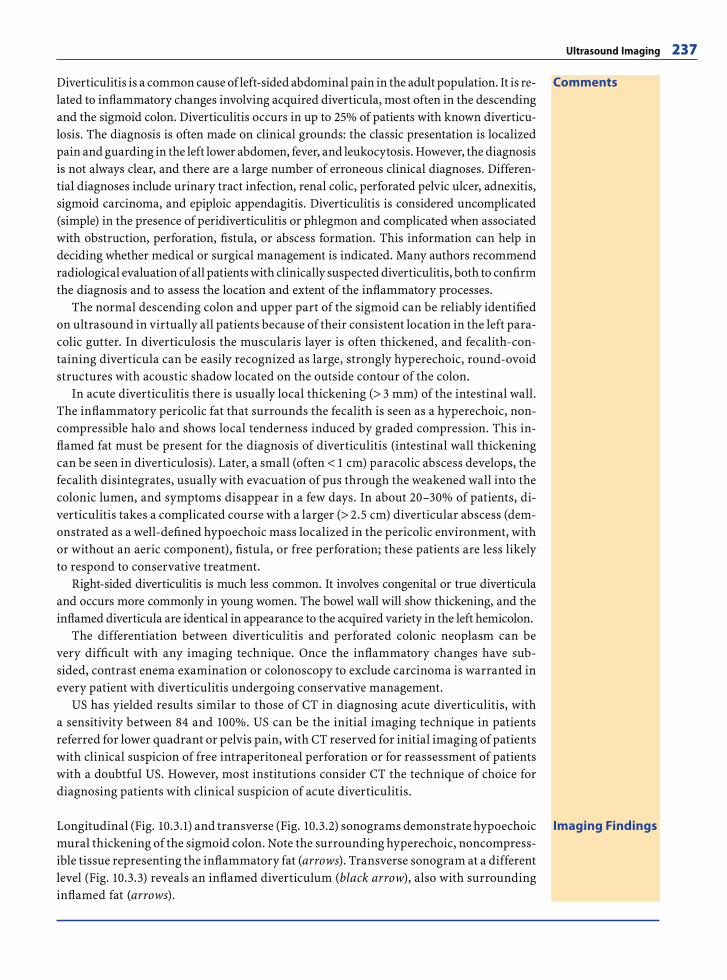
Ultrasound Imaging 237
CommentsDiverticulitis is a common cause of left-sided abdominal pain in the adult population. It is re-lated to infl ammatory changes involving acquired diverticula, most often in the descending and the sigmoid colon. Diverticulitis occurs in up to 25% of patients with known diverticu-losis. The diagnosis is often made on clinical grounds: the classic presentation is localized pain and guarding in the left lower abdomen, fever, and leukocytosis. However, the diagnosis is not always clear, and there are a large number of erroneous clinical diagnoses. Differen-tial diagnoses include urinary tract infection, renal colic, perforated pelvic ulcer, adnexitis, sigmoid carcinoma, and epiploic appendagitis. Diverticulitis is considered uncomplicated (simple) in the presence of peridiverticulitis or phlegmon and complicated when associated with obstruction, perforation, fi stula, or abscess formation. This information can help in deciding whether medical or surgical management is indicated. Many authors recommend radiological evaluation of all patients with clinically suspected diverticulitis, both to confi rm the diagnosis and to assess the location and extent of the infl ammatory processes.
The normal descending colon and upper part of the sigmoid can be reliably identifi ed on ultrasound in virtually all patients because of their consistent location in the left para-colic gutter. In diverticulosis the muscularis layer is often thickened, and fecalith-con-taining diverticula can be easily recognized as large, strongly hyperechoic, round-ovoid structures with acoustic shadow located on the outside contour of the colon.
In acute diverticulitis there is usually local thickening (> 3 mm) of the intestinal wall. The infl ammatory pericolic fat that surrounds the fecalith is seen as a hyperechoic, non-compressible halo and shows local tenderness induced by graded compression. This in-fl amed fat must be present for the diagnosis of diverticulitis (intestinal wall thickening can be seen in diverticulosis). Later, a small (often < 1 cm) paracolic abscess develops, the fecalith disintegrates, usually with evacuation of pus through the weakened wall into the colonic lumen, and symptoms disappear in a few days. In about 20–30% of patients, di-verticulitis takes a complicated course with a larger (> 2.5 cm) diverticular abscess (dem-onstrated as a well-defi ned hypoechoic mass localized in the pericolic environment, with or without an aeric component), fi stula, or free perforation; these patients are less likely to respond to conservative treatment.
Right-sided diverticulitis is much less common. It involves congenital or true diverticula and occurs more commonly in young women. The bowel wall will show thickening, and the infl amed diverticula are identical in appearance to the acquired variety in the left hemicolon.
The differentiation between diverticulitis and perforated colonic neoplasm can be very diffi cult with any imaging technique. Once the infl ammatory changes have sub-sided, contrast enema examination or colonoscopy to exclude carcinoma is warranted in every patient with diverticulitis undergoing conservative management.
US has yielded results similar to those of CT in diagnosing acute diverticulitis, with a sensitivity between 84 and 100%. US can be the initial imaging technique in patients referred for lower quadrant or pelvis pain, with CT reserved for initial imaging of patients with clinical suspicion of free intraperitoneal perforation or for reassessment of patients with a doubtful US. However, most institutions consider CT the technique of choice for diagnosing patients with clinical suspicion of acute diverticulitis.
Imaging FindingsLongitudinal (Fig. 10.3.1) and transverse (Fig. 10.3.2) sonograms demonstrate hypoechoic mural thickening of the sigmoid colon. Note the surrounding hyperechoic, noncompress-ible tissue representing the infl ammatory fat (arrows). Transverse sonogram at a different level (Fig. 10.3.3) reveals an infl amed diverticulum (black arrow), also with surrounding infl amed fat (arrows).
RibesLunaRos.indb 237 18.03.2008 12:54:19
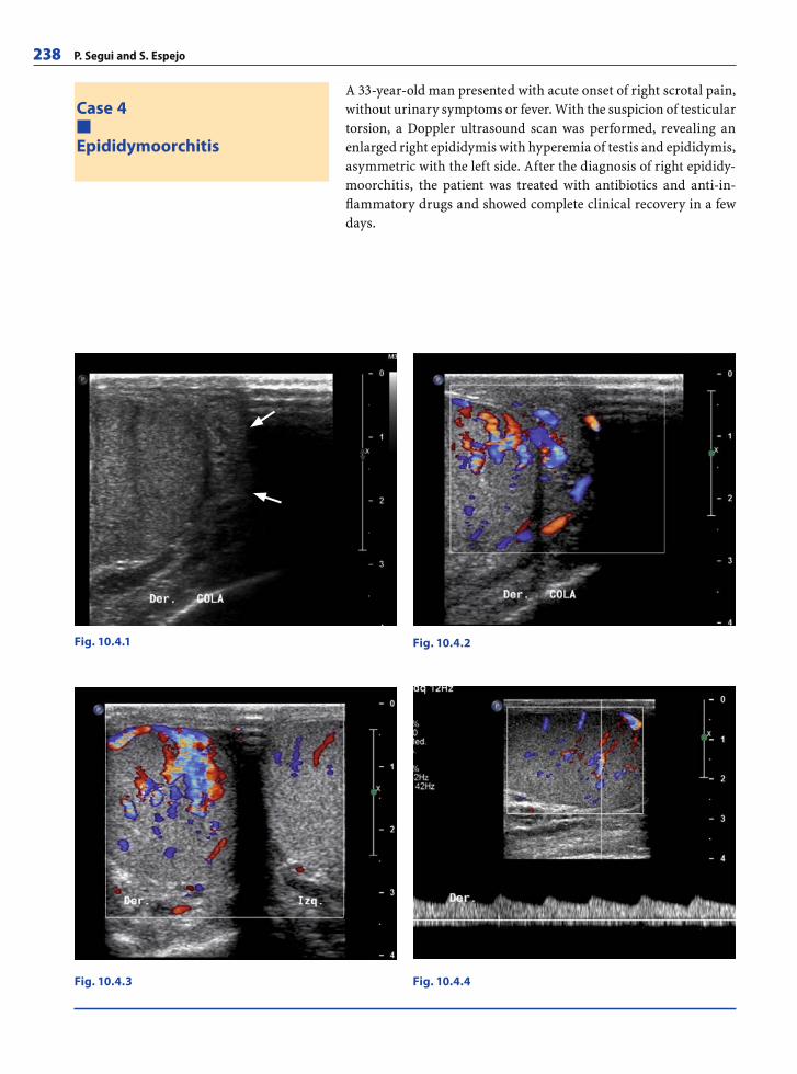
238 P. Segui and S. Espejo
A 33-year-old man presented with acute onset of right scrotal pain, without urinary symptoms or fever. With the suspicion of testicular torsion, a Doppler ultrasound scan was performed, revealing an enlarged right epididymis with hyperemia of testis and epididymis, asymmetric with the left side. After the diagnosis of right epididy-moorchitis, the patient was treated with antibiotics and anti-in-fl ammatory drugs and showed complete clinical recovery in a few days.
Case 4
Epididymoorchitis
Fig. 10.4.1 Fig. 10.4.2
Fig. 10.4.4Fig. 10.4.3
RibesLunaRos.indb 238 18.03.2008 12:54:19
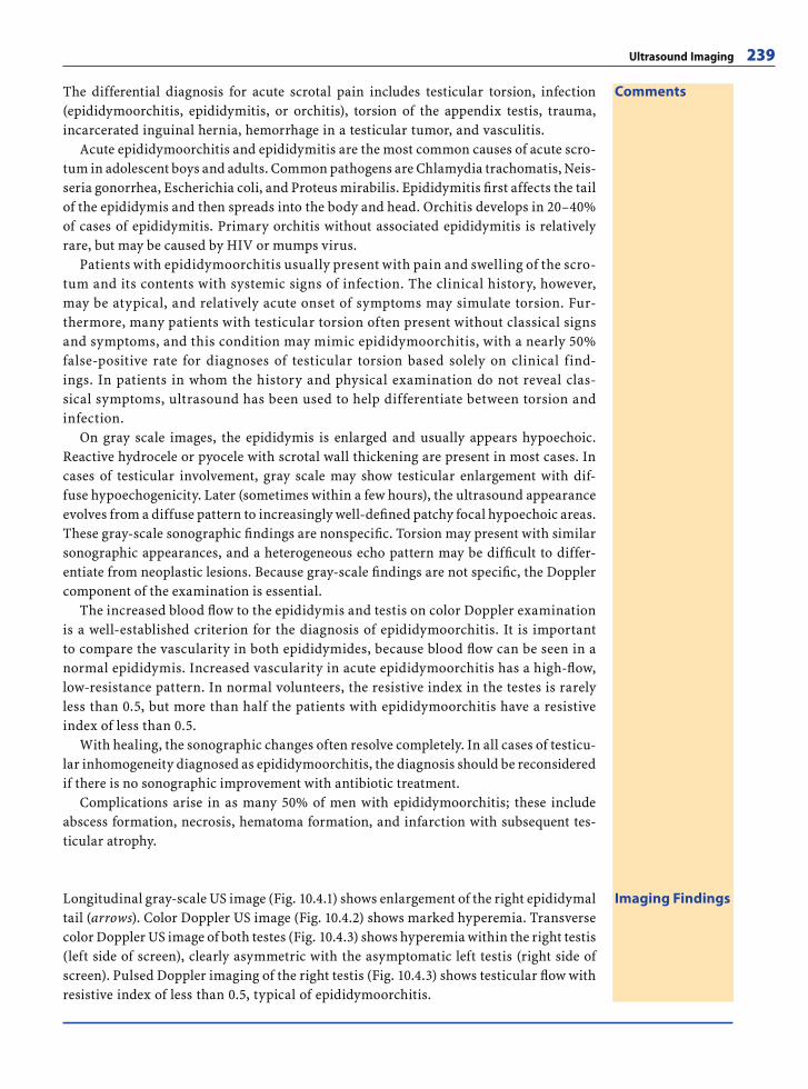
Ultrasound Imaging 239
CommentsThe differential diagnosis for acute scrotal pain includes testicular torsion, infection (epididymoorchitis, epididymitis, or orchitis), torsion of the appendix testis, trauma, incarcerated inguinal hernia, hemorrhage in a testicular tumor, and vasculitis.
Acute epididymoorchitis and epididymitis are the most common causes of acute scro-tum in adolescent boys and adults. Common pathogens are Chlamydia trachomatis, Neis-seria gonorrhea, Escherichia coli, and Proteus mirabilis. Epididymitis fi rst affects the tail of the epididymis and then spreads into the body and head. Orchitis develops in 20–40% of cases of epididymitis. Primary orchitis without associated epididymitis is relatively rare, but may be caused by HIV or mumps virus.
Patients with epididymoorchitis usually present with pain and swelling of the scro-tum and its contents with systemic signs of infection. The clinical history, however, may be atypical, and relatively acute onset of symptoms may simulate torsion. Fur-thermore, many patients with testicular torsion often present without classical signs and symptoms, and this condition may mimic epididymoorchitis, with a nearly 50% false-positive rate for diagnoses of testicular torsion based solely on clinical find-ings. In patients in whom the history and physical examination do not reveal clas-sical symptoms, ultrasound has been used to help differentiate between torsion and infection.
On gray scale images, the epididymis is enlarged and usually appears hypoechoic. Reactive hydrocele or pyocele with scrotal wall thickening are present in most cases. In cases of testicular involvement, gray scale may show testicular enlargement with dif-fuse hypoechogenicity. Later (sometimes within a few hours), the ultrasound appearance evolves from a diffuse pattern to increasingly well-defi ned patchy focal hypoechoic areas. These gray-scale sonographic fi ndings are nonspecifi c. Torsion may present with similar sonographic appearances, and a heterogeneous echo pattern may be diffi cult to differ-entiate from neoplastic lesions. Because gray-scale fi ndings are not specifi c, the Doppler component of the examination is essential.
The increased blood fl ow to the epididymis and testis on color Doppler examination is a well-established criterion for the diagnosis of epididymoorchitis. It is important to compare the vascularity in both epididymides, because blood fl ow can be seen in a normal epididymis. Increased vascularity in acute epididymoorchitis has a high-fl ow, low-resistance pattern. In normal volunteers, the resistive index in the testes is rarely less than 0.5, but more than half the patients with epididymoorchitis have a resistive index of less than 0.5.
With healing, the sonographic changes often resolve completely. In all cases of testicu-lar inhomogeneity diagnosed as epididymoorchitis, the diagnosis should be reconsidered if there is no sonographic improvement with antibiotic treatment.
Complications arise in as many 50% of men with epididymoorchitis; these include abscess formation, necrosis, hematoma formation, and infarction with subsequent tes-ticular atrophy.
Imaging FindingsLongitudinal gray-scale US image (Fig. 10.4.1) shows enlargement of the right epididymal tail (arrows). Color Doppler US image (Fig. 10.4.2) shows marked hyperemia. Transverse color Doppler US image of both testes (Fig. 10.4.3) shows hyperemia within the right testis (left side of screen), clearly asymmetric with the asymptomatic left testis (right side of screen). Pulsed Doppler imaging of the right testis (Fig. 10.4.3) shows testicular fl ow with resistive index of less than 0.5, typical of epididymoorchitis.
RibesLunaRos.indb 239 18.03.2008 12:54:26
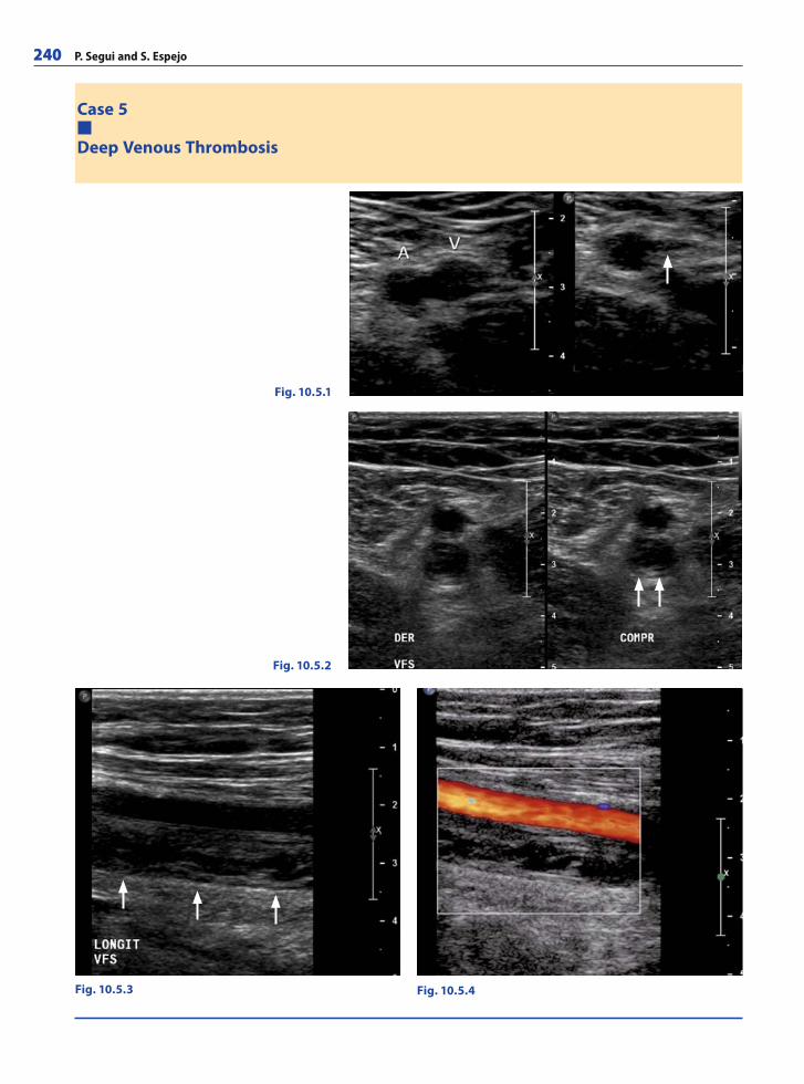
240 P. Segui and S. Espejo
Case 5
Deep Venous Thrombosis
Fig. 10.5.1
Fig. 10.5.2
Fig. 10.5.4Fig. 10.5.3
RibesLunaRos.indb 240 18.03.2008 12:54:26
Ultrasound Imaging 241
A 51-year-old man experienced a gradual onset of pain and swelling in his right calf 5 days before admission. The patient had been immobilized for three weeks for an ankle sprain. A venous US scan of the right leg showed deep vein thrombosis in the femoropopliteal segment. Anticoagulant treatment was initiated and resulted in rapid improvement of symptoms.
CommentsDeep vein thrombosis (DVT) is a common problem in the acute care setting. Commonly, DVT begins in the veins of the calf and progresses proximally over time. Less than 20% of patients with confi rmed DVT have thrombi isolated to the calf veins. While the thrombus is isolated in the iliofemoral region in only approximately 10% of patients with lower-extremity DVT, this is a common presentation of DVT during pregnancy, usually on the left side. The more central the thrombi, the greater the risk of pulmonary emboli. About 50% of patients with DVT develop pulmonary emboli, although many of these may be subsegmental and undiagnosed. Patients with acute calf-popliteal-vein thrombosis usu-ally present with symptoms of unilateral pain and swelling in the calf and this condition may be associated with warmth, redness, and tenderness.
The signs and symptoms of DVT are notoriously unreliable, so a noninvasive screening test is usually necessary.
Compression ultrasonography (CUS) is the diagnostic procedure of choice for the as-sessment of patients with suspected DVT. It has been shown to be highly sensitive and specifi c for the diagnosis of DVT, particularly in the lower extremities in symptomatic patients. Sensitivity and specifi city are near 100% in the femoropopliteal segment. Sensi-tivity decreases in isolated calf thrombi and in asymptomatic patients.
CUS of the deep venous system of the lower extremities is performed with the patient in the supine position, with the leg externally rotated and slightly fl exed at the knee. Patients may be placed prone to assess the popliteal fossa and occasionally in the sit-ting position if there is diffi culty in identifying the calf veins. A linear transducer with a frequency in the 5–12 MHz range is used. The transducer is placed transversely, moving down the leg following the common femoral vein, superfi cial femoral vein, and popliteal vein until it divides. Gentle pressure is applied to the vessels with the transducer at 1 cm intervals and, in the absence of DVT, the lumen of the vein should collapse. In the pres-ence of DVT, the lumen does not collapse, and this is the most sensitive and specifi c sign of DVT. In acute DVT, the thrombosed vein is distended and clot echogenicity is variable. Duplex and color Doppler are not required but can be helpful. No spectral or color fl ow can be demonstrated at the site of total occlusion, and there is loss of phasic response in the segment distal to it.
The value of performing CUS of the calf veins when proximal veins are normal remains controversial.
Imaging FindingsUS scan of a healthy volunteer (Fig. 10.5.1). Split screen transverse US image of a normal superfi cial femoral vein (V) and artery (A). Uncompressed (left side) and compressed (right side) images show that, in the absence of thrombosis, compression results in com-plete apposition of the walls of the vein (arrow) while the artery remains patent.
In our patient with femoral vein thrombosis split screen transverse US image (Fig. 10.5.2) shows a non-compressible superfi cial femoral vein (arrows). Longitudinal US image (Fig. 10.5.3) shows the distended vein (posterior to the artery) with intralumi-nal clot (arrows). Color Doppler longitudinal image (Fig. 10.5.4) shows normal fl ow in the superfi cial femoral artery and no fl ow in the superfi cial femoral vein.
RibesLunaRos.indb 241 18.03.2008 12:54:31
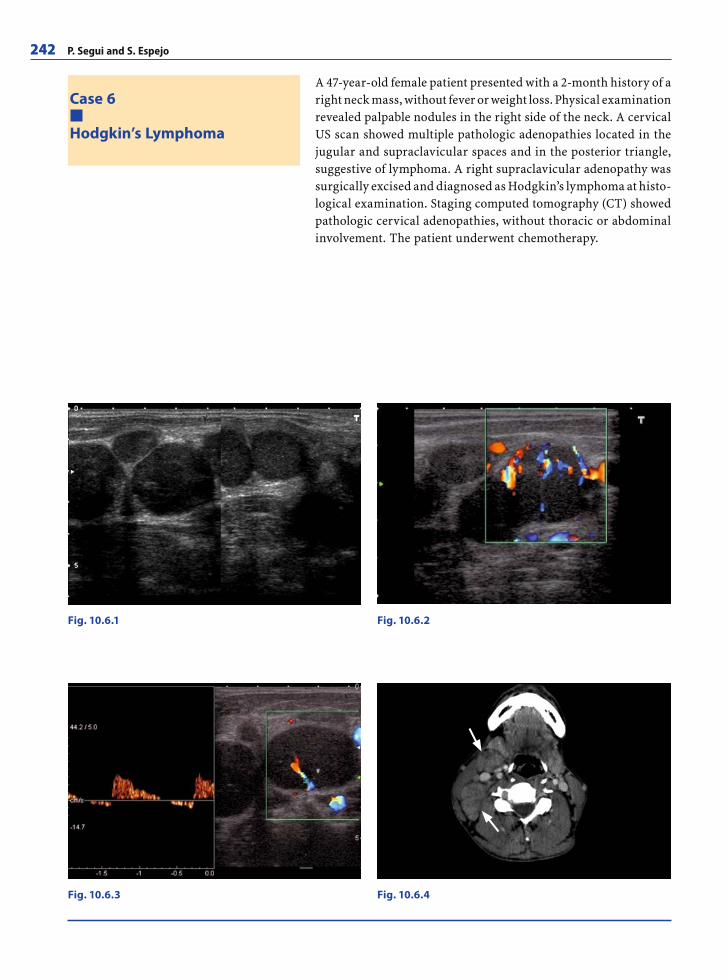
242 P. Segui and S. Espejo
A 47-year-old female patient presented with a 2-month history of a right neck mass, without fever or weight loss. Physical examination revealed palpable nodules in the right side of the neck. A cervical US scan showed multiple pathologic adenopathies located in the jugular and supraclavicular spaces and in the posterior triangle, suggestive of lymphoma. A right supraclavicular adenopathy was surgically excised and diagnosed as Hodgkin’s lymphoma at histo-logical examination. Staging computed tomography (CT) showed pathologic cervical adenopathies, without thoracic or abdominal involvement. The patient underwent chemotherapy.
Case 6
Hodgkin’s Lymphoma
Fig. 10.6.1 Fig. 10.6.2
Fig. 10.6.3 Fig. 10.6.4
RibesLunaRos.indb 242 18.03.2008 12:54:31
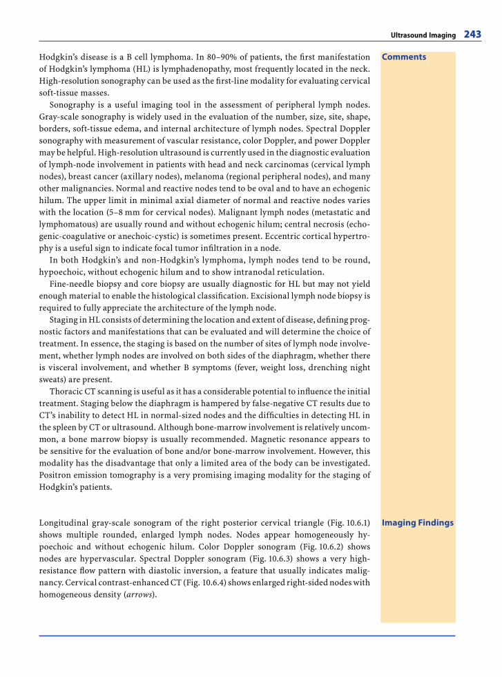
Ultrasound Imaging 243
CommentsHodgkin’s disease is a B cell lymphoma. In 80–90% of patients, the fi rst manifestation of Hodgkin’s lymphoma (HL) is lymphadenopathy, most frequently located in the neck. High-resolution sonography can be used as the fi rst-line modality for evaluating cervical soft-tissue masses.
Sonography is a useful imaging tool in the assessment of peripheral lymph nodes. Gray-scale sonography is widely used in the evaluation of the number, size, site, shape, borders, soft-tissue edema, and internal architecture of lymph nodes. Spectral Doppler sonography with measurement of vascular resistance, color Doppler, and power Doppler may be helpful. High-resolution ultrasound is currently used in the diagnostic evaluation of lymph-node involvement in patients with head and neck carcinomas (cervical lymph nodes), breast cancer (axillary nodes), melanoma (regional peripheral nodes), and many other malignancies. Normal and reactive nodes tend to be oval and to have an echogenic hilum. The upper limit in minimal axial diameter of normal and reactive nodes varies with the location (5–8 mm for cervical nodes). Malignant lymph nodes (metastatic and lymphomatous) are usually round and without echogenic hilum; central necrosis (echo-genic-coagulative or anechoic-cystic) is sometimes present. Eccentric cortical hypertro-phy is a useful sign to indicate focal tumor infi ltration in a node.
In both Hodgkin’s and non-Hodgkin’s lymphoma, lymph nodes tend to be round, hypoechoic, without echogenic hilum and to show intranodal reticulation.
Fine-needle biopsy and core biopsy are usually diagnostic for HL but may not yield enough material to enable the histological classifi cation. Excisional lymph node biopsy is required to fully appreciate the architecture of the lymph node.
Staging in HL consists of determining the location and extent of disease, defi ning prog-nostic factors and manifestations that can be evaluated and will determine the choice of treatment. In essence, the staging is based on the number of sites of lymph node involve-ment, whether lymph nodes are involved on both sides of the diaphragm, whether there is visceral involvement, and whether B symptoms (fever, weight loss, drenching night sweats) are present.
Thoracic CT scanning is useful as it has a considerable potential to infl uence the initial treatment. Staging below the diaphragm is hampered by false-negative CT results due to CT’s inability to detect HL in normal-sized nodes and the diffi culties in detecting HL in the spleen by CT or ultrasound. Although bone-marrow involvement is relatively uncom-mon, a bone marrow biopsy is usually recommended. Magnetic resonance appears to be sensitive for the evaluation of bone and/or bone-marrow involvement. However, this modality has the disadvantage that only a limited area of the body can be investigated. Positron emission tomography is a very promising imaging modality for the staging of Hodgkin’s patients.
Imaging FindingsLongitudinal gray-scale sonogram of the right posterior cervical triangle (Fig. 10.6.1) shows multiple rounded, enlarged lymph nodes. Nodes appear homogeneously hy-poechoic and without echogenic hilum. Color Doppler sonogram (Fig. 10.6.2) shows nodes are hypervascular. Spectral Doppler sonogram (Fig. 10.6.3) shows a very high-resistance fl ow pattern with diastolic inversion, a feature that usually indicates malig-nancy. Cervical contrast-enhanced CT (Fig. 10.6.4) shows enlarged right-sided nodes with homogeneous density (arrows).
RibesLunaRos.indb 243 18.03.2008 12:54:36

244 P. Segui and S. Espejo
Case 7
Abdominal Wall Endometriosis
Fig. 10.7.1
Fig. 10.7.3
Fig. 10.7.2
RibesLunaRos.indb 244 18.03.2008 12:54:36
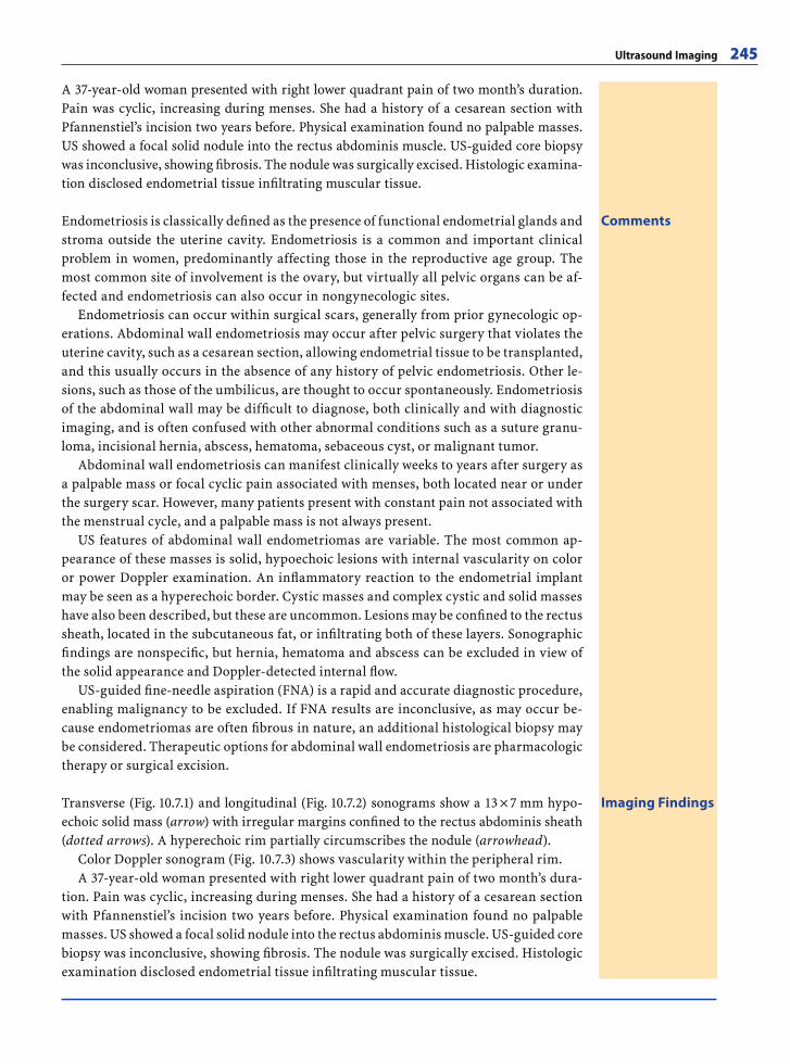
Ultrasound Imaging 245
CommentsEndometriosis is classically defi ned as the presence of functional endometrial glands and stroma outside the uterine cavity. Endometriosis is a common and important clinical problem in women, predominantly affecting those in the reproductive age group. The most common site of involvement is the ovary, but virtually all pelvic organs can be af-fected and endometriosis can also occur in nongynecologic sites.
Endometriosis can occur within surgical scars, generally from prior gynecologic op-erations. Abdominal wall endometriosis may occur after pelvic surgery that violates the uterine cavity, such as a cesarean section, allowing endometrial tissue to be transplanted, and this usually occurs in the absence of any history of pelvic endometriosis. Other le-sions, such as those of the umbilicus, are thought to occur spontaneously. Endometriosis of the abdominal wall may be diffi cult to diagnose, both clinically and with diagnostic imaging, and is often confused with other abnormal conditions such as a suture granu-loma, incisional hernia, abscess, hematoma, sebaceous cyst, or malignant tumor.
Abdominal wall endometriosis can manifest clinically weeks to years after surgery as a palpable mass or focal cyclic pain associated with menses, both located near or under the surgery scar. However, many patients present with constant pain not associated with the menstrual cycle, and a palpable mass is not always present.
US features of abdominal wall endometriomas are variable. The most common ap-pearance of these masses is solid, hypoechoic lesions with internal vascularity on color or power Doppler examination. An infl ammatory reaction to the endometrial implant may be seen as a hyperechoic border. Cystic masses and complex cystic and solid masses have also been described, but these are uncommon. Lesions may be confi ned to the rectus sheath, located in the subcutaneous fat, or infi ltrating both of these layers. Sonographic fi ndings are nonspecifi c, but hernia, hematoma and abscess can be excluded in view of the solid appearance and Doppler-detected internal fl ow.
US-guided fi ne-needle aspiration (FNA) is a rapid and accurate diagnostic procedure, enabling malignancy to be excluded. If FNA results are inconclusive, as may occur be-cause endometriomas are often fi brous in nature, an additional histological biopsy may be considered. Therapeutic options for abdominal wall endometriosis are pharmacologic therapy or surgical excision.
Imaging FindingsTransverse (Fig. 10.7.1) and longitudinal (Fig. 10.7.2) sonograms show a 13 × 7 mm hypo-echoic solid mass (arrow) with irregular margins confi ned to the rectus abdominis sheath (dotted arrows). A hyperechoic rim partially circumscribes the nodule (arrowhead).
Color Doppler sonogram (Fig. 10.7.3) shows vascularity within the peripheral rim.A 37-year-old woman presented with right lower quadrant pain of two month’s dura-
tion. Pain was cyclic, increasing during menses. She had a history of a cesarean section with Pfannenstiel’s incision two years before. Physical examination found no palpable masses. US showed a focal solid nodule into the rectus abdominis muscle. US-guided core biopsy was inconclusive, showing fi brosis. The nodule was surgically excised. Histologic examination disclosed endometrial tissue infi ltrating muscular tissue.
A 37-year-old woman presented with right lower quadrant pain of two month’s duration. Pain was cyclic, increasing during menses. She had a history of a cesarean section with Pfannenstiel’s incision two years before. Physical examination found no palpable masses. US showed a focal solid nodule into the rectus abdominis muscle. US-guided core biopsy was inconclusive, showing fi brosis. The nodule was surgically excised. Histologic examina-tion disclosed endometrial tissue infi ltrating muscular tissue.
RibesLunaRos.indb 245 18.03.2008 12:54:39
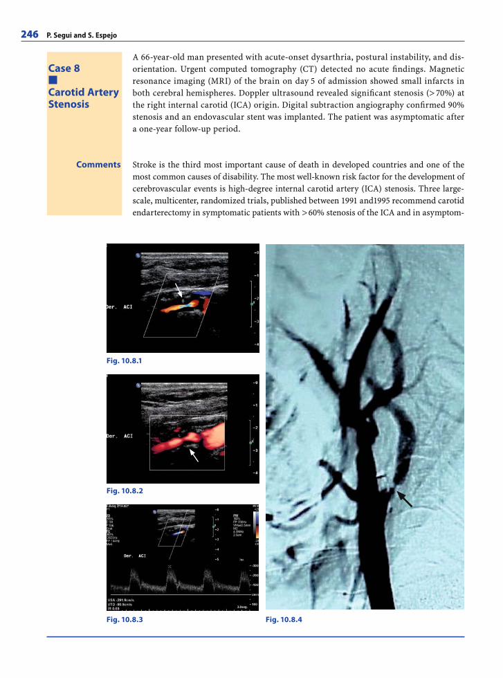
246 P. Segui and S. Espejo
A 66-year-old man presented with acute-onset dysarthria, postural instability, and dis-orientation. Urgent computed tomography (CT) detected no acute fi ndings. Magnetic resonance imaging (MRI) of the brain on day 5 of admission showed small infarcts in both cerebral hemispheres. Doppler ultrasound revealed signifi cant stenosis (> 70%) at the right internal carotid (ICA) origin. Digital subtraction angiography confi rmed 90% stenosis and an endovascular stent was implanted. The patient was asymptomatic after a one-year follow-up period.
Case 8
Carotid ArteryStenosis
Comments Stroke is the third most important cause of death in developed countries and one of the most common causes of disability. The most well-known risk factor for the development of cerebrovascular events is high-degree internal carotid artery (ICA) stenosis. Three large-scale, multicenter, randomized trials, published between 1991 and1995 recommend carotid endarterectomy in symptomatic patients with > 60% stenosis of the ICA and in asymptom-
Fig. 10.8.1
Fig. 10.8.2
Fig. 10.8.3 Fig. 10.8.4
RibesLunaRos.indb 246 18.03.2008 12:54:39
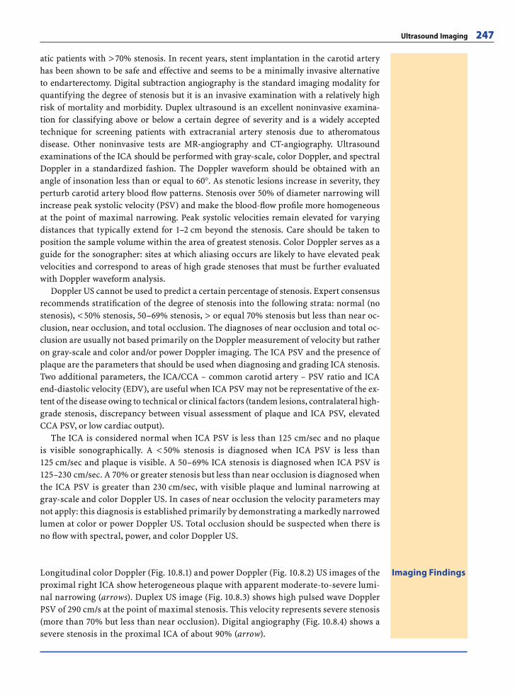
Ultrasound Imaging 247
Longitudinal color Doppler (Fig. 10.8.1) and power Doppler (Fig. 10.8.2) US images of the proximal right ICA show heterogeneous plaque with apparent moderate-to-severe lumi-nal narrowing (arrows). Duplex US image (Fig. 10.8.3) shows high pulsed wave Doppler PSV of 290 cm/s at the point of maximal stenosis. This velocity represents severe stenosis (more than 70% but less than near occlusion). Digital angiography (Fig. 10.8.4) shows a severe stenosis in the proximal ICA of about 90% (arrow).
Imaging Findings
atic patients with > 70% stenosis. In recent years, stent implantation in the carotid artery has been shown to be safe and effective and seems to be a minimally invasive alternative to endarterectomy. Digital subtraction angiography is the standard imaging modality for quantifying the degree of stenosis but it is an invasive examination with a relatively high risk of mortality and morbidity. Duplex ultrasound is an excellent noninvasive examina-tion for classifying above or below a certain degree of severity and is a widely accepted technique for screening patients with extracranial artery stenosis due to atheromatous disease. Other noninvasive tests are MR-angiography and CT-angiography. Ultrasound examinations of the ICA should be performed with gray-scale, color Doppler, and spectral Doppler in a standardized fashion. The Doppler waveform should be obtained with an angle of insonation less than or equal to 60 . As stenotic lesions increase in severity, they perturb carotid artery blood fl ow patterns. Stenosis over 50% of diameter narrowing will increase peak systolic velocity (PSV) and make the blood-fl ow profi le more homogeneous at the point of maximal narrowing. Peak systolic velocities remain elevated for varying distances that typically extend for 1–2 cm beyond the stenosis. Care should be taken to position the sample volume within the area of greatest stenosis. Color Doppler serves as a guide for the sonographer: sites at which aliasing occurs are likely to have elevated peak velocities and correspond to areas of high grade stenoses that must be further evaluated with Doppler waveform analysis.
Doppler US cannot be used to predict a certain percentage of stenosis. Expert consensus recommends stratifi cation of the degree of stenosis into the following strata: normal (no stenosis), < 50% stenosis, 50–69% stenosis, > or equal 70% stenosis but less than near oc-clusion, near occlusion, and total occlusion. The diagnoses of near occlusion and total oc-clusion are usually not based primarily on the Doppler measurement of velocity but rather on gray-scale and color and/or power Doppler imaging. The ICA PSV and the presence of plaque are the parameters that should be used when diagnosing and grading ICA stenosis. Two additional parameters, the ICA/CCA – common carotid artery – PSV ratio and ICA end-diastolic velocity (EDV), are useful when ICA PSV may not be representative of the ex-tent of the disease owing to technical or clinical factors (tandem lesions, contralateral high-grade stenosis, discrepancy between visual assessment of plaque and ICA PSV, elevated CCA PSV, or low cardiac output).
The ICA is considered normal when ICA PSV is less than 125 cm/sec and no plaque is visible sonographically. A < 50% stenosis is diagnosed when ICA PSV is less than 125 cm/sec and plaque is visible. A 50–69% ICA stenosis is diagnosed when ICA PSV is 125–230 cm/sec. A 70% or greater stenosis but less than near occlusion is diagnosed when the ICA PSV is greater than 230 cm/sec, with visible plaque and luminal narrowing at gray-scale and color Doppler US. In cases of near occlusion the velocity parameters may not apply: this diagnosis is established primarily by demonstrating a markedly narrowed lumen at color or power Doppler US. Total occlusion should be suspected when there is no fl ow with spectral, power, and color Doppler US.
RibesLunaRos.indb 247 18.03.2008 12:54:49
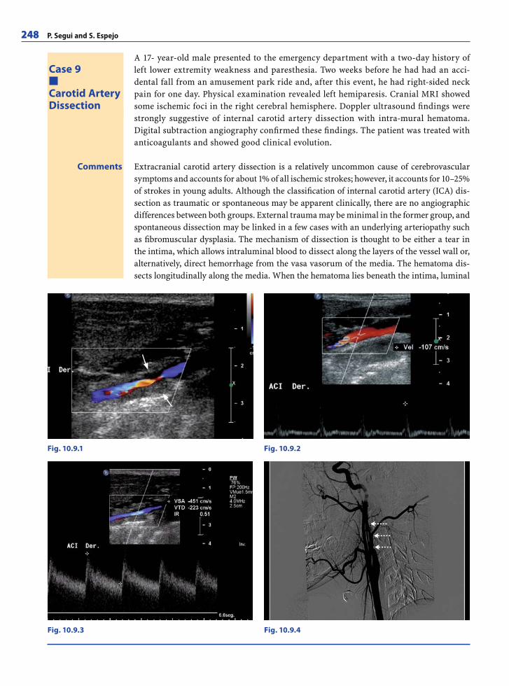
248 P. Segui and S. Espejo
A 17- year-old male presented to the emergency department with a two-day history of left lower extremity weakness and paresthesia. Two weeks before he had had an acci-dental fall from an amusement park ride and, after this event, he had right-sided neck pain for one day. Physical examination revealed left hemiparesis. Cranial MRI showed some ischemic foci in the right cerebral hemisphere. Doppler ultrasound fi ndings were strongly suggestive of internal carotid artery dissection with intra-mural hematoma. Digital subtraction angiography confi rmed these fi ndings. The patient was treated with anticoagulants and showed good clinical evolution.
Case 9
Carotid ArteryDissection
Fig. 10.9.1 Fig. 10.9.2
Comments Extracranial carotid artery dissection is a relatively uncommon cause of cerebrovascular symptoms and accounts for about 1% of all ischemic strokes; however, it accounts for 10–25% of strokes in young adults. Although the classifi cation of internal carotid artery (ICA) dis-section as traumatic or spontaneous may be apparent clinically, there are no angiographic differences between both groups. External trauma may be minimal in the former group, and spontaneous dissection may be linked in a few cases with an underlying arteriopathy such as fi bromuscular dysplasia. The mechanism of dissection is thought to be either a tear in the intima, which allows intraluminal blood to dissect along the layers of the vessel wall or, alternatively, direct hemorrhage from the vasa vasorum of the media. The hematoma dis-sects longitudinally along the media. When the hematoma lies beneath the intima, luminal
Fig. 10.9.4Fig. 10.9.3
RibesLunaRos.indb 248 18.03.2008 12:54:49

Ultrasound Imaging 249
narrowing or occlusion occurs. If the hematoma dissects beneath the adventitia, a pseudoa-neurysm forms. A false lumen occurs if blood reenters the true lumen; this false lumen may remain patent, resolve completely, or thrombose and cause narrowing of the true lumen.
The typical patient with carotid dissection presents with pain on one side of the head, face, or neck, accompanied by a partial Horner’s syndrome followed hours or days later by cerebral or retinal ischemia. This triad of symptoms is found in less than one third of patients. Cerebral infarcts are reported in 50% and transient ischemic attacks (TIA) in 20% of patients with ICA dissection.
The commonest location for a spontaneous ICA dissection is the cervical segment 2–3 cm distal to the carotid bulb. Dissections extend for a variable length but they rarely reach the point of entry of the ICA into the petrous temporal bone. In the absence of ar-teriopathy, recurrent dissection is rare.
Intraarterial angiography has been the gold standard in the diagnosis of dissections, but it is increasingly being replaced by noninvasive imaging techniques. The most com-mon angiographic fi nding, reported in more than 50% cases, is an irregular stenosis starting about 2–3 cm distal to the carotid bulb. Other, less common angiographic fi nd-ings (5–30% cases) are a fl ame-shaped occlusion, a tapered narrowing of the ICA (“rat’s tail” or “string sign”), and pseudoaneurysm. All these appearances are indicative of dis-section, but they are non-specifi c. The only pathognomic fi nding is an intimal fl ap with double lumen, but these fl aps are detected in less than 10% cases. Angiography does not allow direct visualization of the vessel wall when there is a thrombus in the false lumen.
US provides direct visualization of the pathological fi ndings, hemodynamic informa-tion, fl ow direction and velocity data, and enables evaluation of the vessel wall and lumen patency. The commonest gray-scale fi nding is the presence of a hypoechoic thrombus (cor-responding to intramural hematoma or thrombosed false lumen) 2–3 cm cranially from the bulb, with or without true lumen narrowing. The presence of an arterial luminal fl ap (mem-brane) in the longitudinal and axial views is pathognomic, but this fi nding is uncommon.
The commonest spectral Doppler US fi nding in the affected ICA is a high resistance pattern or absence of signal in cases of total occlusion. Another, less common spectral pattern is a damped spectral waveform (lower amplitude with biphasic pattern). Visual-ization of intramural hematoma combined with high-resistance fl ow strongly suggests dissection. The diagnostic sensitivity of US decreases if an ICA dissection results in a low-grade stenosis, and gray-scale and spectral fi ndings can be normal in these cases. US is less reliable for dissections located in the subpetrous segment and the carotid canal.
At present, the diagnosis of carotid dissection is essentially established with cervical MR and MR-angiography. The former can provide direct visualization of an intramural hematoma, which is the hallmark of a dissection, and the later allows noninvasive visual-ization of blood vessels. However, MR is not always available around the clock and many centers use ultrasound to assess carotid dissection. Furthermore, follow-up US studies can document recanalization, stabilization or progressive vascular occlusion.
Imaging FindingsLongitudinal color Doppler US (Fig. 10.9.1) of the right internal carotid artery (ICA) dem-onstrating a low-refl ective intramural hematoma (arrows) compressing the true lumen of the internal carotid artery 2 cm cranially from the bulb. Color and spectral Doppler US of the ICA immediately prior to dissection (Fig. 10.9.2) shows a high-resistance triphasic waveform. Spectral Doppler at the point of maximal lumen stenosis (Fig. 10.9.3) shows a very high peak systolic velocity over 4 m/s. Digital angiography (Fig. 10.9.4) confi rmed an irregular stenosis (arrows) starting 2 cm distal to the carotid bulb.
RibesLunaRos.indb 249 18.03.2008 12:54:56
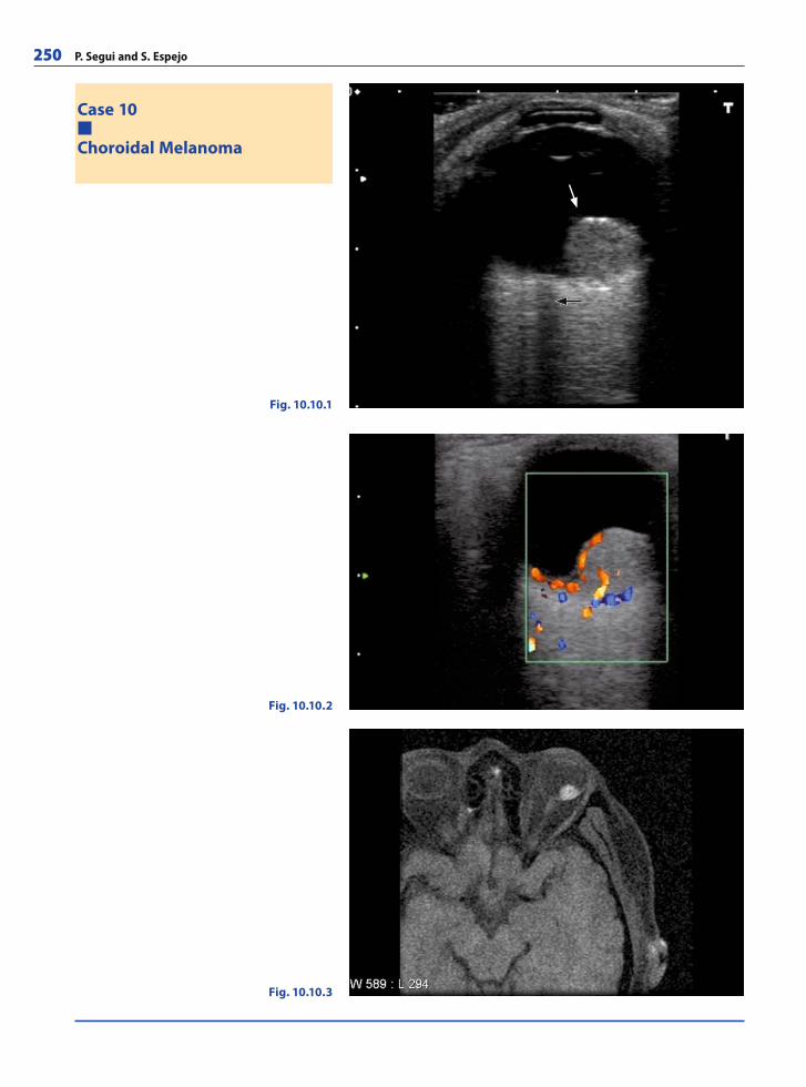
250 P. Segui and S. Espejo
Case 10
Choroidal Melanoma
Fig. 10.10.1
Fig. 10.10.3
Fig. 10.10.2
RibesLunaRos.indb 250 18.03.2008 12:54:56
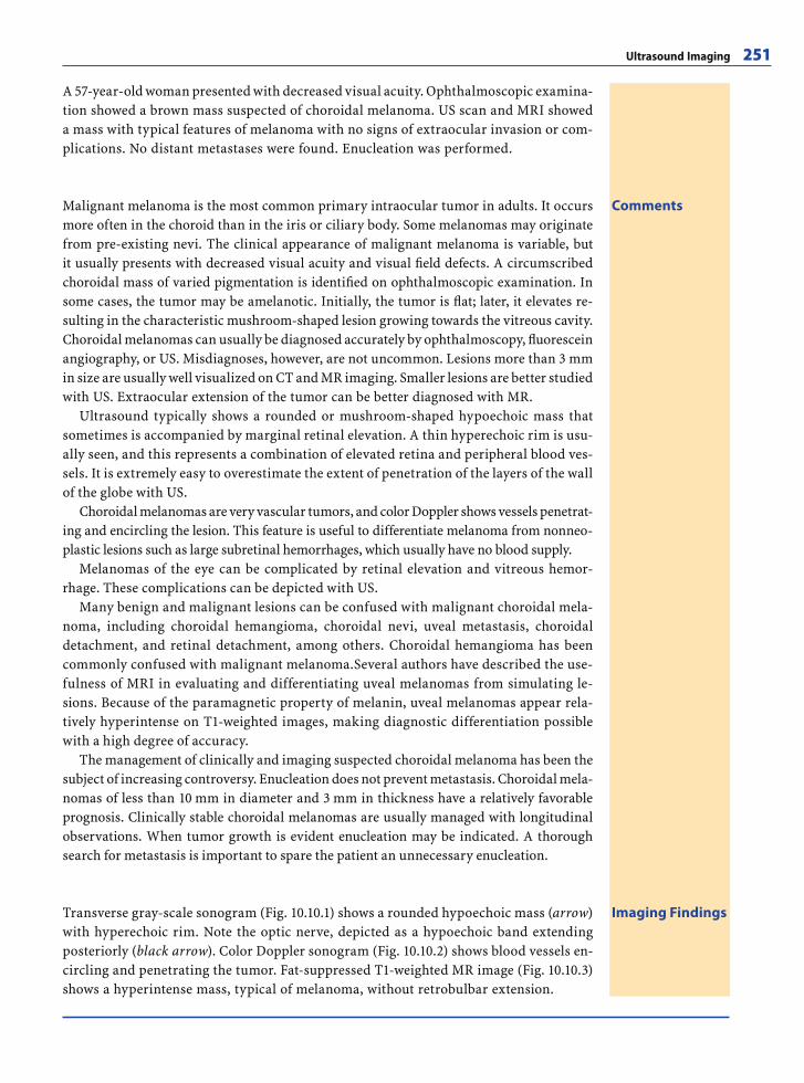
Ultrasound Imaging 251
A 57-year-old woman presented with decreased visual acuity. Ophthalmoscopic examina-tion showed a brown mass suspected of choroidal melanoma. US scan and MRI showed a mass with typical features of melanoma with no signs of extraocular invasion or com-plications. No distant metastases were found. Enucleation was performed.
CommentsMalignant melanoma is the most common primary intraocular tumor in adults. It occurs more often in the choroid than in the iris or ciliary body. Some melanomas may originate from pre-existing nevi. The clinical appearance of malignant melanoma is variable, but it usually presents with decreased visual acuity and visual fi eld defects. A circumscribed choroidal mass of varied pigmentation is identifi ed on ophthalmoscopic examination. In some cases, the tumor may be amelanotic. Initially, the tumor is fl at; later, it elevates re-sulting in the characteristic mushroom-shaped lesion growing towards the vitreous cavity. Choroidal melanomas can usually be diagnosed accurately by ophthalmoscopy, fl uorescein angiography, or US. Misdiagnoses, however, are not uncommon. Lesions more than 3 mm in size are usually well visualized on CT and MR imaging. Smaller lesions are better studied with US. Extraocular extension of the tumor can be better diagnosed with MR.
Ultrasound typically shows a rounded or mushroom-shaped hypoechoic mass that sometimes is accompanied by marginal retinal elevation. A thin hyperechoic rim is usu-ally seen, and this represents a combination of elevated retina and peripheral blood ves-sels. It is extremely easy to overestimate the extent of penetration of the layers of the wall of the globe with US.
Choroidal melanomas are very vascular tumors, and color Doppler shows vessels penetrat-ing and encircling the lesion. This feature is useful to differentiate melanoma from nonneo-plastic lesions such as large subretinal hemorrhages, which usually have no blood supply.
Melanomas of the eye can be complicated by retinal elevation and vitreous hemor-rhage. These complications can be depicted with US.
Many benign and malignant lesions can be confused with malignant choroidal mela-noma, including choroidal hemangioma, choroidal nevi, uveal metastasis, choroidal detachment, and retinal detachment, among others. Choroidal hemangioma has been commonly confused with malignant melanoma.Several authors have described the use-fulness of MRI in evaluating and differentiating uveal melanomas from simulating le-sions. Because of the paramagnetic property of melanin, uveal melanomas appear rela-tively hyperintense on T1-weighted images, making diagnostic differentiation possible with a high degree of accuracy.
The management of clinically and imaging suspected choroidal melanoma has been the subject of increasing controversy. Enucleation does not prevent metastasis. Choroidal mela-nomas of less than 10 mm in diameter and 3 mm in thickness have a relatively favorable prognosis. Clinically stable choroidal melanomas are usually managed with longitudinal observations. When tumor growth is evident enucleation may be indicated. A thorough search for metastasis is important to spare the patient an unnecessary enucleation.
Imaging FindingsTransverse gray-scale sonogram (Fig. 10.10.1) shows a rounded hypoechoic mass (arrow) with hyperechoic rim. Note the optic nerve, depicted as a hypoechoic band extending posteriorly (black arrow). Color Doppler sonogram (Fig. 10.10.2) shows blood vessels en-circling and penetrating the tumor. Fat-suppressed T1-weighted MR image (Fig. 10.10.3) shows a hyperintense mass, typical of melanoma, without retrobulbar extension.
RibesLunaRos.indb 251 18.03.2008 12:54:58
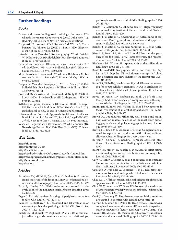
252 P. Segui and S. Espejo
Further Readings
Books
Categorical course in diagnostic radiology: fi ndings at US-what do they mean? Cooperberg PL (2002) Oak Brook (IL). Radiological Society of North America publications
Diagnostic Ultrasound. 3rd ed. Rumack CM, Wilson S, Char-boneau JW, Johnson JA (2005) St. Louis (MO). Elsevier- Mosby. ISBN-13: 9780323020237
Introduction to Vascular Ultrasonography. 5th ed. Zwiebel W, Pellerito J (2004) Philadelphia (PA). Elsevier- Saunders. ISBN-13: 9780721606316
General and Vascular Ultrasound: case review series. 2nd ed. Middleton WD (2007) Philadelphia (PA). Elsevier- Saunders. ISBN-13: 9781416039891
Musculoskeletal Ultrasound. 2nd ed. van Holsbeeck M, In-trocaso J (2001) St. Louis (MO) Elsevier-Mosby. ISBN-13: 9780323000185
Peripheral Vascular Sonography. 2nd ed. Polak JF (2004) Philadelphia (PA). Lippincott Williams & Wilkins. ISBN-13: 9780781748711
Practical Musculoskeletal Ultrasound. McNally E (2004) St. Louis (MO). Elsevier-Churchill Livingstone. ISBN-13: 9780443073502
Syllabus: A Special Course in Ultrasound. Bluth EI, Arger PH, Hertzberg BS, Middleton WD (1996) Oak Brook (IL). Radiological Society of North America publications
Ultrasound: A Practical Approach to Clinical Problems. Bluth EI, Arger PH, Benson CB, Ralls PW, Siegel MJ (2007) 2nd ed. New York (NY). Thieme. ISBN-13: 9783131168320
Vascular Diagnosis with Ultrasound. 2nd ed. Hennerici MG, Neuerburg-Heusler D (2006) New York (NY). Thieme. ISBN-13: 9783131038326
Web-Links
http://aium.orghttp://auntminnie.comhttp://emedicine.comhttp://med-ed.virginia.edu/courses/rad/edus/index.htlmhttp://radiographics.rsnajnls.org/cgi/collection/ultrasoundhttp://sonoworld.comhttp://sru.org
Articles
Bartolotta TV, Midiri M, Quaia E, et al. Benign focal liver le-sions: spectrum of fi ndings on SonoVue-enhanced pulse-inversion ultrasonography. Eur Radiol 2005; 15:1643–1649
Basu S, Howlet DC. High-resolution ultrasound in the evaluation of the nonacute testis. Abdom Imaging 2001; 26:425–432
Beggs I. Pictorial review: Imaging of peripheral nerve tu-mours. Clin Radiol 1997; 52:8–17
Bennett GL, Balthazar EJ. Ultrasound and CT evaluation of emergent gallbladder pathology. Radiol Clin North Am 2003; 41:1203–1216
Bialek EJ, Jakubowski W, Zajkowski P, et al. US of the ma-jor salivary glands: anatomy and spatial relationships,
pathologic conditions, and pitfalls. Radiographics 2006; 26:745–763
Bianchi S, Martinoli C, Abdelwahab IF. High-frequency ultrasound examination of the wrist and hand. Skeletal Radiol 1999; 28:121–129
Bianchi S, Martinoli C, Abdelwahab IF. Ultrasound of ten-don tears. Part 1:general considerations and upper ex-tremity. Skeletal Radiol 2005; 34:500–512
Bianchi S, Martinoli C, Bianchi-Zamorani MP, et al. Ultra-sound of the joints. Eur Radiol 2002; 12:56–61
Bianchi S, Poletti PA, Martinoli C, et al. Ultrasound appear-ance of tendon tears. Part 2: lower extremity and myoten-dinous tears. Skeletal Radiol 2006; 35:63–77
Birnbaum BA, Wilson SR. Appendicitis at the millennium. Radiology 2000; 215:337–348
Boote EJ. AAPM/RSNA physics tutorial for residents: Top-ics in US: Doppler US techniques: concepts of blood fl ow detection and fl ow dynamics. Radiographics 2003; 23:1315–1327
Bottelli R, Tibballs J, Hochhauser D, et al. Ultrasound screen-ing for hepatocellular carcinoma (HCC) in cirrhosis: the evidence for an established clinical practice. Clin Radiol 1998; 53:713–716
Boyse TD, Fessell DP, Jacobson JA, et al. US of soft-tissue foreign bodies and associated complications with surgi-cal correlation. Radiographics 2001; 21:1251–1256
Brannigan M, Burns PN, Wilson SR. Blood fl ow patterns in focal liver lesions at microbubble-enhanced US. Radio-graphics 2004; 24:921–935
Brown DL, Doubilet PM, Miller FH, et al. Benign and malig-nant ovarian masses: selection of the most discriminat-ing gray-scale and doppler sonographic features. Radiol-ogy 1998; 208:103–110
Brown ED, Chen MY, Wolfman NT, et al. Complications of renal transplantation: evaluation with US and radionu-clide imaging. Radiographics 2000; 20:607–622
Bureau NJ, Chhem RK, Cardinal E. Musculoskeletal infec-tions: US manifestations. Radiographics 1999; 19:1585–1592
Bushby LH, Miller FN, Rosairo S, et al. Scrotal calcifi cation: ultrasound appearances, distribution and aetiology. Br J Radiol 2002; 75:283–288
Carr JC, Hanly S, Griffi n J, et al. Sonography of the patellar tendon and adjacent structures in pediatric and adult pa-tients. AJR Am J Roentgenol 2001; 176:1535–1539
Catalano O, Nunziata A, Lobianco R, et al. Real-time har-monic contrast material-specifi c US of focal liver lesions. Radiographics 2005; 25:333–349
Chau CL, Griffi th JF. Musculoskeletal infections: ultrasound appearances. Clin Radiol 2005; 60:149–159
Chin EE, Zimmerman PT, Grant EG. Sonographic evaluation of upper extremity deep venous thrombosis. J Ultrasound Med 2005; 24:829–838
Cook JL, Dewbury K. The changes seen on high-resolution ultrasound in orchitis. Clin Radiol 2000; 55:13–18
Cornuz J, Pearson SD, Polak JF. Deep venous thrombosis: complete lower extremity venous US evaluation in patients without known risk factors. Radiology 1999; 211:637–641
Crossin JD, Muradali D, Wilson SR. US of liver transplants: normal and abnormal. Radiographics 2003;23:1093–1114
RibesLunaRos.indb 252 18.03.2008 12:54:58
Ultrasound Imaging 253
Davison BD, Polak JF. Arterial injuries: a sonographic ap-proach. Radiol Clin North Am 2004; 42:383–396
Deurdulian C, Mittelstaedt CA, Chong WK, et al. US of acute scrotal trauma: optimal technique, imaging fi ndings, and management. Radiographics 2007; 27:357–369
Dogra V, Bhatt S. Acute painful scrotum. Radiol Clin North Am 2004; 42:349–363
Dogra VS, Gottlieb RH, Oka M, et al. Sonography of the scro-tum. Radiology 2003; 227:18–36
Durr-e-Sabih, Khan AN, Craig M, et al. Sonographic mimics of renal calculi. J Ultrasound Med 2004; 23:1361–1367
Esen G. Ultrasound of superfi cial lymph nodes. Eur J Radiol 2006; 58:345–359
Flis CM, Jager HR, Sidhu PS. Carotid and vertebral artery dissections: clinical aspects, imaging features and endo-vascular treatment. Eur Radiol 2007; 17:820–834
Fraser JD, Anderson DR. Deep venous thrombosis: recent advances and optimal investigation with US. Radiology 1999; 211:9–24
Fraser JD, Anderson DR. Venous protocols, techniques, and interpretations of the upper and lower extremities. Radiol Clin North Am 2004; 42:279–296
Frates MC, Benson CB, Charboneau JW, et al. Management of thyroid nodules detected at US: Society of Radiologists in Ultrasound consensus conference statement. Radiology 2005; 237:794–800
Gahn G, von Kummer R. Ultrasound in acute stroke: a re-view. Neuroradiology 2001; 43:702–711
Gaitini D, Soudack M. Diagnosing carotid stenosis by Dop-pler sonography: state of the art. J Ultrasound Med 2005; 24:1127–1136
Gore RM, Yaghmai V, Newmark GM, et al. Imaging benign and malignant disease of the gallbladder. Radiol Clin North AM 2002; 40:1307–1323
Grant EG, Benson CB, Moneta GL, et al. Carotid artery ste-nosis; gray-scale and Doppler US diagnosis – Society of Radiologists in Ultrasound consensus conference. Radi-ology 2003; 229:340–346
Hanbidge AE, Lynch D, Wilson SR. US of the peritoneum. Radiographics 2003; 23:663–685
Hangiandreou NJ. AAPM/RSNA physics tutorial for resi-dents: Topics in US: B-mode US: basic concepts and new technology. Radiographics 2003; 23:1019–1033
Hensen JH, Van Breda AC, Puylaert JB. Abdominal wall en-dometriosis: clinical presentation and imaging features with emphasis on sonography. AJR Am J Roentgenol 2006; 186:616–620
Hermsen K, Chong WK. Ultrasound evaluation of abdomi-nal aortic and iliac aneurysms and mesenteric ischemia. Radiol Clin North Am 2004; 42:365–381
Horrow MM, Stassi J. Sonography of the vertebral arteries: a window to disease of proximal great vessels. AJR Am J Roentgenol 2001; 177:53–59
Howlett DC, Alyas F, Wong KT, et al. Sonographic assessment of the submandibular space. Clin Radiol 2004; 59:1070–1078
Howlett, DC, Marchbank ND, Sallomi DF. Ultrasound of the testis. Clin Radiol 2000; 55:595–601
Inampudi P, Jacobson JA, Fessell DP, et al. Soft-tissue lipo-mas: accuracy of sonography in diagnosis with patho-logic correlation. Radiology 2004; 233:763–767
Jacobson JA. Ultrasound in sport medicine. Radiol Clin North Am. 2002; 40:363–386
Jamadar DA, Jacobson JA, Theisen SE, et al. Sonography of the painful calf: differential considerations. AJR Am J Roentgenol 2002; 179:709–716
Kaakaji Y, Nghiem HV, Nodell C, et al. Sonography of ob-stetric and gynecologic emergencies: Part II, gynecologic emergencies. AJR Am J Roentgenol 2000; 174:651–656
Kessler N, Cyteval C, Gallix B, et al. Appendicitis: evalua-tion of sensitivity, specifi city, and predictive values of US, Doppler US, and laboratory fi ndings. Radiology 2004; 230:472–478
Khoury V, Cardinal E, Bureau NJ. Musculoskeletal sonogra-phy: a dynamic tool for usual and unusual disorders. AJR Am J Roentgenol 2007; 188:W63–W73
Kim EK, Park CS, Chung WY, et al. New Sonographic criteria for recommending fi ne-needle aspiration biopsy of non-palpable solid nodules of the thyroid. AJR Am J Roent-genol 2002; 178:687–691
Koh DM, Burke S, Davies N, et al. Transthoracic US of the chest: clinical uses and applications. Radiographics 2002; 22:1e
Kono Y, Mattrey RF. Ultrasound of the liver. Radiol Clin North Am 2005; 43:815–826
Kruskal JB, Newman PA, Sammons LG, et al. Optimizing Doppler and Color fl ow US: application to hepatic sonog-raphy. Radiographics 2004; 24:657–675
Landwehr P, Schulte O, Voshage G. Ultrasound examina-tion of carotid and vertebral arteries. Eur Radiol 2001; 11:1521–1534
Ledermann HP, Börner N, Strunk H, et al. Bowel wall thick-ening on transabdominal sonography. AJR Am J Roent-genol 2000; 174:107–117
Lieb WE. Color Doppler imaging of the eye and orbit. Radiol Clin N Am 1998; 36:1059–1099
Lin J, Fessell DP, Jacobson JA, et al. An illustrated tutorial of musculoskeletal sonography: Part I, introduction and general principles. AJR Am J Roentgenol. 2000; 175:637–345
Lin J, Fessell DP, Jacobson JA, et al. An illustrated tutorial of musculoskeletal sonography: Part 3, lower extremity. AJR Am J Roentgenol 2000; 175:1313–1321
Lin J, Jacobson JA, Fessell DP, et al. An illustrated tutorial of musculoskeletal sonography: Part 2, upper extremity. AJR Am J Roentgenol 2000; 175:1071–1079
Lin J, Jacobson JA, Fessell DP, et al. An illustrated tutorial of musculoskeletal sonography: Part 4, musculoskeletal masses, sonographically guided interventions and mis-cellaneous topics. AJR Am J Roentgenol 2000; 175:1711–1719
Madani G, Beale T. Infl ammatory conditions of the salivary glands. Semin Ultrasound CT MR 2006; 27:440–451
Martinez-Noguera A, D´Onofrio. Ultrasonography of the pancreas. 1. Conventional imaging. Abdom Imaging 2007; 32:136–149
Martinoli C, Bianchi S, Gandolfo N, et al. US of nerve en-trapments in osteofi brous tunnels of the upper and lower limbs. Radiographics 2000; 20:199S–217S
Martinoli C, Bianchi S, Prato N, et al. US of the shoulder: non-rotator cuff disorders. Radiographics 2003; 23:381–401
RibesLunaRos.indb 253 18.03.2008 12:54:59
254 P. Segui and S. Espejo
McGahan JP, Richards J, Fogata ML. Emergency ultrasound in trauma patients. Radiol Clin North Am 2004; 42:417–425
McGahan JP, Richards J, Gillen M. The focused abdominal sonography for trauma scan: pearls and pitfalls. J Ultra-sound Med 2002; 21:789–800
McGahan JP, Richards JR. Blunt abdominal trauma: the role of emergent sonography and a review of the literature. AJR Am J Roentgenol 1999; 172:897–903
McGahan JP, Wang L, Richards JR. From the RSNA refresher courses: Focused abdominal US for trauma. Radiograph-ics 2001; 21:191S–199S
McNamara MM, Lockhart ME, Robbin ML. Emergency Doppler evaluation of the liver and kidneys. Radiol Clin North Am 2004; 42:397–415
Miller FN, Rosairo S, Clarke JL, et al. Testicular calcifi ca-tion and microlithiasis association with primary intra-testicular malignancy in 3477 patients. Eur Radiol. 2007; 17:363–369
Naik KS, Bury RF. Review: Imaging the thyroid. Clin Radiol 1998; 53:630–639
Nakamoto DA, Haaga JR. Emergent ultrasound interven-tions. Radiol Clin North Am 2004; 42:457–478
Nikolaidis P, Amin RS, Hwang CM, et al. Role of sonogra-phy in pancreatic transplantation. Radiographics 2003; 23:939–949
Paspulati RM, Bhatt S. Sonography in benign and malignant renal masses. Radiol Clin North Am 2006; 44:787–803
Puylaert JB. Ultrasonography of the acute abdomen: gastroin-testinal conditions. Radiol Clin N Am 2003; 41:1227–1242
Rajiah P, Lim YY, Taylor P. Renal transplant imaging and complications. Abdom Imaging 2006; 31:735–746
Reeder SB, Desser TS, Weigel RJ, et al. Sonography in pri-mary hyperparathyroidism: review with emphasis on scanning technique. J Ultrasound Med. 2002; 21:539–552
Ripollés T, Agramunt M, Martínez MJ, et al. The role of ul-trasound in the diagnosis, management and evolutive prognosis of acute left-sided colonic diverticulitis: a re-view of 208 patients. Eur Radiol 2003; 13:2587–2595
Romero JM, Lev MH, Chan ST, et al. US of neurovascular occlusive disease: interpretive pearls and pitfalls. Radio-graphics 2002; 22:1165–1176
Rubens DJ. Hepatobiliary imaging and its pitfalls. Radiol Clin North Am. 2004; 42:257–278
Rutten MJ, Jager GJ, Blickman JG. From the RSNA refresher courses: US of the rotator cuff: pitfalls, limitations and artifacts. Radiographics 2006; 26:589–604
Saifuddin A, Burnett SJ, Mitchell R. Pictorial review: Ultra-sonography of primary bone tumours. Clin Radiol 1998; 53:239–246
Schellong SM, Schwarz T, Hallbritter K, et al. Complete com-pression ultrasonography of the leg veins as a single test for the diagnosis of deep vein thrombosis. Thromb Hae-most 2003; 89:228–234
Sheth S, Hamper UM, Stanley LB et al. US guidance for tho-racic biopsy: a valuable alternative to CT. Radiology 1999; 210:721–726
Sidhu PS. Clinical and imaging features of testicular torsion: role of ultrasound. Clin Radiol. 1999; 54:343–352
Stewart VR, Sidhu PS. The testis: the unusual, the rare and the bizarre. Clin Radiol 2007; 62:289–302
Strauss S, Gottlieb P, Kessler A, et al. Non-neoplastic intra-testicular lesions mimicking tumour on ultrasound. Eur Radiol 2000; 10:1628–1635
Stuart RM, Koh ES, Breidahl WH. Sonography of peripheral nerve pathology. AJR Am J Roentgenol. 2004; 182:123–129
Tahmasebpour HR, Buckley AR, Cooperberg PL, et al. Sono-graphic examination of the carotid arteries. Radiograph-ics 2005; 25:1561–1575
Teefey SA, Roarke MC, Brink JA, et al. Bowel wall thick-ening: differentiation of infl ammation from ischemia with color Doppler and Duplex US. Radiology 1996; 198:547–551
Tessler FN, Tublin ME. Thyroid sonography: current ap-plications and future directions. AJR Am J Roentgenol. 1999; 173:437–443
Tublin ME, Bude RO, Platt JF. The resistive index in re-nal Doppler sonography: where do we stand? AJR Am J Roentgenol 2003; 80:885–892
Vourganti S, Agarwal PK, Bodner DR, et al. Ultrasono-graphic evaluation of renal infections. Radiol Clin North Am 2006; 44:763–775
Weber TM. Sonography of benign renal cystic disease. Ra-diol Clin North Am 2006; 44:777–786
Wernecke K. Ultrasound study of the pleura. Eur Radiol. 2000; 10:1515–1523
Wilson SR, Burns PN. An algorithm for the diagnosis of fo-cal liver masses using microbubble contrast-enhanced pulse-inversion sonography. AJR Am J Roentgenol 2006; 186:1401–1412
Ying M, Ahuja A. Sonography of neck lymph nodes. Part I: Normal lymph nodes. Clin Radiol 2003; 58:351–358
RibesLunaRos.indb 254 18.03.2008 12:54:59