Embed Size (px)
Citation preview
plants
Article
Vacillantins A and B, New Anthrone C-glycosides,and a New Dihydroisocoumarin Glucoside fromAloe vacillans and Its Antioxidant Activities
Maram Al-Tamimi 1, Shaza M. Al-Massarani 1 , Ali A. El-Gamal 1,2,*, Omer A. Basudan 1,Maged S. Abdel-Kader 3,4 and Wael M. Abdel-Mageed 1,5
1 Department of Pharmacognosy, College of Pharmacy, King Saud University, PO. Box 2457, Riyadh 11451,Saudi Arabia; [email protected] (M.A.-T.); [email protected] (S.M.A.-M.);[email protected] (O.A.B.); [email protected] (W.M.A.-M.)
2 Department of Pharmacognosy, Faculty of Pharmacy, Mansoura University, El-Mansoura 35516, Egypt3 Pharmacognosy Department, College of Pharmacy, Sattam Bin Abdulaziz University, Al-kharj 11942,
Saudi Arabia; [email protected] Department of Pharmacognosy, College of Pharmacy, Alexandria University, Alexandria 21215, Egypt5 Pharmacognosy Department, Faculty of Pharmacy, Assiut University, Assiut 71526, Egypt* Correspondence: [email protected] or [email protected]; Tel.: +96-6569780176
Received: 15 October 2020; Accepted: 19 November 2020; Published: 24 November 2020 �����������������
Abstract: A new dihydroisocoumarin glucoside, vacillanoside (3), and two new anthrone C-glycosidesmicrodantin derivatives; vacillantin A (10) and B (11), together with nine known compounds belongingto the anthraquinone, anthrone and isocoumarin groups were isolated from the leaves of Aloe vacillans.The structures were determined based on spectroscopic evidence including 1D and 2D nuclearmagnetic resonance (NMR) spectroscopy and high resolution mass spectrometry (HRESIMS) data,along with comparisons to reported data. The leaves were used to extract compounds with differentsolvents. The extracts were tested for antioxidant activity with a variety of in vitro tests including2,2-diphenyl-1-picrylhydrazyl (DPPH•), 2,2′-azino-bis (3-ethylbenzothiazoline-6-sulfonate (ABTS•+),ferric reducing antioxidant power assay (FRAP), superoxide, and nitric oxide radical scavengingassays. The dichloromethane fraction was most active, displaying significant free radical scavengingactivity. The n-butanol fraction also showed notable activity in all assays. Therefore, these findingssupport the potential use of A. vacillans leaves as an antioxidant medication due to the presence ofpolyphenolic compounds.
Keywords: Aloe vacillans; Asphodelaceae; dihydroisocoumarin glucoside; anthraquinone; 9-anthroneC-glycoside; antioxidant activity
1. Introduction
Aloe spp. are members of the bitter or Asphodelaceae family (previously known as Liliaceae).This family is represented by more than 600 species endemic to tropical and southern Africa, Madagascar,Jordan, the Arabian Peninsula, East Asian countries, and various islands in the Indian Ocean [1].Aloe plants are used as traditional medicines and dietary supplements in several countries includingEgypt, China, and India [2,3].
In Arabic, Aloe is known as “Alloeh”, which means “shiny substance with bitter taste”, in referenceto its exudate [4]. Aloe spp., with a waxy surface on succulent leaves, are well-adapted to harsh climaticconditions with infrequent precipitation [5]. Traditionally, Aloe was used as a purgative and bowelcleansing agent, a blood purifier, a gargle for a sore throat, and externally to treat burns, venereal ulcers,and shingles [6]. The Greek Herbal of Dioscorides (41–68 AD) recommended oral use of Aloe spp.
Plants 2020, 9, 1632; doi:10.3390/plants9121632 www.mdpi.com/journal/plants
Plants 2020, 9, 1632 2 of 15
for constipation, and external application for the treatment of wounds, hemorrhoids, ulcers, and hairloss [3,5].
A vast number of reports are available on the biological activities of different extracts fromAloe spp. and isolated secondary metabolites. For example, anti-inflammatory [7], antioxidant [8],anti-aging [9], anti-diabetic [4], anticancer [10], and immunomodulatory [11] effects have been observed.Aloe is also commonly used in the food supplement industry for the management of obesity andhyperlipidemia [12]. Furthermore, in the cosmetics industry, Aloe gel is incorporated into manypharmaceutical preparations that are used externally such as cleansers, moisturizers, shampoos, lotions,and sunscreen products [5]. The internal use of this gel is regulated as a dietary supplement in theUSA [13] and Europe [14]. An edible coating material made from A. vera gel increases the shelf-life ofgrapes and reduces the total microbial counts of stored products [15]. Different plant parts including theleaves, roots, and gels from various Aloe species have been thoroughly investigated, affording severalclasses of secondary metabolites including alkaloids, pre-anthraquinones, anthraquinones, anthrones,chromones, flavones, coumarin derivatives, and pyrones [16].
With approximately 24 reported species, the Aloe genus is considered one of the largest groups ofsucculent plants growing in the Kingdom of Saudi Arabia [17]. Aloe vacillans, Forssk. (Syn. A. dhalensisLavrans, and A. audhalica Lavrans and hardy) grows on rocky mountain slopes in Yemen and SaudiArabia at an altitude of approximately 8000 ft [18]. The plant is stemless, forming small rosette-shapedsucculent leaves that show brown tooth margins at the base of the plant. Bright yellow to orange-redflowers are grouped in inflorescences [17].
The reported biological, therapeutic, and economic importance of the genus Aloe encouragedus to explore the chemical composition and potential biological activities of the constituents fromthe endemic species A. vacillans Forssk. (Figure S1) The present study deals with the isolationand identification of some constituents of A. vacillans growing in Saudi Arabia and the evaluationof the antioxidant and free radical scavenging activities of the extract and its different fractionsincluding 2,2-diphenyl-1-picrylhydrazyl (DPPH•), 2,2′-azino-bis (3-ethylbenzothiazoline-6-sulfonate)(ABTS•+), ferric reducing antioxidant power assay (FRAP), and superoxide and nitric oxide radicalscavenging assays.
2. Results and Discussion
2.1. Structure Elucidation of New Compounds
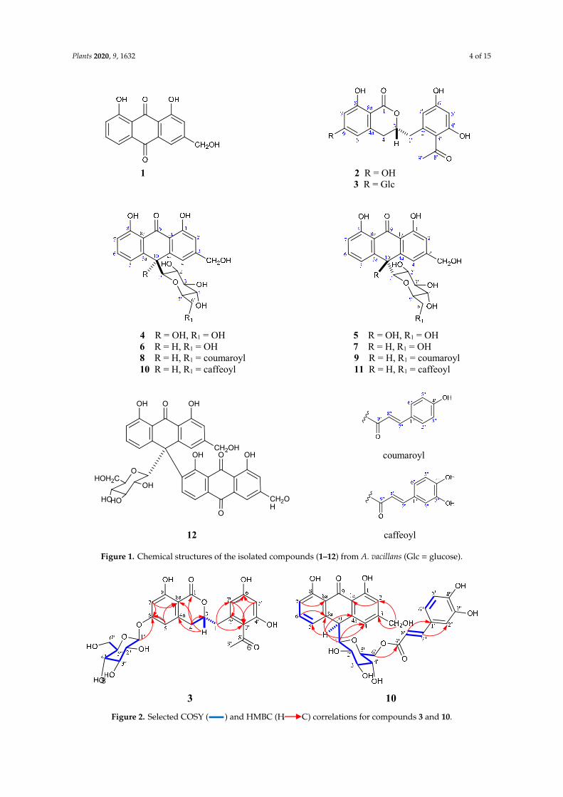
A phytochemical study of the CH2Cl2 and EtOAc soluble fractions of the leaves of A. vacillansusing diverse chromatographic methods, afforded twelve compounds. The known compounds wereidentified as aloe-emodin (1) [19], feralolide (2) [20,21], 10-hydroxyaloins A and B (4, 5) [22], aloin Aand B (6, 7) [19], microdontin A and B (8, 9) [23], and elgonica-dimer A (12) [24,25] (Figure 1 andFigure S2) (Tables S1 and S2). The structures of the isolated compounds were identified based ona variety of spectroscopic techniques including 1D (1H, 13C, and DEPT-13C experiments) and 2D(1H-1H COSY, 1H-13C HSQC, 1H-13C HMBC, and 1H-1H NOESY) nuclear magnetic resonance (NMR)spectroscopy. Accurate mass measurements and comparisons with published data were also used(Figures S2–S24), and electronic circular dichroism (ECD) experiments were conducted to determinethe absolute configurations.
Compound (3) was isolated as a white amorphous powder. The spectral data of (3) including IR,UV, and NMR were very similar to that of feralolide (2) [20], suggesting a similar dihydorisocoumarinskeleton. HRESIMS showed quasi-molecular ion peaks at m/z 507.1500 [M+H]+ (calcd 507.1503for C24H27O12), m/z 529.1320 [M+Na]+ (calcd 529.1322 for C24H26O12Na), and m/z 545.1069 [M+K]+
(calcd 545.1061 for C24H26O12K) with 162 amu more than that of 2, suggesting the presence of amonosaccharide moiety. A significant fragment using the high-resolution electron impact mode(HR-EIMS) appeared at 327.0868 corresponding to C18H15O6 (M+-glucose). A positive Molisch’s test
Plants 2020, 9, 1632 3 of 15
reflected the glycosidic nature [26]. Absorption bands at 3451, and 1645 cm−1 were observed in the IRspectrum assigned to OH and C=O, respectively.
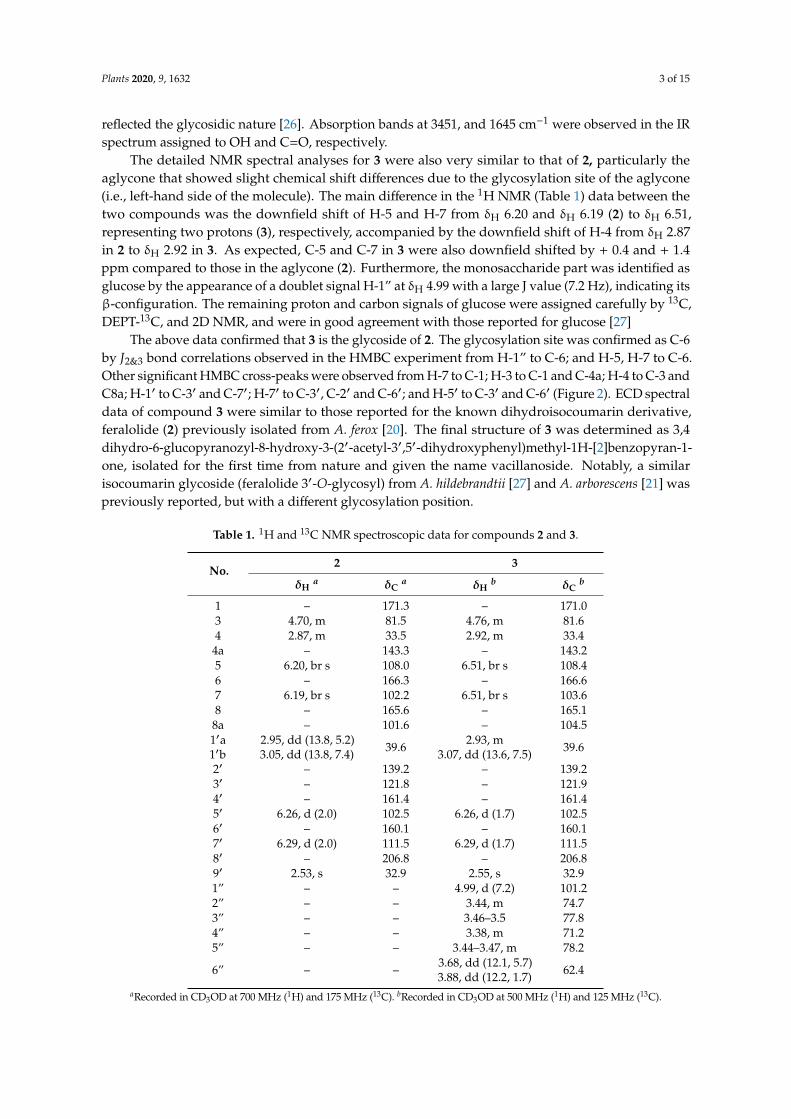
The detailed NMR spectral analyses for 3 were also very similar to that of 2, particularly theaglycone that showed slight chemical shift differences due to the glycosylation site of the aglycone(i.e., left-hand side of the molecule). The main difference in the 1H NMR (Table 1) data between thetwo compounds was the downfield shift of H-5 and H-7 from δH 6.20 and δH 6.19 (2) to δH 6.51,representing two protons (3), respectively, accompanied by the downfield shift of H-4 from δH 2.87in 2 to δH 2.92 in 3. As expected, C-5 and C-7 in 3 were also downfield shifted by + 0.4 and + 1.4ppm compared to those in the aglycone (2). Furthermore, the monosaccharide part was identified asglucose by the appearance of a doublet signal H-1” at δH 4.99 with a large J value (7.2 Hz), indicating itsβ-configuration. The remaining proton and carbon signals of glucose were assigned carefully by 13C,DEPT-13C, and 2D NMR, and were in good agreement with those reported for glucose [27]
The above data confirmed that 3 is the glycoside of 2. The glycosylation site was confirmed as C-6by J2&3 bond correlations observed in the HMBC experiment from H-1” to C-6; and H-5, H-7 to C-6.Other significant HMBC cross-peaks were observed from H-7 to C-1; H-3 to C-1 and C-4a; H-4 to C-3 andC8a; H-1′ to C-3′ and C-7′; H-7′ to C-3′, C-2′ and C-6′; and H-5′ to C-3′ and C-6′ (Figure 2). ECD spectraldata of compound 3 were similar to those reported for the known dihydroisocoumarin derivative,feralolide (2) previously isolated from A. ferox [20]. The final structure of 3 was determined as 3,4dihydro-6-glucopyranozyl-8-hydroxy-3-(2′-acetyl-3′,5′-dihydroxyphenyl)methyl-1H-[2]benzopyran-1-one, isolated for the first time from nature and given the name vacillanoside. Notably, a similarisocoumarin glycoside (feralolide 3′-O-glycosyl) from A. hildebrandtii [27] and A. arborescens [21] waspreviously reported, but with a different glycosylation position.
Table 1. 1H and 13C NMR spectroscopic data for compounds 2 and 3.
No.2 3
δHa δC
a δHb δC
b
1 – 171.3 – 171.03 4.70, m 81.5 4.76, m 81.64 2.87, m 33.5 2.92, m 33.4
4a – 143.3 – 143.25 6.20, br s 108.0 6.51, br s 108.46 – 166.3 – 166.67 6.19, br s 102.2 6.51, br s 103.68 – 165.6 – 165.1
8a – 101.6 – 104.51′a1′b
2.95, dd (13.8, 5.2)3.05, dd (13.8, 7.4) 39.6 2.93, m
3.07, dd (13.6, 7.5) 39.6
2′ – 139.2 – 139.23′ – 121.8 – 121.94′ – 161.4 – 161.45′ 6.26, d (2.0) 102.5 6.26, d (1.7) 102.56′ – 160.1 – 160.17′ 6.29, d (2.0) 111.5 6.29, d (1.7) 111.58′ – 206.8 – 206.89′ 2.53, s 32.9 2.55, s 32.91” – – 4.99, d (7.2) 101.22” – – 3.44, m 74.73” – – 3.46–3.5 77.84” – – 3.38, m 71.25” – – 3.44–3.47, m 78.2
6” – – 3.68, dd (12.1, 5.7)3.88, dd (12.2, 1.7) 62.4
aRecorded in CD3OD at 700 MHz (1H) and 175 MHz (13C). bRecorded in CD3OD at 500 MHz (1H) and 125 MHz (13C).
Plants 2020, 9, 1632 4 of 15
Plants 2020, 9, x FOR PEER REVIEW 4 of 14
Figure 1. Chemical structures of the isolated compounds (1–12) from A. vacillans (Glc = glucose).
Figure 2. Selected COSY ( ) and HMBC (H C) correlations for compounds 3 and 10.
1 2 R = OH 3 R = Glc
4 R = OH, R1 = OH 5 R = OH, R1 = OH 6 R = H, R1 = OH 7 R = H, R1 = OH 8 R = H, R1 = coumaroyl 9 R = H, R1 = coumaroyl 10 R = H, R1 = caffeoyl 11 R = H, R1 = caffeoyl
coumaroyl
12 caffeoyl
OOH OH
CH2OHOOH OH
CH2OH
O
OHOH2C
HOHO
OH
6
8
3 10
Figure 1. Chemical structures of the isolated compounds (1–12) from A. vacillans (Glc = glucose).
Plants 2020, 9, x FOR PEER REVIEW 4 of 14
Figure 1. Chemical structures of the isolated compounds (1–12) from A. vacillans (Glc = glucose).
Figure 2. Selected COSY ( ) and HMBC (H C) correlations for compounds 3 and 10.
1 2 R = OH 3 R = Glc
4 R = OH, R1 = OH 5 R = OH, R1 = OH 6 R = H, R1 = OH 7 R = H, R1 = OH 8 R = H, R1 = coumaroyl 9 R = H, R1 = coumaroyl 10 R = H, R1 = caffeoyl 11 R = H, R1 = caffeoyl
coumaroyl
12 caffeoyl
OOH OH
CH2OHOOH OH
CH2OH
O
OHOH2C
HOHO
OH
6
8
3 10
Figure 2. Selected COSY (
Plants 2020, 9, x FOR PEER REVIEW 4 of 14
Figure 1. Chemical structures of the isolated compounds (1–12) from A. vacillans (Glc = glucose).
Figure 2. Selected COSY ( ) and HMBC (H C) correlations for compounds 3 and 10.
1 2 R = OH 3 R = Glc
4 R = OH, R1 = OH 5 R = OH, R1 = OH 6 R = H, R1 = OH 7 R = H, R1 = OH 8 R = H, R1 = coumaroyl 9 R = H, R1 = coumaroyl 10 R = H, R1 = caffeoyl 11 R = H, R1 = caffeoyl
coumaroyl
12 caffeoyl
OOH OH
CH2OHOOH OH
CH2OH
O
OHOH2C
HOHO
OH
6
8
3 10
) and HMBC (H
Plants 2020, 9, x FOR PEER REVIEW 4 of 14
Figure 1. Chemical structures of the isolated compounds (1–12) from A. vacillans (Glc = glucose).
Figure 2. Selected COSY ( ) and HMBC (H C) correlations for compounds 3 and 10.
1 2 R = OH 3 R = Glc
4 R = OH, R1 = OH 5 R = OH, R1 = OH 6 R = H, R1 = OH 7 R = H, R1 = OH 8 R = H, R1 = coumaroyl 9 R = H, R1 = coumaroyl 10 R = H, R1 = caffeoyl 11 R = H, R1 = caffeoyl
coumaroyl
12 caffeoyl
OOH OH
CH2OHOOH OH
CH2OH
O
OHOH2C
HOHO
OH
6
8
3 10
C) correlations for compounds 3 and 10.
Plants 2020, 9, 1632 5 of 15
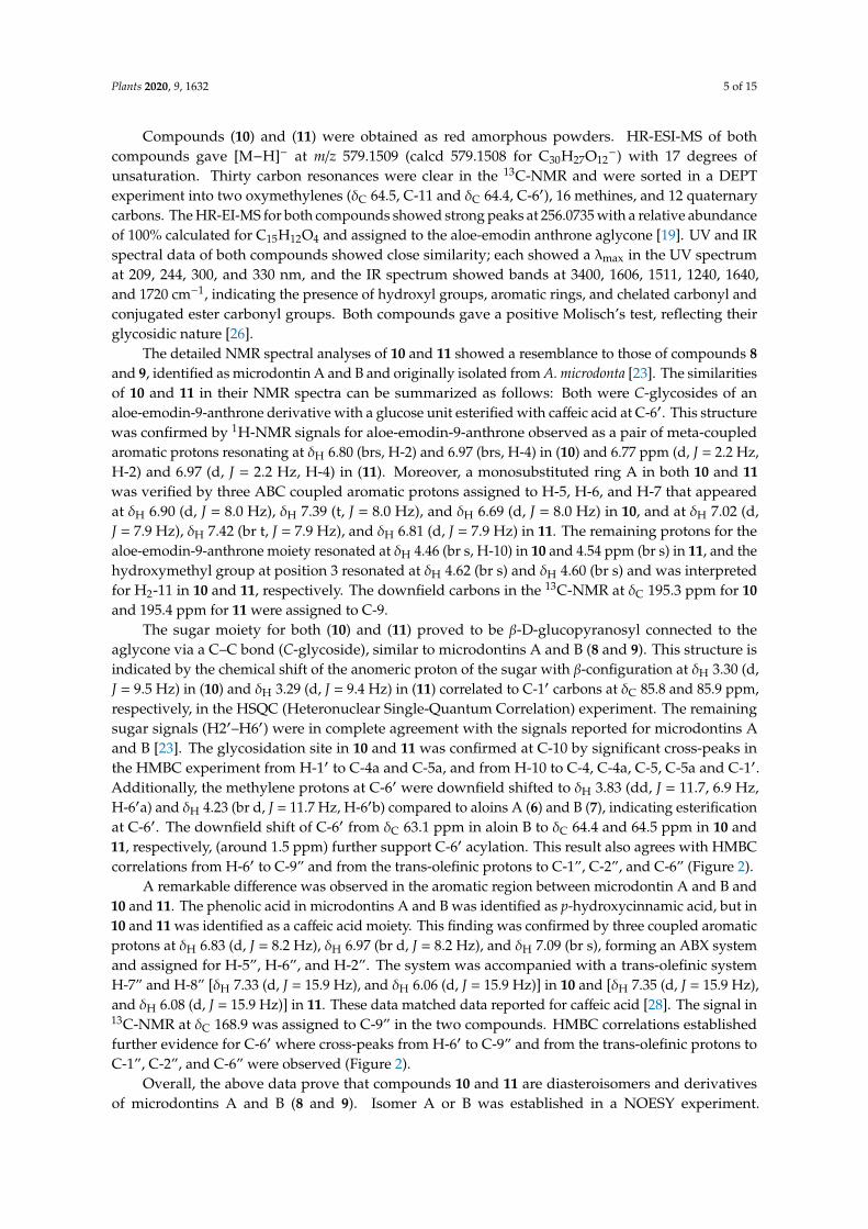
Compounds (10) and (11) were obtained as red amorphous powders. HR-ESI-MS of bothcompounds gave [M−H]− at m/z 579.1509 (calcd 579.1508 for C30H27O12
−) with 17 degrees ofunsaturation. Thirty carbon resonances were clear in the 13C-NMR and were sorted in a DEPTexperiment into two oxymethylenes (δC 64.5, C-11 and δC 64.4, C-6′), 16 methines, and 12 quaternarycarbons. The HR-EI-MS for both compounds showed strong peaks at 256.0735 with a relative abundanceof 100% calculated for C15H12O4 and assigned to the aloe-emodin anthrone aglycone [19]. UV and IRspectral data of both compounds showed close similarity; each showed a λmax in the UV spectrumat 209, 244, 300, and 330 nm, and the IR spectrum showed bands at 3400, 1606, 1511, 1240, 1640,and 1720 cm−1, indicating the presence of hydroxyl groups, aromatic rings, and chelated carbonyl andconjugated ester carbonyl groups. Both compounds gave a positive Molisch’s test, reflecting theirglycosidic nature [26].
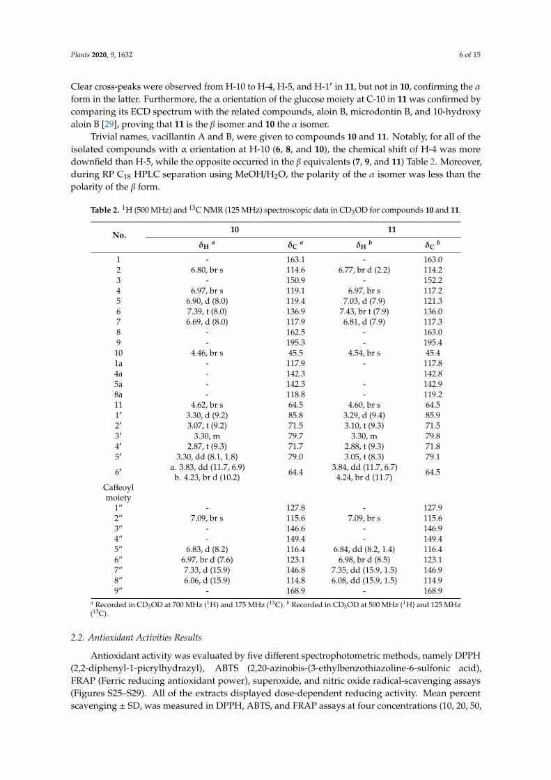
The detailed NMR spectral analyses of 10 and 11 showed a resemblance to those of compounds 8and 9, identified as microdontin A and B and originally isolated from A. microdonta [23]. The similaritiesof 10 and 11 in their NMR spectra can be summarized as follows: Both were C-glycosides of analoe-emodin-9-anthrone derivative with a glucose unit esterified with caffeic acid at C-6′. This structurewas confirmed by 1H-NMR signals for aloe-emodin-9-anthrone observed as a pair of meta-coupledaromatic protons resonating at δH 6.80 (brs, H-2) and 6.97 (brs, H-4) in (10) and 6.77 ppm (d, J = 2.2 Hz,H-2) and 6.97 (d, J = 2.2 Hz, H-4) in (11). Moreover, a monosubstituted ring A in both 10 and 11was verified by three ABC coupled aromatic protons assigned to H-5, H-6, and H-7 that appearedat δH 6.90 (d, J = 8.0 Hz), δH 7.39 (t, J = 8.0 Hz), and δH 6.69 (d, J = 8.0 Hz) in 10, and at δH 7.02 (d,J = 7.9 Hz), δH 7.42 (br t, J = 7.9 Hz), and δH 6.81 (d, J = 7.9 Hz) in 11. The remaining protons for thealoe-emodin-9-anthrone moiety resonated at δH 4.46 (br s, H-10) in 10 and 4.54 ppm (br s) in 11, and thehydroxymethyl group at position 3 resonated at δH 4.62 (br s) and δH 4.60 (br s) and was interpretedfor H2-11 in 10 and 11, respectively. The downfield carbons in the 13C-NMR at δC 195.3 ppm for 10and 195.4 ppm for 11 were assigned to C-9.
The sugar moiety for both (10) and (11) proved to be β-D-glucopyranosyl connected to theaglycone via a C–C bond (C-glycoside), similar to microdontins A and B (8 and 9). This structure isindicated by the chemical shift of the anomeric proton of the sugar with β-configuration at δH 3.30 (d,J = 9.5 Hz) in (10) and δH 3.29 (d, J = 9.4 Hz) in (11) correlated to C-1′ carbons at δC 85.8 and 85.9 ppm,respectively, in the HSQC (Heteronuclear Single-Quantum Correlation) experiment. The remainingsugar signals (H2′–H6′) were in complete agreement with the signals reported for microdontins Aand B [23]. The glycosidation site in 10 and 11 was confirmed at C-10 by significant cross-peaks inthe HMBC experiment from H-1′ to C-4a and C-5a, and from H-10 to C-4, C-4a, C-5, C-5a and C-1′.Additionally, the methylene protons at C-6′ were downfield shifted to δH 3.83 (dd, J = 11.7, 6.9 Hz,H-6′a) and δH 4.23 (br d, J = 11.7 Hz, H-6′b) compared to aloins A (6) and B (7), indicating esterificationat C-6′. The downfield shift of C-6′ from δC 63.1 ppm in aloin B to δC 64.4 and 64.5 ppm in 10 and11, respectively, (around 1.5 ppm) further support C-6′ acylation. This result also agrees with HMBCcorrelations from H-6′ to C-9” and from the trans-olefinic protons to C-1”, C-2”, and C-6” (Figure 2).
A remarkable difference was observed in the aromatic region between microdontin A and B and10 and 11. The phenolic acid in microdontins A and B was identified as p-hydroxycinnamic acid, but in10 and 11 was identified as a caffeic acid moiety. This finding was confirmed by three coupled aromaticprotons at δH 6.83 (d, J = 8.2 Hz), δH 6.97 (br d, J = 8.2 Hz), and δH 7.09 (br s), forming an ABX systemand assigned for H-5”, H-6”, and H-2”. The system was accompanied with a trans-olefinic systemH-7” and H-8” [δH 7.33 (d, J = 15.9 Hz), and δH 6.06 (d, J = 15.9 Hz)] in 10 and [δH 7.35 (d, J = 15.9 Hz),and δH 6.08 (d, J = 15.9 Hz)] in 11. These data matched data reported for caffeic acid [28]. The signal in13C-NMR at δC 168.9 was assigned to C-9” in the two compounds. HMBC correlations establishedfurther evidence for C-6′ where cross-peaks from H-6′ to C-9” and from the trans-olefinic protons toC-1”, C-2”, and C-6” were observed (Figure 2).
Overall, the above data prove that compounds 10 and 11 are diasteroisomers and derivativesof microdontins A and B (8 and 9). Isomer A or B was established in a NOESY experiment.
Plants 2020, 9, 1632 6 of 15
Clear cross-peaks were observed from H-10 to H-4, H-5, and H-1′ in 11, but not in 10, confirming the αform in the latter. Furthermore, the α orientation of the glucose moiety at C-10 in 11 was confirmed bycomparing its ECD spectrum with the related compounds, aloin B, microdontin B, and 10-hydroxyaloin B [29], proving that 11 is the β isomer and 10 the α isomer.
Trivial names, vacillantin A and B, were given to compounds 10 and 11. Notably, for all of theisolated compounds with α orientation at H-10 (6, 8, and 10), the chemical shift of H-4 was moredownfield than H-5, while the opposite occurred in the β equivalents (7, 9, and 11) Table 2. Moreover,during RP C18 HPLC separation using MeOH/H2O, the polarity of the α isomer was less than thepolarity of the β form.
Table 2. 1H (500 MHz) and 13C NMR (125 MHz) spectroscopic data in CD3OD for compounds 10 and 11.
No.10 11
δHa δC
a δHb δC
b
1 - 163.1 - 163.02 6.80, br s 114.6 6.77, br d (2.2) 114.23 - 150.9 - 152.24 6.97, br s 119.1 6.97, br s 117.25 6.90, d (8.0) 119.4 7.03, d (7.9) 121.36 7.39, t (8.0) 136.9 7.43, br t (7.9) 136.07 6.69, d (8.0) 117.9 6.81, d (7.9) 117.38 - 162.5 - 163.09 - 195.3 - 195.4
10 4.46, br s 45.5 4.54, br s 45.41a - 117.9 - 117.84a - 142.3 142.85a - 142.3 - 142.98a - 118.8 - 119.211 4.62, br s 64.5 4.60, br s 64.51′ 3.30, d (9.2) 85.8 3.29, d (9.4) 85.92′ 3.07, t (9.2) 71.5 3.10, t (9.3) 71.53′ 3.30, m 79.7 3.30, m 79.84′ 2.87, t (9.3) 71.7 2.88, t (9.3) 71.85′ 3.30, dd (8.1, 1.8) 79.0 3.05, t (8.3) 79.1
6′ a. 3.83, dd (11.7, 6.9)b. 4.23, br d (10.2) 64.4 3.84, dd (11.7, 6.7)
4.24, br d (11.7) 64.5
Caffeoylmoiety
1” - 127.8 - 127.92” 7.09, br s 115.6 7.09, br s 115.63” - 146.6 - 146.94” - 149.4 - 149.45” 6.83, d (8.2) 116.4 6.84, dd (8.2, 1.4) 116.46” 6.97, br d (7.6) 123.1 6.98, br d (8.5) 123.17” 7.33, d (15.9) 146.8 7.35, dd (15.9, 1.5) 146.98” 6.06, d (15.9) 114.8 6.08, dd (15.9, 1.5) 114.99” - 168.9 - 168.9
a Recorded in CD3OD at 700 MHz (1H) and 175 MHz (13C). b Recorded in CD3OD at 500 MHz (1H) and 125 MHz(13C).
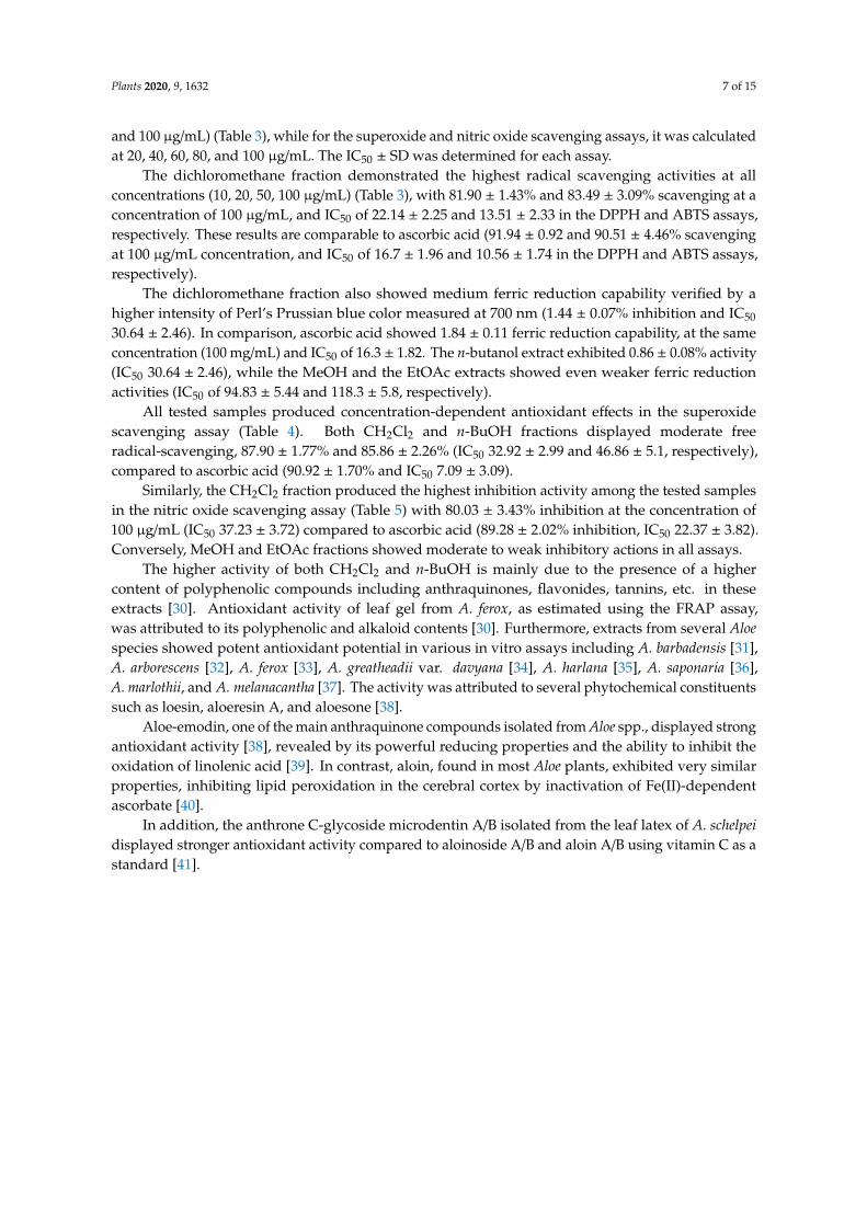
2.2. Antioxidant Activities Results
Antioxidant activity was evaluated by five different spectrophotometric methods, namely DPPH(2,2-diphenyl-1-picrylhydrazyl), ABTS (2,20-azinobis-(3-ethylbenzothiazoline-6-sulfonic acid),FRAP (Ferric reducing antioxidant power), superoxide, and nitric oxide radical-scavenging assays(Figures S25–S29). All of the extracts displayed dose-dependent reducing activity. Mean percentscavenging ± SD, was measured in DPPH, ABTS, and FRAP assays at four concentrations (10, 20, 50,
Plants 2020, 9, 1632 7 of 15
and 100 µg/mL) (Table 3), while for the superoxide and nitric oxide scavenging assays, it was calculatedat 20, 40, 60, 80, and 100 µg/mL. The IC50 ± SD was determined for each assay.
The dichloromethane fraction demonstrated the highest radical scavenging activities at allconcentrations (10, 20, 50, 100 µg/mL) (Table 3), with 81.90 ± 1.43% and 83.49 ± 3.09% scavenging at aconcentration of 100 µg/mL, and IC50 of 22.14 ± 2.25 and 13.51 ± 2.33 in the DPPH and ABTS assays,respectively. These results are comparable to ascorbic acid (91.94 ± 0.92 and 90.51 ± 4.46% scavengingat 100 µg/mL concentration, and IC50 of 16.7 ± 1.96 and 10.56 ± 1.74 in the DPPH and ABTS assays,respectively).
The dichloromethane fraction also showed medium ferric reduction capability verified by ahigher intensity of Perl’s Prussian blue color measured at 700 nm (1.44 ± 0.07% inhibition and IC50
30.64 ± 2.46). In comparison, ascorbic acid showed 1.84 ± 0.11 ferric reduction capability, at the sameconcentration (100 mg/mL) and IC50 of 16.3 ± 1.82. The n-butanol extract exhibited 0.86 ± 0.08% activity(IC50 30.64 ± 2.46), while the MeOH and the EtOAc extracts showed even weaker ferric reductionactivities (IC50 of 94.83 ± 5.44 and 118.3 ± 5.8, respectively).
All tested samples produced concentration-dependent antioxidant effects in the superoxidescavenging assay (Table 4). Both CH2Cl2 and n-BuOH fractions displayed moderate freeradical-scavenging, 87.90 ± 1.77% and 85.86 ± 2.26% (IC50 32.92 ± 2.99 and 46.86 ± 5.1, respectively),compared to ascorbic acid (90.92 ± 1.70% and IC50 7.09 ± 3.09).
Similarly, the CH2Cl2 fraction produced the highest inhibition activity among the tested samplesin the nitric oxide scavenging assay (Table 5) with 80.03 ± 3.43% inhibition at the concentration of100 µg/mL (IC50 37.23 ± 3.72) compared to ascorbic acid (89.28 ± 2.02% inhibition, IC50 22.37 ± 3.82).Conversely, MeOH and EtOAc fractions showed moderate to weak inhibitory actions in all assays.
The higher activity of both CH2Cl2 and n-BuOH is mainly due to the presence of a highercontent of polyphenolic compounds including anthraquinones, flavonides, tannins, etc. in theseextracts [30]. Antioxidant activity of leaf gel from A. ferox, as estimated using the FRAP assay,was attributed to its polyphenolic and alkaloid contents [30]. Furthermore, extracts from several Aloespecies showed potent antioxidant potential in various in vitro assays including A. barbadensis [31],A. arborescens [32], A. ferox [33], A. greatheadii var. davyana [34], A. harlana [35], A. saponaria [36],A. marlothii, and A. melanacantha [37]. The activity was attributed to several phytochemical constituentssuch as loesin, aloeresin A, and aloesone [38].
Aloe-emodin, one of the main anthraquinone compounds isolated from Aloe spp., displayed strongantioxidant activity [38], revealed by its powerful reducing properties and the ability to inhibit theoxidation of linolenic acid [39]. In contrast, aloin, found in most Aloe plants, exhibited very similarproperties, inhibiting lipid peroxidation in the cerebral cortex by inactivation of Fe(II)-dependentascorbate [40].
In addition, the anthrone C-glycoside microdentin A/B isolated from the leaf latex of A. schelpeidisplayed stronger antioxidant activity compared to aloinoside A/B and aloin A/B using vitamin C as astandard [41].
Plants 2020, 9, 1632 8 of 15
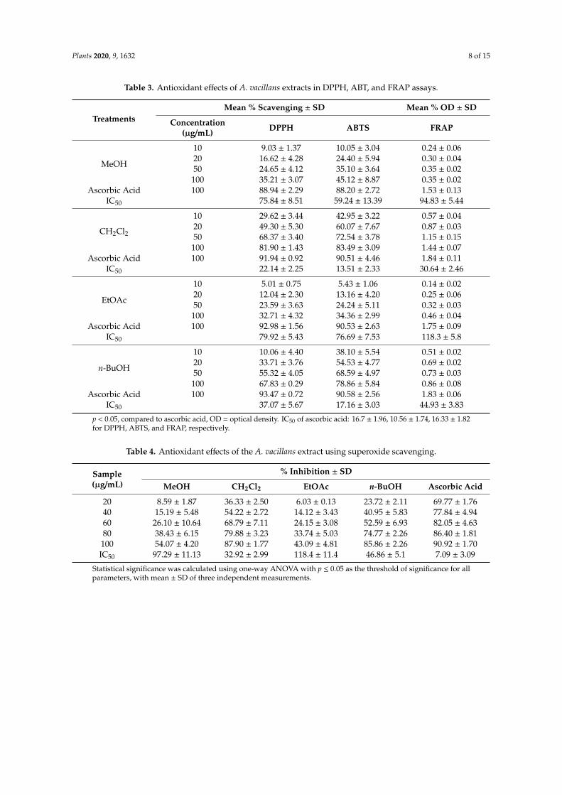
Table 3. Antioxidant effects of A. vacillans extracts in DPPH, ABT, and FRAP assays.
TreatmentsMean % Scavenging ± SD Mean % OD ± SD
Concentration(µg/mL) DPPH ABTS FRAP
MeOH
10 9.03 ± 1.37 10.05 ± 3.04 0.24 ± 0.0620 16.62 ± 4.28 24.40 ± 5.94 0.30 ± 0.0450 24.65 ± 4.12 35.10 ± 3.64 0.35 ± 0.02
100 35.21 ± 3.07 45.12 ± 8.87 0.35 ± 0.02Ascorbic Acid 100 88.94 ± 2.29 88.20 ± 2.72 1.53 ± 0.13
IC50 75.84 ± 8.51 59.24 ± 13.39 94.83 ± 5.44
CH2Cl2
10 29.62 ± 3.44 42.95 ± 3.22 0.57 ± 0.0420 49.30 ± 5.30 60.07 ± 7.67 0.87 ± 0.0350 68.37 ± 3.40 72.54 ± 3.78 1.15 ± 0.15
100 81.90 ± 1.43 83.49 ± 3.09 1.44 ± 0.07Ascorbic Acid 100 91.94 ± 0.92 90.51 ± 4.46 1.84 ± 0.11
IC50 22.14 ± 2.25 13.51 ± 2.33 30.64 ± 2.46
EtOAc
10 5.01 ± 0.75 5.43 ± 1.06 0.14 ± 0.0220 12.04 ± 2.30 13.16 ± 4.20 0.25 ± 0.0650 23.59 ± 3.63 24.24 ± 5.11 0.32 ± 0.03
100 32.71 ± 4.32 34.36 ± 2.99 0.46 ± 0.04Ascorbic Acid 100 92.98 ± 1.56 90.53 ± 2.63 1.75 ± 0.09
IC50 79.92 ± 5.43 76.69 ± 7.53 118.3 ± 5.8
n-BuOH
10 10.06 ± 4.40 38.10 ± 5.54 0.51 ± 0.0220 33.71 ± 3.76 54.53 ± 4.77 0.69 ± 0.0250 55.32 ± 4.05 68.59 ± 4.97 0.73 ± 0.03
100 67.83 ± 0.29 78.86 ± 5.84 0.86 ± 0.08Ascorbic Acid 100 93.47 ± 0.72 90.58 ± 2.56 1.83 ± 0.06
IC50 37.07 ± 5.67 17.16 ± 3.03 44.93 ± 3.83
p < 0.05, compared to ascorbic acid, OD = optical density. IC50 of ascorbic acid: 16.7 ± 1.96, 10.56 ± 1.74, 16.33 ± 1.82for DPPH, ABTS, and FRAP, respectively.
Table 4. Antioxidant effects of the A. vacillans extract using superoxide scavenging.
Sample(µg/mL)
% Inhibition ± SD
MeOH CH2Cl2 EtOAc n-BuOH Ascorbic Acid
20 8.59 ± 1.87 36.33 ± 2.50 6.03 ± 0.13 23.72 ± 2.11 69.77 ± 1.7640 15.19 ± 5.48 54.22 ± 2.72 14.12 ± 3.43 40.95 ± 5.83 77.84 ± 4.9460 26.10 ± 10.64 68.79 ± 7.11 24.15 ± 3.08 52.59 ± 6.93 82.05 ± 4.6380 38.43 ± 6.15 79.88 ± 3.23 33.74 ± 5.03 74.77 ± 2.26 86.40 ± 1.81
100 54.07 ± 4.20 87.90 ± 1.77 43.09 ± 4.81 85.86 ± 2.26 90.92 ± 1.70IC50 97.29 ± 11.13 32.92 ± 2.99 118.4 ± 11.4 46.86 ± 5.1 7.09 ± 3.09
Statistical significance was calculated using one-way ANOVA with p ≤ 0.05 as the threshold of significance for allparameters, with mean ± SD of three independent measurements.
Plants 2020, 9, 1632 9 of 15
Table 5. Antioxidant effect of thee A. vacillans extract using nitric oxide scavenging.
Sample(µg/mL)
% Inhibition ± SD
MeOH CH2Cl2 EtOAc n-BuOH Ascorbic Acid
20 6.32 ± 3.50 34.83 ± 1.97 6.80 ± 4.49 42.09 ± 3.73 48.51 ± 7.9240 15.21 ± 6.63 49.66 ± 3.85 16.69 ± 4.24 51.62 ± 3.73 64.26 ± 2.4860 28.57 ± 2.06 60.80 ± 5.61 29.74 ± 1.28 61.16 ± 1.82 71.42 ± 4.8180 42.05 ± 5.49 71.85 ± 3.23 35.08 ± 1.56 68.27 ± 2.84 79.82 ± 1.51
100 46.78 ± 7.61 80.03 ± 3.43 40.40 ± 2.10 72.29 ± 4.00 89.28 ± 2.02IC50 103.2 ± 12.13 37.23 ± 3.72 128.7 ± 16.3 32.44 ± 3.82 22.37 ± 3.82
Statistical significance was calculated using one-way ANOVA with p ≤ 0.05 as the threshold of significance for allparameters, with mean ± SD of three independent measurements.
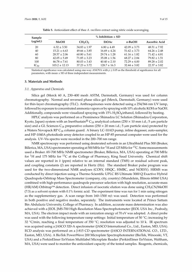
3. Materials and Methods
3.1. Apparatus and Chemicals
Silica gel (Merck 60 A, 230–400 mesh ASTM, Darmstadt, Germany) was used for columnchromatography. Normal and reversed phase silica gel (Merck, Darmstadt, Germany) were usedfor thin-layer chromatography (TLC). Anthraquinones were detected using a 254/366 nm UV lamp,followed by exposure to concentrated ammonia vapors or by spraying with 10% alcoholic KOH or NaOH.Additionally, compounds were visualized spraying with 15% H2SO4/ethanol, followed by heating.
HPLC analysis was performed on a Prominence Shimadzu LC Solution (Shimadzu Corporation,Kyoto, Japan) system with an InertSustain® C18 analytical column (250 × 10 mm i.d.; 5 µm particlesize) and a GL Sciences C18 preparative column (250 × 20 mm i.d.; 5 µm particle size) protected bya Waters Novapack RP C18 column guard. A binary LC-10AD pump, inline degasser, auto-sampler,and HP-1040A photodiode array detector coupled to an HP-85 personal computer were used for theanalysis. UV–Vis spectra were recorded in the 200–700 nm range.
NMR spectroscopy was performed using deuterated solvents in an UltraShield Plus 500 (Bruker,Billerica, MA, USA) spectrometer operating at 500 MHz for 1H and 125 MHz for 13C. Some measurementsused a Bruker AV-700 MHz NMR spectrometer (Bruker, Billerica, MA, USA) operating at 700 MHzfor 1H and 175 MHz for 13C at the College of Pharmacy, King Saud University. Chemical shiftvalues are reported in δ (ppm) relative to an internal standard (TMS) or residual solvent peak,and coupling constants (J) are reported in Hertz (Hz). The standard Bruker pulse program wasused for the two-dimensional NMR analyses (COSY, HSQC, HMBC, and NOESY). HRMS wasconducted by direct injection using a Thermo Scientific UPLC RS Ultimate 3000 Q Exactive HybridQuadrupole-Orbitrap Mass Spectrometer (company, city, country) (Mundelein, Illinois 60060 USA)combined with high-performance quadrupole precursor selection with high resolution, accurate-mass(HR/AM) Orbitrap™ detection. Direct infusion of isocratic elution was done using CH3CN/MeOH(7:3) as a solvent system with 0.1% formic acid. The experiment time was run for 1 min using nitrogenas the supplementary gas. A scan range from 160–1500 m/z was used. Detection was performedin both positive and negative modes, separately. The instruments were located at Prince SattamBin Abdulaziz University, College of Pharmacy. In addition, accurate mass determination was alsoachieved with a JEOL JMS-700 High-Resolution Mass Spectrophotometer (JEOL USA Inc., Peabody,MA, USA). The electron impact mode with an ionization energy of 70 eV was adopted. A direct probewas used with the following temperature ramp settings: Initial temperature of 50 ◦C; increasing by32 ◦C/min, reaching a final temperature of 350 ◦C; resolution was adjusted to 10 k. IR spectrumwas acquired using a JASCO 320-A spectrometer (JASCO International Co., Ltd., Easton, MD, USA).ECD analysis was performed on a J-815 CD spectrometer (JASCO INTERNATIONAL CO., LTD.,Easton, MD, USA). A BioTek PowerWave 200 Microplate Spectrophotometer (BioTek, Winooski, VT,USA) and a PerkinElmer EnVision Multilabel Microplate Reader (PerkinElmer EnVision, Waltham,MA, USA) were used to monitor the antioxidant capacity of the tested samples. Reagents, chemicals,
Plants 2020, 9, 1632 10 of 15
and solvents were analytical grade, purchased from Sigma-Aldrich (St. Louis, MO, USA), Loba ChemiePvt. Ltd. (Mumbai, India), and SD Fine Chem. Ltd. (Mumbai, India).
3.2. Plant Material
The leaves of A. vacillans Forssk were collected in February 2018 in Mahayil Asir, in the southwesternregion of Saudi Arabia (latitude: 18◦13′0.4692” N and longitude: 42◦30′13.5540” E). The specimenswere kindly identified by Dr Raja Krishnan, Botany and Microbiology Department at the College ofScience, King Saud University, Riyadh, Saudi Arabia. A voucher specimen (#11965) was submitted tothe herbarium of the College of Science, King Saud University.
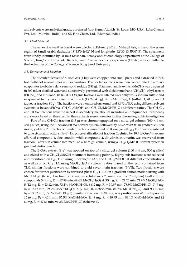
3.3. Extraction and Isolation
The succulent leaves of A. vacillans (4 kg) were chopped into small pieces and extracted in 70%hot methanol several times until exhaustion. The pooled extracts were then concentrated in a rotaryevaporator to obtain a dark semi-solid residue (180 g). Total methanolic extract (MeOH) was dispersedin 300 mL of distilled water and successively partitioned with dichloromethane (CH2Cl2), ethyl acetate(EtOAc), and n-butanol (n-BuOH). Organic fractions were filtered over anhydrous sodium sulfate andevaporated to dryness to yield fractions A (DCM, 4.0 g), B (EtOAc, 8.5 g), C (n-BuOH, 70 g), and D(aqueous fraction, 90 g). The fractions were monitored on normal and RP C18 TLC using different solventsystems: n-hexane/EtOAc, CH2Cl2/MeOH, and CH2Cl2/MeOH/H2O at different ratios. The CH2Cl2and EtOAc fractions were the richest in secondary metabolites including anthraquinones, triterpenes,and sterols; based on these results, these extracts were chosen for further chromatographic investigation.
Part of the CH2Cl2 fraction (3.5 g) was chromatographed on a silica gel column (100 × 4 cm,358 g silica) using the n-hexane/EtOAc solvent system, followed by EtOAc/MeOH in gradient elutionmode, yielding 251 fractions. Similar fractions, monitored on Kiesel gel 60 F254 TLC, were combinedto give six main fractions (A–F). Direct crystallization of fraction C, eluted by 40% EtOAc/n-hexane,afforded compound 1, aloe-emodin, while compound 2, dihydroisocoumarin, was recovered fromfraction E after sub-column treatment, on a silica gel column, using a CH2Cl2/MeOH solvent system ingradient elution mode.
The EtOAc extract (8 g) was applied on top of a silica gel column (100 × 4 cm, 500 g silica)and eluted with a CH2Cl2/MeOH mixture of increasing polarity. Eighty sub-fractions were collectedand monitored on F254 TLC using n-hexane/EtOAc, and CHCl3/MeOH at different concentrationsas well as on RP C18 TLC using MeOH/H2O at different ratios. Based on the results obtained fromTLC, similar fractions were combined to yield seven main fractions (I–VII). Two fractions werechosen for further purification by reversed-phase C18-HPLC in a gradient elution mode starting withMeOH-H2O (60:40). Fraction II (130 mg) was eluted over 70 min (flow rate, 2 mL/min) to afford purecompounds 3 (1 mg, Rt = 17.88 min, 69.4% MeOH/H2O), 4 (15 mg, Rt = 21.25 min, 71.9% MeOH/H2O),5 (12 mg, Rt = 23.12 min, 73.1% MeOH/H2O), 6 (12 mg, Rt = 30.97 min, 78.9% MeOH/H2O), 7 (9 mg,Rt = 32.42 min, 79.9% MeOH/H2O), 8 (7 mg, Rt = 38.95 min, 84.7% MeOH/H2O), and 9 (13 mg,Rt = 39.82 min, 85.3% MeOH/H2O). Similarly, fraction III (300 mg) was purified over 70 min to provide10 (6 mg, Rt = 40.1 min, 85.5% MeOH/H2O), 11 (8 mg, Rt = 40.95 min, 86.1% MeOH/H2O), and 12(3 mg, Rt = 47.86 min, 91.2% MeOH/H2O) (Scheme 1).
Plants 2020, 9, 1632 11 of 15
Plants 2020, 9, x FOR PEER REVIEW 10 of 14
The EtOAc extract (8 g) was applied on top of a silica gel column (100 × 4 cm, 500 g silica) and eluted with a CH2Cl2/MeOH mixture of increasing polarity. Eighty sub-fractions were collected and monitored on F254 TLC using n-hexane/EtOAc, and CHCl3/MeOH at different concentrations as well as on RP C18 TLC using MeOH/H2O at different ratios. Based on the results obtained from TLC, similar fractions were combined to yield seven main fractions (I–VII). Two fractions were chosen for further purification by reversed-phase C18-HPLC in a gradient elution mode starting with MeOH-H2O (60:40). Fraction II (130 mg) was eluted over 70 min (flow rate, 2 mL/min) to afford pure compounds 3 (1 mg, Rt = 17.88 min, 69.4% MeOH/H2O), 4 (15 mg, Rt = 21.25 min, 71.9% MeOH/H2O), 5 (12 mg, Rt = 23.12 min, 73.1% MeOH/H2O), 6 (12 mg, Rt = 30.97 min, 78.9% MeOH/H2O), 7 (9 mg, Rt
= 32.42 min, 79.9% MeOH/H2O), 8 (7 mg, Rt = 38.95 min, 84.7% MeOH/H2O), and 9 (13 mg, Rt = 39.82 min, 85.3% MeOH/H2O). Similarly, fraction III (300 mg) was purified over 70 min to provide 10 (6 mg, Rt = 40.1 min, 85.5% MeOH/H2O), 11 (8 mg, Rt = 40.95 min, 86.1% MeOH/H2O), and 12 (3 mg, Rt = 47.86 min, 91.2% MeOH/H2O) (Scheme 1).
Aloe vacillans leaves (4 kg)
70% hot methanol several times
Total methanolic extract (180 g)
-dispersed in 300 mL of distilled water-partitioned with CH2Cl2, EtOAc, and n-BuOH
Aqueous fraction (90 g)
Dichloromethane fraction (3.5 g)
Ethyl acetate fraction (8 g)
n-Butanol fraction (70 g)
CC, n-hexane:EtOAc followed by EtOAc/MeOH, gradient
CC, CH2Cl2:MeOH, gradientFraction C Fraction E
40% EtOAc/n-hexane
Compound 1 Compound 2
CC,CH2Cl2/MeOH, gradient
Seven fractions (I-VII)
RP-C18-HPLC
Fraction II (130 mg)
Faction III (300 mg)
RP-C18-HPLC
10 11 12
3. 1 mg, Rt = 17.88 min (69.4% MeOH/H2O)4. 15 mg, Rt = 21.25 min (71.9% MeOH/H2)5. 12 mg, Rt = 23.12 min (73.1% MeOH/H2O)6. 12 mg, Rt = 30.97 min (78.93% MeOH/H2O)7. 9 mg, Rt = 32.42 min (79.95% MeOH/H2O)8 . 7 mg, Rt = 38.95 min (84.75% MeOH/H2O)9. 13 mg, Rt = 39.82 min (85.33% MeOH/H2O)
3 4 5 6 7 8 9
Compound No.
Six fractions (A-F)
Compound No.
10 . 6 mg, Rt = 40.1 min (85.5%MeOH/H2O)11 . 1 mg, Rt = 40.95 min (86.14%% MeOH/H2O)12 . 3 mg, Rt = 47.86 min (91.2%MeOH/H2O)
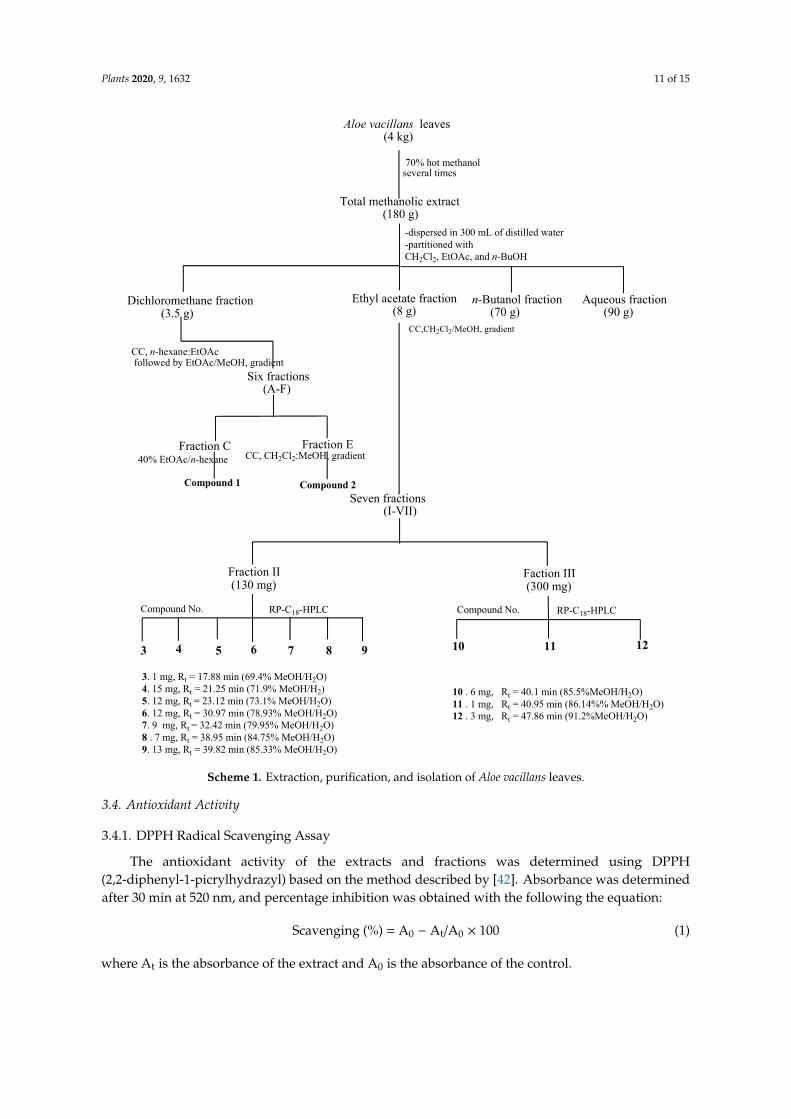
Scheme 1. Extraction, purification, and isolation of Aloe vacillans leaves.
Scheme 1. Extraction, purification, and isolation of Aloe vacillans leaves.
3.4. Antioxidant Activity
3.4.1. DPPH Radical Scavenging Assay
The antioxidant activity of the extracts and fractions was determined using DPPH(2,2-diphenyl-1-picrylhydrazyl) based on the method described by [42]. Absorbance was determinedafter 30 min at 520 nm, and percentage inhibition was obtained with the following the equation:
Scavenging (%) = A0 − At/A0 × 100 (1)
where At is the absorbance of the extract and A0 is the absorbance of the control.
Plants 2020, 9, 1632 12 of 15
3.4.2. ABTS Radical Cation Scavenging Assay
The assay was performed following the procedure described by [43]. The ability of samples toreduce the ABTS free radical (2,20-azinobis-(3-ethylbenzothiazoline-6-sulfonic acid) was also estimatedusing the above formula.
3.4.3. Reducing Power Assay
The reducing power of the extracts was determined using the method adapted by [44].The antioxidant method (i.e., FRAP) is based on the capability of a test sample to reduce ferricions (Fe3+) to ferrous ions (Fe2+) by electron donation.
3.4.4. Superoxide Radical Anion Scavenging Assay
Superoxide anion radical scavenging activity was assessed as previously described [45] withslight modification. Superoxide radicals were created by oxidation of NADH in a PMS-NADH system,and antioxidant activity was measured by the extent to which the extract and fractions of A. vacillansreduced nitro blue tetrazolium (NBT). The percentage of superoxide radical scavenging was alsocalculated using the above formula.
3.4.5. Nitric Oxide Radical Scavenging Assay
The assay was performed as previously described [46]. The free radical scavenging activity of theextract and fractions was determined by evaluating the % inhibition of the nitrite ions generated fromthe interaction of nitric oxide with oxygen using the same equation above-mentioned.
3.4.6. Spectral Data of the New Compounds
Vacillanoside (3)
White amorphous powder (1 mg); [α]23D − 56.2◦ (c 0.1, MeOH; UV λmax MeOH (log ε) nm:
211 (4.46), 274 (4.22), 306 (3.99); IR (KBr) vmax 3451, 1645, 1631, 1608, 1595, 1054, 1032, and 1016 cm−1;1H and 13C NMR (500, 125 MHz, in CD3OD) HR-ESI-MS: m/z 505.1353 [M −H]+ (calcd 505.1346 forC24H25O12), m/z 507.1500 [M + H]+ (calcd 507.1503 for C24H26O12+H), m/z 529.1320 [M + Na]+ (calcd529.1322 for C24H26O12Na), m/z 545.1069 [M + K]+ (calcd 545.1061 for C24H26O12K).
Vacillantin A (10)
Red amorphous powder (6 mg); [α]23D + 16.8◦ (c 0.05, MeOH); UV λmax MeOH (log ε) nm:
209 (4.63), 244 (4.42), 300 (3.92), 330 (3.61); IR (KBr) vmax 3400, 1720, 1640, 1606, 1511, 1240 cm−1; 1H and13C NMR (see Table 2); HR-ESI-MS: m/z 579.1509 [M − H]+ (calcd 579.1503 for C30H27O12).
Vacillantin B (11)
Red amorphous powder (8 mg); [α]23D − 4.7◦ (c 0.05, MeOH); UV λmax MeOH (log ε) nm:
209 (4.63), 244 (4.42), 300 (3.92), 330 (3.61); IR (KBr) vmax 3400, 1720, 1640, 1606, 1511, 1240 cm−1; 1Hand 13C NMR (see Table 2); HR-ESI-MS: m/z 579.1509 [M − H]+ (calcd 579.1503 for C30H27O12), m/z581.1651 [M + H]+ (calcd 581.1659 for C30H28O12 + H), m/z 603.1469 [M + Na]+ (calcd 603.1478 forC30H28O12 + Na).
3.5. Statistical Analysis
Analysis of variance (ANOVA) was used to evaluate significance differences, followed by theStudent’s t-test. Data were expressed as mean ± SD, and the difference was considered significantat p < 0.05 compared to the control. All statistical calculations used OriginLab software (version 8,Northampton, MA, USA) and Microsoft Excel.
Plants 2020, 9, 1632 13 of 15
4. Conclusions
In summary, a new dihydroisocoumarin derivative, vacillanoside (3), 3,4 dihydro-6glucopyranozyl-8-hydroxy-3-(2′-acetyl-3′,5′-dihydroxyphenyl)-methyl-1H-[2]benzopyran-1-one (6),and two new anthraquinone derivatives, vacillantins A and B (10 and 11) were isolated from the leavesof A. vacillans (Asphodelaceae) together with nine known compounds (1, 2, 4–9, and 12). The structuresof these compounds were elucidated through extensive spectroscopic analyses. The total alcoholextract and different fractions were tested for their antioxidant activities in five spectrophotometricassays. The dichloromethane fraction exhibited promising free radical scavenging activity in mostof the assays. Our findings add new information to the literature on the structural diversity andpharmacological activities of Aloe species. Our results suggest A. vacillans as a potential source ofsecondary metabolites with pharmacological and industrial importance. Moreover, these resultsadvocate further investigation of the remaining fractions with the aim of isolating bioactive compoundsexhibiting interesting biological capacities.
Supplementary Materials: The following are available online at http://www.mdpi.com/2223-7747/9/12/1632/s1,Figure S1: A photo of Aloe vacillans., Figures S2–S24 NMR spectral data (1D and 2D0 of compounds 3, 10 and 11:while Figures S9, S16 and S23 are HRESIMS spectra of compounds 3,10 and 11 and Figure S24 is the HRESIMS forthe known compounds.
Author Contributions: M.A.-T., S.M.A.-M. and A.A.E.-G. chose the plants and designed the practical part. M.A.-T.achieved the practical part of the project. O.A.B. and M.S.A.-K. measured, interpreted, and assigned the NMRdata and helped in preparing/revising the manuscript. W.M.A.-M. wrote, revised the manuscript, and preparedthe supplementary material. S.M.A.-M. and A.A.E.-G. wrote the paper, interpreted the NMR data, and supervised.All authors have read and agreed to the published version of the manuscript.
Funding: This work was funded by the Deanship of Scientific Research at King Saud University through theResearch Group Project No. RG 1437-021.
Acknowledgments: The authors would like to thank the Deanship of Scientific Research at King Saud Universityfor funding the work through the Research Group Project No. RG 1437-021.
Conflicts of Interest: The authors declare no conflict of interest.
References
1. Demmisew, S. Botanical aspects of Aloes of North East Africa. Bull. Chem. Soc. Ethiop. 1996, 10, 74–88.2. Gao, Y.; Kuok, K.I.; Jin, Y.; Wang, R. Biomedical applications of Aloe vera. Crit. Rev. Food Sci. Nutr. 2019, 59
(Suppl. S1), S244–S256. [CrossRef]3. Kojo, E.; Qian, H. Aloe Vera: A Valuable Ingredient for the Food, Pharmaceutical and Cosmetic
Industries—A Review. Crit. Rev. Food Sci. Nutr. 2004, 44, 91–96.4. Dagne, E.; Bisrat, D.; Viljoen, A.; Van Wyk, B.E. Chemistry of Aloe Species. Curr. Org. Chem. 2000, 4,
1055–1078. [CrossRef]5. Steenkamp, V.; Stewart, M.J. Medicinal applications and toxicological activities of Aloe products.
Pharmbiol. Biol. 2007, 45, 411–420. [CrossRef]6. Cock, I.E. The Genus Aloe: Phytochemistry and Therapeutic Uses Including Treatments for Gastrointestinal
Conditions and Chronic Inflammation. Prog. Drug Res. 2015, 70, 179–235. [PubMed]7. Yagi, A.; Kabash, A.; Okamura, N.; Haraguchi, H.; Moustafa, S.M.; Khalifa, T.I. Antioxidant, free radical
scavenging and anti-inflammatory effects of aloesin derivatives in Aloe vera. Planta Med. 2002, 68, 957–960.[CrossRef] [PubMed]
8. Yagi, A.; Kabash, A.; Mizuno, K.; Moustafa, S.M.; Khalifa, T.I.; Tsuji, H. Radical scavenging glycoproteininhibiting cyclooxygenase-2 and thromboxane A2 synthase from Aloe vera gel. Planta Med. 2003, 69, 269–271.[CrossRef]
9. Lim, B.O.; Seong, N.S.; Choue, R.W.; Kim, J.D.; Lee, H.Y.; Kim, S.Y.; Yu, B.P.; Jeon, T.I.; Park, D.K.Efficacy of dietary Aloe vera supplementation on hepatic cholesterol and oxidative status in aged rats. J. Nutr.Sci. Vitaminol. 2003, 49, 292–296. [CrossRef]
Plants 2020, 9, 1632 14 of 15
10. Su, Y.T.; Chang, H.L.; Shyue, S.K.; Hsu, S.L. Emodin induces apoptosis in human lung adenocarcinoma cellsthrough a reactive oxygen species-dependent mitochondrial signaling pathway. Biochem. Pharmacol. 2005,70, 229–241. [CrossRef] [PubMed]
11. Im, S.A.; Oh, S.T.; Song, S.; Kim, M.R.; Kim, D.S.; Woo, S.S.; Jo, T.H.; Park, Y.I.; Lee, C.K. Identification ofoptimal molecular size of modified Aloe polysaccharides with maximum immunomodulatory activity.Int. Immunopharmacol. 2005, 5, 271–279. [CrossRef]
12. Wang, Z.W.; Wang, Y.; Huang, Z.S. The radio-protective effect of Aloe polysaccharides on irradiated mice.Chin. Trad. Herb. Drugs 2002, 33, 251–254.
13. US Food and Drug Administration. Code of Federal Regulations (Food and Drugs), Title 21; US GovernmentPrinting Office: Washington, DC, USA, 1991; pp. 170–199.
14. Council of Europe. Flavoring Substances and Natural Source of Flavorings, 3rd ed.; Maisonneuve: Moulins-lesMetz, France, 1981; p. 376.
15. Valverde, J.M.; Valero, D.; Martinez-Romera, D.; Guillén, A.; Castillo, S.; Serrano, M. Novel edible coatingbased on Aloe vera gel to maintain table grape quality and safety. J. Agric. Food Chem. 2005, 53, 7807–7813.[CrossRef]
16. Abdalla, H.I.; Shaaban, M.; Shaaban, K.A.; Abu-Gabal, N.S.; Shalaby, N.M.; Laatsch, H. New bioactivecompounds from Aloe hijazensis. Nat. Prod. Res. 2009, 23, 1035–1049. [CrossRef] [PubMed]
17. Collenette, S. Wild Flowers of Saudi Arabia. Riyadh; National Commission for Wild Life Conservation andDevelopment (NCWCD): Riyadh, Saudi Arabia, 1999; p. 18.
18. Wood, J.R.I.; Thomas, H.H. A Handbook of the Yemen Flora; Royal Botanic Gardens, Kew: Richmond, UK, 1997.19. Zhong, J.; Huang, Y.; Ding, W.; Wu, X.; Wan, J.; Luo, H. Chemical constituents of Aloe barbadensis Miller and
their inhibitory effects on phosphodiesterase-4D. Fitoterapia 2013, 91, 159–165. [CrossRef] [PubMed]20. Speranza, G.; Manitto, P.; Monti, D. Feralolide, a dihydroisocoumarin from Cape aloe. Phytochemistry 1993, 3,
175–178. [CrossRef]21. Kurizaki, A.; Watanabe, T.; Devkota, H.P. Chemical constituents from the flowers of Aloe arborescens.
Nat. Prod. Commun. 2019, 14, 1–4. [CrossRef]22. Yasuda, K.; Uehara, S.; Takano, I.; Shindo, T.; Nishijima, M. Stability of barbaloin in aqueous solution.
Food Preserv. Sci. 2000, 26, 85–90. [CrossRef]23. Farah, M.H.; Andersson, R.; Samuelsson, G. Microdontin A and B: Two new aloin derivatives from
Aloe microdonta. Planta Med. 1992, 58, 88–93. [CrossRef]24. Shin, K.H.; Woo, W.S.; Lim, S.S.; Shim, C.S.; Chung, H.S.; Kennelly, E.J.; Kinghorn, D. Elgonica-Dimers A and
B, two potent alcohol metabolism inhibitory constituents of Aloe arborescens. J. Nat. Prod. 1997, 60, 1180–1182.[CrossRef]
25. Conner, J.M.; Alexander, I.; Gray, A.I.; Peter, G.; Waterman, P.G.; Reynolds, T. Novel anthrone-anthraquinonedimers from Aloe elgonica. J. Nat. Prod. 1990, 53, 1362–1364. [CrossRef]
26. Abd-Alrahman, S.H.; Salem-Bekhit, M.M.; Elhalwagy, M.E.A.; Abdel-Mageed, W.M.; Radwan, A.A.Phytochemical Screening and Antimicrobial Activity of EthOH/Water Ziziphus jujuba Seeds Extracts. J. PureAppl. Microbiol. 2013, 7, 823–828.
27. Veitch, N.C.; Simmonds, M.S.J.; Blaney, W.M.; Reynolds, T. A dihydroisocoumarin glucoside from Aloehildebrandtii. Phytochemistry 1994, 35, 1163–1166. [CrossRef]
28. Dagne, E.; Bisrat, D.; Van Wyk, B.E.; Viljoen, A.; Hellwig, V.; Steglich, W. Anthrones from Aloe microstigma.Phytochemistry 1997, 44, 1271–1274. [CrossRef]
29. Rauwald, H.W.; Lohse, K. Structure revision of 4-hydroxyaloin: 10-hydroxyaloins A and B as main InVitro-oxidation products of the diastereomeric aloins. Planta Med. 1992, 58, 259–262. [CrossRef]
30. Loots, D.T.; van der Westhuizen, F.H.; Botes, L. Aloe ferox leaf gel phytochemical content, antioxidant capacity,and possible health benefits. J. Agric. Food Chem. 2007, 55, 6891–6896. [CrossRef]
31. Hu, Y.; Xu, J.; Hu, Q. Evaluation of antioxidant potential of Aloe vera (Aloe barbadensis miller) extracts. J. Agric.Food Chem. 2003, 51, 7788–7791. [CrossRef]
32. Beppu, H.; Koike, T.; Shimpo, K.; Chihara, T.; Hoshino, M.; Ida, C.; Kuzuya, H. Radical-scavenging effects ofAloe arborescens Miller on prevention of pancreatic islet B-cell destruction in rats. J. Ethnopharmacol. 2003, 89,37–45. [CrossRef]
33. Wintola, O.A.; Afolayan, A.J. Phytochemical constituents and antioxidant activities of the whole leaf extractof Aloe ferox mill. Pharmacogn. Mag. 2011, 7, 325–333.
Plants 2020, 9, 1632 15 of 15
34. Botes, L.; van der Westhuizen, F.H.; Loots, D.T. Phytochemical contents and antioxidant capacities of twoAloe Greatheadii Var. Davyana extracts. Molecules 2008, 13, 2169–2180. [CrossRef]
35. Asamenew, G.; Bisrat, D.; Mazumder, A.; Asres, K. In vitro antimicrobial and antioxidant activities ofanthrone and chromone from the latex of Aloe harlana reynolds. Phytother. Res. 2011, 25, 1756–1760.[CrossRef] [PubMed]
36. Yoo, E.A.; Kim, S.D.; Lee, W.M.; Park, H.J.; Kim, S.K.; Cho, J.Y.; Min, W.; Rhee, M.H. Evaluation of Antioxidant,antinociceptive, and anti-Inflammatory activities of ethanol extracts from Aloe Saponaria Haw. Phytother. Res.2008, 22, 1389–1395. [CrossRef] [PubMed]
37. Cardarellia, M.; Rouphaelb, Y.; Pellizzonic, M.; Collad, G.; Lucin, L. Profile of bioactive secondary metabolitesand antioxidant capacity of leaf exudates from eighteen Aloe species. Ind. Crops Prod. 2017, 108, 44–51.[CrossRef]
38. Salehi, B.; Albayrak, S.; Antolak, H.; Kregiel, D.; Pawlikowska, E.; Sharifi-Rad, M.; Uprety, Y.; Tsouh Fokou, P.V.;Yousef, Z.; Amiruddin Zakaria, Z.; et al. Aloe genus plants: From farm to food applications andphytopharmacotherapy. Int. J. Mol. Sci. 2018, 19, 2843. [CrossRef] [PubMed]
39. Abdul Qadir, M.; Shahzadi, S.K.; Bashir, A.; Munir, A.; Shahzad, S. Evaluation of phenolic compounds andantioxidant and antimicrobial activities of some common herbs. Int. J. Anal. Chem. 2017, 2017, 3475738.[CrossRef] [PubMed]
40. Hes, M.; Dziedzic, K.; Górecka, D.; Jedrusek-Golinska, A.; Gujska, E. Aloe vera (L.) Webb.: Natural Sources ofAntioxidants—A Review. Plant. Foods Hum. Nutr. 2019, 74, 255–265.
41. Teka, T.; Kassahun, H. Characterization and Evaluation of Antioxidant Activity of Aloe schelpei Reynolds.Drug Des. Dev. Ther. 2020, 14, 1003. [CrossRef] [PubMed]
42. Braca, A.; Tommasi, N.D.; Bari, L.D.; Pizza, C.; Politi, M.; Morelli, I. Antioxidant principles from Bauhiniaterapotensis. J. Nat. Prod. 2001, 64, 892–895. [CrossRef]
43. Re, R.; Pellegrini, N.; Proteggente, A.; Pannala, A.; Yang, M.; Rice-Evans, C. Antioxidant activity applying animproved ABTS radical cation decolorization assay. Free Radic. Biol. Med. 1999, 26, 1231–1237. [CrossRef]
44. Oyaizu, M. Studies on products of browning reactions. Antioxidative activities of products of browningreaction prepared from glucosamine. Jpn. J. Nutr. 1986, 44, 307–315. [CrossRef]
45. Fontana, L.; Giagulli, C.; Minuz, P.; Lechi, A.; Laudanna, C. 8-Iso-PGF2 alpha induces beta 2-integrinmediatedrapid adhesion of human polymorphonuclear neutrophils: A link between oxidative stress andischemia/reperfusion injury. Arterioschler Thromb. Vasc. Biol. 2001, 21, 55–60. [CrossRef] [PubMed]
46. Nagmoti, D.M.; Khatri, D.K.; Juvekar, P.R.; Juvekar, A.R. Antioxidant activity and free radical scavengingpotential of Pithecellobium dulce Benth seed extracts. Free Rad. Antiox. 2011, 2, 37–43. [CrossRef]
Publisher’s Note: MDPI stays neutral with regard to jurisdictional claims in published maps and institutionalaffiliations.
© 2020 by the authors. Licensee MDPI, Basel, Switzerland. This article is an open accessarticle distributed under the terms and conditions of the Creative Commons Attribution(CC BY) license (http://creativecommons.org/licenses/by/4.0/).