Embed Size (px)
Citation preview
AbstractA five-year-old Holstein cow with a history of vaginal prolapse was
admitted to the Large Animal Clinic, Faculty of Veterinary Medicine,University of Tabriz. According to the owner, she had a 5-month history ofvaginal prolapse and frequent sanguineous discharge from the vagina.Appetite was normal and its last parturition was two months ago. Clinicalexamination revealed a tumor like mass that attached to the vaginal roof andpartly protruded through the vagina without any vaginal prolapse. Afterepidural anesthesia, the mass was surgically removed with some of thesurrounding tissue. The dimensions of the mass were 5.2 × 4 × 3.4 cm.Macroscopically, it was relatively well circumscribed with a lobulated andulcerated surface. In cut section, the mass was homogenously creamy incolor. There were no foci of necrosis or hemorrhage. Microscopically, thegrowth was composed of spindle-shaped fibroblastic type tumor cells thatformed interlacing and intersecting bundles. The neoplastic cells showedpleomorphism, karyomegaly and slight nuclear hyperchromatism. Themitotic index was less than five. Based on the site, biological behavior,immunohistochemistry results, macroscopic and microscopiccharacteristics, the mass was diagnosed as a well-differentiatedfibrosarcoma. This is the first report of cow vaginal fibrosarcoma from Iran.
Vaginal fibrosarcoma in cow (A case report)Hamali, H. *; Ashrafihelan, J.1 2
1 2Department of Clinical Sciences, Faculty of Veterinary Medicine, University of Tabriz, Tabriz, Iran. Department of Pathobiology, Facultyof Veterinary Medicine, University of Tabriz, Tabriz, Iran.
Key Words:
Correspondence
Fibrosarcoma; tumor; cow; vagina;immunohistochemistry.
Hamali, H.,Department of Clinical Sciences,Faculty of Veterinary Medicine,University of Tabriz, Tabriz, IranTel: +98(914)1145575Fax: +98(411)3357834Email: [email protected]
Received: 11 April 2009Accepted: 10 August 2010
Int.J.Vet.Res. (2010), 4; 4: 225-228 225
International Journal of Veterinary Research
Introduction
Tumors of the genital tract in cattle have beenreported from different countries of the world(Yeruham ., 1999; Anderson ., 1969; McEnteeet al., 1976; Susaneck 1981). However, reports onbovine fibrosarcoma compared with other tumors ofcattle are very rare (Yeruham and Orgad, 1999; Musal
., 2007; Birgit ., 2004).Fibrosarcoma is a type of sarcoma, a malignant
tumor of soft tissue that connects, supports or surroundsother structures and organs of the body. They aremalignant tumors of fibroblasts that show no otherevidence of cell differentiation. Immunohistochemically,fibrosarcoma can be separated from other spindle celltumors (Maxie, 2007; Meuten, 2004). These tumors arecommon in dogs and cats and uncommon in otherdomestic species. Although these can be found in anylocation of the body, they are unusual mesenchymaltumors of the bovine vagina (Masud, 2007; Maxie,2007). The most important effect of these tumors on thecattle industry is the increased culling rate due tometastases to the other critical organs, such as the lungs,liver and draining lymph nodes, which in turn causessevere complications (Yeruham ., 1999).
Vaginal fibroma, fibropapilloma, rhabdomyoma,
et al et al
et al et al
et al
leiomyoma and leiomyosarcoma have previously beenreported in cattle in our country but there has been noreport of vaginal fibrosarcoma from Iran (Sohrabi-Haghdoost ., 1990; Naghshineh ., 1991;Sohrabi-Haghdoost, 1991). The purpose of this reportwas to describe the clinical investigation, gross andhistopathological findings, and surgical treatment of avaginal fibrosarcoma in a cow.
A 5-year-old black and white Holstein cow wasadmitted in the Large Animal Clinic, Faculty ofVeterinary Medicine, University of Tabriz, during latespring 2009, due to a 5-month history of vaginalprolapse and a frequent mucosanguineous dischargefrom the vagina.
Clinical examination revealed no otherabnormalities, and its appetite was normal and the lastparturition was two months ago. Vaginal examinationrevealed no evidence of vaginal prolapsed, but a tumor-like vaginal mass was detected that was attached to thevaginal roof and partially protruded through the vagina(Figure 1). The mass was congested. It was hard inconsistency with a broad sessile-based and lobulated
et al et al
Case history
Clinical findings
Hamali, H.Vaginal fibrosarcoma in cow (A case report)
appearance that occupied the vaginal lumen. Its mucosalsurface was wet, ulcerated and frequently was bled.
Hematological examination of the cow prior tosurgical excision showed a mild leukocytosis andanemia. The total WBC count was elevated to 14.3×10/µL and the lymphocyte count were 7.320×10 /µL. Itorder to perform surgical excision, the cow wasrestrained and anesthetized by the administration of 10ml of 2% lidocaine (Manufactured by the PasteurInstitute of Iran) into the epidural space. Also, localanesthesia was performed with the infiltration of 20 mlof 2% lidocaine within the vaginal mucosa surroundingthe base of the tumor mass. Because local invasion wasas evident as finger-like projections of tumor intosurrounding tissues, the mass was completely removedwith a part of peripheral tissues and the base wascauterized. Postoperative parental antibiotic and localwound healing agents were administrated for three andfive days, respectively.
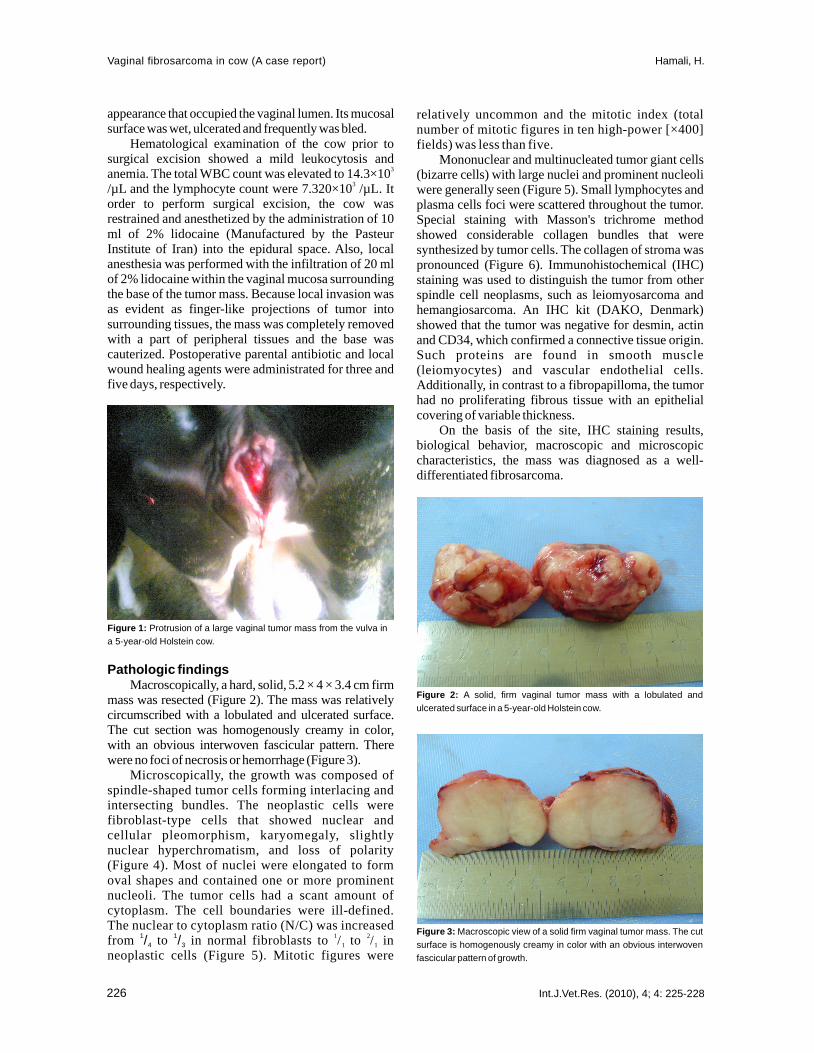
Macroscopically, a hard, solid, 5.2 × 4 × 3.4 cm firmmass was resected (Figure 2). The mass was relativelycircumscribed with a lobulated and ulcerated surface.The cut section was homogenously creamy in color,with an obvious interwoven fascicular pattern. Therewere no foci of necrosis or hemorrhage (Figure 3).
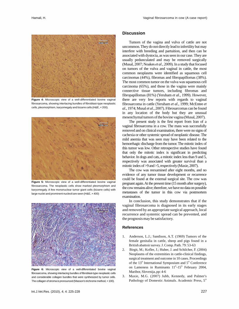
Microscopically, the growth was composed ofspindle-shaped tumor cells forming interlacing andintersecting bundles. The neoplastic cells werefibroblast-type cells that showed nuclear andcellular pleomorphism, karyomegaly, slightlynuclear hyperchromatism, and loss of polarity(Figure 4). Most of nuclei were elongated to formoval shapes and contained one or more prominentnucleoli. The tumor cells had a scant amount ofcytoplasm. The cell boundaries were ill-defined.The nuclear to cytoplasm ratio (N/C) was increasedfrom to in normal fibroblasts to / to / inneoplastic cells (Figure 5). Mitotic figures were
3
3
1 2
Pathologic findings
1 1/ /4 3 1 1
relatively uncommon and the mitotic index (totalnumber of mitotic figures in ten high-power [×400]fields) was less than five.
Mononuclear and multinucleated tumor giant cells(bizarre cells) with large nuclei and prominent nucleoliwere generally seen (Figure 5). Small lymphocytes andplasma cells foci were scattered throughout the tumor.Special staining with Masson's trichrome methodshowed considerable collagen bundles that weresynthesized by tumor cells. The collagen of stroma waspronounced (Figure 6). Immunohistochemical (IHC)staining was used to distinguish the tumor from otherspindle cell neoplasms, such as leiomyosarcoma andhemangiosarcoma. An IHC kit (DAKO, Denmark)showed that the tumor was negative for desmin, actinand CD34, which confirmed a connective tissue origin.Such proteins are found in smooth muscle(leiomyocytes) and vascular endothelial cells.Additionally, in contrast to a fibropapilloma, the tumorhad no proliferating fibrous tissue with an epithelialcovering of variable thickness.
On the basis of the site, IHC staining results,biological behavior, macroscopic and microscopiccharacteristics, the mass was diagnosed as a well-differentiated fibrosarcoma.
Figure 1: Protrusion of a large vaginal tumor mass from the vulva ina 5-year-old Holstein cow.
Figure 2: A solid, firm vaginal tumor mass with a lobulated andulcerated surface in a 5-year-old Holstein cow.
Figure 3: Macroscopic view of a solid firm vaginal tumor mass. The cutsurface is homogenously creamy in color with an obvious interwovenfascicular pattern of growth.
Int.J.Vet.Res. (2010), 4; 4: 225-228226
Discussion
Tumors of the vagina and vulva of cattle are notuncommon. They do not directly lead to infertility but mayinterfere with breeding and partuition, and then can beassociated with dystocia, as was seen in our case. They areusually pedunculated and may be removed surgically(Musal, 2007; Noakes ., 2009). In a study that focusedon tumors of the vulva and vaginal in cattle, the mostcommon neoplasms were identified as squamous cellcarcinomas (44%), fibromas and fibropapillomas (38%).The most common tumor on the vulva was squamous cellcarcinoma (65%), and those in the vagina were mainlyconnective tissue tumors, including fibromas andfibropapillomas (81%) (Yeruham ., 1999). However,there are very few reports with regards to vaginalfibrosarcoma in cattle (Yeruham ., 1999; McEntee
., 1974; Musal ., 2007). Fibrosarcomas can be foundin any location of the body but they are unusualmesenchymal tumors of the bovine vagina (Musal, 2007).
The present study is the first report from Iran of avaginal fibrosarcoma in a cow. The mass was successfullyremoved and on clinical examination, there were no signs ofcachexia or other systemic spread of neoplastic disease. Themild anemia that was seen may have been related to thehemorrhagic discharge from the tumor. The mitotic index ofthis tumor was low. Other retrospective studies have foundthat only the mitotic index is significant in predictingbehavior. In dogs and cats, a mitotic index less than 9 and 5,respectively was associated with greater survival than amitotic index of >9 and >5, respectively (Maxie, 2007).
The cow was reexamined after eight months, and noevidence of any tumor tissue development or recurrencecould be found at the external surgical site. The cow waspregnant again. At the present time (15 month after surgery),the cow remains alive; therefore, we have no data on possiblemetastases of the tumor in this cow via postmortemexamination.
In conclusion, this study demonstrates that if thevaginal fibrosarcoma is diagnosed in its early stagesand removed by an appropriate surgical approach, localrecurrence and systemic spread can be prevented, andthe prognosis may be satisfactory.
et al
et al
et al etal et al
References
1. Anderson, L.J.; Sandison, A.T. (1969) Tumors of thefemale genitalia in cattle, sheep and pigs found in aBritish abattoir survey. J. Comp. Path. 79: 53-63
2. Birgit, M.; Kofler, J.; Huber, J. and Schilcher, F. (2004)Neoplasms of the extremities in cattle-clinical findings,surgical treatment and outcome in 10 cases. Proceedingsof the 13 International Symposium and 5 Conferenceon Lameness in Ruminants 11 -15 February 2004,Maribor, Slovenija, pp: 4-6
3. Maxie, M.G. (2007) Jubb, Kennedy, and Palmer'sPathology of Domestic Animals. Academic Press, 5
th th
th th
th
Figure 4: Microscopic view of a well-differentiated bovine vaginalfibrosarcoma, showing interlacing bundles of fibroblast-type neoplasticcells, pleomorphism, karyomegaly and bizarre cells (H&E, × 200).
Figure 5: Microscopic view of a well-differentiated bovine vaginalfibrosarcoma. The neoplastic cells show marked pleomorphism andkaryomegaly. A few mononuclear tumor giant cells (bizarre cells) withlarge nuclei and prominent nucleoli are seen (H&E, × 400)
Figure 6: Microscopic view of a well-differentiated bovine vaginalfibrosarcoma, showing interlacing bundles of fibroblast-type neoplastic cellsand considerable collagen bundles that were synthesized by tumor cells.The collagen of stroma is pronounced (Masson's trichrome method, × 100).
Int.J.Vet.Res. (2010), 4; 4: 225-228 227
Hamali, H. Vaginal fibrosarcoma in cow (A case report)

edition., Saunders Elsevier, USA. Vol. 2, pp: 764-7664. McEntee, K.; Nielsen, S.W. (1976) Tumors of the female
genital tract. Bulletin of the World Health Organization.53: 217-226.
5. Meuten, D.J. (2004) Tumors in domestic animals. 4edition., Iowa state press, Ames, Iowa, USA. pp: 290-292.
6. Musal, B.; Ultas, P. and Aydogan, A. (2007) Vaginalfibrosarcoma in a cow. Irish
7. Naghshineh, R.; Sohrabi-Haghdoost, I. and Mokhber-Dezfuli, M.R. (1991) A retrospective study of theincidence of bovine neoplasms in Iran. J. Com. Pathol.105: 235 -239.
8. Noakes, D.E.; Parkinson, T.J. and England, G.C.W. (2009)Arthur's veterinary Reproduction & Obstetrics. 9 edition.,Philadelphia, W.B. Saunders. pp: 407.
9. Sohrabi-Haghdoost, I.; Boloorchi, M. and Sasani, F. (1990)Leiomyosarcoma in Vagina of a Heifer (The First CaseReport in Iran). J. Fac. Vet. Med. Univ. Tehran. 45: 83 -89.
10. Sohrabi-Haghdoost, I. (1991). Oncogenesis andVeterinary Oncology. 1 edition, Tehran Universitypublication, No. 2076, pp: 123-125 and 245-250.
11. Susaneck, S.J. (1981) Tumors of the female reproductivetract. In: Oncology Notes. Fort Collins: ComparativeOncology Unit, Colorado State University. pp: 7-11.
12. Yeruham, I., Perl, S., Orgad, U. and Yakobson, B. (1999).Tumors of the vulva and vagina in cattle - a 10-yearsurvey. Vet. J. 158: 237-239.
th
th
st
Vet. J. 60: 424-425
Int.J.Vet.Res. (2010), 4; 4: 225-228 228
Hamali, H.Vaginal fibrosarcoma in cow (A case report)