Embed Size (px)
Citation preview
Developmental Validation Studies of RSIDTM-Saliva Lateral Flow Immunochromatographic Strip test for the
forensic detection of Saliva
Dr. Jennifer Old, Dr. Brett Schweers, Dr. P.W. Boonlayangoor and Dr. Karl Reich
Independent Forensics, Hillside IL 60162
Independent Forensics RSIDTM-Developmental Validation Rev. C 2010

Table of Contents Introduction ................................................................................................... 2
Configuration of the Salivary Amylase lateral flow test .......................... 5
Quantification of salivary amylase strip tests .................................................. 7
Sensitivity Testing: Saliva Extract and Human Oral Swab Extract .............. 9
RSIDTM-Saliva Limit of Detection .............................................................. 11
Body Fluid Specificity ...................................................................................... 14
Species Specificity ................................................................................................. 17
Testing for High Dose Hook Effect ............................................................. 18
Stability Testing of RSIDTM-Saliva ............................................................. 21
Detection of saliva and STR analysis from Forensic -like Samples ………. 23 Aluminum coke can, plastic coffee lid Plastic and glass water bottle Cigarette butts, envelopes
References ……………………………………………………………………… 33
Acknowledgements ………………………………………………………. 33
RSIDTM-Saliva Validation Studies, IFI – 2 2010 pg. 1

Validation Study of Rapid Stain IDentification Test for Saliva (RSIDTM-Saliva) by
Independent Forensics
Introduction
The identification of human saliva can be important for both legal and
investigative purposes. There is often a need to determine if saliva was left or
deposited on evidence collected at crime scenes, on discarded samples, or on other
evidence items such as envelopes, aluminum cans, glass or plastic bottles, coffee mugs
or fabric to (a) reconstruct what may have occurred during the crime and/or (b) to
determine which items of evidence should be processed for DNA-STR testing. Current
methods to determine the presence of saliva have significant drawbacks including lack
of specificity, lack of sensitivity, and lack of integration into current DNA-based
protocols. In addition, current saliva detection methods require significant time and
effort by crime laboratory personnel. Here we present RSIDTM-Saliva, a new, lateral
flow immunochromatographic strip test for human saliva detection and we illustrate
experimental results demonstrating that this test is accurate, reproducible, easy to use,
highly specific for human saliva and can identify saliva from a variety of materials and
surfaces.
Current crime laboratory methods used to identify saliva generally assay for the
enzymic activity of α-amylase. This enzyme is widely distributed in animals, plants,
bacteria, and fungi, (Svensson, 1988). In humans, two main isozymes of α-amylase
exist, salivary and pancreatic, and current methods used to detect α-amylase enzyme
activity cannot distinguish between these different α-amylase isozymes. Thus, the
RSIDTM-Saliva Validation Studies, IFI – 2 2010 pg. 2

current enzyme based methods (i.e., those methods using Phadebus or similar
substrates) used to detect saliva will not distinguish between the many sources of this
enzyme as bacterial, fungal and pancreatic α-amylase all score positive with this assay.
The Rapid Stain IDentification Test (RSIDTM) for saliva from Independent
Forensics is a lateral flow immunochromatographic strip test designed to detect the
presence of human salivary α-amylase, an enzyme found in high quantity in human
saliva. The enzyme’s physiological role is to aid in the digestion of dietary starches.
The RSIDTM -Saliva uses two anti-salivary amylase monoclonal antibodies in a lateral
flow format which detects the presence of salivary amylase, rather than the activity of the
enzyme. Here we detail studies on the sensitivity, body fluid specificity, species
specificity, and stability of the RSIDTM -Saliva as well as numerous experiments
demonstrating the ability of the test to detect human saliva from a variety of objects that
are typically encountered in forensic laboratory case work.
The new Rapid Stain IDentification (RSIDTM) Saliva is designed for fast, easy,
and reliable detection of human saliva; test development is complete within 10 minutes
and the stated limit of detection of the assay is 1µl of human saliva (nominal or
experimental limit of detection (LOD) is much lower). The detection protocol can be
completely integrated into standard forensic laboratory procedures for DNA analysis.
The test detects saliva from envelopes, glass bottles, cans, swabs, and plastic lids
BEFORE they are processed for DNA-STR analysis. Test sensitivity has been adjusted
such that if saliva is detected, using the provided protocol, there should be sufficient
RSIDTM-Saliva Validation Studies, IFI – 2 2010 pg. 3

biological material for generating an STR profile. Suggested protocols are included in
the technical documentation included in each kit.
RSID™-Saliva Buffer components
The RSID™-Saliva laboratory kit includes extraction buffer (25 ml) and running buffer
(5 ml) that is required for use with the RSID™-Saliva kit. Each experiment included in
this validation document uses RSID™-Saliva extraction buffer for extraction of the
sample/s (extraction volumes are specified); the final volume of the sample to be tested
is brought to 100 µl with RSID™-Saliva running buffer. RSID™-Saliva extraction buffer
+ RSID™-Saliva running buffer (100 µl) is loaded onto the cassette and results are
recorded after 10 minutes.
RSID™-Saliva extraction buffer is designed to efficiently extract the protein α-
amylase from questioned stains and swabs. RSID™-Saliva running buffer is designed
to dissolve the antibody-colloidal gold conjugate from the conjugate pad, maintain an
extract at the appropriate pH, and facilitate correct running of the test. Components of
the extraction and running buffer include buffer and salts (Tris, NaCl, KCl) for
physiological stability, a chelating agent (EDTA) for stability, detergents and surfactants
(Triton X-100 and Tween 20) for extraction efficiency and solubility maintenance,
protein (BSA) for reducing non-specific adsorption and loss, and a preservative (sodium
azide).
RSIDTM-Saliva Validation Studies, IFI – 2 2010 pg. 4

Configuration of the salivary amylase lateral flow test
The RSID-SalivaTM test is an immunochromatographic assay that uses two
monoclonal antibodies specific for human salivary α-amylase. The system consists of
overlapping components (conjugate pad, membrane, and wick), assembled such that
the tested fluid is transported from the conjugate pad to the membrane and is finally
retained on the wick (see figure below). The conjugate pad and membrane are pre-
treated before assembly such that the user need only add his/her extract in running
buffer to initiate the test. Once the tested sample is added to the sample window, the
running buffer and sample diffuse through the conjugate pad, which has pre-dispersed
colloidal gold-conjugated anti-human salivary α-amylase monoclonal antibodies. The
sample redissolves the colloidal gold-labeled anti α-amylase antibodies, which will bind
salivary amylase if it is present in the sample. Salivary α-amylase-colloidal gold
antibody complexes are transported by bulk fluid flow to the membrane phase of the
test strip. These complexes, if present, migrate along the membrane and are bound at
the ‘test line’ by the second anti-salivary α-amylase antibody, creating a red ‘line’ (see
figure below; note that this figure depicts an RSIDTM-Saliva strip test that has already
been developed with saliva present in the sample and therefore both the test and
control lines are visible on the membrane; test and control lines are not visible on an
unused strip test).
RSIDTM-Saliva Validation Studies, IFI – 2 2010 pg. 5
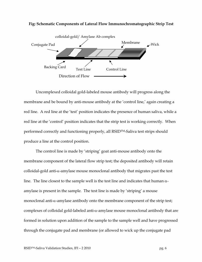
Fig: Schematic Components of Lateral Flow Immunochromatographic Strip Test
colloidal-gold/ Amylase Ab complex
Membrane
Test Line Control Line
Wick Conjugate Pad
Backing Card
Direction of Flow
Uncomplexed colloidal gold-labeled mouse antibody will progress along the
membrane and be bound by anti-mouse antibody at the ‘control line,’ again creating a
red line. A red line at the ‘test’ position indicates the presence of human saliva, while a
red line at the ‘control’ position indicates that the strip test is working correctly. When
performed correctly and functioning properly, all RSIDTM-Saliva test strips should
produce a line at the control position.
The control line is made by ‘striping’ goat anti-mouse antibody onto the
membrane component of the lateral flow strip test; the deposited antibody will retain
colloidal-gold anti-α-amylase mouse monoclonal antibody that migrates past the test
line. The line closest to the sample well is the test line and indicates that human α-
amylase is present in the sample. The test line is made by ‘striping’ a mouse
monoclonal anti-α-amylase antibody onto the membrane component of the strip test;
complexes of colloidal gold-labeled anti-α-amylase mouse monoclonal antibody that are
formed in solution upon addition of the sample to the sample well and have progressed
through the conjugate pad and membrane (or allowed to wick up the conjugate pad
RSIDTM-Saliva Validation Studies, IFI – 2 2010 pg. 6

when the strip is tested outside of a plastic housing, e.g., in a 12 x 75 test tube) will be
retained at the test line. A red control line must be visible at 10 minutes after sample
addition in order to interpret results.
Quantification of salivary amylase strip tests results
In order to maintain test-to-test consistency throughout the validation studies of
RSIDTM-Saliva, strip test results were quantified by comparing the intensity of the
observed results (i.e., how dark the test and control lines were) with a published
reference set of test and control lines. This score sheet, which consists of a series of
graded reddish lines is visually compared to all results. In addition, a digital picture of
the results was also recorded: both quantitative and pictorial results are presented.
RSIDTM-Saliva is not a quantitative test for the amount of saliva present in a given
sample. This procedure was used for the Development Validation and the design of
internal QA/QC production standards and is not used for the forensic application of
the test. RSID™-Saliva is a qualitative test and therefore test results are to be
interpreted as data for the presence or absence of α-amylase and by extentsion, saliva.
Specimens
Human saliva, blood, and urine samples were obtained voluntarily from laboratory
staff and deposited on sterile cotton swabs in aliquots of 50 µl. Unwashed semen was
obtained from a local sperm bank and deposited on sterile cotton swabs in aliquots of
50 µl. Human breast milk samples were obtained from SRI (Richmond, CA). Briefly,
human breast milk was collected from lactating mothers in a manner that would
RSIDTM-Saliva Validation Studies, IFI – 2 2010 pg. 7

preclude contamination with other body fluids and deposited on sterile cotton swabs
and air dried. Human fecal samples were obtained from a library of body fluid samples
obtained under IRB supervision. Post-coital vaginal swabs were obtained from
volunteers (laboratory staff). Animal saliva samples were kindly provided by the
Brookfield Zoo, Brookfield, Illinois.
Preparation of body fluid extracts
For body fluid extracts (saliva, semen, blood, urine, and breast milk), 50 µl of fluid was
deposited on a sterile cotton swab and allowed to air-dry. The cotton batting was
removed using laboratory clean technique and placed in a 1.5 ml microcentrifuge tube
and extracted in 1 ml of RSIDTM-Saliva extraction buffer for 1 hour at room temperature.
Assuming 100% extraction efficiency each microliter of extract will contain 50 nl (0.05
µl) of whole fluid. Oral swab extracts were made by swabbing the inside of an
individual’s cheek for 10 seconds with a cotton swab, and extracting the swab in 1 ml
RSIDTM-Saliva extraction buffer for 1 hour at room temperature. Negative control
extracts were made in an identical manner, but omitting the addition of body fluid to
the swab before extraction.
Experimental samples were prepared by combining the noted volume of
extraction solution with sufficient running buffer to produce a final volume of 100 µl
(RSIDTM- Saliva extract sample volume + RSIDTM- Saliva running buffer = 100 µl). Most
samples were tested on strips placed in cassettes, but for photographic clarity, some
RSIDTM-Saliva Validation Studies, IFI – 2 2010 pg. 8

experiments were performed in 12 x 75 mm test tubes; in all cases results were recorded
10 minutes after sample addition.
DNA extraction and STR DNA Analysis
DNA was extracted from swabs used to sample a plastic coffee lids, and an aluminum
soda can, and from cigarette butt paper, using a Chelex extraction protocol. The
extracted DNA was amplified using Identifiler (ABI) following a low copy number
protocol. The amplification reactions were run on an ABI Prism 310 Genetic Analyzer
and analyzed with Genescan (v. 3.7) and Genotyper (v 3.7) using an allele threshold of
75 RFU.
Sensitivity Testing: Saliva Extract and Human Oral Swab Extract
Methods: For sensitivity studies, we tested saliva extracts and human oral swab
extracts. For saliva extracts, 50 µl of human saliva was deposited on a sterile cotton
swab and allowed to air-dry. The end of the swab with the cotton batting was cut off
using laboratory clean technique and placed in a 1.5 mL microcentrifuge tube. The
swab head was extracted in 1 mL of RSIDTM-Saliva extraction buffer for 1 hour at room
temperature. We calculate that the extract will contain approximately 50 nL (0.05 µL) of
saliva (assuming 100% extraction efficiency) per microliter of extract. An oral swab
extract was made by swabbing the inside of an individual’s cheek for 10 seconds with a
cotton swab, and extracting the swab in 1 ml RSIDTM-Saliva extraction buffer for 1 hour
at room temperature in a 1.5 ml microcentrifuge tube. Negative control extracts were
RSIDTM-Saliva Validation Studies, IFI – 2 2010 pg. 9

made in an identical manner, but omitting the addition of saliva or oral extract to the
swab before extraction.
Two volumes of saliva extract were generally tested, 1 µL (equivalent to ~50 nL
saliva) and 5 µL (equivalent to ~250 nL saliva) by adding the indicated volume of saliva
extract to RSIDTM-Saliva running buffer and bringing the total volume to 100 µL. The
full 100 µL, containing both extract and running buffer, was placed in the sample
window of the cassette. Extracts from oral swabs were tested in an identical manner;
the quantity of saliva in an oral swab could only be estimated; we assume that 10 µL of
an oral swab extract is equivalent to ~ 0.5 µl of saliva. The control and test lines in the
test strip window were scored after 10 minutes.
Results- Sensitivity of Saliva Extract and Oral Swab Extract
After 10 minutes, the test line of the 1 and 5 µL saliva extracts were scored as positive,
respectively (see photo, left panel). These results indicate that the limit of detection for
RSIDTM-Saliva is approximately 50 nL (0.05 µL) of saliva. This experiment was repeated
with the buccal swab extract (see photo, right panel) and the results for 1, 5, and 10 µL
of oral extract were scored as positive, whereas 0 µl extract was negative.
RSIDTM-Saliva Validation Studies, IFI – 2 2010 pg. 10

Control line
Test line
0 1 µl 5 µl 0 1 µl 5 µl 10 µl
saliva extract oral swab extract
RSIDTM-Saliva Limit of detection Experiment.
In this experiment a broad range of saliva extract volumes were analyzed with RSIDTM-
Saliva in order to better determine the lower limit of detection. Positive control extract,
50 µl of saliva deposited on a sterile cotton swab and extracted in 1.0 ml of RSIDTM-
Saliva extraction buffer, was used throughout. Equivalent saliva volumes are calculated
assuming 100% extraction efficiency.
Strip Extract Amount (µl) RSIDTM-Saliva RB Equivalent Saliva (µl) Score
1 0 100 0 - 2 1 µl from 1:10 dilution 99 0.005 - 3 1 µl from 1:5 dilution 99 0.01 - 4 1 µl from 1:2 dilution 99 0.025 - 5 1 µL 99 0.05 + 6 2 µL 98 0.1 +
7 5 µL 95 0.25 +
8 10 µL 90 0.5 +
9 20 µL 80 1 +
RSIDTM-Saliva Validation Studies, IFI – 2 2010 pg. 11
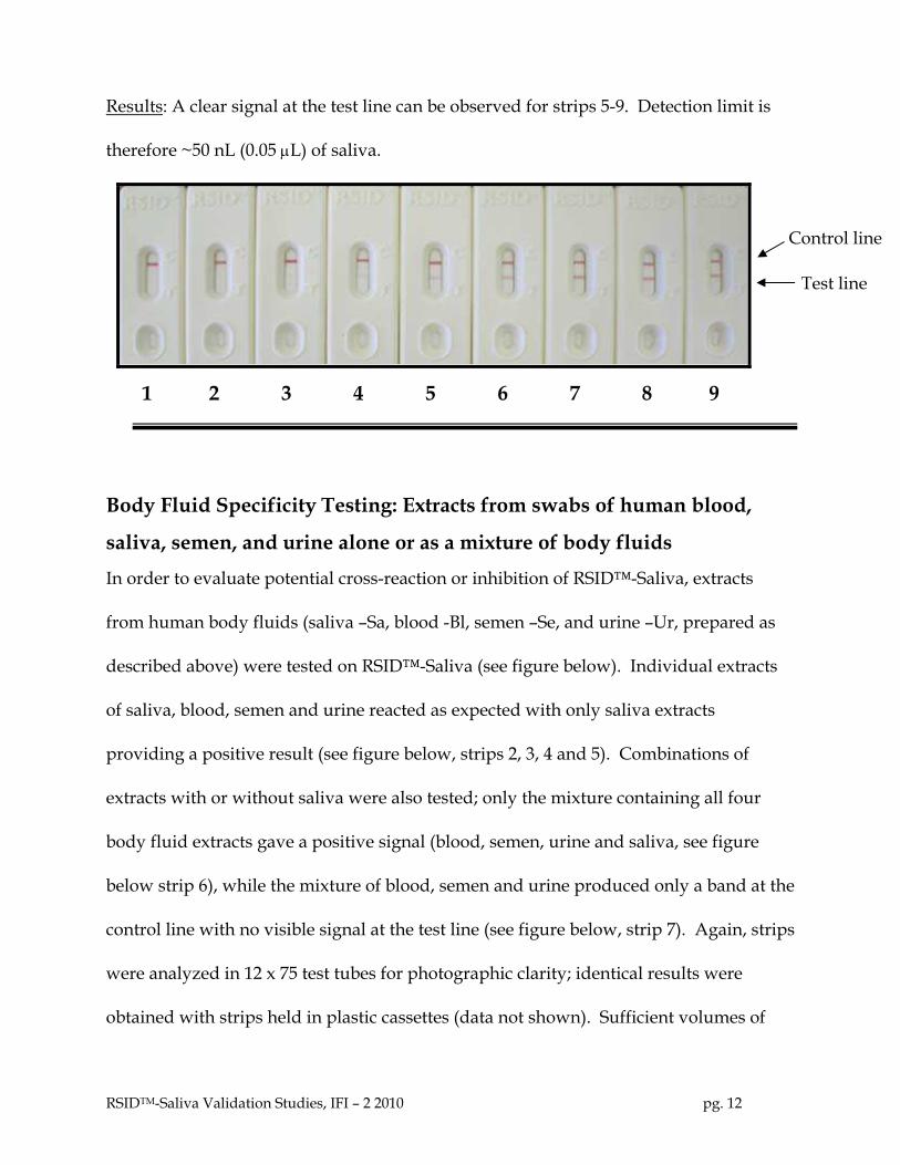
Results: A clear signal at the test line can be observed for strips 5-9. Detection limit is
therefore ~50 nL (0.05 µL) of saliva.
Control line
Test line
1 2 3 4 5 6 7 8 9
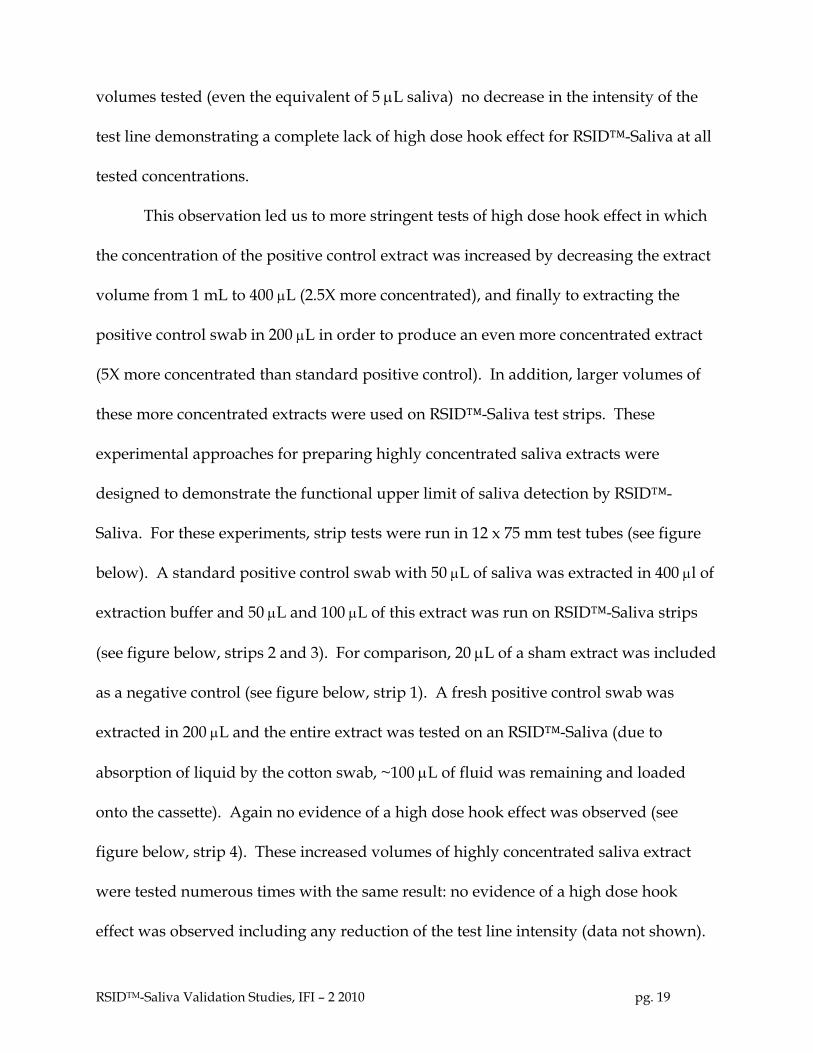
Body Fluid Specificity Testing: Extracts from swabs of human blood,
saliva, semen, and urine alone or as a mixture of body fluids
In order to evaluate potential cross-reaction or inhibition of RSID™-Saliva, extracts
from human body fluids (saliva –Sa, blood -Bl, semen –Se, and urine –Ur, prepared as
described above) were tested on RSID™-Saliva (see figure below). Individual extracts
of saliva, blood, semen and urine reacted as expected with only saliva extracts
providing a positive result (see figure below, strips 2, 3, 4 and 5). Combinations of
extracts with or without saliva were also tested; only the mixture containing all four
body fluid extracts gave a positive signal (blood, semen, urine and saliva, see figure
below strip 6), while the mixture of blood, semen and urine produced only a band at the
control line with no visible signal at the test line (see figure below, strip 7). Again, strips
were analyzed in 12 x 75 test tubes for photographic clarity; identical results were
obtained with strips held in plastic cassettes (data not shown). Sufficient volumes of
RSIDTM-Saliva Validation Studies, IFI – 2 2010 pg. 12

extract, 25 µLof each extract equivalent to 1.25 µL of each body fluid, were tested to
insure that even low levels of cross-reactivity would be observed, if present. For
comparison, a negative control was included in the experiment (see figure below, strip
1). As an additional test of specificity, extracts of saliva, blood, semen and urine were
combined at different ratios (1:1, 1:3, and 3:1) and tested with RSIDTM-Saliva. Again,
RSIDTM-Saliva did not cross-react with mixed extracts from urine, blood, or semen at
any ratio tested (data not shown). Taken together, these experiments demonstrate that
RSIDTM-Saliva does not cross react with the body fluids tested. The presence of semen,
blood, and urine does not interfere with the detection of saliva, an important issue since
multiple body fluids are often present on evidence collected at crime scenes.
1 2 3 4 5 6 7
Control line
Test line
(-) Sa Bl Se Ur 4 3
RSIDTM-Saliva Validation Studies, IFI – 2 2010 pg. 13

Specificity of RSIDTM-Saliva: Detection of Salivary α-Amylase in Human
Breast Milk and Fecal samples
It is well-documented that human breast milk contains low-levels of salivary α-amylase
that is probably present as an aid to carbohydrate digestion in infants (8, 9). Therefore,
we tested if human breast milk would give a positive signal with RSID™-Saliva.
Samples of human breast milk (50 µL) (kindly provided by SRI, Richmond CA and
described in Specimens) were extracted and various volumes of breast milk extract- 1, 5,
10 and 20 µL, equivalent to 0.05, 0.25, 0.5 and 1.0 µL of human breast milk- were
analyzed with RSIDTM-Saliva and compared side by side with equivalent volumes of
authentic human saliva (see figure below, strips 1-10).
As expected, RSIDTM-Saliva demonstrates a weak positive result with extracts
prepared from human breast milk (see figure below, strips 6, 7, and 8). By comparing
equivalent volumes of saliva and human breast milk (see figure below, strips 2, 3, 4, and
5) we estimate that breast milk is at least twenty fold less reactive on RSID™-Saliva than
authentic human saliva (see figure below, strips 2 and 7, strips 3 and 8).
1 2 3 4 5 6 7 8 9
Control line
Test line
(-) 1 1 5 5 10 10 20 20
Saliva Extract (µL) Breast Milk Extract (µL)
RSIDTM-Saliva Validation Studies, IFI – 2 2010 pg. 14

Since the majority of saliva is swallowed, we expected RSIDTM-Saliva to detect
salivary α-amylase in fecal samples. Six fecal samples from a human stain library were
extracted in 1 mL RSIDTM-Saliva extraction buffer for 1 hour at room temperature and 5,
20, and 100 µL of extract were analyzed with RSIDTM-Saliva. 100 µL of extract from
each of the six samples showed a weak positive while the other extract volumes were
negative (data not shown). In the same experiment, 1 µL of saliva extract (50 nL of
equivalent saliva) produced a strong positive, indicating that saliva is many times more
reactive on RSIDTM-Saliva test strips than fecal samples. Due to the unknown amount
of fecal matter present on the swabs, direct quantitative comparison with α-amylase
levels in saliva is not possible. This finding must be considered when anal swabs from
sexual assault evidence kits are tested with RSIDTM-Saliva.
A significant disadvantage of using α-amylase as a forensic indicator for saliva is
the distribution of this enzyme in human breast milk and feces, thereby making any test
using α-amylase a presumptive test. When using RSIDTM-Saliva, some conclusions
based on the signal intensity must be carefully considered. Fecal swabs tested on
RSID™-Saliva only generate a weak RSID™-Saliva positive, as do human breast milk
samples. A weak RSID™-Saliva positive signal can indicate either minimal amounts of
saliva, a fecal sample or breast milk sample, or inefficient sample extraction. RSIDTM-
Saliva cannot overcome the biological distribution of α-amylase, but as the relative
concentration of α-amylase varies considerably between these three body fluids, a
strong positive RSIDTM-Saliva result indicates, but does not proves, the presence of
saliva.
RSIDTM-Saliva Validation Studies, IFI – 2 2010 pg. 15

Specificity of RSID™-Saliva: Testing Extracts from Vaginal Swabs
The ability to detect human saliva from sexual assault evidence is an important issue for
forensic scientists. Therefore, we tested the ability of RSID™-Saliva to reliably identify
saliva from a series of vaginal swabs obtained from a subject with a well defined sexual
contact history. Post-coital swabs collected at 0-7, 9, and 11-13 days following
intercourse without a condom, were extracted with RSID™-Saliva extraction buffer and
analyzed with RSID™-Saliva test strips. Contact history included both semen
deposition (day 0) and oral contact (day 5). To increase the stringency of the test, swabs
were extracted in 300 µL of extraction buffer and 20 µL of this extract was combined
with 80 µL of RSID™-Saliva running buffer and then tested on RSID™-Saliva test strips.
The results clearly demonstrate that in this sample set, RSID™-Saliva does not
cross-react with post-coital vaginal swab extracts as no signal was observed from
samples taken 0, 1, 2, 3, 4, or 5 days post intercourse (see figure below, strips 0, 1, 2, 3, 4,
and 5, respectively). However, oral contact on day 5 was confirmed using RSID™-
Saliva when day 6 vaginal swabs were tested (see figure below, strip 6 designated by
arrow). No other RSIDTM-Saliva positive samples were observed from this
experimental series, demonstrating the specificity of RSID™-Saliva; RSID™-Saliva
results correlated precisely with the known sexual history of the samples. The lack of
cross-reactivity of the vaginal fluid extracts observed in this experiment is
representative of results seen with over 20 additional subjects, in which no signal was
detected in extracts from vaginal swabs with no reported presence of semen (data not
RSIDTM-Saliva Validation Studies, IFI – 2 2010 pg. 16

shown). This supports the conclusion that RSIDTM-Saliva does not cross-react with
vaginal fluid.
Test line
Control line - + 0 1 2 3 4 5* 6 7 9 11 12 13
Days Post-Coital
These data indicate that using mock sexual assault samples, RSIDTM-Saliva does
not cross react with semen or vaginal fluid and can easily and specifically detect saliva
from collected vaginal swabs. It should be noted that we have demonstrated body fluid
specificity using RSIDTM-Saliva for only the tested human body fluids of semen, saliva,
urine, blood, and vaginal fluid as well as detection of α-amylase in breast milk and fecal
samples. We have not tested RSIDTM-Saliva on samples obtained from cadavers or
other decomposing specimens; forensic lore states that cadaver samples present
particularly difficult body fluid identification issues.
Species Specificity of RSID™-Saliva: Testing of Animal Samples
Saliva swabs from various animal species, both exotic and companion animals,
were kindly provided by the Brookfield Zoo, Brookfield, Illinois. Extracts were
prepared as previously described and 25 µL of each extract was tested with RSIDTM-
Saliva. No cross reactivity was observed with saliva from the following animals: dog,
RSIDTM-Saliva Validation Studies, IFI – 2 2010 pg. 17

opossum, guinea pig, woodchuck, cow, domestic cat, domestic rabbit, tokay gecko,
cuckoo, mongoose, chameleon, domestic pig, llama, sheep, horse, goat, grey gull, ferret,
hedgehog, skunk, lion, tiger, rhinoceros, marsh snake, Sykes monkey, Capuchin
monkey, tamarin, and marmoset. A positive signal was obtained from the saliva of
gorilla (data not shown).
High Dose Hook Effect
The high dose hook effect can induce a false negative result on some lateral flow
immunochromatographic strip tests when high levels of target antigen are present in
the tested sample. The false negative result due to a high dose hook effect occurs when
the amount of target antigen in the sample is sufficiently high that a significant amount
of target antigen remains unbound by the colloidal gold-labeled antibody in the
conjugate pad. Free antigen then migrates to the membrane ahead of the labeled
antibody-antigen complexes, thereby occupying the bound antibody on the test line
with unlabeled antigen and leaving no sites for the gold labeled antibody-antigen
complexes. By blocking the test line with unlabeled antigen, the test result appears
negative. Most forensic laboratory personnel are familiar with high dose hook effects
and test a dilution of the questioned stain extract to insure that the observed result is a
true negative, and not due to a high dose hook effect. We evaluated RSID™-Saliva with
increasing amounts of saliva extract to evaluate RSID™-Saliva’s response to high levels
of antigen. Positive control extracts of 0, 5, 25, 50, 75, and 100 µL were prepared and run
on RSID™-Saliva (see figure below, strips 1-6, respectively). Note that at all extract
RSIDTM-Saliva Validation Studies, IFI – 2 2010 pg. 18
volumes tested (even the equivalent of 5 µL saliva) no decrease in the intensity of the
test line demonstrating a complete lack of high dose hook effect for RSID™-Saliva at all
tested concentrations.
This observation led us to more stringent tests of high dose hook effect in which
the concentration of the positive control extract was increased by decreasing the extract
volume from 1 mL to 400 µL (2.5X more concentrated), and finally to extracting the
positive control swab in 200 µL in order to produce an even more concentrated extract
(5X more concentrated than standard positive control). In addition, larger volumes of
these more concentrated extracts were used on RSID™-Saliva test strips. These
experimental approaches for preparing highly concentrated saliva extracts were
designed to demonstrate the functional upper limit of saliva detection by RSID™-
Saliva. For these experiments, strip tests were run in 12 x 75 mm test tubes (see figure
below). A standard positive control swab with 50 µL of saliva was extracted in 400 µl of
extraction buffer and 50 µL and 100 µL of this extract was run on RSID™-Saliva strips
(see figure below, strips 2 and 3). For comparison, 20 µL of a sham extract was included
as a negative control (see figure below, strip 1). A fresh positive control swab was
extracted in 200 µL and the entire extract was tested on an RSID™-Saliva (due to
absorption of liquid by the cotton swab, ~100 µL of fluid was remaining and loaded
onto the cassette). Again no evidence of a high dose hook effect was observed (see
figure below, strip 4). These increased volumes of highly concentrated saliva extract
were tested numerous times with the same result: no evidence of a high dose hook
effect was observed including any reduction of the test line intensity (data not shown).
RSIDTM-Saliva Validation Studies, IFI – 2 2010 pg. 19

Users of RSID™-Saliva can expect no false negative results due to high dose hook
effects.
The lack of high dose hook effect will facilitate the integration of RSIDTM-Saliva
into DNA forensic laboratory protocols, as a wide range of saliva concentrations, and
stain sizes, can be tested without performing dilutions of questioned stain extracts.
Test for High Dose Hook Effect (I)
Control line
75 µl 0 µL 5 µL 25 µL 50 µL 75 µL 100 µL
Test line
As an additional test for High dose hook effect, the concentration of the saliva extract
was increased such that 50 µL of saliva on a sterile swab was extracted into 400 µL or
into 200 µl RSIDTM-Saliva extraction buffer. From these concentrated saliva extracts, 50
µL and 100 µL of the 400 µL extraction and the entire volume from the 200 µLextract
were tested on RSIDTM-Saliva (due to absorption of liquid by the cotton swab, ~100 µL
of fluid was remaining and loaded onto the cassette).
Results : No diminution of signal was observed for the concentrated saliva extracts
even at the highest concentration of saliva tested. No high dose hook effect was
observed.
RSIDTM-Saliva Validation Studies, IFI – 2 2010 pg. 20

Strip 1– Buffer only Test for High Dose Hook
Effect (II) Strip 2– 50 µL saliva extract (50 µL saliva
extracted in 400 µL)
Test line
Control line
Strip 3– 100 µL saliva extract (50 µL saliva
extracted in 400 µL)
Strip 4 – 60 µL saliva extract (50 µl saliva
extracted in 200 µL) 1 2 3 4
Test strips have been removed
from cassettes for clarity
Conclusion: At all saliva extract volumes tested, no high dose hook effect
was observed with RSIDTM-Saliva and users can expect to observe no
false negative results due to high dose hook reactions.
Stability Testing of RSIDTM-Saliva
We have previously demonstrated that RSIDTM-Saliva is both specific and
sensitive for human saliva. Here we test the stability of the assembled cassettes by
storage at elevated temperatures. Assembled strip tests were stored at 37oC to increase
aging and potential degradation of the strips and subjected to a heat shock of 56oC,
again to test stability of the assembled test cassettes.
Extracts from positive control swabs were prepared and 0, 5 and 25 µL of extract
(equivalent to 0, 0.25 and 1.25 µL of saliva) were tested with RSIDTM-Saliva which has
been stored at 37oC for 11 days (condition designed to mimic storage for ~134 days at
room temperature) and with RSIDTM-Saliva that had been exposed to 56oC for 30
minutes.
RSIDTM-Saliva Validation Studies, IFI – 2 2010 pg. 21

Stability Test, RSIDTM-Saliva
Control line
Test line
0 5 25 0 5 25 0 5 25 11 days RT 11 days 37o 30 min 56o
We performed an additional stability experiment meant to simulate 1 year storage of
RSIDTM-Saliva. Here we tested 0, 5 µL, and 25 µL of positive control extract (equivalent
to 0, 0.25 µL and 1.25 µL saliva) with RSIDTM-Saliva after storage of the strips at 37oC for
30 days and compared the results with RSIDTM-Saliva that has been stored at room
temperature for the same amount of time.
Results: Test strips stored under conditions to mimic storage at room temperature for
one year showed a small but measurable decrease in signal intensity. Positive control
saliva extracts, 5 and 25 µL scored positive (respectively) for test strips stored at 37oC
for one month. Similarly, positives were observed for test strips stored at room
temperature for one month. Overall sensitivity of RSIDTM-Saliva was not significantly
affected.
RSIDTM-Saliva Stability Test
0 5 25 0 5 25
Control line
Test line
Test strips have been removed from cassettes for clarity
30 days RT 30 days 37oC
Test strips have been removed from cassettes for clarity
RSIDTM-Saliva Validation Studies, IFI – 2 2010 pg. 22

Conclusions: RSIDTM-Saliva cassettes are stable to storage without
significant loss of sensitivity.
Detection of Saliva from Forensic Exhibit-like Samples
We have clearly established that RSIDTM-Saliva can detect saliva from a laboratory
prepared control sample; here we demonstrate the ability of RSIDTM-Saliva to detect
saliva from samples likely to be encountered in forensic laboratory case work. In
addition we show that RSIDTM-Saliva can be incorporated into DNA-STR analysis and
suggest protocols such that saliva detection can be performed prior to DNA-STR
analysis.
Test Sample 1: Aluminum Coke can
Test Sample 2: Plastic coffee cup lid
Test Sample 3: Plastic Water Bottle
Test Sample 4: Glass Water Bottle
Test Sample 5: Cigarette Butts
Test Sample 6: Clippings from swabs used to sample plastic coffee lids (2) and
aluminum cans (2).
Procedure: Sterile cotton swabs were moistened with ddH20 and used to ‘sponge’ the
can lip and ‘pop-top’ opening of the can, and coffee cup lip. The swabs were extracted
in 300 µL RSIDTM-Saliva extraction buffer for 2 hours at room temperature. 25 µL of the
extract was removed for RSIDTM-Saliva testing and the remaining contents of the tube
(including the swab batting) were processed for DNA extraction and STR analysis as
RSIDTM-Saliva Validation Studies, IFI – 2 2010 pg. 23
per laboratory protocol. A buccal swab/oral swab used as a positive control was
extracted and processed in an identical manner.
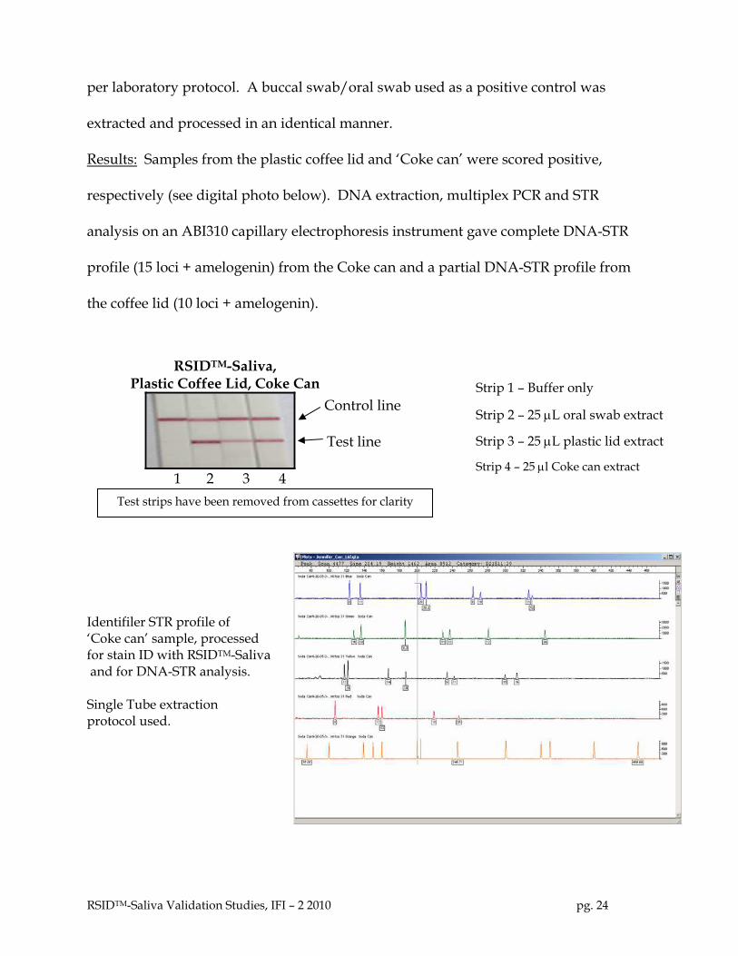
Results: Samples from the plastic coffee lid and ‘Coke can’ were scored positive,
respectively (see digital photo below). DNA extraction, multiplex PCR and STR
analysis on an ABI310 capillary electrophoresis instrument gave complete DNA-STR
profile (15 loci + amelogenin) from the Coke can and a partial DNA-STR profile from
the coffee lid (10 loci + amelogenin).
RSIDTM-Saliva, Plastic Coffee Lid, Coke Can Strip 1 – Buffer only
Control line Strip 2 – 25 µL oral swab extract
Test line Strip 3 – 25 µL plastic lid extract
Strip 4 – 25 µl Coke can extract 1 2 3 4 Test strips have been removed from cassettes for clarity Identifiler STR profile of ‘Coke can’ sample, processed for stain ID with RSIDTM-Saliva and for DNA-STR analysis. Single Tube extraction protocol used.
RSIDTM-Saliva Validation Studies, IFI – 2 2010 pg. 24

Identifiler STR profile of ‘coffee lid’ sample processed for stain ID with RSIDTM-Saliva and for DNA-STR analysis Single Tube extraction protocol used.
Test Sample 3: Plastic Water Bottle
Test Sample 4: Glass water bottle (Perrier)
Procedure: Moistened sterile cotton swabs were used to ‘sponge’ the openings of both
bottles and subsequently extracted in 200 µL RSIDTM-Saliva extraction buffer for 2 hours
at room temperature. A total of 30 µl of the extract was removed for RSIDTM-Saliva
testing, i.e., 5 and 25 µL aliquots, were used for analysis with RSIDTM-Saliva. The
remaining extract (including the swab) was used for DNA extraction and STR analysis.
Oral swab extract (in 1 mL) was used as a positive control.
Results: Saliva from Sample 3, the plastic water bottle was readily detected and RSIDTM-
Saliva was scored positive for both 5 and 25 µL extract, respectively (see figure below).
Saliva extract from the glass bottle also scored positive with 5 and 25 µL of extract,
respectively (see figure below).
STR analysis did however provide a full profile (15 loci + amelogenin) from the glass
bottle, whereas only two loci were obtained from the plastic bottle. Correlating the
RSIDTM-Saliva Validation Studies, IFI – 2 2010 pg. 25

intensity of the RSIDTM-Saliva test results with the observed DNA-STR results may not
be straightforward: a number of variables including person to person variation,
extraction methods, and amplification kit used, may all affect the ability of the analyst
to obtain a full DNA profile from the tested sample.
RSIDTM-Saliva, Plastic and Glass Bottle
Control line
Test line
0 5 25 0 5 25 0 5 25 oral swab plastic glass
Test strips have been removed from cassettes for clarity Identifiler STR profile of ‘Glass bottle’ sample, processed for stain ID with RSIDTM-Saliva and for DNA-STR analysis. Single Tube extraction protocol used.
RSIDTM-Saliva Validation Studies, IFI – 2 2010 pg. 26

Identifiler STR profile of ‘Plastic bottle’ sample, processed for stain ID with RSIDTM-Saliva and for DNA-STR analysis. Single Tube extraction protocol used.
Test Sample 5: Saliva detection from Cigarette butts.
Procedure: Two received cigarette butts (samples 5a and 5b) were sampled using
moistened sterile cotton swabs which were subsequently extracted in 200 µL of RSIDTM-
Saliva extraction buffer; an aliquot of the extraction was used for RSIDTM-Saliva (25 µL)
while the majority of the extract was processed for DNA-STR analysis.
Results: Positive control saliva extracts gave positive results for 5 and 25 µL of saliva
extract (respectively), 25 µl of samples 5a and 5b gave test lines of low intensity but
were clearly above background levels. Sample 5a was analyzed for Y-STRs and
provided clear data for 14 loci (see below).
RSIDTM-Saliva Validation Studies, IFI – 2 2010 pg. 27

RSIDTM-Saliva, Strip 1 – Buffer only Cigarette Butts
Strip 2 – 5 µL saliva extract Control line
Strip 3 – 25 µL saliva extract Test line
Strip 4 – 25 µL extract Cig Butt #5a
1 2 3 4 5 6 Strip 5 – 25 µL extract Cig Butt #5b
Strip 6 – 25 µL extract Cig Butt neg Test strips have been removed from cassettes for clarity
Cigarette Butt was analyzed for Y-STR using Y-Filer. Single tube extraction protocol used for both stain ID and DNA-STR processing.
Conclusion: RSIDTM-Saliva detects saliva from cigarette butts.
RSIDTM-Saliva Validation Studies, IFI – 2 2010 pg. 28

RSIDTM-Saliva Analysis of swab cuttings from swabs used to sample plastic lids and
aluminum cans, alternative to single tube extraction protocol. Our laboratory protocol
uses a single extraction step for both stain identification and DNA-STR analysis. The
advantages of this approach are clear and include less sample loss (one tube for sample
extraction, stain identification and DNA-extraction), less manipulation of the sample
(cuttings and repeated testing of evidence swabs are eliminated), less chance of
contamination (fewer procedural steps) and it eliminates variation due to
inhomogeneous swabs. Many laboratories however, use an alternative approach, of
testing small cuttings from swabs that were used to ‘sponge’ or sample questioned
stains. Here we demonstrate that RSIDTM-Saliva can be used with cuttings obtained
from swabs used to absorb questioned stains.
Test Sample 6: Extracts were prepared from cuttings from swabs used to sample two
plastic coffee lids and two aluminum soda cans. Moistened swabs were used to
‘sponge’ the areas of 2 plastic coffee lids and 2 aluminum soda cans most likely to have
been in contact with saliva. Swabs were allowed to dry in a protected environment and
cuttings from the swabs were removed and placed individually in microcentrifuge
tubes. These cuttings were extracted in 50 µL RSIDTM-Saliva extraction buffer for 1 hour
at room temperature at which time ~30 µL (all the volume available in the tube) was
used for analysis with RSIDTM-Saliva.
Results: Clipping from the positive control scored clear positive for 5 and 25 µL saliva
extract, respectively. Extracts from the swab cuttings, ~30 µL each, scored weak
positive but clearly above background signal (see figure below). The tested swabs were
RSIDTM-Saliva Validation Studies, IFI – 2 2010 pg. 29

processed for DNA-STR analysis with mixed results: one plastic lid provided a full
profile (15 loci + amelogenin), the other provided a partial profile (13 loci + amelogenin)
while analysis of the swabs from the cans gave partial profiles.
RSIDTM-Saliva, Strip 1 – Buffer only Test from Swab Cuttings
Strip 2 – 5 µL saliva extract
Control line Strip 3 – 25 µL saliva extract
Strip 4 – 30 µL 1st plastic lid lStrip 5 – 30 µL 1st alum can clipping Test line
Strip 6 – 30 µL 2nd plastic lid clipping
Strip 7 – 30 µL 2nd alum can(2) clipping 1 2 3 4 5 6 7
Test strips have been removed from cassettes for clarity
Identifiler STR profile of ‘plastic coffee lid’ sample processed for stain ID with RSIDTM-Saliva and for DNA-STR analysis. Multiple cutting protocol used for stain ID and DNA-STR analysis.
Identifiler STR profile of ‘Soda Can’ sample processed for stain ID with RSIDTM-Saliva and for DNA-STR analysis. Multiple cutting protocol used for stain ID and DNA-STR analysis.
RSIDTM-Saliva Validation Studies, IFI – 2 2010 pg. 30

Identifiler STR profile of ‘Soda Can (2)’ sample processed for stain ID with RSIDTM-Saliva and for DNA-STR analysis. Multiple cutting protocol used for stain ID and DNA-STR analysis.
Identifiler STR profile of ‘plastic coffee lid(2)’ sample processed for stain ID with RSIDTM-Saliva and for DNA-STR analysis. Multiple cutting protocol used for stain ID and DNA-STR analysis.
Conclusion: RSIDTM-Saliva can detect saliva from swab cuttings derived from swabs
used to sample cans and plastic lids.
RSIDTM-Saliva Validation Studies, IFI – 2 2010 pg. 31

Additional testing with RSIDTM-Saliva – Forensic-like Samples.
We sampled a variety of surfaces and materials in an effort to rigorously test RSIDTM-
Saliva. The samples include envelopes, additional plastic bottles, and a different metal
soda can.
Procedure: All samples were ‘sponged’ with a moistened sterile cotton swab and after
air-drying in a protected environment, the swab batting was extracted in 400 µl of
RSIDTM-Saliva extraction buffer in a microcentrifuge tube. 25 µl of each extract was
tested with RSIDTM-Saliva. Positive control was an oral swab extracted in 1.0 ml of
RSIDTM-Saliva extraction buffer, 5 µl of extract tested. Samples included:
1) Negative Control 2) Positive Control 3) Envelope, licked, sealed, steamed open and upper flap sampled with swab technique 4) Envelope, licked, sealed, steamed open and lower flap sampled with swab technique 5) Plastic bottle, threads and cap tested. 6) Glass bottle, threads and cap tested 7) oral swab 8) metal soda can
Results: As was expected the amount of saliva in the above eight samples varied widely
and this was reflected in the intensity scores of the test line (see figure below).
Sample (1) Neg (2) Pos (3) Envelope upper flap (4) Envelope lower flap (5) Plastic Bottle (6) Glass. Bottle (7) Oral Swab (8) Metal can
RSIDTM-Saliva Validation Studies, IFI – 2 2010 pg. 32
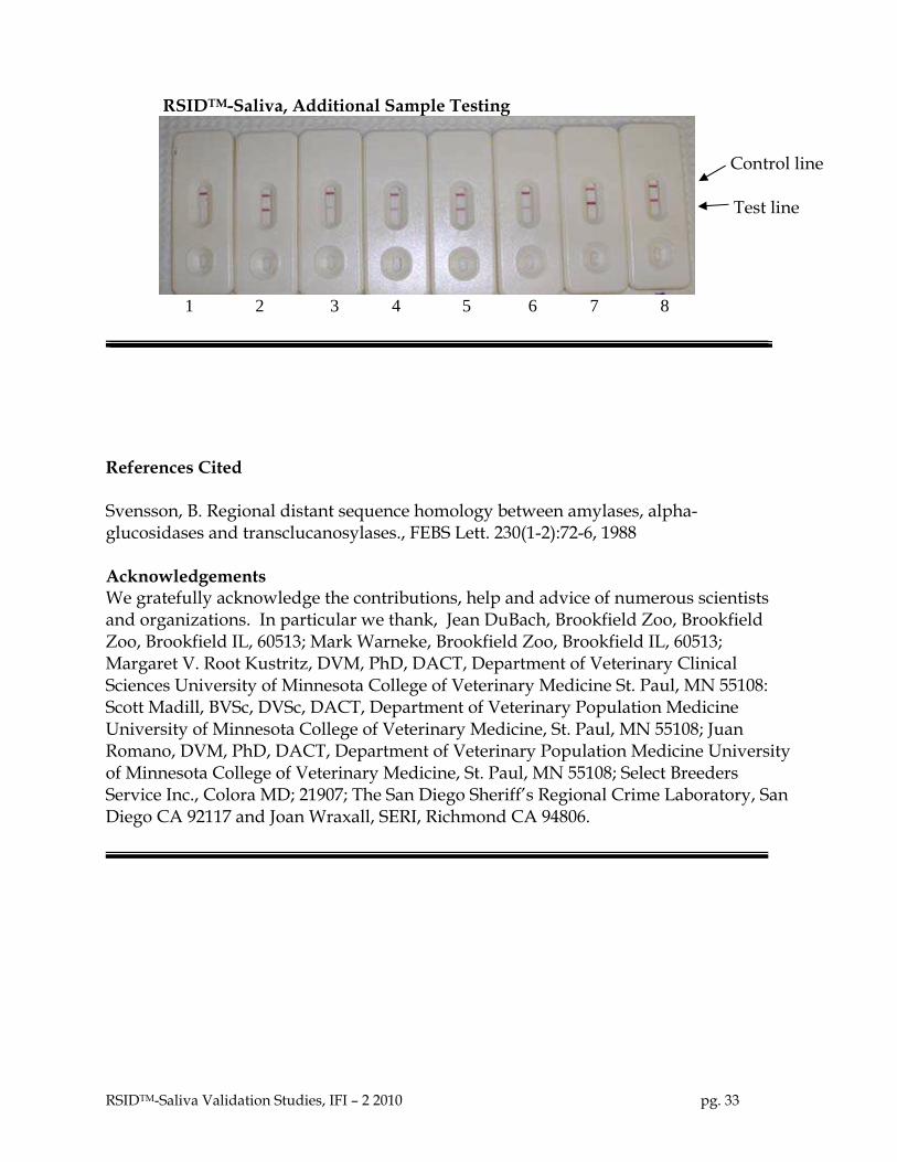
RSIDTM-Saliva, Additional Sample Testing
Control line
Test line
1 2 3 4 5 6 7 8
References Cited Svensson, B. Regional distant sequence homology between amylases, alpha-glucosidases and transclucanosylases., FEBS Lett. 230(1-2):72-6, 1988 Acknowledgements We gratefully acknowledge the contributions, help and advice of numerous scientists and organizations. In particular we thank, Jean DuBach, Brookfield Zoo, Brookfield Zoo, Brookfield IL, 60513; Mark Warneke, Brookfield Zoo, Brookfield IL, 60513; Margaret V. Root Kustritz, DVM, PhD, DACT, Department of Veterinary Clinical Sciences University of Minnesota College of Veterinary Medicine St. Paul, MN 55108: Scott Madill, BVSc, DVSc, DACT, Department of Veterinary Population Medicine University of Minnesota College of Veterinary Medicine, St. Paul, MN 55108; Juan Romano, DVM, PhD, DACT, Department of Veterinary Population Medicine University of Minnesota College of Veterinary Medicine, St. Paul, MN 55108; Select Breeders Service Inc., Colora MD; 21907; The San Diego Sheriff’s Regional Crime Laboratory, San Diego CA 92117 and Joan Wraxall, SERI, Richmond CA 94806.
RSIDTM-Saliva Validation Studies, IFI – 2 2010 pg. 33