Embed Size (px)
Citation preview
Grand Valley State UniversityScholarWorks@GVSU
Masters Theses Graduate Research and Creative Practice
1996
Validation of the Neonatal Infant Pain ScaleAnnette L. BackusGrand Valley State University
Follow this and additional works at: http://scholarworks.gvsu.edu/theses
Part of the Nursing Commons
This Thesis is brought to you for free and open access by the Graduate Research and Creative Practice at ScholarWorks@GVSU. It has been acceptedfor inclusion in Masters Theses by an authorized administrator of ScholarWorks@GVSU. For more information, please [email protected].
Recommended CitationBackus, Annette L., "Validation of the Neonatal Infant Pain Scale" (1996). Masters Theses. 280.http://scholarworks.gvsu.edu/theses/280
VALIDATION OF THE NEONATAL INFANT PAIN SCALE
ByAnnette L. Backus
A THESISSubmitted to
Grand Valley State University in partial fulfillment of the requirements for the
degree ofMASTER OF SCIENCE IN NURSINGKirkhof School of Nursing
1996
Thesis Committee Members: Andrea C.Bostrom, Ph.D.,R,N.
Joyce French, Ph.D., R.N. Thomas Herzog, Ph.D.
ABSTRACTVALIDATION OF THE NEONATAL INFANT PAIN SCALE
By
Annette L. Backus
The purpose of this study was to validate the Neonatal Infant Pain Scale (NIPS). This was accomplished by determining inter-rater reliability, the ability to measure
pain in infants of any gestational age, and the relationship between the NIPS behavioral scores and physiological parameters of heart rate, respiratory rate and oxygen saturation.
Inter-rater reliability on total scores obtained before, during and after the procedure yielded correlations ranging from 0.69 to 0.90, which were significant at p<.001. All groups showed significant increases in NIPS scores after a
painful procedure was started. Total mean scores before, during and after a procedure were 0.44, 3.04 and 0.6 respectively. There were no significant correlations between NIPS scores and heart rate, respiratory rate or oxygen saturation.
The NIPS appears to be a reliable tool for evaluating pain in neonates of any gestational age. Physiological measures were not reliable indicators of pain in neonates.

Acknowledgments
I would like to express my sincere appreciation to all those who have contributed to the success of this research project.
I would like to offer a special acknowledgement and thanks to the chairperson of my committee, Andrea Bostrom, Ph.D., RN, along with Joyce French, Ph.D., RN, and Thomas Herzog, Ph.D. for their support and guidance.
Special appreciation to my colleagues at Bronson Methodist Hospital for their assistance and collaboration during pursuit of my educational goals.
A very special thank you to the phlebotomist at Bronson Methodist Hospital for their cooperation and assistance during this research project.
And last, but not least, I would like to thank Bob, Robyn and David for their patience, love and support; without them I would never have accomplished my goals.
Table of Contents
List of Tables .............................................. vList of Appendices ....................................... vii
CHAPTER1 INTRODUCTION ........................................ 12 CONCEPTUAL FRAMEWORK AND REVIEW OF LITERATURE ..... 4
Conceptual Framework ............................. 4Review of the Literature ........................ 7Summary and Indication for Study ............... 13Hypothesis....................................... 14Definition of Terms ............................. 14
3 METHODOLOGY ........................................ 16Design ........................................... 16Sample .......................................... 17Instrument ...................................... 17Procedure ....................................... 18
4 DATA ANALYSIS ...................................... 21Sample .......................................... 21Hypotheses ...................................... 23
Hypothesis 1 ................................ 23Hypothesis 2 ................................ 28Hypothesis 3 ................................ 31
Additional Data Analysis ....................... 355 DISCUSSION AND IMPLICATIONS ....................... 40
Application to Practice ........................ 44Limitations ..................................... 45Suggestions for Further Research ............... 45
APPENDICES ................................................. 47REFERENCES ................................................. 57
IV
List of Tables
Table1 Characteristics of Sample ............................ 222 Diagnoses of Sample .................................. 23
3 Pearson's Correlations on Total Scores, Rater A toRarer B, Before, During and After Procedure ........ 25
4 Paired t-tests on total Scores, Rater A to Rater B,Before, During and After Procedure .................. 26
5 Percent Agreement by NIPS Scoring Items ............. 276 Percent Agreement of Total NIPS Scores Before,
During and After Procedure .......................... 27
7 Mean Scores for Each Gestational Age Group, Before,During and After Procedure .......................... 28
8 Repeated Measures ANOVA .............................. 29
9 Paired t-tests for Each Gestational Age on Before toDuring Procedure and During to After Procedure ..... 30
10 Mean NIPS Scores and Physiological Parameters ....... 3211 Mean Changes of NIPS Scores and Physiological
Parameters from Before to During Procedures ........ 3312 Correlation of Changes in NIPS Scores to Changes
in Selected Physiological Parameters From Before Procedure to During Procedure ....................... 34
13 Pearson's Correlations on Rater A to Rater B Total Scores for NIPS Minus Arm and Leg ObservationsBefore, During and After Procedure .................. 36
14 Paired t-test on Rater A to Rater B Total ScoresMminus Arm and Leg Observations Before, During and After Procedure ...................................... 37

15 Comparison of Mean NIPS Scores Without Leg Observation to NIPS Scores Without Arm or Leg Observations .......................................... 38
16 Paired t-test on Total Scores Minus Arm and Leg Observations on Changes in NIPS from Before to During the Procedure and During to After the Procedure .... 39
VI
List of i^pendices
Appendix
A Neonatal Infant Pain Scale ......................... 47
B Operational Definitions ............................ 48C Permission to Use Neonatal Infant Pain Scale ....... 49D Adapted Neonatal Infant Pain Scale ................ 50E Human Use Committee Approval ....................... 51F Permission to Conduct Research by Grand Valley
State University ................................... 52G Information for Partioipation in Research
Project ............................................. 53H Consent Form ........................................ 54
I Subject Characteristics ............................ 55
V I 1
CHAPTER 1 INTRODUCTION
Traditionally the determination of severity of a person's pain has been difficult. Perception of pain is influenced by a person's history and culture, and can be interpreted differently by medical and nursing staff depending on their background and beliefs (Clancy, Anand, & Lally, 1992). Questions have often been raised about the relief of pain for many classifications of patients.
The pre-verbal infant presents many difficulties associated with the evaluation for presence of pain.Long held beliefs that infants feel no pain are slow to change, despite evidence to the contrary that neonates perceive and respond to pain (Butler, 1988; Owens, 1981). Many clinicians continue to believe that either infants do not feel pain, or, if they do, they do not remember it (Brown, 1987; Budreau, 1991; Dale, 1986; Owens & Todt, 1984; Rich, Marshall, & Volpe, 1974). Yet, research supports that pain does occur in pre-verbal infants and can be determined by observation of behavioral cues (Anand, Phil, & Hickey, 1987; Franck, 1986).
The Agency for Health Care Policy and Research (AHCPR) determined that management of pain is an area of practice
that should be studied. In 1992 the U.S. Department of Health and Human Services published a set of guidelines for adults and one for infants, children and adolescents. Incorporated
into the guidelines were numerous scales that can be used to determine severity of pain. These scales do not, however, address the pre-verbal infant.
As stated in the AHCPR guidelines "the obligation to manage pain and relieve a patient's suffering is a crucial
element of a health professional's commitment" (DHHS, 1992, p.). This supports an obligation to find an objective, consistent, and documentâtle process for determining pain in infants (Butler, 1988). There should be no further reason to demonstrate that infants are capable of having pain. Instead, steps should be taken to determine how much pain and what interventions relieve pain.
At the present time, pain in infants is determined by the
nurse's skill at assessing infant behavior and watching for physiologic changes such as increased heart rate, blood pressure, and respiratory rate. Assessment of infant behavior is highly subjective and behaviors that are observed can change from nurse to nurse. So, despite the availability of effective techniques for pain management, infants in the Neonatal Intensive Care Units (NICUs) often have very inconsistent levels of pain relief because of inconsistent assessment.
There are few scales that have been developed to evaluate neonatal pain (Attia, Amiel-Tison, Mayer, & Shnider, 1987;
Franck, 1986; Ross & Ross, 1988). One that shows promise is the Neonatal Infant Pain Scale (NIPS) (Lawrence, Alcock,
McGrath, Kay, MacMurray, & Dulberg, 1993) . Confirming the validity and reliability of this tool could result in an instrument for collection of objective data for use in documentâting, planning care, and evaluating relief of pain in infants. Examining validity and reliability of the NIPS will be addressed in this thesis.
CHAPTER 2CONCEPTUAL FRAMEWOEIK AND REVIEW OF LITERATURE
Conceptual Framework
Als' Synactive Model of Neonatal Behavioral Organization and Levine's Conceptual Model of Conservation were used as conceptual frameworks for this study. Together they provide a comprehensive basis for developing a nursing model for neonates.
The synactive model identifies the behavioral opportunities available to the infant to cope with environmental stress. Synaction holds that the various subsystems exist side by side, either interactive or in a relative supporting, holding pattern, as if providing a steady presence for the subsystem currently being utilized. The infant's functioning is seen in a model of continuous intraorganism subsystem interaction, and the organism is seen in continuous interaction with the environment (Als, 1986). If the infant's own regulatory capacity is exceeded, the effort expended to regain regulation begins to impinge on all other systems. When the presence of pain exceeds the infant's regulatory capacity it causes behavioral and physiological changes in relation to the severity of pain (Als, 1986) .
Synactive theory proposes that environmental modification can bring about a reduction in stressors and aid self-
regulatory behaviors that will improve medical and developmental outcomes. If state regulatory measures can be assisted through comforting, positioning, or relieving pain, then infant's will have increasing capabilities for self regulation as they mature.
Pain causes observable changes in the infant's behavioral organization and there are effective techniques to decrease
the effect of pain in infants. These include positioning techniques, calming supports and administering analgesia.
Levine's (1969) Conceptual Model of Conservation provides a process for conserving as many of the infant's resources as possible. It promotes the infant holistically, realizing that all of the infant's systems are dependent on one another.
Levine's view of nursing is that the nurse conserves for the patient while the patient adapts to the environment. Levine's four Principles of Conservation (Levine, 1967) help to define the process the nurse uses to conserve the patient's
resources. These principles include:1. Awareness that an environment influences behavior
at all times.2. Conservation of patients' energy is a consequence
of nursing intervention.
3. Components of nursing interventions are conservation of individual patient's structural
integrity, personal integrity, and social integrity.
4 . Nurses are participants in every patient 'senvironment and influence patient's adaptation.
Synactive theory and Levine's Conservation Model propose that environmental modification can bring about a reduction in stressors and aid behaviors that will attempt to keep the infant in a stable state and improve medical and developmental outcomes. State-regulating behaviors are the observable behavioral strategies used by the infant to maintain a balanced, relatively stable and relaxed state of subsystem integration (Als, 1982, 1986). If state regulatory measures, such as comforting, positioning, or relieving pain conserve energy then infant's will have increasing capabilities for self regulation as they mature. Being able to identify and provide relief of pain would allow the nurse to aid the infant in maintaining control. It would help the infant avoid periods of irritation or periods in which the infant engages in behavior that disrupts all of his resources (Levine, 1971).
In summary, pain causes observable changes in the infant's behavioral organization (Als synactive model) and there are effective techniques that can be used (Levine's conservation model) to decrease the effects of pain in infants. These include positioning techniques, calming supports, and administering analgesia.
Review of the Literature
Determination of pain in adults is usually dependent on verbal complaints and descriptions of the pain. Some research has been done attempting to quantify nonverbal measures of pain in adults and children. Behavior indicators include changes in facial expression, cry and posture (Craig & Prkachin, 1983). Very little has been done to quantify infant pain. Much of this is due to a belief that infants, especially preterm infants, do not feel pain.
Studies have shown that there is pain experienced in neonates. A paper by Anand, Phil and Hickey (1987) reviewed neurophysiologic research that showed there was cortical maturation and myelination of pain pathways early in gestation. This paper points out that pain has a strong emotional association and suggests that pain in infants should
be discussed as neural pathway or nociceptive activity.Neural pathways for pain start at sensory receptors in
the skin of an infant and lead to sensory areas in the cerebral cortex. The density of nociceptive nerve endings in newborns is similar or greater than in adults. By the twentieth week of gestation all cutaneous and mucosal surfaces have sensory receptors (Gleiss & Stuttgen, 1970).
Lack of myelination of nerve fibers is often used as an argument that preterm or full term neonates are not capable of pain perception. Incomplete myelination would account for slower conductivity. However, in the infant shorter distances of the neural pathway may offset this. Myelinated a-fibers
are responsible for initial pain sensation, the sharp stinging feeling. Unmyelinated fibers, c-fibers, are responsible for the transmission of burning, aching sensations which begin after stimulation has ceased and can last for an extended
period of time. In adult peripheral nerves, nociceptor impulses are carried through unmyelinated and myelinated fibers. It has also been shown that nociceptive nerve tracts undergo myelination during the second and third trimester of gestation {Gilles, Shankle, & Dooling, 1983) . This does help support viewing infant pain as nociceptive activity, and avoiding the emotional connotations of pain.
Physiological parameters used to indicate infant response to pain have been measured, and shown to react in a significantly negative way. These parameters include sustained increased heart rate with fullterm infants observed during heel lances. Results from this study were less clear for preterm infants (Owens & Todt, 1984). The use of local anesthesia during circumcision in ten fullterm infants prevented changes in heart rate and blood pressure in a study by Williamson and Williamson (1983). Marshall (1989) related changes in heart rate, respiratory rate, systolic blood pressure, transcutaneous P02, cry, facial expression and state of arousal to heel stick procedures.
Franck (1986) used photogrammetry to quantitatively determine behavioral responses to a painful stimulus. Photogrammetry is a technique used to videotape and then view the tape. Observation and recording of behavior on the tapes
are done by raters with their chins on stabilizers at the level of the video monitor. The video is viewed through a
grid to aid in measuring movement. Though a small sample was used (N=10) photogrammetry demonstrated responses to painful stimuli and suggested some memory of pain in term infants evidenced by quicker, sharper responses with repeated stimuli.
Dale (1986) conducted a study videotaping intramuscular injections on newborns up to 6 months of age. The researcher identified cry, facial expressions and body movements as being increased with pain. This study looked at reactions to pain and possible differences in response with a second procedure. While the sample was small (N=10), they were able to isolate the previously listed behaviors and saw some anticipation of the pain on repeated injections.
Franck (1987) later conducted a national survey of 143 hospitals with level III neonatal units with more than 20 beds. One neonatal nurse from each institution was asked to respond to the survey. A total of 76 surveys were returned representing 7 6 hospitals and 34 states. The survey consisted of a 15 item questionnaire. The questions were classified into 5 categories: 1) beliefs regarding pain and adequacy of medication used to treat pain in infants, 2) methods of assessment of infant pain, 3) interventions used to manage infant pain, 4) descriptors of agitated behavior in neonatal intensive care unit patients, and 5) uses of medication to manage pain and agitation in infants. This survey identified common behaviors assessed in infants to determine pain. Cry
and change in activity were identified as the most common behaviors assessed. Other surveys of nurses' abilities to assess pain from infant behavior have shown high reliability (Page & Halvorson, 1991; Lawrence, et al., 1993; Maloni, Stegman, Taylor, Brownell, 1986). However Franck's survey (1987) showed large differences in attitudes regarding pain and in the practice of alleviating pain.
A scale that relates behavioral changes to pain stimuli would be useful in objectively assessing neonatal pain.
Nurses are consistently reliable in identifying pain in infants, but beliefs that infants do not feel pain may preclude looking for it. When nurses do look for pain, interventions are often inconsistent because of an inability to establish the degree of pain or agitation. Therefore development of a reliable and validated tool can provide for more consistent assessment (Franck, 1987).
Available in the literature are results of the use of two
tools designed for use with pre-verbal infants. Both are based on the observation of behavioral changes that are believed to reflect the presence of pain. Attia et al.,(1987) presented an abstract for a post operative pain scale.
The scale was used on 23 infants to measure effects of analgesia post operatively. Behaviors observed included sleep, facial expression, cry, spontaneous motor activity, excitability, flexion of fingers/toes, sucking, tone, consolability and sociability. With a possible score of 0 - 2 for each area the scale, the score ranges from 0 to 20.
10
Scoring ten different behaviors may be too detailed for clinical use. Reliability and validity were not stated. Further publication of this scale could not be found.
The Neonatal Infant Pain Scale (NIPS) developed at Children’s Hospital of Eastern Ontario (CHEG) (Lawrence, Alcock, McGrath, Kay, MacMurray, & Dulberg, 1993) is based on 6 items: cry, facial expression, breathing patterns, positioning of arms and legs, and state of arousal. Initially a survey of 43 experienced neonatal nurses was conducted to identify behaviors associated with pain. The pilot study used a scale with eight criteria for rating. These included facial expression, facial color, arm and leg position, torso movement, breathing patterns, cry, and state of arousal. This scale was used to document behavior changes on 20 videotaped
needle-intrusive procedures. Results of the pilot study showed that changes in facial color could be caused by illness and often changed frequently. There were also difficulties observing torso movement. The final version of the NIPS used in the CHEG study deleted these two indicators.
The resulting scale (Appendix A) provides a tool that
clinically may be easy to use and reliable. The NIPS lists six behavioral components, the description of the behavior and the potential scores. Operational definitions of the behaviors are printed on the tool for easy reference (Appendix B) . Each behavior except cry has a possible score of 0 or 1. Cry has a possible maximum score of 2 (0=no cry, l=whimper, 2=vigorous cry). Across the top of the scale are columns for
11
recording observations at one minute intervals, before, during and after a procedure. The scale could also be used at a specified time to determine presence of pain. Scores are totaled at the bottom with scores ranging from 0 - 7 . The higher the score the more likely there is the presence of pain.
The CHEO trial of the NIPS included 38 infants videotaped during 90 procedures. Using the same infant in more than one video rating may prove a problem in the determination of validity. Sixty-seven procedures were on preterm infants and 23 on fullterm infants. Other than preterm or fullterm, exact gestational age was not designated. There is a need for better correlation of the NIPS with preterm infant pain assessment.
Inter-rater reliability for this study (Lawrence et al., 1993) was high. Twenty procedures were scored by both the research assistant and an independent observer. Comparisons of the scores from the two raters at three times, once before, during and after the procedures were calculated. Pearson's correlations ranged from .92 to .97 and were statistically significant (p<.05). Results from paired t-tests indicated only small inter-rater differences ranging from 0-0.3, which did not approach statistical significance. It was concluded that the NIPS has a high inter-rater reliability at that institution.
To determine if there was a change in NIPS scores over time as an indication of a change in intensity of pain, a
12

repeated measures ANOVA was done on NIPS scores for 22 infants undergoing painful procedures. Results were significant (F=18.97, df=2,42, p=<.001). Mean NIPS scores before the procedure were 1.1, during the procedure were 4.8, and after the procedure were 2.0. The Friedman test was used to confirm that there was statistical significance in the pattern of
increase followed by decrease of NIPS scores.Internal consistency was tested using scores before,
during and after a procedure. The NIPS showed a high internal
consistency with Cronbach's alphas of .95, .87, and .88, respectively.
There were no reported problems using videotapes for rating. Results showed increases in NIPS scores with painful procedures and suggest the NIPS is a reliable and valid instrument.Summarv and Indication for Study
There is a definite need for a tool that can accurately
assess pain and relief of pain in infants. At the time this study was done there were only two available scales that were not well tested or widely used.
The Neonatal Infant Pain Scale has been used and reported from only one institution at this time. While data reported from the study are good it needs to be more widely used to establish inter-rater reliability on a wider scale. In addition the need to determine the tool's ability to predict pain in an infant of any gestational age is very important.
13
The purpose of this thesis was to examine the validity and reliability of the NIPS, and determine the ability of the NIPS to validly evaluate pain in term and preterm infants. Hypotheses
Hypothesis 1 - The degree of pain and relief of pain can be assessed reliably at any gestational age using the Neonatal Infant Pain Scale.
Hypothesis 2 - Infants score higher on the Neonatal Infant Pain Scale when assessed during a painful procedure than before or after the procedure and this change in score occurs reliably at any gestational age infant.
Hypothesis 3 - With an increase in the Neonatal Infant Pain Scale score, indicating an increase in pain, the heart rate and respiratory rate will increase and oxygen saturation will decrease.Definition of Terms
The theoretical and operational terms used in this study were:Pain - unpleasant sensory and emotional experience
associated with actual or potential tissue damage or described in terms of such damage.
Physiological changes - represented by changes in the heart rate, respiratory rate and oxygen saturation measured by external monitoring systems.
Behavioral changes - responses to altered regulatory states caused by pain which present as altered facial expression, cry, breathing patterns, state of arousal, and changes in the
14

infant's arm. positioning. The Neonatal Infant Pain Scale was used to operationalize these changes (Appendix B).
15
CHAPTER 3 METHODOLOGY
Design
This was a criterion related instrument validation study. It describes the relationship between assessment of behavioral cues using the Neonatal Infant Pain Scale (NIPS), physiological cues, and the identification of pain in infants. There is documented evidence that the NIPS has the ability to relate the observation of behavioral cues to the identification of pain in infants.
The sample was one of convenience with no random sampling. This may pose a threat to external validity and may possibly limit the ability to generalize findings to other types of patients or NICDs.
Alternate explanations for changes in NIPS scores are possible. NIPS criteria were developed by looking at behaviors that change when pain is observed in a neonate.There is a possibility that there could be a change in NIPS scores for reasons other than a reaction to pain. An example of this could be the reaction to an increased environmental temperature where the infant may become lethargic with an increased respiratory rate and heart rate.
16
Sample
The sample came from infants admitted to a level III Neonatal Intensive Care Unit in the Midwest. Infants were included after obtaining informed consent from a parent or guardian. The sample was divided into four age groups: the beginning of 37 weeks gestation to the end of 41 weeks gestation (term), the beginning of 30 weeks gestation to the end of 36 weeks gestation (premature), the beginning of 27 weeks gestation to the end of 29 weeks gestation (low birth weight), and less than 27 weeks gestation (very low birth weight). Gestational age was confirmed by physician exam on admission. There were 20 subjects each in the term, premature and low birth weight age groups, and 10 in the very low birth
weight group. A total of 70 subjects were videotaped.Criteria for inclusion into study.a) Any infant in NICU who received a heel poke for
blood draw.
b) Parents must have understood and signed the consent form.
Instrument
Behavioral changes were assessed using the Neonatal Infant Pain Scale (Lawrence et al., 1993). This tool was developed in the NICU at Children's Hospital of Eastern Ontario (CHEO). The tool uses six behavioral cues, observed over time, to determine the amount of pain or change in pain the infant is experiencing. Behaviors to be observed are: facial expression, cry, breathing patterns, position of arms,
17
position of legs, and state of arousal. Permission to use and modify the tool was obtained (Appendix C).
The NIPS was modified by removing the score for legs, making the total possible score a maximum of 6. Reasons for
this include increased use of swaddling and other means of confining limbs. In addition, the definition for scoring legs gives a zero (0) for restrained legs. Finally, the procedure for drawing blood from the heel often obscured the view of the
legs. The NIPS was further modified to include heart rate, respiratory rate and oxygen saturation. The adapted NIPS can be found in Appendix D.Procedure
Permission for study and human subject approval was obtained from the institution where the study took place (Appendix E) and from Grand Valley State University (Appendix F). Subjects were recruited from admissions to the NICU.Heel stick blood draws are frequent occurrences and are likely to be done on any infant admitted to the NICU. Analgesics and sedatives are not given for these procedures. The parents or legal guardians of any infant who was admitted and met the
stated criteria were asked to allow their infant to participate in the study. The information given to the
parents of selected infants and the informed consent they signed can be found in Appendices G and H respectively. Following parents' agreement, data descriptive of infant characteristics were recorded on a researcher developed form (Appendix I).
18
Video taping was completed within the first 14 days after birth. Only one procedure was videotaped on each infant.This videotaping took place when infants were free of analgesia for at least 3 hours (in the unlikely event they had been medicated) and for procedures that took no longer than 5 minutes from beginning to completion with one attempt only.
Videotaping was done with the video camera placed on a tripod. The camera lens was set at the height of the infant and at a distance of 2 feet from the isolette or warmer.Infants were left in their own isolette, warmer or crib. The beds were pulled out of the isolettes as far as they could go and heat lights were put over the infants, lights were placed over the open cribs. Swaddling or blankets, if any, were opened. The infants were then allowed to stabilize and calm prior to beginning the videotaping. Because the infants were often in isolettes the ability to view the legs was severely diminished. The camera was focused on the infant's face and included the entire upper body. Because of this, the evaluation of leg movements on the NIPS scale was impossible. While this may have posed a problem with validity, it is more realistic because swaddling of infants has become very common, even in the very sick infant. Unswaddling to view the
extremities would contradict generally accepted care procedures. The heel stick procedures were performed by lab personnel.
During videotaping physiological readings were taken by the videotaper from the normal NICU monitoring equipment and
19
coordinated to the elapsed time on the videotape. These readings included the infant's heart rate, respiratory rate, and oxygen saturation. Elaters of the videotaped behaviors
were unaware of any physiological changes while rating the videotapes.
Videotapes were recorded by the investigator and one trained videotaping assistant. Videotapes ran continuously for 3 minutes prior to procedure, during the procedure for a maximum of 3 minutes, and for 2 minutes after the completion of the procedure.
All infant videotapes were rated independently by two research assistants who were experienced NICU staff nurses. The NIPS was the only instrument used to rate the tapes. Observations took place at each elapsed minute, which was
indicated on the videotape. Rater training was done in the presence of infants (n=3) and with videotaped infants (n=3). This was done using the NIPS to help the assistants consistently identify behaviors. Total scores of the two raters were 94% consistent by the third infant scored and remained consistent at 94% or better when scoring the
videotaped infants.
20
CHAPTER 4 DATA ANALYSIS
Sample
Seventy infants were videotaped over a 9 month period. Group 1 was comprised of 10 infants. This was the youngest age group. Infants admitted to this group were 26 weeks 6 days gestation or less at birth. Groups 2, 3 and 4 had 20 infants each. The infants in group 2 were 27 weeks to 29 weeks 6 days gestation at birth. Group 3 infants were from 30 weeks to 36 weeks 6 days gestation. The fourth group included term infants from 37 weeks to 41 weeks 6 days gestation at birth.
Mean adjusted gestational age for all infants in the sample at the time of procedure was 32 weeks 5 days, the range 25 weeks 3 days to 41 weeks 6 days. The number of female infants was 33, males were 37. Mean age at the time of the
videotaping was 7.21 days with a range of 1 - 14 days. The mean one minute APGAR score was 6.5 (s.d.=2.54) and the two minute APGAR score was 8.4 (s.d.=1.89). Diagnoses included prematurity, respiratory distress syndrome, sepsis, pneumothorax, hydrops, gastroschisis and infant of diabetic mother. Tables 1 and 2 further describe the sample by group.
2 1
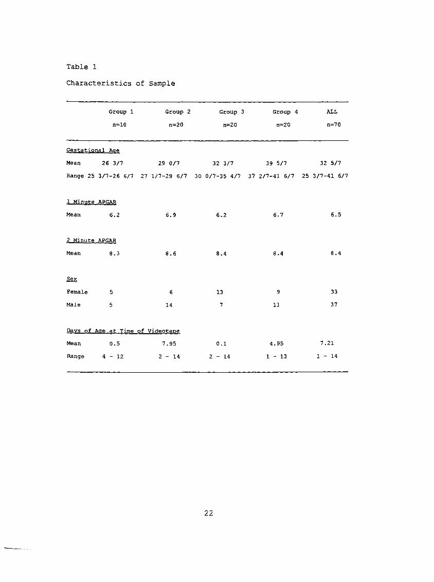
Table 1
Characteristics of Sample
Group 1 Group 2 Group 3 Group 4 ALL
n=10 n=20 n=20 n=20 n=70
Gestational Aae
Mean 26 3/7 29 0/7 32 3/7 39 5/7 32 5/7
Range 25 3/7-26 6/7 27 1/7-29 6/7 30 0/7-35 4/7 37 2/7-41 6/7 25 3/7-41 5/7
1 Minute APGAR
Mean 6.2 6.9 6.2 6.7 6.5
2 Minute APGAR
Mean 8.3 8.6 8.4 8.4 8.4
Sex
Female 5 6 13 9 33
Male 5 14 7 11 37
Davs of Aae at Time of Videotaoe
Mean 0.5 7.95 0.1 4.95 7.21
Range 4 - 1 2 2 - 1 4 2 - 1 4 1 - 1 3 1 - 1 4
22

Table 2Diagnosis of Sample
Diagnoes Group 1 Group 2 Group 3 Group 4 ALL
Prematurity, RDS 10 19 16 - 45
Prematurity 1 2 - 2
Prematurity, Sepsis - 1 - 1
Meconium Ileus - - 1 - 1
RDS - - 6 6
Sepsis - - - 9 9
Pneumothorax - - - 1 1
Hydrops - - 1 1
Gastroschisis - - 1 1
Cardiac - - - 1 1
Infant of
Diabetic Mother — - 1 1
HvDothesesHvDOthesis 1. For the first hypothesis paired t-tests and
Pearson's correlations were used to determine if the degree of
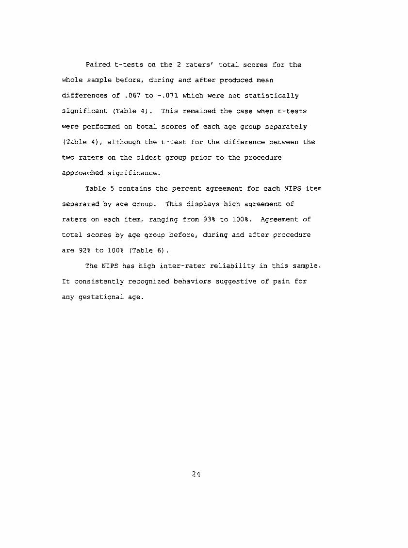
pain and the relief of pain could be assessed reliably at any gestational age using the NIPS. Inter-rater reliability was determined by comparing the total scores of two separate raters before, during and after the procedure. Pearson's correlations between the 2 raters on 70 videotapes ranged from 0.91 to 0.97 and were significant (p<.001) (Table 3). When each age group was examined separately the correlations remained consistent and significant (Table 3) with correlations ranging from .75 to .99.
23
Paired t-tests on the 2 raters' total scores for the whole sample before, during and after produced mean differences of .067 to -.071 which were not statistically significant (Table 4). This remained the case when t-tests were performed on total scores of each age group separately (Table 4), although the t-test for the difference between the two raters on the oldest group prior to the procedure approached significance.
Table 5 contains the percent agreement for each NIPS item separated by age group. This displays high agreement of raters on each item, ranging from 93% to 100%. Agreement of total scores by age group before, during and after procedure
are 92% to 100% (Table 6).The NIPS has high inter-rater reliability in this sample.
It consistently recognized behaviors suggestive of pain for any gestational age.
24
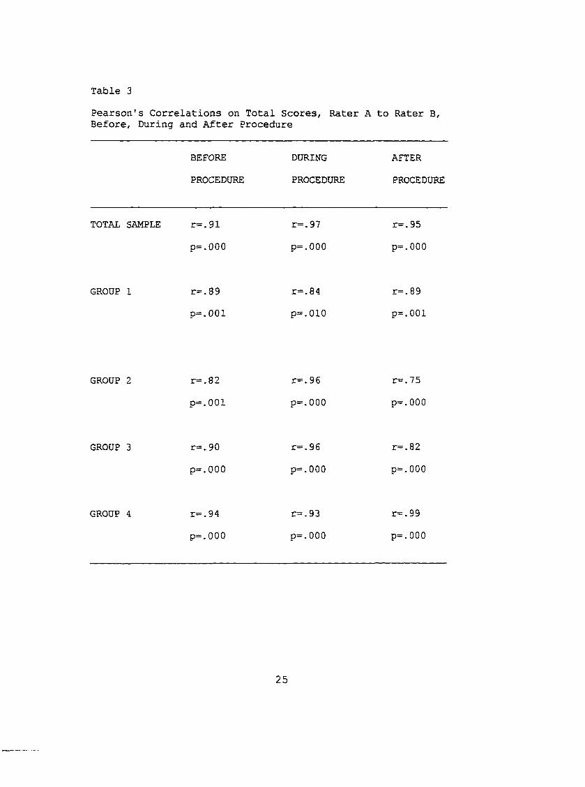
Table 3Pearson's Correlations on Total Scores, Rater A to Rater B, Before, During and After Procedure
BEFORE DURING AFTERPROCEDURE PROCEDURE PROCEDURE
TOTAL SAMPLE r=.91 r=.97 r=. 95
p=.000 p=.000 p=.000
GROUP 1 r=.89 r=. 84 r=.89p=.0 01 p=.010 p=.001
GROUP 2 r=.82 r=. 96 r=. 75
p=.001 p=.000 p=.000
GROUP 3 r=.90 r=. 96 r=.82
p=.000 p=.000 p=.000
GROUP 4 r=. 94 r=.93 r=.99p=.000 p=.000 p=.000
25
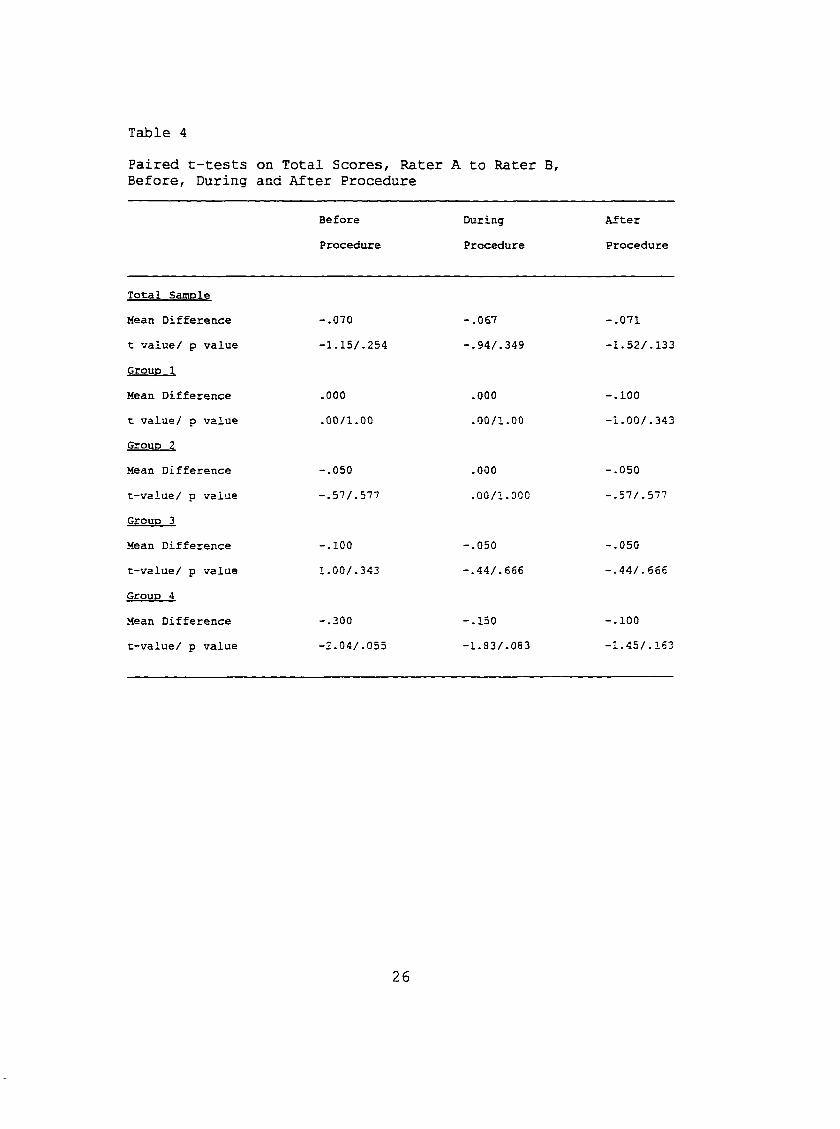
Table 4Paired t-tests Before, During
on Total Scores, Rater and After Procedure
A to Rater B,
Before During After
Procedure Procedure Procedure
Total Samole
Mean Difference -.070 -.067 -.071
t value/ p value -1.15/.254 -.94/.349 -1.52/.133
GrouD 1
Mean Difference .000 .000 -.100
t value/ p value .00/1.00 .00/1.00 -1.00/.343
Groun 2
Mean Difference -.050 .000 -.050
t-value/ p value -.57/.577 .00/1.000 -.57/.577
GrouD 3
Mean Difference -.100 -.050 -.050
t-value/ p value 1.00/.343 -.44/.666 -.44/.666
GrouD 4
Mean Difference -.300 -.150 -.100
t-value/ p value -2.04/.055 -1.83/.083 -1.45/.163
26
Table 5Percent Agreement by NIPS Scoring Items
Group 1 Group 2 Group 3 Group 4 ALL
FACE 97% 97% 98% 93% 96%CRY 93% 93% 94% 97% 96%BREATH 97% 98% 100% 97% 97%
ARMS 100% 100% 97% 100% 99%
AROUSAL 100% 100% 98% 95% 98%
TOTAL 96% 98% 99% 96% 97%
Table 6Percent Agreement of Total NIPS After Procedure
Scores Before , During and
Before During After
Procedure Procedure Procedure
Group 1 96% 92% 100%
Group 2 98% 96% 99%
Group 3 100% 96% 100%
Group 4 97% 94% 98%
TOTAL SAMPLE 98% 95% 99%
27
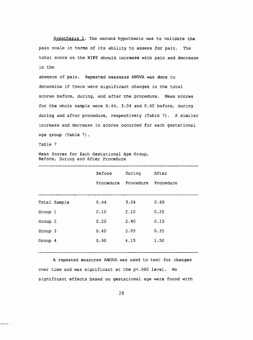
Hypothesis 2 . The second hypothesis was to validate the pain scale in terms of its ability to assess for pain. The
total score on the NIPS should increase with pain and decrease in the
absence of pain. Repeated measures ANOVA was done to determine if there were significant changes in the total scores before, during, and after the procedure. Mean scores for the whole sample were 0.44, 3.04 and 0.60 before, during during and after procedure, respectively (Table 7). A similar increase and decrease in scores occurred for each gestational age group (Table 7).Table 7
Mean Scores for Each Gestational Age Group,Before, During and After Procedure
Before During After
Procedure Procedure Procedure
Total Sample 0.44 3.04 0.60Group 1 0.10 2.10 0.20Group 2 0.20 2.40 0.15Group 3 0.40 3.05 0.35Group 4 0.90 4.15 1.50
A repeated measures ANOVA was used to test for changes over time and was significant at the p<.000 level. No significant effects based on gestational age were found with
28
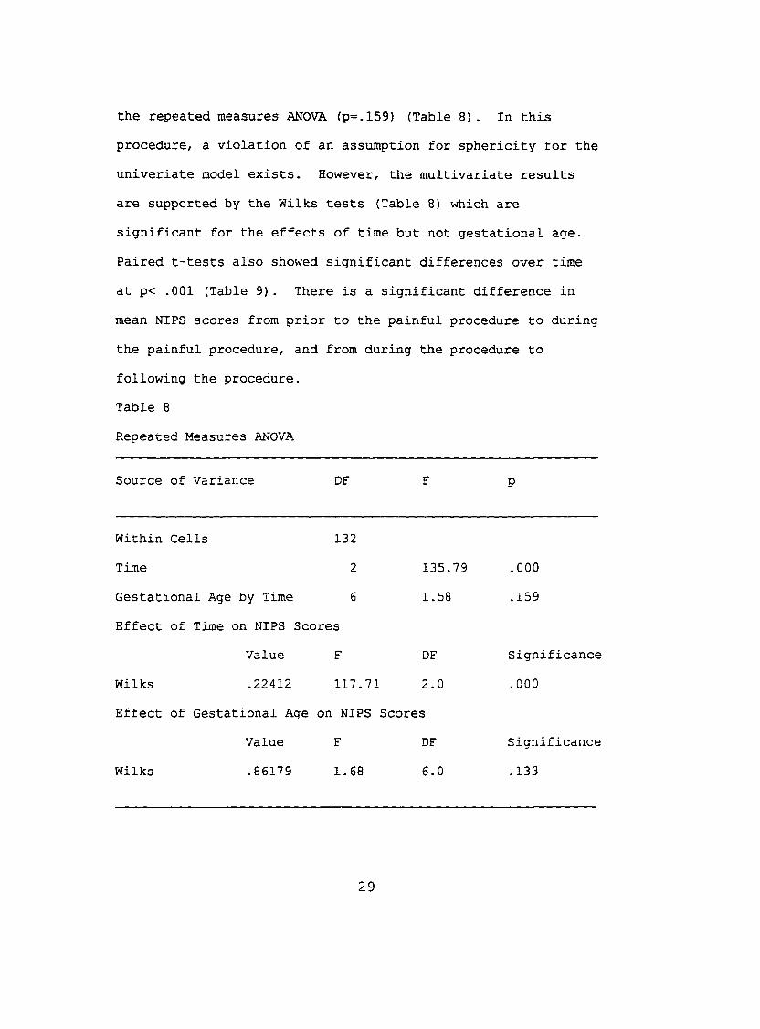
the repeated measures ANOVA (p=.159) (Table 8). In this procedure, a violation of an assumption for sphericity for the univeriate model exists. However, the multivariate results are supported by the Wilks tests (Table 8) which are significant for the effects of time but not gestational age. Paired t-tests also showed significant differences over time at p< .001 (Table 9). There is a significant difference in mean NIPS scores from prior to the painful procedure to during the painful procedure, and from during the procedure to following the procedure.Table 8Repeated Measures ANOVA
Source of Variance DF F P
Within Cells 132 Time 2 135.79 .000
Gestational Age by Time 6 1.58 .159
Effect of Time on NIPS Scores Value F DF Significance
Wilks .22412 117.71 2.0 .000Effect of Gestational Age on NIPS Scores
Value F DF Significance
Wilks .86179 1.68 6.0 .133
29
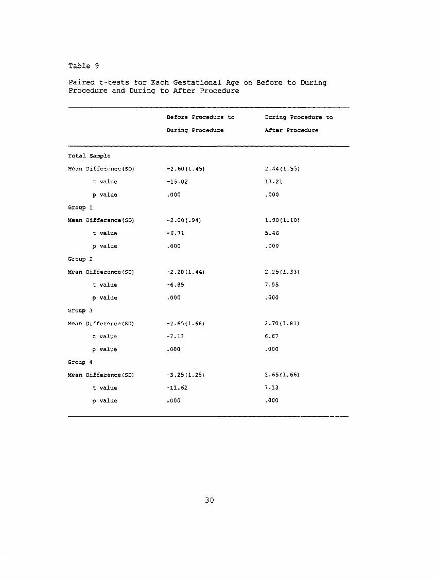
Table 9
Paired t-tests for Each Gestational Age on Before to During Procedure and During to After Procedure
Before Procedure to
During Procedure
During Procedure to
After Procedure
Total Sample
Mean Difference(SO) -2.60(1.45) 2.44(1.55)
t value -15.02 13.21
p value .000 .000
Group 1
Mean Difference(SD) -2.00(.94) 1.90(1.10)
t value -6.71 5.46
p value .000 .000
Group 2
Mean Difference(SD) -2.20(1.44) 2.25(1.33)
t value -6.85 7.55
p value .000 .000
Group 3
Mean Difference(SD) -2.65(1.66) 2.70(1.81)
t value -7.13 6.67
p value .000 .000
Group 4
Mean Difference(SD) -3.25(1.25) 2.65(1.66)
t value -11.62 7.13
p value .000 .000
30

Hypothesis 3. In this hypothesis the correlation of the changes in NIPS score to changes in heart rate, respiratory rate or oxygen saturation was examined. No significant correlation between NIPS scores and any of the physiological parameters was found. NIPS scores, heart rate, respiratory rate and oxygen saturation usually changed in the direction expected (Table 10). Table 11 shows the mean changes of the NIPS scores and the physiological parameters from the period prior to the painful procedure to during that procedure.These changes may lead to a belief that there is a correlation between the NIPS and the physiological parameters. However, Pearson's correlations between changes in the NIPS and changes in physiological measures ranged from 0.0654 to 0.3535 (p>.05) (Table 12) and were not significant.
31
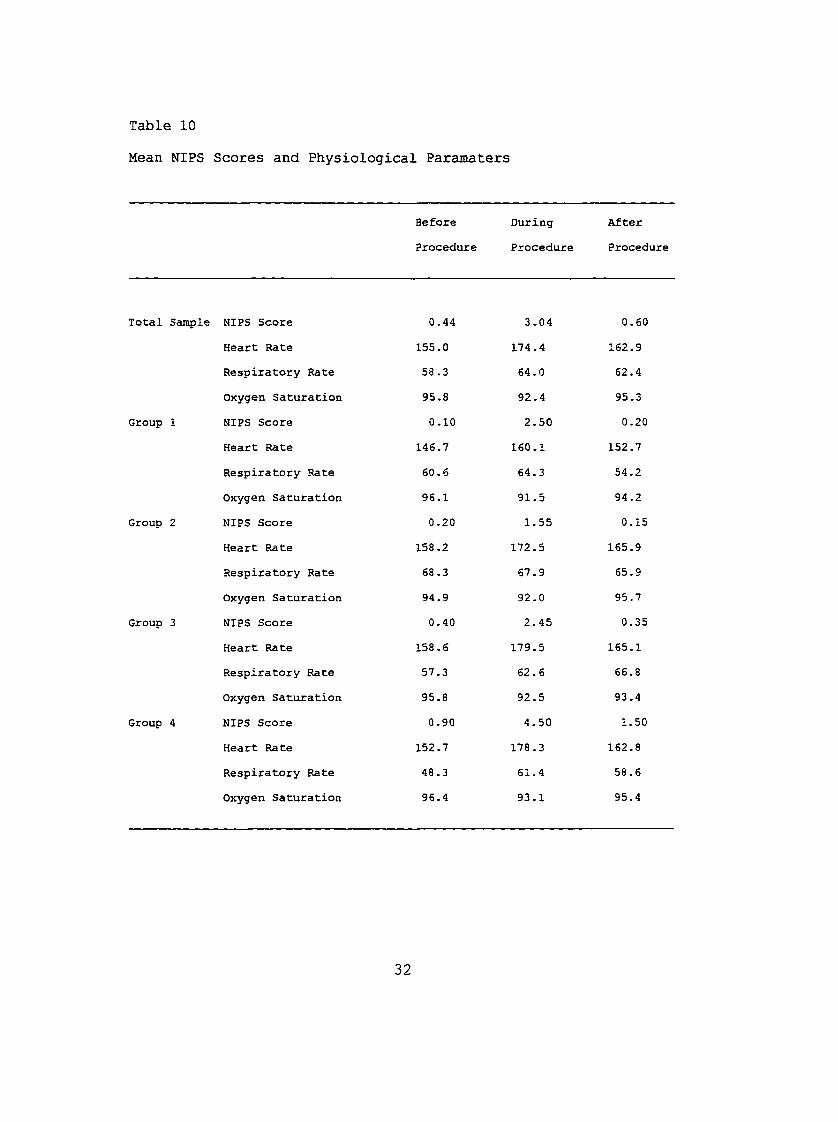
Table 10
Mean NIPS Scores and Physiological Paramaters
Before During After
Procedure Procedure Procedure
Total Sample NIPS Score 0.44 3.04 0.60
Heart Rate 155.0 174.4 162.9
Respiratory Rate 58 .3 64.0 62.4
Oxygen Saturation 95.8 92.4 95.3
Group 1 NIPS Score 0.10 2.50 0.20
Heart Rate 146.7 160.1 152.7
Respiratory Rate 60.6 64.3 54.2
Oxygen Saturation 96.1 91.5 94.2
Group 2 NIPS Score 0.20 1.55 0.15
Heart Rate 158.2 172.5 165.9
Respiratory Rate 68.3 67.9 65.9
Oxygen Saturation 94.9 92.0 95.7
Group 3 NIPS Score 0.40 2.45 0.35
Heart Rate 158.6 179.5 165.1
Respiratory Rate 57.3 62.6 66.8
Oxygen Saturation 95.8 92.5 93.4
Group 4 NIPS Score 0.90 4.50 1.50
Heart Rate 152.7 178.3 162.8
Respiratory Rate 48.3 61.4 58.6
Oxygen Saturation 96.4 93.1 95.4
32

Table 11Mean Changes of NIPS Scores and Physiological Parameters from Before to During Procedure
Mean Differences Group 1 Group 2 Group 3 Group 4 Total
Pain
Mean 2.0 2.2 2.7 3.3 2.6
Range 2.0 5.0 6.0 5.0 6.0
Heart Rate
Mean 12.0 12.9 9.3 15.8 12.5
Range 36.0 39.0 41.0 79.0 79.0
Respiratory Rate
Mean 9.2 5.9 3.0 14.6 8.0
Range 43.0 73.0 87.0 103.0 115.0
Oxygen Saturation
Mean -3.2 -2.2 -1.4 -1.2 -1.8
Range 12.0 11.0 9.0 9.0 14.0
33
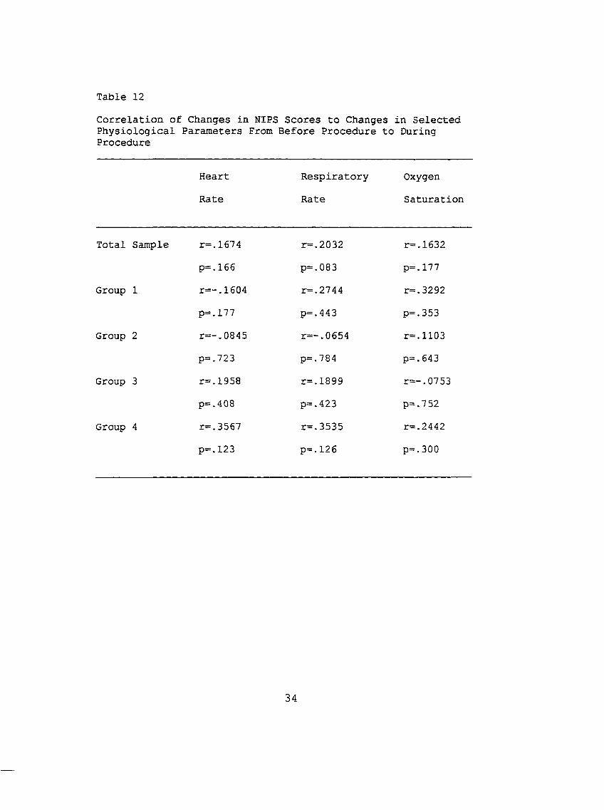
Table 12Correlation of Changes in NIPS Scores to Changes in Selected Physiological Parameters From Before Procedure to During Procedure
Heart
RateRespiratoryRate
OxygenSaturation
Total Sample r=.1674 r=.2032 r=.1632p=.165 p=.083 p=.177
Group 1 r=-.1604 r=.2744 r=.3292p=.177 p=.443 p=.353
Group 2 r=-.0845 r=-.0654 r=.1103p=.723 p=.7 8 4 p=.643
Group 3 r=.1958 r=.1899 r=-.0753p=.408 p=.423 p=.752
Group 4 r=.3567 r=.3535 r=.2442
p=.123 p=.126 p=.300
34
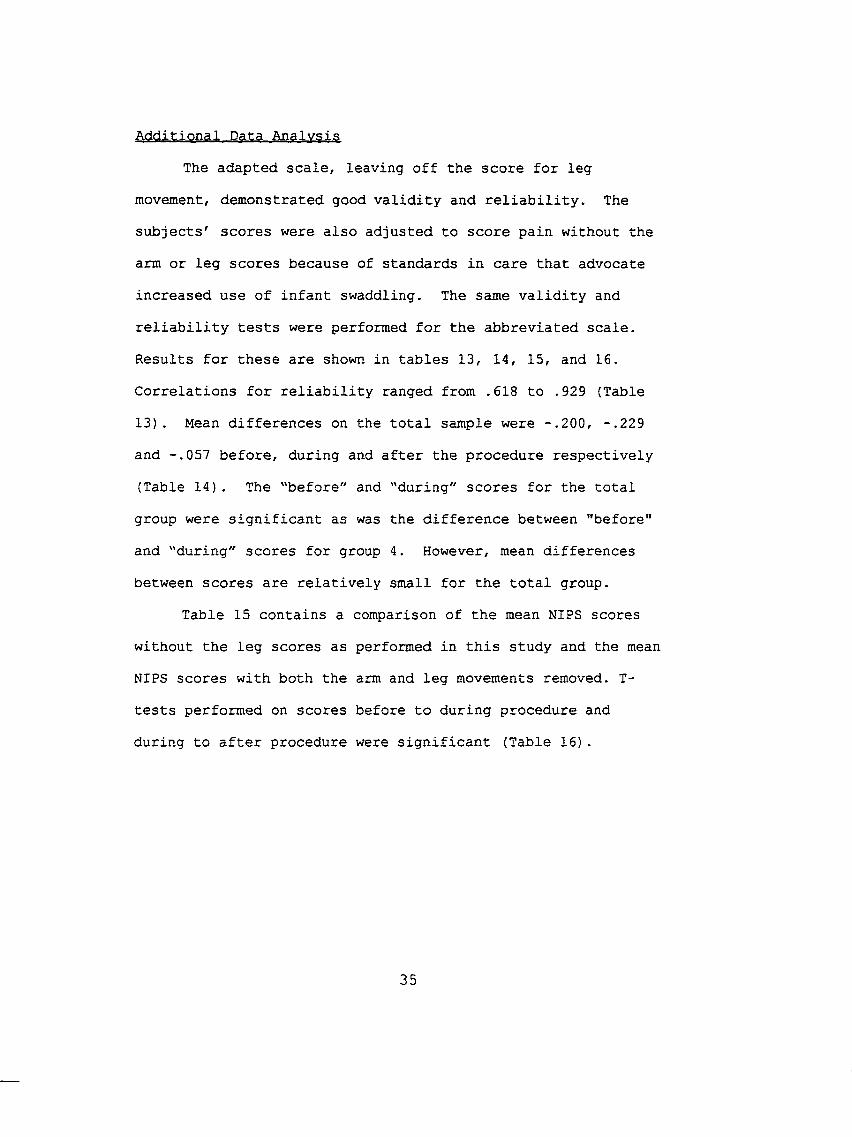
Additional Data AnalysisThe adapted scale, leaving off the score for leg
movement, demonstrated good validity and reliability. The subjects' scores were also adjusted to score pain without the arm or leg scores because of standards in care that advocate increased use of infant swaddling. The same validity and reliability tests were performed for the abbreviated scale. Results for these are shown in tables 13, 14, 15, and 16.
Correlations for reliability ranged from .618 to .929 (Table 13). Mean differences on the total sample were -.200, -.229 and -.057 before, during and after the procedure respectively (Table 14). The "before" and "during" scores for the total
group were significant as was the difference between "before" and "during" scores for group 4. However, mean differences
between scores are relatively small for the total group.Table 15 contains a comparison of the mean NIPS scores
without the leg scores as performed in this study and the mean NIPS scores with both the arm and leg movements removed. T- tests performed on scores before to during procedure and during to after procedure were significant (Table 16).
35

Table 13Pearson's Correlations on Rater A to Rater B Total Scores for NIPS Minus Arm and Leg Observations Before, During and After Procedure
BeforeProcedure
During
Procedure
AfterProcedure
Total Sample r= .837 r= .836 r= .870P= .000 P= .000 P= .000
Group 1 r= .811 r= .852 r= .885P= .000 P= .001 P= .001
Group 2 r= .618 r= .807 r= .807
P= .001 P= .000 P= .000
Group 3 r= .719 r= .673 r= .772
P= .000 P= .001 P= .000
Group 4 r= .929 r= .678 r= .856P= .000 P= .001 P= .000
36
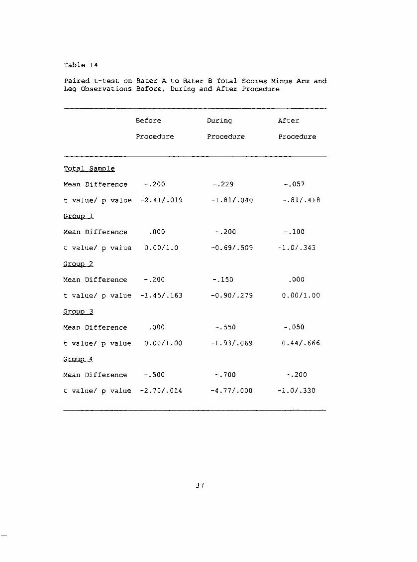
Table 14Paired t-test on Rater A to Rater B Total Scores Minus Arm and Leg Observations Before, During and After Procedure
Before During After
Procedure Procedure Procedure
Total SamoleMean Difference -.200 -.229 -.057
t value/ p value -2.41/.019 -1.81/.040 -.81/.418Group 1Mean Difference .000 -.200 -.100
t value/ p value 0.00/1.0 -0.69/.509 -1.0/.343Grouo 2
Mean Difference -.200 -.150 .000
t value/ p value -1.45/.163 -0.90/.279 0.00/1.00
Grouo 3Mean Difference .000 -.550 -.050
t value/ p value 0.00/1.00 -1.93/.069 0.44/.666Grouo 4
Mean Difference -.500 -.700 -.200
t value/ p value -2.70/.014 -4.77/.000 -1.0/.330
37

Table 15Comparison of Mean NIPS Scores Without Leg Observations to Mean NIPS Scores Without Arm and Leg Observations
Mean NIPS Score Minus
Legs
Mean NIPS
Legs
Scores Minus
and Arms
Before
Procedure
During
Procedure
After
Procedure
Before
Procedure
During After
Procedure Procedure
Total Sample 0.44 3.04 0.06 0.56 2.60 0.57
Group 1 0.10 2.50 0.20 0.10 2.00 0.30
Group 2 0.20 1.55 0.15 0.35 1.30 0.15
Group 3 0.40 2.45 0.35 0.35 2.50 0.30
Group 4 0.90 4.50 1.50 1.20 4.30 1.40
38
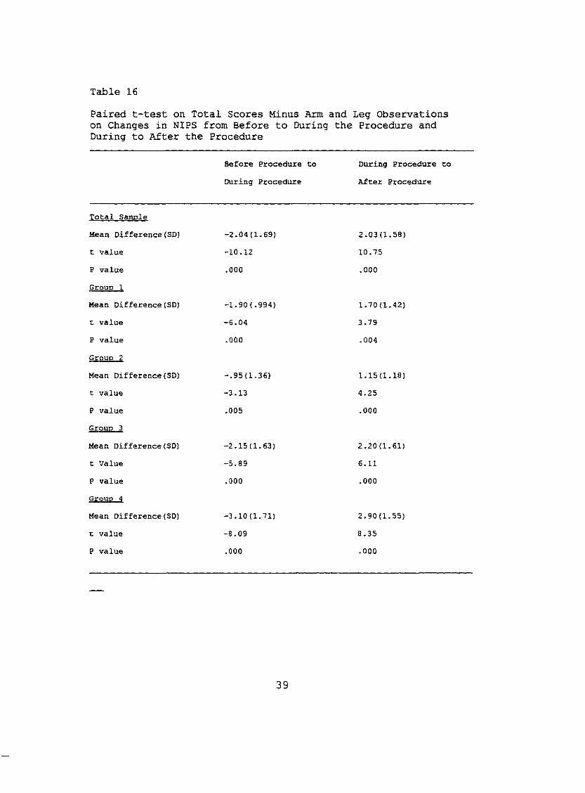
Table 16Paired t-test on Total Scores Minus Arm and Leg Observations on Changes in NIPS from Before to During the Procedure and During to After the Procedure
Before Procedure to
During Procedure
During Procedure to
After Procedure
Total Samole
Mean Difference(SD) -2.04(1.69) 2.03(1.58)
t value -10.12 10.75
P value .000 .000
Grouo 1
Mean Difference(SD) -1.90(.994) 1.70(1.42)
t value -6.04 3.79
P value .000 .004
Grouo 2
Mean Difference(SD) -.95(1.36) 1.15(1.18)
t value -3.13 4.25
P value .005 .000
Grouo 3
Mean Difference(SD) -2.15(1.63) 2.20(1.61)
t Value -5.89 6.11
P value .000 .000
Grouo 4
Mean Difference(SD) -3.10(1.71) 2.90(1.55)
t value -8.09 8.35
P value .000 .000
39
CHAPTER 5 DISCUSSION AND IMPLICATIONS
Infants in the NICU are already compromised. The staff must be able to provide an environment that will decrease stress and support infants as they mature. The addition of
pain to compromised infants often pushes infants beyond their ability to cope. Determination of pain is difficult because of the differing attitudes of caregivers in relation to pain.
It has been thoroughly established that there is a need for a pain scale for nonverbal infants. The attitudes regarding pain are varied and many, and there must be a way to provide some objectivity and standardization in describing pain and relief of pain. When we can provide a tool to assess for pain we can begin to change attitudes about pain and pain relief measures. This should bring a more consistent awareness of pain and the detrimental effects it can have on the compromised neonate. From then on the treatment of pain in the neonate should improve.
There is no "Gold Standard" for the identification of infant pain. Without it there can only be conjecture that the behaviors are indeed associated with pain. Research continues to be done on identification of facial expressions, cry and
40
physiological measures that may provide that needed "Gold Standard."Using the NIPS scale required relatively little
preparation. Rater training was simple. Rater agreement was quickly achieved. The face and cry categories had a slightly lower rate of agreement. In the face category a zero score is for a calm face, a 1 is for a grimaced face. The cry score is zero for no cry, 1 for a whimper and 2 for a vigorous cry. In an intubated infant there is no audible cry, so scoring is based on the amount of facial expression that resembles
crying. The difference between a grimace and non-audible crying was sometimes difficult to discern and was a subjective choice. This usually accounted for the difference in the percentage of agreement.
In hypothesis 1, the reliability of the instrument was tested using Pearson's correlations. Paired t-tests and percent of agreement. Pearson's correlations were significant for the total sample as well as each gestational age group.Group 1, those infants less than 27 weeks had consistent correlations with slightly less significant p values. This may be in relation to the lower number of subjects at this age group. Paired t-tests support the high reliability across all gestational age groups.
When arm and leg scores were removed from the score the results remained consistent. Correlations between raters were significant for all age groups. Paired t-tests showed mean differences ranging from .000 to -.055 (Table 14).
41
Percent agreement on arms was high for the total sample (98%) and for each gestational age group (96% to 100%). A closer look shows that 98% of the time (206 out of 210 times)
there was agreement of no movement of the arras. This leads one to question the appropriateness of this item as an
indication of pain in the neonate. Since legs were not in view or rated, the usefulness of legs is also questionable.
The scale with the absence of arms and legs drops the
possible total score from 7 to 5. This has the potential to seriously limit the sensitivity of the tool to determine pain. However, in this study the scores without these items were sensitive enough to measure behavioral changes during a painful experience.
Hypothesis 2, related to validity, was supported with significant increases in NIPS over the elapsed time of a painful procedure for all gestational ages. The removal of the leg scores should not and did not appear to have affected the validity because the scale gives a zero for restrained legs. It does, as noted above, decrease the total possible score from 7 to 6, which may decrease the sensitivity of the scale.
Further adapting the scale with the removal of the arm score as well as the leg score produced significant mean differences for the measurement points during the painful procedure on the paired t-tests. They were essentially consistent with mean differences using the total scale. This
42
is likely because of the minimal effect arm scores had on the complete scale.
Hypothesis 3 attempted to look at the effect of pain on physiological as well as behavioral measures. When looking for a "Gold Standard" in determining infant pain physiological
parameters have been considered and discarded. This study did not demonstrate any correlation between NIPS scores and the parameters of heart rate, respiratory rate and oxygen saturation. There were changes in all the mean scores from before to during to after the procedures. The direction of
the change for these parameters could not be guaranteed, and often changed independently. Examples of unexpected results would be the painful procedure during which infants hold their breath causing a decrease in heart rate or a rapid increase in respiratory effort that increases the oxygen saturation.
In conclusion, the NIPS reliably and validly identified behaviors indicative of pain since there was a significant difference in scores when behaviors were measured during a painful procedure and without nursing intervention. This leads to some confidence in the scale's ability to identify and quantify pain in neonates. However parts of this scale, legs and arms, involve observing behaviors that are visible only when the infant is totally exposed. This scale appears valid when only facial expression, cry, breathing patterns and state of arousal are used. Yet a range of scores over only 5
43
points does not provide as much sensitivity as a scale that ranges over 7 points.
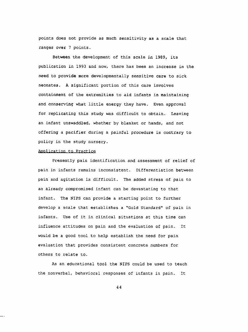
Between the development of this scale in 1989, its publication in 1993 and now, there has been an increase in the
need to provide more developmentally sensitive care to sick neonates. A significant portion of this care involves containment of the extremities to aid infants in maintaining and conserving what little energy they have. Even approval for replicating this study was difficult to obtain. Leaving
an infant unswaddled, whether by blanket or hands, and not offering a pacifier during a painful procedure is contrary to policy in the study nursery.
Application, to PracticePresently pain identification and assessment of relief of
pain in infants remains inconsistent. Differentiation between pain and agitation is difficult. The added stress of pain to an already compromised infant can be devastating to that infant. The NIPS can provide a starting point to further develop a scale that establishes a "Gold Standard" of pain in infants. Use of it in clinical situations at this time can
influence attitudes on pain and the evaluation of pain. It would be a good tool to help establish the need for pain evaluation that provides consistent concrete numbers for others to relate to.
As an educational tool the NIPS could be used to teach the nonverbal, behavioral responses of infants in pain. It
44

would provide a consistent basic set of behaviors to base the determination of pain and relief of pain on.
With better pain control comes faster recovery. This often translates into decreased length of stay and lowered cost of hospitalization.Limitations
Limitations to this study include the relatively small sample of convenience, using only two raters and leaving the leg score out of the scale. The entire sample also came from one Neonatal Intensive Care Unit.Suggestions for Further Research
There is a great need for a pain scale that can be used universally for identification and assessment of pain treatments. This scale must stay consistent with the developmentally supportive regime being introduced and embraced in the neonatal intensive care community. If developmental research calls for swaddling wherever possible,
we then must be able to evaluate pain while the infant is swaddled.
All studies to date have been done on procedures sure to elicit a pain response. Studies need to be completed that look at postoperative pain and a scale's ability to detect relief of pain.
During the completion of this study several more pain scales became available. The CRIES: Neonatal postoperative pain assessment scale (Bildner & Krechel, 1996) and the Premie
45
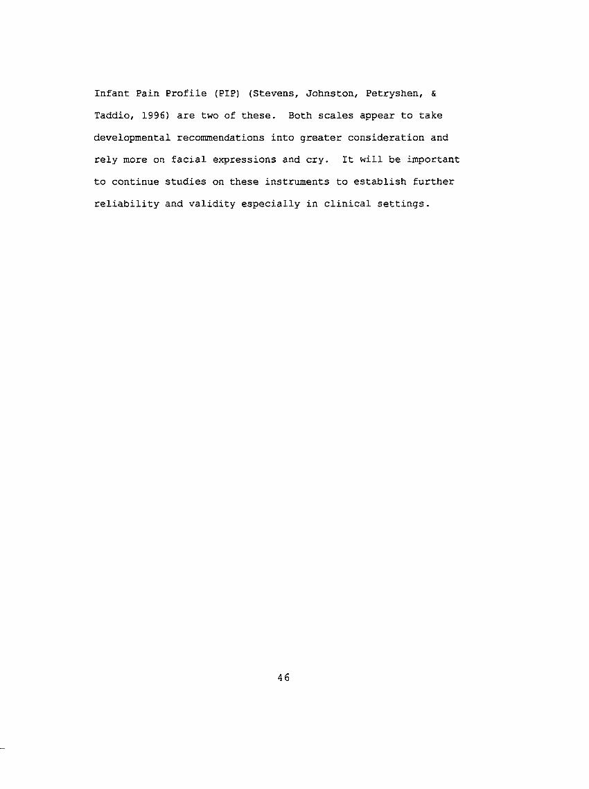
Infant Pain Profile (PIP) (Stevens, Johnston, Petryshen, & Taddio, 1996) are two of these. Both scales appear to take
developmental recommendations into greater consideration and rely more on facial expressions and cry. It will be important
to continue studies on these instruments to establish further reliability and validity especially in clinical settings.
46

APPENDICES

APPENDIX A Neonatal Infant Pain Scale
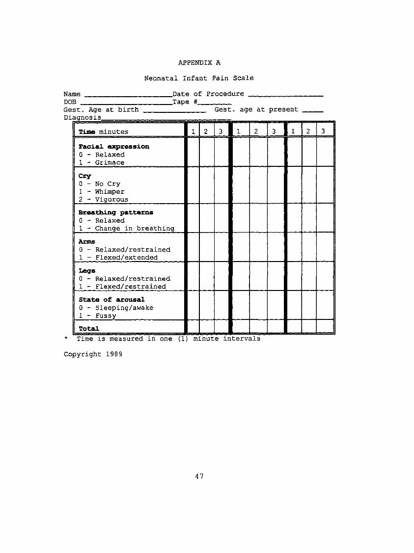
APPENDIX A Neonatal Infant Pain Scale
Name ______________DOB _______________Gest. Age at birth
_Date of Procedure .Tape #________
Gest. age at present
Time minutes 1 2 3 1 2 3 1 2 3Facial es ression.0 - Relaxed1 - GrimaceCry0 - No Cry1 - Whimper2 - VigorousBreathing patterns0 - Relaxed1 - Change in breathingArms0 - Relaxed/restrained1 - Flexed/extendedLegs0 - Relaxed/restrained1 - Flexed/restrainedState of arousal0 - Sleeping/awake1 - FussyTotal
* Time is measured in one (1) minute intervals Copyright 1989
47

APPENDIX B Operational Definitions

Facial Expression 0- Relaxed Muscles1-
Cry0 -
1 -
2-
Grimace
No cry Whimper Vigorous cry
Breathing Patterns 0- Relaxed
APPENDIX B Operational Definitions
Restful face, neutral expressionTight facial muscles, furrowed brow, chin, jaw
Quiet, not cryingMild moaning, intermittentLoud scream, shrill, continuous (Note: Silent cry may be scored if baby is intubated, as evidenced by obvious mouth, facial movement)Usual breathing pattern for this baby
1-
Arms0 -
1 -
Legs0-
1 -
Change in breathing
Relaxed/Restrained
Flexed/extended
Relaxed/restrained
Flexed/extendedState of Arousal 0- Sleeping/awake
1 - Fussy
Indrawing, irregular, faster than usual, gagging, breath holding
No muscular rigidity, occasional random movements of armsTense, straight arms, rigid and/or rapid extension/flexionNo muscular rigidity, occasional random leg movementTense, straight legs, rigid and/or rapid extension/flexionQuiet, peaceful, sleeping or alert and settledAlert, restless, and thrashing
(Copyright 1989, Children's Hospital of Eastern Ontario. Reprinted by permission.)
48

APPENDIX C
Permission to use Neonatal Infant Pain Scale
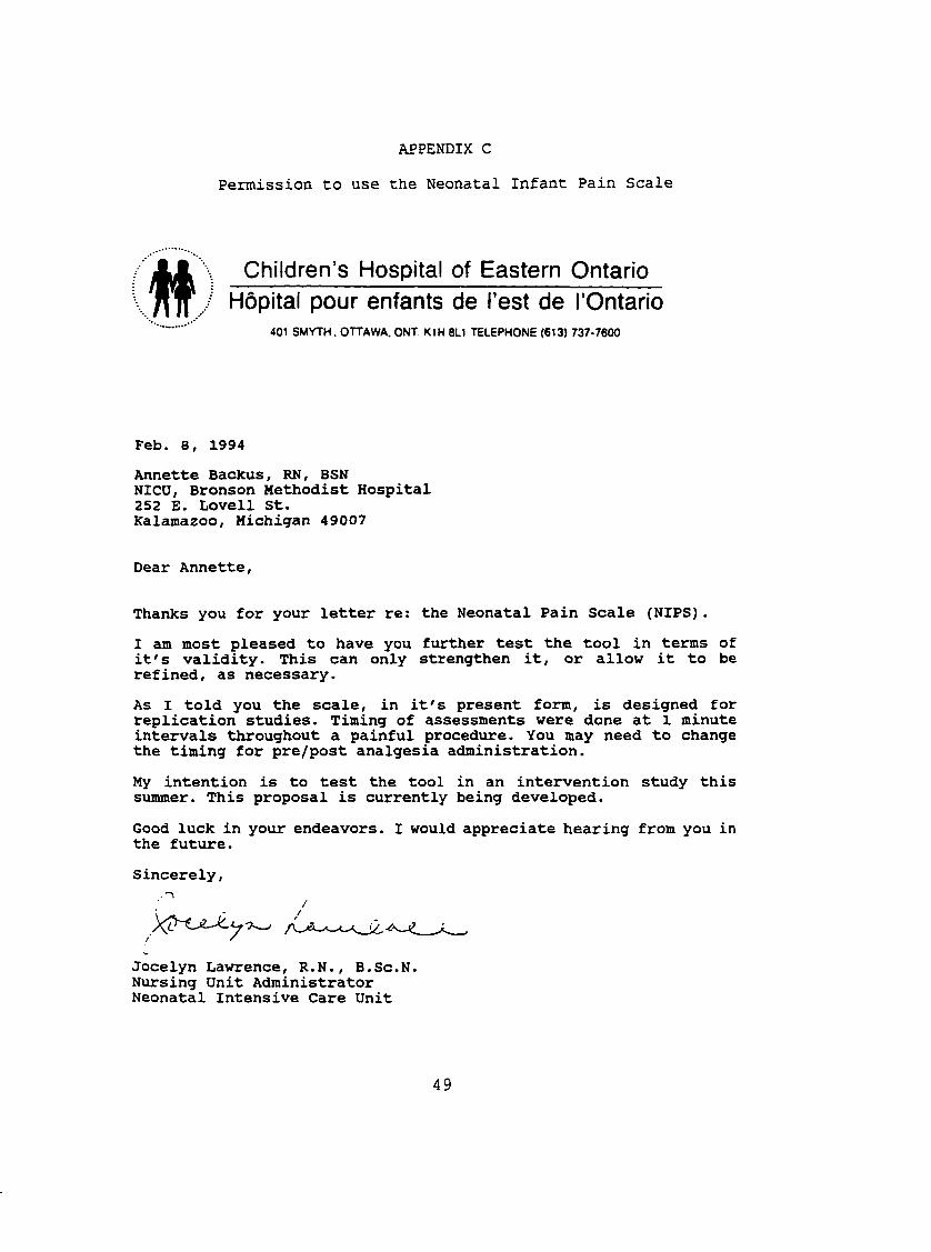
#
APPENDIX CPermission to use the Neonatal Infant Pain Scale
Children’s Hospital of Eastern Ontario Hôpital pour enfants de l’e s t de l’Ontario
401 SMYTH. OTTAWA. ONT K1H 8L1 TELEPHONE (613) 737-7600
Feb. 8, 1994Annette Backus, RN, BSNNICü, Bronson Methodist Hospital252 E. Lovell St.Kalamazoo, Michigan 49007
Dear Annette,
Thanks you for your letter re: the Neonatal Pain Scale (NIPS).I am most pleased to have you further test the tool in terms of it's validity. This can only strengthen it, or allow it to be refined, as necessary.As I told you the scale, in it's present form, is designed for replication studies. Timing of assessments were done at 1 minute intervals throughout a painful procedure. You may need to change the timing for pre/post analgesia administration.My intention is to test the tool in an intervention study this summer. This proposal is currently being developed.Good luck in your endeavors. I would appreciate hearing from you in the future.Sincerely,
n
Jocelyn Lawrence, R.N., B.Sc.N. Nursing Unit Administrator Neonatal Intensive Care Unit
49

APPENDIX D
Adapted Neonatal Infant Pain Scale
APPENDIX D Adapted Neonatal Infant Pain Scale
Name ______________DOB _______________Gest. Age at birth
.Date of Procedure
.Tape #________Gest. age at present
Time minutes 1 2 3 1 2 3 1 2 3Faciaü. es^ression0 - Relaxed1 - GrimaceCry0 - No Cry1 - Whimper2 - VigorousBreathing patterns0 - Relaxed1 - Change in breathingArms0 - Relaxed/restrained1 - Flexed/extendedState of arousal0 - Sleeping/awake1 - FussyTotal
* Time is measured in one (1) minute intervals
Time minutes 1 2 3 1 2 3 1 2 3Heart RateRespiratory RatePulse Oximetercomments :
* Time is measured in one (1) minute intervals
50

APPENDIX E Human Use Committee Aooroval
APPENDIX E
Human Use Committee Approval
BMTT9IU V alid ario n o f t h e N gnnntn l Tnfmnt P a in S g a la rAT.R«<»lnig)
At the December 6,1994 Meeting of the Expedited Review Committee Meeting, BMH954 was approved as EXEMPT 6om review.
Robert H. Hume, MLD., Chairman ' DateBronson Methodist HospitalHuman Use Committee252 East Lovell StreetKalamazoo, MI 49007(616) 341-7988
cc: ALBackus
51
APPENDIX F Permission to Conduct Research by
Grand Valley State University

APPENDIX F Permission to Conduct Research by
Grand Valley State University
.GRAND VALLEY
STATE, UNIVERSITY
1 CAMPUS DRIVE • ALLENDALE MICHIGAN 49401-9403 • 616/895-6611
January 10,1995
Annette L. Backus 5735 Roanoke St. Portage, MI 49002
Dear Annette:
The Human Research Review Committee of Grand Valley State University is charged to examine proposals with respect to protection of human subjects. The Committee has considered your proposal, " Validation o f Infant Pain Scale", and is satisfied that you have complied with the intent of the regulations published in the Federal Register 46 (16): 8386-8392, January 26,1981.
Sincerely,
Paul Huizenga, ChairHuman Research Review Committee
52
APPENDIX GInformation for Participation in Research Project
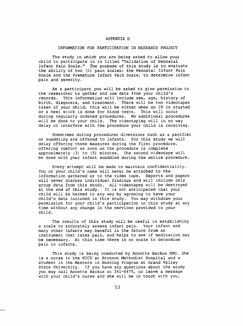
APPENDIX GINFORMATION FOR PARTICIPATION IN RESEARCH PROJECT
The study in which you are being asked to allow your child to participate in is titled "Validation of Neonatal Infant Pain Scale." The purpose of this study is to evaluate the ability of two (2) pain scales; the Neonatal Infant Pain Scale and the Premature Infant Pain Scale, to determine infant pain and severity.
As a participant you will be asked to give permission to the researcher to gather and use data from your child's records. This information will include sex, age, history of birth, diagnosis, and treatment. There will be two videotapes taken of your child, this will be either when an IV is started or a heel stick is done for blood tests. This will occur during regularly ordered procedures. No additional procedures will be done to your child. The videotaping will in no way delay or interfere with the procedure your child is receiving.
Sometimes during procedures diversions such as a pacifier or swaddling are offered to infants. For this study we will delay offering these measures during the first procedure, offering comfort as soon as the procedure is complete approximately (3) to (5) minutes. The second videotape will be done with your infant swaddled during the entire procedure.
Every attempt will be made to maintain confidentiality. You or your child's name will never be attached to the information gathered or to the video tape. Reports and papers will never discuss individual findings and will include only group data from this study. All videotapes will be destroyed at the end of this study. It is not anticipated that your child will be harmed in any way by agreeing to have your child's data included in this study. You may withdraw your permission for your child's participation in this study at any time without any change in the services provided to your child.
The results of this study will be useful in establishing a scale to accurately assess infant pain. Your infant and many other infants may benefit in the future from an instrument that rates pain, and helps to see if medication may be necessary. At this time there is no scale to determine pain in infants.
This study is being conducted by Annette Backus RNC. She is a nurse in the NICU at Bronson Methodist Hospital and a student in the Masters in Nursing Program at Grand Valley State University. If you have any questions about the study you may call Annette Backus at 341-6475, or leave a message with your child's nurse and she will be in touch with you.
53

APPENDIX H Informed Consent for Participatant
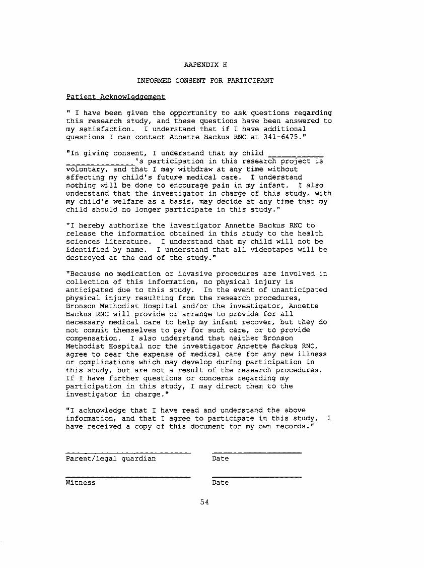
AAPENDIX H INFORMED CONSENT FOR PARTICIPANT
Patient Acknowledgement" I have been given the opportunity to ask questions regarding this research study, and these questions have been answered to my satisfaction. I understand that if I have additional questions I can contact Annette Backus RNC at 341-6475.""In giving consent, I understand that my child _____________
's participation in this research project isvoluntary, and that I may withdraw at any time without affecting my child's future medical care. I understand nothing will be done to encourage pain in my infant. I also understand that the investigator in charge of this study, with my child's welfare as a basis, may decide at any time that my child should no longer participate in this study.""I hereby authorize the investigator Annette Backus RNC to release the information obtained in this study to the health sciences literature. I understand that my child will not be identified by name. I understand that all videotapes will be destroyed at the end of the study.""Because no medication or invasive procedures are involved in collection of this information, no physical injury is anticipated due to this study. In the event of unanticipated physical injury resulting from the research procedures,Bronson Methodist Hospital and/or the investigator, Annette Backus RNC will provide or arrange to provide for all necessary medical care to help my infant recover, but they do not commit themselves to pay for such care, or to provide compensation. I also understand that neither Bronson Methodist Hospital nor the investigator Annette Backus RNC, agree to bear the expense of medical care for any new illness or complications which may develop during participation in this study, but are not a result of the research procedures.If I have further questions or concerns regarding my participation in this study, I may direct them to the investigator in charge.""I acknowledge that I have read and understand the above information, and that I agree to participate in this study. I have received a copy of this document for my own records."
Parent/legal guardian Date
Witness Date
54

APPENDIX I
Subject Characteristics
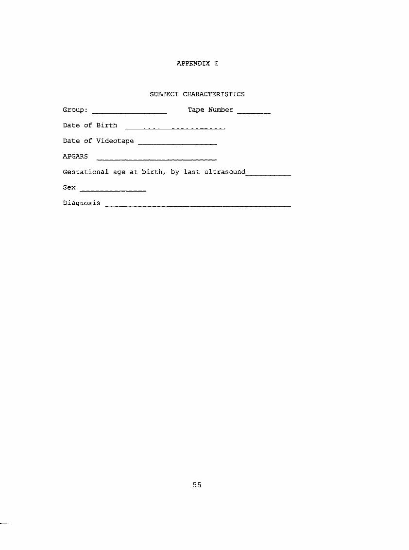
APPENDIX I
SUBJECT CHARACTERISTICSGroup: ___________________ Tape Number___
Date of Birth _________________________
Date of Videotape ____________________APGARSGestational age at birth, by last ultrasound_ Sex _________________Diagnosis ____________________________________
55

LIST OF REFERENCES
List of ReferencesAls, H. (1982). Toward a synactive theory of development:
Promise for the assessment and support of infant
individuality. Infant Mental Health Journal. 2/ 229-243.Als, H. (1986). A synactive model of neonatal behavioral
organization: Framework for the assessment of
neurobehavioral development in the premature infant and for the support of infants and parents in the neonatal intensive care environment. Physical & Occupational Theraov in Pediatrics. ^(3/4), 3-55.
Anand, K.J.S., Phil, D., & Hickey, P.R. (1987). Pain and its' effects in the human neonate and fetus. New England Journal of Medicine. 317. 1321-1329.
Attia, J., Amiel-Tison, C., Mayer, M., & Shnider, S. (1987). Measurement of postoperative pain and narcotic administration in infants using a new clinical scoring system. Anesthesiology. 67. A352.
Bildner, J. & Krechel, S. (1996). Increasing staff nurseawareness of postoperative pain management in the NICU. Neonatal Network. 15.(1), 11-16.
Brown, L. (1987). Physiologic responses to cutaneous pain in neonates. Neonatal Network. . .(3), 18-22.
Budreau, G. (1991). Clinical indicators of infant irritability. Neonatal Network. i,(5) , 23-29.
56
Butler, N. (1988). How to raise professional awareness of the need for adequate pain relief for infants. Birth. 15(1). 38-41.
Clancy, G., Anand, K., & Lally, P. (1992). Neonatal pain management. Critical Care Nursing Clinics of North America.4. 527-535.
Craig, K. & Prkachin, K. (1983). Nonverbal measures of pain.In R. Melzach (Ed.) Pain Measurement and Assessment.(pp.173-179). New York: Raven Press.
Dale, J.C. (1986). A multidimensional study of infants'responses to painful stimuli. Pediatric Nursing.12(1). 27-31.
Franck, L.S. (1986). A new method to quantitatively describe pain behaviors in infants. Nursing Research. 35. 28-
31.Franck, L.S. (1987) . A national survey of the assessment and
treatment of pain and agitation in the neonatal intensive care unit. Journal of Obstetric. Gvnocoloaical and Neonatal Nursing, 16, 387-393.
Gilles, F., Shankle, W., & Cooling, E. (1983). Myelinatedtracts: Growth patterns. In F. Gilles, A. Leviton & E . Cooling (Eds.) The Developing Human Brain: Growth and Epidemiologic Neurooathologv (pp. 117-183). Boston: John
Wright.
57
Gleiss, M.,& Stuttgen, G. (1970). Morphologic and functional development of the skin. In U. Stave (Ed.) Phvsioloav
of the Perinatal Period (Vol. 2, pp 889-906). New York: Appleton-Century-Crofts.
Lawrence, J., Alcock, D., McGrath, P., Kay, J., MacMurray, S., & Dulberg, C. (1993). The development of a tool to assess neonatal pain. Neonatal Network. 12(6), 59-66.
Levine, M.E. (1967). The four conservation principles of
nursing. Nursing Forum. ^(1), 45-59.Levine, M.E. (1969). The pursuit of wholeness. American
Journal of Nursing, 69. 93-98.Levine, M.E. (1971). Holistic Nursing. Nursing Clinics of
North America. 253-264.Maloni, J., Stegman, C ., Taylor, P., & Brownell, C. (1986).
Validation of infant behavior identified by neonatal nurses. Nursing Research. 35. 133-138.
Marshall, R. (1989). Neonatal pain associated with caregiving procedures. Pediatric Clinics of North America. 36. 885- 901.
Owens, M.E. (1981). Pain in infancy: Conceptual and methodological issues. Pain. 20. 213-230.
Owens, M.E., & Todt, E.H. (1984). Pain in infancy: Neonatal reaction to heel lance. Pain. 20. 77-86.
Page, G., & Halvorson, M. (1991). Pediatric nurses : The assessment and control of pain in preverbal infants.
Journal of Pediatric Nursing. ^(2),99-106.
58
Rich, E.C., Marshall, R.E., & Volpe, J. (1974). The normalneonates' response to pinprick. Dev Med Child Neurol. 16, 432-434.
Ross, D., & Ross, S. (1988). Assessment of pediatric pain: An overview. Issues In Comprehensive Pediatric Nursing,11, 73-91.
Sagraves, R.(1992). Pediatric pain management (Part I).Journal of Pediatric Health Care. 6, 87-92.
Sagraves, R. (1992). Pediatric pain management (Part II). Journal of Pediatric Health Care. S, 214-218.
Stevens, B ., Johnston,C., Petryshen, P., & Taddlo, A. (1996). The development and validation of a measure to assess pain In premature Infants. The Journal of Pain and Svmprom Management. 12(1).
U.S. Department of Health and Human Services. (1992). Acute pain management In Infants, children, and adolescents: Operative and medical procedures. Quick reference guide
for clinicians (AHCPR Publication No. 92-0020) .Rockville, MD.
Williamson, P.S., & Williamson, M. (1983). Physiologic stress reduction by a local anesthetic during newborn circumcision. Pediatrics. 71. 36-40.
59