Embed Size (px)
Citation preview

ARC Journal of Hepatology and Gastroenterology
Volume 4, Issue 1, 2019, PP 11-23
www.arcjournals.org
ARC Journal of Hepatology and Gastroenterology Page | 11
Validity of Different Fibrosis Scores for Assessment of Hepatic
Fibrosis in Chronic Hepatitis B Patients
Ghada M Galal1, Amira Maher
1, Nagwa Sayed Ahmed
2 , El-Zahraa M. Meghezel
1*
1Department of Tropical Medicine and Gastroenterology
2Medical Biochemistry, Sohag Faculty of Medicine, Sohag University
1. INTRODUCTION
Hepatitis B virus (HBV) infection is one of the
world’s most significant public health problems
[1]. Globally, about 260 million people are
estimated to be chronically infected with HBV.
Liver fibrogenes is; a consequence of chronic
hepatitis B (CHB) infection, is a dynamic,
process responsible for driving the progressive
excess accumulation of extracellular matrix
(ECM) components(i.e., liver fibrosis) and
sustained by the activation of hepatic stellate
cells (HSCs) [2,3]. Liver biopsy has
traditionally been considered the reference
method for evaluation of hepatic fibrosis in
patients with chronic liver disease.
However, it is costly and invasive procedure
that requires physicians and pathologists to be
sufficiently trained in order to obtain adequate
and representative results. These limitations
have led to the development of non-invasive
methods for assessment of liver fibrosis. These
methods rely on two different approaches: a
‘‘biological’’ approach based on the
quantification of biomarkers in serum samples
or a ‘‘physical’’ approach based on the
measurement of liver stiffness [4].
Abstract
Background and aim: Severity of liver fibrosis could be discriminated through biological and physical
method. This study was conducted to evaluate the diagnostic value of serum markers of fibrosis and ARFI
elastography for CHB related liver fibrosis compared to liver biopsy.
Methods: A total of 92 CHB adult Egyptian patients were included in this study, all were subjected to liver
biopsy with staging of fibrosis using METAVIR scoring system.
Liver stiffness measured through acoustic radiation force impulse (ARFI) and non-invasive fibrosis scores
including AST/ALT ratio (AAR), AST/platelet ratio (APRI), fibrosis index based on four factors (FIB-4), fibro-
Q test, King's score, BARD score, cirrhosis discriminate score (CDS), S index and APAG score were
compared to biopsy result.
Results: Among the studied scores, APRI score, FIB-4, Fibro-Q test and King's score were significant
predictors for advanced fibrosis in CHB patients, with King's score showing the highest AUROCs for
predicting advanced fibrosis. The optimal King's score cut off value for predicting advanced fibrosis was
>9.62 with 58.33% sensitivity and 98.75 specificity. ARFI elastography was not effective predictor for
advanced fibrosis in CHB patients. Multivariate analysis showed that diabetes mellitus (D.M) was the only
predictor for advanced fibrosis in CHB patients.
Keywords: AAR, APRI, ARFI, chronic hepatitis B, FIB-4, King's score, non-invasive scores.
Abbreviations
AAR=AST/ALT ratio,ALT= alanine transaminase, Anti HBe= hepatitis B e antibody, APAG=age, platelets,
albumin and gamma glutamyltrnsferase, APRI= AST/platelet ratio, ARFI= acoustic radiation force impulse
imaging, AST= aspartate transaminase, BMI= body mass index, CBC= complete blood count,
CDS=Cirrhosis Discriminate Score, CHB=chronic hepatitis B, CHC= chronic hepatitis C, D.M=diabetes
mellitus,DNA= deoxyribonucleic acid, ECM= extracellular matrix, FIB-4= fibrosis index based on four
factors, GGT= gammaglutamyltrasferase, HB=hemoglobin, HBeAg= hepatitis B e antigen, HBsAg= hepatitis
B surface antigen, HBV= Hepatitis B virus, HCV= hepatitis C virus infection, HSCs= hepatic stellate cells,
INR= international randomized ratio,IQR= interquartile range, mL= milliliters, MRE= magnetic resonance
elastography, NAFLD= non-alcoholic fatty liver disease, PCR= polymerase chain reaction, PT=prothrombin
time,ROC= Receiver operating characteristic, ROI= region of interest,SSI= supersonic shear wave imaging,
TE= transient elastography.
*Corresponding Author: El-Zahraa Mohammed Meghezel, Department of Tropical Medicine and
Gastroenterology, Email: [email protected]

Validity of Different Fibrosis Scores for Assessment of Hepatic Fibrosis in Chronic Hepatitis B Patients
ARC Journal of Hepatology and Gastroenterology Page | 12
Serum markers are grouped into two main
categories: direct and indirect biomarkers.
Direct Markers are either linked to matrix
deposition, or cytokines and chemokines linked
to liver fibrosis [5].Indirect markers are
numerous indices based on routine biochemical
blood tests that reflect liver injury. Examples are
AST/ALT ratio (AAR), AST/platelet ratio
(APRI), fibrosis index based on four factors
(FIB-4), King's score [5], S index, age, platelets,
albumin and gamma glutamyltrnsferase (APAG)
score [6], and BARD score[7].
Liver stiffness, which may change significantly
as fibrosis develops, can be measured by a
noninvasive imaging-based technique called
elastography. Research over the past two
decades has led to significant developments in
elastographic methods, including magnetic
resonance elastography (MRE), transient
elastography (TE), and acoustic radiation force
impulse imaging (ARFI), shear wave elasticity
(SWE) and supersonic shear wave imaging
(SSI)[8].
The aim of the current study was to evaluate the
diagnostic value of serum markers of fibrosis
and ARFI elastography for CHB related liver
fibrosis compared to liver biopsy.
2. PATIENTS AND METHODS
2.1. Selection of Patients
This cross sectional study was performed on a
total of 92 patients recruited from attendants to
Tropical Medicine and Gastroenterology out-
patient clinic, Sohag University Hospital during
the period from December 2016 to June 2018.
ARFIelastographywas performed in Sohag
Cardiology and Gastroenterology Centre.
Inclusion criteria were patients with symptom-
matic or asymptomatic CHB infection based on
positive hepatitis B surface antigen (HBsAg) for
more than 6 months. Exclusion criteria were:
Patients with serological evidence of hepatitis C
virus infection (HCV) or human immune-
odeficiency virus infection, HBV patients who
had received or currently under anti-viral
therapy, alcohol consumption, decompensated
liver disease, Patients with hepatocellular
carcinoma, Patients known to have other chronic
liver disease (e.g. autoimmune hepatitis,
primary biliary cirrhosis, Wilson’s disease,
haemochromatosis, non-alcoholic fatty liver
disease (NAFLD), or drug induced chronic
hepatitis), and patients with a contraindication to
liver biopsy such as: uncooperative
patient,Prothrombin time >4 seconds more than
control, INR greater than 1.6[9], platelets count
<100.000/mm[10].
According to histological staging of fibrosis,
patients were grouped into 2 groups: Group1:
included 80 patients with mild to moderate
fibrosis (F0, F1 and F2 METAVIR score),
Group 2: included 12 patients with advanced
fibrosis (F3 and F4 METAVIR score).
2.2. Clinical Assessment
Baseline patient characteristics including age,
gender, body mass index (BMI), Symptoms
suggestive of chronic liver disease or liver cell
failure and presence of diabetes mellitus (D.M)
or systemic hypertension were collected from all
participants.
2.3. Laboratory Investigations
Peripheral venous blood sample of 5 milliliters
(mL) was collected from each participant, and
HBsAg, hepatitis B e antigen (HBeAg),
hepatitis B e antibody (Anti HBe), polymerase
chain reaction (PCR) for HBV deoxyribonucleic
acid (DNA), gammaglutamyltrasferase (GGT),
HCV antibody, Complete blood count (CBC),
alanine transaminase (ALT), aspartate
transaminase (AST), total bilirubin, prothrombin
time (PT) and international randomized ratio
(INR) were measured.
2.4. Calculation Of Fibrosis Scores
The score were calculated as described in
the original articles.
AAR: AAR = AST (IU/L) / ALT (IU/L) [11]
.
APRI: APRI = (AST level (IU/L) / AST
ULN (IU/L)) × 100 / PLT (109/L)
[12].
FIB-4 score: FIB-4 = (Age (years) × AST
(IU/L)) / (platelets (109/L)× √ALT)
[13].
The Fibro-Q test: FibroQ = 10 × (age
(years) × AST (IU/L) × PT-INR) / (ALT
(IU/L) ×platelets (109/L))
[14].
King’s score: king' score = (Age (years) ×
AST (IU/L) × INR) / platelets (109/L)
[15].
BARD score:The BARD score is composed
of three variables and the possible score
ranges from 0 to 4 points, that is AST/ALT
ratio at least 0.8 (2 points); a BMI at least
28 kg/m2 (1 point); and presence of type 2
DM (1 point)[16]
.
Cirrhosis Discriminate Score (CDS): CDS
is composed of three variables: AST/ALT,
PT-INR and platelet count. Different points

Validity of Different Fibrosis Scores for Assessment of Hepatic Fibrosis in Chronic Hepatitis B Patients
ARC Journal of Hepatology and Gastroenterology Page | 13
are given to ingredients of this index and the
possible score ranges from 0 to 11 points [17]
.
S index: S index = (1000×GGT (IU/L) /
(platelets (109/L) ×albumin² (g/dl))
[18].
APAG: APAG = eᴾ/ (1+eᴾ)
ᴾ = -9.340 + 0.997×ln(age) + 6.355×ln(PT) -
3.372×ln(albumin(g/L)) + 0.677 × ln (GGT
(IU/L))[19]
.
2.5. Liver Stiffness Measurement using ARFI
Elastograpy
Liver stiffness was measured by ARFI
elastography using a Siemens ACUSON S2000
Ultrasound System (Siemens AG) with a 6C1
HD transducer, by using Virtual Touch Tissue
Quantification application in all patients. The
measurement was performed in the right liver
lobe over segments 8[20]. Under fasting
conditions for at least 8 hour [21], 10 valid
ARFI measurements were performed for each
patient by the intercostal approach. The patient
was placed in supine position with the right arm
in maximum abduction. Minimal scanning
pressure was applied by the operator; the patient
was asked to stop normal breathing for a
moment to minimize breathing motion [22].
ARFI measurements were obtained in a selected
region of interest(ROI; a box with dimension of
1 cm × 0.5 cm),at a depth of 1 to 2 cm from the
liver capsule, avoiding large vessels and bile
ducts [23].Reliable measurements were defined
as a median of 10 valid measurements with an
interquartile range (IQR) to median value ratio
less than 30% and the result is expressed in
m/s[24](Figure 1, Figure 2).
Figure1. Acoustic radiation force impulse of the liver performed with the Siemens system through intercostal
access. The measurement is given in meters per seconds [25]
.
Figure2. Output report of ARFI examination wit means, median and standard deviation.
2.6. Ultrasound Guided Percutaneous Liver
Biopsy
Ninety two liver biopsy specimens were
included in the study.
The obtained tissue cores (15 mm in length
each) were fixed in 10 % formaldehyde,
processed as usual, embedded in paraffin and
sections of 4 µm thickness were prepared and
stained with hematoxylin and eosin to assess
both the grade and the stage of chronic viral
hepatitis using METAVIR staging systems [26].
Liver biopsies were examined by a single
pathologist.
2.7. Data Analysis
Data were analyzed using IBM SPSS Statistics
for Windows version 20 and Medcalc version
15.8.0. Quantitative data were expressed as
means ± standard deviation for normally

Validity of Different Fibrosis Scores for Assessment of Hepatic Fibrosis in Chronic Hepatitis B Patients
ARC Journal of Hepatology and Gastroenterology Page | 14
distributed data, median and IQR for skewed
data. Qualitative data was expressed as number
and percentage. Quantitative data was tested for
normality by Shapiro–Wilk test. Mann–Whitney
U test and Spearman's correlation were used for
data which wasn't normally distributed. Chi-
square (χ2) test and Fisher's Exact Test were
used for comparison of qualitative variables as
appropriate. Univariate and multivariate analysis
were used to evaluate the predictors of advanced
liver fibrosis. Receiver operating characteristic
(ROC) curve was constructed for fibrosis
markers, for optimum cut off point in predicting
advanced fibrosis, and the area under the ROC
curve value with 95% CI was calculated.
Optimal cut-off value was determined;
sensitivity, specificity, positive predictive value,
negative predictive value were calculated. A 5%
level was chosen as a level of significance in all
statistical tests used in the study.
2.8. Ethical Considerations
The study protocol was approved by the ethical
committee of Sohag Faculty of Medicine, Sohag
University, Egypt. A written informed consent
was obtained from each patient before
enrollment in this study.
3. RESULTS
From December 2016 to June 2018, 92 patients
(73 males and 19 females) were included in the
study. The mean age of the participants was
36.41 ± 11.03. Based on METAVIR score,
patients were categorized into 2 groups: patients
with advanced fibrosis (12 patients (13%), mean
age 44.83 ± 11.74, 10 males (83.3%)), and
patients with mild to moderate fibrosis (80
patients (87%), mean age 35.15 ± 10.42, 63
males (78.8%)). Liver fibrosis stage and
inflammation grade by METAVIR score in
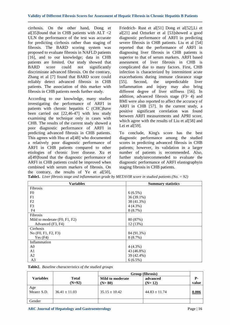
studied patients are summarized in Table 1.
Baseline characteristics of the studied groups
are shown in Table 2 which shows statistically
significant differences among: age (P = 0.006),
D.M (P = 0.045)and AST (P = 0.042).
Table 3 summarizes non-invasive markers of
fibrosis in the studied groups.Mean shear wave
velocity tends to be higher among patients with
advanced fibrosis (mean velocity 1.64 m/sec ±
0.56) compared to patients with mild to
moderate fibrosis (mean velocity 1.39 m/sec ±
0.33) but without statistically significant
difference between both groups. APRI score
showed statistically significant difference
between patients with mild to moderate fibrosis
and patients with advanced fibrosis (P = 0.23)
which can be also seen in Figure 3,Table 3. As
seen in Figure 4, Table 3, FIB-4 model can
significantly distinguish between patients with
mild to moderate fibrosis and patients with
advanced fibrosis (P = 0.02). Figure 5, Table 3
demonstrates the statistically significant
difference of fibro-Q test between the studied
groups (P = 0.024). King's score also showed
statistically significant difference between both
groups (P = 0.013)and this was demonstrated in
Figure 6, Table 3.
Table 4shows the diagnostic ability of the
studied markers as presented by AUROCs for
advanced fibrosis. King's score had the highest
AUROCs (0.723) in predicting advanced
fibrosis. It had 98.75% specificity and 58.33%
sensitivity at a cutoff value >9.62. APRI score
had the next AUROCs (0.707) in predicting
advanced fibrosis. It had 87.5% specificity and
58.33% sensitivity at a cutoff value >0.43. FIB-
4 had an AUROC of 0.706 in predicting
advanced fibrosis with 98.75% specificity and
50% sensitivity at a cutoff value >1.83. Fibro-Q
test had an AUROC of 0.703 in predicting
advanced fibrosis with 92.5% specificity and
50% sensitivity at a cutoff value >3.43.
Table 5 summarizes the correlation between
shear wave velocity and serum markers of
fibrosis among the studied patients. There was a
statistically significant positive correlation
between shear wave velocity and APRI score (r
= 0.226) (P = 0.031)(Figure 7). There was
positive correlation between shear wave velocity
and GGT level, AAR, FIB-4 model, fibro Q test,
King's score, CDS and S index but without
statistical significance.
Multivariate logistic regression analysis for
predictors of advanced liver fibrosis is
summarized in Table 6. Presence of D.M was
the only independent predictor for advanced
fibrosis with an odds ratio (OR) of 23.51 (P =
0.028).
4. DISCUSSION
Previous studies have shown that the severity of
liver fibrosis could be discriminated through
measurement of shear wave velocity by ARFI
image [27], and also blood-based indices such
as APRI[28] and Fib-4 score [29-30].However,
the performance of these indices in guiding
indications for antiviral therapy in CHB has not
been elucidated [20].

Validity of Different Fibrosis Scores for Assessment of Hepatic Fibrosis in Chronic Hepatitis B Patients
ARC Journal of Hepatology and Gastroenterology Page | 15
Our study evaluated the validity of ten
noninvasive serum markers and ARFI
elastographyto predict advanced fibrosis
compared to liver biopsy in Egyptian patients
with HBV related liver disease. Our results
showed that APRI score, FIB-4, Fibro-Q test
and King's score were significant predictors of
advanced fibrosis. AUROC analysis showed
that King's score was superior to APRI, FIB-4
and Fibro-Q test in predicting advanced fibrosis
at a cutoff value of >9.62 with 0.723 AUROC,
58.33% sensitivity and 98.75% specificity. To
our knowledge, the relationship between King's
score and the severity of fibrosis in CHB
patients was first studied by [31]. His study
compared between the performances of five
noninvasive models in distinguishing high
fibrosis from low fibrosis. These models
included King's score, AAR, APRI, CDS and
age/platelet index. He found that King's score
had the highest correlation with liver fibrosis but
AUROC analysis showed that the other four
indices were superior to king's score in
predicting high fibrosis. Hamidi et al [32] and
Liu et al[33]found that King's score could
successfully predict advanced fibrosis in CHB
patients. A cutoff value of ≥8.16 was
determined by Hamidi et al [32] with 0.629%
sensitivity and 0.576% specificity. On the other
hand, Dong et al [34] documented that King's
score was one of the best noninvasive models
for discriminating significant fibrosis but it was
less accurate in predicting advanced fibrosis.
Moreover, Dong et al [35] reported that King's
score had moderate diagnostic value in
prediction of advanced fibrosis in both
treatment-naïve and treated CHB patients.
Our results also showed that the APRI model
was effective in distinguishing advanced fibrosis
from mild to moderate fibrosis in CHB patients
at a cutoff value >0.43. The diagnostic
performance of APRI score for staging of
fibrosis in CHB patients was studied by many
authors. Eminler et al [31], Ma et al [36],
Hamidi et al[32]Wu et al[37] and Lang et
al[38]found that the APRI score could
effectively predict advanced fibrosis at variable
cutoff values. Eminler et al [31] described a
cutoff value of >0.58, with sensitivity and
specificity of 57% and 76% respectively.
Hamidi et al [32] documented a similar cutoff
value with 60% sensitivity and 55.3%
specificity. Moreover, Wu et al[37]reported that
APRI and FIB-4 models had better diagnostic
performance for advanced fibrosis than that for
significant fibrosis.
Our results showed a good diagnostic
performance of the FIB-4 for predicting
advanced fibrosis in CHB patients at cutoff
value of >1.83 (an AUROC 0.706, sensitivity
50% and specificity 98.75%). This is in
agreement with many authors. Liu et
al[39]evaluated the optimum cut off values of
FIB-4 to predict different stages of fibrosis in
CHB patients. For advanced fibrosis, the cutoff
value was >1.727 with 65.8% sensitivity and
78.9% specificity. Addissouky et al[40]reported
that FIB-4 was an efficient predictor of
advanced fibrosis at a cut off value of >3.92
(AUROC 0.880, sensitivity 87.5% and
specificity 82.35%). An AUROC of 0.81 was
found by Mallet et al[41]at a cutoff value of
>1.45.
Our results also showed that the Fibro-Q model
could predict advanced fibrosis at a cutoff value
of >3.43 with AUROC 0.703, but it were
inferior to King's score, APRI and FIB-4
models. Fibro-Q test was first proposed by
Hsieh et al[14]to predict significant fibrosis
(≥F2 METAVIR score) and cirrhosis in chronic
viral hepatitis patients. He found that it was
better than APRI and similar to AAR for
predicting significant fibrosis and cirrhosis.El-
Saeid et al[42]evaluated the diagnostic
performance of Fibro-Q test in both CHB and
CHC patients and he found that the test could
significantly distinguish mild fibrosis from
advanced fibrosis. The AUROC was 0.91 at a
cutoff point of 2.795. Ma et al. [36] and Coskun
et al[43]reported that Fibro-Q could distinguish
marked fibrosis but FIB-4 was more precise.
According to our results, serum GGT levels,
AAR, CDS, BARD score and S index could not
significantly distinguish advanced fibrosis. Our
results agree with Eminler et al[31], Lang et
al[38], Dong et al[34], Hamidi et al[32]and
Chen et al[44]who reported a poor performance
of AAR in predicting severe fibrosis.Our results
also agree withDemir et al[45]who found that
serum GGT level was not a predictor of severe
fibrosis in CHB patients.
To our knowledge, CDS was extensively
evaluated in patients with alcoholic liver disease
and NAFLD with few studies in CHB patients.
Our results agree with Dong et al[34]and
Hamidi et al [32]who found a poor performance
of CDS in discriminating advanced fibrosis in
CHB patients. S-index was designed especially
for CHB patients by Zhou et al [18]and he
proved an accurate diagnostic performance of
the test for predicting significant fibrosis and
Validity of Different Fibrosis Scores for Assessment of Hepatic Fibrosis in Chronic Hepatitis B Patients
ARC Journal of Hepatology and Gastroenterology Page | 16
cirrhosis. On the other hand, Dong et
al[35]found that in CHB patients with ALT <2
ULN the performance of the test was accurate
for predicting cirrhosis rather than staging of
fibrosis. The BARD scoring system was
proposed to evaluate fibrosis in NAFLD patients
[16], and to our knowledge; data in CHB
patients are limited. Our study showed that
BARD score could not significantly
discriminate advanced fibrosis. On the contrary,
Zhang et al [7] found that BARD score could
reliably detect advanced fibrosis in CHB
patients. The association of this marker with
fibrosis in CHB patients needs further study.
According to our knowledge, many studies
investigating the performance of ARFI in
patients with chronic hepatitis C (CHC)have
been carried out [22,46-47] with less study
examining the technique only in cases with
CHB. The results of the current study showed a
poor diagnostic performance of ARFI in
predicting advanced fibrosis in CHB patients.
This agrees with Hsu et al[48] who documented
a relatively poor diagnostic performance of
ARFI in CHB patients compared to other
etiologies of chronic liver disease. Xu et
al[49]found that the diagnostic performance of
ARFI in CHB patients could be improved when
combined with serum markers of fibrosis. On
the contrary, the results of Ye et al[50],
Friedrich‐ Rust et al[51] Dong et al[52],Li et
al[21] and Ozturker et al [53]showed a good
diagnostic performance of ARFI in predicting
severe fibrosis in CHB patients. Liu et al [54]
reported that the performance of ARFI in
diagnosing liver fibrosis in CHB patients is
superior to that of serum markers. ARFI based
assessment of liver fibrosis in CHB is
complicated due to many factors. First, CHB
infection is characterized by intermittent acute
exacerbations during immune clearance stage
[55]. Second, the unpredictable liver
inflammation and injury may also bring
different degree of liver stiffness [56]. In
addition, advanced fibrosis stage (F3‑ 4) and
BMI were also reported to affect the accuracy of
ARFI in CHB [57]. In the current study, a
positive significant correlation was found
between ARFI measurements and APRI score,
which agree with the results of Liu et al[58] and
Lei et al[59].
To conclude, King's score has the best
diagnostic performance among the studied
scores in predicting advanced fibrosis in CHB
patients; however, its validation in a larger
number of patients is recommended. Also,
further studyisrecommended to evaluate the
diagnostic performance of ARFI elastographyin
staging fibrosis in CHB patients.
Table1. Liver fibrosis stage and inflammation grade by METAVIR score in studied patients (No. = 92)
Variables Summary statistics
Fibrosis
F0
F1
F2
F3
F4
6 (6.5%)
36 (39.1%)
38 (41.3%)
4 (4.3%)
8 (8.7%)
Fibrosis
Mild to moderate (F0, F1, F2)
Advanced (F3, F4)
80 (87%)
12 (13%)
Cirrhosis
No (F0, F1, F2, F3)
Yes (F4)
84 (91.3%)
8 (8.7%)
Inflammation
A0
A1
A2
A3
4 (4.3%)
43 (46.8%)
39 (42.4%)
6 (6.5%)
Table2. Baseline characteristics of the studied groups
Variables
Total
(N=92)
Group (fibrosis)
P-
value Mild to moderate
(N= 80)
advanced
(N= 12)
Age
Mean± S.D.
36.41 ± 11.03
35.15 ± 10.42
44.83 ± 11.74
0.006
Gender

Validity of Different Fibrosis Scores for Assessment of Hepatic Fibrosis in Chronic Hepatitis B Patients
ARC Journal of Hepatology and Gastroenterology Page | 17
Female
Male
19 (20.7%)
73 (79.3%)
17 (21.2%)
63 (78.8%)
2 (16.7%)
10 (83.3%)
1
Diabetes
Mellitus
No
Yes
85 (92.4%)
7 (7.6%)
76 (95%)
4 (5%)
9 (75%)
3 (25%)
0.045
Hypertension
No
Yes
89 (96.7%)
3 (3.3%)
78 (97.5%)
2 (2.5%)
11 (91.7%)
1 (8.3%)
0.346
BMI
Mean± S.D.
25.51 ± 4.74
25.3 ± 4.86
26.96 ± 3.74
0.135
HBV DNA
Mean± S.D.
6426399.64±30583121.65
5244005.06±27051969.14
14309030.17±49030971.99
0.114
HBe Ag
Negative
Positive
81 (88%)
11 (12%)
70 (87.5%)
10 (12.5%)
11 (91.7%)
1 (8.3%)
1
Anti HB e
Negative
Positive
25 (27.2%)
67 (72.8%)
23 (28.8%)
57 (71.2%)
2 (16.7%)
10 (83.3%)
0.502
ALT (IU/l)
Median
(IQR)
24.85 (18.93 – 41)
24.6 (18.93 – 40.5)
38 (16 – 47)
0.378
AST (IU/l)
Mean± S.D.
27.08 ± 12.09
25.65 ± 10.26
36.63 ± 18.44 0.042
Total
bilirubin
(mg/dl)
Mean± S.D.
0.74 ± 0.24
0.73 ± 0.24
0.78 ± 0.19
0.498
Prothrombin
time
(seconds)
Mean± S.D.
12.88 ± 1.02
12.85 ± 1.01
13.03 ± 1.08
0.223
INR
Mean± S.D.
1.05 ± 0.11
1.04 ± 0.11
1.07 ± 0.1
0.286
HB (g/dl)
Mean± S.D.
13.98 ± 1.73
14.08 ± 1.69
13.25 ± 1.83
0.16
Platelets
(x1,000/mm3)
Mean± S.D.
227.74 ± 67.45
232.74 ± 64.45
194.42 ± 80.05
0.056
BMI: body mass index, HBV DNA: hepatitis B virus deoxyribonucleic acid, HbeAg: hepatitis B e antigen, Anti-
HBe: hepatitis B e antibody,ALT: alanine transaminase, AST: aspartate transaminase, INR: international
randomized ratio, HB: hemoglobin.
Table3. non-invasive markers of fibrosis in the studied groups.
Marker Total
(N=92)
Group (fibrosis) P-value
Mild to moderate
(N=80)
Advanced
(N=12)
Shear wave velocity
(m/sec)
Mean± S.D.
1.43 ± 0.37
1.39 ± 0.33
1.64 ± 0.56
0.171
GGT(U/L)
Median (IQR)
17 (14 – 28)
17 (13.25 – 27.75)
22.5 (14.5 – 32.75)
0.27
AAR
Mean± S.D.
1.03 ± 0.41
1.01 ± 0.35
1.17 ± 0.72
0.706
APRI
Median (IQR)
0.28 (0.19 – 0.39)
0.26 (0.19 – 0.37)
0.51 (0.21 – 0.95) 0.023
FIB-4
Median (IQR)
0.71 (0.55 – 1.12)
0.69 (0.54 – 1.01)
1.67 (0.59 – 2.85) 0.02
BARD
0
22 (23.9%)
19 (23.8%)
3 (25%)
0.181

Validity of Different Fibrosis Scores for Assessment of Hepatic Fibrosis in Chronic Hepatitis B Patients
ARC Journal of Hepatology and Gastroenterology Page | 18
1
2
3
50 (54.3%)
18 (19.6%)
2 (2.2%)
46 (57.5%)
14 (17.5%)
1 (1.2%)
4 (33.35%)
4 (33.35%)
1 (8.3%)
Fibro-Q
Median (IQ range)
1.44 (1.03 – 2.27)
1.42 (1.02 – 2.14)
2.84 (1.23 – 5.14) 0.024
King's score
Median (IQR)
3.93 (2.75 – 5.54)
3.87 (2.73 – 5.06)
12.99 (3.19 – 16.23)
0.013
CDS
0
1
2
3
4
5
6
7
8
1 (1.1%)
1 (1.1%)
5 (5.4%)
12 (13%)
26 (28.3%)
31 (33.7%)
13 (14.1%)
1 (1.1%)
2 (2.2%)
1 (1.2%)
1 (1.2%)
4 (5%)
12 (15%)
23 (28.8%)
27 (33.8%)
10 (12.5%)
0 (0.0%)
2 (2.5%)
0 (0.0%)
0 (0.0%)
1 (8.3%)
0 (0.0%)
3 (25%)
4 (33.3%)
3 (25%)
1 (8.3%)
0 (0.0%)
0.233
APAG
1
92 (100%)
80 (100%)
12 (100%)
NA
S index
Median (IQ R)
4.38 (3.17 – 6.49)
4.35 (3.05 – 6.45)
5.31 (4.09 – 9.17)
0.082
GGT: gamma glutamyltransferase, AAR: AST/ALT, APRI: AST/platelets, Fib-4: the fibrosis index based on the
four factors, fibro Q: fibro-quotient, CDS: cirrhosis discriminant score, APAG: age, platelets, albumin and
gamma glutamyltrnsferase, NA- not applicable.
Figure3. comparison between the studied groups regarding APRI measures.
APRI: AST/ platelets ratio
Figure4. comparison between the studied groups regarding FIB-4 measures.
Fib-4: The fibrosis index based on the four factors

Validity of Different Fibrosis Scores for Assessment of Hepatic Fibrosis in Chronic Hepatitis B Patients
ARC Journal of Hepatology and Gastroenterology Page | 19
Figure5. comparison between the studied groups regarding Fibro-Q measures.
Fibro Q: fibro-quotient.
Figure6 comparison between the studied groups regarding King's score.
Table4. Comparison between AUROCs of the studied markers for prediction of advanced fibrosis.
Measures Cutoff AUC CI Sensitivity
(%)
Specificity
(%)
PPV
(%)
NPV
(%)
P-value
GGT(U/L) >31 0.599 0.492 to 0.7 41.67 87.50 33.3 90.9 0.297
AAR >1.3 0.534 0.427 to 0.639 33.33 83.75 23.5 89.3 0.719
APRI >0.43 0.707 0.603 to 0.797 58.33 87.50 41.2 93.3 0.023
FIB-4 >1.83 0.706 0.602 to 0.796 50 98.75 85.7 92.9 0.022
BARD >1 0.588 0.480 to 0.689 41.67 81.25 25 90.3 0.379
Fibro-Q >3.43 0.703 0.599 to 0.794 50 92.5 50 92.5 0.041
King's
score
>9.62 0.723 0.620 to 0.811 58.33 98.75 87.5 94 0.042
CDS >5 0.633 0.526 to 0.731 33.33 85 25 89.5 0.334
S index >3.98 0.656 0.550 to 0.752 91.67 47.5 20.8 97.4 0.082
Shear wave
velocity
>2.24 0.601 0.493 to 0.701 33.33 98.75 80 97.4 0.357
AUROCs: area under the receiver operator characteristic curve, AUC: area under the curve, CI: confidence
interval, PPV: positive predictive value, NPV: negative predictive value, GGT: gamma glutamyltransferase,
AAR: AST/ALT, APRI: AST/platelets, FIB-4: the fibrosis index based on the four factors, fibro Q: fibro-quotient,
CDS: cirrhosis discriminant score.
Table5. Correlation between shear wave velocity and serum fibrosis markers among the studied patient
Fibrosis markers Shear wave velocity
r P-value
GGT(U/L) 0.017 0.870
AAR 0.087 0.407
APRI 0.226 0.031
FIB-4 0.169 0.108
BARD -0.025 0.813
Fibro-Q 0.133 0.208
King's score 0.149 0.156
CDS 0.170 0.104
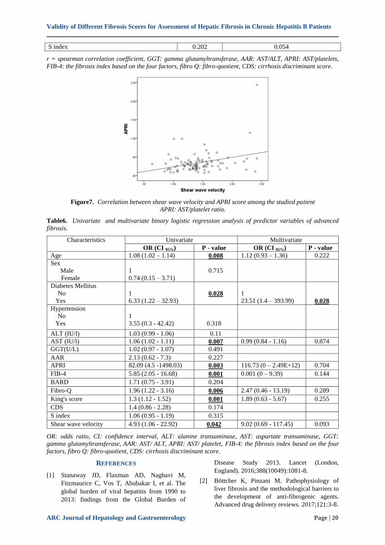
Validity of Different Fibrosis Scores for Assessment of Hepatic Fibrosis in Chronic Hepatitis B Patients
ARC Journal of Hepatology and Gastroenterology Page | 20
S index 0.202 0.054
r = spearman correlation coefficient, GGT: gamma glutamyltransferase, AAR: AST/ALT, APRI: AST/platelets,
FIB-4: the fibrosis index based on the four factors, fibro Q: fibro-quotient, CDS: cirrhosis discriminant score.
Figure7. Correlation between shear wave velocity and APRI score among the studied patient APRI: AST/platelet ratio.
Table6. Univariate and multivariate binary logistic regression analysis of predictor variables of advanced
fibrosis.
Characteristics Univariate Multivariate
OR (CI 95%) P - value OR (CI 95%) P - value
Age 1.08 (1.02 – 1.14) 0.008 1.12 (0.93 – 1.36) 0.222
Sex
Male
Female
1
0.74 (0.15 – 3.71)
0.715
Diabetes Mellitus
No
Yes
1
6.33 (1.22 – 32.93)
0.028
1
23.51 (1.4 – 393.99)
0.028
Hypertension
No
Yes
1
3.55 (0.3 - 42.42)
0.318
ALT (IU/l) 1.03 (0.99 - 1.06) 0.11
AST (IU/l) 1.06 (1.02 - 1.11) 0.007 0.99 (0.84 - 1.16) 0.874
GGT(U/L) 1.02 (0.97 - 1.07) 0.491
AAR 2.13 (0.62 - 7.3) 0.227
APRI 82.09 (4.5 -1498.03) 0.003 116.73 (0 – 2.49E+12) 0.704
FIB-4 5.85 (2.05 - 16.68) 0.001 0.001 (0 – 9.39) 0.144
BARD 1.71 (0.75 - 3.91) 0.204
Fibro-Q 1.96 (1.22 - 3.16) 0.006 2.47 (0.46 - 13.19) 0.289
King's score 1.3 (1.12 - 1.52) 0.001 1.89 (0.63 - 5.67) 0.255
CDS 1.4 (0.86 - 2.28) 0.174
S index 1.06 (0.95 - 1.19) 0.315
Shear wave velocity 4.93 (1.06 - 22.92) 0.042 9.02 (0.69 - 117.45) 0.093
OR: odds ratio, CI: confidence interval, ALT: alanine transaminase, AST: aspartate transaminase, GGT:
gamma glutamyltransferase, AAR: AST/ ALT, APRI: AST/ platelet, FIB-4: the fibrosis index based on the four
factors, fibro Q: fibro-quotient, CDS: cirrhosis discriminant score.
REFERENCES
[1] Stanaway JD, Flaxman AD, Naghavi M,
Fitzmaurice C, Vos T, Abubakar I, et al. The
global burden of viral hepatitis from 1990 to
2013: findings from the Global Burden of
Disease Study 2013. Lancet (London,
England). 2016;388(10049):1081-8.
[2] Böttcher K, Pinzani M. Pathophysiology of
liver fibrosis and the methodological barriers to
the development of anti-fibrogenic agents.
Advanced drug delivery reviews. 2017;121:3-8.

Validity of Different Fibrosis Scores for Assessment of Hepatic Fibrosis in Chronic Hepatitis B Patients
ARC Journal of Hepatology and Gastroenterology Page | 21
[3] Higashi T, Friedman SL, Hoshida Y. Hepatic
stellate cells as key target in liver fibrosis.
Advanced drug delivery reviews. 2017;121:27-
42.
[4] European Association for the Study of the
Liver. EASL-ALEH Clinical Practice
Guidelines: Non-invasive tests for evaluation of
liver disease severity and prognosis. Journal of
hepatology. 2015;63(1):237-64.
[5] Fallatah HI. Noninvasive biomarkers of liver
fibrosis: an overview. Advances in Hepatology.
2014;2014.
[6] Cheng J, Hou J, Ding H, Chen G, Xie Q, Wang
Y, et al. Validation of ten noninvasive
diagnostic models for prediction of liver
fibrosis in patients with chronic hepatitis B.
PloS one. 2015;10(12):e0144425.
[7] Zhang X, Zhang Y, Qiu Q, Zhang C, Wu C.
Diagnostic value of transient elastography
combined with noninvasive scores for the
detection of advanced liver fibrosis in chronic
hepatitis B patients. International journal of
clinical and experimental medicine. 2016;9(2)
:3692-7.
[8] Sarvazyan A, J Hall T, W Urban M, Fatemi M,
R Aglyamov S, S Garra B. An overview of
elastography-an emerging branch of medical
imaging. Current medical imaging reviews.
2011;7(4):255-82.
[9] Rustagi T, Newton E, Kar P. Percutaneous liver
biopsy. Tropical Gastroenterology. 2010;31(3)
:199-212.
[10] Gilmore I, Burroughs A, Murray-Lyon I,
Williams R, Jenkins D, Hopkins A. Indications,
methods, and outcomes of percutaneous liver
biopsy in England and Wales: an audit by the
British Society of Gastroenterology and the
Royal College of Physicians of London. Gut.
1995;36(3):437-41.
[11] Sheth SG, Flamm SL, Gordon FD, Chopra S.
AST/ALT ratio predicts cirrhosis in patients
with chronic hepatitis C virus infection. The
American journal of gastroenterology. 1998
;93(1):44.
[12] Wai CT, Greenson JK, Fontana RJ, Kalbfleisch
JD, Marrero JA, Conjeevaram HS, et al. A
simple noninvasive index can predict both
significant fibrosis and cirrhosis in patients
with chronic hepatitis C. Hepatology. 2003
;38(2):518-26.
[13] Vallet‐Pichard A, Mallet V, Nalpas B, Verkarre
V, Nalpas A, Dhalluin‐Venier V, et al. FIB‐4:
an inexpensive and accurate marker of fibrosis
in HCV infection. comparison with liver biopsy
and fibrotest. Hepatology. 2007;46(1):32-6.
[14] Hsieh Y-Y, Tung S-Y, Lee K, Wu C-S, Wei K-
L, Shen C-H, et al. Routine blood tests to
predict liver fibrosis in chronic hepatitis C.
World journal of gastroenterology: WJG.
2012;18(8):746.
[15] Cross TJ, Rizzi P, Berry PA, Bruce M,
Portmann B, Harrison PM. King's Score: an
accurate marker of cirrhosis in chronic hepatitis
C. European journal of gastroenterology &
hepatology. 2009;21(7):730-8.
[16] Harrison SA, Oliver D, Arnold HL, Gogia S,
Neuschwander-Tetri BA. Development and
validation of a simple NAFLD clinical scoring
system for identifying patients without
advanced disease. Gut. 2008;57(10):1441-7.
[17] Bonacini M, Hadi G, Govindarajan S, Lindsay
K. Utility of a discriminant score for
diagnosing advanced fibrosis or cirrhosis in
patients with chronic hepatitis C virus infection.
American journal of gastroenterology. 1997
;92(8).
[18] Zhou K, Gao CF, Zhao YP, Liu HL, Zheng RD,
Xian JC, et al. Simpler score of routine
laboratory tests predicts liver fibrosis in
patients with chronic hepatitis B. Journal of
gastroenterology and hepatology. 2010;25(9)
:1569-77.
[19] Wu S, Liu L, Li L, Zhu X, Wang J.
Development and validation of noninvasive
models to predict significant fibrosis in patients
with chronic hepatitis B. Chin J Dig. 2010
;30:478-80.
[20] Tseng CH, Chang CY, Mo LR, Lin JT, Tai
CM, Perng DS, et al. Acoustic radiation force
impulse elastography with APRI and FIB-4 to
identify significant liver fibrosis in chronic
hepatitis B patients. Annals of hepatology.
2018;17(5):789-94.
[21] Li J, Yu J, Peng X-Y, Du T-T, Wang J-J, Tong
J, et al. Acoustic Radiation Force Impulse
(ARFI) Elastography and Serological Markers
in Assessment of Liver Fibrosis and Free Portal
Pressure in Patients with Hepatitis B. Medical
science monitor: international medical journal
of experimental and clinical research. 2017;
23:3585.
[22] Elhosary YA, Saleh SM, Ezzat WM, Clevert D-
A. Diagnostic accuracy of acoustic radiation
force impulse (ARFI) in diagnosis of liver
fibrosis among Egyptian patients with chronic
HCV infection. Open access Macedonian
journal of medical sciences. 2016;4(3):374.
[23] Alem SA, Said M, Anwar I, Abdellatif Z, Elbaz
T, Eletreby R, et al. Improvement of liver
stiffness measurement, acoustic radiation force
impulse measurements, and noninvasive fibrosis
markers after direct‐acting antivirals for hepatitis
C virus G4 recurrence post living donor liver
transplantation: Egyptian cohort. Journal of
medical virology. 2018;90(9):1508-15.
[24] Bota S, Herkner H, Sporea I, Salzl P, Sirli R,
Neghina AM, et al. Meta‐analysis: ARFI

Validity of Different Fibrosis Scores for Assessment of Hepatic Fibrosis in Chronic Hepatitis B Patients
ARC Journal of Hepatology and Gastroenterology Page | 22
elastography versus transient elastography for
the evaluation of liver fibrosis. Liver
International. 2013;33(8):1138-47.
[25] Ferraioli G, Parekh P, Levitov AB, Filice C.
Shear wave elastography for evaluation of liver
fibrosis. Journal of Ultrasound in Medicine.
2014;33(2):197-203.
[26] Bedossa P, Poynard T. An algorithm for the
grading of activity in chronic hepatitis C.
Hepatology. 1996;24(2):289-93.
[27] Takahashi H, Ono N, Eguchi Y, Eguchi T,
Kitajima Y, Kawaguchi Y, et al. Evaluation of
acoustic radiation force impulse elastography for
fibrosis staging of chronic liver disease: a pilot
study. Liver international. 2010;30(4) :538-45.
[28] Jin W, Lin Z, Xin Y, Jiang X, Dong Q, Xuan S.
Diagnostic accuracy of the aspartate
aminotransferase-to-platelet ratio index for the
prediction of hepatitis B-related fibrosis: a
leading meta-analysis. BMC gastroenterology.
2012;12(1):14.
[29] Houot M, Ngo Y, Munteanu M, Marque S,
Poynard T. Systematic review with meta‐ analysis: direct comparisons of biomarkers for
the diagnosis of fibrosis in chronic hepatitis C
and B. Alimentary pharmacology & therapeutics
2016;43(1):16-2 9 .
[30] Xiao G, Yang J, Yan L. Comparison of
diagnostic accuracy of aspartate aminotrans
ferase to platelet ratio index and fibrosis‐4
index for detecting liver fibrosis in adult
patients with chronic hepatitis B virus
infection: a systemic review and meta‐analysis.
Hepatology. 2015;61(1):292-302.
[31] Eminler AT, Ayyildiz T, Irak K, Kiyici M,
Gurel S, Dolar E, et al. AST/ALT ratio is not
useful in predicting the degree of fibrosis in
chronic viral hepatitis patients. European
journal of gastroenterology & hepatology.
2015;27(12):1361-6.
[32] Hamidi AA, Oncul A, Ozguven BY, Sevgi DY,
Gunduz A, Uzun N, et al. Diagnostic accuracy
of different noninvasive scores for detecting
advanced fibrosis in chronic hepatitis B.
European journal of gastroenterology &
hepatology. 2019.
[33] Liu J, Li Y, Yang X, Ji Y, Zhang Y, Wan Q, et
al. Comparison of Two-Dimensional Shear
Wave Elastography with Nine Serum Fibrosis
Indices to Assess Liver Fibrosis in Patients with
Chronic Hepatitis B: A Prospective Cohort
Study. Ultraschall in der Medizin-European
Journal of Ultrasound. 2019;40(02):237-46.
[34] Dong M, Wu J, Yu X, Li J, Yang S, Qi X, et al.
Validation and comparison of seventeen
noninvasive models for evaluating liver fibrosis
in Chinese hepatitis B patients. Liver
International. 2018; 38(9):1562-70.
[35] Dong XQ, Wu Z, Zhao H, Wang GQ, China
HepB‐Related Fibrosis Assessment Research
Group. Evaluation and comparison of thirty
noninvasive models for diagnosing liver
fibrosis in chinese hepatitis B patients. Journal
of viral hepatitis. 2019;26(2):297-307.
[36] Ma J, Jiang Y, Gong G. Evaluation of seven
noninvasive models in staging liver fibrosis in
patients with chronic hepatitis B virus infection.
European journal of gastroenterology &
hepatology. 2013;25(4):428-34.
[37] Wu X, Cai B, Su Z, Li Y, Xu J, Deng R, et al.
Aspartate transaminase to platelet ratio index
and gamma‐glutamyl transpeptidase‐to‐platelet
ratio outweigh fibrosis index based on four
factors and red cell distribution width‐platelet
ratio in diagnosing liver fibrosis and
inflammation in chronic hepatitis B. Journal of
clinical laboratory analysis. 2018;32(4):e22341.
[38] Lang S, Kütting F, Staub A, Schramowski J,
Schramm C, Kasper P, et al. Performance of
simple noninvasive scoring systems for the
prediction of advanced fibrosis in patients with
chronic hepatitis B. European journal of
gastroenterology & hepatology. 2017;29(11):
1235-40.
[39] Liu DP, Lu W, Zhang ZQ, Wang YB, Ding RR,
Zhou XL, et al. Comparative evaluation of GPR
versus APRI and FIB‐4 in predicting different
levels of liver fibrosis of chronic hepatitis B.
Journal of viral hepatitis. 2018;25(5):581-9.
[40] Addissouky TA, El Agroudy AE, Eltorgman
AA. Efficiency of alternative markers to assess
liver fibrosis levels in viral hepatitis B patients.
Biomedical Research. 2019;30(2):351-6.
[41] Mallet V, Dhalluin‐Venier V, Roussin C,
Bourliere M, Pettinelli M, Giry C, et al. The
accuracy of the FIB‐4 index for the diagnosis of
mild fibrosis in chronic hepatitis B. Alimentary
pharmacology & therapeutics. 2009;29(4):409-
15.
[42] El-Saeid GK, El-Sharawy AA, Tahaa HE,
Fathy WM, Bedira ISE-m. Assessment of
laminin level and its comparison with five liver
fibrosis indices in chronic hepatitis B and C
patients. Menoufia Medical Journal. 2016;29(2)
:354.
[43] Coskun BD, Altınkaya E, Sevinc E, Ozen M,
Karaman H, Karaman A, et al. The diagnostic
value of a globulin/platelet model for
evaluating liver fibrosis in chronic hepatitis B
patients. Revista Española de Enfermedades
Digestivas. 2015;107(12):740-4.
[44] Chen Y, Wang Y, Chen Y, Yu Z, Chi X, Hu
KQ, et al. A Novel Noninvasive Program for
Staging Liver Fibrosis in Untreated Patients
With Chronic Hepatitis B. Clinical and
translational gastroenterology. 2019;10(5):1-12.
Validity of Different Fibrosis Scores for Assessment of Hepatic Fibrosis in Chronic Hepatitis B Patients
ARC Journal of Hepatology and Gastroenterology Page | 23
[45] Demir NA, Kolgelier S, Ozcimen S, Gungor G,
Sumer S, Demir LS, et al. Evaluation of the
relation between hepatic fibrosis and basic
laboratory parameters in patients with chronic
hepatitis B fibrosis and basic laboratory
parameters. Hepatitis monthly. 2014;14(4).
[46] Frulio N, Trillaud H, Perez P, Asselineau J,
Vandenhende M, Hessamfar M, et al. Acoustic
Radiation Force Impulse (ARFI) and Transient
Elastography (TE) for evaluation of liver
fibrosis in HIV-HCV co-infected patients.
BMC infectious diseases. 2014;14(1):405.
[47] Bignulin S, Falleti E, Cmet S, Cappello D,
Cussigh A, Lenisa I, et al. Usefulness of
acoustic radiation force impulse and fibrotest in
liver fibrosis assessment after liver transplant.
Annals of hepatology. 2016;15(2):200-6.
[48] Hsu TH, Tsui PH, Yu WT, Huang SF, Tai J,
Wan YL, et al. Cutoff values of acoustic
radiation force impulse two-location
measurements in different etiologies of liver
fibrosis. J Med Ultrasound 2019;27(3):130-4.
[49] Xu B, Zhou NM, Cao WT, Li XJ. Evaluation of
elastography combined with serological
indexes for hepatic fibrosis in patients with
chronic hepatitis B. World journal of
gastroenterology. 2018;24(37):4272.
[50] Ye XP, Ran HT, Cheng J, Zhu YF, Zhang DZ,
Zhang P, et al. Liver and spleen stiffness
measured by acoustic radiation force impulse
elastography for noninvasive assessment of
liver fibrosis and esophageal varices in patients
with chronic hepatitis B. Journal of Ultrasound
in Medicine. 2012;31(8):1245-53.
[51] Friedrich‐Rust M, Buggisch P, De Knegt R,
Dries V, Shi Y, Matschenz K, et al. Acoustic
radiation force impulse imaging for non‐invasive
assessment of liver fibrosis in chronic hepatitis
B. Journal of viral hepatitis. 2013;20(4):240-7.
[52] Dong CF, Xiao J, Shan LB, Li HY, Xiong YJ,
Yang GL, et al. Combined acoustic radiation
force impulse, aminotransferase to platelet ratio
index and Forns index assessment for hepatic
fibrosis grading in hepatitis B. World journal of
hepatology. 2016;8(14):616.
[53] Ozturker C, Karagoz E, Sivrioglu AK, Kara K.
Clinical usefulness and performance of acoustic
radiation force impulse in patients with chronic
hepatitis B. European journal of gastro
enterology & hepatology. 2017;29(6):66 3-8.
[54] Liu J, Ji Y, Ai H, Ning B, Zhao J, Zhang Y, et
al. Liver shear-wave velocity and serum
fibrosis markers to diagnose hepatic fibrosis in
patients with chronic viral hepatitis B. Korean
journal of radiology. 2016;17(3):396-404.
[55] Tai DI, Tsay PK, Jeng WJ, Weng CC, Huang
SF, Huang CH, et al. Differences in liver
fibrosis between patients with chronic hepatitis
B and C: evaluation by acoustic radiation force
impulse measurements at 2 locations. Journal of
Ultrasound in Medicine. 2015;34(5):813-21.
[56] Hong M-Z, Ye L, Jin L-X, Ren Y-D, Yu X-F,
Liu X-B, et al. Noninvasive scoring system for
significant inflammation related to chronic
hepatitis B. Scientific reports. 2017;7:43752.
[57] Park MS, Kim SW, Yoon KT, Kim SU, Park
SY, Tak WY, et al. Factors influencing the
diagnostic accuracy of acoustic radiation force
impulse elastography in patients with chronic
hepatitis B. Gut and liver. 2016;10(2):275.
[58] Liu Y, Dong CF, Yang G, Liu J, Yao S, Li Hy,
et al. Optimal linear combination of ARFI,
transient elastography and APRI for the
assessment of fibrosis in chronic hepatitis B.
Liver international. 2015;35(3):816-25.
[59] Lei B, Liu Y, Dong C, Chen X, Zhang X, Diao
X, et al. Assessment of liver fibrosis in chronic
hepatitis B via multimodal data. Neurocomp
uting. 2017;253:169-76.
Citation: Ghada M Galal. Validity of Different Fibrosis Scores for Assessment of Hepatic Fibrosis in
Chronic Hepatitis B Patients. ARC Journal of Hepatology and Gastroenterology.2019; 4(1):11-23.
Copyright: © 2019 Authors. This is an open-access article distributed under the terms of the Creative
Commons Attribution License, which permits unrestricted use, distribution, and reproduction in any medium,
provided the original author and source are credited.