Embed Size (px)
Citation preview

Valve Therapy for Treatment of Severe EmphysemaPatient Reference Guide
Caution: Federal (USA) Law restricts this device to sale by or on the order of a physician.

2 3
bronchi
air sacs
Figure 1.
TABLE OF CONTENTS
CHRONIC OBSTRUCTIVE PULMONARY DISEASE (COPD) AND EMPHYSEMA–If you are reading this booklet then it’s most likely that you or a loved one has been diagnosed with COPD. COPD is an umbrella term used to describe progressive lung diseases including emphysema, chronic bronchitis, refractory (non-reversible) asthma, and some forms of bronchiectasis.
Every affected person’s experience is different. The characteristic symptoms of COPD include frequent coughing, wheezing, shortness of breath, feeling tired — especially when doing daily activities and exercise — a tight feeling in the chest, and increased mucus in the lungs.
As the chronic disease progresses, your symptoms will tend to get worse as more damage occurs in the lungs.
COPD and Emphysema . . . . . . . . . . . . . . . . . . . . . . . . . . . . . . . . . . . . . . . . . . .3
Treatment Choices . . . . . . . . . . . . . . . . . . . . . . . . . . . . . . . . . . . . . . . . . . . . . . .5
Device Description . . . . . . . . . . . . . . . . . . . . . . . . . . . . . . . . . . . . . . . . . . . . . . .6
Intended Use . . . . . . . . . . . . . . . . . . . . . . . . . . . . . . . . . . . . . . . . . . . . . . . . . . .8
Potential Risks and Benefits . . . . . . . . . . . . . . . . . . . . . . . . . . . . . . . . . . . . . . . .9
The Procedure . . . . . . . . . . . . . . . . . . . . . . . . . . . . . . . . . . . . . . . . . . . . . . . . . 12
Patient Information Card . . . . . . . . . . . . . . . . . . . . . . . . . . . . . . . . . . . . . . . . . .16
My Care Regimen . . . . . . . . . . . . . . . . . . . . . . . . . . . . . . . . . . . . . . . . . . . . . . . 17
Valve Removal . . . . . . . . . . . . . . . . . . . . . . . . . . . . . . . . . . . . . . . . . . . . . . . . .18
Frequently Asked Questions . . . . . . . . . . . . . . . . . . . . . . . . . . . . . . . . . . . . . . .19
Glossary . . . . . . . . . . . . . . . . . . . . . . . . . . . . . . . . . . . . . . . . . . . . . . . . . . . . . . 21
My Lungs . . . . . . . . . . . . . . . . . . . . . . . . . . . . . . . . . . . . . . . . . . . . . . . . . . . . . 24
My Notes . . . . . . . . . . . . . . . . . . . . . . . . . . . . . . . . . . . . . . . . . . . . . . . . . . . . . 25
My Procedure . . . . . . . . . . . . . . . . . . . . . . . . . . . . . . . . . . . . . . . . . . . . . . . . . .26

4 5
TREATMENT CHOICES–Treatments for emphysema include drugs, oxygen, or in some cases, surgery. Surgery may not be appropriate for some patients, depending on their health status. Doctors advise patients on the appropriate therapy options.
If symptoms are mild, doctors frequently recommend:
n Avoiding smoking and other risk factors
n Having vaccinations against flu and pneumonia
n Short-acting drugs to expand the bronchi (bronchodilators), taken as needed
In the case of moderate symptoms:
n Long-acting drugs to expand the bronchi (bronchodilators), taken as a regular medication
n Pulmonary rehabilitation with exercise to strengthen the lungs, including power, stamina and relaxation elements, as well as training in correct use of inhalation systems
In the case of severe symptoms:
n Combinations of long-acting drugs
n Oxygen treatment
n Pulmonary rehabilitation with exercise as advised by your physician
n Bronchial valve therapy (Spiration® Valve Procedure)
n Surgery (for example: lung volume reduction or lung transplant)
Figure 2.
Emphysema
A consequence of chronic inflammation of lung tissue is pulmonary
emphysema. Emphysema is one of the Chronic Obstructive
Pulmonary Diseases.
With every breath, lungs deliver oxygen to the rest of the body to perform
essential life functions. Lungs are made up of tiny air sacs which absorb
the oxygen in the air you breathe (Figure 2) called alveoli. In patients
with emphysema, these air sacs are gradually destroyed and lose their
elastic strength, which makes it difficult for air to exit the air sacs. Air then
becomes trapped in the most diseased areas of the lungs, causing over-
inflated lungs. The trapped air interferes with the function of the healthier
areas of the lung, which adds to difficulty in breathing.
Every affected person’s experience is different, but the characteristic
symptoms include chronic coughing, shortness of breath, chest infections,
a tight feeling in the chest, and increased mucus in the lungs.
The symptoms usually develop slowly over many years. As a result, most
patients only consult a doctor when they suffer breathlessness—even
when exerting themselves only a little.
Healthy Alveoli
normal alveoli
Healthy Lungs Lungs of a Smoker
Alveoli with Emphysema
damaged alveoli

6 7
DEVICE DESCRIPTIONS–The Spiration® Valve System consists of the following parts:
The Valve
The umbrella shaped valve is made of a flexible metal frame (nitinol — composed of titanium and nickel) covered by a thin flexible membrane, (see Figure 3). The valve is designed to fit the shape and size of airways of the lung. The valve is a one-way device, which means that the flexible umbrella blocks air from moving further into your lungs, but allows trapped air to move out of your lungs as you breathe out.
Removal RodAllows doctor to remove the valve, if necessary.
UmbrellaThe umbrella redirects air you breathe in and allows secretions to escape naturally.
Tiny AnchorsLightly grasps airway to help stay in place.
Actual size of 7mm valve
Figure 3. Valve design
The Deployment Catheter
The valve is loaded into a long narrow tube called the deployment catheter. The deployment catheter (see Figure 4) places the valve in your airway.
Figure 4. The Deployment Catheter
Required Accessories:
Balloon Catheter
The doctor will be using a balloon catheter to measure the size of the airway that the proper valve size can be used.
Bronchoscope
In this procedure, the doctor will use a bronchoscope to see the airways of your lung. The deployment catheter and balloon catheter go through the bronchoscope to reach the airways.
The Spiration® Valve System is intended to treat patients with severe emphysema and evidence of low collateral ventilation, such as fissure integrity, by limiting airflow to selected areas of the lung.

8 9
INDICATIONS FOR USE
–Spiration® Valves are one-way endobronchial valves indicated for adult patients with shortness of breath and hyperinflation associated with severe emphysema in regions of the lung that have evidence of
low collateral ventilation.
Am I a candidate for valve therapy?
Valve therapy is suitable primarily for COPD (Chronic Obstructive Pulmonary Disease) patients suffering from severe emphysema who are not feeling better —despite drugs, pulmonary rehabilitation and oxygen therapy.
Valve therapy will only work in select patients when air can’t flow from one lobe of the lung to another lobe. A doctor will have the patient take several lung function tests and a CT scan to see if patients will benefit from valve therapy.
Valve therapy is usually not possible in the case of prior lung surgery, pleural
diseases, cancer, or severe heart disease.
When the Spiration Valve System Should Not Be Used
(Contraindications)
Your doctor will not place a Spiration Valve in your lungs if you are unable to have a bronchoscope placed through your nose or mouth and into your lungs. If you suspect or know that you are sensitive to or allergic to nickel, let your doctor know, you shouldn’t have a Spiration Valve placed in your lungs.
You are not a candidate for valve therapy:
n if you are still smoking
n if you have large bullae (holes) in the lung
n if you have certain types of emphysema
n if you have an active pulmonary infection
POTENTIAL RISKS AND BENEFITS OF SPIRATION® VALVE THERAPY
–Potential Risks: Warnings and Precautions
Valve therapy is a minimally invasive procedure that is well tolerated by most patients, but is not without risks. Please discuss these potential risks with your doctor. Call your doctor immediately if you have any discomfort, pain or any other concerns after your procedure.
n The Spiration Valve System should not be used for patients who have active asthma, bronchitis or clinically significant bronchiectasis.
n If you have swelling of airways (active asthma), inflammation of lung tissue (bronchitis), or airways that have gotten larger and/or infected (bronchiectasis), you should not have Spiration Valves placed in your lungs.
n You will be given drugs to make you unaware of pain during the procedure. These will make you sleepy (sedation) or unconscious (anesthesia). Talk with your doctor about the problems that can occur with sedation or anesthesia.
n The Spiration Valves are MR-conditional, which means that you can have an MRI procedure (a method for taking pictures of your internal organs) under certain conditions while the valves are implanted in your lungs. You will be given an information card to carry in your wallet. Show this card to a health care professional if you require an MRI.
n Although rare, as with all drugs and devices, it is possible that you may have an allergic reaction to the materials used in the Spiration Valve System.

10 11
POTENTIAL COMPLICATIONS
–
POTENTIAL BENEFITS
–Spiration® Valves may reduce the volume of diseased parts of the lung, and redirect air to the healthier parts. This may help improve overall lung function and quality of life for people living with emphysema.
In a recent clinical trial with the Spiration Valve System, the EMPROVE study, patients treated with medication and the Spiration Valve System were compared to a group of patients treated with medication only. Results of this study showed that the Spiration Valve System treated group experienced the following benefits compared to the medication-only patients:
n Improved lung function: The volume of air that can be forced out in one second after taking a deep breath (FEV1) is an important measure of lung function. The treated group improved their FEV1 by approximately 100ml over 6-month and 12-month time periods. This represented a 12% improvement in lung function, which indicates a clinically meaningful benefit to patients.
n Reduction of hyperinflation (air trapped in the diseased portion of the lung): Hyperinflation is a measurement of air trapped in the diseased portion of the lung. The treated group showed reduced hyperinflation over six months. Reducing hyperinflation helps healthier portions of the lung to work more efficiently and allows improvement in overall lung function.
n Decreased shortness of breath (dyspnea): The treated group showed reduced dyspnea over 6-month and 12-month time periods. This means that patients experienced decreased episodes of shortness of breath, which may lead to increased ability to accomplish daily activities.
n Improved quality of life: Doctors measure quality of life using a survey called the St. George‘s Respiratory Questionnaire (SGRQ), and generally agree that improvements over 4 points represent a clinically meaningful benefit to patients. The treated group showed improvement in SGRQ of 13.0 points (over six months) and 9.5 points (over twelve months).
The following may be associated with bronchoscopy and/or valve
placement include, but are not limited to:
n Altered arterial blood gas
n Anesthesia complications
n Atelectasis
n Bronchial injury
n Bronchitis
n Bronchospasm
n Chest Pain
n Chronic Obstructive Pulmonary Disease (COPD) exacerbation
n Death
n Dyspnea
n Empyemea/Lung abcess
n Hemoptysis (or bleeding)
n Hemothorax
n Hypoxemia
n Iatrogenic injuries
n Infection
n Migration of a valve out of the lung or within the lung
n Persistent cough
n Pleural effusion
n Pneumonia
n Pneumothorax
n Respiratory failure
n Sore throat
n Thorax pain
n Tissue hyperplasia or other reaction at valve site
n Valve fracture
n Vocal cord injury
n Wheezing
n Other procedure-related complications may occur
12 13
THE PROCEDURE
–
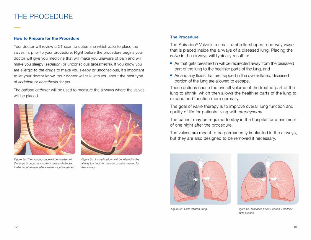
How to Prepare for the Procedure
Your doctor will review a CT scan to determine which lobe to place the
valves in, prior to your procedure. Right before the procedure begins your
doctor will give you medicine that will make you unaware of pain and will
make you sleepy (sedation) or unconscious (anesthesia). If you know you
are allergic to the drugs to make you sleepy or unconscious, it’s important
to let your doctor know. Your doctor will talk with you about the best type
of sedation or anesthesia for you.
The balloon catheter will be used to measure the airways where the valves
will be placed.
Diseased Lung Diseased Parts Reduce, Healthier Parts Expand
The Procedure
The Spiration® Valve is a small, umbrella-shaped, one-way valve that is placed inside the airways of a diseased lung. Placing the valve in the airways will typically result in:
n Air that gets breathed in will be redirected away from the diseased part of the lung to the healthier parts of the lung, and
n Air and any fluids that are trapped in the over-inflated, diseased portion of the lung are allowed to escape.
These actions cause the overall volume of the treated part of the lung to shrink, which then allows the healthier parts of the lung to expand and function more normally.
The goal of valve therapy is to improve overall lung function and quality of life for patients living with emphysema.
The patient may be required to stay in the hospital for a minimum of one night after the procedure.
The valves are meant to be permanently implanted in the airways, but they are also designed to be removed if necessary.
Figure 6a. Over-Inflated Lung Figure 6b. Diseased Parts Reduce, Healthier Parts Expand
Figure 5a. The bronchoscope will be inserted into the lungs through the mouth or nose and directed to the target airways where valves might be placed.
Figure 5b. A small balloon will be inflated in the airway to check for the size of valve needed for that airway.

14 15
THE PROCEDURE
–
What to Expect After the Placement Procedure
After the procedure you may be required to stay in the hospital for a minimum of one night.
Your doctor will likely order a chest x-ray to see how your lungs are responding to the valve treatment. They will be watching for any complications, including a pneumothorax. A pneumothorax is air that becomes trapped between the lung and chest wall. If you have a pneumothorax you may need to have a chest tube to allow the trapped air to escape, and your doctor might need to remove a valve, which can be replaced at a later date.
Your doctor will give you instructions for your care at home. This will include information on what medicines you should take and follow-up visits.
You will be given a Patient Information Card for your wallet (purse) that says you have one or more Spiration® Valves and shows where they are placed (see Figure 11). This card will also have your doctor’s contact information. It’s important to keep this card with you at all times and to show it to anyone who gives you medical care, especially emergency room staff.
Call your doctor immediately if you have any discomfort, pain, or any other concerns after your procedure.
n If you suddenly have severe chest pain or breathing becomes increasingly difficult, get immediate emergency care, and advise your treating physician.
Figure 7. Surgery is not needed to place valves in the airways of the lung. The Spiration® Valves are placed using a bronchoscope.
Figure 8. The valve is placed in the airway using a catheter that goes down the bronchoscope. Once the valve is in the airway, it expands and contracts with breathing.
Figure 9. More than one valve will be placed. The valve allows trapped air and fluids to flow past it, but blocks air from entering the diseased part of the lung.
Figure 10. The valve redirects air to healthier parts of the lung and reduces over-inflation.

16 17
PATIENT INFORMATION CARDSpiration® Valve System
Patient Name ______________________________________
Patient ID ________________________________________
Physician _________________________________________
Of�ce Phone ______________________________________
Hospital ___________________________________________
Address __________________________________________
Procedure Date: ______ /______ /______ (MM/DD/YY)
INSTRUCTIONS: Please carry this card at all times and show it to any medical personnel who may be treating you. Notify your physician if symptoms fail to improve, worsen, or new symptoms develop.
MRI Information
The Spiration Valve was determined to be MR-conditional according to the terminology speci�ed in the American Society for Testing and Materials (ASTM) International, Designation: F2503. Standard Practice for Marking Medical Devices and Other Items for Safety in the Magnetic Resonance Environment.
Non-clinical testing demonstrated that the Spiration Valve is MR Conditional. A patient with this implant can be scanned safely immediately after placement under the following conditions:- Static magnetic �eld of 3-Tesla or less- Spatial magnetic gradient �eld of 720-Gauss/cm or less- Maximum MR system reported whole-body-averaged speci�c absorption rate (SAR) of 3-W/kg for 15 minutes of scanning.
In non-clinical testing, the Spiration Valve produced a temperature rise of less than or equal to 0.5º C at a maximum MR system reported whole-body-average speci�c absorption rate (SAR) of 3-W/kg for 15 minutes of MR scanning in a 3-Tesla MR system (Excite, Software G3.0-052B, General Electric Healthcare, Milwaukee, WI).
MR image quality may be compromised if the area of interest is in the same area or relatively close to the position of the Spiration Valve. Optimization of MR imaging parameters is recommended.
MR Conditional
Valve Location Place Removable Tracking Label Below
PI-05357 Rev AA
Apical (RB1)
Apical (LB1)
Posterior (LB2)
Anterior (LB3)
Superior (LB4)
Inferior (LB5)
Anterior (LB8)
Lateral (LB9)
Posterior (LB10)
Superior (LB6)
Apical-Posterior(LB1+2)
Lingula (LB4-5)
RIGHT LEFT
Posterior (RB2)
Anterior (RB3)
Lateral (RB4)
Medial (RB5)
Anterior (RB8)
Lateral (RB9)
Posterior (RB10)
Medial (RB7)
Superior (RB6)
1
2
3
4
5
6
7
8
9
10
11
12
13
14
15
16
17
19
20
21
22
ManufacturerSpiration, Inc. d/b/aOlympus Respiratory America6675 185th Avenue NERedmond WA 98052
Distributed byOlympus America, Inc.3500 Corporate Parkway Center Valley PA 18034
Figure 11. The Spiration Valve Patient Card is your record of the procedure. An example of the front and back of the card are shown here.
MY CARE REGIMEN
–Follow your doctor’s post-surgical instructions and life style recommendations. These may include regular follow-up visits to your pulmonologist, participating in a pulmonary rehabilitation program. As you start to feel better, do NOT start smoking again.
Due to the chronic nature of emphysema, anyone who has received valve therapy must continue regular medical checkups. These checkups will examine the effect of the valve therapy. When you come in for follow-up visits, your doctor may want to inspect your valves using a bronchoscope.
This therapy is not a substitute for treatment of COPD by medication or pulmonary rehabilitation.
Although you may feel much better after the procedure, it’s very important to allow your lungs to adjust to the therapy so that you don’t suffer any setbacks.
Plan to rest and limit your activities for at least one week following the procedure — give the therapy some time to work.
PATIENT INFORMATION CARD

18 19
VALVE REMOVAL
–
What to Expect if a Spiration® Valve Needs to be Removed
The Spiration Valve is designed to be a permanent, long-term implant. However, if there are any complications, your doctor may decide to remove a valve. Valve removal is a fairly simple, straight-forward procedure.
Like valve placement, the valve removal procedure is done using sedation or anesthesia. A bronchoscope will be used as it was during valve placement.
Forceps will be placed into the bronchoscope to grab and remove the valve (see Figure 12a and 12b).
Figure 12a. Forceps placed through the bronchoscope will grab the valve.
Figure 12b. The valve is removed along with the bronchoscope.
FREQUENTLY ASKED QUESTIONS
–
1. Will I be able to feel the Spiration® Valve in my lung?
No, you will not be able to feel the Spiration Valve after it’s been
placed in your lung.
2. How long will the procedure take?
The bronchoscopy procedure used to place the Spiration Valve
usually takes less than one hour but may take up to two hours.
3. When will I feel better after procedure?
The amount of time that it takes to feel better after your procedure
varies from patient to patient. You should discuss how you are
feeling regularly with your doctor.
4. Can it be coughed out?
In Spiration Valve clinical trials, no patients have coughed out a
Spiration Valve. However, in a rare case, a valve may be coughed out.
5. Will I be able to stop or decrease using my oxygen and other
COPD medications?
Although you may feel better after receiving the Spiration Valves,
always consult your doctor before you make any changes to using
oxygen and COPD medications. Valve therapy is not a substitute for
treatment of COPD by medication or oxygen therapy.
6. How long will it stay implanted in me? Is the procedure
reversible?
The Spiration Valve is intended to be permanent, however it’s
designed to be removed if necessary.

20 21
7. How many valves will the doctor place in me?
Every person’s lungs are different, however on average about
4 Spiration Valves are typically used.
8. Does the doctor ever need to go back to check on them?
Yes. As part of your regular follow-up visits with your doctor
(pulmonologist), they will examine the effect of the valve
therapy. Your doctor may want to inspect your valves using a
bronchoscope.
9. What happens when I go through security at the airport?
The Spiration Valve will not set off the metal detectors used by
airport security.
10. What is the expected length of recovery from the procedure?
When can I go back to normal activities?
Although you may feel much better after the procedure, it’s very
important to allow your lungs to adjust to the recent procedure
so that you don’t suffer any setbacks. Follow your doctor’s post-
surgical instructions, and plan to rest and limit your activities for at
least one week following the procedure.
GLOSSARY
–Airway: The tubes in the lungs that
pass air to and from the lung tissue.
Anesthesia: A medical procedure
that makes the patient unconscious and
makes the body insensitive to pain.
Asthma: Swelling of the airways in
the lung, making breathing difficult.
Atelectasis: Collapse of all or part
of the lung.
Balloon Catheter: A narrow,
flexible tube with a small balloon at
the end.
Bronchi: The large air passages that
lead from the trachea (windpipe) to
the lungs.
Bronchiectasis: A lung condition
in which the airways of the lungs get
bigger and often get infected.
Bronchitis: An infection that causes
inflammation of lung tissue.
Bronchodilators: Drugs which
relax the bronchial muscle, causing
the airways to dilate and thus alleviate
the symptoms of COPD.
Bronchoscope: A narrow, flexible
tube, with a camera on the end that is
used to see the airways inside the lungs.
Bronchospasm: A tightening of the
muscles that line the airways (bronchi)
in your lungs. When these muscles
tighten, your airways narrow. It causes
difficulty in breathing which can be
very mild to severe.
CT Scan: A radiographic image
taken of a body part.
Carbon dioxide: Result of the
chemical reaction between oxygen
(from the air breathed in) and carbon
(from carbohydrates in the food);
replaced in the lung by “fresh” oxygen.
Chest tube: A plastic tube that is
inserted through the skin and rib cage
and into the chest cavity to allow air
and fluids leaking from the lung to exit
the body.
Chest X-ray: a picture of the chest
that shows your heart, lungs, airway,
blood vessels, and lymph nodes.
Chronic: Permanent, long-term.
Collateral Ventilation: The
ventilation of alveolar structures
through passages or channels that
bypass the normal airways.
Contraindication: A specific
situation in which the valve therapy
procedure should not be used because
it may be harmful to the patient.
FREQUENTLY ASKED QUESTIONS (Continued)
–
For more information on the Spiration Valve System, please visit svs.olympusamerica.com/patients-and-family

22 23
GLOSSARY (Continued)
Deployment Catheter: A narrow,
flexible tube used to deliver the Spiration®
Valve to target airways of the lung.
Dyspnea: Shortness of breath,
often described as an intense
tightening in the chest, air hunger
or a feeling of suffocation.
Emphysema: A lung condition in
which the air sacs of the lungs are
damaged and enlarged, causing
breathlessness.
Expiration: When a person breaths out.
Fissure Integrity: Lung fissures are
divisions from the surface to the interior
of the lung tissue which divides it into
lobes. Therefore the completeness of
separation between the lobes.
Forced expiratory volume in one second (FEV1): Forced
expiratory volume in one second (FEV1)
corresponds to the quantity of air the
patient can breathe out within one second
after breathing in as deeply as possible.
Forceps: A common tool used in
medical procedures to help doctors
grab onto tissue and other objects in
the body.
Hemoptysis: The coughing up of
blood or blood-stained mucus from
the bronchi, trachea, or lungs.
Hemothorax: A condition when
blood collects between your chest wall
and your lungs—in your pleural cavity.
Hypoxemia: A below-normal level
of oxygen in the blood. It’s a sign
of a problem related to breathing or
circulation.
Inhaled Corticosteroids (ICS): A type of steroid medication that is
inhaled. Corticosteroids are very good
at reducing inflammation (swelling)
and mucus production in the airways
of the lungs. They also help other
quick-relief medicines work better.
Lobe: A clear anatomical division of
an organ. The right lung is made up of
the right upper lobe, right middle lobe
and right middle lobe. The left lung is
made up of the left upper lobe and the
left lower lobe.
Lung Function Test: These include
a variety of tests that check how well
the lungs work.
Lung Volume Reduction Surgery (LVRS): A surgical treatment for
patients with advanced emphysema
whose breathlessness is poorly
controlled by medical management.
LVRS reduces the lung volume by
wedge excisions of diseased tissue.
Migration: The movement from one
location to another.
Nitinol: A metal made of nickel and
titanium distinguished from other
materials by its shape memory and
super elastic characteristics.
Oxygen therapy: Administration of
air with an increased concentration of
oxygen to make breathing easier.
Pleural disease: Any type of
disease that affects the pleura. The
pleura is the membrane that lines the
chest cavity and covers the lungs. The
pleural space is the area between the
lungs and the chest wall.
Pneumonia: A lung infection with
symptoms like a cough, fever, and
difficulty breathing.
Pneumothorax: A pneumothorax
occurs when air leaks into the space
between your lung and chest wall.
This air pushes on the outside of your
lung and makes it collapse.
Pulmonary Rehabilitation: A
program of exercise, education and
support to help COPD patients to
breathe and function at the highest
level possible.
Respiratory failure: Is a syndrome
in which the respiratory system fails
in one or both of its gas exchange
functions: oxygenation and carbon
dioxide elimination.
Respiratory compromise: Breathing difficulty — causes less
oxygen to be available to the body
and negatively affects bodily functions.
Sedation: A medical procedure in
which medicine is given to the patient
to make him or her comfortable and
unaware of pain during surgery. It
relaxes the central nervous system
and can cause sleepiness.
Thorax pain: Chest pain, which has
many possible causes — and some of
them are serious. It‘s important to see
your doctor about chest pain.
Tissue hyperplasia: An abnormal
increase in volume of a tissue or organ
(i.e. the lung) caused by the formation
and growth of new cells.
Valve: The Spiration® Valve is a small,
umbrella-shaped one-way device that
is placed inside the airways of the lung
that redirects air away from damaged
lung tissue.

24 25
Trachea
Lower lobe
Lower lobe
Middle lobe
Upper lobe
Upper lobe
Right Lung Left Lung
MY LUNGS
–Talk with your doctor about your Spiration® Valve Procedure. Use this picture to understand where valves could be placed.
MY NOTES
–

26 27
MY PROCEDURE
–
My Doctor’s Name:
Hospital:
Planned Procedure:
Date: Time:

3500 Corporate Parkway, PO Box 610, Center Valley, PA 18034
www.medical.olympusamerica.com
©2020 Olympus America Inc. All rights reserved. Printed in USA OAIRES0120BRO33491