Embed Size (px)
Citation preview

1
Vascular Evaluation of the Foot
Alexander M. Reyzelman DPMAssociate Professor, Dept Medicine
California School of Podiatric MedicineCo-Director, UCSF Center For Limb
Preservation
What is Peripheral Arterial Disease ?
Peripheral Vascular Disease
• 15-20% of patients with DM have PAD at 10 yrs
• 45% of patients with DM have PAD at 20yrs
What is Ischemia?
Ischemia = Demand > Supply
• Absolute Ischemia– absolute amount of flow present
• Relative Ischemia– discrepancy between amount of flow
available and amount needed by the currentclinical situation
2
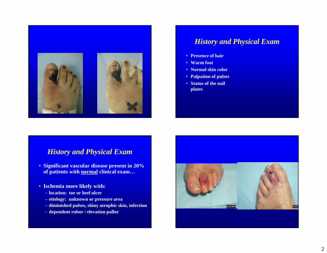
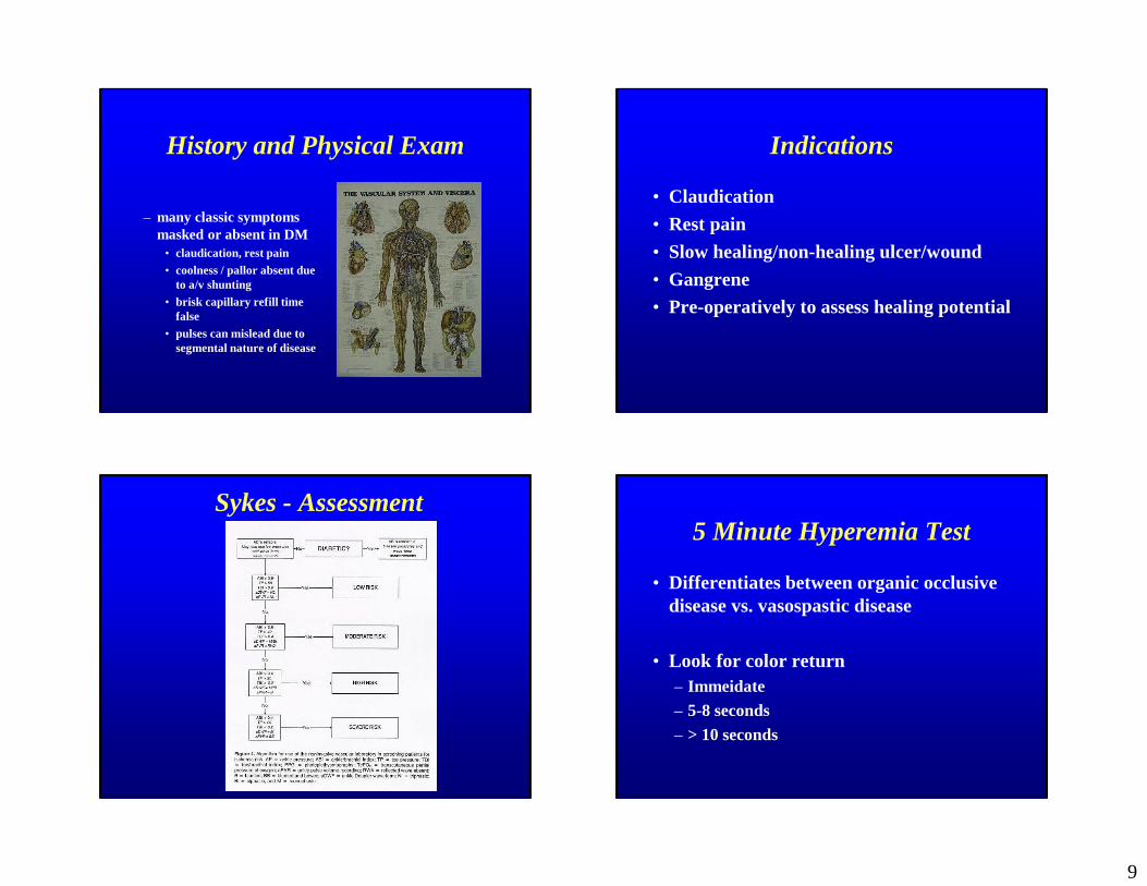
History and Physical Exam
• Presence of hair• Warm foot• Normal skin color• Palpation of pulses• Status of the nail
plates
History and Physical Exam
• Significant vascular disease present in 20% of patients with normal clinical exam…
• Ischemia more likely with:– location: toe or heel ulcer– etiology: unknown or pressure area– diminished pulses, shiny atrophic skin, infection– dependent rubor / elevation pallor

3
When to order NIV arterial testing?
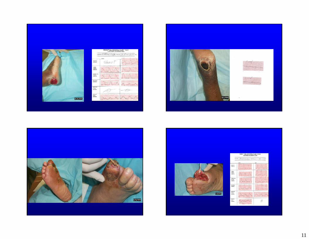
• Chronic non-healing ulcer/wound
• Pre-operatively to assess healing of proposed foot surgery
• Ulcers of the digits, or boney prominences
• Symptoms of claudication
Noninvasive Modalities
• Ankle Brachial Index• Toe Brachial Index• Toe Pressure• Segmental Pressures• Doppler waveforms• Photoplethysmography• Pulse Volume Recordings
ABI / TBI
• Ankle Brachial Index:– ratio of ankle / arm
systolic blood pressure
– normal 0.9 to 1.2– false elevation in DM
due to medial calcification
– good screening test in non-diabetic patients
• Toe Brachial Index:– ratio of hallux / arm
systolic pressure– > 0.6 low risk– < 0.2 severe risk– digital vessels less
affected by calcification in DM
Medial Calcinosis
• Tunica media
• neuropathy
• elevated pressures
• Goebel and Fuessel, Edmonds

4
Ankle and Toe Indices
• TBI Exam:• ABI Exam:
Doppler Waveforms
• Interpretation– triphasic/biphasic/monophasic– normal flow appears as narrow peak,
followed by one or two smaller peaks– faster flow --> higher audible pitch,
waveform resembles teepee– slower flow--> lower pitch, igloo
waveform– as flow deteriorates, waves flatten
Doppler Waveform –Triphasic and Biphasic
Doppler Waveform - Monophasic
5
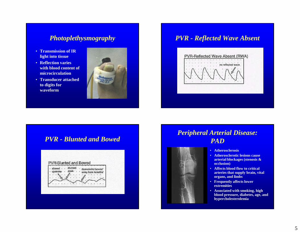
Photoplethysmography
• Transmission of IR light into tissue
• Reflection varies with blood content of microcirculation
• Transducer attached to digits for waveform
PVR - Reflected Wave Absent
PVR - Blunted and BowedPeripheral Arterial Disease:
PAD• Atherosclerosis• Atherosclerotic lesions cause
arterial blockages (stenosis & occlusion)
• Affects blood flow to critical arteries that supply brain, vital organs, and limbs
• Frequently affects lower extremities
• Associated with smoking, high blood pressure, diabetes, age, and hypercholesterolemia
6
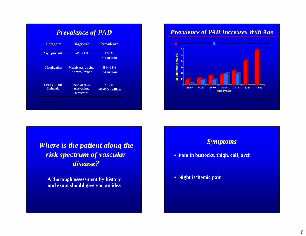
Category Diagnosis Prevalence
Asymptomatic ABI < 0.9 >50%4-6 million
Claudication Muscle pain, ache, cramps, fatigue
30%-35%2-4 million
Critical Limb Ischemia
Pain at rest, ulceration, gangrene
<10%400,000-1 million
Hiatt WR, NEJM 2001
Prevalence of PAD
1. Meijer WT, et al. Arterioscler Thromb Vasc Biol. 1998;18:185-192. 2. Criqui MH, et al. Circulation. 1985;71:510-515.
Rotterdam Study (ABI <0.9)1 San Diego Study (PAD by noninvasive tests)2
0
10
20
30
40
50
60
Pat
ien
ts W
ith
PA
D (
%)
55-59 60-64 65-69 70-74 75-79 80-84 85-89
Age (years)
Prevalence of PAD Increases With Age
ABI=ankle-brachial index
Where is the patient along the risk spectrum of vascular
disease?
A thorough assessment by history and exam should give you an idea
Symptoms
• Pain in buttocks, thigh, calf, arch
• Night ischemic pain

7
Peripheral Arterial Disease
• Intermittent claudication– “claudico” – to limp– Pain with walking– Relieved by rest
• Critical Limb Ischemia– Pain at rest, ulcers, or
gangrene
Peripheral Arterial Disease
• Intermittent claudication– “claudico” – to limp– Pain with walking– Relieved by rest
• Critical Limb Ischemia– Pain at rest, ulcers, or
gangrene
Intermittent Claudication
Measured in amount of blocks
Reproducible
Consistent
Not Limb Threatening
Intermittent Claudication
• Differential Diagnosis– Pseudoclaudication
– Degenerative Joint Disease
– Diabetic Peripheral Neuropathy

8
Natural History of CLI1 Year Outcomes
Critical LimbIschemia1%-2%
Alive with both limbs
50%
Amputation25%
Dead25%
Weitz JI, Circulation 1996
•Rest Pain
•Ulceration
•Gangrene
Diabetic vs. Nondiabetic Ischemic Patterns
• Diabetic:– Distal: popliteal,
‘trifurcation,’ tibial, pedal
– Collateral Pathways: internal iliac, profunda femoral, tibial
– Calcified vessel walls
– Symmetrical
• Nondiabetic:– Proximal: aorta,
iliac, femoral
– Axial Pathways: aorta, iliac, superficial femoral
– Usually noncalcified– Symmetrical
Timing of Vascular Assessment
• In the acute presentation:– priority is to address
limb threatening infection
– should not delay necessary debridement
– prompt bypass integral to limb salvage
• In routine outpatient management:– initial evaluation– non-healing wound– follow progression of
known disease
History and Physical Exam
• Hx of CABG• Hx of CEA• Hx of tobacco• Hx of MI• Hx of CVA• Hx of Angina
• Thigh, buttocks, calf pain upon walking
• Hx of previous ulcerations and how long it took to heal
9
History and Physical Exam
– many classic symptoms masked or absent in DM
• claudication, rest pain• coolness / pallor absent due
to a/v shunting• brisk capillary refill time
false• pulses can mislead due to
segmental nature of disease
Indications
• Claudication
• Rest pain
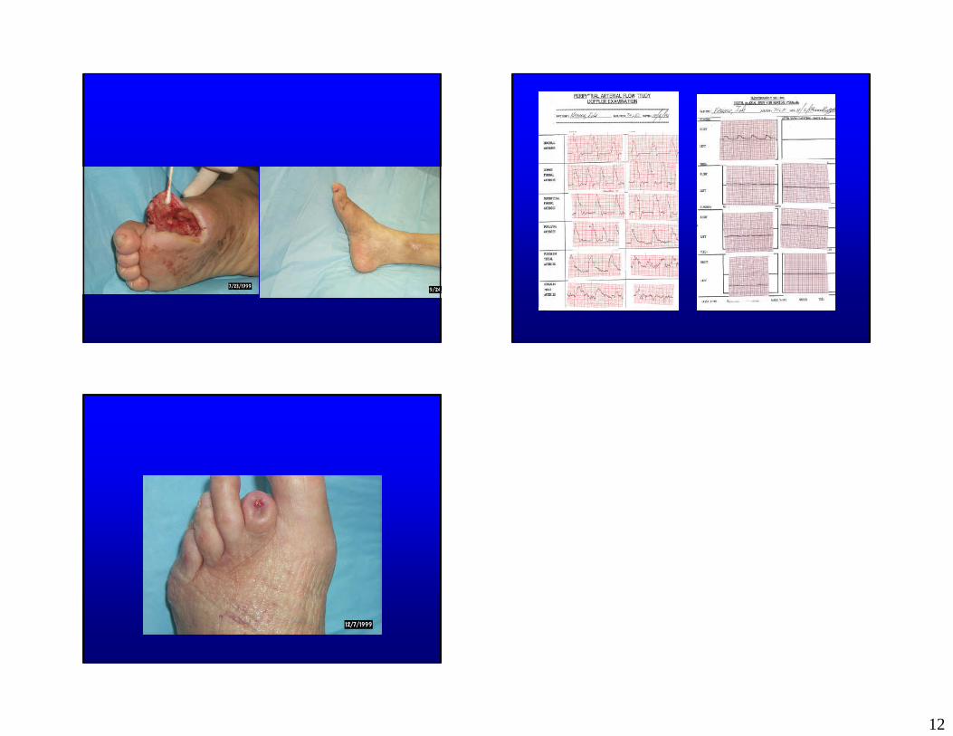
• Slow healing/non-healing ulcer/wound
• Gangrene• Pre-operatively to assess healing potential
Sykes - Assessment5 Minute Hyperemia Test
• Differentiates between organic occlusive disease vs. vasospastic disease
• Look for color return – Immeidate– 5-8 seconds– > 10 seconds

10
11
12