Embed Size (px)
Citation preview

12th Edition - Starters Package in LaparoscopyIRCAD/EITS Strasbourg (France) – October 20+21, 2011.
Teacher : Prof. Paul Van Schil – UZA Edegem
VATS for thoracic diseases. 1
VATS for thoracic diseases
Paul Van Schil, MD, PhDDepartment of Thoracic and Vascular SurgeryAntwerp University Hospital
Starters’ Package 12th edition, October 20, 2011
VATS for thoracic diseases
introductionbasic principles : accesspneumothoraxpleural diseasesmediastinal diseases

12th Edition - Starters Package in LaparoscopyIRCAD/EITS Strasbourg (France) – October 20+21, 2011.
Teacher : Prof. Paul Van Schil – UZA Edegem
VATS for thoracic diseases. 2
introductionbasic principles : accesspneumothoraxpleural diseasesmediastinal diseases
VATS for thoracic diseases
THORACOSCOPIC SURGERY
• H.C. Jacobaeus (1910)modified cystoscopeintrapleural pneumonolysis
• after 1945 : rapid declinediagnostic procedures
• from 1991 : revivallaparoscopyvideo-assisted techniquesnew instruments (endo-staplers)3D – robotic surgery

12th Edition - Starters Package in LaparoscopyIRCAD/EITS Strasbourg (France) – October 20+21, 2011.
Teacher : Prof. Paul Van Schil – UZA Edegem
VATS for thoracic diseases. 3
VATSVideo-assisted thoracic surgery
• minimally invasive technique• major operative procedure• general anesthesia• single lung ventilation (sometimes: CO2 insufflation)
– double lumen tube– bronchial blocker
• conversion to thoracotomy or sternotomy
video monitor, thoracoscope, camera
VATSTheoretical advantages
• smaller incisions• less muscle destruction• no rib retraction• less pain• shorter hospital stay• reduced cost
12th Edition - Starters Package in LaparoscopyIRCAD/EITS Strasbourg (France) – October 20+21, 2011.
Teacher : Prof. Paul Van Schil – UZA Edegem
VATS for thoracic diseases. 4
VATSDisadvantages
• less exposure• less adequate operation
limitations monitor, instruments, ribs
• loss of digital palpation• single lung ventilation• expensive equipment• long-term results in lung cancer (selection bias) ?
VATSDifferent procedures
Pleural and mediastinal access
• mediastinoscopy (superior mediastinum, N2-3)• pleuroscopy (local anesthesia)• thoracoscopy, VATS
small incisions (2 cm) « pure VATS»small utility thoracotomy (3-5 cm), no rib spreadingminithoracotomy with rib retraction
VATS = approach

12th Edition - Starters Package in LaparoscopyIRCAD/EITS Strasbourg (France) – October 20+21, 2011.
Teacher : Prof. Paul Van Schil – UZA Edegem
VATS for thoracic diseases. 5
VATS for Thoracic Diseases
• pleura : biopsy, effusion, pleurectomy• lung : bullectomy, biopsy, lung resection• mediastinum : biopsy, resection, staging• esophagus : tumors, staging, myotomy• pericardium : effusion, resection• heart : PM, ICD• autonomic disorders : sympathectomy• thoracic outlet syndrome : 1st rib resection
Khraim FM. The wider scope of video-assisted thoracoscopic surgery. AORN J 2007; 85:1199-1208
introductionbasic principles : accesspneumothoraxpleural diseasesmediastinal diseases
VATS for thoracic diseases

12th Edition - Starters Package in LaparoscopyIRCAD/EITS Strasbourg (France) – October 20+21, 2011.
Teacher : Prof. Paul Van Schil – UZA Edegem
VATS for thoracic diseases. 6
VATSThoracoports
# ports 1 exploration, sympathectomy (exceptional)2 simple VATS (biopsy)3 or more most procedures
• prepare for thoracotomy• lateral position except
bilateral LVRS (lung volume reduction surgery)bilateral sympathectomythymectomy
• open interspaces : breaking tablepillows, bagsmechanical bridge
VATSThoracoports
12th Edition - Starters Package in LaparoscopyIRCAD/EITS Strasbourg (France) – October 20+21, 2011.
Teacher : Prof. Paul Van Schil – UZA Edegem
VATS for thoracic diseases. 7
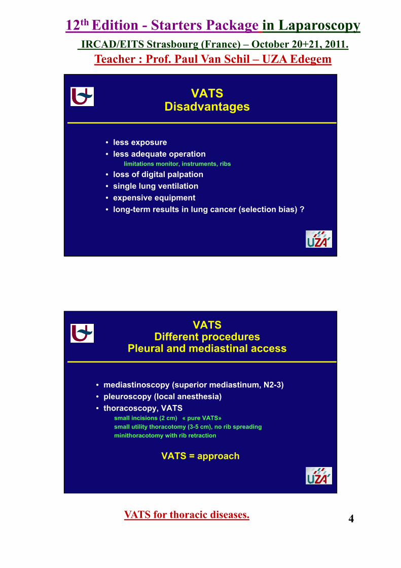
VATSThoracoports
• triangular configuration• optimum angle optic – instruments : 30 – 60° (< 90°) • no crossing of instruments or camera• do not work towards yourself• thoracoscope : central or posterior position
VATSdouble lumen tube
12th Edition - Starters Package in LaparoscopyIRCAD/EITS Strasbourg (France) – October 20+21, 2011.
Teacher : Prof. Paul Van Schil – UZA Edegem
VATS for thoracic diseases. 8
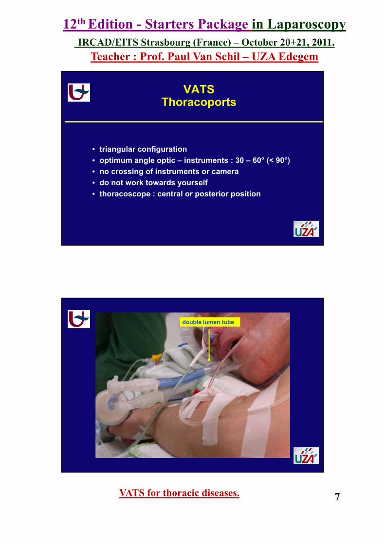
VATS
epidural catheter
VATSPORTS - LOCATION
• anterior : interspaces widerlarger instrumentsposition of drain
• line of thoracotomy• not high anterior (cosmetic) • avoid previous drain sites, especially when infected

12th Edition - Starters Package in LaparoscopyIRCAD/EITS Strasbourg (France) – October 20+21, 2011.
Teacher : Prof. Paul Van Schil – UZA Edegem
VATS for thoracic diseases. 9
VATS
VATS

12th Edition - Starters Package in LaparoscopyIRCAD/EITS Strasbourg (France) – October 20+21, 2011.
Teacher : Prof. Paul Van Schil – UZA Edegem
VATS for thoracic diseases. 10
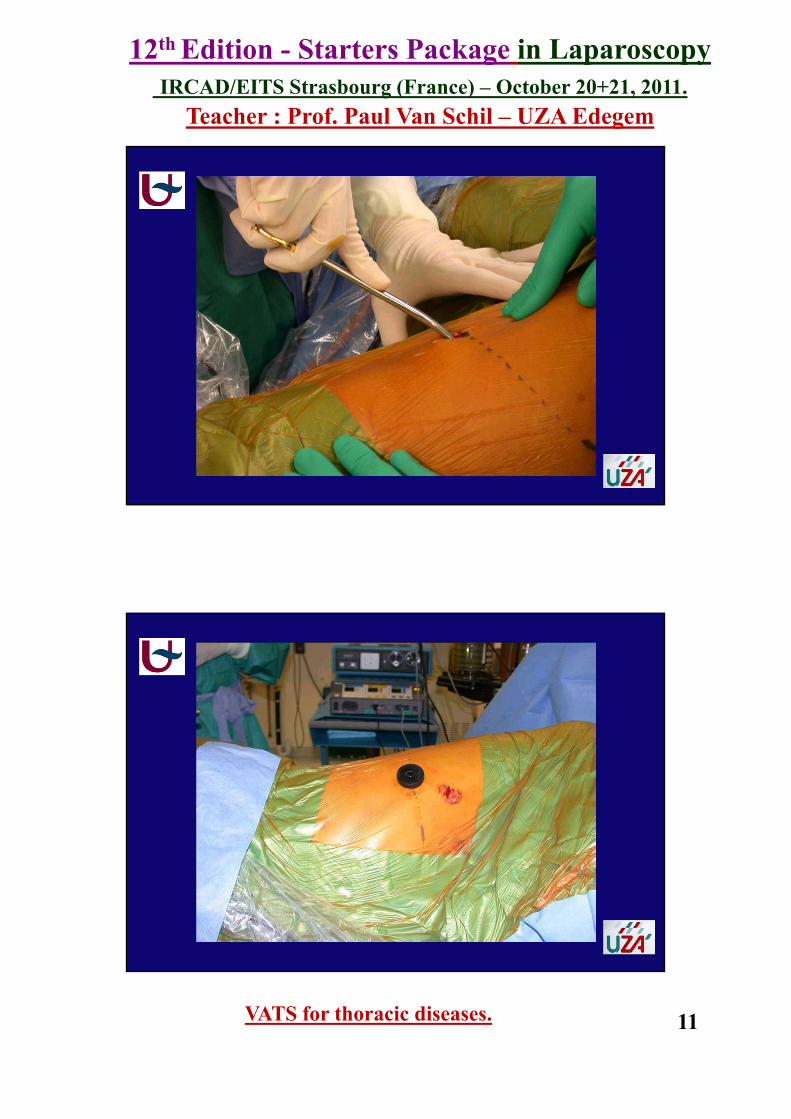
VATSPORT INSERTION
• 2 – 2.5 cm incision (larger in fat patients)• cautery subcutaneous fat• divide or split muscle fibres• cautery over top border of rib • cut parietal pleura• insert finger (adhesions)• insert thoracoport, thoracoscope
VATS
12th Edition - Starters Package in LaparoscopyIRCAD/EITS Strasbourg (France) – October 20+21, 2011.
Teacher : Prof. Paul Van Schil – UZA Edegem
VATS for thoracic diseases. 11
VATS
VATS

12th Edition - Starters Package in LaparoscopyIRCAD/EITS Strasbourg (France) – October 20+21, 2011.
Teacher : Prof. Paul Van Schil – UZA Edegem
VATS for thoracic diseases. 12
VATSACCESS THROUGH PORT SITE
• standard port : thoracoscope (to keep it clean)• subsequent ports : under direct vision• instruments : no ports necessary• avoid excessive leverage, angulation
(intercostal nerve damage)
VATS
12th Edition - Starters Package in LaparoscopyIRCAD/EITS Strasbourg (France) – October 20+21, 2011.
Teacher : Prof. Paul Van Schil – UZA Edegem
VATS for thoracic diseases. 13
VATS
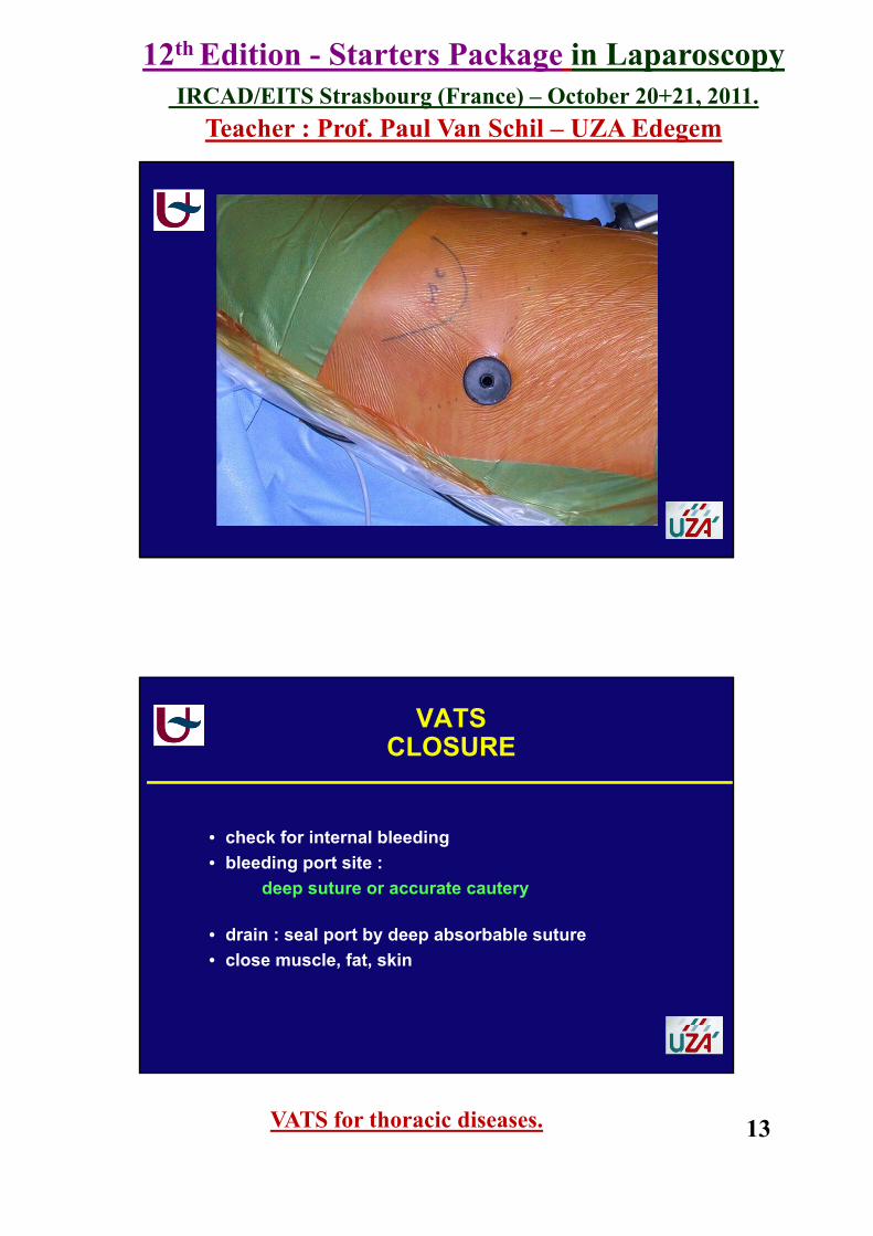
VATSCLOSURE
• check for internal bleeding• bleeding port site :
deep suture or accurate cautery
• drain : seal port by deep absorbable suture• close muscle, fat, skin

12th Edition - Starters Package in LaparoscopyIRCAD/EITS Strasbourg (France) – October 20+21, 2011.
Teacher : Prof. Paul Van Schil – UZA Edegem
VATS for thoracic diseases. 14
VATS
introductionbasic principles : accesspneumothoraxpleural diseasesmediastinal diseases
VATS for thoracic diseases

12th Edition - Starters Package in LaparoscopyIRCAD/EITS Strasbourg (France) – October 20+21, 2011.
Teacher : Prof. Paul Van Schil – UZA Edegem
VATS for thoracic diseases. 15
Pneumothorax : classification
air entering pleural spaceloss of negative pressure, lung collapse
classification
• spontaneous : primary, secondary• traumatic : blunt, penetrating chest injury• iatrogenic (subclavian vein puncture)
Pneumothorax : treatment options
• observation• needle aspiration• thoracic drain (tube thoracostomy)
water - seal, suction, Heimlich valve• instillation of pleural irritant (chemical pleurodesis)• VATS• thoracotomy, sternotomy
De Leyn P et al. Guidelines on the management of spontaneous pneumothorax. Acta Chir Belg 2005; 105:537-38

12th Edition - Starters Package in LaparoscopyIRCAD/EITS Strasbourg (France) – October 20+21, 2011.
Teacher : Prof. Paul Van Schil – UZA Edegem
VATS for thoracic diseases. 16
Indications for operative intervention
2nd ipsilateral pneumothoraxrecurrence after 1st episode of SP treated with thoracic drainage 30%, after 2nd episode 50%, after 3rd episode 80%
1st contralateral pneumothoraxbilateral spontaneous pneumothoraxpersistent air leak (> 5-7 days)no re-expansion after 3 to 5 days treatmentcombined spontaneous hemothorax profession at risk: divers, pilots, aircraft personnel,…
VATS for pneumothorax
general anesthesiasingle lung ventilationlateral position of the patientthree port approach 5th or 6th intercostal space
1. anterior axillary line2. posterior axillary line3. subscapular
intervention on pleura and lung

12th Edition - Starters Package in LaparoscopyIRCAD/EITS Strasbourg (France) – October 20+21, 2011.
Teacher : Prof. Paul Van Schil – UZA Edegem
VATS for thoracic diseases. 17
VATS : treatment of the lung
resection of apical bullae (90%)recurrence ↓ 20% → 1.5% with bullectomy Naunheim et al. J Thorac Cardiovasc Surg 1995; 109:1198-1204
use of endostaplersalternatives: endoloop, endoscopic suture, electrocoagulation of small bullae
OUR CURRENT PRACTICE :Resection blebs or bullae with endostaplers
VATS : treatment of parietal pleura
pleurectomy (partial / total) versus mechanical pleurodesis (electrocoagulation / abrasion / laser)no randomized studies
OUR CURRENT PRACTICE :partial apical pleurectomy + abrasionThe thoracic cavity remains accessible for other procedures in the future

12th Edition - Starters Package in LaparoscopyIRCAD/EITS Strasbourg (France) – October 20+21, 2011.
Teacher : Prof. Paul Van Schil – UZA Edegem
VATS for thoracic diseases. 18
• ♂ °27/06/1971History
• smoker: cigarettes, cannabis• perforation gastric ulcer• pneumothorax R• dyspnea on exertion
A 37-year-old patient withbilateral bullous emphysema
Admission general hospital 08/08
• pain R hemithorax, fever • bilateral bullous emphysema• infected bulla
A 37-year-old patient withbilateral bullous emphysema
chest X-ray 250808

12th Edition - Starters Package in LaparoscopyIRCAD/EITS Strasbourg (France) – October 20+21, 2011.
Teacher : Prof. Paul Van Schil – UZA Edegem
VATS for thoracic diseases. 19
chest CT 270808
A 37-year-old patient withbilateral bullous emphysema
• IV antibiotics: resolution of infection• normal lung function• referred for bullectomy:
• history of pneumothorax• symptomatic: risk of recurrent infection
• VATS approach 011208: bullectomy, apical pleurectomy, volume reduction
A 37-year-old patient withbilateral bullous emphysema
12th Edition - Starters Package in LaparoscopyIRCAD/EITS Strasbourg (France) – October 20+21, 2011.
Teacher : Prof. Paul Van Schil – UZA Edegem
VATS for thoracic diseases. 20
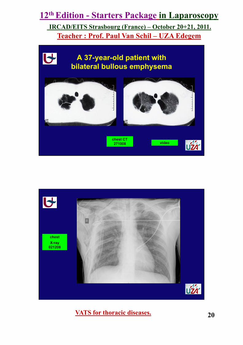
chest CT 271008
A 37-year-old patient withbilateral bullous emphysema
video
chest X-ray
021208
12th Edition - Starters Package in LaparoscopyIRCAD/EITS Strasbourg (France) – October 20+21, 2011.
Teacher : Prof. Paul Van Schil – UZA Edegem
VATS for thoracic diseases. 21
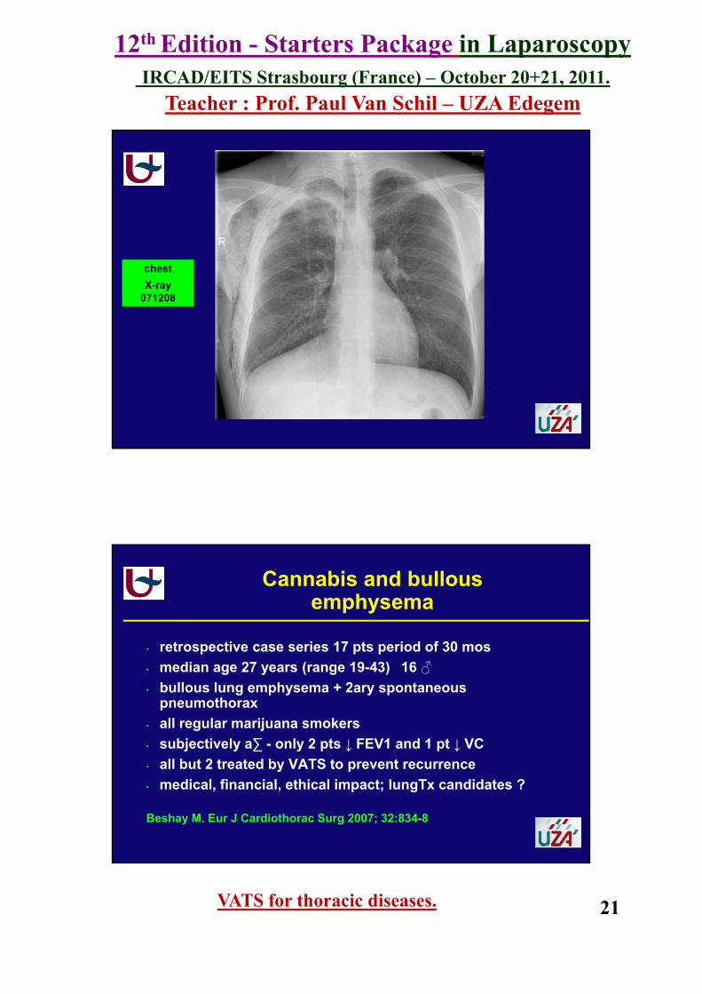
chest X-ray
071208
• retrospective case series 17 pts period of 30 mos • median age 27 years (range 19-43) 16 ♂• bullous lung emphysema + 2ary spontaneous
pneumothorax• all regular marijuana smokers• subjectively a∑ - only 2 pts ↓ FEV1 and 1 pt ↓ VC• all but 2 treated by VATS to prevent recurrence• medical, financial, ethical impact; lungTx candidates ?
Beshay M. Eur J Cardiothorac Surg 2007; 32:834-8
Cannabis and bullousemphysema
12th Edition - Starters Package in LaparoscopyIRCAD/EITS Strasbourg (France) – October 20+21, 2011.
Teacher : Prof. Paul Van Schil – UZA Edegem
VATS for thoracic diseases. 22
introductionbasic principles : accesspneumothoraxpleural diseasesmediastinal diseases
VATS for thoracic diseases
VATS for Pleural Diseases
Indications
• pleural effusion • malignancy : primary, metastatic• empyema : fibrinopurulent stage• pneumothorax : treatment of pleura• hemothorax (thoracic trauma < 2 weeks)• chylothorax
Khraim FM. The wider scope of video-assisted thoracoscopic surgery. AORN J 2007; 85:1199-1208
12th Edition - Starters Package in LaparoscopyIRCAD/EITS Strasbourg (France) – October 20+21, 2011.
Teacher : Prof. Paul Van Schil – UZA Edegem
VATS for thoracic diseases. 23
VATS for Pleural Diseases
Procedures
• pleural biopsy
• drainage procedureseffusionloculated fluid collectionremoval of debrisirrigation
• exploration for hemothorax
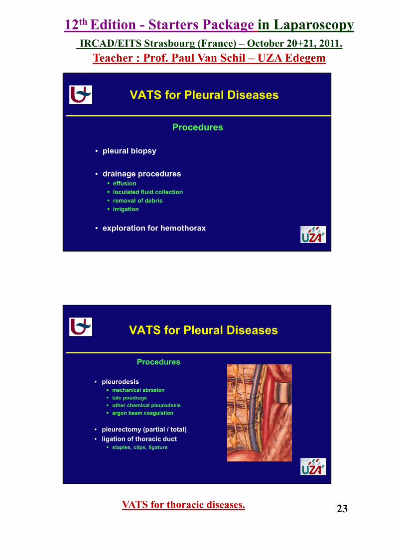
VATS for Pleural Diseases
Procedures
• pleurodesismechanical abrasiontalc poudrageother chemical pleurodesisargon beam coagulation
• pleurectomy (partial / total)• ligation of thoracic duct
staples, clips, ligature

12th Edition - Starters Package in LaparoscopyIRCAD/EITS Strasbourg (France) – October 20+21, 2011.
Teacher : Prof. Paul Van Schil – UZA Edegem
VATS for thoracic diseases. 24
64-year-old ♂recurrent pleural effusion
thoracentesis : atypical cells ?
VATS for Pleural Diseases
VATS for Pleural Diseases
12th Edition - Starters Package in LaparoscopyIRCAD/EITS Strasbourg (France) – October 20+21, 2011.
Teacher : Prof. Paul Van Schil – UZA Edegem
VATS for thoracic diseases. 25
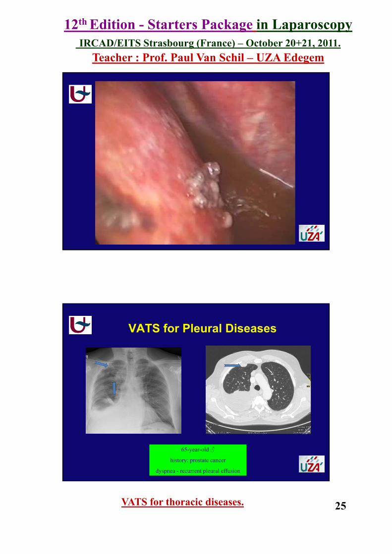
65-year-old ♂
history: prostate cancer
dyspnea - recurrent pleural effusion
VATS for Pleural Diseases
12th Edition - Starters Package in LaparoscopyIRCAD/EITS Strasbourg (France) – October 20+21, 2011.
Teacher : Prof. Paul Van Schil – UZA Edegem
VATS for thoracic diseases. 26
introductionbasic principles : accesspneumothoraxpleural diseasesmediastinal diseases
VATS for thoracic diseases
12th Edition - Starters Package in LaparoscopyIRCAD/EITS Strasbourg (France) – October 20+21, 2011.
Teacher : Prof. Paul Van Schil – UZA Edegem
VATS for thoracic diseases. 27
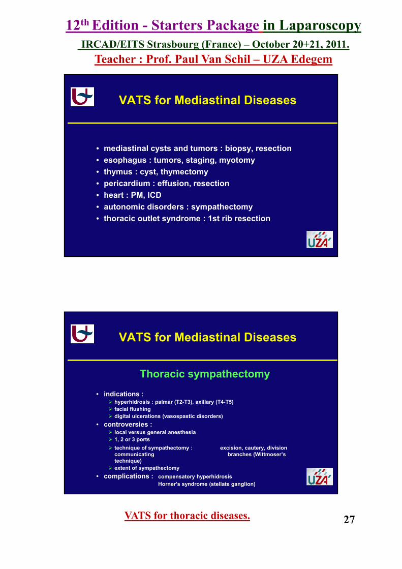
VATS for Mediastinal Diseases
• mediastinal cysts and tumors : biopsy, resection• esophagus : tumors, staging, myotomy• thymus : cyst, thymectomy• pericardium : effusion, resection• heart : PM, ICD• autonomic disorders : sympathectomy• thoracic outlet syndrome : 1st rib resection
VATS for Mediastinal Diseases
Thoracic sympathectomy
• indications : hyperhidrosis : palmar (T2-T3), axillary (T4-T5)facial flushingdigital ulcerations (vasospastic disorders)
• controversies :local versus general anesthesia1, 2 or 3 portstechnique of sympathectomy : excision, cautery, division communicating branches (Wittmoser’stechnique)extent of sympathectomy
• complications : compensatory hyperhidrosisHorner’s syndrome (stellate ganglion)

12th Edition - Starters Package in LaparoscopyIRCAD/EITS Strasbourg (France) – October 20+21, 2011.
Teacher : Prof. Paul Van Schil – UZA Edegem
VATS for thoracic diseases. 28
VATS for Mediastinal Diseases
VIDEO
VATS for Mediastinal Diseases
Thymectomy
• thymic hyperplasia (myasthenia gravis) • thymoma ? (small lesions)• 3D : da Vinci robotic system• controversies :
approach : R or L side, R+L, combined with cervical incisionextent of resection : complete, radical

12th Edition - Starters Package in LaparoscopyIRCAD/EITS Strasbourg (France) – October 20+21, 2011.
Teacher : Prof. Paul Van Schil – UZA Edegem
VATS for thoracic diseases. 29
VATSConclusions
indications variety of pulmonary, pleural and mediastinal diseasespneumothorax : treatment of lung + pleura
advantages minimally invasive techniquesuperb visualization (difficult regions)
disadvantages technical possibilities ≠ open techniquecost
VATSConclusions
position as lateral thoracotomy for most proceduresanesthesia double lumen tube (single lung ventilation)
epidural cathetertechnique 3 - 4 thoracoports
future 3D robotic systemsremote, superb handling of instruments


![Research13 - PatientPop · thoracic disc disease, thoracoscopy. VATS] §pine 2002; 27:871-879 The incidence of clinically significant thoracic disc her- niation, according to results](https://img.pdfslide.net/doc/110x75/5f11631833a9a5236f6a5b46/research13-patientpop-thoracic-disc-disease-thoracoscopy-vats-pine-2002.jpg)