Embed Size (px)
Citation preview
Mycologia Iranica 1(2): 75 – 85, 2014
Submitted 15 February 2014, accepted for Publication 11 Oct. 2014 Corresponding Author: E-mail: [email protected]
© 2014, Published by the Iranian Mycological Society http://mi.iranjournals.ir
Original Article
Vegetative compatibility and rep-PCR DNA fingerprinting
groups of Fusarium solani isolates obtained from different
hosts and their pathogenicity
M. Fallahi
M. Javan-Nikkhah
Kh. B. Fotouhifar
Department of Plant Protection, College of
Agriculture and Natural Resources, University of
Tehran, Karaj, Iran
M. Moradzadeh Eskandari
Plant Protection Research Department,
Agricultural and Natural Resources Research
Center of Khorasan-Razavi province, Mashhad,
Iran
Abstract: Fusarium solani is the most important
pathogen of a variety of host plants worldwide,
especially potato, which causes tuber rot in storage
and root rot of potato plants in the field. Fifty four
isolates obtained from potato, common bean, chickpea
and cucurbits (melon, watermelon and cucumber)
were subjected to analysis of vegetative compatibility
groups (VCGs) and rep-PCR DNA fingerprinting. Nit
mutants were used to force heterokaryon formation to
determine VCGs. Twenty three groups were determined,
which were designated as VCG A to VCG W. VCG
A was the largest group with 18 isolates and VCG B,
VCG C and VCG D were composed of 8, 6 and 3
isolates, respectively. Other groups were identified as
two or single-member VCGs. Presence of a high
degree of single-member VCGs indicates the large
amount of genetic diversity among isolates. Further-
more, the isolates of each host are classified in
different VCGs. Dendrogram generated using data of
rep-PCR, suggests a high level of genetic diversity
among the isolates. No correlation was found between
the DNA fingerprinting groups and host or geographical
origin of the isolates. Pathogenicity of twenty three
F. solani isolates as VCGs representatives originated
from different hosts was examined on plants and
tubers of Agria cultivar of potato. Except for four and
two isolates, other isolates were pathogenic on potato
plants and tubers, respectively. Pathogenicity tests
demonstrated that F. solani isolates do not have a host
specific behavior and isolates obtained from the non-
potato hosts are able to cause disease on potato plants
and tubers.
Key words: genetic diversity, Nit mutants, host specificity, molecular marker
INTRODUCTION
Fusarium solani (Mart.) Sacc. is one of the most
frequently isolated fungi from soil and plant debris
(Zhang et al. 2006). This species comprises phytopat-
hogenic and saprophytic strains. Phytopathogenic
strains are grouped in formae speciales based on their
host specificity (Snyder & Hansen 1941, Sakurai &
Matuo 1961, Roy 1997). The mentioned pathogen is
one of the major causes of potato disease in Iran, but unfortunately, the specific association between the
isolates of F. solani and potato plants has not been
approved and no special host has been established for
them (Moradzadeh Eskandari 2010). Specific forms
of this fungus are not distinguishable based on the
morphological characteristics (Suga et al. 2002). The
separation and identification of specific forms and
assessment of races diversity within a specific form,
solely based on the pathogenicity tests is not accurate.
These tests are often influenced by environmental
variables, such as temperature, host age and inocula- tion methods (Correll 1991).
Due to the soil born nature and long-term survival
of chlamydospores of this fungus in the soil (Hooker
1981), crop rotation strategy to control the disease
cannot be effective. One of the primary goals of plant
pathology has been to formulate strategies for disease
control. These methods usually have been based on
genetic resistance in the host plant or the application
of chemical fungicides. Understanding of genetic
diversity of fungi is very important to select the best
Strategies (Leslie 1993).
Access to the information about the pathogenic variability and genetic transmission potential can be
beneficial for researchers to understand the
relationships between fungal species and resistance
phenotype in breeding programs (McDonald & Linde
2002).
Hence, researchers have used genetic markers for
more precise identification and characterization.
Determination of vegetative compatibility groups can
be a useful method for the identification of specific
forms and the establishment of effective strategies for
controlling the disease (McDonald & Linde 2002). Fungal isolates that anastomose and form heterok-
76 Mycologia Iranica - Vol. 1, 2014
aryons with one another are considered to be
vegetative compatible and assigned to a single
vegetative compatible group (VCG). Conversely,
isolates that are incapable of anastomosing and
therefore fail to establish heterokaryons are referred
to as vegetative incompatible isolates (Anagnostakis 1982).
Correlations between VCGs and other characters
such as pathogenicity could lead to effectual
diagnostics (Leslie 1993). On the other hand, due to
the existence of self-incompatible isolates in the
fungus populations and inability of vegetative
compatibility groups in the expression of genetic
similarity between these isolates, using more
powerful tools is essential to overcome the existing
limitations. Thus, the use of molecular markers based
on PCR has been considered for this purpose.
Repetitive-sequence based polymerase chain reaction (rep-PCR) is based on PCR-mediated amplification of
DNA sequences located between specific interspersed
repeated sequences in prokaryotic genomes. These
repeated elements are termed as BOX, REP and ERIC
elements. The objectives of this study were
determination of vegetative compatibility groups to
clarify the genetic diversity among F. solani isolates
from different hosts and comparison of the
pathogenicity of vegetative compatibility groups’
representatives on potato plants in glasshouse and on
potato tubers in laboratory to examine the host specificity of the isolates, as well as comparison of
vegetative compatibility groups with rep-PCR.
MATERIALS AND METHODS
Fungal isolates Fifty four isolates of F. solani including twenty nine
isolates from potato (Solanum tuberosum), nine isolates
from chickpea (Cicera rietinum), nine isolates from
common bean (Phaseolus vulgaris), two isolates from cucumber (Cucumis sativus), three isolates from
watermelon (Citrullus lanatus), two isolates from melon
(Cucumis melo var. inodorus) collected from different
regions of Khorasan-Razavi province in east northern
Iran were selected for VCG determination, rep-PCR
DNA and pathogenicity tests.
Media Minimal medium (MM) was prepared by adding
2 g of NaNO3 in l L basal medium (Correll et al. 1987).
In an initial test, chlorate-resistant mutants were
generated on the medium amended with chlorate
(KClO3). In this study, we used three chlorate-
containing media, including MMC (MM medium
with Chlorate), PDC (Potato Dextrose Agar with
Chlorate) and CDAC (Czapek medium with Chlorate). To determine the suitable amount of KClO3, 15, 20,
30, 50 and 70 g/l KClO3 in PDA, MM and CDA was
tested. Extremely, 50-70 g/l KClO3 was selected so
that MMC was prepared by adding 50-70 g/l KClO3
to 1 L MM medium, PDC was made by adding 39 g
PDA, 5 g Davis agar and 50-70 g/l KCIO3 to 1 L
distilled water and CDAC was prepared by adding
39 g CDA and 50-70 g/l KClO3 in 1 L distilled water.
Generation and characterization of Nit mutants
For each isolate, mycelium was transferred from
PDA cultures to the medium containing KClO3.
Growth of wild-type strains of F. solani was restricted
by chlorate on MMC according to Correll et al.
(1986). The plates were incubated at 24 ± 2°C in the
dark for 10-14 days. Then, the rapidly expanding
sectors growing away from the restricted growth zone
were transferred to MM, and those that grew as the thin expansive colonies with no aerial mycelium on
MM were considered as nit mutants. The physiological phenotypes of nit mutants
were established by growing them on media containing
one of the four different nitrogen sources (nitrate,
nitrite, hypoxanthine and ammonium tartrate) following
Correll et al. (1987) (Table 1).
Complementation tests
Complementation tests were conducted on MM. Three 2 mm2 agar blocks containing a Nit M mutant
grown on MM were placed equidistantly apart across
the center of the Petri dish, and agar blocks of nit 1 or
nit 3 mutants grown on MM were placed at the three
matching positions, opposing the Nit M blocks in two
rows and 1.5 mm on either side of the Nit M blocks. This arrangement provided for complementation between different nit 1 or nit 3 mutants and a single
Nit M mutant. Complementation indicating heterokaryon
formation was recognized as a line of dense aerial
mycelial growth where two nit mutants met together
on MM (Correll et al. 1987).
Table 1. Identification of nit mutants of Fusarium solani isolates by growing them on different nitrogen sources (Correl et al. 1987).
Mutant designation
Medium supplement
Nitrate Nitrite Ammonium Hypoxanthine Chlorate
Wild type + + + + -
nit 1 - + + + +
nit 3 - - + + +
Nit M - + + - +
Crn* + + + + + +: Typical wild-type growing, -: Thin growing with no aerial mycelium, Crn*: chlorate resistant utilizing nitrate
FALLAHI ET AL.: Vegetative compatibility and rep-PCR DNA fingerprinting groups of Fusarium solani 77
DNA extraction Three to four mycelia plugs (each 4 mm in
diameter) from PDA cultures were transferred to
flasks containing 60 ml of potato dextrose broth, and
incubated at room temperature for 6–8 days. Mycelial
mass was filtered through a filter paper, washed three
times with sterile water, air-dried and then frozen at -
80°C and lyophilized prior to use. Lyophilized
mycelia were ground in liquid nitrogen into a fine
powder with a mortar and pestle. Fungal genomic
DNA was extracted using Core-one tm Plant Genomic
DNA isolation Kit (Core Bio, Korea) following the
manufacturers’ instructions.
Rep-PCR analysis Rep-PCR reaction was performed using BOX
primer (1A-1R 5'-CTACGGCAAGGCGACGCTG
ACG-3') (McDonald et al. 2000). PCR was carried
out using PCR Master Mix kit (CinnaGen PCR
Master Kit). Each reaction consisted of the following
components: 10 μL of PCR Master Mix (containing 1
U of Taq DNA polymerase, 2 μL PCR buffer, 3 mM
MgCl2 and 0.25 mM of each dNTPs), 10 pM primer,
3 μL (30 ng/μl) of fungal genomic DNA and 5.5 μL
distilled water in a final reaction volume of 20 μL.
PCR was performed in a palm Thermal Cycler (CG1-96, Corbett Research, Australia) with the following
PCR program. An initial denaturation step of 5 minutes
at 95°C followed by 35 cycles of denaturation at 94°C
for 3s and 92°C for 30s, 1 min of annealing at 49°C,
extension for 7 min at 72°C and a final extension for
10 min at 72°C.
Data analysis To determine the genetic relationships among the
isolates, the presence or absence of DNA bands was
converted into binary data (1 for presence and 0 for
absence of each band). Similarity matrix was
calculated with Dice’s coefficient and the SIMQUAL
program of NTSYSpc, ver. 2.1. Cluster analysis was
carried out within the SAHN program by using the UPGMA (Unweighted Pair Group Method with
Arithmetic Mean).
Virulence of VCGs representatives on potato
plants in greenhouse For greenhouse pathogenicity tests, spore suspens-
ions were created following Romberg & Davis (2007). Macroconidia and microconidia were harvested from
2-week-old cultures grown on PDA at 25ºC by adding
three ml of sterile water to the plates and scraping the
surface of the agar plate with a sterile glass slide. The
resulting conidial or mycelial suspension was filtered
through eight layers of cheesecloth to remove the
mycelial fragments. Conidial concentrations were
calculated using a hem cytometer and diluted in water
to a concentration of 106 macro- and microconidia/ml
for pathogenicity tests. The pathogenicity of twenty
three F. solani isolates obtained from potato, common
bean, chickpea, cucumber, watermelon and melon as
VCGs representatives was determined on healthy 8-
10 cm-tall potato seedlings of Agria cv., the most frequently potato cultivar in all potato growing
regions of Khorasan-Razavi province, Iran. After
trimming the roots, the entire root system was
immersed in a 106 macro- and microconidia/ml
suspension of each isolate. Each seedling was
transplanted into three separate 15-cm-diameter pots
containing sterile soil (5:3:2:1 mixture of farm soil,
sand, animal manure and compost). Each treatment
was replicated three times. The inoculated plants were maintained in greenhouse at 25ºC to 28ºC for 45- 50
days, and then removed from the pots. Then, they
were scored for disease severity based on a 0–3 scale
(0 = No disease, 1 = mild root rot, 2 = vascular
discoloration of the root and crown, 3 = vascular
discoloration of the root, crown and stem) (Moradzadeh
Eskandari 2010).
Virulence of VCGs representatives on potato
tubers under lab conditions Tubers of Agria cv. were used to determine the
virulence of the same VCGs representatives as those
used in the pathogenicity tests on plants. Initially, the
tubers appearing healthy and uniform in size (100-
120 g) were selected and washed to remove excess
soil, surface sterilized in 0.5% sodium hypochlorite
solution for 10 min, rinsed with three changes of
sterile distilled water and then air dried. Afterwards, the tubers were wounded with a four mm-diameter
cork borer to a depth of four mm (Theron & Holz
1989) and inoculated with all of the fresh F. solani
mycelia (from 1-week-fresh cultures grown on PDA
at 25°C) by putting 7 mm PDA blocks. All the
wounded potato tubers were wrapped in paper bags
(Manici & Cerato 1994) and incubated at 20°C under
dark conditions for three weeks. Blocks of PDA
medium were used as control. Each treatment was
replicated three times. At the end of incubation,
tubers were cut through the inoculation points, and
the degree of rot was estimated based on a zero to five (or A-F) scale, basically according to Theron and
Holz (1989): 0. (A) - Limited discoloration, no exten-
ded dry rot in inoculated areas; 1. (B) - Limited
discoloration with the development of dry rot in
inoculated areas; 2. (C) - Extensive discoloration with
increased dry rot in inoculated areas; 3. (D) - Extensive
discoloration with extensive dry rot in inoculated
areas; 4. (E) - discoloration and very extensive dry rot,
tubers not disappearing completely; 5. (F) - discolora-
tion and very extensive dry rot, tubers disappearing
completely.
RESULTS
Determination of VCGs Production of chlorate-resistant sectors was very
low on MMC, PDC and CDAC containing 15 g/l
KC103, but after increasing the concentration of
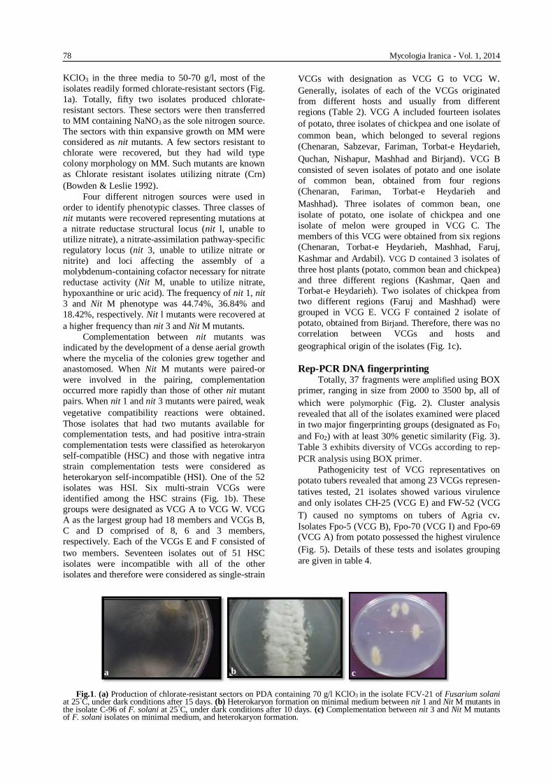
78 Mycologia Iranica - Vol. 1, 2014
KClO3 in the three media to 50-70 g/l, most of the
isolates readily formed chlorate-resistant sectors (Fig.
1a). Totally, fifty two isolates produced chlorate-
resistant sectors. These sectors were then transferred
to MM containing NaNO3 as the sole nitrogen source.
The sectors with thin expansive growth on MM were considered as nit mutants. A few sectors resistant to
chlorate were recovered, but they had wild type
colony morphology on MM. Such mutants are known
as Chlorate resistant isolates utilizing nitrate (Crn)
(Bowden & Leslie 1992). Four different nitrogen sources were used in
order to identify phenotypic classes. Three classes of
nit mutants were recovered representing mutations at
a nitrate reductase structural locus (nit l, unable to utilize nitrate), a nitrate-assimilation pathway-specific
regulatory locus (nit 3, unable to utilize nitrate or
nitrite) and loci affecting the assembly of a
molybdenum-containing cofactor necessary for nitrate
reductase activity (Nit M, unable to utilize nitrate,
hypoxanthine or uric acid). The frequency of nit 1, nit
3 and Nit M phenotype was 44.74%, 36.84% and
18.42%, respectively. Nit l mutants were recovered at
a higher frequency than nit 3 and Nit M mutants. Complementation between nit mutants was
indicated by the development of a dense aerial growth
where the mycelia of the colonies grew together and
anastomosed. When Nit M mutants were paired-or
were involved in the pairing, complementation
occurred more rapidly than those of other nit mutant
pairs. When nit 1 and nit 3 mutants were paired, weak
vegetative compatibility reactions were obtained. Those isolates that had two mutants available for complementation tests, and had positive intra-strain
complementation tests were classified as heterokaryon
self-compatible (HSC) and those with negative intra
strain complementation tests were considered as
heterokaryon self-incompatible (HSI). One of the 52
isolates was HSI. Six multi-strain VCGs were
identified among the HSC strains (Fig. 1b). These
groups were designated as VCG A to VCG W. VCG
A as the largest group had 18 members and VCGs B,
C and D comprised of 8, 6 and 3 members,
respectively. Each of the VCGs E and F consisted of
two members. Seventeen isolates out of 51 HSC
isolates were incompatible with all of the other
isolates and therefore were considered as single-strain
VCGs with designation as VCG G to VCG W.
Generally, isolates of each of the VCGs originated
from different hosts and usually from different
regions (Table 2). VCG A included fourteen isolates
of potato, three isolates of chickpea and one isolate of
common bean, which belonged to several regions
(Chenaran, Sabzevar, Fariman, Torbat-e Heydarieh,
Quchan, Nishapur, Mashhad and Birjand). VCG B
consisted of seven isolates of potato and one isolate
of common bean, obtained from four regions
(Chenaran, Fariman, Torbat-e Heydarieh and
Mashhad). Three isolates of common bean, one
isolate of potato, one isolate of chickpea and one
isolate of melon were grouped in VCG C. The
members of this VCG were obtained from six regions
(Chenaran, Torbat-e Heydarieh, Mashhad, Faruj,
Kashmar and Ardabil). VCG D contained 3 isolates of
three host plants (potato, common bean and chickpea)
and three different regions (Kashmar, Qaen and Torbat-e Heydarieh). Two isolates of chickpea from
two different regions (Faruj and Mashhad) were
grouped in VCG E. VCG F contained 2 isolate of
potato, obtained from Birjand. Therefore, there was no
correlation between VCGs and hosts and
geographical origin of the isolates (Fig. 1c).
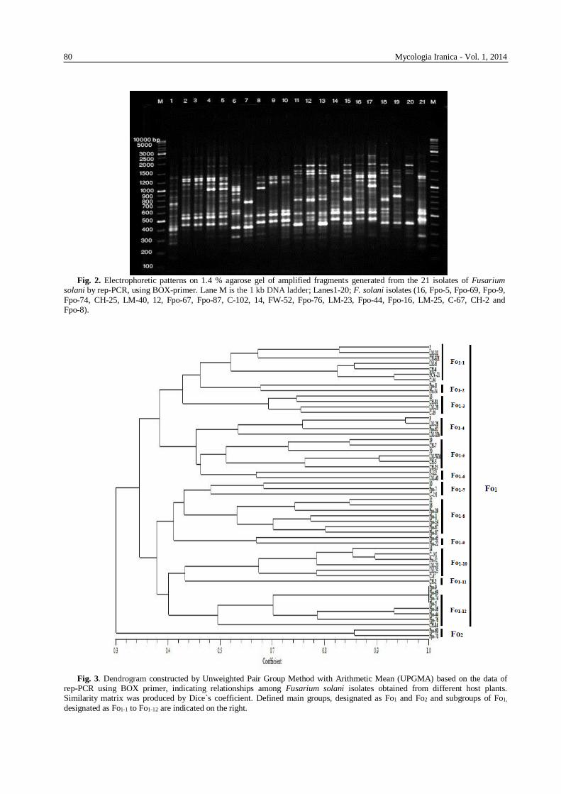
Rep-PCR DNA fingerprinting Totally, 37 fragments were amplified using BOX
primer, ranging in size from 2000 to 3500 bp, all of
which were polymorphic (Fig. 2). Cluster analysis
revealed that all of the isolates examined were placed
in two major fingerprinting groups (designated as Fo1
and Fo2) with at least 30% genetic similarity (Fig. 3). Table 3 exhibits diversity of VCGs according to rep-
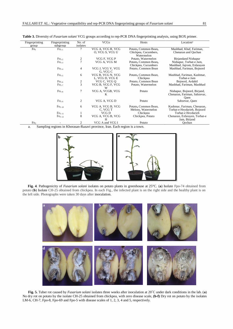
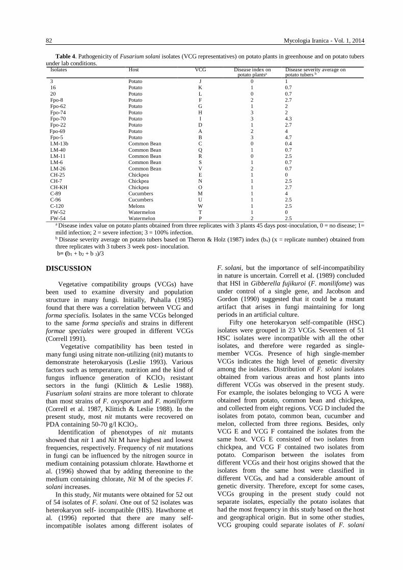
PCR analysis using BOX primer. Pathogenicity test of VCG representatives on
potato tubers revealed that among 23 VCGs represen-
tatives tested, 21 isolates showed various virulence
and only isolates CH-25 (VCG E) and FW-52 (VCG
T) caused no symptoms on tubers of Agria cv. Isolates Fpo-5 (VCG B), Fpo-70 (VCG I) and Fpo-69 (VCG A) from potato possessed the highest virulence
(Fig. 5). Details of these tests and isolates grouping
are given in table 4.
Fig.1. (a) Production of chlorate-resistant sectors on PDA containing 70 g/l KClO3 in the isolate FCV-21 of Fusarium solani
at 25°C, under dark conditions after 15 days. (b) Heterokaryon formation on minimal medium between nit 1 and Nit M mutants in the isolate C-96 of F. solani at 25°C, under dark conditions after 10 days. (c) Complementation between nit 3 and Nit M mutants of F. solani isolates on minimal medium, and heterokaryon formation.
a b c
FALLAHI ET AL.: Vegetative compatibility and rep-PCR DNA fingerprinting groups of Fusarium solani 79
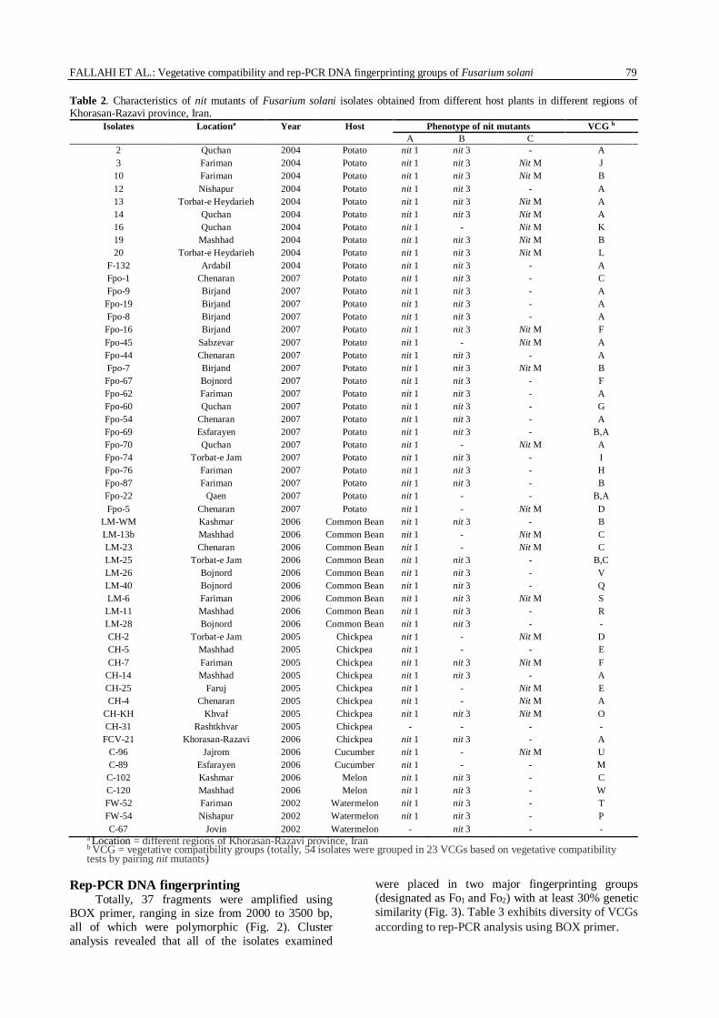
Table 2. Characteristics of nit mutants of Fusarium solani isolates obtained from different host plants in different regions of Khorasan-Razavi province, Iran.
Isolates Locationa Year Host Phenotype of nit mutants VCG b
A B C
2 Quchan 2004 Potato nit 1 nit 3 - A
3 Fariman 2004 Potato nit 1 nit 3 Nit M J
10 Fariman 2004 Potato nit 1 nit 3 Nit M B
12 Nishapur 2004 Potato nit 1 nit 3 - A
13 Torbat-e Heydarieh 2004 Potato nit 1 nit 3 Nit M A
14 Quchan 2004 Potato nit 1 nit 3 Nit M A
16 Quchan 2004 Potato nit 1 - Nit M K
19 Mashhad 2004 Potato nit 1 nit 3 Nit M B
20 Torbat-e Heydarieh 2004 Potato nit 1 nit 3 Nit M L
F-132 Ardabil 2004 Potato nit 1 nit 3 - A
Fpo-1 Chenaran 2007 Potato nit 1 nit 3 - C
Fpo-9 Birjand 2007 Potato nit 1 nit 3 - A
Fpo-19 Birjand 2007 Potato nit 1 nit 3 - A
Fpo-8 Birjand 2007 Potato nit 1 nit 3 - A
Fpo-16 Birjand 2007 Potato nit 1 nit 3 Nit M F
Fpo-45 Sabzevar 2007 Potato nit 1 - Nit M A
Fpo-44 Chenaran 2007 Potato nit 1 nit 3 - A
Fpo-7 Birjand 2007 Potato nit 1 nit 3 Nit M B
Fpo-67 Bojnord 2007 Potato nit 1 nit 3 - F
Fpo-62 Fariman 2007 Potato nit 1 nit 3 - A
Fpo-60 Quchan 2007 Potato nit 1 nit 3 - G
Fpo-54 Chenaran 2007 Potato nit 1 nit 3 - A
Fpo-69 Esfarayen 2007 Potato nit 1 nit 3 - B,A
Fpo-70 Quchan 2007 Potato nit 1 - Nit M A
Fpo-74 Torbat-e Jam 2007 Potato nit 1 nit 3 - I
Fpo-76 Fariman 2007 Potato nit 1 nit 3 - H
Fpo-87 Fariman 2007 Potato nit 1 nit 3 - B
Fpo-22 Qaen 2007 Potato nit 1 - - B,A
Fpo-5 Chenaran 2007 Potato nit 1 - Nit M D
LM-WM Kashmar 2006 Common Bean nit 1 nit 3 - B
LM-13b Mashhad 2006 Common Bean nit 1 - Nit M C
LM-23 Chenaran 2006 Common Bean nit 1 - Nit M C
LM-25 Torbat-e Jam 2006 Common Bean nit 1 nit 3 - B,C
LM-26 Bojnord 2006 Common Bean nit 1 nit 3 - V
LM-40 Bojnord 2006 Common Bean nit 1 nit 3 - Q
LM-6 Fariman 2006 Common Bean nit 1 nit 3 Nit M S
LM-11 Mashhad 2006 Common Bean nit 1 nit 3 - R
LM-28 Bojnord 2006 Common Bean nit 1 nit 3 - -
CH-2 Torbat-e Jam 2005 Chickpea nit 1 - Nit M D
CH-5 Mashhad 2005 Chickpea nit 1 - - E
CH-7 Fariman 2005 Chickpea nit 1 nit 3 Nit M F
CH-14 Mashhad 2005 Chickpea nit 1 nit 3 - A
CH-25 Faruj 2005 Chickpea nit 1 - Nit M E
CH-4 Chenaran 2005 Chickpea nit 1 - Nit M A
CH-KH Khvaf 2005 Chickpea nit 1 nit 3 Nit M O
CH-31 Rashtkhvar 2005 Chickpea - - - -
FCV-21 Khorasan-Razavi 2006 Chickpea nit 1 nit 3 - A
C-96 Jajrom 2006 Cucumber nit 1 - Nit M U
C-89 Esfarayen 2006 Cucumber nit 1 - - M
C-102 Kashmar 2006 Melon nit 1 nit 3 - C
C-120 Mashhad 2006 Melon nit 1 nit 3 - W
FW-52 Fariman 2002 Watermelon nit 1 nit 3 - T
FW-54 Nishapur 2002 Watermelon nit 1 nit 3 - P
C-67 Jovin 2002 Watermelon - nit 3 - - a Location = different regions of Khorasan-Razavi province, Iran b VCG = vegetative compatibility groups (totally, 54 isolates were grouped in 23 VCGs based on vegetative compatibility tests by pairing nit mutants)
Rep-PCR DNA fingerprinting Totally, 37 fragments were amplified using
BOX primer, ranging in size from 2000 to 3500 bp,
all of which were polymorphic (Fig. 2). Cluster
analysis revealed that all of the isolates examined
were placed in two major fingerprinting groups
(designated as Fo1 and Fo2) with at least 30% genetic
similarity (Fig. 3). Table 3 exhibits diversity of VCGs
according to rep-PCR analysis using BOX primer.
80 Mycologia Iranica - Vol. 1, 2014
Fig. 2. Electrophoretic patterns on 1.4 % agarose gel of amplified fragments generated from the 21 isolates of Fusarium
solani by rep-PCR, using BOX-primer. Lane M is the 1 kb DNA ladder; Lanes1-20; F. solani isolates (16, Fpo-5, Fpo-69, Fpo-9,
Fpo-74, CH-25, LM-40, 12, Fpo-67, Fpo-87, C-102, 14, FW-52, Fpo-76, LM-23, Fpo-44, Fpo-16, LM-25, C-67, CH-2 and Fpo-8).
Fig. 3. Dendrogram constructed by Unweighted Pair Group Method with Arithmetic Mean (UPGMA) based on the data of
rep-PCR using BOX primer, indicating relationships among Fusarium solani isolates obtained from different host plants. Similarity matrix was produced by Dice`s coefficient. Defined main groups, designated as Fo1 and Fo2 and subgroups of Fo1,
designated as Fo1-1 to Fo1-12 are indicated on the right.
FALLAHI ET AL.: Vegetative compatibility and rep-PCR DNA fingerprinting groups of Fusarium solani 81
Table 3. Diversity of Fusarium solani VCG groups according to rep-PCR DNA fingerprinting analysis, using BOX primer.
Fingerprinting group
Fingerprinting subgroup
No. of isolates
VCGs Hosts Locationa
Fo1 Fo1-1 7 VCG A, VCG R, VCG O, VCG S, VCG U
Potato, Common Bean, Chickpea, Cucumbers,
Watermelon
Mashhad, Khaf, Fariman, Chenaran and Quchan
Fo1-2 2 VCG F, VCG P Potato, Watermelon Birjandand Nishapur Fo1-3 7 VCG A, VCG M Potato, Common Bean,
Chickpea, Cucumbers Nishapur, Torbat-e Jam,
Mashhad, Jajrom, Esfarayen Fo1-4 4 VCG J, VCG V, VCG
G, VCG C Potato, Common Bean Mashhad, Fariman, Bojnord
Fo1-5 6 VCG B, VCG N, VCG L, VCG D, VCG E
Potato, Common Bean, Chickpea
Mashhad, Fariman, Kashmar, Torbat-e Jam
Fo1-6 2 VCG C, VCG Q Potato, Common Bean Bojnord, Ardabil Fo1-7 3 VCG B, VCG F, VCG
W Potato, Watermelon Mashhad, Fariman, Mashhad
Fo1-8 7 VCG A, VCGB, VCG K
Potato Nishapur, Bojnord, Birjand, Chenaran, Fariman, Sabzevar,
Qaen Fo1-9 2 VCG A, VCG D Potato Sabzevar, Qaen
Fo1-10 6 VCG A, VCG B, VCG C, VCG T
Potato, Common Bean, Melons, Watermelon
Kashmar, Fariman, Chenaran, Torbat-e Heydarieh, Bojnord
Fo1-11 1 VCG D Chickpea Torbat-e Heydarieh Fo1-12 8 VCG A, VCG B, VCG
H Chickpea, Potato Chenaran, Esfarayen, Torbat-e
Jam, Birjand Fo2 - 2 VCG A and VCG I Potato Qochan
a. Sampling regions in Khorasan-Razavi province, Iran. Each region is a town.
Fig. 4. Pathogenicity of Fusarium solani isolates on potato plants in greenhouse at 25°C. (a) Isolate Fpo-74 obtained from potato (b) Isolate CH-25 obtained from chickpea. In each Fig., the infected plant is on the right side and the healthy plant is on
the left side. Photographs were taken 30 days after inoculation.
Fig. 5. Tuber rot caused by Fusarium solani isolates three weeks after inoculation at 20°C under dark conditions in the lab. (a)
No dry rot on potato by the isolate CH-25 obtained from chickpea, with zero disease scale, (b-f) Dry rot on potato by the isolates LM-6, CH-7, Fpo-8, Fpo-69 and Fpo-5 with disease scales of 1, 2, 3, 4 and 5, respectively.
a b
a b c
d e f
82 Mycologia Iranica - Vol. 1, 2014
Table 4. Pathogenicity of Fusarium solani isolates (VCG representatives) on potato plants in greenhouse and on potato tubers under lab conditions.
Isolates Host VCG Disease index on apotato plants
Disease severity average on b tubers potato
3 Potato J 0 1
16 Potato K 1 0.7
20 Potato L 0 0.7
Fpo-8 Potato F 2 2.7
Fpo-62 Potato G 1 2
Fpo-74 Potato H 3 2
Fpo-70 Potato I 3 4.3
Fpo-22 Potato D 1 2.7
Fpo-69 Potato A 2 4
Fpo-5 Potato B 3 4.7
LM-13b Common Bean C 0 0.4
LM-40 Common Bean Q 1 0.7
LM-11 Common Bean R 0 2.5
LM-6 Common Bean S 1 0.7
LM-26 Common Bean V 2 0.7
CH-25 Chickpea E 1 0
CH-7 Chickpea N 1 2.5
CH-KH Chickpea O 1 2.7
C-89 Cucumbers M 1 4
C-96 Cucumbers U 1 2.5
C-120 Melons W 1 2.5
FW-52 Watermelon T 1 0
FW-54 Watermelon P 2 2.5 a Disease index value on potato plants obtained from three replicates with 3 plants 45 days post-inoculation, 0 = no disease; 1= mild infection; 2 = severe infection; 3 = 100% infection. b Disease severity average on potato tubers based on Theron & Holz (1987) index (bx) (x = replicate number) obtained from three replicates with 3 tubers 3 week post- inoculation. b= (b1 + b2 + b 3)/3
DISCUSSION
Vegetative compatibility groups (VCGs) have
been used to examine diversity and population
structure in many fungi. Initially, Puhalla (1985)
found that there was a correlation between VCG and
forma specialis. Isolates in the same VCGs belonged to the same forma specialis and strains in different
formae speciales were grouped in different VCGs
(Correll 1991).
Vegetative compatibility has been tested in
many fungi using nitrate non-utilizing (nit) mutants to
demonstrate heterokaryosis (Leslie 1993). Various
factors such as temperature, nutrition and the kind of
fungus influence generation of KClO3 resistant
sectors in the fungi (Klittich & Leslie 1988).
Fusarium solani strains are more tolerant to chlorate
than most strains of F. oxysporum and F. moniliform
(Correll et al. 1987, Klittich & Leslie 1988). In the present study, most nit mutants were recovered on
PDA containing 50-70 g/l KClO3.
Identification of phenotypes of nit mutants
showed that nit 1 and Nit M have highest and lowest
frequencies, respectively. Frequency of nit mutations
in fungi can be influenced by the nitrogen source in
medium containing potassium chlorate. Hawthorne et
al. (1996) showed that by adding thereonine to the
medium containing chlorate, Nit M of the species F.
solani increases.
In this study, Nit mutants were obtained for 52 out of 54 isolates of F. solani. One out of 52 isolates was
heterokaryon self- incompatible (HIS). Hawthorne et
al. (1996) reported that there are many self-
incompatible isolates among different isolates of
F. solani, but the importance of self-incompatibility
in nature is uncertain. Correll et al. (1989) concluded
that HSI in Gibberella fujikuroi (F. monilifome) was
under control of a single gene, and Jacobson and
Gordon (1990) suggested that it could be a mutant
artifact that arises in fungi maintaining for long
periods in an artificial culture.
Fifty one heterokaryon self-compatible (HSC)
isolates were grouped in 23 VCGs. Seventeen of 51
HSC isolates were incompatible with all the other isolates, and therefore were regarded as single-
member VCGs. Presence of high single-member
VCGs indicates the high level of genetic diversity
among the isolates. Distribution of F. solani isolates
obtained from various areas and host plants into
different VCGs was observed in the present study.
For example, the isolates belonging to VCG A were
obtained from potato, common bean and chickpea,
and collected from eight regions. VCG D included the
isolates from potato, common bean, cucumber and
melon, collected from three regions. Besides, only VCG E and VCG F contained the isolates from the
same host. VCG E consisted of two isolates from
chickpea, and VCG F contained two isolates from
potato. Comparison between the isolates from
different VCGs and their host origins showed that the
isolates from the same host were classified in
different VCGs, and had a considerable amount of
genetic diversity. Therefore, except for some cases,
VCGs grouping in the present study could not
separate isolates, especially the potato isolates that
had the most frequency in this study based on the host
and geographical origin. But in some other studies, VCG grouping could separate isolates of F. solani
FALLAHI ET AL.: Vegetative compatibility and rep-PCR DNA fingerprinting groups of Fusarium solani 83
based on the host origin. For instance, Hawthorn et al. (1996) placed 57 F. solani isolates in 35 VCGs, and
reported a direct relationship between VCG and host
origin of the isolates. Also, there was no correlation
between VCG grouping and geographical origins of
the isolates in the present study, except for VCG F
with two members that were collected from the same
region. On the other hand, the isolates of different
geographical areas were placed in the same VCG.
This suggests the possibility of distribution of the
fungus through infected tubers and agricultural
equipment, wind and rain in different regions.
According to Elmer (1991), existence of the isolates from different regions in the same VCG, or in the
other words, existence of a specific VCG in several
areas indicates the selective survival of vegetative
compatibility groups. This result was similar to the
results of Raouffi et al. (2004) and Mohammadi &
Banihashemi (2005). On the other hand, some studies
have demonstrated the correlation between vegetative
compatibility groups and geographical origins. Rahk-
hodaei (2000) reported that isolates of F. solani from
potato in each VCG were collected from the same
region. Dendrogram generated using data of BOX
primers showed that 54 isolates collected from
different regions of Khorasan-Razavi province and
from different hosts showed >30% similarity. This
result suggests a high level of genetic diversity among
the isolates. There was no correlation between the
identified fingerprinting groups and host and
geographical origins of the fungal isolates. These
results are in agreement with results from the
previous studies on F. solani (Moradzadeh Eskandari
2010, Romberg & Davis 2007, Baghai Raveri et al.
2007). Missing relationships between DNA fingerpr- inting groups and VCGs observed in this study
showed that it could be impossible to separate the
isolates based on VCGs using BOX primer. Using
VCG grouping and rep-PCR in the present study
showed the genetic diversity within F. solani isolates,
but both methods were unable to classify the isolates
based on the host origin. Therefore, using other
methods such as investigation of mating populations
will generate further information about the host
population of F. solani. Pathogenicity test of twenty
three F. solani isolates obtained from different hosts as VCGs representatives on potato plants showed that
there was no relation between the host origin and
virulence of the isolates, so that two out of four
isolates in pathogenicity zero group belonged to
potato host. In contrast, non-potato isolates in some
cases had more virulence than potato isolates, and
were comparable with potato isolates in severity of
symptoms. Also, the pathogenicity of twenty three
isolates as VCGs representatives on potato tubers
showed that except VCG T and VCG E represent-
tatives (from non-potato hosts), the rest of VCGs
representatives were able to cause dry rot on potato tubers with diverse virulence. This result confirmed
the results of their pathogenicity on potato plants. In a
way, the isolates tested in both pathogenicity tests, regardless of the host origin were grouped in different
pathogenicity groups. These results indicated that the
pathogenicity of F. solani isolates was not host specific
and the isolates obtained from non-potato hosts were
able to cause disease on potato plants and tubers. On
the other hand, the isolates obtained from potato
showed no symptom on potato plants. This result
confirmed the results of previous studies (Moradzadeh
Eskandari 2010, Romberg & Davis 2007).
ACKNOWLEDGMENTS
We gratefully acknowledge the High Council for
Research of University of Tehran for financially support and providing materials under grant number
7110024/6/24.
REFERENCES
Anagnostakis SL. 1982. Genetic analysis of
Endothia parasitica, linkage data for four single
genes and three vegetative compatibility types.
Genetics 102: 25-28.
Baghai Raveri S, Falahati Rastegar M, Jafarpor B,
Shokohifar F, Moradzadeh Eskandari M. 2007.
DNA fingerprinting of Fusarium solani isolates causing wilt and dry rot of potato in Razavi and
Northern Khorasan provinces using molecular
markers based on PCR. Iranian Journal of Plant
Pathology 42: 417-437.
Bowden RL, Leslie JF. 1992. Nitrate-nonutilizing
mutants of Gibberella zeae (Fusarium graminearum)
and their use in determining vegetative
compatibility. Experimental Mycology 16: 308–
315.
Correll JC. 1991. The relationship between formae
speciales, races, and vegetative compatibility groups in Fusarium oxysporum. Phytopathology
81: 1061-1064.
Correll JC, Klittich CJ, Leslie JF. 1989. Heterokaryon
self-incompatibility in Gibberella fujikuroi
(Fusarium moniliforme). Mycological Research
93: 21-27.
Correll JC, Klittich CJ, Leslie JF. 1987. Nitrate non-
utilizing mutants of Fusarium oxysporum and
their use in vegetative compatibility test.
Phytopathology 77: 1640-1646.
Correll JC, Puhalla JE, Schneider RW. 1986.
Identification of Fusarium oxysporum f. sp. apii on the basis of colony size, virulence and
vegetative compatibility. Phytopathology 76: 396-
400.
Elmer WH. 1991. Vegetative compatibility groups of
Fusarium proliferatum from asparagus and
comparisons of virulence, growth rates and
colonization of asparagus residues among groups.
Phytopathology 81: 852-857.
Hawthorne BT, Ball RD, Rees-George J. 1996. Use
of nitrate non-utilizing mutants to study vegetative
incompatibility in Fusarium solani (Nectria
84 Mycologia Iranica - Vol. 1, 2014
haematococca) especially members of mating populations I, V and VI. Mycological Research
100: 1075-1081.
Hooker WJ. 1981. Compendium of potato diseases.
The American Phytopathological Society Press
125 pp.
Jacobson DJ, Gordon TR. 1990. Variability of
mitochondrial DNA as an indicator of
relationships between populations of Fusarium
oxysporum f. sp. melonis. Mycological Research
94: 734-744.
Klittich CJ, Leslie JF. 1988. Nitrate reduction mutants
of Fusarium moniliforme (Gibberella fujikuroi). Genetics 118: 417-423.
Leslie JF. 1993. Fungal vegetative compatibility.
Annual Review of Phytopathology 31: 12–151.
Manici LM, Cerato C. 1994. Pathogenecity of
Fusarium oxysporum f. sp. tuberosi isolates from
tubers and potato plants. Potato Research 37: 129-
134. McDonald BA, Linde C. 2002. Pathogen population
genetics, evolutionary potential and durable resistance. Annual Review of Phytopathology 40:
349-379.
McDonald JG, Wong E, White GP. 2000. Differen-
tiation of Tilletia species by rep-PCR genomic
fingerprinting. Plant Disease 84: 1121–1125.
Mohammadi H, Banihashemi Z. 2005. Distribution,
pathogenicity and survival of Fusarium spp. the
causal agents of chickpea wilt and root rot in Fars
province. Iranian Journal of Plant Pathology. 41:
687-706.
Moradzadeh Eskandari M. 2010. Study on the population structure of Fusarium solani isolated
from potato in Khorasan provinces, determination
of phylogenetic relationship among some of its
formae speciales. Ph.D thesis, University of
Tehran, Iran 150 pp.
Puhalla JE. 1985. Classification of strains of Fusarium
oxysporum on the basis of vegetative compatibility.
Canadian Journal of Botany 63: 179-183.
Rahkhodaei E. 2000. Vegetative compatibility groups and pathogenicity of Fusarium solani and
Fusarium oxysporum from potato in Fars and
Khuzestan Provinces. M. Sc. Thesis, University of
Shahid Chamran, Ahvaz, Iran 124 pp. Raouffi M, Farrokhi-Nejad R, Mahmoudi SB. 2004.
Population genetic diversity of Fusarium solani
the causal agent of sugar beet root rot, using
vegetative compatibility groups (VCGs) and its
relationship to virulence of isolates. Sugar Beet
201: 39-53. Romberg MK, Davis RM. 2007. Host range and
phylogeny of Fusarium solani f. sp. eumartii from
potato and tomato in California. Plant Disease 91:
585-592. Roy KW. 1997. Fusarium solani on soybean roots,
nomenclature of the causal agent of sudden death
syndrome and identity and relevance of F. solani
form B. Plant Disease 81: 259–266. Sakurai Y, Matuo T. 1961. Taxonomy of the causal
fungus of trunk-blight of Xanthoxylum piperitum
and heterothallism in this fungus. Annals of the
Phytopathological Society of Japan 26: 112–117.
Snyder WC, Hansen HN. 1941. The species concept
in Fusarium with reference to Section Martiella.
American Journal of Botany 28: 738-742. Theron DJ, Holz G. 1989. Fusarium species associated
with dry and stem-end rot of potatoes in South
Africa. Phytophylactica 21: 175-181. Suga H, Ikeda S, Taga M, Kageyama K, Hyakumachi
M. 2002. Electrophoretic karyotyping and gene
mapping of seven formae speciales in
Fusarium solani. Current Genetic 41: 254-260. Zhang N, O’Donnell K, Sutton DA, Nalim FA,
Summerbell RC, Padhye AA, Geiser DM. 2006. Members of the Fusarium solani species complex
that cause infections in both humans and plants
are common in the environment. Journal of
Clinical Microbiology 44: 2186–2190.
FALLAHI ET AL.: Vegetative compatibility and rep-PCR DNA fingerprinting groups of Fusarium solani 85
در جدایه های rep-PCRبر اساس DNAگروه های سازگاری رویشی و انگشت نگاری
Fusarium solani بدست آمده از میزبان های مختلف و بیماریزایی آنها
2یاسکندر مرادزادهی مجتب و 1فری فتوحی برد لیخل ،1کخواهین جوان محمد ،1یفالح میمر
کرج تهران، دانشگاهی عیطب منابع وی کشاورز سیپرد ،یاهپزشکیگ گروه -1
مرکز تحقیقات کشاورزی و منابع طبیعی استان خراسان رضوی، مشهد ،گروه گیاهپزشکی -2
از مهمترین بیمارگرهای گیاهی می باشد که روی دامنه ی وسیعی از گیاهان از جمله سیب Fusarium solaniقارچ : چکیده
به عنوان یکی از عوامل اصلی پوسیدگی ریشه و غده ی سیب زمینی در انبار و زمینی در دنیا ایجاد خسارت می نماید. این گونه
از سیب زمینی، نخود، F. solaniجدایه گونه 45مزرعه محسوب می شود. به دلیل اهمیت این بیماری و شدت زیاد خسارت وارده،
بر اساس نشانگر DNAری رویشی و انگشت نگاری لوبیا، هندوانه، خربزه و خیار انتخاب شدند تا بر اساس شناسایی گروه های سازگا
rep-PCR مورد بررسی قرار گیرند. در این راستا شناسایی گروه های سازگاری رویشی بر اساس جداسازی جهش یافتگانnit و
بررسی بین جدایه های مورد (VCG)گروه سازگاری رویشی 22بررسی تشکیل هتروکاریون پایدار بین آنها انجام شد. در نهایت
عضو به عنوان بزرگترین گروه و 11با VCG Aنام گذاری شدند. گروه VCG Wتا VCG A مشخص شد. این گروه ها با اسامی
VCG B ،VCG C ،VCG D گروه به عنوان 11به ترتیب با هشت، شش و سه عضو مشخص شدند، همچنین دو گروه دو عضوی و
د زیاد گروه های تک عضوی نشان دهنده تنوع ژنتیکی نسبتاً زیاد در بین جدایه گروه های تک عضوی شناسایی شدند. وجود تعدا
های مورد مطالعه بود. از طرفی بین منشا میزبانی جدایه ها با گروه های سازگار رویشی مشخص شده، ارتباطی مشاهده نشد و
rep-PCRقرار گرفتند. بررسی دندروگرام رسم شده بر اساس نشانگر VCGجدایه های مربوط به هر میزبان در گروه های مختلف
نیز نشان دهنده ی تنوع ژنتیکی باال در جدایه های مورد بررسی در این مطالعه بود. و با توجه به نتایج در این بخش، همبستگی
مشاهده نشد. همچنین آزمون rep-PCRمشخصی بین منشاء جغرافیایی و میزبانی جدایه ها با گروه های مشخص شده با نشانگر
جدایه منتخب گروه های سازگار رویشی روی بوته و غده ی سیب زمینی رقم آگریا نشان داد که به ترتیب به جز 22بیماریزایی
چهار و دو جدایه که عالیمی روی بوته و غده ی سیب زمینی نشان ندادند و در گروه صفر قرار گرفتند، بقیه نمایندگان گروه های
VCG حداقل توانایی ایجاد پوسیدگی خفیف ریشه و کاهش رشد در بوته و پوسیدگی خشک در غده سیب زمینی را دارا بودند. این
نتایج نشان دهنده ی عدم اختصاصیت میزبانی در جدایه های مورد آزمون بود. چرا که در بین این جدایه ها عدم بیماریزایی جدایه
ی گیاه سیب زمینی وجود داشت، از طرف دیگر، جدایه های از میزبان غیر سیب زمینی قادر بودند هایی از میزبان سیب زمینی رو
شدت زیادی از بیماری را روی گیاه و غده سیب زمینی ایجاد نمایند.
، نشانگر مولکولی، اختصاصیت میزبانی Nitیافتگانجهش : تنوع ژنتیکی،کلمات کلیدی
Email: [email protected] محمد جوان نیکخواه مکاتبه کننده: