Embed Size (px)
Citation preview
Venous Thromboembolism
Core Rounds
April 10, 2003
A.F. Chad, MD, CCFP
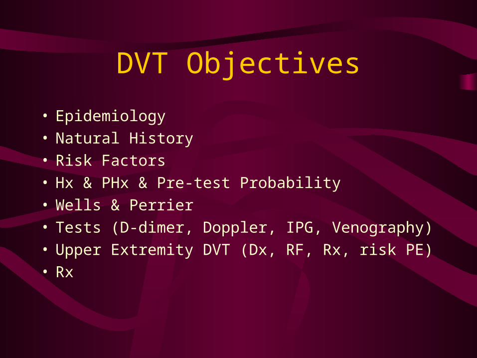
DVT Objectives
• Epidemiology• Natural History• Risk Factors• Hx & PHx & Pre-test Probability• Wells & Perrier• Tests (D-dimer, Doppler, IPG, Venography)• Upper Extremity DVT (Dx, RF, Rx, risk PE)• Rx

DVT: Submission move by
Jake “The Snake” Roberts OR Badness in the Veins?
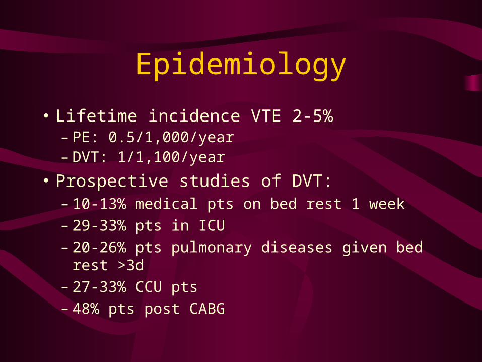
Epidemiology
• Lifetime incidence VTE 2-5%– PE: 0.5/1,000/year– DVT: 1/1,100/year
• Prospective studies of DVT:– 10-13% medical pts on bed rest 1 week– 29-33% pts in ICU – 20-26% pts pulmonary diseases given bed rest >3d – 27-33% CCU pts– 48% pts post CABG
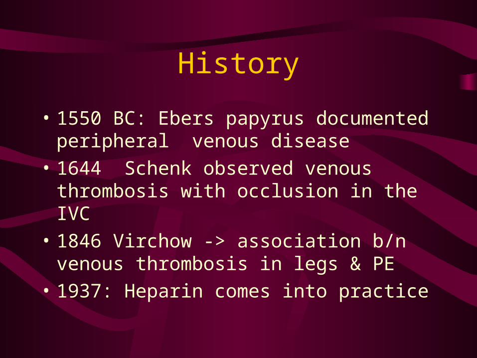
History
• 1550 BC: Ebers papyrus documented peripheral venous disease
• 1644 Schenk observed venous thrombosis with occlusion in the IVC
• 1846 Virchow -> association b/n venous thrombosis in legs & PE
• 1937: Heparin comes into practice

Natural History
• 19th C Virchow’s triad: venous stasis, injury to intima, hypercoagulability
• Thrombosis: platelet nidus near venous valves->platelets and fibrin -> thrombus -> occlusion, embolism
• Endogenous thrombolytic system -> partial dissolution-> organized into venous wall

Natural History
• Most go away w/o Rx
• 20% propagate proximally
• Organize into vein by day 5-10
• Biggest risk of propagation, embolization is before this

Natural History
• Debate whether isolated calf thrombi are important
• Some said to have low risk PE
• Others said just as bad
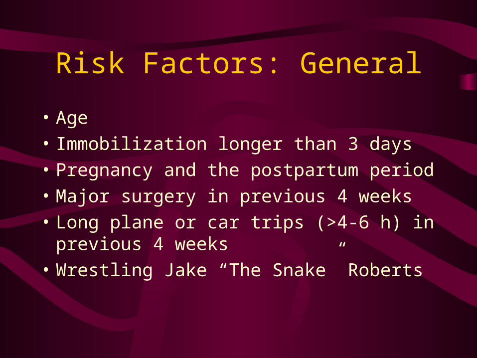
Risk Factors: General
• Age
• Immobilization longer than 3 days
• Pregnancy and the postpartum period
• Major surgery in previous 4 weeks
• Long plane or car trips (>4-6 h) in previous 4 weeks
• Wrestling Jake “The Snake” Roberts
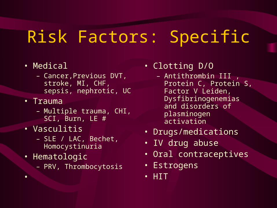
Risk Factors: Specific
• Medical – Cancer,Previous DVT, stroke,
MI, CHF, sepsis, nephrotic, UC
• Trauma – Multiple trauma, CHI, SCI,
Burn, LE #
• Vasculitis – SLE / LAC, Bechet,
Homocystinuria
• Hematologic – PRV, Thrombocytosis
•
• Clotting D/O– Antithrombin III , Protein
C, Protein S, Factor V Leiden, Dysfibrinogenemias and disorders of plasminogen activation
• Drugs/medications • IV drug abuse• Oral contraceptives• Estrogens• HIT
Risk Factors
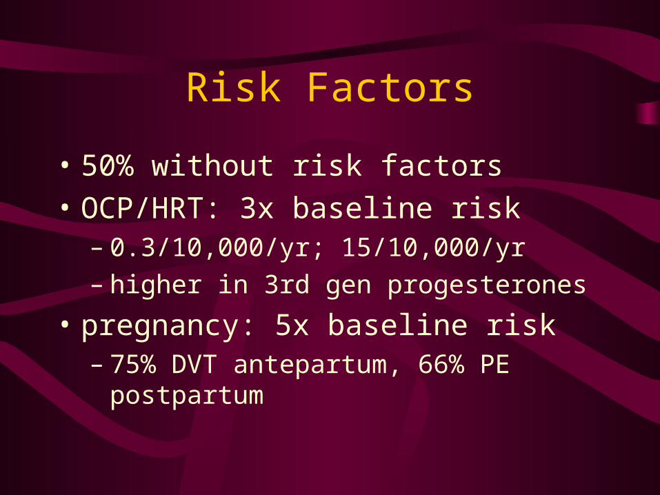
• 50% without risk factors
• OCP/HRT: 3x baseline risk – 0.3/10,000/yr; 15/10,000/yr– higher in 3rd gen progesterones
• pregnancy: 5x baseline risk– 75% DVT antepartum, 66% PE postpartum
Pathophysiology:Source of VTE
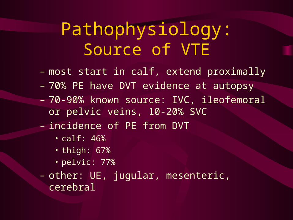
– most start in calf, extend proximally
– 70% PE have DVT evidence at autopsy
– 70-90% known source: IVC, ileofemoral or pelvic veins, 10-20% SVC
– incidence of PE from DVT• calf: 46%
• thigh: 67%
• pelvic: 77%
– other: UE, jugular, mesenteric, cerebral
History
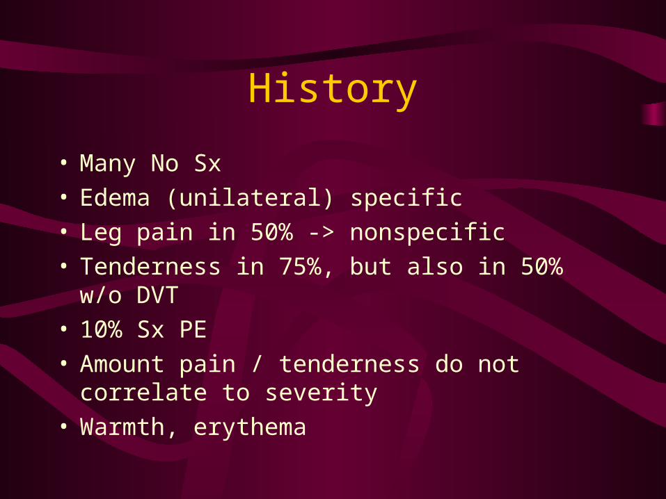
• Many No Sx• Edema (unilateral) specific• Leg pain in 50% -> nonspecific• Tenderness in 75%, but also in 50% w/o DVT• 10% Sx PE• Amount pain / tenderness do not correlate to
severity• Warmth, erythema
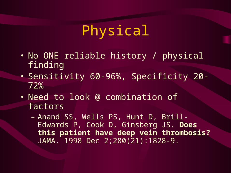
Physical
• No ONE reliable history / physical finding• Sensitivity 60-96%, Specificity 20-72%• Need to look @ combination of factors
– Anand SS, Wells PS, Hunt D, Brill-Edwards P, Cook D, Ginsberg JS. Does this patient have deep vein thrombosis? JAMA. 1998 Dec 2;280(21):1828-9.
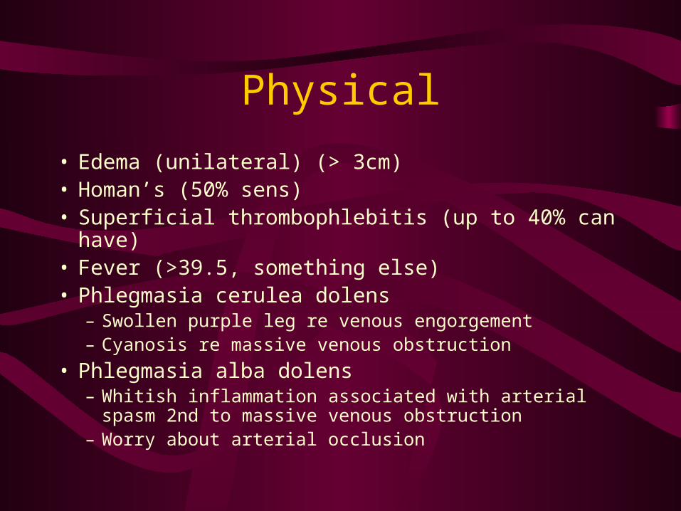
Physical
• Edema (unilateral) (> 3cm)• Homan’s (50% sens)• Superficial thrombophlebitis (up to 40% can have)• Fever (>39.5, something else)• Phlegmasia cerulea dolens
– Swollen purple leg re venous engorgement– Cyanosis re massive venous obstruction
• Phlegmasia alba dolens– Whitish inflammation associated with arterial spasm 2nd to
massive venous obstruction– Worry about arterial occlusion
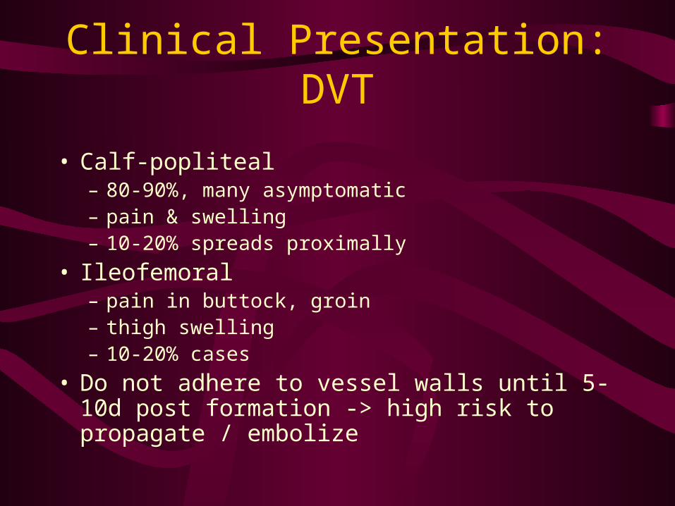
Clinical Presentation:DVT
• Calf-popliteal– 80-90%, many asymptomatic– pain & swelling– 10-20% spreads proximally
• Ileofemoral– pain in buttock, groin– thigh swelling– 10-20% cases
• Do not adhere to vessel walls until 5-10d post formation -> high risk to propagate / embolize
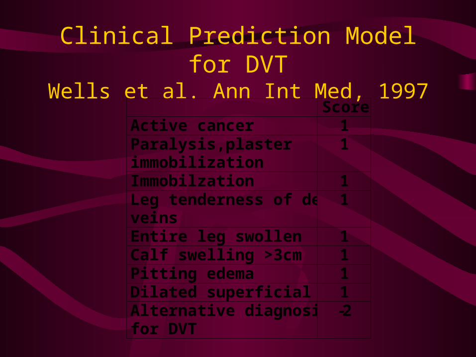
Clinical Prediction Model for DVTWells et al. Ann Int Med, 1997
Score Active cancer 1 Paralysis,plaster immobilization
1
Immobilzation 1 Leg tenderness of deep veins
1
Entire leg swollen 1 Calf swelling >3cm 1 Pitting edema 1 Dilated superficial veins 1 Alternative diagnosis for DVT
-2
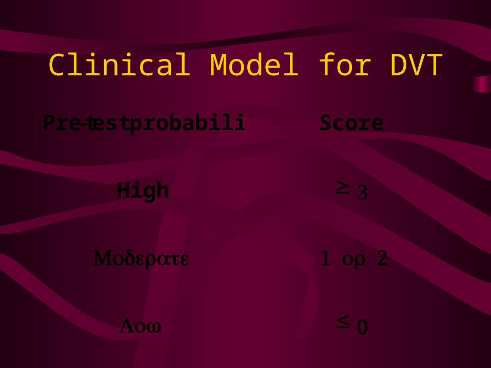
Clinical Model for DVT
Pre-test probability Score
High ≥ 3
Moderate 1 2or
Low ≤ 0
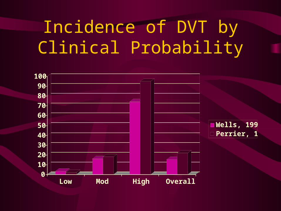
Incidence of DVT by Clinical Probability
0
10
20
30
40
50
60
70
80
90
100
Low Mod High Overall
Wells, 1997Perrier, 1999
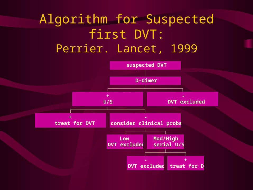
Algorithm for Suspected first DVT:Perrier. Lancet, 1999
+treat for DVT
LowDVT excluded
-DVT excluded
+treat for DVT
Mod/Highserial U/S
-consider clinical probability
+U/S
- DVT excluded
D-dimer
suspected DVT
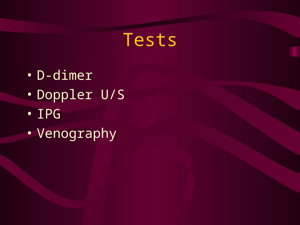
Tests
• D-dimer
• Doppler U/S
• IPG
• Venography
D-Dimer
• Not “Clot specific”
• recent surgery, trauma, MI, pregnancy, CA can all give false +
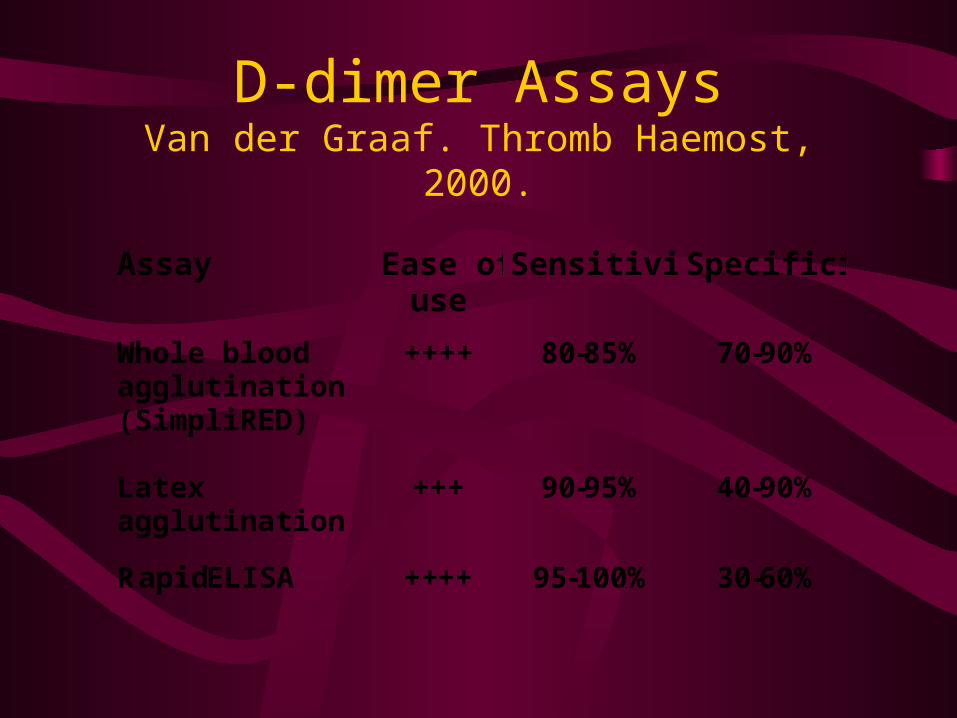
D-dimer AssaysVan der Graaf. Thromb Haemost, 2000.
Assay Ease of use
Sensitivity Specificity
Whole blood agglutination (SimpliRED)
++++ 80-85% 70-90%
Latex agglutination
+++ 90-95% 40-90%
Rapid ELISA ++++ 95-100% 30-60%
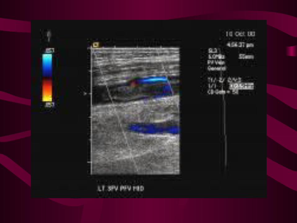
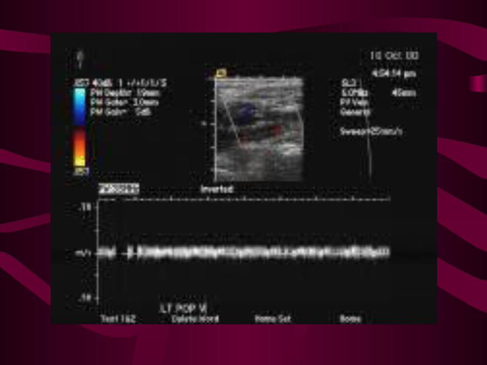
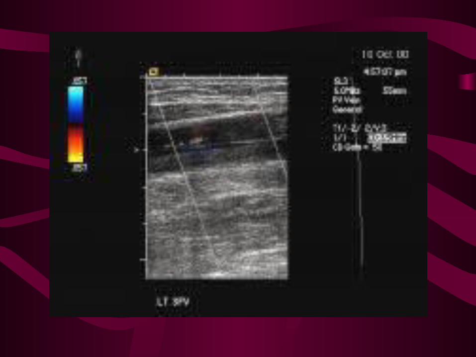
Diagnostic Imaging for DVT
• Duplex / compression U/S– non-invasive, portable– direct visualization of veins and flow– loss of compression = DVT– 97% sensitive & specific for symptomatic
proximal/popliteal DVT– 62% sensitive for asymptomatic DVT– +ve in 30-50% PE; 5% non-dx V/Q scans
Serial Venous U/S
• 2 protocols: Wells & Hull
• may avoid angiography in ?PE
• 2% +ve in 2 weeks (?PE)
• if U/S -ve 2 weeks apart, <2% have VTE in next 6 mos
Diagnostic Imaging for DVT
• IPG– detects changes in flow before and after cuff
inflated– sensitivity 60%
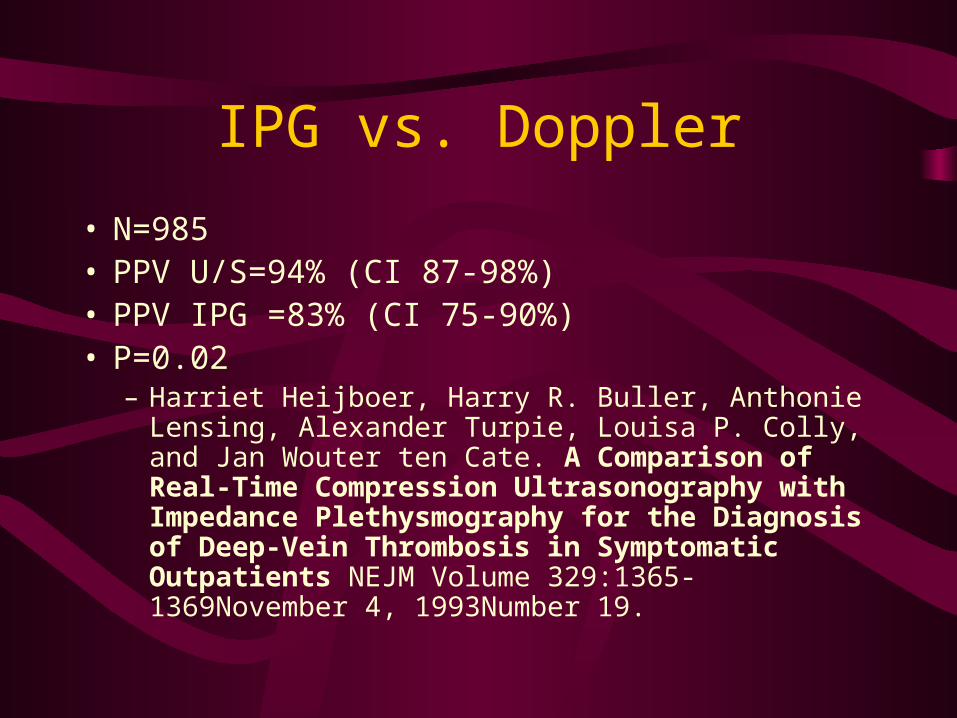
IPG vs. Doppler
• N=985• PPV U/S=94% (CI 87-98%)• PPV IPG =83% (CI 75-90%)• P=0.02
– Harriet Heijboer, Harry R. Buller, Anthonie Lensing, Alexander Turpie, Louisa P. Colly, and Jan Wouter ten Cate. A Comparison of Real-Time Compression Ultrasonography with Impedance Plethysmography for the Diagnosis of Deep-Vein Thrombosis in Symptomatic Outpatients NEJM Volume 329:1365-1369November 4, 1993Number 19.

Venography
• “?Gold Standard?”
• Invasive
• Contrast
• Need experienced readers
• Non-diagnostic up to 25%
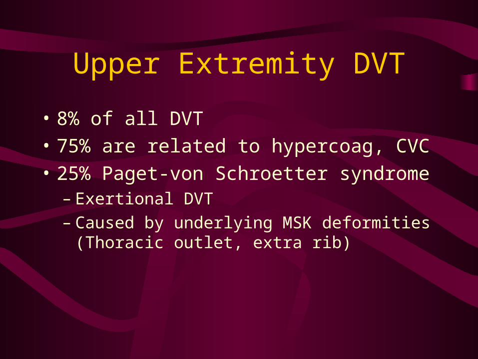
Upper Extremity DVT
• 8% of all DVT
• 75% are related to hypercoag, CVC
• 25% Paget-von Schroetter syndrome– Exertional DVT– Caused by underlying MSK deformities
(Thoracic outlet, extra rib)
Upper Extremity DVT
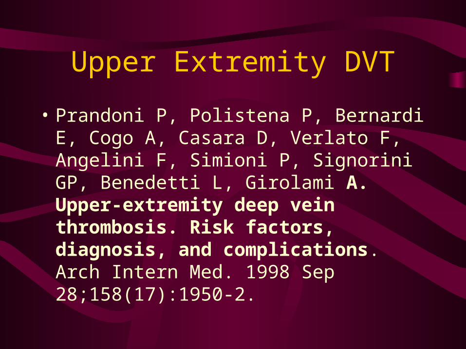
• Prandoni P, Polistena P, Bernardi E, Cogo A, Casara D, Verlato F, Angelini F, Simioni P, Signorini GP, Benedetti L, Girolami A. Upper-extremity deep vein thrombosis. Risk factors, diagnosis, and complications. Arch Intern Med. 1998 Sep 28;158(17):1950-2.
Upper Extremity DVT
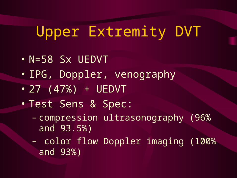
• N=58 Sx UEDVT
• IPG, Doppler, venography
• 27 (47%) + UEDVT
• Test Sens & Spec:– compression ultrasonography (96% and 93.5%) – color flow Doppler imaging (100% and 93%)
Upper Extremity DVT
• PE “Objectively” found in 36%
• 2 yr F/U: 2 recurrent VTE
• RF:– CVC– Thrombophilia– Previous VTE
U/S Upper Extremity DVT
• The sensitivity of duplex ultrasonography ranged from 56% to 100%, and the specificity ranged from 94% to 100%
• Unsure if Helpful– Bisher O. Mustafa, MD; Suman W. Rathbun, MD;
Thomas L. Whitsett, MD; Gary E. Raskob, PhD Sensitivity and Specificity of Ultrasonography in the Diagnosis of Upper Extremity Deep Vein Thrombosis: A Systematic Review Arch Int Med Vol. 162 No. 4, February 25, 2002.
Upper Extremity DVT
• 10-30% incidence PE associated
• Therapy:– Usual Rx– Local thrombolytics appears to be Rx of choice
with literature mainly case studies
Treatment of VTE:Goals
• reduce mortality
• prevent extension/recurrence
• restore pulmonary vascular resistance
• prevent pulmonary hypertension
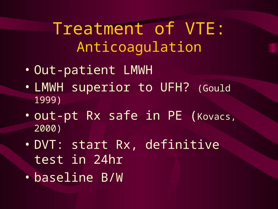
Treatment of VTE:Anticoagulation
• Out-patient LMWH• LMWH superior to UFH? (Gould 1999)
• out-pt Rx safe in PE (Kovacs, 2000)
• DVT: start Rx, definitive test in 24hr
• baseline B/W
Anticoagulation
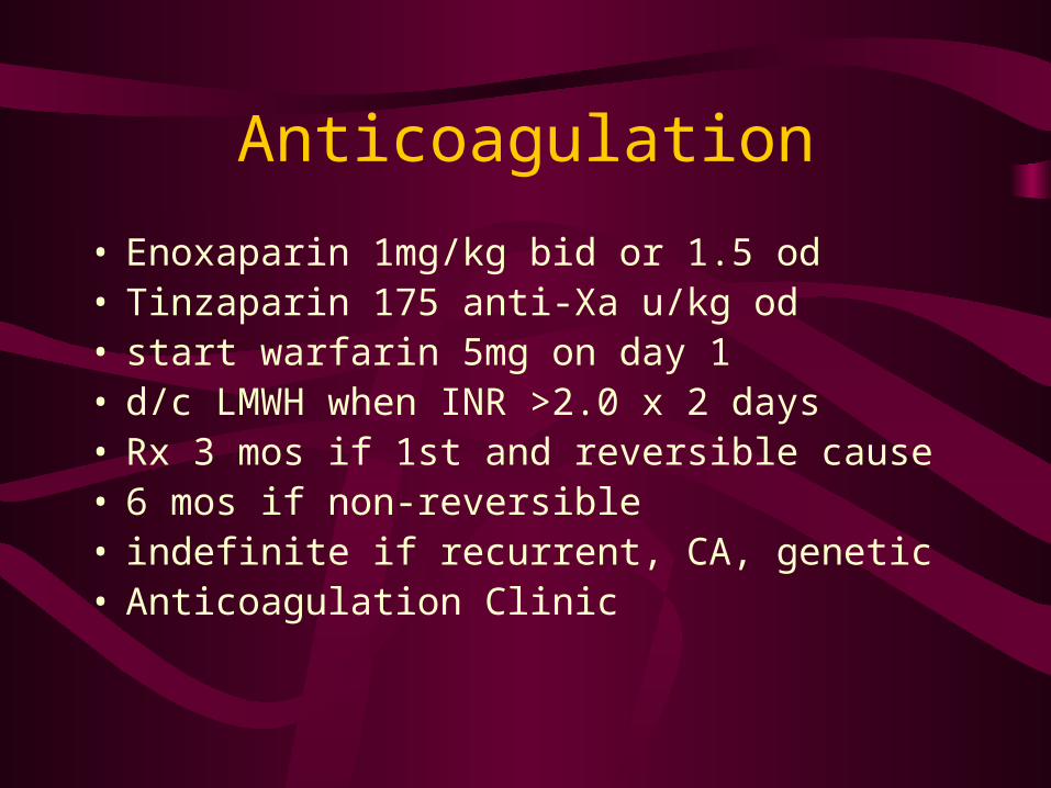
• Enoxaparin 1mg/kg bid or 1.5 od• Tinzaparin 175 anti-Xa u/kg od• start warfarin 5mg on day 1• d/c LMWH when INR >2.0 x 2 days• Rx 3 mos if 1st and reversible cause• 6 mos if non-reversible• indefinite if recurrent, CA, genetic• Anticoagulation Clinic
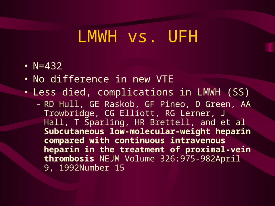
LMWH vs. UFH
• N=432• No difference in new VTE• Less died, complications in LMWH (SS)
– RD Hull, GE Raskob, GF Pineo, D Green, AA Trowbridge, CG Elliott, RG Lerner, J Hall, T Sparling, HR Brettell, and et al Subcutaneous low-molecular-weight heparin compared with continuous intravenous heparin in the treatment of proximal-vein thrombosis NEJM Volume 326:975-982April 9, 1992Number 15
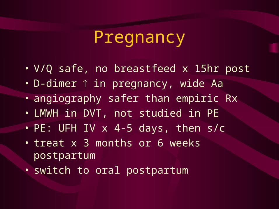
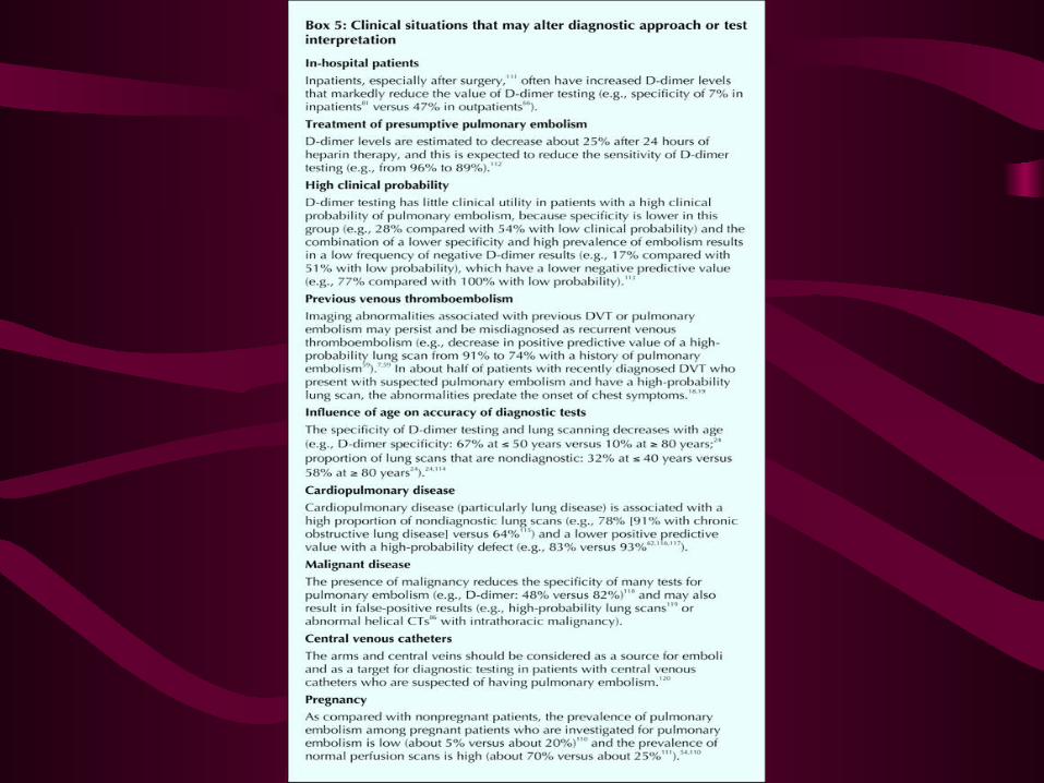
Pregnancy
• V/Q safe, no breastfeed x 15hr post• D-dimer in pregnancy, wide Aa • angiography safer than empiric Rx• LMWH in DVT, not studied in PE• PE: UFH IV x 4-5 days, then s/c• treat x 3 months or 6 weeks postpartum• switch to oral postpartum

PE: Early Rappers OR
Badness in the Veins?
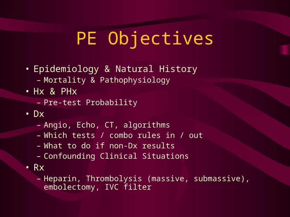
PE Objectives
• Epidemiology & Natural History – Mortality & Pathophysiology
• Hx & PHx– Pre-test Probability
• Dx – Angio, Echo, CT, algorithms – Which tests / combo rules in / out – What to do if non-Dx results – Confounding Clinical Situations
• Rx – Heparin, Thrombolysis (massive, submassive), embolectomy, IVC
filter
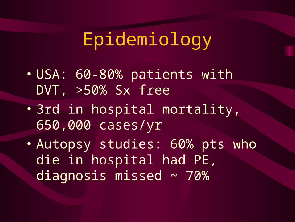
Epidemiology
• USA: 60-80% patients with DVT, >50% Sx free
• 3rd in hospital mortality, 650,000 cases/yr
• Autopsy studies: 60% pts who die in hospital had PE, diagnosis missed ~ 70%
Natural History
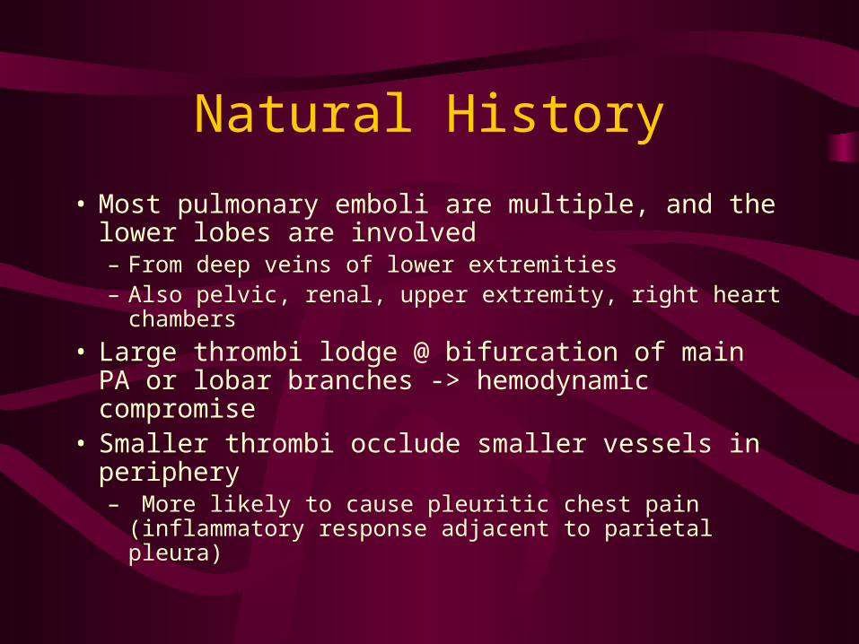
• Most pulmonary emboli are multiple, and the lower lobes are involved– From deep veins of lower extremities – Also pelvic, renal, upper extremity, right heart chambers
• Large thrombi lodge @ bifurcation of main PA or lobar branches -> hemodynamic compromise
• Smaller thrombi occlude smaller vessels in periphery– More likely to cause pleuritic chest pain (inflammatory
response adjacent to parietal pleura)
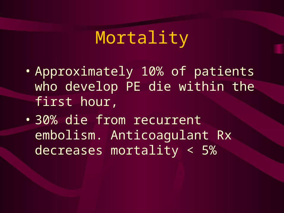
Mortality
• Approximately 10% of patients who develop PE die within the first hour,
• 30% die from recurrent embolism. Anticoagulant Rx decreases mortality < 5%
Pathophysiology Review
• Normal RV has a narrow range over which it can compensate for acute increases in afterload. The pericardium has a limited ability to distend.
• Increased RV afterload elevation in RV wall pressures dilation and hypokinesis of the RV wall
shift of intraventricular septum towards left ventricle (tricuspid regurgitation) and decreased LV output.
Respiratory Consequences
• Early– Increased alveolar dead space, Pneumoconstriction, hypoxemia,
hyperventilation
• Late: – regional loss surfactant, pulmonary infarction
• Arterial hypoxemia frequent, not universal– V/Q mismatch, shunts, reduced CO, intracardiac shunt via PFO
• Infarction uncommon re bronchial arterial collateral circulation
Hemodynamic Consequences
• Reduces X-sectional area of pulmonary vascular bed -> incr pulmonary vascular resistance -> RV afterload -> RV failure
• Reflex PA constriction
• Prior poor cardiopulmonary status important factor re hemodynamic collapse
Resolution
• Anticoagulant therapy -> resolution of emboli rapidly 2 weeks Rx
• Significant long-term nonresolution of emboli causing pulmonary HTN or cardiopulmonary symptoms uncommon
History: Size Matters
• DVT Risk factors, DVT• Massive:
– Shock, arrest (“Do you have any cousins with Factor V Leiden?”)
• Acute pulmonary Infarction: – pleuritic CP, SOB, hemoptysis
• Acute Emboli: – SOB, non-specific CP
• Multiple Small Emboli:– Progressive SOB, SOBOE, exertional CP, Cor Pulmonale

PIOPED Sx
• dyspnea (73%)
• pleuritic chest pain (66%)
• cough (37%)
• hemoptysis (13%)
Physical: Size Matters
• Massive pulmonary embolism– Shock , hypotension, poor perfusion,
tachycardia, and tachypnea.– Signs of pulmonary hypertension
• palpable impulse over 2nd LICS, loud P2, RV S3 gallop, and a systolic murmur louder on inspiration at left sternal border (TR)
Physical: Size Matters
• Acute pulmonary infarction– Decreased excursion of involved hemithorax,
palpable or audible pleural friction rub, localized tenderness
– Signs of pleural effusion
Physical: Size Matters
• Acute pulmonary Embolus (no infarct)– Non-specific– Tachypnea, tachycardia, pleuritic pain, crackles
and local wheeze @ embolus site

Physical: Size Matters
• Multiple pulmonary emboli or thrombi– Non-specific– Pulmonary HTN and cor pulmonale– High JVD, RV heave, palpable impulse 2nd
LICS, RV S3 gallop, systolic murmur over the left sternal border that is louder during inspiration, hepatomegaly, ascites, dependent pitting edema.
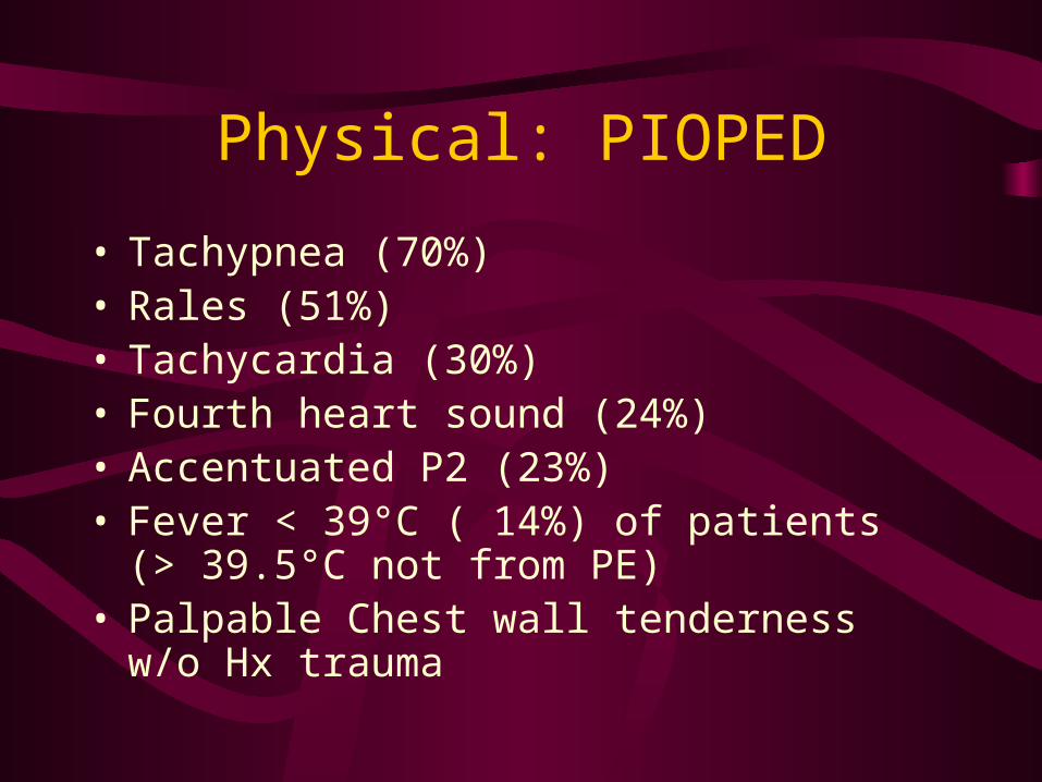
Physical: PIOPED
• Tachypnea (70%)• Rales (51%)• Tachycardia (30%)• Fourth heart sound (24%)• Accentuated P2 (23%)• Fever < 39°C ( 14%) of patients (> 39.5°C not
from PE) • Palpable Chest wall tenderness w/o Hx trauma
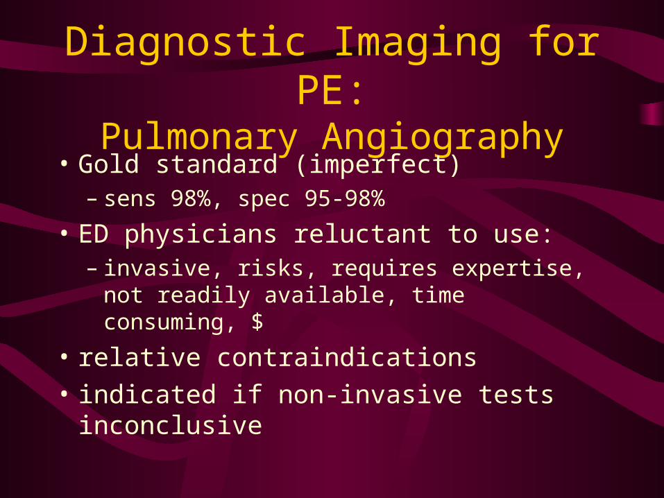
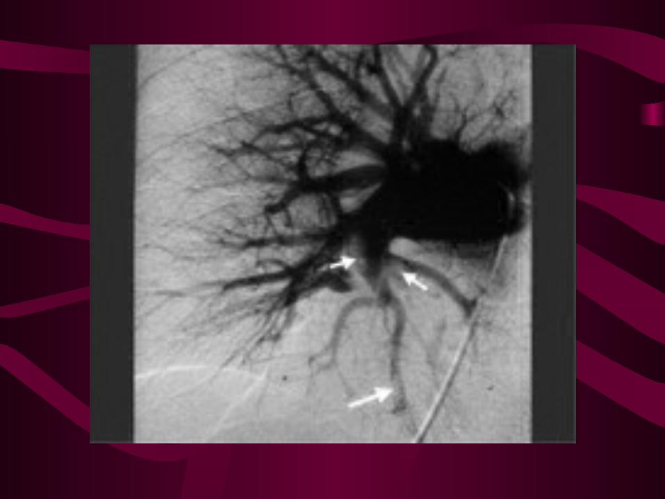
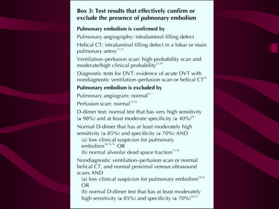
Diagnostic Imaging for PE:Pulmonary Angiography
• Gold standard (imperfect)– sens 98%, spec 95-98%
• ED physicians reluctant to use:– invasive, risks, requires expertise, not readily
available, time consuming, $
• relative contraindications
• indicated if non-invasive tests inconclusive
Diagnostic Imaging in PE:Echocardiography
• useful for patients in shock/arrest – r/o DDx: tamponade, Ao dissection, AMI
• indirect evidence of PE:– RV overload, septal shift to L, TR, PA pressure, RV
wall motion abn – sens 93%, spec 81%
• ‘sub-massive’ PE: independent predictor of mortality (?significance)

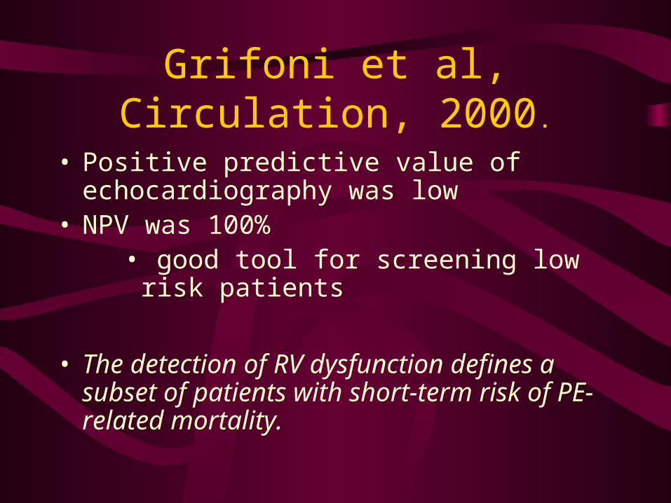
Grifoni et al. Short-Term Clinical Outcome of Patient With Acute
Pulmonary Embolism, Normal Blood Pressure and Echocardiographic Right Ventricular Dysfunction. Circulation,
101. 2000,
• Prospective clinical outcome study– 209 consecutive patients with documented PE– all patients had an TTE within 1 hr of admission– patients stratified into one of four groups– results only for in-hospital period
Grifoni et al, Circulation, 2000.
• 4 groups– Shock (N=28,13.4%)
• SBP<100 with signs of organ hypoperfusion– Hypotensive without signs of shock (N=19,
9.1%)– Normotensive with RV strain (N=65, 31.1%)– Normotensive without RV strain (N=97,
46.4%)
Grifoni et al, Circulation, 2000.
• Patients with hypotension/shock (22%, N=47)– Mortality 19%
• Normotensive without evidence of RV strain (46.5%, n=97)– 0 PE-related deaths
• Normotensive with RV strain (31.1%, N=65)– 10% (n=6) clinically deteriorated due to PE
recurrence– 5% (n=3) PE-related deaths
Grifoni et al, Circulation, 2000.
• Positive predictive value of echocardiography was low
• NPV was 100%• good tool for screening low risk patients
• The detection of RV dysfunction defines a subset of patients with short-term risk of PE-related mortality.
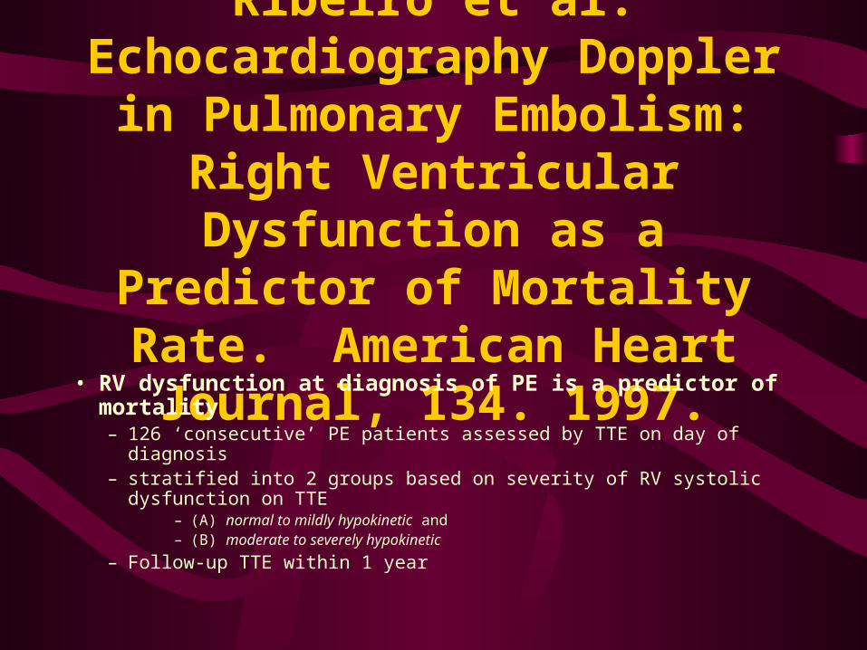
Ribeiro et al. Echocardiography Doppler in Pulmonary Embolism: Right Ventricular Dysfunction as a
Predictor of Mortality Rate. American Heart Journal, 134.
1997.• RV dysfunction at diagnosis of PE is a predictor of mortality
– 126 ‘consecutive’ PE patients assessed by TTE on day of diagnosis– stratified into 2 groups based on severity of RV systolic dysfunction on
TTE– (A) normal to mildly hypokinetic and – (B) moderate to severely hypokinetic
– Follow-up TTE within 1 year
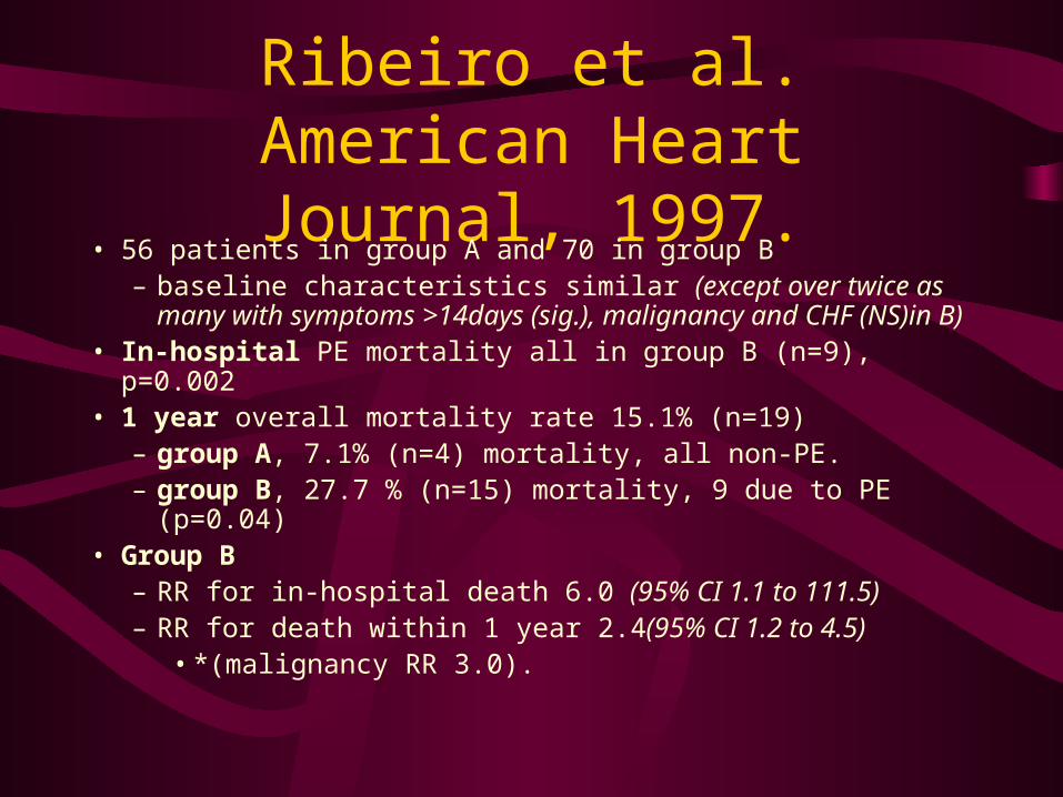
Ribeiro et al. American Heart Journal, 1997.
• 56 patients in group A and 70 in group B– baseline characteristics similar (except over twice as many
with symptoms >14days (sig.), malignancy and CHF (NS)in B)• In-hospital PE mortality all in group B (n=9), p=0.002• 1 year overall mortality rate 15.1% (n=19)
– group A, 7.1% (n=4) mortality, all non-PE.– group B, 27.7 % (n=15) mortality, 9 due to PE (p=0.04)
• Group B– RR for in-hospital death 6.0 (95% CI 1.1 to 111.5)– RR for death within 1 year 2.4(95% CI 1.2 to 4.5)
• *(malignancy RR 3.0).
Ribeiro et al. American Heart Journal, 1997.
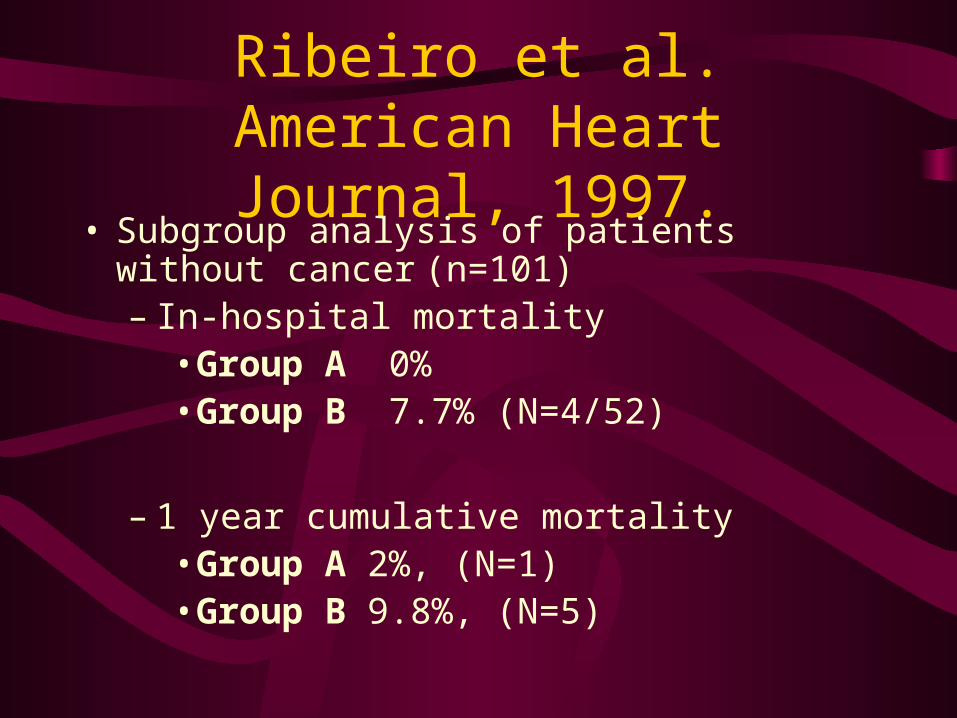
• Subgroup analysis of patients without cancer (n=101)– In-hospital mortality
• Group A 0%• Group B 7.7% (N=4/52)
– 1 year cumulative mortality• Group A 2%, (N=1)• Group B 9.8%, (N=5)
Moore, et al. Determination of Left Ventricular Function by Emergency
Physician Echocardiography of Hypotensive Patients. Academic
Emergency Medicine, vol. 9, no. 3, 2002.
• Prospective, observational study, convenience sample of 51.• EPs with prior US training underwent focused echo training• inclusion: symptomatic hypotension• exclusion: trauma, CPR, ECG of AMI• EPs estimation of EF
– compared with cardiologist; correlation coefficient of 0.86– between cardiologists 0.84
• EP categorization of EF, – agreement 84% (kappa 0.61)
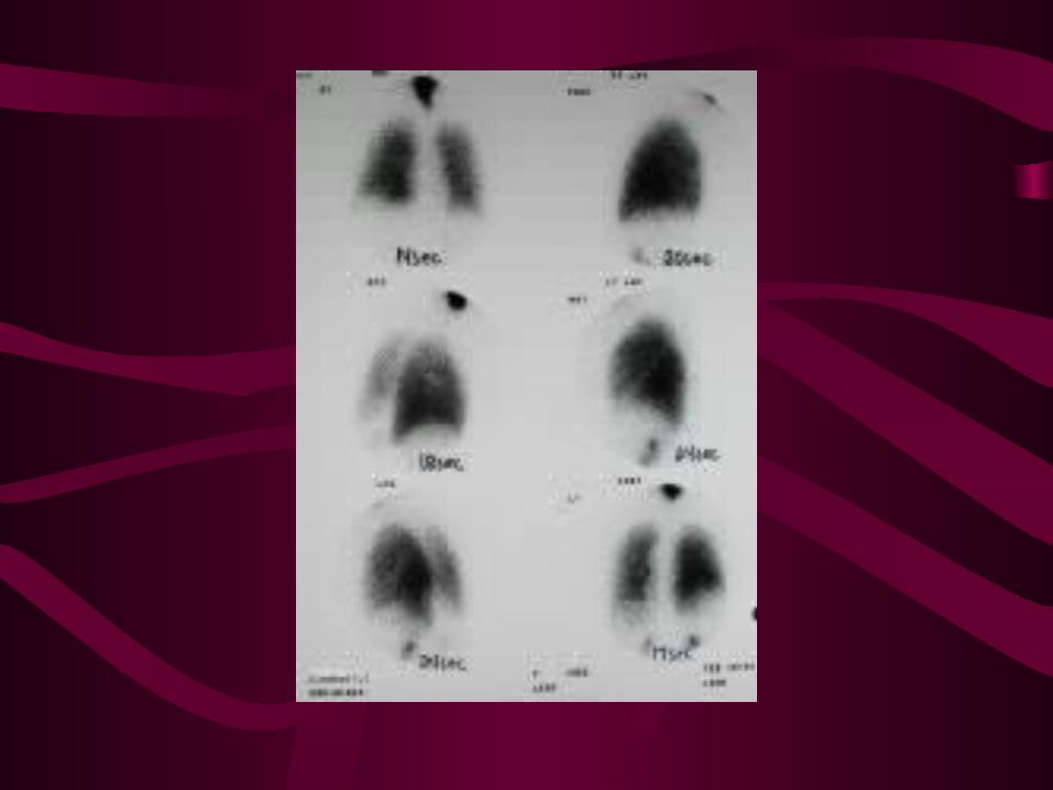
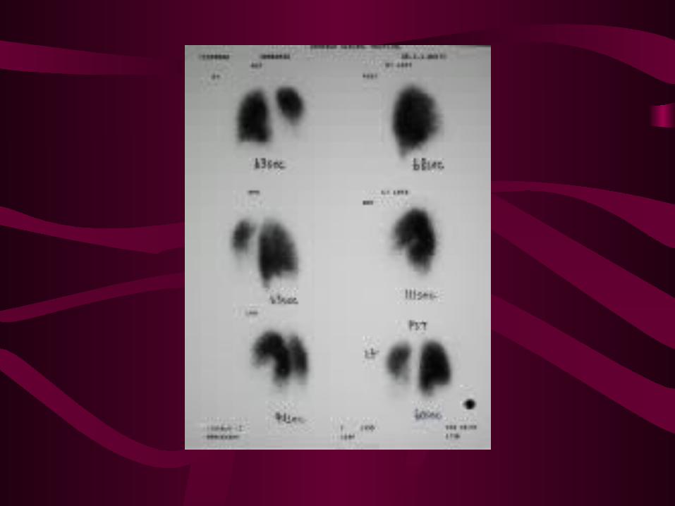
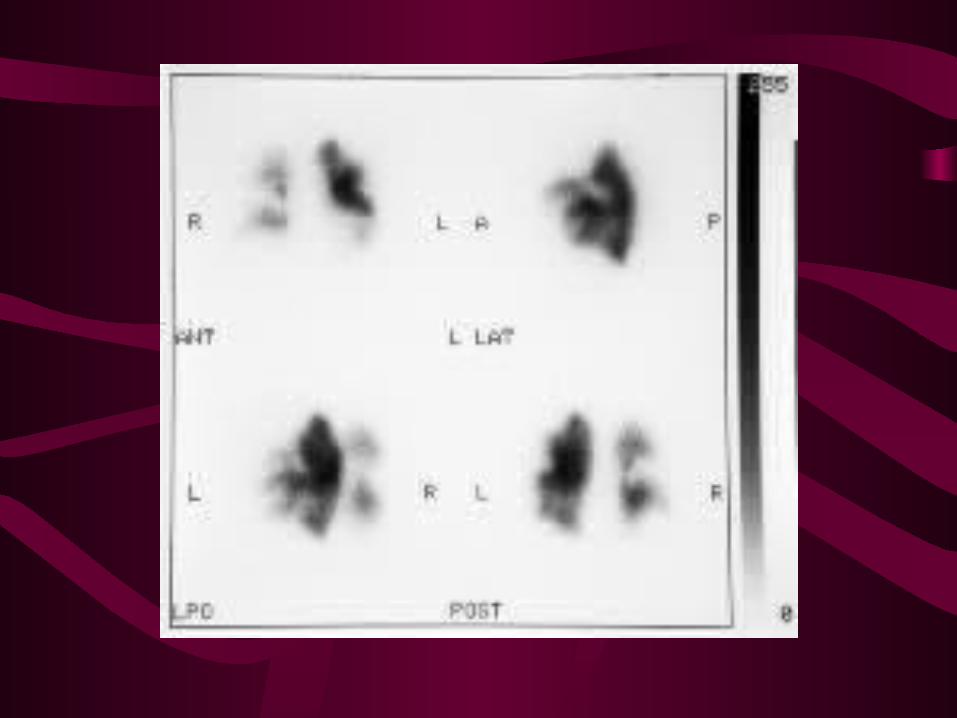
Diagnostic Imaging for PE:V/Q scan
• PIOPED: ventilation component adds little info• PISAPED criteria:
– normal, non-diagnostic, high probability
– 25%, 50%, 25% respectively
– high prob: 85-90% PPV
– non-diagnostic: 25% PE
• interpret in context of PTP
Relationship between degree of RV dysfunction and degree of perfusion scan
deficits• Wolfe, 1994. N=90
– degree of perfusion deficit greater in patients with RVD (54% vs 30%, p<0.001)
• all patients with recurrent PE in group with initial RVD, p<0.01
• Ribiero, 1998– correlation between RVD and perfusion scan deficit but
wide CI.• Miller, 1998. N=64
– failed to demonstrate a correlation between RVD and perfusion deficit
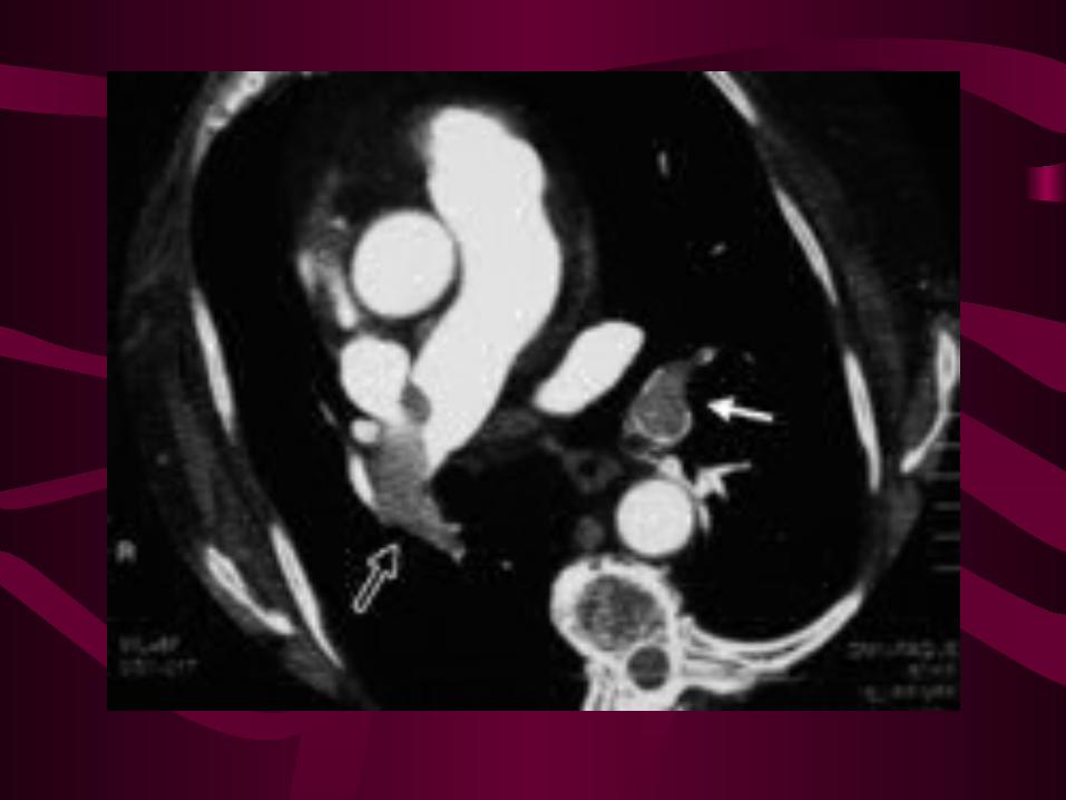
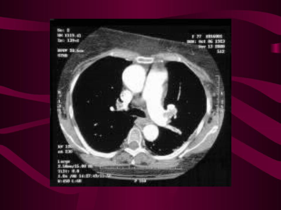
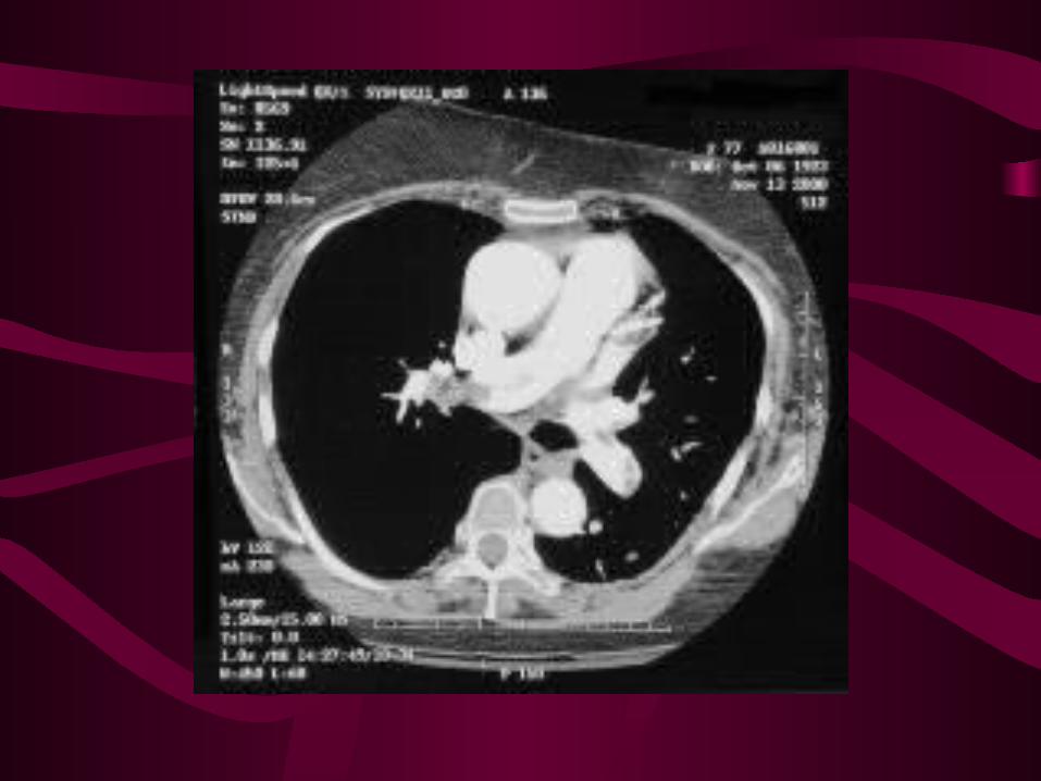
Diagnostic Imaging for PE:Spiral CT
• IV contrast, direct visualization• subsegmental PE not well seen• more specific, underlying lung dx• sens depends on CT, experience• wide variation in studies
– Rathbun. Ann Intern Med, 2000 (review)• sens 53-100%, spec 81-100%• poor methodology of studies
Spiral CT
• Perrier. Ann Intern Med, 2001– sens 70%, spec 91% , 4% inconclusive– good interobserver agreement
• CT venography:– benefit over U/S not determined
• role? – no evidence to withhold Rx if CT negative– may replace angiography
Clive Kearon. Diagnosis of pulmonary embolism. CMAJ:
January 21, 2003; 168 (2)
Non -Invasive Testing
• NEED TO Dx PE as HIGH MORTALITY IN THOSE NOT Dx or MISDIAGNOSED!
• Angiography carries risk
• Mortality 0.5%, invasive, labour intensive
• Can make Dx without P. angio
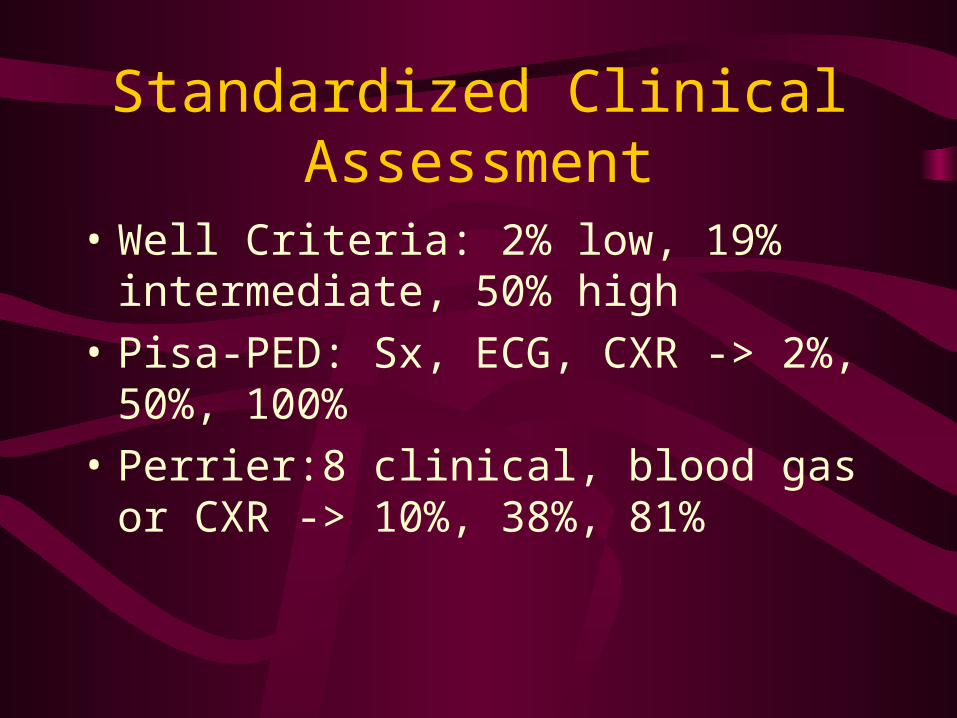
Standardized Clinical Assessment
• Well Criteria: 2% low, 19% intermediate, 50% high
• Pisa-PED: Sx, ECG, CXR -> 2%, 50%, 100%
• Perrier:8 clinical, blood gas or CXR -> 10%, 38%, 81%
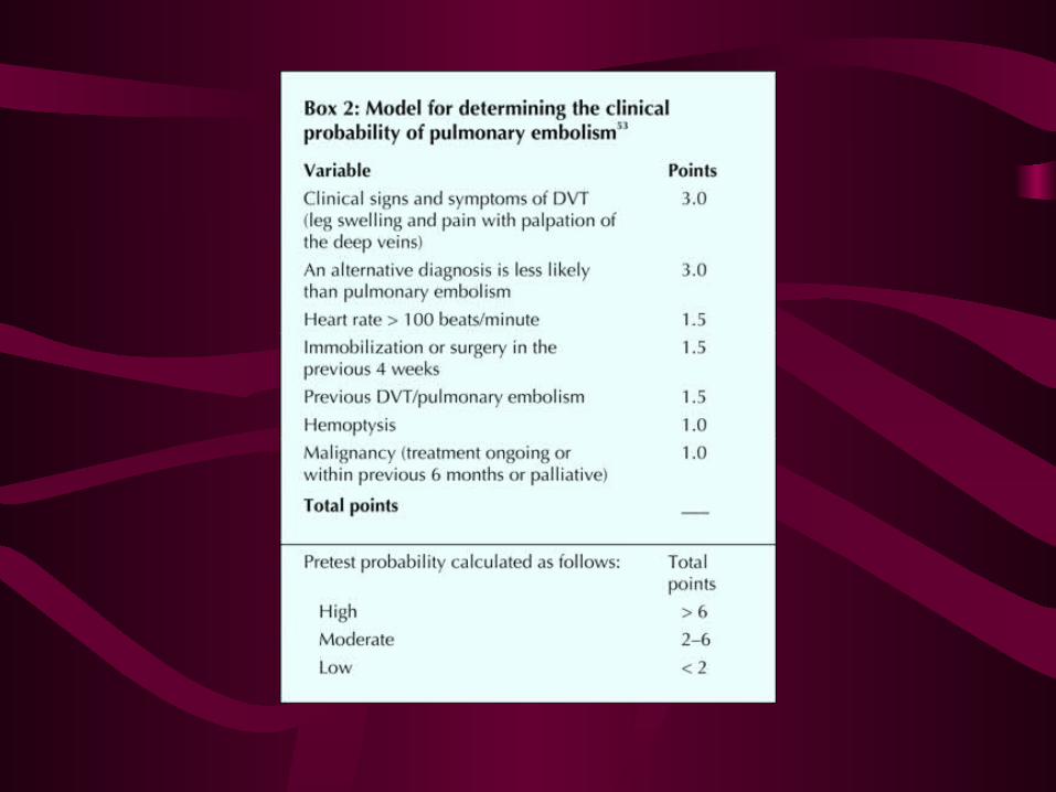
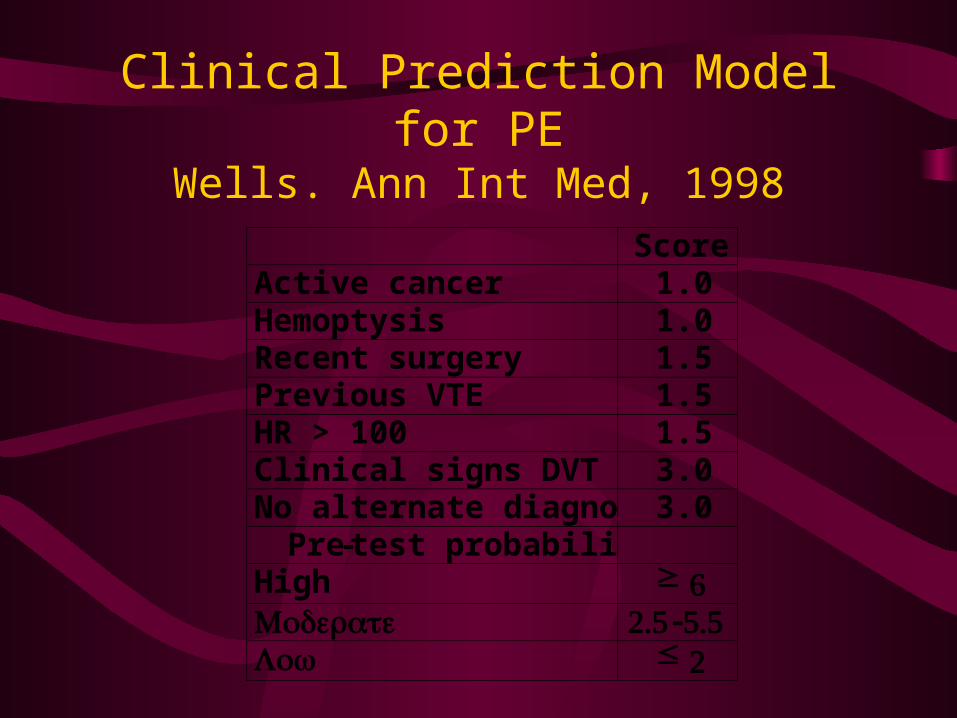
Clinical Prediction Model for PEWells. Ann Int Med, 1998
Score Active cancer 1.0 Hemoptysis 1.0 Recent surgery 1.5 Previous VTE 1.5 HR > 100 1.5 Clinical signs DVT 3.0 No alternate diagnosis 3.0
Pre-test probability High ≥ 6 Moderate 2.5 -5.5 Low ≤ 2
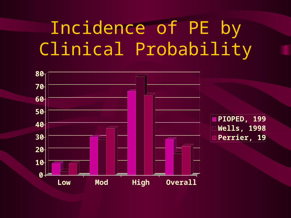
Incidence of PE by Clinical Probability
0
10
20
30
40
50
60
70
80
Low Mod High Overall
PIOPED, 1990Wells, 1998Perrier, 1999
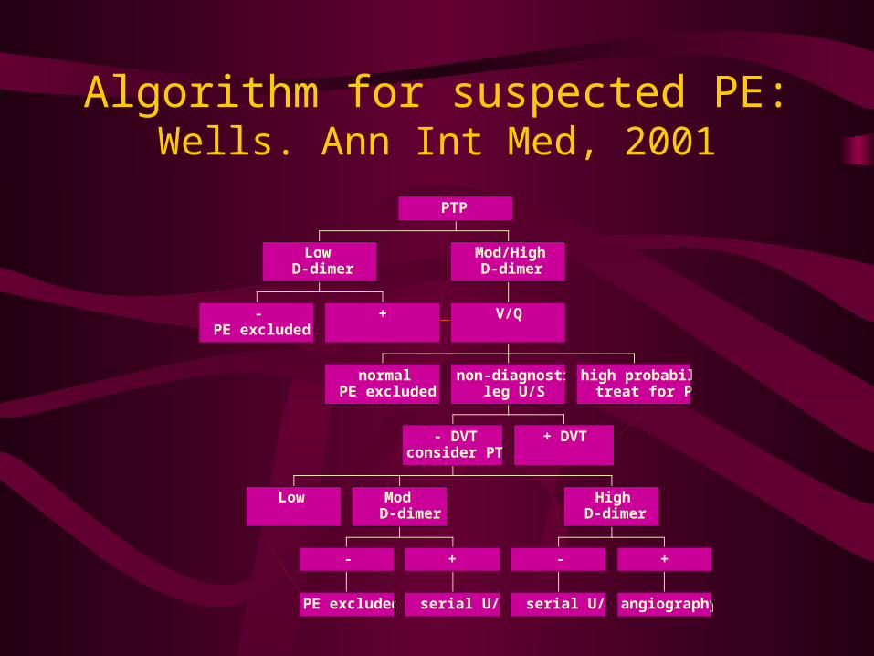
Algorithm for suspected PE:Wells. Ann Int Med, 2001
-PE excluded
+
LowD-dimer
normalPE excluded
Low
PE excluded
-
serial U/S
+
Mod D-dimer
serial U/S
-
angiography
+
HighD-dimer
- DVTconsider PTP
+ DVT
non-diagnosticleg U/S
high probabilitytreat for PE
V/Q
Mod/HighD-dimer
PTP
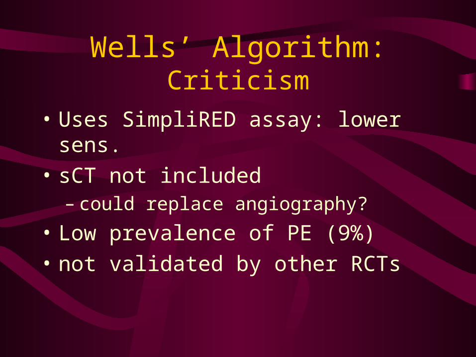
Wells’ Algorithm:Criticism
• Uses SimpliRED assay: lower sens.
• sCT not included– could replace angiography?
• Low prevalence of PE (9%)
• not validated by other RCTs
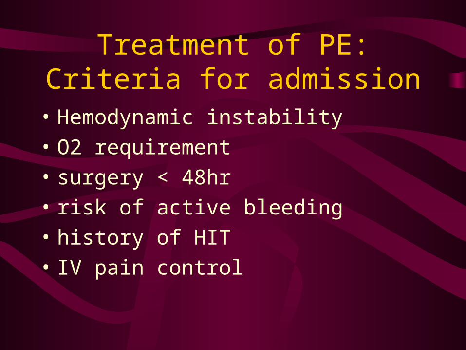
Treatment of PE:Criteria for admission
• Hemodynamic instability
• O2 requirement
• surgery < 48hr
• risk of active bleeding
• history of HIT
• IV pain control
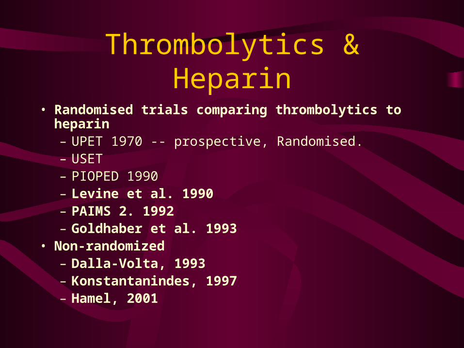
Thrombolytics & Heparin
• Randomised trials comparing thrombolytics to heparin– UPET 1970 -- prospective, Randomised.– USET– PIOPED 1990– Levine et al. 1990– PAIMS 2. 1992– Goldhaber et al. 1993
• Non-randomized– Dalla-Volta, 1993– Konstantanindes, 1997– Hamel, 2001
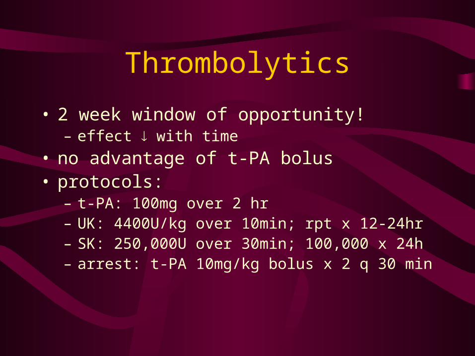
Thrombolytics
• 2 week window of opportunity!– effect with time
• no advantage of t-PA bolus• protocols:
– t-PA: 100mg over 2 hr– UK: 4400U/kg over 10min; rpt x 12-24hr– SK: 250,000U over 30min; 100,000 x 24h– arrest: t-PA 10mg/kg bolus x 2 q 30 min
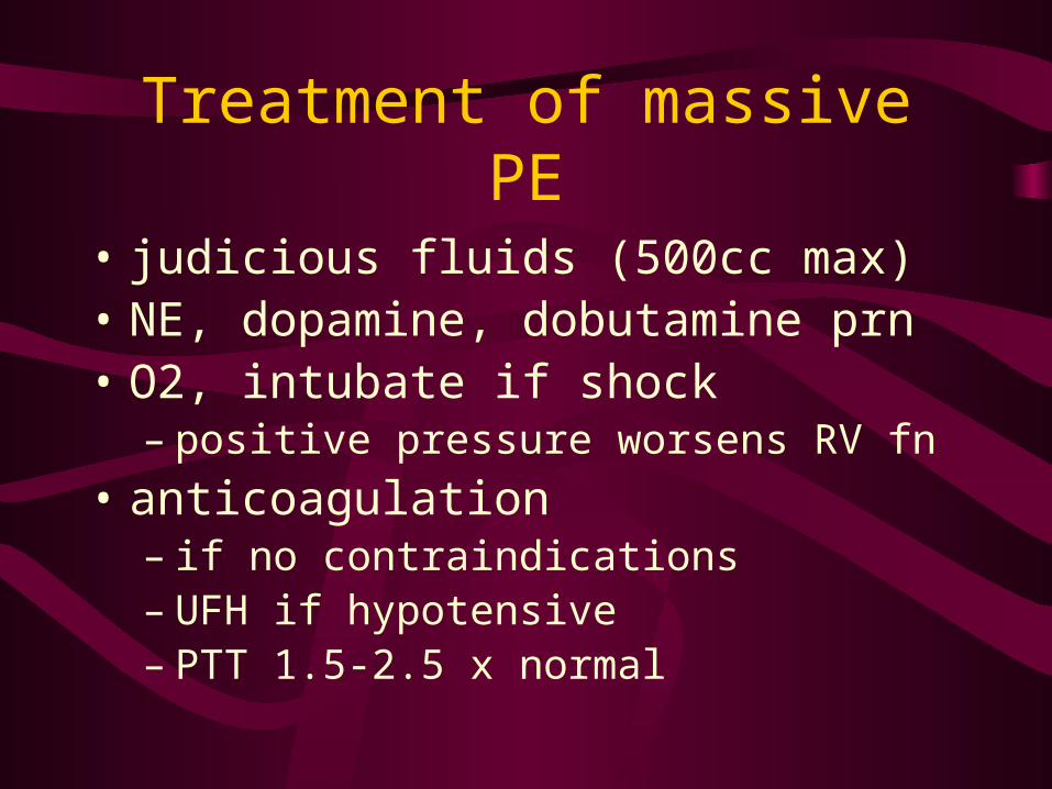
Treatment of massive PE
• judicious fluids (500cc max)• NE, dopamine, dobutamine prn• O2, intubate if shock
– positive pressure worsens RV fn
• anticoagulation– if no contraindications– UFH if hypotensive– PTT 1.5-2.5 x normal
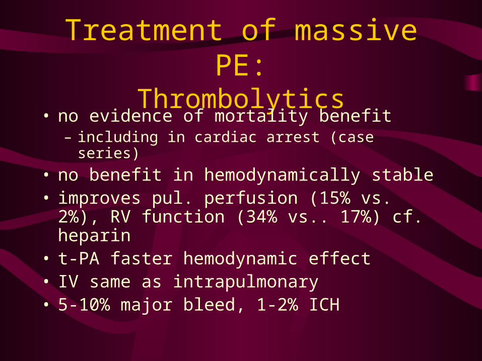
Treatment of massive PE:Thrombolytics
• no evidence of mortality benefit– including in cardiac arrest (case series)
• no benefit in hemodynamically stable• improves pul. perfusion (15% vs. 2%), RV
function (34% vs.. 17%) cf. heparin• t-PA faster hemodynamic effect• IV same as intrapulmonary• 5-10% major bleed, 1-2% ICH
Treatment of massive PE:Thrombolytics
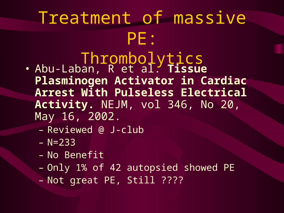
• Abu-Laban, R et al. Tissue Plasminogen Activator in Cardiac Arrest With Pulseless Electrical Activity. NEJM, vol 346, No 20, May 16, 2002.– Reviewed @ J-club– N=233– No Benefit– Only 1% of 42 autopsied showed PE– Not great PE, Still ????
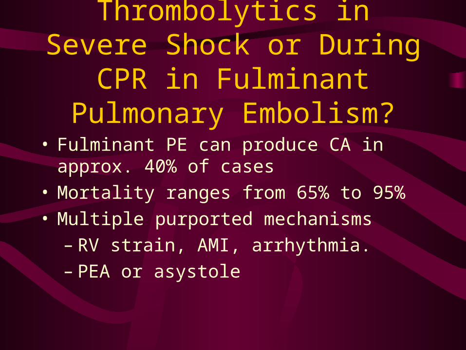
Thrombolytics in Severe Shock or During CPR in Fulminant
Pulmonary Embolism?
• Fulminant PE can produce CA in approx. 40% of cases
• Mortality ranges from 65% to 95%• Multiple purported mechanisms
– RV strain, AMI, arrhythmia.– PEA or asystole
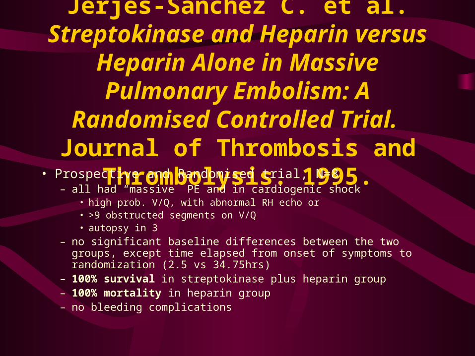
Jerjes-Sanchez C. et al. Streptokinase and Heparin versus Heparin Alone in
Massive Pulmonary Embolism: A Randomised Controlled Trial. Journal
of Thrombosis and Thrombolysis. 1995.• Prospective and Randomised trial, N=8
– all had “massive” PE and in cardiogenic shock• high prob. V/Q, with abnormal RH echo or• >9 obstructed segments on V/Q• autopsy in 3
– no significant baseline differences between the two groups, except time elapsed from onset of symptoms to randomization (2.5 vs 34.75hrs)
– 100% survival in streptokinase plus heparin group – 100% mortality in heparin group– no bleeding complications
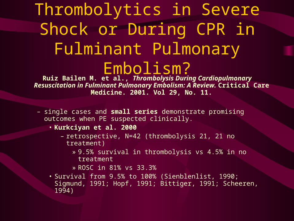
Thrombolytics in Severe Shock or During CPR in Fulminant
Pulmonary Embolism?Ruiz Bailen M. et al., Thrombolysis During Cardiopulmonary
Resuscitation in Fulminant Pulmonary Embolism: A Review. Critical Care Medicine. 2001. Vol 29, No. 11.
– single cases and small series demonstrate promising outcomes when PE suspected clinically.
• Kurkciyan et al. 2000 – retrospective, N=42 (thrombolysis 21, 21 no treatment)
» 9.5% survival in thrombolysis vs 4.5% in no treatment» ROSC in 81% vs 33.3%
• Survival from 9.5% to 100% (Sienblenlist, 1990; Sigmund, 1991; Hopf, 1991; Bittiger, 1991; Scheeren, 1994)
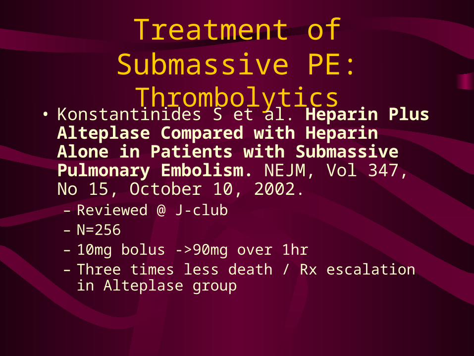
Treatment of Submassive PE:Thrombolytics
• Konstantinides S et al. Heparin Plus Alteplase Compared with Heparin Alone in Patients with Submassive Pulmonary Embolism. NEJM, Vol 347, No 15, October 10, 2002.– Reviewed @ J-club– N=256– 10mg bolus ->90mg over 1hr– Three times less death / Rx escalation in Alteplase
group
Goldhaber, S. et al. Alteplase versus Heparin in Acute Pulmonary
Embolism: Randomised Trial Assessing Right-Ventricular Function and
Pulmonary Perfusion. The Lancet. 1993, no 8844. vol 341.
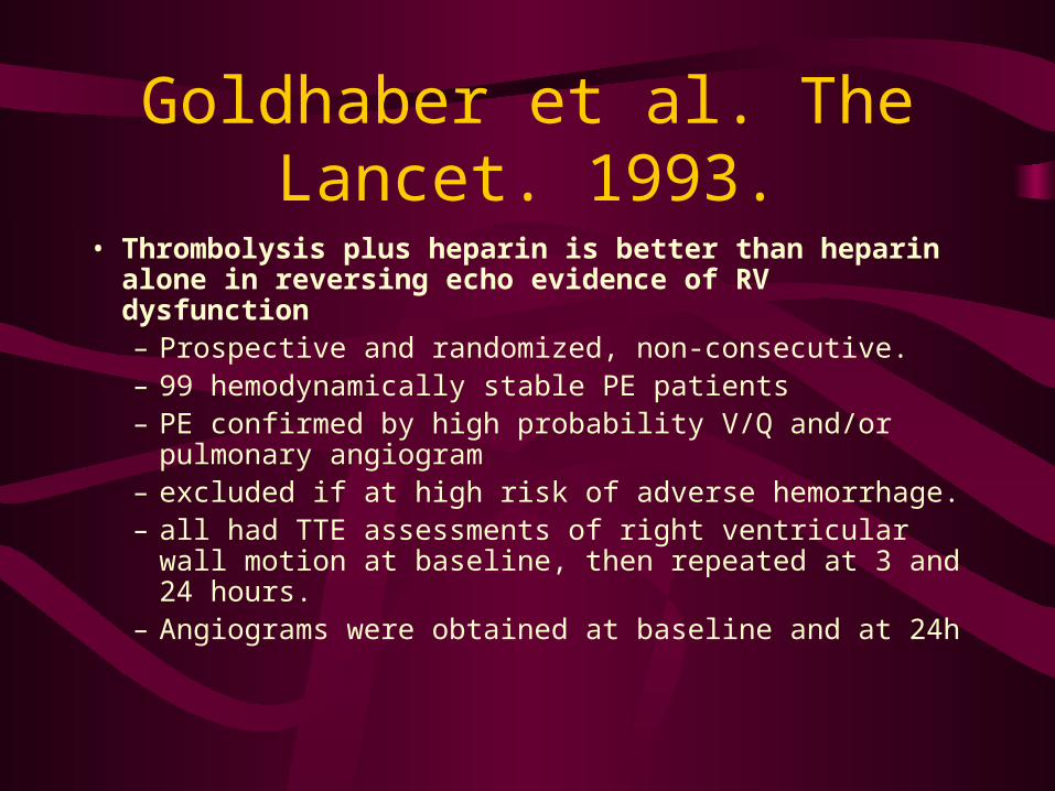
Goldhaber et al. The Lancet. 1993.
• Thrombolysis plus heparin is better than heparin alone in reversing echo evidence of RV dysfunction– Prospective and randomized, non-consecutive.– 99 hemodynamically stable PE patients– PE confirmed by high probability V/Q and/or
pulmonary angiogram– excluded if at high risk of adverse hemorrhage.– all had TTE assessments of right ventricular wall
motion at baseline, then repeated at 3 and 24 hours.– Angiograms were obtained at baseline and at 24h
Goldhaber et al. The Lancet. 1993.
• 46 patients randomized to rt-PA followed by heparin and 55 to heparin alone
• Endpoints; mortality, recurrent PE and major bleeding (72h)
• Followed for 14 days for adverse outcomes (PE recurrence or death), or longer if in hospital. 72 hrs for bleeding.
Goldhaber et al. The Lancet. 1993.
• Results
– follow-up echo (89 patients)
• rtPA group vs heparin
– 3 hrs -- greater improvement in RV wall motion (p=0.01)
– 24 hrs -- 39% improved, 2% worse vs 17% improved and 17% worse vs. 17% improvement and 17% worse in heparin group (p=0.005)
– follow-up angiogram at 24hrs (95 patients)
• rtPA vs heparin -- mean absolute improvement in pulmonary perfusion of 14.6% vs 1.5% in heparin (p<0.0001).
Goldhaber et al. The Lancet. 1993
• Subgroup analysis – patients with right ventricular hypokinesis on echo (N=36)
• rtPA -- 89% improvement, 6% worsened• heparin -- 44% improvement, 28% worsened (p=0.03)
• Deaths– 2 in heparin group (1 refractory CA and 1 with CI to tPA)
• Recurrent PEs– rtPA -- none– heparin -- 5 (2 fatal)
• Significant hemorrhage– heparin -- 1– rtPA -- 3
Goldhaber et al. The Lancet. 1993.
• Conclusions– rtPA group
• improved right heart function at 24 hours
• improvement in pulmonary perfusion
• decrease in recurrent PEs
• lower rate of death
• Strong points• randomization and similarities between groups
• echo and angiogram readers blinded to treatment and timing in relation to therapy
• Limitations• non-blinded to clinicians and open-labeled
• no long -term morbidity or mortality data
Konstantinides, et al. Association Between Thrombolytic Treatment and
the prognosis of hemodynamically Stable Patients with Major Pulmonary
Embolism: Results of a Multicenter Registry. Circulation, 96. 1997
• Early thrombolysis favorably affects in-hospital clinical outcome.– Multicentred, registry study– 719 consecutive patients analyzed; 73% PE confirmed by one or more imaging
study– evidence of either increased right ventricular afterload or pulmonary
hypertension based on TTE or cath.– all patients “hemodynamically stable”
• also included patients who were hypotensive (SBP<90) without signs of shock and those on low dose (<5mcg/kg/min) dopamine.
Konstantinides, et al. Circulation. 1997
• primary end-point -- overall 30-day mortality
• secondary endpoints -- PE recurrence, major bleeding
Konstantinides, et al. Circulation. 1997
• Treatment decisions made at discretion of physician
• 23.5% (n=169) received thrombolytic therapy within 24h of diagnosis followed by heparin
• remaining patients treated with heparin alone– unless the physician thought that they required
thrombolytics after the first 24h of heparin.
Konstantinides, et al. Circulation. 1997
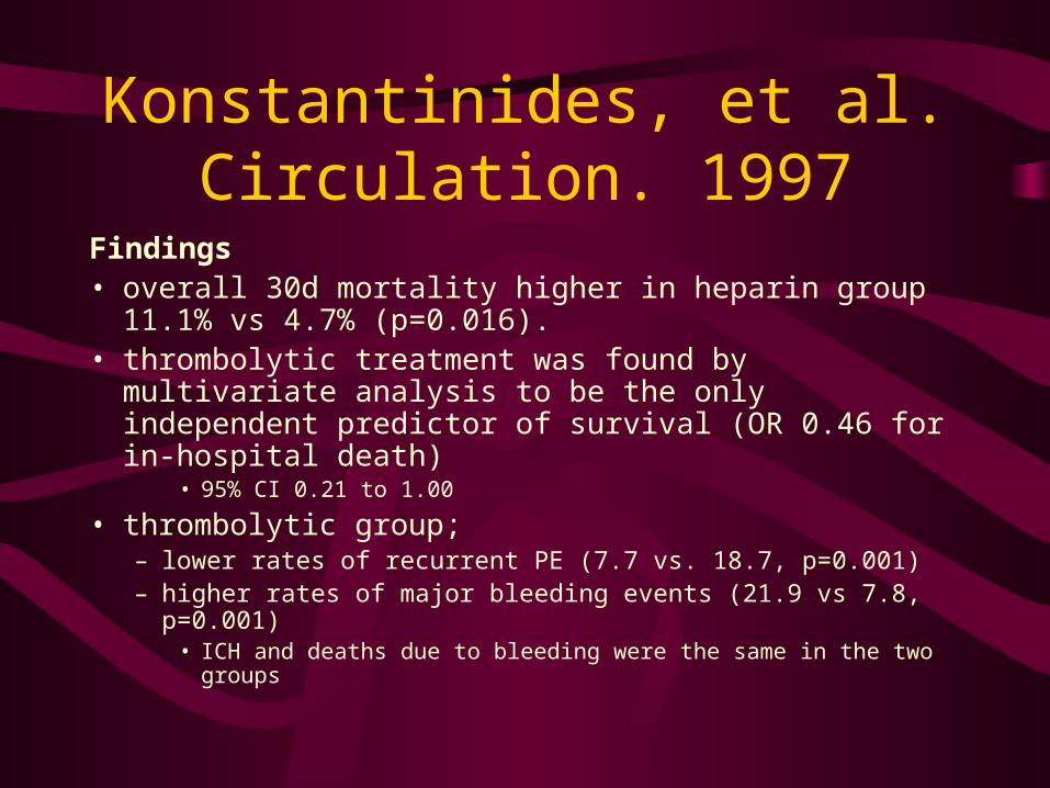
Findings• overall 30d mortality higher in heparin group 11.1% vs
4.7% (p=0.016).• thrombolytic treatment was found by multivariate analysis
to be the only independent predictor of survival (OR 0.46 for in-hospital death)
• 95% CI 0.21 to 1.00
• thrombolytic group;– lower rates of recurrent PE (7.7 vs. 18.7, p=0.001)– higher rates of major bleeding events (21.9 vs 7.8, p=0.001)
• ICH and deaths due to bleeding were the same in the two groups
Konstantinides, et al. Circulation. 1997
• Subgroup analysis– patients with a dilated right ventricle on echo
• 30 day mortality in (N=380) 10% compared with 4.1% in those without (p=0.018), a 58% reduction in mortality.
• 58% reduction in mortality in patients treated with thrombolytics (4.7% vs 11.1% heparin, p=0.16)
Konstantinides, et al. Circulation. 1997
• Limitations– study design;
• non-randomised, heterogeneous thrombolytic regimens• many patients had clinical signs of disease severity • more with chronic lung disease in UF heparin group• choice of treatment was at the discretion of the physician
– selection bias is likely • distribution of many clinical variables were statistically
different between the two groups (esp. age, pre-existing CHF, higher in heparin)
• major end point analyses required multivariate regression model to account for the unequal distribution of clinical variables
Konstantinides, et al. Circulation. 1997
• 40% of patients thrombolysed had contraindications to lytics
• 25% in the heparin group ‘crossed over’ and received thrombolytics. This data was not reported.
Hamel et al. Thrombolysis or Heparin Therapy in Massive
Pulmonary Embolism With Right Ventricular Dilation. Chest, 2001.
Vol. 120:1.• There is a benefit to thrombolysis over heparin in stable PE
patients with RVD– Retrospective, cohort study of 153 consecutive patients– PE confirmed by, V/Q or angiography– RV function evaluated by TT E on admission– 64 patients in each treatment group were matched on the basis of
RV/LV diameter ratio– perfusion scans repeated on day 7 to 10 or earlier if recurrent PE
suspected
Hamel et al. Chest, 2001.
• Inclusion criteria – included PIOPED criteria for high prob. V/Q
– Pulmonary vascular obstruction >40% on V/Q or Miller index of 20/34
– RV to LV ratio of >0.6* in absence of LV or Mv disease
• Exclusion criteria– SBP <90
– contraindications to thrombolysis
– inotropes
– syncope prior to presentation
Hamel et al. Chest, 2001.
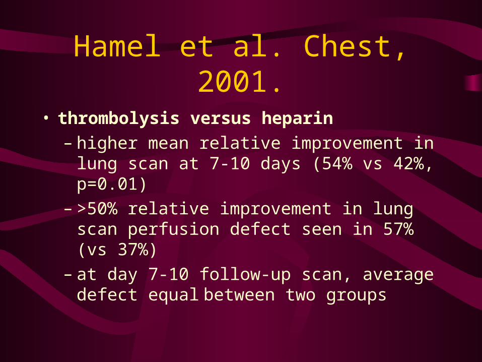
• thrombolysis versus heparin– higher mean relative improvement in lung scan
at 7-10 days (54% vs 42%, p=0.01)– >50% relative improvement in lung scan
perfusion defect seen in 57% (vs 37%)– at day 7-10 follow-up scan, average defect
equal between two groups
Hamel et al. Chest, 2001.
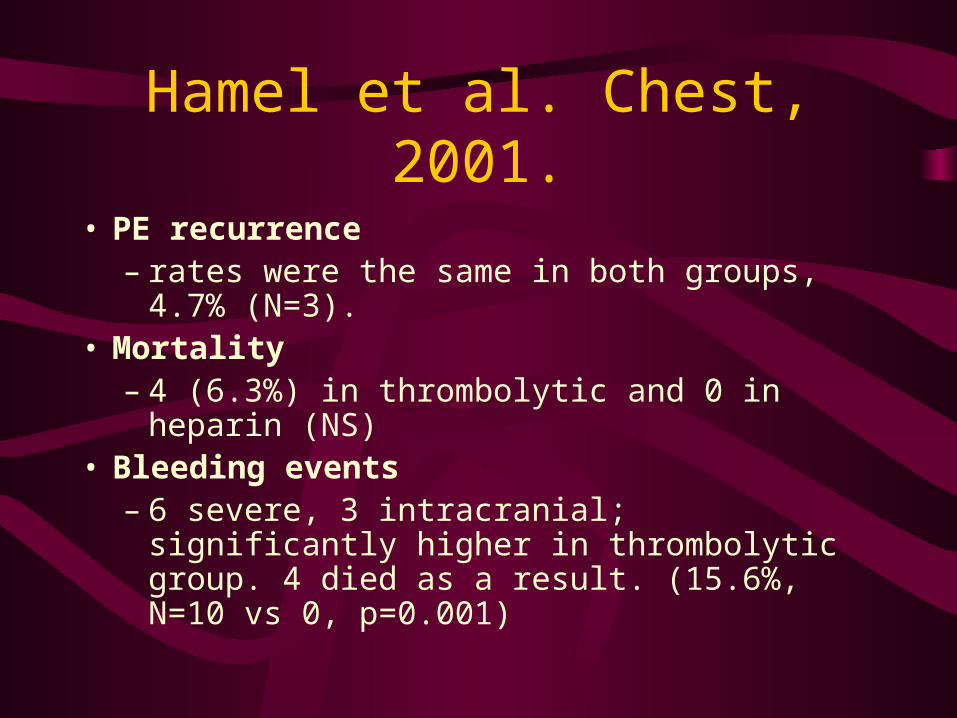
• PE recurrence – rates were the same in both groups, 4.7%
(N=3).• Mortality
– 4 (6.3%) in thrombolytic and 0 in heparin (NS)• Bleeding events
– 6 severe, 3 intracranial; significantly higher in thrombolytic group. 4 died as a result. (15.6%, N=10 vs 0, p=0.001)
Hamel et al. Chest, 2001.
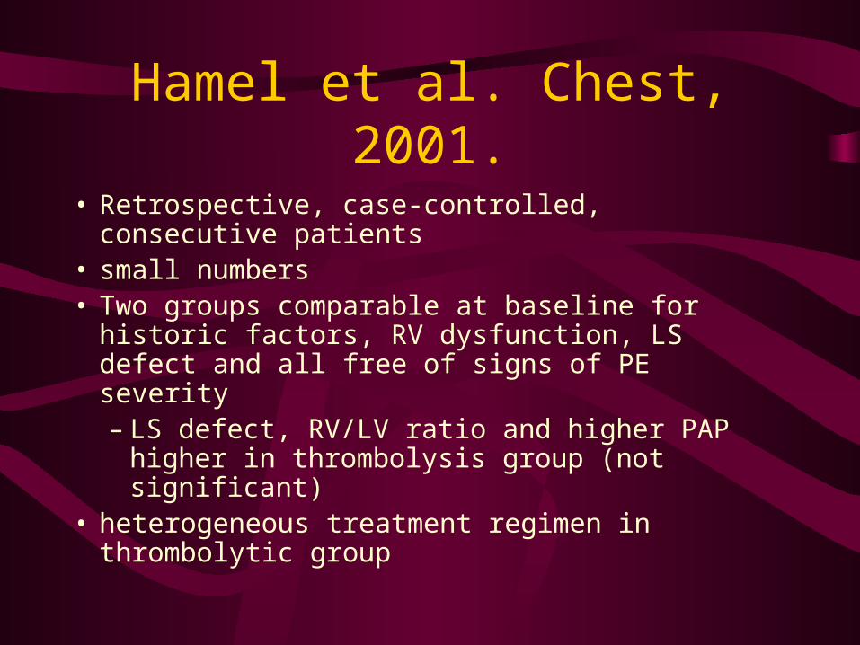
• Retrospective, case-controlled, consecutive patients
• small numbers• Two groups comparable at baseline for historic
factors, RV dysfunction, LS defect and all free of signs of PE severity – LS defect, RV/LV ratio and higher PAP higher
in thrombolysis group (not significant)• heterogeneous treatment regimen in thrombolytic
group
Levine et al. A Randomised Trial of a Single Bolus Dosage Regimen of Recombinant Tissue Plasminogen Activator in Patients with Acute
Pulmonary Embolism. Chest. 1990. 98:1473.
• rt-PA will benefit pulmonary perfusion in patients with PE and demonstrated perfusion deficits– Inclusion -- ‘symptomatic’ patients with either high probability V/Q or
angiographically proven PE and no contraindications to thrombolytics.– Excluded if hypotensive or hemodynamically unstable– All patients received heparin bolus. Then randomized to either rt-PA
(0.6mg/kg, given as a bolus over 2min) or placebo.– 10 day study period
Levine et al. Chest. 1990
• End-points were >50% improvement in perfusion defect over baseline and major bleeding events;– intracranial, retroperitoneal, requires
transfusion >2U or fall in Hgb >20g/L
Levine et al. Chest. 1990
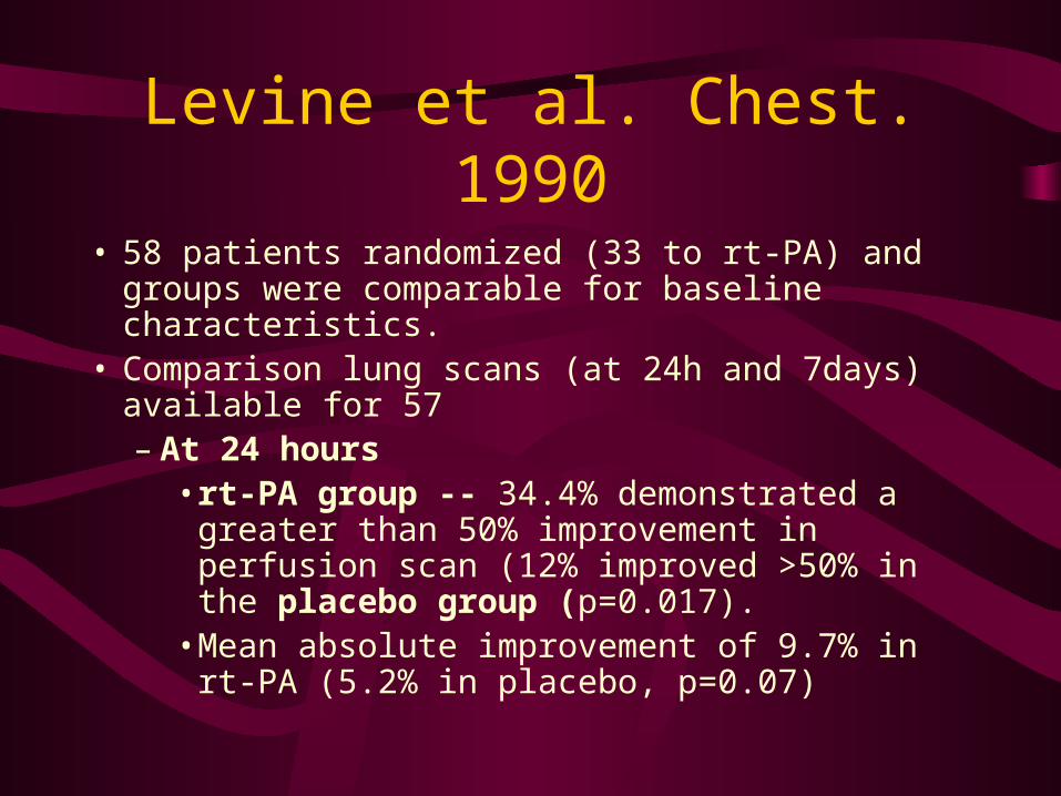
• 58 patients randomized (33 to rt-PA) and groups were comparable for baseline characteristics.
• Comparison lung scans (at 24h and 7days) available for 57– At 24 hours
• rt-PA group -- 34.4% demonstrated a greater than 50% improvement in perfusion scan (12% improved >50% in the placebo group (p=0.017).
• Mean absolute improvement of 9.7% in rt-PA (5.2% in placebo, p=0.07)
Levine et al. Chest. 1990
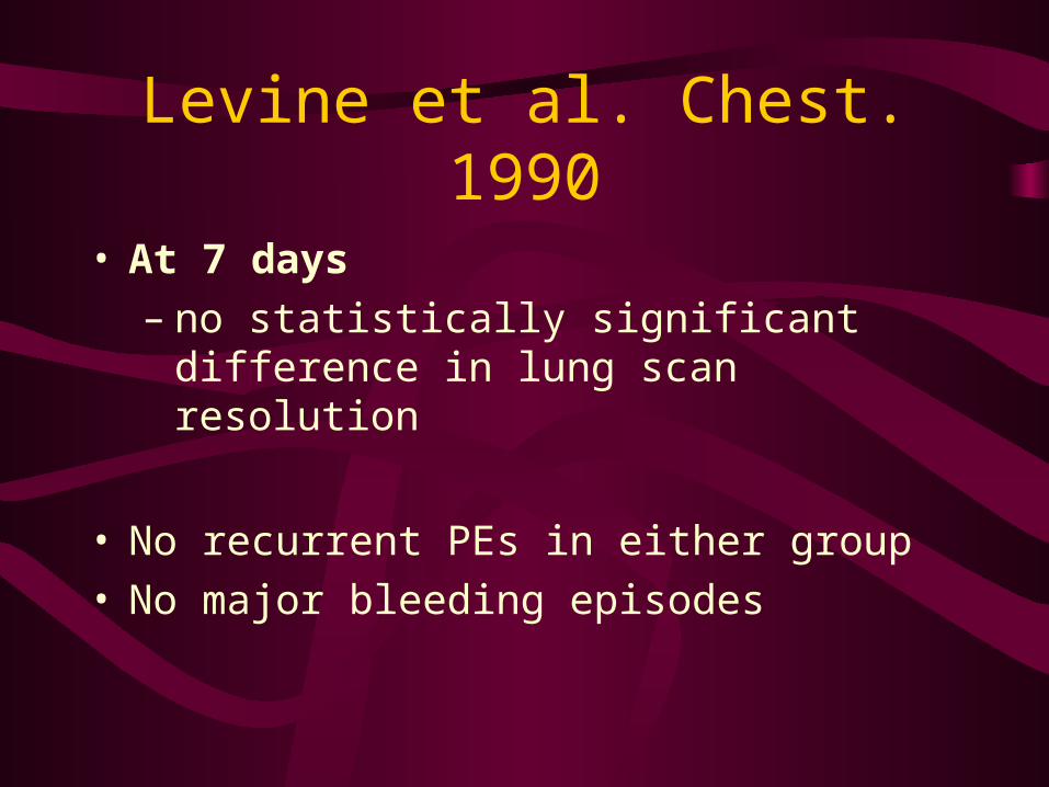
• At 7 days– no statistically significant difference in lung
scan resolution
• No recurrent PEs in either group• No major bleeding episodes
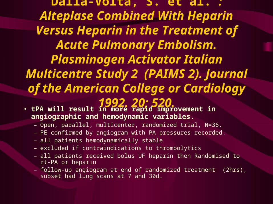
Dalla-Volta, S. et al. : Alteplase Combined With Heparin Versus Heparin in the
Treatment of Acute Pulmonary Embolism. Plasminogen Activator Italian Multicentre
Study 2 (PAIMS 2). Journal of the American College or Cardiology 1992. 20;
520.• tPA will result in more rapid improvement in angiographic and
hemodynamic variables.– Open, parallel, multicenter, randomized trial, N=36.– PE confirmed by angiogram with PA pressures recorded. – all patients hemodynamically stable– excluded if contraindications to thrombolytics– all patients received bolus UF heparin then Randomised to rt-PA or heparin– follow-up angiogram at end of randomized treatment (2hrs), subset had lung
scans at 7 and 30d.
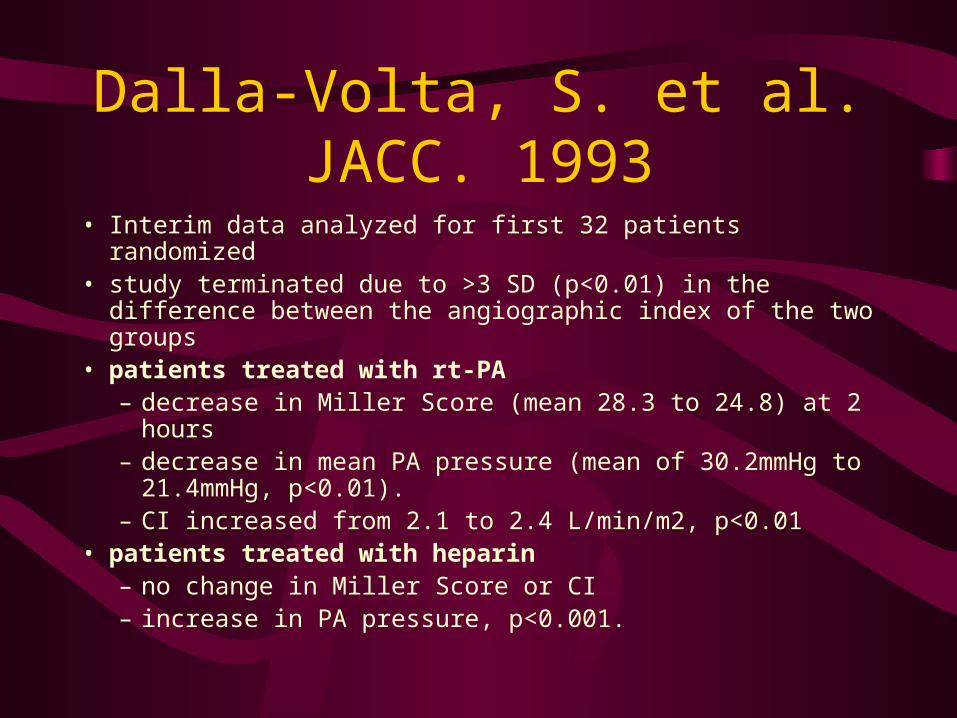
Dalla-Volta, S. et al. JACC. 1993
• Interim data analyzed for first 32 patients randomized• study terminated due to >3 SD (p<0.01) in the difference
between the angiographic index of the two groups• patients treated with rt-PA
– decrease in Miller Score (mean 28.3 to 24.8) at 2 hours– decrease in mean PA pressure (mean of 30.2mmHg to
21.4mmHg, p<0.01).– CI increased from 2.1 to 2.4 L/min/m2, p<0.01
• patients treated with heparin– no change in Miller Score or CI– increase in PA pressure, p<0.001.
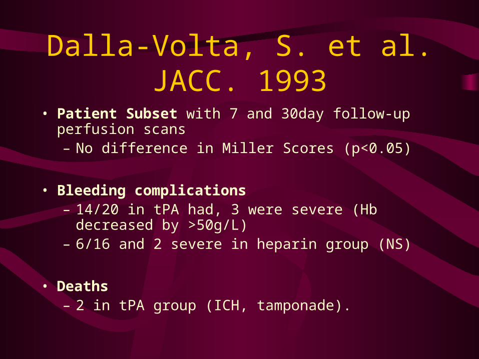
Dalla-Volta, S. et al. JACC. 1993
• Patient Subset with 7 and 30day follow-up perfusion scans– No difference in Miller Scores (p<0.05)
• Bleeding complications – 14/20 in tPA had, 3 were severe (Hb decreased by
>50g/L) – 6/16 and 2 severe in heparin group (NS)
• Deaths – 2 in tPA group (ICH, tamponade).
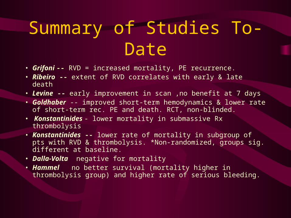
Summary of Studies To-Date
• Grifoni -- RVD = increased mortality, PE recurrence.• Ribeiro -- extent of RVD correlates with early & late death• Levine -- early improvement in scan ,no benefit at 7 days• Goldhaber -- improved short-term hemodynamics & lower rate of
short-term rec. PE and death. RCT, non-blinded. • Konstantinides - lower mortality in submassive Rx thrombolysis• Konstantinides -- lower rate of mortality in subgroup of pts with
RVD & thrombolysis. *Non-randomized, groups sig. different at baseline.
• Dalla-Volta negative for mortality• Hammel no better survival (mortality higher in thrombolysis
group) and higher rate of serious bleeding.
Embolectomy
• Indicated in acute, massive PE if:– contraindication to thrombolytics– unresponsive to medical mgt
• moribund pt poor results
• no evidence cf. with thrombolytics
• percutaneous vs.. surgical– ?role
IVC Filters
• Indications:– contraindication to anticoagulation– recurrent VTE despite anticoagulation– after surgical embolectomy
• no long term adv vs.. anticoagulation
• anticoagulate if no contraindications– DVT and IVC occlusion
The END
• Special Thanks to:– You
• For sitting through 133 slides
– Dr L. Mabon• For clinical insight
– Dr A. Oster• For “borrowed slides”
– Dr D. Watt• For “borrowed slides”