Embed Size (px)
Citation preview
VERTEBRAL COLUMN ANATOMY
IN CNS COURSE
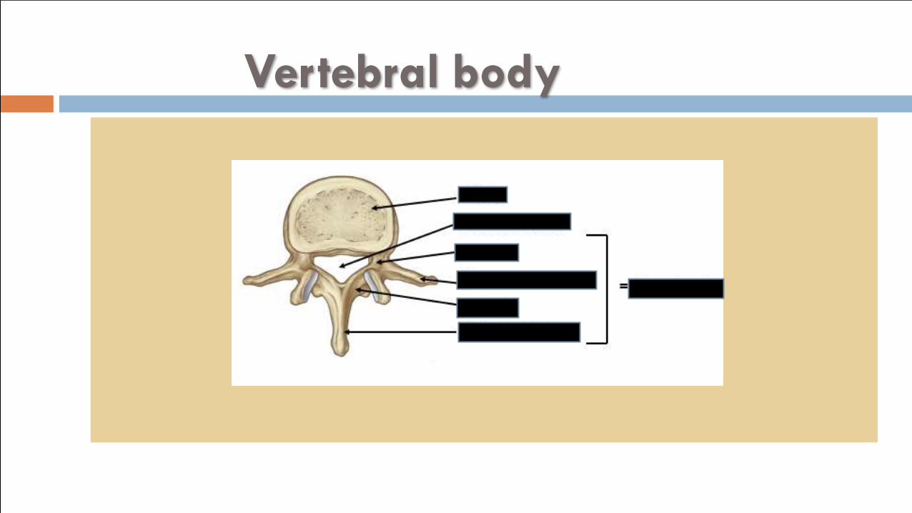
Vertebral body
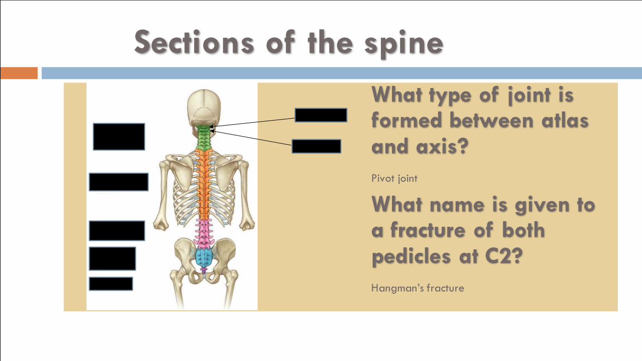
Sections of the spine
Atlas (C1)
Axis (C2)
What type of joint is formed between atlas and axis?
Pivot joint
What name is given to a fracture of both pedicles at C2?
Hangman’s fracture
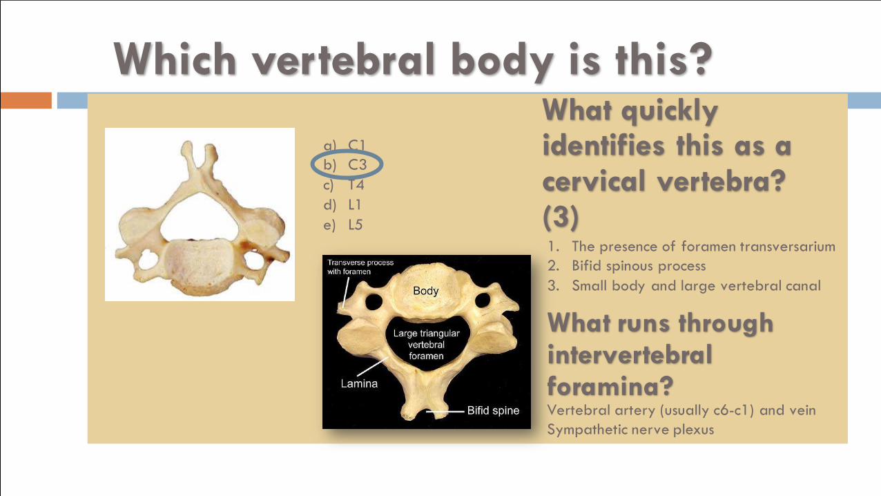
Which vertebral body is this?
a) C1
b) C3
c) T4
d) L1
e) L5
What quickly identifies this as a cervical vertebra? (3)1. The presence of foramen transversarium
2. Bifid spinous process
3. Small body and large vertebral canal
What runs through intervertebral foramina?Vertebral artery (usually c6-c1) and vein
Sympathetic nerve plexus
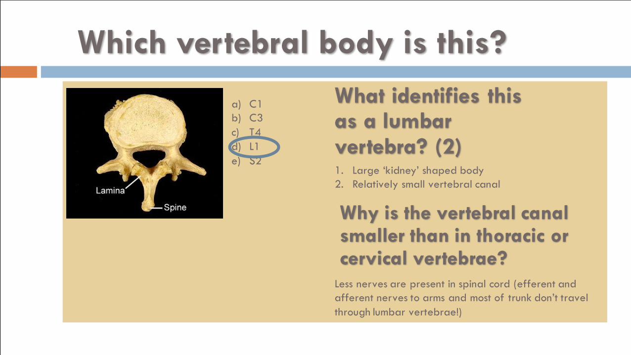
Which vertebral body is this?
a) C1
b) C3
c) T4
d) L1
e) S2
What identifies this as a lumbar vertebra? (2)1. Large ‘kidney’ shaped body
2. Relatively small vertebral canal
Why is the vertebral canal smaller than in thoracic or cervical vertebrae?
Less nerves are present in spinal cord (efferent and
afferent nerves to arms and most of trunk don’t travel
through lumbar vertebrae!)
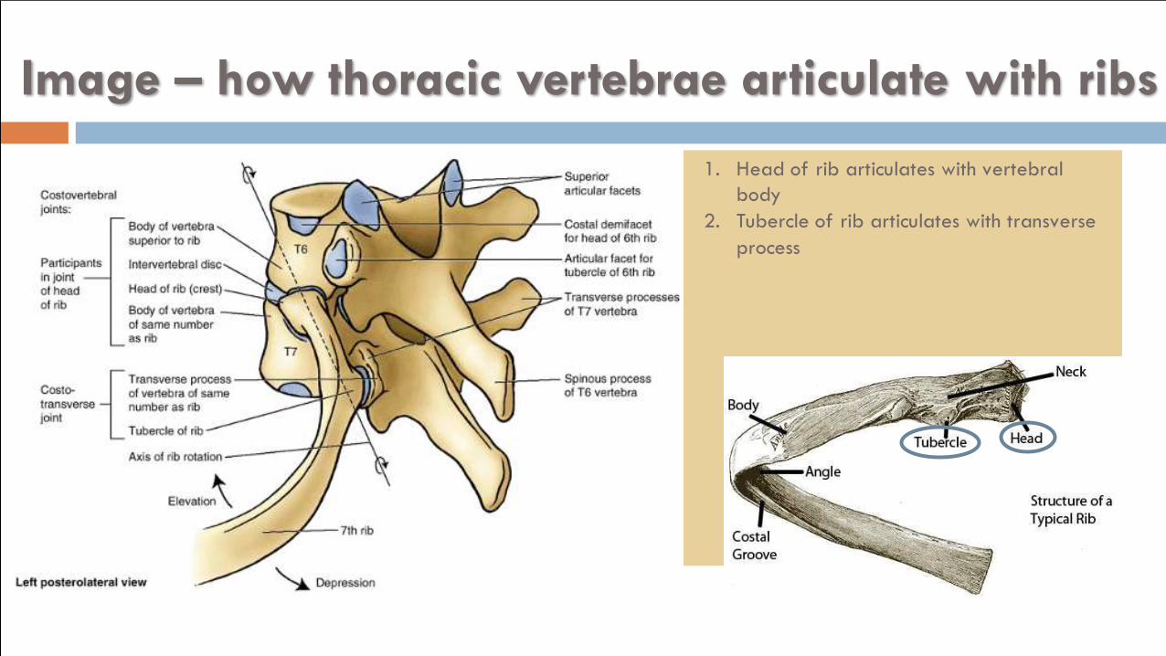
Image – how thoracic vertebrae articulate with ribs
1. Head of rib articulates with vertebral
body
2. Tubercle of rib articulates with transverse
process
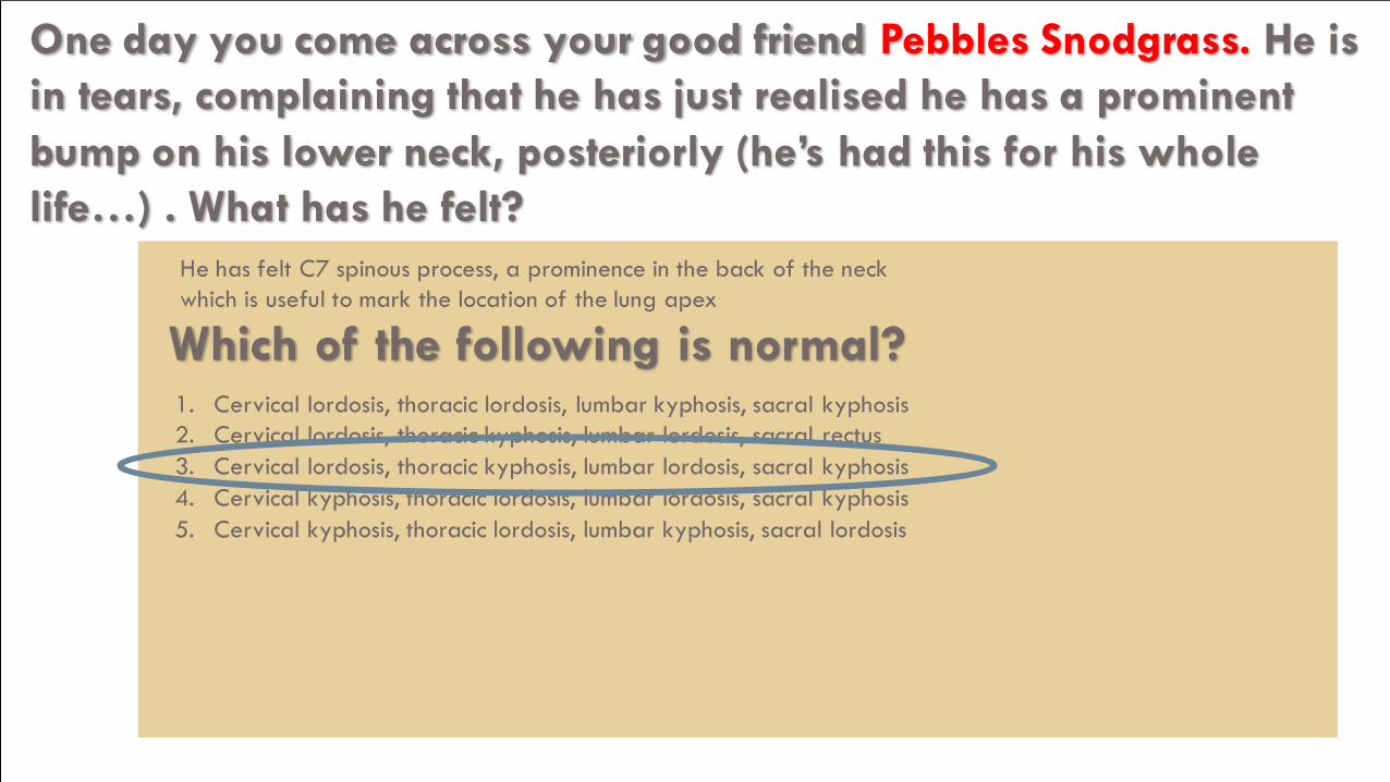
One day you come across your good friend Pebbles Snodgrass. He is
in tears, complaining that he has just realised he has a prominent
bump on his lower neck, posteriorly (he’s had this for his whole
life…) . What has he felt?
He has felt C7 spinous process, a prominence in the back of the neck
which is useful to mark the location of the lung apex
Which of the following is normal?1. Cervical lordosis, thoracic lordosis, lumbar kyphosis, sacral kyphosis
2. Cervical lordosis, thoracic kyphosis, lumbar lordosis, sacral rectus
3. Cervical lordosis, thoracic kyphosis, lumbar lordosis, sacral kyphosis
4. Cervical kyphosis, thoracic lordosis, lumbar lordosis, sacral kyphosis
5. Cervical kyphosis, thoracic lordosis, lumbar kyphosis, sacral lordosis
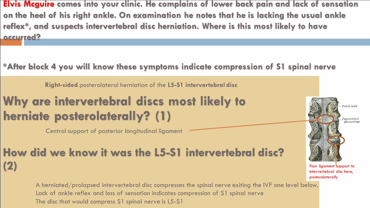
Elvis Mcguire comes into your clinic. He complains of lower back pain and lack of sensation
on the heel of his right ankle. On examination he notes that he is lacking the usual ankle
reflex*, and suspects intervertebral disc herniation. Where is this most likely to have
occurred?
Right-sided posterolateral herniation of the L5-S1 intervertebral disc
Why are intervertebral discs most likely to herniate posterolaterally? (1)
How did we know it was the L5-S1 intervertebral disc? (2)
A herniated/prolapsed intervertebral disc compresses the spinal nerve exiting the IVF one level below.
Lack of ankle reflex and loss of sensation indicates compression of S1 spinal nerve
The disc that would compress S1 spinal nerve is L5-S1
Central support of posterior longitudinal ligament
*After block 4 you will know these symptoms indicate compression of S1 spinal nerve
Poor ligament support to
intervertebral disc here,
posterolaterally
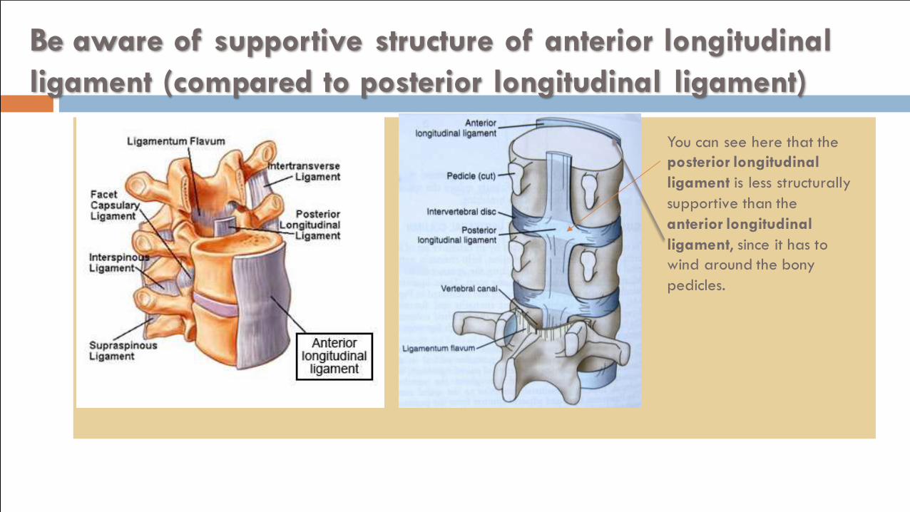
Be aware of supportive structure of anterior longitudinal
ligament (compared to posterior longitudinal ligament)
You can see here that the
posterior longitudinal
ligament is less structurally
supportive than the
anterior longitudinal
ligament, since it has to
wind around the bony
pedicles.
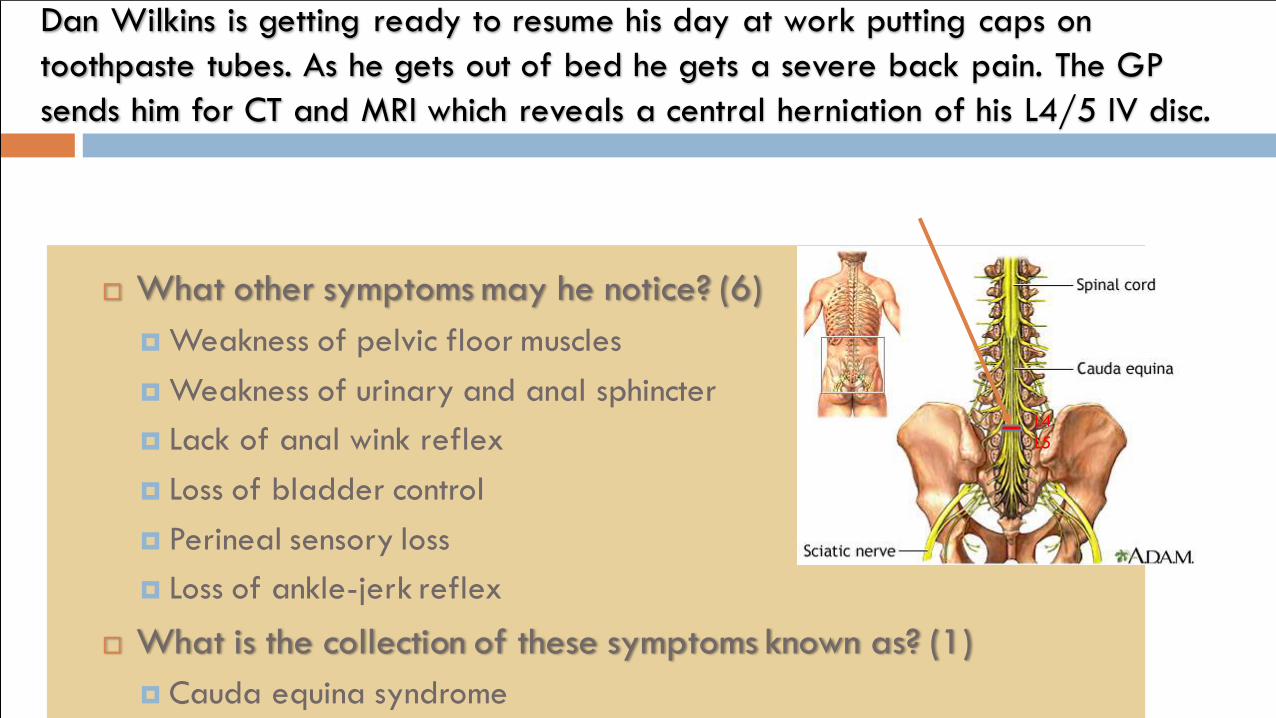
What other symptoms may he notice? (6)
Weakness of pelvic floor muscles
Weakness of urinary and anal sphincter
Lack of anal wink reflex
Loss of bladder control
Perineal sensory loss
Loss of ankle-jerk reflex
What is the collection of these symptoms known as? (1)
Cauda equina syndrome
Dan Wilkins is getting ready to resume his day at work putting caps on
toothpaste tubes. As he gets out of bed he gets a severe back pain. The GP
sends him for CT and MRI which reveals a central herniation of his L4/5 IV disc.
L4
L5
BONUS:
What are 4 other causes of CAUDA EQUINA syndrome? (4)
Tumours
Spinal stenosis
Spondylolisthesis
Vertebral canal stenosis
(There are others, )
What is spondylolisthesis? (1)
Dislocation of the vertebral body
What is spondylolysis? (1)
Fracture of the vertebrae, which often leads to spondylolisthesis
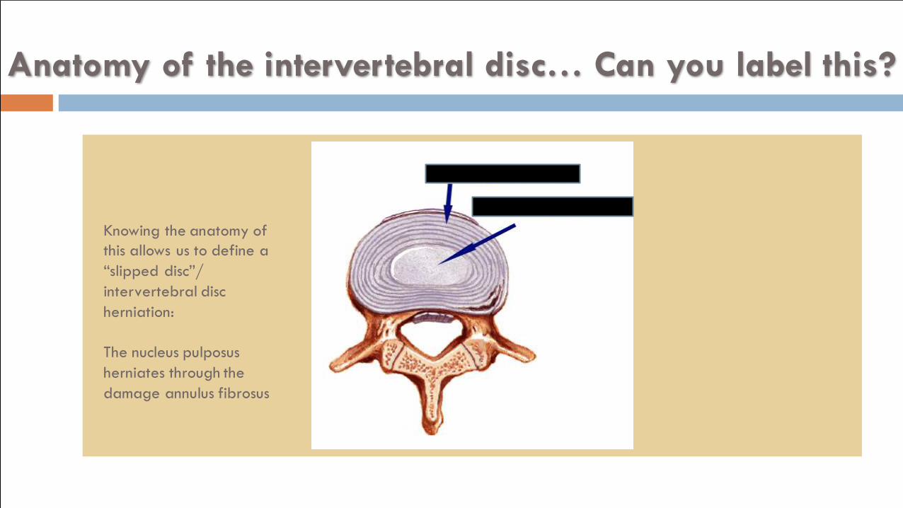
Anatomy of the intervertebral disc… Can you label this?
Knowing the anatomy of
this allows us to define a
“slipped disc”/
intervertebral disc
herniation:
The nucleus pulposus
herniates through the
damage annulus fibrosus
A woman goes into labor and is in need of an
epidural anesthesia…
Which spinal level would you access at (in an adult)? Why?
L2/3 to L5/S1 (prefer higher up as there is more interspinous space
Spinal cord terminates at L1/2 (can terminate down to L3)
Where would you perform a lumbar puncture in a baby? Why?
L4/5, L5/S1
Spinal cord terminates at L3/4
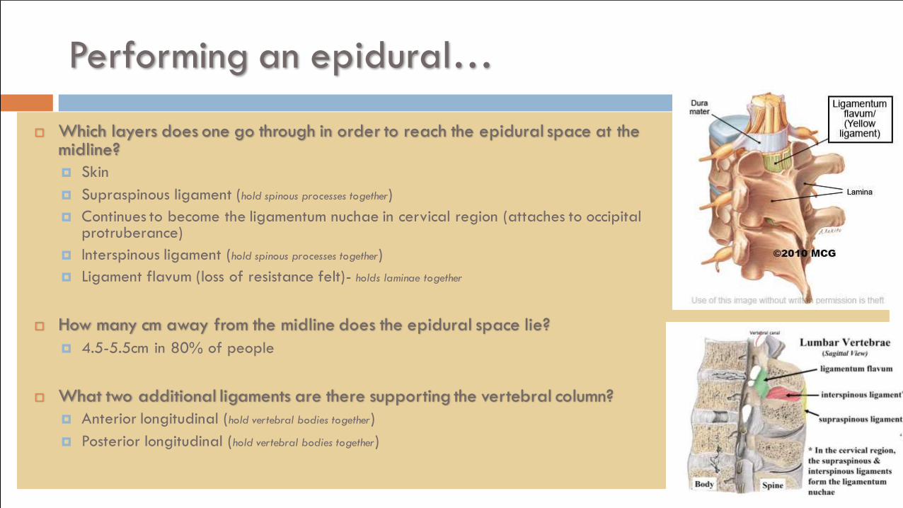
Performing an epidural…
Which layers does one go through in order to reach the epidural space at the midline?
Skin
Supraspinous ligament (hold spinous processes together)
Continues to become the ligamentum nuchae in cervical region (attaches to occipital protruberance)
Interspinous ligament (hold spinous processes together)
Ligament flavum (loss of resistance felt)- holds laminae together
How many cm away from the midline does the epidural space lie?
4.5-5.5cm in 80% of people
What two additional ligaments are there supporting the vertebral column?
Anterior longitudinal (hold vertebral bodies together)
Posterior longitudinal (hold vertebral bodies together)
During a lumbar tap, after the epidural space, what other layers need
to be passed before collecting CSF?
Dura mater
(Subdural space)
Arachnoid mater
(Subarachnoid space) where CSF is contained