Embed Size (px)
Citation preview

Veterinary Microbiology and Microbial Disease


Veterinary Microbiology and Microbial DiseaseSecond Edition
A John Wiley & Sons, Ltd., Publication
P.J. Quinn MVB, PhD, MRCVSProfessor Emeritus, Former Professor of Veterinary Microbiology and Parasitology, School of Veterinary Medicine, University College Dublin
B.K. Markey MVB, PhD, Dip Stat, MRCVSSenior Lecturer in Veterinary Microbiology, School of Veterinary Medicine, University College Dublin
F.C. Leonard MVB, PhD, MRCVSSenior Lecturer in Veterinary Microbiology, School of Veterinary Medicine, University College Dublin
E.S. FitzPatrick FIBMSChief Technical Offi cer, School of Veterinary Medicine, University College Dublin
S. Fanning BSc, PhDProfessor of Food Safety and Zoonoses, Director of Academic Centre for Food Safety, University College Dublin
P.J. Hartigan BSc, MVM, MA, PhD, MRCVSFormer Senior Lecturer in Veterinary Pathology, Trinity College Dublin
This edition fi rst published 2011© 2002 by Blackwell Science Ltd© 2011 by P.J. Quinn, B.K. Markey, F.C. Leonard, E.S. FitzPatrick, S. Fanning and P.J. Hartigan
Wiley-Blackwell is an imprint of John Wiley & Sons, formed by the merger of Wiley’s global Scientifi c, Technical and Medical business with Blackwell Publishing.
Registered offi ce: John Wiley & Sons Ltd, The Atrium, Southern Gate, Chichester, West Sussex, PO19 8SQ, UK
Editorial offi ces: 9600 Garsington Road, Oxford, OX4 2DQ, UK The Atrium, Southern Gate, Chichester, West Sussex, PO19 8SQ, UK 2121 State Avenue, Ames, Iowa 50014-8300, USA
For details of our global editorial offi ces, for customer services and for information about how to apply for permission to reuse the copyright material in this book please see our website at www.wiley.com/wiley-blackwell.
The right of the author to be identifi ed as the author of this work has been asserted in accordance with the UK Copyright, Designs and Patents Act 1988.
All rights reserved. No part of this publication may be reproduced, stored in a retrieval system, or transmitted, in any form or by any means, electronic, mechanical, photocopying, recording or otherwise, except as permitted by the UK Copyright, Designs and Patents Act 1988, without the prior permission of the publisher.
Designations used by companies to distinguish their products are often claimed as trademarks. All brand names and product names used in this book are trade names, service marks, trademarks or registered trademarks of their respective owners. The publisher is not associated with any product or vendor mentioned in this book. This publication is designed to provide accurate and authoritative information in regard to the subject matter covered. It is sold on the understanding that the publisher is not engaged in rendering professional services. If professional advice or other expert assistance is required, the services of a competent professional should be sought.
Library of Congress Cataloging-in-Publication Data
Veterinary microbiology and microbial disease / P.J. Quinn, MVB, PhD, MRCVS, Professor Emeritus, Former Professor of Veterinary Microbiology and Parasitology, School of Veterinary Medicine, University College, Dublin, B.K. Markey, MVB, PhD, Dip Stat, MRCVS, Senior Lecturer in Veterinary Microbiology, School of Veterinary Medicine, University College, Dublin, F.C. Leonard, MVB, PhD, MRCVS, Senior Lecturer in Veterinary Microbiology, School of Veterinary Medicine, University College, Dublin, E.S. FitzPatrick, FIBMS, Chief Technical Officer, School of Veterinary Medicine, University College, Dublin, S. Fanning, BSc, PhD, Professor of Food Safety and Zoonoses, Director of Academic Centre for Food Safety, University College Dublin, P.J. Hartigan, BSc, MVM, MA, PhD, MRCVS, Former Senior Lecturer in Veterinary Pathology, Trinity College, Dublin. – Second Edition. p. ; cm. Includes bibliographical references and index. ISBN 978-1-4051-5823-7 (pbk. : alk. paper) 1. Veterinary microbiology. I. Quinn, P. J. (Patrick J.), author. II. Markey, B. K. (Bryan K.), author. III. Leonard, F. C., author. IV. FitzPatrick, E. S., author. V. Fanning, S., author. VI. Hartigan, P. J., author. [DNLM: 1. Microbiology. 2. Veterinary Medicine. 3. Communicable Diseases–microbiology. 4. Communicable Diseases–veterinary. QW 70] SF780.2.V485 2011 636.089'69041–dc22 2010049404
A catalogue record for this book is available from the British Library.
Set in 10/12 pt Palatino by Toppan Best-set Premedia Limited, Hong Kong
1 2011

Contents
v
Preface ixAcknowledgements xAuthor biographies xi
Section I Introduction to Microbiology, Infection, Immunity and Molecular Diagnostic Methods
1 Microbiology, microbial pathogens and infectious disease 3
2 Subdivisions, classifi cation and morphological characterization of infectious agents 7
3 Infection and immunity 144 Immunodefi ciency diseases 675 Vaccines and vaccination 806 Molecular diagnostic methods 95
Section II Introductory Bacteriology7 The structure of bacterial cells 1158 Cultivation, preservation and
inactivation of bacteria 1239 Bacterial genetics, mechanisms of
genetic variation and gene databases 12910 Laboratory diagnosis of bacterial disease 14311 Antibacterial agents 14912 Antibacterial resistance 15713 Bacterial colonization, tissue
invasion and clinical disease 165
Section III Pathogenic Bacteria14 Staphylococcus species 17915 Streptococci 18816 Actinobacteria 19617 Corynebacterium species 20718 Rhodococcus equi 21319 Listeria species 21720 Erysipelothrix rhusiopathiae 22221 Bacillus species 22722 Clostridium species 23323 Mycobacterium species 25024 Enterobacteriaceae 26325 Pseudomonas aeruginosa and
Burkholderia species 28726 Actinobacillus species 29327 Pasteurella species, Mannheimia
haemolytica and Bibersteinia trehalosi 30028 Francisella tularensis 30929 Histophilus somni, Haemophilus parasuis
and Avibacterium paragallinarum 314
30 Taylorella species 32131 Bordetella species 32532 Moraxella species 33033 Brucella species 33434 Campylobacter and Helicobacter species 34235 Lawsonia intracellularis 35136 Spirochaetes 35437 Pathogenic anaerobic non-spore-forming
Gram-negative bacteria 36738 Mycoplasmas 37339 Chlamydia and Chlamydophila species 38440 Rickettsiales and Coxiella burnetii 39441 Bacterial species of limited
pathogenic signifi cance 405
Section IV Mycology42 General features of fungi associated
with disease in animals 41343 Dermatophytes 41944 Aspergillus species 42545 Yeasts and disease production 43046 Dimorphic fungi 43947 Zygomycetes of veterinary importance 44648 Fungus-like organisms of
veterinary importance 45249 Pneumocystis carinii 45750 Opportunistic infections caused
predominantly by phaeoid fungi 45951 Mycotoxins and mycotoxicoses 46352 Pathogenic algae and cyanobacteria 47853 Antifungal chemotherapy 483
Section V Introductory Virology54 Nature, structure and taxonomy of viruses 50555 Replication of viruses 51456 Genetics and evolution of viruses 52257 Propagation of viruses and virus–cell
interactions 52758 Pathogenesis of viral diseases 53459 Laboratory diagnosis of viral infections 54160 Antiviral chemotherapy 548
Section VI Viruses and Prions61 Herpesviridae 56762 Papillomaviridae 58363 Adenoviridae 58864 Poxviridae 59365 Asfarviridae 60366 Parvoviridae 607

vi Contents
67 Circoviridae 61568 Retroviridae 61869 Reoviridae 63570 Birnaviridae 64471 Orthomyxoviridae 64772 Paramyxoviridae 65673 Rhabdoviridae 66874 Bornaviridae 67675 Bunyaviridae 67976 Picornaviridae 68477 Caliciviridae 69278 Astroviridae 69879 Coronaviridae 70080 Arteriviridae 71381 Flaviviridae 71882 Togaviridae 72983 Prions: unconventional infectious agents 734
Section VII Microbial Agents and Disease Production
84 Tissue and system preferences of bacterial, fungal and viral pathogens and the nature of the diseases caused by these infectious agents 745
85 Interactions of microbial pathogens with the nervous system 759
86 Interactions of microbial pathogens with the male and female reproductive systems 765
87 The role of microbial pathogens in intestinal disease 773
88 The role of microbial pathogens in respiratory disease 778
89 Interactions of microbial pathogens with the renal system 787
90 Microbial diseases of the cardiovascular system 797
91 Interactions of microbial pathogens with the musculoskeletal system 806
92 The role of microbial pathogens in diseases of the integumentary system 826
93 Bacterial causes of bovine mastitis 83794 Disinfection, biosecurity and other
aspects of disease control 851
Appendix: Relevant websites 890Index 893
Additional resources for lecturers are available on the supporting companion website at:
www.wiley.com/go/quinn/veterinarymicrobiology

This book is dedicated to the memory of Margery E. Carter
and W.J.C. (Bill) Donnelly, co - authors of the fi rst edition

ix
The pace of change in microbiology has accelerated in recent years as molecular techniques, applied to microbial pathogens, elucidate the pathogenesis of many infectious diseases and improve the reliability of diagnostic test procedures. Today, microbiology occupies a central position in the veterinary curriculum and has developed into a subject of vast complexity. Since the publication of Veterinary Microbiology and Microbial Disease in 2002, many changes have occurred in veterinary microbiology, some on the recommendations of international com-mittees and others as a consequence of relevant research.
The second edition of our book incorporates changes in individual chapters which have been updated and expanded. In addition, new chapters on immunodefi ciency diseases, vaccines and vaccination, molecular diag-nostic methods, antibacterial resistance, antifungal chemotherapy, antiviral chemotherapy and microbial dis-eases of the urinary tract, cardiovascular system, musculoskeletal system and the integumentary system have been added.
This edition is divided into seven sections. The fi rst section provides an introduction to microbiology, infec-tion, immunity and molecular diagnostic methods. Section II contains chapters on introductory bacteriology. Pathogenic bacteria are dealt with in Section III. The twelve chapters in Section IV are concerned with mycology. Introductory virology is presented in Section V. Viruses and prions are covered in Section VI. The fi nal section, Section VII, includes chapters on the interactions of microbial pathogens with body systems. A separate chapter in this section deals with bovine mastitis and the fi nal chapter provides a comprehensive review of disinfection, biosecurity and other aspects of disease control.
To facilitate readers requiring additional information on topics included in the book, a list of websites is pro-vided at the end of Section VII.
The use of colour in this edition enhances the quality of the illustrations and facilitates the interpretation of complex diagrams.
The authors would be pleased to receive notifi cation of errors or inaccuracies in this edition of our book.
Preface

x
We wish to acknowledge the constructive comments of the following colleagues who offered scientifi c, technical and editorial advice on individual chapters or who assisted in other ways: Hester McAllister, Marijka Beltman, Aidan Kelly, Paul Stanley, Carolyn Cummins, Eva Maischberger, Jane Irwin, Robert Shiel, Clodagh Kearney, Gr á inne McCarthy, Hanne Jahns, Joe Cassidy, Yvonne Abbott, Dores Maguire, Frances LeMatti, Ruth Henry, Pauline Coyle, Sean Hogan, Jarlath Nally, Steve Gordon, Brian Sheahan, Mark Rogers, Shane Cooney, Orla Condell, Marta Martins, Matthew McCusker, Stephen O ’ Brien, Katie Solomon, Karen Power, Paul Whyte, Patrick Wall and Theo De Waal, School of Veterinary Medicine, University College Dublin; Cliona O ’ Farrelly, Tim Foster and Patrick Prendergast, Trinity College Dublin; Pat Lenihan, Maire McElroy, Kevin Kenny, Peter O ’ Neill and Pat Raleigh, Central Veterinary Research Laboratory, Backweston; Patrick Rogan, Department of Agriculture, Fisheries and Food; Hywel Ball, Agri - Food and Biosciences Institute, Stormont; Patrick McDonough, College of Veterinary Medicine, Cornell University; Helen O ’ Shea, Department of Biological Sciences, Cork Institute of Technology; Alan Reilly and Wayne Anderson, Food Safety Authority of Ireland; Brendan Crowley, Department of Medical Microbiology, St. James ’ s Hospital, Dublin; Donal Walsh, School of Veterinary Medicine, University of California, Davis; Ross Fitzgerald, Universitry of Edinburgh; Davida Smyth, NewYork University; Clive Lee, Royal College of Surgeons in Ireland; James Buckley, Veterinary Department, Cork County Council.
The facilities and support provided by the librarian, Mr. Diarmuid Stokes, and staff at the veterinary library, Paul Gogarty, Michelle Latimer, Vanessa Buckley, Kathryn Smith and Marie McGourn is acknowledged with gratitude.
Justinia Wood, Nick Morgan, Lucy Nash and their colleagues at Wiley - Blackwell provided advice and assist-ance throughout this long project. The careful editing of the manuscript by Mary Sayers, copy editor, improved the accuracy of the text, illustrations and references. As Project Manager, Ruth Swan coordinated corrections and advised the authors on technical aspects of changes to the manuscript.
Dublin, July 2011
Acknowledgements
xi
P.J. Quinn , MVB, PhD, MRCVS, was Professor of Veterinary Microbiology and Parasitology and Head of the Department in the Faculty of Veterinary Medicine, University College Dublin, from 1985 to 2002. After graduat-ing from University College Dublin in 1965, he spent some time in veterinary practice before enrolling as a postgraduate student in Ontario Veterinary College, University of Guelph, Canada. In 1970, he was awarded a PhD for research in veterinary immunology and he remained on the staff of Ontario Veterinary College until his return to the Faculty of Veterinary Medicine, University College Dublin, in 1973.
His research interests have included allergic skin reactions in the horse to biting insects, the epidemiology of toxoplasmosis in sheep, immune mechanisms in the respiratory tract of calves, leptospirosis in dairy cattle, immunomodulation, mechanisms of immunity in the respiratory tract of specifi c pathogen - free and conventional cats, botulism in gulls around the Irish coastline, factors infl uencing the tuberculin test in cattle, airborne dis-persal of bacteria during slurry spreading, and evaluation of the effi cacy of chemical disinfectants against Brucella abortus and Mycobacterium bovis.
In addition to many refereed publications in journals and chapters in books, he edited Cell - mediated Immunity (1984), is senior co - author of Animal Diseases Exotic to Ireland (1992), Clinical Veterinary Microbiology (1994), Microbial and Parasitic Diseases of the Dog and Cat (1997), Veterinary Microbiology and Microbial Disease (2002) and Concise Review of Veterinary Microbiology (2003) and is co - author of Veterinary Embryology (2006).
He was awarded the title Professor Emeritus by University College Dublin in 2002. In 2006, he was recipient of the Association of Veterinary Teachers and Research Workers outstanding teaching award. For his contribu-tion to teaching and faculty development in the Faculty of Veterinary Medicine in Tirana, he was awarded an honorary doctorate by the Agricultural University of Tirana, Albania, in May 2010.
Bryan K. Markey , MVB, PhD, MRCVS, Dip Stat, graduated from the Faculty of Veterinary Medicine, University College Dublin, in 1985. Following a short period in general practice he was appointed house surgeon in the Faculty of Veterinary Medicine, University College Dublin. In 1986, he joined the academic staff as an assistant lecturer in the Department of Veterinary Microbiology and Parasitology. He spent one year on study leave at the Veterinary Sciences Division, Belfast, and enrolled for a PhD degree at Queen ’ s University. He was awarded a PhD from Queen ’ s University, Belfast in 1991 and was promoted to senior lecturer in veterinary microbiology in 1997. From 2002 to 2004 he served as Head of Department. In 2005 he was visiting professor at the College of Life Sciences, Queensland University of Technology, Brisbane.
His research interests include chlamydial infections of domestic animals and methicillin - resistant Staphylococcus aureus infection in veterinary species. He has contributed chapters to books on veterinary disinfection and is co - author of Animal Diseases Exotic to Ireland (1992), Clinical Veterinary Microbiology (1994), Microbial and Parasitic Diseases of the Dog and Cat (1997), Veterinary Microbiology and Microbial Disease (2002) and Concise Review of Veterinary Microbiology (2003).
Finola C. Leonard , MVB, PhD, MRCVS, graduated from the Faculty of Veterinary Medicine, University College Dublin, in 1983. She was house surgeon in the Department of Large Animal Medicine, Royal (Dick) School of Veterinary Studies, Edinburgh, for one year and engaged in veterinary practice for three years. She commenced postgraduate studies in the Faculty of Veterinary Medicine, University College Dublin, on leptospirosis in dairy cattle while based at Teagasc, Moorepark, Co. Cork, and was awarded a PhD for research on this topic in 1991. She remained in Moorepark as a postdoctoral research worker until 1997. Her research was concerned with foot lameness in dairy cattle and the infl uence of housing on the behaviour and welfare of cattle and pigs.
She was appointed college lecturer in the Department of Veterinary Microbiology and Parasitology in the Faculty of Veterinary Medicine, University College Dublin in 1997 and was promoted to senior lecturer in vet-erinary microbiology in 2002. Her research interests include Salmonella infection in pigs, other zoonotic infec-tions, and antimicrobial resistance, including methicillin - resistant Staphylococcus aureus infection in farm and companion animals.
Eamonn S. FitzPatrick , FIBMS, was awarded Fellowship of the Institute of Biomedical Science in 1978 and was appointed to the post of Principal Technician in the Department of Veterinary Anatomy, University College
Author biographies
xii Author biographies
Dublin. He was appointed to the Histopathology Advisory Committee of the Irish Academy of Medical Laboratory Sciences in 1979. From 1987 to 1989 he was External Examiner for the Diploma in Medical Laboratory Science — Histopathology Option, at the Dublin Institute of Technology, where he also lectured for many years on electron microscopy in the Medical Laboratory Sciences Degree course. He was appointed Chief Technical Offi cer in the Veterinary Science Unit of the School of Veterinary Medicine, University College Dublin, in 2006. He has been teaching veterinary anatomy and histology for over 25 years.
Recent published work includes papers on hormone receptors in the bovine reproductive tract and the effect of diet supplements on the alimentary tracts of weanling pigs. His current research interests are centred mainly on mucins, mucus gels and the interaction of microbial pathogens with epithelial surfaces, especially of the bovine and equine reproductive tracts. He is co - author of Veterinary Embryology (2006).
S é amus Fanning , BSc, PhD, graduated in Biochemistry and Microbiology from University College Cork. He was awarded a Fulbright Fellowship in 1995 and worked at Baylor College of Medicine, Houston. In 2002 he was appointed as the Professor of Food Safety and Zoonoses at University College Dublin and set up the UCD Centre for Food Safety. Currently, his research interests include the application of molecular methods to food safety to aid in the control of zoonotic bacteria. A signifi cant part of his research is related to the characterization of the genetic mechanisms contributing to the emergence of multiple drug resistance in food - borne pathogens. In particular, this work is related to strain virulence and its infl uence on survival in the food chain. His research group is involved in characterizing the emerging pathogen, Cronobacter species (formerly known as Enterobacter sakazakii ), linked to powdered infant milk formula. The UCD Centre for Food Safety was designated as the World Health Organization (WHO) Collaborating Centre for Research, Reference and Training on Cronobacter .
Patrick J. Hartigan , BSc, MVM, MA, PhD, MRCVS, graduated from the Veterinary College of Ireland in 1955. After a decade in large animal practice in Co. Kerry, he registered as a graduate student at the School of Veterinary Medicine, Trinity College, Dublin. His studies on uterine pathology in repeat breeder cows were rewarded with a PhD in 1970. After 10 years as a pathologist in the School of Veterinary Medicine, he moved to a post as Senior Lecturer in Reproductive Physiology at the Department of Physiology in the Faculty of Health Sciences at Trinity College, where he remained until retirement. At present, he is a Research Associate in the Department of Physiology.
Section I Section I Introduction to Microbiology,
Infection, Immunity and Molecular Diagnostic Methods

SE
CT
ION
I
3
The earliest forms of life on this planet are presumed to have had characteristics resembling those of bacte-ria, most likely anaerobic bacteria. It is postulated that prokaryotes evolved from primitive forms of life and that the subsequent availability of oxygen resulting from photosynthesis contributed to microbial diver-sity. The chronological sequence of evolutionary events relating to the emergence of microbial life and, subsequently, eukaryotic cells is outlined in Fig. 1.1 . This proposed scheme is based on limited factual information, some deriving from information gleaned from fossilized remains of prokaryotic cells approxi-mately 3.5 billion years old and also from studies of ribosomal RNA among microorganisms.
Before the causes of infectious diseases could be discussed and evaluated in a rational manner, events associated with the emergence of life forms required explanation. Traditional views on the origin of life were strongly infl uenced by the writings of classical Greek and Roman scholars, many of whom espoused the view of spontaneous generation of small living entities. Disease was often attributed to evil forces associated with disturbances in the upper atmosphere, poisonous vapours called miasmas, supernatural events and other infl uences unrelated to biology. Awareness of the possible existence of forms of life not visible to the naked eye emerged slowly. As early as 1546, in his treatise De Contagione , Girolamo Fracastoro suggested that animate agents were responsible for disease. Concepts of infectious diseases were closely related to the demonstration of organisms too small to be observed without magnifi cation and to the isola-tion and characterization of these small organisms, termed microorganisms. Major developments in microbiology, the study of these microorganisms, began with theories relating to the causes of infectious
Chapter 1
Microbiology, m icrobial p athogens and i nfectious d isease
diseases and continued with the development of microscopy, which confi rmed the existence of micro-organisms visible only by substantial magnifi cation. Towards the middle of the nineteenth century, the pio-neering work of Louis Pasteur and Robert Koch con-fi rmed the microbial aetiology of infectious diseases. Progressive developments contributed to the rapid expansion of knowledge and the establishment of microbiology as a subject of major importance not only in human and animal health but also in food processing and preservation.
Spontaneous generation as an explanation for the emergence of life from decaying organic matter was a commonly held view for many centuries. It was pos-tulated that life began as a consequence of putrefaction or some other associated change in organic matter. A number of practical experiments aimed at testing this concept were carried out, often with equivocal results. Improved scientifi c methodology and the availability of suitable instrumentation gradually challenged the acceptance of spontaneous generation. The develop-ment of the microscope around 1600 offered a means of exploring minute living entities, and the amateur Dutch scientist, Antonie van Leeuwenhoek, took a keen interest in the examination of water, fl uids and organic material. In fl uids he observed large numbers of motile structures, not visible to the naked eye, which he called ‘ animalcules ’ . In 1675, van Leeuwenhoek recorded the structures he observed, which were prob-ably bacteria, yeasts and protozoa. However, van Leeuwenhoek ’ s discovery of microorganisms did not resolve the issue of spontaneous generation.
The occurrence of maggots on putrefying meat was taken as evidence of spontaneous generation. The Italian physician and naturalist Francesco Reddi (1626 – 1697) carried out relevant experiments on this
Veterinary Microbiology and Microbial Disease, Second Edition. P.J. Quinn, B.K. Markey, F.C. Leonard, E.S. FitzPatrick, S. Fanning, P.J. Hartigan.© 2011 P.J. Quinn, B.K. Markey, F.C. Leonard, E.S. FitzPatrick, S. Fanning and P.J. Hartigan. Published 2011 by Blackwell Publishing Ltd.
4 Veterinary Microbiology and Microbial Disease
SE
CT
ION
I
morphologically from brewers’ yeast was responsible for the spoilage. He deduced that both alcoholic and lactic fermentation resulted from the metabolism and replication of the living yeast cells. The solution to the spoilage problem during fermentation of wine and beer products lay in heating the raw materials to about 120 ° F (49 ° C) in order to kill contaminating microor-ganisms prior to the addition of the appropriate yeast cells. This process, now known as pasteurization, is widely used to reduce microbial contamination in order to prolong the shelf - life of milk and some other foods.
Pasteur effectively ended the controversy about spontaneous generation through defi nitive confi rma-tion of Spallanzani ’ s experiments. Furthermore, he demonstrated that contamination of nutrient broth when exposed to air resulted from microorganisms in dust particles settling on the fl uid.
An important technical advance, which stemmed from Pasteur ’ s fermentation studies, was the develop-ment of a fl uid medium suitable for culturing yeast cells. He then developed other liquid media contain-ing specifi c ingredients that favoured the growth of particular pathogenic bacteria. It was this develop-ment which eventually allowed him to formulate the germ theory of disease. The germ theory formed the
topic and demonstrated that maggots developed in meat only when fl ies laid their eggs on it. In the mid - eighteenth century, the English naturalist John Needham investigated the effect of boiling broth on the survival of microorganisms. He claimed to have detected microorganisms in boiled broth several days later. Needham ’ s experimental procedures were shown subsequently to have been unreliable. In 1769, Lazzaro Spallanzani repeated Needham ’ s experiments and demonstrated that no organisms survived in broth boiled for 1 hour. Needham argued that air was essential for all life and that Spallanzani had excluded air from the fl asks containing broth. As a defi ned branch of science, microbiology could not advance until the concept of spontaneous generation was dis-proved. When the French chemist Louis Pasteur (1822 – 1895) became involved in investigations relat-ing to microbiology, his careful planning and intuitive understanding of biology brought a new energy and appropriate methodology which conclusively refuted the prevailing theories of spontaneous generation. Pasteur ’ s interest in spontaneous generation was prompted by experiments which he had conducted on spoilage during the fermentation of beet alcohol. He showed that a contaminating yeast that produced lactic acid during fermentation and which differed
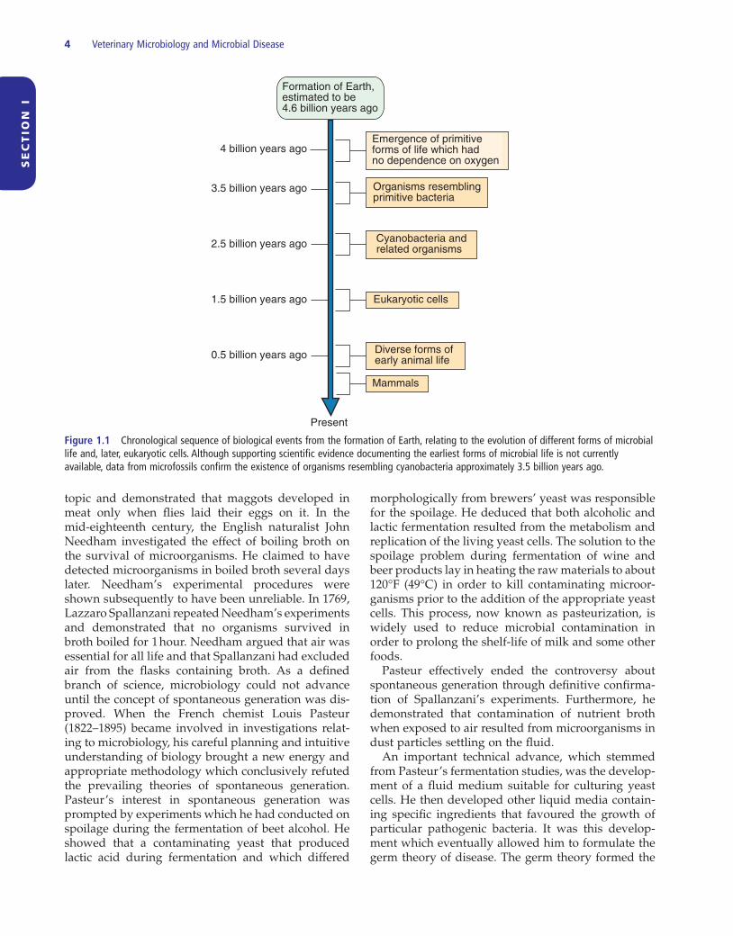
Figure 1.1 Chronological sequence of biological events from the formation of Earth, relating to the evolution of different forms of microbial life and, later, eukaryotic cells. Although supporting scientifi c evidence documenting the earliest forms of microbial life is not currently available, data from microfossils confi rm the existence of organisms resembling cyanobacteria approximately 3.5 billion years ago.
Formation of Earth,estimated to be4.6 billion years ago
2.5 billion years ago
1.5 billion years ago
0.5 billion years ago
3.5 billion years ago
4 billion years ago
Present
Emergence of primitiveforms of life which hadno dependence on oxygen
Organisms resemblingprimitive bacteria
Cyanobacteria andrelated organisms
Eukaryotic cells
Diverse forms ofearly animal life
Mammals
Microbiology, microbial pathogens and infectious disease 5
SE
CT
ION
I
such serious infectious diseases as rabies, smallpox, foot - and - mouth disease and rinderpest. Despite the absence of specifi c knowledge about the aetiology of these diseases, successful vaccines were introduced both for smallpox, by Edward Jenner in the late eighteenth century, and for rabies, by Pasteur and his associates in the latter half of the nineteenth century. The development by Pasteur ’ s co - worker, Charles Chamberland, of the porcelain fi lter to produce bacte-riologically - sterile water for use in culture media, eventually facilitated isolation of the fi lterable agents which caused viral diseases. Remarkably, the tech-nique was fi rst used to elucidate the cause of a plant viral disease, tobacco mosaic disease.
Dmitri Ivanovsky, a Russian scientist, reported in 1892 that it was possible to transmit tobacco mosaic disease from diseased to healthy plants using fi l-tered leaf extract as inoculum. The fi lters used by Ivanovsky were Chamberland porcelain fi lters designed to remove bacteria from drinking water. In 1898, Martinus Beijerinck, unaware of the work of Ivanovsky, also demonstrated the fi lterability of the agent of tobacco mosaic disease. Moreover, he real-ized that the disease could not be due to a toxin as the fi ltered sap from infected plants could be used for serial transmission of the disease without loss of potency. In the same year, Loeffl er and Frosch identi-fi ed the fi rst fi lterable agent from animals, the virus of foot - and - mouth disease. Yellow fever virus, a fi lter-able agent pathogenic for humans, was described by Walter Reed and his team in 1901. Ellerman and Bang, in 1908, demonstrated the oncogenic potential of a fi lterable agent, the cause of avian leukosis. In 1915, Frederick Twort observed that bacteria were suscep-tible to a fi lterable agent, and two years later Felix d ’ Herelle made a similar observation. D ’ Herelle named these viruses ‘ bacteriophages ’ and developed a technique for establishing their concentration in active preparations. Bacteriophages have proved to be particularly useful in studies on viral replication and bacterial genetics.
Initially, the only method available for recovering large quantities of virus was through infecting suscep-tible animals. In 1913 Steinhardt and his colleagues succeeded in growing vaccinia virus in explants of guinea - pig cornea embedded in clotted plasma. Some 20 years later, Furth and Sturmia used mice as a host species for propagating viruses, while Woodruff and Goodpasture were successful in propagating fowlpox virus on the chorioallantoic membrane of embryo-nated eggs. A major advance was made in the early 1950s with the development of single cell cultures. Factors critical in this development included the avail-ability of antibiotics to control bacterial contamina-tion, and the use of trypsin to obtain cell suspensions
basis for Pasteur ’ s experiments on vaccination against fowl cholera, anthrax and rabies. An additional practi-cal application of the theory was the introduction of phenol as a disinfectant for surgical procedures by the British surgeon Joseph Lister.
Together with Pasteur, the German physician Robert Koch is considered to be a co - founder of modern microbiology. Having observed bacilli in the blood of animals that had died from anthrax, Koch demon-strated their pathogenicity by injecting mice with the blood. The injected mice died and the bacilli were present in preparations from their swollen spleens. He was also able to transfer the infection from mouse to mouse and to demonstrate the bacilli in each newly infected mouse. Initially, Koch used blood serum for growing the anthrax bacillus in vitro . Later, he devel-oped solid media which allowed isolation of individ-ual bacterial colonies. Using a solid medium, he was eventually able to isolate the tubercle bacillus from the tissues of an experimental animal in which he had demonstrated microscopically the presence of the organism. As a result of these observations, Koch for-mulated certain principles for proving that a specifi c microorganism caused a particular disease (Box 1.1 ). Pasteur ’ s germ theory of disease and Koch ’ s postu-lates are the two cornerstones on which microbiology is based and without which this branch of biology could not have advanced.
By the end of the nineteenth century a number of important infectious diseases had been confi rmed as bacterial in origin. Both Pasteur and Koch contributed to the identifi cation and confi rmation of the causal agent of anthrax. Pasteur demonstrated that fowl cholera, malignant oedema and suppurative lesions were each associated with a specifi c bacterial infec-tion. The causative organisms of tuberculosis and typhoid fever were recognized by Koch and his associ-ates. Other bacterial agents responsible for serious infectious diseases including glanders, gas gangrene, diphtheria and dysentery were isolated by laboratory scientists in Europe, North America and Japan.
The basic technical approaches, pioneered by Pasteur and Koch, failed to shed light on the causes of
Box 1.1 Koch ’ s postulates
• The pathogenic microorganism must be present in every case of the disease but absent from healthy animals
• The suspected microorganism must be isolated and grown in pure culture
• The same disease must occur when the isolated microor-ganism is injected into healthy susceptible animals
• The same microorganisms must be isolated again from the injected animals which developed disease
6 Veterinary Microbiology and Microbial Disease
SE
CT
ION
I
(4) the diameter of the nucleocapsid (helical viruses) or the number of capsomers (icosahedral viruses). The discovery of the enzyme reverse transcriptase in 1970 by Temin and Baltimore helped to elucidate retrovirus replication and provided an essential tool for produc-ing complementary DNA (cDNA). This ushered in the recombinant DNA revolution. The study of retrovi-ruses has made a substantial contribution to the advancement of basic research in neoplasia and the role of oncogenes in the emergence of malignant tumours.
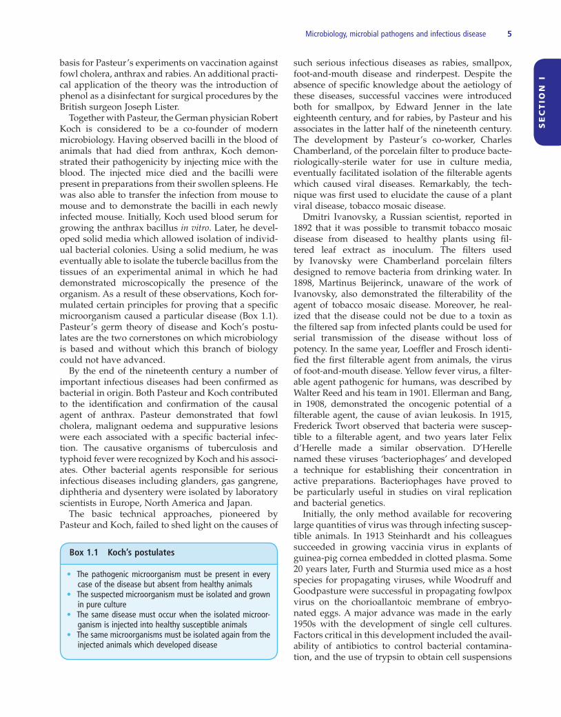
During the past century, major developments have taken place in microbiological concepts, techniques and applications. Modern microbiology encompasses the study of bacteria, fungi, viruses and other micro-scopic and submicroscopic organisms (Fig. 1.2 ). In veterinary microbiology, emphasis is placed on those microorganisms associated with infectious diseases of animals. Immunology, the study of host responses to infectious agents, is a discipline closely related to microbiology and is sometimes considered a distinct but cognate subject.
Further r eading
Dunlop , R.H. and Williams , D.J. ( 1996 ). Veterinary Medicine: An Illustrated History . Mosby , St. Louis, Missouri .
Frankland , P. and Frankland , P. ( 1901 ). Pasteur . Cassell , London .
Lechevalier , H.A. and Solotorovsky , M. ( 1965 ). Three Centuries of Microbiology . McGraw - Hill , New York .
Pelczar , M.J. , Chan , E.C.S. and Krieg , N.R. ( 1993 ). Microbiology Concepts and Applications . McGraw - Hill , New York .
Porter , R. ( 1999 ). The Greatest Benefi t to Mankind . Fontana , London .
Prescott , L.M. , Harley , J.P. and Klein , D.A. ( 2002 ). Microbiology . Fifth Edition . McGraw - Hill , New York .
van Regenmortel , M.H.V. ( 1990 ). Virus species, a much over-looked but essential concept in virus classifi cation . Intervirology , 31 , 241 – 254 .
from embryonic or adult tissue. The separated cells could then be grown as monolayers on glass surfaces. Continuous cell lines, capable of multiplying indefi -nitely, provided a reliable source of cells for virus cultivation.
In 1887 Buist observed vaccinia virus using a light microscope. However, because of the limited resolv-ing power of this type of microscopy, the structure of the virus was not discernible. In 1939 Kausche and his co - workers employed the newly - developed electron microscope and a metal shadowing technique to iden-tify tobacco mosaic virus in purifi ed preparations. Ultrastructural studies of viruses were greatly expanded and enhanced in the 1950s by the develop-ment of negative staining and methods for cutting ultrathin sections. X - ray diffraction methods have been applied to viruses since the 1930s, when it was discovered that simple viruses could be crystallized. The fi rst complete high - resolution structure of a crys-talline virus, tomato bushy stunt virus, was obtained by Harrison and his co - workers in 1978. Computer analysis of the diffraction patterns obtained by such studies has contributed to knowledge of the molecular structure of viruses.
The crystallization of tobacco mosaic virus (TMV) by Stanley in 1935 provided a boost to the analysis of the chemical composition of viruses. In 1937 Bawden and Pirie showed that TMV contained nucleic acid as well as proteins, and helped to promote the idea that viruses consisted of nucleic acid contained within a protein coat. Having elucidated the structure of DNA and observed the limited coding capacity of viral nucleic acid, Watson and Crick in 1956 suggested that viral nucleic acid was surrounded by a shell of identi-cal protein subunits. In 1962 Lwoff and his colleagues proposed a universal system on which the modern classifi cation of viruses is based. The method of clas-sifi cation proposed was based on the following crite-ria: (1) the type of nucleic acid; (2) the symmetry of the virus; (3) the presence or absence of an envelope;
Figure 1.2 Subdivisions of microbiology, a subject which has areas of common interest with pathology, immunology, pharmacology, medicine and therapeutics.
Microbiology
Bacteriology, study of bacteria
Mycology, study of fungi
Virology, study of viruses
Study of unconventional infectiousagents, including prions

SE
CT
ION
I
7
Living cells, the smallest units capable of independent existence, can be divided into two sharply differenti-ated groups, eukaryotes and prokaryotes. The main differentiating features of eukaryotic and prokaryotic cells are presented in Table 2.1 . Eukaryotes possess true nuclei which contain chromosomes, and individ-ual cells replicate by mitosis. In addition, a typical eukaryotic cell contains organelles such as mitochon-dria, a Golgi apparatus, lysosomes and relatively large ribosomes. Organisms in the domains Archaea and Bacteria , which are less complex than eukaryotic organisms, are prokaryotes which lack true membrane - bound nuclei. Their genetic information is contained in a single circular chromosome. In some prokaryotic cells such as bacteria, extrachromosomal DNA in the form of plasmids encodes for certain characteristics of the organism. Although the origin of life is a much debated subject, it is probable that primitive microor-ganisms originated from ancestral life forms several billion years ago. The degree of relatedness among microorganisms can be assessed by comparison of their ribosomal ribonucleic acid (rRNA). There is some evidence that all organisms developed from a group of primitive cells rather than from a single organism (Doolittle, 1999 ). Prokaryotes are considered as one branch of the phylogenetic tree and eukaryotes as the second branch (Fig. 2.1 ). Lateral as well as horizontal transfer of genetic material probably occurred in the course of evolutionary development, with some bacte-rial genes incorporated into members of the Archaea and perhaps with some prokaryotic genes incorpo-rated into eukaryotes. This lateral gene transfer may
Chapter 2
Subdivisions, c lassifi cation and m orphological c haracterization of i nfectious a gents
explain how complex eukaryotic cells acquired some of their genes and organelles. The endosymbiosis hypothesis proposes that at some stage in their early development, eukaryotic cells became primitive phagocytes and acquired particular bacterial cell types which enhanced their respiratory activity (de Duve, 1996 ). It is proposed that the engulfed bacteria pro-vided extra energy through this enhanced respiration to the host cell and eventually evolved into mitochon-dria. A similar phenomenon may account for the development of chloroplasts in plant cells. The cyto-plasmic membrane is the site of respiratory or photo-synthetic energy generation in prokaryotes, unlike eukaryotes in which these activities occur in the mem-branes of mitochondria and chloroplasts.
Microscopical t echniques
A number of different microscopical methods are employed for examining microorganisms. These include bright - fi eld, dark - fi eld, phase - contrast and electron microscopy. Table 2.2 summarizes common techniques employed for the examination of microor-ganisms and the particular types of microorganisms for which the techniques are appropriate. Units of measurement employed in microscopy are indicated in Table 2.3 .
The maximum magnifi cation obtainable by bright - fi eld microscopy, using oil - immersion objectives, is approximately 1,000 × . With bright - fi eld microscopy, suitably stained bacteria as small as 0.2 μ m in size can
Veterinary Microbiology and Microbial Disease, Second Edition. P.J. Quinn, B.K. Markey, F.C. Leonard, E.S. FitzPatrick, S. Fanning, P.J. Hartigan.© 2011 P.J. Quinn, B.K. Markey, F.C. Leonard, E.S. FitzPatrick, S. Fanning and P.J. Hartigan. Published 2011 by Blackwell Publishing Ltd.

8 Veterinary Microbiology and Microbial Disease
SE
CT
ION
I
Figure 2.1 The evolutionary relationships of living organisms. Endosymbiosis is one of the postulated mechanisms whereby eukaryotic cells acquired mitochondria or chloroplasts by incorporation of prokaryotic cells.
Endosymbiosis(chloroplasts)
Endosymbiosis(mitochondria)
Prokaryota
Eukaryota
Archaea Bacteria
Fungi Protozoa Animalia
Primitive forms of life
Plantae
Chromista
Table 2.1 Comparative features of prokaryotic and eukaryotic cells.
Feature Prokaryotic cell Eukaryotic cell
Size of individual cells
Usually less than 5 μ m in greatest dimension
Typically greater than 5 μ m
Genetic material Not separated from cytoplasm
Nucleus separated from cytoplasm by a nuclear membrane
Characteristics of chromosomes
Usually single and circular
Multiple and linear
Mitochondria Absent Present
Golgi apparatus Absent Present
Endoplasmic reticulum
Absent Present
Location of ribosomes
Dispersed throughout cytoplasm
Dispersed throughout cytoplasm and also attached to endoplasmic reticulum
Cell division Binary fi ssion Mitosis
Table 2.2 Microscopical techniques used in microbiology.
Technique Comments
Bright - fi eld microscopy
Used for demonstrating the morphology and size of stained bacteria and fungi; staining affi nity may allow preliminary classifi cation of bacteria and the morphology of fungal structures permits identifi cation of the genus
Phase - contrast microscopy
Used for examining unstained cells in suspension
Dark - fi eld microscopy
Used for examining unstained bacteria such as spirochaetes in suspension
Fluorescence microscopy
Used for identifying microorganisms with specifi c antibodies conjugated with fl uorochromes
Transmission electron microscopy
Used for demonstrating viruses in biological material and for identifying ultrastructural details of bacterial, fungal and mammalian cells
Scanning electron microscopy
Used for demonstrating the three - dimensional structure of microorganisms
be visualized. With dark - fi eld microscopy, the scatter-ing of light by fi ne microorganisms such as spirocha-etes suspended in liquid allows them to be observed against a dark background. In common with dark - fi eld techniques, phase - contrast microscopy can be used to examine unstained specimens. This procedure is more appropriate for research purposes than for routine diagnostic microbiology.
In transmission electron microscopy, beams of elec-trons are used in place of visible light to visualize
small structures such as viruses. Specimens, placed on grids, are negatively stained with electron - dense com-pounds such as potassium phosphotungstate and viewed as magnifi ed images on a fl uorescent screen. Magnifi cations greater than 100,000 × are possible with modern instruments. Scanning electron microscopy is used to obtain three - dimensional views of microor-ganisms when coated with a thin fi lm of heavy metal. With this technique a wide range of magnifi cations up to 100,000 × is feasible.

Subdivisions, classifi cation and morphological characterization of infectious agents 9
SE
CT
ION
I
Pathogenic m icroorganisms
Most microorganisms found in nature are not harm-ful to humans, animals or plants. Indeed, many bac-teria and fungi make an important contribution to biological activities which take place in soil, in water and in the alimentary tract of animals and humans. Those microorganisms that can cause disease in animals or humans are referred to as pathogenic microorganisms.
Bacteria
Microorganisms belonging to the domain Archaea (for-merly Archaebacteria ) are not associated with diseases of domestic animals. Organisms (bacteria) belonging to the domain Bacteria (formerly Eubacteria ) include many pathogens of veterinary importance.
Bacteria are unicellular and are smaller and less complex than eukaryotic cells such as mammalian red blood cells (Table 2.4 ). They usually have rigid cell walls containing a peptidoglycan layer, multiply by binary fi ssion and exhibit considerable morphological diversity. They occur as rods, cocci and helical forms and occasionally as branching fi laments. Despite their morphological diversity, most bacteria are between 0.5 μ m and 5 μ m in length. Motile bacteria possess fl agella by which they can move through liquid media. The majority of bacteria can grow on suitable inert media; some require special growth supplements and
Table 2.3 Units of measurement used in microbiology.
Unit Abbreviation Comments
Millimetre mm One thousandth of a metre (10 − 3 m). Bacterial and fungal colony sizes are usually measured in mm. When growing on a suitable medium, bacterial colonies range in size from 0.5 mm to 5 mm
Micrometre (micron)
μ m One thousandth of a millimetre (10 – 6 m). Used for measuring the size of bacterial and fungal cells. Most bacteria range in size from 0.5 μ m to 5 μ m. A small number of bacteria may exceed 20 μ m in length
Nanometre nm One thousandth of a micrometre (10 − 9 m). Used for expressing the size of viruses. Most viruses of veterinary importance range in size from 20 nm to 300 nm
Table 2.4 A comparison of the morphology and size of bacterial cells relative to a mammalian red blood cell.
Cell Morphology / size Comments
Red blood cell
7 μm
Readily seen using conventional light microscopy
Bacillus
5 μm
Rod - shaped cells, usually stained by the Gram method. Using bright - fi eld microscopy, a magnifi cation of 1,000 × is required to observe most bacterial cells
Coccus
1 μm
Spherical cells, often occurring in chains or in grape - like clusters
Spirochaete
10 μm
Thin, helical bacteria. Dark - fi eld microscopy (without staining) or special staining methods are required to demonstrate these unusual microorganisms
particular atmospheric conditions for growth. Two groups of small bacteria, rickettsiae and chlamydiae, which are unable to multiply on inert media, require living cells for in vitro growth. Cyanobacteria, for-merly referred to as blue - green algae, utilize chloro-phyll for some metabolic pathways. Unlike algae, which store chlorophyll in organelles referred to as chloroplasts, cyanobacteria have chlorophyll distrib-uted inside their cell membranes.
Fungi
Yeasts, moulds and mushrooms belong to a large group of non - photosynthetic eukaryotes termed fungi. Fungi may be either unicellular or multicellular. Multicellular fungi produce fi lamentous microscopic structures called moulds; yeasts, which are unicellular, have a spherical or ovoid shape and multiply by budding. In moulds, the cells are cylindrical and attached end to end, forming branched hyphae (Table 2.5 ). A notable feature of fungi is their ability to secrete potent enzymes that can digest organic matter. When moisture is present and other environmental condi-tions are favourable, fungi can degrade a wide variety of organic substrates. A small number of yeasts and moulds are pathogenic for humans and animals. Some fungi invade tissues whereas others produce toxic

10 Veterinary Microbiology and Microbial Disease
SE
CT
ION
I
serious disease by invading and destroying host cells. A small number of viruses are aetiologically impli-cated in the development of malignant tumours in humans and animals.
Prions
Infectious particles that are smaller than viruses have been implicated in the neurological diseases of animals and humans that are termed transmissible spongiform encephalopathies. These particles, called prions, are distinct from viruses and appear to be devoid of nucleic acid. Prions seem to be composed of an abnormally - folded protein capable of inducing conformational changes in homologous normal host cell protein. Following the induced changes, structurally - altered abnormal protein accumulates in and damages long - lived cells such as neurons. Genetic factors seem to infl uence the susceptibility of humans and animals to prion diseases. Prions exhibit remark-able resistance to physical and chemical inactivation procedures.
Biological c lassifi cation and n omenclature
Microscopic living organisms were formerly classifi ed on the basis of phenotypic expression including mor-
substances called mycotoxins which, if present on crops or in stored food such as grain or nuts, can cause disease in animals and humans.
Algae
A morphologically and physiologically diverse group of organisms, algae are usually considered plant - like because they contain chlorophyll. Many algae are free - living in water; others grow on the surfaces of rocks and on other structures in the environment. Some algae produce pigments which impart distinct colora-tion to water surfaces containing algal blooms. When water temperatures are high, algal growth may be marked, leading to the production of toxins that can accumulate in shellfi sh or in water containing algal blooms.
Viruses
Unlike bacteria and fungi, viruses are not cells. A virus particle or virion consists of nucleic acid, either DNA or RNA, enclosed in a protein coat called a capsid. In addition, some viruses are surrounded by envelopes. Viruses are much smaller than bacteria, and typically range in size from 20 nm to 300 nm in diameter (Table 2.6 ). Despite their simple structure, viruses occur in many shapes. Some are spherical, others are brick - shaped or bullet - shaped and a few have an elongated appearance. Because they lack the structures and enzymes necessary for metabolism and independent reproduction, viruses can multiply only within living cells. Both prokaryotic and eukaryotic cells are suscep-tible to infection by viruses. Those viruses that invade bacterial cells are called bacteriophages. Pathogenic viruses which infect humans and animals can cause
Table 2.5 A comparison of the morphology and size of a bacterial cell and two fungal forms.
Structure Morphology / size Comments
Bacterial cell
Coccus 1 μm
Often occur in chains or grape - like clusters
Fungal forms
Yeast 5 μm
Reproduce by budding
Mould
30 μm
Branched structures (hyphae) composed of many cells
Table 2.6 A comparison of a bacterial cell and a large and a small virus. a
Structure Morphology / size Comments
Bacterial cell
Coccus
1 μm
Readily seen at magnifi cation of 1,000 ×
Viruses
Poxvirus 300 nm
Viruses cannot be seen using conventional bright - fi eld microscopy
Parvovirus 20 nm
Electron microscopy at a magnifi cation of up to 100,000 × is used to demonstrate viruses in clinical specimens or in laboratory preparations
a , not drawn to scale.
Subdivisions, classifi cation and morphological characterization of infectious agents 11
SE
CT
ION
I
In terms of sexually reproducing higher organisms, a species is defi ned as a group or population com-posed of similar individuals that are capable of inter-breeding naturally and are reproductively isolated from other groups. However, the defi nition of what constitutes a species poses particular problems in microbiology. Members of the Bacteria and Archaea do not undergo true reproduction. As a result, bacteria tend to be defi ned operationally or in subjective terms as a collection of strains that share many similar prop-erties but differ signifi cantly from other strains. It is possible to be more precise by using a defi nition based on genetic data with a species expected to share 70% or greater binding in standardized DNA – DNA hy-bridization studies and/or over 97% gene - sequence identity for 16S ribosomal RNA (rRNA). Recently a threshold range of 98.7 to 99% sequence similarity has been recommended as the point at which DNA – DNA reassociation experiments should be required for testing the genomic uniqueness of a novel isolate (Stackebrandt and Ebers, 2006 ). Further debate and refi nement regarding the defi nition of a bacterial species is on - going.
Microorganisms are generally named according to the binomial system devised by the Swedish botanist Carolus Linnaeus in the eighteenth century. The names are Latin or Latinized Greek derivations and are printed in italics. There are two parts, a capitalized generic name and a specifi c epithet. For example, the bacterium that causes anthrax in humans and animals is termed Bacillus anthracis, Bacillus being the generic name and anthracis the specifi c name. The naming of species and higher groups of bacteria is regulated by the Bacteriological Code – The International Code of Nomenclature of Bacteria . The International Journal of Systematic and Evolutionary Microbiology is the offi -cial publication for recording taxonomical changes of Bacteria and Archaea . Websites providing listings of approved names include List of Prokaryotic Names with Standing in Nomenclature ( http://www.bacterio.cict.fr ) and Bacterial Nomenclature Up - to - Date ( http://www.dsmz.de/bactnom/bactname.htm ).
Viruses pose particular taxonomic problems in that they are regarded as subcellular, non - living, infectious entities and are often ignored or considered alongside their host species by scientists dealing with the global taxonomy of organisms. However, virologists are agreed that viruses should be considered as a separate group of organisms regardless of their host species, and have established a free - standing virus taxonomy derived from classical Linnean systematics but with rules unique to the discipline of virology. The system is operated by the International Committee on Taxonomy of Viruses (ICTV) with the latest informa-tion on viral taxonomy published periodically in
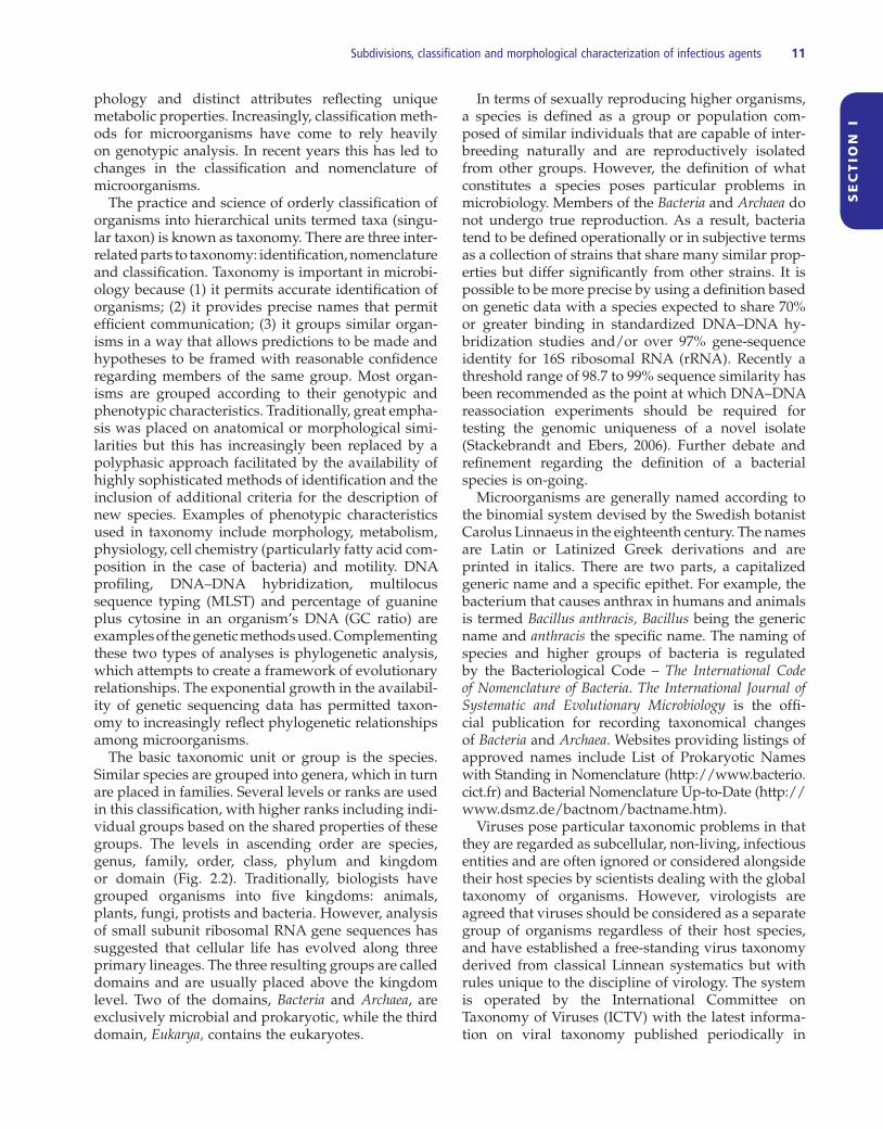
phology and distinct attributes refl ecting unique metabolic properties. Increasingly, classifi cation meth-ods for microorganisms have come to rely heavily on genotypic analysis. In recent years this has led to changes in the classifi cation and nomenclature of microorganisms.
The practice and science of orderly classifi cation of organisms into hierarchical units termed taxa (singu-lar taxon) is known as taxonomy. There are three inter-related parts to taxonomy: identifi cation, nomenclature and classifi cation. Taxonomy is important in microbi-ology because (1) it permits accurate identifi cation of organisms; (2) it provides precise names that permit effi cient communication; (3) it groups similar organ-isms in a way that allows predictions to be made and hypotheses to be framed with reasonable confi dence regarding members of the same group. Most organ-isms are grouped according to their genotypic and phenotypic characteristics. Traditionally, great empha-sis was placed on anatomical or morphological simi-larities but this has increasingly been replaced by a polyphasic approach facilitated by the availability of highly sophisticated methods of identifi cation and the inclusion of additional criteria for the description of new species. Examples of phenotypic characteristics used in taxonomy include morphology, metabolism, physiology, cell chemistry (particularly fatty acid com-position in the case of bacteria) and motility. DNA profi ling, DNA – DNA hybridization, multilocus sequence typing (MLST) and percentage of guanine plus cytosine in an organism ’ s DNA (GC ratio) are examples of the genetic methods used. Complementing these two types of analyses is phylogenetic analysis, which attempts to create a framework of evolutionary relationships. The exponential growth in the availabil-ity of genetic sequencing data has permitted taxon-omy to increasingly refl ect phylogenetic relationships among microorganisms.
The basic taxonomic unit or group is the species. Similar species are grouped into genera, which in turn are placed in families. Several levels or ranks are used in this classifi cation, with higher ranks including indi-vidual groups based on the shared properties of these groups. The levels in ascending order are species, genus, family, order, class, phylum and kingdom or domain (Fig. 2.2 ). Traditionally, biologists have grouped organisms into fi ve kingdoms: animals, plants, fungi, protists and bacteria. However, analysis of small subunit ribosomal RNA gene sequences has suggested that cellular life has evolved along three primary lineages. The three resulting groups are called domains and are usually placed above the kingdom level. Two of the domains, Bacteria and Archaea , are exclusively microbial and prokaryotic, while the third domain, Eukarya, contains the eukaryotes.
12
Veterinary Microbiology and M
icrobial Disease
SECT ION I
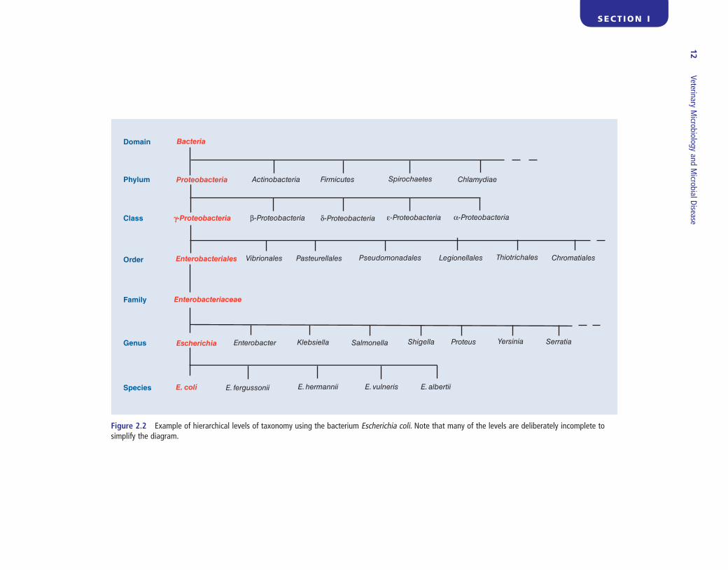
Figure 2.2 Example of hierarchical levels of taxonomy using the bacterium Escherichia coli . Note that many of the levels are deliberately incomplete to simplify the diagram.
Bacteria
Proteobacteria
g-Proteobacteria δ-Proteobacteria α-Proteobacteriaε-Proteobacteriaβ-Proteobacteria
Enterobacteriales Vibrionales Pasteurellales Pseudomonadales Legionellales Thiotrichales Chromatiales
Domain
Phylum
Class
Order
Family
Genus
Species
Enterobacteriaceae
Escherichia Enterobacter Klebsiella Salmonella Shigella Proteus Yersinia Serratia
E. coli E. fergussonii E. hermannii E. vulneris E. albertii
Actinobacteria Firmicutes Spirochaetes Chlamydiae
Subdivisions, classifi cation and morphological characterization of infectious agents 13
SE
CT
ION
I
known as the order of presentation, are used based on the composition and structure of the viral genome, including genome polarity and reverse transcription: double - stranded DNA viruses, single - stranded DNA viruses, DNA and RNA reverse transcribing viruses, double - stranded RNA viruses, negative - stranded single - stranded RNA viruses and positive - stranded single - stranded RNA viruses. In addition there is a category of unassigned viruses and one for subviral agents including viroids, satellites and prions.
References
de Duve , C. ( 1996 ). The birth of complex cells . Scientifi c American , 274 , 38 – 45 .
Doolittle , W.F. ( 1999 ). Phylogenetic classifi cation and the universal tree . Science , 284 , 2124 – 2128 .
Fauquet , C.M. , Mayo , M.A. , Maniloff , J. , Desselberger , U. and Ball , L.A. ( 2005 ). Eighth Report of the ICTV . Elsevier , Amsterdam .
Stackebrandt , E. and Ebers , J. ( 2006 ). Taxonomic parameters revisited: tarnished gold standards . Microbiology Today , 33 , 152 – 155 .
Further r eading
Madigan , M.T. , Martinko , J.M. , Dunlap , P.V. and Clark , D.P. ( 2009 ). Brock Biology of Microorganisms . Twelfth Edition . Pearson Benjamin Cummings , San Francisco .
Prescott , L.M. , Harley , J.P. and Klein , D.A. ( 2005 ). Microbiology . Sixth Edition . McGraw Hill , Boston .
Schlegel , H.G. ( 1993 ). General Microbiology . Seventh Edition . Cambridge University Press , Cambridge .
report form (Fauquet et al ., 2005 ) or available elec-tronically at http://ictvonline.org/virusTaxonomy.asp . A number of points must be borne in mind when considering the taxonomy of viruses: (1) it is consid-ered unlikely that viruses evolved from a single origi-nal protovirus and, as a result, the highest level recognized is that of order; (2) some viruses frequently undergo genetic recombination and reassortment resulting in chimeric organisms with polyphyletic genomes; (3) some viruses infect both vertebrate and invertebrate hosts, evolving differently in the differ-ent host species; (4) some viruses integrate into the genome of their host but can switch between horizon-tal and vertical transmission by moving in and out of the host cell genome, which may result in the incor-poration of host genes into the viral genome. As a result, it is inevitable that the classifi cation system will at times appear artifi cial and give rise to misfi ts. A non - systematic, polythetic, hierarchical system of classifi cation is used for viruses. In essence a virus species is defi ned by a consensus group of properties without any one of these properties being essential. Viruses are generally grouped in families based on virion morphology and nucleic acid type. Further subdivision of pathogenic animal viruses frequently relates to the species of host affected and to the clini-cal disease which is produced. Each viral genus con-tains a type species, which is defi ned as the virus species responsible for the original creation of the genus and whose name is linked to the use of the genus name. Virus species names are commonly abbreviated, for example BPIV - 3 for bovine parainfl u-enza virus 3. In presenting taxonomic descriptions of viruses, several informal categories, collectively
SE
CT
ION
I
14
Infectious disease is a major cause of morbidity and mortality in avian and mammalian species. Individual body systems supply the host ’ s respiratory, nutritional and sensory needs, and the immune system is uniquely equipped to provide defence against microbial or par-asitic infection irrespective of the source or the route of transmission. Some microorganisms cause oppor-tunistic infections in domestic animals; other infec-tious agents, termed pathogenic microorganisms, are capable of causing serious infection if they gain entry into the body. The immune system is composed of an array of structures, cells and secretions which offer defence not only against opportunistic infections but also against pathogenic microorganisms which can cause life - threatening infections in susceptible animals.
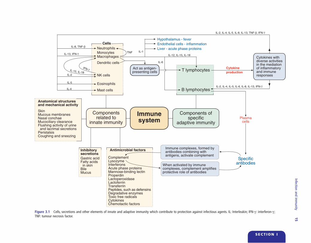
The fi rst barriers to infection that offer rapid, protec-tive responses are components of innate immunity. These components include anatomical structures such as the skin and mucous membranes, inhibitory secre-tions, antimicrobial factors and phagocytic cells (Fig. 3.1 ). If an infectious agent enters the tissues, material from this invading pathogen can be presented to lym-phocytes by phagocytic cells such as macrophages. These lymphocytes then undergo functional changes, proliferate and secrete soluble factors which promote the involvement of other cells of the immune system in an attempt to contain the infection. This response on the part of lymphocytes is referred to as an adap-tive immune response. Moreover, following an encounter with a microbial pathogen, the body ’ s immune system learns from the experience by responding in a specifi c manner to the pathogen and by ‘ remembering ’ the interaction. Immunological memory resides in some lymphocytes that are pro-
Chapter 3
Infection and i mmunity
duced in the course of a response to an infectious agent, and these memory cells react quickly to subse-quent invasion by the same agent. The immune system, therefore, has components that function as innate, non - specifi c barriers to infectious agents, and components that exhibit specifi city combined with immunological memory. It provides protection against a vast array of actual or potential pathogens present in the immediate environment of animals. Immune responses, however, are not confi ned to infectious agents and responses to innocuous substances such as pollens, foreign proteins and some therapeutic drugs, can cause potentially destructive hypersensitivity reactions. Although the primary activity of the immune system is usually considered to be associated with protection against infectious agents, it has a defi ned role in immune surveillance for the detection of neo-plastic tissue changes and, in some instances, elimina-tion of such undesirable mutated cells or neoplastic cells by immune mechanisms.
Soon after birth the external surfaces of the body, extensive portions of the alimentary tract and regions of the respiratory and urinary tracts become colonized by bacteria. The host and colonizing bacteria live in a relatively peaceful state of coexistence, with microor-ganisms restricted to parts of the body where they can be tolerated and microbial invasion of tissues can be prevented by natural antibacterial defence mecha-nisms. Bacteria that colonize tissues of the body without producing disease constitute part of the normal fl ora. This harmonious relationship between animals and their environment can be reinforced by good management systems, optimal nutrition, adequate fl oor space and effective disease control
Veterinary Microbiology and Microbial Disease, Second Edition. P.J. Quinn, B.K. Markey, F.C. Leonard, E.S. FitzPatrick, S. Fanning, P.J. Hartigan.© 2011 P.J. Quinn, B.K. Markey, F.C. Leonard, E.S. FitzPatrick, S. Fanning and P.J. Hartigan. Published 2011 by Blackwell Publishing Ltd.
Infection and imm
unity
15
SECT ION I
Figure 3.1 Cells, secretions and other elements of innate and adaptive immunity which contribute to protection against infectious agents. IL: Interleukin; IFN - γ : interferon - γ ; TNF: tumour necrosis factor.
Hypothalamus - fever
Endothelial cells - inflammation
Liver - acute phase proteins
T lymphocytes
Cells
Act as antigen-presenting cells
Cytokines withdiverse activitiesin the mediationof inflammatoryand immuneresponses
Componentsrelated to
innate immunity
Immunesystem
Components ofspecific
adaptive immunity
Specificantibodies
IL-8, TNF-β
IL-2
IL-5
IL-6
TNF
IL-4
IL-1Neutrophils
MonocytesMacrophages
Dendritic cells
NK cells
Eosinophils
Mast cells
IL-12, IL-15, IL-18
B lymphocytes
Immune complexes, formed byantibodies combining withantigens, activate complement
When activated by immunecomplexes, complement amplifiesprotective role of antibodies
Inhibitorysecretions
Gastric acidFatty acids in skinBileMucus
Antimicrobial factors
ComplementLysozymeInterferonsAcute phase proteinsMannose-binding lectinProperdinLactoperoxidaseLactoferrinTransferrinPeptides, such as defensinsDegradative enzymesToxic free radicalsCytokinesChemotactic factors
Anatomical structuresand mechanical activity
SkinMucous membranesNasal conchaeMucociliary clearanceFlushing activity of urine and lacrimal secretionsPeristalsisCoughing and sneezing
Plasmacells
IFN-γ
IL-13, IFN-γ
IL-2, IL-4, IL-5, IL-8, IL-13, TNF-β, IFN-γ
IL-2, IL-4, IL-5, IL-6, IL-8, IL-13, IFN-γ
Cytokine
productionIL-15, IL-18
16 Veterinary Microbiology and Microbial Disease
SE
CT
ION
I
other microorganisms from the animal ’ s immediate environment may colonize particular sites on the skin and regions of the alimentary, respiratory or urogeni-tal tracts. Microorganisms that compete successfully for particular sites gradually form a stable normal fl ora. Different regions of the body may have a distinc-tive resident fl ora suggesting that regional coloniza-tion may refl ect a selective advantage on the part of successful microorganisms. The ability to survive acidic conditions in the alimentary tract or tolerance for some naturally occurring antimicrobial factors confers particular survival capabilities on some resi-dent fl ora. Adherence to host cells or synthesis of metabolic substances antagonistic to competitors may enhance colonization of the skin, mucous membranes or parts of the alimentary tract by some bacteria and yeasts. There is evidence that the normal fl ora can compete with and sometimes prevent establishment of pathogenic microorganisms. This may be achieved by competition for nutrients, by formation of inhibi-tory substances, or by attachment to receptors on cell surfaces, thereby preventing colonization by invading pathogens. Although the normal fl ora is not directly associated with non - specifi c immunity, their competi-tive role can be considered benefi cial for the host. In
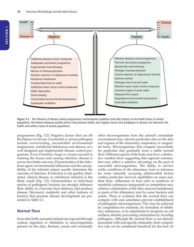
programmes (Fig. 3.2 ). Negative factors that can tilt the balance in favour of potential or actual pathogens include overcrowding, uncontrolled environmental temperature, nutritional imbalances and absence of a well designed and implemented disease control pro-gramme. Even if bacteria, fungi or viruses succeed in entering the tissues and causing infection, disease is not an inevitable outcome. Characteristics of the infec-tious agent, environmental infl uences and the suscep-tibility of the infected animal usually determine the outcome of infection. If infection is not quickly elimi-nated, clinical disease or subclinical infection is the likely result (Fig. 3.3 ). Characteristics of individual species of pathogenic bacteria can strongly infl uence their ability to overcome host defences and produce disease. Structural, metabolic and other features of bacteria that promote disease development are pre-sented in Table 3.1 .
Normal fl ora
Soon after birth, neonatal animals are exposed through contact, ingestion or inhalation to microorganisms present on the dam. Bacteria, yeasts and eventually
Figure 3.2 The infl uence of disease control programmes, environmental conditions and other factors on the health status of animal populations. The balance between positive factors that promote health, and negative factors that predispose to disease can determine the health and welfare status of animal populations.
PositiveNegative
Health
Disease
Disease
Health
Ineffective disease control measures
Inadequate vaccination programme
Inappropriate chemotherapy
Misuse of chemoprophylaxis
Random selection of replacement animals
Nutritional imbalances
Contaminated food or water
Ineffective insect vector control measures
Water deprivation
Overcrowding
Uncontrolled environmental temperature
Inadequate ventilation
Effective disease control programme
Planned vaccination programme
Appropriate chemotherapy
Strategic chemoprophylaxis
Careful selection of replacement animals
Optimal nutrition
Pathogen-free food and water
Effective insect vector control measures
Constant supply of clean water
Adequate floor space
Regulated environmental temperature
Controlled ventilation