Embed Size (px)
Citation preview
METHODOLOGY Open Access
VIBRANT: automated recovery, annotationand curation of microbial viruses, andevaluation of viral community functionfrom genomic sequencesKristopher Kieft, Zhichao Zhou and Karthik Anantharaman*
Abstract
Background: Viruses are central to microbial community structure in all environments. The ability to generate largemetagenomic assemblies of mixed microbial and viral sequences provides the opportunity to tease apart complexmicrobiome dynamics, but these analyses are currently limited by the tools available for analyses of viral genomesand assessing their metabolic impacts on microbiomes.
Design: Here we present VIBRANT, the first method to utilize a hybrid machine learning and protein similarityapproach that is not reliant on sequence features for automated recovery and annotation of viruses, determinationof genome quality and completeness, and characterization of viral community function from metagenomic assemblies.VIBRANT uses neural networks of protein signatures and a newly developed v-score metric that circumvents traditionalboundaries to maximize identification of lytic viral genomes and integrated proviruses, including highly diverse viruses.VIBRANT highlights viral auxiliary metabolic genes and metabolic pathways, thereby serving as a user-friendly platformfor evaluating viral community function. VIBRANT was trained and validated on reference virus datasets as well asmicrobiome and virome data.
Results: VIBRANT showed superior performance in recovering higher quality viruses and concurrently reduced the falseidentification of non-viral genome fragments in comparison to other virus identification programs, specifically VirSorter,VirFinder, and MARVEL. When applied to 120,834 metagenome-derived viral sequences representing several humanand natural environments, VIBRANT recovered an average of 94% of the viruses, whereas VirFinder, VirSorter, andMARVEL achieved less powerful performance, averaging 48%, 87%, and 71%, respectively. Similarly, VIBRANT identifiedmore total viral sequence and proteins when applied to real metagenomes. When compared to PHASTER, ProphageHunter, and VirSorter for the ability to extract integrated provirus regions from host scaffolds, VIBRANT performedcomparably and even identified proviruses that the other programs did not. To demonstrate applications of VIBRANT,we studied viromes associated with Crohn’s disease to show that specific viral groups, namely Enterobacteriales-likeviruses, as well as putative dysbiosis associated viral proteins are more abundant compared to healthy individuals,providing a possible viral link to maintenance of diseased states.
(Continued on next page)
© The Author(s). 2020 Open Access This article is licensed under a Creative Commons Attribution 4.0 International License,which permits use, sharing, adaptation, distribution and reproduction in any medium or format, as long as you giveappropriate credit to the original author(s) and the source, provide a link to the Creative Commons licence, and indicate ifchanges were made. The images or other third party material in this article are included in the article's Creative Commonslicence, unless indicated otherwise in a credit line to the material. If material is not included in the article's Creative Commonslicence and your intended use is not permitted by statutory regulation or exceeds the permitted use, you will need to obtainpermission directly from the copyright holder. To view a copy of this licence, visit http://creativecommons.org/licenses/by/4.0/.The Creative Commons Public Domain Dedication waiver (http://creativecommons.org/publicdomain/zero/1.0/) applies to thedata made available in this article, unless otherwise stated in a credit line to the data.
* Correspondence: [email protected] of Bacteriology, University of Wisconsin–Madison, Madison, WI53706, USA
Kieft et al. Microbiome (2020) 8:90 https://doi.org/10.1186/s40168-020-00867-0
(Continued from previous page)
Conclusions: The ability to accurately recover viruses and explore viral impacts on microbial community metabolismwill greatly advance our understanding of microbiomes, host-microbe interactions, and ecosystem dynamics.
Keywords: Virome, Virus, Bacteriophage, Metagenome, Machine learning, Auxiliary metabolism, Software
BackgroundViruses that infect bacteria and archaea are globally abun-dant and outnumber their hosts in most environments[1–3]. Viruses are obligate intracellular pathogenic geneticelements capable of reprogramming host cellular meta-bolic states during infection and can cause the lysis of 20–40% of microorganisms in diverse environments every day[4, 5]. Due to their abundance and widespread activity, vi-ruses are key facets in microbial communities as they con-tribute to cycling of essential nutrients such as carbon,nitrogen, phosphorus, and sulfur [6–10]. In human sys-tems, viruses have been implicated in contributing todysbiosis that can lead to various diseases, such as inflam-matory bowel diseases, or even have a symbiotic role withthe immune system [11–13].Viruses harbor vast potential for diverse genetic con-
tent, arrangement, and encoded functions [14–17]. Rec-ognizing their genetic diversity, there has beensubstantial interest in “mining” these viral sequences fornovel anti-microbial drug candidates, enzymes for bio-technological applications, and for bioremediation [18–22]. Recently, it has been appreciated that viruses maydirectly link biogeochemical cycling of nutrients by spe-cifically driving metabolic processes [23–27]. For ex-ample, during infection, viruses can acquire 40–90% oftheir required nutrients from the surrounding environ-ment by taking over and subsequently directing host me-tabolism [28–30]. To manipulate host metabolicframeworks, some viruses selectively “steal” metabolicgenes from their host. These host-derived genes, collect-ively termed auxiliary metabolic genes (AMGs), can beactively expressed during infection to provide viruseswith fitness advantages [31–34]. Due to the need tostudy the role of viruses in microbiomes and integrateviruses into models of ecosystem function, it has becomeof great interest to determine which sequences withinwhole microbial communities are derived from viruses.These sequences can include free virions, active intracel-lular infections (which may be the case for as many as30% of all bacteria at any given time [35]), particle orhost-attached virions [36], and host-integrated or epi-somal viral genomes (i.e., proviruses).Multiple tools exist for the identification of viruses
from mixed metagenomic assemblies. For several years,VirSorter [37], which succeeded tools such as VIROME[38] and Metavir [39], has been the most widely used for
its ability to identify viral metagenomic fragments (scaf-folds) from large metagenomic assemblies. VirSorterpredominantly relies on database searches of predictedproteins, using both reference homology as well as prob-abilistic similarity, to compile metrics of enrichment ofvirus-like proteins and simultaneous depletion of otherproteins. To do this, it uses a virus-specific curated data-base as well as Pfam [40] for non-virus annotations,though it does not fully differentiate viral from non-viralPfam annotations. It also incorporates sequence signa-tures of viral genomes, such as encoding short genes orhaving low levels of strand switching between genes.VirSorter is also unique in its ability to use these annota-tions and sequence metrics to identify and extract inte-grated provirus regions from host scaffolds.More recent tools have been developed as alternatives
or supplements of VirSorter. VirFinder [41] was the firsttool to implement machine learning and be completelyindependent of annotation databases for predicting vi-ruses, which was a platform later implemented in PPR-Meta [42]. VirFinder was built with the considerationthat viruses tend to display distinctive patterns of 8-nucleotide frequencies (otherwise known as 8-mers),which was proposed despite the knowledge that virusescan share remarkably similar nucleotide patterns withtheir host [43]. These 8-mer patterns are used to quicklyclassify sequences as short as 500 bp and generatemodel-derived scores, though it is up to the user to de-fine the score cutoffs. VirFinder was shown to greatlyimprove the ability to recover viruses compared to Vir-Sorter, but it also demonstrated substantial host andsource environment biases in predicting diverse viruses,likely due to reference database-associated biases whiletraining the machine learning model [41]. Moreover,under-recovery of viruses from certain environmentswas identified [44].Additional recent tools have been developed that
utilize slightly different methods for identifying viruses.MARVEL [45], for example, leverages annotation,sequence signatures, and machine learning to identify vi-ruses from metagenomic bins. MARVEL differs fromVirSorter in that it only utilizes a single virus-specificdatabase for annotation. However, MARVEL providesno consideration for integrated proviruses and is onlysuitable for identifying bacterial dsDNA viruses from theorder Caudovirales which substantially limits its ability
Kieft et al. Microbiome (2020) 8:90 Page 2 of 23
to discover novel viruses. Another recently developedtool, VirMiner [46], is unique in that it functions to usemetagenomic reads and associated assembly data toidentify viruses and performs best for highly abundantviruses. VirMiner is a web-based server that utilizes ahybrid approach of employing both homology-basedsearches to a virus-specific database as well as machinelearning. VirMiner was found to have improved abilityto recover viruses compared to both VirSorter and Vir-Finder but was concurrently much less accurate.Thus far, VirSorter remains the most efficient tool for
identifying integrated proviruses within metagenomic as-semblies. Other tools, predominantly PHASTER [47]and Prophage Hunter [48], are specialized in identifyingintegrated proviruses from whole genomes rather thanscaffolds generated by metagenomic assemblies. Similarto VirSorter, these two provirus predictors rely on refer-ence homology and viral sequence signatures with slid-ing windows to identify regions of a host genome thatbelong to a virus. Although they are useful for whole ge-nomes, they lack the capability of identifying scaffoldsbelonging to lytic (i.e., non-integrated) viruses and per-form slower for large datasets. In addition, both PHA-STER and Prophage Hunter are exclusively available asweb-based servers and offer no stand-alone commandline tools.Here we developed VIBRANT (Virus Identification By
iteRative ANnoTation), a tool for automated recovery,annotation, and curation of both free and integrated vi-ruses from metagenomic assemblies and genome se-quences. VIBRANT is capable of identifying diversedsDNA, ssDNA, and RNA viruses infecting both bacteriaand archaea, and to our knowledge has no evident envir-onmental biases. VIBRANT uses neural networks of pro-tein annotation signatures from non-reference-basedsimilarity searches with Hidden Markov Models(HMMs) as well as a unique “v-score” metric tomaximize identification of diverse and novel viruses.After identifying viruses, VIBRANT implements curationsteps to validate predictions. VIBRANT additionallycharacterizes viral community function by highlightingAMGs and assesses the metabolic pathways present inviral communities. All viral genomes, proteins, annota-tions, and metabolic profiles are compiled into formatsfor user-friendly downstream analyses and visualization.When applied to reference viruses, non-reference virusdatasets, and various assembled metagenomes, VI-BRANT outperformed VirFinder, VirSorter, and MAR-VEL in the ability to maximize virus recovery andminimize false discovery. When compared to PHASTER,Prophage Hunter, and VirSorter for the ability to extractintegrated provirus regions from host scaffolds, VI-BRANT performed comparably. VIBRANT was alsoused to identify differences in metabolic capabilities
between viruses originating from various environments.When applied to three separate cohorts of individualswith Crohn’s disease, VIBRANT was able to identifyboth differentially abundant viral groups compared tohealthy controls as well as virally encoded genes puta-tively influencing a diseased state. VIBRANT is freelyavailable for download at https://github.com/Ananthara-manLab/VIBRANT. VIBRANT is also available as auser-friendly, web-based application through theCyVerse Discovery Environment at https://de.cyverse.org/de [49].
MethodsDataset for generation and comparison of metricsTo generate training and testing datasets, sequencesrepresenting bacteria, archaea, plasmids, and viruseswere downloaded from the National Center for Biotech-nology Information (NCBI) RefSeq and Genbank data-bases (accessed July 2019) (Additional File 1: Table S1).For bacteria/archaea, 181 genomes were chosen byselecting from diverse phylogenetic groups. Likewise, atotal of 1452 bacterial plasmids were chosen. For viruses,NCBI taxids associated with viruses that infect bacteriaor archaea were used to download reference virus ge-nomes, which were then limited to only sequences above3 kb. This included viruses with both DNA and RNA ge-nomes, though RNA genomes must first be converted tocomplementary DNA. Sequences not associated with ge-nomes, such as partial genomic regions, were identifiedaccording to sequence headers and removed. Thisresulted in 15,238 total viral partial and complete ge-nomes. To be consistent between all sequences acquiredfrom NCBI, proteins and genes were predicted usingProdigal (-p meta, v2.6.3) [50]. All sequences were splitinto non-overlapping, non-redundant fragments between3 and 15 kb to simulate metagenome-assembled scaf-folds. These simulated scaffolds are hereafter called frag-ments and were used throughout training and testingVIBRANT. For RNA virus detection, 33 viral (bacterio-phage) genomes from NCBI RefSeq and 37 from Krish-namurthy et al. were used [51], and for archaeal virusdetection, all genomes were acquired from NCBI RefSeq.The RNA and archaeal viral genomes were representedin both the training and testing datasets as genomic frag-ments, and recall evaluation was performed on wholegenomes. These were the only datasets in which trainingand evaluation datasets were semi-redundant. See Sup-plemental Methods (Additional File 16) for additionaldatasets and sequences used.Integrated viruses are common in both bacteria and
archaea. To address this for generating a dataset devoidof viruses, PHASTER (accessed July 2019) was used topredict putative integrated viruses in the 181 bacteria/ar-chaea genomes. Using BLASTn [52], any fragments that
Kieft et al. Microbiome (2020) 8:90 Page 3 of 23
had significant similarity (at least 95% identity, at least 3kb coverage, and e-value < 1e−10) to the PHASTER pre-dictions were removed as contaminant virus sequence.The new bacteria/archaea dataset was considered de-pleted of proviruses but not entirely devoid of contamin-ation. Next, the datasets for bacteria/archaea andplasmids were annotated with KEGG, Pfam, and VOGHMMs (hmmsearch (v3.1), e-value < 1e−5) [53] to fur-ther remove contaminant virus sequence (see next sec-tion for details of HMMs). Plasmids were includedbecause it was noted that the dataset appeared to con-tain virus sequences, possibly due to misclassification ofepisomal proviruses as plasmids. Using manual inspec-tion of the KEGG, Pfam, and VOG annotations, any se-quence that clearly belonged to a virus was removed.Manual inspection was guided first by the number ofKEGG, Pfam, and VOG annotations and then by the an-notations themselves. For example, sequences with moreVOG than KEGG or Pfam annotations were inspectedand removed if multiple viral hallmark genes were foundor if the majority of annotations represented viral-likegenes. The final datasets consisted of 400,291 fragmentsfor bacteria/archaea, 14,739 for plasmids, and 111,963for viruses. Total number of fragments for all datasetsused can be found in Additional File 2: Table S2.
Databases used by VIBRANTVIBRANT uses HMM profiles from three different data-bases: Kyoto Encyclopedia of Genes and Genomes(KEGG) KoFam (March 2019 release) [54, 55], Pfam(v32) [40], and Virus Orthologous Groups (VOG) (re-lease 94, vogdb.org). For Pfam, all HMM profiles wereused. To increase speed, KEGG and VOG HMM data-bases were reduced in size to contain only profiles likelyto annotate the viruses of interest. For KEGG, this wasdone by only retaining profiles considered to be relevantto “prokaryotes” as determined by KEGG documenta-tion. For VOG, this was done by only retaining profilesthat had at least one significant hit to any of the 15,238NCBI-acquired viruses using BLASTp. The resulting da-tabases consisted of 10,033 HMM profiles for KEGG; 17,929 for Pfam; and 19,182 for VOG (Additional File 3:Table S3).
V-score generationPredicted proteins from reference viral genomes fromNCBI and VOG database viral proteins were combinedto generate v-scores, which resulted in a total of 633,194proteins. Redundancy was removed from the viral pro-tein dataset using CD-HIT (v4.6) [56] with an identitycutoff of 95%, which resulted in a total of 240,728 viralproteins. This was the final dataset used to generate v-scores. All KEGG HMM profiles were used to annotatethe viral proteins. A v-score for each KEGG HMM
profile was determined by the number of significant (e-value < 1e−5) hits by hmmsearch, divided by 100, and amaximum value was set at 10 after division. The same v-score generation was done for Pfam and VOG databases.Any HMM profile with no significant hits to the virusdataset was given a v-score of zero. For KEGG and Pfamdatabases, any annotation that was given a v-score abovezero and contained the keyword “phage” was given aminimum v-score of 1. To highlight viral hallmarkgenes, any annotation within all three databases with thekeyword portal, terminase, spike, capsid, sheath, tail,coat, virion, lysin, holin, base plate, lysozyme, head, orstructural was given a minimum v-score of 1. Non-prokaryotic virus annotations (e.g., reovirus core-spikeprotein) were not considered. Each HMM is assigned av-score and represents a metric of virus association (i.e.,do not take into account virus specificity or associationwith non-viruses) and are manually tuned to put greaterweight on viral hallmark genes (Additional File 4: TableS4). Overall, annotations that are likely non-viral willhave a low v-score whereas annotations that are com-monly associated with viruses will have a high v-score.Raw HMM table outputs for v-score generation can befound in Additional Files 5, 6, and 7 for KEGG, Pfam,and VOG, respectively (Additional File 5: Table S5, Add-itional File 6: Table S6, and Additional File 7: Table S7).
Training and testing VIBRANTThe bacteria/archaea genomic, plasmid, and virus data-sets described above were used to train and test the ma-chine learning model. Scikit-Learn (v0.21.3) [57] librarieswere used to assess various machine learning strategiesto identify the best performing algorithm. Among sup-port vector machines, neural networks, and random for-ests, we found that neural networks lead to the mostaccurate and comprehensive identification of viruses.Therefore, Scikit-Learn’s supervised neural networkmulti-layer perceptron classifier (hereafter called neuralnetwork) was used. The portion of VIBRANT’s workflowup until the neural network classifier (i.e., KEGG, Pfam,and VOG annotation) was used to compile the 27 anno-tation metrics for each scaffold. To account for differ-ences in scaffold sizes, all metrics are normalized (i.e.,divided by) to the total number of proteins encoded bythe scaffold. The first metric, for total proteins, was nor-malized to log base 10 of itself. Each metric wasweighted equally, though it is worth noting that the re-moval of several metrics did not significantly impact theaccuracy of model’s prediction. The normalized resultswere randomized, and non-redundant portions of theseresults were taken for training or testing the neural net-work. In total, 93,913 fragments were used for training,and 9000 different fragments were used for testing the
Kieft et al. Microbiome (2020) 8:90 Page 4 of 23
neural network specifically (Additional File 8: Table S8and Additional File 9: Table S9).To test the performance of VIBRANT in its entirety, a
new testing dataset was generated consisting of frag-ments from the neural network testing set as well asadditional fragments non-redundant to the previoustraining dataset (hereafter called comprehensive testdataset). This new comprehensive test dataset was com-prised of 256,713 genomic fragments from bacteria/ar-chaea, 29,926 from viruses, and 8968 from plasmids.Each met the minimum protein number requirement ofVIBRANT: at least four open reading frames.
Calculation of evaluation metrics and benchmarking ofVIBRANTFor comparison of VIBRANT (v1.2.0) to VirFinder(v1.1), VirSorter (v1.0.3), and MARVEL (v0.2), the com-prehensive test dataset was used. Two intervals for Vir-Finder and VirSorter were used for comparison. ForVirSorter, the intervals selected were (1) category 1 and2 predictions, and (2) category 1 and 2 predictions usingthe virome decontamination mode. Categories 1 and 2are generally considered trustworthy, but category 3 pre-dictions are more likely to contain false identifications.VirSorter was ran using the “Virome” database. For Vir-Finder, the intervals were (1) scores greater than orequal to 0.90 (approximately equivalent to a p value of0.013) and (2) scores greater than or equal to 0.75 (ap-proximately equivalent to a p value of 0.037). SinceMARVEL was built for the identification of viral bins,each scaffold was evaluated separately as a single “bin.”To ensure proper identification by MARVEL and VI-BRANT, different versions of Scikit-Learn were used foreach (v0.19.1 and v0.21.3, respectively).Several metrics were used to compare performance of
all four programs: recall, precision, accuracy, specificity,Mathews Correlation Coefficient (MCC), and F1 score.When calculating metrics, the larger bacteria/archaeaand plasmid dataset was normalized to the size of thesmaller viral dataset in order to make accurate calcula-tions. All equations used can be found in Additional File10: Table S10 and the results of each calculation can befound in Additional File 11: Table S11. Comparison met-rics were visualized using R (v3.5.2) package “ggplot2.”It is worth noting that although VIBRANT was tested
using sequences that were not used for training, biases maystill be associated with reported metrics due to the relianceof KEGG, Pfam, and VOG HMMs on NCBI databases. Thatis, NCBI databases in part were used to construct the HMMsand therefore are well suited at annotating NCBI-derived se-quences. This same type of bias will be seen in the evaluationof VirSorter and MARVEL, both of which rely on NCBI-reliant databases. Although VirFinder does not use annota-tion databases, the machine-learning algorithm it employs
was trained on NCBI-derived sequences. Similarly, biaseswith comparisons to VirFinder, VirSorter, and MARVEL willarise when using NCBI databases. Sequences from NCBIwere used for training each of the three programs and there-fore will likely contain redundancy to VIBRANT’s compre-hensive test dataset. This redundancy will cause artificiallyenhanced performance. To address these biases, we furthercompared all four programs to non-NCBI datasets (seebelow).
AMG identificationKEGG annotations were used to classify potential AMGs(Additional File 12: Table S12). KEGG annotations fall-ing under the “metabolic pathways” category as well as“sulfur relay system” were considered. Manual inspectionwas used to remove non-AMG annotations, such asnrdAB and thyAX. Other annotations not consideredwere associated with direct nucleotide to nucleotide con-versions. All AMGs were associated with a KEGG meta-bolic pathway map.
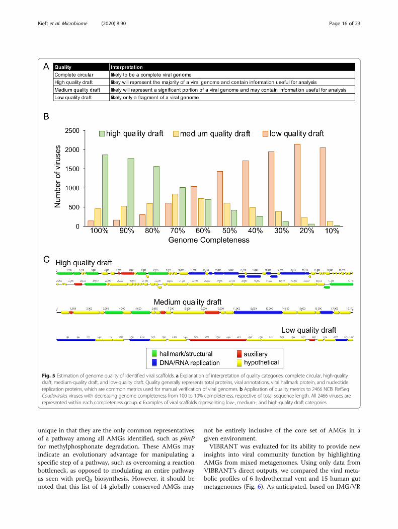
Completeness estimationScaffold completeness is determined based on four met-rics: circularization of scaffold sequence, VOG annota-tions, total VOG nucleotide replication proteins, andtotal VOG viral hallmark proteins (Additional File 13:Table S13). In order to be considered a complete gen-ome, a sequence must be identified as likely circular. Akmer-based approach is used to do this. Specifically, thefirst 20 nucleotides are compared to 20-mer sliding win-dows within the last 900 bp of the sequence. If acomplete match is identified, the sequence is considereda circular template. Scaffolds can also be considered alow-, medium-, or high-quality draft. To benchmarkcompleteness, 2466 NCBI RefSeq viruses identified asCaudovirales, limited to 10 kb in length, were used toestimate completeness by stepwise removing 10% viralsequence at a time. VIBRANT was found to identify2465 of the 2466 viruses. This set of viruses was add-itionally used to assess the error rate of cutting provirusregions. Viral genome diagrams to depict genome qualityand completeness, provirus predictions, and novel virusidentification were made using Geneious Prime 2019.0.3.
Analysis of Crohn’s disease metagenomesMetagenomic reads from He et al. [58] were assembled byPasolli et al. [59] and used for analysis. VIBRANT (-l 5000)was used to predict viruses from 49 metagenomes originat-ing from individuals with Crohn’s disease and 53 fromhealthy individuals (102 total samples). A total of 14,121 vi-ruses were identified. Viral sequences were dereplicatedusing Mash [60] and Nucmer [61] to 95% nucleotide iden-tity and 70% sequence coverage. The longest sequence waskept as the representative for a total of 8822 dereplicated
Kieft et al. Microbiome (2020) 8:90 Page 5 of 23
viruses. A total of 96 read sets were used (59 Crohn’s dis-ease and 37 healthy), trimmed using Sickle and aligned tothe dereplicated viruses using Bowtie2 (-N 1, v2.3.4.1) [62],and the resulting coverages were normalized to total reads.The normalized relative coverage of each virus for all 96samples were compared using DESeq2 [63] (Additional File14: Table S14). Viruses that displayed significantly differentabundance between Crohn’s disease and control sampleswere determined by a p value cutoff of 0.05. iRep (defaultparameters) [64] was used to estimate replication activity oftwo highly abundant Crohn’s-associated viruses. EasyFig(v2.2.2) [65] was used to generate genome alignments ofEscherichia phage Lambda (NCBI accession number NC_001416.1) and three Crohn’s-associated viruses. vCon-TACT2 (v0.9.8) was run using default parameters on theCyVerse Discovery Environment platform. Putative hosts ofCrohn’s-associated and healthy-associated were estimatedusing proximity of vConTACT2 protein clustering andBLASTp identity (NCBI non-redundant protein database,assessed October 2019). Two additional read sets fromGevers et al. [66] and Ijaz et al. [67] were likewise assem-bled by Pasolli et al. VIBRANT (-l 5000 -o 10) was used topredict viruses from 43 metagenomes originating from in-dividuals with Crohn’s disease and 21 from healthy individ-uals (64 total samples). In contrast to the discovery, datasetviral genomes were not dereplicated, and differential abun-dance was not determined. Instead, viruses from eachgroup were directly clustered using vConTACT2. Abun-dances of dysbiosis-associated genes in the validation setwere normalized to total viruses. Validation of dysbiosis-associated genes’ presence on viral genomes, rather thanmicrobial contamination, was done by identifying viral hall-mark genes on the viral scaffold (Additional File 15: TableS15). Protein networks were visualized using Cytoscape(v3.7.2) [68].
ResultsVIBRANT was built to extract and analyze bacterial and ar-chaeal viruses from assembled metagenomic and genome se-quences, as well as provide a platform for characterizingmetabolic proteins and functions in a comprehensive man-ner. The concept behind VIBRANT’s mechanism of virusidentification stems from the understanding that arduousmanual inspection of annotated genomic sequences pro-duces the most dependable results. As such, the primarymetrics used to inform validated curation standards and totrain VIBRANT’s machine learning based neural network toidentify viruses reflects human-guided intuition, though in ahigh-throughput automated fashion.
Determination of v-scoreWe developed a unique “v-score” metric as an approachfor providing quantitative information to VIBRANT’s al-gorithm in order to assess the qualitative nature of
annotation information. A v-score is a value assigned toeach possible protein annotation that scores its associ-ation to known viral genomes (see “Methods” section).V-score differs from the previously used “virus quotient”metric [69, 70] in that it does not take into account theannotation’s relatedness to bacteria or archaea. Not in-cluding significant similarity to non-viral genomes in thecalculation of v-scores has important implications forthis metric’s utility. Foremost is that annotations sharedbetween viruses and their hosts, such as ribonucleotidereductases, will be assigned a v-score reflecting its asso-ciation to viruses, not necessarily virus-specificity. Manygenes are commonly associated with viruses and host or-ganisms but when encoded on viral genomes can becentral to virus replication efficiency (e.g., ribonucleotidereductases [71]). Therefore, a metric representing virus-association rather than virus-specificity would be moreappropriate in identifying if an unknown scaffold is viralor not. Secondly, this approach takes into account wide-spread horizontal gene transfer of host genes by virusesas well as the presence of AMGs.
VIBRANT workflowVIBRANT utilizes several annotation metrics in order toguide removal of non-viral scaffolds before curation ofreliable viral scaffolds. The annotation metrics used arederived from HMM-based probabilistic searches of pro-tein families from the KEGG, Pfam, and VOG databases.VIBRANT is not reliant on reference-based similarityand therefore accounts for the large diversity of viruseson Earth and their respective proteins. Consequently,widespread horizontal gene transfer, rapid mutation, andthe vast amount of novel sequences do not hinder VI-BRANT’s ability to identify known and novel viruses. VI-BRANT does not rely on non-annotation features, suchas rates of open reading frame strand switching, becausethese features were not as well conserved in genomicscaffolds in contrast to whole genomes.VIBRANT’s workflow consists of four main steps (Fig. 1a).
Briefly, proteins (predicted or user input) are used by VI-BRANT to first eliminate non-viral sequences by assessingnon-viral annotation signatures derived from KEGG andPfam HMM annotations. At this step, potential host scaffoldsare fragmented using sliding windows of KEGG annotationv-scores in order to extract integrated provirus sequences.Following the elimination of most non-viral scaffolds andrough excision of provirus regions, proteins are annotated byVOG HMMs. Before analysis by the neural network machinelearning model, any extracted putative provirus is trimmedto exclude any remaining non-viral sequences. Annotationsfrom KEGG, Pfam, and VOG are used to compile 27 metricsthat are utilized by the neural network to predict viral se-quences (Additional File 16: Supplemental Methods). These
Kieft et al. Microbiome (2020) 8:90 Page 6 of 23
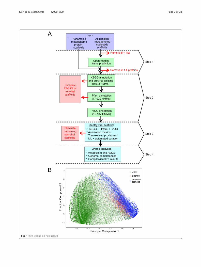
Fig. 1 (See legend on next page.)
Kieft et al. Microbiome (2020) 8:90 Page 7 of 23
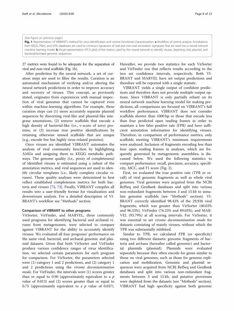
27 metrics were found to be adequate for the separation ofviral and non-viral scaffolds (Fig. 1b).After prediction by the neural network, a set of cur-
ation steps are used to filter the results. Curation is anautomated mechanism of verifying and/or altering theneural network predictions in order to improve accuracyand recovery of viruses. This concept, as previouslystated, originates from experiences with manual inspec-tion of viral genomes that cannot be captured evenwithin machine-learning algorithms. For example, thesecuration steps can (1) more accurately separate plasmidsequences by discerning viral-like and plasmid-like inte-grase annotations, (2) remove scaffolds that encode ahigh density of bacterial-like (i.e., v-score of zero) pro-teins, or (3) increase true positive identifications byretaining otherwise missed scaffolds that are unique(e.g., encode few but highly virus-related proteins).Once viruses are identified VIBRANT automates the
analysis of viral community function by highlightingAMGs and assigning them to KEGG metabolic path-ways. The genome quality (i.e., proxy of completeness)of identified viruses is estimated using a subset of theannotation metrics, and viral sequences are used to iden-tify circular templates (i.e., likely complete circular vi-ruses). These quality analyses were determined to bestreflect established completeness metrics for both bac-teria and viruses [72, 73]. Finally, VIBRANT compiles allresults into a user-friendly format for visualization anddownstream analysis. For a detailed description of VI-BRANT’s workflow see “Methods” section.
Comparison of VIBRANT to other programsVirSorter, VirFinder, and MARVEL, three commonlyused programs for identifying bacterial and archaeal vi-ruses from metagenomes, were selected to compareagainst VIBRANT for the ability to accurately identifyviruses. We evaluated all four programs’ performance onthe same viral, bacterial, and archaeal genomic and plas-mid datasets. Given that both VirSorter and VirFinderproduce various confidence ranges of virus identifica-tion, we selected certain parameters for each programfor comparison. For VirSorter, the parameters selectedwere (1) category 1 and 2 predictions, and (2) category 1and 2 predictions using the virome decontaminationmode. For VirFinder, the intervals were (1) scores greaterthan or equal to 0.90 (approximately equivalent to a pvalue of 0.013) and (2) scores greater than or equal to0.75 (approximately equivalent to a p value of 0.037).
Hereafter, we provide two statistics for each VirSorterand VirFinder run that reflects results according to thetwo set confidence intervals, respectively. Both VI-BRANT and MARVEL have set output predictions andtherefore will be reported with a single statistic.VIBRANT yields a single output of confident predic-
tions and therefore does not provide multiple output op-tions. Since VIBRANT is only partially reliant on itsneural network machine learning model for making pre-dictions, all comparisons are focused on VIBRANT’s fullworkflow performance. VIBRANT does not considerscaffolds shorter than 1000 bp or those that encode lessthan four predicted open reading frames in order tomaintain a low false positive rate (FPR) and have suffi-cient annotation information for identifying viruses.Therefore, in comparison of performance metrics, onlyscaffolds meeting VIBRANT’s minimum requirementswere analyzed. Inclusion of fragments encoding less thanfour open reading frames in analyses, which are fre-quently generated by metagenomic assemblies, is dis-cussed below. We used the following statistics tocompare performance: recall, precision, accuracy, specifi-city, MCC, and F1 score (Fig. 2).First, we evaluated the true positive rate (TPR or re-
call) of viral genomic fragments as well as whole viralgenomes. Viral genomes were acquired from the NCBIRefSeq and GenBank databases and split into variousnon-redundant fragments between 3 and 15 kb to simu-late genomic scaffolds (see “Methods” section). VI-BRANT correctly identified 98.43% of the 29,926 viralfragments, which was greater than VirSorter (40.03%and 96.53%), VirFinder (76.23% and 89.03%), and MAR-VEL (93.79%) at all scoring intervals. For VirSorter, itwas essential to set virome decontamination mode fordatasets consisting of mainly viruses, without which theTPR was substantially inhibited.Similar to TPR, we calculated FPR (or specificity)
using two different datasets: genomic fragments of bac-teria and archaea (hereafter called genomic) and bacter-ial plasmids (plasmid). Plasmids were evaluatedseparately because they often encode for genes similar tothose on viral genomes, such as those for genome repli-cation and mobilization. Genomic and plasmid se-quences were acquired from NCBI RefSeq and GenBankdatabases and split into various non-redundant frag-ments between 3 and 15 kb, and putative proviruseswere depleted from the datasets (see “Methods” section).VIBRANT had high specificity against both genomic
(See figure on previous page.)Fig. 1 Representation of VIBRANT’s method for virus identification and virome functional characterization. a Workflow of virome analysis. Annotationsfrom KEGG, Pfam, and VOG databases are used to construct signatures of viral and non-viral annotation signatures that are read into a neural networkmachine learning model. b Visual representation (PCA plot) of the metrics used by the neural network to identify viruses, depicting viral, plasmid, andbacterial/archaeal genomic sequences
Kieft et al. Microbiome (2020) 8:90 Page 8 of 23
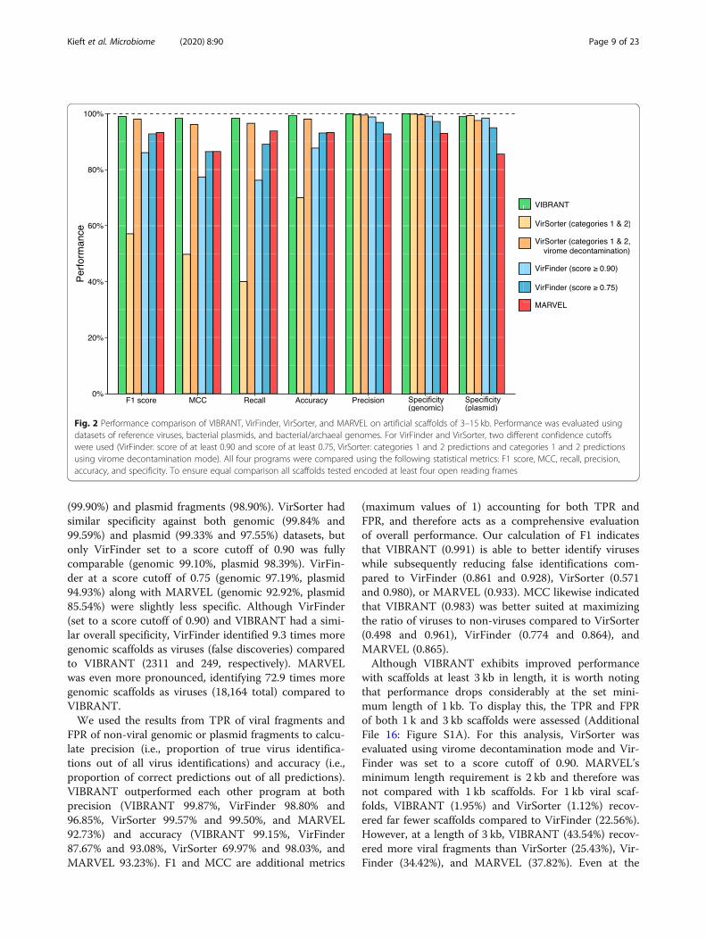
(99.90%) and plasmid fragments (98.90%). VirSorter hadsimilar specificity against both genomic (99.84% and99.59%) and plasmid (99.33% and 97.55%) datasets, butonly VirFinder set to a score cutoff of 0.90 was fullycomparable (genomic 99.10%, plasmid 98.39%). VirFin-der at a score cutoff of 0.75 (genomic 97.19%, plasmid94.93%) along with MARVEL (genomic 92.92%, plasmid85.54%) were slightly less specific. Although VirFinder(set to a score cutoff of 0.90) and VIBRANT had a simi-lar overall specificity, VirFinder identified 9.3 times moregenomic scaffolds as viruses (false discoveries) comparedto VIBRANT (2311 and 249, respectively). MARVELwas even more pronounced, identifying 72.9 times moregenomic scaffolds as viruses (18,164 total) compared toVIBRANT.We used the results from TPR of viral fragments and
FPR of non-viral genomic or plasmid fragments to calcu-late precision (i.e., proportion of true virus identifica-tions out of all virus identifications) and accuracy (i.e.,proportion of correct predictions out of all predictions).VIBRANT outperformed each other program at bothprecision (VIBRANT 99.87%, VirFinder 98.80% and96.85%, VirSorter 99.57% and 99.50%, and MARVEL92.73%) and accuracy (VIBRANT 99.15%, VirFinder87.67% and 93.08%, VirSorter 69.97% and 98.03%, andMARVEL 93.23%). F1 and MCC are additional metrics
(maximum values of 1) accounting for both TPR andFPR, and therefore acts as a comprehensive evaluationof overall performance. Our calculation of F1 indicatesthat VIBRANT (0.991) is able to better identify viruseswhile subsequently reducing false identifications com-pared to VirFinder (0.861 and 0.928), VirSorter (0.571and 0.980), or MARVEL (0.933). MCC likewise indicatedthat VIBRANT (0.983) was better suited at maximizingthe ratio of viruses to non-viruses compared to VirSorter(0.498 and 0.961), VirFinder (0.774 and 0.864), andMARVEL (0.865).Although VIBRANT exhibits improved performance
with scaffolds at least 3 kb in length, it is worth notingthat performance drops considerably at the set mini-mum length of 1 kb. To display this, the TPR and FPRof both 1 k and 3 kb scaffolds were assessed (AdditionalFile 16: Figure S1A). For this analysis, VirSorter wasevaluated using virome decontamination mode and Vir-Finder was set to a score cutoff of 0.90. MARVEL’sminimum length requirement is 2 kb and therefore wasnot compared with 1 kb scaffolds. For 1 kb viral scaf-folds, VIBRANT (1.95%) and VirSorter (1.12%) recov-ered far fewer scaffolds compared to VirFinder (22.56%).However, at a length of 3 kb, VIBRANT (43.54%) recov-ered more viral fragments than VirSorter (25.43%), Vir-Finder (34.42%), and MARVEL (37.82%). Even at the
Fig. 2 Performance comparison of VIBRANT, VirFinder, VirSorter, and MARVEL on artificial scaffolds of 3–15 kb. Performance was evaluated usingdatasets of reference viruses, bacterial plasmids, and bacterial/archaeal genomes. For VirFinder and VirSorter, two different confidence cutoffswere used (VirFinder: score of at least 0.90 and score of at least 0.75, VirSorter: categories 1 and 2 predictions and categories 1 and 2 predictionsusing virome decontamination mode). All four programs were compared using the following statistical metrics: F1 score, MCC, recall, precision,accuracy, and specificity. To ensure equal comparison all scaffolds tested encoded at least four open reading frames
Kieft et al. Microbiome (2020) 8:90 Page 9 of 23
low resolution of short scaffolds, VIBRANT’s FPR is notimpacted. For 1 kb genomic and 1 kb plasmid scaffolds,VIBRANT (< 0.00% and 0.07%) and VirSorter (<0.00%and 0.10%) had fewer false positive discoveries than Vir-Finder (2.61% and 3.70%). Similarly, for 3 kb genomicand 3 kb plasmid scaffolds, VIBRANT (0.10% and 2.69%)and VirSorter (0.11% and 2.41%) falsely identified fewersequences than VirFinder (2.26% and 5.54%) or MAR-VEL (6.08% and 16.30%). Overall, this suggests that Vir-Finder is uniquely able to accurately recover short (e.g.,1 kb) viral scaffolds while maintaining a relatively lowFPR, but this ability is not maintained with longer scaf-folds. Moreover, our current abilities to sequence andassemble scaffolds of lengths over 3 kb will likely lead toa greater focus on longer viral sequences that are moreamenable to downstream analysis, such as taxonomicclassification and functional analyses.Next, we assessed the ability of VIBRANT to filter out
eukaryotic contamination rather than falsely identifythese sequences as viral since eukaryotes were not repre-sented in the training or testing datasets. However, thesecontaminants should be sparse because the majority ofeukaryotic KEGG and VOG HMMs were removed fromthe annotation databases (see “Methods” section). Like-wise, eukaryotic-like annotations should receive a low v-score. A total of 8672 eukaryotic sequences ranging from1 to 15 kb were assessed. VIBRANT (0.62%), VirSorter(0.05% and 0.05%), and MARVEL (0.44%) performedwell with recovering few sequences, whereas VirFinder(4.92% and 15.44%) recovered contamination at a greaterrate (Additional File 16: Figure S1B).Finally, viruses with RNA genomes as well as those
that infect archaea are rare in current culture systemsand sequence databases compared to bacterial dsDNAviruses. However, the true abundance of RNA and ar-chaeal viruses has yet to be explored mainly due tobiases towards dsDNA in genome extracting and se-quencing methods [74] and the low abundance of ar-chaea in most environments. VIBRANT was built toidentify all prokaryotic viruses in order to expand ourknowledge of understudied groups. A total of 70 RNAviral genomes and 93 archaeal viral genomes were usedto evaluate recall. VIBRANT was able to recover 47% ofRNA viruses or 84% of those that encoded at least fourpredicted open reading frames. In comparison, VirSorter(7% and 70%), VirFinder (33% and 57%), and MARVEL(68%) ranged from lower to higher recovery (AdditionalFile 16: Figure S1C). The high recovery of RNA virusesby MARVEL is intriguing since the software was trainedexclusively on dsDNA Caudovirales but may be ex-plained by the greater rate of false positive discovery.For archaeal viruses, VIBRANT (96.77%) identified sig-nificantly more viruses than VirSorter (70.97% and93.55%), VirFinder (46.24% and 74.19%), and MARVEL
(80.65%) (Additional File 16: Figure S1D). Taken to-gether, VIBRANT has the potential to identify RNA andarchaeal viruses, though the significance of this differ-ence is hard to distinguish due to the current dearth ofreference genomes with which to validate.
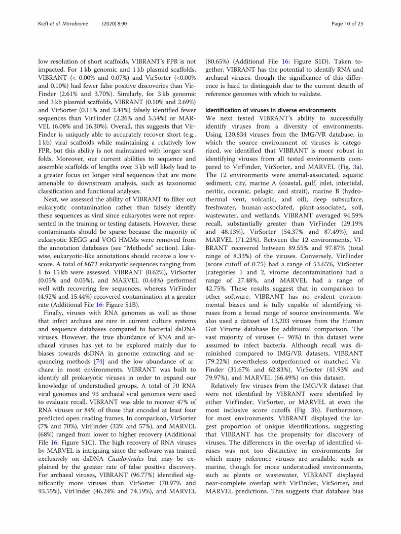
Identification of viruses in diverse environmentsWe next tested VIBRANT’s ability to successfullyidentify viruses from a diversity of environments.Using 120,834 viruses from the IMG/VR database, inwhich the source environment of viruses is catego-rized, we identified that VIBRANT is more robust inidentifying viruses from all tested environments com-pared to VirFinder, VirSorter, and MARVEL (Fig. 3a).The 12 environments were animal-associated, aquaticsediment, city, marine A (coastal, gulf, inlet, intertidal,neritic, oceanic, pelagic, and strait), marine B (hydro-thermal vent, volcanic, and oil), deep subsurface,freshwater, human-associated, plant-associated, soil,wastewater, and wetlands. VIBRANT averaged 94.59%recall, substantially greater than VirFinder (29.19%and 48.13%), VirSorter (54.37% and 87.49%), andMARVEL (71.23%). Between the 12 environments, VI-BRANT recovered between 89.55% and 97.87% (totalrange of 8.33%) of the viruses. Conversely, VirFinder(score cutoff of 0.75) had a range of 53.65%, VirSorter(categories 1 and 2, virome decontamination) had arange of 27.48%, and MARVEL had a range of42.75%. These results suggest that in comparison toother software, VIBRANT has no evident environ-mental biases and is fully capable of identifying vi-ruses from a broad range of source environments. Wealso used a dataset of 13,203 viruses from the HumanGut Virome database for additional comparison. Thevast majority of viruses (~ 96%) in this dataset wereassumed to infect bacteria. Although recall was di-minished compared to IMG/VR datasets, VIBRANT(79.22%) nevertheless outperformed or matched Vir-Finder (31.67% and 62.83%), VirSorter (41.93% and79.97%), and MARVEL (66.49%) on this dataset.Relatively few viruses from the IMG/VR dataset that
were not identified by VIBRANT were identified byeither VirFinder, VirSorter, or MARVEL at even themost inclusive score cutoffs (Fig. 3b). Furthermore,for most environments, VIBRANT displayed the lar-gest proportion of unique identifications, suggestingthat VIBRANT has the propensity for discovery ofviruses. The differences in the overlap of identified vi-ruses was not too distinctive in environments forwhich many reference viruses are available, such asmarine, though for more understudied environments,such as plants or wastewater, VIBRANT displayednear-complete overlap with VirFinder, VirSorter, andMARVEL predictions. This suggests that database bias
Kieft et al. Microbiome (2020) 8:90 Page 10 of 23
may not affect VIBRANT’s performance to a signifi-cant degree. Although VirFinder does not rely on anannotation database, it still has been trained on adataset of reference viral genomes which can contrib-ute to database dependency and recall bias.
Identification of viruses in mixed metagenomesMetagenomes assembled using short read technologycontain many scaffolds that do not meet VIBRANT’sminimum length requirements and therefore are notconsidered during analysis. Despite this, VIBRANT’s
Fig. 3 Effect of source environment on predictive abilities of VIBRANT, VirFinder, VirSorter, and MARVEL. Viral scaffolds from IMG/VR and HGVdatabase were used to test if VIBRANT displays biases associated with specific environments. a The recall (or recovery) of viral scaffolds from 12environment groups was compared between VIBRANT and two confidence cutoffs for both VirFinder and VirSorter. Marine environments wereclassified into two groups: marine A (coastal, gulf, inlet, intertidal, neritic, oceanic, pelagic, and strait) and marine B (hydrothermal vent, volcanic,and oil). b Comparison of the overlap in the scaffolds identified as viruses by all three programs. Cutoffs for VirFinder (scores greater than orequal to 0.75) and VirSorter (categories 1 and 2 using virome decontamination mode) were set to display each program’s ability to recoverdiverse viruses
Kieft et al. Microbiome (2020) 8:90 Page 11 of 23
predictions contain more annotation information andgreater total viral sequence length than tools built toidentify short sequences, such as scaffolds with less thanfour open reading frames. VIBRANT, VirFinder (scorecutoff of 0.90), and VirSorter (categories 1 and 2) wereused to identify viruses from human gut, freshwater lake,and thermophilic compost metagenome sequences(Table 1). In addition, alternate program settings—VI-BRANT virome mode, VirFinder at a score cutoff of0.75, and VirSorter virome decontamination mode—
were used to identify viruses from an estuary viromedataset. MARVEL was not considered in this analysisdue to the inability to achieve comparable precision.Each metagenomic assembly was limited to sequences ofat least 1000 bp but no minimum open reading framelimit was set. For these metagenomes, 31 to 40% of thescaffolds were of sufficient length (at least four openreading frames) to be analyzed by VIBRANT; for the es-tuary virome, 62% was of sufficient length. In compari-son, 100% of scaffolds from each dataset was long
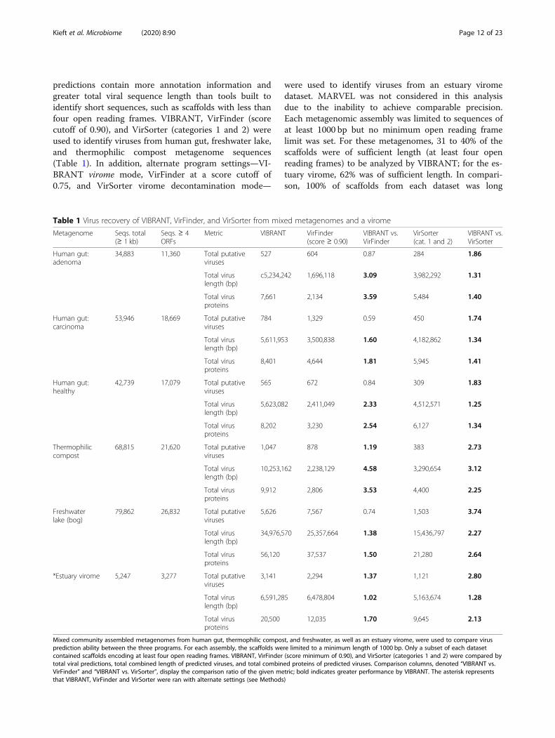
Table 1 Virus recovery of VIBRANT, VirFinder, and VirSorter from mixed metagenomes and a virome
Metagenome Seqs. total(≥ 1 kb)
Seqs. ≥ 4ORFs
Metric VIBRANT VirFinder(score ≥ 0.90)
VIBRANT vs.VirFinder
VirSorter(cat. 1 and 2)
VIBRANT vs.VirSorter
Human gut:adenoma
34,883 11,360 Total putativeviruses
527 604 0.87 284 1.86
Total viruslength (bp)
c5,234,242 1,696,118 3.09 3,982,292 1.31
Total virusproteins
7,661 2,134 3.59 5,484 1.40
Human gut:carcinoma
53,946 18,669 Total putativeviruses
784 1,329 0.59 450 1.74
Total viruslength (bp)
5,611,953 3,500,838 1.60 4,182,862 1.34
Total virusproteins
8,401 4,644 1.81 5,945 1.41
Human gut:healthy
42,739 17,079 Total putativeviruses
565 672 0.84 309 1.83
Total viruslength (bp)
5,623,082 2,411,049 2.33 4,512,571 1.25
Total virusproteins
8,202 3,230 2.54 6,127 1.34
Thermophiliccompost
68,815 21,620 Total putativeviruses
1,047 878 1.19 383 2.73
Total viruslength (bp)
10,253,162 2,238,129 4.58 3,290,654 3.12
Total virusproteins
9,912 2,806 3.53 4,400 2.25
Freshwaterlake (bog)
79,862 26,832 Total putativeviruses
5,626 7,567 0.74 1,503 3.74
Total viruslength (bp)
34,976,570 25,357,664 1.38 15,436,797 2.27
Total virusproteins
56,120 37,537 1.50 21,280 2.64
*Estuary virome 5,247 3,277 Total putativeviruses
3,141 2,294 1.37 1,121 2.80
Total viruslength (bp)
6,591,285 6,478,804 1.02 5,163,674 1.28
Total virusproteins
20,500 12,035 1.70 9,645 2.13
Mixed community assembled metagenomes from human gut, thermophilic compost, and freshwater, as well as an estuary virome, were used to compare virusprediction ability between the three programs. For each assembly, the scaffolds were limited to a minimum length of 1000 bp. Only a subset of each datasetcontained scaffolds encoding at least four open reading frames. VIBRANT, VirFinder (score minimum of 0.90), and VirSorter (categories 1 and 2) were compared bytotal viral predictions, total combined length of predicted viruses, and total combined proteins of predicted viruses. Comparison columns, denoted “VIBRANT vs.VirFinder” and “VIBRANT vs. VirSorter”, display the comparison ratio of the given metric; bold indicates greater performance by VIBRANT. The asterisk representsthat VIBRANT, VirFinder and VirSorter were ran with alternate settings (see Methods)
Kieft et al. Microbiome (2020) 8:90 Page 12 of 23
enough to be analyzed by VirFinder. The ability of Vir-Finder to make a prediction with each scaffold is consid-ered the major strength of the tool.For all six assemblies, VirFinder averaged approxi-
mately 1.16 times more virus identifications than VI-BRANT, though for both thermophilic compost and theestuary virome VIBRANT identified a greater number.Despite VirFinder averaging more total virus identifica-tions, VIBRANT averaged 2.33 times more total viral se-quence length and 2.44 times more total viral proteins.This is the result of VIBRANT having the capability toidentify more viruses of higher quality and longersequence length. For example, among all six datasets,VIBRANT identified 1320 total viruses at least 10 kb inlength in comparison to VirFinder’s 479.VIBRANT was also able to outperform VirSorter in all
metrics, averaging 2.45 times more virus identifications,1.76 times more total viral sequence length, and 1.86times more encoded viral proteins.VIBRANT’s method of predicting viruses provides a
unique opportunity in comparison to similar tools inthat it yields sequences of higher quality which are moreamenable for analyzing protein function from viromedata. It is an important distinction that the total numberof viruses identified may not be correlated with the totalviruses identified or the total number of encoded pro-teins. Even if VIBRANT identified fewer total virusescompared to other tools in certain circumstances, moredata of higher quality was generated as viral sequencesof longer length were identified as compared to manyshort fragments. This provides an important distinctionthat the metric of total viral predictions is not necessar-ily an accurate representation for the quality or quantityof the data generated.
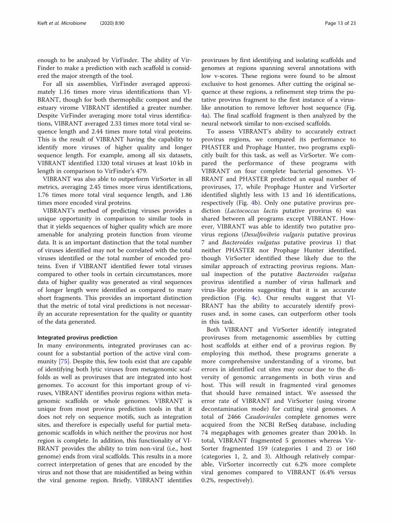
Integrated provirus predictionIn many environments, integrated proviruses can ac-count for a substantial portion of the active viral com-munity [75]. Despite this, few tools exist that are capableof identifying both lytic viruses from metagenomic scaf-folds as well as proviruses that are integrated into hostgenomes. To account for this important group of vi-ruses, VIBRANT identifies provirus regions within meta-genomic scaffolds or whole genomes. VIBRANT isunique from most provirus prediction tools in that itdoes not rely on sequence motifs, such as integrationsites, and therefore is especially useful for partial meta-genomic scaffolds in which neither the provirus nor hostregion is complete. In addition, this functionality of VI-BRANT provides the ability to trim non-viral (i.e., hostgenome) ends from viral scaffolds. This results in a morecorrect interpretation of genes that are encoded by thevirus and not those that are misidentified as being withinthe viral genome region. Briefly, VIBRANT identifies
proviruses by first identifying and isolating scaffolds andgenomes at regions spanning several annotations withlow v-scores. These regions were found to be almostexclusive to host genomes. After cutting the original se-quence at these regions, a refinement step trims the pu-tative provirus fragment to the first instance of a virus-like annotation to remove leftover host sequence (Fig.4a). The final scaffold fragment is then analyzed by theneural network similar to non-excised scaffolds.To assess VIBRANT’s ability to accurately extract
provirus regions, we compared its performance toPHASTER and Prophage Hunter, two programs expli-citly built for this task, as well as VirSorter. We com-pared the performance of these programs withVIBRANT on four complete bacterial genomes. VI-BRANT and PHASTER predicted an equal number ofproviruses, 17, while Prophage Hunter and VirSorteridentified slightly less with 13 and 16 identifications,respectively (Fig. 4b). Only one putative provirus pre-diction (Lactococcus lactis putative provirus 6) wasshared between all programs except VIBRANT. How-ever, VIBRANT was able to identify two putative pro-virus regions (Desulfovibrio vulgaris putative provirus7 and Bacteroides vulgatus putative provirus 1) thatneither PHASTER nor Prophage Hunter identified,though VirSorter identified these likely due to thesimilar approach of extracting provirus regions. Man-ual inspection of the putative Bacteroides vulgatusprovirus identified a number of virus hallmark andvirus-like proteins suggesting that it is an accurateprediction (Fig. 4c). Our results suggest that VI-BRANT has the ability to accurately identify provi-ruses and, in some cases, can outperform other toolsin this task.Both VIBRANT and VirSorter identify integrated
proviruses from metagenomic assemblies by cuttinghost scaffolds at either end of a provirus region. Byemploying this method, these programs generate amore comprehensive understanding of a virome, buterrors in identified cut sites may occur due to the di-versity of genomic arrangements in both virus andhost. This will result in fragmented viral genomesthat should have remained intact. We assessed theerror rate of VIBRANT and VirSorter (using viromedecontamination mode) for cutting viral genomes. Atotal of 2466 Caudovirales complete genomes wereacquired from the NCBI RefSeq database, including74 megaphages with genomes greater than 200 kb. Intotal, VIBRANT fragmented 5 genomes whereas Vir-Sorter fragmented 159 (categories 1 and 2) or 160(categories 1, 2, and 3). Although relatively compar-able, VirSorter incorrectly cut 6.2% more completeviral genomes compared to VIBRANT (6.4% versus0.2%, respectively).
Kieft et al. Microbiome (2020) 8:90 Page 13 of 23
Fig. 4 Prediction of integrated proviruses by VIBRANT and comparison to PHASTER, Prophage Hunter, and VirSorter. a Schematic representing themethod used by VIBRANT to identify and extract provirus regions from host scaffolds using annotations. Briefly, v-scores are used to cut scaffolds athost-specific sites and fragments are trimmed to the nearest viral annotation. b Comparison of proviral predictions within four complete bacterialgenomes between VIBRANT, PHASTER, Prophage Hunter, and VirSorter. For PHASTER, putative proviruses are colored according to “incomplete” (red),“questionable” (blue), and “intact” (green) predictions. Prophage Hunter is colored according to “active” (green) and “ambiguous” (blue) predictions. AllVirSorter predictions for categories 1 and 2 are shown in green. c Manual validation of the Bacteroides vulgatus provirus prediction made by VIBRANT.The presence of viral hallmark protein, integrase and genome replication proteins strongly suggests this is an accurate prediction
Kieft et al. Microbiome (2020) 8:90 Page 14 of 23
Evaluating quality and completeness of predicted viralsequencesDetermination of quality, in relation to completeness, ofa predicted viral sequence has been notoriously difficultdue to the absence of universally conserved viral genes.To date, the most reliable metric of completeness formetagenome- assembled viruses is to identify circular se-quences (i.e., complete circular genomes). Therefore, theremaining alternatives rely on estimation based onencoded proteins that function in central viral processes:replication of genomes and assembly of new viralparticles.VIBRANT estimates the quality of predicted viral se-
quences, a relative proxy for completeness, and indicatessequences that are circular. To do this, VIBRANT usesannotation metrics of nucleotide replication and viralhallmark proteins. Hallmark proteins are those typicallyspecific to viruses and those that are required for pro-ductive infection, such as structural (e.g., capsid, tail,baseplate), terminase, or viral holin/lysin proteins.Nucleotide replication proteins are a variety of proteinsassociated with either replication or metabolism, such asnucleases, polymerases, and DNA/RNA binding pro-teins. Viruses are categorized as low-, medium-, or high-quality draft as determined by VOG annotations (Fig. 5a,Additional File 17: Table S16). High-quality draft repre-sents sequences that are likely to contain the majority ofa virus’s complete genome and will contain annotationsthat are likely to aid in analysis of the virus, such asphylogenetic relationships and true positive verification.Medium draft quality represents the majority of acomplete viral genome but is more likely to be a smallerportion in comparison to high quality. These sequencesmay contain annotations useful for analysis but areunder less strict requirements compared to high quality.Finally, low draft quality constitutes sequences that werenot found to be of high or medium quality. Many meta-genomic scaffolds will likely be low-quality genome frag-ments, but this quality category may still contain thehigher quality genomes of some highly divergent viruses.We benchmarked VIBRANT’s viral genome quality es-
timation using a total of 2466 Caudovirales genomesfrom NCBI RefSeq database. Genomes were evaluatedeither as complete sequences or by removing 10% of thesequence at a time stepwise between 100 and 10% com-pleteness (Fig. 5b). The results of VIBRANT’s qualityanalysis displayed a linear trend in indicating morecomplete genomes as high quality and less complete ge-nomes as lower quality. The transition from categorizinggenomes as high quality to medium quality ranged from60 to 70% completeness. Although we acknowledge thatVIBRANT’s metrics are not perfect, we demonstrate thefirst benchmarked approach to quantify and characterizegenome quality associated with completeness of viral
sequences. Manual inspection and visual verification ofviral genomes that were characterized into each of thesegenome quality categories showed that quality estima-tions matched annotations (Fig. 5c).
Identifying function in viral communities: metabolicanalysisViruses are a dynamic and key facet in the metabolicnetworks of microbial communities and can reprogramthe landscape of host and community metabolism dur-ing infection. This can often be achieved by modulatinghost metabolic networks through expression of AMGsencoded on viral genomes. Identifying these AMGs andtheir associated role in the function of communities isimperative for understanding complex microbiome dy-namics, or in some cases can be used to predict virus-host relationships. VIBRANT is optimized for the evalu-ation of viral community function by identifying andclassifying the metabolic capabilities encoded by a vir-ome. To do this, VIBRANT identifies AMGs and assignsthem into specific metabolic pathways and broader cat-egories as designated by KEGG annotations.To highlight the utility of this feature, we compared
the metabolic function of IMG/VR viruses derived fromseveral diverse environments: freshwater, marine, soil,human-associated, and city (Additional File 16: FigureS2). We found natural environments (freshwater, mar-ine, and soil) to display a different pattern of metaboliccapabilities compared to human environments (human-associated and city). Viruses originating from natural en-vironments tend to largely encode AMGs for amino acidand cofactor/vitamin metabolism with a more secondaryfocus on carbohydrate and glycan metabolism. On theother hand, AMGs from city and human environmentsare dominated by amino acid metabolism, and to someextent cofactor/vitamin and sulfur relay metabolism. Inaddition to this broad distinction, all five environmentsappear slightly different from each other. Despite fresh-water and marine environments appearing similar in theratio of AMGs by metabolic category, the overlap in spe-cific AMGs is less extensive. The dissimilarity betweennatural and human environments is likewise corrobo-rated by the relatively low overlap in individual AMGs.A useful observation provided by VIBRANT’s meta-
bolic analysis is that there appears to be globally con-served AMGs (i.e., present within at least 10 of the 12environments tested). These 14 genes—dcm, cysH, folE,phnP, ubiG, ubiE, waaF, moeB, ahbD, cobS, mec, queE,queD, queC—likely perform functions that are central toviral replication regardless of host or environment. Not-ably, folE, queD, queE, and queC constitute the entire 7-cyano-7-deazaguanine (preQ0) biosynthesis pathway, butthe remainder of queuosine biosynthesis are entirely ab-sent with the exception of queF. Certain AMGs are
Kieft et al. Microbiome (2020) 8:90 Page 15 of 23
unique in that they are the only common representativesof a pathway among all AMGs identified, such as phnPfor methylphosphonate degradation. These AMGs mayindicate an evolutionary advantage for manipulating aspecific step of a pathway, such as overcoming a reactionbottleneck, as opposed to modulating an entire pathwayas seen with preQ0 biosynthesis. However, it should benoted that this list of 14 globally conserved AMGs may
not be entirely inclusive of the core set of AMGs in agiven environment.VIBRANT was evaluated for its ability to provide new
insights into viral community function by highlightingAMGs from mixed metagenomes. Using only data fromVIBRANT’s direct outputs, we compared the viral meta-bolic profiles of 6 hydrothermal vent and 15 human gutmetagenomes (Fig. 6). As anticipated, based on IMG/VR
Fig. 5 Estimation of genome quality of identified viral scaffolds. a Explanation of interpretation of quality categories: complete circular, high-qualitydraft, medium-quality draft, and low-quality draft. Quality generally represents total proteins, viral annotations, viral hallmark protein, and nucleotidereplication proteins, which are common metrics used for manual verification of viral genomes. b Application of quality metrics to 2466 NCBI RefSeqCaudovirales viruses with decreasing genome completeness from 100 to 10% completeness, respective of total sequence length. All 2466 viruses arerepresented within each completeness group. c Examples of viral scaffolds representing low-, medium-, and high-quality draft categories
Kieft et al. Microbiome (2020) 8:90 Page 16 of 23
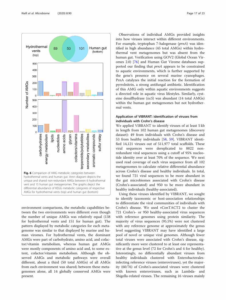
environment comparisons, the metabolic capabilities be-tween the two environments were different even thoughthe number of unique AMGs was relatively equal (138for hydrothermal vents and 151 for human gut). Thepattern displayed by metabolic categories for each meta-genome was similar to that displayed by marine and hu-man viromes. For hydrothermal vents, the dominantAMGs were part of carbohydrate, amino acid, and cofac-tor/vitamin metabolism, whereas human gut AMGswere mostly components of amino acid and, to some ex-tent, cofactor/vitamin metabolism. Although the ob-served AMGs and metabolic pathways were overalldifferent, about a third (50 total AMGs) of all AMGsfrom each environment was shared; between these meta-genomes alone, all 14 globally conserved AMGs werepresent.
Observations of individual AMGs provided insightsinto how viruses interact within different environments.For example, tryptophan 7-halogenase (prnA) was iden-tified in high abundance (45 total AMGs) within hydro-thermal vent metagenomes but was absent from thehuman gut. Verification using GOV2 (Global Ocean Vir-omes 2.0) [76] and Human Gut Virome databases sup-ported our finding that prnA appears to be constrainedto aquatic environments, which is further supported bythe gene’s presence on several marine cyanophages.PrnA catalyzes the initial reaction for the formation ofpyrrolnitrin, a strong antifungal antibiotic. Identificationof this AMG only within aquatic environments suggestsa directed role in aquatic virus lifestyles. Similarly, cyst-eine desulfhydrase (iscS) was abundant (14 total AMGs)within the human gut metagenomes but not hydrother-mal vents.
Application of VIBRANT: identification of viruses fromindividuals with Crohn’s diseaseWe applied VIBRANT to identify viruses of at least 5 kbin length from 102 human gut metagenomes (discoverydataset): 49 from individuals with Crohn’s disease and53 from healthy individuals [58, 59]. VIBRANT identi-fied 14,121 viruses out of 511,977 total scaffolds. Theseviral sequences were dereplicated to 8822 non-redundant viral sequences using a cutoff of 95% nucleo-tide identity over at least 70% of the sequence. We nextused read coverage of each virus sequence from all 102metagenomes to calculate relative differential abundanceacross Crohn’s disease and healthy individuals. In total,we found 721 viral sequences to be more abundant inthe gut microbiomes associated with Crohn’s disease(Crohn’s-associated) and 950 to be more abundant inhealthy individuals (healthy-associated).Using these viruses identified by VIBRANT, we sought
to identify taxonomic or host-association relationshipsto differentiate the viral communities of individuals withCrohn’s disease. We used vConTACT2 to cluster the721 Crohn’s- or 950 healthy-associated virus sequenceswith reference genomes using protein similarity. Themajority of virus sequences (95.5%) were not clusteredwith any reference genome at approximately the genuslevel suggesting VIBRANT may have identified a largepool of novel or unique viral genomes. Although fewertotal viruses were associated with Crohn’s disease, sig-nificantly more were clustered to at least one representa-tive at the genus level (72 for Crohn’s and 4 for healthy).Interestingly, no differentially abundant viruses fromhealthy individuals clustered with Enterobacterales-infecting reference viruses (enteroviruses), yet the major-ity (60/76) of Crohn’s-associated viruses were clusteredwith known enteroviruses, such as Lambda- andShigella-related viruses. The remaining 16 viruses mainly
Fig. 6 Comparison of AMG metabolic categories betweenhydrothermal vents and human gut. Venn diagram depicts theunique and shared non-redundant AMGs between 6 hydrothermalvent and 15 human gut metagenomes. The graphs depict thedifferential abundance of KEGG metabolic categories of respectiveAMGs for hydrothermal vents (top) and human gut (bottom)
Kieft et al. Microbiome (2020) 8:90 Page 17 of 23
clustered with Caudovirales infecting Lactococcus, Clos-tridium, Riemerella, Klebsiella, and Salmonella species,though Microviridae and a likely complete crAssphagewere also identified. A significant proportion of allCrohn’s-associated viruses (250/721) and the majority ofgenus-level clustered viruses (42/76) were found to beintegrated sequences within a microbial genomic scaf-fold but were able to be identified due to VIBRANT’sability to excise proviruses.We also generated a protein sharing network contain-
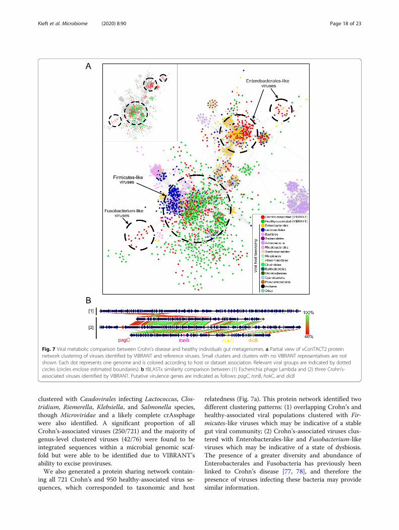
ing all 721 Crohn’s and 950 healthy-associated virus se-quences, which corresponded to taxonomic and host
relatedness (Fig. 7a). This protein network identified twodifferent clustering patterns: (1) overlapping Crohn’s andhealthy-associated viral populations clustered with Fir-micutes-like viruses which may be indicative of a stablegut viral community; (2) Crohn’s-associated viruses clus-tered with Enterobacterales-like and Fusobacterium-likeviruses which may be indicative of a state of dysbiosis.The presence of a greater diversity and abundance ofEnterobacterales and Fusobacteria has previously beenlinked to Crohn’s disease [77, 78], and therefore thepresence of viruses infecting these bacteria may providesimilar information.
Fig. 7 Viral metabolic comparison between Crohn’s disease and healthy individuals gut metagenomes. a Partial view of vConTACT2 proteinnetwork clustering of viruses identified by VIBRANT and reference viruses. Small clusters and clusters with no VIBRANT representatives are notshown. Each dot represents one genome and is colored according to host or dataset association. Relevant viral groups are indicated by dottedcircles (circles enclose estimated boundaries). b tBLASTx similarity comparison between (1) Escherichia phage Lambda and (2) three Crohn’s-associated viruses identified by VIBRANT. Putative virulence genes are indicated as follows: pagC, tonB, hokC, and dicB
Kieft et al. Microbiome (2020) 8:90 Page 18 of 23
VIBRANT provides annotation information for all ofthe identified viruses which can be used to infer func-tional characteristics in conjunction with host associ-ation. Comparison of Crohn’s-associated Lambda-likevirus genomic content and arrangement suggested apossible role of virally encoded host-persistence andvirulence genes that are absent in the healthy-associatedvirome (Fig. 7b). Among all Crohn’s-associated viruses,17 total genes (bor, dicB, dicC, hokC, kilR, pagC, ydaS,ydaT, yfdN, yfdP, yfdQ, yfdR, yfdS, yfdT, ymfL, ymfM,and tonB) that have the potential to impact host survivalor virulence were identified. Importantly, no healthy-associated viruses encoded such genes (Table 2). Thepresence of these putative dysbiosis-associated genes(DAGs) may contribute to the manifestation and/or per-sistence of disease, similar to what has been proposedfor the bacterial microbiome [79–81]. For example, pagCencodes an outer membrane virulence factor associatedwith enhanced survival of the host bacterium within thegut [82]. The identification of dicB encoded on a puta-tive Escherichia virus is unique in that it may represent a“cryptic” provirus that protects the host from lytic viralinfection, thus likely to enhance the ability of the host tosurvive within the gut [83]. Finally, hokC may indicatemechanisms of virally encoded virulence [84].To characterize the distribution and association of
DAGs with Crohn’s disease, we calculated differentialabundance for two highly abundant DAG-encoding vi-ruses across all metagenome samples. The first virus
encoded pagC and yfdN, and the second encoded dicB,dicC, and hokC. Comparison of Crohn’s disease tohealthy metagenomes indicates that these viruses arepresent within the gut metagenomes of multiple individ-uals but more abundant in association with Crohn’s dis-ease (Additional File 16: Figure S3A). This suggests anassociation of disease with not only putative DAGs, butalso specific and potentially persistent, viral groups thatencode them. In order to correlate increased abundancewith biological activity, we calculated the index of repli-cation (iRep) for each of the two viruses [64]. Briefly,iRep is a function of differential read coverage which isable to provide an estimate of active genome replication.Seven metagenomes containing the greatest abundancefor each virus were selected for iRep analysis and indi-cated that each virus was likely active at the time of col-lection (Additional File 16: Figure S3B).To validate these aforementioned findings, we applied
VIBRANT to two additional metagenomic datasets fromcohorts of individuals with Crohn’s disease and healthy in-dividuals (validation dataset): 43 from individuals withCrohn’s disease and 21 from healthy individuals [66, 67].VIBRANT identified 3759 redundant viral genomes fromCrohn’s-associated metagenomes and 1444 from healthy-associated metagenomes. Determination of protein net-works and visualization similarly identified clustering ofCrohn’s-associated viruses with reference enteroviruses(Additional File 16: Figure S4). Likewise, we were able toidentify 15 out of the 17 putative DAGs to be present in
Table 2 Identification of putative DAGs encoded by Crohn’s-associated viruses
ID Gene Name Crohn’s disease Healthy
PF06291.11 bor Bor protein 8 0
K22304 dicB Cell division inhibition protein 8 0
K22302 dicC Transcriptional repressor of cell division inhibition gene dicB 18 0
K18919 hokC Protein HokC/D 16 0
VOG11478 kilR Killing protein 15 0
K07804 pagC Putative virulence related protein 13 0
PF15943.5 ydaS Putative antitoxin of bacterial toxin-antitoxin system 22 0
PF06254.11 ydaT Putative bacterial toxin 18 0
VOG04806 yfdN Uncharacterized protein 19 0
VOG01357 yfdP Uncharacterized protein 11 0
VOG11472 yfdQ Uncharacterized protein 11 0
VOG01639 yfdR Uncharacterized protein 17 0
VOG01103 yfdS Uncharacterized protein 18 0
VOG16442 yfdT Uncharacterized protein 8 0
VOG00672 ymfL Uncharacterized protein 25 0
VOG21507 ymfM Uncharacterized protein 9 0
K03832 tonB periplasmic protein 3 0
The differential abundance between Crohn’s disease and healthy metagenomes of 17 putative DAGs. Abundance of each gene represents non-redundantannotations or total gene copy number, from Crohn’s-associated and healthy-associated viruses
Kieft et al. Microbiome (2020) 8:90 Page 19 of 23
higher abundance in the Crohn’s disease microbiome(Additional File 18: Table S17). This validates our findingsof the presence of unique viruses and proteins associatedwith Crohn’s disease and suggests that Enterobacterales-like viruses and putative DAGs may act as markers ofCrohn’s disease. Overall, our results suggest that VI-BRANT provides a platform for characterizing theserelationships.
DiscussionViruses that infect bacteria and archaea are key compo-nents in the structure, dynamics, and interactions of mi-crobial communities [2, 6, 10, 76, 85]. Tools that arecapable of efficient recovery of these viral genomes frommixed metagenomic samples are likely to be fundamen-tal to the growing applications of metagenomic sequen-cing and analyses. Importantly, such tools would need toreduce bias associated with specific viral groups (e.g.,Caudovirales) and highly represented environments (e.g.,marine). Moreover, viruses that exist as integrated provi-ruses within host genomes should not be ignored as theycan represent a substantial fraction of infections in cer-tain conditions and also persistent infections within acommunity [75].Here we have presented VIBRANT, a newly described
method for the automated recovery of both free and in-tegrated viral genomes from metagenomes that hybrid-izes neural network machine learning and proteinsignatures. VIBRANT utilizes metrics of non-reference-based protein similarity annotation from KEGG, Pfam,and VOG databases in conjunction with a unique “v-score” metric to recover viruses with little to no biases.VIBRANT was built with the consideration of the hu-man guided intuition used to manually inspect metage-nomic scaffolds for viral genomes and packages theseideas into an automated software. This platform origi-nates from the notion that proteins generally consideredas non-viral, such as ribosomal proteins [86], may be de-cidedly common among viruses and should be consid-ered accordingly when viewing annotations. V-scores aremeant to provide a quantitative metric for the level ofvirus-association for each annotation used by VIBRANT,especially for Pfam and KEGG HMMs. That is, v-scoresprovide a means for both highlighting common or hall-mark viral proteins as well as differentiating viral fromnon-viral annotations. In addition, v-scores give a quan-tifiable value to viral hallmark genes instead of categoriz-ing them in a binary fashion.VIBRANT was not only built for the recovery of
viral genomes, but also to act as a platform for inves-tigating the function of a viral community. VIBRANTsupports the analysis of viromes by assembling usefulannotation data and categorizing the metabolic path-ways of viral AMGs. Using annotation signatures,
VIBRANT furthermore is capable of estimating gen-ome quality and distinguishing between lytic and lyso-genic viruses. To our knowledge, VIBRANT is thefirst software that integrates virus identification, anno-tation, and estimation of genome completeness into astand-alone program.Benchmarking and validation of VIBRANT indicated
improved performance compared to VirSorter [37], Vir-Finder [41], and MARVEL [45], three commonly usedprograms for identifying viruses from metagenomes.This included a substantial increase in the relationshipbetween true virus identifications (recall, true positiverate) and false non-virus identifications (specificity, falsepositive rate). That is, VIBRANT recovered more viruseswith no discernable expense to false identifications. Theresult was that VIBRANT was able to recover an averageof 2.3 and 1.7 more viral sequence from real metagen-omes than VirFinder and VirSorter, respectively. Whentested on metagenome-assembled viral genomes fromIMG/VR [87] representing diverse environments, VI-BRANT was found to have no perceivable environmentbias towards identifying viruses. In comparison to pro-virus prediction tools, specifically PHASTER [47], Pro-phage Hunter [48], and VirSorter, VIBRANT was shownto be proficient in identifying viral regions within bacter-ial genomes. This included the identification of a puta-tive Bacteroides provirus that PHASTER and ProphageHunter were unable to identify. The importance of inte-grated provirus prediction was underscored in the ana-lysis of Crohn’s disease metagenomes since it was foundthat a significant proportion of disease-related viruseswere temperate viruses existing as host-integratedgenomes.VIBRANT’s method allows for the distinction between
scaffold size and coding capacity in designating the mini-mum length of virus identifications. Traditionally, a cut-off of 5000 bp has been used to filter for scaffolds of asufficient length for analysis. This is under the presump-tion that a longer sequence will be likely to encode moreproteins. For example, this cutoff has been adopted byIMG/VR. However, we suggest a total protein cutoff offour open reading frames rather than sequence lengthcutoff to be more suitable for comprehensivecharacterization of the viral community. VIBRANT’smethod works as a strict function of total encoded pro-teins and is completely agnostic to sequence length foranalysis. Therefore, the boundary of minimum encodedproteins will support a more guided cutoff for qualitycontrol of virus identifications. For example, increasingthe minimum sequence length to 5000 bp will have noeffect on accuracy or ability to recall viruses since VI-BRANT will only be considerate of the minimum totalproteins, which is set to four. The result will be the lossof all 1000 to 4999 bp viruses that still encode at least
Kieft et al. Microbiome (2020) 8:90 Page 20 of 23
four proteins. To visualize this distinction, we appliedVIBRANT with various length cutoffs to the previouslyused estuary virome (see Table 1). Input sequences werestepwise limited from 1000 to 10,000 bp (1000 bp steps)or four open reading frames to 13 open reading frames(one open reading frame steps) in length. Limiting toopen reading frames indicated a reduced drop-off intotal virus identifications and total viral sequence com-pared to a minimum sequence length limit (AdditionalFile 16: Figure S5).The output data generated by VIBRANT—protein/gene
annotation information, protein/gene sequences, HMMscores and e-values, viral sequences in FASTA and Gen-Bank format, indication of AMGs, genome quality, etc.—provides a platform for easily replicated pipeline analyses.Application of VIBRANT to characterize the function ofCrohn’s-associated viruses emphasizes this utility. VI-BRANT was not only able to identify a substantial numberof viral genomes, but also provided meaningful informationregarding putative DAGs, viral sequences for differentialabundance calculation and genome alignment, viral pro-teins for clustering, and AMGs for metabolic comparisons.
ConclusionsOur construction of the VIBRANT platform expands thecurrent potential for virus identification and characterizationfrom metagenomic and genomic sequences. When com-pared to two widely used software programs, VirFinder andVirSorter, we show that VIBRANT improves total viral se-quence and protein recovery from diverse human and nat-ural environments. As sequencing technologies improve andmetagenomic datasets contain longer sequences, VIBRANTwill continue to outcompete programs built for short scaf-folds (e.g., 500–3000 bp) by identifying more higher qualitygenomes. Our workflow, through the annotation of viral ge-nomes, aids in the capacity to discover how viruses of bac-teria and archaea may shape an environment, such as drivingspecific metabolism during infection or dysbiosis in the hu-man gut. Furthermore, VIBRANT is the first virus identifica-tion software to incorporate annotation information into thecuration of predictions, estimation of genome quality, and in-fection mechanism (i.e., lytic vs lysogenic). We anticipate thatthe incorporation of VIBRANT into microbiome analyseswill provide easy interpretation of viral data, enabled by VI-BRANT’s comprehensive functional analysis platform andvisualization of information.
Supplementary informationSupplementary information accompanies this paper at https://doi.org/10.1186/s40168-020-00867-0.
Additional File 1: Table S1. List of NCBI accession numbers forbacterial and archaeal genomes, plasmids, and viral genomes used in thisstudy.
Additional File 2: Table S2. Number and sizes of sequence fragmentsused to train and test VIBRANT for viruses, plasmids, and bacteria andarchaea.
Additional File 3: Table S3. List of all HMM names used by VIBRANT.
Additional File 4: Table S4. List of all KEGG, Pfam and VOG annotationnames and associated v-scores (if greater than zero).
Additional File 5: Table S5. Unparsed HMM table output from KEGGannotations used to generate KEGG v-scores.
Additional File 6: Table S6. Unparsed HMM table output from Pfamannotations used to generate Pfam v-scores.
Additional File 7: Table S7. Unparsed HMM table output from VOGannotations used to generate VOG v-scores.
Additional File 8: Table S8. Normalized data used to train the neuralnetwork machine learning classifier.
Additional File 9: Table S9. Normalized data used to test the neuralnetwork machine learning classifier.
Additional File 10: Table S10. Equations used for benchmarkinganalyses.
Additional File 11: Table S11. Calculations and results ofbenchmarking analyses for VIBRANT, VirSorter, VirFinder and MARVEL.
Additional File 12: Table S12. List of all KEGG annotations determinedas AMGs.
Additional File 13: Table S13. Lists of KEGG, Pfam and VOGannotations used to generate annotation metrics for neural networkclassification. Designated by asterisks are the lists of all VOG annotationsdetermined as nucleotide replication-associated or viral hallmark-associated, which are used during prediction and quality estimation.
Additional File 14: Table S14. Results from DESeq2 analysis for 8,789non-redundant viruses from the Crohn’s Disease discovery dataset.
Additional File 15: Table S15. Complete VIBRANT-derived annotationsof validated viral scaffolds that encode putative DAGs, respective of Table2.
Additional File 16: Supplemental Methods and Supplemental FiguresS1-S5.
Additional File 17: Table S16. Number of Caudovirales genomes andgenomic fragments identified per quality estimation category, exact rulesused to estimate genome quality and the interpretation of qualityestimations.
Additional File 18: Table S17. Validation of the greater abundance ofviral DAGs in individuals with Crohn’s Disease compared to healthyindividuals.
Additional File 19: Table S18. Description of set cutoffs implementedbefore neural network machine learning analysis for KEGG and Pfamannotations.
Additional File 20: Table S19. List of datasets used from He et al., Ijazet al. and Gevers et al.
Additional File 21: Table S20. VIBRANT runtimes and resourcerequirements for datasets of various sizes and compositions.
AcknowledgementsWe thank Upendra Devisetty for his assistance with dockerizing andintegrating VIBRANT as a web-based application in the CyVerse Discovery En-vironment. We thank the University of Wisconsin—Office of the Vice Chan-cellor for Research and Graduate Education, University ofWisconsin—Department of Bacteriology, and University of Wisconsin—Col-lege of Agriculture and Life Sciences for their support.
Authors’ contributionsK.K and K.A designed the study, performed all analyses and interpretation ofdata, and wrote the manuscript. Z.Z contributed to conceptualization ofstudy design and reviewed the manuscript. All authors have reviewed andapproved the final manuscript.
Kieft et al. Microbiome (2020) 8:90 Page 21 of 23
Availability of data and materialsVIBRANT is implemented in Python and all scripts and associated files arefreely available at https://github.com/AnantharamanLab/VIBRANT/. Thedatasets supporting the conclusions of this article are included within thearticle and its additional files (Additional File 1: Table S1 and Additional File20: Table S19). VIBRANT is also freely available for use as an applicationthrough the CyVerse Discovery Environment; to use the application visithttps://de.cyverse.org/de/. Additional details of relevant data are availablefrom the corresponding author on request.
Ethics approval and consent to participateNot applicable.
Consent for publicationNot applicable.
Competing interestsThe authors declare that they have no competing interests.
Received: 27 February 2020 Accepted: 13 May 2020
References1. Breitbart M, Rohwer F. Here a virus, there a virus, everywhere the same
virus? Trends in Microbiology. 2005;13:278–84.2. Wommack KE, Colwell RR. Virioplankton: viruses in aquatic ecosystems.
Microbiol Mol Biol Rev. 2000;64:69–114.3. Danovaro R, Serresi M. Viral density and virus-to-bacterium ratio in deep-sea
sediments of the Eastern Mediterranean. Appl Environ Microbiol. 2000;66:1857–61.
4. Suttle CA. Marine viruses — major players in the global ecosystem. NatureReviews Microbiology. 2007;5:801–12.
5. Heldal M, Bratbak G. Production and decay of viruses in aquaticenvironments. Mar Ecol Prog Ser. 1991;72:205–12.
6. Gobler CJ, Hutchins DA, Fisher NS, Cosper EM, Saňudo-Wilhelmy SA. Releaseand bioavailability of C, N, P Se, and Fe following viral lysis of a marinechrysophyte. Limnology and Oceanography. 1997;42:1492–504.
7. Jiao N, Herndl GJ, Hansell DA, Benner R, Kattner G, Wilhelm SW, et al.Microbial production of recalcitrant dissolved organic matter: long-termcarbon storage in the global ocean. Nature Reviews Microbiology. 2010;8:593–9.
8. Brussaard CPD, Wilhelm SW, Thingstad F, Weinbauer MG, Bratbak G, HeldalM, et al. Global-scale processes with a nanoscale drive: the role of marineviruses. The ISME Journal. 2008;2:575–8.
9. Fuhrman JA. Marine viruses and their biogeochemical and ecologicaleffects. Nature. 1999;399:541–8.
10. Wilhelm SW, Suttle CA. Viruses and nutrient cycles in the sea. BioScience.1999;49:8.
11. Norman JM, Handley SA, Baldridge MT, Droit L, Liu CY, Keller BC, et al.Disease-specific alterations in the enteric virome in inflammatory boweldisease. Cell. 2015;160:447–60.
12. Barr JJ. Missing a phage: unraveling tripartite symbioses within the humangut. mSystems. 2019;4:e00105–19.
13. Barr JJ, Auro R, Furlan M, Whiteson KL, Erb ML, Pogliano J, et al.Bacteriophage adhering to mucus provide a non-host-derived immunity.Proceedings of the National Academy of Sciences. 2013;110:10771–6.
14. Rohwer F. Global phage diversity. Cell. 2003;113:141.15. Jiang SC, Paul JH. Gene transfer by transduction in the marine environment.
APPL ENVIRON MICROBIOL. 1998;64:8.16. Gregory AC, Solonenko SA, Ignacio-Espinoza JC, LaButti K, Copeland A,
Sudek S, et al. Genomic differentiation among wild cyanophages despitewidespread horizontal gene transfer. BMC Genomics. 2016;17:930.
17. Sanjuán R, Nebot MR, Chirico N, Mansky LM, Belshaw R. Viral mutation rates.J Virol. 2010;84:9733–48.
18. Kim B, Kim ES, Yoo Y-J, Bae H-W, Chung I-Y, Cho Y-H. Phage-derivedantibacterials: harnessing the simplicity, plasticity, and diversity of phages.Viruses [Internet]. 2019 [cited 2019 Oct 24];11. Available from: https://www.ncbi.nlm.nih.gov/pmc/articles/PMC6466130/.
19. Peng S-Y, You R-I, Lai M-J, Lin N-T, Chen L-K, Chang K-C. Highly potentantimicrobial modified peptides derived from the Acinetobacter baumanniiphage endolysin LysAB2. Sci Rep. 2017;7:1–12.
20. Holt A, Cahill J, Ramsey J, O’Leary C, Moreland R, Martin C, et al. Phage-encoded cationic antimicrobial peptide used for outer membranedisruption in lysis. bioRxiv. 2019;515445.
21. Harada LK, Silva EC, Campos WF, Del Fiol FS, Vila M, Dąbrowska K, et al.Biotechnological applications of bacteriophages: state of the art.Microbiological Research. 2018;212–213:38–58.
22. Sharma RS, Karmakar S, Kumar P, Mishra V. Application of filamentousphages in environment: a tectonic shift in the science and practice ofecorestoration. Ecology and Evolution. 2019;9:2263–304.
23. Bragg JG, Chisholm SW. Modeling the fitness consequences of acyanophage-encoded photosynthesis gene. PLOS ONE. 2008;3:e3550.
24. Mann NH, Cook A, Millard A, Bailey S, Clokie M. Bacterial photosynthesisgenes in a virus. Nature. 2003;424:741.
25. Anantharaman K, Duhaime MB, Breier JA, Wendt KA, Toner BM, Dick GJ. Sulfuroxidation genes in diverse deep-sea viruses. Science. 2014;344:757–60.
26. Emerson JB, Roux S, Brum JR, Bolduc B, Woodcroft BJ, Jang HB, et al. Host-linked soil viral ecology along a permafrost thaw gradient. NatureMicrobiology. 2018;3:870.
27. Trubl G, Jang HB, Roux S, Emerson JB, Solonenko N, Vik DR, et al. Soil virusesare underexplored players in ecosystem carbon processing. mSystems. 2018;3:e00076–18.
28. Waldbauer JR, Coleman ML, Rizzo AI, Campbell KL, Lotus J, Zhang L.Nitrogen sourcing during viral infection of marine cyanobacteria. PNAS.2019;116:15590–5.
29. Stent GS, Maaløe O. Radioactive phosphorus tracer studies on thereproduction of T4 bacteriophage: II. Kinetics of phosphorus assimilation.Biochimica et Biophysica Acta. 1953;10:55–69.
30. Kozloff LM, Knowlton K, Putnam FW, Evans EA. Biochemical studies of virusreproduction V. the origin of bacteriophage nitrogen. J Biol Chem. 1951;188:101–16.
31. Thompson LR, Zeng Q, Kelly L, Huang KH, Singer AU, Stubbe J, et al. Phageauxiliary metabolic genes and the redirection of cyanobacterial host carbonmetabolism. PNAS. 2011;108:E757–64.
32. Breitbart M, Thompson L, Suttle C, Sullivan M. Exploring the vast diversity ofmarine viruses. Oceanography. 2007;20:135–9.
33. Hurwitz BL, Hallam SJ, Sullivan MB. Metabolic reprogramming by viruses inthe sunlit and dark ocean. Genome Biology. 2013;14:R123.
34. Roux S, Hawley AK, Beltran MT, Scofield M, Schwientek P, Stepanauskas R, et al.Ecology and evolution of viruses infecting uncultivated SUP05 bacteria asrevealed by single-cell- and meta-genomics. eLife Sciences. 2014;3:e03125.
35. Labonté JM, Swan BK, Poulos B, Luo H, Koren S, Hallam SJ, et al. Single-cellgenomics-based analysis of virus–host interactions in marine surfacebacterioplankton. ISME J. 2015;9:2386–99.
36. Trubl G, Solonenko N, Chittick L, Solonenko SA, Rich VI, Sullivan MB.Optimization of viral resuspension methods for carbon-rich soils along apermafrost thaw gradient. PeerJ. 2016;4:e1999.
37. Roux S, Enault F, Hurwitz BL, Sullivan MB. VirSorter: mining viral signal frommicrobial genomic data. PeerJ. 2015;3:e985.
38. Wommack KE, Bhavsar J, Polson SW, Chen J, Dumas M, Srinivasiah S, et al.VIROME: a standard operating procedure for analysis of viral metagenomesequences. Standards in Genomic Sciences. 2012;6:427.
39. Roux S, Faubladier M, Mahul A, Paulhe N, Bernard A, Debroas D, et al.Metavir: a web server dedicated to virome analysis. Bioinformatics. 2011;27:3074–5.
40. El-Gebali S, Mistry J, Bateman A, Eddy SR, Luciani A, Potter SC, et al. ThePfam protein families database in 2019. Nucleic Acids Res. 2019;47:D427–32.
41. Ren J, Ahlgren NA, Lu YY, Fuhrman JA, Sun F. VirFinder: a novel k-mer basedtool for identifying viral sequences from assembled metagenomic data.Microbiome. 2017;5:69.
42. Fang Z, Tan J, Wu S, Li M, Xu C, Xie Z, et al. PPR-Meta: a tool for identifyingphages and plasmids from metagenomic fragments using deep learning.Gigascience [Internet]. 2019 [cited 2019 Aug 5];8. Available from: https://academic.oup.com/gigascience/article/8/6/giz066/5521157.
43. Ahlgren NA, Ren J, Lu YY, Fuhrman JA, Sun F. Alignment-free $d_2^*$oligonucleotide frequency dissimilarity measure improves prediction ofhosts from metagenomically-derived viral sequences. Nucleic Acids Res.2017;45:39–53.
44. Ponsero AJ, Hurwitz BL. The promises and pitfalls of machine learning fordetecting viruses in aquatic metagenomes. Front Microbiol [Internet]. 2019[cited 2019 Oct 24];10. Available from: https://www.frontiersin.org/articles/10.3389/fmicb.2019.00806/full.
Kieft et al. Microbiome (2020) 8:90 Page 22 of 23
45. Amgarten D, Braga LPP, da Silva AM, Setubal JC. MARVEL, a tool forprediction of bacteriophage sequences in metagenomic bins. Front Genet[Internet]. 2018 [cited 2019 Aug 5];9. Available from: https://www.frontiersin.org/articles/10.3389/fgene.2018.00304/full.
46. Zheng T, Li J, Ni Y, Kang K, Misiakou M-A, Imamovic L, et al. Mining,analyzing, and integrating viral signals from metagenomic data.Microbiome. 2019;7:42.
47. Arndt D, Grant JR, Marcu A, Sajed T, Pon A, Liang Y, et al. PHASTER: a better,faster version of the PHAST phage search tool. Nucleic Acids Res. 2016;44:W16–21.
48. Song W, Sun H-X, Zhang C, Cheng L, Peng Y, Deng Z, et al. ProphageHunter: an integrative hunting tool for active prophages. Nucleic Acids Res.2019;47:W74–80.
49. Merchant N, Lyons E, Goff S, Vaughn M, Ware D, Micklos D, et al. The iPlantcollaborative: cyberinfrastructure for enabling data to discovery for the lifesciences. PLOS Biology. 2016;14:e1002342.
50. Hyatt D, Chen G-L, LoCascio PF, Land ML, Larimer FW, Hauser LJ. Prodigal:prokaryotic gene recognition and translation initiation site identification.BMC Bioinformatics. 2010;11:119.
51. Krishnamurthy SR, Janowski AB, Zhao G, Barouch D, Wang D.Hyperexpansion of RNA bacteriophage diversity. PLOS Biology. 2016;14:e1002409.
52. Altschul SF, Gish W, Miller W, Myers EW, Lipman DJ. Basic local alignmentsearch tool. J Mol Biol. 1990;215:403–10.
53. Eddy SR. Profile hidden Markov models. Bioinformatics. 1998;14:755–63.54. Kanehisa M, Goto S. KEGG: Kyoto Encyclopedia of Genes and Genomes.
Nucleic Acids Res. 2000;28:27–30.55. Aramaki T, Blanc-Mathieu R, Endo H, Ohkubo K, Kanehisa M, Goto S, et al.
KofamKOALA: KEGG ortholog assignment based on profile HMM andadaptive score threshold. bioRxiv. 2019:602110.
56. Fu L, Niu B, Zhu Z, Wu S, Li W. CD-HIT: accelerated for clustering the next-generation sequencing data. Bioinformatics. 2012;28:3150–2.
57. Pedregosa F, Varoquaux G, Gramfort A, Michel V, Thirion B, Grisel O, et al.Scikit-learn: machine learning in python. Journal of Machine LearningResearch. 2011;12:2825–30.
58. He Q, Gao Y, Jie Z, Yu X, Laursen JM, Xiao L, et al. Two distinctmetacommunities characterize the gut microbiota in Crohn’s diseasepatients. Gigascience. 2017;6:1–11.
59. Pasolli E, Asnicar F, Manara S, Zolfo M, Karcher N, Armanini F, et al. Extensiveunexplored human microbiome diversity revealed by over 150,000genomes from metagenomes spanning age, geography, and lifestyle. Cell.2019;176:649–662.e20.
60. Ondov BD, Treangen TJ, Melsted P, Mallonee AB, Bergman NH, Koren S,et al. Mash: fast genome and metagenome distance estimation usingMinHash. Genome Biology. 2016;17:132.
61. Delcher AL. Fast algorithms for large-scale genome alignment andcomparison. Nucleic Acids Research. 2002;30:2478–83.
62. Langmead B, Salzberg SL. Fast gapped-read alignment with Bowtie 2. NatMethods. 2012;9:357–9.
63. Love MI, Huber W, Anders S. Moderated estimation of fold change anddispersion for RNA-seq data with DESeq2. Genome Biology. 2014;15:550.
64. Brown CT, Olm MR, Thomas BC, Banfield JF. Measurement of bacterialreplication rates in microbial communities. Nature Biotechnology. 2016;34:1256–63.
65. Sullivan MJ, Petty NK, Beatson SA. Easyfig: a genome comparison visualizer.Bioinformatics. 2011;27:1009–10.
66. Gevers D, Kugathasan S, Denson LA, Vázquez-Baeza Y, Van Treuren W, RenB, et al. The treatment-naive microbiome in new-onset Crohn’s disease. CellHost Microbe. 2014;15:382–92.
67. Ijaz UZ, Quince C, Hanske L, Loman N, Calus ST, Bertz M, et al. The distinctfeatures of microbial “dysbiosis” of Crohn’s disease do not occur to thesame extent in their unaffected, genetically-linked kindred. PLoS ONE. 2017;12:e0172605.
68. Shannon P. Cytoscape: a software environment for integrated models ofbiomolecular interaction networks. Genome Research. 2003;13:2498–504.
69. Kristensen DM, Waller AS, Yamada T, Bork P, Mushegian AR, Koonin EV.Orthologous gene clusters and taxon signature genes for viruses ofprokaryotes. Journal of Bacteriology. 2013;195:941–50.
70. Grazziotin AL, Koonin EV, Kristensen DM. Prokaryotic Virus OrthologousGroups (pVOGs): a resource for comparative genomics and protein familyannotation. Nucleic Acids Res. 2017;45:D491–8.
71. Hendricks SP, Mathews CK. Regulation of T4 phage aerobic ribonucleotidereductase: simultaneous assay of the four activities. J Biol Chem. 1997;272:2861–5.
72. Roux S, Adriaenssens EM, Dutilh BE, Koonin EV, Kropinski AM, Krupovic M,et al. Minimum information about an uncultivated virus genome (MIUViG).Nature Biotechnology. 2019;37:29–37.
73. Bowers RM, Kyrpides NC, Stepanauskas R, Harmon-Smith M, Doud D, ReddyTBK, et al. Minimum information about a single amplified genome (MISAG)and a metagenome-assembled genome (MIMAG) of bacteria and archaea.Nature Biotechnology. 2017;35:725–31.
74. Tucker KP, Parsons R, Symonds EM, Breitbart M. Diversity and distribution ofsingle-stranded DNA phages in the North Atlantic Ocean. ISME J. 2011;5:822–30.
75. Payet JP, Suttle CA. To kill or not to kill: the balance between lytic andlysogenic viral infection is driven by trophic status. Limnology andOceanography. 2013;58:465–74.
76. Gregory AC, Zayed AA, Conceição-Neto N, Temperton B, Bolduc B, Alberti A,et al. Marine DNA viral macro- and microdiversity from pole to pole. Cell[Internet]. 2019 [cited 2019 Apr 30]; Available from: http://www.sciencedirect.com/science/article/pii/S0092867419303411.
77. Morgan XC, Tickle TL, Sokol H, Gevers D, Devaney KL, Ward DV, et al.Dysfunction of the intestinal microbiome in inflammatory bowel diseaseand treatment. Genome Biology. 2012;13:R79.
78. Strauss J, Kaplan GG, Beck PL, Rioux K, Panaccione R, Devinney R, et al.Invasive potential of gut mucosa-derived Fusobacterium nucleatumpositively correlates with IBD status of the host. Inflamm Bowel Dis. 2011;17:1971–8.
79. Shreiner AB, Kao JY, Young VB. The gut microbiome in health and indisease. Curr Opin Gastroenterol. 2015;31:69–75.
80. Clemente JC, Ursell LK, Parfrey LW, Knight R. The impact of the gutmicrobiota on human health: an integrative view. Cell. 2012;148:1258–70.
81. Minot SS, Willis AD. Clustering co-abundant genes identifies components ofthe gut microbiome that are reproducibly associated with colorectal cancerand inflammatory bowel disease. Microbiome. 2019;7:110.
82. Nishio M, Okada N, Miki T, Haneda T, Danbara H. Identification of the outer-membrane protein PagC required for the serum resistance phenotype inSalmonella enterica serovar Choleraesuis. Microbiology (Reading, Engl).2005;151:863–73.
83. Ragunathan PT, Vanderpool CK. Cryptic-prophage-encoded small proteinDicB protects Escherichia coli from phage infection by inhibiting innermembrane receptor proteins. Journal of Bacteriology [Internet]. 2019 [cited2019 Nov 11];201. Available from: https://jb.asm.org/content/201/23/e00475-19.
84. Rasko DA, Rosovitz MJ, Myers GSA, Mongodin EF, Fricke WF, Gajer P, et al.The pangenome structure of Escherichia coli: comparative genomic analysisof E. coli commensal and pathogenic isolates. Journal of Bacteriology. 2008;190:6881–93.
85. Jover LF, Effler TC, Buchan A, Wilhelm SW, Weitz JS. The elementalcomposition of virus particles: implications for marine biogeochemicalcycles. Nature Reviews Microbiology. 2014;12:519–28.
86. Mizuno CM, Guyomar C, Roux S, Lavigne R, Rodriguez-Valera F, Sullivan MB,et al. Numerous cultivated and uncultivated viruses encode ribosomalproteins. Nature Communications. 2019;10:752.
87. Paez-Espino D, Roux S, Chen I-MA, Palaniappan K, Ratner A, Chu K, et al.IMG/VR v.2.0: an integrated data management and analysis system forcultivated and environmental viral genomes. Nucleic Acids Res. 2019;47:D678–86.
Publisher’s NoteSpringer Nature remains neutral with regard to jurisdictional claims inpublished maps and institutional affiliations.
Kieft et al. Microbiome (2020) 8:90 Page 23 of 23